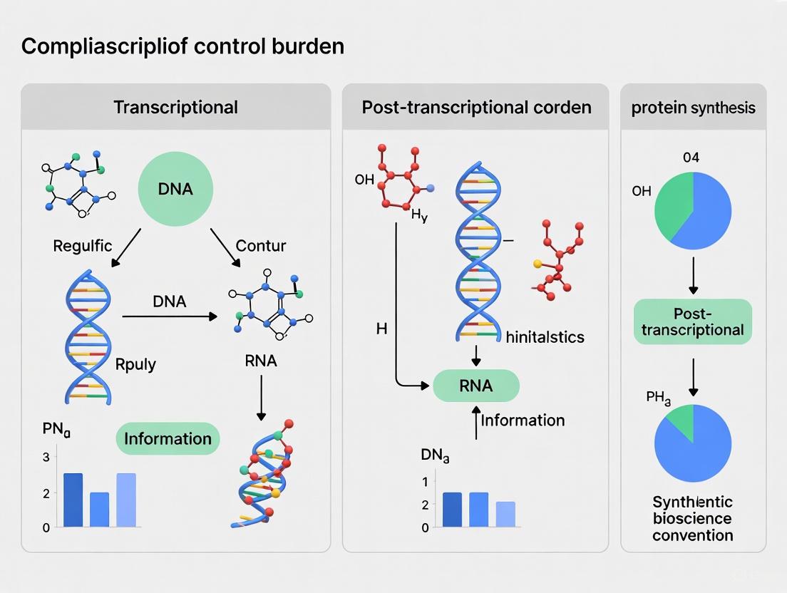
Transcriptional vs. Post-Transcriptional Control: Navigating Gene Expression Burden in Biomedical Research
This article provides a comprehensive comparison of transcriptional and post-transcriptional control mechanisms, with a specific focus on understanding and mitigating the cellular burden associated with gene expression.
Transcriptional vs. Post-Transcriptional Control: Navigating Gene Expression Burden in Biomedical Research
Abstract
This article provides a comprehensive comparison of transcriptional and post-transcriptional control mechanisms, with a specific focus on understanding and mitigating the cellular burden associated with gene expression. Tailored for researchers, scientists, and drug development professionals, it explores the foundational principles of resource competition in synthetic circuits, methodologies for monitoring burden, and strategies for circuit optimization. The scope includes practical troubleshooting for predictable outcomes in therapeutic development and a comparative analysis validating the distinct advantages and collaborative roles of each regulatory tier in reducing load and enhancing expression fidelity. The synthesis offers critical insights for designing more reliable genetic constructs and therapies.
The Cellular Battle for Resources: Foundations of Gene Expression Burden
Defining Transcriptional and Post-Transcriptional Control
Gene expression is a tightly orchestrated process fundamental to all biological systems, from development to disease. Transcriptional and post-transcriptional control represent two pivotal regulatory layers that determine the timing, location, and abundance of gene products. While transcriptional regulation governs the initial decision of whether a gene is transcribed, post-transcriptional regulation fine-tunes this output by controlling the fate and functionality of the resulting RNA transcripts. Understanding the distinct mechanisms, experimental approaches, and functional outcomes of these processes is crucial for researchers and drug development professionals aiming to manipulate gene expression for therapeutic purposes. This guide provides a comparative analysis of these regulatory domains, supported by current experimental data and methodologies.
Defining the Regulatory Layers
Transcriptional Control
Transcriptional control is the primary level of gene regulation, determining whether a gene is activated or silenced by directing the initiation of RNA synthesis from DNA. This process involves the complex interplay between transcription factors (TFs), regulatory DNA sequences, and the basal transcriptional machinery.
- Core Mechanism: The binding of transcription factors to specific promoter and enhancer sequences recruits or activates RNA polymerase II (Pol II), initiating transcription. The combinatorial interaction of TFs allows for precise control based on cellular context and signals.
- Key Regulators: Transcription factors are proteins that recognize specific DNA sequences. A single TF can act as both an activator and repressor depending on context, a phenomenon known as duality or nonmonotonicity. This switch can be tuned merely by changes in TF-DNA binding affinity, without requiring different coregulatory proteins [1].
- Efficiency Mechanisms: Many eukaryotic TFs contain intrinsically disordered regions (IDRs) that enhance their binding affinity and search efficiency for target DNA sites. The IDR acts as a flexible polymeric tail, increasing the area of effective interaction between the TF and DNA, thereby stabilizing TF-DNA interactions and speeding up the target location process [2].
Post-Transcriptional Control
Post-transcriptional control encompasses all regulatory events that occur after a primary RNA transcript has been synthesized, influencing its processing, stability, localization, and translation. This layer provides a faster, more reversible means of adjusting gene expression without altering the transcriptional rate.
- Scope of Regulation: This control layer includes RNA splicing, 3' end processing, modification, editing, transport, translation, and degradation.
- Key Processes: Recent research highlights several crucial post-transcriptional mechanisms:
- Post-transcriptional splicing: Up to 40% of mammalian introns are retained after transcription termination and subsequently removed while transcripts remain chromatin-associated. This delayed splicing serves as a key regulatory step during development, stress response, and disease progression [3].
- 3'UTR-mediated regulation: The 3' untranslated region serves as a major hub for regulatory information, influencing RNA stability and translational efficiency. Massively parallel reporter assays have revealed an "unexpectedly large role for 3'UTR-specified translational control" [4].
- RNA modifications: Processes such as methylation (m6A) and pseudouridylation affect transcript fate, with mitochondrial mRNAs undergoing extensive post-transcriptional modifications that determine their stability and translation efficiency [5].
Table 1: Core Characteristics of Transcriptional and Post-Transcriptional Control
| Feature | Transcriptional Control | Post-Transcriptional Control |
|---|---|---|
| Primary Function | Initiate or repress RNA synthesis from DNA | Process, stabilize, or degrade RNA transcripts |
| Key Regulators | Transcription factors, chromatin modifiers [6] | RNA-binding proteins, miRNAs, RNA modification enzymes [4] [3] [5] |
| Speed of Response | Relatively slow (minutes to hours) | Rapid (seconds to minutes) |
| Energy Requirement | Can occur at equilibrium [1] | Often requires energy expenditure (nonequilibrium) [1] [3] |
| Main Regulatory Elements | Promoters, enhancers, insulators | 3'UTRs, 5'UTRs, coding sequences [4] |
Key Experimental Approaches and Data
Advancements in genomic technologies have provided unprecedented insights into both transcriptional and post-transcriptional regulatory mechanisms, enabling their systematic comparison.
Investigating Transcriptional Control
The study of transcription has been revolutionized by computational models and high-throughput assays that can predict and measure regulatory activity across diverse cellular contexts.
- Foundation Models: The General Expression Transformer (GET) is an interpretable foundation model that predicts gene expression from chromatin accessibility data and sequence information across 213 human fetal and adult cell types. GET achieves "experimental-level accuracy" in predicting gene expression even in unseen cell types (Pearson correlation = 0.94), outperforming previous models that lacked generalizability [6].
- Reporter Assays: Lentivirus-based massively parallel reporter assays (lentiMPRAs) enable high-throughput testing of regulatory sequences. GET demonstrated superior performance in zero-shot prediction of lentiMPRA readouts (Pearson's r = 0.55) compared to Enformer (r = 0.44), despite not being trained on lentiMPRA data [6].
- Biophysical Modeling: Mathematical models exploring TF behavior reveal that the switch between activation and repression can be tuned by TF-DNA binding affinity alone. This "incoherent" regulation, where a TF simultaneously favors and hinders transcription, can produce nonmonotonic responses only under nonequilibrium conditions that require energy dissipation [1].
Investigating Post-Transcriptional Control
Post-transcriptional regulation requires specialized methodologies that capture RNA processing, modification, and translational efficiency beyond mere transcript abundance.
- Polysome Profiling: By sequencing both polysome-bound and total RNAs, researchers can identify genes subject to post-transcriptional regulation during critical processes like germ layer commitment in embryonic development. This approach revealed "substantial post-transcriptional modulation" during lineage commitment, with the translatome capturing regulatory nuances overlooked by transcriptome analysis alone [7].
- Direct RNA Sequencing: Nanopore-based direct RNA sequencing enables the detection of native RNA modifications, poly(A) tail lengths, and alternative splicing isoforms without cDNA conversion. Application to peanut pod development identified 14,627 new transcripts and revealed dynamic changes in poly(A) tail length and alternative polyadenylation site usage across developmental stages [8].
- Massively Parallel Reporter Assays for 3'UTRs: Specialized MPRAs evaluating >1,400 full-length human 3'UTRs have quantified their impact on RNA abundance, stability, translational regulation, and protein output. These studies demonstrate that "much of 3'UTR-encoded regulation is mediated by concerted regulation of translation plus decay" [4].
Table 2: Comparative Experimental Data for Transcriptional and Post-Transcriptional Regulation
| Experimental Approach | Key Finding for Transcriptional Control | Key Finding for Post-Transcriptional Control |
|---|---|---|
| Genome-wide Profiling | GET model predicts expression with r=0.94 in unseen astrocytes [6] | Polysome profiling reveals substantial modulation during germ layer commitment [7] |
| Reporter Assays | GET zero-shot lentiMPRA prediction: r=0.55 [6] | 3'UTR MPRA shows large role for translational control [4] |
| Single-molecule/Long-read Analysis | N/A | DRS identifies 40% of mammalian introns undergo post-transcriptional splicing [3] |
| Functional Perturbation | Changing TF-DNA binding affinity can tune activation vs. repression [1] | β-catenin directly binds ER-β transcript to modulate splicing [9] |
Integrated Regulatory Networks
Gene expression control rarely operates through isolated mechanisms; instead, transcriptional and post-transcriptional regulations form interconnected networks that ensure precise spatiotemporal control of gene expression.
Coordination Across Regulatory Layers
Several research findings highlight the sophisticated coordination between transcription and post-transcriptional processing:
- Splicing-Transcription Coupling: While traditionally viewed as co-transcriptional, recent studies using long-read sequencing of chromatin-associated RNA show that "post-transcriptional splicing occurs for one-third of human introns and >75% of transcripts" [3]. This delayed splicing serves regulatory functions, with partially spliced transcripts being nuclear-retained until proper signals trigger completion of splicing.
- Promoter-Post-transcriptional Interplay: Comparison of 3'UTR regulation under control of two dissimilar promoters revealed "promoter-associated differences in post-transcriptional regulation for certain 3'UTRs" [4], indicating that the transcriptional start site can influence downstream RNA processing.
- Multi-layer Regulation in Development: Studies in early cell fate commitment demonstrate coordinated regulation across levels, with polymer physics models helping to describe "how chromatin-encoded information is transduced from localized transcriptional events to global gene expression patterns" [10].
Case Study: β-Catenin as a Dual-Regulator
β-catenin exemplifies the blurring of boundaries between regulatory layers. While well-established as a transcriptional co-activator in Wnt signaling, accumulating evidence reveals extensive post-transcriptional functions:
- Traditional Roles: β-catenin serves as the central mediator of canonical Wnt signaling, translocating to the nucleus to activate target genes like MYC and CCND1 upon pathway activation [9].
- Post-transcriptional Functions: β-catenin associates with splicing regulatory RNA-binding proteins (e.g., FUS and TLS) and can modulate splice site selection. It directly binds the ER-β transcript and promotes expression of a novel ER-β Δ5-6 variant with dominant-negative activity [9].
- Implications: This bifunctionality suggests that "β-catenin gene expression [is controlled] through both transcriptional and post-transcriptional processes," complicating traditional genetic approaches that target only one regulatory layer [9].
The Scientist's Toolkit: Essential Research Reagents
Table 3: Key Research Reagents for Studying Transcriptional and Post-Transcriptional Regulation
| Reagent/Technology | Primary Application | Function in Research |
|---|---|---|
| GET (General Expression Transformer) Model [6] | Transcriptional prediction | Foundation model predicting gene expression from chromatin accessibility and sequence |
| Polysome Profiling [7] | Post-transcriptional analysis | Separates and sequences ribosome-bound mRNAs to assess translational efficiency |
| LentiMPRA (Lentiviral MPRAs) [6] | Both regulatory layers | High-throughput testing of regulatory sequence activity in chromatin context |
| Direct RNA Sequencing (Nanopore) [8] | Post-transcriptional analysis | Detects native RNA modifications, poly(A) tail lengths, and splicing isoforms |
| RNA-centric Biophysical Models [1] | Theoretical framework | Mathematical models distinguishing equilibrium vs. nonequilibrium regulation |
| Chromatin-associated RNA-seq [3] | Splicing-transcription coupling | Identifies post-transcriptionally spliced introns and nuclear-retained transcripts |
Transcriptional and post-transcriptional control represent complementary, interconnected layers of gene regulation that operate across different timescales and through distinct molecular mechanisms. Transcriptional regulation, governed by transcription factors and chromatin environment, provides the foundational decision-making layer for gene activation. Post-transcriptional mechanisms, including splicing, 3'UTR-mediated control, and RNA modification, offer refined, dynamic control over the final gene output. The emerging paradigm recognizes that these processes form integrated networks rather than linear pathways, with factors like β-catenin operating across multiple regulatory layers. For researchers and drug development professionals, this complexity presents both challenges and opportunities—while complicating simple interventions, it provides multiple potential targets for precise therapeutic manipulation of gene expression. Future research will continue to elucidate the sophisticated crosstalk between these regulatory domains across different biological contexts and disease states.
A central challenge in synthetic biology is context dependence, where genetic circuits do not function reliably when removed from their original design context. A major culprit is resource competition, which arises when multiple genetic modules within an engineered cell compete for a finite pool of shared cellular resources [11] [12]. Critical shared resources include RNA polymerase (RNAP) for transcription and the ribosome for translation, both required for gene expression [12]. When one module increases its resource consumption, it inevitably deprives other modules, leading to unintended coupling and performance degradation [13] [12]. This review synthesizes recent experimental evidence quantifying resource competition in mammalian cells and compares strategies to mitigate its effects, providing a guide for researchers developing robust synthetic gene circuits and biotherapeutics.
Quantitative Evidence of Resource Competition in Mammalian Cells
Measuring the Resource Footprint of Genetic Components
A foundational 2023 study established a framework to directly quantify the resource load imposed by various genetic components in mammalian cells (HEK293T and CHO-K1) [13]. The experimental system used a fluorescence-based capacity monitor (a CMV promoter-driven mKATE cassette) co-transfected with a library of modular test plasmids. As the test plasmid consumes more cellular resources for its own expression, fewer resources remain available for the capacity monitor, resulting in decreased mKATE fluorescence, which serves as a proxy for resource availability [13].
Table 1: Impact of Genetic Components on Resource Competition and Output [13]
| Genetic Component | Parameter Varied | Effect on Test Plasmid Output | Effect on Capacity Monitor | Inference on Limiting Resources |
|---|---|---|---|---|
| Promoter | Strength (7 constitutive, 1 inducible) | Stronger promoters increased output. | Stronger promoters caused greater suppression. | Transcriptional resources are a primary bottleneck. |
| PolyA Signal | Sequence (6 different variants) | Output varied significantly (up to ~3-fold). | Specific polyAs (e.g., PGKpA, SV40pA_rv) caused strong suppression. | Impacts mRNA stability/termination; effect is promoter and cell-line dependent. |
| Kozak Sequence | Translational efficiency (3 variants) | Moderate effect on output (~1.5-fold variation). | Minimal impact except with very strong promoters. | Translational resources are less limiting than transcriptional ones. |
| Inducible System | Doxycycline concentration | Higher induction increased output. | Higher induction caused greater suppression. | Confirms titratable resource consumption. |
Transcriptional vs. Translational Resource Limitations
A critical finding from these quantitative studies is a fundamental difference between prokaryotes and mammals regarding the primary source of resource competition. In bacteria, competition for translational resources (ribosomes) is typically dominant [11] [12]. In contrast, evidence from mammalian cells indicates that competition for transcriptional resources (RNA polymerases and associated factors) is the more significant bottleneck [13] [11].
This conclusion is supported by several key observations:
- Promoter strength is a major driver of resource load, with stronger promoters causing a more severe reduction in capacity monitor expression [13].
- Variations in the Kozak sequence, which directly modulates translation initiation efficiency, had a minimal impact on the capacity monitor unless paired with a very strong promoter [13].
- Experiments involving mRNA co-transfection (bypassing transcription) confirmed that differences in translational efficiency did not alter the expression of a co-delivered monitor mRNA, in stark contrast to the effects observed at the DNA level [13].
Diagram 1: Resource Competition arises from modules sharing a finite resource pool. Consumption by one module depletes availability for others, creating unintended coupling.
Comparative Analysis of Mitigation Strategies
Local vs. Global Control Strategies
Strategies to mitigate resource competition fall into two broad categories: local control, where individual modules are engineered to be robust to resource fluctuations, and global control, where the shared resource pool itself is regulated [12].
Table 2: Comparison of Strategies to Mitigate Resource Competition [14] [11] [12]
| Strategy | Mechanism | Key Example | Pros & Cons |
|---|---|---|---|
| Local Control: Incoherent Feed-Forward Loop (iFFL) | Uses microRNAs or endoribonucleases to buffer output against resource fluctuations. | miRNA-iFFL redistributes translational resources, enhancing operational capacity [14]. | Pro: Module-specific, portable.Con: Adds genetic complexity and its own resource load. |
| Local Control: Resource-Aware Parts | Selecting parts with optimal performance-to-footprint ratio. | UB promoter showed high output with lower resource load in CHO-K1 cells [13]. | Pro: Simple, uses characterized parts.Con: Performance is cell-type and context dependent. |
| Global Control: Small Molecule Treatment | Pharmacologically reprograms host cell state to free up resources. | DECCODE algorithm identified Filgotinib to enhance transgene expression [14]. | Pro: Genetically non-invasive, rapid effect.Con: Host-dependent response, off-target effects possible. |
| Global Control: Orthogonal Expression Systems | Uses dedicated, non-competing resources from other organisms. | Bacterial transcription factors in plant circuits reduce host cross-talk [15]. | Pro: High insulation from host.Con: Limited availability and capacity in mammalian cells. |
Small-Molecule Intervention: A Non-Genetic Global Approach
A 2025 study demonstrated a novel global control strategy using small molecules to mimic the effect of resource-optimizing genetic circuits [14]. Researchers used the DECCODE (Drug Enhanced Cell COnversion using Differential Expression) algorithm to match the transcriptomic signature of cells hosting an efficient miRNA-iFFL circuit against a database of drug-induced profiles. This unbiased approach identified several drugs, including Filgotinib and Ruxolitinib, that boosted transgene expression by 10-50% in various experimental settings, including viral transduction [14]. This suggests these compounds induce a host cell state that reallocates internal resources towards heterologous gene expression, providing a powerful, non-genetic tool for enhancing bioproduction.
Detailed Experimental Protocols
Protocol: Quantifying Resource Load with a Capacity Monitor
This protocol is adapted from the resource-aware construct design study [13].
Objective: To quantify the resource footprint of a genetic component (e.g., a promoter) in mammalian cells.
Key Reagents:
- Capacity Monitor Plasmid: Constitutively expresses a fluorescent reporter (e.g., mKATE2) under a strong promoter like CMV.
- Modular Test Plasmid: A vector where the component of interest drives an expression cassette for a different fluorescent reporter (e.g., EGFP).
- Cell Lines: Adherent mammalian cells such as HEK293T or CHO-K1.
Workflow:
- Co-transfection: Seed cells in a multi-well plate and co-transfect with a fixed amount of the capacity monitor plasmid and the test plasmid (or an empty vector control).
- Expression Analysis: After 24-48 hours, analyze the cells using flow cytometry to measure the fluorescence intensities of both the test plasmid reporter (EGFP) and the capacity monitor reporter (mKATE2) at the single-cell level.
- Data Interpretation: For a given test plasmid, the EGFP signal indicates its output, while the mKATE2 signal inversely correlates with the resource load. A design with a low resource footprint will show high EGFP without severely suppressing mKATE2.
Diagram 2: Capacity Monitor Workflow measures the resource load of a test plasmid by its impact on a co-transfected reporter.
Protocol: Computational Identification of Resource-Boosting Drugs
This protocol is based on the DECCODE method for identifying small molecules that enhance gene expression [14].
Objective: To find small molecules that increase cellular capacity for synthetic circuit expression without genetic modifications.
Workflow:
- Generate Transcriptomic Signature:
- Engineer cells with a resource-efficient circuit (e.g., a miRNA-iFFL) and a control circuit (Open Loop).
- Perform RNA-sequencing on sorted, transfected cell populations.
- Conduct differential expression analysis to define a transcriptional model representing the "high-capacity" state.
- Computational Drug Matching with DECCODE:
- Convert the differential expression profile into a pathway expression profile using Gene Ontology terms.
- Compare this pathway profile against the LINCS database containing thousands of drug-induced transcriptomic signatures.
- Rank compounds based on the similarity of their induced signatures to the target "high-capacity" signature.
- Experimental Validation:
- Treat engineered cells expressing a reporter construct with the top-ranked drugs.
- Quantify the enhancement of reporter expression (e.g., via fluorescence) across different cell lines and delivery methods (e.g., transfection, viral transduction).
The Scientist's Toolkit: Essential Research Reagents
Table 3: Key Research Reagent Solutions for Studying Resource Competition
| Reagent / Solution | Function in Research | Specific Examples & Notes |
|---|---|---|
| Fluorescent Reporter Plasmids | Quantifying gene expression output and resource load in live cells. | mKATE2, EGFP, mCherry. Use in pairs for test and monitor cassettes [13]. |
| Modular Cloning System | Rapid assembly of genetic parts to test different combinations. | Golden Gate or Gibson Assembly systems for swapping promoters, polyAs, etc. [13]. |
| Inducible Expression Systems | Titratable control over gene expression to study dose-dependent resource use. | TET-ON system (doxycycline-inducible) [13]; NF-κB or E'-box inducible systems [16]. |
| Small Molecule Libraries & Databases | Screening for pharmacological modulators of cellular capacity. | LINCS database for transcriptomic signatures; FDA-approved JAK inhibitors (Filgotinib, Ruxolitinib) [14]. |
| Orthogonal Regulatory Parts | Insulating synthetic circuits from host cross-talk. | Bacterial transcription factors (e.g., in plant circuits) [15]; orthogonal RNA polymerases. |
Resource competition presents a significant barrier to the reliable engineering of mammalian cells, with transcriptional resources identified as a primary limiting factor. Evidence from quantitative studies enables a resource-aware design paradigm, where genetic components are selected not only for strength but also for their efficiency and minimal footprint. Mitigation strategies are diverse, ranging from local solutions like iFFLs and optimized parts to global approaches including small molecule treatments identified via computational transcriptomic matching. The choice of strategy depends on the application: local control offers precision for defined circuits, while global control via small molecules presents a powerful, rapid option for boosting bioproduction pipelines. As the field progresses, integrating these resource-aware principles and tools will be crucial for developing next-generation, robust synthetic biology applications in biotherapeutics and beyond.
The engineering of synthetic biological systems is fundamentally constrained by the host cell's finite resource pools. Competition for these shared resources, specifically the transcriptional machinery (e.g., RNA polymerase) and the translational machinery (e.g., ribosomes, tRNAs), introduces significant coupling between genetic modules, leading to unpredictable performance and burden on the host. The primary limiting factor, however, is not universal; it is highly dependent on the cellular context. The table below summarizes the core distinctions between these two limiting resource pools.
Table 1: Core Distinctions Between Transcriptional and Translational Resource Pools
| Feature | Transcriptional Resource Pool | Translational Resource Pool |
|---|---|---|
| Primary Limiting Factor | RNA Polymerase (RNAP) and associated factors [11] | Ribosomes and transfer RNAs (tRNAs) [11] [17] |
| Dominant Context | Mammalian cells [11] [18] | Bacterial cells (e.g., E. coli) [11] [19] |
| Main Consequence of Competition | Sequestration of RNAP reduces mRNA production for all genes, both exogenous and endogenous [18]. | Sequestration of ribosomes and depletion of charged tRNAs reduce global protein synthesis and slow growth [19] [17]. |
| Key Experimental Evidence | Negative correlation in mRNA levels of co-expressed genes; reduction in endogenous mRNA levels upon transfection [18]. | Inverse linear relationship between heterologous protein expression and host cell growth rate [19] [17]. |
| Impact on Circuit Behavior | Can lead to non-monotonic dose-response curves and unintended coupling between independent modules [18]. | Drives evolutionary instability, amplifies gene expression noise, and can create emergent bistability [19] [20]. |
Mechanistic Insights and System-Level Impacts
Resource Competition as a Source of Emergent Behavior
The fight for limited resources does more than just slow growth; it can fundamentally alter the qualitative behavior of synthetic circuits. In bacteria, where ribosomes are a primary bottleneck, competition for these translational resources can create unintended feedback loops. For instance, in a two-gene inhibition cascade, resource competition can generate a double-negative feedback loop, leading to "winner-takes-all" bistability and stochastic switching between states, thereby amplifying gene expression noise [20]. This means a circuit designed for graded expression can unpredictably switch between high and low output states due solely to competition for ribosomes.
Furthermore, the consumption of translational resources for heterologous protein expression reduces the capacity for native protein production, imposing a growth burden [19]. This initiates a multiscale growth feedback loop: burden reduces growth rate, which in turn alters the dilution rate of circuit components and can change the physiological state of the cell, further impacting circuit output [11]. This interplay can lead to the emergence or loss of stable states (e.g., bistability, tristability) in circuits like self-activation switches, contravening their intended design principles [11].
The Critical Role of Codon Usage in Translational Burden
Within the realm of translational limitation, the specific codon usage of an exogenous gene is a critical modulator of burden. Translation elongation is not a uniform process; the cellular pool of charged tRNAs is finite and biased towards optimal codons. Simulations and experiments in E. coli demonstrate that the relationship between codon usage, protein yield, and burden is nuanced [17].
A key finding is that burden depends not only on the exogenous gene's codon usage but also on how well it matches the host's overall tRNA abundance. Simulations predict an "overoptimization" domain, where maximizing optimal codon usage beyond the host's tRNA capacity can paradoxically increase burden and reduce yield [17]. This occurs because an over-optimized sequence can skew the demand for tRNAs away from the optimal supply, slowing ribosome elongation globally. Experimental data expressing sfGFP and mCherry2 with varying codon optimization levels confirm that the slope of the burden-yield relationship is modulated by codon usage [17].
Key Experimental Evidence and Methodologies
Demonstrating Resource Competition in Mammalian Cells
A seminal study in mammalian cells (HEK293T and H1299) provided direct evidence of resource competition by co-transfecting two constitutively expressed fluorescent proteins (mCitrine and mRuby3) in varying molar ratios [18].
Experimental Workflow:
- Design: Two fluorescent reporter genes (mCitrine, mRuby3) under identical (EF1α) or different (CMV, PGK) promoters were cloned onto separate plasmids.
- Transfection: Cells were co-transfected with these plasmids in different molar ratios (from 1:4 to 4:1) and at two total DNA amounts (50 ng and 500 ng).
- Measurement: Protein expression was quantified via flow cytometry, and mRNA levels were quantified using RT-qPCR. Endogenous gene expression was also measured in sorted transfected cells.
Key Findings:
- Negative Correlation: At high total DNA (500 ng), a strong negative correlation was observed between mCitrine and mRuby3 fluorescence, indicating direct competition.
- mRNA-Level Competition: Increasing the plasmid ratio for one gene led to a decrease in the mRNA level of the other, proving competition for transcriptional resources [18].
- Endogenous Burden: Expression of endogenous genes (e.g., CyCA2, eIF4E, GAPDH) was reduced in transfected cells compared to non-transfected controls, showing that synthetic circuits also compete with native cellular processes [18].
The diagram below visualizes this experimental design and its core finding.
Quantifying Translational Burden and the Effect of Codon Usage
The quantitative link between translational resource consumption, codon usage, and host fitness has been rigorously characterized in bacterial systems [17].
Experimental Workflow:
- Construct Design: Fluorescent protein genes (sfGFP, mCherry2) were recoded to create variants with Fraction of Optimal Codons (FOP) ranging from 10% to 90%.
- Expression Tuning: Each variant was paired with one of five different Ribosome Binding Site (RBS) sequences to create a range of translation initiation strengths.
- Growth & Measurement: E. coli strains harboring these constructs were induced during exponential growth. The growth rate (OD) and fluorescence (protein yield) were tracked simultaneously.
Key Findings:
- Linear Burden-Yield Relationship: A negative linear relationship existed between protein production and host growth rate, regardless of codon optimization level.
- Codon Usage Modulates Burden: The slope of this burden-yield relationship was steepest for genes with codon usage that deviated significantly from the host's optimal profile.
- Validation of Overoptimization: For one protein (mCherry2), the 90% FOP variant showed lower yield and higher burden than the 75% FOP variant, confirming the predicted "overoptimization" effect where excessive optimal codon usage can be detrimental [17].
The Scientist's Toolkit: Essential Reagents and Methods
Table 2: Key Research Reagent Solutions for Studying Resource Pools
| Tool / Reagent | Function/Description | Application Context |
|---|---|---|
| Dual-Fluorescent Reporter System [18] | Two fluorescent proteins (e.g., mCitrine, mRuby3) on separate plasmids under constitutive or inducible promoters. | Directly visualizing resource competition by titrating plasmid ratios and measuring correlated expression outputs. |
| Codon-Recoded Gene Variants [17] | A library of the same protein-coding sequence synthesized with varying levels of codon optimization (e.g., 10%-90% FOP). | Disentangling the effects of codon usage and expression level on translational burden and protein yield. |
| Ribosome Profiling (Ribo-Seq) [21] [22] | High-throughput sequencing of ribosome-protected mRNA fragments to map the positions of actively translating ribosomes. | Genome-wide measurement of translation dynamics and efficiency (e.g., under burden) and discovery of novel translated open reading frames (ORFs). |
| SUnSET (Surface Sensing of Translation) Assay [21] | Immunodetection of puromycin-labeled nascent peptides to measure global protein synthesis rates. | A simple, cellular-level method to quantify changes in translational activity in response to genetic perturbations or burden. |
| Host-Aware Mathematical Models [11] [19] | ODE-based models integrating circuit expression, resource kinetics, and host growth. | Predicting emergent dynamics from circuit-host interactions (e.g., growth feedback, bistability) and in silico testing of burden-mitigation strategies. |
Visualizing an Integrated Experimental Workflow
The following diagram synthesizes key methodologies into a cohesive workflow for dissecting transcriptional and translational resource limitation.
In mammalian synthetic biology, the introduction of exogenous genetic circuits often leads to unanticipated functional failures and discrepancies between intended and actual performance. This phenomenon, termed gene expression burden, arises from competition for limited cellular resources between endogenous and synthetic genetic components [18]. Despite recent advances in circuit engineering, the design of genetic networks in mammalian cells remains painstakingly slow and fraught with inexplicable failures, creating significant challenges for basic research and therapeutic development [18].
The core issue lies in the poor predictability of gene expression in transfected cells, which stems from the dependence of synthetic circuits on limited host resources shared with endogenous pathways. This resource competition creates an indirect coupling between otherwise independent genes, leading to trade-offs in their expression levels and ultimately degrading cellular performance [18]. Understanding and mitigating these burden effects is crucial for advancing synthetic biology applications, including the engineering of recombinant protein-producing cells and the creation of novel cell-based therapies.
Table 1: Key Concepts in Gene Expression Burden
| Concept | Description | Impact on Circuit Performance |
|---|---|---|
| Transcriptional Burden | Competition for limited transcriptional resources (RNA polymerase, transcription factors) | Reduced mRNA levels for both endogenous and exogenous genes |
| Translational Burden | Competition for limited translational resources (ribosomes, tRNAs, amino acids) | Reduced protein output despite sufficient mRNA levels |
| Resource Coupling | Indirect interaction between independent genes sharing cellular resources | Negative correlation between expression levels of co-expressed genes |
| Burden Mitigation | Engineering strategies to minimize resource competition | Improved predictability and performance of synthetic circuits |
Experimental Evidence of Burden-Induced Coupling
Quantitative Demonstration of Resource Competition
Groundbreaking research has systematically investigated burden effects by designing genetic constructs that uncouple transcription and translation processes. In one key experiment, HEK293T cells were co-transfected with two constitutively expressed fluorescent proteins (mCitrine and mRuby3) driven by EF1α promoters in varying molar ratios [18]. The experimental setup involved transfecting cells with total plasmid amounts of 50 ng (low burden) versus 500 ng (high burden), then measuring fluorescence outputs to quantify expression coupling.
The results demonstrated that higher plasmid concentrations (500 ng) caused a dramatic drop in gene expression compared to lower concentrations (50 ng). Furthermore, fluorescence levels of mCitrine and mRuby3 showed strong negative correlation—as expression of one increased, the other decreased—with this effect being more severe at higher plasmid concentrations [18]. This coupling occurred despite the absence of direct regulatory connections between the two genes, providing direct evidence of resource-mediated coupling.
Similar findings were observed with different promoter combinations (CMV and PGK) and in multiple cell lines (HEK293T and H1299), confirming the generalizability of burden effects across different genetic contexts [18]. The use of a Doxycycline-repressed promoter system further demonstrated that increased repression of one transgene (X-tra) corresponded to increased expression of a co-transfected capacity monitor, establishing that burden effects can be dynamically modulated [18].
Distinct Transcriptional and Translational Limitations
To dissect the specific resource pools responsible for burden effects, researchers implemented specialized genetic circuits that selectively overload transcriptional or translational resources [18]. A self-cleaving hepatitis delta virus (HDV) ribozyme system was used to create transcripts that are rapidly degraded, overloading transcriptional resources without sequestering translational machinery.
Experiments measuring mRNA levels in cells expressing different ratios of X-tra to capacity monitor constructs revealed that as X-tra mRNA increased, capacity monitor mRNA levels decreased [18]. This demonstrated that shared transcriptional resources are indeed a limiting factor in mammalian synthetic gene co-expression. Additional studies showed that endogenous gene expression is also affected by heterologous genetic payloads, with transfected cells showing decreased expression of housekeeping genes like CyCA2, eIF4E, and GAPDH compared to non-transfected populations [18].
Table 2: Experimental Evidence of Gene Expression Burden
| Experimental Approach | Key Findings | Implications |
|---|---|---|
| Dual fluorescent protein co-expression | Negative correlation between expression of independent genes; dose-dependent effect | Direct evidence of resource competition |
| Inducible promoter systems | Increased repression of one gene enhances expression of another | Burden effects can be dynamically modulated |
| Transcriptional burden assessment | Increased X-tra mRNA reduces capacity monitor mRNA | Transcriptional resources are fundamentally limiting |
| Endogenous gene monitoring | Reduced expression of housekeeping genes in transfected cells | Burden affects core cellular functions |
| Ribozyme-mediated transcript degradation | Transcriptional overload without protein translation | Enables separation of transcriptional vs. translational burden |
Methodologies for Burden Characterization
Experimental Workflows for Burden Quantification
The standard approach for quantifying burden effects involves a co-transfection system with carefully calibrated controls. The foundational protocol consists of the following steps [18]:
Construct Design: Develop a "sensor" gene (capacity monitor) with consistent expression characteristics and a "load" gene (X-tra) with tunable expression levels.
Transfection Setup: Co-transfect cells with fixed amounts of capacity monitor plasmid and varying amounts of X-tra plasmid across a concentration gradient.
Expression Measurement: Quantify output signals (fluorescence, luminescence) for both genes using flow cytometry, fluorescence microscopy, or plate readers.
Data Normalization: Normalize expression levels to control transfections containing only the capacity monitor construct.
Correlation Analysis: Calculate correlation coefficients between the expression levels of co-transfected genes across the concentration gradient.
For transcriptional burden assessment specifically, researchers should include mRNA quantification via RT-qPCR to distinguish transcriptional from post-transcriptional effects [18]. The self-cleaving HDV ribozyme system provides a method to isolate transcriptional burden by generating unstable transcripts that consume transcriptional resources without producing functional proteins [18].
Mathematical Modeling of Resource Competition
A resource-aware mathematical model has been developed to predict and explain burden effects [18]. This framework replaces standard reaction rates with effective rates that account for resource sharing, following principles originally used to describe competitive enzyme inhibition. The model successfully recapitulates non-monotonic dose-response behaviors observed in simple inducible expression systems and provides a quantitative foundation for predicting circuit performance under resource constraints.
The modeling approach incorporates:
- Resource pools for transcription and translation machinery
- Competitive binding kinetics between genetic constructs
- Effective rate constants that decrease with increasing resource demand
- Coupling terms that connect expression of independent genes through shared resources
This computational framework enables context-aware prediction of synthetic circuit performance and provides guidance for burden-mitigating circuit designs [18].
Mitigation Strategies for Expression Burden
Circuit Engineering Solutions
Research has identified several effective strategies for mitigating burden effects in synthetic genetic circuits. Guided by mathematical modeling, researchers have engineered natural and synthetic miRNA-based incoherent feedforward loop (iFFL) circuits that significantly reduce gene expression burden [18]. These circuits function by detecting and compensating for resource limitations before they disrupt circuit performance.
The iFFL topology is particularly effective for burden mitigation because it creates a counterbalancing effect—as burden increases and potentially reduces output gene expression, the iFFL simultaneously reduces demand for resources, thereby maintaining more consistent output levels [18]. This approach has been shown to rescue expression levels of genes of interest despite changes in available cellular resources due to transgene loading effects.
Implementation of these circuits features the use of endogenous miRNAs as elementary components, creating a versatile hybrid design that enables burden mitigation across different cell lines with minimal resource requirements [18]. Both RNA-binding proteins (RBPs) and microRNAs (miRNAs) have demonstrated effectiveness in reallocating resources, making them valuable tools for burden-mitigation circuits.
Practical Implementation Guidelines
Based on experimental findings, researchers can employ several practical approaches to minimize burden effects:
Titration-Based Optimization: Systematically vary plasmid ratios and total amounts to identify working ranges where burden effects are minimized.
Promoter Selection: Use promoters with appropriate strengths for specific applications, avoiding excessively strong promoters when not necessary.
iFFL Integration: Implement miRNA-based iFFL circuits to maintain consistent expression across varying resource conditions.
Resource Balancing: Distribute genetic load across transcriptional and translational resources by careful circuit design.
Monitoring Systems: Include burden sensors in experimental designs to detect and quantify resource competition effects.
These strategies collectively enable more predictable synthetic circuit performance and reduce the design-build-test-learn iterations that traditionally plague mammalian synthetic biology [18].
Research Reagent Solutions
Table 3: Essential Research Reagents for Burden Studies
| Reagent/Category | Specific Examples | Function in Burden Research |
|---|---|---|
| Fluorescent Reporters | mCitrine, mRuby3, EGFP | Capacity monitors for quantifying gene expression and resource competition |
| Inducible Systems | Doxycycline-repressed promoters | Enable dynamic control of genetic load to study burden effects |
| Burden Mitigation Components | miRNA-based iFFL circuits, RNA-binding proteins | Reallocate cellular resources to maintain circuit function |
| Transcriptional Burden Tools | HDV ribozyme systems | Selective overload of transcriptional resources without translation |
| Mathematical Modeling Frameworks | Resource-aware models | Predict circuit performance under resource constraints |
| Cell Lines | HEK293T, H1299 | Model systems for characterizing burden across cellular contexts |
Visualizing Burden Concepts and Experimental Approaches
The following diagrams illustrate key concepts and methodological approaches in burden research, created using DOT language and compliant with the specified formatting guidelines.
Diagram 1: Resource competition leading to gene coupling.
Diagram 2: Experimental workflow for burden quantification.
Gene expression burden represents a fundamental challenge in synthetic biology, creating unintended coupling between independent genes and degrading circuit performance through competition for limited cellular resources. The experimental evidence clearly demonstrates that both transcriptional and translational resources can become limiting factors, affecting both exogenous and endogenous gene expression.
The development of resource-aware mathematical models and burden-mitigation strategies, particularly miRNA-based iFFL circuits, provides powerful approaches to enhance the predictability and reliability of synthetic genetic circuits in mammalian cells. By acknowledging and explicitly addressing resource constraints, researchers can advance toward more rational synthetic construct design, accelerating progress in basic research and therapeutic development.
As the field continues to evolve, integrating burden assessment into standard genetic engineering workflows will be essential for achieving robust, predictable circuit behavior across diverse cellular contexts and application domains.
Impact on Endogenous Gene Expression and Cellular Health
The advancement of gene and cell therapies hinges on the precise control of therapeutic gene expression. Unregulated production of therapeutic genes can lead to decreased clinical utility and severe complications, underscoring the critical importance of controlled gene expression systems [23] [24]. As therapies become more sophisticated, researchers face the fundamental challenge of balancing therapeutic efficacy with cellular health. The introduction of synthetic genetic circuits inevitably imposes a metabolic burden on host cells, potentially disrupting native gene expression networks and compromising cellular function [25]. This comparative guide examines three pioneering gene regulation technologies, evaluating their impact on endogenous gene expression and cellular homeostasis through direct experimental comparison. Understanding these interactions is paramount for developing next-generation therapies that maintain both therapeutic potency and cellular viability.
Comparative Analysis of Gene Regulation Technologies
The table below provides a systematic comparison of three distinct gene regulation approaches based on current research findings.
Table 1: Comparison of Gene Regulation Technologies and Their Cellular Impact
| Technology | Regulatory Level | Key Mechanism | Therapeutic Context | Impact on Endogenous Genes | Cellular Burden Evidence |
|---|---|---|---|---|---|
| Endogenous Promoter Knock-in [26] | Transcriptional | CRISPR-HDR to place transgenes under native promoters (e.g., NR4A2, RGS16) | Armoured CAR-T cells (IL-12, IL-2 delivery) | Leverages native gene regulation; minimal disruption when targeted to safe loci | Maintained T-cell polyfunctionality; no overt toxicity in murine models |
| Csr Network Rewiring [25] | Post-transcriptional | Engineered RNA-protein interactions using native Csr system | Bacterial synthetic circuits; multi-layer logic gates | Operates within native regulatory network; potentially lower metabolic burden | No growth defects observed upon induction; utilizes conserved native machinery |
| TriLoS System [27] | Multi-layered (transcriptional & translational) | Engineered tristate logic gates mimicking electronic circuits | Mammalian cell computation; metabolic disease therapy | Modular design reduces unintended crosstalk; predictable behavior | Enabled complex computation (full adder) without noted toxicity; stable long-term function |
Experimental Protocols and Methodologies
Endogenous Promoter-Driven Expression in CAR-T Cells
Key Experimental Protocol [26]:
- CRISPR Knock-in Strategy: Utilizing homology-directed repair (HDR) to insert transgenes (e.g., GFP, cytokine genes) immediately downstream of start codons in endogenous genes with tumor-restricted expression patterns.
- Promoter Screening: RNA-seq comparison of CD8+ CAR-T cells isolated from tumor versus spleen tissues identified NR4A2 and RGS16 as optimal promoters with high tumor-specific expression.
- Functional Validation: Edited CAR-T cells were stimulated with target tumor cells followed by flow cytometry analysis of GFP expression. In vivo testing employed syngeneic and xenogeneic tumor models with monitoring of both antitumor efficacy and systemic toxicity.
- Key Reagents: Primary murine T cells, homologous repair templates, CRISPR-Cas9 system, tumor cell lines (AT-3-ova, OVCAR-3).
Bacterial Post-Transcriptional Control System
Key Experimental Protocol [25]:
- Circuit Construction: The 5' UTR from the CsrA-repressed glgC transcript (-61 to -1 relative to translation start site) was fused to reporter genes (gfpmut3) under a weak constitutive promoter (PCon).
- Induction System: Wild-type CsrB sRNA was placed under IPTG-inducible PLlacO promoter to sequester CsrA and activate translation.
- Validation Experiments: The system was tested in csrA::kan strains and with mutated CsrA binding sites to confirm mechanism specificity. Fluorescence was measured over time with IPTG titration.
- Key Reagents: E. coli K-12 MG1655 strains, csrA::kan mutant, IPTG, glgC 5' UTR scaffolds with engineered GGA motifs.
Mammalian Tristate Logic System
Key Experimental Protocol [27]:
- Layered Circuit Design: Transcription-level control using vanillic acid (VA)-responsive systems upstream of translation-level control using grazoprevir (Gra)-regulated switches.
- Gate Assembly: Four fundamental tristate logic units (BUFIF1, NOTIF1, BUFIF0, NOTIF0) were constructed by combining transcriptional and translational regulators.
- Complex Circuit Implementation: Multi-input systems incorporated Cre recombinase as a third input signal, enabling higher-order computation through modular assembly.
- Key Reagents: HEK-293 cells, vanillic acid, grazoprevir, Cre recombinase, customized gene constructs with specialized 5' and 3' UTR elements.
Signaling Pathways and Regulatory Mechanisms
Endogenous Gene Rewiring for Tumor-Restricted Payload Delivery
Diagram 1: Endogenous promoter system for tumor immunotherapy.
Csr Network Post-Transcriptional Regulation
Diagram 2: Engineered Csr network for bacterial gene regulation.
Mammalian Tristate Logic Control System
Diagram 3: Mammalian tristate logic system for therapeutic control.
The Scientist's Toolkit: Essential Research Reagents
Table 2: Key Research Reagents for Gene Regulation Studies
| Reagent/Category | Specific Examples | Function/Purpose |
|---|---|---|
| Gene Editing Tools | CRISPR-Cas9 system, Homology-directed repair (HDR) templates | Precise genomic integration of regulatory elements [26] |
| Reporter Systems | GFP (mut3 variants), Secreted cytokines (IL-12, IL-2, TNF) | Monitoring gene expression dynamics and therapeutic output [26] |
| Induction Systems | IPTG-inducible PLlacO, Vanillic acid (VA), Grazoprevir (Gra) | Controlled activation of genetic circuits [25] [27] |
| Native Regulatory Elements | Endogenous promoters (NR4A2, RGS16), CsrA protein, 5' UTR scaffolds | Leveraging natural regulation for synthetic control [26] [25] |
| Analysis Platforms | Single-cell RNA-seq, Flow cytometry, Bulk RNA sequencing | Comprehensive assessment of gene expression and cellular impacts [26] [28] |
Discussion: Transcriptional vs. Post-Transcriptional Control Burden
The comparative analysis of these three systems reveals distinctive patterns in how synthetic gene regulation impacts endogenous cellular processes. The endogenous promoter approach demonstrates that leveraging native transcriptional regulation provides exceptional tissue specificity with minimal cellular disruption, as evidenced by maintained T-cell polyfunctionality and absence of toxicity [26]. This strategy essentially "hijacks" existing regulatory mechanisms rather than introducing foreign control systems.
The Csr network system highlights the potential advantages of post-transcriptional control for reducing metabolic burden. By operating through native RNA-protein interactions rather than introducing orthogonal components, this system showed no growth defects despite implementing complex logic operations [25]. This aligns with emerging understanding that post-transcriptional regulation may impose lower energetic costs compared to transcriptional circuits [29].
The TriLoS platform represents a hybrid approach, employing both transcriptional and translational control layers in mammalian cells. Its modular design philosophy enables complex computation while minimizing the need for excessive genetic elements that could burden cellular resources [27]. The system's ability to implement full adder and subtractor circuits demonstrates that sophisticated operations can be achieved without apparent toxicity.
Notably, all three successful systems share a common principle: they work with, rather than against, native cellular regulation. This contrasts with earlier synthetic biology approaches that often operated orthogonally to host networks, resulting in significant metabolic burden and reduced cellular fitness [25]. The findings collectively suggest that future gene regulation technologies should prioritize integration with endogenous networks to maintain cellular health while achieving therapeutic objectives.
The integration of precise gene regulation technologies represents a paradigm shift in therapeutic development. As evidenced by the compared systems, the future of gene and cell therapy lies in strategies that maintain cellular homeostasis while achieving therapeutic precision. The endogenous promoter system offers exceptional specificity for cell therapies, the Csr network provides a blueprint for low-burden bacterial engineering, and the TriLoS platform enables unprecedented computational sophistication in mammalian cells.
Future research directions should focus on further elucidating the relationship between regulatory complexity and cellular burden, particularly through multi-omics approaches that can capture system-wide impacts on gene expression [29] [30]. Additionally, the development of next-generation tools that can dynamically adapt to cellular states while minimizing resource competition will be crucial for clinical translation. As these technologies mature, their thoughtful integration will enable transformative therapies that respect the intricate balance of cellular homeostasis while providing precise therapeutic control.
Tools and Techniques: Measuring Burden and Engineering Solutions
Experimental Setups for Quantifying Transcriptional and Translational Load
Quantifying the transcriptional and translational load imposed by genetic circuits and synthetic biology constructs is crucial for understanding cellular resource allocation and optimizing heterologous gene expression. This burden, often manifested through the sequestration of essential machinery like RNA polymerases (RNAPs) and ribosomes, can significantly impact host cell physiology and reduce the performance of engineered systems. This guide objectively compares the performance of modern experimental methodologies designed to measure these loads with nucleotide resolution and in absolute units, providing researchers with the data needed to select the appropriate tool for their investigations.
Methodologies for Global Quantification of Transcriptional and Translational Activity
Ribosome Profiling (Ribo-seq) Combined with Quantitative RNA-seq
This integrated approach enables the simultaneous measurement of transcriptional and translational activity at a genome-wide scale. By sequencing ribosome-protected mRNA fragments (RPFs), Ribo-seq provides a snapshot of actively translating ribosomes, while quantitative RNA-seq, often employing RNA spike-ins at known concentrations, allows for the conversion of sequencing reads into absolute transcript copy numbers per cell [31].
- Experimental Protocol: Cells are first treated with cycloheximide to arrest translating ribosomes. The cell lysate is then treated with RNase I, which digests mRNA regions not protected by ribosomes. The resulting RPFs (typically 25-28 nucleotides) are purified, and a dedicated library is prepared for sequencing. In parallel, for RNA-seq, a set of synthetic RNA spike-ins of varying lengths and known concentrations is added to the samples before RNA extraction and library construction. This allows for the calculation of absolute mRNA abundances [31] [21].
- Data Interpretation: The Ribo-seq data, after alignment, reveals ribosome occupancy at codon-level resolution. When weighted by codon-specific translation times, this coverage can be converted into a ribosome flux [31]. RNA-seq data, calibrated with spike-ins and incorporating transcript-specific degradation rates, is used to generate transcription profiles representing RNAP flux in RNAP/s units [31].
Direct Analysis of Ribosome Targeting (DART)
DART is a more recent high-throughput method specifically optimized for profiling translation initiation on a massive scale, including on therapeutic mRNAs containing modified nucleotides like N1-methylpseudouridine (m1Ψ) [32].
- Experimental Protocol: DART utilizes a cell-free system, such as HeLa cytoplasmic extract. Libraries of DNA sequences encoding the 5' UTRs of interest are transcribed in vitro. These RNA libraries are then incubated with the translation-competent extract to form initiation complexes. The key step involves the purification of these 80S ribosomes positioned at the start codon, followed by sequencing of the associated RNAs to quantify ribosome recruitment efficiency for each 5' UTR variant [32].
- Data Interpretation: The sequencing read count for each 5' UTR in the purified ribosome fraction is directly proportional to its translation initiation efficiency. This allows for the quantitative ranking of thousands of sequences and the identification of short regulatory elements that potently affect translational output [32].
Single-Molecule Imaging of Nascent Peptides (SINAPs)
SINAPs allows for the real-time visualization and quantification of translation in live cells at the single-mRNA level, providing insights into elongation kinetics and heterogeneity [33].
- Experimental Protocol: An mRNA reporter construct is designed containing tandem epitope tags (e.g., SunTag) in the coding sequence and MS2 binding sites (MBS) in the 3' UTR. The cells expressing this mRNA also express a single-chain antibody fragment fused to GFP (scFv-GFP) that binds the epitope tags upon their emergence from the ribosome, and an MCP-RFP protein that binds the MBS to mark the mRNA location. Using total internal reflection fluorescence (TIRF) microscopy, the appearance of the GFP signal at the RFP-marked mRNA site indicates active translation [33].
- Data Interpretation: For mRNAs translated by a single ribosome, the elongation rate is calculated by dividing the ORF length (in amino acids) by the time between the initiation and termination fluorescent pulses. For polysomes, cells are treated with harringtonine, which inhibits initiation, and the elongation rate is derived from the time taken for ribosomes to "run-off" the mRNA [33]. A more advanced version uses a stopless-ORF circular RNA (socRNA) to trap ribosomes, enabling long-term observation of a single ribosome's progression [33].
Table 1: Performance Comparison of Key Quantitative Methodologies
| Method | Throughput | Resolution | Primary Outputs | Key Advantages | Key Limitations |
|---|---|---|---|---|---|
| Ribo-seq & RNA-seq | Genome-wide | Codon-level | Ribosome flux, RNAP flux, TE | Provides a comprehensive, global snapshot of both transcription and translation; does not require genetic modification of the circuit [31] [34] | Provides a snapshot rather than real-time dynamics; computational complexity can be high [31] |
| DART | High (10,000s of 5' UTRs) | 5' UTR-level | Ribosome recruitment efficiency | Ideal for dissecting initiation and engineering 5' UTRs for therapeutics; can test modified nucleotides [32] | Conducted in cell-free systems, which may not fully recapitulate the intracellular environment [32] |
| SINAPs | Low (single mRNAs) | Single-Ribosome | Elongation kinetics, initiation/termination times | Measures real-time kinetics in living cells; reveals heterogeneity and subcellular localization [33] | Low throughput; requires significant genetic engineering with fluorescent protein tags [33] |
| Single-Molecule Coupling Assay | Low (single complexes) | Single-Complex | Transcription/translation coupling, elongation rates | Directly observes functional coupling between RNAP and ribosome in real time; provides mechanistic insights [35] | Highly specialized, complex in vitro reconstitution required; very low throughput [35] |
The Scientist's Toolkit: Essential Research Reagents
Table 2: Key Research Reagent Solutions for Load Quantification
| Reagent / Tool | Function in Experiment | Specific Example / Note |
|---|---|---|
| RNA Spike-ins | Absolute mRNA quantification | Synthetic RNAs (e.g., 250-2000 nt) added at known concentrations before RNA-seq library prep for copy number calibration [31]. |
| Harringtonine | Translation initiation inhibitor | Used in Ribo-seq and SINAPs to synchronize elongating ribosomes and measure "run-off" for elongation rate calculations [33]. |
| Epitope Tag Systems (SunTag, AlfaTag) | Visualizing nascent peptides | Short peptide arrays that allow for signal amplification by recruiting multiple scFv-GFP molecules, making single ribosome detection feasible [33]. |
| Modified Nucleotides (m1Ψ) | Reducing immunogenicity of therapeutic mRNAs | Incorporated into mRNA templates in DART assays to study their impact on translation initiation efficiency [32]. |
| Puromycin | Global translation measurement | An aminoacyl-tRNA analog incorporated into nascent chains; detected via antibodies in the SUnSET assay to measure global protein synthesis [21]. |
Visualization of Experimental Workflows
The following diagrams illustrate the core workflows for two primary methodologies, highlighting the key steps involved in generating data on transcriptional and translational load.
Diagram 1: Integrated Ribo-seq and Quantitative RNA-seq Workflow
Diagram 2: DART (Direct Analysis of Ribosome Targeting) Workflow
The choice of an experimental setup for quantifying transcriptional and translational load depends heavily on the specific research question. For a comprehensive, systems-level view of burden across the entire host cell, the integrated Ribo-seq/RNA-seq approach is unparalleled. When the goal is to dissect the regulatory role of specific UTR sequences or to optimize therapeutic mRNAs, high-throughput in vitro methods like DART are highly effective. Conversely, for investigating the real-time kinetics of translation and uncovering heterogeneity masked by bulk populations, single-molecule techniques like SINAPs are the most powerful. By leveraging the data and comparisons presented in this guide, researchers can make informed decisions to accurately measure and ultimately mitigate the metabolic burden of synthetic genetic constructs.
Mathematical Models for Predicting Resource-Limited Gene Expression
In the design of genetic circuits, a fundamental challenge is the phenomenon of resource competition, where synthetic genes compete with each other and endogenous cellular processes for limited transcriptional and translational machinery. This competition leads to unpredictable gene expression, context-dependent behavior, and coupling between otherwise independent genetic modules [36]. Understanding and modeling this resource-limited environment is crucial for advancing synthetic biology applications in therapeutics and biotechnology. This guide provides a comparative analysis of mathematical frameworks designed to predict gene expression under resource constraints, equipping researchers with methodologies to enhance circuit reliability and performance.
Quantitative Comparison of Modeling Frameworks
The choice of a mathematical model depends on the specific biological question, available system knowledge, and desired predictive goals. The table below compares the primary model types used for analyzing resource-limited gene expression.
Table 1: Comparison of Mathematical Models for Resource-Limited Gene Expression
| Model Type | Core Principle | Key Applications | Advantages | Limitations |
|---|---|---|---|---|
| Resource-Aware Differential Equations [36] [37] | Extends ODEs by incorporating shared, finite cellular resources (e.g., RNA polymerase, ribosomes) as variables. | Quantifying burden-induced coupling; Predicting non-monotonic dose-responses; Simulating circuit performance. | High quantitative accuracy; Captures system dynamics and steady states; Mechanistically interpretable. | Requires extensive parameter estimation; Computationally intensive for large networks. |
| Thermodynamic Models [38] | Calculates gene expression based on statistical mechanics of transcription factor-DNA binding and enhancer occupancy. | Predicting gene expression from cis-regulatory DNA sequences; Analyzing enhancer architecture. | Directly links DNA sequence to function; Strong biophysical foundation. | Primarily focuses on transcriptional regulation; Often ignores post-transcriptional resource limits. |
| Boolean & Discrete Dynamical Systems [38] [39] | Represents gene activity with discrete states (e.g., ON/OFF) governed by logical rules. | Qualitative network analysis; Steady-state prediction of large-scale networks. | Simple, requires few parameters; Computationally efficient for large networks. | Loses quantitative granularity; Does not model continuous resource dynamics. |
| Deep Generative Models (e.g., PRnet) [40] | Uses deep learning to predict transcriptional responses to perturbations from chemical structures and unperturbed cell states. | In-silico screening of novel chemical perturbations; Predicting cell-type-specific responses. | Scalable to vast perturbation spaces; Can generalize to novel compounds and cell lines. | "Black box" nature limits mechanistic insight; Requires massive training datasets. |
Experimental Protocols for Quantifying Resource Burden
A critical step in modeling is the experimental quantification of resource competition. The following protocols detail key methodologies for characterizing burden in mammalian cells.
Protocol for Measuring Transcriptional and Translational Coupling
This protocol, derived from studies in HEK293T and H1299 cells, quantifies how the expression of one gene affects another through resource competition [36].
- Genetic Construct Design: Create two independent genetic modules.
- Module 1 (Capacity Monitor): A constitutively expressed fluorescent reporter (e.g., mCitrine driven by an EF1α promoter).
- Module 2 (Tunable Load): A second constitutively expressed fluorescent reporter (e.g., mRuby3) or a tunable system like a Doxycycline-repressed promoter controlling a transgene ("X-tra").
- Cell Transfection and Titration: Co-transfect cells with both modules, systematically varying the molar ratio of the plasmids (e.g., from 1:4 to 4:1) while keeping the total amount of DNA constant. Include conditions with low (e.g., 50 ng) and high (e.g., 500 ng) total DNA to vary the absolute load.
- Flow Cytometry Analysis: At 48 hours post-transfection, analyze cells using flow cytometry. Gate for transfected cells and measure the median fluorescence intensity for both reporters.
- Data Interpretation: A negative correlation between the two fluorescence signals indicates resource competition. Higher total DNA amounts typically intensify this coupling, demonstrating the load-dependent nature of the effect [36].
Protocol for Characterizing Transcriptional Resource Sharing (Squelching)
This protocol specifically assesses how transcriptional activators sequester shared factors, a phenomenon known as "squelching" [37].
- System Design:
- Module 1 (Sensor): A constitutive promoter (e.g., CMV) driving an output reporter (output1).
- Module 2 (Load): A constitutive promoter (e.g., hEF1a) driving a transcriptional activator (e.g., Gal4-VP16, Gal4-VPR) and its cognate inducible promoter (UAS) driving a second reporter (output2).
- Transfection and Titration: Transfect cells with a fixed amount of Module 1 and titrate increasing amounts of Module 2. A control with an empty vector or Gal4-DBD alone (no activation domain) establishes the baseline.
- qPCR and Flow Cytometry: Use qPCR to measure mRNA levels of output1 and flow cytometry to measure protein levels. This confirms the effect is primarily transcriptional.
- Data Interpretation: A dose-dependent decrease in output1 expression with increasing amounts of the transcriptional activator confirms squelching. Stronger activation domains (e.g., VPR) typically cause more severe squelching [37].
Visualizing Resource Competition and Mitigation
The following diagram illustrates the core concept of resource competition and a key engineering solution to mitigate it.
Diagram 1: Gene resource competition and feedforward control. The diagram contrasts the problem of resource competition, where a competing gene reduces the output of a Gene of Interest (GOI) by depleting shared cellular resources, with a feedforward controller solution. This controller uses an endoribonuclease (e.g., CasE) that is induced by the resource load to cleave the GOI's mRNA, dynamically adjusting its translation and maintaining a robust output level [37].
The Scientist's Toolkit: Key Research Reagents
The table below lists essential reagents and tools used in the featured experiments for studying resource-limited gene expression.
Table 2: Research Reagent Solutions for Burden Experiments
| Reagent / Tool | Function in Experiment | Specific Examples |
|---|---|---|
| Fluorescent Reporters | Quantifying protein expression levels from different genetic modules via flow cytometry. | mCitrine, mRuby3, EGFP, mKate [36]. |
| Constitutive Promoters | Driving constant expression of reporter genes or transcriptional activators; viral and human promoters show different load sensitivities. | CMV (viral), EF1α (human), PGK (human) [36] [37]. |
| Inducible Promoter Systems | Providing tunable control over transgene expression to titrate resource load. | Doxycycline-repressed systems; Gal4-UAS system [36] [37]. |
| Transcriptional Activators (TAs) | Engineered proteins used to apply a defined load on transcriptional resources (squelching). | Gal4-VP16, Gal4-p65, Gal4-VPR [37]. |
| Feedforward Controller | Synthetic circuit that maintains constant GOI expression by sensing and compensating for resource load. | CasE endoribonuclease-based controller [37]. |
| Microfluidic System | Enabling precise, dynamic control of the cellular environment and high-throughput parameter measurement for model fitting. | Used to measure parameters for the Dynamic Delay Model (DDM) [41]. |
The predictability of synthetic genetic circuits is paramount for their successful application. As demonstrated, resource competition presents a significant barrier to this goal. This guide has outlined the core mathematical and experimental frameworks for understanding and overcoming this challenge. The continued development of resource-aware models, coupled with the engineering of robust control circuits like feedforward controllers, is paving the way for the next generation of reliable and effective genetic devices in mammalian cells.
Harnessing Endogenous miRNAs for Burden Mitigation
MicroRNAs (miRNAs) are small, non-coding RNA molecules, approximately 18–26 nucleotides in length, that play a fundamental role in the post-transcriptional regulation of gene expression [42]. The term "burden" in molecular biology context refers to the metabolic load and regulatory complexity a cell undertakes to maintain gene expression fidelity. Harnessing endogenous miRNAs presents a strategic opportunity to mitigate this burden by leveraging the cell's native regulatory machinery, offering advantages over artificial interventions such as small interfering RNAs (siRNAs) or exogenous miRNA mimics [43] [44]. Endogenous miRNAs function as integral components of cellular regulatory networks, providing fine-tuned control that minimizes off-target effects and cellular stress associated with foreign molecular entities [45].
The biogenesis of endogenous miRNAs occurs through canonical and non-canonical pathways. In the canonical pathway, primary miRNAs (pri-miRNAs) are transcribed by RNA polymerase II and processed in the nucleus by the Microprocessor complex, consisting of the RNase III enzyme Drosha and its binding partner DGCR8 [42] [46]. This complex cleaves pri-miRNAs to generate precursor miRNAs (pre-miRNAs), which are exported to the cytoplasm by Exportin-5. The cytoplasmic RNase III enzyme Dicer then processes pre-miRNAs into mature miRNA duplexes [42] [47]. One strand of this duplex is loaded into the Argonaute (AGO) family of proteins to form the miRNA-induced silencing complex (miRISC), which guides the complex to target messenger RNAs (mRNAs) through sequence complementarity [42] [46]. This sophisticated endogenous processing pathway represents an evolved, burden-optimized system for gene regulation that artificial systems attempt to replicate.
Comparative Analysis: Endogenous miRNAs Versus Artificial Regulatory Approaches
Mechanism and Specificity Comparison
Table 1: Comparative Analysis of Endogenous miRNAs vs. Artificial Regulatory Approaches
| Feature | Endogenous miRNAs | Artificial siRNAs/miRNA Mimics | Experimental Support |
|---|---|---|---|
| Origin & Processing | Processed through native canonical (Drosha/Dicer) or non-canonical pathways [42] [46] | Directly loaded into RISC, bypassing early biogenesis steps [43] [44] | Dicer knockout studies show abolished endogenous miRNA function but not necessarily siRNA function [43] |
| Seed Match Requirement | Require seed pairing (nucleotides 2-8); efficacy enhanced by 3' pairing and supplementary determinants [43] [47] | Primarily rely on perfect seed complementarity; more tolerant of mismatches in non-seed regions [43] | Microarray data following miRNA transfection shows strong down-regulation associated with 8mer, 7mer, and 6mer seed matches [43] |
| Target Specificity | Combinatorial regulation; single miRNA can target hundreds of mRNAs [48] | Designed for highly specific single-gene targeting, though off-target effects via seed region are common [43] [49] | Proteomic studies show single miRNA can repress hundreds of proteins; siRNAs show similar off-target profiles to miRNAs [43] [49] |
| Regulatory Outcome | Predominantly mRNA destabilization and translational repression [43] [48] | mRNA cleavage (if perfect complementarity) or translational repression [44] | AGO2 immunoprecipitation shows association with mRNA decay proteins; let-7 and lin-4 affect mRNA stability of targets [43] |
| Contextual Determinants | Efficacy influenced by local AU content, nucleotide oppositions (t1A, t9A/U), and secondary structure [43] [45] | Efficacy less dependent on endogenous contextual features [43] | Analysis of mRNA fold changes identified adenosine opposite miRNA base 1 and adenosine/uridine opposite base 9 as enhancing repression [43] |
| Cellular Burden | Low burden; integrated into native regulatory networks | Higher burden; competition for RISC components, potential saturation effects [49] | Competition experiments show endogenous and exogenous miRNAs compete for limited cellular machinery [49] |
Efficacy and Burden Metrics
Table 2: Quantitative Efficacy and Burden Metrics of miRNA-Mediated Regulation
| Parameter | Endogenous miRNAs | Artificial siRNAs/miRNA Mimics | Experimental Evidence |
|---|---|---|---|
| Repression Efficiency | Log-additive repression with increasing seed match count; single 8mer ≈ two 7mer sites [43] | Varies by design; generally strong repression of primary targets | Analysis of microarray data following miRNA overexpression shows multiplicative fold change increases with seed match count [43] |
| Optimal 3' UTR Length | Most effective in short to medium length 3' UTRs (248-629 nucleotides) [49] | Effective across various lengths, but performance drops in very long UTRs (>4000 nts) [49] | Microarray and proteomics data from transfection experiments show genes with very long 3' UTRs are poor targets [49] |
| Conservation Dependency | Strongly associated with evolutionary conservation in endogenous settings [43] | Less dependent on conservation; can target non-conserved sites [49] | Comparative analysis of conserved vs. non-conserved sites shows inconsistent conservation effects in high-throughput experiments [49] |
| Expression Level Impact | Effectively target both highly and lowly expressed genes in native context | Highly expressed genes may show more pronounced repression in some assays [49] | Microarray data interpretation confounded by gene expression levels; highly expressed genes may show stronger signals [49] |
| Physiological Relevance | High; naturally expressed at physiological levels with spatial and temporal precision [50] | Variable; often overexpressed beyond physiological levels, potentially causing artifacts [50] | Recent research indicates over half of annotated miRNAs may not reach functionally relevant levels, questioning overexpression studies [50] |
Experimental Validation: Methodologies for Assessing Endogenous miRNA Function
Protocol for Evaluating Endogenous miRNA Targeting Determinants
Objective: To identify and validate functional targets of endogenous miRNAs and characterize contextual determinants of targeting efficacy [43] [47].
Step-by-Step Workflow:
Target Prediction and Prioritization:
- Computational Prediction: Utilize multiple miRNA target prediction tools (e.g., TargetScan, miRanda) that employ different algorithms. Key features to analyze include:
- Seed Match Type: Classify sites as 8mer, 7mer-m8, 7mer-A1, or 6mer [43] [47].
- Auxiliary Pairing: Assess complementarity to miRNA nucleotides 13-16, which can stabilize binding [45].
- Conservation: Evaluate evolutionary conservation of target sites across species [43] [49].
- Local Context: Analyze AU-rich nucleotide content surrounding the seed match and secondary structure accessibility [43] [47].
- Prioritization: Rank candidate targets based on composite scores from multiple algorithms and features.
- Computational Prediction: Utilize multiple miRNA target prediction tools (e.g., TargetScan, miRanda) that employ different algorithms. Key features to analyze include:
Functional Validation:
- Gene Manipulation: Modulate endogenous miRNA levels using:
- Control: Include appropriate scrambled sequence controls and monitor for potential competition effects between endogenous and introduced miRNAs [49].
Downstream Analysis:
- mRNA Measurement: Quantify target mRNA levels using qRT-PCR [47]. Use specific qRT-PCR kits designed for miRNA quantification that include polyadenylation and reverse transcription with special primers [51].
- Protein Measurement: Assess protein output changes via western blotting or proteomic approaches. This is crucial as miRNAs can exert effects without dramatic mRNA changes [43] [44].
- Global Profiling: Employ high-throughput methods like RNA-seq or microarray analysis to capture genome-wide effects and identify indirect targets [47] [49].
Contextual Validation:
- Reporter Assays: Clone wild-type and mutant 3' UTRs of candidate targets into luciferase reporter vectors.
- Site-Directed Mutagenesis: Mutate seed matches and contextual nucleotides (e.g., t1A, t9A/U) to confirm specificity and contribution of determinants [43].
- Co-transfection: Introduce reporter constructs alongside miRNA mimics/inhibitors into relevant cell lines and measure luciferase activity.
Diagram 1: Experimental workflow for validating endogenous miRNA targets and determinants. The process progresses through four major phases from computational prediction to functional confirmation.
Protocol for Assessing Burden Mitigation Through Endogenous miRNA Utilization
Objective: To quantitatively compare the cellular burden and off-target effects of endogenous miRNA pathways versus artificial RNAi approaches [49] [44].
Step-by-Step Workflow:
System Establishment:
- Cell Line Selection: Choose relevant cell lines with well-characterized miRNA expression profiles.
- Endpoint Design: Design fluorescent reporters with identical target sites for endogenous miRNAs and artificial siRNAs.
Burden Assessment:
- Competition Assay: Co-transfect increasing doses of artificial siRNAs with constant levels of endogenous miRNA reporters. Measure fluorescence output to assess saturation of RISC components [49].
- Global Transcriptomic Analysis: Perform RNA-seq on cells treated with:
- Artificial siRNAs
- Endogenous miRNA mimics/antagomiRs
- Appropriate negative controls
- Pathway Analysis: Analyze differentially expressed genes for enrichment in stress response pathways, apoptosis, and immune activation.
Phenotypic Monitoring:
- Cell Viability: Assess metabolic activity and proliferation rates using assays (e.g., MTT, ATP-based assays).
- Morphological Analysis: Document changes in cell morphology indicative of cellular stress.
Data Integration:
- Burden Index Calculation: Develop a composite metric incorporating:
- Number of off-target transcripts
- Activation of stress pathways
- Impact on cell viability
- Saturation kinetics from competition assays
- Burden Index Calculation: Develop a composite metric incorporating:
Diagram 2: Framework for assessing cellular burden in miRNA experimental systems. Key metrics are evaluated through specific methodological approaches to generate an integrated burden index.
Table 3: Essential Research Reagents and Resources for Endogenous miRNA Studies
| Category | Specific Tool/Reagent | Function & Application | Key Considerations |
|---|---|---|---|
| Detection & Quantification | qRT-PCR Kits (e.g., TaqMan, SYBR Green) | Sensitive quantification of mature miRNA expression levels [51] | Requires special design for short miRNA sequences; often includes polyadenylation and stem-loop RT primers |
| miRNA Microarrays | Medium-to-high throughput profiling of miRNA expression [51] | Lower sensitivity than NGS but cost-effective for large sample numbers | |
| Next-Generation Sequencing (NGS) | Comprehensive discovery and profiling of all miRNAs (miRNA-Seq) [51] | Provides highest sensitivity and can discover novel miRNAs; requires bioinformatics expertise | |
| Functional Manipulation | AntagomiRs (Inhibitors) | Chemically modified antisense oligonucleotides to inhibit endogenous miRNA function [44] | Various chemical modifications (e.g., 2'-O-methyl, LNA) enhance stability and binding affinity |
| miRNA Mimics | Synthetic double-stranded RNAs to restore or enhance miRNA function [44] | Designed to resemble endogenous mature miRNAs; should be used at physiological concentrations | |
| Sponge Inhibitors | mRNA transcripts with multiple binding sites to sequester specific miRNAs [44] | Provides stable, long-term inhibition; can be delivered via viral vectors | |
| Target Identification | Bioinformatics Tools (e.g., TargetScan, miRanda) | Computational prediction of miRNA targets based on sequence and contextual features [47] | Multi-algorithm approach recommended; consider both conserved and non-conserved sites |
| AGO-CLIP Kits | Identify miRNA-mRNA interactions directly by crosslinking and immunoprecipitating AGO complexes [49] | Provides direct evidence of binding but requires specialized expertise and controls | |
| Delivery & Expression | Lipid-Based Transfection Reagents | Deliver oligonucleotides (mimics, inhibitors) into cells [44] | Optimization required for different cell types; can cause cellular stress at high concentrations |
| Viral Vectors (Lentivirus, AAV) | Stable expression of miRNA sponges, inhibitors, or precursor sequences [44] | Enables long-term studies and use in hard-to-transfect cells; biosafety considerations apply | |
| Validation & Reporting | Luciferase Reporter Vectors | Functional validation of specific miRNA-target interactions [47] | Should include mutagenesis of seed matches to confirm specificity |
| Antibodies for Western Blot | Confirm changes in protein levels of predicted targets [43] | Essential as miRNAs often affect protein output without dramatic mRNA changes |
The strategic harnessing of endogenous miRNAs offers a burden-mitigated approach to gene regulation that capitalizes on evolved cellular machinery. The comparative data presented in this analysis demonstrates that endogenous miRNAs provide superior context integration, reduced off-target effects, and lower cellular burden compared to artificial regulatory RNAs. For research applications, leveraging endogenous mechanisms through physiological manipulation rather than high-level overexpression can yield more biologically relevant results [50]. For therapeutic development, approaches that modulate endogenous miRNA activity or design synthetic miRNAs that closely mimic endogenous properties (including structural considerations [45]) present promising pathways for effective, burden-minimized interventions [44]. As the field advances, the integration of multi-omics data, sophisticated bioinformatics, and precise gene editing technologies will further enhance our ability to strategically harness these endogenous regulators for both basic research and clinical applications.
Engineering Incoherent Feedforward Loops (iFFLs) for Robust Performance
A central challenge in synthetic biology is designing genetic circuits whose functions remain predictable and robust after integration into a living host cell. Circuit-host interactions, such as metabolic burden and resource competition, often lead to emergent and undesired dynamics, contravening the modular design principles foundational to engineering disciplines [11]. A key manifestation of this is growth feedback, a multiscale loop where a synthetic circuit consumes cellular resources, burdening the host and reducing its growth rate, which in turn alters the circuit's behavior by changing the dilution rate of its components [11] [52]. Within this context, the Incoherent Feed-Forward Loop (iFFL) stands out as a network motif renowned for its ability to perform essential functions like pulse generation and adaptation. However, its performance is not immune to these contextual factors [53]. This guide provides a comparative analysis of contemporary strategies for engineering robust iFFLs, evaluating their performance in mitigating the effects of resource competition and growth feedback to ensure reliable operation.
Core iFFL Function and the Imperative for Robust Design
The iFFL is a three-node motif where an input activates both the output and a repressor of that output. This structure enables key dynamical behaviors, including pulse generation, response acceleration, and perfect adaptation—the ability to return to a baseline output level after a persistent input stimulus [53] [52].
The Dosage Compensation Role: A quintessential function of the iFFL is gene dosage compensation, which ensures stable protein expression levels despite variations in gene copy number that occur during the cell cycle, in polyploid cells, or from the use of multi-copy plasmids [54]. This robust property is critical for both native biological systems and synthetic biology applications, such as gene therapy, where over-expression can be as detrimental as under-expression [54].
Performance Degradation in Host Contexts: Despite its inherent capabilities, an iFFL's performance is context-dependent. Retroactivity—the loading effect when an upstream component is connected to a downstream node—can significantly alter an iFFL's behavior. Contrary to the purely detrimental effect it has on negative autoregulatory loops, retroactivity can either speed up or slow down an iFFL's response time and modulate its pulse amplitude [53]. Furthermore, growth feedback can induce functional failures in adaptive circuits, manifesting as deformed response curves, induced oscillations, or bistable switching [52]. These interactions underscore the need for engineered solutions that bolster iFFL robustness.
Comparative Analysis of Engineering Strategies
Here, we objectively compare three advanced strategies for engineering robust iFFLs, summarizing their core principles, experimental validation, and key performance metrics.
Table 1: Comparison of Engineering Strategies for Robust iFFLs
| Engineering Strategy | Core Mechanism | Key Performance Findings | Identified Strengths | Identified Limitations |
|---|---|---|---|---|
| Leveraging Native Post-Transcriptional Networks [25] | Co-opts the endogenous E. coli Csr system. Engineered 5' UTRs are repressed by the global RNA-binding protein CsrA; induction of the sRNA CsrB sequesters CsrA, de-repressing output. | • Achieved up to 15-fold activation range.• Enabled construction of Boolean logic gates (OR, NOR, AND, NAND).• Successfully ported to multiple bacterial species with minimal optimization. | • Reduced metabolic burden by integrating with native regulation.• High portability across species with conserved networks. | • Function is dependent on the specific, natively repressed 5' UTR used (e.g., glgC).• Limited by the copy number and induction dynamics of the sRNA. |
| Recombinase-Based Feedback & Feedforward Control [55] | Uses site-specific recombinases to invert promoter orientation. Negative Feedback (NF): flips a strong promoter to a weaker one to reduce resource demand. Feedforward (FF): pre-emptively flips a weaker promoter to a stronger one. | • Effectively reduced resource coupling between co-expressed genes.• Demonstrated tunability via recombinase enzyme levels.• Improved performance in high-copy plasmid systems. | • Dynamic DNA-level control decouples expression from transient transcriptional signals.• Highly versatile and tunable platform. | • Requires the introduction of multiple recombinase genes and recognition sites, increasing genetic complexity.• Flipping efficiency and kinetics must be carefully characterized. |
| Topological Selection for Innate Robustness [52] | A systematic computational screen of 435 three-node circuit topologies to identify those that maintain adaptation performance under growth feedback. | • Identified a small subset of optimal topologies resilient to growth feedback.• Discovered a scaling law between a robustness measure and feedback strength.• Revealed three failure mechanisms: response curve deformation, induced oscillations, and bistability. | • Provides a priori design rules to select inherently robust circuits.• Avoids the complexity of adding extra control modules. | • The identified topologies may be more complex to implement synthetically than simpler iFFLs.• Robustness is specific to the modeled function (e.g., adaptation). |
Table 2: Summary of Quantitative Performance Data from Key Studies
| Study | System | Key Quantitative Metrics | Impact of Contextual Factors |
|---|---|---|---|
| Csr Network Engineering [25] | Post-transcriptional BUFFER/NOT Gates | • 8-fold activation in initial design.• Tunable over 10–1000 μM IPTG range.• 15-fold expression range with engineered RBS/CsrB. | • No observed growth defect upon induction, indicating low metabolic burden. |
| Retroactivity in iFFLs [53] | Computational Model of iFFL | • Retroactivity could increase or decrease response time.• Pulse amplitude could be modulated. | • Effect is flexible and potentially beneficial, unlike in negative autoregulation where it is purely detrimental. |
| Growth Feedback Screen [52] | 435 Adaptive Circuit Topologies | • A vast number of circuits showed functional failure under growth feedback.• An optimal group maintained high robustness, following a scaling law. | • Circuit topology is a critical determinant of resilience to growth-mediated feedback. |
Detailed Experimental Protocols
To ensure reproducibility, this section outlines the core methodologies from the cited studies.
Protocol for Implementing a Native Post-Transcriptional iFFL
This protocol is adapted from the work on rewiring the Csr regulatory network [25].
- 1. Plasmid Construction: Clone the chosen CsrA-repressible 5' UTR (e.g., the minimal glgC sequence from -61 to -1) directly upstream of your gene of interest (GOI, e.g., gfpmut3) on an expression plasmid. On the same or a compatible plasmid, place the gene for the sRNA csrB under an inducible promoter (e.g., PLlacO, inducible by IPTG). Use a ColE1 origin of replication and appropriate antibiotic resistance.
- 2. Strain Transformation and Culturing: Transform the constructed plasmid into the desired bacterial strain (e.g., E. coli K-12 MG1655). Grow transformed colonies overnight in LB medium with the appropriate antibiotic.
- 3. Circuit Induction and Measurement: Dilute the overnight culture and grow to mid-log phase. Induce the circuit by adding IPTG across a concentration gradient (e.g., 10 to 1000 μM). Monitor the output (e.g., fluorescence) and optical density (OD) over time using a plate reader.
- 4. Validation and Tuning:
- Specificity Control: Perform the experiment in a csrA knockout strain; the activation upon induction should be minimal.
- UTR Mutagenesis: Test a control construct with mutated GGA motifs in the 5' UTR to confirm CsrA-dependent regulation.
- Tuning: To adjust the dynamic range, rationally engineer the RBS strength of the GOI and the sequence of the CsrB sRNA.
Protocol for the Re-NF-FF-Controller
This protocol summarizes the implementation of the recombinase-based controller [55].
- 1. Circuit Assembly: Construct the controller on a plasmid using standardized bio-bricks (e.g., from the BioBrick registry). The key components are:
- A GOI (e.g., GFP) under the control of a strong, inducible promoter (e.g., Pbad, inducible by L-arabinose).
- The same GOI also placed under a weaker constitutive promoter.
- Recombinase sites (e.g., attB/P or attL/R) flanking the promoter-GOI units in opposing orientations.
- A gene for a site-specific recombinase (e.g., ΦC31, Bxb1) under a tunable promoter.
- 2. Induction and Time-Course Analysis: Transform the circuit into E. coli (e.g., MG1655ΔlacIΔaraCBAD). Induce with L-arabinose and sample cells at various time points (e.g., 0, 3, 6, 9, 12 hours).
- 3. Monitoring Promoter Flipping: Extract plasmids from samples and use PCR with specific primers to distinguish between the two promoter orientations. Analyze PCR product concentrations via gel electrophoresis to quantify the flipping efficiency over time.
- 4. Performance Assessment: Measure fluorescence (GOI output) and OD in a 96-well plate reader. Compare the expression stability and resource coupling of the controlled system against a non-controlled system under varying induction strengths and resource conditions.
Visualizing Key Concepts and Workflows
Core iFFL Structure and Dosage Compensation
Diagram 1: iFFL compensates gene dosage variation to maintain stable protein output.
Post-Transcriptional iFFL Using the Csr System
Diagram 2: Csr system forms a post-transcriptional iFFL where CsrB induction relieves CsrA repression.
Recombinase-Based Controller Workflow
Diagram 3: Recombinase controller uses promoter flipping to dynamically manage resource demand.
The Scientist's Toolkit: Essential Research Reagents
Table 3: Key Reagents for Engineering Robust iFFLs
| Reagent / Tool | Function in Research | Example from Literature |
|---|---|---|
| CsrA-Repressible 5' UTRs | Engineered RNA element placed upstream of a GOI; binding of the CsrA protein represses translation. | The minimal glgC 5' UTR was used to construct a Csr-based BUFFER gate [25]. |
| sRNA CsrB (Inducible) | The key regulatory input; upon induction, it sequesters CsrA, de-repressing the target UTR and activating the GOI. | Expressed from a PLlacO promoter on a plasmid, induced with IPTG [25]. |
| Site-Specific Recombinases | Enzymes that catalyze DNA inversion, excision, or integration at specific recognition sites, enabling dynamic DNA rewriting. | ΦC31 or Bxb1 integrase was used to flip promoter regions and implement feedback control [55]. |
| Orthogonal Promoter Systems | Sets of promoters that do not cross-react, allowing independent control of multiple circuit modules to mitigate resource competition. | Used in the Re-NF-FF-Controller to independently regulate recombinase and reporter genes [55]. |
| Fluorescent Reporter Proteins | Easily measurable outputs (e.g., GFP, RFP, CFP) used to quantitatively characterize circuit performance and dynamics. | gfpmut3 was used as the output for the CsrA-regulated BUFFER gate; RFP and CFP were used as competing loads [25] [55]. |
| Database of Regulatory Interactions | A curated repository of known TF-miRNA-gene interactions to inform the design of networks that mimic native robust topologies. | RegNetwork provides a comprehensive database of regulatory relationships for human and mouse [56]. |
The pursuit of robust iFFLs has moved beyond simple conceptual designs to sophisticated strategies that actively manage or co-opt circuit-host interactions. The comparative analysis presented here reveals a trade-off between implementation complexity and the level of robustness achieved. Leveraging native networks offers a low-burden, portable solution but is constrained by the specific regulatory mechanism used. Recombinase-based controllers provide dynamic, tunable, and powerful decoupling but require more complex genetic constructs. Finally, topological selection offers a fundamental design-first approach, identifying circuits that are inherently resilient without additional control modules.
Future research must address several outstanding questions [11]: Can the emergent control strategies identified in simple circuits be scaled to complex, multi-module architectures? To what extent can these redesign principles be generalized across different host organisms and environmental conditions? Answering these questions will be paramount for deploying reliable synthetic gene circuits in real-world applications, from advanced metabolic engineering to next-generation diagnostic and therapeutic devices.
Leveraging RNA-Binding Proteins and Epitranscriptomics for Control
The central dogma of biology has been expanded with the discovery of sophisticated layers of gene regulation that occur after transcription. RNA-binding proteins (RBPs) and epitranscriptomic modifications represent two pivotal mechanisms that govern the post-transcriptional fate of RNA molecules, influencing their splicing, stability, localization, translation, and decay [57]. Together, these mechanisms form a dynamic regulatory network that enables cells to fine-tune gene expression rapidly in response to developmental cues and environmental stresses. Dysregulation of these processes is intimately linked to human diseases, particularly cancer, making them attractive targets for therapeutic intervention [58] [59]. This guide provides a comparative analysis of these control systems, examining their distinct and overlapping functions, the experimental methods used to investigate them, and their emerging roles in drug discovery.
The field of epitranscriptomics, which encompasses post-transcriptional chemical modifications to RNA, has undergone tremendous growth since the discovery that modifications like N⁶-methyladenosine (m⁶A) are reversible and distributed throughout the transcriptome [60]. Similarly, research on RBPs has revealed their crucial roles in numerous biological processes through direct interactions with RNA molecules [59]. Understanding the interplay between these systems—how RBPs recognize and interpret epitranscriptomic marks—is essential for unraveling the complexity of post-transcriptional control networks and leveraging them for therapeutic purposes.
Comparative Analysis of Key Regulatory Mechanisms
RNA-Binding Proteins: Masters of RNA Fate
RNA-binding proteins constitute approximately 7.5% of the human proteome, with around 2,000 RBPs identified to date [59]. These proteins contain specialized RNA-binding domains (RBDs) that enable them to recognize and bind specific RNA sequences or structures. Well-characterized RBDs include the RNA recognition motif (RRM), K homology (KH) domain, double-stranded RNA-binding domain (dsRBD), cold-shock domain (CSD), zinc fingers (ZnF), and pumilio homology domain (PHD) [59]. The combinatorial use of these domains allows RBPs to achieve remarkable specificity in RNA recognition.
RBPs regulate all aspects of RNA metabolism, including splicing, polyadenylation, transport, localization, stability, and translation [61] [59]. For example, heterogeneous nuclear ribonucleoproteins (hnRNPs) and serine/arginine-rich (SR) proteins are essential for splicing regulation, while proteins like HuR stabilize target mRNAs and enhance their translation [61]. The functional diversity of RBPs is further expanded through their organization into large multicomponent complexes such as the spliceosome, which comprises five small nuclear ribonucleoproteins (snRNPs) and numerous associated proteins [62].
Table 1: Major Families of RNA-Binding Proteins and Their Functions
| RBP Family | Key Members | Primary Functions | Role in Disease |
|---|---|---|---|
| hnRNPs | hnRNP A1, hnRNP C | mRNA packaging, splicing regulation, telomere maintenance | Cancer, neurodegeneration |
| SR Proteins | SRSF1, SRSF2 | Splicing regulation, mRNA export | Cancer, myelodysplastic syndromes |
| Hu Family | HuR, HuB, HuC, HuD | mRNA stability, translation regulation | Cancer, paraneoplastic disorders |
| Musashi Family | MSI1, MSI2 | Translation repression, stem cell maintenance | Cancer, brain disorders |
| Pumilio Family | PUM1, PUM2 | Translation repression, mRNA decay | Neurological disorders, infertility |
| YTHDF Family | YTHDF1, YTHDF2, YTHDF3 | m⁶A recognition, mRNA fate determination | Cancer, developmental disorders |
Epitranscriptomic Modifications: Dynamic RNA Marks
Epitranscriptomic modifications represent a layer of regulation that parallels epigenetic DNA modifications. Over 300 distinct RNA modifications have been identified across all domains of life, with approximately 25 being extensively studied [63]. These modifications are installed, removed, and interpreted by specialized proteins often categorized as "writers," "erasers," and "readers" [57]. The reversible nature of these modifications allows for dynamic regulation of RNA function in response to cellular signals.
The most extensively studied mRNA modification is N⁶-methyladenosine (m⁶A), which is the most abundant internal modification in mammalian mRNA [58] [63]. Other important modifications include pseudouridine (Ψ), 5-methylcytosine (m⁵C), N¹-methyladenosine (m¹A), N⁷-methylguanosine (m⁷G), and inosine (I) resulting from adenosine-to-inosine (A-to-I) editing [58] [63] [64]. Each modification has distinct writers, erasers, readers, and functional consequences.
Table 2: Key Epitranscriptomic Modifications and Their Regulatory Apparatus
| Modification | Writer Enzymes | Eraser Enzymes | Reader Proteins | Primary Functions |
|---|---|---|---|---|
| m⁶A | METTL3/METTL14 complex | FTO, ALKBH5 | YTHDF1-3, YTHDC1-2 | Splicing, translation, stability, decay |
| m⁵C | NSUN2, DNMT2 | TET enzymes | ALYREF | Nuclear export, translation |
| m¹A | TRMT6/TRMT61A | ALKBH1, ALKBH3 | - | Translation regulation, structural effects |
| Ψ | PUS1, PUS7, DKC1 | - | - | RNA structure, translation, splicing |
| A-to-I Editing | ADAR1, ADAR2 | - | - | Codon change, splicing, immune response |
| m⁷G | METTL1/WDR4 complex | - | - | tRNA stability, translation efficiency |
Functional Comparison of Regulatory Mechanisms
When comparing the functional attributes of RBPs and epitranscriptomic modifications, several key distinctions emerge. RBPs typically achieve regulation through protein-RNA interactions that are often determined by specific RNA sequences or secondary structures. In contrast, epitranscriptomic marks function as chemical signals that can directly alter RNA base-pairing properties or serve as docking sites for reader proteins.
Both systems exhibit remarkable context dependency. The same RBP or epitranscriptomic mark can have different effects depending on the cellular context, transcript identity, and position within the transcript. For example, m⁶A deposition in coding regions typically promotes translation, while in 3'UTRs it often accelerates decay [57]. Similarly, the RBP HuR can either stabilize or destabilize target mRNAs depending on its interacting partners and subcellular localization [61].
The most powerful regulatory outcomes often emerge from the interplay between these systems. Many RBPs function as readers of specific epitranscriptomic marks, thereby connecting the modification status of an RNA to its functional outcomes. The YTH family of readers, for instance, specifically recognizes m⁶A marks and directs the modified transcripts toward distinct fates such as enhanced translation (YTHDF1) or decay (YTHDF2) [58] [57].
Experimental Approaches and Methodologies
Mapping RNA-Protein Interactions
Several well-established methods enable the transcriptome-wide mapping of RBP binding sites. Cross-linking and immunoprecipitation (CLIP) and its variants (HITS-CLIP, iCLIP, eCLIP) represent the gold standard for identifying RBP binding sites [57]. These methods involve in vivo cross-linking of proteins to RNA, immunoprecipitation of the protein-RNA complexes, and high-throughput sequencing of the bound RNA fragments.
More recent advances include biotin-based cross-linking methods that offer improved efficiency and sensitivity. For researchers comparing these methodologies, the key considerations include cross-linking efficiency, antibody specificity, background signal, and input material requirements. CLIP variants generally provide higher resolution but require specific antibodies and optimized conditions for each RBP.
Detecting Epitranscriptomic Modifications
The detection and mapping of RNA modifications have evolved significantly with the development of antibody-based and chemical-based methods. Antibody-dependent approaches such as MeRIP-seq and m⁶A-seq use immunoprecipitation with modification-specific antibodies to enrich modified RNA fragments, while miCLIP achieves single-nucleotide resolution through cross-linking and mutation profiling [65] [64].
Direct sequencing technologies, particularly Oxford Nanopore direct RNA sequencing (DRS), have revolutionized the field by allowing direct detection of modifications in native RNA molecules without conversion to cDNA [65]. Computational tools like m6Anet and pum6a have been developed to interpret the signal variations in nanopore data and identify modification sites with high accuracy [65]. The pum6a framework, which employs an attention-based positive and unlabeled multi-instance learning strategy, has demonstrated superior performance in identifying m⁶A sites, particularly in low-coverage loci [65].
Table 3: Comparison of Key Methods for Epitranscriptomic Mapping
| Method | Principle | Resolution | Throughput | Key Advantages | Key Limitations |
|---|---|---|---|---|---|
| MeRIP-seq/m⁶A-seq | Antibody immunoprecipitation | 100-200 nt | High | Established protocol, broad application | Low resolution, antibody specificity issues |
| miCLIP | Cross-linking, immunoprecipitation, mutation profiling | Single-nucleotide | Medium | Single-base resolution, high specificity | Complex protocol, lower throughput |
| SCARLET | RNase cleavage, ligation, electrophoresis | Single-nucleotide | Low | Exact stoichiometry measurement | Low throughput, technically challenging |
| Nanopore DRS | Direct RNA sequencing, signal interpretation | Single-nucleotide | High | Direct detection, long reads, multiple modifications | Computational complexity, signal deconvolution challenges |
| Mass Spectrometry | LC-MS/MS quantification | Nucleoside level | Medium | Absolute quantification, discovery potential | No sequence context, bulk measurement |
Functional Validation Experiments
Following the identification of RBP binding sites or epitranscriptomic marks, functional validation is essential. For RBPs, RNAi-mediated knockdown or CRISPR-based knockout followed by transcriptomic analysis (RNA-seq) can reveal global functional impacts. More targeted approaches include reporter assays where RBP binding sites or epitranscriptomic marks are inserted into reporter genes to measure their effects on RNA stability, localization, or translation.
For epitranscriptomic modifications, catalytic inactivation of writer or eraser enzymes (through CRISPR/Cas9 or dominant-negative approaches) followed by phenotypic analysis can establish functional relevance. Rescue experiments with wild-type or catalytically dead versions of the enzymes provide additional validation.
Therapeutic Targeting and Clinical Applications
Targeting RNA-Binding Proteins
The development of small molecules targeting RBPs has historically been challenging due to the absence of classic binding pockets. However, several successful examples have emerged recently. Nusinersen (Spinraza), an antisense oligonucleotide that modulates splicing by displacing hnRNP proteins at the ISS-N1 site in the SMN2 gene, has been approved for spinal muscular atrophy [59]. Small molecule inhibitors targeting RBPs such as eIF4F, FTO, SF3B1, nucleolin, and RBM39 are in various stages of development, with some reaching early-phase clinical trials for cancer [59].
Notably, PRMT5 inhibitors like GSK3326595 and JNJ-64619178 are in Phase I/II trials for various cancers characterized by spliceosome mutations or MTAP deletions [59]. These developments demonstrate the growing feasibility of targeting RBPs for therapeutic purposes.
Targeting Epitranscriptomic Pathways
The epitranscriptomic machinery offers diverse targeting opportunities through writer, eraser, and reader proteins. The first-in-class METTL3 inhibitor STC-15 is currently in early-phase clinical trials for solid tumors (NCT05584111, NCT06975293) [58]. In acute myeloid leukemia (AML), METTL3 has been identified as an essential gene for cell survival in genome-wide CRISPR-Cas9 screens, positioning it as a compelling therapeutic target [58].
The demethylases FTO and ALKBH5 are frequently upregulated in AML and other cancers, where they promote leukemogenesis by demethylating oncogenic transcripts [58]. Small molecule inhibitors against these erasers, such as FB23-2 targeting FTO, have shown promising anti-leukemic effects in preclinical models [58]. Additionally, ADAR1 inhibitors are being explored to overcome resistance to cancer immunotherapies [57].
Integrated Signaling Pathways and Experimental Workflows
The following diagrams visualize key signaling pathways and experimental workflows in the study of RBPs and epitranscriptomics, created using Graphviz DOT language with high color contrast for clarity.
Diagram 1: Integrated regulatory network of epitranscriptomic modifications and RBPs.
Diagram 2: Experimental and therapeutic development workflows.
The Scientist's Toolkit: Essential Research Reagents
Table 4: Key Research Reagent Solutions for RBP and Epitranscriptomic Studies
| Reagent Category | Specific Examples | Primary Applications | Key Considerations |
|---|---|---|---|
| Modification-Specific Antibodies | Anti-m⁶A (D9D9W) Rabbit mAb #56593, Anti-FTO (D6Z8W) Rabbit mAb #31687 [57] | Immunoprecipitation, Western blot, immunofluorescence | Specificity validation, lot-to-lot consistency, application-specific optimization |
| RBP-Specific Antibodies | Anti-YTHDC1 (E4I9E) Rabbit mAb #77422, Anti-METTL3 (E3F2A) Rabbit mAb #86132 [57] | CLIP experiments, Western blot, immunohistochemistry | Cross-reactivity testing, immunoprecipitation efficiency |
| CRISPR Tools | KO cell lines for METTL3, FTO, ALKBH5; dCas13-based targeted editing [58] | Functional validation, mechanistic studies | Off-target effects, editing efficiency, phenotypic robustness |
| Chemical Inhibitors | STC-15 (METTL3 inhibitor), FB23-2 (FTO inhibitor) [58] | Pathway modulation, therapeutic testing | Specificity, cytotoxicity, optimal concentration |
| Sequencing Kits | Nanopore Direct RNA Sequencing Kit, MeRIP-seq kits [65] | Modification mapping, transcriptome analysis | Input requirements, read length, base-calling accuracy |
| Bioinformatics Tools | pum6a, m6Anet, exomePeak, HPeak [65] [64] | Data analysis, peak calling, visualization | Algorithm selection, parameter optimization, statistical thresholds |
The comparative analysis of RNA-binding proteins and epitranscriptomic modifications reveals both distinct and complementary mechanisms for post-transcriptional control. RBPs provide sequence and structure-specific recognition, while epitranscriptomic marks offer dynamic, reversible regulation that can be interpreted by dedicated reader proteins. The integration of these systems creates a sophisticated network that enables precise control of gene expression in development, homeostasis, and disease.
Future research directions will likely focus on elucidating the crosstalk between different modifications and their collective impact on RNA fate, developing more precise tools for mapping modifications at single-molecule resolution, and advancing therapeutic strategies that simultaneously target multiple components of these regulatory networks. The clinical translation of epitranscriptomic therapeutics, particularly in oncology, represents one of the most promising frontiers in the field.
As technologies for studying these systems continue to advance—particularly in single-cell analysis and direct RNA sequencing—our understanding of their integrated functions will deepen, opening new avenues for manipulating post-transcriptional control for therapeutic benefit across a spectrum of human diseases.
Predictability and Performance: Overcoming Burden in Therapeutic Design
In the engineering of synthetic genetic circuits, achieving predictable and robust outcomes is paramount for both basic research and therapeutic applications. A significant challenge in this field is the inherent and often unexplained "noise" in gene expression, which can lead to circuit failure and unreliable performance. This noise, characterized by random fluctuations in mRNA and protein levels, arises from the stochastic nature of biochemical reactions, such as transcription factor binding and mRNA degradation [66]. Furthermore, the metabolic burden imposed by synthetic circuits—where high expression of exogenous components competes for the host's limited resources—can exacerbate this noise and lead to unexpected failures [25]. This guide objectively compares the performance of transcriptional and post-transcriptional control strategies, two principal methods for regulating gene expression, focusing on their inherent susceptibility to noise and their associated burden on the host cell. By providing summarized experimental data and standardized protocols, we aim to equip researchers with the knowledge to make informed decisions when designing robust genetic systems for drug development and bioproduction.
Comparative Performance: Transcriptional vs. Post-Transcriptional Control
The choice between transcriptional and post-transcriptional control mechanisms profoundly impacts the stability, noise, and metabolic cost of a genetic circuit. The following table summarizes key performance characteristics based on recent experimental studies.
Table 1: Performance Comparison of Transcriptional and Post-Transcriptional Regulatory Systems
| Feature | Transcriptional Control | Post-Transcriptional Control (Csr System) |
|---|---|---|
| Activation Dynamics | Slower (hours); relies on transcription & translation | Rapid (saturation in 40-60 minutes) [25] |
| Tunability Range | Varies by promoter strength | 15-fold range of expression output [25] |
| Noise (Fano Factor) | Lower noise (Median Fano ~1.19) in TFIID-dependent genes [67] | Higher noise in CR (Cofactor-Redundant) gene classes [67] |
| Metabolic Burden | High; imposes significant load due to protein expression [25] | Lower; co-opts native machinery, reducing burden [25] |
| Conservation & Portability | Often requires re-engineering for new species | High; functions in multiple industrially relevant bacteria with minimal optimization [25] |
| Logic Gate Implementation | Standard resource-intensive approach | Successful implementation of OR, NOR, AND, NAND gates [25] |
Experimental Data and Key Insights
- Noise Profiles: Genome-wide studies in yeast reveal that most genes are expressed with low variance (median Fano factor = 1.19). However, a subset of genes, particularly the Cofactor-Redundant (CR) class which is dependent on complexes like SAGA and TFIID, exhibits high transcriptional noise and bursting behavior. This noise is further amplified in TATA-containing CR genes [67]. This is a critical consideration when selecting promoter elements for circuit design.
- Burden and Portability: A major advantage of post-transcriptional control is its ability to leverage native cellular machinery. By rewiring the E. coli Csr (Carbon Storage Regulatory) system, researchers have built complex circuits, including buffer and NOT gates, that function with minimal optimization in diverse bacterial species. This contrasts with traditional transcriptional circuits, which often operate orthogonally at high concentrations, imposing a significant metabolic burden that can limit performance and lead to failure [25].
- Fail-Safe Mechanisms: Post-transcriptional regulation can also incorporate robust silencing. The regulation of the HAC1 mRNA in yeast employs a dual fail-safe mechanism: inhibited translation initiation combined with accelerated degradation of any protein that is produced. This ensures complete repression until a specific signal (splicing) relieves it [68].
Experimental Protocols for Assessing Noise and Burden
To objectively compare control strategies, standardized experimental protocols are essential. The following methodologies are widely used in the field to quantify the key parameters discussed above.
Protocol 1: Measuring Transcriptional Noise with Nucleotide Recoding scRNA-seq (NR-scRNA-seq)
This protocol measures gene-specific transcriptional noise and the fraction of active cells (Fon) on a genome-wide scale [67].
- Metabolic Labeling: Grow yeast cells in log phase and treat with 4-thiouracil (4TU) for a time course (e.g., 5, 10, and 15 minutes).
- Cell Lysis and Alkylation: Harvest cells and lyse. Treat the lysate with alkylating chemistry to modify 4TU nucleobases in newly transcribed RNAs.
- Single-Cell RNA-seq Library Preparation: Prepare droplet-based single-cell RNA-seq libraries (e.g., using 10× Genomics) from the alkylated samples.
- Sequencing and Data Analysis:
- Sequence the libraries and process the data to identify T-to-C "recoding" signatures, which are specific to nascent RNAs.
- Use a Bayesian modeling approach (e.g., bakR software) to estimate the fraction of new RNAs.
- Calculate the Fano factor (mean-normalized variance) for each gene across the cell population. A Fano factor >1 indicates non-Poissonian bursting behavior.
Protocol 2: Characterizing Post-Transcriptional Regulation via a CsrA-Based Buffer Gate
This protocol details the construction and testing of a post-transcriptional buffer gate in E. coli by leveraging the native Csr network [25].
- Plasmid Construction:
- Clone a gene of interest (e.g., gfpmut3) downstream of a constitutive promoter (e.g., PCon12) and a CsrA-repressed 5' UTR (e.g., the minimal glgC 5' UTR from -61 to -1).
- On the same plasmid, place the wild-type CsrB sRNA sequence under an inducible promoter (e.g., PLlacO1).
- Transformation and Induction: Transform the constructed plasmid into wild-type E. coli and a csrA knockout strain as a control. Grow cultures and induce CsrB expression with a titrated concentration of IPTG (e.g., 10–1000 μM).
- Measurement and Validation:
- Monitor fluorescence output over time (e.g., every 20 minutes) to measure activation dynamics and saturating time points.
- Compare induced vs. uninduced fluorescence in both strains to confirm CsrA-dependent activation.
- Measure cell growth (OD600) in parallel to ensure induced conditions do not cause growth defects.
Visualization of Regulatory Networks and Noise
To aid in the understanding of the molecular relationships and experimental workflows, the following diagrams were generated using Graphviz.
The Csr Post-Transcriptional Regulatory Network
This diagram illustrates the core components and interactions of the engineered Csr system used for post-transcriptional logic gates.
Transcriptional Bursting and Noise Generation
This diagram depicts the kinetic parameters that lead to transcriptional bursting, a major source of expression noise.
The Scientist's Toolkit: Essential Research Reagents
Successful experimentation in this field relies on a core set of reagents and tools. The following table details key materials for constructing and analyzing genetic circuits.
Table 2: Key Research Reagents for Circuit Construction and Noise Analysis
| Reagent/Tool | Function | Example Use Case |
|---|---|---|
| CsrA-Regulated 5' UTRs | Engineered RNA scaffold that binds CsrA to repress translation. | Core component for building post-transcriptional BUFFER and NOT gates [25]. |
| Inducible CsrB sRNA | Expressed to sequester CsrA, de-repressing target transcripts. | Acts as the control input for Csr-based circuits [25]. |
| NR-scRNA-seq (4-thiouracil) | Metabolic label for nascent RNA, enabling time-resolved measurement of transcription. | Genome-wide quantification of transcriptional noise and burst kinetics [67]. |
| MS2/MCP System | Live-cell imaging system for tracking nascent mRNA transcription in real time. | Visualizing transcriptional bursting frequency and duration in living cells [66]. |
| csrA::kan Strain | Engineered E. coli strain with a knockout of the csrA gene. | Essential control for confirming CsrA-specific effects in circuit characterization [25]. |
In synthetic biology, the predictable design of genetic circuits is fundamentally challenged by resource competition and context-dependent effects. When multiple genetic modules are expressed, they compete for a finite pool of cellular resources—such as RNA polymerase, ribosomes, and nucleotides—leading to unintended coupling between otherwise independent modules and disrupting circuit functionality [55] [37] [69]. This phenomenon, often termed "metabolic burden" or "load," compromises the modularity principle that underpins synthetic biology and introduces uncertainty in engineering biological systems [55] [69]. To address these challenges, researchers have developed innovative strategies operating at both transcriptional and post-transcriptional levels. This guide provides a comparative analysis of these strategies, focusing on their operating principles, experimental performance, and practical implementation for researchers and drug development professionals.
Comparative Analysis of Decoupling Strategies
The table below summarizes the core characteristics of the primary strategies for mitigating resource competition in genetic circuits.
Table 1: Comparison of Gene Expression Decoupling Strategies
| Strategy | Core Mechanism | Key Components | Regulatory Level | Reported Performance | Organism/System |
|---|---|---|---|---|---|
| Recombinase-based Feedback/Feedforward Controller (Re-NF-FF-Controller) [55] | Dynamic DNA rewriting via promoter inversion to reallocate resources. | Site-specific recombinase (e.g., integrase), inducible promoters, reporter genes. | Transcriptional | Effectively reduces resource coupling; ensures robust gene expression and modularity. Tunable via recombinase levels. | E. coli |
| Endoribonuclease-based Feedforward Controller [37] | Enzymatic degradation of target mRNA to insulate expression from resource load. | Endoribonuclease (e.g., CasE), target site in 5' UTR of mRNA, transcriptional activators. | Post-transcriptional | Enables near-perfect adaptation to resource loads; maintains desired GOI expression despite squelching. Portable across cell lines. | Mammalian Cells (HEK293, etc.) |
| Engineering the Gene Expression Machinery (GEM) [69] | Modifying or competing with core transcription/translation machinery to reallocate global resources. | Orthogonal RNA polymerases (e.g., T7), engineered ribosomes, modified RNAP subunits. | Transcriptional & Translational | Shifts natural resource allocation balance; can improve tolerance and production of desired compounds. | Bacteria (e.g., E. coli, Streptomyces) |
Detailed Experimental Protocols and Workflows
Protocol for Recombinase-Based Controller in E. coli
This protocol outlines the implementation of the Re-NF-FF-Controller as described in [55].
- Circuit Design and Cloning: Construct genetic circuits using plasmid backbones such as pSB1A2, pSB1C3, or pSB3K3. The core circuit involves a resource-demanding module (e.g., a red fluorescent protein, RFP, under a strong inducible promoter) and a module of interest (e.g., a green fluorescent protein, GFP). The controller uses a recombinase (e.g., integrase) under a tunable promoter (e.g., Pbad). The recombinase recognition sites (e.g., attB/P) are arranged to flip the promoter driving the gene of interest (GOI), dynamically altering its expression in response to resource status [55].
- Strain and Cultivation: Use E. coli strains like K-12 MG1655ΔlacIΔaraCBAD. Grow single colonies overnight in LB medium with appropriate antibiotics (e.g., chloramphenicol, kanamycin, or ampicillin) at 37°C with shaking at 250 rpm [55].
- Circuit Induction and Measurement: Dilute the overnight culture and transfer to a 96-well plate containing LB medium with a gradient of inducer (e.g., L-arabinose for the Pbad promoter). Incubate the plate overnight at 37°C with shaking to allow the circuit to reach a steady state. Measure the optical density (OD600) and fluorescence (e.g., GFP: 485/515 nm, RFP: 546/607 nm) using a plate reader. Normalize fluorescence values by OD to account for cell density [55].
- Validation - Promoter Flipping Efficiency: To directly quantify the controller's action, induce the circuit with L-arabinose over a time course (e.g., 0, 3, 6, 9, 12 hours). Extract plasmids at each time point and use PCR with primers specific to the pre- and post-flipping configurations. Analyze PCR products on an agarose gel and quantify band intensities to determine the percentage of plasmids that have undergone recombination [55].
The following diagram illustrates the logical workflow of this experimental protocol.
Protocol for Endoribonuclease Controller in Mammalian Cells
This protocol is adapted from the endoribonuclease-based feedforward controller study in mammalian cells [37].
- Plasmid Constructs and Transfection:
- Module 1 (Sensitive Reporter): A constitutive promoter (e.g., CMV) drives expression of an output fluorescent protein (e.g., GFP).
- Module 2 (Load & Controller): A plasmid expresses a potent transcriptional activator (e.g., Gal4-VPR) to create a resource load. A second plasmid contains the GOI (e.g., another fluorescent protein) with a 5' UTR engineered to include the endoribonuclease (e.g., CasE) target site. The endoribonuclease itself is expressed constitutively or under a regulated promoter.
- Transfect the chosen mammalian cell line (e.g., HEK293) with the plasmid mixture using a standard transfection reagent. Include controls without the load and without the controller.
- Gating and Flow Cytometry: After 24-48 hours, analyze cells by flow cytometry. A critical step is the gating strategy: measure reporter outputs as the median fluorescence of cells gated positive for either the reporter or a transfection marker. This minimizes measurement inaccuracies caused by the resource load itself affecting the marker [37].
- qPCR Validation: To confirm that the observed effects occur at the intended post-transcriptional level, perform qPCR on mRNA extracted from transfected cells. Compare the mRNA levels of the GOI with and without the load and controller to distinguish transcriptional from post-transcriptional regulation [37].
Mechanism Visualization
Bacterial Recombinase-Based Control
The Re-NF-FF-Controller uses dynamic DNA inversion to reallocate transcriptional and translational resources.
Mammalian Endoribonuclease-Based Control
The endoribonuclease-based controller acts at the mRNA level to ensure robust protein output.
The Scientist's Toolkit: Key Research Reagents
The table below lists essential reagents and their functions for implementing the discussed strategies.
Table 2: Research Reagent Solutions for Resource Decoupling Experiments
| Reagent / Component | Function | Example Use Cases |
|---|---|---|
| Site-Specific Recombinases (e.g., integrase) | Catalyzes inversion, excision, or integration of DNA segments flanked by recognition sites. Enables dynamic circuit rewiring. | Re-NF-FF-Controller for promoter flipping [55]. |
| Orthogonal RNA Polymerases (e.g., T7 RNAP) | Provides a separate transcription machinery that does not compete with the host's endogenous RNAP, decoupling transcription. | Engineering alternative transcription machinery in bacteria [69]. |
| Endoribonucleases (e.g., CasE/EcoCas6e) | Binds and cleaves specific RNA sequences in the 5' UTR of an mRNA, preventing its translation. Acts as a fast, tunable post-transcriptional regulator. | Feedforward controller to degrade mRNA and maintain protein output in mammalian cells [37]. |
| Potent Transcriptional Activators (e.g., Gal4-VPR) | Fusion proteins used to create a strong demand on transcriptional resources (e.g., co-activators, GTFs), modeling resource load/squelching. | Characterizing and inducing resource competition in mammalian cells [37]. |
| Inducible Promoter Systems (e.g., Pbad, Tet-On/Off) | Allows precise, tunable control of gene expression levels in response to small molecules (e.g., arabinose, doxycycline). | Titrating the expression of recombinases, load genes, or genes of interest [55] [37]. |
| Fluorescent Reporter Proteins (e.g., GFP, RFP, CFP) | Serves as quantitative proxies for gene expression levels of different modules, enabling easy measurement of coupling and performance. | All cited experimental protocols for real-time, non-destructive monitoring [55] [37]. |
| Polysome Profiling | Technique to separate and analyze ribosome-bound mRNAs. Used to assess translational efficiency and post-transcriptional regulation. | Studying post-transcriptional regulation during cell differentiation [70]. |
Optimizing Promoter Strength and mRNA Stability for Balanced Output
Achieving balanced and predictable gene expression output is a fundamental challenge in biotechnology and therapeutic development. This balance hinges on the intricate interplay between promoter-driven transcription and post-transcriptional regulation of mRNA stability. While strong promoters can maximize transcription initiation, this effort is wasted if the resulting mRNAs are unstable and degraded before translation. Conversely, optimizing mRNA stability without sufficient transcriptional input limits maximum expression potential. This guide objectively compares strategies for optimizing these two critical control points, drawing on recent experimental data to highlight their performance in reducing cellular burden and enhancing output predictability.
The regulation of gene expression extends beyond transcription initiation. Post-transcriptional mechanisms, including RNA processing, stability, and decay, profoundly influence final protein yields [71]. Furthermore, emerging evidence indicates that synonymous codon changes during optimization can unexpectedly alter chromatin accessibility and nucleosome positioning, thereby affecting transcription initiation itself—a critical consideration often overlooked in traditional optimization pipelines [72].
Performance Comparison of Optimization Strategies
Various strategies have been developed to optimize promoter strength and mRNA stability, each with distinct advantages and limitations. The following tables summarize the experimental performance of these approaches, providing a direct comparison of their effectiveness.
Table 1: Performance Comparison of mRNA Optimization & UTR Engineering Strategies
| Strategy | Key Mechanism | Experimental System | Performance Improvement | Key Supporting Data |
|---|---|---|---|---|
| RiboDecode (AI-Driven Codon Optimization) | Deep learning from ribosome profiling data; optimizes translation & stability [73]. | In vivo mouse studies (influenza HA, nerve growth factor) | • 10x stronger neutralizing antibodies• Equivalent neuroprotection at 1/5 dose [73] | Robust performance across mRNA formats (unmodified, m1Ψ-modified, circular). |
| Synthetic Dual UTRs | Concatenates UTRs enhancing transcription and translation [74]. | E. coli; β-lactamase and mCherry expression | • Dramatically enhanced β-lactamase vs. wild-type• Improved mCherry transcript levels, fluorescence, half-life, & solubility [74] | Library screening of <400,000 randomized UTRs. |
| 5' UTR Engineering with RBS Calculators | Rational design of ribosome binding sites and leader sequences [74]. | E. coli and B. subtilis; metabolite production | • 100,000-fold dynamic range in translation initiation• 72% increase in cadaverine, 28% increase in L-proline [74] | Precise fine-tuning of metabolic pathway enzyme levels. |
| AU-Rich Element Engineering | S1 protein stabilizes AU-rich mRNAs by recruiting ribosomes and preventing degradation [74]. | E. coli; GFP and RFP expression | • Knockdown of S1 reduced GFP mRNA by 34% and RFP mRNA by 61% [74] | Hfq protein can compensate for S1 loss to protect mRNAs. |
| RG4 Structures in UTRs | Bulky structures in 5' UTR guide ribosome movement; 3' UTR incorporation enhances stability [74]. | Recombinant protein expression (luciferase, PfLDH) | • 1.8-fold increase in luciferase• 3.4-fold increase in PfLDH [74] | Acts as an internal ribosome entry site (IRES). |
Table 2: Comparative Analysis of Strategy Advantages and Limitations
| Strategy | Primary Advantage | Key Limitations | Impact on Cellular Burden |
|---|---|---|---|
| RiboDecode (AI-Driven Codon Optimization) | Context-aware; explores vast sequence space beyond heuristic rules; integrates stability & translation [73]. | High computational cost; requires extensive training data (Ribo-seq) [73]. | Data-driven approach may reduce burden by avoiding non-optimal, resource-intensive sequences. |
| Synthetic Dual UTRs | Simultaneously enhances transcription and translation; improves protein solubility [74]. | Requires high-throughput screening for identification [74]. | Enhanced efficiency can reduce the need for high-copy plasmids, lowering replication burden. |
| 5' UTR Engineering with RBS Calculators | Enables extremely precise, predictable fine-tuning over a wide dynamic range [74]. | Computational models may not fully capture in vivo complexity for all sequences [74]. | Optimal tuning prevents overexpression toxicity and balances metabolic flux, minimizing burden. |
| AU-Rich Element Engineering | Stabilizes mRNAs through endogenous proteins (S1, Hfq) [74]. | Long AU-rich elements may increase accessibility to RNase E, reducing stability [74]. | Relies on host machinery; overuse may sequester key proteins (Hfq), disrupting native regulation. |
| RG4 Structures in UTRs | Enables cap-independent translation and enhances mRNA stability [74]. | Strong structures may potentially impede ribosomal scanning if not properly positioned [74]. | IRES-like activity could bypass normal translational control, potentially increasing burden. |
Experimental Protocols for Key Comparisons
To ensure reproducibility and provide a clear framework for head-to-head evaluation, this section outlines detailed methodologies for generating the critical performance data cited above.
Protocol: In Vivo Efficacy Testing of Optimized mRNA Therapeutics
This protocol is derived from the validation experiments for the RiboDecode framework, which demonstrated significant performance improvements in live animal models [73].
- mRNA Construct Preparation: Design the optimized mRNA sequence using the AI framework (e.g., RiboDecode) and a traditional method (e.g., codon adaptation index). For the optimized sequence, use a parameter
w = 0to optimize for translation only, or0 < w < 1to jointly optimize translation and stability via minimum free energy (MFE). Transcribe and cap the mRNAs, and package them using identical lipid nanoparticles (LNPs) for both groups. - Animal Immunization (e.g., Influenza Model):
- Subjects: Groups of mice (e.g., n=8-10 per group).
- Administration: Inject mice intramuscularly with a fixed dose (e.g., 1 µg) of either RiboDecode-optimized influenza hemagglutinin (HA) mRNA or the unoptimized control mRNA.
- Schedule: Administer a prime and a boost injection 3-4 weeks apart.
- Sample Collection and Analysis:
- Serum Collection: Draw blood from the mice 1-2 weeks after the boost injection. Isolate serum.
- Neutralizing Antibody Assay: Incubate serial dilutions of the mouse serum with live influenza virus. Then, add the mixture to cultured cells (e.g., MDCK cells). After an incubation period, quantify the cytopathic effect or viral plaque formation. The neutralizing antibody titer is reported as the highest serum dilution that inhibits 50% of viral infection.
- Data Interpretation: A statistically significant increase in the neutralizing antibody titer in the RiboDecode group compared to the control group, as observed in the cited study (10-fold increase), demonstrates the superior in vivo performance of the optimization strategy [73].
Protocol: Quantifying Chromatin Accessibility Changes Post-Codon Optimization
This protocol is based on research revealing that synonymous codon optimization can inadvertently alter transcriptional efficiency by modulating chromatin structure [72].
- Strain Generation:
- Clone the gene of interest (e.g., 0432 or Fluc) into an expression vector under a strong promoter (e.g., PGAP).
- Generate two variants: one with the original native codon sequence (
ori) and one with a fully codon-optimized (opt) sequence for the host (e.g., P. pastoris). - Linearize the plasmids and integrate them into the host genome at a specific locus (e.g.,
his4). Verify positive transformants with single integration copies by PCR.
- Chromatin Immunoprecipitation (ChIP) Assay:
- Cross-linking: Culture the generated yeast strains to the stationary phase. Treat cells with 1% formaldehyde for 10 minutes to cross-link proteins to DNA. Quench the reaction with glycine.
- Cell Lysis and Sonication: Lyse the cells and isolate chromatin. Sonicate the chromatin to shear DNA into fragments of 200-800 bp.
- Immunoprecipitation: Incubate the chromatin lysate overnight at 4°C with an antibody against histone H3. Use a sample without antibody as a control. Capture the antibody-protein-DNA complexes using Protein G beads.
- DNA Purification and qPCR: Wash the beads, reverse the cross-links, and purify the bound DNA. Analyze the enriched DNA by quantitative PCR (qPCR) using primers specific to the promoter and coding regions of the integrated gene. Normalize the data to input DNA controls.
- Data Interpretation: A higher recovery of DNA in the codon-optimized (
opt) strain from the ChIP assay using the histone H3 antibody indicates increased nucleosome occupancy (i.e., more closed chromatin) at the locus, providing a mechanistic explanation for any observed reduction in mRNA levels [72].
Visualizing the Optimization Workflow and Outcomes
The following diagram illustrates the logical workflow for optimizing gene expression output, integrating both transcriptional and post-transcriptional considerations, and leading to the comparative performance outcomes discussed in this guide.
Diagram: Gene Expression Optimization Workflow and Outcomes. This workflow integrates modern, data-driven strategies with validation steps to achieve balanced output and minimize cellular burden.
Successful optimization requires a suite of specialized reagents and computational tools. The following table catalogs key solutions used in the featured experiments and the broader field.
Table 3: Key Research Reagent Solutions for Expression Optimization
| Item Name / System | Function / Application | Key Characteristics |
|---|---|---|
| RiboDecode Framework | AI-driven mRNA codon optimization for enhanced translation and stability [73]. | Directly learns from ribosome profiling (Ribo-seq) data; generative exploration of sequence space; context-aware for cellular environment. |
| RBS Calculator / UTR Library Designer | Computational design of 5' UTRs for fine-tuning translation initiation rates [74]. | Predicts translation initiation rate; enables library design with a wide dynamic range (>10,000-fold) for metabolic engineering. |
| LinearDesign Algorithm | Joint optimization of translation efficiency and mRNA stability (minimum free energy) [73]. | Uses a linear programming approach to explore a wider sequence space than earlier heuristic methods. |
| Pichia pastoris GS115 & pGAPZ Vector | Eukaryotic expression chassis for testing optimization outcomes and chromatin effects [72]. | Well-characterized system; used to demonstrate codon optimization effects on chromatin accessibility and transcription. |
| Chromatin Immunoprecipitation (ChIP) Kit | Measures nucleosome occupancy and histone modifications to assess chromatin state [72]. | Critical for quantifying unintended transcriptional consequences of codon optimization (e.g., using Histone H3 antibody). |
| Massively Parallel Reporter Assays (MPRA) | High-throughput measurement of regulatory element activity (e.g., UTRs) [75]. | Enables screening of thousands of sequence variants in a single experiment to identify optimal regulatory elements. |
| Ribosome Profiling (Ribo-seq) | Provides genome-wide snapshot of translating ribosomes for training AI models [73]. | Key source of experimental data for data-driven optimization tools; reveals in vivo translation dynamics. |
In the intricate landscape of gene expression, cellular systems must navigate fundamental trade-offs between regulatory speed, reversibility, and energy expenditure. Transcriptional and post-transcriptional control mechanisms represent two dominant strategies that cells employ to manage gene expression, each with distinct functional characteristics and costs. While transcriptional regulation operates at the DNA level to control whether and when a gene is transcribed, post-transcriptional regulation governs the fate of RNA molecules after their synthesis, influencing splicing, stability, localization, and translation efficiency.
Understanding the comparative advantages and limitations of these regulatory strategies is paramount for both basic science and translational applications. For researchers and drug development professionals, selecting the appropriate level of intervention—transcriptional or post-transcriptional—can determine the success of experimental approaches and therapeutic strategies. This guide provides an objective comparison of these mechanisms based on current research, with structured data and experimental protocols to inform strategic decisions in both laboratory and clinical settings.
Quantitative Comparison of Regulatory Mechanisms
The table below summarizes key performance characteristics of transcriptional and post-transcriptional control mechanisms based on current experimental evidence.
Table 1: Performance Comparison of Gene Regulatory Mechanisms
| Regulatory Feature | Transcriptional Control | Post-Transcriptional Control |
|---|---|---|
| Speed of Response | Slower (minutes to hours) due to chromatin remodeling requirements [76] [77] | Faster (seconds to minutes) as it acts on existing mRNA pools [78] |
| Reversibility | Potentially slower reversal due to stable chromatin states [79] | Highly reversible through RBP dissociation and miRNA regulation [80] |
| Energy Cost | High (ATP-dependent chromatin remodeling, ~44 kBT per nucleosome) [77] | Variable (lower for RBP binding, higher for degradation pathways) [76] |
| Information Processing Capacity | Optimized for information transmission through non-equilibrium dynamics [81] | Enables spatial control and local protein synthesis in neurons [78] |
| Regulatory Specificity | Kinetic proofreading enhances specificity at energy cost [77] | High spatial specificity through RNA localization [78] |
| Therapeutic Targeting | Transcription factors increasingly druggable with PROTACs [82] [83] | RNA-targeting therapies (ASO, CRISPR) showing promise [78] |
Theoretical Frameworks: Equilibrium vs. Non-Equilibrium Dynamics
The Energy Cost of Precision
Gene regulation operates not at thermodynamic equilibrium but in energy-consuming non-equilibrium states that enable precise control. A cell at thermodynamic equilibrium with its surroundings is, fundamentally, a dead cell [76]. Non-equilibrium processes consume energy but provide critical functional advantages: improved regulatory fidelity, enhanced specificity through kinetic proofreading, noise reduction, faster relaxation times, and improved information transmission [76].
The energy budget for transcriptional regulation is surprisingly modest compared to other cellular processes. While the average human cell produces 10^8-10^9 ATP molecules per second, transcriptional regulation costs are dwarfed by protein synthesis (over 10^7 ATP/s) and cytoskeleton maintenance (approximately 50% of total ATP consumption) [76]. This relatively minor energy investment enables significant functional gains in regulatory precision.
Non-Equilibrium Signatures in Transcriptional Regulation
Recent single-molecule studies reveal that transcriptional regulation operates through non-equilibrium, irreversible cycles rather than equilibrium binding. In yeast, analysis of PHO5 gene transcription demonstrated peaked dwell-time distributions for active transcriptional periods—a statistical signature of non-equilibrium dynamics that violate detailed balance conditions [77]. This irreversibility requires at least a four-state model with energy input, typically from ATP-dependent chromatin remodelers that consume approximately 44 kBT per nucleosome remodeled [77].
Table 2: Experimental Signatures of Non-Equilibrium Regulation
| Experimental Observation | Interpretation | System |
|---|---|---|
| Peaked (non-monotonic) ON period dwell-time distribution [77] | Violation of detailed balance requiring cyclic, irreversible reactions | Yeast PHO5 gene |
| Invariant switching correlation time (TC) across expression levels [81] | Promoter architecture optimized for information transmission | Drosophila gap genes |
| Energy-dependent nucleosome remodeling [77] | ATP consumption maintains non-equilibrium steady state | Eukaryotic chromatin |
Experimental Approaches and Key Findings
Single-Gene Analysis of Transcriptional Dynamics
Protocol: Single-Gene Transcription Imaging in Yeast
- Strain Engineering: Insert 14 binding sequences for PP7 phage coat protein into the 5'-untranslated region of the target gene (e.g., PHO5) [77].
- Fluorescent Reporter Expression: Express PP7 coat protein as a fusion with GFP under a constitutive promoter (e.g., RPS2) [77].
- Image Acquisition: Use multifocus microscopy (MFM) to simultaneously acquire images at multiple focal planes (e.g., 7 images spaced 500nm apart) every 2.5 seconds over 1250 seconds [77].
- Change Point Analysis: Apply computational methods to delineate periods of transcriptional activity (ON) and inactivity (OFF) from fluorescence time series [77].
- Dwell-Time Analysis: Calculate survival curves P(T>t) for ON and OFF periods, fit with exponential models, and test for non-monotonic distributions indicating non-equilibrium dynamics [77].
Key Findings: This approach revealed sigmoidal survival curves for ON periods that fit biexponential functions, indicating at least three microstates (two ON states and one OFF state) and requiring cyclic, irreversible reaction graphs [77].
Information-Theoretic Analysis of Promoter Function
Protocol: Measuring Information Transmission in Gene Regulation
- Precision Measurement: Quantify gene expression variability using live imaging of fluorescent reporters in developing Drosophila embryos [81].
- Parameter Inference: Apply Bayesian inference to expression data to estimate promoter switching parameters [81].
- Model Selection: Test candidate regulatory architectures against empirical constraints:
- Transcriptional bursting with tunable PON (activation probability)
- Large dynamic range (E = max PON/min PON)
- Approximately constant switching correlation time (TC) across expression levels [81]
- Information Calculation: Compute mutual information between transcription factor concentrations and gene expression outputs [81].
Key Findings: Invariant switching correlation times (TC~1.5 minutes) across expression levels emerge naturally from non-equilibrium models with at least four states, maximizing information transmission while constrained by promoter switching speed [81].
Visualization of Regulatory Pathways and Experimental Workflows
Non-Equilibrium Transcriptional Regulation Cycle
Diagram 1: Non-equilibrium transcriptional cycle requiring energy input.
Post-Transcriptional Control Pathways
Diagram 2: Post-transcriptional regulation pathways controlling mRNA fate.
Single-Gene Transcription Imaging Workflow
Diagram 3: Experimental workflow for single-gene transcription dynamics analysis.
The Scientist's Toolkit: Essential Research Reagents
Table 3: Key Research Reagents for Studying Regulatory Mechanisms
| Reagent/Category | Function | Example Applications |
|---|---|---|
| PP7 Stem-Loop System [77] | Labels specific mRNA molecules for live imaging | Single-gene transcription dynamics in yeast |
| PROTACs (Proteolysis Targeting Chimeras) [82] [83] | Induces targeted degradation of transcription factors | Therapeutic targeting of "undruggable" TFs |
| Splice-Switching Oligonucleotides [78] | Modifies alternative splicing patterns | Correcting disease-associated splicing defects |
| CRISPR-based RNA Targeting [78] | Edits or modifies RNA sequences without genomic changes | Manipulating post-transcriptional regulation |
| Bayesian Inference Algorithms [81] | Infers regulatory parameters from noisy expression data | Identifying non-equilibrium dynamics from live imaging |
| ATP Analogs | Tracks energy consumption in regulatory processes | Measuring energy costs of chromatin remodeling |
Discussion: Strategic Selection of Regulatory Interventions
The comparative analysis reveals that transcriptional and post-transcriptional control mechanisms offer complementary advantages that can be strategically leveraged based on experimental or therapeutic objectives.
When to prioritize transcriptional control: Transcriptional intervention is preferable when establishing stable cellular states, achieving high-fidelity gene expression patterns, or implementing long-term changes in gene expression programs. The development of PROTAC-based transcription factor degraders and direct inhibitors demonstrates the therapeutic potential of transcriptional intervention, particularly for cancer applications where stable cell fate changes are desirable [82] [83].
When to prioritize post-transcriptional control: Post-transcriptional mechanisms are superior for applications requiring rapid response, spatial precision, or reversible regulation. RNA-targeting therapies such as splice-switching oligonucleotides and RNA-targeting CRISPR systems offer advantages for neurological disorders where local translation control is critical, and for conditions requiring rapid modulation of gene expression without permanent genomic changes [78].
The emerging recognition that both transcriptional and post-transcriptional regulation operate through non-equilibrium, energy-consuming mechanisms suggests that energy cost may be a secondary consideration to functional performance in many biological contexts. Rather than minimizing energy expenditure, evolution appears to have optimized regulatory architecture for precision, speed, and information transmission within tolerable energy budgets [76] [81] [77].
For drug development professionals, this comparative analysis underscores the importance of mechanism selection based on kinetic requirements, desired persistence of effect, and the specific biological context of the target pathway. The growing toolbox for targeting both transcriptional and post-transcriptional processes promises increasingly precise interventions for complex diseases.
Engineering predictable function into synthetic gene circuits is a central challenge in mammalian synthetic biology. A primary obstacle is cellular resource burden, where the introduction of synthetic genetic elements competes with essential host processes for finite transcriptional and translational capacity [14] [19]. This competition often leads to unpredictable circuit performance, reduced host cell fitness, and the eventual selection of non-functional mutant cells that evade this burden. The choice of regulatory strategy—whether at the transcriptional or post-transcriptional level—profoundly impacts a circuit's burden, evolutionary stability, and final performance. This guide compares these control paradigms, providing a structured analysis of their mechanisms, experimental data, and protocols to inform the design of robust, predictable circuits for therapeutic and bioproduction applications.
Comparative Analysis of Control Paradigms
The following table summarizes the core characteristics, advantages, and limitations of transcriptional and post-transcriptional control strategies.
Table 1: Comparison of Transcriptional and Post-Transcriptional Control Strategies
| Feature | Transcriptional Control | Post-Transcriptional Control |
|---|---|---|
| Primary Components | Transcription Factors (TFs), synthetic promoters [84] | RNA-binding proteins, microRNAs, small RNAs (sRNAs), toehold switches [84] [19] |
| Typical Mechanism | Regulation of transcription initiation & rate [84] | Regulation of mRNA stability, translation efficiency, and degradation [29] [19] |
| Speed of Response | Slower (involves transcription & translation) | Faster (acts on existing mRNA pools) [85] |
| Burden Impact | High (costly TF protein production) [19] | Lower (e.g., sRNAs provide amplification with minimal burden) [19] |
| Evolutionary Longevity | Shorter functional half-life; prone to loss-of-function mutations [19] | Longer functional half-life; superior at maintaining output [19] |
| Orthogonality | Can be limited by host cell machinery | High orthogonality possible with engineered elements [85] |
| Key Applications | Complex logic gates, genetic memory [84] | Burden mitigation, resource re-distribution, fine-tuning expression [14] [19] |
Quantitative Performance Data
Experimental studies directly comparing control strategies or quantifying their performance provide critical insights for design decisions.
Table 2: Experimental Performance Metrics of Different Control Strategies
| Control Strategy | Experimental Context | Key Performance Metric | Result | Source |
|---|---|---|---|---|
| Post-transcriptional (sRNA) Controller | In silico model for evolutionary longevity in bacteria | Circuit output half-life (τ50) | >3x increase vs. open-loop circuit [19] | [19] |
| miRNA-based Incoherent Feed-Forward Loop (iFFL) | H1299 mammalian cells | Increase in transgene (fluorescent protein) expression | ~10-50% increase vs. open-loop design [14] | [14] |
| DECCODE-Identified Small Molecules (e.g., Filgotinib) | H1299 cells with transient & stable transgenes | Enhancement of genetic payload expression | Effective across various cell lines and delivery methods (AAV, lentivirus) [14] | [14] |
| Open-Loop Circuit | In silico model for evolutionary longevity | Time for output to fall outside P₀ ± 10% (τ±10) | Rapid functional decline under selective pressure [19] | [19] |
Detailed Experimental Protocols
Protocol: Computational Drug Identification (DECCODE) for Enhanced Expression
This protocol leverages the DECCODE method to identify small molecules that boost cellular productivity by mimicking beneficial transcriptional signatures [14].
Generate Transcriptional Signature:
- Experimental Arm: Perform RNA-sequencing (RNA-seq) on the mammalian cell line of interest (e.g., H1299) engineered with a performance-enhancing genetic circuit, such as a miRNA-based incoherent feed-forward loop (iFFL). Include controls with open-loop circuits.
- Computational Arm: Conduct differential expression analysis to identify the unique transcriptional signature of cells with enhanced operational capacity (iFFL) compared to standard cells (open-loop).
Data Conversion: Convert the differential expression profile into a pathway-centric format using a database like Gene Ontology-Biological Process (GO-BP).
Database Matching: Compare the pathway expression profile against a large library of drug-induced transcriptional profiles, such as the Library of Integrated Network-Based Cellular Signatures (LINCS), which contains thousands of compound signatures [14] [86].
Candidate Ranking & Selection: Use a similarity scoring algorithm (e.g., in DECCODE) to rank compounds based on how closely their induced transcriptional profile matches the target iFFL signature. Prioritize the top-ranked compounds for experimental validation.
Validation: Test selected compounds (e.g., Filgotinib, Ruxolitinib) in the target cell line. Treat cells 4 hours post-transfection with a genetic payload and measure output (e.g., fluorescence) to confirm enhanced expression [14].
Protocol: Dynamic Delay Model (DDM) for Circuit Prediction
This protocol uses the DDM to quantitatively link circuit dynamics with measurable parameters, improving prediction accuracy [41].
Parameter Measurement: Use a microfluidic system to measure the dynamic expression profiles of individual synthetic biological components (e.g., 8 activators and 5 repressors) in the mammalian cell line.
Model Application: The DDM framework incorporates two main parts:
- Dynamic Determining Part: This part, often treated as a delay, is explicitly defined with a mathematical formula that describes the time-dependent behavior of the circuit.
- Steady-State-Determining Part: This part relates to the dose-response characteristics that define the system's final output level.
Parameter Fitting: Fit all measured parameters from step 1 into the DDM.
Circuit Prediction: Use the parameterized model to predict the dynamic behavior of more complex synthetic circuits built from these characterized components. The DDM has been shown to notably improve prediction accuracy for various circuit architectures [41].
Signaling Pathways and Workflows
Transcriptional vs. Post-Transcriptional Control Mechanisms
This diagram illustrates the fundamental differences in operation and resource consumption between the two primary control strategies.
DECCODE Workflow for Small Molecule Identification
This diagram outlines the computational and experimental pipeline for the DECCODE method, which identifies productivity-enhancing drugs.
The Scientist's Toolkit: Essential Research Reagents
This table lists key reagents and tools used in the featured experiments and this field of research.
Table 3: Key Research Reagents and Solutions for Synthetic Circuit Engineering
| Reagent / Solution | Function / Application | Examples / Notes |
|---|---|---|
| miRNA-based iFFL Plasmids | Genetic circuit design to enhance operational capacity and re-distribute translational resources. | Placing miR-31 target sites in the 5' or 3' UTR of a reporter gene [14]. |
| DECCODE Algorithm | Computational tool to identify small molecules that mimic a target transcriptional signature. | Matches user RNA-seq data to drug profiles in the LINCS database [14]. |
| LINCS Database | Public repository of gene expression profiles from drug-treated cell lines. | Used as a reference for DECCODE analysis [14] [86]. |
| Filgotinib / Ruxolitinib | Small molecule candidates identified via DECCODE to boost transgene expression. | FDA-approved drugs; enhance expression in various cell lines and with viral transduction [14]. |
| Dynamic Delay Model (DDM) | Mathematical framework for predicting dynamic behaviors of synthetic gene circuits. | Requires parameterization with microfluidic data for accurate prediction [41]. |
| Orthogonal Serine Integrases (e.g., Bxb1) | DNA-level recombinases for creating stable, inheritable genetic memory devices. | Enable irreversible genetic switching and counting circuits [84]. |
| Programmable Epigenetic Regulators (e.g., CRISPRoff/on) | Tools for establishing stable, heritable transcriptional states without altering DNA sequence. | dCas9 fused to methyltransferases (silencing) or demethylases (activation) [84]. |
| Small RNAs (sRNAs) | Post-transcriptional regulators for silencing target mRNAs. | Provide strong control with low burden, ideal for feedback controllers [19]. |
Head-to-Head: Validating the Efficacy of Transcriptional and Post-Transcriptional Strategies
In synthetic biology, the metabolic burden imposed by engineered genetic circuits on host cells is a fundamental challenge that can limit their long-term utility and application in industrial and therapeutic settings [19]. This burden arises when cellular resources, such as ribosomes and amino acids, are diverted from native host processes to support synthetic circuit expression, often resulting in reduced growth rates and a selective advantage for mutant cells that have lost circuit function [19] [87]. As system complexity increases, so does the potential for failure, creating an urgent need for regulatory tools that can achieve complex computation while minimizing this burden [25].
This analysis examines the burden mitigation capacity of two distinct regulatory tiers: traditional transcriptional control and emerging post-transcriptional control strategies. We focus specifically on their evolutionary longevity, dynamic range, and implementation requirements within the context of drug development and industrial biotechnology. By comparing their performance characteristics through experimental data and computational modeling, this guide provides researchers and scientists with evidence-based insights for selecting appropriate regulatory strategies that balance performance with reduced cellular cost.
Fundamental Mechanisms and Burden Profiles
Transcriptional Control Systems
Transcriptional regulation represents the conventional approach for genetic circuit design, typically employing protein transcription factors (TFs) to activate or repress promoter activity. CRISPR-based systems (CRISPRi/a) have emerged as powerful transcriptional regulators, utilizing a catalytically inactive dCas9 protein complexed with guide RNAs (gRNAs) to target specific promoter regions [88]. These systems function through steric hindrance, where dCas9 binding physically blocks RNA polymerase assembly or progression, thereby modulating transcription initiation.
A critical engineering consideration for CRISPRi systems is the choice between heterogeneous or identical gRNA target sites. Heterogeneous sites require separate dCas9 binding events for each unique target sequence, while identical sites allow a single dCas9-gRNA complex to potentially regulate multiple identical sites through lateral diffusion along DNA [88]. Simulation-based analyses predict that identical gRNA target sites yield significantly stronger transcriptional repression than heterogeneous sites, with repression efficacy scaling more efficiently with additional target sites [88].
Post-Transcriptional Control Systems
Post-transcriptional regulation operates at the RNA level, offering a more direct and potentially less burdensome control point. The Carbon Storage Regulatory (Csr) system of E. coli represents a elegantly simple yet powerful post-transcriptional regulatory network that has been successfully repurposed for synthetic biology applications [25]. At its core, the Csr system comprises the CsrA RNA-binding protein, which canonically binds to A(N)GGA motifs in the 5' untranslated region (UTR) of target mRNAs, occluding the ribosome binding site and preventing translation [25]. The system also includes two small RNAs (sRNAs), CsrB and CsrC, which contain multiple GGA motifs that sequester CsrA proteins, thereby antagonizing CsrA-mediated repression.
This native regulatory topology can be rewired to create synthetic genetic circuits that respond to external inducers. By placing the CsrB sRNA under an inducible promoter and fusing CsrA-responsive 5' UTRs to genes of interest, researchers have developed BUFFER gates where CsrB induction relieves CsrA-mediated repression of target genes [25]. The system's versatility is further demonstrated by the development of NOT gates and more complex Boolean logic circuits (OR, NOR, AND, NAND) through strategic integration of different CsrA-responsive UTRs [25].
Table 1: Core Components of the Csr Post-Transcriptional Regulatory System
| Component | Type | Function in Native System | Repurposed Synthetic Function |
|---|---|---|---|
| CsrA | RNA-binding protein | Binds GGA motifs in 5' UTRs, repressing translation by occluding RBS | Global post-transcriptional repressor |
| CsrB/C | Small RNA (sRNA) | Sequesters CsrA through multiple GGA motifs, antagonizing repression | Inducible actuator that modulates CsrA activity |
| glgC 5' UTR | mRNA regulatory element | Natively repressed by CsrA in carbon metabolism | Engineered repression module for synthetic targets |
| uxuB 5' UTR + 100nt CDS | mRNA regulatory element | Exhibits concentration-dependent CsrA regulation | Scaffold for bandpass filter circuits |
Performance Comparison: Key Metrics and Experimental Data
Burden Mitigation and Evolutionary Longevity
Computational modeling using host-aware frameworks that capture interactions between host and circuit expression reveals significant differences in evolutionary longevity between regulatory strategies. These multi-scale models simulate competing populations of engineered cells, incorporating mutation, selection, and resource competition to quantify how long circuit function persists [19] [87].
Post-transcriptional controllers consistently outperform transcriptional ones across multiple longevity metrics, particularly when implementing negative feedback architectures [19]. This performance advantage stems from reduced resource consumption and more efficient actuation mechanisms. sRNA-based post-transcriptional regulation provides an amplification step that enables strong control with reduced controller burden compared to protein-based transcriptional regulation [19].
Table 2: Evolutionary Longevity Metrics Across Regulatory Architectures
| Regulatory Architecture | Control Input | Actuation Method | Short-Term Performance (τ±10) | Long-Term Performance (τ50) | Relative Burden |
|---|---|---|---|---|---|
| Open-loop transcriptional | N/A | Transcription factor | Baseline | Baseline | High |
| Negative autoregulation | Circuit output | Transcription factor | ~1.5x improvement | ~1.2x improvement | Moderate |
| Growth-based feedback | Host growth rate | Transcription factor | ~1.1x improvement | ~2.0x improvement | Low-Moderate |
| Open-loop post-transcriptional | N/A | sRNA silencing | ~1.3x improvement | ~1.8x improvement | Low |
| Intra-circuit feedback | Circuit output | sRNA silencing | ~1.8x improvement | ~2.2x improvement | Low |
| Growth-based feedback | Host growth rate | sRNA silencing | ~1.5x improvement | ~3.0x improvement | Lowest |
Dynamic Range and Tunability
The Csr post-transcriptional system demonstrates substantial dynamic range and tunability. The foundational CsrA-regulated BUFFER gate achieves approximately 8-fold activation between uninduced and induced states [25]. Through rational engineering of RNA-protein interactions, this platform has been expanded to include 12 BUFFER gates spanning a 15-fold expression range, providing significant flexibility for circuit design [25].
The system's tunability is further evidenced by its dose-responsive behavior, with output levels that can be titrated using different inducer concentrations (10-1000 μM IPTG) [25]. This titratability is comparable to traditional transcriptional BUFFER gates while operating through a fundamentally different regulatory mechanism. The recent discovery of concentration-dependent CsrA regulation of the uxuB transcript, which can transition between ON and OFF states based on free CsrA concentration, enables even more sophisticated bandpass filter behavior without additional synthetic components [89].
Portability and Implementation Complexity
A significant advantage of native post-transcriptional systems like Csr is their portability across species. The Csr-regulated BUFFER gate has been successfully ported into three industrially relevant bacterial species simply by leveraging the conserved Csr network in each species [25]. This cross-species functionality demonstrates a key benefit of co-opting endogenous global regulators rather than implementing fully orthogonal systems.
Transcriptional systems often require host-specific optimization, particularly when working with non-model organisms or industrial strains. CRISPRi systems, while powerful, may need customization of gRNA sequences and promoter elements for different hosts. The Csr system's conservation across diverse bacterial species provides a more universally applicable regulatory platform with minimal optimization requirements [25].
Experimental Protocols and Methodologies
Implementing Csr-Based Post-Transcriptional Control
Circuit Design and Assembly:
- Component Selection: For a basic BUFFER gate, select the glgC 5' UTR (-61 to -1 relative to native translation start site) as the CsrA-responsive element. This minimal sequence contains the essential hairpin and all CsrA binding sites [25].
- Spacer Addition: Append a five-nucleotide "TTGGT" spacer to the 3' end of the UTR sequence to maintain the native secondary structure while enabling RBS tunability [25].
- Gene Fusion: Clone the glgC 5' UTR upstream of your gene of interest (e.g., gfpmut3 for fluorescent reporting) under a weak constitutive promoter (e.g., PCon12).
- Regulator Placement: Place the wild-type CsrB sequence under an inducible promoter (e.g., PLlacO for IPTG induction) on the same plasmid.
- Validation Controls: Include controls with mutated CsrA binding sites in the engineered glgC 5' UTR and test in csrA-deficient strains to confirm CsrA-specific regulation.
Experimental Characterization:
- Time-Course Analysis: Measure output signal (e.g., fluorescence) every 20 minutes post-induction to capture activation kinetics. The Csr system typically shows signal accumulation within 20 minutes and saturation within 40-60 minutes [25].
- Dose-Response Curving: Titrate inducer concentration (e.g., 10-1000 μM IPTG) to characterize system tunability and dynamic range.
- Growth Monitoring: Track cell density (OD600) throughout experiments to assess burden impact. The Csr system typically shows no growth defects between induced and uninduced samples [25].
- Cross-Species Validation: For portability assessment, transform the constructed plasmid into target species with conserved Csr networks and repeat characterization.
CRISPRi Transcriptional Control Implementation
Promoter Design Strategies:
- Target Site Selection: Identify potential gRNA binding sites within the target promoter region, prioritizing locations that overlap with RNA polymerase binding or transcription initiation sites.
- Architecture Decision: Choose between heterogeneous (unique gRNA sequences) or identical (repeated gRNA targets) site architectures based on repression strength requirements and engineering constraints. Simulations strongly favor identical sites for maximal repression [88].
- Multiplexing Implementation: For heterogeneous systems, express multiple gRNAs from a single array using appropriate processing elements. For identical systems, engineer tandem repeats of the target sequence within the promoter.
Parameter Optimization:
- gRNA Expression Tuning: Modulate gRNA expression levels through promoter strength and terminator selection to balance repression efficacy and burden.
- dCas9 Level Optimization: Titrate dCas9 expression to achieve sufficient target occupancy without introducing toxicity.
- Repression Quantification: Measure fold-repression as the ratio of output levels between unrepressed and fully repressed states, comparing to control promoters lacking target sites.
Signaling Pathways and Regulatory Logic
Csr System Regulatory Topology
Diagram 1: Csr Post-Transcriptional Control Logic - This diagram illustrates the core regulatory logic of the Csr-based BUFFER gate, showing how induced CsrB expression sequesters CsrA, relieving repression of the target gene.
CRISPRi Transcriptional Repression Mechanisms
Diagram 2: CRISPRi Transcriptional Repression - This diagram shows the mechanism of CRISPRi-mediated transcriptional repression, where dCas9-gRNA complexes bind promoter regions and sterically hinder RNA polymerase activity.
Research Reagent Solutions
Table 3: Essential Research Reagents for Regulatory Circuit Implementation
| Reagent/Category | Specific Examples | Function/Application | Key Characteristics |
|---|---|---|---|
| RNA-Binding Proteins | CsrA (from E. coli) | Global post-transcriptional regulator | Binds GGA motifs in 5' UTRs; conserved across species |
| Regulatory sRNAs | CsrB, CsrC | Sequesters CsrA; inducible actuator | Multiple GGA motifs for CsrA binding; inducible expression |
| Engineered 5' UTRs | glgC 5' UTR (-61 to -1) | CsrA-repressed element for BUFFER gates | Minimal sequence with hairpin and CsrA binding sites |
| Concentration-Sensitive UTRs | uxuB 5' UTR + 100nt CDS | Bandpass filter scaffold | Heterogeneous CsrA binding for concentration-dependent regulation |
| CRISPR Components | dCas9, gRNA expression vectors | Transcriptional repression system | Catalytically inactive Cas9 with guide RNAs for targeting |
| Inducible Promoters | PLlacO, other inducible systems | Controlled expression of regulators | IPTG-inducible; tunable expression levels |
| Reporter Proteins | gfpmut3, other fluorescent proteins | Circuit output quantification | Fluorescent reporters for real-time monitoring |
| Characterization Tools | Flow cytometry, plate readers | Quantitative circuit performance assessment | Single-cell resolution and population-level data |
This comparative analysis demonstrates that post-transcriptional regulatory systems, particularly those leveraging native global regulators like the Csr network, offer significant advantages for burden mitigation while maintaining robust regulatory function. The experimental data reveals that post-transcriptional control architectures achieve superior evolutionary longevity, reduced metabolic burden, and enhanced portability compared to traditional transcriptional systems.
For researchers and drug development professionals designing genetic circuits for long-term applications, the evidence strongly supports consideration of post-transcriptional strategies as a primary option rather than fallback solution. The Csr system specifically provides a versatile platform with demonstrated capabilities for complex Boolean logic, bi-directional regulation, and pulse generation without imposing growth defects [25]. As the field advances, hybrid approaches that combine the best features of both transcriptional and post-transcriptional control may offer the optimal path forward for next-generation genetic circuit design with minimal host burden.
In synthetic biology and therapeutic development, a fundamental trade-off exists between how quickly a genetic control system can be activated and how long it can maintain its function. Transcriptional control mechanisms, which operate at the DNA level, and post-transcriptional control mechanisms, which regulate RNA processing, stability, and translation, exhibit fundamentally different temporal dynamics and burden profiles [90] [3]. Understanding these differences is crucial for selecting appropriate control strategies for applications ranging from metabolic engineering to cell-based therapies, where both rapid response and long-term stability are often desired. This guide objectively compares the temporal characteristics of transcriptional and post-transcriptional control systems, drawing on experimental data from prokaryotic and eukaryotic systems to inform selection for specific research and development applications.
Performance Comparison: Transcriptional vs. Post-Transcriptional Control
The table below summarizes key temporal and burden characteristics of transcriptional and post-transcriptional control mechanisms based on experimental findings from recent studies.
Table 1: Performance Comparison of Genetic Control Mechanisms
| Characteristic | Transcriptional Control | Post-Transcriptional Control | Experimental Support |
|---|---|---|---|
| Response Speed | Slower (minutes to hours)Requires transcription & translation | Faster (seconds to minutes)Direct regulation of existing mRNA | Mathematical modeling shows faster dynamics for RNA-based regulation [19] |
| Persistence of Effect | Generally longer-lastingNew protein synthesis required for turnover | Generally shorter-livedRapid degradation of regulatory elements | Engineered circuits show 3X longer functional half-life with specific controllers [19] |
| Burden Impact | Higher resource consumptionCompetes for transcriptional machinery | Lower resource burdenMore efficient resource utilization | Demonstrated coupling and trade-offs in mammalian cells [18] |
| Evolutionary Longevity | Prone to mutational inactivationFaster functional decline | Enhanced evolutionary stabilityMaintained function over generations | Controllers improve circuit half-life over threefold [19] |
| Regulatory Precision | Broader, system-level modulation | Fine-tuned, target-specific regulation | Enables precise control of specific transcripts [91] [92] |
Fundamental Mechanisms and Temporal Implications
Transcriptional Control Pathways
Transcriptional regulation initiates with transcription factor binding to specific DNA sequences, leading to recruitment or inhibition of RNA polymerase and the pre-initiation complex. This multi-step process involves chromatin remodeling, transcription factor binding, PIC assembly, initiation, elongation, and termination [90] [3]. The significant time required for each step contributes to slower response kinetics compared to post-transcriptional mechanisms.
Figure 1: Transcriptional control pathway with highlighted slow steps contributing to delayed response times.
Post-Transcriptional Control Pathways
Post-transcriptional regulation operates on existing mRNA molecules through mechanisms including alternative splicing, RNA editing, mRNA stability control, and translational regulation. These processes can occur co-transcriptionally or after transcript completion, with recent studies revealing that approximately 40% of mammalian introns are retained after transcription termination and subsequently removed while transcripts remain chromatin-associated [3].
Figure 2: Post-transcriptional control pathways enabling rapid protein-level adjustments through direct regulation of existing mRNA.
Experimental Evidence and Methodologies
Quantifying Evolutionary Longevity in Bacterial Controllers
Experimental Objective: To evaluate the evolutionary longevity of transcriptional versus post-transcriptional controllers in maintaining synthetic gene circuit function [19].
Protocol:
- Circuit Design: Engineered synthetic genetic circuits with identical output genes but different control architectures (transcriptional vs. post-transcriptional).
- Culture Conditions: Conducted repeated batch cultures with nutrient replenishment every 24 hours to simulate long-term growth.
- Mutation Modeling: Implemented a multi-scale model with four mutation states (100%, 67%, 33%, and 0% of nominal transcription rates) where transition rates between populations favored function-reducing mutations.
- Output Measurement: Quantified total protein output across entire populations using fluorescence reporters.
- Longevity Metrics: Calculated:
- τ±10: Time until population output falls outside ±10% of initial value
- τ50: Time until population output declines to 50% of initial value
Key Findings: Post-transcriptional controllers based on small RNAs (sRNAs) generally outperformed transcriptional controllers, providing stronger control with reduced cellular burden. Growth-based feedback significantly extended long-term performance (τ50), while negative autoregulation improved short-term stability (τ±10) [19].
Measuring Resource Burden in Mammalian Cells
Experimental Objective: To characterize and compare burden imposed by transcriptional and post-transcriptional control systems in mammalian cells [18].
Protocol:
- Construct Design: Created genetic constructs with:
- Transcriptional burden circuit: Self-cleaving HDV ribozyme system to overload transcriptional resources without sequestering translational machinery.
- Translational burden circuit: Structured untranslated regions to sequester ribosomes without consuming transcriptional resources.
- Cell Culture: Transfected HEK293T and H1299 cell lines with tunable "X-tra" load genes and "capacity monitor" sensor genes.
- Resource Competition Assay: Co-transfected constitutively expressed fluorescent proteins (mCitrine and mRuby3) in varying molar ratios while keeping total DNA constant.
- mRNA Quantification: Measured mRNA levels via RT-qPCR to assess transcriptional resource limitations.
- Flow Cytometry: Analyzed fluorescent protein expression at single-cell level to quantify coupling between independently expressed genes.
Key Findings: Both transcriptional and translational resources are limiting in mammalian cells. Gene expression coupling was more severe at higher plasmid concentrations (500ng vs 50ng), demonstrating direct resource competition. miRNA-based incoherent feedforward loops (iFFLs) were identified as effective burden-mitigating circuits [18].
Table 2: Experimental Approaches for Temporal Dynamics Analysis
| Method | Key Measured Parameters | System | Temporal Resolution |
|---|---|---|---|
| Longevity Modeling [19] | τ±10, τ50, population output decay | E. coli synthetic circuits | Days to weeks (simulated) |
| Resource Competition Assay [18] | Fluorescence coupling, mRNA quantification, burden quantification | HEK293T, H1299 cells | Hours to days |
| Post-transcriptional Splicing Analysis [3] | Intron retention rates, splicing kinetics, nuclear export timing | Mammalian cells, Drosophila | Minutes to hours |
| Machine Learning Prediction [93] | Expression level classification based on 5' mRNA sequence features | E. coli PTRR variants | Predictive (pre-experimental) |
The Scientist's Toolkit: Essential Research Reagents
Table 3: Key Reagents for Investigating Temporal Control Dynamics
| Reagent/Circuit | Function | Application Examples |
|---|---|---|
| sRNA-based Controllers [19] | Post-transcriptional regulation via targeted mRNA silencing | Evolutionary-stable circuit design; Burden mitigation |
| miRNA-based iFFL Circuits [18] | Burden mitigation through incoherent feedforward loops | Resource allocation in mammalian cells; Expression stabilization |
| PTRR Variant Libraries [93] | Systematic analysis of post-transcriptional regulatory regions | Prediction of expression levels from 5' mRNA sequences |
| Dual-Fluorescence Reporter Systems [18] | Quantification of resource competition and coupling effects | Measurement of transcriptional/translational burden |
| HDV Ribozyme Systems [18] | Selective overload of transcriptional resources | Isolation of transcriptional burden effects |
| ApiAP2 Transcription Factors [94] | Plant-like transcriptional regulators in parasites | Study of transcriptional control in developmental transitions |
| KSR1-SRSF9-EPSTI1 Pathway Components [91] | Post-transcriptional regulation of EMT in cancer | Analysis of splicing control in disease progression |
Application Guidelines and Selection Framework
When to Prioritize Transcriptional Control
- Long-term, stable expression requirements in industrial biotechnology
- Metabolic engineering projects where consistent pathway expression is critical
- Developmental biology applications requiring sustained differentiation signals
- Therapeutic applications needing durable, long-lasting effects
When to Prioritize Post-Transcriptional Control
- Rapid response applications such as biosensors or emergency response circuits
- Environments with high mutational pressure where evolutionary longevity is crucial
- Resource-limited contexts where cellular burden must be minimized
- Precise, dynamic control requirements in metabolic engineering
- Therapeutic applications needing rapid but transient activity modulation
Hybrid Approach for Optimal Performance
The most effective strategies often combine both control modalities: using transcriptional control to set overall expression capacity and post-transcriptional mechanisms for fine-tuning and rapid adjustments. This approach leverages the persistence of transcriptional regulation while incorporating the responsiveness and burden-reduction benefits of post-transcriptional control [19] [18].
The efficacy of genetic control systems is not universal; their performance is profoundly shaped by the cellular and tissue context in which they operate. Understanding this context-dependent behavior is crucial for applications in synthetic biology and therapeutic development. This guide objectively compares the performance of transcriptional and post-transcriptional control systems across diverse biological settings, drawing on recent experimental data. The analysis is framed within a broader thesis on the burden these systems impose on host cells, a key factor influencing their evolutionary stability and functional longevity. The following sections provide a structured comparison of system performance, detailed experimental methodologies, and essential research tools for the field.
Performance Comparison of Genetic Control Systems
The performance of genetic control systems varies significantly based on their operating principle (e.g., transcriptional vs. post-transcriptional), the host cell type, and environmental conditions. The tables below summarize key performance metrics from recent studies.
Table 1: Performance of Post-Transcriptional Control Systems Based on the Csr Network
| Performance Metric | E. coli | Other Bacterial Species | Contextual Notes | Source |
|---|---|---|---|---|
| Activation Fold-Change | ~8-fold (initial design); Up to 15-fold (tuned) | Functional with minimal optimization | Performance tunable via RBS and CsrB sequence engineering | [25] |
| Response Time | Signal saturation in 40-60 minutes | Information not specified | Rapid signal accumulation within 20 minutes of induction | [25] |
| Tunability | Titratable with 10–1000 μM IPTG | Information not specified | Comparable to transcriptional BUFFER gates | [25] |
| Metabolic Burden | No observed growth defects upon induction | Information not specified | Attributed to using native, low-cost regulatory networks | [25] |
| Evolutionary Longevity | Information not specified | Information not specified | Post-transcriptional control theoretically reduces burden and extends half-life | [19] |
Table 2: Context-Dependent Variations in Mammalian Systems
| Context Factor | Observed Effect on Gene Expression or System Performance | Experimental System | Source |
|---|---|---|---|
| Tissue Site (Immune Cells) | Dominant site-specific effects on composition/function; Age effects manifest in a tissue-specific manner (e.g., in lung macrophages, lymphoid B cells) | Human immune cells from blood, lymphoid & mucosal tissues | [95] |
| Culture Condition (2D vs 3D) | 3D culture induces unique transcriptomic/epigenetic reprogramming; Alters fitness genes for mitochondrial complexes, TGFβ-SMAD signaling, and epigenetic modifiers | MYC-driven murine liver cancer cells | [96] |
| Oxygen Level (Normoxia vs Hypoxia) | Hypoxia (1% O₂) dominantly induces intron retention, while 3D normoxia dominantly induces exon skipping; Alters essentiality of VHL-HIF1 pathway genes | MYC-driven murine liver cancer cells | [96] |
| Cell Type in Forecasting | Accuracy of computational expression forecasting varies significantly across cell types and datasets | 11 human perturbation transcriptomics datasets (e.g., K562, PSCs) | [97] |
Experimental Protocols for Key Studies
Engineering Post-Transcriptional Control in Bacteria
Objective: To rewire the native E. coli Csr regulatory network to build modular genetic circuits with reduced metabolic burden [25].
Detailed Protocol:
Circuit Design:
- BUFFER Gate: A 5' Untranslated Region (UTR) from the natively CsrA-repressed glgC transcript was fused to a reporter gene (gfpmut3). The system was designed so that the global RNA-binding protein CsrA binds the glgC 5' UTR, occluding the Ribosome Binding Site (RBS) and repressing translation.
- Activation Mechanism: The sRNA CsrB was placed under an inducible promoter (PLlacO). Upon induction with IPTG, CsrB is expressed and sequesters CsrA proteins, de-repressing the glgC 5' UTR and allowing translation of the reporter.
Molecular Cloning: Both genetic components (reporter with engineered UTR and inducible CsrB) were constructed on a single plasmid with a ColE1 origin of replication.
Validation and Tuning:
- Functionality Testing: The circuit was tested in wild-type and csrA::kan knockout strains to confirm CsrA-dependent operation.
- Tunability: The system's dynamic range was expanded to 15-fold by rationally engineering the RBS and CsrB sequences.
- Burden Assessment: Bacterial growth curves were compared between induced and uninduced states to detect any metabolic burden.
Cross-Species Portability: The engineered BUFFER gate was transferred into three other industrially relevant bacterial species by leveraging the conserved Csr network in each, with minimal optimization required [25].
Profiling Context-Dependency in Human Immune Cells
Objective: To comprehensively analyze the effect of tissue environment and age on human immune cell function [95].
Detailed Protocol:
Sample Acquisition: Immune cells were isolated from 24 organ donors (aged 20-75). Cells were obtained from 14 tissue sites, including blood, bone marrow, spleen, various lymph nodes, lungs, and jejunum.
Single-Cell Multimodal Profiling: Cellular Indexing of Transcriptomes and Epitopes by Sequencing (CITE-seq) was performed on over 1.25 million cells. This allowed for simultaneous quantification of:
- Single-Cell RNA-seq (scRNA-seq): Whole-transcriptome analysis.
- Surface Protein Expression: Quantification of >125 surface proteins using antibody-derived tags.
Data Integration and Annotation: The Multi-resolution Variational Inference (MrVI) tool was used to harmonize data across donors and sites. Cell types were annotated using the MultiModal Classifier Hierarchy (MMoCHi), which integrates both surface protein and gene expression data for high-resolution classification into lineages and subsets.
Differential Analysis: The composition and functional state of immune cell subsets were compared across tissue sites and donor ages to identify tissue-specific signatures and age-associated effects.
Identifying Fitness Genes in 2D vs. 3D Cultures
Objective: To identify genetic dependencies that differ between standard 2D monolayers and more physiologically relevant 3D spheroid cultures [96].
Detailed Protocol:
Cell Culture and Conditioning: A MYC-driven murine liver cancer cell line (NEJF10) was cultured under three conditions:
- 2D monolayer under normoxia (21% O₂)
- 2D monolayer under chronic hypoxia (1% O₂)
- 3D spheroids under normoxia
Genome-Wide CRISPR Screening: A pooled CRISPR/Cas9 library was used to knock out every gene in the genome. Cells were transduced and cultured under the three conditions above.
Fitness Gene Analysis: Next-generation sequencing was used to track guide RNA abundance before and after selection. Genes whose knockout led to a significant change in cell growth or survival (fitness) under each condition were identified.
Multi-Omic Integration: The fitness genes were integrated with parallel transcriptomic (RNA-seq) and epigenetic data to understand the molecular mechanisms behind the context-dependent vulnerabilities.
Signaling Pathways and Regulatory Networks
The Bacterial Csr Post-Transcriptional Control Network
- Title: Bacterial Csr Network Post-Transcriptional Regulation
This diagram illustrates the native Csr regulatory cascade rewired for synthetic control. Environmental signals influence the protease CsrD, which degrades the sRNAs CsrB and CsrC [25]. These sRNAs act as molecular sponges, sequestering the global RNA-binding protein CsrA. When free, CsrA binds to GGA motifs in the 5' UTR of target mRNAs (e.g., the engineered glgC UTR), occluding the RBS and repressing translation. Expressing CsrB synthetically sequesters CsrA, de-repressing the target mRNA and turning on translation.
Gene Expression Regulation Across Central Dogma
- Title: Gene Expression Regulation and Burden Attenuation
This diagram outlines the multi-stage process of gene expression, highlighting key regulatory checkpoints. Studies show that expression changes at the pre-RNA level are often larger than the corresponding changes at the protein level, demonstrating that cells have evolved mechanisms to buffer fluctuations and ensure robustness [29] [30]. Post-transcriptional regulation (PTR) plays a critical homeostatic role, often amplifying normal tissue-specific expression but reducing expression changes in disease contexts [29]. This attenuation from RNA to protein levels helps mitigate the functional burden of aberrant expression.
The Scientist's Toolkit: Research Reagent Solutions
Table 3: Essential Research Reagents and Materials
| Reagent/Material | Function in Research | Specific Example/Application |
|---|---|---|
| CITE-seq Antibody Panels | Simultaneous profiling of transcriptome and >125 surface proteins at single-cell resolution. | Identifying tissue-specific immune cell subsets and states across blood, lymphoid, and mucosal tissues [95]. |
| Genome-Wide CRISPR Libraries | Pooled knockout screening to identify essential fitness genes and context-dependent genetic vulnerabilities. | Comparing gene essentiality in 2D monolayers vs. 3D spheroids and under hypoxia [96]. |
| Inducible Expression Systems | Precise temporal control of gene expression (e.g., for sRNAs or circuit components). | IPTG-inducible PLlacO promoter for controlled expression of CsrB sRNA in synthetic circuits [25]. |
| Engineered Genetic Parts | Standardized, tunable components for building synthetic gene circuits. | Rationally engineered 5' UTRs and RBSs for CsrA-based BUFFER and NOT gates [25]. |
| Epigenetic Modifiers | Chemical inhibitors to probe the role of chromatin state in gene regulation. | Trichostatin A (HDAC inhibitor) and 5-Aza-2'-deoxycytidine (DNMT inhibitor) to study epigenetic regulation of young genes [98]. |
In the pursuit of engineering biological systems, synthetic biologists are often confronted with a fundamental trade-off: the choice between transcriptional and post-transcriptional control mechanisms. This decision critically influences the precision, versatility, and implementation burden of genetic circuits, with direct implications for metabolic engineering, therapeutic development, and basic research. Transcriptional control, primarily enacted through DNA-binding proteins and synthetic promoters, regulates the initial production of RNA transcripts. In contrast, post-transcriptional control, mediated by RNA-binding proteins and regulatory RNAs, fine-tunes gene expression by influencing mRNA stability, localization, and translation efficiency. This guide objectively compares these strategies by synthesizing current experimental data, highlighting their performance characteristics to inform selection for specific research and development goals. Understanding these trade-offs is paramount for designing efficient genetic circuits that minimize cellular burden while achieving desired regulatory outcomes, ultimately accelerating progress in biotechnology and medicine.
Comparative Performance Analysis of Control Systems
The choice between transcriptional and post-transcriptional control systems involves weighing multiple performance factors. The following table summarizes key comparative metrics based on current experimental findings.
Table 1: Performance Comparison of Transcriptional vs. Post-Transcriptional Control Systems
| Feature | Transcriptional Control (e.g., CRISPR-based) | Post-Transcriptional Control (e.g., Csr System) |
|---|---|---|
| Theoretical Precision | High; direct DNA targeting [99] | Moderate; depends on RNA motif specificity [25] |
| Measured Dynamic Range | Up to 1000-fold (CRISPRa/i) [99] | 15-fold (Engineered Csr BUFFER Gates) [25] |
| Activation Kinetics | Hours (involves transcription) [99] | 20-60 minutes (direct translation regulation) [25] |
| Ease of Implementation | Moderate; requires protein engineering & gRNA design [100] | High; leverages native RNA-protein interactions [25] |
| Versatility & Scalability | High; gRNA redesign is straightforward [100] [99] | Moderate; context-dependent RNA folding is a constraint [25] |
| Cellular Burden | High; foreign protein expression and DNA damage response [99] | Lower; co-opts native global regulators [25] |
| Orthogonality | High (with specific Cas variants & PAMs) [99] | Moderate to High (with engineered RNA motifs) [25] |
| Primary Applications | Gene knockouts, activation, repression, epigenome editing [100] [99] | Fine-tuning pathway enzymes, multi-layered logic circuits [25] |
Experimental Protocols for Key Comparative Studies
Protocol: Assessing CRISPR-Cas9 Mediated Transcriptional Control
Objective: To evaluate the precision and efficiency of CRISPR-Cas9 for gene knockout and transcriptional activation.
- Guide RNA (gRNA) Design and Cloning: Design a 20-nucleotide gRNA sequence complementary to the target genomic DNA region adjacent to a 5'-NGG-3' Protospacer Adjacent Motif (PAM). Clone the gRNA sequence into a plasmid vector containing a human U6 promoter.
- Effector Plasmid Construction: For knockout experiments, use a plasmid expressing the Cas9 nuclease. For transcriptional activation (CRISPRa), use a catalytically dead Cas9 (dCas9) fused to transcriptional activation domains (e.g., VP64, p65AD).
- Cell Transfection and Delivery: Transfect target cells (e.g., HEK293T) with the gRNA and effector plasmids using a suitable method (e.g., lipofection, electroporation). Include controls with non-targeting gRNA.
- Efficiency and Precision Analysis:
- Efficiency: 72 hours post-transfection, extract genomic DNA. Use T7 Endonuclease I assay or tracking of indels by decomposition (TIDE) to quantify insertion/deletion (indel) frequencies at the target site.
- Precision (Off-target Assessment): Perform whole-genome sequencing or targeted deep sequencing of potential off-target sites predicted by in silico tools (e.g., Cas-OFFinder) to identify and quantify unintended edits [100] [99].
Protocol: Engineering a Post-Transcriptional BUFFER Gate Using the Csr System
Objective: To construct and characterize a tunable, post-transcriptional BUFFER gate in E. coli by rewiring the native CsrA-CsrB regulatory network.
- Circuit Design and Plasmid Construction:
- Reporter Plasmid: Fuse the well-characterized glgC 5' Untranslated Region (UTR), which contains native CsrA-binding GGA motifs, upstream of a reporter gene (e.g., gfpmut3). Place this construct under a weak constitutive promoter (e.g., PCon).
- Inducer Plasmid: Clone the wild-type csrB sRNA sequence under an inducible promoter (e.g., PLlacO-1).
- Transformation and Cultivation: Co-transform both plasmids into an E. coli host strain (e.g., MG1655). Grow colonies in selective medium to mid-log phase.
- System Induction and Characterization:
- Titration and Tunability: Induce the system with a gradient of IPTG concentrations (e.g., 0, 10, 100, 1000 µM) to express CsrB sRNA.
- Kinetic Profiling: Measure reporter fluorescence (e.g., GFP intensity) every 20 minutes for 2-3 hours to track activation kinetics.
- Specificity Control: Repeat the experiment in a csrA knockout strain and with a mutant glgC 5' UTR where the GGA motifs are disrupted, confirming the response is dependent on CsrA-UTR interaction [25].
- Data Analysis: Calculate the fold activation by comparing the fluorescence of fully induced samples to uninduced controls. The dynamic range is defined by the maximum fold activation achieved.
Signaling Pathways and Experimental Workflows
Csr System Post-Transcriptional Control Pathway
Diagram Title: Csr System Post-Transcriptional Regulation
CRISPR-Cas9 Transcriptional Control Workflow
Diagram Title: CRISPR-Cas9 Genome Editing Workflow
The Scientist's Toolkit: Essential Research Reagents
Successful implementation of transcriptional and post-transcriptional control systems requires a standardized set of molecular tools. The following table details key reagents and their functions.
Table 2: Essential Reagents for Genetic Control Experiments
| Reagent / Solution | Function | Example & Notes |
|---|---|---|
| Cas9 Nuclease Expression Plasmid | Executes DNA cleavage at target sites. | pSpCas9(BB). A high-fidelity version (e.g., SpCas9-HF1) reduces off-target effects [99]. |
| Guide RNA (gRNA) Expression Plasmid | Directs Cas9 to specific genomic loci. | Contains a U6 promoter driving gRNA expression. Multiple gRNAs enable multiplexing [100]. |
| dCas9-Effector Fusions | Enables transcription modulation without cleavage. | dCas9-VP64 (activator) or dCas9-KRAB (repressor) for CRISPRa/i applications [99]. |
| Engineered 5' UTR Scaffolds | Serves as a platform for post-transcriptional regulation. | The glgC 5' UTR (-61 to -1) with CsrA binding motifs for repressing translation [25]. |
| Regulatory sRNA Expression Plasmid | Expresses RNA molecules that sequester RBPs. | Plasmid with inducible promoter (e.g., PLlacO-1) controlling csrB sRNA expression [25]. |
| High-Fidelity DNA Polymerase | Amplifies DNA fragments for cloning with low error rates. | Essential for constructing error-free plasmids and repair templates for HDR [100]. |
| Reporter Genes | Quantifies the output of genetic circuits. | gfpmut3 (fluorescent) or luciferase (luminescent) genes fused to regulatory elements [25]. |
| Delivery Vectors | Transports genetic material into target cells. | Lentiviral, AAV, or electroporation-based systems suitable for the target organism or cell type [99]. |
Gene expression is governed by a sophisticated, multi-layered system of controls that operates at transcriptional, post-transcriptional, and pre-transcriptional levels. While each layer possesses its own regulatory logic and experimental toolkit, the most profound insights into cellular behavior, particularly in development and disease, emerge from understanding how these layers interact synergistically. The conventional linear view of the central dogma has been expanded by discoveries of extensive cross-talk between regulatory systems, where transcription factors influence RNA processing and RNA-binding proteins modulate transcriptional outcomes. This guide provides a comparative analysis of transcriptional and post-transcriptional control mechanisms, highlighting how their integration offers superior explanatory power and experimental leverage compared to single-layer approaches. We focus specifically on the technical methodologies, experimental data, and reagent solutions that enable researchers to decode these synergistic relationships in disease contexts such as cancer and neurological disorders.
Comparative Analysis of Control Mechanisms
Transcriptional Control Systems
Transcriptional regulation represents the most extensively characterized layer of gene control, operating through DNA sequence elements, transcription factors, and chromatin modifications. The core promoter elements—including TATA boxes, initiator elements (Inr), Y patches, CAAT boxes, and B Recognition Elements (BREs)—serve as fundamental regulatory handles whose combinatorial arrangement fine-tunes transcription initiation rates [90]. Advances in computational biology have yielded sophisticated models like the General Expression Transformer (GET), which leverages chromatin accessibility data and sequence information to predict gene expression across diverse human cell types with experimental-level accuracy (Pearson correlation of 0.94 in leave-out astrocytes) [6]. This foundation model demonstrates remarkable generalizability across sequencing platforms and cell types, enabling identification of distal regulatory elements missed by previous approaches.
Table 1: Key Transcriptional Control Elements and Their Functions
| Control Element | Consensus Sequence | Position Relative to TSS | Function | Effect of Perturbation |
|---|---|---|---|---|
| TATA Box | TATA(A/T)A(A/T) | -23 to -59 bp (Arabidopsis) | PIC assembly site; determines transcription start point | Deletion/mutation negatively affects transcription [90] |
| Initiator (Inr) | YYCARR (Arabidopsis) | Overlaps TSS | Recruits TFIID subunits (TAF1, TAF2) | Reduces transcription; functional in TATA-less promoters [90] |
| Y Patch | CCCTCCCC (example) | ~ -13 bp (Arabidopsis) | Plant-specific; determines transcription direction | Boosts expression by 10-15% [90] |
| CAAT Box | GGCCAATCT | -60 to -100 bp | Recruits NF-Y complex (NF-YA, NF-YB, NF-YC) | Loss of NF-Y binding decreases transcription [90] |
| BREu | (SSRCGCC) | Upstream of TATA | Recruits TFIIB; enhances TF binding | Increases promoter strength ~25% in maize [90] |
Post-Transcriptional Control Systems
Post-transcriptional regulation has emerged as an equally critical layer, particularly through mechanisms like alternative splicing, which affects up to 40% of mammalian introns [3]. Recent analyses with unprecedented spatiotemporal resolution reveal that a substantial proportion of introns are retained after transcription termination and removed later, while transcripts remain chromatin-associated. This post-transcriptional splicing serves as a crucial regulatory layer during development, stress response, and disease progression, controlling protein production through delayed splicing, nuclear export, nuclear retention, and transcript degradation [3]. Beyond splicing, β-catenin—well-established as a transcriptional co-activator in the Wnt signaling pathway—has demonstrated unexpected roles in post-transcriptional processes, including direct binding to RNA and modulation of splice site selection [9].
Table 2: Post-Transcriptional Control Mechanisms and Functional Impacts
| Mechanism | Key Proteins/Factors | Biological Functions | Experimental Evidence |
|---|---|---|---|
| Alternative Splicing | Splicing factors (FUS, TLS), RNA-binding proteins | Generates protein diversity; regulates transcript stability & localization | β-catenin transfection modulated E1A minigene splicing & induced novel ER-β variant [9] |
| Intron Retention | Splicing machinery, nuclear export factors | Delays protein production; regulates stemness/differentiation switches | Detained introns regulate adult neurogenic niche; create vulnerabilities in malignant glioma [3] |
| m6A RNA Methylation | METTL3/14, FTO, ALKBH5 | Influences RNA stability, splicing, translation | No direct correlation with gene expression in peanut pod development [101] |
| RNA-Binding Protein Activity | Pumilio proteins, β-catenin | Controls mRNA stability, translation, localization | Pum1 promotes stem cell differentiation; Pum2 supports self-renewal [102] |
| Poly(A) Tail Length | Poly(A) polymerases, deadenylases | Regulates mRNA stability and translation efficiency | Negative relationship with transcript abundance in peanut pod development [101] |
Direct Comparative Analysis: Strengths and Limitations
Table 3: Transcriptional vs. Post-Transcriptional Control: Experimental Comparison
| Parameter | Transcriptional Control | Post-Transcriptional Control |
|---|---|---|
| Speed of Response | Slower (chromatin remodeling, transcription) | Faster (modification of existing transcripts) |
| Energy Cost | Higher (new synthesis) | Lower (modification of existing molecules) |
| Regulatory Plasticity | Binary decisions (on/off) | Fine-tuning (graded responses) |
| Experimental Resolution | High (GET model: Pearson r=0.94) [6] | Moderate (evolving technologies) |
| Disease Relevance | Cancer driver mutations, promoter methylation | Splicing defects in neurological disorders, cancer |
| Therapeutic Targeting | Historically more druggable | Emerging opportunities with RNA therapeutics |
Synergistic Applications: Integrated Regulatory Analysis
Experimental Evidence for Cross-Talk Between Layers
The most compelling evidence for synergistic control comes from experimental systems that simultaneously monitor multiple regulatory layers. In colorectal cancer cells, β-catenin—the central mediator of Wnt signaling—interacts with splicing regulatory RNA-binding proteins (FUS, TLS) and can directly bind to the ER-β transcript, modulating its splicing pattern to generate a novel variant (ER-β Δ5-6) with dominant-negative activity [9]. This demonstrates how a canonical transcriptional co-activator directly influences post-transcriptional processing. Similarly, during neuronal differentiation, partially spliced pre-mRNAs are retained in the nucleus and undergo post-transcriptional splicing, which dynamically regulates gene expression levels in response to developmental cues [3]. Heat shock provides another illustrative example, where global post-transcriptional splicing is inhibited while co-transcriptional splicing of heat-shock response genes remains active, enabling prioritized expression of protective proteins [3].
Methodologies for Multi-Layer Regulatory Analysis
Chromatin and Transcript Analysis: Combining ATAC-seq for chromatin accessibility with RNA-seq for transcript quantification enables correlation of pre-transcriptional and transcriptional states. The GET model architecture exemplifies this approach, using pseudobulk chromatin accessibility from scATAC-seq across 213 human fetal and adult cell types to predict expression patterns, achieving experimental-level accuracy even in unseen cell types [6].
Direct RNA Sequencing: This emerging technology simultaneously captures sequence information and RNA modifications, enabling integrated analysis of transcription, polyadenylation, and epitranscriptomic regulation. In peanut pod development, Direct RNA sequencing revealed 14,627 new transcripts and identified a negative relationship between poly(A) tail length and transcript abundance across developmental stages [101].
Single-Cell Multiomics: Platforms that combine scATAC-seq with scRNA-seq from the same cells provide the highest resolution view of regulatory relationships, allowing direct correlation of chromatin accessibility with transcriptional output at cellular resolution.
Experimental Protocols for Integrated Analysis
Protocol 1: Investigating β-Catenin's Dual Regulatory Roles
- Objective: To characterize both transcriptional and post-transcriptional functions of β-catenin in a disease model.
- Methodology:
- Transcriptional Assay: Perform ChIP-seq for β-catenin and TCF/LEF factors in DLD-1 colorectal cancer cells to identify canonical target genes (e.g., MYC, BIRC5, CCND1) [9].
- Post-Transcriptional Analysis: Conduct RNA immunoprecipitation (RIP-seq) to identify β-catenin-bound transcripts. Alternatively, transfert cells with β-catenin and analyze splicing changes using the adenovirus E1A splicing reporter minigene or endogenous targets like ER-β [9].
- Functional Validation: Utilize systemic evolution of ligands by exponential enrichment (SELEX) to generate nuclear RNA aptamers against ARM 1-12 of β-catenin to specifically disrupt its RNA-binding capacity and assess functional consequences [9].
- Expected Outcomes: Identification of transcripts whose splicing is modulated by β-catenin, distinct from its transcriptional targets, revealing its full regulatory potential.
Protocol 2: Mapping Splicing Kinetics During Differentiation
- Objective: To determine the coordination between transcriptional bursts and post-transcriptional splicing during cell fate transitions.
- Methodology:
- Nascent Transcript Capture: Use long-read direct RNA sequencing of chromatin-associated polyadenylated RNA to distinguish co-transcriptional from post-transcriptional splicing events [3].
- Temporal Tracking: Apply this approach across a neuronal differentiation time course, focusing on genes with known roles in stemness maintenance and differentiation commitment.
- Data Integration: Correlate splicing kinetics with transcriptional activity and chromatin accessibility data from the same system.
- Expected Outcomes: Identification of "regulatory detained introns" that maintain transcripts in a temporarily inactive state until differentiation signals trigger their splicing and nuclear export [3].
The Scientist's Toolkit: Essential Research Reagents
Table 4: Key Reagent Solutions for Multi-Layer Regulatory Studies
| Reagent/Category | Specific Examples | Function/Application | Experimental Context |
|---|---|---|---|
| Splicing Reporters | Adenovirus E1A minigene | Quantifies splice site selection in response to regulatory proteins | β-catenin dose-dependent splicing modulation [9] |
| Chromatin Accessibility Kits | scATAC-seq reagents | Maps open chromatin regions and TF binding sites genome-wide | GET model training on 213 human cell types [6] |
| RNA-Binding Protein Tools | SELEX kits, RIP/CLIP reagents | Identifies RNA targets and binding motifs of RBPs | β-catenin RNA aptamer generation [9] |
| Direct RNA Sequencing Kits | Oxford Nanopore DRTS | Simultaneously sequences transcripts and detects modifications | Peanut pod development analysis (m6A, polyA) [101] |
| Computational Models | GET framework, Enformer | Predicts expression from sequence and chromatin context | Zero-shot prediction of regulatory elements [6] |
| Stem Cell Differentiation Tools | Pumilio expression vectors | Manipulates post-transcriptional control in stem cell fate | Pum1/Pum2 functional studies [102] |
The synergistic application of transcriptional and post-transcriptional control analyses represents a paradigm shift in gene regulation research. Rather than existing as isolated regulatory modules, these systems engage in extensive cross-talk, as exemplified by β-catenin's dual functionality and the coordinated timing of splicing relative to transcription. The experimental evidence clearly demonstrates that multi-layer approaches uncover regulatory mechanisms invisible to single-focus methodologies. Future research directions will likely focus on expanding multi-omic profiling technologies, developing more sophisticated computational models that can integrate additional regulatory layers, and creating targeted interventions that simultaneously exploit multiple control points. For drug development professionals, this integrated perspective offers new therapeutic opportunities—particularly for conditions like cancer and neurological disorders where both transcriptional and post-transcriptional dysregulation have been established. As the tools for studying these interactions become more accessible and comprehensive, our ability to predictably manipulate gene expression programs for research and therapeutic purposes will correspondingly advance.
Conclusion
The strategic management of gene expression burden is paramount for the advancement of reliable synthetic biology and therapeutic development. This analysis demonstrates that while transcriptional control is a powerful tool, it is highly susceptible to resource competition, leading to circuit failure and unpredictable outcomes. Post-transcriptional regulation, particularly through RNA-level mechanisms like miRNAs and iFFLs, offers a faster, more flexible, and often more effective means to buffer against cellular load, ensuring robust and predictable gene expression. The future of biomedical research lies in moving beyond viewing these mechanisms in isolation. Instead, a holistic, context-aware approach that intelligently combines the strengths of both transcriptional and post-transcriptional control will be essential. This will pave the way for next-generation cell therapies, sophisticated diagnostic tools, and high-yield bioproduction systems that are both powerful and harmonious with cellular physiology, ultimately accelerating the transition from basic research to clinical application.
References
- 1. Emergence of activation or repression in transcriptional ... [https://pmc.ncbi.nlm.nih.gov/articles/PMC12501156/]
- 2. Design principles of transcription factors with intrinsically ... [https://elifesciences.org/reviewed-preprints/104956]
- 3. The regulation and function of post-transcriptional RNA ... [https://www.nature.com/articles/s41576-025-00836-z]
- 4. The quantitative impact of 3'UTRs on gene expression - PubMed [https://pubmed.ncbi.nlm.nih.gov/40586305/]
- 5. Post-transcriptional modifications and regulation of mRNAs ... [https://www.sciencedirect.com/science/article/pii/S0300908425001282]
- 6. A foundation model of transcription across human cell types [https://www.nature.com/articles/s41586-024-08391-z]
- 7. Post-transcriptional regulation in early cell fate commitment ... [https://pubmed.ncbi.nlm.nih.gov/40055639/]
- 8. Identification of post-transcriptional regulation reveals ... [https://link.springer.com/article/10.1007/s42994-025-00224-5]
- 9. Post-transcriptional control of gene expression by β-catenin [https://www.nature.com/articles/s41388-025-03470-5]
- 10. Bridging spatial and temporal scales of developmental ... [https://www.sciencedirect.com/science/article/pii/S0959437X25000206]
- 11. Context-Dependent Redesign of Robust Synthetic Gene ... [https://pmc.ncbi.nlm.nih.gov/articles/PMC11223972/]
- 12. Design of genetic circuits that are robust to resource ... [https://www.sciencedirect.com/science/article/abs/pii/S2452310021000512]
- 13. Resource-aware construct design in mammalian cells [https://www.nature.com/articles/s41467-023-39252-4]
- 14. Computational identification of small molecules for ... [https://www.nature.com/articles/s41467-025-62529-9]
- 15. The switch‐liker's guide to plant synthetic gene circuits - PMC [https://pmc.ncbi.nlm.nih.gov/articles/PMC11887007/]
- 16. Dual-responsive synthetic gene circuit for dynamic biologic ... [https://jbioleng.biomedcentral.com/articles/10.1186/s13036-025-00519-7]
- 17. Codon usage modulates the relationship between the burden ... [https://jbioleng.biomedcentral.com/articles/10.1186/s13036-025-00521-z]
- 18. Characterization and mitigation of gene expression burden ... [https://www.nature.com/articles/s41467-020-18392-x]
- 19. Genetic controllers for enhancing the evolutionary ... [https://www.nature.com/articles/s41467-025-63627-4]
- 20. Resource competition-driven bistability and stochastic ... [https://journals.plos.org/ploscompbiol/article?id=10.1371/journal.pcbi.1012931]
- 21. Regulation of Protein Synthesis at the Translational Level [https://pmc.ncbi.nlm.nih.gov/articles/PMC12108789/]
- 22. Landscape and regulation of mRNA translation in the early ... [https://www.sciencedirect.com/science/article/pii/S2211124725005492]
- 23. Gene regulation technologies for gene and cell therapy [https://www.sciencedirect.com/science/article/pii/S1525001625002783]
- 24. Gene Regulation Technologies for Gene and Cell Therapy [https://pubmed.ncbi.nlm.nih.gov/40195118/]
- 25. Rewiring native post-transcriptional global regulators to ... [https://www.nature.com/articles/s41467-024-52976-1]
- 26. Rewiring endogenous genes in CAR T cells for tumour ... [https://www.nature.com/articles/s41586-025-09212-7]
- 27. 生物三态门基因表达调控系统让细胞进行智能计算 - 医学院 [https://medicine.westlake.edu.cn/News/202408/t20240802_41882.shtml]
- 28. Expression and potential regulatory mechanism of cellular ... [https://www.frontiersin.org/journals/neuroscience/articles/10.3389/fnins.2025.1595847/full]
- 29. Post-transcriptional regulation supports the homeostatic ... [https://pmc.ncbi.nlm.nih.gov/articles/PMC11801271/]
- 30. Gene expression and protein abundance: Just how ... [https://www.sciencedirect.com/science/article/pii/S073497502500206X]
- 31. Absolute quantification of translational regulation and burden ... [https://pmc.ncbi.nlm.nih.gov/articles/PMC6498945/]
- 32. Quantitative profiling of human translation initiation reveals ... [https://www.sciencedirect.com/science/article/abs/pii/S1097276524009559]
- 33. Translation elongation: measurements and applications - PMC [https://pmc.ncbi.nlm.nih.gov/articles/PMC12087489/]
- 34. Translational regulation shapes the molecular landscape ... [https://www.nature.com/articles/ncomms8200]
- 35. Tracking transcription–translation coupling in real time [https://www.nature.com/articles/s41586-024-08308-w]
- 36. Characterization and mitigation of gene expression burden ... [https://pmc.ncbi.nlm.nih.gov/articles/PMC7492461/]
- 37. An endoribonuclease-based feedforward controller for ... [https://www.nature.com/articles/s41467-020-19126-9]
- 38. Mathematical modeling of gene expression: a guide for the ... [https://pmc.ncbi.nlm.nih.gov/articles/PMC3086598/]
- 39. A Nonlinear Discrete Dynamical Model for Transcriptional ... [https://pmc.ncbi.nlm.nih.gov/articles/PMC1304049/]
- 40. Predicting transcriptional responses to novel chemical ... [https://www.nature.com/articles/s41467-024-53457-1]
- 41. The dynamic-process characterization and prediction of ... [https://www.sciencedirect.com/science/article/pii/S2589004224003638]
- 42. Overview of MicroRNA Biogenesis, Mechanisms of Actions ... [https://pmc.ncbi.nlm.nih.gov/articles/PMC6085463/]
- 43. Determinants of targeting by endogenous and exogenous ... [https://pmc.ncbi.nlm.nih.gov/articles/PMC2040081/]
- 44. targeted gene regulation strategies for enhanced cancer therapy [https://pmc.ncbi.nlm.nih.gov/articles/PMC12405681/]
- 45. Endogenous and artificial miRNAs explore a rich variety of ... [https://www.nature.com/articles/s41598-019-57289-8]
- 46. The expanding role of microRNAs in the biology ... [https://www.frontiersin.org/journals/veterinary-science/articles/10.3389/fvets.2025.1668932/full]
- 47. miRNA Targets: From Prediction Tools to Experimental ... [https://pmc.ncbi.nlm.nih.gov/articles/PMC7839038/]
- 48. microRNA [https://en.wikipedia.org/wiki/MicroRNA]
- 49. Target gene expression levels and competition between ... [https://pmc.ncbi.nlm.nih.gov/articles/PMC3293725/]
- 50. Article Half of annotated human microRNAs are expressed ... [https://www.sciencedirect.com/science/article/pii/S2589004225017067]
- 51. miRNA Tools and Services Market Size 2024 - 2034 [https://www.towardshealthcare.com/insights/mirna-tools-and-services-market-sizing]
- 52. Effects of growth feedback on gene circuits: A dynamical ... [https://elifesciences.org/reviewed-preprints/89170v1]
- 53. Article How Retroactivity Affects the Behavior of Incoherent ... [https://www.sciencedirect.com/science/article/pii/S2589004220309767]
- 54. 5. Incoherent feed-forward loops serve as dosage compensators [https://biocircuits.github.io/chapters/05_dosage_compensation.html]
- 55. Integrating recombinase-based feedback and feedforward ... [https://pmc.ncbi.nlm.nih.gov/articles/PMC12448909/]
- 56. RegNetwork 2025: an integrative data repository for gene ... [https://pubmed.ncbi.nlm.nih.gov/40804431/]
- 57. RNA Regulation, Epitranscriptomics, and Disease [https://www.cellsignal.com/science-resources/rna-regulation-and-disease?srsltid=AfmBOorHr28k5N_spH0jE1EP497rAPzOfKM108OO8a0RPsCV0VtSIU2L]
- 58. Epitranscriptomic advances in normal and malignant ... [https://www.nature.com/articles/s41375-025-02765-6]
- 59. Targeting RNA-binding proteins with small molecules [https://www.frontiersin.org/journals/chemistry/articles/10.3389/fchem.2025.1649692/full]
- 60. The rise of epitranscriptomics: recent developments and ... [https://www.sciencedirect.com/science/article/pii/S0165614723002547]
- 61. Bibliometric analysis of RNA-binding proteins in osteosarcoma [https://pmc.ncbi.nlm.nih.gov/articles/PMC12463949/]
- 62. Epigenetic Regulation of Alternative Splicing: How LncRNAs ... [https://pmc.ncbi.nlm.nih.gov/articles/PMC8005942/]
- 63. Epitranscriptome: Review of Top 25 Most-Studied RNA ... [https://pmc.ncbi.nlm.nih.gov/articles/PMC9695239/]
- 64. Toward A Better Understanding of RNA Modifications [https://pmc.ncbi.nlm.nih.gov/articles/PMC5487522/]
- 65. Decoding the m6A epitranscriptomic landscape for ... [https://www.nature.com/articles/s41467-025-56173-6]
- 66. Buffering and Amplifying Transcriptional Noise During Cell ... [https://www.frontiersin.org/journals/genetics/articles/10.3389/fgene.2018.00591/full]
- 67. Transcriptional noise, gene activation, and roles of SAGA ... [https://pmc.ncbi.nlm.nih.gov/articles/PMC11405135/]
- 68. The fail-safe mechanism of post-transcriptional silencing ... [https://elifesciences.org/articles/20069]
- 69. Resource Reallocation in Bacteria by Reengineering the ... [https://www.sciencedirect.com/science/article/abs/pii/S0966842X16302153]
- 70. Post-transcriptional regulation in early cell fate commitment of ... [https://bmcgenomics.biomedcentral.com/articles/10.1186/s12864-025-11400-8]
- 71. Post-transcriptional regulation of gene expression and ... [https://www.sciencedirect.com/science/article/abs/pii/S0955067417301539]
- 72. DNA Sequence Changes Resulting from Codon ... [https://pmc.ncbi.nlm.nih.gov/articles/PMC12029099/]
- 73. Deep generative optimization of mRNA codon sequences ... [https://www.nature.com/articles/s41467-025-64894-x]
- 74. Untranslated region engineering strategies for gene ... [https://jmicrobiol.or.kr/journal/view.php?doi=10.71150/jm.2501033]
- 75. Predicting synthetic mRNA stability using massively parallel ... [https://pmc.ncbi.nlm.nih.gov/articles/PMC11541907/]
- 76. Eukaryotic gene regulation at equilibrium, or non? - PMC [https://pmc.ncbi.nlm.nih.gov/articles/PMC9802646/]
- 77. Single gene analysis in yeast suggests nonequilibrium ... [https://www.nature.com/articles/s41467-024-50419-5]
- 78. Frontiers | Editorial: Come as you R(NA): post - transcriptional ... [https://www.frontiersin.org/journals/molecular-neuroscience/articles/10.3389/fnmol.2025.1644067/full]
- 79. Feedforward Regulation Ensures Stability and Rapid ... [https://www.sciencedirect.com/science/article/pii/S1097276513002955]
- 80. - Post of Gene Expression | Biology for Majors I Transcriptional Control [https://courses.lumenlearning.com/wm-biology1/chapter/reading-post-translational-control-of-gene-expression/]
- 81. Invariant non-equilibrium dynamics of transcriptional ... [https://arxiv.org/html/2507.12395v1]
- 82. The role of transcription factors in next-gen drug discovery [https://www.news-medical.net/whitepaper/20250826/The-role-of-transcription-factors-in-next-gen-drug-discovery.aspx]
- 83. Transcription Factors and Methods for the Pharmacological ... [https://pubmed.ncbi.nlm.nih.gov/40650173/]
- 84. Genetic circuits in synthetic biology: broadening ... [https://www.frontiersin.org/journals/synthetic-biology/articles/10.3389/fsybi.2025.1548572/full]
- 85. Perspectives on synthetic protein circuits in mammalian cells [https://www.sciencedirect.com/science/article/abs/pii/S2468451124000357]
- 86. Comparing structural and transcriptional drug networks ... [https://www.nature.com/articles/s41540-017-0022-3]
- 87. Genetic controllers for enhancing the evolutionary longevity of ... [https://pmc.ncbi.nlm.nih.gov/articles/PMC12479964/]
- 88. Simulations predict stronger CRISPRi transcriptional ... [https://pmc.ncbi.nlm.nih.gov/articles/PMC12007490/]
- 89. Concentration-Dependent CsrA Regulation of the uxuB ... [https://pubmed.ncbi.nlm.nih.gov/40202123/]
- 90. Transcriptional and post-transcriptional controls for tuning ... [https://pmc.ncbi.nlm.nih.gov/articles/PMC12061055/]
- 91. "Post-Transcriptional Control of the Epithelial-to- ... [https://digitalcommons.unmc.edu/etd/661/]
- 92. Transcriptional and post-transcriptional regulation in ... - IDEALS [https://www.ideals.illinois.edu/items/127748]
- 93. Dual-channel heterogeneous feature fusion neural network for ... [https://bmcbioinformatics.biomedcentral.com/articles/10.1186/s12859-025-06284-0]
- 94. Study of the transcriptional and post ... [https://theses.hal.science/tel-04368490v1]
- 95. Multimodal profiling reveals tissue-directed signatures of ... [https://www.nature.com/articles/s41590-025-02241-4]
- 96. The context-dependent epigenetic and organogenesis ... [https://pmc.ncbi.nlm.nih.gov/articles/PMC11160912/]
- 97. A comparison of computational methods for expression ... [https://genomebiology.biomedcentral.com/articles/10.1186/s13059-025-03840-y]
- 98. Transcriptional and post-transcriptional regulation of young ... [https://bmcbiol.biomedcentral.com/articles/10.1186/s12915-022-01339-7]
- 99. Advances in CRISPR-Cas technology and its applications [https://pmc.ncbi.nlm.nih.gov/articles/PMC11669675/]
- 100. Comparing Gene Editing Platforms: CRISPR vs. Traditional ... [https://blog.crownbio.com/comparing-gene-editing-platforms-crispr-vs-traditional-methods]
- 101. Identification of post-transcriptional regulation reveals ... [https://pubmed.ncbi.nlm.nih.gov/40994439/]
- 102. 'A completely new world': Yale Stem Cell Center director ... [https://www.browndailyherald.com/article/2025/09/a-completely-new-world-yale-stem-cell-center-director-discusses-novel-gene-regulation-research]