Titrating CRISPRi Knockdown: Strategies for Targeting Essential Genes in Disease Research and Drug Development
This article provides a comprehensive guide for researchers and drug development professionals on implementing and validating titration-based CRISPR interference (CRISPRi) for studying essential genes.
Titrating CRISPRi Knockdown: Strategies for Targeting Essential Genes in Disease Research and Drug Development
Abstract
This article provides a comprehensive guide for researchers and drug development professionals on implementing and validating titration-based CRISPR interference (CRISPRi) for studying essential genes. Covering foundational principles to advanced applications, we explore how precise gene dosage control overcomes the limitations of traditional knockout studies. The content details optimized protocols for achieving graded knockdown, troubleshooting for low efficiency, and robust validation techniques using RNA-seq and functional assays. By synthesizing recent advances in repressor engineering and screening methodologies, this resource enables the functional dissection of previously intractable therapeutic targets in cancer and genetic disorders.
Why Binary Knockouts Fail: The Critical Need for Titrated Knockdown in Essential Gene Analysis
Frequently Asked Questions (FAQs)
FAQ 1: What fundamentally distinguishes an essential gene from a non-essential one in a knockout experiment?
Answer: Essential genes are those that a cell relies on for survival. Disrupting these genes via conventional CRISPR-Cas9 knockout, which creates double-strand breaks and induces frameshift mutations, leads to lethality (cell death) [1]. This is because the proteins they encode are critical for core cellular processes such as central metabolism, DNA replication, and cell division [1]. In contrast, knocking out non-essential genes may alter phenotypes but is not lethal to the cell, allowing for viable clones to be isolated and studied.
FAQ 2: If conventional knockout is lethal, what are the primary alternative methods to study essential gene function?
Answer: The primary alternative is CRISPR interference (CRISPRi), a knockdown (rather than knockout) approach that allows for the titratable repression of gene expression without cutting DNA or causing lethal mutations [2] [3].
The table below summarizes the core differences between these methods:
| Feature | Conventional CRISPR Knockout | CRISPRi Knockdown |
|---|---|---|
| Mechanism | Catalytically active Cas9 creates double-strand breaks, leading to frameshift mutations and gene disruption [4]. | Catalytically dead Cas9 (dCas9) is fused to repressor domains and blocks transcription [2] [3]. |
| Outcome | Irreversible gene knockout. | Reversible gene repression (knockdown) [3]. |
| Effect on Essential Genes | Lethal, preventing the study of their function in viable cells [1]. | Enables the study of essential gene function by allowing for partial, tunable repression [2]. |
| Key Advantage | Complete and permanent gene disruption. | Avoids DNA damage, enables titration of gene expression levels, and is less likely to be confounded by compensatory mutations [3]. |
FAQ 3: What are the most common technical reasons for a failed CRISPR experiment on an essential gene?
Answer: Failure often stems from mischaracterization of the target or suboptimal experimental conditions. Key reasons include:
- Ploidy and Copy Number Variation (CNV): In polyploid or aneuploid cell lines, multiple copies of the essential gene exist. A conventional knockout may edit only one allele, leaving wild-type copies to sustain cell viability and masking the functional effect [1].
- Inefficient Delivery or Editing: Low transfection efficiency or poor gRNA design can result in an insufficient number of cells receiving the edit, leading to no observable phenotype [5].
- Use of Inappropriate Controls: Without a lethal positive control (e.g., targeting PLK1), it is impossible to distinguish between a failed experiment and a genuine lack of phenotype for a given gene [6].
FAQ 4: How can I validate that my CRISPRi knockdown is working effectively?
Answer: A multi-faceted validation approach is recommended:
- qPCR: Measure the reduction in target gene mRNA levels to directly confirm transcriptional repression [7].
- Western Blotting: Detect the decrease in target protein abundance, which is the ultimate functional readout for many genes [5].
- Phenotypic Assays: Monitor for expected cellular changes, such as inhibition of cell proliferation or specific morphological alterations, which indicate successful functional knockdown [2].
- Inclusion of Controls: Use non-targeting sgRNAs as negative controls and sgRNAs targeting known essential genes as positive controls for phenotypic effects [6].
Troubleshooting Guide: From Conventional Knockout to CRISPRi Knockdown
Problem 1: Cell Death Following Attempted Essential Gene Editing
Symptoms: High cell death or no viable clones recovered after transfection with CRISPR-Cas9 and gene-specific sgRNAs, while non-targeting control conditions show healthy growth.
Root Cause: The target gene is essential for cell survival. Successful knockout by CRISPR-Cas9 is inducing lethality [1].
Solution: Switch to a CRISPRi knockdown approach.
- Use dCas9: Replace the catalytically active Cas9 with a dead Cas9 (dCas9) variant.
- Fuse to Repressor Domains: Enhance repression efficiency by using dCas9 fused to potent repressor domains. Recent research has identified highly effective combinations, such as dCas9-ZIM3(KRAB)-MeCP2(t) [3].
- Target the Transcription Start Site (TSS): Design sgRNAs that guide the dCas9-repressor complex to the promoter or TSS of the essential gene to block RNA polymerase [3].
Problem 2: Incomplete or Variable Knockdown with CRISPRi
Symptoms: The target gene shows inconsistent repression across replicates, or the level of knockdown is insufficient to produce a clear phenotype.
Root Cause: Suboptimal sgRNA activity, low expression of the CRISPRi machinery, or inherent variability in the system [5] [3].
Solution: Optimize the CRISPRi system for higher efficiency and consistency.
- sgRNA Design: Use bioinformatics tools (e.g., CRISPR Design Tool, Benchling) to design and select 3-5 highly specific sgRNAs with high predicted efficiency for your target. Test them in parallel to identify the best performer [5].
- Enhanced Repressors: Employ newly engineered, high-efficacy repressor fusion proteins like dCas9-ZIM3(KRAB)-MeCP2(t), which have been shown to provide more robust and consistent knockdown across different cell lines and gene targets [3].
- Stable Cell Lines: Generate cell lines that stably express dCas9-repressor fusions to ensure consistent expression levels and improve experimental reproducibility [5].
Problem 3: No Phenotype Observed Despite Successful Knockdown
Symptoms: Molecular validation (qPCR/Western) confirms reduced gene expression, but no expected phenotypic change is detected.
Root Cause: The level of knockdown may be insufficient, the gene might not be essential in your specific cell type or under your culture conditions, or there could be functional redundancy [7] [8].
Solution: Systematically rule out potential causes.
- Titrate Knockdown Level: Use a tunable system (e.g., inducible dCas9 or varying sgRNA expression) to achieve a gradient of repression and determine if a more severe knockdown elicits a phenotype [2].
- Consult Gene Dependency Data: Check resources like the DepMap portal to confirm the essentiality of your gene in a similar cellular context [1].
- Investigate Genetic Interactions: The gene's function might be buffered by redundant pathways. Techniques like CRISPRi-TnSeq can map interactions between your target essential gene and non-essential genes, revealing suppressor relationships and hidden redundancies [7].
The Scientist's Toolkit: Key Research Reagents
The following table lists essential reagents and resources for studying essential genes using CRISPRi.
| Reagent / Resource | Function & Application | Examples / Notes |
|---|---|---|
| dCas9 Repressor Fusions | Core CRISPRi effector; binds DNA without cutting and recruits transcriptional repressors. | dCas9-ZIM3(KRAB)-MeCP2(t) (novel, high-efficiency) [3]; dCas9-KOX1(KRAB) (earlier "gold standard") [3]. |
| Validated sgRNAs | Guides the dCas9 complex to the specific DNA target sequence. | Use multiple sgRNAs per gene (3-5) to ensure efficacy [5] [8]. Design with tools like CRISPR Design Tool or Benchling [5]. |
| Positive Control sgRNAs | Confirms the CRISPRi system is functional. | Target a known essential gene (e.g., PLK1) to induce a lethal phenotype, or a gene with a clear, measurable output [6]. |
| Negative Control sgRNAs | Distinguishes specific effects from background noise. | Non-targeting sgRNAs with no known genomic target [6]. |
| Safe Harbor Targeting Controls | Acts as a dual-purpose control. | Targets like AAVS1; verifies editing/repression works (positive control) without causing a phenotype (negative control) [6]. |
| Bioinformatics Tools | Designs sgRNAs and analyzes screening data. | MAGeCK (for screen analysis) [8]; DepMap (to check gene essentiality) [1]. |
Visualizing the Workflows
Diagram 1: Conventional Knockout vs. CRISPRi Knockdown
Diagram 2: CRISPRi-TnSeq for Mapping Genetic Interactions
Technical Support Center
Frequently Asked Questions (FAQs)
Q1: What is the core advantage of using CRISPRi over CRISPR-Cas9 knockout for studying essential genes?
CRISPRi (CRISPR interference) provides a major advantage for essential gene research because it enables reversible, titratable knockdown rather than permanent knockout. While Cas9 nuclease creates lethal double-strand breaks in essential genes, CRISPRi uses a catalytically dead Cas9 (dCas9) fused to repressor domains to silence transcription without damaging DNA [3] [9]. This allows researchers to study genes where complete loss would be cell-lethal by creating partial loss-of-function states, effectively functioning as a "dimmer switch" for gene expression [9].
Q2: My CRISPRi system is producing inconsistent knockdown across different gene targets. What could be causing this?
Performance variability is a recognized challenge in CRISPRi experiments. The issue often stems from sgRNA sequence dependence and choice of repressor domain. Recent studies have addressed this by developing novel repressor fusions like dCas9-ZIM3(KRAB)-MeCP2(t) that show reduced dependence on guide RNA sequences and more consistent performance across targets [3]. Additionally, employing a dual-sgRNA approach – where two highly active sgRNAs target the same gene – can substantially improve knockdown consistency [9].
Q3: How can I achieve different levels of gene repression rather than just complete knockdown?
You can titrate gene expression levels using systematically mismatched sgRNAs. By introducing specific mismatches between the sgRNA and its DNA target site, researchers can create a series of sgRNAs with modulated activities that produce varying degrees of knockdown [10]. The position and type of mismatch strongly influence the repression level, with mismatches closer to the PAM sequence typically causing greater attenuation of activity [10].
Q4: What controls should I include in my CRISPRi experiments to properly interpret results?
Essential controls include:
- Positive editing controls: Validated sgRNAs known to have high editing efficiencies (e.g., targeting human TRAC, RELA, or CDC42BPB genes) [11]
- Negative editing controls: Non-targeting scrambled guide RNAs or delivery of Cas9/gRNA components separately [11]
- Mock controls: Cells undergoing the transfection process without any CRISPR components to account for stress responses [11]
- Transfection controls: Fluorescent reporters to verify successful delivery of components into cells [11]
Troubleshooting Guides
Problem: Insufficient Gene Knockdown
Potential Causes and Solutions:
Suboptimal sgRNA Design
- Solution: Design sgRNAs to target the region from -50 to +300 bp relative to the transcription start site (TSS), with maximum activity typically in the +50 to +100 bp window just downstream of the TSS [12]. Use dual-sgRNA constructs where two highly active sgRNAs are combined in a single cassette to significantly improve knockdown efficacy [9].
Inefficient CRISPRi Effector
- Solution: Use improved CRISPRi repressors like dCas9-ZIM3(KRAB)-MeCP2(t), which shows enhanced gene repression across multiple cell lines compared to traditional dCas9-KRAB [3]. Ensure your effector construct includes strong repressor domains such as ZIM3(KRAB) or MeCP2 fusions.
Poor Component Delivery
Problem: High Variability in Knockdown Efficiency Between Replicates
Potential Causes and Solutions:
Inconsistent sgRNA Activity
Variable Effector Expression
- Solution: Generate clonal cell lines with stable integration of the CRISPRi effector to ensure consistent expression [9]. Verify effector protein expression levels across replicates by Western blotting if possible.
Cell Line-Specific Factors
Problem: Unintended Phenomena or Off-Target Effects
Potential Causes and Solutions:
Off-Target Binding
- Solution: Design highly specific sgRNA sequences using available prediction tools. CRISPRi is generally highly specific to intended targets, with minimal off-target effects when properly designed [12]. Mismatches between the sgRNA and target DNA significantly reduce repression activity, enhancing specificity [12] [10].
Cellular Stress Responses
- Solution: Include proper mock and negative controls to distinguish true phenotypic effects from cellular stress responses to the transfection process itself [11].
Incomplete Repression of Essential Genes
- Solution: For essential genes where even partial knockdown causes phenotypes, utilize titratable sgRNAs with systematically modulated activities to establish dose-response relationships rather than expecting complete knockdown [10].
Quantitative Data Tables
Table 1: CRISPRi Efficacy Based on sgRNA Targeting Position
| Target Region Relative to TSS | Repression Efficacy | Optimal Use Cases |
|---|---|---|
| -50 to +300 bp | High | Standard CRISPRi knockdown |
| +50 to +100 bp | Maximum | Optimal for strong repression |
| Promoter regions | Variable | CRISPR interference |
| Within gene body | Lower | Transcription elongation blocking |
Data derived from tiling screens of 49 genes showing that targeting dCas9-KRAB to the window from -50 to +300 bp relative to the TSS yields strong CRISPRi activity, with maximum activity approximately 50-100 bp downstream of the TSS [12].
Table 2: Performance Comparison of CRISPRi Effector Systems
| Effector System | Knockdown Efficiency | Key Features |
|---|---|---|
| dCas9-ZIM3(KRAB)-MeCP2(t) | Highest (20-30% improvement) | Novel repressor fusion, reduced guide RNA sequence dependence, consistent across cell lines [3] |
| dCas9-ZIM3(KRAB) | High | Potent KRAB domain, improved over traditional KOX1(KRAB) [3] [9] |
| dCas9-KOX1(KRAB)-MeCP2 | High | Gold standard repressor, well-characterized [3] |
| dCas9-KRAB | Moderate | Traditional CRISPRi repressor, variable performance [12] |
| dCas9 alone | Low | Steric blockade only, minimal repression [3] |
Table 3: Impact of sgRNA Modifications on Knockdown Efficiency
| Modification Type | Effect on Activity | Application |
|---|---|---|
| PAM-proximal mismatches (positions 1-10) | Strong attenuation | Fine-tuning expression levels, creating hypomorphic alleles [10] |
| PAM-distal mismatches (positions 11-20) | Variable attenuation | Moderate titration of gene expression |
| rG:dT mismatches | Retain substantial activity | Maintain partial function while reducing off-target potential |
| Double mismatches | Mostly inactive | Effective negative controls |
| Constant region modifications | Varying attenuation | Orthogonal approach for titrating activity [10] |
Data from large-scale screens measuring growth phenotypes imparted by mismatched sgRNAs, showing that specific mismatch types and positions enable predictable titration of CRISPRi activity [10].
Experimental Protocols
Protocol 1: Titrating Gene Expression Using Mismatched sgRNAs
This protocol enables precise control over gene expression levels by introducing systematic mismatches in sgRNA sequences.
Materials:
- dCas9-repressor fusion stable cell line (e.g., expressing dCas9-ZIM3(KRAB))
- Lentiviral vectors for sgRNA expression
- Synthesized sgRNA library with designed mismatches
- Selection antibiotics (e.g., puromycin)
- RNA extraction kit and qPCR reagents for validation
Procedure:
- Design Mismatched sgRNA Series: For each target gene, design an "allelic series" of sgRNAs containing:
- A perfectly matched sgRNA
- Singly mismatched variants across all positions
- Select doubly mismatched variants for stronger attenuation [10]
Prioritize Mismatch Types: Focus on:
- PAM-proximal mismatches for strong attenuation
- rG:dT mismatches for intermediate attenuation
- Consider mismatch-surrounding nucleotides which influence effect size [10]
Clone and Deliver: Clone sgRNA series into lentiviral vectors and transduce target cells at low MOI to ensure single integration.
Validate Knockdown Levels: Measure mRNA expression changes using qPCR for each mismatched sgRNA.
Correlate with Phenotype: Stage cells along the continuum of expression levels and measure phenotypic responses [10].
Protocol 2: Enhanced Knockdown Using Dual-sgRNA Strategy
This protocol uses two sgRNAs per gene to significantly improve knockdown efficacy, enabling more compact library designs.
Materials:
- Dual-sgRNA expression backbone
- Validated highly active sgRNA pairs
- Lentiviral packaging system
- Next-generation sequencing capability
Procedure:
- Select sgRNA Pairs: Identify the two most active sgRNAs for your target gene from existing data or pre-test single sgRNAs.
Clone Tandem Cassette: Clone selected sgRNAs into a dual-sgRNA expression vector containing two sgRNA expression cassettes in tandem.
Package and Transduce: Generate lentivirus and transduce cells expressing your chosen CRISPRi effector (e.g., Zim3-dCas9).
Validate Knockdown: Assess target gene expression at both transcript and protein level.
Monitor Phenotypes: In screening contexts, harvest cells at multiple time points and sequence dual-sgRNA cassettes from genomic DNA to calculate growth phenotypes [9].
The Scientist's Toolkit: Research Reagent Solutions
| Reagent Type | Specific Examples | Function | Key Features |
|---|---|---|---|
| CRISPRi Effectors | dCas9-ZIM3(KRAB)-MeCP2(t) [3] | Transcriptional repressor fusion | High efficacy, reduced guide-RNA dependence |
| Zim3-dCas9 [9] | Optimized repressor | Balance of strong knockdown and minimal non-specific effects | |
| sgRNA Libraries | Dual-sgRNA library [9] | Ultra-compact, highly active screening | 1-3 elements per gene, improved knockdown |
| Mismatched sgRNA series [10] | Titratable expression control | Enables dose-response studies | |
| Control Reagents | Validated positive control sgRNAs (TRAC, RELA) [11] | Experimental benchmarking | Assess delivery and editing efficiency |
| Non-targeting scrambled sgRNAs [11] | Negative controls | Establish baseline phenotypes | |
| Delivery Systems | Lentiviral vectors with puromycin resistance [9] | Stable component delivery | Ensures consistent expression |
| Fluorescent reporter plasmids [11] | Transfection efficiency control | Verifies delivery success |
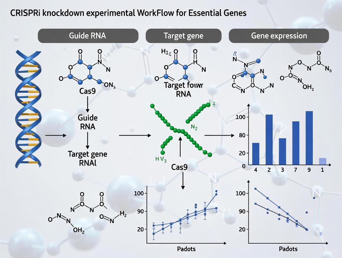
Workflow Visualization
CRISPRi Experimental Workflow
CRISPRi Titration Mechanism
Frequently Asked Questions (FAQs)
FAQ 1: Why does my CRISPRi screen show different essential genes in different cell types? Genetic dependencies are often context-dependent, meaning a gene essential for one cell type's survival might be dispensable in another. This can be due to differences in genetic background, metabolic state, or compensatory pathway expression. When performing cross-cell-type analyses, it is crucial to use a consistent CRISPRi platform and normalization method to ensure results are comparable. Differences in basal gene expression and the cellular proteome can also influence how different cell lines respond to the same genetic perturbation [3] [13].
FAQ 2: How can I achieve intermediate gene knockdowns instead of a full knockout to study essential genes? Traditional CRISPRi aims for maximal knockdown, but titrating expression is possible using systematically attenuated sgRNAs. You can use sgRNAs with designed mismatches to their target DNA [10] or employ sgRNAs with modified constant regions [10]. These modifications reduce the binding efficiency or activity of the dCas9-repressor complex, leading to a spectrum of knockdown levels rather than a complete shut-off. This allows you to stage cells along a continuum of gene expression to identify phenotypically critical thresholds [10].
FAQ 3: My CRISPRi repression is inefficient. What are the key factors to optimize? Inefficient repression can stem from several factors. The most common are:
- sgRNA Design: Ensure the sgRNA targets a region 0-300 base pairs downstream of the transcription start site (TSS) [13]. Using algorithms that consider chromatin accessibility can improve design.
- Repressor Domain Strength: The repressor domain fused to dCas9 significantly impacts efficiency. Novel repressor domains like ZIM3(KRAB) and combinations like dCas9-ZIM3(KRAB)-MeCP2(t) have been shown to provide more potent and consistent knockdown across cell lines and gene targets compared to traditional KRAB domains [3].
- Delivery and Expression: Ensure efficient delivery of both the sgRNA and the dCas9-repressor construct. Using cell lines that stably express the dCas9-repressor fusion can improve consistency and reproducibility [3] [5].
FAQ 4: What is the advantage of using CRISPRi over CRISPR nuclease (CRISPRko) for functional genomics screens? CRISPRi (knockdown) offers several distinct advantages:
- Reversibility: Repression is reversible, allowing for the study of essential genes where permanent knockout would be lethal [3] [13].
- No DNA Damage: CRISPRi uses a deactivated Cas9 (dCas9) that does not cut DNA, thus avoiding the activation of DNA damage response pathways that can confound phenotypic readouts [3].
- Titratable Knockdown: As mentioned, expression can be titrated to study dose-dependent phenotypes [10].
- Study of Non-Coding RNAs: It can be used to interrogate the function of non-coding RNAs and regulatory elements [3].
Troubleshooting Guide for CRISPRi Screens
Table 1: Common CRISPRi Issues and Solutions
| Problem | Potential Cause | Recommended Solution |
|---|---|---|
| Low Knockdown Efficiency | Suboptimal sgRNA design or targeting | Redesign sgRNAs using validated algorithms; target region 0-300bp downstream of TSS [13]; test multiple sgRNAs per gene [5]. |
| Weak repressor domain | Use enhanced repressor domains (e.g., ZIM3(KRAB), dCas9-ZIM3(KRAB)-MeCP2(t)) [3]. | |
| Low delivery/expression efficiency | Use stable cell lines expressing dCas9-repressor; optimize transfection/nucleofection methods [5]. | |
| High Variability Across Cell Lines | Cell-type specific chromatin state | Utilize sgRNAs designed with chromatin accessibility data [13]; consider using stronger, more consistent repressor domains [3]. |
| Differential expression of co-factors | The repressor effect can depend on cell-type-specific expression of transcriptional co-factors [3]. | |
| Inconsistent Results in Pooled Screens | Guide RNA performance variability | Use a library of mismatched sgRNAs to find intermediates; pool multiple sgRNAs per gene to enhance and average repression [10] [13]. |
| Off-target Effects | sgRNA binding to unintended genomic sites | Use bioinformatics tools to design sgRNAs with high specificity; utilize modern repressor domains (e.g., dCas9-SALL1-SDS3) noted for high specificity [5] [13]. |
Advanced Methodologies for Titrating Gene Expression
Table 2: Methods for Titrating CRISPRi Knockdown Levels
| Method | Principle | Key Experimental Consideration |
|---|---|---|
| Mismatched sgRNAs | Introducing single or double mismatches in the sgRNA targeting sequence systematically reduces its binding affinity and knockdown activity [10]. | The position and type of mismatch are critical. PAM-proximal mismatches, especially beyond position 9, have the strongest attenuating effect. rG:dT mismatches can retain substantial activity [10]. |
| Modified sgRNA Constant Regions | Engineering nucleotides in the sgRNA scaffold region can modulate its interaction with dCas9, thereby tuning its overall activity [10]. | A large library of constant region variants exists, with 409 identified variants conferring intermediate activity. Requires screening or use of pre-validated designs [10]. |
| Multiplexed sgRNA Pooling | Transfecting a pool of several sgRNAs targeting the same gene can lead to a more uniform and potent knockdown compared to individual guides [13]. | Pooling 3-5 sgRNAs is a practical strategy to drive maximal repression and mitigate the variable performance of any single sgRNA [13]. |
Experimental Protocol: Systematic Titration with Mismatched sgRNAs
This protocol is adapted from a study that used a library of mismatched sgRNAs to titrate expression of essential genes [10].
- Design an Allelic Series: For each target gene, design a series of sgRNAs containing the perfectly matched sequence and 22-23 variants harboring one or two mismatches. Hold the first nucleotide of the sgRNA as a 'G' for U6 promoter expression, regardless of genomic match.
- Pooled Library Cloning and Production: Synthesize the oligo pool and clone it into your preferred sgRNA expression backbone. Produce lentivirus at a low MOI to ensure single integration.
- Screen and Phenotype: Transduce the cell population of interest (e.g., K562, Jurkat) with the library and culture for enough time to elicit a growth phenotype (e.g., 14-21 days). Use deep sequencing to track sgRNA abundance over time.
- Calculate Relative Activities: For each mismatched sgRNA, calculate a growth phenotype (γ). Normalize this value to the growth phenotype of its corresponding perfectly matched sgRNA to derive a "relative activity" (0 = no activity, 1 = full activity). Average results from multiple cell lines for robust rules.
- Validate Titration: Use single-cell RNA-seq or RT-qPCR on cells harboring specific sgRNAs to confirm the correlation between relative activity and actual mRNA knockdown levels.
The Scientist's Toolkit: Key Research Reagents
Table 3: Essential Reagents for Titratable CRISPRi Screens
| Research Reagent | Function | Technical Notes |
|---|---|---|
| dCas9 Repressor Fusion | The effector protein; binds DNA target and recruits transcriptional repressive machinery. | dCas9-ZIM3(KRAB)-MeCP2(t): A next-generation fusion showing improved repression across cell lines [3]. dCas9-SALL1-SDS3: A proprietary fusion that recruits chromatin remodeling complexes for potent silencing [13]. |
| Attenuated sgRNA Library | A collection of guides with systematically modulated activities to titrate gene expression. | Can be designed in silico using deep learning models trained on empirical mismatch data [10]. Enables staging cells along a continuum of gene expression. |
| Stable dCas9 Cell Lines | Cell lines engineered to constitutively express the dCas9-repressor fusion. | Ensures consistent repressor expression, improving experimental reproducibility and simplifying the screening process [5]. |
| Synthetic sgRNA | Chemically synthesized guide RNA. | Enables fast, transient experiments; repression is evident within 24 hours and maximal at 48-72 hours post-transfection. Ideal for multiplexing [13]. |
Visualizing Experimental Workflows and Relationships
CRISPRi Titration Screening Workflow
The following diagram outlines the key steps in a pooled CRISPRi screen using mismatched sgRNAs to titrate gene expression.
Factors Influencing Mismatched sgRNA Activity
This diagram illustrates the key factors that determine the effectiveness of a mismatched sgRNA in titrating knockdown.
Troubleshooting Guide: Common Experimental Challenges & Solutions
Researchers analyzing mRNA translation in stem cells using CRISPRi often encounter specific technical hurdles. The table below outlines common issues, their potential causes, and recommended solutions.
| Problem Area | Specific Problem | Potential Cause | Recommended Solution |
|---|---|---|---|
| CRISPRi Knockdown | Incomplete gene knockdown leading to ambiguous phenotypes | Weak repressor domain, suboptimal sgRNA design, or variable dCas9 expression | Use a high-efficacy repressor like dCas9-ZIM3(KRAB)-MeCP2(t) [3] and design 4-5 perfect-match sgRNAs per target [14]. |
| Cell State Control | Heterogeneous or inefficient differentiation of pluripotent cells | Spontaneous differentiation in pluripotent cultures or inefficient priming protocol | For naive (ground state) pluripotency, culture mouse ESCs in 2iL medium (GSK3- and MEK-inhibitors with LIF) [15]. |
| Translation Profiling | Poor quality in ribosome profiling or polysome profiling data | RNA degradation, low RIN, or improper nuclease digestion in ribosome profiling | Use high-quality RNA (RIN > 8) and optimize RNase I concentration for ribosome footprinting; validate with footprint periodicity analysis [15]. |
| Data Interpretation | Discrepancy between mRNA abundance and protein levels | Translational buffering or post-translational regulation | Perform integrated analysis of RNA-Seq, Ribo-Seq, and proteomics; a stable mRNA level with changing ribosome density suggests translational control [15]. |
Frequently Asked Questions (FAQs)
FAQ 1: How can I titrate the knockdown level of an essential gene using CRISPRi? You can titrate knockdown by using two types of sgRNAs: (1) Perfect match sgRNAs for maximal knockdown, and (2) Single-base mismatch sgRNAs to create a gradient of partial knockdown [14]. Furthermore, using an inducible promoter (e.g., with IPTG) for dCas9-repressor expression allows you to control the timing and dosage of the knockdown [7].
FAQ 2: What are the key molecular differences in translation between pluripotent and differentiated states? Ground state pluripotent cells (like 2iL-cultured mESCs) display a higher global translation rate and increased ribosome density on a selective set of mRNAs compared to primed or differentiated states [15]. This is counterintuitive, as undifferentiated ESCs were previously thought to have lower translation rates. Key mRNAs undergoing this efficient translation include those encoding polyA-RNA-binding proteins and ribosomal proteins.
FAQ 3: My CRISPRi repression is inefficient. How can I improve it? Consider upgrading your repressor domain. The novel repressor fusion dCas9-ZIM3(KRAB)-MeCP2(t) has been shown to provide significantly improved gene repression at both the transcript and protein level across multiple cell lines compared to older standards like dCas9-KOX1(KRAB) [3].
FAQ 4: How do I confirm that a phenotypic change is due to altered translation of a specific mRNA and not just its transcript level? You must perform ribosome profiling (Ribo-Seq) alongside conventional RNA-Seq. Ribo-Seq measures the number of ribosomes bound to an mRNA, which is a direct indicator of translation efficiency. By comparing the RNA-Seq data (transcript abundance) with the Ribo-Seq data (ribosome footprints), you can identify genes where the translation efficiency (TE) changes independently of the mRNA level [15].
The Scientist's Toolkit: Research Reagent Solutions
The table below catalogs essential reagents and their functions for conducting this case study's research.
| Reagent / Tool | Function / Application | Key Considerations |
|---|---|---|
| dCas9-ZIM3(KRAB)-MeCP2(t) | Next-generation CRISPRi repressor for high-efficacy gene knockdown [3] | Provides more consistent performance across different gene targets and cell lines with less guide-dependent variability. |
| Modified mRNA (5mC/psi) | For expressing reprogramming or differentiation factors without triggering innate immunity [16] | Incorporates 5-methylcytidine and pseudouridine to evade antiviral sensors; enables highly efficient protein expression. |
| B18R Protein | Interferon inhibitor used in modified mRNA protocols [16] | Suppresses residual interferon response that can occur even with modified nucleosides, improving cell viability. |
| 2iL Medium | Chemically defined medium for maintaining mouse ESCs in a naive (ground state) of pluripotency [15] | Contains GSK3 inhibitor (CHIR99021), MEK inhibitor (PD0325901), and LIF. Essential for studying ground-state translatome. |
| Ribosome Profiling Kit | For genome-wide analysis of mRNA translation (Ribo-Seq) [15] | Provides protocol for generating ribosome-protected mRNA footprints, allowing measurement of translation efficiency. |
Experimental Protocols
Protocol: Titrating Essential Gene Knockdown with CRISPRi
This protocol is adapted from pooled CRISPRi library screens in bacteria [14] and optimized for mammalian cells using modern repressors [3].
sgRNA Library Design:
- For each essential gene target, design a set of sgRNAs comprising:
- ~4 perfect match sgRNAs to achieve maximal knockdown.
- ~10 single-base mismatch sgRNAs to create a spectrum of partial knockdown strengths [14].
- Include a set of non-targeting control sgRNAs.
- For each essential gene target, design a set of sgRNAs comprising:
Cell Line Engineering:
Induction and Phenotyping:
- Induce CRISPRi knockdown. In prokaryotic systems, this can be done with IPTG [7]; for mammalian systems, use a compatible inducible system (e.g., doxycycline).
- After induction, split the culture. Use one part for polysome profiling to assess global and specific changes in translation. Use another part to extract total RNA for RNA-Seq to control for transcriptional changes [15].
Analysis:
- Sequence the sgRNA pool at different time points to track depletion of guides targeting essential genes.
- Calculate a fitness score (e.g., log2 fold change) for each sgRNA. Genes with guides that show strong depletion are acutely sensitive to knockdown, indicating high vulnerability [14].
- Correlate the degree of knockdown (from sgRNA depletion) with changes in translation efficiency (from polysome/RNA-Seq data).
Protocol: Comparing Translation Efficiency via Polysome Profiling
This protocol is used to analyze the translational landscape during the naive-to-primed pluripotency transition [15].
Cell Lysis and Fractionation:
- Lyse cells from your conditions of interest (e.g., 2iL-ESCs, SL-ESCs, EPI) with a cycloheximide-containing buffer to freeze ribosomes on mRNA.
- Layer the lysate onto a 10-50% sucrose density gradient.
- Centrifuge at high speed (e.g., 35,000 rpm for 3 hours in a ultracentrifuge) to separate ribosomal complexes by mass.
Fraction Collection and RNA Isolation:
- Fractionate the gradient using a density gradient fractionation system while monitoring absorbance at 254 nm to identify fractions containing free RNA, 40S/60S subunits, 80S monosomes, and polysomes.
- Isolate RNA from the total lysate (for input RNA-Seq) and from the polysome-containing fractions.
Library Preparation and Sequencing:
- Convert the RNA from the polysomal fractions and total input into cDNA libraries for RNA-Seq.
- For deeper insight, ribosome profiling libraries can be prepared from nuclease-digested lysates to obtain ribosome-protected mRNA footprints [15].
Data Analysis:
- Map the sequencing reads to the transcriptome.
- For each mRNA, calculate its Translation Efficiency (TE) by normalizing the reads from the polysomal fraction (or ribosome footprints) to its abundance in the total RNA input.
- Identify differentially translated mRNAs by comparing TE between cell states (e.g., 2iL vs. EPI).
Experimental Workflow & Pathway Visualization
Experimental Workflow for mRNA Translation Analysis
Translation State Across Cell Types
Building Your Titration Toolkit: CRISPRi Systems, sgRNA Design, and Experimental Workflows
This technical support center is framed within the context of a broader thesis on titrating CRISPRi knockdown levels for essential genes research, providing troubleshooting guides and FAQs to assist researchers in optimizing repressor systems for precise gene silencing.
Troubleshooting Guides and FAQs
Frequently Asked Questions
Q1: Why is my dCas9 repressor system showing low repression efficiency in essential gene knockdown experiments? A: Low repression efficiency can result from suboptimal sgRNA design, insufficient dCas9-repressor expression, or epigenetic barriers. Ensure sgRNAs target within -50 to +300 bp relative to the transcription start site (TSS), use high-efficiency delivery methods (e.g., lentiviral transduction), and validate repressor expression with Western blotting. For essential genes, titrate repressor levels by varying inducer concentrations (e.g., doxycycline) to avoid complete knockdown that may cause cell death.
Q2: How can I minimize off-target effects when using dCas9-ZIM3(KRAB)-MeCP2(t) for titration studies? A: Off-target effects are reduced by using high-specificity sgRNAs with minimal off-target scores (evaluated by tools like CRISPRscan or ChopChop), incorporating truncated guide sequences (17-18 nt), and employing control experiments with non-targeting sgRNAs. Additionally, use low concentrations of repressor plasmids and monitor global transcriptome changes via RNA-seq to assess specificity.
Q3: What causes variable knockdown levels between replicates in dCas9-KRAB experiments? A: Variability often stems from inconsistent transfection/transduction efficiency, cell line heterogeneity, or fluctuations in repressor expression. Standardize cell culture conditions, use polyclonal cell pools after selection, and employ fluorescence-activated cell sorting (FACS) to isolate cells with uniform repressor expression. For titration, include internal controls and use quantitative PCR (qPCR) for precise measurement of gene expression.
Q4: How do I titrate knockdown levels effectively with dCas9-ZIM3(KRAB) in essential gene research? A: Titration involves modulating repressor activity through inducible systems (e.g., tetracycline-inducible promoters) or varying sgRNA concentrations. Perform dose-response curves with different inducer levels (e.g., 0-1000 ng/mL doxycycline) and measure gene expression via qPCR or reporter assays. Use the dynamic range of each repressor to fine-tune knockdown, ensuring partial repression for essential genes to maintain cell viability.
Q5: What are the key differences in repression potency among dCas9-KRAB, dCas9-ZIM3(KRAB), and dCas9-ZIM3(KRAB)-MeCP2(t)? A: dCas9-KRAB provides moderate repression (~60-80%), dCas9-ZIM3(KRAB) offers enhanced potency (~70-90%) due to stronger recruitment of repressive complexes, and dCas9-ZIM3(KRAB)-MeCP2(t) achieves the highest repression (~80-95%) by combining chromatin remodeling with transcriptional silencing. However, higher potency may increase off-target risks, so choose based on the required titration range and specificity needs.
Troubleshooting Guides
Issue: Poor Repression Across All Repressor Constructs
- Cause: Inefficient sgRNA binding or low dCas9 expression.
- Solution: Redesign sgRNAs to target near the TSS, verify dCas9 expression with antibodies, and optimize delivery method (e.g., use lipofection for transient or lentivirus for stable expression).
Issue: Cell Toxicity or Death with dCas9-ZIM3(KRAB)-MeCP2(t)
- Cause: Over-repression of essential genes or non-specific effects.
- Solution: Titrate repressor expression using inducible promoters, reduce sgRNA concentration, and perform viability assays (e.g., MTT) to identify non-toxic conditions.
Issue: Inconsistent Titration Results in Essential Gene Knockdown
- Cause: Uncontrolled repressor dynamics or cell cycle effects.
- Solution: Use synchronized cell cultures, monitor repressor levels over time with flow cytometry, and employ single-cell analysis (e.g., scRNA-seq) to capture heterogeneity.
Data Presentation
Table 1: Comparison of Repressor Efficiency and Specificity
| Repressor Construct | Repression Efficiency (%) | Dynamic Range (Fold Change) | Off-Target Score (1-10, lower is better) | Optimal Titration Range (Inducer Concentration) |
|---|---|---|---|---|
| dCas9-KRAB | 60-80 | 5-10x | 3 | 10-100 ng/mL doxycycline |
| dCas9-ZIM3(KRAB) | 70-90 | 10-20x | 5 | 1-50 ng/mL doxycycline |
| dCas9-ZIM3(KRAB)-MeCP2(t) | 80-95 | 20-50x | 7 | 0.1-10 ng/mL doxycycline |
Table 2: Key Experimental Parameters for Titration Studies
| Parameter | dCas9-KRAB | dCas9-ZIM3(KRAB) | dCas9-ZIM3(KRAB)-MeCP2(t) |
|---|---|---|---|
| Recommended sgRNA Length | 20 nt | 20 nt | 18 nt |
| Expression System | Lentiviral | Lentiviral | Doxycycline-inducible |
| Time to Max Repression | 48-72 h | 24-48 h | 12-24 h |
| Cell Viability at Optimal Titration | High | Moderate | Low (requires careful titration) |
Experimental Protocols
Protocol 1: Setting Up CRISPRi Titration with Inducible Repressors
- Design sgRNAs: Use bioinformatics tools (e.g., CRISPRko or CHOPCHOP) to design 3-5 sgRNAs per target gene, focusing on regions -50 to +300 bp from TSS. Include non-targeting sgRNA controls.
- Clone sgRNAs: Clone sgRNAs into a lentiviral vector (e.g., lentiGuide-Puro) using BsmBI restriction sites.
- Produce Lentivirus: Co-transfect HEK293T cells with repressor plasmid (e.g., dCas9-KRAB), packaging plasmids (psPAX2, pMD2.G), and sgRNA vector using PEI transfection. Harvest virus at 48 and 72 hours post-transfection.
- Transduce Cells: Infect target cells (e.g., HEK293 or primary cells) with lentivirus in the presence of polybrene (8 μg/mL). Select with puromycin (1-2 μg/mL) for 3-5 days.
- Induce Repression: Add doxycycline (0-1000 ng/mL) to culture medium for 24-72 hours to titrate repressor expression.
- Quantify Knockdown: Harvest cells, extract RNA, and perform qPCR with gene-specific primers. Normalize to housekeeping genes (e.g., GAPDH) and calculate fold change relative to non-targeting controls.
Protocol 2: Assessing Repression Efficiency and Specificity
- Western Blot for Repressor Expression: Lyse cells in RIPA buffer, separate proteins via SDS-PAGE, transfer to PVDF membrane, and probe with anti-FLAG (for tagged repressors) and anti-β-actin antibodies.
- RNA-seq for Global Transcriptome Analysis: Extract total RNA, prepare libraries with poly-A selection, and sequence on Illumina platform. Analyze data with tools like DESeq2 to identify differentially expressed genes and off-target effects.
- Cell Viability Assay: Seed cells in 96-well plates, treat with titrated repressor inducers, and assess viability after 72 hours using MTT assay according to manufacturer's instructions.
Mandatory Visualization
Diagram 1: CRISPRi Repression Mechanism
Title: CRISPRi Repression Mechanism
Diagram 2: Experimental Workflow for Titration
Title: Titration Experimental Workflow
Diagram 3: Repressor Comparison Logic
Title: Repressor Potency Comparison
The Scientist's Toolkit
Table 3: Essential Research Reagent Solutions
| Reagent | Function | Example Product |
|---|---|---|
| dCas9 Repressor Plasmids | Expresses catalytically dead Cas9 fused to repressor domains | Addgene #110821 (dCas9-KRAB) |
| Lentiviral Packaging Plasmids | Produces lentiviral particles for stable delivery | Addgene #12259 (psPAX2), #12260 (pMD2.G) |
| sgRNA Cloning Vector | Harbors sgRNA sequence for CRISPRi targeting | Addgene #52963 (lentiGuide-Puro) |
| Doxycycline Inducer | Titrates repressor expression in inducible systems | Sigma D9891 |
| Puromycin | Selects for transduced cells | Thermo Fisher A1113803 |
| Anti-FLAG Antibody | Detects FLAG-tagged repressors in Western blot | Sigma F1804 |
| qPCR Master Mix | Quantifies gene expression changes | Thermo Fisher 4367659 |
| MTT Assay Kit | Measures cell viability | Sigma TOX1 |
sgRNA Design Principles for Consistent Knockout Efficiency Across Genomic Loci
Frequently Asked Questions (FAQs)
Q1: Why do I observe variable knockout efficiency when using different sgRNAs targeting the same gene?
Different sgRNAs targeting the same gene often exhibit substantial variability in editing efficiency due to their unique sequence and structural features [8]. This occurs because CRISPR editing efficacy is highly influenced by the intrinsic properties of each sgRNA sequence [17]. Key factors affecting efficiency include:
- GC content (optimal range: 40-80%) [18]
- Secondary structure formation of the sgRNA [5]
- Proximity to transcription start sites [5]
- Specific nucleotide composition at particular positions [19]
To mitigate this variability, always design 3-5 sgRNAs per gene to ensure at least one produces efficient knockout [8].
Low knockout efficiency can result from multiple factors. Implement these evidence-based solutions:
Optimize sgRNA structure: Extend the sgRNA duplex by approximately 5 bp and mutate the fourth thymine (T) in the continuous T sequence to cytosine (C) or guanine (G) [19]. This optimized structure significantly increases knockout efficiency across multiple cell lines and target genes [19].
Improve delivery efficiency: Use validated transfection methods appropriate for your cell type. Lipid-based transfection reagents (e.g., DharmaFECT, Lipofectamine) or electroporation often improve Cas9 and sgRNA delivery [5].
Utilize stable Cas9 cell lines: Cells with stable Cas9 expression provide more consistent editing compared to transient transfection [5].
Validate sgRNA design: Use bioinformatics tools like Synthego's Design Tool or CHOPCHOP to select sgRNAs with predicted high on-target activity [18].
Q3: What specific sgRNA modifications significantly enhance knockout efficiency?
Systematic investigation of sgRNA structure reveals two key modifications that dramatically improve efficiency:
Duplex extension: Extending the sgRNA duplex by approximately 5 bp significantly increases knockout efficiency [19]. The beneficial effect typically peaks around 5 bp, with similar efficiency observed for 4-6 bp extensions [19].
Poly-T tract mutation: Mutating the fourth thymine in the continuous T sequence to cytosine (C) or guanine (G) prevents premature transcription termination and enhances efficiency [19]. T→C and T→G mutations generally yield higher efficiency than T→A mutations [19].
Table 1: Optimized sgRNA Structural Modifications for Enhanced Knockout Efficiency
| Modification Type | Optimal Parameter | Efficiency Improvement | Key Considerations |
|---|---|---|---|
| Duplex Extension | 5 bp (range: 4-6 bp) | Significant increase (dramatic for some sgRNAs) | Pattern slightly varies for different sgRNAs |
| Poly-T Tract Mutation | Position 4: T→C or T→G | Significant increase across all tested sgRNAs | T→C may provide slightly better efficiency in some cases |
| Combined Modifications | Extended duplex + T→C/G | Most dramatic improvement (up to 10× for gene deletions) | Enables challenging applications like non-coding gene deletion |
Q4: How does the choice of Cas9 cell line affect knockout consistency?
Cell line selection critically impacts editing outcomes:
- Stably expressing Cas9 cell lines provide more consistent editing efficiency compared to transient transfection [5].
- Cell-type specific responses: Immortalized lines (HEK293, HeLa) are generally more amenable to editing than primary cells [20].
- DNA repair capacity variation: Some cell lines (e.g., HeLa) possess strong DNA repair abilities that can reduce knockout efficiency [5].
Q5: What computational tools are available for designing high-efficiency sgRNAs?
Several bioinformatics tools incorporate efficiency predictions:
- sgDesigner (http://crispr.wustl.edu): Machine learning-based tool that outperforms existing design algorithms [17]
- Synthego Design Tool: Considers on-target efficiency and off-target effects [18]
- CHOPCHOP: Supports multiple Cas nucleases and PAM recognition [18]
- CRISPR Design Tool (Benchling): Evaluates secondary structures, GC content, and specificity [5]
Q6: How can I titrate gene expression levels using modified sgRNAs?
CRISPRi with mutated sgRNAs enables precise titration of gene expression [21]:
- Strategic sgRNA mutation: Introduce mismatches at specific positions in the sgRNA targeting region
- Seed region importance: Mutations closer to the PAM site have more severe effects on knockdown strength
- Compounding mutations: Incrementally adding mismatches from distal to proximal positions creates gradated expression effects
- Molecular barcoding: Detect and correct for mutations that "escape" CRISPRi targeting
This approach reveals gene-by-environment interactions that remain undetected with maximal knockdown approaches [21].
Troubleshooting Guides
Problem: Consistently Low Knockout Efficiency Across Multiple sgRNAs
Potential Causes and Solutions:
Table 2: Comprehensive Troubleshooting for Low Knockout Efficiency
| Problem Cause | Evidence-Based Solution | Expected Outcome |
|---|---|---|
| Suboptimal sgRNA structure | Implement extended duplex (+5 bp) with T→C/G mutation at position 4 [19] | Dramatic efficiency improvement (confirmed across 16 sgRNAs) |
| Inefficient delivery | Switch to lipid nanoparticles or electroporation; use stably expressing Cas9 cells [5] | Higher transfection efficiency and more consistent editing |
| High secondary structure | Use design tools to predict and avoid sgRNAs with stable secondary structures [5] | Improved sgRNA accessibility and binding |
| Insufficient coverage | Design 3-5 sgRNAs per gene; ensure adequate sequencing depth (≥200×) [8] | Reduced false negatives; more reliable results |
Problem: Inconsistent Efficiency Across Genomic Loci
Solutions:
- Locus-specific optimization: Account for local chromatin accessibility and epigenetic features not captured by standard design rules [17]
- Dual-sgRNA approach: Use tandem sgRNA cassettes targeting the same gene, which significantly improves knockdown consistency compared to single sgRNAs [9]
- Validate multiple sgRNAs: Always test several sgRNAs per target locus to identify the most effective one [8]
Experimental Protocols
Protocol 1: sgRNA Structural Optimization for Enhanced Efficiency
This protocol is adapted from systematic investigation of sgRNA structure [19]:
- Design sgRNAs with 17-23 nucleotide guide sequences targeting your gene of interest
- Extend the duplex by adding 5 bp to the standard sgRNA structure
- Mutate the poly-T tract by changing the fourth thymine to cytosine or guanine
- Clone into appropriate expression vector using standard molecular biology techniques
- Transfert into target cells alongside Cas9 nuclease
- Evaluate efficiency using T7E1 assay, sequencing, or functional assays
Validation: Compare to unmodified sgRNA controls. The optimized structure typically shows significant efficiency improvements across multiple targets [19].
Protocol 2: Dual-sgRNA Approach for Enhanced Knockdown Consistency
Based on compact dual-sgRNA library design [9]:
- Select two optimal sgRNAs per gene using predictive algorithms
- Clone as tandem cassette into lentiviral transfer plasmid
- Package lentivirus and transduce target cells
- Select with puromycin for stable integration
- Harvest cells at T0 (immediately after selection) and Tfinal (after phenotypic expression)
- Sequence sgRNA cassettes from genomic DNA to quantify abundance changes
- Calculate phenotypes based on sgRNA abundance changes between timepoints
This approach produces significantly stronger growth phenotypes for essential genes compared to single-sgRNA designs [9].
Research Reagent Solutions
Table 3: Essential Reagents for Optimized sgRNA Experiments
| Reagent/Cell Line | Function | Application Notes |
|---|---|---|
| Zim3-dCas9 effector | CRISPRi repression | Provides optimal balance of strong knockdown and minimal non-specific effects [9] |
| Stably expressing Cas9 cells | Consistent nuclease expression | Improves reproducibility compared to transient transfection [5] |
| Dual-sgRNA lentiviral library | Compact, highly active screening | Targets each gene with two sgRNAs; improves knockdown strength [9] |
| Modified sgRNA templates | Enhanced knockout efficiency | Structural optimizations (extended duplex + poly-T mutation) [19] |
| BsmBI-digested backbone | sgRNA library cloning | Enables efficient assembly of sgRNA expression constructs [17] |
Workflow Visualization
This guide details the establishment of a doxycycline-inducible CRISPR interference (CRISPRi) system in human pluripotent stem cells (hPSCs) for inducible, multiplexed gene silencing. The system enables reversible knockdown of essential genes, which is vital for studying their function without creating permanent, non-viable knockout cells [22] [3]. The core CRISPRi system consists of a nuclease-deactivated Cas9 (dCas9) fused to transcriptional repressor domains, which is recruited by a guide RNA (sgRNA) to specific DNA sequences to block transcription [23] [3].
Frequently Asked Questions (FAQs)
Q1: Why choose CRISPRi over CRISPR knockout (CRISPRko) for essential genes research? CRISPRi offers reversible gene knockdown instead of permanent knockout. This is crucial for studying essential genes, as complete and permanent knockout can lead to cell death, preventing functional analysis. CRISPRi also avoids DNA double-strand breaks, reducing potential confounders like activation of DNA damage response pathways and off-target mutations [3].
Q2: What are the key advantages of an inducible system? An inducible system, such as the one using a doxycycline-inducible promoter, allows precise temporal control over gene repression. This enables researchers to study gene function at specific time points during differentiation or upon treatment, which is essential for dissecting the roles of genes critical for cell viability or processes like lineage specification [22].
Q3: How can I improve the knockdown efficiency of my CRISPRi system? Knockdown efficiency can be enhanced by using novel, high-performance repressor domains. Recent research has identified fusion proteins like dCas9-ZIM3(KRAB)-MeCP2(t) that provide significantly stronger and more consistent repression across different gene targets and cell lines compared to earlier standards like dCas9-KOX1(KRAB) [3]. Careful design and selection of sgRNAs is also critical [24].
Q4: What is the function of the repressor domain fused to dCas9? The repressor domain (e.g., KRAB) recruits chromatin-modifying complexes to the target gene's promoter, leading to the introduction of repressive histone marks and subsequent transcriptional silencing. The dCas9 protein itself serves as a programmable DNA-binding module that directs this repressive machinery to the desired genomic locus [3].
Step-by-Step Experimental Protocol
Establishing Host CRISPRi hPSCs
The goal of this phase is to create a stable parental hPSC line that expresses the inducible dCas9-repressor fusion protein.
- Repressor Selection: Clone your chosen dCas9-repressor construct (e.g., dCas9-KRAB or the advanced dCas9-ZIM3(KRAB)-MeCP2(t) [3]) into a lentiviral vector under the control of a doxycycline-inducible promoter.
- Cell Preparation: Culture and passage your hPSCs to ensure they are in a healthy, undifferentiated, and log-phase growth state.
- Lentiviral Transduction: Transduce the hPSCs with the lentiviral vector containing the dCas9-repressor construct. A low multiplicity of infection (MOI) is recommended to minimize the risk of multiple integrations.
- Selection and Expansion: After transduction, select transduced cells using an appropriate antibiotic (e.g., puromycin) for 1-2 weeks. Expand the polyclonal population of stable cells.
- Validation: Validate the expression of the dCas9-repressor fusion protein by inducing with doxycycline and performing immunoblotting or immunocytochemistry. This creates the "host CRISPRi hPSC" line ready for sgRNA delivery [22].
Designing, Cloning, and Delivering sgRNAs
This phase involves creating sgRNA constructs targeting your genes of interest and introducing them into the host CRISPRi hPSCs.
- sgRNA Design: Design sgRNAs to target the transcription start site (TSS) of your essential gene(s). Use established algorithms to ensure high on-target activity and minimize potential off-target effects. For multiple-gene silencing, design a pool of sgRNAs [22] [24].
- sgRNA Cloning: Clone the synthesized sgRNA oligonucleotides into a lentiviral sgRNA expression vector. The vector should contain a puromycin resistance gene for selection and be compatible with the host cell line [22].
- Lentiviral Production: Produce lentivirus containing the sgRNA expression construct.
- Cell Line Generation: Transduce the host CRISPRi hPSCs with the sgRNA lentivirus. After transduction, select with puromycin to generate a polyclonal population of cells. For more rigorous experiments, derive monoclonal cell lines by single-cell cloning and expansion [22].
Executing and Validating Gene Silencing Experiments
This final phase covers the induction of gene silencing and the assessment of its effects.
- Induction of Knockdown: Add doxycycline to the culture medium to induce expression of the dCas9-repressor fusion and initiate gene silencing. A typical working concentration is 1-2 µg/mL, but this should be optimized for your system.
- Phenotypic Analysis: Proceed with your downstream functional assays. For essential genes, this may include monitoring cell proliferation, viability, or changes in differentiation capacity over time.
- Validation of Knockdown: Always confirm the efficiency of gene silencing at the transcript level using RT-qPCR and, if possible, at the protein level via western blotting or flow cytometry [3].
Table 1: Key Reagents and Materials for Establishing Inducible CRISPRi in hPSCs
| Reagent/Material | Function/Description | Example or Source |
|---|---|---|
| dCas9-Repressor Plasmid | Expresses the inducible, repressor-fused dCas9 protein. | Constructs like dCas9-ZIM3(KRAB)-MeCP2(t) [3] |
| sgRNA Expression Plasmid | Expresses the guide RNA targeting the gene of interest. | Lentiviral backbone with selection marker [22] |
| Human Pluripotent Stem Cells | The host cells for the CRISPRi system. | e.g., induced PSCs (iPSCs) or embryonic stem cells (ESCs) |
| Lentiviral Packaging System | Produces lentiviral particles for gene delivery. | Includes packaging and envelope plasmids |
| Doxycycline | Small molecule inducer for the system; triggers dCas9 expression. | Prepare a stock solution (e.g., 1 mg/mL in sterile water) |
| Selection Antibiotics | Selects for cells successfully transduced with the vectors. | e.g., Puromycin, Blasticidin |
Troubleshooting Guide
Table 2: Common Experimental Issues and Recommended Solutions
| Problem | Potential Cause | Recommended Solution |
|---|---|---|
| Low Knockdown Efficiency | Inefficient sgRNA design or repressor domain. | Redesign sgRNAs using computational tools; switch to a more potent repressor like dCas9-ZIM3(KRAB)-MeCP2(t) [3]. |
| Low dCas9-repressor expression. | Optimize doxycycline concentration and induction time; verify plasmid sequence and integrity [24]. | |
| Poor Cell Viability Post-Transduction | Toxicity from viral transduction. | Titrate the viral titer to use the lowest effective MOI; ensure cells are healthy before transduction. |
| Essential gene knockdown is too severe. | Titrate the doxycycline concentration to achieve a partial, tolerable knockdown rather than full repression [23]. | |
| High Background Repression (Without Induction) | Leaky expression from the inducible promoter. | Use a tighter inducible promoter system; ensure all media and reagents are free from contaminating inducers. |
| No Cleavage or Modification Band | Transfection efficiency too low; target site inaccessible. | Optimize transfection protocol; design new sgRNAs targeting nearby, more accessible sequences [24]. |
| Off-Target Effects | sgRNA homology with other genomic regions. | Carefully design crRNA targets to avoid homology with other genomic regions; use bioinformatic tools to predict off-target sites [24]. |
Quantitative Data and Performance Metrics
Table 3: Comparison of CRISPRi Repressor Performance
| dCas9-Repressor Construct | Reported Knockdown Efficiency | Key Characteristics & Applications |
|---|---|---|
| dCas9-KOX1(KRAB) | Baseline | The first widely used repressor; moderate efficiency, performance can be variable [3]. |
| dCas9-ZIM3(KRAB) | ~20-30% better than KOX1(KRAB) | A "gold standard" repressor; improved gene silencing across many targets [3]. |
| dCas9-KOX1(KRAB)-MeCP2 | High | A bipartite repressor showing enhanced knockdown compared to KRAB alone [3]. |
| dCas9-ZIM3(KRAB)-MeCP2(t) | ~20-30% better than dCas9-ZIM3(KRAB) [3] | A next-generation, tripartite repressor; offers high efficacy, lower variability, and consistent performance across cell lines. Ideal for genome-wide screens. |
Visual Workflows and Diagrams
Diagram 1: CRISPRi Experimental Workflow. This flowchart outlines the three major phases of implementing the inducible CRISPRi system in hPSCs.
Diagram 2: Mechanism of Inducible CRISPRi Knockdown. Doxycycline induces expression of the dCas9-repressor fusion, which complexes with the sgRNA and binds to the target gene's promoter, physically blocking RNA polymerase and silencing gene transcription.
FAQs: Troubleshooting Common Experimental Challenges
Q1: Our combinatorial CRISPR screen shows high variability between replicates. What are the primary quality control metrics we should check?
A1: High replicate variability often stems from issues with library representation or screen execution. Key Quality Control (QC) metrics to assess include [25] [26]:
- Library Coverage: Ensure coverage is >300x across most replicates to reliably detect signal [26].
- Gini Index: This metric should ideally be <0.5, indicating good guide distribution without extreme outliers [26].
- BAGEL2 AUC: Replicates should have an Area Under the Curve >0.88 for essential gene detection [26].
- NNMD: Exclude samples with a null-normalized mean difference (NNMD) greater than -2 [26].
Q2: When analyzing dual-guide CRISPRi screens for genetic interactions, which scoring method should I use to identify synthetic lethal pairs?
A2: Select a scoring method based on your screen's design and the type of genetic interactions you aim to capture. Recent benchmarking of five major scoring methods recommends Gemini-Sensitive as a robust first choice [25]. Key considerations include [25]:
- Gemini-Sensitive performs well across diverse datasets and can capture 'modest synergy' [25].
- It is available as an R package with comprehensive documentation, making it accessible for most researchers [25].
- For screens requiring detection of 'high synergy' interactions, Gemini-Strong may be more appropriate [25].
- The Parrish score also shows reasonable performance but is less standardized in its implementation [25].
Q3: Our CRISPRi knockdown efficiency varies significantly across different gene targets and cell lines. How can we improve consistency?
A3: Variable knockdown efficiency is a common challenge. Implement these strategies to enhance performance [3] [27]:
- Optimize Repressor Domains: Utilize novel repressor fusions like dCas9-ZIM3(KRAB)-MeCP2(t) which demonstrate improved repression across multiple cell lines and reduced guide-dependent variability [3].
- Validate Essential Genes: For essential gene research, confirm >90% of targeted genes show viability defects with at least one sgRNA [27].
- Employ Multiple sgRNAs: Use at least two sgRNAs per gene to control for sgRNA-specific effects, as efficiency can differ substantially between guides [27] [28].
Q4: What are the critical parameters for optimizing an inducible Cas9 system in human pluripotent stem cells (hPSCs) for gene knockout studies?
A4: Achieving high editing efficiency in hPSCs requires systematic optimization of these parameters [28]:
- Cell Tolerance: Assess and optimize cell tolerance to nucleofection stress.
- Transfection Methods: Refine nucleofection protocols specifically for hPSCs.
- sgRNA Stability: Use chemically modified sgRNAs with 2'-O-methyl-3'-thiophosphonoacetate modifications at both ends.
- Cell-to-sgRNA Ratio: Optimize this critical ratio (e.g., 5μg sgRNA for 8×10⁵ cells).
- Nucleofection Frequency: Implement repeated nucleofection 3 days after initial transfection.
Experimental Protocols & Workflows
Protocol: Dual-Guide CRISPR Library Screen for Synthetic Lethality
Objective: Identify synthetic lethal gene pairs across multiple cancer cell lines.
Materials:
- Dual-guide CRISPR library (22,823 unique guide pairs targeting 472 gene pairs) [26]
- Cas9-positive cell lines (melanoma, NSCLC, pancreatic cancer) [26]
- Lentiviral packaging system
- Next-generation sequencing platform
Procedure:
- Library Design: Configure guides to produce 18-32 guide combinations targeting each gene pair [26].
- Lentiviral Production: Generate high-titer lentivirus from the dual-guide library.
- Cell Infection: Transduce Cas9-positive cell lines at MOI=0.3 to ensure most cells receive single viral integrants [26].
- Screen Execution: Maintain cells for 28 days with 1000x library representation, collecting samples at days 0, 7, 14, 21, and 28 [26].
- DNA Extraction & Sequencing: Extract genomic DNA and prepare sequencing libraries for guide abundance quantification.
- Quality Control: Apply QC metrics (Gini Index <0.5, NNMD >-2, BAGEL2 AUC >0.88) [26].
- Data Analysis: Process sequencing data through Gemini-Sensitive scoring pipeline to identify synthetic lethal interactions [25].
Troubleshooting Tips:
- Include "safe-targeting" controls targeting non-functional genomic regions to calculate single vs. double knockout effects [26].
- For paralog targeting, use guides with low off-target scores to minimize cross-reactivity between related sequences [26].
- Sequence plasmid library pre- and post-screen to monitor library integrity.
Protocol: Titrating CRISPRi Knockdown Levels for Essential Gene Research
Objective: Achieve graded knockdown of essential genes to study dose-dependent phenotypes.
Materials:
- Optimized CRISPRi system (dCas9-ZIM3(KRAB)-MeCP2(t)) [3]
- Inducible dCas9 expression system (e.g., xylose-inducible for C. difficile, doxycycline-inducible for mammalian cells) [27] [28]
- FM4-64 and Hoechst 33342 stains for morphological assessment [27]
Procedure:
- System Selection: Implement dCas9-ZIM3(KRAB)-MeCP2(t) for enhanced repression efficiency [3].
- Inducible Control: Use tunable promoters (xylose or doxycycline) to titrate dCas9-repressor expression [27] [28].
- Multi-sgRNA Approach: Design 2+ sgRNAs per gene with varying efficiency scores [27] [28].
- Time-Course Analysis: Monitor viability and morphology over 6+ population doublings post-knockdown induction [27].
- Phenotypic Scoring:
- Validation: Correlate transcript/protein reduction with phenotypic severity using Western blotting or RT-qPCR [3] [28].
Troubleshooting Tips:
- For genes in operons, target only one gene per transcription unit to avoid polarity confusion [27].
- Include non-targeting scrambled sgRNA controls to identify off-target effects [27].
- For hPSCs, optimize nucleofection parameters and use chemically modified sgRNAs for enhanced stability [28].
Data Presentation: Scoring Methods & Reagent Solutions
Comparison of Genetic Interaction Scoring Methods
Table: Benchmarking of Synthetic Lethality Scoring Algorithms
| Scoring Method | Optimal Use Case | Performance Characteristics | Implementation | Key Considerations |
|---|---|---|---|---|
| Gemini-Sensitive | General first choice | Captures 'modest synergy'; performs well across diverse datasets [25] | R package with comprehensive documentation [25] | Removes gene pairs where single KO causes >50% depletion [25] |
| Gemini-Strong | High-confidence interactions | Identifies interactions with 'high synergy' [25] | Available in Gemini R package [25] | More stringent threshold for interaction calls [25] |
| Parrish Score | Specific screen designs | Reasonable performance across datasets [25] | Custom implementation [25] | Less standardized than Gemini [25] |
| zdLFC | Simple interaction metrics | Genetic interaction = expected DMF minus observed DMF [25] | Python notebooks [25] | Applies z-transformation after truncating extremes [25] |
| Orthrus | Orientation-specific effects | Assumes additive linear model for expected LFC [25] | R package available [25] | Can be configured to ignore orientation when needed [25] |
Research Reagent Solutions for Combinatorial Screening
Table: Essential Materials for CRISPRi Genetic Interaction Studies
| Reagent / Tool | Function | Application Notes | Key Features |
|---|---|---|---|
| dCas9-ZIM3(KRAB)-MeCP2(t) | Next-generation CRISPRi repressor [3] | Enhanced repression across cell lines; reduced guide-dependent variability [3] | Bipartite repressor fusion; improved reproducibility [3] |
| Dual-Promoter Lentiviral Vector | Expresses two gRNAs from hU6 and mU6 promoters [26] | Enables combinatorial gene targeting; reduces viral recombination risk [26] | Modified spacer and tracr sequences to prevent recombination [26] |
| Chemically Modified sgRNAs | Enhanced stability for improved editing efficiency [28] | Critical for hard-to-transfect cells (e.g., hPSCs) [28] | 2'-O-methyl-3'-thiophosphonoacetate modifications at both ends [28] |
| "Safe-Targeting" Controls | Target non-functional genomic regions [26] | Calculate single vs. double knockout effects; baseline normalization [26] | Generate double-strand breaks without disrupting gene function [26] |
| Inducible Cas9 Systems | Tunable nuclease expression (doxycycline/xylose) [27] [28] | Enables temporal control of editing; improves viability of edited cells [28] | Cost-effective; achieves up to 93% INDEL efficiency in hPSCs [28] |
Workflow Visualization
Combinatorial CRISPR Screening Workflow
Dual-Guide CRISPR Screening Pipeline
CRISPRi Titration for Essential Genes
CRISPRi Titration Workflow
Solving the Efficiency Puzzle: Overcoming Low Knockdown and Variable Performance
Core Concepts: CRISPRi Knockdown vs. CRISPR Nuclease
FAQ: How is CRISPRi different from CRISPR nuclease (CRISPRn) for studying essential genes?
CRISPR interference (CRISPRi) and CRISPR nuclease (CRISPRn) are distinct tools for loss-of-function studies. CRISPRi uses a catalytically dead Cas9 (dCas9) fused to repressor domains (like KRAB) to block transcription and reversibly knock down gene expression. In contrast, CRISPRn uses an active Cas9 nuclease to create double-strand DNA breaks, permanently disrupting the gene [29].
For essential genes research, CRISPRi offers key advantages:
- Reversibility & Titratability: Knockdown can be tuned and reversed, allowing study of essential genes whose complete knockout is lethal [29] [9].
- Homogeneity: CRISPRi produces more uniform knockdown across a cell population compared to the mixed indels generated by CRISPRn, leading to clearer phenotypes [29].
- No DNA Damage: Since dCas9 does not cut DNA, it avoids confounding cellular responses to DNA damage and genomic instability [9] [3].
FAQ: What is the significance of titrating knockdown levels for essential genes?
Many essential genes are dosage-sensitive. Precise titration of their expression levels is crucial for modeling disease states caused by haploinsufficiency, understanding developmental pathways, and identifying the minimal level of a gene product required for cell viability, which can inform therapeutic strategies [29] [10].
Systematic Diagnosis: A Troubleshooting Framework
Low knockdown efficiency can stem from multiple factors. The diagram below outlines a systematic workflow for diagnosing the problem.
Effector and gRNA Component Failure
This is the most common source of problems. If the core CRISPRi machinery is not optimally designed or functioning, knockdown will fail.
Troubleshooting Guide:
Problem: Suboptimal sgRNA Design and Activity
- Cause: The single guide RNA (sgRNA) may have low on-target activity due to poor sequence composition, secondary structure, or targeting an inaccessible genomic region [5].
- Solutions:
- Use Dual-sgRNA Cassettes: Target a single gene with two sgRNAs simultaneously. This strategy has been shown to produce significantly stronger knockdown and more robust phenotypic effects compared to single sgRNAs [9].
- Employ Mismatched sgRNAs for Titration: For fine control over knockdown levels, use sgRNAs with designed mismatches to their target site. These can yield intermediate levels of repression, allowing for precise titration of gene expression [10].
- Leverage Bioinformatics Tools: Use validated algorithms (e.g., from Horlbeck et al.) to design sgRNAs with high predicted on-target activity and minimal off-target effects [9]. Always target the transcription start site (TSS) for maximal CRISPRi efficacy [30].
Problem: Weak or Inconsistent Effector Potency
- Cause: The dCas9-repressor fusion protein is not sufficiently blocking transcription.
- Solutions:
- Upgrade Your Effector: The dCas9 fusion protein itself is critical. Recent studies show that novel repressor domain combinations can significantly enhance knockdown.
- Validate with a Potent System: Use a highly active CRISPRi effector like Zim3-dCas9 or dCas9-ZIM3(KRAB)-MeCP2(t), which provide an excellent balance of strong on-target knockdown and minimal non-specific effects on cell growth or the transcriptome [9] [3].
Delivery and Expression Failure
Even well-designed components will fail if they are not efficiently delivered and expressed in your target cells.
Troubleshooting Guide:
Problem: Low Delivery or Transfection Efficiency
- Cause: Only a small fraction of cells receive the CRISPRi components, leading to a weak population-level signal [5].
- Solutions:
- Optimize Delivery Method: For hard-to-transfect cells, use electroporation or high-efficiency viral vectors (lentivirus) instead of lipid-based transfection [5].
- Use Stable Cell Lines: Generate or obtain cell lines that stably express the dCas9-effector protein. This ensures consistent, uniform expression across the entire cell population, greatly improving reproducibility and knockdown efficiency [5] [9].
Problem: Inefficient Component Expression
- Cause: The promoters driving dCas9 or sgRNA expression are not optimal for your cell type, or the inducible system is leaky or poorly induced [29].
- Solutions:
- Verify Promoter Compatibility: Use strong, ubiquitous promoters (e.g., CAG, EF1α) known to work in your cell type.
- Characterize Inducible Systems: For inducible systems (e.g., doxycycline), use RNA-Seq or Western blot to confirm that dCas9-repressor expression is undetectable without inducer and robustly induced with it [29].
Target and Assay Failure
Sometimes the components and delivery work, but the biological target or readout is the issue.
Troubleshooting Guide:
- Problem: Inadequate Validation Assays
- Cause: Relying on a single, potentially insensitive method to measure knockdown.
- Solutions:
- Use Orthogonal Assays: Always confirm knockdown at both the mRNA level (using RT-qPCR) and the protein level (using Western blot or flow cytometry) [29] [5].
- Implement Functional Phenotyping: For essential genes, couple molecular validation with a functional growth assay. Effective knockdown of an essential gene should impair cell proliferation, providing a biological readout of efficacy [23] [9].
Experimental Protocols for Key Validation Experiments
Protocol 1: Validating sgRNA Activity and Knockdown Efficiency
This protocol is adapted from methods used to characterize CRISPRi in human iPSCs and other mammalian cells [29] [9].
- Stable Cell Line Generation: Create a cell line stably expressing your dCas9-effector (e.g., Zim3-dCas9). This can be done via lentiviral transduction followed by antibiotic selection.
- sgRNA Delivery: Transduce the stable effector cell line with lentivirus containing your sgRNA(s) of interest (e.g., a dual-sgRNA cassette targeting your essential gene). Include a non-targeting control sgRNA.
- Induction and Harvest: Induce the CRISPRi system with doxycycline (if using an inducible system) for 48-72 hours. Harvest cells for analysis.
- Molecular Validation:
- mRNA Analysis: Extract total RNA and perform RT-qPCR for the target gene. Normalize to housekeeping genes. Calculate knockdown efficiency relative to the non-targeting control.
- Protein Analysis: Lyse cells and perform Western blotting for the target protein. Confirm loss of protein signal.
- Functional Validation (for essential genes): Continuously passage cells under CRISPRi induction and monitor growth rates over 1-2 weeks. Compare the growth phenotype (γ) to control cells. Effective knockdown of an essential gene will result in a negative growth phenotype [9].
Protocol 2: Characterizing an Inducible CRISPRi System
This protocol ensures your inducible system is tightly regulated, a critical factor for titrating essential genes [29].
- Test for Leakiness:
- Culture cells harboring the inducible dCas9-effector without the inducer (e.g., doxycycline).
- Use Western blot, immunostaining, or flow cytometry to detect dCas9-effector protein. It should be undetectable.
- Test for Robust Induction:
- Add inducer to the culture medium.
- After 48 hours, check for protein expression. The vast majority of cells should show strong, homogeneous expression.
- Test Reversibility:
- Induce cells for 48 hours, then wash out the inducer.
- Monitor protein levels over subsequent days. The dCas9-effector should rapidly degrade, returning to baseline levels.
- Transcriptomic Off-Target Check:
- Perform RNA-Seq on cells with and without inducer (but without any specific gRNA).
- The global transcriptome should remain virtually unchanged, with only potential minor changes due to the inducer itself [29].
Quantitative Data and Reagent Solutions
Table 1: Comparison of CRISPRi Effector Performance
Data derived from head-to-head comparisons of CRISPRi effectors shows clear performance differences [9] [3].
| Effector Protein | Key Characteristics | Relative Knockdown Efficiency | Growth Phenotype (γ) for Essential Genes | Non-Specific Transcriptome Effects |
|---|---|---|---|---|
| dCas9-ZIM3(KRAB)-MeCP2(t) | Novel tripartite fusion; next-generation platform | ~20-30% better than dCas9-ZIM3(KRAB) [3] | Not specified | Minimal non-specific effects [3] |
| Zim3-dCas9 | Excellent balance of potency and specificity | High | Stronger (more negative γ) [9] | Minimal [9] |
| dCas9-KOX1(KRAB) (Original KRAB) | First characterized repressor; widely used | Baseline | Weaker (less negative γ) [9] | Minimal |
Table 2: Comparison of Single vs. Dual-sgRNA Library Performance
Empirical data from genome-wide screens demonstrates the advantage of dual-sgRNA designs [9].
| Library Design | sgRNAs per Gene | Median Growth Phenotype (γ) for Essential Genes | Key Advantages |
|---|---|---|---|
| Dual-sgRNA | 2 (expressed as a tandem cassette) | -0.26 [9] | Stronger knockdown; more compact library; ultra-efficient |
| Single-sgRNA | 1 (best performing) | -0.20 [9] | Standard approach; simpler library design |
The Scientist's Toolkit: Essential Research Reagents
| Reagent / Tool | Function in CRISPRi Experiments | Examples & Notes |
|---|---|---|
| Potent Effector Plasmids | Provides the dCas9-repressor fusion for transcriptional knockdown. | dCas9-ZIM3(KRAB)-MeCP2(t): A top-performing effector. Zim3-dCas9: Excellent balance of efficacy and specificity [9] [3]. |
| Dual-sgRNA Libraries | Targets a gene with two sgRNAs for enhanced, more reliable knockdown. | Ultra-compact, highly active library designs can target each gene with just 1-3 elements, improving screen scalability [9]. |
| Stable Cell Lines | Provides uniform, consistent expression of the dCas9-effector, critical for reproducibility. | K562, RPE1, Jurkat, and other lines stably expressing Zim3-dCas9 are available and show robust knockdown [9]. |
| Validated sgRNAs | Pre-designed and tested guide RNAs ensure high on-target activity. | Using bioinformatically optimized and empirically validated sgRNAs saves time and resources [30] [9]. |
| Mismatched sgRNA Series | Enables fine titration of gene expression levels for essential genes. | sgRNAs with 1-2 mismatches to their target can produce a spectrum of knockdown levels between fully on and off [10]. |
Frequently Asked Questions (FAQs)
FAQ 1: What are the most critical factors for designing highly efficient sgRNAs?
The design of the single-guide RNA (sgRNA) is one of the most critical determinants of a successful CRISPR experiment. Key factors include:
- Prediction Algorithm Selection: The choice of computational tool for predicting sgRNA efficiency significantly impacts success. A 2025 study that systematically evaluated widely used algorithms found that Benchling provided the most accurate predictions for on-target activity [28].
- Experimental Validation: In silico predictions are not infallible. Some sgRNAs can produce high INDEL rates (e.g., 80%) yet fail to knock out the target protein, making them "ineffective" [28]. Therefore, integrating rapid validation methods, such as Western blotting for protein detection, is essential to confirm functional knockout beyond DNA-level sequencing [28].
- Chemical Modifications: sgRNA stability can be enhanced through chemical synthesis with modifications like 2'-O-methyl-3'-thiophosphonoacetate at both the 5' and 3' ends, which protects the RNA from degradation and can improve editing outcomes [28].
- Advanced Prediction Models: Newer deep learning models, such as CRISPR_HNN, are being developed to better capture local and global sequence features, further improving the accuracy of sgRNA on-target activity predictions [31].
Table 1: Key Considerations for sgRNA Design and Validation
| Factor | Recommendation | Purpose |
|---|---|---|
| Algorithm Selection | Use the Benchling platform [28]. | To improve the accuracy of predicting which sgRNAs will have high on-target activity. |
| Functional Validation | Integrate Western blot analysis post-editing [28]. | To rapidly identify and discard sgRNAs that edit DNA but fail to eliminate the target protein. |
| sgRNA Stability | Use chemically synthesized sgRNAs with stability-enhancing modifications [28]. | To increase resistance to cellular nucleases, leading to more consistent and efficient editing. |
FAQ 2: Which delivery method should I choose for my CRISPRi experiment in hard-to-transfect cells?
The optimal delivery method depends on your cell type, with a major distinction between viral and non-viral approaches [32].
- Lentiviral Delivery: For hard-to-transfect suspension cells like THP-1 monocytes, lentiviral delivery of the CRISPR-Cas9 system is a robust and optimized protocol. It ensures stable gene delivery with high efficiency, overcoming the limitations of traditional transfection and electroporation [33].
- Non-Viral Delivery (Advanced): Newer non-viral methods are addressing historical challenges of low efficiency. For instance, Lipid Nanoparticle Spherical Nucleic Acids (LNP-SNAs) represent a major advance. This technology wraps CRISPR components in a protective DNA shell, which is recognized by cell surface receptors. LNP-SNAs have demonstrated a threefold increase in gene-editing efficiency and significantly reduced toxicity compared to standard lipid nanoparticles in various human cell types [34].
- Virus-Like Particles (VLPs): For primary or post-mitotic cells like neurons, VLPs engineered to deliver Cas9 ribonucleoprotein (RNP) complexes offer a transient, non-viral option. They can achieve high transduction efficiency (up to 97% in some studies) without leaving a DNA footprint, which is advantageous for therapeutic applications [35].
Table 2: Comparison of CRISPR Delivery Methods
| Method | Mechanism | Best For | Advantages | Disadvantages/Limitations |
|---|---|---|---|---|
| Lentivirus [33] | Viral vector integrates into host genome. | Hard-to-transfect suspension cell lines (e.g., THP-1). | High efficiency; stable, long-term expression. | Potential for insertional mutagenesis; immunogenicity. |
| LNP-SNAs [34] | Non-viral; DNA-coated lipid nanoparticles. | A broad range of cell types; in vivo therapeutic applications. | High efficiency (3x boost reported); low toxicity; modular targeting. | Emerging technology, not yet widely adopted in all labs. |
| VLPs [35] | Non-viral; engineered particles deliver protein RNPs. | Primary cells and post-mitotic cells (e.g., neurons, T cells). | High efficiency; transient activity; no genomic integration. | Optimization of pseudotype needed for different cell types. |
| Electroporation [32] | Electrical field creates pores in cell membrane. | In vitro applications and cell lines amenable to physical stress. | Direct delivery of RNPs; avoids viral safety concerns. | Can be toxic to sensitive cell types; low efficiency for some. |
FAQ 3: How does my choice of cell line impact CRISPR editing outcomes?
The cell type is not a passive recipient but an active determinant of editing outcomes due to its intrinsic DNA repair machinery.
- Dividing vs. Non-Dividing Cells: There is a dramatic difference in how dividing cells and non-dividing (post-mitotic) cells repair CRISPR-induced DNA breaks. Dividing cells, like iPSCs, frequently use repair pathways that result in larger deletions. In contrast, post-mitotic cells like neurons and cardiomyocytes predominantly use non-homologous end joining (NHEJ), yielding a narrower distribution of smaller indels [35].
- Kinetics of Editing: The timeline for achieving maximal editing is vastly different. In dividing cells, indels typically plateau within a few days. However, in non-dividing cells, the accumulation of indels can continue for up to two weeks or more after Cas9 delivery. This necessitates longer experimental timelines when working with neurons or cardiomyocytes [35].
- CRISPRi Repressor Performance: The efficiency of CRISPR interference (CRISPRi) can vary significantly across different cell lines. A 2025 study highlighted that novel, multi-domain repressor fusion proteins, such as dCas9-ZIM3(KRAB)-MeCP2(t), show improved gene repression and reduced performance variability across several cell lines, making them more reliable tools for knockdown studies [36].
FAQ 4: My CRISPRi knockdown efficiency is low and variable. How can I improve it?
Incomplete knockdown is a common challenge in CRISPRi experiments. Recent research provides two key optimization paths:
- Enhance the Repressor Machinery: The standard KRAB repressor domain can be replaced or augmented with more potent domains. Screening of novel repressor fusions has identified dCas9-ZIM3(KRAB)-MeCP2(t) as a next-generation CRISPRi platform. This bipartite repressor demonstrates significantly enhanced target gene silencing and lower variability across different gene targets and cell lines compared to older "gold standard" repressors [36].
- Supercharge Delivery: As noted in FAQ 2, ensuring the CRISPRi components efficiently enter the cell is paramount. Employing advanced delivery systems like LNP-SNAs, which can triple cellular uptake and editing efficiency, could directly translate to more robust and consistent knockdown by ensuring a higher dose of dCas9-repressor complexes reaches the nucleus [34].
Troubleshooting Guides
Problem: Inefficient Gene Knockout Despite High Predicted sgRNA Score
- Potential Cause 1: Ineffective sgRNA. The sgRNA may be creating indels that do not disrupt the reading frame or protein function.
- Solution: Validate knockout at the protein level using Western blot. A 2025 study identified an sgRNA targeting ACE2 exon 2 that had 80% INDELs but retained protein expression [28].
- Solution: Re-design sgRNAs using the Benchling algorithm and target early exons critical for protein function [28].
- Potential Cause 2: Suboptimal Delivery. The CRISPR machinery is not efficiently entering the cells.
- Solution: For hard-to-transfect cells, switch to a lentiviral or advanced non-viral method like LNP-SNAs [33] [34].
- Solution: Optimize parameters like cell-to-sgRNA ratio and nucleofection frequency. One optimized protocol achieved >80% knockout efficiency by systematically refining these parameters [28].
Problem: Low Knockdown Efficiency in CRISPRi Experiment
- Potential Cause: Weak or Variable Repressor Activity. The dCas9 fusion protein may not be recruiting a strong enough repressive complex.
- Solution: Upgrade your CRISPRi system by using a novel, high-efficacy repressor like dCas9-ZIM3(KRAB)-MeCP2(t) [36].
- Solution: Ensure your sgRNA is targeting the transcription start site (TSS) effectively, as placement is critical for CRISPRi [36].
Problem: High Cell Toxicity or Death After Transfection/Transduction
- Potential Cause: Toxicity of the Delivery Vehicle. Standard lipid nanoparticles or electroporation can be stressful to cells.
- Solution: Implement a next-generation delivery system. LNP-SNAs have been shown to cause far less toxicity compared to standard lipid delivery methods [34].
- Solution: Titrate the amount of CRISPR reagent and delivery vehicle to find the minimum effective dose.
The Scientist's Toolkit: Essential Research Reagents
Table 3: Key Reagent Solutions for Optimized CRISPR Workflows
| Reagent / Material | Function | Example / Note |
|---|---|---|
| dCas9-ZIM3(KRAB)-MeCP2(t) [36] | A next-generation CRISPRi repressor fusion. | Provides significantly enhanced and more consistent gene knockdown across cell lines. |
| Chemically Modified sgRNA [28] | Enhanced stability guide RNA. | 2'-O-methyl-3'-thiophosphonoacetate modifications protect from degradation. |
| LNP-SNAs [34] | Advanced non-viral delivery vehicle. | Boosts editing efficiency 3x and reduces toxicity; a modular platform. |
| Virus-Like Particles (VLPs) [35] | Protein-based delivery for hard-to-transfect cells. | Enables high-efficiency RNP delivery to primary and post-mitotic cells. |
| ICE Analysis Tool [28] | Algorithm for analyzing Sanger sequencing data. | Used for Inference of CRISPR Edits to quantify INDEL efficiency. |
The following diagram illustrates a generalized, optimized workflow for a CRISPRi gene knockdown experiment, integrating the key optimization points discussed in this guide.
Detailed Protocol: Rapid sgRNA Validation via Western Blot
This protocol supplements the workflow, providing a method to quickly identify ineffective sgRNAs that fail to ablate protein expression, a critical issue highlighted in recent literature [28].
- Objective: To rapidly confirm that a successful DNA edit leads to a functional protein knockout.
- Materials:
- Method:
- Edit Cells: Deliver your sgRNA(s) and Cas9 (as RNP or via expression system) into your target cells using an optimized nucleofection protocol [28].
- Culture: Allow cells to recover and expand for a sufficient period to allow for protein turnover (typically 3-7 days, but cell-type dependent).
- Harvest and Lyse: Collect a portion of the edited cell pool. Wash with PBS and lyse the cell pellet using ice-cold lysis buffer. Centrifuge to clear debris.
- Western Blot: Separate the proteins by SDS-PAGE and transfer to a membrane. Probe the membrane with the antibody against your target protein, followed by a corresponding secondary antibody. Re-probe for a housekeeping protein to confirm equal loading.
- Expected Outcome: A successful sgRNA will show a clear loss of the target protein band in the edited pool compared to a non-treated control. An "ineffective" sgRNA will show a band similar to the control, despite potential high INDEL rates measured by DNA sequencing [28].
Why is protein-level confirmation non-negotiable for CRISPRi experiments, especially when studying essential genes?
While DNA sequencing confirms the presence of your CRISPRi system, and qRT-PCR can show reduced mRNA levels, only protein-level analysis directly confirms the functional outcome of your experiment. This is particularly critical when titrating the knockdown of essential genes, where even small amounts of residual protein can maintain cell viability and mask true phenotypic consequences.
Key Reasons for Protein-Level Validation:
- Transcriptional vs. Translational Discordance: mRNA knockdown does not always correlate directly with protein reduction due to post-transcriptional regulation and varying protein half-lives. A study screening novel CRISPRi repressors demonstrated that transcript-level reduction must be confirmed at the protein level to validate biological impact [3].
- Identification of Ineffective sgRNAs: Research in hPSCs revealed instances where CRISPR editing achieved 80% INDELs at the DNA level, but the target protein (ACE2) was still expressed, highlighting sgRNAs that edit DNA but fail to eliminate protein function [28].
- Titration of Essential Genes: For essential genes, complete knockout is often lethal, preventing functional study. CRISPRi enables partial knockdown, allowing researchers to study dose-dependent effects. Protein-level quantification is essential to correlate the degree of knockdown with the observed phenotypic strength [9].
Table: Advantages and Limitations of Different Knockdown Validation Methods
| Method | What It Measures | Advantages | Limitations |
|---|---|---|---|
| DNA Sequencing | Presence of CRISPR system/sgRNA | Confirms correct targeting; rules out integration failures | Does not confirm functional protein knockdown |
| qRT-PCR | mRNA transcript levels | High sensitivity; quantitative; relatively fast | Poor predictor of functional protein levels due to post-transcriptional regulation |
| Western Blot | Target protein levels | Directly measures functional gene product; semi-quantitative | Lower throughput; requires specific, validated antibodies |
| In-Cell Western Assay | Target protein levels in cultured cells | Higher throughput; good for screening; consistent data (Z' factor) [37] | Lower resolution than standard Western blot |
My CRISPRi knockdown is efficient at the mRNA level, but I see no phenotypic change. Why?
This common issue often stems from insufficient protein knockdown. The following troubleshooting guide and workflow can help you diagnose and resolve the problem.
Troubleshooting Guide: No Phenotype Despite mRNA Knockdown
| Problem Area | Possible Cause | Solution & Validation Experiments |
|---|---|---|
| Insufficient Protein Knockdown | - Long protein half-life- Post-transcriptional compensation | - Perform Western Blot: Directly measure protein levels. If high, consider: - Extending knockdown time (>96 hours) to allow for protein turnover [13]. - Using a more potent CRISPRi effector (e.g., dCas9-ZIM3(KRAB)-MeCP2) [3]. |
| Inefficient CRISPRi System | - Weak repressor domain- Suboptimal sgRNA binding | - Validate Repressor Efficacy: Use a positive control sgRNA (e.g., targeting highly expressed gene like PPIB) [13]. - Use Dual-sgRNAs: Employ a dual-sgRNA cassette to improve knockdown efficacy and consistency [9]. |
| Biological Redundancy | - Functional compensation by paralogs or related pathways | - Multiplex Targeting: Use CRISPRi to simultaneously knock down multiple genes in the same pathway or family [13]. |
Diagram: Diagnostic workflow for troubleshooting a lack of phenotype after confirmed mRNA knockdown.
What are the best experimental methods to confirm protein knockdown?
The choice of method depends on your need for throughput, quantification, and compatibility with your cell line.
Recommended Protein Detection Methods:
- Quantitative Western Blotting: This is the gold-standard for confirming the effects of gene knockdown/knockout. It allows you to compare protein levels between control and knockdown cells, confirming both the presence and the size of the target protein. The use of fluorescent secondary antibodies and Odyssey Imagers can improve quantification and consistency [37].
- In-Cell Western Assay: This is a higher-throughput, cell-based immunoassay performed in microplates. It is excellent for functional siRNA or CRISPRi screens, providing exceptionally consistent data (Z' factor) and is faster and less expensive than high-content immunofluorescent microscopy while offering similar or better statistical replicability [37].
- Flow Cytometry (FACS): If your target protein is a cell surface marker, FACS is an ideal method for quantifying knockdown efficiency at a single-cell level. In screening, the top and bottom 5–10% of cells based on fluorescence intensity are typically sorted to identify enriched or depleted sgRNAs [8].
- Immunofluorescence: Provides visual confirmation of protein knockdown and information on subcellular localization, but is generally less quantitative than other methods.
How can I improve my CRISPRi knockdown efficiency from the start?
Optimizing your system design is key to achieving robust protein-level knockdown.
Preemptive Optimization Strategies:
- Select a High-Efficacy Effector: Move beyond first-generation repressors. Recent studies show that novel repressor fusion proteins like dCas9-ZIM3(KRAB)-MeCP2(t) offer significantly enhanced target gene silencing, lower variability across gene targets, and consistent performance across several cell lines [3]. Other commercial systems, such as the dCas9-SALL1-SDS3 repressor, have also been shown to mediate more potent target gene repression than traditional dCas9-KRAB [13].
- Employ a Dual-sgRNA Strategy: Instead of relying on a single sgRNA, use a library element that encodes a dual-sgRNA cassette. This approach has been shown to produce significantly stronger growth phenotypes when targeting essential genes compared to single-sgRNA constructs, implying stronger depletion of the target gene product [9].
- Optimize sgRNA Design and Delivery: Ensure sgRNAs are designed to target regions 0-300 base pairs downstream of the transcription start site (TSS). Using pools of multiple sgRNAs can also enhance gene repression levels compared to individual guides [13]. For delivery, consider using stably expressing Cas9 cell lines to avoid variability from transient transfection [5].
Table: Research Reagent Solutions for Enhanced CRISPRi
| Reagent Type | Specific Example | Function & Rationale |
|---|---|---|
| Novel Repressor Effectors | dCas9-ZIM3(KRAB)-MeCP2(t) [3] | Bipartite repressor fusion; improves gene repression across cell lines and reduces guide-dependent variability. |
| Commercial CRISPRi System | dCas9-SALL1-SDS3 [13] | Proprietary repressor construct; inhibits transcription by recruiting proteins for chromatin remodeling and silencing. |
| Optimized sgRNA Format | Synthetic, chemically modified sgRNA [13] | Enhances sgRNA stability within cells, leading to more reliable and efficient repression. |
| Analysis Software | MAGeCK [38] | A widely-used computational tool for analyzing CRISPR screen data, capable of identifying positively and negatively selected genes. |
How do I validate my findings and rule out false positives?
Orthogonal validation is the cornerstone of robust scientific discovery.
Validation Strategies:
- Orthogonal Validation with RNAi: Use a different gene silencing method, such as siRNA or shRNA, to target the same gene. Observing a congruent phenotype with an independent technology significantly strengthens the evidence that the phenotype is due to the specific gene knockdown and not an artifact of the CRISPRi system [13] [39].
- CRISPRko Parallelism: Where appropriate, use CRISPR nuclease (CRISPRko) to create a complete knockout of the gene. If the knockout phenotype is similar but more severe than the CRISPRi knockdown phenotype, it validates your functional findings. Note that this is not suitable for essential genes where knockout is lethal.
- Rescue Experiments: Re-introduce a functional, CRISPRi-resistant version of the target gene (e.g., with silent mutations in the sgRNA target site) into the knocked-down cells. If the phenotype reverts to wild-type, it confirms that the observed effect is specifically due to the loss of that gene.
FAQs and Troubleshooting Guides
FAQ 1: My CRISPRi screen shows high variability in knockdown efficiency across different cell lines. How can I improve consistency?
Answer: Inconsistent performance across cell lines is a common challenge. To address this, implement a multi-pronged approach:
- Utilize Next-Generation Effectors: Replace first-generation CRISPRi effectors like dCas9-KRAB with newly engineered, more potent repressors. Recent studies have demonstrated that effectors such as dCas9-ZIM3(KRAB)-MeCP2(t) show significantly enhanced and more consistent gene silencing across multiple cell lines, including K562, RPE1, and Jurkat, reducing variability that depends on the cell lineage [36] [9]. The fusion of optimized repressor domains like the MeCP2 N-coR/SMRT interaction domain (NID) can enhance knockdown performance by an average of ~40% [40].
- Optimize Nuclear Localization: Ensure robust nuclear delivery of your CRISPRi machinery. Affixing a carboxy-terminal nuclear localization signal (NLS) to the repressor fusion has been shown to enhance gene knockdown efficiency by an average of ~50% [40].
- Validate Effector Expression: Confirm consistent and high expression of the dCas9-repressor fusion in your target cell lines. Western blot analysis is recommended. Stable cell lines engineered to express effectors like Zim3-dCas9 provide a more consistent foundation for screens compared to transient transfection [9].
FAQ 2: I am screening in a complex model (e.g., 3D organoids or in vivo). How do I overcome bottleneck effects and high heterogeneity?
Answer: Conventional pooled screens fail in complex models due to low engraftment rates and clonal heterogeneity, which introduce massive noise. To overcome this:
- Adopt Internally Controlled Methods: Use innovative screening paradigms like CRISPR-StAR (Stochastic Activation by Recombination). This method uses Cre-inducible sgRNA expression and single-cell barcoding to generate an internal control within each cell-derived clone. This controls for both intrinsic and extrinsic heterogeneity, allowing for high-resolution genetic screening even in challenging in vivo or organoid models where traditional screens are confounded by noise [41].
- Ensure High Cellular Coverage: In pooled organoid screens, maintain a high cellular coverage of >1000 cells per sgRNA from the outset to ensure library representation despite bottlenecks [42].
- Employ Single-Cell Readouts: Combine CRISPR perturbations with single-cell RNA sequencing (e.g., Perturb-seq). This allows you to resolve how genetic alterations interact with treatments at the level of individual cells, directly addressing cellular heterogeneity within the model [42] [9].
FAQ 3: How can I design a more compact and effective sgRNA library without sacrificing coverage?
Answer: Moving beyond single-sgRNA designs is key to creating compact, highly active libraries.
- Implement Dual-sgRNA Libraries: Design libraries where each gene is targeted by a single lentiviral construct expressing a tandem cassette of the two most active sgRNAs. This ultra-compact design (1-3 elements per gene) has been shown to produce significantly stronger growth phenotypes for essential genes (mean 29% decrease in growth rate) compared to single-sgRNA libraries, while maintaining near-perfect recall of essential genes [9].
- Leverage Empirically Validated Guides: Select sgRNAs based on large-scale empirical data rather than predictive algorithms alone. Aggregating data from hundreds of screens can identify the most effective sgRNAs for inclusion in dual-guide cassettes [9].
FAQ 4: How do I accurately assign gene essentiality and avoid false positives from polar effects in CRISPRi screens?
Answer: Polar effects, where repression of one gene disrupts downstream genes in an operon, are a known pitfall.
- Choose RNA-Targeting CRISPRi for Prokaryotes: When working with bacterial systems or bacteriophages, use CRISPRi-ART, which employs RNA-binding dCas13d. This system targets phage mRNAs and avoids polar effects that are common with DNA-targeting dCas9 or dCas12a. It has been proven to accurately assign essentiality without misclassifying non-essential downstream genes [43].
- Verify with Complementation: For critical hits, perform complementation assays. Expressing the target gene from an exogenous plasmid should rescue the observed phenotype, confirming the gene's role and ruling out polar effects or off-targets [43].
Table 1: Comparison of Advanced CRISPRi Effector Systems
| Effector System | Key Components | Reported Performance Improvement | Key Advantage |
|---|---|---|---|
| dCas9-ZIM3-NID-MXD1-NLS [40] | ZIM3(KRAB), MeCP2 NID, MXD1, C-terminal NLS | Superior gene silencing capabilities; ~40% avg. improvement from NID; ~50% avg. improvement from NLS. | Uniquely potent transcriptional repressor. |
| dCas9-ZIM3(KRAB)-MeCP2(t) [36] | ZIM3(KRAB), truncated MeCP2 | Improved gene repression at transcript and protein level across several cell lines. | Enhanced reproducibility and utility in mammalian cells. |
| Zim3-dCas9 [9] | ZIM3(KRAB) domain fused to dCas9 | Excellent balance of strong on-target knockdown and minimal non-specific effects on cell growth/transcriptome. | Recommended for wide adoption in genetic screening. |
Table 2: Comparison of High-Throughput Screening Methodologies for Complex Models
| Screening Method | Application Context | Key Feature | Outcome |
|---|---|---|---|
| CRISPR-StAR [41] | In vivo, organoids, high-heterogeneity models | Internal control generated via Cre-inducible sgRNA activation within single-cell-derived clones. | Overcomes bottleneck and heterogeneity noise; maintains high reproducibility (R > 0.68) even at low sgRNA coverage. |
| Dual-sgRNA Library [9] | Mammalian cell lines (e.g., K562) | Single lentiviral construct expressing two sgRNAs per gene target. | Stronger growth phenotypes (mean γ = -0.26 vs. -0.20 for single guides) and compact library size. |
| CRISPRi/a in 3D Organoids [42] | Primary human gastric organoids | Full suite of CRISPR knockout, interference (i), and activation (a) screens in a physiologically relevant model. | Enables systematic dissection of gene-drug interactions in a human tissue context. |
Experimental Protocols
Protocol 1: Establishing a High-Throughput CRISPRi Screen in 3D Organoids
This protocol enables genome-wide CRISPRi screens in primary human organoid models, as demonstrated in gastric organoids [42].
System Engineering:
- Generate organoid lines with a doxycycline-inducible dCas9-KRAB (iCRISPRi) system using a sequential two-vector lentiviral approach.
- First, transduce organoids with a lentivirus expressing rtTA. Select stable populations.
- Second, transduce with a lentivirus containing a doxycycline-inducible dCas9-KRAB fusion and a fluorescent reporter (e.g., mCherry).
- Sort mCherry-positive cells to establish a stable iCRISPRi organoid line. Verify dCas9-KRAB expression by Western blot.
Library Transduction:
- Design and clone a pooled sgRNA library targeting your genes of interest.
- Transduce the iCRISPRi organoids with the pooled lentiviral sgRNA library at a low MOI (e.g., ~0.3) to ensure most cells receive only one sgRNA.
- Maintain cellular coverage of >1000 cells per sgRNA throughout the transduction and selection process. Use puromycin selection to enrich for transduced cells.
Screen Execution:
- After selection, split organoids and add doxycycline to induce dCas9-KRAB expression and initiate gene knockdown.
- Harvest a baseline sample (T0) for genomic DNA extraction.
- Apply the desired selective pressure (e.g., drug treatment like cisplatin). Culture the remaining organoids for the duration of the experiment, maintaining high coverage.
- Harvest the final sample (Tfinal).
Analysis and Hit Calling:
- Extract genomic DNA from T0 and Tfinal samples.
- Amplify the integrated sgRNA cassettes via PCR and subject to next-generation sequencing.
- Quantify sgRNA abundance in each sample. Compare the fold-change of each sgRNA from T0 to Tfinal to identify sgRNAs that are significantly depleted or enriched under the selection condition.
Protocol 2: Implementing a CRISPR-StAR Screen for In Vivo Applications
This protocol outlines the use of CRISPR-StAR for internally controlled genetic screens in vivo [41].
Vector Construction and Library Cloning:
- Clone your sgRNA library into the CRISPR-StAR backbone. This vector features a Cre-inducible sgRNA construct with intercalated loxP and lox5171 sites, enabling mutually exclusive recombination to either an active or inactive state.
- The vector should also incorporate a system for unique molecular identifiers (UMIs) to barcode single-cell-derived clones.
Cell Line Preparation:
- Use a cell line that stably expresses Cas9 and a tamoxifen-inducible Cre recombinase (Cre-ERT2).
- Transduce these cells with the CRISPR-StAR sgRNA library at high coverage (e.g., >1000 cells per sgRNA). Select for transduced cells.
Engraftment and Clone Expansion:
- Transplant the transduced cell population into immunodeficient mice. Allow tumors to form. This step creates the initial bottleneck and establishes single-cell-derived clones, each marked by a unique UMI.
- Once tumors are established, administer tamoxifen to induce Cre-ERT2 activity. This triggers stochastic recombination, generating a mixed population within each clone: some cells with active sgRNAs and others with inactive sgRNAs (the internal control).
Tumor Harvest and Sequencing:
- After a sufficient period for phenotype development, harvest the tumors.
- Extract genomic DNA and perform PCR to amplify the sgRNA cassettes along with their associated UMIs.
- Subject the amplicons to next-generation sequencing.
Data Analysis:
- For each UMI (clone), quantify the relative abundance of the active sgRNA versus the inactive sgRNA control.
- A depletion of the active sgRNA within a UMI population indicates a negative fitness effect caused by the genetic perturbation. This internal comparison controls for clonal heterogeneity and microenvironment effects.
Workflow and Pathway Diagrams
Diagram 1: A decision workflow for planning a high-throughput CRISPR screen, highlighting optimized choices (in green) for sgRNA-cell line pairing.
Diagram 2: The CRISPR-StAR workflow for internally controlled in vivo screening, which overcomes bottleneck and heterogeneity issues.
The Scientist's Toolkit: Research Reagent Solutions
Table 3: Essential Reagents for Optimized High-Throughput CRISPR Screening
| Reagent Category | Specific Example / System | Function and Application |
|---|---|---|
| Optimized CRISPRi Effectors | dCas9-ZIM3(KRAB)-MeCP2(t) [36] | A next-generation transcriptional repressor for consistent, potent gene knockdown across diverse mammalian cell lines. |
| dCas9-ZIM3-NID-MXD1-NLS [40] | A highly optimized repressor fusion combining potent domains and NLS for superior silencing capabilities. | |
| Advanced Screening Systems | CRISPR-StAR Vector System [41] | A Cre-inducible sgRNA vector for generating internal controls, enabling high-resolution screens in complex in vivo models. |
| Inducible dCas9-KRAB/VPR (iCRISPRi/a) [42] | Doxycycline-inducible systems for temporal control of gene knockdown or activation, particularly useful in sensitive models like organoids. | |
| Specialized CRISPR Modalities | CRISPRi-ART (dCas13d) [43] | An RNA-targeting CRISPRi system for functional genomics in bacteriophages and bacteria, avoiding polar effects. |
| Validated sgRNA Libraries | Ultra-Compact Dual-sgRNA Library [9] | A genome-wide library where each gene is targeted by a tandem cassette of two sgRNAs, offering high efficacy in a compact format. |
| Stable Cell Lines | Zim3-dCas9 Engineered Lines (e.g., K562, RPE1, Jurkat) [9] | Ready-to-use cell lines with stable expression of an optimized CRISPRi effector, ensuring consistent baseline performance for screens. |
Beyond Amplicon Sequencing: Comprehensive Validation Strategies for CRISPRi Experiments
This multi-level validation framework provides a structured approach to ensure the reliability and interpretability of CRISPR interference (CRISPRi) experiments, particularly those focused on titrating knockdown levels of essential genes. CRISPRi, which uses a catalytically inactive dCas9 to block transcription, is exceptionally valuable for studying essential genes because it allows for tunable gene repression without causing cell death, unlike conventional knockout methods [44]. The framework guides researchers from initial genetic perturbation through molecular validation to final phenotypic confirmation, ensuring that observed phenotypes can be confidently linked to the targeted gene knockdown.
The following workflow diagram outlines the core logical pathway of the multi-level validation process, from designing the perturbation to final data interpretation.
Troubleshooting Common Experimental Issues
Low Knockout/Knockdown Efficiency
Problem: Inadequate reduction of target gene expression compromises phenotypic readouts.
- Potential Causes and Solutions:
- Suboptimal sgRNA Design: sgRNA binding efficiency varies significantly based on sequence characteristics. Utilize bioinformatics tools like CRISPR Design Tool or Benchling to predict and select high-performance sgRNAs with appropriate GC content and minimal secondary structure [5]. Always test 3-5 different sgRNAs per gene to identify the most effective one [5] [8].
- Inefficient Delivery: Low transfection efficiency means only a subset of cells receive the CRISPR components. For hard-to-transfect cells, consider switching to viral delivery systems or optimizing electroporation protocols [5]. Using stably expressing dCas9 cell lines can ensure consistent and uniform baseline Cas9 activity [5].
- Insufficient dCas9 Expression: Leaky expression in uninduced conditions can cause background noise. Use a tightly regulated inducible promoter system. For instance, engineering a promoter with two lacO operator sites (Pspac2) instead of one was shown to provide tighter control and a wider dynamic range for induction in Staphylococcus aureus [44].
High Variability in sgRNA Performance
Problem: Different sgRNAs targeting the same gene produce inconsistent results.
- Explanation: This is an inherent property of the CRISPR-Cas9 system, where editing efficiency is highly dependent on the specific sgRNA sequence and local chromatin context [8].
- Solution: Design and utilize a minimum of 3-4 sgRNAs per gene. For analysis, rely on a gene-level score that aggregates the results from all sgRNAs targeting that gene (e.g., using the Robust Rank Aggregation (RRA) algorithm in tools like MAGeCK) rather than relying on a single sgRNA's data [8].
No Significant Phenotypic Enrichment in Screening
Problem: A genome-wide screen fails to identify significantly enriched or depleted genes.
- Primary Cause: This is most commonly due to insufficient selection pressure during the screen, rather than a statistical error [8]. If the selective condition is too mild, the difference in survival between cells with functional vs. non-functional knockouts will be minimal.
- Optimization Strategy: Increase the stringency of the selection pressure (e.g., higher antibiotic concentration, longer duration of drug exposure, or more stringent FACS gating) to create a stronger differential between the experimental and control groups [8]. Including well-validated positive control genes and their corresponding sgRNAs in your library is crucial for confirming that the screening conditions are effective [8].
Poor Sequencing Library Quality
Problem: Sequencing results show a large loss of sgRNAs or low mapping rates.
- If sgRNA loss occurs in the initial library pool: This indicates insufficient library coverage. Re-establish the cell pool with a higher representation of cells per sgRNA to ensure no stochastic loss of constructs occurs before the experiment begins [8].
- If sgRNA loss occurs after selection: This can indicate excessive selection pressure, leading to a massive die-off that reduces complexity [8].
- Low Mapping Rate: While a low percentage of reads that map to the sgRNA library does not inherently compromise the reliability of the results, it is critical that the absolute number of successfully mapped reads is sufficient to maintain a sequencing depth of at least 200x coverage per sgRNA [8].
Research Reagent Solutions
The following table details essential reagents and materials for implementing a robust CRISPRi validation framework.
Table 1: Key Research Reagents for CRISPRi Validation
| Reagent / Material | Function / Explanation | Key Considerations |
|---|---|---|
| Inducible dCas9 Plasmid | Expresses catalytically dead Cas9 for transcriptional repression. | Use a tightly regulated promoter (e.g., Pspac with two lacO sites) to minimize basal expression and enable precise titration of knockdown levels [44]. |
| Genome-wide sgRNA Library | A pooled collection of guides targeting genes across the genome. | Ensure high library coverage (>200x) during cell pool generation. Design multiple sgRNAs (3-5) per gene to account for performance variability [8]. |
| Stably Expressing dCas9 Cell Line | A cell line engineered to constitutively or inducibly express dCas9. | Eliminates variability from transient transfection and provides more reproducible editing efficiency [5]. |
| Bioinformatics Analysis Tools (e.g., MAGeCK) | Software for analyzing CRISPR screen sequencing data. | Tools like MAGeCK incorporate algorithms (RRA, MLE) to identify significantly enriched/depleted genes from sgRNA read counts [8]. |
| Antibiotics / Selection Agents | To apply selective pressure during phenotypic screening. | Titrate concentration to find the optimal level that provides a clear signal-to-noise ratio without causing complete cell death [8]. |
| Golden Gate Cloning System | Molecular tool for efficient assembly of repetitive sgRNA arrays into vectors. | Uses type IIs restriction enzymes (e.g., BsmBI) to seamlessly assemble multiple sgRNA sequences, which is crucial for building complex libraries [44] [45]. |
Essential Experimental Protocols
Protocol: Genome-wide CRISPRi-seq for Antibiotic Susceptibility
This protocol is adapted from studies identifying genes that modulate susceptibility to dalbavancin in Staphylococcus aureus [44].
Library Construction:
- Clone an inducible, tightly regulated dCas9 (e.g., pLOW-Pspac2-dcas9) into your target strain.
- Design a genome-wide sgRNA library using bioinformatics tools. Employ Golden Gate cloning with BsmBI restriction sites to efficiently insert sgRNA sequences into a delivery vector (e.g., pVL2336), which allows for red-white screening to identify successful clones [44].
- Amplify the sgRNA library and sequence to verify completeness and representation.
Library Transduction and Validation:
- Transduce the sgRNA library into the dCas9-expressing strain at a low MOI to ensure most cells receive only one sgRNA. Culture the transduced cells to generate a stable, representative library pool.
- Harvest a sample of the library pool before selection ("T0" control). Extract gDNA and perform PCR to amplify the sgRNA regions for sequencing. This provides the baseline abundance of each sgRNA.
Phenotypic Selection:
- Split the library pool and expose the experimental group to a sub-lethal concentration of the antibiotic (e.g., dalbavancin) for a predetermined period. Maintain a control group without antibiotic [44].
- The concentration and duration of exposure are critical and may require optimization to achieve sufficient selective pressure without killing the entire culture [8].
Post-Selection Analysis:
- Harvest genomic DNA from the post-selection experimental and control groups.
- Amplify and sequence the sgRNA regions. The resulting reads will reflect the abundance of each sgRNA in each condition.
Data Analysis:
- Align sequencing reads to the sgRNA reference library.
- Use specialized software like MAGeCK to compare sgRNA abundances between the pre- and post-selection samples, or between the antibiotic-treated and control groups [8].
- Significantly depleted sgRNAs after antibiotic exposure indicate genes whose knockdown sensitizes cells to the drug. Enriched sgRNAs may indicate genes involved in resistance mechanisms [44] [8].
Protocol: Validating Knockdown Efficiency Across DNA, RNA, and Protein Levels
This protocol is critical for confirming that your CRISPRi system is functioning as intended before investing in large-scale phenotypic screens.
DNA Level: Confirm sgRNA Representation (NGS)
- From the stable library pool, extract gDNA and perform high-throughput sequencing of the sgRNA cassette.
- Analysis: Ensure that the library is well-represented, with no major drop-out of specific sgRNAs. A sequencing depth of at least 200x per sgRNA is recommended [8]. This validates that the perturbation is present at the DNA level.
RNA Level: Quantify Transcript Knockdown (qRT-PCR or RNA-seq)
- For a set of test genes, isolate total RNA from cells induced for dCas9/sgRNA expression and from uninduced controls.
- Perform quantitative RT-PCR (qRT-PCR) for the target genes. For a broader view, RNA-seq can be used.
- Analysis: Calculate the percentage reduction in mRNA levels for the target gene in the induced vs. control samples. Successful knockdown should show a significant (e.g., >70%) reduction in transcript levels.
Protein Level: Assess Target Protein Reduction (Western Blot or Flow Cytometry)
- The most definitive validation of functional knockdown is the reduction of the target protein.
- Prepare protein lysates from induced and uninduced cells and perform a Western blot probed with an antibody against the target protein.
- For surface proteins, flow cytometry can be a quantitative alternative.
- Analysis: Densitometry of Western blot bands or median fluorescence intensity from flow cytometry will confirm the reduction in protein abundance, linking the molecular perturbation to the potential phenotypic effect [5].
The following diagram illustrates the molecular mechanism of CRISPRi and the three key levels of experimental validation.
Data Analysis and Interpretation Guide
Quantitative Data Standards
Adhering to established quantitative benchmarks is crucial for generating reliable and reproducible screening data.
Table 2: Key Quantitative Standards for CRISPR Screening
| Parameter | Recommended Standard | Purpose & Rationale |
|---|---|---|
| Sequencing Depth | ≥ 200x coverage per sgRNA [8] | Ensures sufficient sampling to accurately quantify the abundance of each sgRNA in the library, reducing stochastic noise. |
| Library Coverage | > 99% of sgRNAs represented [8] | Ensures the functional screen is truly genome-wide and not missing key genes due to stochastic loss during library generation. |
| Replicate Correlation | Pearson R > 0.8 between biological replicates [8] | High reproducibility between independent experiments increases confidence in the identified hits. |
| sgRNAs per Gene | 3 - 5 sgRNAs [5] [8] | Mitigates the inherent variability in individual sgRNA efficiency and provides internal biological replicates for each gene. |
Prioritizing Candidate Genes from Screening Data
A common challenge is determining which genes from a long list of candidates to pursue for further validation.
- Primary Method: Rank-Based Selection. When using an analysis tool like MAGeCK with the RRA algorithm, candidate genes are assigned a rank and score. Prioritize genes with the highest RRA scores, as this method integrates multiple metrics related to all sgRNAs targeting a gene into a comprehensive ranking [8].
- Alternative/Complementary Method: LFC and p-value Thresholding. It is also common to apply thresholds, for example, requiring an absolute log-fold change (LFC) > 1 and a p-value < 0.05. Be aware that this method can yield a higher false-positive rate than rank-based selection because it relies on only two parameters [8].
- Best Practice: Use RRA ranking as your primary filter. You can then use LFC and p-value to further prioritize within the top-ranked candidates or to understand the magnitude of the effect. The presence of known pathway members among the top hits is a strong indicator of a successful screen.
Frequently Asked Questions (FAQs)
FAQ 1: How can RNA-seq improve the functional interpretation of CRISPRi knockdowns beyond simple gene expression changes? RNA-seq moves beyond basic gene expression (eQTLs) by capturing multiple modalities of transcriptional regulation. In the context of CRISPRi knockdown, it can detect:
- Splicing Defects: Identify exon skipping, intron retention, and cryptic splice-site activation caused by variants or knockdown effects [46].
- Isoform-Level Changes: Quantify shifts in isoform ratios, which can reveal the usage of specific transcript variants even when total gene expression appears unchanged [47].
- Alternative Promoter/PolyA Site Usage: Detect changes in transcription start sites (TSS) or polyadenylation (polyA) sites, indicating alterations in regulatory mechanisms [47].
- Expression Outliers: Pinpoint genes with significant aberrant expression (both mean and variance shifts) resulting from the knockdown, which can be detected using specialized methods like OUTRIDER or QRscore [46] [48].
FAQ 2: What specific RNA-seq metrics should I prioritize when titrating CRISPRi knockdown levels to avoid off-target or stress responses? When titrating CRISPRi, monitor these RNA-seq metrics to distinguish specific on-target effects from broader cellular stress:
- Gene-Level Quantification: First, confirm the expected dose-dependent decrease in the expression of your target essential gene.
- Splicing Integrity: Check for aberrant splicing events in the target gene and globally. The presence of intron retention or exon skipping in non-target genes can indicate off-target cellular stress [46].
- Expression Variance: Analyze shifts in expression variance, not just the mean. An increase in variance across many genes, detectable with tools like QRscore, can be a hallmark of a generalized stress response [48].
- Pathway Enrichment: Perform gene set enrichment analysis on differentially expressed genes. Look for specific pathway disruption related to the essential gene's function, rather than non-specific enrichment of stress-response pathways (e.g., heat shock, unfolded protein response) [14].
FAQ 3: My CRISPRi knockdown of an essential gene shows minimal change in total gene expression but a strong phenotype. How can RNA-seq help explain this discrepancy? This scenario strongly suggests that the phenotype is driven by transcript-level defects not captured by total read counts. RNA-seq can uncover:
- Aberrant Splicing: The knockdown may induce splicing errors (e.g., exon skipping, intron retention) that produce a dysfunctional transcript while the overall abundance of RNA from the locus remains stable [46].
- Isoform Switching: The functional protein-coding transcript might be downregulated and replaced by a non-functional or dominant-negative isoform, with the total number of reads mapping to the gene remaining constant [47].
- RNA Stability: The perturbation could affect the stability of the transcript without altering its initial transcription rate, which can be investigated through dedicated RNA stability assays integrated with sequencing [47].
FAQ 4: Can RNA-seq reliably detect gene fusions or other structural variations resulting from complex genetic manipulations? Yes, RNA-seq is a powerful tool for detecting expressed structural variations (SVs), particularly gene fusions. This is especially useful when DNA-based sequencing like exome sequencing is inconclusive [49].
- Fusion Detection: Specialized algorithms can identify chimeric transcripts created by translocations or other rearrangements, providing direct functional evidence of a gene fusion [50].
- Complementary to DNA Methods: For a comprehensive view, RNA-seq is often combined with DNA-level SV detection methods like Optical Genome Mapping (OGM) or long-read sequencing. OGM provides the architectural blueprint of the SV, while RNA-seq confirms its functional impact on the transcriptome [49] [51].
Troubleshooting Guides
Issue: Low Diagnostic Yield in Detecting Splicing Defects
Problem: RNA-seq analysis fails to identify splicing defects despite a strong suspicion of splice-altering variants from CRISPRi screens.
Solution: Adopt a multimodal RNA-seq phenotyping approach instead of analyzing only total gene expression.
- Step 1: Expand Phenotyping. Use a framework like Pantry to generate multiple RNA phenotypes from your data simultaneously. This should include:
- Isoform ratios
- Intron excision ratios (splice junction usage)
- Alternative Transcription Start Site (TSS) usage
- Alternative polyA site usage [47]
- Step 2: Cross-Modality QTL Mapping. Perform genetic analysis (QTL mapping) by grouping all phenotypes from all modalities for a given gene. This "cross-modality" mapping deconvolves correlated signals and more accurately identifies the primary regulatory mechanism, significantly increasing the discovery of genes with significant transcriptional changes (xGenes) [47].
- Step 3: Functional Validation. Manually inspect splicing events in genes of interest using a genome browser like the Integrative Genomics Viewer (IGV) to generate Sashimi plots, which visually confirm the abundance of specific splice junctions [46].
Issue: Differentiating Direct from Indirect Transcriptional Effects
Problem: It is challenging to distinguish the direct, mechanistically linked transcriptional consequences of an essential gene knockdown from secondary, indirect effects.
Solution: Integrate CRISPRi with genome-wide knockout screens and multi-omics data.
- Step 1: CRISPRi-TnSeq. Implement a combined approach where an essential gene is knocked down (via CRISPRi) in a background pool of random transposon (Tn) knockouts. Sequence this pool to identify non-essential genes whose loss enhances (negative genetic interaction) or suppresses (positive genetic interaction) the effect of the essential gene knockdown. This directly maps functional genetic networks [7].
- Step 2: Profile Similarity Analysis. Compare your CRISPRi RNA-seq signature to existing datasets. For example, cluster the gene expression or genetic interaction profiles from your knockdown with profiles from antibiotic treatments that target the same essential gene product. High similarity reinforces that you are observing a specific, on-target effect [7].
- Step 3: Temporal Analysis. Perform RNA-seq at multiple time points after CRISPRi induction. Direct, early transcriptional targets will show changes sooner than indirect, adaptive responses.
Table 1: Diagnostic Yield of Multimodal RNA-seq Analysis
| Analysis Method | Genes with Identified Regulatory Loci (xGenes) | Increase Over Gene Expression (eQTL) Analysis Alone | Key Detected Modalities |
|---|---|---|---|
| Gene Expression (eQTL) Only | 7,215 genes (baseline) | - | Total gene expression |
| Multimodal RNA-seq (xQTL) | 11,983 genes | 66% increase | Gene expression, isoform ratios, junction usage, alternative TSS/polyA, RNA stability [47] |
Table 2: Molecular Mechanisms of Variants Resolved by RNA-seq
| Molecular Mechanism | Frequency in Solved Cases | Description |
|---|---|---|
| Exon Skipping | 46% (6 variants) | Complete removal of an exon from the transcript [46]. |
| Intron Retention | 15% (2 variants) | Failure to remove an intron, often introducing a premature stop codon [46]. |
| Cryptic Splice-Site Activation | 8% (1 variant) | Usage of a non-canonical splice site, altering the exon boundary [46]. |
| Positional Enrichment | 15% (2 variants) | Can clarify dosage effects or patterns like X-chromosome inactivation [46]. |
| Multiple Splicing Effects | 15% (2 variants) | A combination of the above mechanisms (e.g., cryptic splicing and exon skipping) [46]. |
Experimental Protocols
Protocol 1: A Multimodal RNA-seq Analysis Workflow Using Pantry
Objective: To systematically extract six different modalities of transcriptional regulation from a single RNA-seq dataset for integration with genetic data (e.g., CRISPRi targets) [47].
Materials:
- Software: Pantry framework [47].
- Input Data: RNA-seq BAM files aligned to an appropriate reference genome (e.g., GRCh37/GRCh38).
- Annotation: A comprehensive gene annotation file (GTF/GFF3) from sources like GENCODE.
Procedure:
- Phenotype Generation: Run Pantry on your RNA-seq BAM files to generate quantitative phenotypes for six modalities:
- Gene Expression: Total counts per gene.
- Isoform Ratios: Relative abundance of each transcript isoform per gene.
- Splice Junction Usage: Intron excision ratios.
- Alternative TSS/PolyA Usage: Relative usage of different transcription start or end sites.
- RNA Stability: Inferred stability metrics (often requires specific experimental design).
- Cross-Modality QTL Mapping: Use the companion tool Pheast to perform cis-QTL mapping.
- Provide the genotype data (e.g., VCF file) for your samples.
- Pheast will group all phenotypes from all modalities for each gene and perform stepwise regression to identify independent genetic signals affecting transcription.
- Integration & Interpretation: Analyze the results to identify conditionally independent QTLs (xQTLs). Note that the primary, strongest signal for a gene is often an eQTL, while subsequent independent signals are frequently found in splicing or isoform ratio modalities [47].
Protocol 2: CRISPRi-TnSeq for Mapping Genetic Interactions
Objective: To identify genome-wide genetic interactions between a knocked-down essential gene (CRISPRi) and non-essential genes (Tn-Seq) [7].
Materials:
- Strains: Streptococcus pneumoniae (or your model organism) strains with integrated, inducible dCas9 and sgRNA targeting an essential gene.
- Libraries: A saturated Tn-mutant library constructed in the CRISPRi strain background.
- Inducer: Isopropyl β-d-1-thiogalactopyranoside (IPTG) for titratable knockdown.
Procedure:
- Culture & Induction: Grow two sets of the Tn-mutant library in liquid media: one with sub-inhibitory IPTG (induction) and one without (control).
- Harvest & Sequence: Harvest genomic DNA from both cultures after multiple generations of growth. Prepare sequencing libraries for the transposon junctions (Tn-Seq).
- Fitness Calculation: Map the sequenced reads to the genome and calculate the fitness of each transposon mutant (W) in both the induced (W
IPTG) and uninduced (WnoIPTG) conditions. - Interaction Score: Identify genetic interactions by comparing the observed fitness under induction to the expected multiplicative fitness (W
noIPTGx EssentialGeneFitness).- A negative interaction (synthetic sickness) occurs if W
IPTGis significantly lower than expected. - A positive interaction (suppression) occurs if W
IPTGis significantly higher than expected [7].
- A negative interaction (synthetic sickness) occurs if W
Experimental Workflow and Pathway Diagrams
The Scientist's Toolkit: Key Research Reagents & Solutions
Table 3: Essential Reagents and Tools for Integrated CRISPRi/RNA-seq Studies
| Item | Function/Description | Example/Reference |
|---|---|---|
| CRISPRi Library with Mismatch sgRNAs | Enables titratable knockdown of essential genes; perfect-match sgRNAs for strong knockdown, single-base mismatch sgRNAs for a gradient of partial knockdown. | [14] |
| Pantry/Pheast Software | Computational framework for generating (Pantry) and genetically mapping (Pheast) six modalities of RNA phenotypes from a single RNA-seq dataset. | [47] |
| OUTRIDER / QRscore | Statistical tools for detecting aberrant expression (OUTRIDER) and differential expression variance (QRscore) in RNA-seq data, crucial for finding unanticipated changes. | [46] [48] |
| Optical Genome Mapping (OGM) | Technology for comprehensive detection of structural variants (SVs) from DNA, which can be combined with RNA-seq to interpret functional consequences. | [49] |
| Stranded RNA-seq Kit | Library preparation kit that preserves strand-of-origin information, critical for accurate transcript assignment and detecting antisense transcription. | [50] |
| Integrative Genomics Viewer (IGV) | Genome browser for visual validation of RNA-seq findings, such as inspecting splicing events with Sashimi plots. | [46] |
Selecting the right gene perturbation technique is critical for successful research, especially when studying essential genes where complete knockout is lethal. CRISPR interference (CRISPRi) is a powerful tool that enables reversible and titratable gene knockdown, making it particularly suitable for such investigations. This guide provides a comparative analysis and troubleshooting advice to help you choose between CRISPRi, CRISPR knockout (CRISPRko), base editing, and RNAi.
Technical Comparison of Gene Perturbation Technologies
The table below summarizes the core characteristics of each technology to inform your experimental design.
| Feature | CRISPRi | CRISPRko | RNAi | Base Editing |
|---|---|---|---|---|
| Mechanism of Action | dCas9 blocks transcription [52] | Cas9 cuts DNA, causes frameshift indels [39] | RISC complex degrades mRNA [39] | Cas9 nickase fused to deaminase changes single bases [53] |
| Level of Perturbation | Transcriptional (DNA level) | Genomic (DNA level) | Post-transcriptional (mRNA level) | Genomic (DNA level) |
| Permanence | Reversible [53] [9] | Irreversible [53] | Reversible [53] | Irreversible |
| Efficiency & Homogeneity | High, consistent knockdown [9] | Variable; mixed indels can create heterogeneous cell populations [9] | Moderate, incomplete knockdown [54] | High for specific point mutations |
| Key Applications | Titratable knockdown, essential gene studies, functional genomics screens [53] [9] | Complete gene knockout, loss-of-function studies [39] | Transient knockdown, rapid phenotype assessment [39] | Introducing precise point mutations (e.g., SNP modeling) |
| Off-Target Effects | Low (catalytically dead Cas9) [9] | Moderate (DNA cleavage at off-target sites) | High (miRNA-like off-target silencing) [39] [54] | Moderate to high (DNA/RNA off-target editing possible) |
FAQs and Troubleshooting Guides
FAQ: When should I definitively choose CRISPRi over other methods?
You should prioritize CRISPRi in the following scenarios:
- Studying Essential Genes: When researching genes whose complete knockout is lethal to cells, CRISPRi's reversible and titratable knockdown allows you to study partial loss-of-function phenotypes [53] [9].
- Requiring Temporal Control: When you need to control the timing of gene repression, such as for studying developmental processes or transient cellular states, CRISPRi's reversibility is a key advantage [53].
- Seeking Homogeneous Knockdown: When a uniform perturbation across your cell population is critical, CRISPRi typically provides more consistent knockdown than the mixed indels often produced by CRISPRko [9].
- Targeting Non-Coding RNAs: When your target is a non-coding RNA, which is insensitive to frameshift mutations from CRISPRko, CRISPRi can effectively block its transcription [9].
Troubleshooting: My CRISPRi Knockdown Efficiency Is Low
Low knockdown efficiency can stem from several factors. The following workflow outlines a systematic approach to diagnose and resolve this common issue.
Specific Actions to Take:
- Optimize sgRNA Design: Ensure your sgRNA targets the gene's promoter region. For dramatically improved efficacy, use a dual-sgRNA library design where a single construct expresses the two most effective sgRNAs for your target gene [9].
- Improve Delivery and Expression: Use ribonucleoprotein (RNP) complexes for delivery, which offer high editing efficiency and reduced off-target effects [39]. For consistent results, use cell lines that stably express the dCas9 effector [5] [9].
- Validate the System: Use a highly active CRISPRi effector protein like Zim3-dCas9, which provides a strong balance between on-target knockdown and minimal non-specific effects on cell health [9]. Always confirm successful expression of your dCas9 effector using functional reporter assays or Western blotting [5].
FAQ: Can I combine CRISPRi with other technologies?
Yes, combining technologies can strengthen your findings. A powerful approach is to use CRISPRko or CRISPRi for initial discovery in a high-throughput screen, and then use RNAi to validate the observed phenotypes. This multi-method validation helps rule out false positives caused by the unique off-target effects of any single technology [55] [56] [54].
Troubleshooting: I Observe Irregular Phenotypes After Perturbation
Unexpected results can arise from various sources. Follow this decision tree to identify potential causes.
Specific Actions to Take:
- Confirm On-Target Editing: Always sequence the target locus to verify the intended edit has occurred. For knockdowns (CRISPRi/RNAi), use Western blotting to confirm reduction of the target protein, as mRNA levels may not reflect functional protein loss [20] [54].
- Investigate Off-Target Effects: This is a major source of artifacts. For RNAi, carefully analyze potential sequence-dependent off-targets where the siRNA may partially hybridize and silence unrelated transcripts [39]. For CRISPRko, use bioinformatics tools to predict and sequence potential off-target sites.
- Consider Biological Context: The core biological difference between a knockdown (partial reduction) and a knockout (complete loss) can yield different phenotypes. This is especially relevant for essential genes or genes involved in complex networks where cells might activate compensatory pathways in response to a slow knockdown but not a rapid knockout [56].
The Scientist's Toolkit: Essential Research Reagents
| Reagent / Tool | Function | Example / Note |
|---|---|---|
| dCas9 Effectors | Engineered Cas9 lacking nuclease activity; backbone for CRISPRi. | Zim3-dCas9 offers an excellent balance of strong knockdown and minimal non-specific effects [9]. |
| Dual-sgRNA Libraries | Lentiviral constructs expressing two sgRNAs from a single cassette. | Dramatically improves knockdown efficacy and enables more compact, efficient genetic screens [9]. |
| Stable Cell Lines | Cell lines engineered to constitutively express dCas9. | Ensures consistent effector expression, improving reproducibility and simplifying experiments [5] [9]. |
| Ribonucleoprotein (RNP) | Pre-complexed Cas9 protein and sgRNA. | The preferred delivery method for high editing efficiency and reduced off-target effects [39]. |
| Bioinformatics Tools | Software for designing and selecting sgRNAs or siRNAs. | Tools like CRISPR Design Tool or Benchling are crucial for predicting on-target efficiency and minimizing off-targets [5] [20]. |
FAQs: Understanding Structural Variations in CRISPR Experiments
What are "on-target structural variations," and why are they a greater concern than simple indels? On-target structural variations (SVs) are large, unintended genomic alterations that occur at the intended CRISPR target site. Unlike small insertions or deletions (indels), these can include megabase-scale deletions, chromosomal translocations, and chromosomal arm losses [57]. The primary concern is that these large alterations can delete critical genes or regulatory elements far beyond the target site, with profound and unpredictable consequences for cell function and safety. Traditional short-read sequencing often fails to detect them, leading to an overestimation of successful editing and an underestimation of risk [57].
How does titrating CRISPRi knockdown levels help maintain genomic integrity? CRISPRi (CRISPR interference) uses a catalytically inactive dCas9 to repress gene expression without cutting DNA. Titrating knockdown levels—for example, by using tunable promoters like a xylose-inducible system—allows for mild versus strong repression of essential genes [23]. This enables the study of gene function without inducing the double-strand breaks that are the primary cause of structural variations [57]. A mild knockdown can reveal phenotypes without triggering catastrophic DNA repair processes, thereby preserving genomic integrity.
What common experimental strategies can inadvertently increase the risk of structural variations? Strategies designed to improve editing outcomes can paradoxically increase SV risk. Key examples include:
- Inhibiting the NHEJ pathway: Using DNA-PKcs inhibitors (e.g., AZD7648) to promote Homology-Directed Repair (HDR) can lead to a thousand-fold increase in chromosomal translocation frequencies [57].
- Using paired nickases: While this strategy can reduce off-target effects, it still introduces substantial on-target structural variations [57].
- High-efficiency nuclease delivery: Overly efficient delivery and high activity of nucleases like Cas9 increase the probability of multiple concurrent double-strand breaks, raising the risk of chromosomal rearrangements [57].
Which methods are recommended for the definitive detection of structural variations? Standard short-read amplicon sequencing is insufficient, as large deletions that remove primer-binding sites remain "invisible" [57]. You should employ specialized, genome-wide methods capable of detecting large rearrangements and translocations. The current gold-standard methods include:
- CAST-Seq (Circularization for Assay of Translocation Sequencing)
- LAM-HTGTS (Linear Amplification-Mediated High-Throughput Genome-Wide Translocation Sequencing) [57].
These techniques are essential for a comprehensive safety assessment in preclinical development.
Troubleshooting Guides
Problem: Suspected Large-Scale Deletions at On-Target Site
Symptoms:
- Discrepancy between high HDR efficiency measured by amplicon sequencing and a lack of functional correction in phenotypic assays.
- PCR amplification failure across the target locus when using distal primers.
- Unexpected loss of heterozygosity or gene expression changes in regions flanking the target site.
Solutions:
- Confirm with SV-Specific Assays: Replace standard amplicon sequencing with CAST-Seq or LAM-HTGTS to definitively confirm or rule out large deletions and translocations [57].
- Re-evaluate HDR Enhancers: If using DNA-PKcs inhibitors, discontinue their use and test alternative HDR-enhancing strategies that may be less genotoxic, such as transient 53BP1 inhibition [57].
- Optimize Delivery: Reduce the amount of nuclease and guide RNA delivered to the cells to minimize the co-occurrence of multiple double-strand breaks [58].
- Implement Long-Read Sequencing: Use technologies like Oxford Nanopore or PacBio to sequence the entire target locus and directly observe any large-scale structural changes not detectable by short-read sequencing.
Problem: Low Editing Efficiency in CRISPRi Titration Experiments
Symptoms:
- Insufficient knockdown of the target essential gene, leading to no observable phenotypic change.
- High variability in knockdown levels across cell populations.
Solutions:
- Verify Transfection Efficiency: Use a fluorescence reporter (e.g., GFP mRNA) in a parallel transfection to confirm efficient delivery of CRISPR components into your cells [11].
- Validate sgRNA Activity: Include a positive control sgRNA targeting a known, well-characterized gene (e.g., TRAC in human cells) to ensure your CRISPRi system is functional [11].
- Tune Induction Levels: For inducible systems (e.g., Pxyl), test a range of inducer concentrations (e.g., xylose) to find the optimal level that provides sufficient knockdown without causing excessive stress. A titratable system can achieve a repression range from 3-fold to 150-fold [23].
- Check sgRNA Design: Ensure sgRNAs are designed to target the template strand near the 5' end of the gene, where CRISPRi is most effective at blocking transcription [23].
Problem: High Cell Death or Arrest During Essential Gene Knockdown
Symptoms:
- Poor cell viability specifically upon induction of CRISPRi.
- Cell cycle arrest or a strong stress response triggered by the knockdown.
Solutions:
- Reduce Knockdown Severity: Lower the concentration of the inducer to create a milder knockdown. Mild knockdowns of essential genes can reveal phenotypes without affecting maximal growth rate, though they may reduce long-term survival, such as stationary phase outgrowth [23].
- Use a Weaker Promoter: Switch from a strong constitutive promoter to a weaker or more tightly regulated inducible promoter for expressing dCas9 or sgRNAs to minimize basal repression and allow finer control [23].
- Employ a Mismatched sgRNA: Use an sgRNA with one or two mismatches to reduce its binding affinity and achieve a partial, rather than complete, knockdown.
- Include a Viability Marker: Use a transfection control (like GFP) and fluorescence-activated cell sorting (FACS) to enrich for successfully transfected cells, ensuring you are analyzing a population where the knockdown is most likely to have occurred [11] [58].
Quantitative Data on Structural Variations
The table below summarizes key quantitative findings from recent studies on CRISPR-induced structural variations, providing a basis for risk assessment.
Table 1: Quantified Risks of Structural Variations in Genome Editing
| Genomic Alteration Type | Frequency / Increase | Experimental Context | Key Contributing Factor |
|---|---|---|---|
| Kilobase- to Megabase-scale deletions [57] | Significantly increased; often undetected by short-read seq | Human cell lines, multiple loci | Use of DNA-PKcs inhibitors (e.g., AZD7648) |
| Chromosomal translocations (off-target mediated) [57] | Thousand-fold increase | Human cell lines treated with inhibitor | Use of DNA-PKcs inhibitors (e.g., AZD7648) |
| Large deletions at BCL11A enhancer [57] | Frequent event | Human Hematopoietic Stem Cells (HSCs) | CRISPR-Cas9 nuclease editing |
| Functional cis-regulatory elements (CREs) in noncoding screens [59] | 4.0% of perturbed bases | K562 cells, ENCODE consortium data | CRISPRi/a and nuclease-based perturbation |
Experimental Protocols
Protocol 1: CRISPRi Knockdown Titration for Essential Genes
This protocol outlines how to establish a titratable CRISPRi system for probing essential gene function while minimizing genotoxic stress, adapted from a foundational bacterial study [23].
Materials:
- CRISPRi Plasmid System: Integrating plasmid with dCas9 under a titratable promoter (e.g., Pxyl or weak IPTG-inducible promoter) and a constitutive promoter (e.g., Pveg) for sgRNA expression [23].
- sgRNA Library: Arrayed library of computationally optimized sgRNAs targeting the 5' end of essential genes.
- Inducer: The molecule for the inducible system (e.g., xylose or IPTG).
- Growth Media appropriate for the cell type.
Methodology:
- Strain Construction: Stably integrate the dCas9 and sgRNA constructs into your cell's genome or use a stable cell line.
- Induction Curve: Grow cultures expressing dCas9 and sgRNAs targeting your gene of interest. Induce with a logarithmic range of inducer concentrations (e.g., 0.001% to 1% xylose).
- Phenotypic Assessment: Measure colony size (for microbes) or cell growth rate and morphology over time. High-throughput microscopy can be used to link morphology defects to specific knockdown levels [23].
- Knockdown Validation: Use qRT-PCR or a functional assay (e.g., antibiotic sensitivity if targeting the drug's pathway) to quantitatively measure the level of gene repression at each inducer concentration.
- Chemical Genomics: For a comprehensive functional network, subject the knockdown library at a mild, sensitizing induction level to a panel of chemical perturbations (e.g., 35+ unique compounds) and measure fitness. This builds a phenotypic signature for each gene [23].
Protocol 2: Detecting Large Structural Variations with CAST-Seq
This protocol describes the general workflow for using CAST-Seq to identify translocations and large deletions, a critical safety assay [57].
Materials:
- Genomic DNA from edited and control cells.
- CAST-Seq Kit or reagents for:
- Restriction digest and linker ligation.
- Two nested PCRs with target-specific and linker-specific primers.
- Next-Generation Sequencing (NGS) library preparation.
- Bioinformatics Pipeline for CAST-Seq data analysis.
Methodology:
- DNA Extraction and Digestion: Extract high-molecular-weight genomic DNA. Digest with a restriction enzyme to create fragments containing the on-target site.
- Circularization: Under diluted conditions, promote intramolecular ligation to circularize DNA fragments. This step links the on-target breakpoint to potential translocation partners or distal deletion junctions.
- Inverse PCR: Use outward-facing primers specific to the on-target site to amplify the circularized fragments.
- NGS Library Prep and Sequencing: Prepare an NGS library from the inverse PCR products and sequence on an Illumina platform.
- Bioinformatic Analysis: Map the sequenced reads to the reference genome. Chimeric reads that span the on-target site and a distant genomic region (translocation) or reveal a large internal deletion indicate a structural variation.
Experimental Workflow and Pathway Diagrams
CRISPRi Titration and Safety Assessment Workflow
DNA Repair Pathways and Structural Variation Risks
The Scientist's Toolkit: Research Reagent Solutions
Table 2: Essential Reagents for CRISPRi and Genomic Integrity Research
| Item | Function / Description | Example or Note |
|---|---|---|
| Titratable dCas9 Vector | Allows precise control of dCas9 expression for knockdown titration. | Use a plasmid with a xylose- or IPTG-inducible promoter (Pxyl) [23]. |
| Validated Positive Control sgRNA | Confirms CRISPR system functionality and optimizes transfection. | Target a well-characterized locus like human TRAC or RELA [11]. |
| Fluorescence Reporter (e.g., GFP mRNA) | Serves as a transfection control to visualize and quantify delivery efficiency [11]. | Critical for troubleshooting low editing efficiency. |
| DNA-PKcs Inhibitor | A research tool to study HDR enhancement and its associated risks of SVs. | AZD7648; use with caution as it drastically increases translocation frequency [57]. |
| SV Detection Kit | Provides optimized reagents for detecting large structural variations. | Kits based on CAST-Seq or LAM-HTGTS methodologies [57]. |
| Long-Range PCR Kit | Amplifies large genomic regions to check for deletions that short-read sequencing misses. | Useful for initial screening of large on-target deletions. |
| Pooled sgRNA Library | Enables genome-wide CRISPRi screens to map genetic interactions and gene function. | CRISPRi-TnSeq libraries can map essential/non-essential gene interactions [7]. |
Conclusion
Titrating CRISPRi knockdown represents a paradigm shift in functional genomics, enabling precise investigation of essential genes that drive disease mechanisms. By implementing the integrated strategies outlined—from next-generation repressor systems and optimized workflows to comprehensive multi-omics validation—researchers can overcome historical limitations in targeting critical pathways. These approaches are already accelerating the identification of novel therapeutic targets in cancer and genetic disorders. Future directions will focus on in vivo delivery optimization, single-cell resolution knockdown, and integrating artificial intelligence for predictive sgRNA design. As CRISPRi technology continues to evolve, its application in titrating essential gene function will undoubtedly unlock new frontiers in both basic research and clinical translation, particularly for developing targeted therapies with reduced toxicity profiles.
References
- 1. Why Are Some Genes Difficult to CRISPR Edit? [https://www.synthego.com/blog/genes-difficult-crispr-edit/]
- 2. Genetic knockdown of genes that are obscure, conserved ... [https://pubmed.ncbi.nlm.nih.gov/37162093/]
- 3. Engineering novel CRISPRi repressors for highly efficient ... [https://genomebiology.biomedcentral.com/articles/10.1186/s13059-025-03640-4]
- 4. Revolutionizing CRISPR technology with artificial intelligence [https://www.nature.com/articles/s12276-025-01462-9]
- 5. Troubleshooting Low Knockout Efficiency in CRISPR ... [https://www.biosynsis.com/blog/troubleshooting-low-knockout-efficiency-in-crispr-experiments/]
- 6. CRISPR Confidence: The Power of Controls in Genome ... [https://www.editco.bio/blog/crispr-confidence-the-power-of-controls-in-genome-editing]
- 7. CRISPRi–TnSeq maps genome-wide interactions between ... [https://www.nature.com/articles/s41564-024-01759-x]
- 8. CRISPR Screen Analysis: Expert Troubleshooting Q&A [https://www.ubigene.us/application/crispr-screening-data-analytics-faq.html]
- 9. Maximizing CRISPRi efficacy and accessibility with dual- ... [https://elifesciences.org/articles/81856]
- 10. Titrating gene expression using libraries of systematically ... [https://pmc.ncbi.nlm.nih.gov/articles/PMC7065968/]
- 11. Ensure Proper Controls in Your CRISPR Experiments [https://www.synthego.com/blog/controls-crispr-experiments/]
- 12. Genome-Scale CRISPR-Mediated Control of Gene ... [https://pmc.ncbi.nlm.nih.gov/articles/PMC4253859/]
- 13. CRISPR interference ( CRISPRi ) is a novel method for specific gene... [https://horizondiscovery.com/en/applications/crisprmod/crispri]
- 14. Essential gene knockdowns reveal genetic vulnerabilities and ... [https://pmc.ncbi.nlm.nih.gov/articles/PMC10865783/]
- 15. The translational landscape of ground state pluripotency [https://www.nature.com/articles/s41467-020-15449-9]
- 16. Highly efficient reprogramming to pluripotency and directed ... [https://pmc.ncbi.nlm.nih.gov/articles/PMC3656821/]
- 17. Generalizable sgRNA design for improved CRISPR/Cas9 ... [https://pmc.ncbi.nlm.nih.gov/articles/PMC7203743/]
- 18. The Complete Guide to Understanding CRISPR sgRNA [https://www.synthego.com/guide/how-to-use-crispr/sgrna/]
- 19. Optimizing sgRNA structure to improve CRISPR-Cas9 ... [https://genomebiology.biomedcentral.com/articles/10.1186/s13059-015-0846-3]
- 20. Irregular Protein Expression After CRISPR Edits [https://www.synthego.com/blog/crispr-irregular-protein-expression/]
- 21. A simplified strategy for titrating gene expression reveals new ... [https://pmc.ncbi.nlm.nih.gov/articles/PMC7797047/]
- 22. Protocol for establishing inducible CRISPR interference ... [https://pubmed.ncbi.nlm.nih.gov/39083383/]
- 23. A Comprehensive, CRISPR-based Functional Analysis of ... [https://pmc.ncbi.nlm.nih.gov/articles/PMC4894308/]
- 24. CRISPR-Based Genome Editing Support—Troubleshooting [https://www.thermofisher.com/us/en/home/technical-resources/technical-reference-library/genome-editing-support-center/crispr-based-genome-editing-support/crispr-based-genome-editing-support-troubleshooting.html]
- 25. Benchmarking genetic interaction scoring methods for ... [https://pmc.ncbi.nlm.nih.gov/articles/PMC12464814/]
- 26. A compendium of synthetic lethal gene pairs defined by ... [https://genomebiology.biomedcentral.com/articles/10.1186/s13059-025-03737-w]
- 27. Analysis of essential genes in Clostridioides difficile by ... [https://pmc.ncbi.nlm.nih.gov/articles/PMC12548448/]
- 28. Optimization of gene knockout approaches and sgRNA ... [https://www.nature.com/articles/s41598-025-24184-4]
- 29. CRISPR Interference Efficiently Induces Specific and ... [https://pmc.ncbi.nlm.nih.gov/articles/PMC4830697/]
- 30. Plan Your CRISPR Experiment [https://www.addgene.org/guides/crispr/plan-your-experiment/]
- 31. Prediction of CRISPR-Cas9 on-target activity based on a ... [https://www.sciencedirect.com/science/article/pii/S2001037025001643]
- 32. Overcoming CRISPR Delivery Hurdles for Therapeutic ... [https://www.synthego.com/blog/crispr-delivery-therapeutics/]
- 33. Protocol for Generation of Single-Gene Knockout in Hard- ... [https://pubmed.ncbi.nlm.nih.gov/40655415/]
- 34. Scientists just made CRISPR three times more effective [https://www.sciencedaily.com/releases/2025/09/250907024543.htm]
- 35. Characterizing and controlling CRISPR repair outcomes in ... [https://www.nature.com/articles/s41467-025-66058-3]
- 36. Engineering novel CRISPRi repressors for highly efficient ... [https://pmc.ncbi.nlm.nih.gov/articles/PMC12164210/]
- 37. Gene Knockdown and Knockout Confirmation [https://www.licorbio.com/applications/gene-knockdown-knockout]
- 38. Bioinformatics approaches to analyzing CRISPR screen data [https://pmc.ncbi.nlm.nih.gov/articles/PMC10019185/]
- 39. RNAi vs. CRISPR: Guide to Selecting the Best Gene ... [https://www.synthego.com/blog/rnai-vs-crispr-guide/]
- 40. A Next-Generation Platform for Highly Optimized CRISPR- ... [https://www.sciencedirect.com/science/article/abs/pii/S0168165625002391]
- 41. CRISPR-StAR enables high-resolution genetic screening ... [https://pmc.ncbi.nlm.nih.gov/articles/PMC12611787/]
- 42. Large-scale CRISPR screening in primary human 3D ... [https://www.nature.com/articles/s41467-025-62818-3]
- 43. CRISPRi-ART enables functional genomics of diverse ... [https://www.nature.com/articles/s41564-025-01935-7]
- 44. Genome-wide CRISPRi screens for high-throughput fitness ... [https://pmc.ncbi.nlm.nih.gov/articles/PMC11265419/]
- 45. Multiplexed CRISPR technologies for gene editing and ... [https://www.nature.com/articles/s41467-020-15053-x]
- 46. RNA sequencing provides functional insights and ... [https://pmc.ncbi.nlm.nih.gov/articles/PMC12619422/]
- 47. Multimodal analysis of RNA sequencing data powers ... [https://www.nature.com/articles/s41467-024-54840-8]
- 48. Robust and adaptive non-parametric tests for detecting ... [https://www.sciencedirect.com/science/article/pii/S2667237525001833]
- 49. Combining optical genome mapping and RNA-seq for ... [https://genomemedicine.biomedcentral.com/articles/10.1186/s13073-024-01382-9]
- 50. A survey of best practices for RNA-seq data analysis [https://genomebiology.biomedcentral.com/articles/10.1186/s13059-016-0881-8]
- 51. Structural Variants: Mechanisms, Mapping, and Interpretation ... [https://pmc.ncbi.nlm.nih.gov/articles/PMC12385565/]
- 52. CRISPR interference (CRISPRi) for sequence-specific ... [https://www.nature.com/articles/nprot.2013.132]
- 53. The rise and future of CRISPR-based approaches for high ... [https://pmc.ncbi.nlm.nih.gov/articles/PMC11409895/]
- 54. CRISPR vs RNAi: Why CRISPR Excels in Genetic Research [https://www.ubigene.us/application/crispr-gene-editing.html]
- 55. Systematic comparison of CRISPR/Cas9 and RNAi screens for... [https://www.nature.com/articles/nbt.3567?error=cookies_not_supported&code=aedfa203-1147-4869-9ba5-2cdadda24274]
- 56. Systematic comparison of CRISPR-Cas9 and RNAi screens for... [https://pmc.ncbi.nlm.nih.gov/articles/PMC4900911/]
- 57. The hidden risks of CRISPR/Cas: structural variations and ... [https://www.nature.com/articles/s41467-025-62606-z]
- 58. CRISPR-Based Genome Editing Support—Troubleshooting [https://www.thermofisher.com/ht/en/home/technical-resources/technical-reference-library/genome-editing-support-center/crispr-based-genome-editing-support/crispr-based-genome-editing-support-troubleshooting.html]
- 59. Multicenter integrated analysis of noncoding CRISPRi screens [https://www.nature.com/articles/s41592-024-02216-7?error=cookies_not_supported&code=cf9969b6-09f1-4b2a-bfbd-214aa3e47a70]