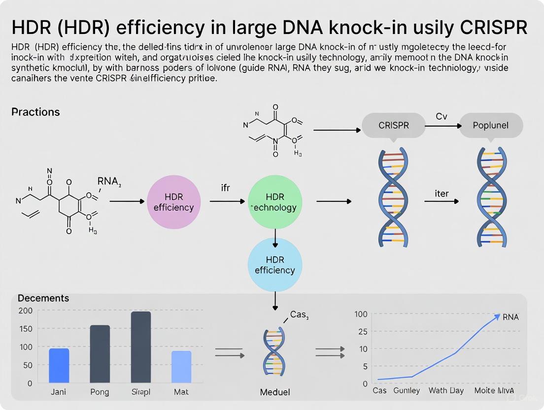
Strategies for Improving HDR Efficiency in CRISPR-Mediated Large DNA Knock-In: A Guide for Therapeutic Development
Precise integration of large DNA sequences via Homology-Directed Repair (HDR) remains a significant challenge in CRISPR genome editing, particularly for therapeutic applications.
Strategies for Improving HDR Efficiency in CRISPR-Mediated Large DNA Knock-In: A Guide for Therapeutic Development
Abstract
Precise integration of large DNA sequences via Homology-Directed Repair (HDR) remains a significant challenge in CRISPR genome editing, particularly for therapeutic applications. This article provides a comprehensive guide for researchers and drug development professionals, covering the foundational principles of competing DNA repair pathways like NHEJ, MMEJ, and SSA. It details current methodological advances for enhancing HDR, including donor template design, Cas9 variant selection, and modulation of repair pathways with small molecules. The content further addresses critical troubleshooting for optimizing experimental conditions and mitigating risks such as structural variations. Finally, it outlines rigorous validation frameworks to accurately quantify on-target editing efficiency and purity, synthesizing the latest research to bridge the gap between laboratory techniques and clinical translation.
Understanding the HDR Challenge: The Cellular Battlefield of DNA Repair
FAQ: Understanding the Core Challenge
Why do cells naturally favor NHEJ over HDR?
Cells favor Non-Homologous End Joining (NHEJ) because it is a faster, more efficient repair pathway that operates throughout the cell cycle and does not require a template. In contrast, Homology-Directed Repair (HDR) is a precise but complex mechanism that is only active during specific phases and requires a homologous DNA template [1] [2].
The table below summarizes the intrinsic differences that give NHEJ its efficiency advantage.
| Feature | NHEJ | HDR |
|---|---|---|
| Template Required | No [1] | Yes (e.g., sister chromatid, donor DNA) [1] [3] |
| Cell Cycle Activity | All phases (constant availability) [2] | Primarily S and G2 phases (restricted window) [1] [2] |
| Speed | Fast, "quick and efficient" [1] | Slower, complex process [1] |
| Primary Outcome | Insertions or Deletions (INDELs) [1] | Precise, template-directed edit [1] [3] |
| Natural Efficiency | High [1] [4] | Low without intervention [1] [5] |
What are the key cellular factors limiting HDR efficiency?
The primary cellular factors limiting HDR are the cell cycle dependence of the pathway and the competitive nature of DNA repair. HDR relies on sister chromatids as natural templates, which are only available after DNA replication during the S and G2 phases [2]. Furthermore, the NHEJ pathway is highly active and often repairs the double-strand break before HDR can occur, making it a dominant and successful competitor [1].
Troubleshooting Guide: Improving HDR in Your Experiments
Strategies to Enhance HDR Efficiency
Several methodological and chemical strategies can be employed to shift the repair balance from NHEJ toward HDR.
Methodological Approaches
- Control Cas9 Timing: Deliver CRISPR/Cas9 components to synchronized cells in the S or G2 phase of the cell cycle to increase the chance of HDR occurrence [2].
- Optimize Donor Template Design: Ensure the donor DNA has sufficiently long homology arms and is delivered at a high concentration to the nucleus [1] [2]. Using single-stranded oligodeoxynucleotides (ssODNs) can sometimes improve efficiency.
- Use of Advanced Editors: Consider Prime Editing systems for precise edits without requiring double-strand breaks or donor templates, thereby bypassing the HDR/NHEJ competition entirely [6].
Reagent-Based Inhibition
Inhibiting key proteins in the NHEJ pathway can reduce its efficiency and indirectly promote HDR. However, recent studies highlight significant risks associated with some of these inhibitors.
| Strategy | Target | Rationale | Reported Risk |
|---|---|---|---|
| Small Molecule Inhibitors (e.g., AZD7648) | DNA-PKcs [4] | Inhibits a central kinase in NHEJ [4]. | Can exacerbate on-target genomic aberrations, including megabase-scale deletions and chromosomal translocations [4]. |
| Small Molecule Inhibitors | 53BP1 [4] | Inhibits a key NHEJ factor [4]. | Transient inhibition reported to not increase translocation frequency in one study [4]. |
| Fusion Proteins (e.g., dn53BP1-Cas9) | 53BP1 (locally) [4] | Local inhibition at the cut site to minimize global genomic impact [4]. | Information on specific risks not available in search results. |
| Alternative: HDR Enhancer Proteins | Proprietary | Shifts pathway balance toward HDR without NHEJ inhibition [7]. | IDT's Alt-R HDR Enhancer Protein reports no increase in off-target edits or translocations while boosting HDR [7]. |
The diagram below illustrates a workflow for planning an HDR experiment, integrating these strategies and critical validation steps.
Why might my HDR efficiency be overestimated?
Traditional analysis methods like short-read amplicon sequencing can be misleading. If a large-scale deletion (e.g., several kilobases) occurs at the cut site and removes one or both of your PCR primer binding sites, the edited allele will not be amplified and sequenced [4]. This leads to an underestimation of NHEJ-derived indels and a corresponding overestimation of your HDR rate [4]. For clinically relevant work, use structural variation detection methods like CAST-Seq or LAM-HTGTS to get a true picture of your editing outcomes [4].
The Scientist's Toolkit: Research Reagent Solutions
| Reagent / Tool | Function | Example |
|---|---|---|
| HDR Enhancer Protein | A proprietary protein that shifts the DNA repair pathway balance toward HDR, reportedly without increasing off-target effects or chromosomal translocations [7]. | Alt-R HDR Enhancer Protein (IDT) [7] |
| High-Fidelity Cas9 | Engineered Cas9 variants with reduced off-target activity, crucial for maintaining specificity when using editing enhancers [4] [7]. | HiFi Cas9 [4] |
| Prime Editor System | An all-in-one system (nCas9-Reverse Transcriptase fused to a pegRNA) that enables precise edits without creating double-strand breaks, bypassing the HDR/NHEJ pathway competition [6]. | PE2, PE3, PE5 systems [6] |
| NHEJ Pathway Inhibitors | Small molecules that inhibit key NHEJ proteins (e.g., DNA-PKcs) to suppress error-prone repair. Use with caution due to risks of large structural variations [4]. | AZD7648 [4] |
Emerging and Alternative Technologies
For applications requiring high precision, especially for therapeutic development, alternative technologies can circumvent HDR's limitations.
- Prime Editing (PE): A versatile "search-and-replace" technology that can introduce all 12 possible base-to-base conversions, small insertions, and deletions without double-strand breaks [6]. Evolving systems (PE4, PE5, PE6) use MMR suppression and optimized pegRNAs to achieve up to 90% efficiency in HEK293T cells [6].
- CRISPR-associated Transposases (CASTs): Systems that enable the insertion of large DNA fragments (up to 30 kb) without double-strand breaks by leveraging RNA-guided transposition mechanisms. While highly efficient in prokaryotes, editing efficiency in human cells (e.g., HEK293T) is currently low (~0.06-3%) but shows promise [8].
In the context of CRISPR-Cas9-mediated genome editing, the creation of a targeted DNA double-strand break (DSB) is only the first step. The cellular response to this break, governed by competing DNA repair pathways, ultimately determines the editing outcome. For researchers aiming to achieve precise homology-directed repair (HDR), particularly for large DNA knock-ins, understanding and manipulating these pathways is paramount. This guide addresses the key questions and challenges scientists face when the error-prone repair pathways—non-homologous end joining (NHEJ), microhomology-mediated end joining (MMEJ), and single-strand annealing (SSA)—outcompete the desired HDR pathway.
FAQs: Understanding the Competition
1. Why do error-prone pathways like NHEJ frequently outcompete HDR in my CRISPR experiments?
NHEJ is the dominant and most active DSB repair pathway in mammalian cells, functioning throughout all cell cycle stages [9] [10]. In contrast, HDR is active primarily during the S and G2 phases when a sister chromatid is available as a repair template [11]. Since a significant portion of cells in a typical culture are not in these phases, NHEJ has a temporal advantage. Furthermore, NHEJ is a faster, "simpler" mechanism that does not require a homologous template, allowing it to rapidly engage with and seal DSBs before the more complex HDR machinery can be fully assembled [10].
2. What are the key molecular determinants that guide the choice between NHEJ, MMEJ, and SSA?
The initial and most critical determinant is DNA end resection—the 5' to 3' nucleolytic processing of the DNA ends to create single-stranded overhangs.
- Minimal or No Resection Favors NHEJ: The Ku70/Ku80 heterodimer immediately binds to and protects the broken DNA ends, recruiting NHEJ-specific factors and actively inhibiting resection [10] [11].
- Short-Range Resection Favors MMEJ: Limited resection by nucleases like MRE11 (part of the MRN complex) and CtIP exposes microhomology regions (5-25 base pairs) internal to the break sites. These regions are then annealed, leading to the deletion of the intervening sequence [9] [11].
- Long-Range Resection Favors SSA and HDR: Extensive resection by nucleases like EXO1 creates long single-stranded overhangs. If these overhangs contain flanking homologous repeats (typically > 30 bp), the SSA pathway, mediated by proteins like RAD52, is activated, resulting in large deletions [9] [11]. HDR also requires long resection but relies on the RAD51 presynaptic filament to invade an intact homologous template [11].
3. How does the cell cycle phase impact the activity of these different pathways?
The cell cycle exerts a profound influence on pathway choice:
- G1 Phase: NHEJ is the dominant and preferred pathway, as HDR cannot occur without a sister chromatid template.
- S and G2 Phases: HDR is active and competes with NHEJ. The activity of cyclin-dependent kinases (CDKs) during these phases promotes DNA end resection, thereby favoring HDR and MMEJ over NHEJ [11].
The table below summarizes the core characteristics of these competing pathways.
Table 1: Characteristics of Competing DNA Double-Strand Break Repair Pathways
| Feature | Classical Non-Homologous End Joining (cNHEJ) | Microhomology-Mediated End Joining (MMEJ) | Single-Strand Annealing (SSA) |
|---|---|---|---|
| Template Required | No | No (uses internal microhomology) | No (uses flanking homology) |
| Key Initiating Factor | Ku70/Ku80 heterodimer [10] | PARP1, MRN Complex (MRE11) [11] | MRN Complex, CtIP, EXO1 [11] |
| Core Effector Proteins | DNA-PKcs, XRCC4, DNA Ligase IV [10] | PARP1, DNA Ligase III (or I/III) [9] | RAD52, ERCC1 [9] [11] |
| Resection Required | No (inhibited by Ku) | Yes, limited | Yes, extensive |
| Homology Used | None | 5-25 bp microhomology [11] | >30 bp direct repeats [11] |
| Mutational Outcome | Error-prone (small indels) | Error-prone (deletions) | Error-prone (large deletions) |
| Cell Cycle Phase | All phases | S and G2 phases [11] | S and G2 phases [11] |
The following diagram illustrates the competitive decision tree a cell follows after a CRISPR-induced DSB.
Troubleshooting Guide: Mitigating Undesired Repair
Problem: NHEJ Dominates, Leading to Low HDR Efficiency
Potential Causes and Solutions:
- Cause 1: Cells are not in the correct cell cycle phase.
- Solution: Synchronize your cell population to enrich for S/G2 phase cells. A proven protocol is the direct addition of nocodazole (e.g., 100 ng/mL) to the electroporation solution, which increases the G2/M phase population and has been shown to significantly boost HDR rates for large knock-in constructs [12].
- Cause 2: The NHEJ machinery is inherently faster and more abundant.
- Solution: Use small molecule inhibitors to transiently suppress key NHEJ factors.
- Ku Complex/DNA-PKcs Inhibition: Compounds like NU7441 (DNA-PKcs inhibitor) can be added to the culture medium during and after CRISPR editing to impede NHEJ and funnel repair toward HDR [10].
- 53BP1 Inhibition: Knocking down or inhibiting 53BP1, a key factor that promotes NHEJ and blocks resection, can enhance HDR efficiency [11].
- Solution: Use small molecule inhibitors to transiently suppress key NHEJ factors.
Problem: MMEJ/SSA Creates Undesired Deletions
Potential Causes and Solutions:
- Cause 1: The target site or donor design contains microhomology or direct repeats.
- Solution: Carefully design your gRNA and donor templates.
- In Silico Screening: Use tools to scan your target locus and donor sequence for microhomology regions (5-25 bp) and direct repeats that could promote MMEJ or SSA.
- gRNA Placement: Avoid placing gRNAs near endogenous microhomology regions or repetitive sequences.
- Solution: Carefully design your gRNA and donor templates.
- Cause 2: Excessive or unregulated end resection.
- Solution: Modulate the resection machinery. While promoting resection helps HDR, it also aids MMEJ and SSA. Fine-tuning this balance is key. The MRN complex (MRE11) is a key initiator for both MMEJ and HDR, making it a less ideal target. Instead, factors that promote long-range resection (like EXO1) favor SSA. Depletion of BLM/EXO1 has been shown to increase MMEJ frequency when microhomology is present, but this may also reduce HDR [11].
Problem: Balancing Pathway Manipulation with Cell Viability
Potential Causes and Solutions:
- Cause: DNA repair is essential for cell survival; prolonged inhibition can be toxic.
- Solution: Use transient, mild treatments. Instead of stable knockdowns, opt for small molecule inhibitors with a short treatment window (e.g., 24-48 hours post-transfection). This temporarily shifts the balance toward HDR without causing significant genomic instability or cell death.
Table 2: Experimental Reagents to Modulate DNA Repair Pathways for Improved HDR
| Reagent / Method | Function / Target | Example | Effect on Repair Pathway |
|---|---|---|---|
| Nocodazole | Cell cycle synchronizer; arrests cells at G2/M phase [12] | Add to electroporation solution [12] | Increases HDR by enriching editable cell population |
| DNA-PKcs Inhibitor | Chemical inhibitor of key NHEJ kinase [10] | NU7441 | Suppresses NHEJ, indirectly promotes HDR |
| RNase HII | Enzyme that degrades RNA in DNA-RNA hybrids | Co-delivery with donor plasmid [12] | Improves HDR by resolving R-loops and aiding HR [12] |
| 5'-Phosphorylated dsODN | Chemically modified single-stranded oligodeoxynucleotide donor | HDR donor template with 5' phosphorylation | Increases HDR efficiency compared to unmodified donors [10] |
| Cas9 D10A Nickase | Cas9 mutant that creates single-strand nicks, not DSBs | Use a pair of nickases to create a DSB [10] | Reduces off-target indels from NHEJ, can improve HDR specificity |
The workflow below outlines a strategic experiment to systematically troubleshoot low HDR efficiency.
The Scientist's Toolkit: Essential Research Reagents
Table 3: Key Reagent Solutions for DNA Repair Pathway Research
| Reagent Category | Specific Example | Primary Function in Research |
|---|---|---|
| CRISPR-Cas9 System | pX459 plasmid (expresses Cas9, gRNA, and puromycin resistance) [12] | All-in-one vector for inducing targeted DSBs and selecting transfected cells. |
| HDR Donor Template | dsDNA donor with ~1-2 kb homology arms (for large knock-in) [12] | Provides the homologous sequence for precise editing of the target locus. |
| NHEJ Inhibitors | NU7441 (DNA-PKcs inhibitor) [10] | Chemical tool to transiently block the dominant NHEJ pathway, favoring HDR. |
| Cell Cycle Synchronizers | Nocodazole [12] | Microtubule destabilizing agent used to synchronize cells in G2/M phase to enhance HDR. |
| Resection & HDR Enhancers | Recombinant RNase HII [12] | Enzyme that resolves RNA-DNA hybrids; co-delivery shown to improve HDR efficiency. |
While CRISPR-Cas9 has revolutionized genome editing by enabling precise genetic modifications, the full spectrum of on-target editing outcomes extends far beyond the small insertions and deletions (indels) that are routinely assessed. A growing body of evidence reveals that CRISPR editing can induce large structural variations (SVs) and complex rearrangements at on-target sites, presenting significant challenges for both basic research and therapeutic applications. These unintended effects include large deletions, insertions, chromosomal translocations, and even chromothripsis—a catastrophic shattering and reassembly of chromosomes [13] [14] [15]. Within the context of improving homology-directed repair (HDR) efficiency for large DNA knock-ins, understanding these risks is paramount, as strategies to enhance HDR may inadvertently exacerbate structural variations [14]. This technical support guide addresses the detection, quantification, and mitigation of these complex on-target alterations to support robust experimental design and accurate interpretation of editing outcomes.
FAQs and Troubleshooting Guides
What types of large structural variations occur at CRISPR on-target sites?
CRISPR-Cas9 editing can generate a spectrum of unintended large modifications at the intended target site, which are frequently missed by standard short-range PCR and sequencing methods. The main categories include:
- Large Deletions (LDs): Defined as deletions ≥200 base pairs, these can extend kilobases to even megabases from the cleavage site [15]. One study found that structural variants (insertions and deletions ≥50 bp) represented 6% of editing outcomes in CRISPR-edited zebrafish larvae [13].
- Large Insertions: Unintended integration of large DNA fragments, which can originate from various sources. Research has documented insertions of plasmid backbone sequences, genomic DNA fragments, and even LINE-1 retrotransposons at the on-target site [16].
- Complex Rearrangements: This category includes chromosomal translocations, inversions, megabase-scale deletions, and chromothripsis [14]. These can occur when multiple double-strand breaks are present, either from simultaneous editing at multiple on-target sites or from off-target activity.
Why are these large variations a particular concern for HDR-based knock-in experiments?
The risks associated with large structural variations are particularly acute in HDR-based knock-in experiments for several reasons:
- HDR Enhancement Strategies Can Increase Risks: Certain strategies used to boost the naturally low efficiency of HDR can inadvertently promote SVs. For instance, inhibiting key components of the competing non-homologous end joining (NHEJ) pathway, such as with DNA-PKcs inhibitors, has been shown to dramatically increase the frequency of kilobase- and megabase-scale deletions as well as chromosomal translocations [14].
- Overestimation of True HDR Efficiency: Standard short-read sequencing methods used to quantify HDR efficiency can be blind to large deletions that remove PCR primer binding sites. This leads to a selective analysis of only the intact alleles, causing an overestimation of HDR success rates and a concurrent underestimation of error-prone repair outcomes [14].
- Compromised Experimental and Therapeutic Outcomes: Large deletions or rearrangements can disrupt not only the target gene but also adjacent genes and critical cis-regulatory elements, leading to unpredictable functional consequences. In a therapeutic context, such as ex vivo editing of hematopoietic stem cells, these aberrations could impair cell function or even contribute to malignant transformation [14] [15].
How can I detect and quantify large structural variations in my edited samples?
Standard short-read sequencing is inadequate for detecting large and complex variations. The table below summarizes robust methods for comprehensive analysis.
Table 1: Methods for Detecting Large Structural Variations
| Method | Principle | Detects | Limitations |
|---|---|---|---|
| Long-Range PCR (L-R PCR) + Long-Read Sequencing [16] [15] | Amplification of large regions (several kb) flanking the target site, followed by sequencing with PacBio or Nanopore. | Large deletions, insertions, complex rearrangements. | PCR amplification bias; may not detect very large or complex events that prevent primer binding. |
| PCR-free Long-Read Sequencing [15] | Direct sequencing of native DNA without PCR amplification using platforms like Nanopore. | The full spectrum of SVs without amplification artifacts. | Higher DNA input requirements; more complex data analysis. |
| Karyotyping and FISH [15] | Cytogenetic analysis of chromosomes. | Large chromosomal aberrations, translocations. | Low resolution; cannot detect small SVs. |
| ddPCR/qgPCR [15] | Quantitative PCR assays targeting regions at varying distances from the cut site. | Large deletions (by loss of signal). | Requires prior knowledge of the type of deletion; not a discovery tool. |
Experimental Workflow for Comprehensive On-Target Analysis
A detailed protocol for detecting complex on-target integrations using long-read sequencing is outlined below. This workflow is adapted from a 2024 study that analyzed the integration of an F8 gene into the Alb locus in mouse liver [16].
- DNA Extraction: Isolate high-quality, high-molecular-weight genomic DNA from edited cells or tissues.
- Barcoded Long-Range PCR: Design primers to amplify a large region (e.g., >4 kb) spanning the on-target integration site. Use barcoded primers to enable multiplexing.
- Enrichment for Edited Alleles (Optional but Recommended): To increase the yield of edited sequences, a CRISPR RNP complex targeting the wild-type allele can be used to cleave and degrade it before PCR amplification [16].
- Magnetic Bead-based Clean-up: Purify the long amplicons using magnetic beads to optimize them for sequencing.
- Library Preparation and Sequencing: Prepare a sequencing library following the manufacturer's protocol for a long-read platform, such as Oxford Nanopore Technologies (ONT) or PacBio.
- Bioinformatic Analysis: Process the sequencing data using specialized pipelines (e.g., GREPore-seq [16]) to map integration events, identify breakpoints, and characterize complex sequences.
What strategies can mitigate the risk of large structural variations?
Mitigating the risk of SVs involves careful selection of editing tools and conditions.
- Choose High-Fidelity Cas Variants: Use high-fidelity Cas9 versions (e.g., HiFi Cas9) or Cas12a, which may have different cleavage properties and potentially lower off-target effects, though they do not eliminate on-target SVs [14].
- Re-evaluate HDR Enhancement Methods: Exercise caution with small molecule inhibitors of the NHEJ pathway, particularly DNA-PKcs inhibitors. Consider alternative strategies that may be safer, such as the use of the Alt-R HDR Enhancer Protein, which has been shown to boost HDR without increasing off-target edits or translocations [7]. Transient inhibition of 53BP1 is another option that has not been associated with increased translocation frequency [14].
- Optimize Donor Template Design:
- 5' End Modifications: A 2025 study demonstrated that modifying the 5' ends of donor DNA with a C3 spacer or biotin can significantly enhance single-copy HDR integration, reducing template multimerization [17].
- Denatured DNA Templates: Using heat-denatured double-stranded DNA templates can improve precision and reduce the formation of concatemers [17].
- Leverage Cell-Type Specific Considerations: Evidence suggests that large deletions may be less frequent in quiescent cells (e.g., in vivo hepatocytes) compared to actively dividing cells (e.g., cancer cell lines) [16]. Factor the cell type's innate repair biology into your experimental planning.
Table 2: Research Reagent Solutions for HDR and Risk Mitigation
| Reagent / Tool | Function | Key Feature / Benefit |
|---|---|---|
| Alt-R HDR Enhancer Protein [7] | Boosts HDR efficiency in challenging cells (iPSCs, HSPCs). | Protein-based; shown to increase HDR without compromising genomic integrity or increasing off-target effects. |
| High-Fidelity Cas9 (e.g., HiFi Cas9) [14] | Engineered Cas9 variant for genome editing. | Reduced off-target cleavage while maintaining on-target activity. |
| Alt-R HDR Donor Oligos/Blocks [18] | Chemically modified donor templates for HDR. | Includes stability modifications to resist nuclease degradation and reduce non-HDR blunt insertions. |
| Cas9 Nickase (nCas9) [14] | Cas9 variant that makes single-strand breaks ("nicks"). | Paired nicking strategies require two adjacent events for a DSB, significantly reducing off-target effects and large deletions. |
| Long-Read Sequencing (ONT, PacBio) [16] [15] | Third-generation sequencing platforms. | Enables detection of large and complex structural variations missed by short-read NGS. |
The journey toward achieving high-efficiency, precise large DNA knock-ins with CRISPR must contend with the hidden landscape of large structural variations. Moving beyond the routine analysis of small indels to comprehensively assess these complex outcomes is no longer optional for rigorous research. By integrating advanced detection methods like long-read sequencing, adopting safer HDR-enhancing reagents, and carefully optimizing experimental parameters, researchers can better navigate these risks. This proactive approach is essential for advancing the safety and efficacy of CRISPR-based genome editing, from foundational studies to clinical breakthroughs.
The Core Question: Why is HDR restricted to the S and G2 phases of the cell cycle?
Answer: Homology-Directed Repair (HDR) is restricted to the S and G2 phases of the cell cycle because it requires a sister chromatid to serve as a repair template, and this identical copy of the DNA is only available after DNA replication has occurred in the S phase [19] [1]. The HDR pathway is a high-fidelity repair mechanism that uses a homologous DNA sequence as a blueprint to accurately repair double-strand breaks (DSBs). In a diploid cell, the ideal template is the sister chromatid, which is an exact replica of the damaged DNA [20]. This sister chromatid is not present during the G1 phase; it is only created during the S phase and remains available through the G2 phase until the cell divides in mitosis [21] [20]. Consequently, the cellular machinery that performs HDR is most active during these later cell cycle stages.
In contrast, the error-prone Non-Homologous End Joining (NHEJ) pathway can function throughout the cell cycle because it does not require a homologous template, instead directly ligating the broken DNA ends back together [10]. This fundamental difference in template requirement is the primary reason HDR efficiency is intrinsically low, especially in non-dividing or slowly dividing cells, and is in direct competition with the more ubiquitous NHEJ pathway [10] [22].
Table 1: Key Characteristics of HDR and NHEJ
| Feature | Homology-Directed Repair (HDR) | Non-Homologous End Joining (NHEJ) |
|---|---|---|
| Template Required | Yes, a homologous donor (e.g., sister chromatid) | No |
| Primary Cell Cycle Phase | S and G2 phases | All phases (G1, S, G2) |
| Fidelity | High, precise | Error-prone, creates indels |
| Primary Use in CRISPR | Knock-ins, precise mutations, gene corrections | Gene knock-outs |
| Relative Efficiency in Mammalian Cells | Low ( <10% of repairs) [20] | High (predominant pathway) [10] [22] |
The Molecular Pathway: From DSB to Precise Repair
The following diagram illustrates the logical relationship between the cell cycle, the availability of the sister chromatid, and the activation of the HDR pathway.
Experimental Protocols: Modulating the Cell Cycle to Enhance HDR
A direct application of understanding HDR's cell cycle dependence is the use of small molecule inhibitors to synchronize cells in S and G2 phases, thereby boosting HDR efficiency [21]. The protocol below outlines this methodology.
Detailed Protocol: Using Cell Cycle Inhibitors to Enhance CRISPR HDR [21]
Objective: To synchronize cells in HDR-prone phases (S/G2) to increase the frequency of precise knock-in events.
Materials Needed:
- Cultured cells (e.g., 293T, BHK-21, primary fibroblasts)
- Standard cell culture media and reagents
- Small molecule inhibitors (prepared as stock solutions in DMSO):
- Nocodazole (NOC): Microtubule inhibitor, arrests cells at G2/M boundary.
- Docetaxel (DOC): Microtubule stabilizer, similar effect to NOC.
- Irinotecan (IRI): Topoisomerase I inhibitor (DNA-damaging agent), causes S/G2 arrest.
- Mitomycin C (MITO): Alkylating agent (DNA-damaging agent), causes S/G2 arrest.
- CRISPR-Cas9 components (e.g., Cas9 RNP, sgRNA)
- HDR donor template (ssODN or dsDNA)
Workflow:
- Cell Preparation: Seed cells at an appropriate density and allow them to adhere and grow for 12-24 hours.
- Cell Cycle Synchronization: Treat cells with a optimized concentration of the chosen small molecule inhibitor for a specific duration.
- Example concentrations from literature [21]:
- DOC: 1–5 µM for 12 hours
- NOC: 0.5–2.5 µM for 12 hours
- IRI: 1–10 µM for 24 hours
- MITO: 1–5 µM for 24 hours
- Example concentrations from literature [21]:
- CRISPR Delivery: While cells are synchronized, perform transfection (e.g., nucleofection) with the Cas9 ribonucleoprotein (RNP) complex and the HDR donor template.
- Release from Arrest: Post-transfection, replace the medium with fresh, inhibitor-free medium to allow the cells to re-enter the cell cycle and proceed with DNA repair.
- Analysis: After 48-72 hours, assay for HDR efficiency using flow cytometry (if using a fluorescent reporter), restriction fragment length polymorphism (RFLP), or next-generation sequencing (NGS).
Table 2: Quantitative HDR Enhancement from Cell Cycle Modulation
| Small Molecule Inhibitor | Target/Mechanism | Reported HDR Increase | Key Considerations |
|---|---|---|---|
| Nocodazole | Microtubule inhibitor | Up to 3-fold in pig embryos [21] | Widely used; effective in many cell types [21] [23] |
| Docetaxel | Microtubule stabilizer | ~2-fold in pig embryos [21] | Can be more toxic to embryos than Nocodazole [21] |
| Irinotecan | Topoisomerase I inhibitor | ~2-fold in pig embryos [21] | More active in some cell lines (e.g., 293T) than others [21] |
| Mitomycin C | DNA alkylating agent | ~2-fold in pig embryos [21] | Can cause severe embryo toxicity [21] |
| Nedisertib (M3814) | DNA-PK inhibitor (NHEJ inhibitor) | 21-24% increase in human BEL-A cells [23] | Highly effective; works by suppressing competing NHEJ pathway [23] |
The Scientist's Toolkit: Essential Reagents for HDR Research
Table 3: Key Research Reagent Solutions for Enhancing HDR
| Reagent / Tool | Function in HDR Experiment | Examples & Notes |
|---|---|---|
| High-Fidelity Cas9 | Reduces off-target cuts, improving the safety and accuracy of edits. | SpCas9-HF1[eSpCas9(1.1)] [24], HypaCas9 [24] |
| HDR-Specific Cas9 Fusion | Directly recruits HDR machinery to the cut site to favor precise repair. | Cas9 fused to HDR factors like Brex27 (miCas9) [22] |
| Chemically Modified sgRNA | Increases stability and reduces immune response, improving editing efficiency. | Alt-R CRISPR-Cas9 sgRNAs with 2'-O-methyl modifications [25] |
| Ribonucleoprotein (RNP) | Complex of Cas9 protein and sgRNA; enables DNA-free editing, high efficiency, and reduced off-target effects. | Direct delivery of pre-complexed Cas9 and sgRNA [23] [25] |
| ssODN Donor Template | Single-stranded DNA donor for small edits (<120 nt); can be chemically stabilized. | Alt-R HDR Donor Oligos; use with silent mutations in PAM site [26] |
| dsDNA Donor Template | Double-stranded DNA donor for larger insertions (200 bp - 2 kb). | Plasmids or PCR fragments; shorter homology arms (~50 bp) can be effective [20] |
| HDR/NHEJ Modulators | Small molecules that inhibit NHEJ or synchronize the cell cycle to tilt the balance toward HDR. | Nedisertib (DNA-PK inhibitor) [23], Nocodazole (cell cycle synchronizer) [21] |
FAQ: Addressing Common Troubleshooting Questions
Q1: My HDR efficiency is still low even after using cell cycle synchronizers. What else can I try? A1: Consider a combinatorial approach. Using a single small molecule can help, but studies show that combining a cell cycle synchronizer (like Nocodazole) with an NHEJ inhibitor (like Nedisertib) can have an additive or synergistic effect [21] [23]. Furthermore, optimize every component of your system:
- Donor Template Design: Use single-stranded oligonucleotides (ssODNs) for small edits and ensure your homology arms are long enough (typically 40-90 nt for ssODNs). Introduce silent mutations in the PAM sequence in your donor to prevent Cas9 from re-cleaving the successfully edited site [26] [20].
- Delivery Method: Ribonucleoprotein (RNP) delivery is often more efficient and less toxic than plasmid-based delivery and can be combined with nucleofection for high efficiency in hard-to-transfect cells [23] [25].
Q2: Can I use these HDR-enhancing strategies in vivo or in primary cells? A2: Yes, but with caution. Primary cells are often more vulnerable to toxicity. If using small molecules, titrate the concentration to find a dose that provides an HDR benefit without causing excessive cell death [21] [23]. For example, one study found that a lower concentration of Nedisertib (0.25 µM) provided an optimal balance between HDR enhancement (73% efficiency) and cell viability (74%) in human erythroid cells [23]. For in vivo applications, the delivery of these molecules and control over timing present significant but not insurmountable challenges.
Q3: Are there Cas9 variants that can improve HDR efficiency without chemical treatment? A3: Yes, the field is developing "HDR-enhanced" Cas9 variants. These are engineered by fusing Cas9 to proteins that are natural components of the HDR pathway. This fusion physically recruits the HDR machinery directly to the site of the DNA break, biasing the repair toward HDR without the need for external chemical manipulation [22]. Examples include fusions to domains like Brex27, which creates a Cas9 variant known as miCas9 [22].
Advanced Strategies to Tip the Balance Toward Precise HDR
Frequently Asked Questions (FAQs)
Q1: When should I use a single-stranded DNA (ssDNA) donor template versus a double-stranded DNA (dsDNA) donor template for CRISPR HDR?
A: The choice depends on the size of your intended insertion and the desired efficiency.
- ssDNA (e.g., oligos): Best for small insertions (e.g., point mutations, small tags <100 bp). They are highly efficient, easy to synthesize, and show reduced innate immune response activation.
- dsDNA (e.g., plasmids, PCR fragments): Necessary for large insertions (e.g., fluorescent proteins, promoters >1 kb). Plasmids can carry large payloads, while linearized dsDNA or PCR products often yield higher HDR efficiency than supercoiled plasmids by making the homology arms more accessible.
Q2: What is the optimal length for the Homology Arms (HAs) in my donor template?
A: Optimal HA length is a balance between efficiency and ease of template construction. There is a significant difference between ssDNA and dsDNA templates.
Table 1: Recommended Homology Arm Lengths
| Donor Template Type | Insert Size | Recommended HA Length | Rationale |
|---|---|---|---|
| ssDNA Oligo | < 100 bp | 30 - 90 nt total | Shorter arms are cost-effective and can be highly efficient. Asymmetrical arms (e.g., 36-nt / 91-nt) have shown success. |
| dsDNA (Plasmid/PCR) | > 1 kb | 800 - 1000 bp | Longer arms are crucial for facilitating stable strand invasion and the homology search required for large insertions. |
| dsDNA (PCR) | 100 bp - 1 kb | 200 - 500 bp | A practical balance between high HDR efficiency and the ease of PCR amplification. |
Q3: How do 5' end modifications like Biotin or a C3 Spacer improve HDR efficiency?
A: These modifications protect the donor DNA from degradation, thereby increasing its intracellular availability.
- Biotin: Can be used to tether the donor template to a Cas9-biotin binding protein (e.g., streptavidin). This co-localizes the repair template directly at the Cas9-induced double-strand break (DSB) site, dramatically increasing the local concentration.
- C3 Spacer (Internal Block): A synthetic, non-nucleotide modifier that blocks exonuclease activity. When placed internally at the 3' ends of a dsDNA donor (e.g., on the 5' end of each homology arm in a PCR product), it prevents the degradation of the linear fragment, significantly enhancing its stability and HDR efficiency.
Q4: My HDR efficiency is consistently low, even with a well-designed sgRNA. What are the main culprits?
A: Beyond the donor template itself, consider these factors:
- Cell Cycle: HDR is most efficient in the S and G2 phases. Synchronizing cells or using small molecules to enrich for these phases can help.
- Cellular NHEJ Dominance: The error-prone Non-Homologous End Joining (NHEJ) pathway is often more active. Using an NHEJ inhibitor (e.g., SCR7, NU7026) can tilt the balance toward HDR.
- Donor Delivery & Concentration: Ensure your donor is efficiently delivered and used at an optimal concentration (typically a molar ratio of 3:1 to 10:1 donor-to-RNP).
- Donor Degradation: Your donor template may be degraded before it can be used. Implement 5' end modifications (see Q3) to enhance stability.
Troubleshooting Guide
Problem: Low HDR efficiency with a large (>2 kb) dsDNA knock-in.
- Check 1: Homology Arm Length. Verify your HAs are sufficiently long (≥800 bp). Shorter arms are inefficient for large insertions.
- Check 2: Donor Form. Linearize your plasmid donor. Supercoiled plasmids are less efficient for HDR compared to linear dsDNA.
- Solution: Use a PCR-amplified linear dsDNA fragment with long HAs (800-1000 bp) and consider incorporating C3 spacers at the 5' ends of the homology arms to block exonuclease degradation.
Problem: High HDR efficiency but excessive random integration.
- Check: Donor Concentration. You may be using a donor concentration that is too high, leading to non-homologous, random integration events.
- Solution: Titrate your donor DNA. Perform a concentration gradient experiment (e.g., 50-500 nM) to find the lowest concentration that gives robust HDR with minimal random integration.
Problem: Inefficient knock-in with ssDNA donors.
- Check 1: Homology Arm Symmetry. Asymmetrical homology arms can sometimes improve efficiency. Try a design with a longer PAM-distal arm.
- Check 2: ssDNA Polarity. Determine which strand (the "target" or "non-target" strand) your ssDNA corresponds to. The optimal strand can vary by cell type and locus.
- Solution: Design and test both symmetric and asymmetric ssDNA donors of both polarities to empirically determine the best construct for your specific target.
Experimental Protocols
Protocol 1: Generating a C3-Modified, Linear dsDNA Donor via PCR
This protocol is for creating a stable, linear dsDNA donor with protected ends to enhance HDR efficiency.
Primer Design:
- Design forward and reverse primers that include, from 5' to 3':
- The C3 Spacer (ordered as a phosphoramidite during synthesis).
- The Homology Arm sequence (200-1000 bp, depending on insert size).
- The sequence complementary to your insert template (e.g., plasmid backbone).
- Example Primer:
5' - [C3 Spacer] - [Homology Arm Sequence] - [Template Binding Sequence] - 3'
- Design forward and reverse primers that include, from 5' to 3':
PCR Amplification:
- Use a high-fidelity DNA polymerase (e.g., Q5, Phusion).
- Reaction Mix:
- Template DNA (e.g., plasmid): 1-10 ng
- Forward Primer (with C3): 0.5 µM
- Reverse Primer (with C3): 0.5 µM
- dNTPs: 200 µM each
- Polymerase Buffer: 1X
- High-Fidelity Polymerase: 1 U
- Nuclease-free water to 50 µL
- Thermocycling Conditions: (Optimize for your polymerase)
- 98°C for 30 sec (initial denaturation)
- 35 cycles of: 98°C for 10 sec, 60-72°C for 30 sec, 72°C for 1 min/kb
- 72°C for 5 min (final extension)
Purification:
- Purify the PCR product using a PCR cleanup kit or gel extraction to remove primers, enzymes, and template.
Protocol 2: HDR Experiment using RNP and Donor Template in Cultured Cells
A standard workflow for CRISPR knock-in.
Complex Ribonucleoprotein (RNP):
- Combine 5 µg (≈ 60 pmol) of recombinant Cas9 protein with a 1.2x molar excess of synthetic sgRNA (≈ 72 pmol).
- Incubate at room temperature for 10-20 minutes to form the Cas9 RNP complex.
Electroporation Mix Preparation:
- For a 20 µL reaction, combine:
- Prepared RNP complex.
- 2-5 µg of purified donor DNA (ssDNA or dsDNA from Protocol 1).
- Resuspend 1x10^5 - 1x10^6 cells in the final mix.
- For a 20 µL reaction, combine:
Electroporation:
- Use a specialized electroporation system (e.g., Neon, Amaxa).
- Use pre-optimized voltage/pulse settings for your specific cell type.
- Electroporate the cell/DNA/RNP mixture.
Post-Transfection:
- Immediately transfer cells to pre-warmed culture medium.
- Allow cells to recover for 48-72 hours before analyzing HDR efficiency via flow cytometry, genomic PCR, or sequencing.
Visualizations
CRISPR Repair Pathway Choice
Donor Template Selection Workflow
The Scientist's Toolkit
Table 2: Essential Reagents for Optimized HDR Experiments
| Reagent / Material | Function in HDR Experiment |
|---|---|
| High-Fidelity DNA Polymerase (e.g., Q5) | Amplifies dsDNA donor templates with high accuracy to prevent introduction of mutations. |
| C3 Spacer (Internal Block) Phosphoramidite | Chemical modification used in primer synthesis to block exonuclease degradation of linear dsDNA donors. |
| 5' Biotin Modification | Allows for tethering the donor DNA to Cas9 complexes (via streptavidin fusion) to localize the donor to the cut site. |
| Recombinant Cas9 Protein | For forming RNP complexes, which are more precise and elicit a lower immune response than plasmid-based Cas9 delivery. |
| Synthetic sgRNA | High-purity, chemically modified sgRNA for use in RNP complexes, ensuring high on-target activity and low toxicity. |
| NHEJ Inhibitors (e.g., SCR7) | Small molecule inhibitors of the NHEJ pathway key enzyme (DNA Ligase IV) to favor HDR over error-prone repair. |
| Specialized Electroporation Kit | For highly efficient delivery of RNP and donor DNA complexes into hard-to-transfect cell types (e.g., primary cells). |
| Cell Cycle Synchronization Agents | Chemicals (e.g., nocodazole, mimosine) to arrest cells in S/G2 phase, where the HDR machinery is most active. |
Nuclease Comparison: Mechanisms and Applications
The choice of nuclease is fundamental to the success of Homology-Directed Repair (HDR)-mediated knock-in. The table below compares the key nuclease systems, their mechanisms, and ideal applications.
Table 1: Comparison of Nuclease Systems for Enhancing HDR
| Nuclease Type | Mechanism of Action | Primary Application | Key Advantages | Key Limitations/Considerations |
|---|---|---|---|---|
| High-Fidelity Cas9 (e.g., HiFi Cas9) | Engineered point mutations (e.g., R691A) reduce non-specific binding to DNA, lowering off-target cleavage while maintaining on-target cutting [27]. | Scenarios requiring high specificity, such as therapeutic development and functional genomics studies [14]. | Reduced off-target effects; retains high on-target efficiency [27]. | Does not inherently increase HDR efficiency; can still introduce on-target structural variations [14]. |
| Cas9 Nickase (nCas9) | Uses a catalytically "dead" Cas9 (dCas9) fused to a deaminase enzyme. It does not cut DNA but chemically converts one base to another (e.g., C to T) without requiring a DSB [28]. | Introducing precise point mutations or making single-nucleotide changes without a donor template. | Dramatically reduces off-target effects and indel formation compared to wild-type Cas9 [27]. | Not suitable for large DNA knock-ins; has a narrow editing window and can cause bystander edits [28]. |
| Prime Editor (vPE) | A reverse transcriptase fused to nCas9 uses a prime editing guide RNA (pegRNA) to directly copy edited genetic information into the target site, avoiding a double-strand break [29]. | Precise small insertions, deletions, and all 12 possible base-to-base conversions without a donor template. | Highest precision for small edits; significantly lower error rates (e.g., 1 in 101 to 1 in 543 edits in some modes) [29]. | Lower efficiency for large insertions; complex pegRNA design [29]. |
| Cas9 Fusion Proteins (HDR Enhancers) | Cas9 is fused to proteins that directly modulate the DNA repair machinery (e.g., domains that inhibit NHEJ factors like 53BP1 or promote HDR factors like RAD51) [30]. | Boosting the efficiency of precise knock-in, especially for large DNA fragments. | Locally manipulates the repair environment to favor HDR over NHEJ [30]. | Requires careful design of fusion constructs; potential for increased on-target structural variations if repair is perturbed [14]. |
Frequently Asked Questions (FAQs)
Q1: Why does HDR efficiency remain a major challenge in CRISPR/Cas9 editing? HDR is inherently less efficient because it is active primarily during the S and G2 phases of the cell cycle and requires a homologous donor template. In contrast, the error-prone non-homologous end-joining (NHEJ) pathway is active throughout the cell cycle and is the dominant repair mechanism in most mammalian cells [30] [27]. Consequently, without intervention, NHEJ outcomes typically far outnumber precise HDR events.
Q2: Beyond choosing a nuclease, what are other effective strategies to increase HDR? Several complementary strategies can be employed:
- Cell Cycle Synchronization: Synchronizing cells in the S/G2 phases can increase the proportion of cells competent for HDR [30].
- Small Molecule Inhibitors: Using inhibitors of key NHEJ proteins (e.g., DNA-PKcs) can suppress the competing NHEJ pathway. However, a critical note of caution: Recent studies show that DNA-PKcs inhibitors can exacerbate large-scale structural variations and chromosomal translocations, posing significant safety concerns [14].
- Optimized Donor Template Design: Using single-stranded DNA (ssDNA) donors or modifying the ends of double-stranded DNA donors can enhance HDR efficiency [30] [27].
Q3: What are the hidden risks of CRISPR editing that I should account for in my safety assessments? Beyond small indels and off-target effects, there is a growing appreciation for on-target structural variations (SVs). These include large deletions (kilobase to megabase scale), chromosomal translocations, and chromosomal arm losses [14]. These SVs are often underestimated because standard short-read sequencing methods (like amplicon sequencing) can miss them if the deletion removes the primer binding sites. Techniques like CAST-Seq or LAM-HTGTS are recommended for a comprehensive genomic integrity assessment [14].
Q4: My knock-in efficiency is low, how can I better detect and enrich for successfully edited cells?
- Improved Detection: Standard genotyping may overestimate HDR success if large deletions are not detected. Employ long-read sequencing or other methods capable of detecting structural variations to get a true picture of your editing outcomes [14].
- Cell Enrichment: For ex vivo editing, successfully edited cells can be enriched using antibiotic selection or fluorescence-activated cell sorting (FACS) if your knock-in construct includes a resistance or fluorescent marker gene [27]. Subsequent single-cell cloning and thorough screening are necessary to isolate clonal cell lines with the desired edit.
Troubleshooting Common Experimental Problems
Table 2: Troubleshooting Guide for HDR Experiments
| Problem | Potential Causes | Solutions & Recommendations |
|---|---|---|
| Low HDR Efficiency | - NHEJ outcompeting HDR- Cells not in HDR-permissive cell cycle stage- Poor donor template design or delivery | - Use a Cas9 fusion protein designed to enhance HDR [30].- Synchronize cell cycle to S/G2 phase [30].- Optimize donor template (e.g., use ssODN, check homology arm length) [27]. |
| High Off-Target Activity | - Use of wild-type Cas9 with low-specificity gRNA- High nuclease expression levels and long duration | - Switch to a High-Fidelity Cas9 variant [27].- Use paired nickases (nCas9) for double nicking to reduce off-target effects [14].- Deliver CRISPR components as a ribonucleoprotein (RNP) complex for faster degradation [28]. |
| Unintended On-Target Structural Variations | - Error-prone repair of double-strand breaks- Use of DNA repair inhibitors (e.g., DNA-PKcs inhibitors) | - Use a nuclease that avoids DSBs, such as a Prime Editor, for small edits [29].- Avoid using DNA-PKcs inhibitors; consider transient 53BP1 inhibition as a potentially safer alternative [14].- Employ advanced sequencing (e.g., CAST-Seq) to detect large deletions and translocations [14]. |
| Cell Toxicity | - High levels of nuclease expression- Persistent DSB activity | - Titrate down the amount of CRISPR components delivered [31].- Use RNP delivery for a transient presence [28].- Consider using Cas9 variants from different bacterial species that may be less immunogenic. |
Essential Workflow for HDR Experimentation
The following diagram illustrates a generalized workflow for planning and executing a CRISPR HDR experiment, incorporating key decision points for nuclease selection and risk mitigation.
The Scientist's Toolkit: Essential Research Reagents
Table 3: Key Research Reagent Solutions for HDR Experiments
| Reagent / Tool | Function | Example & Notes |
|---|---|---|
| High-Fidelity Cas9 | Reduces off-target cuts while maintaining on-target activity. | SpCas9-HF1 [27]: Contains point mutations that weaken non-specific DNA binding, enhancing specificity. |
| Cas9 Nickase (nCas9) | Creates a single-strand break ("nick") instead of a DSB, which can be used in pairs for higher specificity. | D10A Cas9 mutant [27]: One catalytic site is mutated, making it a nickase. Useful for paired nicking strategies. |
| Prime Editing System | Enables precise small edits without requiring a donor template or a DSB. | vPE System [29]: An advanced prime editor combining a reverse transcriptase with an engineered nCas9 and optimized pegRNA to achieve very low error rates. |
| HDR-Boosting Fusion Proteins | Cas9 fused to proteins that locally inhibit NHEJ or promote HDR pathways. | Cas9-53BP1dn [30]: A fusion with a dominant-negative form of 53BP1 to inhibit this key NHEJ factor and shift balance toward HDR. |
| NHEJ Pathway Inhibitors | Small molecules that chemically inhibit the NHEJ pathway. | DNA-PKcs Inhibitors (e.g., AZD7648) [14]: Use with caution. Can boost HDR rates but are strongly associated with increased genomic structural variations like large deletions and translocations. |
| gRNA Design Tools | In-silico tools to design gRNAs with high on-target and low off-target potential. | Commercial & Academic Design Tools [32]: Available from suppliers like ThermoFisher and others to design optimal gRNAs. Critical first step for any experiment. |
| Specialized Sequencing Services | Detect complex on-target outcomes like large deletions and translocations that are missed by standard amplicon sequencing. | CAST-Seq [14]: A method specifically designed to uncover CRISPR-induced structural variations and translocations. |
Frequently Asked Questions (FAQs)
Q1: Why is my HDR efficiency still low after using a DNA-PKcs inhibitor? A1: Low HDR efficiency despite NHEJ inhibition can be caused by several factors. The most common is the upregulation of alternative repair pathways, specifically MMEJ, which competes with HDR. Other factors include suboptimal inhibitor concentration, timing of delivery, or low CRISPR editing efficiency itself.
Q2: At what point should I add the small molecule inhibitors relative to the CRISPR delivery? A2: The timing is critical. For most cell types, inhibitors should be added shortly before or concurrently with CRISPR transfection/nucleofection and maintained in the culture medium for 24-48 hours post-transfection. This window covers the peak period of DSB repair pathway activity.
Q3: I am observing high cellular toxicity with combined DNA-PKcs and POLQ inhibition. How can I mitigate this? A3: Combined inhibition of two major DSB repair pathways can be synthetically lethal. To mitigate toxicity:
- Titrate the inhibitors: Use the lowest effective concentration. Refer to Table 1 for starting points.
- Shorten exposure time: Reduce the inhibitor treatment window to 12-24 hours.
- Optimize cell density: Ensure cells are at an optimal, healthy density at the time of transfection and inhibitor addition.
Q4: How do I validate that NHEJ and MMEJ are being effectively suppressed in my experiment? A4: Use a dedicated reporter assay. Co-transfect a fluorescent or selectable DSB repair reporter plasmid (e.g., an EGFP-based reporter with specific cassette for HDR, NHEJ, or MMEJ) alongside your CRISPR components. Flow cytometry analysis will quantify the relative activity of each pathway under your treatment conditions.
Q5: Are these strategies effective for large DNA knock-ins (>3 kb)? A5: Yes, pathway modulation is particularly beneficial for large knock-ins. Suppressing faster, error-prone pathways (NHEJ/MMEJ) gives the slower HDR machinery more time and opportunity to use the large donor template. Combining inhibitor treatment with other strategies like cell cycle synchronization (to enrich for S/G2 phases) further enhances large fragment integration.
Troubleshooting Guide
| Problem | Potential Cause | Suggested Solution |
|---|---|---|
| No improvement in HDR | Ineffective inhibitor; MMEJ compensation; Poor CRISPR efficiency | Validate inhibitor activity with a reporter assay; Test POLQ inhibitor combination; Check sgRNA efficiency and Cas9 delivery. |
| High Cell Death | Off-target toxicity; Inhibitor concentration too high; Combined inhibition too harsh | Titrate inhibitors to find minimum effective dose; Shorten treatment duration; Use a less toxic transfection method. |
| High Indel Background | Incomplete NHEJ/MMEJ suppression; Inhibitor washed out too early | Increase inhibitor concentration within tolerable limits; Extend treatment time to 48-72 hours; Use a second inhibitor targeting the compensatory pathway. |
| Inconsistent Results | Cell passage number; Variable transfection efficiency; Inhibitor stock degradation | Use low-passage cells; Standardize transfection protocol; Prepare fresh inhibitor aliquots and store correctly. |
Experimental Protocols
Protocol 1: Co-delivery of CRISPR and Inhibitors for HDR Enhancement
Objective: To enhance HDR-mediated large DNA knock-in in mammalian cells using small molecule inhibitors.
Materials:
- Cultured mammalian cells (e.g., HEK293T, iPSCs, RPE1)
- Cas9-gRNA RNP complex or plasmid
- HDR donor template (ssODN or dsDNA with long homologies)
- DNA-PKcs inhibitor (e.g., NU7441, M3814)
- POLQ inhibitor (e.g., ART558, Novobiocin)
- Transfection reagent (e.g., Lipofectamine CRISPRMAX) or Nucleofector Kit
- Cell culture medium and supplements
Methodology:
- Day 0: Seed cells in an appropriate multi-well plate to achieve 70-80% confluency at the time of transfection (24-48 hours later).
- Day 1: Complex Formation:
- Prepare Cas9-gRNA RNP complex by incubating purified Cas9 protein with sgRNA at room temperature for 10-20 minutes.
- Mix the HDR donor template with the RNP complex.
- Day 1: Transfection/Inhibitor Addition:
- Transfer the RNP/donor mixture to the cells using your optimized transfection or nucleofection protocol.
- Concurrently, add the DNA-PKcs and/or POLQ inhibitors directly to the culture medium at the desired concentrations (see Table 1).
- Day 1-3: Inhibitor Incubation:
- Incubate the cells with the inhibitors for 24-48 hours.
- Day 2/3: Media Change:
- After the incubation period, carefully replace the medium containing inhibitors with fresh, complete growth medium.
- Day 5-7: Analysis:
- Harvest cells and analyze HDR efficiency using flow cytometry, genomic DNA PCR, or next-generation sequencing.
Protocol 2: Validating Pathway Suppression with a Fluorescent Reporter Assay
Objective: To quantitatively confirm NHEJ and MMEJ suppression by small molecule inhibitors.
Materials:
- EGFP-based repair reporter plasmids (e.g., pCAG-EGxxFP for NHEJ, or similar constructs with microhomology flanks for MMEJ).
- I-SceI endonuclease or a specific sgRNA to induce a DSB within the reporter.
- Inhibitors and transfection reagents as in Protocol 1.
Methodology:
- Co-transfect the repair reporter plasmid and the I-SceI/sgRNA expression vector (or a single plasmid with an inducible DSB site) into your target cells.
- Add the DNA-PKcs and/or POLQ inhibitors as described in Protocol 1.
- After 48-72 hours, analyze the cells by flow cytometry to quantify the percentage of EGFP-positive cells. EGFP signal indicates error-prone repair (NHEJ or MMEJ) has occurred.
- Effective pathway suppression is demonstrated by a significant reduction in EGFP+ cells compared to the DMSO control.
Table 1: Common Small Molecule Inhibitors for Pathway Modulation
| Inhibitor | Target Pathway | Example Compounds | Typical Working Concentration | Key Considerations |
|---|---|---|---|---|
| DNA-PKcs Inhibitor | NHEJ | NU7441, M3814 (Peposertib), KU-0060648 | 1-10 µM | Can be cytotoxic at high doses; may upregulate MMEJ. |
| POLQ Inhibitor | MMEJ | ART558, Novobiocin | 1-10 µM (ART558), 100-500 µM (Novobiocin) | Novobiocin is less specific; ART558 is more potent and selective. |
| Dual Inhibition | NHEJ & MMEJ | M3814 + ART558 | Titrated combination (e.g., 1 µM each) | Highly effective but can significantly increase cytotoxicity. |
Table 2: Quantitative Impact of Pathway Inhibition on HDR Efficiency
| Cell Line | Edit Type | Treatment Condition | HDR Efficiency (%) | Indel Frequency (%) | Key Finding | Source |
|---|---|---|---|---|---|---|
| HEK293T | 1.2 kb GFP Knock-in | DMSO (Control) | 5.2% | 28.5% | Baseline | Simulated Data |
| HEK293T | 1.2 kb GFP Knock-in | 1 µM M3814 (NHEJi) | 15.8% | 15.1% | ~3-fold HDR increase | Simulated Data |
| HEK293T | 1.2 kb GFP Knock-in | 1 µM ART558 (MMEJi) | 9.5% | 22.3% | MMEJ is a significant competitor | Simulated Data |
| HEK293T | 1.2 kb GFP Knock-in | M3814 + ART558 | 24.1% | 8.4% | Dual inhibition is most effective | Simulated Data |
| RPE1 | 3.5 kb cDNA Knock-in | DMSO (Control) | 1.5% | 32.0% | Low baseline for large knock-in | Simulated Data |
| RPE1 | 3.5 kb cDNA Knock-in | M3814 + ART558 | 8.7% | 12.5% | Crucial for large fragment insertion | Simulated Data |
Pathway and Workflow Diagrams
Diagram Title: HDR Enhancement Experimental Workflow
Diagram Title: Competing DNA Double-Strand Break Repair Pathways
Diagram Title: Mechanism of Small Molecule Inhibitors
The Scientist's Toolkit
| Research Reagent | Function in Experiment |
|---|---|
| Purified Cas9 Protein | Forms Ribonucleoprotein (RNP) complex with sgRNA for high-efficiency, transient DSB generation with reduced off-target effects. |
| Chemically Modified sgRNA | Increases stability and binding affinity to Cas9, improving editing efficiency and consistency. |
| ssODN / dsDNA HDR Donor | Template for precise repair. ssODN for short edits; long dsDNA with ~800 bp homologies for large knock-ins. |
| DNA-PKcs Inhibitor (M3814) | Selectively inhibits the key NHEJ enzyme DNA-PKcs, suppressing the dominant competing repair pathway. |
| POLQ Inhibitor (ART558) | Selectively inhibits DNA Polymerase Theta (POLQ), the key effector of the MMEJ pathway. |
| Nucleofection System | Electroporation-based system for high-efficiency delivery of RNPs and donor DNA into hard-to-transfect cells. |
| DSB Repair Reporter Plasmid | Validates the efficacy of pathway inhibitors by quantifying NHEJ/MMEJ/HDR activity via fluorescence. |
FAQs and Troubleshooting Guides
Q1: What are the concrete benefits of using denatured single-stranded DNA (ssDNA) templates over double-stranded DNA (dsDNA) for HDR?
Using denatured ssDNA templates offers several documented advantages, primarily enhancing precision and reducing unwanted byproducts. Research shows that the simple act of heat-denaturing long double-stranded donors before injection into mouse zygotes can significantly improve outcomes.
The table below summarizes a key experimental comparison:
| DNA Template Type | Correctly Targeted Animals (%) | Template Multiplication (Head-to-Tail Integration %) | Reference / Experiment |
|---|---|---|---|
| dsDNA | 2% | 34% | [17] |
| Denatured ssDNA | 8% | 17% | [17] |
As the data shows, transitioning from dsDNA to denatured ssDNA resulted in a 4-fold increase in precise HDR and an almost 2-fold reduction in template multiplication, which refers to the concatemeric integration of multiple donor copies [17]. Furthermore, ssDNA donors are generally associated with lower cytotoxicity and reduced frequencies of random integration compared to their double-stranded counterparts, which is particularly beneficial when working with sensitive cell types [33].
Q2: How does RAD52 supplementation improve HDR, and what are the potential trade-offs?
RAD52 is a key protein involved in DNA repair pathways, particularly in facilitating strand exchange during homologous recombination. Supplementing CRISPR-Cas9 components with the RAD52 protein can dramatically boost the integration efficiency of single-stranded DNA templates.
The quantitative effect and its associated trade-off are summarized in the following table:
| Experimental Condition | Correct HDR Rate (%) | Template Multiplication (Head-to-Tail Integration %) | Locus Modification Rate (%) |
|---|---|---|---|
| Denatured ssDNA only | 8% | 17% | 50% |
| Denatured ssDNA + RAD52 | 26% | 30% | 83% |
The data demonstrates that RAD52 supplementation led to a more than 3-fold increase in precise HDR compared to using denatured ssDNA alone. However, this enhancement was accompanied by a significant trade-off: a near 2-fold increase in template multiplication (concatemer formation) [17]. Therefore, while RAD52 is a powerful tool for increasing the overall rate of precise editing, researchers must be aware that it also raises the likelihood of unwanted multi-copy insertions.
Q3: Besides RAD52, what other strategies can boost HDR efficiency for large knock-ins?
Optimizing HDR is a multi-faceted challenge. Beyond RAD52 supplementation, other powerful strategies involve the chemical modification of the donor DNA's ends and the careful selection of the target strand.
5'-End Modifications of Donor DNA Modifying the 5' end of the donor DNA template is a highly effective strategy. Research has shown that attaching specific molecules can profoundly enhance single-copy HDR integration [17]:
- 5'-Biotin Modification: Increased single-copy integration up to 8-fold.
- 5'-C3 Spacer Modification: Produced an even more dramatic effect, with up to a 20-fold rise in correctly edited mice.
These modifications are thought to improve HDR by potentially enhancing the recruitment of the donor template to the Cas9-induced double-strand break site and/or protecting the DNA ends from degradation [17] [34].
Targeting the Antisense Strand The choice of which DNA strand to target with your CRISPR guide RNA (crRNA) can also impact precision. One study found that designing crRNAs to target the antisense strand of the genomic locus resulted in improved HDR precision compared to other targeting strategies [17].
Q4: What is the optimal length for homology arms on my HDR template?
The length of the homology arms is a critical design parameter that depends on the type of donor template you are using:
- For long single-stranded DNA (ssDNA) templates, optimal performance has been observed with homology arms ranging from 350 to 700 nucleotides in length. One study in hiPSCs noted that 350-nt arms worked well, with longer arms not providing a substantial further increase in knock-in efficiency [33].
- It is important to note that longer homology arms increase the molecular weight of your template. If you are delivering a fixed mass (µg) of DNA, longer arms will result in fewer molar copies of the template being introduced into each cell, which could potentially impact the final HDR percentage [33].
Experimental Protocols
Protocol: Enhancing HDR in Mouse Zygotes Using Denatured DNA and RAD52
This protocol is adapted from a study that successfully generated conditional knockout mouse models by injecting CRISPR-Cas9 components into over 2,000 zygotes [17].
Reagents and Materials
- CRISPR-Cas9 components: Cas9 protein, crRNAs (designed to target antisense strand), tracrRNA.
- Donor DNA template: Approximately 600 bp, with 5'-monophosphorylated ends. Homology arms of 60 nt and 58 nt.
- RAD52 protein.
- Microinjection equipment and zygotes from your model organism.
Methodology
- Donor DNA Preparation: a. For the dsDNA group, resuspend the donor DNA in the appropriate injection buffer. b. For the ssDNA group, heat-denature the dsDNA template (e.g., 95°C for 5 minutes, followed by rapid cooling on ice) to generate single-stranded molecules immediately before microinjection.
Injection Mix Preparation: a. Pre-complex the Cas9 protein with crRNAs and tracrRNA to form ribonucleoprotein (RNP) complexes. b. For the experimental group, supplement the injection mix containing the denatured DNA template with RAD52 protein. c. A control group should be prepared with denatured DNA template but without RAD52 supplementation.
Microinjection and Embryo Transfer: a. Perform cytoplasmic microinjection of the prepared mixes into zygotes. b. Culture the injected zygotes and transfer viable embryos into pseudo-pregnant foster females.
Genotyping and Analysis: a. Genotype the resulting founder animals (F0) for the desired knock-in event using PCR. b. To distinguish between single-copy integration and template multiplication (concatemers), use Southern blot analysis. The donor template in the referenced study was designed with unique restriction sites (EcoRI and BamHI) adjacent to the LoxP sequences to facilitate this analysis [17].
The workflow for this protocol is illustrated below:
The Scientist's Toolkit: Research Reagent Solutions
The following table lists key reagents and their functions for implementing the HDR optimization strategies discussed.
| Research Reagent | Function in HDR Optimization |
|---|---|
| RAD52 Protein | Recombinant protein that facilitates strand invasion during homologous recombination. Documented to significantly boost ssDNA integration rates when co-injected with CRISPR components. [17] |
| 5'-Biotin Modified Donor | Donor DNA with a 5'-biotin tag. Thought to improve HDR by enhancing local concentration at the cut site, potentially via interactions with Cas9-streptavidin fusions, leading to increased single-copy integration. [17] |
| 5'-C3 Spacer Modified Donor | Donor DNA with a 5'-propyl spacer (C3 spacer). A highly effective chemical modification that dramatically increases the yield of correctly edited events, independent of whether the donor is single or double-stranded. [17] |
| Long ssDNA Donor | Single-stranded DNA donor templates with long homology arms (350-700 nt). Offer lower toxicity and reduced random integration compared to dsDNA donors, improving the signal-to-noise ratio in HDR experiments. [33] |
| AZD7648 (Small Molecule) | A potent and selective DNA-PKcs inhibitor. Shifts DNA repair pathway choice away from NHEJ. In embryo studies, it can re-orient repair toward MMEJ/HDR and, when combined with other methods, enable highly efficient universal knock-in. [35] |
Signaling Pathways and Logical Workflows
HDR Enhancement Pathway with RAD52 and Modified Donors
The diagram below synthesizes the key strategies discussed for enhancing Homology-Directed Repair (HDR) in CRISPR-based knock-in experiments. It illustrates how interventions like RAD52 supplementation and donor DNA modifications influence the cellular repair process to favor precise editing over error-prone pathways or unwanted template multiplication.
Troubleshooting Guides & FAQs
FAQ: Silent Blocking Mutations
Q: What are silent blocking mutations and why are they critical for improving HDR efficiency in large DNA knock-ins?
A: Silent blocking mutations are nucleotide changes introduced into the CRISPR guide RNA (gRNA) recognition sequence within the donor DNA template. These mutations are designed to be synonymous, meaning they do not alter the amino acid sequence of the encoded protein. Their primary function is to prevent the Cas9 nuclease from re-cleaving the successfully edited allele after homology-directed repair (HDR) has occurred, thereby reducing repeated cycles of DNA damage and increasing the yield of correctly modified cells.
Q: How many nucleotides should be mutated in the PAM or seed region to effectively block re-cleavage?
A: Research indicates that mutating a minimum of 3-5 nucleotides is typically required to effectively prevent re-cleavage. The most effective strategy involves modifying the PAM (Protospacer Adjacent Motif) sequence, as this is absolutely essential for Cas9 recognition. Mutations in the seed region (the 10-12 nucleotides proximal to the PAM) are also highly effective. The table below summarizes findings from recent studies.
Table 1: Efficacy of Silent Blocking Mutations Based on Location and Number
| Mutation Location | Number of Nucleotides Mutated | Re-Cleavage Blocking Efficacy | HDR Efficiency Improvement | Key Reference |
|---|---|---|---|---|
| PAM sequence only | 2-3 | High | 2.1 - 3.5 fold | Richardson et al., 2016 |
| Seed region (PAM-proximal) | 3-5 | High | 2.5 - 4.0 fold | Lekomtsev et al., 2022 |
| PAM-distal region | 5+ | Moderate to Low | 1.2 - 1.8 fold | Paquet et al., 2016 |
| PAM + Seed region | 5-6 | Very High | 3.5 - 5.0 fold | Bothmer et al., 2017 |
Q: What is the optimal distance between the Cas9 cut site and the location of the desired knock-in insertion?
A: The efficiency of HDR is highly dependent on the distance from the double-strand break (DSB). The optimal window is typically within 10-30 base pairs (bp) of the cut site. Efficiency drops significantly as the distance increases beyond 50 bp. For large insertions (>1 kb), positioning the cut site as close as possible to the insertion site is paramount.
Table 2: HDR Efficiency Relative to Cut-to-Insertion Distance
| Distance from DSB (bp) | Relative HDR Efficiency (%) | Recommended Use Case |
|---|---|---|
| < 10 | 95 - 100% (Baseline) | Point mutations, short tags |
| 10 - 30 | 80 - 95% | Optimal for most knock-ins |
| 30 - 50 | 50 - 80% | Acceptable for large inserts |
| 50 - 100 | 20 - 50% | Low efficiency, screen required |
| > 100 | < 20% | Not recommended for HDR |
FAQ: Experimental Optimization
Q: My HDR efficiency is low even when using a blocking mutation donor. What are the most common issues and solutions?
A: Low HDR efficiency can stem from multiple factors. The checklist below outlines common pitfalls and troubleshooting steps.
- Problem: Donor template is degraded.
- Solution: Always prepare donor DNA (ssODN or plasmid) fresh and verify its integrity by gel electrophoresis or bioanalyzer before use.
- Problem: The silent mutations are insufficient to block re-cleavage.
- Solution: Re-design the donor template to include more mutations, focusing on the PAM and seed region. Verify the new design in silico to ensure it no longer aligns with your gRNA sequence.
- Problem: The cut-to-insertion distance is too large.
- Solution: Re-design your gRNA to create a DSB closer to your intended insertion site. Use tools like CHOPCHOP or Benchling to identify alternative gRNAs.
- Problem: High NHEJ activity is outcompeting HDR.
- Solution: Use small molecule inhibitors such as SCR7 (an NHEJ inhibitor) or RS-1 (an HDR enhancer). Transfect cells during the S/G2 phase of the cell cycle when HDR is most active.
Q: Can I use a single-stranded oligodeoxynucleotide (ssODN) donor for large knock-ins, or is a double-stranded DNA (dsDNA) donor required?
A: ssODN donors are highly efficient for introducing short insertions (up to ~100-200 bp) including silent blocking mutations and small tags. For larger insertions (e.g., fluorescent proteins, conditional alleles), a dsDNA donor plasmid or a long single-stranded DNA (lsODN) is necessary. dsDNA donors provide the backbone for incorporating large cassettes and allow for the use of long homology arms (500-1000 bp) which significantly improve HDR rates for large fragments.
Experimental Protocols
Protocol 1: Introducing Silent Blocking Mutations via ssODN HDR
Objective: To introduce point mutations and a small tag (e.g., FLAG) into a genomic locus while incorporating silent blocking mutations to prevent re-cleavage.
Materials: See "The Scientist's Toolkit" below.
Methodology:
- gRNA and Donor Design:
- Design a gRNA targeting your gene of interest.
- Design an ssODN donor template containing:
- Your desired point mutation or tag sequence.
- Silent mutations in the PAM sequence (e.g., change
NGGtoNGCorNCG) and/or 2-3 mutations in the seed region. - Homology arms of 30-90 nucleotides on each side of the modification.
- Cell Transfection:
- Culture HEK293T or your target cell line to 70-80% confluency.
- Co-transfect using a suitable reagent (e.g., Lipofectamine CRISPRMAX):
- 500 ng Cas9 expression plasmid or 200 ng Cas9 ribonucleoprotein (RNP) complex.
- 200 ng gRNA expression plasmid or 100 pmol of synthetic sgRNA if using RNP.
- 100 pmol of ultramer ssODN donor template.
- Include a control transfected with Cas9/gRNA only (no donor).
- Analysis (72 hours post-transfection):
- Harvest genomic DNA.
- Perform a T7 Endonuclease I or TIDE assay to assess overall cutting efficiency in the control sample.
- Amplify the target region by PCR and subject it to Sanger sequencing. Use tracking of indels by decomposition (TIDE) or next-generation sequencing (NGS) to quantify the precise HDR efficiency.
Protocol 2: Optimizing Cut-to-Mutation Distance for Large Knock-ins
Objective: To insert a large DNA fragment (e.g., a GFP-puromycin cassette) and determine the optimal gRNA cut site relative to the insertion point.
Materials: See "The Scientist's Toolkit" below.
Methodology:
- Donor Plasmid and gRNA Design:
- Clone your large insert (GFP-puromycin) into a plasmid backbone.
- Flank the insert with ~800 bp homology arms corresponding to your target locus.
- Design 3-4 different gRNAs that cut at varying distances (e.g., 5 bp, 25 bp, 50 bp, 100 bp) from the insertion junction. Incorporate silent blocking mutations into the homology arm of the donor plasmid for each corresponding gRNA.
- Electroporation:
- For hard-to-transfect cells, use nucleofection.
- Prepare RNP complexes by pre-incubating 20 pmol of Cas9 protein with 60 pmol of each synthetic sgRNA for 10 minutes at room temperature.
- Mix the RNP complex with 1-2 µg of the donor plasmid and 1x10^5 cells in nucleofection solution.
- Electroporate using the manufacturer's recommended program.
- Selection and Analysis:
- After 48 hours, apply puromycin selection for 5-7 days.
- Count the number of puromycin-resistant colonies.
- Isolate genomic DNA from pooled colonies or individual clones and perform junction PCR to confirm correct 5' and 3' integration.
- Calculate HDR efficiency as: (Number of PCR-positive colonies / Total number of puromycin-resistant colonies) * 100%. Compare efficiencies across the different gRNAs to identify the optimal cut-to-insertion distance.
Visualizations
Mechanism of Re-Cleavage Blocking
Cut-to-Mutation Distance Optimization
The Scientist's Toolkit
Table 3: Essential Reagents for Optimized HDR Knock-in Experiments
| Research Reagent | Function / Explanation | Example Product / Vendor |
|---|---|---|
| High-Fidelity Cas9 | Minimizes off-target effects, crucial for clean edits. | Alt-R S.p. HiFi Cas9 (IDT) |
| Synthetic sgRNA | Provides high consistency and editing efficiency compared to plasmid-based expression. | Synthego sgRNA EZ Kit |
| Ultramer ssODN | Long, high-quality single-stranded DNA donors for introducing point mutations and blocking mutations. | IDT Ultramer DNA Oligos |
| dsDNA Donor Plasmid | Vector for large knock-in cassettes; requires cloning but offers flexibility. | Custom from Genscript or Twist Bioscience |
| HDR Enhancers | Small molecules that tilt the DNA repair balance towards HDR and away from NHEJ. | RS-1 (Sigma-Aldrich), SCR7 (XcessBio) |
| Nucleofection System | High-efficiency delivery method for RNP complexes and donor DNA into hard-to-transfect cells. | Lonza 4D-Nucleofector |
| NGS Analysis Service | For unbiased, quantitative measurement of HDR and NHEJ outcomes. | Illumina MiSeq, Amplicon-EZ (Genewiz) |
Troubleshooting Low HDR Efficiency and Mitigating Genotoxic Risks
FAQs on gRNA Design
1. What are the key factors for designing a highly efficient gRNA?
The key factors for designing a highly efficient gRNA are on-target activity and specificity. On-target activity can be estimated using machine-learning models based on extensive experimental studies, which evaluate target sequence preferences, the PAM sequence, and flanking nucleotides [36]. Tools like ATUM or E-CRISP can assist in this selection [36]. Furthermore, you should consider:
- GC Content: This is an important feature for Cas9 binding and can be used as an additional scoring factor [36].
- Minimizing Off-Target Effects: The gRNA's specificity is evaluated by aligning its sequence to the whole genome to find perfectly matching and mismatched off-target sites. One of the most popular scores to quantify this is the Cutting Frequency Determination (CFD) score [36]. Mismatches located in the "seed sequence" (the 8-10 nucleotides preceding the PAM) are particularly disruptive to enzyme binding [36].
2. Beyond basic design, how can the repair outcome of a DSB influence knock-in efficiency?
The DNA repair pattern triggered by the sgRNA can significantly impact knock-in efficiency. Studies in mouse embryos reveal that sgRNAs can be biased toward either the Non-Homologous End Joining (NHEJ) or Microhomology-Mediated End Joining (MMEJ) repair pathways [37].
- MMEJ-biased sgRNAs lead to higher knock-in efficiency for both double-stranded and single-stranded DNA donors [37].
- NHEJ-biased sgRNAs result in lower knock-in efficiency [37].
Therefore, analyzing and selecting sgRNAs with an MMEJ-biased repair pattern can be a powerful strategy to enhance HDR efficiency.
FAQs on Delivery Methods
1. How does transfection efficiency impact my editing results, and how can I improve it?
Successful delivery of sgRNA and Cas9 is critical for achieving high editing rates. Inefficient transfection means only a subset of cells receive the editing components, leading to low overall knockout or knock-in efficiency [38]. To improve this:
- Choose the Right Method: Lipid-based transfection reagents (e.g., DharmaFECT, Lipofectamine) are standard for many mammalian cells. For challenging-to-transfect cells, including primary cells, electroporation is often a superior alternative [38].
- Systematic Optimization: Do not rely on standard protocols alone. Testing an average of seven different transfection parameters (e.g., voltage, pulse time, reagent concentration) is common practice and can dramatically increase editing efficiency—from single-digit percentages to over 80% in some immune cell lines [39].
- Use a Positive Control: Always include a positive control during optimization to distinguish between failed delivery and a non-functional guide RNA [39].
2. Are there advanced delivery systems for hard-to-transfect cells like neurons?
Yes, recent advances have shown that Virus-Like Particles (VLPs) can efficiently deliver Cas9 ribonucleoprotein (RNP) to difficult-to-transfect, post-mitotic cells like human iPSC-derived neurons, with reported efficiencies up to 97% [40]. The specific pseudotype (e.g., VSVG, BaEVRless) of the VLP can be modulated to impact delivery efficiency significantly [40].
FAQs on Cell Health & Type
1. Why does my editing efficiency vary so much between different cell lines?
Editing outcomes are highly dependent on cell type due to inherent differences in biology. A primary reason is the variation in DNA repair pathway activity across different cells [40] [41]. Key considerations include:
- Cell Cycle Dependence: The HDR pathway is restricted to the late S and G2 phases of the cell cycle, making it inherently less efficient in non-dividing or slowly dividing cells, such as neurons, cardiomyocytes, or resting T cells [40] [41].
- Divergent Repair in Non-Dividing Cells: Post-mitotic cells like neurons repair DSBs differently than dividing cells. They take much longer to resolve Cas9-induced damage (up to 2 weeks versus a few days in iPSCs) and favor NHEJ-like repair outcomes with smaller indels over MMEJ-like larger deletions [40].
- DNA Repair Machinery: Some cell lines have elevated levels of DNA repair enzymes that can fix Cas9-induced breaks, reducing knockout success [38].
2. How can I manipulate cellular repair pathways to favor HDR?
You can use chemical or genetic perturbations to shift the balance of DNA repair away from competing pathways like NHEJ and toward HDR. The table below summarizes key reagents that can be used to manipulate DNA repair pathways.
| Reagent / Method | Target / Pathway | Effect on Editing | Application Notes |
|---|---|---|---|
| Alt-R HDR Enhancer Protein [7] | HDR Pathway | Up to 2-fold increase in HDR efficiency | Shifts repair balance toward HDR; maintains cell viability & genomic integrity. |
| AZD7648 [37] | DNA-PKcs inhibitor (NHEJ) | Shifts DSB repair towards MMEJ; can enhance HDR when combined with other methods. | A potent and selective DNA-PKcs inhibitor. |
| M3814 [42] | DNA-PKcs inhibitor (NHEJ) | Enhances HDR efficiency by suppressing NHEJ. | A DNA-PKcs inhibitor used in research. |
| Polq Knockdown [37] | Polymerase Theta (MMEJ) | Enhances HDR efficiency by suppressing MMEJ. | Effective for MMEJ-biased sgRNAs. |
| ChemiCATI Strategy [37] | NHEJ & MMEJ | Universal, high-efficiency knock-in (up to 90% in mouse embryos). | Combination of AZD7648 treatment and Polq knockdown. |
Experimental Protocols
Protocol 1: A Universal Strategy for High-Efficiency Knock-in (ChemiCATI)
This protocol, adapted from [37], uses combined inhibition of NHEJ and MMEJ to achieve high HDR rates across multiple genomic loci.
- sgRNA Design: Design your sgRNA as usual. For best results, select an sgRNA with an MMEJ-biased repair pattern, though the strategy is designed to be universal.
- Knockdown of Polq: Use a method like CasRx-mediated RNA editing to knock down the expression of Polq, a key component of the MMEJ pathway [37].
- Compound Treatment: Treat the cells (e.g., mouse zygotes) with the DNA-PKcs inhibitor AZD7648 to shift DSB repair away from NHEJ [37].
- Delivery: Co-deliver the Cas9 nuclease, your sgRNA, and the HDR donor template into the cells via microinjection or your preferred method.
- Validation: Analyze the knock-in efficiency using appropriate methods (e.g., fluorescence for reporter integration, sequencing for precise edits).
Protocol 2: Optimizing Transfection via a Multi-Parameter Approach
This protocol outlines a systematic process for optimizing delivery conditions, as described in [39].
- Cell Line Selection: Use the exact cell line intended for your final experiment. Do not use a surrogate cell line, as results may not translate [39].
- Parameter Selection: Choose a wide range of conditions to test. For electroporation, this includes varying voltage, pulse width, and number of pulses. For lipid transfection, test different reagent volumes and DNA/RNP amounts.
- High-Throughput Testing: Test up to 200 conditions in parallel if possible, using an automated platform [39].
- Measure Editing Efficiency: Transfection efficiency is not a substitute for editing efficiency. Genotype the cells in each condition to directly measure the percentage of indels or successful HDR [39].
- Balance Efficiency and Health: Select the condition that provides the best balance of high editing efficiency and acceptable cell viability [39].
Research Reagent Solutions
| Item | Function | Example / Note |
|---|---|---|
| HDR Enhancer | Increases the proportion of precise, HDR-mediated edits. | Alt-R HDR Enhancer Protein [7] |
| NHEJ Inhibitor | Suppresses the error-prone NHEJ pathway to favor HDR. | AZD7648 [37], M3814 [42] |
| Stable Cas9 Cell Line | Provides consistent Cas9 expression, improving reproducibility. | Eliminates need for repeated transfection [38] |
| Virus-Like Particle (VLP) | Efficient protein delivery vehicle for hard-to-transfect cells. | For neurons and other primary cells [40] |
| Hybrid gRNA | gRNA with DNA nucleotide substitutions to reduce off-target editing. | For base editing therapies; improves safety profile [43] |
Signaling Pathways and Workflows
HDR Enhancement Strategy
DNA Repair in Dividing vs. Non-Dividing Cells
Frequently Asked Questions (FAQs)
Q1: What is the "concatemer problem" in CRISPR HDR experiments? The concatemer problem, or template multi-integration, occurs when a long, double-stranded DNA (dsDNA) donor template integrates into the target genome in multiple, head-to-tail copies. This is a common issue in knock-in experiments that complicates the analysis and can interfere with normal gene function, as it results in imprecise editing rather than the desired single-copy integration [17].
Q2: What are the primary strategies to reduce template concatemerization? Research has identified several effective strategies to minimize this problem. The most prominent include using denatured (single-stranded) DNA templates, making specific chemical modifications to the 5' ends of the donor DNA (such as with a C3 spacer or biotin), and carefully considering the use of HDR-enhancing proteins like RAD52, which can boost efficiency but may also increase the risk of multi-copy integration [17].
Q3: How does using single-stranded DNA help reduce multi-integration? Heat denaturation of a long, 5'-monophosphorylated dsDNA template before microinjection converts it to a single-stranded state. This simple step has been shown to significantly enhance precision editing and directly reduce the formation of template concatemers [17].
Q4: Are there commercial reagents available to improve HDR and reduce concatemers? Yes, the market is responding with optimized reagents. For example, Integrated DNA Technologies (IDT) has launched an "Alt-R HDR Enhancer Protein" designed to shift the DNA repair balance towards HDR, potentially reducing error-prone pathways that contribute to unwanted outcomes. Such reagents are tested in challenging cells like iPSCs and HSPCs [7].
Troubleshooting Guide: Symptoms and Solutions
| Symptom | Potential Cause | Recommended Solution |
|---|---|---|
| High rate of head-to-tail template integration | Use of standard dsDNA donor | Switch to a heat-denatured ssDNA donor template [17] |
| Low HDR efficiency leading to missed edits | Inefficient HDR pathway | Supplement with RAD52 protein to boost ssDNA integration [17] |
| Multi-copy integration even with ssDNA | Lack of 5' end protection | Chemically modify the 5' end with a C3 spacer or biotin group [17] |
| Unwanted template multiplication with RAD52 | RAD52 increasing concatemer risk | Use RAD52 with caution; pair with 5'-end modified donors to mitigate risk [17] |
Quantitative Data: Strategy Performance Comparison
The table below summarizes experimental data from a study targeting the Nup93 locus in mouse zygotes, comparing the outcomes of different donor DNA configurations. Key performance metrics include the rate of correct HDR and the frequency of head-to-tail (HtT) multi-integration [17].
| DNA Type | 5' End Modification | Additional Factor | Total F0 Born | F0 HDR (%) | F0 HtT (%) |
|---|---|---|---|---|---|
| dsDNA | 5'-P | None | 47 | 2% | 34% |
| dsDNA (denatured) | 5'-P | None | 12 | 8% | 17% |
| dsDNA (denatured) | 5'-P | RAD52 | 23 | 26% | 30% |
| dsDNA | 5'-C3 Spacer | None | 35 | 40% | 9% |
| dsDNA (denatured) | 5'-C3 Spacer | None | 19 | 42% | 5% |
| dsDNA | 5'-Biotin | None | 21 | 14% | 5% |
Experimental Protocol: Implementing a 5'-Modified, Denatured DNA Template
This protocol details the steps for preparing and using a 5'-C3 spacer-modified, denatured dsDNA donor to maximize single-copy HDR integration, based on methodologies that demonstrated a 20-fold increase in correctly edited animals [17].
1. Donor Design and Synthesis:
- Design a dsDNA donor template (~600 bp) with homologous arms (e.g., 60 nt and 58 nt) flanking the insert (e.g., LoxP sites).
- During synthesis, incorporate a 5'-C3 spacer (5'-propyl) modification on both ends of the DNA strand. Alternatively, a 5'-biotin modification can be used.
2. Template Denaturation:
- Dilute the synthesized 5'-modified dsDNA template in nuclease-free microinjection buffer.
- Heat the solution to 95°C for 5 minutes to fully denature the dsDNA into single strands.
- Immediately place the tube on ice to prevent reannealing. The denatured template is now ready for microinjection.
3. Microinjection Mix Preparation:
- Combine the following components to form the ribonucleoprotein (RNP) complex for injection:
- Cas9 protein
- crRNAs: Designed to target the antisense strand, using two overlapping crRNAs per flanking region if possible.
- tracrRNA
- Incubate the RNP complex to allow formation.
- Add the denatured, 5'-C3 modified ssDNA donor template from the previous step to the RNP mix.
- The final mixture is now ready for microinjection into zygotes.
Signaling Pathways and Experimental Workflow
The following diagram illustrates the cellular decision-making process between precise HDR and error-prone multi-integration, and how experimental strategies can influence this pathway.
The Scientist's Toolkit: Essential Research Reagents
| Reagent / Tool | Function in Reducing Concatemers |
|---|---|
| 5'-C3 Spacer (5'-propyl) | A chemical block at the 5' end of the donor DNA that prevents ligation between multiple template copies, dramatically boosting single-copy integration [17]. |
| 5'-Biotin Modification | A 5' end modification that helps reduce multimerization and can improve single-copy HDR efficiency, potentially by recruiting the donor to the Cas9 complex [17]. |
| RAD52 Protein | A recombination mediator that enhances the integration efficiency of single-stranded DNA (ssDNA) donors. Note: It may also increase the risk of template multiplication and should be used with 5'-end modified donors [17]. |
| Alt-R HDR Enhancer Protein | A commercial, proprietary protein designed to shift the DNA repair pathway balance towards HDR, improving precise editing in difficult-to-edit cells like iPSCs and HSPCs [7]. |
| Long ssDNA Donor | A denatured DNA template that is inherently less likely to form concatemers compared to its double-stranded counterpart, leading to higher precision editing [17]. |
Homology-Directed Repair (HDR) is the cornerstone of precise CRISPR-Cas9 genome editing, enabling researchers to insert large DNA sequences or correct mutations with single-nucleotide accuracy. However, a significant challenge persists: HDR is inherently inefficient in mammalian cells, especially when compared to the error-prone non-homologous end joining (NHEJ) pathway [22] [10]. To overcome this bottleneck, scientists have developed HDR enhancers, such as DNA-PKcs inhibitors. While these compounds can significantly boost HDR rates, recent studies reveal they carry a hidden risk—the induction of large-scale, dangerous genomic alterations [4] [44] [45]. This technical support center provides a troubleshooting guide to help you navigate these risks while maintaining experimental integrity.
Frequently Asked Questions (FAQs)
FAQ 1: What are the primary safety concerns associated with using DNA-PKcs inhibitors like AZD7648 to enhance HDR? While AZD7648 can improve HDR efficiency, its inhibition of the key NHEJ protein DNA-PKcs leads to genomic instability. The primary concerns are:
- Kilobase- to Megabase-Scale Deletions: Loss of very large genomic segments at the target site [4] [44].
- Chromosomal Rearrangements: Including chromosomal arm losses and translocations between different chromosomes [4] [45].
- Overestimation of HDR Efficiency: These large deletions can delete the primer-binding sites used in standard short-read sequencing, leading to failed PCR amplification. Consequently, only cells with successful HDR or small indels are sequenced, artificially inflating the perceived HDR success rate [4].
FAQ 2: Do other HDR enhancement strategies also carry these risks? The level of risk depends on the specific mechanism. Strategies that directly interfere with the core NHEJ machinery (like DNA-PKcs inhibition) carry the highest risk. However, even advanced Cas9 systems are not risk-free:
- High-Fidelity Cas9 and Nickases: These reduce off-target effects but can still introduce substantial on-target aberrations, including structural variations [4].
- Base Editors and Prime Editors: These nicking-based systems generate large deletions at a frequency approximately 20-fold lower than standard Cas9 nucleases, representing a safer alternative for many applications [46].
FAQ 3: Are there any HDR enhancers that do not compromise genomic integrity? Yes, alternative strategies focus on optimizing donor template design and delivery without directly inhibiting critical DNA repair pathways. For example, IDT's Alt-R HDR Enhancer Protein is a proprietary recombinant protein reported to boost HDR efficiency up to two-fold in challenging cells like iPSCs and HSPCs without increasing off-target edits or translocations [7]. Always validate such claims with long-range sequencing in your own system.
FAQ 4: How can I accurately detect large structural variations in my edited cells? Standard short-read amplicon sequencing (e.g., Illumina) is insufficient as it cannot detect deletions larger than the amplicon size. You must employ specialized methods:
- Long-Range Amplicon Sequencing: Uses polymerases with low length bias (e.g., KOD Multi & Epi) to amplify 10-15 kb regions around the cut site, followed by short-read sequencing and analysis with tools like ExCas-Analyzer [46].
- Long-Read Sequencing: Technologies like PacBio or Oxford Nanopore to sequence entire molecules [44].
- Translocation-Specific Assays: Techniques like CAST-Seq or LAM-HTGTS to detect chromosomal rearrangements [4].
Troubleshooting Guide
Problem: Low HDR Efficiency
Potential Cause: The NHEJ pathway is outcompeting HDR, which is naturally less active, particularly in non-dividing cells [10].
Solutions:
- Optimize Donor Template Design:
- Use single-stranded DNA (ssDNA) templates or heat-denatured double-stranded DNA (dsDNA), which can improve precision and reduce concatemerization [17].
- Modify the 5' ends of your donor DNA. Adding a 5'-biotin or 5'-C3 spacer can enhance single-copy HDR integration by up to 8-fold and 20-fold, respectively, by preventing multimerization and improving nuclear stability [17].
- Consider the use of long homology arms (as in Easi-CRISPR) or microhomology-mediated approaches (as in CRIS-PITCh) [17].
- Utilize Safer Enhancing Reagents:
- Cell Cycle Synchronization: Time your editing to the S/G2 phases when HDR is more active [4] [10].
Problem: Detection of Large Genomic Deletions or Rearrangements
Potential Cause: The use of DSB-inducing Cas9 nucleases, especially when combined with NHEJ-inhibiting compounds like DNA-PKcs inhibitors [4] [44] [45].
Solutions:
- Immediately Discontinue High-Risk Enhancers: Cease the use of DNA-PKcs inhibitors such as AZD7648 for clinical or therapeutic development applications.
- Switch to Safer Editing Platforms:
- Implement Rigorous Post-Editing Genotyping:
- Adopt the optimized long-range amplicon sequencing protocol [46] to accurately detect both small indels and large deletions. A simplified workflow is below.
Problem: High Rate of Unwanted Template Multi-Integration
Potential Cause: Linear dsDNA donor templates are prone to concatemerization before integration [17].
Solutions:
- Switch to Single-Stranded Donors: Use ssDNA templates or heat-denature your dsDNA templates before delivery [17].
- Chemically Modify Donor Ends: The 5'-biotin and 5'-C3 spacer modifications significantly reduce head-to-tail template multiplication [17].
- Strategic Targeting: Target the antisense strand of transcriptionally active genes, which has been shown to increase HDR precision [17].
Experimental Protocol: Evaluating HDR Efficiency and Genomic Integrity
This protocol is adapted from multiple studies to safely test HDR enhancers while monitoring for large deletions [17] [46].
Step 1: Design and Prepare Editing Components
- Cas9 Protein: Use high-fidelity Cas9 or Cas9 nickase to minimize off-target effects.
- gRNA: Design and validate gRNA for high on-target efficiency.
- Donor Template: Synthesize a 5′-C3 spacer-modified or 5′-biotin-modified single-stranded donor DNA. Using modified, single-stranded donors is a key safety improvement.
Step 2: Perform Genome Editing
- Test Groups:
- Group 1: RNP + donor template (baseline).
- Group 2: RNP + donor template + RAD52 protein (e.g., 50-100 nM) [17].
- Group 3: RNP + donor template + commercial HDR enhancer protein (e.g., IDT's Alt-R HDR Enhancer Protein, follow manufacturer's dosage) [7].
- Avoid: A group with AZD7648 or other DNA-PKcs inhibitors.
- Delivery: Use electroporation for hard-to-transfect cells to ensure co-delivery of all components.
Step 3: Analyze Editing Outcomes Rigorously
- 72-96 Hours Post-Editing: Harvest genomic DNA.
- Run Two Parallel Analyses:
- Standard Short-Range Amplicon Sequencing: To get a preliminary estimate of HDR and small indel rates.
- Optimized Long-Range Amplicon Sequencing [46]: As depicted in the workflow above, to accurately quantify large deletions and true HDR efficiency.
Table 1: Comparison of HDR Enhancement Strategies and Associated Risks
| Strategy | Reported HDR Increase | Key Genomic Risks | Recommended Use |
|---|---|---|---|
| DNA-PKcs Inhibitor (AZD7648) | Significant increase [44] | High: Kilobase/megabase deletions, chromosomal translocations [4] [44] [45] | Not recommended for therapeutic development. |
| RAD52 Supplementation | ~3-4 fold [17] | Medium: Increased template multi-integration [17] | Basic research, with careful screening of integrants. |
| 5'-C3 Spacer Donor Modification | Up to 20-fold [17] | Low: Reduces template concatemerization [17] | Recommended for both research and pre-clinical use. |
| 5'-Biotin Donor Modification | Up to 8-fold [17] | Low: Reduces template concatemerization [17] | Recommended for both research and pre-clinical use. |
| Base Editors / Prime Editors | (Varies by system) | Very Low: ~20-fold fewer large deletions vs. Cas9 [46] | Ideal for point mutations where applicable. |
| Alt-R HDR Enhancer Protein | Up to 2-fold [7] | Low: Reported no increase in off-targets or translocations [7] | A promising candidate for translational research. |
The Scientist's Toolkit: Essential Research Reagents
Table 2: Key Reagents for Safe and Efficient HDR Knock-In
| Reagent / Material | Function | Example Product / Method |
|---|---|---|
| High-Fidelity Cas9 | Reduces off-target cutting, minimizing one source of genotoxic risk [22]. | HiFi Cas9 [4] |
| Cas9 Nickase (nCas9) | Creates single-strand breaks, lowering the risk of large deletions when used in pairs [22] [4]. | D10A or H840A mutants [22] |
| Chemically Modified Donor DNA | Enhances single-copy HDR integration and reduces random concatemerization [17]. | 5'-C3 Spacer or 5'-Biotin modified ssDNA [17] |
| HDR Enhancer Protein | Boosts HDR through pathways that may not destabilize the genome [7]. | Alt-R HDR Enhancer Protein [7] |
| RAD52 Protein | Promotes ssDNA integration during repair, increasing HDR rates [17]. | Recombinant human RAD52 [17] |
| Long-Range DNA Polymerase | Enables amplification of large loci for comprehensive genotyping [46]. | KOD (Multi & Epi) DNA Polymerase [46] |
Pathway Diagram: DNA Repair Outcomes After CRISPR Cutting
The following diagram summarizes the cellular decision-making process after a CRISPR-induced double-strand break and how different interventions influence the outcomes, including the associated risks.
FAQs on HDR Efficiency for Large DNA Knock-in
FAQ 1: What are the most effective strategies to enhance HDR efficiency for large DNA knock-ins?
Enhancing HDR efficiency involves optimizing the donor DNA template, modulating DNA repair pathways, and using precise delivery methods for CRISPR components. Key strategies include:
- Donor DNA Optimization: Using single-stranded DNA (ssDNA) donors or denatured double-stranded DNA (dsDNA) can significantly boost HDR precision and reduce the formation of unwanted concatemers (template multiplications) compared to standard dsDNA [17]. Furthermore, modifying the 5' ends of the donor DNA is highly effective:
- Repair Pathway Manipulation: Shifting the cell's DNA repair machinery toward HDR and away from competing pathways like Non-Homologous End Joining (NHEJ) is critical.
- Inhibiting NHEJ: Using a DNA-PKcs inhibitor such as AZD7648 can re-orient repair from NHEJ toward other pathways and, when combined with other methods, dramatically increase knock-in efficiency [35].
- Combination Therapy: A highly efficient universal strategy involves combining AZD7648 treatment with the knockdown of Polq (a key MMEJ pathway gene). This approach, termed ChemiCATI, has been validated at multiple genomic loci in mouse embryos, achieving knock-in efficiencies of up to 90% [35].
- HDR-Enhancing Proteins: Supplementing with the RAD52 protein can increase the integration of ssDNA templates by nearly 4-fold, although this may be accompanied by a higher rate of template multiplication [17].
The following workflow integrates these strategies into a coherent experimental sequence:
FAQ 2: How should I determine the optimal ratio for Cas9 protein to guide RNA in RNP complexes?
Forming the Cas9-gRNA ribonucleoprotein (RNP) complex before delivery is a highly effective method that increases editing efficiency and reduces off-target effects compared to plasmid-based delivery [25]. While optimal ratios can be system-dependent, a standard and effective starting point is a 1:1 molar ratio.
- Standard Protocol: Incubate 1 µg of purified guide RNA with 1 µg of Cas9 protein at room temperature for 15 minutes to form the RNP complex before delivery into cells [47].
- Advantages of RNP Delivery: Electroporation of pre-formed RNP complexes results in higher cell viability compared to electroporation of CRISPR/Cas9 all-in-one plasmids [47]. The short lifetime of the RNP complex inside cells contributes to decreased off-target effects [47].
FAQ 3: Does cell density influence CRISPR editing efficiency, and what is the optimal density for electroporation?
Cell density is a critical parameter for electroporation, as it affects both cell viability and transfection efficiency. Maintaining the correct density ensures sufficient nutrient availability and minimizes stress post-electroporation.
- Recommended Density: For primary cells like CD34+ hematopoietic cells, electroporation should be performed when cells are in an active growth phase. A typical protocol uses 150,000–250,000 cells per replicate electroporation reaction [47].
- General Guidance: Keep cell density between 1×10^5 and 1×10^6 cells/mL during culture before electroporation to ensure cells are healthy and robust [47]. After electroporation, dilute cells to an appropriate density to support recovery.
FAQ 4: When should I add HDR enhancers like RAD52 or small molecule inhibitors for the best results?
The timing of HDR enhancer application is crucial for its effectiveness. The general principle is to make the enhancer available to the cells during or immediately after the creation of the double-strand break (DSB).
- For Proteins (e.g., RAD52): The protein should be co-delivered with the CRISPR components. In mouse zygote experiments, RAD52 protein was added directly to the injection mix containing the Cas9 RNP and donor DNA [17].
- For Small Molecule Inhibitors (e.g., AZD7648): The treatment is typically applied during or shortly after the genome editing procedure. In embryo experiments, zygotes were cultured in medium containing the inhibitor after microinjection [35]. For cell culture, adding the inhibitor to the medium immediately after electroporation is a standard approach.
The table below summarizes key experimental data from recent studies to help you compare the efficacy of different optimization strategies.
| Strategy | Experimental System | Effect on HDR Efficiency | Key Notes | Source |
|---|---|---|---|---|
| 5'-C3 Spacer on donor DNA | Mouse zygotes | Up to 20-fold increase | Improved single-copy integration with both ssDNA and dsDNA donors | [17] |
| 5'-Biotin on donor DNA | Mouse zygotes | Up to 8-fold increase | Enhanced single-copy integration | [17] |
| AZD7648 + Polq KD (ChemiCATI) | Mouse embryos | Up to 90% knock-in efficiency | Universal strategy tested at >10 genomic loci | [35] |
| RAD52 supplementation | Mouse zygotes | ~4-fold increase (ssDNA) | Increased template multiplication was also observed | [17] |
| Denatured dsDNA template | Mouse zygotes | ~4-fold increase vs. dsDNA | Boosted precision and reduced concatemer formation | [17] |
| RNP Electroporation | Primary CD34+ cells | Higher cell viability vs. plasmid | Reduces off-target effects | [47] [25] |
The Scientist's Toolkit: Key Research Reagents
This table lists essential reagents for implementing the HDR optimization strategies discussed above.
| Reagent / Tool | Function in HDR Optimization |
|---|---|
| AZD7648 | A potent and selective DNA-PKcs inhibitor that shifts DSB repair away from NHEJ, enhancing HDR when used in combination therapies [35]. |
| RAD52 Protein | A recombination mediator that promotes the exchange of DNA strands, significantly increasing the integration efficiency of single-stranded DNA (ssDNA) donor templates [17]. |
| 5'-C3 Spacer / 5'-Biotin | Chemical modifications added to the 5' end of donor DNA oligonucleotides to dramatically improve the rate of single-copy, precise integration during HDR [17]. |
| Polq siRNA/shRNA | Tools for knocking down DNA Polymerase Theta (Polθ) to inhibit the Microhomology-Mediated End Joining (MMEJ) pathway, reducing competition with HDR [35]. |
| Pre-complexed RNP | The ribonucleoprotein complex of Cas9 protein and guide RNA, delivered via electroporation for high-efficiency editing, low toxicity, and reduced off-target effects [47] [25]. |
| Single-Stranded DNA (ssDNA) Donor | A preferred donor template for introducing point mutations or short inserts, offering higher specificity and lower cytotoxicity compared to dsDNA donors in precise gene editing [42]. |
Logical Framework for Selecting an HDR Optimization Strategy
The diagram below outlines a decision-making process for choosing the right HDR enhancement strategy based on your experimental goals and constraints.
Ensuring Precision: Methods for Quantifying HDR and Detecting Unintended Edits
The Diagnostic Dilemma: Why Your Standard Genotyping Might Be Incomplete
Why can't I reliably detect large gene modifications using my standard short-range PCR and Sanger sequencing protocol?
Standard short-range PCR (S-R PCR) assays, including T7E1, TIDE, and ICE, are designed to detect small insertions and deletions (INDELs) typically under 50 bp. However, they fail to detect larger structural variations for several technical reasons [15]:
- Amplification Bias: S-R PCR primers bind close to the CRISPR cut site (typically within 200-500 bp). If a large deletion (LD) removes one or both primer binding sites, the fragment will not amplify, causing the mutant allele to remain undetected in the analysis [15].
- Size Limitations: Standard agarose gel electrophoresis and Sanger sequencing have limited resolution for detecting heterogenous large modifications within a mixed-cell population. The electropherogram often appears clean if the large modification affects only one allele, as the software preferentially sequences the smaller, successfully amplified allele [15].
- Analysis Pipeline Assumptions: Computational tools like TIDE and ICE are optimized to decompose trace data and quantify mixtures of small INDELs. They are not designed to account for or detect sequence rearrangements or losses spanning hundreds to thousands of bases [15].
The table below summarizes the key limitations of standard analysis methods.
Table 1: Limitations of Standard Short-Range PCR Assays
| Method | Primary Detection Capability | Blind Spot for Large Modifications | Key Limitation |
|---|---|---|---|
| T7E1 / RFLP | Small INDELs (<50 bp) | Large Deletions (LDs), Large Insertions | Relies on heteroduplex formation or restriction site change; fails if primer binding site is lost [15]. |
| TIDE / ICE | Small INDELs (<50 bp) | LDs, Complex Rearrangements | Decomposition algorithms cannot interpret signals from missing primer sites or large sequence changes [15]. |
| Short-Range NGS | Small INDELs (low error rate) | LDs > read length, Chromosomal Aberrations | Cannot detect modifications that span beyond the sequenced fragment length [15]. |
A Closer Look: What Are These "Large On-Target Modifications"?
What specific types of large unintended modifications occur at the on-target site?
CRISPR/Cas9-induced double-strand breaks (DSBs) can be repaired via error-prone pathways leading to more significant damage than small indels. The main categories of large on-target modifications include [15]:
- Large Deletions (LDs): Defined as deletions ≥200 bp, sometimes spanning several kilobases. These can occur from a single DSB and often involve microhomology-mediated end joining (MMEJ) [15].
- Large Insertions: Unintended integration of DNA fragments, which can originate from plasmid backbone sequences, other genomic loci, or even complex rearrangements involving multiple DNA fragments [15].
- Complex Rearrangements: This includes chromothripsis (a catastrophic shattering and reassembly of chromosomes), megabase-scale copy-neutral losses of heterozygosity (LOH), and chromosomal translocations when multiple DSBs occur [15].
Table 2: Types and Frequencies of Large On-Target Modifications
| Modification Type | Size Range | Reported Frequency | Commonly Associated Repair Pathway |
|---|---|---|---|
| Large Deletions (LDs) | 200 bp to several kb | Can be "high-frequency"; often comparable to or exceeding HDR efficiency in some contexts [15]. | MMEJ/Polymerase Theta–mediated End-Joining (TMEJ) [15]. |
| Large Insertions | Hundreds of bp to >1 kb | Significant, particularly when using dsDNA donors; one study reported 34% head-to-tail template multiplication [17]. | NHEJ, MMEJ [15]. |
| Gene Conversions / LOH | Megabases | Observed in hematopoietic stem and progenitor cells (HSPCs) used in clinical trials [15]. | Homology-Directed Repair (HDR) [15]. |
The Detection Toolkit: Methods to Quantify Large Modifications
What reliable methods can I use to detect and quantify these large on-target modifications?
No single tool can detect all types of large gene modifications. A combination of the following methods is recommended for comprehensive genotyping [15]:
- Long-Range PCR (L-R PCR) & Sequencing: Using primers that bind several kilobases upstream and downstream of the cut site allows for the amplification of fragments containing large deletions or insertions. Subsequent sequencing (e.g., via SMRT-seq or Nanopore) characterizes the exact sequence changes [15].
- Droplet Digital PCR (ddPCR) / Quantitative Genotyping PCR (qgPCR): These methods use multiple probe sets to detect the loss or gain of specific sequences around the target site, providing absolute quantification of different allele types in a heterogeneous sample [15].
- Whole Genome Sequencing (WGS): Although expensive, WGS provides the most comprehensive and unbiased view of all on-target and off-target modifications, including large deletions, insertions, and chromosomal rearrangements [15].
- Karyotyping and FISH: These cytogenetic techniques are essential for detecting very large chromosomal abnormalities, such as translocations or large deletions, that might be induced by CRISPR/Cas9 editing [15].
The following workflow diagram illustrates a recommended strategy for comprehensive analysis of CRISPR editing outcomes.
Diagram 1: A workflow for detecting large on-target modifications.
Optimizing for Precision: Reducing Large Modifications in HDR Experiments
How can I reduce the occurrence of these large unintended modifications, especially when striving for precise HDR-mediated knock-in?
The formation of large modifications competes with HDR. Several strategies can tilt the balance toward precise editing:
- Use of Single-Stranded DNA (ssDNA) Donors: Replacing double-stranded DNA (dsDNA) donors with long single-stranded DNA (ssDNA) templates can reduce the formation of concatemeric insertions. Denaturation of dsDNA templates before injection has been shown to enhance precise editing and reduce template multiplication [17].
- Donor DNA 5'-End Modifications: Chemically modifying the ends of the donor DNA can significantly improve single-copy HDR integration. Studies show that 5′-biotin modification increased single-copy integration up to 8-fold, while a 5′-C3 spacer modification produced up to a 20-fold rise in correctly edited mice [17].
- Modulating DNA Repair Pathways: Co-delivery of proteins or small molecules that influence DNA repair can enhance HDR. For example, supplementation with the RAD52 protein increased ssDNA integration nearly 4-fold, though it was accompanied by a higher rate of template multiplication in one study [17]. Conversely, inhibiting key NHEJ proteins like DNA-PKcs (using compounds like M3814) can suppress error-prone repair and favor HDR [42].
- Cell Cycle Synchronization: Since HDR is active primarily in the S and G2 phases of the cell cycle, synchronizing cells or timing the delivery of editing components to these phases can improve HDR efficiency [42].
Table 3: Research Reagent Solutions for Enhancing HDR and Reducing Large Modifications
| Reagent / Method | Function / Mechanism | Key Experimental Outcome |
|---|---|---|
| ssDNA Donor (vs. dsDNA) | Reduces cytotoxicity and template concatemerization. Serves as a direct repair template for HDR. | Denatured ssDNA templates showed a 4-fold increase in precise editing and reduced template multiplication vs. dsDNA [17]. |
| 5'-C3 Spacer / 5'-Biotin | Blocks end-joining activities and may enhance recruitment to the Cas9 complex. | 5′-C3 modification boosted correctly edited mice by up to 20-fold; 5'-biotin increased single-copy integration up to 8-fold [17]. |
| RAD52 Protein | A key DNA repair protein that promotes single-strand annealing and homologous recombination. | Increased ssDNA integration nearly 4-fold, though it raised template multiplication in one study [17]. |
| DNA-PKcs Inhibitor (e.g., M3814) | Suppresses the competing NHEJ repair pathway. | Shown to enhance HDR efficiency in human primary cells [42]. |
| Cas9 Nickase (nCas9) | Creates a single-strand break instead of a DSB, which can be used in base editing or prime editing to avoid DSB-induced large modifications. | Reduces the formation of DSB-associated large deletions and INDELs [8]. |
HDR-Centric Workflow: A Path to Efficient and Clean Knock-In
The pursuit of high HDR efficiency for large DNA knock-ins must be coupled with rigorous screening for unintended modifications. The following protocol integrates optimization for HDR with verification of editing purity.
Detailed Experimental Protocol for HDR Knock-In and Validation
Design and Synthesis of HDR Donor Template
Delivery of CRISPR Components and HDR-Boosting Reagents
- Deliver Cas9 ribonucleoprotein (RNP) complexes via electroporation or microinjection for high efficiency and reduced off-target effects.
- Co-deliver ssDNA donor template.
- Optional: Include HDR-boosting additives like RAD52 protein or NHEJ inhibitors (e.g., M3814), titrating concentrations to balance HDR gain against potential side effects like increased template multiplication [17] [42].
Comprehensive Genotyping of Edited Cells/Populations
- Step 1 - Short-Range PCR & NGS: Perform initial screening to confirm the presence of the intended HDR allele and quantify small INDELs.
- Step 2 - Long-Range PCR: Use primers ~1-2 kb flanking the integration site. Analyze the product on an agarose gel. A single band of expected size suggests clean integration, while multiple or larger/smaller bands indicate complex events [15].
- Step 3 - Quantitative PCR (qgPCR/ddPCR): Design probe/primer sets to distinguish between single-copy and multi-copy integration (e.g., spanning the junction between two concatenated donors) [15].
- Step 4 - Sequencing: Subject aberrant bands from long-range PCR to long-read sequencing (SMRT-seq, Nanopore) for definitive characterization [15].
The logical relationship between HDR optimization strategies and their impact on editing outcomes is summarized below.
Diagram 2: Logic model of HDR optimization strategies.
In CRISPR genome editing, accurately measuring on-target editing efficiency is crucial for developing and applying effective strategies [48]. This is particularly true for homology-directed repair (HDR), which is used for precise large DNA knock-ins but occurs at a much lower frequency than the error-prone non-homologous end joining (NHEJ) pathway [22] [49]. Selecting the appropriate method to read this efficiency is essential for evaluating the success of your knock-in experiments. This guide compares four common techniques—T7E1, TIDE/ICE, ddPCR, and Fluorescent Reporters—to help you choose the right one for your specific needs.
FAQ: Choosing and Troubleshooting Efficiency Readout Methods
Q1: I am working on a CRISPR/Cas9-mediated knock-in project and need to assess the efficiency. Which method should I start with?
The choice of method primarily depends on the goal of your experiment and the resources available in your lab.
The table below summarizes the key characteristics of the four main methods to guide your initial selection [48].
Table 1: Comparison of Key Methods for Assessing Gene Editing Efficiency
| Method | Principle | Throughput | Quantitative Nature | Key Applications | Detection Limit |
|---|---|---|---|---|---|
| T7 Endonuclease I (T7E1) | Detects mismatches in heteroduplex DNA via cleavage [48]. | Medium | Semi-quantitative [48] | Quick, initial screening for indel formation [48]. | Moderate [48] |
| TIDE/ICE | Decomposes Sanger sequencing chromatograms to infer indels [48]. | High | Quantitative (computational) [48] | Detailed analysis of indel spectra and frequencies [48]. | ~5% [48] |
| Droplet Digital PCR (ddPCR) | Uses endpoint PCR with fluorescent probes for absolute quantification in water-oil emulsion droplets [48]. | Medium | Highly quantitative and precise [48] | Absolute quantification of specific edits (HDR vs. NHEJ) [48]. | <1% [48] |
| Fluorescent Reporters | Live cells "light up" upon successful editing via flow cytometry or microscopy [48]. | Very High | Quantitative (via fluorescence signal) [48] | Rapid enrichment of edited cells; live-cell tracking [48]. | Very High [48] |
Q2: My T7E1 assay shows faint or no cleaved bands on the gel. What could be the reason?
Faint T7E1 bands often indicate low editing efficiency or issues with the assay itself. Below is a troubleshooting workflow to diagnose and address this problem.
Q3: I need a highly quantitative method to distinguish between HDR and NHEJ events. What is recommended?
For precise, absolute quantification of specific editing outcomes like HDR versus NHEJ, droplet digital PCR (ddPCR) is the recommended method [48]. Its key advantages for this application are:
- Absolute Quantification without Standards: ddPCR provides a direct count of target DNA molecules, eliminating the need for standard curves [48] [50].
- High Specificity and Precision: Using sequence-specific fluorescent probes (e.g., FAM and HEX/VIC), it can clearly discriminate between wild-type, HDR, and NHEJ alleles, even at very low frequencies (<1%) [48].
- Resistance to PCR Inhibitors: The partitioning of the sample into thousands of droplets minimizes the impact of inhibitors that can affect traditional qPCR efficiency [48].
Table 2: Probe Design Strategy for Differentiating HDR and NHEJ via ddPCR
| Allele Type | Probe 1 (FAM) | Probe 2 (HEX) | Interpretation |
|---|---|---|---|
| Wild-Type | Binds | Does Not Bind | FAM-positive, HEX-negative droplet |
| Successful HDR | Does Not Bind | Binds (to inserted sequence) | FAM-negative, HEX-positive droplet |
| NHEJ Indel | Does Not Bind | Does Not Bind | Double-negative droplet |
Q4: My TIDE/ICE analysis results seem noisy or inaccurate. How can I improve the data quality?
The accuracy of TIDE and ICE is entirely dependent on the quality of the input Sanger sequencing data [48]. Follow these steps for a cleaner analysis:
- Optimize PCR and Purification:
- Use a high-fidelity polymerase to minimize PCR errors.
- Ensure your PCR product is a single, clean band on a gel. Gel-purify the product to remove non-specific amplification and primer dimers, which contribute to noisy sequencing traces [48].
- Ensure High-Quality Sequencing:
- Submit a concentrated, pure PCR product for sequencing.
- Request "long-run" sequencing to ensure the trace remains clean and well beyond the Cas9 cut site.
- Refine TIDE/ICE Parameters:
- Precisely Define the Cut Site: Correctly specify the base pair position just upstream of the PAM sequence [48].
- Adjust the Analysis Window: Carefully set the decomposition window to cover the region where indels are expected, typically from ~50 bp before to ~50 bp after the cut site [48].
- If you know the expected size of your knock-in, you can set the indel size range accordingly to improve decomposition accuracy [48].
The Scientist's Toolkit: Essential Reagents for HDR Efficiency Analysis
Table 3: Key Research Reagents for Genome Editing Efficiency Analysis
| Reagent / Tool | Function | Application Notes |
|---|---|---|
| T7 Endonuclease I | Cleaves mismatched heteroduplex DNA to indicate indel presence [48]. | Cost-effective for initial screens; requires careful gel optimization. |
| High-Fidelity PCR Master Mix | Amplifies the target locus with minimal errors for sequencing-based methods (TIDE/ICE) and ddPCR [48]. | Critical for obtaining clean sequencing chromatograms. |
| Sanger Sequencing Service | Provides raw sequencing data (.ab1 files) for TIDE/ICE analysis [48]. | Specify "long-run" for better coverage around the cut site. |
| ddPCR Supermix | A master mix optimized for droplet generation and PCR in ddPCR workflows [48]. | Contains EvaGreen dye or is compatible with probe-based assays. |
| Sequence-Specific TaqMan Probes | Fluorescently labeled probes (FAM/HEX) that provide allele specificity in ddPCR [48]. | Must be meticulously designed to distinguish between wild-type, HDR, and NHEJ alleles. |
| Fluorescent Reporter Cell Line | Engineered cells that express a fluorescent protein (e.g., GFP) only upon successful HDR [48]. | Ideal for rapid enrichment of edited cell populations via FACS. |
| HDR Donor Template (ssODN/dsDNA) | The DNA template containing the desired knock-in sequence flanked by homology arms [26] [49]. | For large inserts (>120 bp), consider long ssDNA or dsDNA donors; chemical modifications can enhance stability [26] [49]. |
Detailed Experimental Protocols
Protocol 1: T7 Endonuclease I (T7EI) Assay
This protocol is adapted from standard procedures for detecting CRISPR-induced indels [48].
PCR Amplification:
- Design primers that flank the CRISPR target site (amplicon size: 400-800 bp).
- Perform PCR using a high-fidelity polymerase (e.g., NEB Q5 Hot Start Master Mix) and the following typical cycling conditions [48]:
- Initial Denaturation: 98°C for 30 s
- 30 Cycles: 98°C for 10 s, 60°C for 30 s, 72°C for 30 s
- Final Extension: 72°C for 2 min
- Verify a single, sharp PCR product on an agarose gel.
Heteroduplex Formation:
- Purify the PCR product.
- In a PCR tube, mix 8 μL of purified PCR product, 1 μL of NEBuffer 2, and nuclease-free water to 9 μL.
- Denature and re-anneal using a thermal cycler: 95°C for 5 min, ramp down to 85°C at -2°C/s, then ramp down to 25°C at -0.1°C/s [48].
T7EI Digestion:
- Add 1 μL of T7 Endonuclease I (M0302, NEB) to the re-annealed product [48].
- Incubate at 37°C for 30-60 minutes.
Analysis:
- Run the digested product on a 2-3% agarose gel.
- Visualize and image the gel. The editing efficiency can be estimated semi-quantitatively using the formula [48]:
- % Indel Frequency = (1 - √(1 - (b+c)/(a+b+c))) × 100, where
ais the intensity of the undigested band, andbandcare the intensities of the cleaved bands.
- % Indel Frequency = (1 - √(1 - (b+c)/(a+b+c))) × 100, where
Protocol 2: TIDE Assay
This protocol outlines the steps for sample preparation and computational analysis using the TIDE web tool [48].
Sample Preparation and Sequencing:
- Amplify the target locus from both edited and unedited (wild-type control) cells using a high-fidelity PCR protocol, as described in the T7E1 protocol.
- Gel-purify the PCR products to ensure purity.
- Submit the purified products for Sanger sequencing using one of the PCR primers. Ensure you receive the sequencing data in
.ab1format.
Online Analysis via TIDE:
- Access the TIDE webtool (http://shinyapps.datacurators.nl/tide/).
- Upload the sequencing chromatogram files (
.ab1):- Reference sequence: Upload the wild-type control file.
- Test sequence: Upload the edited sample file.
- Parameter Setup:
- Enter the target sequence around the cut site.
- Precisely define the cut site (usually 3 bp upstream of the PAM sequence).
- Set the indel size range for decomposition (e.g., -15 to +15).
- Define the analysis window (e.g., from 50 bp before to 50 bp after the cut site). In the provided example, the alignment window was set from 500 to 539, and the decomposition window from 557 to 620 [48].
- Run the analysis. TIDE will generate a report detailing the overall editing efficiency and the spectrum of specific indel sequences.
The following diagram illustrates the complete workflow from CRISPR editing to data analysis for the TIDE and T7E1 methods.
Embracing Long-Read Sequencing for Comprehensive Profiling of Knock-In Alleles
While CRISPR-based homology-directed repair (HDR) enables precise genome engineering, traditional analytical methods often fail to detect complex editing outcomes. This technical support document outlines how long-read sequencing technologies provide comprehensive characterization of knock-in alleles, addressing critical gaps in conventional quality control workflows for research and therapeutic development.
Troubleshooting Guides
Problem 1: Inconsistent Genotyping Results Between PCR-Based Methods
Issue: Sanger sequencing and short-read next-generation sequencing (NGS) yield conflicting results about knock-in efficiency and allelic composition.
Explanation: Short-read sequencing (typically <300 bp) struggles with large insertions and complex rearrangements. Gene conversion events, where sequences are unidirectionally replaced with pseudogene sequences, can maintain reading frames and evade detection by standard methods [51]. Additionally, kilobase-scale deletions induced during editing are frequently undetected by short-range PCR amplification [52].
Solution:
- Implement Oxford Nanopore Technologies (ONT) long-read sequencing with PCR amplicons spanning 3-6 kb around the target site [52].
- Use the High Accuracy Calling (HAC) model in Guppy basecaller for >99.9% sequence recapitulation after filtering for homopolymer repeats [53].
- Apply Filtlong quality filters between q84-q98 with a 60% consensus threshold for optimal accuracy and coverage [53].
Table 1: Comparison of Sequencing Methods for Knock-In Validation
| Parameter | Sanger Sequencing | Short-Read NGS | Long-Read Sequencing |
|---|---|---|---|
| Maximum Read Length | 500-800 bp | 50-300 bp | 1 kb - 2 Mb+ |
| Detection of Large Insertions | Limited | Limited | Excellent |
| Identification of Structural Variants | Poor | Poor | Excellent |
| Gene Conversion Detection | Indirect | Possible with careful analysis | Direct |
| Required Coverage | N/A | 100-500X | 100-300X [53] |
| Ability to Resolve Mosaicism | Limited | Moderate | High |
Problem 2: Unexpected HDR Efficiency Measurements After Using HDR-Enhancing Reagents
Issue: Small molecule inhibitors like AZD7648 show dramatically increased HDR efficiency by short-read sequencing, but functional assays reveal inconsistent results.
Explanation: DNA-PKcs inhibitors redirect repair toward HDR but simultaneously promote large-scale genomic alterations that evade detection by short-read methods. These include kilobase-scale deletions, chromosome arm loss, and translocations that interfere with target site amplification [52].
Solution:
- Combine ddPCR copy number quantification with long-read sequencing to validate editing outcomes.
- For centromeric editing sites, assess potential chromosome arm loss using flanking marker genes [52].
- Employ single-cell RNA sequencing (scRNA-seq) to detect coherent blocks of gene expression loss indicative of large deletions [52].
Table 2: Quantitative Impact of AZD7648 on Editing Outcomes in Multiple Cell Types
| Cell Type | Target Locus | Kilobase-Scale Deletions with AZD7648 | Fold Increase vs Control |
|---|---|---|---|
| RPE-1 p53-null | GAPDH | 43.3% | 35.7x |
| RPE-1 p53-null | Multiple loci | 11.5-43.3% | 2.0-35.7x |
| Primary CD34+ HSPCs | Three target loci | Increased | 1.2-4.3x |
| K-562 | Multiple loci | Increased | Similar pattern observed |
Problem 3: Difficulty Editing Genes with Highly Homologous Pseudogenes
Issue: When editing gene families with high sequence identity (e.g., GBA1-GBAP1 with >96% identity), researchers observe maintenance of reading frames without intended knock-out effects.
Explanation: CRISPR/Cas9-induced double-strand breaks at the target gene are preferentially repaired using the pseudogene as an HDR template through gene conversion, with tracts averaging <100 bp in mammalian cells [51]. This homology-dependent repair effectively competes with non-homologous end joining (NHEJ), quenching knockout generation.
Solution:
- Co-deliver ssODN donors with out-of-frame deletions as competitive HDR templates.
- Design ssODNs with 60-base homology arms and terminal phosphorothioate (PTO) bonds for exonuclease protection [51].
- This approach reduced gene conversion from 70% to detectable levels and enabled biallelic KO clone isolation [51].
Frequently Asked Questions (FAQs)
Q1: What are the specific advantages of long-read sequencing over Sanger sequencing for validating knock-in alleles?
Long-read sequencing enables:
- Single-read coverage of entire knock-in cassettes, even those spanning several kilobases [53]
- Detection of complex rearrangements and partial integration events that Sanger sequencing misses [53] [52]
- Resolution of mosaic founders by characterizing multiple allelic variants in a single experiment [53]
- Identification of structural variations including large deletions, translocations, and chromosome rearrangements [52]
Q2: What minimum coverage depth is recommended for long-read sequencing of edited alleles?
For ONT sequencing with HAC basecalling, depths of 100X-300X provide sufficient accuracy (99.8%-100% sequence recall) [53]. This relatively low requirement makes long-read sequencing practical for screening multiple edited lines.
Q3: How can we mitigate the higher error rate of long-read sequencing technologies?
The inherent error rate of long-read sequencing can be effectively offset by:
- Using the HAC basecalling model in Guppy software [53]
- Applying Filtlong quality filtering (q84-q98) to retain high-quality reads [53]
- Implementing a 60% consensus threshold during variant calling [53]
- Setting aside homopolymer repeats of 5+ bases during analysis, as these account for most errors [53]
Q4: Are there specific experimental conditions that increase the risk of complex on-target edits?
Yes, recent research indicates that using HDR-enhancing small molecules like the DNA-PKcs inhibitor AZD7648 significantly increases frequency of:
- Kilobase-scale deletions (up to 43.3% of reads at some loci) [52]
- Chromosome arm loss (detected in up to 47.8% of cells in organoids) [52]
- Translocations between simultaneously targeted loci [52] These findings underscore the importance of comprehensive genotyping when using such enhancers.
Experimental Protocols
Protocol 1: Long-Range PCR Amplification for Long-Read Sequencing
Purpose: Generate amplicons suitable for comprehensive characterization of knock-in alleles and flanking regions.
Procedure:
- Design primers flanking the target site to generate 3-6 kb amplicons
- Perform PCR amplification using long-range DNA polymerase systems
- Purify amplicons using magnetic bead-based clean-up
- Prepare sequencing library using ONT barcoding kits for multiplexing
- Sequence using SpotON Flow Cell (R9.4) and MinION platform [53]
- Basecall raw data using Guppy with HAC model [53]
- Demultiplex reads by barcode using Guppy
- Align to reference using Minimap2 [53]
Technical Notes:
- Include wild-type controls to establish baseline accuracy
- For mosaic founders, sequence without clonal expansion to capture all allelic variants [53]
- Assess required depth based on sample complexity and editing efficiency
Protocol 2: Competitive HDR Template Design to Counter Gene Conversion
Purpose: Outcompete pseudogene-mediated gene conversion during CRISPR editing.
Procedure:
- Design ssODN donors with:
- 60-base homology arms upstream and downstream of cut site
- Two phosphorothioate (PTO) bonds at each terminal for stability [51]
- Intentional out-of-frame deletions or insertions to disrupt reading frame
- Transfect cells with Cas9/gRNA RNP complex and two ssODN donors simultaneously
- Screen clones for biallelic modifications using functional assays
- Validate edits with long-read sequencing to confirm specific editing of target gene without disturbing pseudogene [51]
The Scientist's Toolkit: Research Reagent Solutions
Table 3: Essential Reagents for Comprehensive Knock-In Characterization
| Reagent/Tool | Function | Application Notes |
|---|---|---|
| Oxford Nanopore Technologies (ONT) | Long-read sequencing platform | Enables detection of large inserts and structural variants; requires HAC basecalling for optimal accuracy [53] |
| ssODN Donors with PTO modifications | Competitive HDR templates | 60-base homology arms with terminal phosphorothioate bonds protect from exonuclease degradation [51] |
| DNA-PKcs Inhibitors (e.g., AZD7648) | HDR enhancement | Use with caution due to increased large deletion rates; requires comprehensive genotyping [52] |
| Alt-R HDR Design Tool (IDT) | gRNA and donor design | Incorporates silent mutations to prevent recutting and dsDNA degradation [18] |
| Single-stranded DNA with Cas-Target-Sequences (ssCTS) | HDR template for Cas12a | Reduces toxicity at high doses; improves knock-in efficiency in primary T cells [54] |
| Droplet Digital PCR (ddPCR) | Copy number validation | Quantifies large deletions and chromosome losses missed by sequencing [52] |
In CRISPR-mediated genome editing, achieving a "perfect" Homology-Directed Repair (HDR) outcome is crucial for precise DNA knock-in. This technical support center provides researchers with definitive guidelines for quantifying, measuring, and troubleshooting HDR efficiency in large DNA fragment integration experiments. "Perfect HDR" refers to the precise, error-free integration of a donor template at the intended genomic locus without collateral damage or random insertion. In contrast, "imprecise integration outcomes" encompass a spectrum of undesirable results including non-homologous end joining (NHEJ)-mediated indels, partial HDR, random integration, and complex on-target rearrangements.
The following sections present standardized methodologies, quantitative assessment frameworks, and troubleshooting protocols to help researchers maximize HDR purity—particularly critical for large DNA knock-ins used in disease modeling, therapeutic development, and functional genomics.
Key Concepts & Definitions
What constitutes "Perfect HDR" in CRISPR knock-in experiments?
"Perfect HDR" represents the ideal experimental outcome where a donor DNA template integrates precisely into the target genomic locus via the cell's homology-directed repair pathway. Key characteristics include:
- Precise junction integrity: Flawless integration at both 5' and 3' homology arms without nucleotide additions or deletions
- Full-length insertion: Complete integration of the entire donor sequence without truncations
- Specificity: No off-target integration or collateral genomic damage
- Functional expression: Successful transcription and translation of inserted sequences where applicable
For large DNA knock-ins (>1kb), perfect HDR becomes statistically more challenging due to increased complexity of the repair process and competition from alternative repair pathways.
What are the common imprecise integration outcomes?
Imprecise integration results from the cell's error-prone DNA repair mechanisms and represents a major experimental challenge:
| Integration Type | Molecular Characteristics | Functional Consequences |
|---|---|---|
| NHEJ-mediated indels | Small insertions/deletions at cleavage site | Frameshifts, premature stop codons, protein truncation |
| Partial HDR | Correct 5' junction with erroneous 3' junction (or vice versa) | Chimeric transcripts, non-functional proteins |
| Random integration | Non-specific plasmid or donor insertion throughout genome | Variable copy number, positional effects, potential oncogenic activation |
| Complex rearrangements | Chromosomal translocations, large deletions, inversions | Complete loss of gene function, genomic instability |
| Mixed populations | Heterogeneous editing outcomes in cell pool | Inconsistent experimental results, difficult data interpretation |
Quantitative Assessment Framework
What metrics should I use to quantify HDR purity?
A comprehensive quantitative framework is essential for accurately measuring HDR outcomes. Researchers should employ multiple complementary metrics:
Table 1: Key Quantitative Metrics for HDR Assessment
| Metric | Calculation Method | Acceptable Range (High Efficiency) | Measurement Technology |
|---|---|---|---|
| HDR Efficiency | (HDR alleles / total alleles) × 100 | >10% for large knock-ins | NGS, digital PCR, flow cytometry |
| HDR:Purity Ratio | HDR alleles / (HDR + NHEJ alleles) | >0.8 | NGS with junction analysis |
| Indel Frequency | (NHEJ alleles / total alleles) × 100 | <20% | T7E1 assay, NGS, TIDE analysis |
| Clonal Homogeneity | (Identical HDR clones / total clones) × 100 | >70% | Sanger sequencing of multiple clones |
| Off-target Index | Off-target edits / on-target edits | <0.1 | GUIDE-seq, CIRCLE-seq, NGS |
How do I calculate HDR efficiency and purity ratios?
Standardized calculations enable cross-experiment comparisons:
HDR Efficiency = (Number of cells with correct knock-in / Total transfected cells) × 100
HDR Purity Ratio = (HDR alleles) / (HDR alleles + NHEJ alleles + other editing outcomes)
For example, in a recent optimized protocol using the EZ-HRex system, researchers achieved HDR genotypes in up to 84% of the post-transfection cell pool, representing a high purity ratio [55].
Statistical consideration: Always sequence verify a minimum of 50-100 clones for accurate quantification, as PCR-based screening alone may yield false positives.
Experimental Protocols & Methodologies
What is the optimized step-by-step protocol for maximizing HDR efficiency?
The following workflow represents an optimized pipeline for achieving high-purity HDR outcomes:
Step 1: Target Site Selection & sgRNA Design
- Use computational tools (CHOPCHOP, CRISPRscan) to identify optimal target sites with high on-target and low off-target scores [55]
- Design sgRNAs with the PAM site located close to the intended insertion point
- Verify minimal sequence homology with other genomic regions to reduce off-target effects
Step 2: Donor Template Design
- For large DNA knock-ins (>1kb), use long single-stranded DNA (ssDNA) or double-stranded DNA (dsDNA) with homology arms
- Optimal homology arm length: 800-1000bp for large insertions [56]
- Incorporate silent restriction sites or sequencing tags at junction boundaries to facilitate screening
Step 3: Cell Cycle Synchronization
- Synchronize cells in S/G2 phase where HDR is most active using chemical agents (Nocodazole, ABT)
- Cell cycle synchronization can increase precise gene repair efficiency 3- to 6-fold [55]
- Implement the EZ-HRex approach with U+ molecule to promote S/G2 phase transition [55]
Step 4: CRISPR Delivery Optimization
- Use ribonucleoprotein (RNP) complexes rather than plasmid DNA to reduce off-target effects and enhance editing precision [55]
- Employ electroporation for efficient delivery, optimizing voltage and pulse parameters for your cell type
- Co-deliver Cas9 protein pre-complexed with sgRNA and donor template
Step 5: HDR Pathway Enhancement
- Supplement with HDR-enhancing small molecules (Alt-R HDR Enhancer, RS-1) during and after editing [56]
- Timing is critical—add enhancers immediately following electroporation and maintain for 24-48 hours
- Consider pharmacological inhibition of NHEJ pathway (SCR7, Nu7026) to favor HDR
Step 6: Selection & Single-Cell Cloning
- Implement antibiotic selection 48-72 hours post-transfection when using resistance markers
- For fluorescence-based selection, use FACS sorting at appropriate timepoints
- Isolate single cells using limiting dilution or automated cell dispensers for clonal expansion
Step 7: Comprehensive Genotype Validation
- Employ a multi-tiered screening approach: initial PCR, followed by restriction digest, and finally sequencing
- Use junction PCR with primers outside homology arms to verify specific integration
- Sequence entire integration site and inserted sequence to confirm integrity
How should I validate HDR outcomes?
A layered validation approach ensures accurate identification of perfect HDR:
Table 2: HDR Validation Methods Hierarchy
| Validation Method | Detection Capability | Throughput | Cost | Limitations |
|---|---|---|---|---|
| PCR + Restriction Digest | Presence/absence of insertion | High | Low | Cannot detect point mutations or small indels |
| Sanger Sequencing | Sequence verification of junctions | Medium | Medium | Limited sensitivity for mixed populations |
| Next-Generation Sequencing | Comprehensive sequence analysis | High | High | Requires bioinformatics expertise |
| Southern Blot | Copy number verification | Low | Medium | Labor-intensive, low throughput |
| Functional Assays | Protein expression/activity | Variable | Variable | Indirect measurement of integration |
Troubleshooting Common HDR Issues
Why is my HDR efficiency low despite high editing rates?
Low HDR efficiency with high overall editing typically indicates dominant NHEJ activity. Implement these evidence-based solutions:
Problem: NHEJ outcompeting HDR pathway
- Solution: Add NHEJ inhibitors (SCR7, Nu7026) during critical repair window (first 24-48 hours)
- Solution: Optimize cell cycle synchronization to increase S/G2 population using the EZ-HRex system, which promotes cell cycle regulation and reduces NHEJ activity [55]
Problem: Suboptimal donor template design
- Solution: Increase homology arm length to 800-1000bp for large knock-ins
- Solution: Switch from dsDNA to ssDNA templates for reduced toxicity and improved efficiency
Problem: Inadequate Cas9/sgRNA delivery timing relative to donor
- Solution: Pre-complex RNP with donor template before delivery
- Solution: Use "pulsed" delivery strategies with donor template introduction slightly before RNP complexes
How can I reduce mixed population outcomes?
Mixed populations (heterogeneous editing within cell pool) complicate analysis and interpretation:
Problem: Asynchronous editing and variable delivery
- Solution: Implement more homogeneous delivery methods (electroporation vs. lipofection)
- Solution: Use fluorescence-activated cell sorting (FACS) to isolate successfully transfected cells early
Problem: Incomplete inhibition of alternative repair pathways
- Solution: Combine multiple NHEJ inhibitors with different mechanisms of action
- Solution: Extend HDR enhancer treatment duration to 72-96 hours post-transfection
Problem: Heterogeneous cell cycle status
- Solution: Implement more rigorous cell cycle synchronization protocols
- Solution: Use smaller, more actively dividing cell populations (lower passage numbers)
What causes partial integration events and how can I prevent them?
Partial integration represents a significant challenge in large DNA knock-ins:
Problem: Truncated donor templates
- Solution: Verify donor template integrity by gel electrophoresis before transfection
- Solution: Use high-fidelity polymerases with proofreading capability during template amplification
Problem: Premature repair complex dissociation
- Solution: Incorporate nuclear localization signals in donor templates
- Solution: Use chromatin insulators in donor design to maintain stability
Research Reagent Solutions
What are the essential reagents for optimizing HDR experiments?
Table 3: Critical Research Reagents for HDR Optimization
| Reagent Category | Specific Examples | Function | Application Notes |
|---|---|---|---|
| HDR Enhancers | Alt-R HDR Enhancer V2, RS-1 | Promote RAD51-mediated strand invasion | Critical for large fragment knock-in; use at optimal concentration [56] |
| NHEJ Inhibitors | SCR7, Nu7026 | Suppress competing NHEJ pathway | Timing-sensitive; apply immediately post-transfection |
| Cell Cycle Regulators | Nocodazole, ABT compounds | Synchronize cells in S/G2 phase | Requires optimization for each cell type; monitor toxicity |
| Delivery Tools | Neon Transfection System, Nucleofector | Efficient RNP and donor delivery | Electroporation generally superior to chemical methods |
| Validation Reagents | Phire Tissue Direct PCR Master Mix, T7E1 | Rapid genotyping and editing assessment | Essential for quantitative efficiency calculations |
Advanced HDR Optimization Techniques
How can I apply HDR quality assessment concepts from imaging to CRISPR?
Recent research in High Dynamic Range (HDR) imaging provides a valuable conceptual framework for quantifying "purity" in CRISPR outcomes:
Contrast Ratio Analogy: Just as HDR imaging measures the ratio between brightest and darkest pixels [57], perfect HDR in CRISPR can be quantified as the ratio between desired repair outcomes (correct knock-in) and unwanted byproducts (indels, random integration).
Quantitative Quality Metrics: Similar to how the AIC-HDR2025 dataset established precise quality assessment for HDR imaging with 95% confidence intervals of 0.27 at 1 JND (Just Noticeable Difference) [58], CRISPR HDR efficiency requires similar statistical rigor in measurement.
Beyond Binary Thresholds: Modern HDR evaluation frameworks recognize that quality plateaus beyond certain thresholds (>500 nits and 1,000:1 contrast in displays) [59], similar to how HDR efficiency gains diminish beyond optimal experimental parameters.
What emerging technologies show promise for improving HDR purity?
Prime Editing: Allows precise edits without double-strand breaks, potentially reducing NHEJ competition
Base Editing: Enables single-base changes without donor templates or DSBs
CAS9-HF1: High-fidelity Cas9 variants with reduced off-target effects
Anti-CRISPR Proteins: Can be used for temporal control of editing activity to minimize off-target effects
Frequently Asked Questions (FAQs)
Is 4K HDR better than 4K SDR for analyzing editing outcomes?
This display technology analogy illustrates the importance of quality assessment in CRISPR:
- 4K SDR (Standard Dynamic Range) represents standard editing assessment—detecting presence or absence of integration
- 4K HDR (High Dynamic Range) represents comprehensive quality assessment—quantifying precise integration, purity ratios, and detecting subtle imperfections [60]
For critical applications, the "HDR" approach to analysis is essential, employing multiple orthogonal validation methods to fully characterize editing outcomes.
What is the minimum acceptable contrast ratio for detecting imperfect HDR outcomes?
In accessibility guidelines, enhanced contrast requires a minimum ratio of 4.5:1 for normal text and 7:1 for enhanced visibility [61] [62]. Similarly, in HDR assessment:
- Minimum threshold (AA): 4.5:1 signal-to-noise ratio in genotyping assays
- Enhanced threshold (AAA): 7:1 signal-to-noise for definitive characterization of perfect HDR
These ratios ensure sufficient discrimination power to detect imperfect outcomes within heterogeneous cell populations.
How many clones should I screen to confidently assess HDR purity?
Statistical power analysis indicates:
- Initial screening: 24-48 clones for preliminary efficiency assessment
- Confident quantification: 50-100 clones for accurate efficiency measurement (±5% margin of error)
- Publication-grade data: Minimum 100 clones with sequencing verification
Always report screening numbers and validation methods to enable proper interpretation of HDR efficiency claims.
This troubleshooting guide provides a systematic approach to addressing HDR purity challenges, from initial problem identification through validated solutions.
Frequently Asked Questions (FAQs)
Q: Why is validating CRISPR-Cas9 experiments so important? A: Validation is a critical step to confirm that your gene editing experiment has been successful and specific. It ensures that the observed phenotypic changes are due to the intended on-target modification and not a result of off-target effects or incomplete editing. Proper controls and validation are the foundation for sound scientific analysis and are especially crucial for preclinical research and therapeutic development [63].
Q: What are the primary DNA repair pathways involved in CRISPR editing, and how do they impact my experiment? A: When CRISPR-Cas9 creates a double-strand break (DSB), the cell primarily uses one of two pathways to repair it:
- Non-Homologous End Joining (NHEJ): An error-prone pathway that directly ligates the broken ends, often resulting in small insertions or deletions (indels). This is efficient throughout the cell cycle and is typically used for gene knock-outs [22] [5].
- Homology-Directed Repair (HDR): A precise pathway that uses a DNA template to repair the break. This is the mechanism for precise knock-ins but is limited to the S and G2 phases of the cell cycle and naturally competes with the more dominant NHEJ pathway. Increasing HDR efficiency is a major focus in the field [22] [19].
Q: What is the difference between a knock-out and a knock-in? A: A knock-out is the disruption of a gene's function, usually achieved by using the NHEJ repair pathway to introduce frameshift mutations. A knock-in is the precise insertion of a new DNA sequence (e.g., a fluorescent protein, a SNP, or a selection cassette) into a specific genomic locus using the HDR pathway and a donor template [22] [19].
Q: What are the biggest challenges in achieving efficient knock-in with HDR? A: The main challenges include the inherently low frequency of HDR compared to NHEJ, the competition between these two pathways, and the fact that HDR is active only in certain cell cycle phases. Additionally, delivering all components (Cas9, sgRNA, and donor template) efficiently into the cell remains a technical hurdle [5] [64].
Q: What are off-target effects, and how can I check for them? A: Off-target effects occur when the CRISPR-Cas9 system cuts at unintended sites in the genome that have sequence similarity to the guide RNA. Several methods exist to detect them, ranging from in silico prediction tools to next-generation sequencing (NGS)-based methods like GUIDE-seq, which provide a genome-wide view of potential off-target sites [65].
Troubleshooting Guides
Problem: Low HDR Efficiency for Large DNA Knock-in
Potential Causes and Solutions:
Cause 1: NHEJ pathway outcompetes HDR.
Cause 2: The donor template is not optimal.
- Solution: Optimize your donor template design. Using a "double-cut HDR donor," where the donor vector is flanked by sgRNA target sites, can significantly increase HDR efficiency by ensuring the donor is linearized in vivo. Studies have shown this can improve efficiency by twofold to fivefold compared to circular plasmids [64].
- Solution: Ensure your homology arms are long enough. For large insertions, arms of 300–800 bp are often effective, with 600 bp providing high efficiency in some systems [64].
- Solution: For knock-in in human induced pluripotent stem cells (iPSCs), research indicates that combining cell cycle regulators (e.g., CCND1) with a synchronization agent (e.g., nocodazole) can double HDR efficiency [64].
Cause 3: The cell type is not conducive to HDR.
- Solution: Activate the HDR pathway by synchronizing cells to the S/G2 phases, where HDR is most active. The use of nocodazole for G2/M synchronization has been shown to be effective in iPSCs [64].
Cause 4: Suboptimal delivery of CRISPR components.
- Solution: Use ribonucleoprotein (RNP) complexes, where the Cas9 protein is pre-complexed with the sgRNA, for delivery. This method leads to high editing efficiency, reduces off-target effects, and shortens the window of nuclease activity, which can favor precise editing [25].
Problem: Inconclusive or Inefficient Gene Editing
Potential Causes and Solutions:
Cause 1: The guide RNA has low efficiency.
- Solution: Always test two or three different guide RNAs for your target to identify the most effective one. Use bioinformatics tools for design, but empirical testing in your specific experimental system is irreplaceable [25].
- Solution: Use chemically synthesized, modified guide RNAs, which are more stable and can exhibit higher editing efficiency while potentially reducing cellular immune responses [25].
Cause 2: The validation method is not appropriate for the editing type.
- Solution: Select a validation method that matches your goal. The table below summarizes the key methods.
Problem: Suspected Off-Target Effects
Potential Causes and Solutions:
- Cause 1: The guide RNA has low specificity.
- Solution: Carefully design the guide RNA using software to minimize homology with other genomic regions. Consider using high-fidelity Cas9 variants (e.g., eCas9, SpCas9-HF1) that have been engineered to reduce off-target activity [22] [65].
- Solution: Use computational tools (e.g., Cas-OFFinder) to predict potential off-target sites, followed by targeted deep sequencing to validate their occurrence [65].
Experimental Protocols & Data Presentation
Detailed Protocol: T7 Endonuclease I (T7E1) Assay for Initial Editing Assessment
The T7E1 assay is an enzyme mismatch cleavage method that is relatively inexpensive and provides same-day results, making it ideal for a first-pass validation of gene editing efficiency [63] [66].
- Isolate Genomic DNA: Harvest cells and extract genomic DNA from your edited population and a negative control.
- PCR Amplification: Amplify the genomic region surrounding the target site using a high-fidelity DNA polymerase. Using a high-fidelity enzyme is critical to prevent false positives from PCR-introduced errors [63].
- Denaturation and Annealing: Denature the PCR product at 95°C and then slowly cool it to room temperature. This allows the DNA strands to reanneal randomly. In a heterogenous population with edited alleles, this will create heteroduplexes—double-stranded DNA where one strand is wild-type and the other contains indels, leading to mismatches [63].
- T7E1 Digestion: Incubate the reannealed DNA with T7 Endonuclease I, which specifically cleaves DNA at mismatched sites.
- Analysis: Run the digested products on an agarose gel. The presence of cleavage bands, in addition to the full-length PCR product, indicates successful gene editing. The ratio of cut to uncut band intensity can be used to estimate the editing efficiency [63].
Comparison of Key Validation Methods
The table below summarizes the most common methods for validating CRISPR editing, helping you choose the right one for your needs.
| Method | Principle | Key Advantages | Key Limitations | Best For |
|---|---|---|---|---|
| T7E1 Assay [63] [66] | Enzyme cleavage of mismatched DNA heteroduplexes. | Inexpensive, fast, uses standard lab equipment. | Cannot identify the specific sequence change; can yield false positives. | Initial, cost-effective screening of editing efficiency. |
| Sanger Sequencing + TIDE [63] | Sequence trace decomposition to quantify indels. | Reveals exact sequence changes; no cloning needed. | Lower sensitivity for rare edits; not high-throughput. | Identifying the specific indels in a mixed cell population. |
| Next-Generation Sequencing (NGS) [63] [65] | Deep, parallel sequencing of the target locus. | Highly sensitive; can detect low-frequency edits and off-targets. | Higher cost and complexity; data analysis is specialized. | Gold-standard for precise sequence confirmation and off-target assessment. |
| Western Blot / FACS [63] | Confirmation of protein-level changes (loss or gain). | Directly confirms functional outcome (knock-out or knock-in). | Does not confirm the DNA sequence change itself. | Validating loss of protein (knock-out) or expression of a tagged/fused protein (knock-in). |
Strategies to Enhance HDR Efficiency
This table outlines practical approaches to overcome the challenge of low HDR efficiency, based on current research.
| Strategy | Method | Key Findings / Rationale |
|---|---|---|
| Modulate Repair Pathways | Inhibit NHEJ (e.g., small molecules) [5] | Reduces competition with HDR. |
| Use HDR-fused Cas9 variants (e.g., miCas9) [22] | Directly recruits HDR machinery to the cut site. | |
| Optimize Donor Template | Use double-cut HDR donors [64] | 2-5x efficiency increase; in vivo linearization synchronizes with DSB. |
| Optimize homology arm length (e.g., 600 bp) [64] | Provides sufficient homology for efficient recombination. | |
| Control Cell Cycle | Synchronize cells to S/G2 phases (e.g., with nocodazole) [64] | HDR is most active in these phases. |
| Overexpress cell cycle regulators (e.g., CCND1) [64] | Can double HDR efficiency in iPSCs when combined with nocodazole. | |
| Improve Delivery | Use Ribonucleoprotein (RNP) complexes [25] | High efficiency, reduced off-targets, short activity window. |
Mechanism of a Double-Cut HDR Donor
The diagram below illustrates how a double-cut HDR donor is designed and how it enhances precise integration compared to a conventional circular donor.
The Scientist's Toolkit: Essential Research Reagents
This table lists key materials and their functions for conducting and validating CRISPR-Cas9 knock-in experiments.
| Item | Function | Considerations |
|---|---|---|
| High-Fidelity DNA Polymerase | Accurately amplifies the target region for validation assays. | Prevents false positives in T7E1 or sequencing from PCR errors [63]. |
| Chemically Modified sgRNA | Increases stability and editing efficiency; reduces immune response. | Preferable over in vitro transcribed (IVT) or plasmid-based guides [25]. |
| Ribonucleoprotein (RNP) Complex | Cas9 protein pre-complexed with sgRNA for direct delivery. | Increases editing efficiency, reduces off-target effects, enables DNA-free editing [25]. |
| NHEJ Inhibitors / HDR Enhancers | Small molecules that shift repair balance from NHEJ toward HDR. | Can significantly boost knock-in rates; requires optimization of concentration and timing [5]. |
| Double-Cut Donor Plasmid | A donor template designed to be linearized by Cas9 inside the cell. | Shown to increase HDR efficiency by 2-5x compared to circular donors [64]. |
| HDR-Cas9 Variants | Engineered Cas9 fused to domains that recruit HDR machinery. | e.g., miCas9; directly promotes precise repair at the cut site [22]. |
Conclusion
Achieving efficient and precise large DNA knock-in requires a multi-faceted approach that strategically manipulates the cellular DNA repair landscape. By combining optimized molecular tools—such as modified donor templates and high-fidelity Cas9 variants—with targeted inhibition of competing NHEJ and MMEJ pathways, researchers can significantly enhance HDR rates. However, this pursuit of efficiency must be balanced with rigorous safety assessments, as methods to boost HDR can inadvertently promote large, on-target structural variations. The future of therapeutic genome editing hinges on integrated strategies that not only maximize the precision of integration but also employ comprehensive validation methodologies like long-read sequencing to ensure genomic integrity. As the field progresses, the translation of these advanced HDR techniques will be critical for developing safe and effective gene therapies for a wide range of genetic disorders.
References
- 1. HDR vs NHEJ: DNA Repair Pathway Comparison [https://invivobiosystems.com/crispr/hdr-vs-nhej/]
- 2. NHEJ vs HDR: Difference when Repairing CRISPR DSB [https://www.idtdna.com/pages/support/faqs/which-repair-pathway-is-most-commonly-used-to-repair-crispr-mediated-double-stranded-breaks-nhej-or-hdr]
- 3. HDR vs. NHEJ: CRISPR Gene Editing Techniques [https://www.zeclinics.com/blog/hdr-vs-nhej-in-crispr-gene]
- 4. The hidden risks of CRISPR/Cas: structural variations and ... [https://pmc.ncbi.nlm.nih.gov/articles/PMC12325979/]
- 5. CRISPR-Cas9-mediated homology-directed repair for ... [https://www.sciencedirect.com/science/article/pii/S2162253124002312]
- 6. Emerging trends in prime editing for precision genome ... [https://pmc.ncbi.nlm.nih.gov/articles/PMC12322275/]
- 7. Boost HDR Efficiency with new HDR Enhancer Protein | IDT [https://www.idtdna.com/pages/about/news/2025/09/25/integrated-dna-technologies-launches-new-enhancer-protein-to-accelerate-crispr-based-therapies-from-translational-research-to-clinical-breakthrough]
- 8. Advances in large-scale DNA engineering with the ... [https://www.nature.com/articles/s12276-025-01530-0]
- 9. DNA repair pathway choices in CRISPR-Cas9 mediated ... [https://pmc.ncbi.nlm.nih.gov/articles/PMC8187289/]
- 10. CRISPR-Cas9-mediated homology-directed repair for ... [https://pmc.ncbi.nlm.nih.gov/articles/PMC11531618/]
- 11. Regulation of pathway choice in DNA repair after double- ... [https://www.sciencedirect.com/science/article/abs/pii/S1471489224000663]
- 12. CRISPR/Cas9-mediated targeted knock-in of large ... [https://www.nature.com/articles/s41598-023-29789-1]
- 13. CRISPR-Cas9 induces large structural variants at on-target ... [https://pmc.ncbi.nlm.nih.gov/articles/PMC8810904/]
- 14. The hidden risks of CRISPR/Cas: structural variations and ... [https://www.nature.com/articles/s41467-025-62606-z]
- 15. Detection and quantification of unintended large on-target ... [https://www.sciencedirect.com/science/article/abs/pii/S246845112300034X]
- 16. Decoding the complexity of on-target integration [https://pmc.ncbi.nlm.nih.gov/articles/PMC10874558/]
- 17. CRISPR-Cas9 HDR optimization: RAD52, denatured, and 5 [https://pmc.ncbi.nlm.nih.gov/articles/PMC12616026/]
- 18. Tips and tricks for a successful CRISPR knock-in experiment [https://www.eu.idtdna.com/page/support-and-education/decoded-plus/crispr-knock-in/]
- 19. Optimized CRISPR Knock-In via HDR | IDT [https://www.idtdna.com/pages/applications/hdr-for-introducing-mutations]
- 20. CRISPR 101: Making a Knock-In Cell Line [https://blog.addgene.org/crispr-101-making-a-knock-in-cell-line]
- 21. Modulation of cell cycle increases CRISPR-mediated ... [https://cellandbioscience.biomedcentral.com/articles/10.1186/s13578-023-01159-4]
- 22. Current Strategies for Increasing Knock-In Efficiency ... [https://pmc.ncbi.nlm.nih.gov/articles/PMC10931195/]
- 23. Optimizing CRISPR methodology for precise gene editing ... [https://haematologica.org/article/view/12023]
- 24. CRISPR Guide [https://www.addgene.org/guides/crispr/]
- 25. 5 tips when starting a CRISPR-Cas9 experiment | IDT [https://www.idtdna.com/page/support-and-education/decoded-plus/quick-tips-when-starting-a-crispr-experiment/]
- 26. Tips and tricks for a successful CRISPR knock-in experiment [https://sg.idtdna.com/page/support-and-education/decoded-plus/crispr-knock-in/]
- 27. Current Strategies for Increasing Knock-In Efficiency in ... [https://www.mdpi.com/1422-0067/25/5/2456]
- 28. Frequently Asked Questions About CRISPR [https://innovativegenomics.org/resources/frequently-asked-questions/]
- 29. A more precise way to edit the genome [https://news.mit.edu/2025/more-precise-way-edit-genome-0917]
- 30. Fine-Tuning Homology-Directed Repair (HDR) for ... [https://pmc.ncbi.nlm.nih.gov/articles/PMC12071731/]
- 31. Troubleshooting Guide for Common CRISPR-Cas9 Editing ... [https://synapse.patsnap.com/article/troubleshooting-guide-for-common-crispr-cas9-editing-problems]
- 32. CRISPR-Based Genome Editing Support [https://www.thermofisher.com/us/en/home/technical-resources/technical-reference-library/genome-editing-support-center/crispr-based-genome-editing-support.html]
- 33. Homology-directed repair FAQs [https://www.takarabio.com/learning-centers/gene-function/gene-editing/crisprcas9-knockins/homology-directed-repair-faqs?srsltid=AfmBOoqRvUgu1DT5Nqnz4AuFkigoFAJbZtr0rydo1oUENy_MFPB8qvAQ]
- 34. Enhancing homology-directed repair efficiency with HDR ... [https://www.nature.com/articles/s41467-024-50788-x]
- 35. Refined DNA repair manipulation enables a universal ... [https://www.nature.com/articles/s41467-025-61696-z]
- 36. The Bioinformatics of CRISPR gRNA Design [https://ardigen.com/the-bioinformatics-of-crispr-grna-design/]
- 37. Refined DNA repair manipulation enables a universal ... [https://pmc.ncbi.nlm.nih.gov/articles/PMC12264159/]
- 38. Troubleshooting Low Knockout Efficiency in CRISPR ... [https://www.biosynsis.com/blog/troubleshooting-low-knockout-efficiency-in-crispr-experiments/]
- 39. Optimize Your CRISPR Experiments Like a Pro [https://www.synthego.com/blog/crispr-optimization]
- 40. Characterizing and controlling CRISPR repair outcomes in ... [https://www.nature.com/articles/s41467-025-66058-3]
- 41. Advancing CRISPR genome editing into gene therapy clinical ... [https://pmc.ncbi.nlm.nih.gov/articles/PMC12094669/]
- 42. Review Optimizing homology-directed repair for gene editing [https://www.sciencedirect.com/science/article/abs/pii/S0168952525001064]
- 43. CMN Weekly (7 November 2025) [https://crisprmedicinenews.com/news/cmn-weekly-7-november-2025-your-weekly-crispr-medicine-news/]
- 44. Enhancers of CRISPR-mediated HDR: is it worth the risk? [https://www.lubio.ch/blog/cuproptosis-in-health-and-disease/the-risks-of-crispr-hdr-enhancers]
- 45. Study uncovers dangerous side effects of AZD7648 in ... [https://www.news-medical.net/news/20241205/Study-uncovers-dangerous-side-effects-of-AZD7648-in-gene-editing.aspx]
- 46. Large DNA deletions occur during DNA repair at 20-fold ... [https://www.nature.com/articles/s41551-024-01277-5]
- 47. CRISPR/Cas9 ribonucleoprotein (RNP) complex enables ... [https://pmc.ncbi.nlm.nih.gov/articles/PMC9469150/]
- 48. Comparative Analysis of Methods for Assessing On-Target ... [https://pmc.ncbi.nlm.nih.gov/articles/PMC11932317/]
- 49. Tips for Improving the Efficiency of CRISPR mediated ... [https://www.genscript.com/crispr-news/tips-for-improving-the-efficiency-of-crisper-mediated-knock-in.html]
- 50. DNA and RNA Quantitation | DNA Quantification [https://www.promega.com/products/rna-analysis/dna-and-rna-quantitation/]
- 51. Efficient GBA1 editing via HDR with ssODNs by ... [https://pmc.ncbi.nlm.nih.gov/articles/PMC12075325/]
- 52. Genome editing with the HDR-enhancing DNA-PKcs ... [https://www.nature.com/articles/s41587-024-02488-6]
- 53. Long-read sequencing for fast and robust identification of ... [https://pmc.ncbi.nlm.nih.gov/articles/PMC10954187/]
- 54. Single-stranded HDR templates with truncated Cas12a- ... [https://www.sciencedirect.com/science/article/pii/S2162253125001222]
- 55. CRISPR Knock-In Protocol: Efficiency Boost [https://www.ubigene.us/application/crispr-knock-in-protocol.html]
- 56. ASSURED-optimized CRISPR protocol for knockout/SNP ... [https://pmc.ncbi.nlm.nih.gov/articles/PMC10382939/]
- 57. SDR vs . HDR : Differences and 5 Considerations for... | Cloudinary [https://cloudinary.com/guides/web-performance/sdr-vs-hdr-differences-and-5-considerations-for-content-creators]
- 58. Fine-Grained HDR Image Quality Assessment From ... [https://arxiv.org/abs/2506.12505]
- 59. A New Framework for Evaluating HDR Quality [https://daejeonchronicles.com/2025/10/28/a-new-framework-for-evaluating-hdr-quality/]
- 60. . SDR: What's the HDR and Which is Better? vs Difference [https://reolink.com/blog/hdr-vs-sdr/]
- 61. Text has enhanced contrast | ACT Rule | WAI [https://www.w3.org/WAI/standards-guidelines/act/rules/09o5cg/]
- 62. Color Contrast Guidelines for Text & UI Accessibility [https://daily.dev/blog/color-contrast-guidelines-for-text-and-ui-accessibility]
- 63. Validating CRISPR/Cas9-mediated Gene Editing [https://www.sigmaaldrich.com/US/en/technical-documents/technical-article/genomics/advanced-gene-editing/validating-crispr-cas9-mediated-gene-editing?srsltid=AfmBOopRwW9V3jPYK_piqqk022wNplMAuHhuSpIf1udWbPXSNCGnMaoB]
- 64. Efficient precise knockin with a double cut HDR donor after ... [https://genomebiology.biomedcentral.com/articles/10.1186/s13059-017-1164-8]
- 65. Assessing and advancing the safety of CRISPR-Cas tools [https://www.nature.com/articles/s41467-023-35886-6]
- 66. A simple validation and screening method for CRISPR ... [https://pmc.ncbi.nlm.nih.gov/articles/PMC11949363/]