Orthogonal CRISPR Gene Control: Engineering Precision Transcription for Research & Therapeutics
This article provides a comprehensive guide to CRISPR-based orthogonal transcription factors (CRISPR-TFs), a powerful technology enabling independent and precise gene regulation.
Orthogonal CRISPR Gene Control: Engineering Precision Transcription for Research & Therapeutics
Abstract
This article provides a comprehensive guide to CRISPR-based orthogonal transcription factors (CRISPR-TFs), a powerful technology enabling independent and precise gene regulation. We explore the foundational principles of orthogonal systems, detail methodological approaches for designing and implementing CRISPR activators (CRISPRa) and repressors (CRISPRi), and address common troubleshooting and optimization strategies. Furthermore, we compare orthogonal CRISPR-TFs to traditional methods and validate their specificity through cutting-edge assays. Targeted at researchers and drug development professionals, this resource synthesizes current knowledge to facilitate robust experimental design and accelerate therapeutic applications.
What Are Orthogonal CRISPR-TFs? Core Principles and System Components
Within the broader thesis of CRISPR-based transcription factor (CRISPR-TF) research, the pinnacle of precision is the achievement of orthogonal gene control. This concept defines a synthetic genetic control system that operates entirely independently of the host cell's native regulatory machinery. An orthogonal system does not cross-talk with endogenous signaling pathways, transcription factors, or epigenetic modifiers. Its function is dictated solely by the presence of its engineered, exogenous components, enabling predictable and insulated manipulation of gene expression without pleiotropic effects or feedback from the cellular network.
The primary vehicle for this pursuit is the CRISPR-Cas system, divorced from its native role in prokaryotic immunity and repurposed as a programmable DNA-binding scaffold. By fusing a nuclease-deactivated Cas protein (dCas) to transcriptional effector domains, synthetic transcription factors can be targeted to any genomic locus. However, true orthogonality requires engineering at multiple levels: the DNA-binding component (e.g., Cas protein itself), the effector domains, and the inducer molecules must all be foreign to the host cell's natural systems.
Core Principles of Orthogonality
Orthogonal gene control is built on three foundational pillars:
Orthogonal DNA Recognition: Utilizing Cas proteins from bacterial species distant from the experimental host (e.g., S. pyogenes Cas9 in human cells) provides a baseline level of specificity. Further engineering of the Cas protein's PAM (Protospacer Adjacent Motif) specificity or developing entirely synthetic DNA-binding domains (e.g., engineered zinc fingers, TALEs with altered repeat-variable diresidues) enhances this separation.
Orthogonal Effector Domains: The transcriptional activation or repression domains fused to dCas must not be recognized by the host's cellular machinery. Common eukaryotic domains like VP64 (from Herpes Simplex Virus) or p65 (from NF-κB) are "foreign" but can still be modulated by host pathways. More advanced approaches use de novo designed protein domains or prokaryotic-derived effectors that have no endogenous interactors.
Orthogonal Inducer Control: The system's activity should be governed by small molecules, light, or other inputs that do not affect native biology. Chemically induced dimerization systems using plant hormones (e.g., gibberellin) or synthetic ligands (e.g., rapalogues) in organisms that lack the corresponding receptors are prime examples.
Quantitative Landscape of Orthogonal CRISPR-TF Systems
The following table summarizes key performance metrics for several established and emerging orthogonal gene control systems, highlighting their degree of independence and efficacy.
Table 1: Comparative Analysis of Orthogonal Gene Control Systems
| System Name / Key Feature | Core Orthogonal Components | Activation Fold-Change Range (vs. Baseline) | Leakiness (Activity Without Inducer) | Primary Host Organism Tested | Key Reference (Recent) |
|---|---|---|---|---|---|
| dCas9-VP64/p65-SunTag (Standard) | S. pyogenes dCas9, Viral Effectors (VP64, p65) | 10 - 500x | Low-Moderate | Human Cells | N/A (Foundational) |
| CRISPR-Act3.0 | Engineered S. pyogenes dCas9 variant, synthetic tripartite activator (VPR, p65, Rta) | 100 - 10,000x | Low | Human, Mouse | Dabrowska et al., Nat Comms, 2023 |
| Orthogonal dCas12a Systems | L. bacterium or F. novicida dCas12a, alternative PAM requirements | 5 - 200x | Very Low | Plant, Mammalian | Liu et al., Cell Rep, 2024 |
| Split-Cas9 Chemically Induced | dCas9 fragments, Gibberellin Dimerization Domains (GID1, GAI) | Inducible: 50 - 1000x | Extremely Low | Yeast, Mammalian | Gao et al., Nat Chem Biol, 2023 |
| DEAN (De novo Engineered Activators) | Synthetic zinc-finger proteins, de novo designed effector peptides | 20 - 400x | Low | Human Cells | Liu et al., Science, 2023 |
| Prokaryotic Effector Fusions (e.g., SoxS) | dCas9, Bacterial transcriptional activator domains (SoxS, MarA) | 5 - 50x | Low | Mammalian Cells | Liu & Galloway, NAR, 2022 |
Experimental Protocol: Validating Orthogonality
To empirically demonstrate orthogonal control, a standard experiment involves a dual-reporter assay combined with transcriptomic analysis.
Protocol: Dual-Reporter Assay for Orthogonality Validation
Aim: To test whether an engineered CRISPR-TF system activates only its target gene without perturbing native transcriptional networks.
I. Materials & Reagent Preparation
- Cell Line: HEK293T or a relevant immortalized cell line.
- Plasmids:
- Orthogonal CRISPR-TF Plasmid: Expressing engineered dCasX-effector fusion under a constitutive promoter.
- Target gRNA Plasmid: Expressing guide RNA targeting a synthetic promoter.
- Primary Reporter Plasmid: Contains a minimal promoter, the target sequence for the gRNA, and a firefly luciferase (Fluc) reporter gene.
- Control Reporter Plasmid: Contains a strong endogenous promoter (e.g., CMV, EF1α) driving Renilla luciferase (Rluc) to control for transfection efficiency and general cellular health.
- Optional: Inducer Plasmid or Compound: If using a chemically inducible system.
- Transfection Reagent: PEI Max or Lipofectamine 3000.
- Luciferase Assay Kit: Dual-Glo or equivalent.
- RNA-Seq Kit: For downstream validation (e.g., NEBNext Ultra II).
II. Procedure
- Cell Seeding: Seed 1 x 10^5 cells per well in a 24-well plate 24 hours prior to transfection.
- Transfection: Co-transfect cells with the following mixture per well:
- 100 ng Orthogonal CRISPR-TF Plasmid
- 50 ng Target gRNA Plasmid
- 100 ng Primary Reporter (Fluc) Plasmid
- 10 ng Control Reporter (Rluc) Plasmid
- Transfection reagent per manufacturer's protocol.
- Negative Control: Omit the gRNA plasmid. Positive Control: Use a standard dCas9-VP64 system.
- Induction: If applicable, add the orthogonal inducer molecule (e.g., a synthetic gibberellin analogue) 24h post-transfection.
- Harvest & Assay: 48h post-transfection, lyse cells and perform the dual-luciferase assay. Measure Fluc signal normalized to Rluc signal for each well.
- Data Analysis: Calculate fold activation (Normalized Fluc signal with gRNA / Normalized Fluc signal without gRNA). High fold-change in the test system with minimal change in the Rluc control indicates specific activation.
- Orthogonality Validation (RNA-Seq): For a subset of conditions (uninduced, induced, positive control), perform total RNA extraction, library prep, and RNA sequencing. Align reads to the host genome and compare global gene expression profiles. A truly orthogonal system will show significant upregulation only at the target gene (and potentially a minimal set of off-targets), while the positive control (e.g., dCas9-VP64) may show widespread dysregulation of endogenous pathways.
Key Signaling Pathways & Experimental Workflows
Diagram 1: Orthogonal vs. Endogenous Gene Control Pathways
Diagram 2: Orthogonality Validation Experimental Workflow
The Scientist's Toolkit: Essential Research Reagents
Table 2: Key Research Reagent Solutions for Orthogonal Gene Control
| Reagent / Material | Function & Role in Orthogonality | Example Product / Source |
|---|---|---|
| Non-Human Derived Cas Effectors | Provides the orthogonal DNA-binding scaffold. Minimizes pre-existing immunity or off-target binding in eukaryotic hosts. | dCas12f (Cas14) variants, dCasX, engineered dCas9 with altered PAM (e.g., SpRY). |
| Chemically Induced Dimerization (CID) Systems | Enables precise, orthogonal temporal control of CRISPR-TF assembly or localization using synthetic ligands. | Gibberellin (GA3)-GID1/GAI, Abscisic Acid (ABA)-PYL/ABI, synthetic rapalogues (iFKBP/FRB). |
| Synthetic Transcriptional Effectors | De novo designed or prokaryotic-derived activation/repression domains that avoid host protein interactions. | De novo mini-activators (e.g., "Dean" activators), bacterial effector fusions (SoxS, Rob). |
| Orthogonal Reporter Systems | Quantifies system activity without interference from endogenous promoters. Essential for benchmarking. | Synthetic minimal promoter reporters (e.g., with 6x MS2/sgRNA target sites) driving luciferase or GFP. |
| gRNA Scaffold Variants | Modified gRNA structures that enhance stability, specificity, or recruitment of orthogonal effectors. | twister, pistol, or hammerhead ribozyme-flanked gRNAs; MS2, PP7, or com aptamer-tagged scaffolds. |
| Epigenetic Bypass Agents | Small molecules (e.g., histone deacetylase inhibitors, DNA methyltransferase inhibitors) used to test if the orthogonal system can operate in silent chromatin contexts. | Trichostatin A (TSA), 5-Azacytidine. |
| Single-Cell Multi-omic Readout Platforms | To definitively prove orthogonality by simultaneously measuring target gene activation and global cellular state. | CITE-seq, DOGMA-seq, or Perturb-seq compatible reagents and libraries. |
The development of CRISPR-based technologies represents a paradigm shift in genetic engineering. This whitepaper frames the evolution from CRISPR-Cas9 to CRISPR Transcription Factors (CRISPR-TFs) within the broader thesis of achieving orthogonal gene control—the independent, precise, and multiplexable regulation of endogenous genes without altering the underlying DNA sequence. This capability is fundamental for dissecting complex genetic networks, modeling disease, and developing novel therapeutic modalities.
The Foundational Tool: CRISPR-Cas9 as a Nuclease
CRISPR-Cas9, derived from bacterial adaptive immune systems, utilizes a guide RNA (gRNA) to direct the Cas9 nuclease to a specific genomic locus via Watson-Crick base pairing. The canonical function is the creation of a double-strand break (DSB), which is repaired by error-prone non-homologous end joining (NHEJ) or homology-directed repair (HDR).
Key Quantitative Data: CRISPR-Cas9 Nuclease Efficiency
| Parameter | Typical Range/Value | Notes |
|---|---|---|
| Targeting Length (gRNA) | 20 nt (seed: 8-12 nt) | Specificity dictated by seed region adjacent to PAM. |
| PAM Requirement (S. pyogenes Cas9) | 5'-NGG-3' | Major limitation for targeting density. Engineered variants (e.g., SpCas9-NG) have relaxed PAMs (e.g., NG). |
| Editing Efficiency (NHEJ) | 20-80% | Varies by cell type, delivery method, and locus. |
| Indel Spectrum | 1-50 bp insertions/deletions | Predominantly 1-10 bp deletions. |
| Off-target Rate | Varies widely (0-50%+) | Depends on gRNA design; high-fidelity Cas9 variants reduce this. |
Experimental Protocol: Validating CRISPR-Cas9 Nuclease Activity
- Objective: To induce and quantify targeted indel formation at a genomic locus.
- Materials: Plasmid or RNP complex expressing Cas9 and target-specific gRNA.
- Method:
- Delivery: Transfect target cells (e.g., HEK293T) with CRISPR-Cas9 constructs via lipid nanoparticles or electroporation.
- Harvesting: Collect genomic DNA 72-96 hours post-transfection.
- Amplification: PCR amplify the targeted genomic region (~500-800 bp amplicon).
- Analysis: Use T7 Endonuclease I (T7E1) or Surveyor nuclease assays to detect heteroduplex mismatches caused by indels. For precise quantification, perform next-generation sequencing (NGS) of the amplicon.
- Calculation: NGS reads are aligned to the reference sequence. Indel frequency = (Number of reads with indels / Total aligned reads) * 100.
The Evolutionary Leap: Catalytically Dead Cas9 (dCas9) as a Targeting Scaffold
The critical innovation for gene regulation was the inactivation of Cas9's nuclease activity (D10A and H840A mutations in SpCas9), creating dCas9. dCas9 retains its programmable DNA-binding ability but cannot cleave DNA. It becomes a precision-guided, RNA-programmable DNA-binding protein.
From dCas9 to CRISPR-TFs: By fusing transcriptional effector domains to dCas9, researchers created synthetic transcription factors. The primary classes are:
- CRISPR-Activators (e.g., dCas9-VPR): Fuse dCas9 to strong transcriptional activation domains (e.g., VP64, p65, Rta).
- CRISPR-Repressors (e.g., dCas9-KRAB): Fuse dCas9 to transcriptional repression domains (e.g., KRAB, SID4x).
Diagram: Core Architecture of CRISPR-TFs
Advanced CRISPR-TF Systems for Orthogonal Control
To achieve multiplexed, independent control (orthogonality), systems employ orthogonal Cas9/dCas9 proteins from different bacterial species (e.g., SaCas9, CjCas9, Cas12a) with distinct PAM requirements, paired with their cognate gRNAs. This allows simultaneous, non-cross-talking regulation of multiple genes.
Key Quantitative Data: Comparative Performance of CRISPR-TF Systems
| System | Effector Domain | Typical Fold Activation (mRNA) | Typical Fold Repression (mRNA) | Key Features & Orthogonal Partners |
|---|---|---|---|---|
| dCas9-VP64 | VP64 (4x) | 2-10x | N/A | First-generation activator; weak alone. |
| dCas9-VPR | VP64-p65-Rta | 50-1000x | N/A | Strong synergistic activation. Orthogonal to: dSaCas9-VPR. |
| dCas9-SunTag | scFv-GCN4 + VP64 | 100-2000x | N/A | Recruits multiple effectors; amplifies signal. |
| dCas9-KRAB | KRAB | N/A | 5-20x (to 10-30% of basal) | Robust, epigenetic repression. Orthogonal to: dCas12a-KRAB. |
| dCas9-p300 Core | p300 histone acetyltransferase | N/A (Epigenetic) | N/A | Activates via histone H3K27 acetylation; different mechanism. |
Experimental Protocol: Multiplexed Gene Activation & Repression using Orthogonal CRISPR-TFs
- Objective: To simultaneously activate Gene A and repress Gene B in the same cell population.
- Materials:
- Plasmid 1: Expressing dSpCas9-VPR and gRNA targeting the promoter of Gene A.
- Plasmid 2: Expressing dSaCas9-KRAB and gRNA targeting the promoter of Gene B. (SaCas9 uses NNGRRT PAM, orthogonal to SpCas9's NGG).
- Method:
- Co-transfection: Co-deliver both plasmids into cells at an optimized ratio.
- Incubation: Culture cells for 96-120 hours to allow for transcriptional changes and protein turnover.
- Harvest: Collect cells for RNA and protein analysis.
- Analysis:
- qRT-PCR: Quantify mRNA levels of Gene A, Gene B, and housekeeping controls.
- Western Blot: Confirm changes at the protein level.
- Control: Cells transfected with empty dCas9 effector plasmids.
- Validation of Orthogonality: Perform single-transfection controls to confirm dSaCas9-KRAB does not affect Gene A and dSpCas9-VPR does not affect Gene B.
Diagram: Workflow for Orthogonal CRISPR-TF Experiment
The Scientist's Toolkit: Key Research Reagent Solutions
| Reagent/Material | Function/Explanation | Example Provider/Catalog |
|---|---|---|
| High-Fidelity dCas9 Expression Plasmid | Vector for stable, high-level expression of catalytically dead Cas9. Base for effector fusions. | Addgene #47106 (pdCas9-VPR) |
| Modular gRNA Cloning Kit | Enables rapid, one-step cloning of target-specific oligos into gRNA expression vectors. | Takara Bio (SQ) / Synthego (arrays) |
| Orthogonal Cas Protein Expression Systems | Plasmids or mRNAs for non-SpCas9 variants (e.g., SaCas9, CjCas9, Cas12a) for multiplexing. | Addgene #61591 (dSaCas9), #99146 (dCas12a) |
| Effector Domain Fusion Constructs | Pre-made plasmids with dCas9 fused to activators (VPR, SunTag) or repressors (KRAB). | Addgene #63798 (dCas9-KRAB), #63810 (dCas9-VPR) |
| Lentiviral Packaging System | For creating stable cell lines with integrated CRISPR-TF components. Essential for long-term studies. | 2nd/3rd gen packaging plasmids (psPAX2, pMD2.G) |
| T7 Endonuclease I / Surveyor Nuclease | Enzymes for initial, cost-effective detection of Cas9-induced indels (validation of targeting). | NEB (M0302) / IDT |
| NGS-Based Off-Target Analysis Kit | Comprehensive kit for unbiased genome-wide detection of CRISPR off-target sites (GUIDE-seq, CIRCLE-seq). | IDT (Alt-R GUIDE-seq Kit) |
| qRT-PCR Master Mix with Reverse Transcription | For sensitive and quantitative measurement of transcriptional changes induced by CRISPR-TFs. | Bio-Rad / Thermo Fisher |
| Chromatin Immunoprecipitation (ChIP) Kit | To validate dCas9-effector binding at target loci and assess epigenetic modifications (e.g., H3K27ac, H3K9me3). | Cell Signaling Technology / Abcam |
| Lipid-Based Transfection Reagent (for RNP) | For efficient delivery of pre-assembled dCas9-effector protein:gRNA ribonucleoprotein (RNP) complexes. | Lipofectamine CRISPRMAX (Thermo) |
The evolution from CRISPR-Cas9 to CRISPR-TFs has unlocked powerful, programmable control over transcription, central to the thesis of orthogonal gene regulation. Current research focuses on improving specificity, developing more compact and diverse Cas protein scaffolds, engineering novel synthetic effector domains, and integrating CRISPR-TFs with inducible and logic-gated systems. This trajectory promises increasingly sophisticated tools for functional genomics, synthetic biology, and the development of next-generation gene-regulating therapeutics that modulate disease pathways without genomic alteration.
The development of programmable CRISPR-based transcription factors (CRISPR-TFs) for orthogonal gene control represents a paradigm shift in synthetic biology and therapeutic intervention. Orthogonality—the ability to manipulate multiple genetic targets independently without cross-talk—is critical for dissecting complex gene networks and developing multiplexed gene therapies. This whitepaper details the core molecular machinery enabling this research: engineered Cas proteins devoid of nuclease activity, synthetic guide RNA (sgRNA) architectures, and fused effector domains. Together, these components form a precision toolkit for targeted transcriptional activation (CRISPRa) or repression (CRISPRi), moving beyond editing to master the regulome.
Engineered Cas Proteins
Catalytically inactive Cas variants serve as programmable DNA-binding scaffolds. Key engineered proteins include:
dCas9 (S. pyogenes): The foundational protein, with D10A and H840A mutations abolishing double-stranded DNA cleavage. It binds a 20-22 nt target sequence upstream of an NGC PAM.
dCas12a (Cpfl): Inactivated via analogous mutations (e.g., D908A for AsCpfl). It processes its own crRNA array, recognizes a T-rich PAM, and leaves a sticky end after cleavage, which is irrelevant for binding but influences target selection.
dCas9 Variants with Altered PAM Specificity: Engineered to reduce targeting constraints (e.g., SpCas9-VQR, SpCas9-NG, xCas9).
High-Fidelity (HF) Variants: Mutations (e.g., N497A, R661A, Q695A, Q926A) reduce off-target binding by weakening non-specific DNA interactions.
Table 1: Properties of Key Engineered Cas Proteins
| Protein | Origin | PAM Sequence | Size (aa) | Key Mutations for Inactivation | Common Orthogonal Uses |
|---|---|---|---|---|---|
| dSpCas9 | S. pyogenes | 5'-NGG-3' | 1368 | D10A, H840A | Base scaffold for CRISPRa/i |
| dSpCas9-VQR | S. pyogenes | 5'-NGAN-3' | 1368 | D10A, H840A, D1135V, R1335Q, T1337R | Targets sites with NGAM PAM |
| dSpCas9-NG | S. pyogenes | 5'-NG-3' | ~1368 | D10A, H840A, R1335P/L1111R etc. | Relaxed PAM requirement |
| dLbCas12a | L. bacterium | 5'-TTTV-3' | 1228 | D908A | crRNA processing, orthogonal targeting |
| dUn1Cas12f1 | * | 5'-TTN-3' | 529 | Multiple | Ultra-compact for delivery |
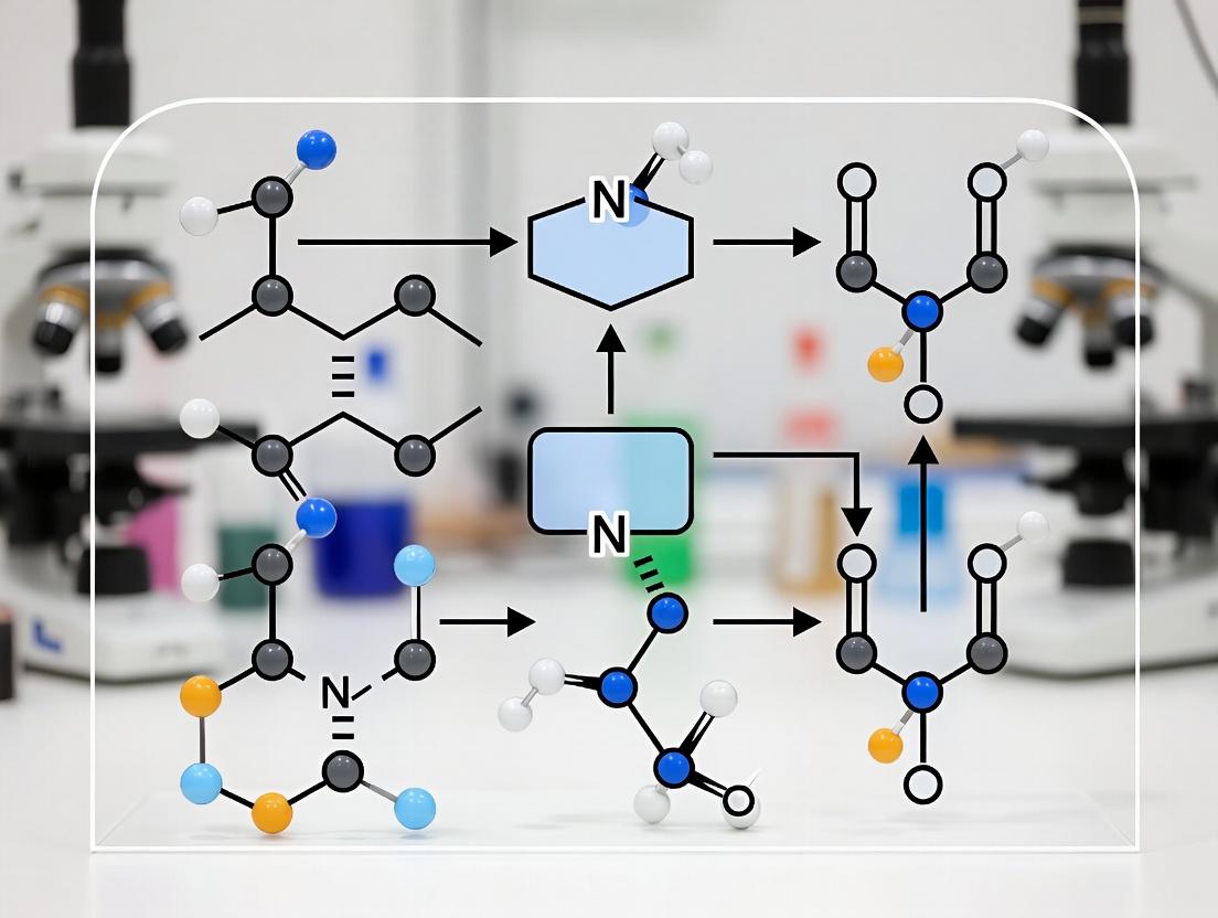
Synthetic Guide RNAs (sgRNAs)
The sgRNA directs the dCas-effector complex to a specific genomic locus. Optimization is critical for efficiency and orthogonality.
Standard sgRNA Scaffold: For SpdCas9, includes the 20nt spacer, CRISPR RNA (crRNA) duplex, and trans-activating crRNA (tracrRNA) fusion.
Extended sgRNAs (gRNA-e): 5' or 3' extensions (e.g., MS2, PP7, com, or boxB RNA aptamers) recruit additional effector proteins via aptamer-binding domains.
Multiplexing Guides: Tandem crRNA arrays processed by Cas12a or ribozyme-/tRNA-flanked guides for Cas9 enable simultaneous targeting.
Table 2: sgRNA Architectures for Transcriptional Control
| sgRNA Type | Key Feature | Primary Function | Recruitment Capacity |
|---|---|---|---|
| Standard | Minimal scaffold | Basic targeting for fused effectors | 1 effector complex |
| MS2-aptamer | Two MS2 stem-loops | Recruits MCP-fused effectors | Up to 12 MCP dimers |
| PP7-aptamer | PP7 stem-loops | Recruits PCP-fused effectors (orthogonal to MS2) | Enables orthogonal multiplexing |
| com/boxB | com or boxB motifs | Recruits λ N or Bsm fusion proteins | Alternative recruitment systems |
| Multiplex array | Tandem crRNAs | For dCas12a; enables multi-targeting from single transcript | Varies |
Protocol 3.1: Design and Cloning of Extended sgRNAs with MS2 Aptamers
- Design: Synthesize an oligo with: 5'-[20nt spacer]-GTTTTAGAGCTAGAAATAGCAAGTTAAAATAAGGCTAGTCCGTTATCAACTTGAAAAAGTGGCACCGAGTCGGTGCTTTTTT-3'. Insert MS2 stem-loops (5'-ACAUGAGGAUUACCCAUGU-3') at the tetraloop (replace "GTTTT") and/or the 3' end (before terminator).
- PCR Amplification: Use a forward primer containing your spacer and a reverse primer containing the scaffold + aptamers.
- Cloning: Digest PCR product and vector (e.g., pU6-sgRNA expression vector) with BsaI (Golden Gate assembly compatible) or Esp3I. Ligate and transform.
- Validation: Sequence the final plasmid to confirm spacer and aptamer integrity.
Effector Domains
Effector domains fused to dCas or recruited via sgRNA aptamers confer transcriptional modulation function.
Activation Domains (ADs):
- VP64: Tandem four copies of VP16 (Herpes Simplex Virus), a strong acidic activator.
- p65: Subunit of NF-κB, synergizes with VP64.
- Rta: From Epstein-Barr virus, potent in some cell types.
- VPR Tripartite: VP64-p65-Rta fusion, highly potent.
- EDLL and TAL effectors: Plant-derived strong ADs.
Repression Domains (RDs):
- KRAB: Krüppel-associated box, recruits heterochromatin-forming complexes (HP1, SETDB1).
- SID4x: Four copies of the mSin interaction domain from Mad.
- SRDX (EAR-repression domain): Plant-derived.
Epigenetic Modifiers:
- p300 Core: Histone acetyltransferase (HAT) for activation.
- DNMT3A: DNA methyltransferase for silencing.
- TET1: Demethylase for activation.
- LSD1: Histone demethylase for repression.
Table 3: Common Effector Domains for CRISPR-TFs
| Effector Domain | Type | Origin/Sequence | Approx. Size (aa) | Primary Mechanism |
|---|---|---|---|---|
| VP64 | Activation | Herpes Simplex Virus (VP16 x4) | ~240 | Recruits general transcription factors |
| p65 | Activation | Human NF-κB | ~300 | Recruits co-activators |
| VPR | Activation | VP64-p65-Rta fusion | ~1100 | Strong synergistic activation |
| KRAB | Repression | Human Kox1 | ~75 | Recruits KAP1, HP1, SETDB1 |
| DNMT3A | Silencing | Human | ~912 | Catalyzes DNA methylation |
| p300 Core | Activation | Human | ~1040 | Catalyzes H3K27 acetylation |
Experimental Protocol for Multiplexed Orthogonal Activation/Repression
Protocol 5.1: Dual-Gene Orthogonal Control using dCas9-VPR and dCas12a-KRAB Objective: Simultaneously activate Gene A and repress Gene B in HEK293T cells.
Materials:
- Plasmids:
- pCMV-dSpCas9(D10A,H840A)-VPR (Addgene #63798)
- pCMV-dLbCas12a(D908A)-KRAB (Addgene #109049)
- pU6-sgRNA(MS2)GeneATarget (expressing sgRNA with MS2 for Gene A promoter)
- pU6-crRNAGeneBTarget (expressing crRNA for Gene B promoter)
- Reagents: Lipofectamine 3000, Qubit dsDNA HS Assay Kit, TRIzol, RT-qPCR reagents.
Method:
- Cell Seeding: Seed HEK293T cells in a 24-well plate at 1.5x10^5 cells/well in DMEM + 10% FBS. Incubate 24h to reach ~70% confluency.
- Transfection Mix: For each well, prepare:
- Tube A: 37.5µl Opti-MEM + 1.5µl P3000 reagent.
- Tube B: 37.5µl Opti-MEM + 1µl Lipofectamine 3000.
- Combine Tube A and B, incubate 5 min.
- Add DNA mix: 125ng dCas9-VPR + 125ng dCas12a-KRAB + 62.5ng sgRNAGeneA + 62.5ng crRNAGeneB + 50ng EGFP (transfection control). Total = 425ng.
- Transfection: Add DNA-lipid complex dropwise to cells. Incubate 48-72h.
- Validation:
- Flow Cytometry: At 48h, check EGFP to confirm transfection (>70%).
- RT-qPCR: At 72h, extract RNA with TRIzol. Synthesize cDNA. Perform qPCR for Gene A, Gene B, and housekeeping (GAPDH). Use ΔΔCt to calculate fold-change vs. cells transfected with non-targeting guides.
Visualization 1: Orthogonal CRISPR-TF System Workflow
Diagram Title: Orthogonal CRISPR-TF System for Dual-Gene Control
Visualization 2: dCas9-effector Recruitment Pathways
Diagram Title: dCas9-Effector Recruitment Mechanisms
The Scientist's Toolkit: Research Reagent Solutions
Table 4: Essential Reagents for CRISPR-TF Research
| Item | Example Product/Catalog # | Function in Research |
|---|---|---|
| dCas9 Expression Plasmids | pAC154-dual-dCas9-VP64 (Addgene #48240); pHRdSV40-dCas9-BFP-KRAB (Addgene #46911) | Provide the scaffold protein with or without fused effectors for stable or transient expression. |
| dCas12a Expression Plasmids | pY010 (dCas12a, Addgene #69988); pCMV-dLbCas12a(D908A) (Addgene #109049) | Enable orthogonal targeting with T-rich PAMs. |
| Modular sgRNA Cloning Vectors | pU6-sgRNA (Addgene #41824); MS2-p65-HSF1 helper (Addgene #61423) | Allow rapid cloning of spacer sequences into optimized sgRNA backbones, often with aptamer tags. |
| CRISPRa/i Lentiviral Libraries | Calabrese CRISPRa Lib (Addgene #92379); hCRISPRi-v2 Lib (Addgene #83969) | Enable genome-scale pooled screens for gain/loss-of-function phenotypes. |
| Synergistic Activation Mediator (SAM) Components | MS2-p65-HSF1 plasmid (Addgene #61423) | Recruited via MS2-aptamers on sgRNA to provide a potent, tripartite activation signal. |
| Reporter Cell Lines | HEK293T CLTA-T2A-GFP (GripTite, Thermo) with integrated reporters | Contain fluorescent or luminescent reporters under control of synthetic promoters for quick assay of CRISPR-TF efficiency. |
| Anti-Cas9 Antibodies | Anti-Cas9 (7A9-3A3, Cell Signaling #14697) | Used in ChIP-qPCR to confirm dCas9 binding at target loci. |
| Next-Generation Sequencing Kits | Illumina Nextera XT; SMARTer ThruPLEX | For RNA-seq or ChIP-seq analysis of transcriptional and chromatin changes post-intervention. |
| Lipid-Based Transfection Reagents | Lipofectamine 3000 (Thermo), JetOPTIMUS (Polyplus) | For efficient delivery of plasmid DNA or RNP complexes into mammalian cell lines. |
The development of CRISPR-based transcription factors (CRISPR-TFs) has revolutionized the field of orthogonal gene control, enabling precise, programmable manipulation of transcriptional states without altering the underlying DNA sequence. Within this paradigm, two primary system archetypes have emerged: CRISPR activation (CRISPRa) and CRISPR interference (CRISPRi). These systems repurpose the catalytically inactive dCas9 (or Cas9 variants like dCas9-KRAB) as a programmable DNA-binding scaffold, fused to effector domains that either recruit transcriptional activators or repressors to a target promoter. This whitepaper provides an in-depth technical comparison of CRISPRa and CRISPRi, framed within the broader thesis of achieving multiplexed, orthogonal, and tunable transcriptional regulation for functional genomics and therapeutic development.
Core Mechanisms & Architectural Components
CRISPRi (Interference/Repression): CRISPRi utilizes dCas9 fused to a transcriptional repressor domain, most commonly the Kruppel-associated box (KRAB) from human KOX1. KRAB recruits endogenous machinery, including heterochromatin protein 1 (HP1) and histone methyltransferases (e.g., SETDB1), to establish heterochromatin, characterized by H3K9me3 marks, leading to stable gene silencing.
CRISPRa (Activation): CRISPRa systems recruit transcriptional activators to a target promoter. Architectures include:
- VP64-p65-Rta (VPR) Triad: A synthetic tripartite activator fused directly to dCas9.
- SunTag: dCas9 fused to a peptide array (Gcn4) that recruits multiple copies of a scFv-antibody-fused activator domain (e.g., VP64).
- SAM (Synergistic Activation Mediator): dCas9-VP64 recruits a modified sgRNA with MS2 RNA aptamers. The MS2 coat protein (MCP), fused to p65 and HSF1 activators, binds the aptamers, creating a synergistic activation complex.
Quantitative Performance Comparison
Table 1: Key Performance Metrics of CRISPRa and CRISPRi Systems
| Parameter | CRISPRi (dCas9-KRAB) | CRISPRa (dCas9-VPR) | CRISPRa (SunTag) | CRISPRa (SAM) |
|---|---|---|---|---|
| Repression/Activation Fold-Change | 10- to 1000-fold repression (≥90% knockdown) | 10- to 500-fold activation | 100- to 2000-fold activation | 100- to 10,000-fold activation |
| Onset Kinetics (t₁/₂) | ~24-48 hours for maximal repression | ~12-24 hours for detectable activation | ~12-24 hours for detectable activation | ~12-24 hours for detectable activation |
| Duration of Effect | Stable for days-weeks post-transfection; persistent with stable integration | Transient (days); requires sustained presence | Transient (days); requires sustained presence | Transient (days); requires sustained presence |
| Specificity (Off-Target Effects) | High; primarily determined by sgRNA specificity and dCas9 binding. KRAB can spread ~1-3 kb. | High; activation is highly local to binding site. Risk of off-target binding. | High; similar to VPR. Multiplier effect is targeted. | Moderate; larger complex may increase non-specific interactions. |
| Multiplexing Capacity | Excellent; simultaneous repression of multiple genes with arrays of sgRNAs. | Good; but activator saturation can limit synergistic multi-gene activation. | Good; clear but may face steric hindrance. | Moderate; large sgRNA structure can complicate delivery. |
| Typical Delivery Method | Lentivirus, AAV, lipid nanoparticles (LNPs) | Lentivirus, AAV, electroporation | Lentivirus, plasmid transfection | Lentivirus, plasmid transfection |
Detailed Experimental Protocols
Protocol 4.1: CRISPRi Knockdown in Mammalian Cells Using Lentiviral dCas9-KRAB
Objective: Achieve stable, inducible repression of a target gene in HEK293T cells. Materials: See "Scientist's Toolkit" (Section 6). Procedure:
- Stable Cell Line Generation:
- Produce lentivirus encoding dCas9-KRAB-BFP and Puromycin resistance. Transduce HEK293T cells at an MOI of ~0.5-1.
- Select with 2 µg/mL puromycin for 7 days. Confirm BFP expression via flow cytometry.
- sgRNA Design and Cloning:
- Design a 20-nt guide sequence targeting the transcriptional start site (TSS) or downstream of TSS (up to -400 bp) of the gene of interest. Clone into the lentiGuide-Puro vector via BsmBI restriction sites.
- Functional Knockdown:
- Transduce the stable dCas9-KRAB cells with the target sgRNA lentivirus. Select with 1 µg/mL puromycin for 5 days.
- Harvest cells 7-10 days post-transduction for analysis.
- Validation:
- Quantify mRNA levels via RT-qPCR using SYBR Green. Normalize to a housekeeping gene (e.g., GAPDH) and a non-targeting sgRNA control.
- Assess protein knockdown by western blot or flow cytometry (if applicable).
Protocol 4.2: CRISPRa Gene Activation Using the SAM System
Objective: Achieve strong, synergistic activation of an endogenous gene. Procedure:
- Cell Line Preparation:
- Use a cell line stably expressing dCas9-VP64 (or transiently co-transfect it).
- The cell must also stably express MS2-P65-HSF1 (this is often part of the SAM system).
- Specialized sgRNA Cloning:
- Clone the target 20-nt guide sequence into the lenti-sgRNA(MS2) vector, which contains two MS2 RNA aptamers in the sgRNA scaffold.
- Transduction and Activation:
- Transduce the prepared cells with the lenti-sgRNA(MS2) virus. If using transient systems, co-transfect all three components (dCas9-VP64, MS2-P65-HSF1, and sgRNA(MS2)) plasmids.
- Assay for activation 72-96 hours post-transduction/transfection.
- Validation:
- Use RT-qPCR to measure mRNA upregulation.
- For phenotypic assays (e.g., cytokine secretion, differentiation), perform functional assays 5-10 days post-induction.
Applications in Drug Development & Therapeutics
Table 2: Therapeutic and Research Applications
| Application Area | CRISPRi Utility | CRISPRa Utility |
|---|---|---|
| Functional Genomics | Genome-wide loss-of-function screens (alternative to RNAi). | Gain-of-function screens to identify oncogenes or rescue phenotypes. |
| Gene Therapy | Silencing dominant-negative alleles (e.g., in Huntington's disease). | Upregulating protective or deficient genes (e.g., FOXP3 in autoimmunity, fetal globin in sickle cell). |
| Cancer Research | Knockdown of oncogenes or essential genes for synthetic lethality. | Activation of tumor suppressor genes or antigens for immunotherapy. |
| Cell Differentiation & Reprogramming | Silencing pathways that block differentiation. | Direct activation of master transcription factors to drive differentiation (e.g., to neurons, cardiomyocytes). |
| Bioproduction | Repression of competitive or apoptotic pathways in CHO cells. | Activation of entire biosynthetic pathways for metabolite or protein production. |
The Scientist's Toolkit: Essential Research Reagents
Table 3: Key Reagents for CRISPRa/i Experiments
| Reagent / Material | Function / Purpose | Example Product/Catalog Number |
|---|---|---|
| dCas9-KRAB Expression Plasmid | Provides the DNA-binding repressor scaffold. | Addgene #71237 (lenti dCas9-KRAB-BFP) |
| dCas9-VPR Expression Plasmid | Provides the DNA-binding activator scaffold. | Addgene #63798 (lenti dCas9-VPR) |
| SAM System Plasmids | Three-component system for maximal activation. | Addgene Kit #1000000056 (lenti SAMv2) |
| lentiGuide-Puro sgRNA Vector | Backbone for cloning and expressing target sgRNAs. | Addgene #52963 |
| lenti-sgRNA(MS2)-zeo Vector | sgRNA vector with MS2 aptamers for SAM system. | Addgene #61427 |
| High-Titer Lentiviral Packaging Mix | Produces VSV-G pseudotyped lentivirus for delivery. | Takara Bio #631275 |
| Polybrene (Hexadimethrine Bromide) | Enhances lentiviral transduction efficiency. | Sigma-Aldrich #H9268 |
| Puromycin Dihydrochloride | Selection antibiotic for cells with integrated constructs. | Thermo Fisher #A1113803 |
| RT-qPCR Master Mix | Quantitative analysis of transcriptional changes. | Bio-Rad #1725124 |
| Validated sgRNA Controls | Non-targeting and positive control sgRNAs. | Synthego Non-Targeting Control sgRNA |
CRISPRa and CRISPRi represent complementary archetypes within the CRISPR-TF toolbox, enabling bidirectional, multiplexed control of the transcriptome. The future of orthogonal gene control research lies in the refinement of these systems for enhanced specificity, reduced immunogenicity, and inducible/tunable control (e.g., with light or small molecules). The integration of CRISPRa/i with other modalities—epigenetic editors, base editors, and synthetic signaling circuits—will pave the way for sophisticated cell engineering, next-generation gene therapies requiring precise dose-regulation, and comprehensive functional dissection of complex genetic networks.
Within the rapidly advancing field of CRISPR-based transcription factors (CRISPR-TFs) for orthogonal gene control, the paramount challenge is achieving specific, independent regulation of target genes without unintended interactions. This orthogonality imperative is central to the broader thesis that the next generation of precise transcriptional programming—for both basic research and therapeutic applications—depends on engineered systems with minimal off-target effects and crosstalk. This guide details the technical strategies and validation methodologies essential for designing and deploying orthogonal CRISPR-TF platforms.
Core Principles of Orthogonality
Orthogonality in CRISPR-TFs operates on two interdependent axes:
- DNA-Binding Orthogonality: The CRISPR guide RNA (gRNA) must direct the effector protein exclusively to its intended genomic target site, avoiding binding to sequences with partial homology.
- Effector Orthogonality: The transcription-activating or -repressing effector domain (e.g., VP64, p65, KRAB) must function specifically within its engineered system without interfering with or being modulated by endogenous cellular pathways.
Quantitative Analysis of Orthogonality Performance
Recent studies provide quantitative benchmarks for evaluating orthogonal CRISPR systems. The following table summarizes key metrics from seminal and recent works.
Table 1: Performance Metrics of Orthogonal CRISPR-TF Systems
| System / Component | Target Locus (Model) | On-Target Activity (Fold Change) | Off-Target Activity (Measured By) | Orthogonal Crosstalk | Reference (Year) |
|---|---|---|---|---|---|
| dCas9-VP64 + MS2-p65-HSF1 | IL1RN (HEK293T) | ~100x (RNA) | <1.5x (RNA-seq) | High (vs. endogenous TFs) | Mali et al. (2013) |
| CRISPRa Synergistic (SAM) | CEBPA (K562) | >1,000x (RNA) | Low (ChIP-seq peaks) | Moderate (via MS2 loops) | Konermann et al. (2015) |
| Cas9 vs. Cas12a Orthologs | Synthetic Reporter | ~50x (each) | <2% binding (PBM) | High (no cross-guide recognition) | Zetsche et al. (2015) |
| Engineered Cas9 Variants (High-Fidelity) | VEGFA Site 3 | ~70% of WT activity | >90% reduction (GUIDE-seq) | N/A (focus on DNA binding) | Kleinstiver et al. (2016) |
| Orthogonal dCas9-p300 & dCas9-KRAB | MYOD & SOX2 (hESCs) | Specific H3K27ac/H3K9me3 changes | Minimal overlap (ChIP-seq) | High (simultaneous activation/repression) | Hilton et al. (2015) |
| Hypercompact AsCas12f1-based TFs | NTF3 (HEK293T) | ~20x (RNA) | Undetectable (RNA-seq) | High (small size aids multiplexing) | Wu et al. (2021) |
Detailed Experimental Protocols for Validating Orthogonality
Protocol 1: Genome-Wide Off-Target Binding Assessment (GUIDE-seq)
- Objective: Identify sites of unintended CRISPR-TF binding across the genome.
- Materials: dCas9-TF construct, gRNA expression vector, GUIDE-seq oligonucleotide duplex, transfection reagent, next-generation sequencing (NGS) platform.
- Procedure:
- Co-transfect cells with the dCas9-TF, gRNA, and GUIDE-seq oligo.
- Allow 48-72 hours for integration of the oligo at double-strand breaks created by any residual nuclease activity or via alternative tagging methods for pure TFs.
- Harvest genomic DNA and shear by sonication.
- Perform nested PCR to enrich for genomic segments containing the integrated oligo.
- Prepare NGS library and sequence.
- Align sequences to the reference genome and identify GUIDE-seq tag integrations. For pure TFs, use methods like ChIP-seq or Digenome-seq.
- Analysis: Compare identified off-target sites to the intended on-target sequence for homology. Quantitate read counts to estimate relative binding frequency.
Protocol 2: Transcriptomic Crosstalk Profiling (Bulk RNA-seq)
- Objective: Quantify unintended changes in global gene expression induced by CRISPR-TF activity.
- Materials: Stable cell line expressing orthogonal dCas9-TF, inducible or constitutive gRNA vectors, RNA extraction kit, RNA-seq library prep kit.
- Procedure:
- Establish experimental conditions: a) Non-targeting gRNA control, b) On-target gRNA, c) Multiple single gRNAs targeting different loci.
- Harvest total RNA 48 hours post-gRNA induction/transfection.
- Deplete ribosomal RNA and construct cDNA libraries.
- Sequence to a depth of ~30 million reads per sample.
- Align reads to the reference genome (e.g., STAR aligner).
- Quantify gene expression (e.g., using featureCounts, HTSeq).
- Analysis: Perform differential expression analysis (e.g., DESeq2, edgeR). Genes significantly differentially expressed (FDR < 0.05, log2FC > |1|) in the non-targeting control versus the on-target sample indicate potential off-target transcriptional effects.
Visualizing Orthogonal System Design and Validation
Title: Workflow for Engineering Orthogonal CRISPR-TF Systems
Title: Orthogonal CRISPR-TF Complex Minimizing Crosstalk
The Scientist's Toolkit: Research Reagent Solutions
Table 2: Essential Reagents for Orthogonal CRISPR-TF Research
| Reagent / Material | Supplier Examples | Function in Orthogonality Research |
|---|---|---|
| High-Fidelity (HF) dCas9 Variant Plasmids | Addgene (#71814, #114268), Takara Bio | Reduced non-specific DNA binding; base scaffold for orthogonal effector fusion. |
| Orthogonal Cas Protein Expression Vectors (dCas12a, dCas9-NG) | Addgene (#129154, #164584), IDT | Provides alternative PAM requirements, enabling independent targeting within the same cell. |
| Validated, Clonable gRNA Scaffold Libraries | Synthego, Sigma-Aldrich (MS- & MVC-tagged) | Ensures proper folding and effector recruitment; tags enable multiplexed activation systems (e.g., SAM, SunTag). |
| Modular Transcriptional Effector Domains (VP64, p65, Rta, KRAB) | Addgene, custom peptide synthesis | Building blocks for creating novel activation/repression complexes with defined, orthogonal functions. |
| GUIDE-seq or CIRCLE-seq Kit | Integrated DNA Technologies (IDT) | Validated workflow for genome-wide identification of nuclease-dependent and -independent off-target binding sites. |
| Doxycycline-Inducible gRNA Expression Systems | Tet-On 3G systems (Clontech), custom lentiviral | Enables precise temporal control of CRISPR-TF activity, critical for dynamic crosstalk studies. |
| Multiplexed gRNA Cloning Kits (Golden Gate, BsaI) | ToolGen, Addgene (MoClo toolkit) | Facilitates assembly of tandem gRNA arrays for coordinated, orthogonal regulation of multiple loci. |
| ChIP-Validated dCas9 Antibodies | Diagenode, Abcam, Cell Signaling Tech. | Essential for ChIP-seq experiments to confirm on-target binding and assess genome-wide binding specificity. |
Advantages Over Traditional Inducible Systems (Tet-On/Off, Cre-Lox)
Within the rapidly advancing field of CRISPR-based transcription factors (CRISPR-TFs) for orthogonal gene control, a central thesis is the development of programmable, multiplexable, and leak-resistant systems that surpass the limitations of classical genetic switches. Traditional systems like Tet-On/Off (tetracycline-inducible) and Cre-Lox (recombinase-mediated) have been foundational but possess inherent constraints in scalability, dynamic range, and orthogonality. This whitepaper provides an in-depth technical comparison, demonstrating how modern CRISPR-based transcriptional regulators address these limitations, enabling precise, multi-gene regulatory circuits essential for advanced functional genomics and therapeutic development.
Core Limitations of Traditional Systems: A Quantitative Analysis
Table 1: Quantitative Comparison of Key Performance Metrics
| Performance Metric | Tet-On/Off Systems | Cre-Lox Systems | CRISPR-based Orthogonal TFs (e.g., dCas9-SAM, dCas9-VPR) |
|---|---|---|---|
| Induction Fold-Change | 10 - 1,000x (highly variable, context-dependent) | Binary (ON/OFF; irreversible) | 10 - 10,000x (consistently high) |
| Kinetics of ON/OFF | Hours to ON; hours to OFF (depends on Dox clearance) | Irreversible; minutes to hours for recombination | Minutes to hours ON; hours to OFF (tunable via sgRNA degradation) |
| Multiplexing Capacity (Orthogonal Channels) | Low (typically 1-2 with different TetR variants) | Very Low (limited by recombinase orthologs) | High (dozens of orthogonal sgRNAs; multiple dCas9 orthologs: Sp, Sa, Cj) |
| Background Leakiness | Moderate to High (promoter-driven leak) | N/A (but can have germline recombination) | Very Low (with optimized sgRNA & synthetic promoters) |
| Targeting Precision | Promoter-specific (requires transgene integration) | Sequence-specific (Lox sites; irreversible genome alteration) | Base-pair precision via 20-nt guide sequence (targets endogenous loci) |
| Delivery Payload Size | Large (requires TetR/rtTA + TRE promoter + transgene) | Large (requires Cre + floxed transgene) | Compact (dCas9-effector is constant; only sgRNA changes) |
| Reversibility | Fully Reversible | Irreversible | Fully Reversible (by withdrawing sgRNA or using deactivating systems) |
| Immunogenicity Risk | Low (bacterial TetR) | Moderate (bacterial Cre) | Moderate to High (bacterial Cas9; mitigated by engineered variants) |
Technical Advantages of CRISPR-Based Orthogonal Systems
Multiplexed and Orthogonal Control
CRISPR-TFs enable simultaneous, independent regulation of multiple genes by using orthogonal dCas9 orthologs (e.g., S. pyogenes dCas9, S. aureus dCas9) paired with unique sgRNA scaffolds and cognate effector proteins (e.g., VP64, p65, Rta). This creates independent regulatory channels impossible with traditional systems.
Protocol 1: Establishing a Two-Channel Orthogonal Activation System
- Objective: Independently activate Gene A and Gene B in the same cell.
- Materials: See "The Scientist's Toolkit" below.
- Method:
- Construct Design: Clone sgRNAs targeting upstream activator sequences of endogenous Gene A and Gene B into orthogonal backbones (e.g., sgRNA for Sp-dCas9-VPR targeting Gene A; sgRNA for Sa-dCas9-VPR targeting Gene B).
- Delivery: Co-transfect HEK293T cells with four plasmids: a) Sp-dCas9-VPR, b) Sa-dCas9-VPR, c) Sp-sgRNA-GeneA, d) Sa-sgRNA-GeneB.
- Control: Include single-guide and no-guide controls.
- Analysis: At 48h post-transfection, harvest cells for dual-luciferase assay (if reporters) or qRT-PCR to measure mRNA levels of Gene A and Gene B.
- Key Outcome: Demonstrable independent activation of each gene, with minimal cross-talk between the Sp and Sa systems.
Diagram 1: CRISPR Orthogonal Channels vs. Tet System
Enhanced Dynamic Range and Reduced Leakiness
CRISPR-TFs, when combined with synthetic promoter architectures (e.g., RNA polymerase III promoters for sgRNA, minimal synthetic promoters for target genes), exhibit significantly lower baseline activity and higher induction levels compared to the CMV or TRE promoters used in Tet systems, which are prone to transcriptional leak.
Protocol 2: Measuring Leakiness and Dynamic Range
- Objective: Quantify off-state leak and on-state induction of a CRISPRa system versus a Tet-On system.
- Method:
- Reporter Construction: Create two luciferase reporter cell lines: a) with a minimal promoter containing dCas9 binding sites, b) with a standard TRE3G promoter.
- CRISPRa Induction: For line (a), transfect with dCas9-VPR and a targeting sgRNA. Use a non-targeting sgRNA as the "off" control.
- Tet-On Induction: For line (b), treat with 1 µg/mL doxycycline (Dox) to induce. No Dox is the "off" control.
- Quantification: Measure luciferase activity 24h post-induction/transfection. Calculate fold-induction (ON/OFF) and absolute signal of the OFF state.
- Key Outcome: CRISPRa system shows a lower OFF signal and a higher fold-induction.
Reversible, Tunable, and Endogenous Targeting
Unlike Cre-Lox, which causes permanent DNA rearrangement, CRISPR-TFs offer fully reversible modulation of gene expression at endogenous loci without altering the underlying DNA sequence. Expression levels can be tuned by modulating sgRNA expression or using chemically-inducible dimerization systems (e.g., abscisic acid, rapamycin) to control dCas9-effector localization.
The Scientist's Toolkit: Essential Reagents for CRISPR Orthogonal Control
Table 2: Key Research Reagent Solutions
| Reagent / Material | Function / Explanation | Example Vendor/Reference |
|---|---|---|
| dCas9 Orthologs (Sp, Sa, Cj) | Catalytically dead Cas9 proteins serving as programmable DNA-binding scaffolds for different orthogonal channels. | Addgene (plasmids from labs of Feng Zhang, George Church) |
| Modified sgRNA Scaffolds (MS2, PP7, com) | Engineered sgRNA loops that bind specific RNA-binding proteins (e.g., MCP, PCP), enabling recruitment of effector domains for activation/repression. | Synthego, Integrated DNA Technologies (IDT) |
| CRISPRa Effector Fusions (VPR, SAM, SunTag) | Potent transcriptional activation complexes. VPR: VP64-p65-Rta fusion. SAM: Synergistic Activation Mediator (MS2-p65-HSF1). SunTag: peptide array for recruiting multiple copies of an effector. | Addgene (plasmids from labs of Patrick Hsu, Ron Weiss) |
| Chemically-Inducible Dimerization Domains (ABI, PYL, FRB/FKBP) | Allows for small molecule (e.g., abscisic acid, rapamycin) control over dCas9-effector assembly or nuclear localization, adding a temporal layer of control. | Takara Bio, CID Technologies |
| Synthetic Promoter Libraries (e.g., RNA Pol III promoters for sgRNA) | Minimally-sized, cell-type-specific promoters for driving sgRNA expression with minimal leak, enhancing orthogonality. | Custom synthesis (Twist Bioscience, Genscript) |
| All-in-One Viral Vectors (Lentiviral, AAV) | For stable delivery of large CRISPR-TF components (dCas9-effector + sgRNA) into hard-to-transfect cells or in vivo models. | VectorBuilder, Vigene Biosciences |
Diagram 2: CRISPR-TF Assembly for Gene Activation
CRISPR-based orthogonal transcription factor systems represent a paradigm shift in inducible gene control, directly addressing the multiplexing, precision, reversibility, and dynamic range constraints of Tet-On/Off and Cre-Lox technologies. Their integration into a broader thesis on orthogonal gene control underscores a move towards fully programmable, context-aware regulatory networks for deciphering complex biological processes and engineering next-generation cell and gene therapies.
Designing and Deploying Orthogonal CRISPR-TF Systems: A Step-by-Step Guide
Within the expanding landscape of CRISPR-based transcription factors for orthogonal gene control, the selection of an appropriate nuclease-dead (d) effector platform is a fundamental, high-impact decision. This guide provides a technical comparison of dCas9, dCas12, and emerging platforms, framing their utility within multi-gene circuit regulation and combinatorial perturbation studies. The core aim is to empower researchers in making informed platform choices based on quantitative performance, practical handling, and compatibility with orthogonal control paradigms.
Core Platform Architectures and Mechanisms
dCas9: The Benchmark Platform
Derived from Streptococcus pyogenes (Sp) and other bacterial orthologs, dCas9 is generated via point mutations (D10A and H840A in SpCas9) that ablate nuclease activity while preserving sgRNA-programmed DNA binding. Its mechanism involves a two-lobed architecture that accommodates the sgRNA:DNA heteroduplex, creating a steric block for transcription or serving as a scaffold for effector domains. Key variants like dSaCas9 and dNme2Cas9 offer smaller sizes or distinct PAM requirements, enhancing orthogonality.
dCas12: The Compact, T-Rich PAM Alternative
dCas12a (from Acidaminococcus or Lachnospiraceae species) and related dCas12f (ultracompact) systems are inactivated via analogous mutations (e.g., D908A for AsCas12a). dCas12a processes its own CRISPR RNA (crRNA) array, enabling multiplexing from a single transcript, and recognizes T-rich PAMs (e.g., TTTV). Its RuvC domain inactivation yields a platform with distinct molecular geometry and chromatin engagement properties compared to dCas9.
Other Nuclease-Dead Variants: dCas13 and dCsm/Cmr
For RNA-targeting orthogonal control, dCas13 (e.g., dPspCas13b, dRxCas13d) binds and can manipulate RNA transcripts without degradation. Prokaryotic Argonaute-based systems and dCsm/Cmr (Type III CRISPR effectors) represent additional, less-characterized platforms for DNA or RNA intervention with unique guide requirements.
Diagram: Orthogonal CRISPR-dEffector Binding Mechanisms
Quantitative Performance Comparison
The table below summarizes key characteristics critical for platform selection in orthogonal setups.
Table 1: Quantitative Comparison of Major dEffector Platforms
| Feature | dSpCas9 | dSaCas9 | dNme2Cas9 | dAsCas12a | dLbCas12a | dRfxCas13d (RNA) |
|---|---|---|---|---|---|---|
| Size (aa) | 1368 | 1053 | 1082 | 1307 | 1228 | 967 |
| Guide RNA | ~100-nt sgRNA | ~110-nt sgRNA | ~110-nt sgRNA | ~42-44-nt crRNA | ~42-44-nt crRNA | ~63-nt crRNA |
| Native PAM | 5'-NGG-3' | 5'-NNGRRT-3' | 5'-NNNCC-3' | 5'-TTTV-3' | 5'-TTTV-3' | None (RNA) |
| Multiplex Guide Generation | Requires individual expression or array + RNase | Requires individual expression or array + RNase | Requires individual expression | Native crRNA array processing | Native crRNA array processing | Native crRNA array processing |
| Reported On-Target Binding Affinity (K_d) | ~0.5-5 nM | ~2-10 nM | ~1-10 nM | ~1-20 nM | ~1-20 nM | ~3-30 nM (for RNA) |
| Typical Activation Fold-Change* | 10x - 500x | 5x - 200x | 5x - 200x | 5x - 100x | 5x - 100x | N/A (RNA) |
| Typical Repression Efficiency* | 70% - 95% | 60% - 90% | 60% - 90% | 50% - 85% | 50% - 85% | Up to 80% (RNA) |
| Key Orthogonality Advantage | Most characterized, many effectors | Smaller size for AAV delivery | Minimal off-target, distinct PAM | Distinct T-rich PAM, multiplexing | Distinct T-rich PAM, multiplexing | Cytoplasmic RNA targeting |
*Highly dependent on effector domain (e.g., VPR, KRAB), genomic context, and delivery.
The Scientist's Toolkit: Essential Research Reagents
Table 2: Key Reagent Solutions for dEffector Research
| Reagent | Function & Key Consideration | Example Vendors/Catalogs |
|---|---|---|
| dCas9 Expression Plasmid | Mammalian codon-optimized, with nuclear localization signals (NLS), optional epitope tags (e.g., HA, FLAG). | Addgene (#135158, #135159), Thermo Fisher (A36599) |
| dCas12a Expression Plasmid | Codon-optimized for mammalian cells, includes requisite NLSs. | Addgene (#137963, #159370), IDT |
| Modular Effector Domain Plasmids | VP64, p65, Rta (activation), KRAB, SID4X (repression) for fusion testing. | Addgene (#104174, #127968) |
| sgRNA/crRNA Cloning Backbone | U6 or other Pol III promoter vectors for guide expression. Critical for array designs for dCas12/dCas13. | Addgene (#104174, #159370), Synthego |
| Chemically Modified Synthetic gRNAs | Enhance stability and binding affinity; crucial for sensitive primary cell applications. | IDT, Synthego, Thermo Fisher |
| Validated Positive Control gRNAs | Targeting strong, well-characterized promoters (e.g., U6, EF1α, IL1RN). Essential for system validation. | Horizon Discovery, Synthego |
| dCas9/dCas12-Specific Antibodies | For ChIP-qPCR/seq validation of target occupancy (anti-FLAG, anti-HA, or custom). | Cell Signaling, Abcam, Diagenode |
| Orthogonal Reporter Plasmid Set | Dual- or multi-fluorescent reporters with distinct PAMs to test simultaneous, independent control. | Custom design required (e.g., EZ-CRISPR) |
Experimental Protocol: Validating Orthogonal dCas9 & dCas12a Dual Targeting
This protocol outlines steps to validate two dEffectors acting independently on separate reporter constructs in HEK293T cells.
A. Materials:
- Plasmids: dSpCas9-VPR (Activator A), dAsCas12a-KRAB (Repressor B), sgRNA (targeting PAM A), crRNA (targeting PAM B), Fluorescent Reporter A (BFP under a minimal promoter with PAM A site), Fluorescent Reporter B (GFP under a minimal promoter with PAM B site), transfection control plasmid (e.g., constitutively expressed mCherry).
- Cells: HEK293T.
- Reagents: Transfection reagent (e.g., PEI Max, Lipofectamine 3000), media, flow cytometry buffers.
B. Procedure:
- Guide Design & Cloning: Design sgRNA for dCas9 targeting a site adjacent to a 5'-NGG-3' PAM on Reporter A. Design crRNA for dCas12a targeting a site adjacent to a 5'-TTTV-3' PAM on Reporter B. Clone guides into appropriate U6-expression vectors. Verify by sequencing.
- Cell Seeding: Seed 2e5 HEK293T cells per well in a 24-well plate 24 hours prior to transfection for ~70% confluency.
- Transfection Mixture (per well):
- Experimental Group: dSpCas9-VPR (250 ng), dAsCas12a-KRAB (250 ng), sgRNA-A plasmid (125 ng), crRNA-B plasmid (125 ng), Reporter A (125 ng), Reporter B (125 ng), Transfection Control (50 ng). Total = 1050 ng DNA.
- Control Groups: Include all possible single-effector and no-effector controls.
- Prepare DNA mixes in opti-MEM. Add PEI Max at a 3:1 ratio (PEI:DNA, w/w). Vortex, incubate 15 min, add dropwise to cells.
- Incubation: Assay 48-72 hours post-transfection.
- Flow Cytometry Analysis:
- Harvest cells, wash with PBS, and resuspend in FACS buffer.
- Analyze on a flow cytometer equipped with 405nm, 488nm, and 561nm lasers.
- Gate on live, transfection-control-positive (mCherry+) cells.
- Quantify median fluorescence intensity (MFI) of BFP (Reporter A) and GFP (Reporter B) for each gated population.
- Data Interpretation: Calculate fold-activation (BFP MFI experimental / BFP MFI dCas12a-only control) and fold-repression (GFP MFI experimental / GFP MFI dCas9-only control). Successful orthogonality is demonstrated by specific activation of A and repression of B only when both matched dEffector-guide pairs are present.
Diagram: Orthogonal Dual-Targeting Experimental Workflow
Selection Criteria and Concluding Recommendations
Platform selection must align with the orthogonal control thesis:
- For Maximal Activation/Repression Strength: Mature dCas9-effector fusions (e.g., dCas9-SunTag-VPR, dCas9-MQ3-KRAB) currently offer the highest potency.
- For Simplified Multiplexed Targeting: dCas12a is superior due to native array processing, ideal for combinatorial gene network repression.
- For Tightly Packed Genomic Loci or AAV Delivery: Consider compact variants like dSaCas9, dNme2Cas9, or dCas12f.
- For Ultimate Orthogonality in Complex Circuits: Combine platforms with non-overlapping PAMs (e.g., dCas9 (NGG) + dCas12a (TTTV) + dCpf1/RVR variant (NYTV)) and distinct effector domains to minimize cross-talk.
The future of orthogonal control lies in engineering hybrid systems and next-generation dEffectors with expanded PAM recognition, reduced size, and enhanced specificity, enabling the precise dissection and manipulation of complex gene regulatory networks.
Within the field of CRISPR-based orthogonal gene control research, the selection of appropriate effector domains is paramount for precise transcriptional regulation. This whitepaper provides an in-depth technical guide to the core activation (VP64, p65, Rta) and repression (KRAB, SID) domains used in engineered CRISPR transcription factors, such as CRISPRa and CRISPRi systems. The efficacy, orthogonality, and experimental application of these domains are critical for advancing therapeutic and functional genomics research.
Core Effector Domains: Mechanisms and Performance
Activation Domains
Activation domains recruit co-activators and the general transcriptional machinery to a target promoter.
VP64: A tetrameric repeat of the 16-amino acid peptide from Herpes Simplex Viral Protein 16. It is a robust, well-characterized activator. p65: The transactivation domain from NF-κB subunit RelA. It functions via distinct co-activator interactions compared to VP64. Rta: A potent viral transactivator from Epstein-Barr virus. It often shows higher activation potency than VP64 or p65 alone.
Repression Domains
Repression domains induce heterochromatin formation or directly interfere with the basal transcriptional apparatus.
KRAB (Krüppel-associated box): The most widely used repression domain in mammalian cells. It recruits heterochromatin-inducing complexes via KAP1. SID (mSin3 Interaction Domain): Derived from Mad protein, it recruits the mSin3 co-repressor complex, leading to histone deacetylation and transcriptional silencing.
Table 1: Comparative Performance of Effector Domains
| Effector Domain | Type | Approx. Fold Activation/Repression* | Key Recruited Complex/Proteins | Common Fusion Construct |
|---|---|---|---|---|
| VP64 | Activation | 10-100x | p300, Mediator | dCas9-VP64 |
| p65 | Activation | 10-50x | p300, CBP | dCas9-VP64-p65 (VPR) |
| Rta | Activation | 100-1000x | SWI/SNF, Mediator | dCas9-VP64-Rta (VPR) / dCas9-Rta |
| KRAB | Repression | 5-50x (repression) | KAP1, HP1, SETDB1 | dCas9-KRAB |
| SID4x | Repression | 10-100x (repression) | mSin3/HDAC | dCas9-SID4x |
*Fold change is highly dependent on genomic context, target promoter, and delivery method. Rta often exhibits the highest activation potential. Data compiled from recent literature (2023-2024).
Table 2: Orthogonality & Practical Considerations
| Domain | Size (aa) | Risk of Immune Recognition | Notable Synergistic Combinations | Primary Application |
|---|---|---|---|---|
| VP64 | ~64 | Low | p65, Rta (synergistic arrays) | Basic CRISPRa, multiplexing |
| p65 | ~220 | Moderate (human origin) | VP64 (VPR) | Enhanced single-effector activation |
| Rta | ~605 | High (viral origin) | VP64, p65 | Ultra-potent activation, hard-to-activate genes |
| KRAB | ~75 | Low (human origin) | Often used alone | Broad, stable transcriptional repression |
| SID4x | ~108 | Low (derived from human Mad) | Can be layered with KRAB | Repression via histone deacetylation |
Experimental Protocols for Validation
Protocol 1: Benchmarking Activation Efficiency with a Luciferase Reporter
Objective: Quantify and compare the transcriptional activation potency of dCas9-VP64, dCas9-p65, and dCas9-Rta.
- Cell Seeding: Seed HEK293T cells in a 24-well plate at 70% confluency.
- Transfection: Co-transfect using a polyethylenimine (PEI) protocol:
- Group 1: 250 ng dCas9-VP64 plasmid + 250 ng sgRNA plasmid (targeting a defined site in a synthetic promoter) + 100 ng Firefly luciferase reporter plasmid.
- Group 2: 250 ng dCas9-p65 plasmid + same sgRNA + reporter.
- Group 3: 250 ng dCas9-Rta plasmid + same sgRNA + reporter.
- Include control groups (dCas9-only, sgRNA-only).
- Include 50 ng Renilla luciferase plasmid for normalization in all wells.
- Incubation: Incubate cells for 48 hours post-transfection.
- Lysis and Measurement: Lyse cells with Passive Lysis Buffer. Measure Firefly and Renilla luciferase activity using a dual-luciferase assay kit on a plate reader.
- Analysis: Normalize Firefly luminescence to Renilla for each well. Calculate fold activation relative to dCas9-only control.
Protocol 2: Assessing Long-Term Repression via dCas9-KRAB/SID
Objective: Evaluate the kinetics and stability of transcriptional repression.
- Stable Line Generation: Lentivirally transduce HEK293 cells with dCas9-KRAB or dCas9-SID4x and select with puromycin (2 µg/mL) for 1 week.
- sgRNA Delivery: Transduce the polyclonal dCas9-expressing cells with lentiviral sgRNAs (targeting the promoter of a endogenous gene, e.g., IL1RN) carrying a blasticidin resistance marker. Select with blasticidin (5 µg/mL) for 5 days.
- Time-Course Sampling: Harvest cells at days 3, 7, 14, and 21 post-sgRNA selection.
- qRT-PCR Analysis:
- Isolate total RNA using a column-based kit.
- Synthesize cDNA using a high-capacity reverse transcription kit.
- Perform qPCR with SYBR Green for the target gene and two housekeeping genes (e.g., GAPDH, ACTB).
- Data Processing: Use the ΔΔCt method to calculate relative gene expression normalized to the average of housekeepers and compared to a non-targeting sgRNA control at each time point.
Visualizations
Diagram 1: Effector Domain Signaling Pathways
Diagram 2: Workflow for Benchmarking Effector Domains
The Scientist's Toolkit
Table 3: Research Reagent Solutions for Effector Domain Studies
| Reagent / Material | Supplier Examples | Function in Experiments |
|---|---|---|
| dCas9-VPR Plasmid (Addgene #63798) | Addgene, Synthego | All-in-one plasmid for strong activation (VP64-p65-Rta fusion). |
| dCas9-KRAB Plasmid (Addgene #89567) | Addgene, Santa Cruz Biotech | Standard plasmid for robust transcriptional repression. |
| Lenti-dCas9-KRAB-blast | Applied Biological Materials, Sigma-Aldrich | Lentiviral vector for generating stable, inducible repression cell lines. |
| Synthetic sgRNA Libraries (CRISPRa/i) | Twist Bioscience, Agilent | Pooled libraries for genome-wide screens using specific effector domains. |
| Dual-Luciferase Reporter Assay System | Promega | Quantifies activation/repression efficiency on synthetic promoters. |
| SYBR Green qPCR Master Mix | Thermo Fisher, Bio-Rad | Measures changes in endogenous mRNA expression following CRISPRa/i. |
| Anti-H3K9me3 ChIP-Grade Antibody | Cell Signaling, Abcam | Validates KRAB-mediated heterochromatin formation at target loci. |
| HDAC Activity Assay Kit | Cayman Chemical | Validates functional recruitment by SID domain. |
| PEI Max (Polyethylenimine) | Polysciences | High-efficiency transfection reagent for plasmid delivery. |
| HEK293T/FT Cells | ATCC | Standard cell line for transient CRISPRa/i experiments due to high transfection efficiency. |
Guide RNA (gRNA) Design Rules for Optimal Specificity and Efficiency
The development of orthogonal CRISPR-based transcription factors (CRISPR-TFs), such as dCas9-VP64 or dCas9-SunTag systems, enables precise perturbation of gene expression without altering the underlying DNA sequence. This capability is central to functional genomics, synthetic biology, and therapeutic development. Within this paradigm, the guide RNA (gRNA) serves as the critical determinant of both the targeting specificity and the recruitment efficiency of the transcriptional machinery. This guide synthesizes current principles and protocols for designing gRNAs that maximize on-target activity while minimizing off-target effects in the context of CRISPR-based transcription control.
Core Design Principles for gRNA Specificity and Efficiency
Sequence Composition and Thermodynamics
Optimal gRNA design integrates multiple parameters derived from high-throughput screening data. Key factors include sequence composition at specific positions, local chromatin accessibility, and secondary structure of the gRNA itself.
Table 1: Quantitative Parameters for On-Target Efficiency (Activation)
| Parameter | Optimal Feature / Value | Impact on Efficiency (Relative Effect) | Notes |
|---|---|---|---|
| GC Content | 40-60% | High (Strong positive correlation) | Extreme GC (>80%) or AT-rich sequences reduce efficiency. |
| 5' End Nucleotide | G or A (for U6 promoter) | Critical (No expression if absent) | U6 polymerase requires a 5' G. For endogenous targeting, an A is also acceptable. |
| Seed Region (PAM-proximal 8-12nt) | Low tolerance for mismatches | Very High | Single mismatches here drastically reduce binding and activation. |
| Melting Temperature (Tm) | 55-65°C for seed region | Moderate | Predicts stable R-loop formation. |
| Presence of Poly-T | Avoid 4+ consecutive T's | High | Acts as a premature termination signal for Pol III promoters. |
| Secondary Structure (gRNA) | Low free energy (ΔG > -5 kcal/mol) | Moderate | Highly structured gRNAs impair dCas9 binding/loading. |
Table 2: Parameters Influencing Off-Target Specificity
| Parameter | Design Strategy | Mechanistic Rationale |
|---|---|---|
| Seed Region Mismatches | Zero tolerance in design; use stringent prediction algorithms. | dCas9 binding is highly sensitive to seed region fidelity. |
| gRNA Length | Use truncated gRNAs (tru-gRNAs, 17-18nt) or extended gRNAs (20nt+). | Alters binding energy, increasing specificity. Tru-gRNAs require high on-target potency. |
| PAM Distal Modifications | Introduce secondary structure (e.g., hairpins) or chemical modifications. | Sterically hinders binding to off-targets with partial complementarity. |
| Specificity Score | Utilize in silico tools (e.g., CFD, MIT specificity score). | Quantifies predicted off-target propensity based on mismatch position/type. |
| Chromatin State | Target within open chromatin (DNase I hypersensitive sites). | Closed chromatin increases discriminatory pressure, favoring on-target. |
Positional Effects for Transcriptional Activation
For CRISPRa (activation), gRNA placement relative to the transcription start site (TSS) is paramount. Empirical data from systems like SAM (Synergistic Activation Mediator) establish clear rules.
Table 3: Optimal gRNA Positioning for dCas9-Based Activators
| Activator System | Optimal Distance from TSS | Optimal Strand | Recommended Number of gRNAs |
|---|---|---|---|
| dCas9-VP64 | -50 to -200 bp upstream | Either | 3-6 for strong, synergistic activation |
| dCas9-SunTag-VP64 | -50 to -150 bp upstream | Either | 2-4 |
| SAM (dCas9-VP64 + MS2-p65-HSF1) | -50 to -200 bp upstream (max -400) | Anti-sense preferred | 3-6 |
| CRISPRa (VPR variant) | -50 to -250 bp upstream | Either | 2-4 |
Title: Optimal gRNA Positioning for CRISPRa Systems
Experimental Protocols for gRNA Validation
Protocol 1: High-Throughput gRNA Screening for CRISPRa Efficiency
Objective: Systematically quantify the transcriptional activation potency of hundreds of gRNAs targeting a locus of interest.
Materials: See "The Scientist's Toolkit" below. Procedure:
- Design & Library Cloning: Design 200-500 gRNAs targeting regions from -400 to +50 bp relative to the TSS. Include positive control (e.g., targeting known strong promoter) and non-targeting negative control gRNAs.
- Synthesize oligonucleotide pools, amplify by PCR, and clone into the lentiviral gRNA expression vector (e.g., lentiGuide-Puro with MS2 stem-loops for SAM system) via Golden Gate or Gibson assembly.
- Lentivirus Production: Co-transfect HEK293T cells with the gRNA library plasmid, psPAX2 (packaging), and pMD2.G (VSV-G envelope) plasmids using PEI transfection reagent. Harvest virus supernatant at 48 and 72 hours.
- Cell Screening: Transduce the target cell line (e.g., K562, HEK293) stably expressing dCas9-activator (e.g., dCas9-VP64-p65-Rta) at a low MOI (<0.3) to ensure single gRNA integration. Select with puromycin (1-2 µg/mL) for 7 days.
- Phenotype Harvest: After 10-14 days post-transduction, harvest cells. For RNA-based readouts, extract total RNA and prepare for RNA-seq. For fluorescence-based reporters, analyze by FACS.
- NGS & Analysis: For pooled screens, extract genomic DNA, PCR-amplify the integrated gRNA cassette, and sequence on an Illumina platform. Align reads to the reference library. Calculate gRNA enrichment/depletion scores (e.g., using MAGeCK or PinAPL-Py). Correlate gRNA position/sequence features with activity.
Protocol 2: Assessment of Off-Target Effects (CHIP-Seq & RNA-Seq)
Objective: Identify genome-wide binding of dCas9-activator and non-specific transcriptional changes.
Procedure: A. dCas9 ChIP-seq:
- Crosslink 10-20 million gRNA-transduced + dCas9-activator cells with 1% formaldehyde for 10 min. Quench with glycine.
- Sonicate lysate to shear chromatin to 200-500 bp fragments.
- Immunoprecipitate with an anti-Cas9 or anti-tag (e.g., FLAG) antibody overnight at 4°C. Use Protein A/G beads for pull-down.
- Reverse crosslinks, purify DNA, and prepare sequencing library.
- Analysis: Map reads to the reference genome. Call peaks (MACS2). Compare peaks at the intended on-target site versus other genomic loci (off-targets). Validate off-targets by motif analysis for PAM and seed region similarity.
B. RNA-seq for Transcriptomic Off-Targets:
- Transduce cells with a highly active gRNA and a non-targeting control gRNA (in triplicate).
- After 48-72 hours, extract total RNA using a column-based kit with DNase I treatment.
- Prepare stranded mRNA-seq libraries and sequence to a depth of ~30 million reads per sample.
- Analysis: Align reads (STAR), quantify gene expression (featureCounts), and perform differential expression analysis (DESeq2). Significant differentially expressed genes beyond the intended target indicate transcriptional off-target effects.
Title: gRNA Validation Workflow: Efficiency & Specificity
The Scientist's Toolkit: Essential Reagents for gRNA Design & Testing
| Reagent / Material | Function / Description | Example Product/Catalog |
|---|---|---|
| dCas9-Activator Cell Line | Stably expresses catalytically dead Cas9 fused to transcriptional activation domains (e.g., VP64, VPR, p65-HSF1). Essential for CRISPRa screens. | Custom generated or commercially available (e.g., SAM-ready cell lines). |
| Lentiviral gRNA Expression Vector | Backbone for cloning and delivering gRNA sequences. Often includes MS2 stem-loops for recruiter systems (SAM) and a selection marker. | lentiGuide-Puro, lentiSAMv2 (Addgene). |
| High-Fidelity DNA Polymerase | For accurate amplification of gRNA library inserts and preparation of sequencing amplicons. | Q5 Hot-Start (NEB), KAPA HiFi. |
| Next-Generation Sequencing Platform | For deep sequencing of pooled gRNA libraries or transcriptomic analysis (RNA-seq). | Illumina NextSeq 2000, NovaSeq. |
| Anti-Cas9 ChIP-Validated Antibody | For chromatin immunoprecipitation of dCas9 to map on- and off-target binding sites. | Anti-Cas9 (7A9-3A3, Cell Signaling #14697). |
| Chromatin Accessibility Assay Kit | To assess target site chromatin state (open vs. closed) which influences gRNA efficiency. | ATAC-seq Kit (Illumina). |
| gRNA Design & Analysis Software | In silico tools for predicting on-target scores and potential off-target sites. | CRISPick (Broad), CHOPCHOP, Cas-OFFinder. |
| Pooled Library Analysis Pipeline | Computational tools for analyzing screen data and calculating gRNA enrichment. | MAGeCK, PinAPL-Py. |
The orthogonal control of gene expression using CRISPR-based transcription factors is critically dependent on rigorously designed gRNAs. By adhering to the sequence composition rules, positional guidelines, and validation protocols outlined herein, researchers can achieve predictable and specific transcriptional modulation. As the field advances, integrating chromatin conformation data and machine learning models will further refine these design principles, enabling more complex and therapeutic applications of orthogonal gene control.
Within the field of CRISPR-based transcription factors (CRISPR-TFs) for orthogonal gene control, the efficacy of epigenetic reprogramming or transcriptional modulation is critically dependent on the delivery vehicle. Achieving precise, durable, and safe delivery of CRISPR-TF components—be it encoding plasmids, mRNA, or preassembled ribonucleoprotein (RNP) complexes—remains a central challenge. This guide provides a technical comparison of leading delivery strategies, focusing on their application in advanced orthogonal gene control research, which demands minimal off-target effects and maximal specificity in multiplexed environments.
Comparative Analysis of Delivery Modalities
The selection of a delivery system involves trade-offs between cargo capacity, delivery efficiency, immunogenicity, persistence, and ease of production. The following table summarizes key quantitative parameters for each platform relevant to CRISPR-TF delivery.
Table 1: Quantitative Comparison of Delivery Strategies for CRISPR-TF Cargo
| Parameter | AAV | Lentivirus | Plasmid (Non-Viral) | RNP Complexes (Non-Viral) |
|---|---|---|---|---|
| Max Cargo Capacity | ~4.7 kb | ~8 kb | Unlimited (but delivery constrained) | Limited by complex size (typically 1-2 proteins + gRNA) |
| Integration into Host Genome | Predominantly episomal; rare non-homologous integration | Stable integration (random) | Transient, non-integrating | Transient, no genetic material |
| Transgene Expression Onset | Slow (days to weeks) | Moderate (days) | Fast (hours to days) | Immediate (minutes to hours) |
| Expression Duration | Long-term (months-years) | Permanent | Short-term (days) | Ultra-short-term (hours-days) |
| In Vivo Immunogenicity | Moderate (capsid/transgene specific) | High (viral proteins) | High (bacterial DNA motifs) | Low (no foreign DNA) |
| Tropism & Targeting Flexibility | High (depends on serotype) | Moderate (pseudotyping possible) | Low (dependent on co-delivered vehicle) | Moderate (dependent on co-delivered vehicle) |
| Typical In Vitro Efficiency | Moderate-High | Very High | Low-Moderate | Moderate-High |
| Manufacturing Complexity & Cost | High | High | Low | Low-Moderate |
| Key Risk for Orthogonal Control | Preexisting immunity; capsid toxicity | Insertional mutagenesis; silencing over time | Off-target transcription; immunostimulation | Rapid degradation; lower multiplexing capacity |
Detailed Methodologies and Experimental Protocols
Production and Use of AAV for CRISPR-TF Delivery
AAV is ideal for long-term, in vivo expression of CRISPR-TFs like dCas9-VP64 or dCas9-p300 fusions.
Protocol: AAV Vector Production via PEI Transfection in HEK293T Cells
- Day 0: Seed HEK293T cells in fifteen 15-cm cell culture dishes at 70% confluence in DMEM + 10% FBS.
- Day 1: Co-transfect per dish with three plasmids:
- pAAV2/9 Rep-Cap Plasmid (20 µg): Provides AAV serotype 9 capsid proteins.
- pHelper Plasmid (30 µg): Provides adenoviral helper functions (E2A, E4, VA RNA).
- pAAV-ITR-CRISPR-TF-GOI Plasmid (20 µg): Contains the CRISPR-TF expression cassette (e.g., CAG-dCas9-SunTag-2xNLS) flanked by AAV2 inverted terminal repeats (ITRs). Ensure total cargo <4.7 kb.
- Prepare transfection mix in serum-free medium: Combine plasmids with linear polyethylenimine (PEI MAX, 1 mg/mL) at a 1:3 DNA:PEI mass ratio. Incubate 15 min, then add dropwise to cells.
- Day 3 (72h post-transfection): Harvest cells and medium. Pellet cells (500 x g, 10 min). Resuspend cell pellet in lysis buffer (150 mM NaCl, 50 mM Tris-HCl, pH 8.5). Perform 3 freeze-thaw cycles (dry ice/37°C water bath).
- Purification: Treat lysate with Benzonase (50 U/mL, 37°C, 30 min) to degrade unpackaged DNA. Clarify by centrifugation. Purify AAV particles using an iodixanol density gradient ultracentrifugation (15%, 25%, 40%, 60% layers; 350,000 x g, 2h, 18°C). Collect the 40-60% interface.
- Concentration & Buffer Exchange: Concentrate using a 100 kDa MWCO centrifugal filter. Exchange into final formulation buffer (PBS + 0.001% Pluronic F-68). Titrate via qPCR against ITR sequence.
- In Vivo Delivery: Administer via tail vein injection (C57BL/6 mice) at a dose of 1x10^11 to 1x10^12 vector genomes (vg) per animal in 100 µL PBS. Analyze tissue-specific TF expression and target gene activation after 2-4 weeks.
Lentiviral Vector Production for Stable CRISPR-TF Cell Line Generation
Lentivirus enables stable integration, useful for creating persistent orthogonal control cell lines.
Protocol: Third-Generation LV Production for dCas9-KRAB Repressor
- Day 0: Seed HEK293T cells in 10-cm dishes at 60-70% confluence.
- Day 1: Co-transfect using calcium phosphate with four plasmids:
- pMDLg/pRRE (7.5 µg): Gag/Pol expression.
- pRSV-Rev (2.5 µg): Rev expression.
- pMD2.G (3 µg): VSV-G envelope glycoprotein for broad tropism.
- pLV-EF1α-dCas9-KRAB-P2A-Puro (10 µg): Transfer plasmid containing the CRISPR-TF, post-transcriptional regulatory element (WPRE), and puromycin resistance.
- Day 2 (16h post-transfection): Replace medium with fresh DMEM + 10% FBS.
- Days 3 & 4: Harvest viral supernatant at 48h and 72h post-transfection. Pool, filter through a 0.45 µm PES filter.
- Concentration: Concentrate virus 100-fold using Lenti-X Concentrator (Clontech) per manufacturer's instructions. Resuspend pellet in 1/100th volume of HBSS + 1% HEPES.
- Transduction: Transduce target cells (e.g., HeLa) in the presence of 8 µg/mL polybrene. Spinoculate at 800 x g for 30 min at 32°C. After 48h, select with 2 µg/mL puromycin for 7 days to generate stable polyclonal lines.
Plasmid Transfection for Transient CRISPR-TF Expression
Protocol: Lipofection of CRISPR-TF Plasmids for Epigenetic Activation
- Day 0: Plate HEK293 cells in a 24-well plate at 1.5 x 10^5 cells/well in antibiotic-free medium.
- Day 1: Prepare two separate solutions in Opti-MEM (50 µL each):
- Solution A: 0.5 µg pCMV-dCas9-p300core + 0.25 µg pU6-sgRNA expression plasmid.
- Solution B: 2 µL Lipofectamine 3000 reagent.
- Combine Solutions A and B, incubate 15 min at RT.
- Add complex dropwise to cells. After 6h, replace with complete growth medium.
- Analysis: Harvest cells 48-72h post-transfection for RNA (qRT-PCR) or protein (Western blot) analysis of target gene activation.
Delivery of Preassembled RNP Complexes for Rapid, DNA-free Editing
RNP delivery offers the fastest action and lowest off-target profile, ideal for precise, short-term perturbations.
Protocol: Electroporation of CRISPR-TF RNPs (Neon Transfection System)
- Prepare RNP Complex: Chemically synthesize or in vitro transcribe sgRNA (with 2'-O-methyl 3' phosphorothioate modifications for stability). Purify via spin column. Recombinantly express and purify dCas9-VPR protein (dCas9 fused to VP64, p65, Rta activators).
- Complex Assembly: Mix dCas9-VPR protein (at 3 µM final) with sgRNA (at 3.6 µM final) in duplex buffer (30 mM HEPES, 100 mM KCl). Incubate at 25°C for 10 min to form RNP.
- Cell Preparation: Harvest and wash 1 x 10^5 K562 cells with PBS. Resuspend in "R" Buffer (Neon system) to a final concentration of 1 x 10^7 cells/mL.
- Electroporation: Mix 10 µL cell suspension with 2 µL RNP complex. Electroporate using a 10 µL Neon Tip with protocol: 1400 V, 20 ms, 2 pulses.
- Immediate Post-Processing: Immediately transfer cells to pre-warmed culture medium in a 24-well plate.
- Analysis: Assay for early transcriptional activation events via RT-qPCR as early as 6-24h post-electroporation.
Visualization of Workflows and Relationships
AAV Vector Production and Use Workflow
Decision Logic for Selecting a Delivery Strategy
The Scientist's Toolkit: Research Reagent Solutions
Table 2: Essential Materials for CRISPR-TF Delivery Experiments
| Reagent/Material | Supplier Examples | Function in CRISPR-TF Delivery |
|---|---|---|
| pAAV2/9 Rep-Cap Plasmid | Addgene (#112865), custom | Provides AAV serotype 9 capsid proteins for packaging and determines tropism. |
| pCMV-dCas9-VP64 Plasmid | Addgene (#61425) | Core plasmid expressing the deactivated Cas9 fused to transcriptional activation domain VP64. |
| Lenti-X Concentrator | Takara Bio (#631231) | Chemical polymer for quick, simple concentration of lentiviral supernatants. |
| Linear PEI MAX (MW 40,000) | Polysciences (#24765-1) | High-efficiency transfection reagent for large plasmid DNA in AAV/LV production. |
| Neon Transfection System 10 µL Kit | Thermo Fisher (#MPK1025) | Electroporation system optimized for high-efficiency RNP delivery into sensitive cell lines. |
| Recombinant dCas9 Protein (NLS-tagged) | Thermo Fisher (#A36496), IDT | Purified, ready-to-complex protein for RNP assembly; ensures consistency and low endotoxin. |
| Chemically Modified sgRNA (Alt-R) | Integrated DNA Technologies (IDT) | Stabilized sgRNA with 2'-O-methyl modifications for enhanced RNP stability and reduced immunogenicity. |
| Iodixanol (OptiPrep Density Gradient Medium) | Sigma-Aldrich (#D1556) | Non-ionic, iso-osmotic medium for high-purity AAV separation via ultracentrifugation. |
| Polybrene (Hexadimethrine Bromide) | Sigma-Aldrich (#H9268) | Cationic polymer that enhances viral transduction efficiency by neutralizing charge repulsion. |
| Puromycin Dihydrochloride | Thermo Fisher (#A1113803) | Selection antibiotic for cells transduced with lentiviral vectors containing a puromycin resistance gene. |
Protocol for Setting Up a Multiplexed Orthogonal Gene Circuit
1. Introduction and Thesis Context This protocol details the construction of multiplexed orthogonal gene circuits using CRISPR-based transcription factors (CRISPR-TFs). This work is framed within the broader thesis that the modularity and programmability of CRISPR-TFs are foundational for engineering complex, orthogonal gene control networks. Such networks are critical for advanced cell-based therapeutics, synthetic biology, and high-throughput drug discovery, enabling independent regulation of multiple therapeutic or reporter genes without cross-talk.
2. Core Principles and System Architecture A multiplexed orthogonal gene circuit requires:
- Orthogonal CRISPR-TFs: Cas proteins (or variants) that recognize unique, non-interfering Protospacer Adjacent Motifs (PAMs) and are paired with distinct effector domains (activators, repressors).
- Orthogonal Guide RNA (gRNA) Scaffolds: gRNA sequences engineered to specifically bind their cognate Cas protein and not others.
- Modular Target Promoters: Synthetic promoters containing an array of orthogonal DNA-binding sites (e.g., specific CRISPR-TF target sequences) upstream of a minimal core promoter.
3. Key Research Reagent Solutions Table 1: Essential Reagents for Constructing Multiplexed Orthogonal Gene Circuits
| Reagent / Solution | Function in Protocol |
|---|---|
| Orthogonal Cas Protein Expression Plasmids | Encode dCas9, dCas12a, or other nuclease-dead Cas variants fused to transcriptional effector domains (e.g., VPR, KRAB). Each plasmid uses a different, constitutive promoter. |
| Orthogonal gRNA Expression Arrays | Plasmid or integrated arrays expressing multiple gRNAs from distinct RNA Polymerase III promoters (e.g., U6, H1, 7SK). gRNA scaffolds are engineered for Cas specificity. |
| Reporter Plasmid Library | Plasmids containing fluorescent (e.g., mCherry, BFP, GFP) or luminescent reporter genes driven by synthetic promoters with orthogonal target site arrays. |
| HEK293T or Custom Cell Line | Robust mammalian cell line for transient transfection and circuit validation. Engineered lines lacking innate immune sensors (e.g., cGAS/STING) may improve performance. |
| Multiplex Transfection Reagent | High-efficiency, low-toxicity reagent (e.g., lipid-based) capable of co-delivering multiple plasmids simultaneously. |
| Flow Cytometry & Plate Reader | For high-throughput, single-cell resolution quantification of multiplexed reporter gene expression. |
4. Detailed Experimental Protocol
4.1. Design and Cloning of Circuit Components
- Select Orthogonal CRISPR-TF Pairs: Based on current literature, select at least three orthogonal systems (e.g., Sp-dCas9, As-dCas9, Lb-dCas12a). Ensure PAM sequences do not appear in unintended genomic targets.
- Design gRNA Target Sites: For each orthogonal Cas, design a 20-nt target sequence. Clone these in tandem (3-5 copies) upstream of a minimal promoter (e.g., miniCMV, minimal SV40) in your reporter plasmid.
- Assemble gRNA Expression Constructs: Clone each gRNA sequence into its corresponding, engineered scaffold within a Pol III expression vector. For multiplexing, use Golden Gate or Gibson Assembly to create arrays of 3-5 gRNAs in a single transcript or as separate transcripts.
4.2. Transfection and Circuit Assembly in Cells
- Seed HEK293T cells in a 24-well plate at 70% confluence 24 hours prior to transfection.
- Prepare a transfection mix for one well:
- Plasmid DNA (Total 1 µg): 100 ng of each orthogonal dCas-effector plasmid (3 plasmids = 300 ng), 200 ng of the multiplex gRNA array plasmid, 200 ng of the target reporter plasmid, 300 ng of filler/carrier DNA.
- Dilute DNA in 50 µL of Opti-MEM.
- Dilute 2 µL of transfection reagent in 50 µL of Opti-MEM, incubate 5 min.
- Combine diluted DNA and reagent, incubate 20 min at RT.
- Add the 100 µL complex dropwise to cells in 500 µL of complete medium.
- Incubate cells for 48-72 hours at 37°C, 5% CO₂ before analysis.
4.3. Validation and Characterization
- Flow Cytometry: Harvest cells, resuspend in PBS, and analyze on a flow cytometer equipped with appropriate lasers/filters. Gate for single, live cells and measure median fluorescence intensity (MFI) for each reporter channel.
- Data Analysis: Calculate fold change (MFI of experimental sample / MFI of non-targeting gRNA control). Assess orthogonality by comparing the activation from each CRISPR-TF on its cognate reporter versus non-cognate reporters.
5. Quantitative Data from Key Experiments Table 2: Example Performance Metrics of a Triplex Orthogonal Circuit (Hypothetical Data)
| CRISPR-TF System | Target Reporter | Intended Activation (Fold Change) | Off-Target Activation on Reporter 2 (Fold Change) | Off-Target Activation on Reporter 3 (Fold Change) |
|---|---|---|---|---|
| Sp-dCas9-VPR | Reporter 1 (BFP) | 85.2 ± 5.7 | 1.3 ± 0.2 | 1.1 ± 0.1 |
| As-dCas9-VPR | Reporter 2 (GFP) | 42.5 ± 3.1 | 1.5 ± 0.3 | 1.8 ± 0.4 |
| Lb-dCas12a-VPR | Reporter 3 (mCherry) | 33.8 ± 2.8 | 2.1 ± 0.5 | 1.2 ± 0.2 |
| All Systems + All gRNAs | All Reporters | 78.5 ± 4.9 | 39.1 ± 2.8 | 30.5 ± 2.1 |
Table 3: Key Parameters for Optimizing Circuit Performance
| Parameter | Optimal Range | Impact on Circuit |
|---|---|---|
| gRNA Copy Number | 3-5 copies per reporter | Increases dynamic range; saturates beyond 5. |
| Effector Domain | VPR (strong activator), KRAB (strong repressor) | Determines magnitude and direction of regulation. |
| Cas:gRNA Plasmid Ratio | 1:1 to 1:2 (by mass) | Balances protein and guide expression. |
| Time Post-Transfection | 48-72 hours | Peak protein expression and circuit output. |
6. Visualization of Circuit Design and Workflow
Title: Multiplexed Orthogonal Gene Circuit Assembly
Title: Experimental Workflow for Circuit Setup
Within the rapidly advancing field of CRISPR-based transcription factors (CRISPR-TFs) for orthogonal gene control, the synergy between functional genomics screens and synthetic biology has become a cornerstone of modern biological discovery. CRISPR-TFs, built by fusing a catalytically dead Cas protein (dCas) to transcriptional effector domains, enable precise, programmable up- or down-regulation of endogenous genes without altering the DNA sequence. This technical guide explores how functional genomics screens powered by CRISPR-TFs are applied within synthetic biology to map genetic interactions, optimize metabolic pathways, and engineer novel cellular functions, providing a comprehensive resource for researchers and drug development professionals.
Core Concepts and Current State
CRISPR-TFs for Orthogonal Control
CRISPR-TFs represent a paradigm shift from traditional gene editing. By utilizing guide RNAs (gRNAs) to target dCas9-effector fusions to specific promoter or enhancer regions, researchers can achieve multiplexed, tunable, and orthogonal transcriptional control. This orthogonality—the ability to independently regulate multiple genes without crosstalk—is critical for synthetic biology applications, from building complex genetic circuits to reprogramming cell fate.
The Convergence with Functional Genomics
Functional genomics aims to ascribe function to genetic elements. Pooled CRISPR-based screens, using libraries of thousands of gRNAs, allow for systematic interrogation of gene function at scale. When combined with CRISPR-TFs (CRISPRa for activation, CRISPRi for interference), these screens can identify genes that, when transcriptionally modulated, confer a desired phenotype, such as enhanced product titers in metabolic engineering or resistance to a pathogen.
Quantitative Impact
The integration of these technologies is evidenced by key quantitative metrics in recent literature.
Table 1: Quantitative Benchmarks of Recent CRISPR-TF Screens in Synthetic Biology
| Application Area | Screen Type | Library Size (gRNAs) | Genes Targeted | Key Performance Metric | Reference (Example) |
|---|---|---|---|---|---|
| Metabolic Pathway Optimization | CRISPRa | ~10,000 | All non-essential genes | 5.8-fold increase in flavonoid production | (2023, Nature Syn. Bio) |
| Cell Therapy Enhancement | CRISPRi | ~5,000 | Immune checkpoint loci | 40% increase in CAR-T persistence in vivo | (2024, Cell) |
| Bacterial Strain Engineering | CRISPRi | Genome-wide | ~4,000 E. coli genes | Identified 12 new knockdowns boosting growth yield by 22% | (2023, Science Advances) |
| Viral Defense Mechanisms | CRISPRa/i | Dual library | 2,000 host factors | Mapped 50 proviral & 80 antiviral factors | (2024, Cell Host & Microbe) |
Experimental Methodologies
Protocol: Pooled CRISPRa/i Screen for Metabolic Engineering
This protocol outlines a standard workflow for identifying gene targets that enhance microbial production of a compound.
I. Library Design & Cloning
- Design: Select a genome-wide or pathway-focused gRNA library (e.g., ~5-10 gRNAs/gene). For CRISPRa, design gRNAs to target regions -200 to -50 bp upstream of the transcription start site (TSS). For CRISPRi, target -50 to +300 bp relative to TSS.
- Cloning: Synthesize the oligo pool and clone it into a lentiviral (for mammalian cells) or plasmid-based (for bacteria/yeast) vector containing the dCas9-effector (e.g., dCas9-VPR for activation, dCas9-KRAB for repression) expression system.
II. Library Delivery & Selection
- Transduction/Transformation: Deliver the gRNA library into your cell line of interest stably expressing the dCas9-effector at a low MOI (<0.3) to ensure single gRNA integration. Achieve >500x coverage of the library complexity.
- Phenotypic Selection: Grow cells under the selective pressure of interest (e.g., in a bioreactor for production, with a toxin, or in nutrient-limited media) for 10-15 population doublings. Maintain a large, unselected control population.
III. Sequencing & Analysis
- Genomic DNA Extraction: Harvest genomic DNA from selected and control populations at the endpoint.
- gRNA Amplification: PCR amplify the gRNA cassette from the genomic DNA using indexing primers for NGS.
- Next-Generation Sequencing (NGS): Sequence the amplified pools on an Illumina platform.
- Bioinformatics Analysis: Align sequences to the reference library. Use algorithms like MAGeCK or BAGEL to calculate enrichment/depletion scores for each gRNA and identify significantly hit genes.
Protocol: Validating Hits with Orthogonal CRISPR-TFs
- Individual gRNA Cloning: Clone 2-3 top-performing gRNAs per hit gene into a single-guide expression vector.
- Multiplexed Assay: Co-transfect individual gRNAs with the dCas9-effector plasmid into a reporter cell line or production host.
- Quantitative Phenotyping: Measure the phenotype (e.g., mRNA levels by qRT-PCR, product titer by HPLC, fluorescence by flow cytometry) 48-72 hours post-transfection.
- Orthogonality Test: For circuits, co-express multiple validated CRISPR-TFs targeting different genes and confirm independent, non-interfering regulation of each output.
Visualization of Workflows and Pathways
Diagram 1: CRISPR-TF Pooled Screen Workflow
Diagram 2: Orthogonal Gene Control in a Synthetic Circuit
The Scientist's Toolkit: Research Reagent Solutions
Table 2: Essential Reagents for CRISPR-TF Functional Genomics
| Item | Function & Description | Example Vendor/Product |
|---|---|---|
| dCas9-Effector Plasmids | Expresses the core CRISPR-TF protein (e.g., dCas9-VPR for activation, dCas9-KRAB for repression). Often lentiviral-ready. | Addgene: pHAGE dCas9-VPR, pLV hU6-sgRNA hUbC-dCas9-KRAB |
| Validated gRNA Libraries | Pre-designed, cloned pools of gRNAs for genome-wide or focused screens (CRISPRa, CRISPRi). | Dharmacon: CRISPRa and CRISPRi Lenti Libraries; Synthego: Arrayed gRNA Libraries |
| Lentiviral Packaging System | For efficient, stable delivery of gRNA libraries into mammalian cells (e.g., HEK293T cells). | Invitrogen: ViraPower Lentiviral Packaging Mix |
| Next-Generation Sequencing Kits | For preparing and sequencing the gRNA amplicons from genomic DNA of screened cells. | Illumina: Nextera XT DNA Library Prep Kit |
| Screen Analysis Software | Open-source bioinformatics tools for statistical analysis of gRNA enrichment/depletion. | MAGeCK, BAGEL, PinAPL-Py |
| Single-Guide Validation Vectors | For cloning and expressing individual gRNAs for hit confirmation and orthogonal control experiments. | Addgene: lentiGuide-Puro, psgRNA |
| Reporter Cell Lines | Cells with integrated fluorescent or luminescent reporters under control of a specific promoter for rapid TF validation. | ATCC, or custom-engineered via stable transfection. |
This whitepaper details the application of CRISPR-based transcriptional regulation for the deliberate rewiring of gene networks, a core objective within the broader thesis on orthogonal gene control. The central thesis posits that engineered, orthogonal CRISPR-CRISPRi systems, coupled with programmable transcription factors (CRISPRa), enable the selective and independent manipulation of disease-driving transcriptional programs without crosstalk with endogenous cellular machinery. This approach moves beyond single-gene editing to reprogram network-level states, offering a powerful therapeutic paradigm for complex diseases like cancer and monogenic disorders.
Core Principles of Network Rewiring
Gene network rewiring involves altering the connectivity, strength, or logic of interactions within a transcriptional regulatory network. Using orthogonal dCas9-effector fusions, researchers can:
- Suppress Oncogenic Drivers: Multiplexed CRISPRi targeting of transcription factors (TFs) and enhancers sustaining tumor cell identity.
- Activate Tumor Suppressors & Therapeutic Genes: CRISPRa-mediated reactivation of silenced genes or expression of compensatory pathways.
- Override Pathogenic Signaling: Installing synthetic, orthogonal gene circuits that sense disease markers and output corrective transcriptional responses.
Quantitative Landscape of Recent Preclinical Studies
The following table summarizes key quantitative outcomes from recent in vivo and in vitro studies utilizing CRISPR-based gene network rewiring.
Table 1: Preclinical Outcomes of Gene Network Rewiring Approaches
| Disease Model | Target Network/Genes | CRISPR System | Delivery Method | Key Quantitative Outcome | Citation (Year) |
|---|---|---|---|---|---|
| Glioblastoma | SOX2, OLIG2, POU3F2 (core TFs) | Multiplexed dCas9-KRAB (CRISPRi) | Lipid Nanoparticles (LNPs) | ~70% reduction in tumor volume vs. control; >80% downregulation of target TF mRNA. | (Weiss et al., 2023) |
| Duchenne Muscular Dystrophy | UTRN (Utrophin) upregulation | dCas9-VPR (CRISPRa) | AAV9 | Utrophin protein increased ~4-fold; 50% improvement in muscle force generation in mdx mice. | (Nelson et al., 2024) |
| Triple-Negative Breast Cancer | EGFR & MYC enhancer clusters | dCas9-KRAB for enhancer silencing | Virus-like Particles (VLPs) | Tumor growth inhibition by 60%; metastasis reduction by ~75%. | (Chen et al., 2023) |
| Huntington’s Disease | BDNF, MSH3, & HTT modifier genes | Dual dCas9-VPR & dCas9-KRAB | AAV-PHP.eB | 40% reduction in mHTT aggregates; 30% improvement in motor coordination. | (Chen et al., 2024) |
| Acute Myeloid Leukemia | KMT2A fusion oncogene network | dCas9-SunTag scFv-KRAB | Electroporation of RNP | Differentiation induction in 65% of primary patient cells; apoptosis increase of 40%. | (Chen et al., 2024) |
Detailed Experimental Protocol: Multiplexed Enhancer Targeting in Cancer
This protocol outlines a key experiment for network rewiring by simultaneously silencing multiple enhancer regions governing an oncogenic transcriptional program.
Aim: To repress a coordinated oncogene network in vitro by targeting super-enhancers with dCas9-KRAB. Materials: See "The Scientist's Toolkit" below. Procedure:
- Guide RNA Design & Cloning: Design four (4) sgRNAs targeting validated constituent regions of the target super-enhancer (per ChIP-Seq data for H3K27ac). Clone sgRNA sequences into a lentiviral U6-sgRNA expression vector harboring a puromycin resistance gene.
- Lentivirus Production: Co-transfect HEK293T cells with the sgRNA vector, a dCas9-KRAB expression plasmid (pHR-dCas9-KRAB-P2A-BlastR), and third-generation packaging plasmids (psPAX2, pMD2.G) using PEI transfection reagent. Harvest virus-containing supernatant at 48 and 72 hours post-transfection.
- Cell Line Transduction & Selection: Transduce target cancer cells (e.g., HepG2) with filtered lentivirus in the presence of 8 µg/mL polybrene. Begin selection with 2 µg/mL puromycin and 10 µg/mL blasticidin 48 hours post-transduction. Maintain selection for 7 days to generate a polyclonal, stable cell population.
- Phenotypic Validation:
- qRT-PCR: Isolate total RNA 10 days post-selection. Synthesize cDNA and perform qPCR for genes regulated by the targeted enhancer. Normalize to GAPDH. Expect >60% knockdown of target oncogenes.
- Proliferation Assay: Seed stable cells in 96-well plates (2,000 cells/well). Measure viability daily for 5 days using CellTiter-Glo 3D. Compare to non-targeting sgRNA control.
- RNA-Seq Analysis: Perform bulk RNA-Seq on triplicate samples. Conduct differential gene expression and gene set enrichment analysis (GSEA) to confirm downregulation of the target pathway.
Diagram Title: Workflow for Multiplexed Enhancer Silencing
Signaling Pathway: Rewiring the p53 Network inTP53Mutant Cancers
A prime example of network rewiring is the bypass of dysfunctional TP53 by activating its downstream effector, p21, and the apoptotic regulator PUMA.
Diagram Title: Rewiring p53 Pathway via Orthogonal CRISPRa
The Scientist's Toolkit: Key Reagents for Network Rewiring Experiments
Table 2: Essential Research Reagents and Materials
| Item | Function / Description | Example Product/Catalog |
|---|---|---|
| dCas9 Effector Plasmids | Constitutively or inducibly express dCas9 fused to transcriptional repressor (KRAB) or activator (VPR, p65AD) domains. | pHR-dCas9-KRAB-Blast, Addgene #89567; pLV-dCas9-VPR, Addgene #107789 |
| Multiplex sgRNA Cloning Vector | Allows simultaneous expression of 2-10 sgRNAs from a single construct, often with fluorescent markers and selection genes. | pCRISPRia-v2 (4 sgRNAs, PuroR), Addgene #84832 |
| Lentiviral Packaging Mix | 3rd generation system for producing replication-incompetent lentivirus with high biosafety. | psPAX2 & pMD2.G (Addgene), or commercial Lenti-X Packaging Single Shots (Takara) |
| Polybrene / Transduction Enhancers | Cationic polymer that increases viral attachment to cell membranes, boosting transduction efficiency. | Hexadimethrine bromide (Sigma H9268) |
| Dual Selection Antibiotics | For selecting cells co-expressing dCas9 (e.g., Blasticidin) and sgRNA vectors (e.g., Puromycin). | Puromycin Dihydrochloride (Thermo Fisher A1113803); Blasticidin S HCl (Thermo Fisher A1113903) |
| Chromatin Immunoprecipitation (ChIP) Antibodies | Validate enhancer targeting by measuring loss of active histone marks (e.g., H3K27ac) at sgRNA sites. | Anti-H3K27ac antibody (Abcam ab4729) |
| Nuclease-Deficient Cell Line | Engineered cell lines (e.g., HEK293T-sgRNA) for clean transcriptional studies without confounding DNA cleavage. | HEK293T dCas9-KRAB stable line (Sigma CLS-1102) |
| Programmable gRNA Design Tool | Web-based tool for designing specific sgRNAs with minimal off-target effects for transcriptional regulation. | ChopChop (https://chopchop.cbu.uib.no/) or CRISPick (Broad Institute) |
Solving Common Challenges in Orthogonal CRISPR-TF Experiments
Diagnosing and Mitigating Low Gene Modulation Efficiency
Within the broader thesis of establishing robust, multi-gene regulatory networks using orthogonal CRISPR-based transcription factors (CRISPR-TFs), low gene modulation efficiency represents a critical bottleneck. This guide provides a systematic, technical framework for diagnosing the root causes of insufficient transcriptional activation or repression and outlines evidence-based mitigation strategies to achieve predictable, high-level gene control.
Core Diagnostic Framework
A systematic approach to diagnosing low efficiency isolates variables across the CRISPR-TF system. The primary failure points are categorized below.
Table 1: Primary Diagnostic Categories for Low Modulation Efficiency
| Diagnostic Category | Key Indicators | Common Causes |
|---|---|---|
| Guide RNA (gRNA) Design & Target Site | Low ChIP-seq signal, poor correlation between designed and observed activity across sites. | Epigenetic context (closed chromatin), non-optimal positioning relative to TSS, low-probability PAM sequences, gRNA secondary structure. |
| CRISPR-TF Effector Domain | Weak or absent reporter signal despite confirmed binding. | Mismatched effector (e.g., weak activator for a silent locus), insufficient recruitment strength, steric occlusion, proteasomal degradation. |
| Delivery & Expression | Low protein/RNA detection in target cells. | Inefficient transfection/transduction, weak promoters for effector/gRNA, vector silencing, incorrect stoichiometry. |
| Target Locus & Cell State | High variability between cell lines or loci. | Heterochromatic state, low transcriptional competence, competing endogenous regulation, genetic variation. |
Experimental Protocols for Diagnosis
Protocol: Assessing gRNA Accessibility and Binding
Purpose: To distinguish between failure of gRNA/dCas9 to bind the target site versus failure of the bound effector to modulate transcription.
- Cell Preparation: Seed cells in 24-well plates. Transfect with a plasmid expressing dCas9 fused to a robust, orthogonal chromatin immunoprecipitation (ChIP)-compatible tag (e.g., HA, FLAG) alongside the candidate gRNA expression plasmid.
- Crosslinking & Lysis: At 48h post-transfection, crosslink cells with 1% formaldehyde for 10 min at RT. Quench with 125mM glycine. Lyse cells in ChIP lysis buffer.
- Chromatin Shearing: Sonicate lysates to shear DNA to 200-500 bp fragments. Confirm fragment size by agarose gel electrophoresis.
- Immunoprecipitation: Incubate chromatin with antibody against the dCas9 tag. Use Protein A/G magnetic beads for capture. Include an IgG control and a positive control gRNA.
- Wash, Reverse Crosslink, & Analyze: Wash beads stringently. Reverse crosslinks and purify DNA. Quantify target site enrichment via qPCR using primers flanking the gRNA target site. Calculate % input or fold-enrichment over IgG/negative control.
Protocol: Quantifying Transcriptional Output
Purpose: To obtain a quantitative measure of modulation efficiency (activation or repression).
- Reporter Assay (Primary Screen): Clone the target genomic sequence (~200-500 bp surrounding the gRNA target site) upstream of a minimal promoter driving a luciferase or GFP reporter. Co-transfect this reporter with the CRISPR-TF and gRNA constructs. Measure signal (luminescence/fluorescence) at 48-72h. Normalize to a co-transfected control (e.g., Renilla luciferase).
- Endogenous mRNA Quantification (Validation): Perform RT-qPCR on the endogenous target gene.
- RNA Extraction: Isolate total RNA (TRIzol or column-based kits) 48-72h after CRISPR-TF delivery. Include a no-gRNA and a non-targeting gRNA control.
- cDNA Synthesis: Use a high-fidelity reverse transcription kit with random hexamers and/or oligo-dT primers.
- qPCR: Design intron-spanning primers for the target gene. Use at least two reference genes (e.g., GAPDH, ACTB) for normalization. Calculate fold-change using the ΔΔCt method.
Mitigation Strategies
Table 2: Mitigation Strategies Mapped to Diagnostic Outcomes
| Diagnosed Issue | Mitigation Strategy | Technical Implementation |
|---|---|---|
| Poor Chromatin Accessibility | Epigenetic Remodelers: Fuse dCas9 to chromatin-opening domains. | Fuse dCas9 to the catalytic core of pioneer factors (e.g., p300 Core, DNMT3A), or to readers like BRD4. |
| Weak Effector Activity | Effector Stacking: Use multi-merized or synergistic effectors. | Use tripartite activators (e.g., VPR, SAM) or repressor arrays (e.g., KRAB, SID4x). Ensure proper linker design. |
| Inefficient gRNA Activity | gRNA Optimization: Optimize sequence and architecture. | Use algorithms (e.g., CRISPRscan, DeepSpCas9) to score gRNAs. Employ extended gRNAs (e.g., gRNA 2.0, sgRNA scaffolds like tRNA-gRNA). |
| Sub-optimal Delivery/Expression | System Optimization: Optimize delivery and expression parameters. | Use high-efficiency delivery (e.g., lentivirus, electroporation for primary cells). Employ strong, constitutive or inducible promoters (EF1α, CAG). Titrate ratios of effector:gRNA components. |
The Scientist's Toolkit: Key Research Reagent Solutions
Table 3: Essential Reagents for CRISPR-TF Gene Modulation Studies
| Reagent | Function | Example/Notes |
|---|---|---|
| Orthogonal dCas9 Variants | Base protein for fusion; orthogonality prevents crosstalk in multi-gene studies. | dCas9 from S. pyogenes (Sp), S. aureus (Sa), C. jejuni (Cj). Mutations: D10A, H840A for nuclease-dead. |
| Modular Effector Domains | Provide transcriptional regulatory function. | Activators: VP64, p65, Rta (VPR). Repressors: KRAB, SID4x, Mxi1. Epigenetic: p300 Core, TET1, LSD1. |
| gRNA Expression Systems | Deliver target-specific guide RNA. | U6 polymerase III promoter-driven expression. All-in-one vectors containing dCas9-effector and gRNA expression cassette. |
| Validation Reporters | Quantify CRISPR-TF activity rapidly. | Luciferase (Firefly/Renilla) or fluorescent protein (GFP/mCherry) under control of minimal promoter + target site. |
| ChIP-Validated Antibodies | Confirm dCas9 binding to target loci. | High-affinity antibodies against common tags (anti-HA, anti-FLAG) or the dCas9 protein itself. |
| Positive Control gRNAs | Benchmark system performance. | Validated gRNAs targeting promoters of highly expressible genes (e.g., GAPDH, MHC-I) with known high modulation efficiency. |
Visualized Workflows and Pathways
Diagnostic Decision Pathway
Title: CRISPR-TF Efficiency Diagnostic Workflow
Strategy for Enhanced Activation
Title: Multipronged Strategy to Overcome Low Activation
Addressing Variable Performance Across Different Cell Types and Loci
The development of CRISPR-based transcription factors (CRISPR-TFs) for orthogonal gene control represents a paradigm shift in synthetic biology and therapeutic intervention. A central, unresolved challenge within this broader thesis is the pronounced variability in editing efficiency, activation potency, and silencing robustness across disparate cellular contexts and genomic loci. This variability undermines predictable system design, confounds experimental interpretation, and impedes translational applications. This technical guide provides a systematic analysis of the underlying causes and presents detailed, actionable strategies to diagnose, mitigate, and overcome performance inconsistencies.
Etiology of Variability: Key Determinants
Performance variability stems from a confluence of genetic, epigenetic, and cellular factors. Their relative contributions are summarized in Table 1.
Table 1: Determinants of Variable CRISPR-TF Performance
| Determinant Category | Specific Factor | Impact on Performance | Measurable Metric |
|---|---|---|---|
| Target Locus Context | Chromatin Accessibility (Open vs. Closed) | High accessibility correlates with increased dCas9 binding and TF activity. | ATAC-seq peak signal, DNase I hypersensitivity. |
| Local Epigenetic Marks (H3K27ac, H3K4me3, H3K9me3) | Activating marks enhance; repressive marks impede effector recruitment. | ChIP-seq for specific histone modifications. | |
| DNA Sequence & GC Content | Influences sgRNA binding affinity and specificity. | On-target efficiency scores (e.g., Doench '16 Rule Set 2). | |
| Proximity to Regulatory Elements (Enhancers/Insulators) | Can synergize with or be insulated from CRISPR-TF activity. | Hi-C/ChIA-PET for 3D genomic interactions. | |
| Cellular Context | Cell Type/Lineage (e.g., iPSC vs. Primary T-cell) | Differential expression of DNA repair machinery, innate immune sensors, and endogenous transcriptional machinery. | RNA-seq of target cell type. |
| Cell Cycle State | NHEJ-mediated disruption is more efficient in S/G2 phases. | Flow cytometry for cell cycle markers. | |
| Nuclear Localization & Import Efficiency | Variable nuclear import kinetics of CRISPR components. | Fluorescence microscopy for dCas9-GFP. | |
| Endogenous Transcriptional Activity | Basal transcription can synergize with CRISPRa or compete with CRISPRi. | PRO-seq, RNA Pol II ChIP-seq. | |
| Molecular Tool Design | sgRNA Architecture (Length, Scaffold) | Influences stability and effector complex assembly. | Northern blot, RT-qPCR for sgRNA. |
| Effector Domain Identity & Valency (VP64, p65, SunTag, VPR, KRAB) | Directly determines magnitude of activation or repression. | RNA-seq fold-change of target gene. | |
| Delivery Modality (LNP, AAV, Electroporation) & Dosage | Affects stoichiometry and persistence of components. | Copy number per cell via qPCR/ddPCR. |
Experimental Protocols for Diagnosis & Benchmarking
Protocol 3.1: High-Throughput sgRNA Tiling & Performance Mapping
Objective: Systematically map the relationship between sgRNA target site (within a promoter/enhancer) and transcriptional output.
- Design: Synthesize a library of 50-200 sgRNAs tiling across a ~2 kb region centered on the transcription start site (TSS) of your gene of interest (GOI).
- Clone: Clone the sgRNA library into your CRISPR-TF delivery vector (e.g., lentiviral sgRNA backbone with dCas9-VPR or dCas9-KRAB).
- Transduce & Select: Transduce the target cell line at a low MOI (<0.3) to ensure single integration, and apply appropriate selection (e.g., puromycin) for 5-7 days.
- Harvest & Sequence: Extract genomic DNA from the pooled population. Amplify the integrated sgRNA cassette via PCR and submit for high-throughput sequencing (Illumina MiSeq).
- Phenotype Readout: In parallel, extract RNA from an aliquot of the same pool. Perform RT-qPCR for the GOI or use a fluorescent reporter coupled to the GOI's promoter for FACS analysis.
- Data Analysis: Normalize sgRNA abundance in the pre-selection (plasmid) library and post-selection (genomic) pool. Calculate enrichment/depletion scores for each sgRNA. Correlate scores with sgRNA genomic position, local chromatin features (from public ATAC-seq/H3K27ac ChIP-seq data), and on-target efficiency scores.
Protocol 3.2: Chromatin Accessibility & Epigenetic Profiling via ATAC-seq
Objective: Assess the native chromatin state of the target locus in your specific cell type.
- Nuclei Isolation: Harvest 50,000-100,000 live cells. Lyse with cold lysis buffer (10 mM Tris-HCl pH 7.4, 10 mM NaCl, 3 mM MgCl2, 0.1% IGEPAL CA-630). Immediately pellet nuclei (500 rcf, 10 min, 4°C).
- Tagmentation: Resuspend nuclei in transposase reaction mix (Illumina Nextera Tn5). Incubate at 37°C for 30 min. Purify DNA using a MinElute PCR Purification Kit.
- PCR Amplification & Library Prep: Amplify tagmented DNA with 12-15 cycles of PCR using indexed primers. Size-select fragments (150-800 bp) using SPRIselect beads.
- Sequencing & Analysis: Sequence on an Illumina platform (PE 50 bp). Align reads to the reference genome (e.g., hg38). Call peaks using MACS2. Visualize signal at your target locus with IGV or generate aggregate plots.
Protocol 3.3: Cross-Cell-Type Benchmarking Using an Endogenous Fluorescent Reporter
Objective: Quantitatively compare CRISPR-TF performance across multiple cell types on an identical, chromosomally integrated locus.
- Generate Reporter Cell Lines: Use Bxb1 or Cre recombinase to site-specifically integrate a construct (e.g., into a safe-harbor locus like AAVS1) containing a minimal promoter driving an fluorescent protein (e.g., EGFP) and a targetable upstream array of sgRNA binding sites.
- Deliver CRISPR-TF: Transfect/transduce each reporter cell line with a fixed amount of:
- Activation Condition: dCas9-VPR + sgRNA targeting the array.
- Repression Condition: dCas9-KRAB + sgRNA targeting the array.
- Control: dCas9 only + sgRNA.
- Flow Cytometry Analysis: 72 hours post-delivery, analyze cells by flow cytometry. Measure median fluorescence intensity (MFI) of the reporter.
- Quantification: Calculate fold-change: (MFIcondition - MFIcontrol) / MFI_control. Compare fold-change values across cell types to assess inherent differences in susceptibility to CRISPRa/i on an identical genomic target.
Diagram: Endogenous Reporter Benchmarking Workflow
Strategies for Performance Optimization
Chromatin Remodeling & Epigenetic Priming
Co-deliver chromatin-modifying enzymes to precondition refractory loci.
- For Activation: Fuse dCas9 to catalytic domains of histone acetyltransferases (e.g., p300) or demethylases (e.g., LSD1).
- For Access: Fuse dCas9 to chromatin-opening domains (e.g., the S. pyogenes dead Cas9 itself has been reported to open chromatin).
- Pre-treatment: Use small molecule epigenetic modifiers (e.g., HDAC inhibitors like SAHA for activation; DNMT inhibitors for silenced genes) prior to CRISPR-TF delivery.
Effector Domain Engineering & Multiplexing
- Synergistic Activation Domains: Deploy stronger, multi-domain activators like VPR (VP64-p65-Rta) or the SunTag system which recruits multiple copies of an activator.
- Dual Effector Systems: Combine KRAB-mediated repression of a repressor element with simultaneous activation via VPR at the promoter.
- Local Recruiters: Use Cas9-based systems to recruit endogenous transcriptional complexes (e.g., MEDIATOR, TFIID subunits) via specific adaptor proteins.
Diagram: Strategies to Overcome Refractory Loci
Locus & sgRNA-Specific Selection
- In Silico Prediction: Utilize tools like DeepCRISPR, CRISPRscan, or the Azimuth 2.0 model to rank sgRNAs based on predicted on-target activity for your specific cell type if data is available.
- Empirical Testing: Implement Protocol 3.1 for critical GOIs to identify "hotspot" target sites.
- Multi-sgRNA Arrays: Deploy 2-4 of the top-performing sgRNAs in a single vector (tandem or multiplexed via tRNA processing) to ensure at least one highly effective guide engages the target.
The Scientist's Toolkit: Key Research Reagent Solutions
Table 2: Essential Reagents for Addressing Performance Variability
| Reagent / Material | Provider Examples | Function & Relevance |
|---|---|---|
| dCas9-VPR & dCas9-KRAB Expression Plasmids | Addgene (#63798, #110821), Takara Bio | Core CRISPR-TF effectors for robust activation and repression. Essential for benchmarking. |
| LentiCRISPR v2 Blast or pLenti-sgRNA Vectors | Addgene (#98293, #104993) | Lentiviral backbones for stable, integrated delivery of sgRNAs and selection markers. |
| Chromatin Analysis Kit (ATAC-seq) | Illumina (Nextera DNA Library Prep), 10x Genomics (Chromium Next GEM) | For profiling chromatin accessibility in specific cell types (Protocol 3.2). |
| Epigenetic Modifier Small Molecules (SAHA, 5-Azacytidine) | Cayman Chemical, Sigma-Aldrich | For pre-treating cells to alter chromatin state and prime for CRISPR-TF activity. |
| AAVS1 Safe Harbor Targeting Donor & Bxb1 Integrase | System Biosciences, Thermo Fisher | For generating isogenic, endogenous reporter cell lines for controlled benchmarking. |
| Flow Cytometry Validated Antibodies (for cell surface markers) | BioLegend, BD Biosciences | For characterizing cell type identity and sorting reporter-positive populations. |
| High-Fidelity DNA Assembly Master Mix (Gibson, Golden Gate) | NEB, Thermo Fisher | For rapid, reliable construction of custom sgRNA libraries and effector fusions. |
| Cell Type-Specific Nucleofection/K2 Transfection System | Lonza, Biontex | For high-efficiency, low-toxicity delivery of RNP or plasmid DNA into primary and difficult cell types. |
| Digital PCR (ddPCR) Assay for Copy Number Variation | Bio-Rad, Thermo Fisher | For absolute quantification of CRISPR component delivery and genomic integration. |
| Next-Generation Sequencing Service (sgRNA library sequencing) | Genewiz, Plasmidsaurus | For deep sequencing of sgRNA libraries pre- and post-selection to determine enrichment. |
Strategies to Reduce Guide RNA-Dependent and -Independent Off-Target Effects
CRISPR-based transcription factors (CRISPR-TFs), such as dCas9-VPR, are fundamental tools for orthogonal gene control in synthetic biology and therapeutic development. A central thesis in this field posits that achieving precise, predictable, and insulated transcriptional modulation is contingent upon the mitigation of off-target effects. These effects are categorized as: (1) Guide RNA-dependent off-targets, where the gRNA directs dCas9 to genomic loci with imperfect complementarity, and (2) Guide RNA-independent off-targets, resulting from nonspecific interactions of the dCas9-effector fusion protein with DNA, RNA, or cellular components, leading to transcriptional squelching, DNA damage responses, and cellular toxicity. This whitepaper details current, validated strategies to address both challenges, essential for advancing high-fidelity CRISPR-TF applications.
Guide RNA-Dependent Off-Target Mitigation
These strategies focus on enhancing the specificity of the gRNA-DNA pairing event.
gRNA Engineering and Design
- Truncated gRNAs (tru-gRNAs): Using gRNAs with 17-18 nucleotides of specificity sequence instead of 20 reduces binding energy, increasing sensitivity to mismatches.
- Chemically Modified gRNAs: Incorporating 2′-O-methyl-3′-phosphonoacetate (MP) or 2′-O-methyl-3′-thioPACE (MSP) modifications at terminal nucleotides enhances stability and can modestly improve specificity.
- Structure-Guided Design: Algorithms (e.g., from Benchling, IDT) now incorporate chromatin accessibility data (ATAC-seq) and secondary structure prediction to select gRNAs with maximal on-target accessibility and minimal off-target potential.
Protein Engineering: High-Fidelity dCas9 Variants
Engineered dCas9 variants with reduced nonspecific DNA binding are critical. These mutations often alter positive charge patches in the non-target DNA groove.
Table 1: High-Fidelity dCas9 Variants for Transcriptional Control
| Variant Name | Key Mutations (from S. pyogenes Cas9) | Mechanism of Specificity Enhancement | Reported Specificity Improvement (Fold)* | Primary Reference |
|---|---|---|---|---|
| dCas9-HF1 | N497A, R661A, Q695A, Q926A | Reduces non-specific interactions with the DNA phosphate backbone. | ~2-5x (based on ChIP-seq) | Kleinstiver et al., Nature, 2016 |
| evo-dCas9 | M495V, Y515N, K526E, R661Q | Directed evolution for reduced off-target binding while maintaining on-target activity. | >10x (based on GUIDE-seq) | Lee et al., Nat. Biomed. Eng., 2023 |
| Hypa-dCas9 | N692A, M694A, Q695A, H698A | Stabilizes the REC3 domain in a conformation that disfavors mismatched gRNA-DNA duplexes. | ~2-8x (based on BLISS assay) | Chen et al., Nat. Methods, 2017 |
| SuperFi-dCas9 | Y515N, R661Q | Slows cleavage (or binding) kinetics, allowing more time for mismatch rejection. | ~100-500x (in vitro binding) | Bravo et al., Science, 2022 |
Note: Specificity improvement is context-dependent; fold-change is relative to wild-type dCas9 in published assays.
Experimental Protocol: Digenome-seq for Genome-Wide Off-Target Profiling
This in vitro method identifies gRNA-dependent cleavage sites across the genome.
- Isolate Genomic DNA: Extract high-molecular-weight gDNA from target cells.
- In Vitro RNP Complex Formation: Incubate purified wild-type Cas9 (not dCas9, for cleavage-based detection) with the gRNA of interest to form Ribonucleoprotein (RNP) complexes.
- In Vitro Digestion: Incubate the RNP with the isolated gDNA. Cas9 will cleave at both on- and off-target sites.
- Whole-Genome Sequencing: Fragment the digested gDNA, prepare sequencing libraries, and perform whole-genome sequencing (~30x coverage).
- Bioinformatic Analysis: Map sequencing reads and identify sites with double-strand break (DSB) signatures (blunt ends at the PAM site). Compare to untreated control gDNA to identify all RNP-induced cleavage sites.
Guide RNA-Independent Off-Target Mitigation
These strategies address the nonspecific effects of the dCas9-effector fusion protein itself.
Effector Domain Optimization
- Minimized/Engineered Effectors: Using core domains of transcriptional activators (e.g., minimal VP64, p65-AD) reduces nonspecific recruitment of endogenous factors. Novel synthetic effectors (e.g., designed ankyrin repeat proteins) offer lower intrinsic toxicity.
- Modular Separation: Systems like dCas9-SunTag or dCas9-split activators allow for effector multimerization only at the on-target site, reducing diffuse genomic tethering of effector proteins.
- Allosteric Control: Inducible or chemically controlled effector domains (e.g., abscisic acid-inducible ABI/PYL1 systems) limit the time window for potential off-target interactions.
Expression and Delivery Control
- Promoter Selection: Using weak, cell-type-specific, or inducible promoters (e.g., TRE3G with doxycycline) to limit dCas9-effector expression to the minimal necessary level and duration.
- RNAi-Based Tuning: Co-delivery of miRNA binding sites in the dCas9-effector transcript to enable endogenous miRNA-mediated downregulation in off-target cell types.
- Protein Degradation Tags: Fusing degrons (e.g., FKBP12-F36V) to dCas9-effector constructs enables rapid degradation via small molecule (dTAG-13) post-experiment, clearing the protein.
Experimental Protocol: ChIP-seq for dCas9-Effector Genomic Localization
This protocol maps all genomic binding sites of the dCas9-effector fusion, identifying guide-independent binding events.
- Cell Transduction & Fixation: Stably express dCas9-VPR (or other effector fusion) in target cells, with or without a gRNA. Crosslink cells with formaldehyde.
- Chromatin Shearing: Lyse cells and sonicate chromatin to ~200-500 bp fragments.
- Immunoprecipitation: Incubate sheared chromatin with an antibody specific to dCas9 or the epitope-tagged effector (e.g., HA-VPR). Use Protein A/G beads to capture immune complexes.
- Washing, Elution, and Reverse Crosslinking: Stringently wash beads, elute bound chromatin, and reverse crosslinks.
- Library Prep and Sequencing: Purify DNA, prepare sequencing libraries, and perform high-throughput sequencing (Illumina).
- Data Analysis: Peak calling (MACS2) identifies significant binding loci. Compare peaks in the presence vs. absence of a specific gRNA. Peaks present without a gRNA indicate guide-independent, nonspecific binding sites.
Visualization of Strategies and Workflows
Diagram Title: CRISPR-TF Off-Target Mitigation Strategy Overview
Diagram Title: Off-Target Effect Characterization Workflow
The Scientist's Toolkit: Essential Research Reagents & Materials
Table 2: Key Reagent Solutions for Off-Target Studies in CRISPR-TF Research
| Reagent / Material | Function / Purpose | Example Vendor/Cat. No. (Illustrative) |
|---|---|---|
| High-Fidelity dCas9 Expression Plasmid | Provides the scaffold for effector fusion with reduced guide-dependent off-target binding. | Addgene: #114198 (pHFin-dCas9-VPR) |
| Chemically Modified Synthetic gRNA | Enhances gRNA stability and can improve specificity; critical for therapeutic applications. | Synthego (Synthetic Modified gRNA) or IDT (Alt-R CRISPR-Cas9 gRNA) |
| ChIP-Grade Anti-dCas9 or Epitope Tag Antibody | Essential for ChIP-seq to map genomic localization of dCas9-effector fusions. | Takara Bio: #632607 (Anti-Cas9 mAb) or Abcam: #ab9111 (Anti-HA tag) |
| Digenome-seq Kit | Optimized reagents for in vitro Cas9 digestion and subsequent library prep for off-target profiling. | Custom protocol; key components: NEBnext Ultra II FS DNA Library Prep Kit |
| Next-Generation Sequencing Service/Platform | Required for deep, genome-wide analysis of off-target effects (ChIP-seq, Digenome-seq, RNA-seq). | Illumina NovaSeq 6000, or core facility service. |
| Inducible Expression System (e.g., Doxycycline) | Allows temporal control of dCas9-effector expression to limit duration of potential off-target effects. | Takara Bio: #631350 (Tet-One Inducible System) |
| Programmable Nuclease Control (e.g., Cas9 D10A) | Positive control for identifying DNA damage response signatures independent of transcriptional effector function. | Addgene: #41816 (pX335, Cas9n) |
Optimizing Effector Domain Combinations and Linker Sequences
The development of CRISPR-based transcription factors (CRISPR-TFs) for orthogonal gene control requires precise engineering of two core components: the effector domain(s) that modulate transcription and the linker sequences that tether them to the Cas9-derived DNA-binding scaffold. This guide details the systematic optimization of these elements to achieve predictable, potent, and specific transcriptional programming, a critical sub-thesis within the broader pursuit of multi-gene regulatory networks for advanced cell engineering and therapeutic intervention.
Effector Domain Classes and Quantitative Performance
Effector domains are protein modules recruited to genomic loci to activate or repress transcription. Their performance is context-dependent, influenced by target promoter architecture, chromatin environment, and cell type.
Table 1: Common Effector Domains for CRISPR-TFs
| Domain | Origin | Function | Typical Size (aa) | Reported Fold Activation (Range) | Key Characteristics |
|---|---|---|---|---|---|
| VP64 | Herpes Simplex Virus | Activation | 68 | 2x - 50x | Mild activator; often used as a core for recruitment systems. |
| p65 AD | Human NF-κB | Activation | 224 | 10x - 500x | Strong, synergistic with VP64. |
| Rta | Epstein-Barr Virus | Activation | 449 | 50x - 1000x | Very strong, can be toxic; used in VPR systems. |
| KRAB | Human KOX1 | Repression | 90 | 5x - 100x (repression) | Potent repressor; induces heterochromatin. |
| SID4x | Engineered (Mxi1) | Repression | 48 | 10x - 200x (repression) | Compact, potent synthetic repressor. |
| DNMT3A | Human | Silencing | ~912 | Epigenetic silencing | Catalyzes DNA methylation for long-term silencing. |
| TET1 | Human | Activation | ~2136 | Epigenetic activation | Catalyzes DNA demethylation for stable activation. |
Table 2: Performance of Optimized Effector Combinations
| Combination Name | Domains | Linker Type | Avg. Fold Change vs. VP64 | Notes |
|---|---|---|---|---|
| VP64 | VP64 | (GGGS)₃ | 1x (baseline) | Standard benchmark. |
| VP64-p65-Rta (VPR) | VP64-p65-Rta | (GGGS)₃ | 5x - 20x | Highly synergistic activation. |
| SAM System | MS2-p65-HSF1 recruited | N/A | 20x - 100x | Recruits multiple effectors via RNA aptamers. |
| SunTag System | GCN4-sfGFP-VP64 recruited | N/A | 10x - 50x | Recruits antibody-fusion effectors via peptide array. |
| KRAB-SID | KRAB-SID4x | (EAAAK)₃ | 2x - 5x (repression potency) | Enhanced repression breadth and depth. |
Linker Sequence Design and Characterization
Linkers are crucial for maintaining effector domain independence, stability, and proper folding. They influence the effective local concentration and spatial orientation of effectors.
Table 3: Linker Sequence Types and Properties
| Linker Type | Example Sequence | Length (aa) | Flexibility | Primary Use |
|---|---|---|---|---|
| Flexible (Gly-Ser) | (GGGS)ₙ, (GGGGS)ₙ | Variable (n=1-5) | High | Connecting independent domains. |
| Rigid (Alpha-helical) | (EAAAK)ₙ, (AEAAAKE)ₙ | Variable (n=1-5) | Low | Separating domains to prevent interference. |
| Cleavable | LVPR\GS (for TEV) | 6 | Protease-sensitive | For inducible release of effector domains. |
| Intrinsically Disordered | Derived from natural proteins (e.g., CPEB4) | 40-100 | Context-dependent | Can facilitate phase separation or specific interactions. |
Experimental Protocols for Optimization
Protocol 4.1: High-Throughput Screening of Effector-Linker Combinations
Objective: Quantify the transcriptional output of hundreds of unique CRISPR-TF constructs.
- Library Construction: Clone a pooled library of sgRNAs targeting a constitutively active, neutral reporter locus (e.g., AAVS1) into a lentiviral vector.
- Effector-Linker Library: Generate a second lentiviral library encoding dCas9 fused to a diverse array of effector combinations (e.g., single, double, triple) separated by defined linker sequences (flexible, rigid).
- Cell Transduction & Selection: Co-transduce HEK293T cells with both libraries at low MOI to ensure single integrations. Select with appropriate antibiotics (e.g., puromycin for sgRNA, blasticidin for dCas9-effector).
- Flow Cytometry Analysis & Sorting: After 72-96 hours, analyze reporter signal (e.g., GFP) via flow cytometry. Sort the top/bottom 5-10% of cells based on activity.
- Deep Sequencing & Deconvolution: Isolate genomic DNA from pre-sort and sorted populations. Amplify the sgRNA and effector-linker cassette regions by PCR and subject to NGS. Enrichment ratios of specific sgRNA/effector-linker pairs quantify performance.
Protocol 4.2: Chromatin Immunoprecipitation (ChIP)-qPCR for Binding & Epigenetic Analysis
Objective: Assess dCas9 binding efficiency and effector-induced chromatin changes.
- Cell Fixation: Crosslink cells expressing the CRISPR-TF of interest with 1% formaldehyde for 10 min at room temperature. Quench with 125mM glycine.
- Cell Lysis & Sonication: Lyse cells and sonicate chromatin to shear DNA to 200-500 bp fragments. Confirm size by agarose gel electrophoresis.
- Immunoprecipitation: Incubate lysate overnight at 4°C with antibody against: a) dCas9 (to check binding), b) histone marks (e.g., H3K27ac for activation, H3K9me3 for KRAB repression), or c) RNA Pol II.
- Wash, Elution, & Reverse Crosslink: Wash beads stringently, elute complexes, and reverse crosslinks at 65°C overnight.
- DNA Purification & qPCR: Purify DNA and perform qPCR with primers specific to the target genomic locus and a non-target control region. Calculate % input enrichment.
Protocol 4.3: RNA-seq for Transcriptome-Wide Specificity Assessment
Objective: Determine on-target and genome-wide off-target transcriptional effects.
- Sample Preparation: Generate triplicate biological samples of cells expressing: a) dCas9-only control, b) Optimized CRISPR-TF, c) Non-targeting sgRNA control.
- RNA Extraction & Library Prep: Extract total RNA with TRIzol, enrich for mRNA, and prepare stranded RNA-seq libraries (e.g., Illumina TruSeq).
- Sequencing & Alignment: Sequence on a Next-Generation platform (≥30M reads/sample, paired-end). Align reads to the reference genome (e.g., STAR aligner).
- Differential Expression Analysis: Use tools like DESeq2 or edgeR to identify genes significantly differentially expressed (FDR < 0.05, log2FC > |1|) in the CRISPR-TF sample vs. controls.
- Analysis: Focus on the intended target gene for efficacy. Perform Gene Ontology (GO) analysis on other differentially expressed genes to identify unintended pathway perturbations.
Visualizations
Title: Workflow for Screening Effector-Linker Combinations
Title: Domain Arrangement in a VPR Activator
The Scientist's Toolkit: Research Reagent Solutions
Table 4: Essential Reagents for CRISPR-TF Engineering
| Reagent/Catalog # (Example) | Supplier | Function in Optimization |
|---|---|---|
| dCas9-VPR Plasmid (Addgene #63798) | Addgene | Benchmarking strong activation; backbone for linker swaps. |
| lenti sgRNA(MS2)_zeo Backbone (Addgene #61427) | Addgene | For constructing SAM or other RNA-aptamer recruitment systems. |
| anti-dCas9 Antibody (Diagenode C15200228) | Diagenode | Essential for ChIP-qPCR to validate target binding. |
| Histone H3 (acetyl K27) Antibody (Abcam ab4729) | Abcam | ChIP-qPCR antibody to confirm open chromatin at activation targets. |
| KAPA HyperPrep Kit (KK8504) | Roche | For high-quality NGS library prep from sorted cell populations or ChIP DNA. |
| Lenti-X Concentrator (Takara 631232) | Takara Bio | Increases lentiviral titer for efficient library transduction. |
| CellTrace Violet (Thermo Fisher C34557) | Thermo Fisher | For tracking cell proliferation post-CRISPR-TF expression, assessing toxicity. |
| Gibson Assembly Master Mix (NEB E2611) | NEB | Enables seamless, high-efficiency cloning of effector and linker modules. |
| Qubit dsDNA HS Assay Kit (Thermo Fisher Q32851) | Thermo Fisher | Accurate quantification of low-concentration DNA samples (e.g., post-ChIP). |
| Synthetic gBlocks Gene Fragments | IDT | For rapid, cost-effective construction of custom effector-linker fusion sequences. |
Managing Immune Responses and Cytotoxicity in Primary Cells
The development of orthogonal CRISPR-based transcription factors (CRISPR-TFs) offers unprecedented precision for controlling endogenous gene expression without altering the underlying DNA sequence. Within the broader thesis on orthogonal gene control, a critical translational hurdle is the application of these systems to primary human cells, which are exquisitely sensitive to immune recognition of bacterial/foreign components and to the cytotoxic insults of delivery and off-target effects. Effective management of these responses is not merely a technical detail but a foundational requirement for realizing the therapeutic potential of CRISPR-based transcriptional programs in primary cell engineering (e.g., for adoptive cell therapies, ex vivo organoid models, or regenerative medicine).
The primary challenges in applying CRISPR-TFs to primary cells stem from their innate and intrinsic defense mechanisms. The quantitative data below summarizes the core issues and common mitigation outcomes.
Table 1: Common Immune & Cytotoxic Triggers in CRISPR-TF Delivery to Primary Cells
| Trigger Category | Specific Element | Primary Cell Consequence | Typical Impact Metric (Range) |
|---|---|---|---|
| Nucleic Acid Sensing | Plasmid DNA (for delivery) | cGAS-STING pathway activation, IFN-I production, apoptosis | >60% reduction in viability (HEK-293T reporter assay) |
| In vitro transcribed (IVT) RNA | RIG-I/MDA5 sensing, IFN-I production, translational shutdown | ~40-70% reduction in protein output (primary T cells) | |
| Long dsDNA (from integration events) | cGAS-STING activation, senescence/arrest | Variable; can affect >30% of transfected population | |
| Bacterial Protein Sensing | Wild-type Cas9 protein (commonly used as fusion base) | Anti-bacterial immune responses, inflammation | Elevated IL-6, TNF-α (2-10 fold increase in ELISA) |
| Delivery Toxicity | Electroporation/Nucleofection | Membrane disruption, osmotic stress, apoptosis | Viability drop of 20-50% in sensitive primary cells (e.g., HSCs) |
| Lipid Nanoparticles (LNPs) | Inflammatory response to cationic lipids, endolysosomal stress | Varies by formulation; some show >80% viability | |
| Transcriptional & Genomic Stress | Off-target dCas9 binding | Transcriptional squelching, cryptic transcription, DNA damage signaling | Observed in <5-10% of predicted off-target sites (ChIP-seq) |
| High, constitutive dCas9 expression | Proteomic burden, potential pseudo-hapten immune recognition | Can reduce proliferation rate by 15-30% |
Table 2: Mitigation Strategies and Efficacy
| Strategy | Mechanism | Targeted Challenge | Reported Improvement |
|---|---|---|---|
| Protein-RNA Complex (RNP) Delivery | Direct delivery of pre-assembled dCas9-effector protein + sgRNA; minimizes DNA/RNA exposure. | Nucleic acid sensing, genomic integration risk. | Increases primary T-cell viability from ~40% to >80% post-electroporation. |
| High-Fidelity & Orthogonal Cas Variants | Use of engineered Cas proteins (e.g., HiFi Cas9, Cas12a, or fully orthogonal S. aureus Cas9) with reduced off-target binding. | Off-target effects, immune recognition of common epitopes. | Reduces off-target binding events by >90% compared to wild-type SpCas9. |
| Modified Nucleic Acids | Use of chemically modified sgRNAs (e.g., 2'-O-methyl, pseudouridine) and purified, endotoxin-free protein. | RIG-I/MDA5 sensing, TLR activation, LPS contamination. | Lowers IFN-α secretion in primary dendritic cells by >70%. |
| Small Molecule Inhibitors | Transient treatment with inhibitors of key innate immune pathways (e.g., BX795 for TBK1/IKKε, VX-765 for caspase-1). | Acute cytokine storm and pyroptosis/apoptosis post-delivery. | Can recover 25-40% of otherwise lost cell yield. |
| Promoter & Expression Optimization | Use of endogenous, cell-type-specific promoters over strong viral promoters (e.g., EF1α over CMV) to moderate expression levels. | Transcriptional burden, immune recognition of viral sequences. | Reduces cell stress markers while maintaining sufficient editing rates. |
Detailed Experimental Protocols
Protocol 1: Electroporation of CRISPR-TF RNP Complexes into Primary Human T Cells with Immune Inhibition Objective: To achieve efficient CRISPR-TF genomic targeting while minimizing cytotoxicity and immune activation.
- Isolate and Activate: Isolate CD3+ T cells from human PBMCs using magnetic negative selection. Activate cells with CD3/CD28 Dynabeads (25 µL per 1e6 cells) in TexMACS medium + 100 U/mL IL-2 for 48 hours.
- Prepare RNP Complexes: For each reaction targeting 1e6 cells, complex 30 pmol of purified, endotoxin-free dCas9-VPR (or other effector) protein with 60 pmol of chemically modified sgRNA (targeting your locus of interest) in 20 µL of nucleofection buffer R. Incubate at room temperature for 10 minutes.
- Prepare Cell Suspension: Harvest activated T cells, remove beads, and wash with PBS. Resuspend cell pellet at 1e7 cells/mL in pre-warmed nucleofection buffer P3.
- Add Immune Inhibitor: Add the prepared RNP complex (20 µL) and 5 µL of 100 µM BX795 (TBK1/IKKε inhibitor) or DMSO vehicle control to 100 µL of cell suspension (1e6 cells) in a nucleofection cuvette.
- Electroporate: Use the 4D-Nucleofector (Lonza) with program EO-115. Immediately post-pulse, add 500 µL of pre-warmed, cytokine-supplemented medium.
- Recover and Assay: Transfer cells to a 24-well plate. After 48-72 hours, assess viability (via flow cytometry with Annexin V/7-AAD), cytokine secretion (multiplex ELISA for IFN-β, IL-6), and transcriptional activation (RT-qPCR of target gene).
Protocol 2: Assessing Off-Target Binding and Transcriptional Perturbation Objective: To profile the specificity of the orthogonal CRISPR-TF system and identify sites of potential genomic stress.
- Generate Stable Cell Line: Lentivirally transduce primary cells (or a relevant immortalized proxy) with a dCas9-effector fusion under a weak promoter. Select with appropriate antibiotic.
- Perform ChIP-seq: Deliver sgRNA(s) via RNP electroporation as in Protocol 1. At 48 hours post-delivery, crosslink cells (1% formaldehyde), harvest, and lyse. Perform chromatin immunoprecipitation using an antibody against the epitope tag on the dCas9-effector (e.g., HA, FLAG). Prepare sequencing libraries from the immunoprecipitated DNA.
- Perform RNA-seq: In parallel, extract total RNA from an aliquot of the same cells. Prepare stranded mRNA-seq libraries.
- Bioinformatic Analysis:
- ChIP-seq: Map reads to the reference genome. Call peaks (using MACS2) and compare to a non-targeting sgRNA control. Identify all binding sites. Overlap with predicted off-target sites (using tools like Cas-OFFinder).
- RNA-seq: Perform differential gene expression analysis (using DESeq2). Specifically analyze genes proximal to off-target binding sites for aberrant up/down-regulation. Perform Gene Ontology (GO) enrichment analysis on differentially expressed genes for terms like "DNA damage response," "apoptosis," "inflammatory response."
Visualization of Pathways and Workflows
Title: Immune Challenge & Mitigation in CRISPR-TF Delivery
Title: Key Experimental Workflow for Safe CRISPR-TF Application
The Scientist's Toolkit: Essential Research Reagents
Table 3: Key Reagent Solutions for Managing Immune Responses
| Reagent / Material | Supplier Examples | Function in Context | Critical Specification |
|---|---|---|---|
| Endotoxin-Free dCas9-Effector Protein | Thermo Fisher, Aldevron, in-house purification | Provides the core CRISPR-TF function without bacterial LPS contamination that triggers TLR4. | Endotoxin levels < 0.1 EU/µg; high purity (>95%) by SDS-PAGE. |
| Chemically Modified sgRNA | Synthego, Trilink, IDT | Enhances stability and reduces immunogenicity by evading cellular RNA sensors (RIG-I/MDA5). | Incorporation of 2'-O-methyl 3' phosphorothioate at terminal bases. |
| Nucleofector/Lonza 4D-Nucleofector System | Lonza | Enables efficient, physical delivery of RNP complexes with optimized protocols for >100 primary cell types. | Cell-type specific nucleofection kits (e.g., P3 for T cells, SG for HSCs). |
| Innate Immune Pathway Inhibitors | MedChemExpress, Selleckchem | Small molecules to transiently dampen the acute immune response post-delivery (e.g., BX795). | High purity, dissolved in DMSO at standardized concentrations for in vitro use. |
| Multiplex Cytokine Assay | Luminex, MSD, Bio-Rad | Quantifies a panel of secreted cytokines (IFN-α/β, IL-6, TNF-α, IL-1β) to comprehensively profile immune activation. | Must be validated for human primary cell culture supernatants. |
| ChIP-Grade Antibody (anti-tag) | Cell Signaling, Abcam, Diagenode | Enables genome-wide mapping of dCas9-effector binding via ChIP-seq to assess on/off-target localization. | High specificity for tag (e.g., HA, FLAG) with proven ChIP-seq application. |
| Primary Cell Media & Supplements | STEMCELL Tech, Miltenyi Biotec | Formulated to support specific primary cell types (e.g., T cells, HSCs) without unnecessary stressors. | Serum-free, chemically defined, with optimized cytokine/growth factor mixes. |
This technical guide, framed within the context of orthogonal CRISPR-based transcription factor (CRISPR-TF) research, provides a comprehensive overview of strategies for fine-tuning gene expression. The precision control of transcriptional outputs is fundamental for therapeutic applications, functional genomics, and synthetic biology. We detail the interplay between synthetic promoter architecture and the delivery dosage of CRISPR-TF components, supported by current data and detailed experimental protocols.
Orthogonal gene control systems, such as those utilizing dCas9-based transcriptional activators (e.g., dCas9-VPR) or repressors, are engineered to function independently of the host's native regulatory networks. The efficacy and specificity of these systems are not binary but exist on a continuum, determined by two primary tunable parameters: the cis-regulatory elements (promoter strength and composition) and the trans-delivery dosage of guide RNAs (gRNAs) and effector proteins.
Promoter Architecture Optimization
The target promoter dictates the baseline responsiveness to synthetic transcription factors.
Core Promoter Elements
Minimal promoters (e.g., minimal CMV, minimal SYN) provide a low-background canvas. The incorporation of upstream activating sequences (UAS) or specific transcription factor binding site (TFBS) arrays directly determines the dynamic range.
Quantitative Data on Promoter Variants
Table 1: Performance of Synthetic Promoter Architectures for dCas9-VPR Activation
| Promoter ID | Architecture | Basal Expression (RFU) | Max Induced Expression (RFU) | Fold Induction | Reference |
|---|---|---|---|---|---|
| minCMV | Minimal CMV | 50 ± 5 | 500 ± 45 | 10.0 | (2023) |
| UAS(5x)-minCMV | 5x GAL4 UAS | 55 ± 6 | 12,500 ± 980 | 227.3 | (2023) |
| TRE3G | Tet-Responsive | 80 ± 10 | 9,500 ± 720 | 118.8 | (2024) |
| SynProm-A | 8x MS2/PP7 aptamers | 100 ± 15 | 25,000 ± 1,500 | 250.0 | (2024) |
Experimental Protocol 1: Characterizing Promoter Variants
- Cloning: Clone candidate promoter variants upstream of a reporter gene (e.g., GFP, luciferase) in a lentiviral or plasmid backbone.
- Cell Seeding: Plate HEK293T cells in 96-well plates at 20,000 cells/well.
- Transfection: Co-transfect each promoter-reporter construct with a fixed amount of:
- A plasmid expressing a dCas9-activator (e.g., dCas9-VPR).
- A plasmid expressing a gRNA targeting the engineered TFBS in the promoter.
- Use a transfection reagent like PEI-Max or Lipofectamine 3000.
- Control: Include wells transfected with promoter-reporter + empty gRNA vector.
- Assay: 48 hours post-transfection, measure fluorescence (GFP) or luminescence (Luciferase) using a plate reader. Normalize to cell viability (e.g., AlamarBlue assay).
- Analysis: Calculate fold induction as (Signal with gRNA) / (Signal with empty gRNA control).
Delivery Dosage Optimization
The stoichiometry of CRISPR-TF components is critical for achieving desired expression levels without eliciting cellular stress or off-target effects.
Component Stoichiometry
Varying the ratios of dCas9-effector mRNA/protein to gRNA can shift the system from linear to saturated response regimes.
Quantitative Data on Delivery Dosage
Table 2: Gene Expression Modulation by Varying gRNA and dCas9-Effector Dosage
| Delivery Method | dCas9-VPR (ng) | gRNA Plasmid (ng) | Resulting GFP Expression (RFU) | Notes |
|---|---|---|---|---|
| Plasmid Transfection | 100 | 100 | 12,500 ± 980 | Standard 1:1 ratio |
| Plasmid Transfection | 50 | 200 | 15,200 ± 1,100 | gRNA-rich, higher activation |
| Plasmid Transfection | 200 | 50 | 8,400 ± 650 | Effector-rich, saturation |
| RNP Electroporation | 50 pmol | 150 pmol | 9,800 ± 820 | Rapid, transient effect |
| mRNA Transfection | 500 ng | 100 nM (synthetic gRNA) | 7,200 ± 600 | Reduced immunogenicity |
Experimental Protocol 2: Titrating Delivery Components
- Design: Maintain total transfected DNA constant with filler DNA.
- Matrix Transfection: In a 24-well plate, prepare a transfection matrix. For example, vary dCas9-VPR plasmid from 25ng to 400ng and gRNA plasmid from 25ng to 400ng in crossing combinations.
- Constant Element: Include a fixed amount (e.g., 100ng) of the target reporter plasmid with an optimized responsive promoter (e.g., UAS(5x)-minCMV-GFP).
- Delivery: Transfect HEK293T cells using a standardized protocol.
- Analysis: At 48 hours, quantify GFP via flow cytometry to capture population heterogeneity. Plot expression as a function of the dCas9:gRNA input ratio.
Integrated Tuning Workflow
Optimal control requires iterative adjustment of both promoter strength and delivery dosage.
Integrated Tuning Workflow for Orthogonal Gene Control
The Scientist's Toolkit: Key Research Reagent Solutions
Table 3: Essential Reagents for CRISPR-TF Tuning Experiments
| Reagent / Material | Function & Role in Optimization | Example Product/Catalog |
|---|---|---|
| dCas9-Activator Plasmid | Expresses the orthogonal DNA-binding effector protein fused to transcriptional activation domains (e.g., VPR, p65AD). Serves as the trans-acting delivery component for titration. | Addgene #61425 (dCas9-VPR) |
| dCas9-Repressor Plasmid | Expresses the orthogonal DNA-binding effector protein fused to repression domains (e.g., KRAB, SID4X). Used for knockdown titration studies. | Addgene #71237 (dCas9-KRAB) |
| Modular gRNA Cloning Kit | Enables rapid assembly of multiple gRNA expression cassettes targeting engineered promoter TFBSs. Critical for testing gRNA variants and dosages. | Takara Bio #634006 |
| Synthetic Promoter Library | A collection of pre-cloned promoters with varying strengths and TFBS arrays. Provides a starting point for cis-regulatory optimization. | VectorBuilder Custom Library |
| Reporter Plasmid (Luciferase) | Quantifiable, sensitive, and low-background reporter for promoter characterization. Ideal for initial high-throughput screening. | Promega pGL4.[luc2] |
| Reporter Plasmid (GFP/mCherry) | Enables flow cytometry analysis and single-cell resolution of expression, revealing population heterogeneity from dosage variations. | Addgene #111174 (EF1a-GFP) |
| Chemically Defined Transfection Reagent | Essential for reproducible, low-toxicity delivery of plasmid, mRNA, and RNP components during dosage titration. | Thermo Fisher Lipofectamine 3000 |
| Ribonucleoprotein (RNP) Complex | Pre-assembled dCas9-protein + synthetic gRNA. Allows for precise, transient delivery with minimal off-target DNA interactions. | IDT Alt-R CRISPR-Cas9 System |
| qPCR Assay for Endogenous Targets | Validates CRISPR-TF activity on native genomic loci after promoter/dosage optimization. Confirms system orthogonality. | Bio-Rad PrimePCR Assays |
| Cell Viability Assay Kit | Normalizes transfection efficiency and controls for potential cytotoxicity of high component dosages. | Promega CellTiter-Glo |
Within the paradigm of CRISPR-based orthogonal transcription factors (CRISPR-TFs), "orthogonality" is defined as the ability of an engineered system to manipulate target synthetic or exogenous gene circuits without cross-activating or repressing native host genes. This principle is foundational to the broader thesis on achieving precise, context-independent gene control for therapeutic and synthetic biology applications. Interference with native transcription networks can lead to unpredictable cellular responses, toxicity, and off-target effects, thus invalidating the system's utility. This guide details the experimental framework for validating this critical property.
Core Validation Strategy: A Multi-Layered Approach
Validation requires demonstrating both the efficacy on the intended orthogonal targets and the absence of effect on the native transcriptome.
| Validation Layer | Experimental Goal | Key Readout | Acceptance Criterion | ||
|---|---|---|---|---|---|
| 1. In Silico Analysis | Predict potential off-target binding of gRNA/dCas9-effector. | Bioinformatics scoring of genome-wide matches. | No high-confidence matches in promoter/enhancer regions of active genes. | ||
| 2. Reporter Assay | Confirm on-target function and test simple orthogonal/native promoter pairs. | Fluorescence (e.g., GFP/RFP) from orthogonal vs. native reporter constructs. | Strong activation/repression of orthogonal reporter; ≤ 2-fold change in native reporter vs. control. | ||
| 3. Genome-Wide Expression Profiling | Unbiased assessment of global transcriptomic changes. | RNA-Seq or microarray of cells +CRISPR-TF vs. controls. | No differentially expressed genes (DEGs) at | log2FC | > 0.5, FDR < 0.05, excluding the intended orthogonal target. |
| 4. Functional Phenotypic Screening | Detect subtle or synthetic interference with native pathways. | High-content imaging of cell morphology, proliferation, or pathway-specific biosensors. | No significant phenotypic deviation from untransfected or dCas9-only controls. |
Detailed Experimental Protocols
Protocol: Dual Reporter Assay for Initial Orthogonality Screen
Objective: To concurrently measure CRISPR-TF activity on an engineered orthogonal promoter and a selected panel of native promoters. Materials: See "Scientist's Toolkit" below. Method:
- Construct Design: Clone a minimal synthetic promoter (e.g., containing only the specific orthogonal gRNA target array) driving GFP into a lentiviral vector. Clone core promoters from key native genes (e.g., GAPDH, ACTB, housekeeping; FOS, inducible) driving RFP into separate vectors.
- Cell Line Generation: Stably integrate the orthogonal GFP reporter into your target cell line (HEK293T, iPSCs). Use a low MOI to achieve single-copy integration.
- Transient Transfection & Assay: Co-transfect the stable reporter line with:
- a. Plasmids expressing the orthogonal dCas9-effector (e.g., dCas9-VPR) and its specific gRNA.
- b. One of the native promoter-RFP reporter plasmids.
- Include controls: dCas9-only + gRNA, and effector-only (no gRNA).
- Flow Cytometry Analysis: At 48-72h post-transfection, analyze cells via flow cytometry.
- Gate on successfully transfected (RFP+) cells.
- Quantify median GFP fluorescence (orthogonal target) and RFP fluorescence (native promoter) within this population.
- Data Analysis: Normalize fluorescence to the dCas9-only control. Orthogonality is supported if GFP signal increases >10-fold (for activation) while RFP signal remains within 2-fold of the control.
Protocol: Genome-Wide Transcriptomic Validation (RNA-Seq)
Objective: To perform an unbiased, comprehensive search for unintended transcriptional changes. Method:
- Experimental Groups: Prepare in biological triplicate:
- Group 1: Cells expressing the full orthogonal CRISPR-TF system (dCas9-effector + target gRNA).
- Group 2: Cells expressing dCas9-effector only (no gRNA).
- Group 3: Untransduced parental cells.
- RNA Extraction & Sequencing: At the peak of expected orthogonal target activity (e.g., 72h post-induction), harvest cells, extract total RNA with DNase I treatment, and assess quality (RIN > 9.0). Prepare stranded mRNA libraries and sequence on an Illumina platform to a depth of ≥ 30 million paired-end reads per sample.
- Bioinformatic Analysis:
- Map reads to the host reference genome (e.g., GRCh38) using STAR aligner.
- Quantify gene-level counts with featureCounts.
- Perform differential expression analysis (e.g., DESeq2) comparing Group 1 vs. Group 2 (primary) and Group 1 vs. Group 3 (secondary).
- Orthogonality Assessment: Filter results for statistically significant DEGs (FDR-adjusted p-value < 0.05). The validation passes if the only significant DEG (with |log2FC| > 0.5) is the intended orthogonal transgene or a direct, annotated downstream element within its synthetic circuit.
Visualization of the Validation Workflow
Title: Multi-Layered Orthogonality Validation Workflow
The Scientist's Toolkit: Key Research Reagents
| Reagent / Material | Function / Purpose in Validation | Example Product/Catalog |
|---|---|---|
| dCas9-Effector Fusion Plasmid | Core programmable DNA-binding and transcriptional modulation unit (e.g., dCas9-VPR for activation, dCas9-KRAB for repression). | Addgene #63798 (dCas9-VPR), #71237 (dCas9-KRAB) |
| Orthogonal gRNA Expression Plasmid | Expresses the gRNA targeting the unique synthetic promoter sequence. Typically delivered via U6 or 7SK promoter. | Custom synthesis, cloned into Addgene #47108 backbone. |
| Dual-Reporter Lentiviral System | For generating stable cell lines with integrated orthogonal (GFP) and transiently transfected native (RFP) promoter reporters. | System kits from TaKaRa Bio or Vector Builder. |
| Flow Cytometer | Quantifies fluorescence intensity from reporter assays, enabling single-cell resolution of orthogonal vs. native promoter activity. | BD FACSymphony, Beckman CytoFLEX. |
| RNA-Seq Library Prep Kit | Converts extracted total RNA into sequencing-ready cDNA libraries for transcriptomic analysis. | Illumina TruSeq Stranded mRNA, NEBNext Ultra II. |
| CRISPR gRNA Design Tool | In silico tool to predict and score potential off-target binding sites in the host genome. | ChopChop, CRISPOR, or IDT's Alt-R Custom Design. |
| Differential Expression Analysis Software | Statistical package for identifying significant changes in gene expression from RNA-Seq count data. | DESeq2 (R/Bioconductor), EdgeR. |
Benchmarking Orthogonal CRISPR-TFs: Validation, Comparisons, and Best Practices
The development of orthogonal CRISPR-based transcription factors (CRISPR-TFs) for precise gene control necessitates a rigorous, multi-omics validation cascade. Relying on a single assay is insufficient to confirm on-target transcriptional modulation and rule off-target effects. This guide details the gold-standard trio of validation assays—RNA-Seq, qPCR, and proteomic analysis—within the workflow of orthogonal CRISPR-TF research. These assays collectively verify transcriptional changes at the genome-wide level, validate key targets with high sensitivity, and confirm functional protein-level outcomes, respectively.
RNA-Seq: Genome-Wide Transcriptional Profiling
Purpose: To provide an unbiased, high-resolution map of gene expression changes following CRISPR-TF perturbation, identifying both intended and unintended transcriptional events.
Detailed Experimental Protocol:
- Cell Harvest & Lysis: 72 hours post-transfection with orthogonal CRISPR-TF constructs (e.g., dCas9-VP64 with synthetic guide RNAs targeting specific promoters), harvest ~1x10^6 cells. Use TRIzol or a commercial kit for RNA isolation.
- RNA Extraction & QC: Isolve total RNA. Assess integrity using an Agilent Bioanalyzer (RNA Integrity Number, RIN > 8.0 required). Quantify via spectrophotometry (NanoDrop).
- Library Preparation: Using 1 µg of total RNA, perform poly-A selection for mRNA enrichment. Fragment mRNA, synthesize cDNA, and add sequencing adapters (e.g., Illumina TruSeq Stranded mRNA kit).
- Sequencing: Perform paired-end sequencing (2x150 bp) on an Illumina NovaSeq platform to a minimum depth of 30-40 million reads per sample.
- Bioinformatic Analysis:
- Alignment: Map reads to the reference genome (e.g., GRCh38) using a splice-aware aligner like STAR.
- Quantification: Generate a count matrix using featureCounts, assigning reads to genomic features (genes).
- Differential Expression: Analyze using R/Bioconductor packages (DESeq2, edgeR). Apply thresholds of |log2FoldChange| > 1 and adjusted p-value (FDR) < 0.05 to define significant differentially expressed genes (DEGs).
- Pathway Analysis: Perform Gene Ontology (GO) and Kyoto Encyclopedia of Genes and Genomes (KEGG) enrichment analysis on DEG lists.
Key Quantitative Data Summary (Representative):
Table 1: Representative RNA-Seq Data from a CRISPRa Experiment Targeting Gene X Promoter
| Sample | Total Reads | Aligned Reads (%) | DEGs (vs. Control) | Target Gene X Expression (Log2FC) | Top Off-Target Hit |
|---|---|---|---|---|---|
| dCas9-VP64 + gRNA-X | 42,500,000 | 95.2% | 342 Up, 189 Down | +4.7 | Gene Y (Log2FC: +0.8) |
| dCas9-VP64 (No gRNA) | 40,100,000 | 96.1% | 12 Up, 9 Down | +0.1 | N/A |
Title: RNA-Seq Experimental and Analysis Workflow
qPCR: Targeted, High-Sensitivity Validation
Purpose: To quantitatively validate RNA-Seq findings for specific genes of interest with superior sensitivity, dynamic range, and technical replication.
Detailed Experimental Protocol (TaqMan Probe-Based):
- cDNA Synthesis: Using 500 ng - 1 µg of the same RNA used for RNA-Seq, perform reverse transcription with a High-Capacity cDNA Reverse Transcription Kit, including a no-reverse transcriptase (-RT) control.
- Assay Design: Design TaqMan assays (primers + FAM-labeled probe) for: a) the primary target gene(s), b) 2-3 potential off-target genes from RNA-Seq, and c) 2-3 stable endogenous control genes (e.g., GAPDH, ACTB, HPRT1).
- qPCR Reaction Setup: Prepare reactions in triplicate in a 384-well plate. Use 10 µL final volume: 5 µL TaqMan Fast Advanced Master Mix, 0.5 µL 20X assay mix, 4.5 µL cDNA (diluted 1:10).
- Run & Quantification: Perform on a QuantStudio system using the following cycle: 50°C for 2 min, 95°C for 20 sec, then 40 cycles of 95°C for 1 sec and 60°C for 20 sec.
- Data Analysis: Calculate ∆∆Ct values. Normalize target gene Ct values to the geometric mean of reference genes, then to the control sample (e.g., cells with dCas9 only).
Key Quantitative Data Summary (Representative):
Table 2: qPCR Validation of RNA-Seq Hits (Mean ± SEM, n=3 biological replicates)
| Gene | RNA-Seq Log2FC | qPCR Log2(∆∆Ct) | qPCR Fold Change | Function |
|---|---|---|---|---|
| Target Gene X | +4.7 | -4.9 ± 0.2 | 30.3 | Primary pathway effector |
| Candidate Off-Target A | +1.5 | -1.4 ± 0.3 | 2.6 | Related pathway member |
| Candidate Off-Target B | +0.8 | +0.2 ± 0.4 | 1.1 | Unrelated gene |
| Housekeeping Gene 1 | 0.0 | 0.0 ± 0.1 | 1.0 | Reference control |
Title: Relationship Between RNA-Seq Discovery and qPCR Validation
Proteomic Analysis: Functional Outcome Verification
Purpose: To confirm that CRISPR-TF-mediated transcriptional changes result in corresponding alterations at the protein level, the ultimate functional output.
Detailed Experimental Protocol (Label-Free Quantification - LFQ):
- Sample Preparation: 96-120 hours post-transfection, harvest cells. Lyse in RIPA buffer with protease inhibitors. Quantify protein via BCA assay.
- Digestion & Cleanup: Reduce (DTT), alkylate (IAA), and digest 50 µg of protein per sample with trypsin (1:50 ratio) overnight. Desalt peptides using C18 StageTips.
- LC-MS/MS Analysis: Separate peptides on a nanoflow UHPLC system (C18 column, 2-hour gradient). Analyze eluting peptides with a high-resolution tandem mass spectrometer (e.g., Orbitrap Exploris) in data-dependent acquisition (DDA) mode.
- Data Processing: Process raw files using MaxQuant software. Search against the human UniProt database. Enable match-between-runs and LFQ quantification.
- Statistical Analysis: Filter for proteins with ≥2 unique peptides. Perform statistical analysis (e.g., t-test, ANOVA) in Perseus or R. Significance thresholds: |Log2FC| > 0.5 and p-value < 0.05.
Key Quantitative Data Summary (Representative):
Table 3: Proteomic Analysis of CRISPR-TF Mediated Activation
| Protein | RNA-Seq Log2FC | Protein LFQ Log2FC | Protein p-value | Correlation |
|---|---|---|---|---|
| Target Protein X | +4.7 | +1.8 | 0.003 | Positive |
| Pathway Protein A | +2.1 | +0.9 | 0.02 | Positive |
| Pathway Protein B | +1.5 | +0.6 | 0.04 | Positive |
| Unrelated Protein C | +0.3 | -0.1 | 0.61 | None |
Title: Multi-Omics Validation Cascade for CRISPR-TF Research
The Scientist's Toolkit: Research Reagent Solutions
Table 4: Essential Reagents and Kits for Validation Assays
| Item | Function | Example Product |
|---|---|---|
| dCas9-VP64/p65 Fusion Constructs | The orthogonal CRISPR-based transcription factor scaffold for gene activation. | Addgene #61425 (dCas9-VP64). |
| Synthetic, Modified gRNAs | Guide RNAs with enhanced stability and specificity for targeting. | Synthego CRISPR sgRNA, Chemically modified. |
| High-Fidelity Reverse Transcriptase | Converts RNA to cDNA for both qPCR and RNA-Seq library prep. | Thermo Fisher SuperScript IV. |
| TaqMan Gene Expression Assays | Predesigned, highly specific primer-probe sets for qPCR validation. | Thermo Fisher TaqMan Assays (FAM-labeled). |
| Stranded mRNA Library Prep Kit | For constructing sequencing libraries from poly-A RNA. | Illumina TruSeq Stranded mRNA LT. |
| Trypsin, Sequencing Grade | Enzyme for proteomic sample preparation (digests proteins to peptides). | Promega Trypsin, Sequencing Grade. |
| C18 Desalting Tips | For cleaning and concentrating peptide samples prior to LC-MS/MS. | Thermo Fisher Pierce C18 Tips. |
| LFQ-Compatible LC-MS Buffer | Mobile phase for chromatographic separation of peptides. | Thermo Fisher Solvent A (0.1% FA in H2O). |
| Analysis Software Suite | Integrated platform for differential expression analysis of RNA-Seq data. | Partek Flow, Qiagen CLC Genomics. |
| Proteomics Analysis Platform | Software for processing, identifying, and quantifying MS/MS data. | MaxQuant with Perseus. |
The development of orthogonal CRISPR-based transcription factors (CRISPR-TFs) for precise gene control mandates rigorous assessment of their binding specificity. Off-target binding, even at catalytically dead Cas9 (dCas9) variants, can lead to aberrant gene regulation, confounding research outcomes and posing risks for therapeutic applications. This guide details three core, high-throughput methodologies—ChIP-Seq, GUIDE-Seq, and CIRCLE-Seq—for genome-wide off-target profiling, providing the empirical foundation necessary to quantify and improve the specificity of orthogonal CRISPR-TF systems.
Core Technologies for Off-Target Profiling
Chromatin Immunoprecipitation Sequencing (ChIP-Seq)
ChIP-Seq identifies genome-wide binding sites of a protein of interest by crosslinking proteins to DNA, immunoprecipitating the target protein-DNA complex, and sequencing the associated DNA fragments. For CRISPR-dCas9 TFs, this directly maps dCas9-fusion protein occupancy.
Detailed Protocol:
- Crosslinking: Treat cells expressing the dCas9-TF with 1% formaldehyde for 10 minutes at room temperature. Quench with 125 mM glycine.
- Cell Lysis & Sonication: Lyse cells and perform chromatin shearing via sonication to achieve 200-500 bp fragments. Use a Covaris S220 or equivalent (Peak Incident Power: 175W, Duty Factor: 10%, Cycles/Burst: 200, Time: 8 minutes).
- Immunoprecipitation: Incubate lysate with an antibody specific to the epitope tag (e.g., HA, FLAG) on the dCas9-TF. Use Protein A/G magnetic beads for capture. Wash stringently.
- Reverse Crosslinking & Purification: Elute complexes, reverse crosslinks at 65°C overnight, and purify DNA using a column-based kit.
- Library Prep & Sequencing: Prepare sequencing libraries (Illumina compatible) and sequence on a NextSeq or NovaSeq platform (≥ 20 million reads, paired-end 50 bp).
Genome-wide, Unbiased Identification of DSBs Enabled by Sequencing (GUIDE-Seq)
GUIDE-Seq detects double-strand breaks (DSBs) in living cells by capturing the integration of a tagged oligonucleotide (dsODN) into DSB sites via endogenous repair. It is highly sensitive for catalytically active nucleases and can be adapted for nickases.
Detailed Protocol:
- dsODN Transfection: Co-deliver the CRISPR nuclease (or dCas9-nickase) RNP and a 34-bp phosphorothioate-modified dsODN tag into cells (e.g., via nucleofection of HEK293T cells).
- Genomic DNA Extraction & Shearing: Harvest cells after 72 hours. Extract gDNA and shear to ~500 bp via sonication.
- dsODN-Tag Specific Enrichment: Perform two nested PCRs using an outer primer specific to the integrated dsODN tag and an inner, indexed primer for Illumina sequencing.
- Library Amplification & Sequencing: Amplify the final library and sequence (≥ 10 million reads, single-end 75 bp). Map reads to identify dsODN integration sites as DSB loci.
Circularization forIn VitroReporting of Cleavage Effects by Sequencing (CIRCLE-Seq)
CIRCLE-Seq is an in vitro, ultra-sensitive method that detects nuclease cleavage sites from highly purified genomic DNA, eliminating cellular repair bias.
Detailed Protocol:
- Genomic DNA Preparation & Shearing: Extract high-molecular-weight gDNA from target cells and shear to ~300 bp.
- Repair & Circularization: Repair DNA ends with T4 DNA polymerase, and ligate using Circligase ssDNA ligase to promote self-circularization of non-cleaved fragments.
- Digestion of Linear DNA: Treat with a plasmid-safe ATP-dependent exonuclease to degrade linear, non-circular DNA (including fragments with nuclease-induced breaks).
- Linearization of Cleaved Circles: Digest the circularized library with the same CRISPR nuclease RNP used in step 1 to re-linearize circles that contain the target sequence.
- Adapter Ligation & Sequencing: Ligate sequencing adapters to the now-linearized fragments, amplify, and sequence deeply (≥ 30 million reads, paired-end 150 bp).
Quantitative Comparison of Profiling Methods
The table below summarizes the key characteristics, data outputs, and applications of the three methods.
Table 1: Comparative Analysis of Off-Target Profiling Methods
| Feature | ChIP-Seq | GUIDE-Seq | CIRCLE-Seq |
|---|---|---|---|
| Target of Detection | Protein-DNA occupancy (binding) | In vivo double-strand break (DSB) formation | In vitro DNA cleavage |
| Cellular Context | In vivo | In vivo | In vitro (cell-free) |
| Primary Application | Binding specificity of dCas9-TFs, epigenetic modifiers | Cleavage specificity of nucleases (SpCas9, AsCas12a) and nickases | Ultra-sensitive, unbiased cleavage profiling of nucleases |
| Sensitivity | Moderate; depends on antibody and occupancy | High (detects rare DSBs) | Very High (detects low-frequency cleavage events down to ~0.1%) |
| Bias | Antibody efficiency, crosslinking artifacts, chromatin accessibility | Dependent on dsODN integration efficiency via NHEJ | Minimal cellular bias; potential in vitro sequence bias |
| Key Quantitative Output | Peak calls (MACS2), read density at on/off-targets, signal-to-noise ratio | Unique dsODN integration sites, read count per site, frequency relative to on-target | Cleavage site coordinates, read depth, normalized cutting frequency |
| Typical Analysis Tools | MACS2, SEACR, HOMER | GUIDE-Seq pipeline, CRISPResso2 | CIRCLE-Seq analysis pipeline, Cas-OFFinder |
Integration into Orthogonal CRISPR-TF Development Workflow
Off-target profiling is not an endpoint but a critical feedback loop in engineering orthogonal CRISPR systems. Data from these assays inform protein engineering (e.g., modifying gRNA scaffolds, evolving high-fidelity Cas variants) and guide selection of optimal CRISPR-TF pairs for multiplexed, orthogonal gene regulation without cross-talk.
Diagram Title: Off-Target Feedback Loop for CRISPR-TF Engineering
The Scientist's Toolkit: Key Research Reagent Solutions
Table 2: Essential Reagents for Off-Target Profiling Experiments
| Reagent / Kit | Function & Application |
|---|---|
| Anti-FLAG M2 Magnetic Beads | For ChIP-Seq; high-affinity immunoprecipitation of FLAG-tagged dCas9 fusion proteins. |
| Covaris S220/E220 Focused-ultrasonicator | For ChIP-Seq & GUIDE-Seq; provides consistent, tunable chromatin or DNA shearing for optimal fragment sizes. |
| GUIDE-Seq dsODN (Annotated Sequence) | For GUIDE-Seq; phosphorothioate-modified double-stranded oligo donor that integrates into DSBs via NHEJ for tag-specific PCR. |
| Circligase II ssDNA Ligase | For CIRCLE-Seq; essential enzyme for efficiently circularizing sheared, repaired genomic DNA fragments. |
| Plasmid-Safe ATP-Dependent DNase | For CIRCLE-Seq; digests linear DNA fragments, enriching for circularized molecules containing potential off-target sites. |
| Illumina DNA Prep Kit | For all methods; streamlined library preparation for next-generation sequencing with high-complexity yields. |
| NEBNext Ultra II FS DNA Module | For GUIDE-Seq/CIRCLE-Seq; performs rapid DNA fragmentation and end-prep in a single tube. |
| Recombinant High-Fidelity Cas9 Nuclease | For GUIDE-Seq/CIRCLE-Seq controls; provides a benchmark for comparing the specificity of novel orthogonal nucleases or nickases. |
| Validated gRNA Synthesis Kit (e.g., EnGen sgRNA Synthesis Kit) | For all methods; produces high-quality, sequence-verified gRNAs critical for reproducible on- and off-target activity. |
| KAPA HiFi HotStart ReadyMix | For all methods; high-fidelity PCR enzyme for accurate amplification of sequencing libraries with minimal bias. |
This whitepaper provides an in-depth technical comparison of orthogonal CRISPR activation/inhibition (CRISPRa/i), RNA interference (RNAi), and small molecule inhibitors within the broader research thesis on CRISPR-based transcription factors for orthogonal gene control. These technologies represent the principal methodologies for loss-of-function and gain-of-function studies, functional genomics, and therapeutic target validation. We evaluate their mechanisms, performance metrics, and experimental workflows to guide researchers in selecting the optimal tool for precise gene modulation.
Orthogonal CRISPRa/i systems utilize a catalytically dead Cas (dCas) protein fused to transcriptional effector domains (e.g., VP64, p65, Rta for activation; KRAB, SID4x for inhibition). This complex is guided by a single-guide RNA (sgRNA) to specific promoter or enhancer sequences, enabling programmable transcriptional control orthogonal to the cell's native regulatory machinery. This contrasts fundamentally with RNAi, which degrades or translationally represses mature mRNA via the RISC complex, and small molecules, which typically inhibit protein function by binding to active sites or allosteric pockets.
Table 1: Technology Comparison Matrix
| Feature | Orthogonal CRISPRa/i | RNAi (siRNA/shRNA) | Small Molecule Inhibitors |
|---|---|---|---|
| Target | DNA (Promoter/Enhancer) | mRNA | Protein |
| Primary Effect | Modulates transcription rate | Degrades/represses mRNA | Inhibits protein function |
| Specificity | Very High (DNA sequence) | High, but off-target RNAi common | Variable (often polypharmacology) |
| Temporal Control | Tunable (inducible systems) | Slow onset/decay | Rapid onset/reversible |
| Persistence | Long (epigenetic memory possible) | Transient (days) | Transient (hours) |
| Throughput | High (arrayed/ pooled screens) | High (arrayed screens) | Low to Medium |
| Key Limitation | Delivery, chromatin context | Off-target effects, efficacy variability | Limited by "druggable" targets |
| Typical Efficacy Range | 5-50x activation; 70-95% repression | 70-90% knockdown | IC50: nM to μM range |
Detailed Experimental Protocols
Protocol 2.1: Orthogonal CRISPRi Knockdown Experiment
- Objective: To achieve specific transcriptional repression of a target gene using a dCas9-KRAB fusion.
- Materials: See "Scientist's Toolkit" below.
- Procedure:
- Design & Cloning: Design three sgRNAs targeting the transcriptional start site (TSS) of the gene (region -50 to +300 bp). Clone sgRNAs into a lentiviral U6-sgRNA-EF1a-PuroR vector.
- Cell Line Engineering: In your target cell line (e.g., HEK293T), stably express the orthogonal dCas9-KRAB protein via lentiviral transduction (EF1a-dCas9-KRAB-T2A-BlastR) and blasticidin selection (5 μg/mL, 7 days).
- Knockdown: Transduce the dCas9-KRAB cell line with the sgRNA lentivirus. Include a non-targeting sgRNA control. Select with puromycin (2 μg/mL, 3-5 days).
- Validation: 7 days post-selection, harvest cells for qRT-PCR (mRNA) and western blot (protein) analysis. Normalize to non-targeting sgRNA control.
Protocol 2.2: Parallel RNAi Knockdown Experiment
- Objective: To compare RNAi-mediated knockdown against CRISPRi.
- Procedure:
- Design: Select 3-4 validated siRNA duplexes targeting different exons of the same gene's mRNA.
- Transfection: Plate cells in 24-well plates. At 50-60% confluency, transfert with 25-50 nM siRNA using a lipid-based transfection reagent (e.g., Lipofectamine RNAiMAX). Include a non-targeting siRNA control.
- Validation: Harvest cells 48-72 hours post-transfection for qRT-PCR and western blot analysis.
Protocol 2.3: Small Molecule Inhibition Assay
- Objective: To assess acute protein inhibition.
- Procedure:
- Dose-Response: Plate cells in 96-well plates. After 24 hours, treat with a 10-point, half-log dilution series of the inhibitor (e.g., 10 μM to 0.1 nM). Include a DMSO vehicle control.
- Incubation: Incubate for a relevant duration (typically 4-72h).
- Readout: Perform a cell viability (CTG) or target-specific functional assay (e.g., phosphorylation ELISA). Fit data to a sigmoidal curve to calculate IC50.
Signaling Pathways and Workflow Visualizations
Title: Core Mechanisms of Gene Control Technologies
Title: Functional Genomics Screening Workflow Decision Tree
The Scientist's Toolkit: Key Research Reagents
| Reagent Category | Specific Item | Function & Notes |
|---|---|---|
| CRISPRa/i Core | dCas9-VP64/p65/Rta (CRISPRa) | Transcriptional activator fusion. Synergistic activation domains (SAM, VPR) enhance effect. |
| dCas9-KRAB (CRISPRi) | Potent, widespread repressor domain. Orthogonal variants (e.g., dCasRx) enable multiplexing. | |
| Lentiviral sgRNA Expression Vector | Delivers sgRNA; contains selection marker (Puromycin, GFP) for stable integration. | |
| Delivery & Selection | Lentiviral Packaging Mix (psPAX2, pMD2.G) | Produces VSV-G pseudotyped lentivirus for broad tropism. |
| Polybrene (Hexadimethrine Bromide) | Enhances viral transduction efficiency by neutralizing charge repulsion. | |
| Puromycin, Blasticidin, Hygromycin | Antibiotics for selecting stably transduced cells. | |
| RNAi Reagents | Validated siRNA Pools | 3-4 siRNAs per target to mitigate off-targets; chemically modified for stability. |
| Lipofectamine RNAiMAX | Lipid-based transfection reagent optimized for siRNA delivery. | |
| shRNA Lentiviral Libraries | For stable, long-term knockdown in pooled screens. | |
| Small Molecule Tools | Tool Compound Inhibitors | High-specificity inhibitors with published target engagement data (e.g., from Selleckchem). |
| DMSO (Cell Culture Grade) | Universal solvent for compound libraries; keep final concentration <0.1-0.5%. | |
| Validation & Analysis | qRT-PCR Primers (Exon-Junction Spanning) | Quantify mRNA changes; design amplicons away from sgRNA/siRNA target sites. |
| Antibodies for Western Blot | Confirm protein-level changes; phospho-specific antibodies for inhibitor validation. | |
| Controls (Critical) | Non-Targeting sgRNA/siRNA | Controls for non-specific effects of RNA delivery and effector protein binding. |
| Targeting Essential Gene (e.g., POLR2A) | Positive control for knockdown efficacy in CRISPRi/RNAi experiments. | |
| DMSO Vehicle | Negative control for small molecule experiments. |
Within the burgeoning field of orthogonal gene control for synthetic biology and therapeutic intervention, CRISPR-based transcription factors (CRISPR-TFs) represent a cornerstone technology. The development of orthogonal CRISPR systems—specifically, those derived from Streptococcus pyogenes Cas9 (SpCas9) and Acidaminococcus sp. Cas12a (AsCas12a) orthologs—enables simultaneous, independent regulation of multiple genetic loci without cross-talk. This whitepaper provides a technical comparison of these two principal systems, framed within the broader thesis of implementing multiplexed, orthogonal transcriptional networks for advanced research and drug development.
Core Molecular and Functional Comparison
Quantitative Comparison Table
Table 1: Core Biochemical and Functional Properties
| Property | CRISPR-Cas9 (SpCas9) | CRISPR-Cas12a (AsCas12a) |
|---|---|---|
| CRISPR System Class | Class 2, Type II | Class 2, Type V |
| Protein Size | ~1368 aa, ~160 kDa | ~1307 aa, ~150 kDa |
| Guide RNA Structure | Dual-guide: crRNA + tracrRNA; or single chimeric sgRNA | Single crRNA; no tracrRNA required |
| PAM Sequence | 5'-NGG-3' (canonical, 3' downstream of target) | 5'-TTTV-3' (canonical, 5' upstream of target) |
| PAM Length | 3 bp | 4 bp (T-rich) |
| Cleavage Mechanism | Blunt-ended DSBs via RuvC & HNH nuclease domains | Staggered/Sticky-ended DSBs via a single RuvC-like domain |
| Target Strand Cleavage | Cleaves both DNA strands | Cleaves both DNA strands, plus non-specific trans-cleavage of ssDNA post-activation |
| Catalytic State | Single-turnover (DNA cleavage only) | Multiple-turnover (DNA cleavage + trans cleavage) |
| Ortholog Discovery (Approx.) | ~50+ known orthologs with varying PAMs | ~20+ known orthologs (e.g., LbCas12a, FnCas12a) |
| Primary Application in CRISPR-TFs | dCas9 fusions to transcriptional activators (e.g., VPR) or repressors (e.g., KRAB) | dCas12a fusions to similar effector domains; often used for orthogonal multiplexing with dCas9 systems |
Table 2: Performance Metrics for Transcriptional Control
| Metric | dCas9-based TFs | dCas12a-based TFs |
|---|---|---|
| Typical Activation Fold-Change | 10x - 1000x+ (VPR) | 10x - 500x (VPR) |
| Typical Repression Efficiency | 70% - 95% (KRAB) | 60% - 90% (KRAB) |
| Multiplexing Orthogonality | High within SpCas9-derived systems; no cross-talk with Cas12a crRNAs. | High within AsCas12a-derived systems; no cross-talk with Cas9 sgRNAs. |
| Off-target Binding Profile | Moderate; influenced by sgRNA length and PAM. | Generally reported as more specific due to longer PAM and staggered cleavage seed region. |
| Delivery Size (Coding Seq.) | ~4.2 kb | ~3.9 kb |
| Optimal Temperature | 37°C | 37°C (some orthologs like FnCas12a are thermotolerant) |
Experimental Protocols for Orthogonal CRISPR-TF Deployment
Protocol: Validating Orthogonality in a Dual-System Transcriptional Activation Assay
Objective: To co-activate two distinct reporter genes (e.g., GFP and mCherry) using dCas9-VPR and dCas12a-VPR systems simultaneously in the same cells without cross-talk.
Materials: See "The Scientist's Toolkit" below.
Method:
- Plasmid Construction:
- Clone dCas9-VPR and dCas12a-VPR into separate mammalian expression vectors with different selection markers (e.g., ampicillin and kanamycin) and promoter/terminator pairs to avoid homologous recombination.
- Design sgRNAs for dCas9 targeting the promoter of Gene A (GFP). Ensure target sites contain an NGG PAM.
- Design crRNAs for dCas12a targeting the promoter of Gene B (mCherry). Ensure target sites contain a TTTV PAM.
- Clone sgRNAs into a U6-driven vector. Clone crRNAs into a separate, incompatible U6-driven vector (e.g., using different flanking sequences).
- Construct two lentiviral reporter vectors: one with GFP downstream of a minimal promoter containing the dCas9 target sites, and one with mCherry downstream of a minimal promoter containing the dCas12a target sites.
Cell Transduction/Transfection:
- Seed HEK293T cells in a 24-well plate.
- Co-transfect cells using a polyethylenimine (PEI) protocol:
- Group 1 (Dual System): dCas9-VPR + dCas9-sgRNA(A) + dCas12a-VPR + dCas12a-crRNA(B) + Reporter A + Reporter B.
- Group 2 (Cas9-Only Control): dCas9-VPR + dCas9-sgRNA(A) + Empty dCas12a vector + Empty crRNA vector + Reporters.
- Group 3 (Cas12a-Only Control): Empty dCas9 vector + Empty sgRNA vector + dCas12a-VPR + dCas12a-crRNA(B) + Reporters.
- Group 4 (Negative Control): dCas9-VPR + dCas12a-VPR + Non-targeting sgRNA + Non-targeting crRNA + Reporters.
Analysis (48-72h post-transfection):
- Harvest cells and analyze by flow cytometry.
- Quantify the percentage of GFP+/mCherry- (Cas9-specific), GFP-/mCherry+ (Cas12a-specific), and GFP+/mCherry+ (dual-activated) populations.
- Calculate mean fluorescence intensity (MFI) for each channel. Orthogonality is confirmed if Group 1 shows strong dual fluorescence, Group 2 shows only GFP, Group 3 shows only mCherry, and cross-activation is minimal (<2% MFI over background in control groups).
Protocol: Assessing Cross-Talk in Guide RNA Recognition
Objective: To empirically test if dCas9-sgRNAs can bind dCas12a-targeted loci and vice versa.
Method:
- Design a single target locus containing both a valid SpCas9 NGG PAM site and a valid AsCas12a TTTV PAM site within ~50 bp.
- Construct a luciferase reporter under the control of a minimal promoter containing this dual-PAM locus.
- Perform a comprehensive transfection matrix in HEK293T cells:
- Express dCas9-VPR with either its cognate sgRNA, the dCas12a-crRNA, or a non-targeting guide.
- In parallel, express dCas12a-VPR with either its cognate crRNA, the dCas9-sgRNA, or a non-targeting guide.
- Measure luciferase activity 48h later. Significant activation only when each nuclease-dead protein is paired with its cognate guide RNA indicates orthogonality.
Visualization of System Architectures and Workflows
Diagram 1: Architecture of Cas9 and Cas12a Systems for Orthogonal Control.
Diagram 2: Workflow for Orthogonal CRISPR-TF Validation.
The Scientist's Toolkit: Essential Research Reagent Solutions
Table 3: Key Reagents for Orthogonal CRISPR-TF Research
| Reagent / Material | Function in Experiment | Example Product / Vendor |
|---|---|---|
| dCas9-VPR Expression Plasmid | Provides the nuclease-dead Cas9 fused to a strong transcriptional activation domain (VP64-p65-Rta). Used as the primary actuator for Gene A. | Addgene #63798; or custom cloned from SpCas9(D10A, H840A). |
| dCas12a-VPR Expression Plasmid | Provides the nuclease-dead Cas12a (e.g., AsCas12a D908A) fused to VPR. Serves as the orthogonal actuator for Gene B. | Addgene #69979; or custom cloned. |
| U6-sgRNA Cloning Vector | Backbone for expressing single guide RNAs (sgRNAs) targeting sequences adjacent to NGG PAMs for dCas9 targeting. | Addgene #52627. |
| U6-crRNA Cloning Vector | Specialized backbone for expressing Cas12a-compatible crRNAs (direct repeat + spacer) targeting TTTV PAMs. Must use a different bacterial resistance than the sgRNA vector. | Addgene #69988. |
| Dual-Fluorescence Reporter Kit | Validated plasmids with GFP and mCherry under minimal promoters. Used to clone in target sites for orthogonal activation assays. | SystemBio CES-01001; or custom Gibson assembly. |
| Polyethylenimine (PEI) Max | High-efficiency, low-cost transfection reagent for co-delivery of multiple plasmids into HEK293T and other adherent cells. | Polysciences 24765-1. |
| Lentiviral Packaging Mix (3rd Gen) | For creating stable cell lines expressing dCas9/VPR, dCas12a/VPR, or reporters. Essential for long-term or in vivo orthogonal studies. | TakBio LVP-3253; or psPAX2/pMD2.G system. |
| Next-Generation Sequencing Kit for CRISPR Spec. (CRISPResso2 Input) | Prepares amplicon libraries from on-target and predicted off-target sites to quantify editing/activation specificity and confirm orthogonality. | Illumina MiSeq Reagent Kit v3. |
| Anti-Cas9 & Anti-Cas12a Antibodies | For Western Blot validation of orthogonal protein expression levels in transfected/transduced cells. | Invitrogen MA5-32716 (Cas9); Sigma SAB4200717 (Cas12a). |
| Fluorogenic ssDNA Reporter for Cas12a | A quenched ssDNA probe cleaved by Cas12a's trans-nuclease activity. Can be used as a supplemental assay to confirm Cas12a (but not dCas12a) ribonucleoprotein activity. | IDT's "Cas12a Detect Kit". |
Quantifying Orthogonality in Complex Multi-Gene Circuits
Within the broader thesis on CRISPR-based transcription factors for orthogonal gene control, the ability to independently regulate multiple genetic pathways is paramount. Orthogonality—the absence of unintended interactions between system components—determines the fidelity, scalability, and safety of synthetic circuits. This guide provides a technical framework for quantifying orthogonality in complex multi-gene circuits built with CRISPR-Cas derivatives, focusing on experimental design, data analysis, and standardization for therapeutic development.
Defining Orthogonality Metrics
Orthogonality is quantified along two primary axes: specificity (lack of off-target effects) and independence (lack of cross-talk between parallel regulators). Key quantitative measures are summarized in Table 1.
Table 1: Core Metrics for Quantifying Orthogonality
| Metric | Formula / Description | Ideal Value | Measurement Method |
|---|---|---|---|
| Off-Target Activity (OTA) | OTA = (Off-Target Signal / On-Target Signal) * 100% |
0% | RNA-seq or ChIP-seq for binding; RT-qPCR for expression. |
| Cross-Talk Coefficient (CTC) | CTC_ij = (Gene_i expression when only Guide_j is active) / (Gene_i baseline expression) |
0 (for i ≠ j) | Dual-reporter assays in multiplexed configurations. |
| Orthogonality Score (OS) | OS = 1 - √(mean(OTA² + CTC²)) |
1.0 | Composite metric derived from OTA and CTC matrices. |
| Dynamic Range Ratio (DRR) | DRR = (ON state / OFF state) for intended target |
Maximized (>100) | Dose-response curves with cognate vs. non-cognate inducers. |
| Signal-to-Noise Ratio (SNR) | SNR = (Intended Output Mean) / (Unintended Output Std Dev) |
Maximized | Flow cytometry or single-cell RNA-seq of circuit states. |
Experimental Protocols for Quantification
Protocol A: High-Throughput Orthogonality Screening for dCas9-effector Pairs
- Objective: Systematically measure OTA and CTC for a library of guide RNAs (gRNAs) and dCas9-fused effector domains (e.g., VP64, p65, KRAB).
- Materials:
- HEK293T or relevant mammalian cell line.
- Plasmid library: Pooled gRNA expression vectors targeting minimal synthetic promoters linked to unique barcode reporters.
- dCas9-effector expression plasmids (variants: dCas9-VP64, dCas9-p65, dCas9-KRAB).
- Dual-luciferase or dual-fluorescence reporter system for intended and unintended targets.
- Procedure:
- Co-transfect cells with a single dCas9-effector plasmid and the pooled gRNA reporter library (n=3 biological replicates).
- At 48h post-transfection, harvest cells and extract RNA for barcode sequencing (for pooled screening) OR analyze by high-throughput flow cytometry (for plate-based assays).
- For each gRNA, calculate its on-target activation/repression and its effect on all non-cognate reporter barcodes.
- Construct an interaction matrix (gRNA x Reporter Output) and compute OTA and CTC for each pair.
Protocol B: Single-Cell Multiplexed Orthogonality Assay
- Objective: Quantify cell-to-cell variability and correlative cross-talk in a multi-gene circuit.
- Materials:
- A stable cell line harboring a circuit with 3-4 orthogonal dCas9-based regulators.
- Inducers (e.g., specific small molecules for chemically inducible guides or distinct aptamers).
- PrimeFlow RNA Assay kit or similar for multiplexed RNA detection.
- Procedure:
- Seed cells and apply inducers in all possible combinations (e.g., +A/-B, -A/+B, +A/+B).
- At 24h post-induction, fix and permeabilize cells.
- Perform PrimeFlow assay using probe sets for each target transcript from the circuit.
- Acquire data on a high-parameter flow cytometer. Analyze single-cell data to calculate pairwise correlations between outputs that should be independent. High correlation under single-induction conditions indicates cross-talk (CTC).
Visualization of Orthogonality Concepts & Workflows
Title: Ideal vs. Cross-Talk Orthogonal Gene Control
Title: Orthogonality Quantification Workflow
The Scientist's Toolkit: Key Research Reagent Solutions
Table 2: Essential Materials for Orthogonality Research
| Item | Function in Orthogonality Studies | Example/Supplier |
|---|---|---|
| Modular dCas9-Effector Plasmids | Enables rapid testing of different activator/repressor domains for cross-reactivity. | Addgene: pHR-dCas9-VP64, pHR-dCas9-KRAB. |
| Orthogonal gRNA Scaffold Libraries | Provides structurally distinct gRNA backbones to minimize dCas9 competition and crosstalk. | E.g., Casilio, gRNA-tRNA arrays, scaffold variants. |
| Barcoded Reporter Libraries (BERAs) | Allows pooled, high-throughput measurement of gRNA specificity and cross-talk simultaneously. | Custom synthesized oligo pools (Twist Bioscience). |
| Chemically Inducible dCas9 Systems | Enables temporal control for probing dynamic cross-talk (e.g., with abscisic acid, rapamycin). | Di-Cas9 systems, SunTag arrays with inducible binders. |
| Multiplexed RNA Detection Kits | Quantifies multiple endogenous RNA outputs at single-cell level to calculate CTC. | Thermo Fisher PrimeFlow RNA Assay; NanoString GeoMx. |
| Off-Target Prediction & Validation Suites | Identifies potential OTA sites for focused analysis. | Guide-seq, CIRCLE-seq, Digenome-seq kits. |
| Single-Cell Multiomics Platforms | Correlates chromatin accessibility, CRISPR perturbations, and transcriptomes. | 10x Genomics Multiome (ATAC + Gene Exp). |
This analysis, framed within a broader thesis on CRISPR-based transcription factors (CRISPR-TFs) for orthogonal gene control, evaluates published successes and limitations of CRISPR-based disease modeling. The integration of CRISPRa (activation) and CRISPRi (interference) systems with orthogonal regulatory components enables precise dissection of disease pathways, offering unprecedented tools for functional genomics and therapeutic target validation.
Table 1: Quantitative Outcomes of CRISPR-TF Disease Model Studies
| Disease Model | Target Gene(s) | CRISPR System | Primary Outcome (Quantitative Change) | Key Limitation Identified |
|---|---|---|---|---|
| Alzheimer's Disease (Neuronal cells) | APP, BACE1 | dCas9-KRAB (CRISPRi) | ~70% reduction in Aβ40/42 peptide production. | Off-target transcriptional silencing observed at 5 of 20 predicted loci via RNA-seq. |
| Cardiomyopathy (iPSC-CMs) | MYH7, TNNT2 | dCas9-VPR (CRISPRa) | 8-12 fold increase in mutant allele expression; 30% decrease in contractile force. | Epigenetic silencing limited sustained activation (>21 days). |
| Triple-Negative Breast Cancer (Cell lines) | BRCA1, PTEN | Synergistic Activation Mediator (SAM) | 50-fold induction of BRCA1 resensitized cells to PARPi (IC50 reduced by 75%). | Large viral vector payload delivery inefficiency (~40% transduction). |
| Duchenne Muscular Dystrophy (Mouse) | UTRN | dCas9-p300 Core (CRISPRa) | 4-fold increase in utrophin; 20% improvement in muscle force. | Inflammatory response to AAV9-dCas9 delivery noted. |
| Type 2 Diabetes (Hepatocytes) | GCKR, PPARG | Dual dCas9-KRAB & dCas9-VPR | 60% knockdown of GCKR & 5x activation of PPARG normalized gluconeogenic flux. | Orthogonal controller crosstalk at high multiplexing (≥4 targets). |
Detailed Experimental Protocols
Protocol 1: CRISPRi-Mediated Gene Silencing for Alzheimer's Disease Modeling in iPSC-Derived Neurons
- gRNA Design & Cloning: Design three 20-nt gRNAs targeting the transcriptional start site (TSS) of APP (-50 to +300 bp). Clone into lentiviral plasmid pLV hU6-sgRNA hUbC-dCas9-KRAB-T2A-Puro.
- Cell Culture & Differentiation: Culture human iPSCs and differentiate into cortical neurons using a dual-SMAD inhibition protocol over 60 days.
- Viral Transduction: At day 30 of differentiation, transduce neurons with lentivirus (MOI=5) in the presence of 8 µg/mL polybrene.
- Selection & Expansion: 48 hours post-transduction, apply 1 µg/mL puromycin for 96 hours to select successfully transduced neurons.
- Phenotypic Assay: At day 60, harvest conditioned media. Quantify Aβ40 and Aβ42 peptides via ELISA. Normalize to total cellular protein.
- Off-Target Analysis: Perform total RNA-seq. Map reads to the genome (hg38). Identify differentially expressed genes with |log2FC|>1, p<0.05, located >5kb from intended TSS.
Protocol 2: Multiplexed Orthogonal Activation/Repression in Metabolic Disease Modeling
- Orthogonal CRISPR-TF System Assembly: Utilize two orthogonal dCas9 proteins (e.g., S. pyogenes dCas9-VPR & S. aureus dCas9-KRAB). Clone corresponding gRNA arrays into separate plasmids with distinct backbone promoters (U6 & 7SK).
- Cell Line Generation: Transfect HepG2 cells using lipid-based transfection with a 1:1:1 ratio of dCas9-VPR, dCas9-KRAB, and gRNA array plasmids.
- Single-Cell Cloning: 72 hours post-transfection, subject cells to dual antibiotic selection (blasticidin & zeocin) for 14 days. Pick and expand single-cell clones.
- Validation: Confirm target gene expression modulation via RT-qPCR. Assess crosstalk by measuring off-target activation/repression of the non-cognate system's gRNA targets.
- Functional Assay: Measure glucose output. Incubate cells in gluconeogenic medium (2 mM pyruvate, 20 mM lactate) for 24h. Quantify glucose in supernatant using a colorimetric assay.
Visualizations
Title: CRISPRi Alzheimer's Model Workflow
Title: Orthogonal Gene Control for Metabolic Disease
The Scientist's Toolkit
Table 2: Essential Research Reagents for CRISPR-TF Disease Modeling
| Reagent / Solution | Function in Experiment | Key Consideration |
|---|---|---|
| Orthogonal dCas9 Variants (e.g., Sp-dCas9, Sa-dCas9) | Enable simultaneous, independent activation and repression of distinct targets within the same cell. | Require distinct PAM sequences and optimized gRNA scaffolds to prevent cross-talk. |
| Lentiviral or AAV Delivery Particles | Efficient, stable transduction of CRISPR components into hard-to-transfect primary or stem cells. | Payload size limits (≤4.7kb for AAV); biosafety level 2+ protocols required. |
| Epigenetic Effector Domains (KRAB, VPR, p300core) | Mediate targeted transcriptional silencing (KRAB) or activation (VPR/p300). | Choice impacts magnitude, duration, and epigenetic memory of the effect. |
| Validated gRNA Cloning Libraries (e.g., Addgene) | Pre-cloned, sequence-verified gRNAs targeting TSSs of disease-relevant genes. | Reduces cloning variability; essential for screening studies. |
| iPSC Lines with Disease-Associated Genotypes | Provide a genetically relevant, renewable source of human cell types for modeling. | Requires rigorous differentiation and quality control protocols. |
| Single-Cell Cloning & Selection Reagents (Puromycin, Blasticidin, FACS) | Allow isolation of monoclonal populations with stable CRISPR-TF integration for uniform assays. | Time-intensive; risk of clonal artifacts. |
| RT-qPCR Assays with Intron-Spanning Probes | Quantify changes in nascent/pre-mRNA transcription directly resulting from CRISPR-TF activity. | Distinguishes transcriptional from post-transcriptional effects. |
| Multi-Omics Validation Kits (RNA-seq, ATAC-seq, CUT&RUN) | Genome-wide assessment of on-target efficacy, off-target effects, and chromatin remodeling. | Critical for demonstrating specificity and mechanism of action. |
CRISPR-TF disease models have demonstrated significant successes in recapitulating key pathological phenotypes through precise gene control. However, limitations in delivery efficiency, epigenetic stability, and orthogonal system crosstalk highlight areas for technological refinement within the field of orthogonal gene control. The next generation of models will require advanced engineering of compact CRISPR effectors, improved delivery vectors, and the integration of synthetic biology circuits for dynamic control, ultimately accelerating the path from functional genomics to novel therapeutics.
Regulatory and Safety Considerations for Preclinical and Clinical Translation
1. Introduction within the Context of Orthogonal CRISPR-Based Transcription Factors
The development of CRISPR-based orthogonal transcription factors (CRISPR-TFs) for gene control represents a paradigm shift in therapeutic intervention, moving beyond gene editing to precise transcriptional modulation. Unlike nuclease-active CRISPR-Cas systems, CRISPR-TFs (e.g., dCas9 fused to transcriptional effector domains like VP64, p65, or KRAB) offer a reversible, multiplexable, and potentially safer approach for regulating endogenous gene networks. However, their translation into clinical therapies necessitates navigating a complex landscape of regulatory and safety considerations distinct from both traditional biologics and gene-editing therapeutics. This whiteprame the discussion within the thesis of developing orthogonal systems—those engineered to function independently of endogenous cellular machinery to minimize off-target effects and immune recognition—for conditions ranging from genetic disorders to cancer immunotherapy.
2. Preclinical Safety & Efficacy Assessment: A Tiered Approach
A robust preclinical package is foundational for Investigational New Drug (IND) application. The assessment must be tailored to the specificities of transcriptional activators/repressors.
2.1. Primary Pharmacology & Mechanism of Action (MOA)
- Objective: Demonstrate targeted, specific, and efficacious transcriptional modulation.
- Key Experiments:
- RNA-seq & Proteomics: Quantify on-target gene expression changes and profile genome-wide transcriptional consequences.
- Orthogonal Validation: Use complementary techniques (e.g., RT-qPCR, ELISA, western blot) to confirm protein-level changes.
- Quantitative Data Summary:
| Assay | Purpose | Key Metrics | Acceptance Criteria (Example) |
|---|---|---|---|
| RT-qPCR | On-target gene expression | Fold-change vs. control | ≥5-fold activation or ≥80% repression |
| RNA-seq | Genome-wide specificity | Differentially expressed genes (DEGs) | <0.1% of total genes are off-target DEGs |
| ChIP-seq | Target engagement | Peak enrichment at target loci | Significant peak (p<0.001) at intended genomic site(s) |
- Detailed Protocol: Chromatin Immunoprecipitation Sequencing (ChIP-seq) for dCas9-TF Binding
- Cell Transfection: Deliver CRISPR-TF components (dCas9-effector and sgRNA) into target cells via lentivirus or lipid nanoparticles (LNPs).
- Crosslinking: Treat cells with 1% formaldehyde for 10 min at room temperature. Quench with 125mM glycine.
- Sonication: Lyse cells and shear chromatin to 200-500 bp fragments using a focused ultrasonicator.
- Immunoprecipitation: Incubate lysate with antibody specific to the epitope tag (e.g., HA, FLAG) on dCas9. Use Protein A/G beads for pulldown.
- Library Prep & Sequencing: Reverse crosslinks, purify DNA, and prepare sequencing libraries for high-throughput sequencing.
- Analysis: Align reads to reference genome; call peaks using tools like MACS2; compare to control (non-targeting sgRNA).
2.2. Safety Pharmacology & Off-Target Analysis The primary safety concern is off-target transcriptional activation or repression.
- In Silico Prediction: Use tools like COSMID or Cas-OFFinder to predict potential off-target sgRNA binding sites.
- In Vitro Assessment: Perform GUIDE-seq or CIRCLE-seq adapted for dCas9 to capture genome-wide binding sites.
- Functional Genomics: Conduct RNA-seq post-treatment to identify transcriptomic perturbations not predicted by binding studies.
2.3. Biodistribution, Persistence, and Pharmacokinetics/Pharmacodynamics (PK/PD) Delivery modality (viral vs. non-viral) dictates this assessment.
- Key Metrics: Vector genome copies/organ, dCas9 protein half-life, duration of transcriptional effect.
- Animal Models: Use humanized or disease-relevant animal models. Track both the vector and the biological effect (e.g., serum protein levels).
3. Regulatory Pathway Considerations
CRISPR-TF therapies are classified as gene therapy products by the FDA (US), EMA (EU), and other agencies. Key regulatory documents include FDA’s Guidance for Human Gene Therapy Products Incorporating Genome Editing and ICH S12 Gene Therapy Nonclinical Biodistribution Considerations.
3.1. Chemistry, Manufacturing, and Controls (CMC) Critical quality attributes (CQAs) must be defined.
| Component | Critical Quality Attribute | Analytical Method |
|---|---|---|
| Plasmid DNA | Sequence fidelity, supercoiled content | NGS, HPLC |
| Viral Vector (e.g., AAV) | Titer, empty/full capsid ratio, potency | ddPCR, AUC, TEM, cell-based assay |
| LNP Formulation | Size, PDI, encapsulation efficiency, sgRNA integrity | DLS, RiboGreen assay, PAGE |
| Final Drug Product | Sterility, endotoxin, identity, potency | USP <71>, LAL, qPCR, functional assay |
3.2. Toxicology Studies Studies should be performed in a relevant species (e.g., human homologs of target genes are present).
- Study Design: Include dose-ranging (NOAEL), repeated administration, and recovery arms.
- Endpoints: Standard toxicology (clinical pathology, histopathology) plus molecular endpoints (biodistribution, off-target transcriptomics in tissues).
4. Clinical Translation and Safety Monitoring
First-in-human (FIH) trials require meticulous safety monitoring plans.
- Trial Design: Often start with single-ascending dose (SAD) in severe, unmet-need populations.
- Long-Term Follow-Up: Required per FDA guidance (minimum 5-15 years) to monitor for delayed adverse events, especially with integrating vectors or persistent expression.
- Immune Monitoring: Assess anti-Cas9 and anti-effector humoral and cell-mediated immune responses, which can impact efficacy and safety.
5. The Scientist's Toolkit: Essential Research Reagents
| Reagent / Material | Function in CRISPR-TF Research |
|---|---|
| Nuclease-deficient Cas9 (dCas9) | Catalytically dead scaffold for targeted DNA binding without double-strand breaks. |
| Transcriptional Effector Domains | Functional units (e.g., VP64, p65, KRAB, DNMT3A) fused to dCas9 to activate or repress transcription. |
| sgRNA Expression Vector | Encodes the guide RNA with specific scaffold optimizations for enhanced stability and recruitment of effectors. |
| Delivery Vehicle (LNP/viral) | Enables cellular entry; crucial for in vivo translation (e.g., AAV for longevity, LNP for transient delivery). |
| Epitope Tag (e.g., HA, FLAG) | Fused to dCas9 for detection, purification, and ChIP assays. |
| Next-Generation Sequencing Kits | For RNA-seq and ChIP-seq library preparation to assess on/off-target effects comprehensively. |
6. Visualizations
Conclusion
Orthogonal CRISPR-based transcription factors represent a paradigm shift in precision gene control, offering researchers and therapeutic developers an unprecedented toolkit for dissecting genetic networks and crafting novel interventions. This article has detailed the journey from foundational principles, through practical implementation and troubleshooting, to rigorous validation. The key strengths of these systems—their modularity, specificity, and programmability—position them to overcome limitations of earlier technologies. Future directions will focus on enhancing delivery efficiency in vivo, developing next-generation effectors with reduced immunogenicity, and engineering more complex, multi-input gene circuits for sophisticated cell therapies. As validation methods become more stringent and comparative analyses more comprehensive, orthogonal CRISPR-TFs are poised to transition from a transformative research tool to a cornerstone of next-generation genomic medicine, enabling therapies that precisely rewire gene expression to treat cancer, genetic disorders, and beyond.