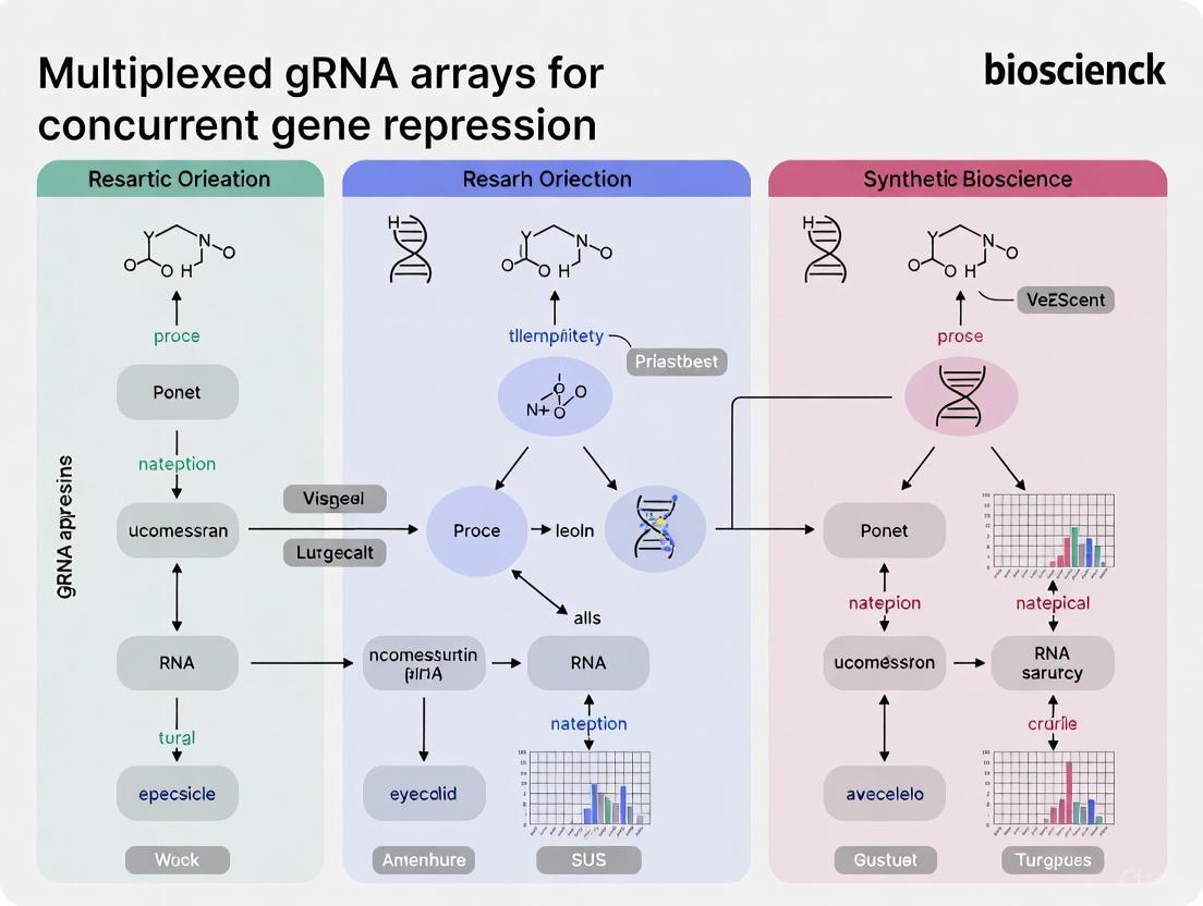
Multiplexed gRNA Arrays: A Comprehensive Guide for Concurrent Gene Repression in Research and Therapy
Multiplexed gRNA arrays represent a transformative advancement in CRISPR technology, enabling the simultaneous repression of multiple genes to dissect complex biological networks and polygenic diseases.
Multiplexed gRNA Arrays: A Comprehensive Guide for Concurrent Gene Repression in Research and Therapy
Abstract
Multiplexed gRNA arrays represent a transformative advancement in CRISPR technology, enabling the simultaneous repression of multiple genes to dissect complex biological networks and polygenic diseases. This article provides a foundational understanding of CRISPR interference (CRISPRi) and the architecture of gRNA arrays, explores the latest methodologies for array assembly and delivery across different model systems, and offers practical strategies for troubleshooting and optimizing repression efficiency. By comparing the performance of different systems and validating their outcomes, we equip researchers and drug development professionals with the knowledge to design robust, high-throughput experiments for functional genomics and therapeutic discovery.
The Principles of Multiplexed CRISPRi: From dCas9 Basics to Array Architecture
CRISPR interference (CRISPRi) is a powerful technology derived from the CRISPR-Cas9 system that allows for precise, programmable repression of gene transcription without altering the underlying DNA sequence. The core component of CRISPRi is a catalytically dead Cas9 (dCas9) protein, which retains its ability to bind DNA in a guide RNA-directed manner but lacks endonuclease activity. This dCas9 protein serves as a programmable DNA-binding scaffold that can be fused to transcriptional repressor domains, enabling targeted gene knockdown [1] [2]. Unlike CRISPR knockout techniques that permanently disrupt genes, CRISPRi offers reversible gene expression control, does not induce DNA damage, and avoids activating endogenous DNA repair pathways that can confound experimental results [1]. When framed within research on multiplexed gRNA arrays, CRISPRi becomes an exceptionally powerful tool for conducting complex genetic perturbations, allowing researchers to repress multiple genes concurrently to study genetic networks, synthetic lethality, and polygenic traits [3] [4].
The Core Mechanism of dCas9-Mediated Repression
The fundamental mechanism of CRISPRi involves the guided localization of a dCas9-repressor fusion complex to specific genomic loci to block transcription. This process can be broken down into several key steps:
Assembly of the dCas9-gRNA Ribonucleoprotein Complex
The system begins with the formation of a complex between the dCas9 protein and a single-guide RNA (sgRNA). The sgRNA, through its ~20 nucleotide spacer sequence, provides the targeting specificity by binding to complementary DNA sequences adjacent to a Protospacer Adjacent Motif (PAM), typically 5'-NGG-3' for the commonly used Streptococcus pyogenes Cas9 [5].
DNA Binding and Transcriptional Blockade
Once the dCas9-sgRNA complex binds to its target DNA, it can repress transcription through multiple mechanisms:
- Physical Steric Hindrance: When targeted to a gene's transcription start site (TSS), the dCas9 complex physically blocks the binding or progression of RNA polymerase, effectively preventing transcription initiation or elongation [1] [2].
- Recruitment of Chromatin-Modifying Complexes: Fused repressor domains recruit additional proteins that establish a repressive chromatin environment, leading to more potent and durable gene silencing [1] [6].
The following diagram illustrates the core repression mechanism and the enhanced repression achieved with advanced repressor domains:
Key Repressor Domains and Their Efficiencies
The efficacy of CRISPRi systems depends significantly on the choice of repressor domain fused to dCas9. Different repressor domains employ distinct mechanisms to silence transcription, primarily by recruiting chromatin-modifying complexes that promote a transcriptionally inactive state. Recent research has systematically evaluated numerous repressor domains and their combinations to identify highly effective configurations.
Table 1: Comparison of Key dCas9-Repressor Domain Fusions
| Repressor Domain | Type | Mechanism of Action | Reported Knockdown Efficiency | Key Features |
|---|---|---|---|---|
| KOX1(KRAB) [1] | KRAB domain | Recruits KAP1, HP1, and histone methyltransferases to establish heterochromatin [1] | Baseline repression | First characterized CRISPRi repressor; widely used but variable performance |
| ZIM3(KRAB) [1] | KRAB domain | Enhanced heterochromatin formation compared to KOX1(KRAB) [1] | ~20-30% better than KOX1(KRAB) [1] | Improved consistency across cell lines and gene targets |
| MeCP2 [1] | Methyl-DNA binding domain | Interacts with SIN3A and histone deacetylases (HDACs) [1] | Comparable to top KRAB domains | Synergistic when combined with KRAB domains |
| MeCP2(t) [1] | Truncated MeCP2 | Conserved repressive function in a shorter 80aa domain [1] | Similar to full-length MeCP2 [1] | Smaller size may improve protein stability and delivery |
| SALL1-SDS3 [2] | Proprietary fusion | Recruits proteins involved in chromatin remodeling and silencing [2] | More potent than dCas9-KRAB in head-to-head tests [2] | Commercial system with optimized performance |
| dCas9-ZIM3(KRAB)-MeCP2(t) [1] | Bipartite fusion | Combines enhanced KRAB activity with MeCP2-mediated repression [1] | Significantly improved across multiple cell lines [1] | Next-generation repressor with reduced guide-dependent variability |
Experimental Protocol for CRISPRi Gene Repression
This section provides a detailed methodology for implementing CRISPRi in mammalian cells, from vector design to validation of repression.
sgRNA Design and Cloning
- Target Selection: Design sgRNAs to bind within -50 to +300 bp relative to the transcription start site (TSS) of your target gene. Using multiple sgRNAs per gene (a pool) typically enhances repression efficacy [2].
- Algorithmic Design: Utilize established algorithms (e.g., CRISPRi v2.1) that incorporate chromatin accessibility, position, and sequence data to predict highly effective sgRNAs [2].
- Cloning into Expression Vectors: Clone annealed oligonucleotides encoding the sgRNA spacer sequence into appropriate CRISPRi vectors using restriction enzymes like BbsI or BsaI [7].
Delivery of CRISPRi Components
- Transient Transfection: For rapid assessment, co-transfect plasmids expressing dCas9-repressor and sgRNA into cells using lipid-based transfection reagents. Gene repression can be observed as early as 24 hours post-transfection, with maximal effects typically at 48-72 hours [2].
- Lentiviral Transduction: For stable, long-term repression or in hard-to-transfect cells, create lentiviral particles encoding the dCas9-repressor and sgRNA(s). This allows for the generation of stable cell lines with integrated CRISPRi components [2].
- RNP Delivery: For minimal off-target effects and highest precision, form ribonucleoprotein (RNP) complexes in vitro by mixing purified dCas9-repressor protein with in vitro-transcribed sgRNA, then deliver via electroporation.
Validation of Repression
- RT-qPCR: The most common and rapid method to quantify changes in mRNA levels. Harvest cells 48-72 hours after CRISPRi delivery, extract total RNA, and perform reverse transcription followed by quantitative PCR. Calculate relative expression using the ∆∆Cq method, normalizing to a housekeeping gene (e.g., GAPDH or ACTB) and a non-targeting control sgRNA [2].
- Western Blotting: Confirm repression at the protein level 5-7 days post-treatment, as protein half-lives may cause a delay in observable knockdown.
- Flow Cytometry: If targeting a gene encoding a surface protein, staining with a fluorescent antibody and analysis by flow cytometry provides a quantitative measure of repression at the single-cell level.
- Phenotypic Assays: Perform functional assays relevant to your target gene (e.g., proliferation assays for essential genes, differentiation assays for developmental genes) to confirm the biological consequence of repression.
Integrating CRISPRi with Multiplexed gRNA Arrays
A principal advantage of CRISPRi is its exceptional compatibility with multiplexing—the simultaneous targeting of multiple genomic loci. This is achieved by expressing several guide RNAs from a single polycistronic transcript, a critical capability for studying genetic networks and combinatorial gene functions [3] [4].
Strategies for Multiplexed gRNA Expression
- tRNA-based System: gRNAs are flanked by tRNA sequences, which are recognized and cleaved by endogenous eukaryotic RNases P and Z to release individual, functional gRNAs [7].
- Csy4-based System: The Pseudomonas aeruginosa Csy4 ribonuclease cleaves at a specific 28-base recognition site. Co-expression of Csy4 with a transcript containing gRNAs separated by its target site enables efficient processing [4] [7].
- Cas12a-based System: The native ability of Cas12a to process its own crRNA arrays can be harnessed. A single transcript encoding multiple crRNAs (the guide component for Cas12a) is automatically processed into individual units by the Cas12a protein itself [3].
Inducible Multiplexed CRISPRi
For precise temporal control, which is crucial when repressing essential genes or studying dynamic processes, inducible systems have been developed. One effective strategy uses a Tet-ON system combined with a Tet-OFF silencing system to tightly regulate a polycistronic gRNA array [4]. In the uninduced state, a mutTetR-Mxi1 repressor bound to mutTetO sites silences the entire array. Upon addition of anhydrotetracycline (aTc), the rtTA-Gal4 activator binds to TetO sites and drives array expression, initiating CRISPRi activity. This design has achieved up to 96-98% silencing of basal activity in the uninduced state [4].
The following diagram illustrates the workflow for creating and implementing a multiplexed CRISPRi system:
The Scientist's Toolkit: Essential Reagents for CRISPRi Research
Table 2: Key Research Reagent Solutions for CRISPRi Experiments
| Reagent / Tool | Function | Examples & Notes |
|---|---|---|
| dCas9-Repressor Vectors | Provides the programmable DNA-binding protein and repressor machinery | dCas9-ZIM3(KRAB)-MeCP2(t) [1], dCas9-SALL1-SDS3 [2]; available with various selection markers and fluorescent tags. |
| sgRNA Cloning Vectors | Templates for expressing guide RNAs | Vectors with U6 or H1 Pol III promoters; some designed for multiplexing with Type IIS restriction sites (BbsI, BsaI) [7]. |
| gRNA Array Systems | Enables simultaneous expression of multiple gRNAs | tRNA-gRNA arrays, Csy4-processing arrays, Cas12a crRNA arrays; compatible with Golden Gate assembly [3] [7]. |
| Inducible Systems | Provides temporal control over CRISPRi activity | Tet-ON/Tet-OFF systems for gRNA arrays [4]; chemical- or light-inducible dCas9 systems. |
| Delivery Reagents | Facilitates introduction of CRISPRi components into cells | Lipid-based transfection reagents (e.g., DharmaFECT), electroporation systems (e.g., Lonza Nucleofector), lentiviral packaging systems. |
| Validation Tools | Confirms gene repression and specificity | RT-qPCR assays, antibodies for Western blotting/flow cytometry, RNA-seq for genome-wide specificity profiling. |
| Positive Control sgRNAs | Validates system functionality | sgRNAs targeting genes with clear phenotypes (e.g., PPIB [2]) or reporters (e.g., eGFP [1]). |
Why Multiplex? Overcoming Genetic Redundancy and Modeling Complex Traits
In the functional analysis of complex biological systems, researchers frequently encounter two significant obstacles: genetic redundancy, where multiple genes perform overlapping functions, masking phenotypic consequences when only one is disrupted, and polygenic traits, which arise from the combined subtle effects of numerous genetic loci. Single-gene editing approaches are often inadequate for dissecting these complexities. Multiplexed CRISPR-Cas technology, which enables the simultaneous targeting of multiple genomic sites using arrays of guide RNAs (gRNAs), provides a powerful solution to these challenges [8] [3]. By facilitating concurrent repression of several genes, this approach allows for the functional interrogation of entire pathways, the modeling of complex diseases, and the identification of synthetic lethal interactions that are invisible to single-gene knockout studies [8]. This document outlines the quantitative foundations and detailed protocols for applying multiplexed gRNA arrays in concurrent gene repression research, providing a framework for overcoming the limitations of traditional genetic screening.
Quantitative Foundations of Multiplexed Gene Repression
The efficacy of multiplexed CRISPR systems is well-established across various organisms. The data, summarized in the table below, highlights key performance metrics for different multiplexed repression systems.
Table 1: Performance Metrics of Multiplexed CRISPR Repression Systems
| Organism | System Name | Maximum Number of Targets Demonstrated | Reported Efficiency | Key Application |
|---|---|---|---|---|
| S. cerevisiae | GTR-CRISPR | 8 genes | 87% | Metabolic pathway engineering [9] |
| S. cerevisiae | Lightning GTR-CRISPR | 6 genes | 60% (with pre-validated gRNAs) | Rapid strain development [9] |
| E. coli | Shortened Cas9 Arrays | Not Specified | Up to 24-fold repression per target | Bacterial gene silencing [10] |
| Mammalian Cells | dCas9-based CRISPRi | Multiple loci | Varies by locus | Combinatorial genetic perturbations [3] |
The relationship between the number of gRNAs in an array and the editing efficiency is not linear. As shown in the data from S. cerevisiae, efficiency remains high for up to five targets but can decrease sharply as more gRNAs are added to a single transcript [9]. This underscores the importance of system design for successful multiplexing. Furthermore, the repression strength for an individual target can vary significantly (e.g., from 2.3-fold to 24-fold in E. coli) based on factors such as the target site location within the gene and the specific gRNA sequence used [10].
Experimental Protocols for Multiplexed gRNA Array Assembly and Testing
Protocol 1: Construction of a gRNA-tRNA Array for Yeast (GTR-CRISPR)
This protocol describes the assembly of a multiplex gRNA system using a tRNA-processing system for highly efficient, simultaneous gene disruptions in S. cerevisiae [9].
Design of gRNA-tRNA Array:
- Design gRNA sequences targeting your genes of interest.
- Assemble these gRNAs into a single synthetic gene array where each gRNA is flanked by endogenous tRNAGly sequences (71 bp). This array is transcribed as a single transcript under the control of an RNA polymerase III promoter (e.g., SNR52 promoter).
Golden Gate Assembly:
- The array is cloned into a suitable expression plasmid using Golden Gate assembly, which leverages type IIS restriction enzymes to efficiently assemble multiple gRNA units in a specific orientation [9].
- The final plasmid also carries the Cas9 (or dCas9 for repression) gene and a selectable marker.
Yeast Transformation and Selection:
- Co-transform the assembled GTR-CRISPR plasmid and the PCR-amplified homologous donor DNA templates (for gene disruption) into S. cerevisiae.
- Plate the transformation mix onto appropriate selective media and incubate until colonies form.
Validation and Screening:
- Pick individual colonies and screen for successful gene disruptions via colony PCR and DNA sequencing.
- For repression studies (using dCas9), quantify the knockdown efficiency using RT-qPCR to measure transcript levels or directly assay the phenotypic outcome.
Protocol 2: Rapid, Cloning-Free Multiplexing in Yeast (Lightning GTR-CRISPR)
For applications requiring extreme speed, this accelerated method bypasses the conventional cloning step in E. coli [9].
Preparation of gRNA Array:
- Perform the Golden Gate assembly reaction as in Protocol 1 to assemble the gRNA-tRNA array.
Direct Yeast Transformation:
- Instead of transforming the assembly reaction into E. coli for plasmid propagation, directly transform the entire Golden Gate reaction mix, which contains the assembled plasmid, into competent S. cerevisiae cells along with the donor DNA fragments.
Outcome: This method enables disruption of up to 6 genes in just 3 days, with an efficiency of approximately 60% when using pre-validated gRNAs [9].
Protocol 3: Implementing Shortened CRISPR-Cas9 Arrays in Bacteria
This protocol is optimized for efficient multiplexed gene repression in E. coli using compact, processed-like CRISPR arrays [10].
Design of Shortened Arrays:
- Design a CRISPR array with a leader sequence followed by repeat-spacer subunits.
- Systematically shorten the spacer sequences (from the 5' end) and the repeat sequences (from the 3' end) to mimic naturally processed crRNAs. Spacers can often be trimmed from 30 nt to ~24 nt while maintaining functionality, and repeats can be shortened from 36 nt.
Array Assembly and Transformation:
- Assemble the shortened array into a plasmid backbone containing the tracrRNA sequence and a dCas9 gene (e.g., SpdCas9 for repression) using a specific cloning method with defined 4-bp assembly junctions [10].
- Co-transform this plasmid with a reporter plasmid (if testing) into your bacterial strain.
Efficiency Measurement:
- Measure gene repression efficiency by comparing the expression level of the target gene(s) (e.g., via fluorescence if using a reporter) in strains containing the targeted array versus a non-targeting control array.
Visualization of Multiplexed gRNA Workflows and Applications
The following diagrams illustrate the core concepts and experimental workflows for multiplexed CRISPR technologies.
Diagram 1: Core workflow for multiplexed gRNA array assembly and application.
Diagram 2: gRNA-tRNA array processing mechanism for multiplexed repression.
The Scientist's Toolkit: Essential Reagents for Multiplexed Repression
Successful implementation of multiplexed gene repression relies on a core set of research reagents. The following table details these essential components and their functions.
Table 2: Key Research Reagent Solutions for Multiplexed Gene Repression
| Reagent / Solution | Function | Example & Notes |
|---|---|---|
| Cas9/dCas9 Vector | Provides the nuclease or DNA-binding protein. | Catalytically dead Cas9 (dCas9) for repression; can be constitutively expressed or inducible [8] [10]. |
| gRNA Expression Backbone | Plasmid for hosting the gRNA array. | Contains a Pol III promoter (e.g., SNR52 in yeast, U6 in mammals) and terminator [9] [3]. |
| tRNA-gRNA Array Construct | Single transcript encoding multiple gRNAs. | gRNAs are flanked by tRNA sequences (e.g., tRNAGly) for efficient processing by endogenous RNases [9]. |
| Type IIS Restriction Enzymes | Enzymes for modular assembly of gRNA arrays. | Essential for Golden Gate assembly (e.g., BsaI) to seamlessly combine multiple gRNA units [9] [3]. |
| Homology Donor Templates | DNA for introducing specific mutations or markers. | Used in knockout experiments; not required for repression-only (dCas9) applications [9]. |
| Processing Enzymes (Optional) | Proteins for processing gRNA arrays. | e.g., Csy4 endoribonuclease for cleaving arrays at specific recognition sites [3]. |
In the field of genetic engineering, the ability to perform concurrent repression (or activation) of multiple genes is a cornerstone for advanced cellular reprogramming, metabolic engineering, and foundational biological research. Clustered Regularly Interspaced Short Palindromic Repeats (CRISPR) inhibition (CRISPRi) and activation (CRISPRa) have emerged as powerful synthetic tools for modulating endogenous gene expression [11]. The coordinated activation and inhibition (CRISPRai) of target genes allows researchers to fully explore transcriptional landscapes and modify cellular behavior, which is especially important in metabolic engineering where fluxes must be redirected towards a desired product by upregulating desired reactions and downregulating competing pathways [11].
A significant limitation of early CRISPR systems was their capacity to target only single genetic loci. However, desired cellular behaviors are often achieved by altering the expression of a large number of targets simultaneously [11]. Multiplexed gRNA arrays—single transcriptional units expressing multiple guide RNAs—solve this challenge by enabling coordinated targeting of several genomic sites within the same cell. This application note provides a detailed comparison of gRNA array architectures, complete with experimental protocols and reagent solutions, framed within the context of concurrent gene repression research.
gRNA Array Architectures: A Comparative Analysis
Different strategies have been developed to express multiple gRNAs from a single construct, each with distinct mechanisms for processing individual guides. The table below summarizes the core architectural designs.
Table 1: Comparative Analysis of Multiplexed gRNA Array Architectures
| Array Architecture | Processing Mechanism | Key Components | Maximum gRNAs Demonstrated | Key Advantages | Reported Efficiency/Performance |
|---|---|---|---|---|---|
| Csy4 Processed Array | Csy4 endonuclease cleavage | Csy4 gene, gRNAs flanked by Csy4 recognition sequences | 24 gRNAs [11] | High multiplexing capacity; Compatible with two orthogonal CRISPR/Cas systems | Enabled 45-fold increase in succinic acid production in yeast via 11-gRNA targeting [11] |
| Ribozyme Processed Array | Self-cleaving ribozymes (e.g., HDV) | gRNAs flanked by ribozyme sequences | 4 gRNAs [12] | No need for exogenous protein expression; Self-cleaving | Requires relatively long constructs; Activity can be strain-specific [12] |
| tRNA-Processed Array | Endogenous RNase P and RNase Z | tRNA promoter, gRNA sequences | Limited in longer arrays [12] | Utilizes host machinery; Avoids supplementary components | Endogenous RNase activity is insufficient for longer RNA arrays [12] |
| Individual Promoter Array (Golden Gate) | Multiple independent RNA Pol III promoters | Multiple human U6 promoters, each driving one gRNA | 30 gRNAs [13] | Avoids sequence repetition; High efficiency per gRNA | Sequential assembly method; Highly flexible design [13] |
The Inducible Array Solution for Reduced Fitness Cost
A significant challenge in multiplexed CRISPRai is that prolonged transcriptional perturbation can impose a fitness cost, leading to genetic instability and phenotypic loss [11]. Furthermore, regulating essential genes continuously can impact cell growth or be impossible [11]. To address this, an advanced inducible system for polycistronic arrays containing up to 24 gRNAs was developed for S. cerevisiae [11].
This system uses the opposing actions of orthogonal Tet-ON and Tet-OFF systems to achieve near leak-free inducibility. The Tet-ON system (rtTA-Gal4) binds to Tet operator (TetO) sites to drive array expression in the presence of the inducer anhydrotetracycline (aTc). The Tet-OFF system (mutTetR-Mxi1) binds to an orthogonal TetO variant (mutTetO) to silence transcription across the entire array in the absence of the inducer [11]. This design ensures that in the uninduced state, reporter expression remains at 96–98% of maximum, demonstrating efficient silencing and resolving the problem of basal gRNA array transcription that can occur even without a promoter [11].
Table 2: Performance Metrics of Inducible gRNA Array System
| Parameter | Design 1 (Low-Leak Promoter) | Design 2 (Leak-Free Promoter) | Design 3 (Tet-ON/OFF Silencing) |
|---|---|---|---|
| Basal CRISPRi Activity (Uninduced) | 10% of max. reporter expression | 54% of max. reporter expression | 2-4% of max. reporter expression |
| Induced CRISPRi Activity | Not specified | Not specified | No significant difference from constitutive expression |
| Key Mechanism | Promoter control | Promoter control | Array silencing with mutTetR-Mxi1 between gRNA clusters |
| Optimal gRNAs Between Silencing Sites | Not applicable | Not applicable | Up to 6 gRNAs |
Essential Research Reagent Solutions
The following toolkit comprises key reagents required for implementing multiplexed gRNA array systems.
Table 3: Essential Research Reagent Solutions for gRNA Array Construction and Application
| Reagent / Material | Function / Purpose | Specific Examples / Notes |
|---|---|---|
| Cas Proteins | Target DNA binding and cleavage or functional modulation | dCas9: Transcriptional repression when fused to Mxi1 domain; dCas12a: Orthogonal system for simultaneous activation/repression [11] |
| Assembly Plasmids | Backbone vectors for gRNA array construction | pMA-SpCas9-g1 to g10 modular plasmids (Addgene IDs 80784-80793); pMA-MsgRNA-EGFP array plasmid (Addgene ID 80794) [13] |
| Restriction Enzymes | Type IIS enzymes for Golden Gate assembly | BbsI (for single gRNA cloning); BsaI/BsmBI (for array assembly) [13] |
| Polymerase & Ligase | Amplification and ligation of DNA fragments | DreamTaq DNA polymerase; T4 DNA ligase [13] |
| Endonucleases for Processing | Intein-mediated array processing | Csy4 endonuclease for processing gRNAs from long transcript [11] |
| Induction System | Chemical control of gRNA expression | Tet-ON (rtTA-Gal4) and Tet-OFF (mutTetR-Mxi1) systems with aTc inducer [11] |
| Reporter Strains | Evaluation of editing efficiency | Prototrophic S. cerevisiae strains (e.g., CEN.PK113-7D) with auxotrophic markers for perturbation assessment [12] |
Detailed Experimental Protocols
Protocol 1: Golden Gate Assembly of Multiplexed gRNA Arrays
This protocol enables efficient assembly of 2-30 gRNA expression cassettes into a single vector within 7 days using Golden Gate cloning [13].
Materials and Reagents:
- Competent E. coli cells (recombination deficient)
- Modular gRNA plasmids (pMA-SpCas9-g1 to g10, Addgene IDs 80784-80793)
- Array plasmid (pMA-MsgRNA-EGFP, Addgene ID 80794 for 11-30 gRNAs)
- Restriction enzymes: BbsI (FastDigest), BsaI/BsmBI (FastDigest)
- T4 DNA ligase (5 U/μl)
- NEB Buffer 2
- Ampicillin, Spectinomycin
- Universal primers for screening: U6 Forward and Scr Reverse [13]
Procedure:
gRNA Oligonucleotide Design and Preparation:
- Design target gRNA oligonucleotides using online tools (e.g., crispr.mit.edu).
- The first nucleotide should preferably be a 'G' for efficient transcription by the human U6 promoter.
- Exclude gRNA oligonucleotides containing BbsI, BsaI, or BsmBI recognition sites.
- For target sites starting with 'G', add overhangs: Sense: 5'-CACC(N20); Antisense: 5'-AAAC(N20)
- For other starting nucleotides: Sense: 5'-CACCG(N20); Antisense: 5'-AAAC(N20)C
- Order and dilute oligonucleotides to 100 μM stock concentration [13].
Annealing of gRNA Oligos:
- Mix in a 1.5 ml tube: 1 μl sense oligo (100 μM), 1 μl antisense oligo (100 μM), 2 μl 10× NEB Buffer 2, and ddH₂O to 20 μl.
- Denature at 95°C for 5 minutes in a heating block.
- Let the block cool slowly to room temperature (1-2 hours) for annealing.
- Centrifuge briefly and store at -20°C [13].
Ligation of Annealed Oligos into Single Modular Vectors:
- Digest 2 μg of the appropriate pMA-SpCas9-g# vector with BbsI in 1× FastDigest Green Buffer for 1 hour at 37°C.
- Ligate 50 ng of digested vector with 1 μl of annealed oligo duplex using T4 DNA ligase for 1 hour at 25°C.
- Transform into competent E. coli and select on ampicillin plates.
- Verify clones by colony PCR or sequencing using universal primers [13].
Golden Gate Assembly of gRNA Arrays:
- For arrays of 2-10 gRNAs: Perform a one-pot Golden Gate reaction with all individual pMA-T# plasmids, using BsaI and T4 ligase in NEB Buffer 2. Cycle between 37°C (5 min) and 16°C (10 min) for 30 cycles, followed by 55°C for 15 min and 80°C for 15 min.
- For arrays of 11-30 gRNAs: First assemble 2-3 sub-arrays, then perform a second Golden Gate assembly with BsmBI to combine them into the final pMA-MsgRNA-EGFP vector [13].
- Transform the assembly reaction into competent E. coli and select on spectinomycin plates.
- Verify correct assembly by restriction digest and sequencing.
Golden Gate gRNA Array Assembly Workflow
Protocol 2: Evaluation of gRNA Array Performance in S. cerevisiae
This protocol describes the evaluation of multiplex CRISPR-Cas9 system performance by assessing the success rate of introducing perturbations within target loci [12].
Materials and Reagents:
- Prototrophic S. cerevisiae strain (e.g., CEN.PK113-7D)
- Cas9-expression vector
- Assembled gRNA array vector
- YPD medium (1% yeast extract, 2% peptone, 2% glucose)
- Complete Supplement Mixture (CSM) with appropriate dropouts
- Antibiotics: Nourseothricin (0.1 mg/ml), G418 (0.5 mg/ml)
- L-canavanine sulfate (60 μg/ml in CSM-Arg medium for CAN1 assessment) [12]
Procedure:
Yeast Transformation:
- Co-transform S. cerevisiae with the Cas9-expression vector and the assembled gRNA array vector using standard lithium acetate transformation protocol.
- Plate on appropriate selective media based on the plasmid markers (e.g., CSM-Ura, CSM-Leu) and incubate at 30°C for 2-3 days [12].
Phenotypic Evaluation of Editing Efficiency:
- For each target gene, design a growth-based assay. The example below uses marker genes where successful perturbation prevents growth on specific media.
- Patch transformants onto various selective media to assess the knockout of each target gene.
- For the CAN1 gene, plate on CSM-Arg medium containing L-canavanine. Successful CAN1 disruption confers resistance to this toxic analog, allowing growth [12].
- Calculate editing efficiency as the percentage of transformants showing the expected phenotype for each targeted gene.
Analysis of Multiplexing Efficiency:
- To assess simultaneous multi-locus editing, replica-plate colonies onto media selecting for all desired perturbations.
- The fraction of colonies showing all expected phenotypes indicates the co-editing efficiency.
- In one study, this method successfully introduced up to five simultaneous perturbations within single yeast cells [12].
gRNA Array Performance Evaluation Workflow
The development of sophisticated gRNA array architectures has dramatically expanded our capability for multiplexed gene repression and activation. From Csy4-processed arrays enabling two dozen simultaneous guides to inducible systems that minimize fitness costs, these blueprints provide researchers with a versatile toolkit for complex genetic engineering. The choice of architecture—whether ribozyme-based, tRNA-processed, Csy4-dependent, or multi-promoter driven—depends on the specific application, desired number of targets, and host organism. As these technologies continue to evolve, they will further empower scientists to tackle increasingly complex challenges in systems biology, metabolic engineering, and therapeutic development.
Clustered Regularly Interspaced Short Palindromic Repeats (CRISPR) and CRISPR-associated (Cas) proteins constitute an adaptive immune system in bacteria and archaea that protects against invasive genetic elements like viruses and plasmids [14]. This natural system has been repurposed as a revolutionary genome engineering tool, with its primary advantage lying in its simplicity and programmability via guide RNA (gRNA) molecules [8]. Unlike previous gene-editing technologies such as Zinc Finger Nucleases (ZFNs) and Transcription Activator-Like Effector Nucleases (TALENs), which require protein engineering for each new target, CRISPR-Cas systems require only the synthesis of a new guide RNA to redirect the nuclease to a specific DNA sequence [14]. This fundamental characteristic makes CRISPR systems particularly amenable to multiplexed genome editing—the simultaneous targeting of multiple genomic sites using arrays of guide RNAs [8].
The natural CRISPR-Cas adaptive immunity operates in three distinct stages: (1) Adaptation, where Cas proteins capture and integrate short fragments of foreign DNA as spacers into the CRISPR array; (2) Expression, where the CRISPR array is transcribed and processed into individual CRISPR RNAs (crRNAs); and (3) Interference, where Cas protein-crRNA complexes recognize and cleave complementary foreign DNA sequences [15] [14]. Synthetic array design directly mimics this natural crRNA processing pathway, particularly the expression stage where multiple spacers and direct repeats are organized into functional arrays that can be processed into individual guide RNAs [16]. This bioinspired approach enables researchers to engineer multiplexed gRNA arrays for concurrent gene repression, activation, or editing—opening new frontiers in functional genomics, synthetic biology, and therapeutic development.
Table: Comparison of Genome Editing Technologies
| Parameter | ZFN | TALEN | CRISPR-Cas |
|---|---|---|---|
| Efficiency | 0–12% (low) | 0–76% (moderate) | 0–81% (high) |
| Target Recognition | Protein-DNA | Protein-DNA | RNA-DNA |
| Target Site Length | 18–36 bp/ZFN pair | 30–40 bp/TALEN pair | 22 bp |
| Multiplexing Feasibility | Less feasible | Less feasible | Highly feasible |
| Ease of Designing | Difficult | Difficult | Easy |
| Large-Scale Library Construction | Challenging | Challenging | Easy |
Evolutionary Classification of Natural CRISPR-Cas Systems
The natural diversity of CRISPR-Cas systems continues to expand as genomic and metagenomic databases grow. According to the most recent evolutionary classification, CRISPR-Cas systems are divided into 2 classes, 7 types, and 46 subtypes, representing a significant expansion from the 6 types and 33 subtypes identified five years ago [15]. This classification is based on a polythetic approach that combines comparisons of CRISPR-cas locus architecture and gene composition with sequence similarity clustering and phylogenetic analysis of conserved Cas proteins, particularly Cas1, the integrase central to the adaptation stage [15].
Class 1 systems (including types I, III, IV, and VII) utilize multi-protein effector complexes, while Class 2 systems (including types II, V, and VI) employ single-protein effectors such as the well-characterized Cas9 [15]. The recently characterized type VII systems, found predominantly in diverse archaeal genomes, feature a metallo-β-lactamase (β-CASP) effector nuclease designated Cas14, which is encoded in a predicted operon with Cas7 and Cas5 subunits [15]. Type VII loci typically lack adaptation modules and associated CRISPR arrays often contain multiple substitutions, suggesting limited incorporation of new spacers. These systems have been shown to target RNA in a crRNA-dependent manner, cleaving targets via the nuclease activity of Cas14 [15].
The expanding classification reveals that previously defined systems are relatively common, while more recently characterized variants represent the "long tail" of the CRISPR-Cas distribution in prokaryotes and their viruses [15]. This evolutionary diversity provides a rich repository of molecular machinery that can be harnessed for synthetic array design, with different systems offering distinct advantages for specific applications. For instance, the multi-protein complexes of Class 1 systems offer potential for more complex regulatory functions, while the simplicity of Class 2 systems makes them particularly amenable to engineering and delivery in therapeutic contexts.
Table: Key Characteristics of CRISPR-Cas Classes
| Feature | Class 1 Systems | Class 2 Systems |
|---|---|---|
| Effector Complex | Multi-protein | Single protein |
| Types | I, III, IV, VII | II, V, VI |
| Representative Proteins | Cas3, Cas10, Cas14 | Cas9, Cas12, Cas13 |
| crRNA Processing | Requires multiple Cas proteins | Often self-contained in effector |
| Engineering Complexity | Higher | Lower |
| Therapeutic Delivery | More challenging | Simplified |
Design Principles for Synthetic Multiplexed gRNA Arrays
Architectural Framework from Natural Systems
Synthetic gRNA array design directly emulates the natural organization of CRISPR arrays, where short direct repeats (DRs) alternate with variable spacers that determine target specificity [16]. In natural systems, these arrays are transcribed as long precursor RNAs that are subsequently processed into individual crRNAs through the activity of Cas proteins and associated nucleases [16]. The synthetic implementation of this principle involves designing expression constructs where multiple gRNA units—each consisting of a target-specific spacer flanked by appropriate direct repeat sequences—are arranged in tandem. This bioinspired approach capitalizes on the natural processing mechanisms to generate multiple functional guide RNAs from a single transcriptional unit, significantly simplifying the delivery of multiplexed CRISPR systems.
The design of effective synthetic arrays requires careful consideration of several factors: (1) Direct repeat identity: Consistent repeat sequences that can be recognized by the processing machinery; (2) Spacer specificity: 20-nucleotide target sequences with appropriate protospacer adjacent motif (PAM) requirements for the Cas protein being used; (3) Array length: Balancing the number of gRNAs with processing efficiency and delivery constraints; and (4) Transcriptional regulation: Selection of appropriate promoters and terminators for optimal expression [16]. Natural systems provide valuable insights into each of these parameters, with bioinformatic analyses of native CRISPR arrays revealing optimal arrangements that have been evolutionarily refined for efficient processing and function.
Processing Mechanisms and Cas Protein Compatibility
Different CRISPR-Cas systems employ distinct crRNA processing mechanisms that must be considered in synthetic array design. Type II systems typically rely on a trans-activating crRNA (tracrRNA) that base-pairs with direct repeats in the precursor transcript, facilitating processing by RNase III in the presence of Cas9 [14]. Type I and III systems often utilize dedicated Cas6-like nucleases that recognize specific secondary structures within the direct repeats [15]. Type V systems may employ different processing mechanisms, sometimes leveraging the Cas12 protein itself for maturation of the crRNAs [15].
For synthetic biology applications, these natural processing pathways can be engineered to optimize functionality. In some cases, endogenous processing systems can be utilized by designing arrays with compatible direct repeats. Alternatively, processing can be simplified through the use of engineered systems that incorporate ribozyme sequences or tRNA motifs that facilitate precise cleavage into individual gRNAs [8]. The choice of processing strategy depends on the specific application, with each approach offering distinct advantages in terms of efficiency, specificity, and ease of implementation. Understanding the natural processing mechanisms provides a foundation for engineering optimized synthetic systems that maintain high efficiency while enabling multiplexed targeting capabilities.
Experimental Protocol: Implementing Multiplexed gRNA Arrays for Concurrent Gene Repression
Vector Assembly and gRNA Array Cloning
The construction of multiplexed gRNA arrays typically employs modular cloning strategies such as Golden Gate assembly, which allows efficient, directional assembly of multiple gRNA expression units into a single vector [8]. This protocol outlines the steps for creating a 4-gRNA array targeting multiple genes for concurrent repression using a Cas9-based system:
Materials:
- Backbone vector with Cas9 expression cassette, bacterial resistance marker, and appropriate origin of replication
- Promoter modules (typically U6 or other Pol III promoters)
- gRNA modules containing target-specific spacers with appropriate flanking sequences
- Terminator modules (typically polyT tracts for Pol III transcripts)
- Type IIS restriction enzymes (e.g., BsaI, BbsI) with corresponding buffers
- T4 DNA Ligase and reaction buffer
- Competent E. coli cells for transformation
- LB agar plates with appropriate selection antibiotics
- PCR reagents for colony screening
- DNA sequencing primers for verification
Procedure:
- Design gRNA targets: Select 20-nucleotide target sequences adjacent to 5'-NGG PAM sequences in the genes of interest. Verify specificity using genome-wide off-target prediction tools.
- Phosphorylate and anneal oligonucleotides: For each gRNA target, synthesize complementary oligonucleotides with appropriate overhangs for Golden Gate assembly, phosphorylate, and anneal to form double-stranded inserts.
- Golden Gate assembly: Set up reaction containing backbone vector, promoter modules, gRNA modules, terminator modules, Type IIS restriction enzyme, T4 DNA Ligase, and reaction buffer. Cycle between digestion (37°C) and ligation (16°C) 25-30 times.
- Transform and screen: Transform reaction into competent E. coli cells, plate on selective media, and screen colonies by PCR and restriction digest.
- Sequence verification: Isolate plasmid DNA from positive clones and verify array sequence by Sanger sequencing using appropriate primers.
- Functional validation: Before proceeding to full experiments, validate array functionality by transfection into appropriate cell lines and assessment of editing efficiency at each target site.
This modular approach enables rapid assembly of complex gRNA arrays and can be scaled to accommodate different numbers of targets based on experimental needs. The use of Type IIS restriction enzymes creates unique overhangs that ensure proper orientation and order of gRNA units within the array [8].
Delivery and Analysis of Multiplexed Repression
Cell Culture and Transfection:
- Culture appropriate cell lines (e.g., HEK293T, K562, or cell lines relevant to your research) under standard conditions.
- Seed cells in appropriate multi-well plates 24 hours before transfection to achieve 70-80% confluence at time of transfection.
- Transfect with CRISPR plasmids using preferred method (lipofection, electroporation, etc.) with appropriate controls including empty vector and single gRNA transfections.
- After 48-72 hours, harvest cells for analysis, splitting for both genomic DNA extraction and RNA/protein analysis.
Analysis of Editing Efficiency:
- Genomic DNA extraction: Use commercial kits to isolate high-quality genomic DNA from transfected cells.
- PCR amplification of target sites: Design primers flanking each target site and amplify regions of interest.
- Assessment of editing efficiency: Utilize T7 Endonuclease I assay or tracking of indels by decomposition (TIDE) analysis to quantify mutation rates at each target site.
- Next-generation sequencing: For comprehensive analysis, amplify target regions with barcoded primers and subject to high-throughput sequencing to characterize all induced mutations.
Functional Assessment of Repression:
- RNA extraction and qRT-PCR: Isolate total RNA and perform quantitative RT-PCR to measure transcript levels of targeted genes.
- Western blot analysis: Assess protein levels of targeted genes to confirm functional knockdown.
- Phenotypic assays: Perform relevant functional assays based on the biological processes being targeted by the multiplexed repression.
This protocol enables efficient implementation of multiplexed gene repression using synthetic gRNA arrays modeled after natural CRISPR systems. The approach can be adapted for different cell types, delivery methods, and analysis techniques based on specific research requirements [8].
Research Reagent Solutions for Multiplexed CRISPR Applications
Table: Essential Research Reagents for Multiplexed CRISPR Experiments
| Reagent Category | Specific Examples | Function & Application Notes |
|---|---|---|
| Cas Expression Systems | Cas9, Cas12a, dCas9-KRAB, dCas9-VPR | Nuclease activity or transcriptional regulation; choice depends on desired outcome (knockout vs. modulation) |
| gRNA Cloning Systems | Golden Gate assembly kits, tRNA-gRNA vectors, Ribozyme-gRNA constructs | Efficient assembly of multiplexed gRNA arrays with various processing mechanisms |
| Delivery Vehicles | Lentiviral vectors, AAV vectors, Lipid nanoparticles | Stable or transient delivery to target cells; consider payload size and tropism |
| Validation Tools | T7E1 assay kits, Tracking of Indels by Decomposition (TIDE), NGS libraries | Assessment of editing efficiency and specificity at each target site |
| Cell Culture Resources | Appropriate cell lines, Selection antibiotics, Transfection reagents | Model systems for testing multiplexed arrays; consider relevance to biological question |
| Analysis Reagents | RNA extraction kits, qPCR reagents, Western blot materials | Functional assessment of gene repression efficiency and off-target effects |
Visualization of Multiplexed gRNA Array Workflow
Diagram Title: Multiplexed gRNA Array Workflow
Applications in Biomedical Research and Therapeutic Development
Multiplexed CRISPR systems have revolutionized functional genomics by enabling comprehensive analysis of gene networks and synthetic lethal interactions. The CRISPR-based double-knockout (CDKO) system exemplifies this approach, allowing researchers to target gene pairs genome-wide to identify synthetic lethal interactions [8]. In one notable application, a CDKO library containing 490,000 gRNA pairs was used to identify synthetic lethal targets of drugs in K562 cells, revealing potential combination therapies [8]. Similarly, multiplexed CRISPR screening integrated with single-cell RNA sequencing has been employed to unravel mammalian unfolded protein responses by targeting multiple genes simultaneously and profiling transcriptional outcomes at single-cell resolution [8].
In therapeutic development, multiplexed CRISPR approaches show particular promise for complex diseases like cancer, where multiple genetic pathways often need simultaneous targeting. A groundbreaking study demonstrated that numerous targeted double-strand breaks specific to cancer cells could induce cell death in malignant but not normal cells, suggesting a novel CRISPR-mediated cancer therapy approach [8]. This strategy leverages the concept that cancer cells, with their compromised DNA repair mechanisms, are more vulnerable to multiple simultaneous DNA breaks than healthy cells. Beyond oncology, multiplexed CRISPR systems are being explored for treating multigenic disorders, persistent viral infections, and for engineering advanced cell therapies.
The technology also enables creation of more sophisticated disease models. By introducing multiple genetic alterations simultaneously, researchers can better recapitulate the complexity of human diseases in animal models or organoid systems [14]. These advanced models provide more physiologically relevant platforms for drug screening and validation, potentially accelerating the drug development process. Furthermore, multiplexed epigenetic editing using engineered CRISPR-Cas systems specialized for direct repression or activation of gene expression allows researchers to manipulate multiple gene regulatory elements concurrently, opening new avenues for understanding and treating diseases driven by epigenetic dysregulation [8].
Troubleshooting and Optimization Strategies
Low Editing Efficiency at Multiple Targets:
- Verify gRNA processing: Ensure direct repeats or processing elements (tRNA, ribozymes) are compatible with your system
- Optimize delivery efficiency: Use appropriate controls to confirm transfection/transduction efficiency exceeds 70%
- Check Cas9 expression levels: Ensure sufficient nuclease is present to handle multiple targets simultaneously
- Validate gRNA functionality: Test individual gRNAs separately to identify any problematic guides
- Consider array position effects: Rearrange gRNA order as processing efficiency may vary along the array
Unintended Large Deletions or Rearrangements:
- Design gRNAs with appropriate spacing: When targeting the same gene or adjacent regions, be aware that simultaneous cuts can cause large deletions
- Use nickase pairs: For applications requiring reduced off-target effects, consider using Cas9 nickases that require paired targeting for double-strand breaks
- Implement controlled expression: Use inducible systems to limit Cas9 activity duration and reduce unintended effects
Variable Repression Efficiency Across Targets:
- Validate gRNA target accessibility: Chromatin state can significantly impact gRNA efficiency; consider ATAC-seq data in gRNA design
- Optimize promoter selection: Different Pol III promoters may have varying strengths in your cell type of interest
- Balance gRNA ratios: In some cases, competition between gRNAs can occur; adjust array order or consider separate expression systems
Cell Toxicity with Multiplexed Editing:
- Titrate Cas9 and gRNA levels: Excessive nuclease activity can induce cellular stress
- Consider alternative Cas proteins: Cas12a may induce different cellular responses than Cas9
- Implement staggered delivery: Deliver Cas9 and gRNA arrays separately or use sequential editing approaches
Regular monitoring of editing outcomes through multiple assessment methods (T7E1, TIDE, NGS) is crucial for optimizing multiplexed CRISPR systems. Additionally, include appropriate controls such as non-targeting gRNAs and mock-transfected cells to distinguish specific from non-specific effects [8].
Building and Implementing gRNA Arrays: From Design to Functional Analysis
Within advanced genetic research, particularly in multiplexed CRISPR-based gene repression studies, the construction of complex DNA molecules is a foundational step. The ability to simultaneously target multiple genomic loci with guide RNA (gRNA) arrays has revolutionized our capacity to dissect complex genetic networks and polygenic traits [17]. The efficiency and success of these investigations are directly contingent on the method chosen for assembling these multi-component genetic circuits. This article provides a detailed technical overview of three prominent DNA assembly strategies—Golden Gate, Gibson Assembly, and a novel high-accuracy method—framed within the context of constructing multiplexed gRNA arrays. We will explore the underlying mechanisms, provide step-by-step application protocols, and discuss the relative advantages of each method to empower researchers in selecting the optimal technique for their specific projects in synthetic biology and therapeutic development.
The selection of an assembly strategy is critical for the successful construction of functional gRNA arrays. The table below summarizes the core characteristics of the three methods discussed in this article.
Table 1: Core Characteristics of DNA Assembly Methods for gRNA Arrays
| Feature | Golden Gate Assembly | Gibson Assembly | Novel High-Accuracy crRNA Array Method |
|---|---|---|---|
| Core Principle | Type IIS restriction enzyme (e.g., BsaI, SapI) digestion and ligation [18] | Single-tube, isothermal reaction using exonuclease, polymerase, and ligase [19] [20] | Proprietary, streamlined single-reaction assembly [21] |
| Key Enzymes | BsaI-HFv2, SapI, T4 DNA Ligase [18] | 5' Exonuclease, DNA Polymerase, DNA Ligase [19] [22] | Not specified |
| Typical Overhang/Homology | Defined by Type IIS enzyme (typically 4 bp) [18] | 15-40 base pair overlaps [20] [22] | Not specified |
| Primary Advantage | High efficiency for modular, repetitive assembly; seamless [18] | Seamless; flexible fragment joining without sequence constraints [20] | High accuracy, cost- and time-saving for large arrays [21] |
| Ideal Use Case | Assembling standardized genetic parts, modular cloning | Assembling larger, complex constructs from multiple PCR fragments [20] | Simultaneous assembly of very long CRISPR arrays (e.g., 12-15 crRNAs) [21] |
| Throughput | High for repetitive assemblies | High for multiple fragment assembly | Very High (demonstrated for 12-15 guides in one reaction) [21] |
Detailed Experimental Protocols
Golden Gate Assembly Protocol
Golden Gate Assembly is highly effective for assembling multiple gRNA expression units due to its precision and modularity [18].
Table 2: Golden Gate Assembly Reaction Setup
| Component | 2-Fragment Assembly (µL) | 3-6 Fragment Assembly (µL) | 7+ Fragment Assembly (µL) |
|---|---|---|---|
| NEBridge Ligase Master Mix (3X) | 5 | 5 | 10 |
| DNA Fragments (0.05 pmol each) | Variable | Variable | Variable |
| BsaI-HFv2 | 1 | 1 | 1 (if BsaI) |
| Molecular Water | To 15 µL | To 15 µL | To 30 µL |
| Total Volume | 15 | 15 | 30 |
Procedure:
- Fragment Preparation: Amplify DNA fragments (promoters, gRNA scaffolds, terminators, vector backbone) with BsaI recognition sites. Purify the PCR products [18].
- Reaction Setup: On ice, combine components in a PCR tube according to Table 2. Pipette gently to mix [18].
- Thermocycling: Run the appropriate thermocycling protocol based on the number of fragments:
- Post-Assembly: Transform 2-5 µL of the reaction into competent E. coli or analyze by agarose gel electrophoresis [18].
Gibson Assembly Protocol
Gibson Assembly is ideal for seamlessly joining multiple DNA fragments, such as a linearized backbone with several gRNA cassettes, in a single, isothermal reaction [20].
Procedure:
- DNA Fragment Preparation:
- Design: Design DNA fragments with 20-40 bp homologous overlaps. Use software tools like NEBuilder or SnapGene for accuracy. The overlaps should have a high GC content and a melting temperature (Tm) >50°C for stable annealing [20].
- Generation: Generate fragments via PCR using a high-fidelity DNA polymerase. Purify the PCR products and verify them by gel electrophoresis. Linearize your destination vector by PCR or restriction enzyme digestion [20].
- Gibson Reaction Assembly:
- Transformation and Screening:
- Transform 2-5 µL of the assembly reaction into high-efficiency competent E. coli.
- Plate on selective LB agar plates.
- Screen resulting colonies by colony PCR, restriction digest, or sequencing to confirm correct assembly [20].
Novel High-Accuracy crRNA Array Assembly Strategy
This recently developed strategy offers a streamlined, highly accurate, and efficient method for assembling CRISPR RNA (crRNA) arrays for multiplexed targeting [21].
Procedure:
- Design: Design oligonucleotides for the desired crRNAs, ensuring specificity for the target loci (e.g., for use with AsCas12a or RfxCas13d effectors) [21].
- Single-Reaction Assembly: Combine all oligonucleotides in a single, optimized reaction mixture. The specific enzymes and buffer conditions are proprietary to this method, but it is designed to be both convenient and highly accurate [21].
- Cloning and Validation: Clone the assembled array into an appropriate expression vector. The method has been demonstrated to efficiently assemble 12 crRNAs for AsCas12a and 15 crRNAs for RfxCas13d in a single reaction, significantly outperforming traditional methods in speed and cost for large arrays [21].
- Promoter Consideration: Note that arrays driven by RNA Polymerase II (Pol II) promoters exhibit distinct expression patterns compared to those driven by Pol III promoters, which can be exploited for specific distributions of CRISPR intensity in repression studies [21].
The Scientist's Toolkit: Essential Reagents
Table 3: Key Research Reagent Solutions for DNA Assembly
| Reagent / Kit | Function / Application |
|---|---|
| NEBridge Ligase Master Mix (NEB #M1100) | Pre-mixed ligase and BsaI or SapI restriction enzyme for streamlined Golden Gate Assembly [18]. |
| Gibson Assembly Master Mix (e.g., NEB #E2611) | Pre-mixed cocktail containing the 5' exonuclease, polymerase, and ligase for a simple, one-step Gibson Assembly reaction [22]. |
| GeneArt Gibson Assembly HiFi Master Mix | A commercial master mix optimized for high-fidelity assembly of complex constructs [20]. |
| BsaI-HFv2 Restriction Enzyme | A high-fidelity Type IIS restriction enzyme used to create defined overhangs in Golden Gate Assembly [18]. |
| High-Fidelity DNA Polymerase (e.g., Platinum SuperFi II) | Used to generate high-quality, error-free PCR fragments for assembly, critical for both Gibson and Golden Gate methods [20]. |
| High-Efficiency Competent E. coli (e.g., One Shot TOP10) | Essential for transforming the assembled DNA construct to achieve a high number of correct clones [20]. |
Workflow Visualization and Decision Pathway
Selecting the right assembly method depends on the project's specific requirements. The following diagram outlines a decision pathway to guide researchers.
Application Note: Multiplex CRISPR for Selection Marker Excision
Background: Selectable marker genes (SMGs) are vital for developing transgenic plants but pose biosafety and regulatory concerns for commercial release [23]. A CRISPR/Cas9-based strategy was employed to precisely eliminate the SMG from established transgenic tobacco lines, addressing these concerns directly [23].
Methods:
- Vector Design: A CRISPR vector was constructed to express four guide RNAs (gRNAs) specifically designed to target the flanking regions of the DsRED (SMG) cassette in the transgenic plant's genome [23].
- Plant Transformation: Leaf discs from the transgenic plants were re-transformed with this multiplex CRISPR vector via Agrobacterium [23].
- Screening and Analysis: Regenerated shoots were screened for loss of red fluorescence. Successful excision of the SMG cassette was confirmed by PCR (evidenced by a smaller amplicon) and sequencing, which revealed small indels at the target sites and the intended large deletion [23].
Results and Outcomes:
- Efficiency: Approximately 20% of regenerated shoots lost red fluorescence. About half of these (~10% overall) were confirmed by PCR to carry the deletion, resulting in a final SMG excision efficiency of around 10% [23].
- Characterization: qPCR confirmed the absence of DsRED expression in edited lines, while the gene of interest and Cas9 remained expressed. The SMG-free plants developed normally, and the CRISPR transgene itself was successfully segregated out in the T1 generation, yielding clean, marker-free transgenic plants [23].
This application demonstrates the power of multiplex CRISPR assembly for precise genome engineering, enabling the creation of commercial-grade transgenic organisms free of superfluous genetic material.
In multiplexed gene repression research, the precise control of guide RNA (gRNA) expression is paramount. The choice between RNA Polymerase II (Pol II) and RNA Polymerase III (Pol III) promoters, coupled with specific RNA processing mechanisms, fundamentally shapes the efficiency, specificity, and versatility of CRISPR-based applications [24] [25]. While Pol III promoters like U6 are mainstays for constitutive gRNA expression, their inability to mediate complex regulation limits advanced strategies requiring spatial, temporal, or conditional control [24] [25]. This has driven the adoption of Pol II systems, which necessitate robust mechanisms to release functional gRNAs from larger transcript precursors [24]. This Application Note details the operational distinctions between Pol II and Pol III systems and provides standardized protocols for implementing three primary processing mechanisms—Csy4, tRNA, and ribozymes—to empower researchers in constructing sophisticated multiplexed gRNA arrays for concurrent gene repression.
Core Concepts: Pol II vs. Pol III Promoters
Table 1: Characteristics of Pol II and Pol III Promoters for gRNA Expression
| Feature | RNA Polymerase II (Pol II) | RNA Polymerase III (Pol III) |
|---|---|---|
| Primary Transcript | Pre-mRNA (5' cap, poly-A tail) | Short, unstructured RNA (e.g., gRNA) |
| Regulatory Capacity | High (inducible, tissue-specific) | Low (typically constitutive) |
| gRNA Processing | Mandatory (requires excision from transcript) | Not required |
| Endogenous Examples | CaMV 35S, EF1α, CAG | U6, U3, H1, tRNA promoters |
| Ideal For | Inducible systems, complex circuits, multiplexed arrays | Simple, constitutive single-gRNA expression |
RNA Polymerase III is specialized for transcribing short, abundant non-coding RNAs, such as tRNAs and 5S rRNA [26] [27]. Its promoters, notably the U6 snRNA promoter, are therefore a natural fit for driving gRNA expression. Pol III initiates transcription at a well-defined start site and terminates at a poly-T tract, producing a transcript with precisely defined ends without subsequent modifications like 5' capping or polyadenylation [24]. This makes it ideal for simple, high-level constitutive gRNA expression. However, this system lacks the flexibility for temporal or spatial control and is less suited for producing complex transcriptional units [25].
In contrast, RNA Polymerase II transcribes all mRNA and many non-coding RNAs. Its promoters enable exquisite spatial/temporal control and are easily tuned or inducible. However, Pol II transcripts undergo extensive processing, including 5' capping, splicing, and 3' polyadenylation [28]. These modifications can interfere with gRNA function and localization, making direct gRNA expression from Pol II promoters ineffective. Consequently, gRNAs must be excised from the larger Pol II transcript using embedded processing mechanisms [24] [25] [29].
Processing Mechanisms for Multiplexed gRNA Arrays
Table 2: Comparison of Primary gRNA Processing Mechanisms
| Mechanism | Principle | Key Components | Processing Efficiency | Background (Leakiness) | Multiplexing Suitability |
|---|---|---|---|---|---|
| tRNA | Endogenous RNase P & Z cleavage | tRNA-gRNA array | High [24] | High (has intrinsic Pol-III activity) [25] | Excellent |
| Ribozyme | Catalytic self-cleavage of RNA | Hammerhead (5') and HDV (3') ribozymes | Moderate to High [24] | Low | Good |
| Csy4 | Sequence-specific endoribonuclease | Csy4 enzyme & 28-nt recognition site | Very High [29] | Low (but Csy4 can be cytotoxic) [24] | Excellent |
tRNA-Processing System
The tRNA-processing system exploits the cell's endogenous machinery for tRNA maturation. RNase P cleaves at the 5' end and RNase Z at the 3' end of a pre-tRNA, precisely releasing the mature tRNA. In this system, gRNAs are designed to flank one or more tRNA scaffolds. The entire array—tRNA-gRNA-tRNA...—is transcribed as a single polycistronic RNA, and the endogenous RNases process it into individual functional gRNAs [24] [25].
A critical consideration is that wild-type tRNA scaffolds possess strong intrinsic Pol III promoter activity, leading to constitutive "leaky" gRNA expression even when placed downstream of a Pol II promoter [25]. To overcome this, engineered tRNA variants have been developed. For instance, a minimal human tRNAPro scaffold with a deleted D-loop and anticodon (ΔtRNAPro) and specific point mutations (e.g., ΔC55A) exhibits drastically reduced promoter activity while retaining high processing efficiency, enabling true Pol-II-specific gRNA expression [25].
Protocol: Implementing the tRNA-gRNA System for Multiplexing
- Array Design: Select an engineered tRNA scaffold (e.g., human tRNAPro-ΔC55A) to minimize promoter leakiness [25]. Design your gRNA sequences and assemble them in a tandem array:
[tRNA]-[gRNA1]-[tRNA]-[gRNA2]-...[tRNA]-[gRNA-N]. - Vector Construction: Synthesize the tRNA-gRNA array as a gene block and clone it downstream of a Pol II promoter (e.g., a CMV or inducible promoter) in your expression vector.
- Delivery & Expression: Transfect the construct into your target cells. No additional processing enzymes are required, as endogenous RNase P and Z will cleave the transcript.
- Validation:
- Functional Assay: Use a reporter system (e.g., ECFP reporter) to measure the production of functional gRNAs [25].
- Processing Efficiency: Analyze RNA extracts via northern blot or RT-PCR to confirm precise cleavage and release of individual gRNAs.
Csy4-Processing System
The Csy4 system utilizes a bacterial endoribonuclease from Pseudomonas aeruginosa that binds and cleaves with exceptional specificity a 28-nucleotide RNA stem-loop sequence. A single Csy4 recognition site is placed immediately upstream and downstream of each gRNA in an array. When the array is transcribed as a single unit, co-expressed Csy4 protein cleaves at these sites, releasing mature gRNAs with uniform, precise ends [24] [29].
Protocol: Implementing the Csy4-gRNA System for Multiplexing
- Vector Design: Create a construct where the Csy4 nuclease is co-expressed with the gRNA array, often via a P2A self-cleaving peptide for bicistronic expression (e.g.,
Cas9-P2A-Csy4) [29]. - Array Assembly: Assemble the gRNA array as
[Csy4 site]-[gRNA1]-[Csy4 site]-[gRNA2]-...[Csy4 site]-[gRNA-N]. This entire array is cloned downstream of a Pol II promoter. - Delivery & Processing: Deliver the construct into cells. The expressed Csy4 protein will bind and cleave at its recognition sites, processing the long transcript into discrete gRNAs.
- Validation:
- Assess editing efficiency at each target locus via T7E1 assay or next-generation sequencing.
- Monitor for potential cytotoxicity from high levels of Csy4 expression [24].
Ribozyme-Processing System
Ribozymes are catalytic RNA molecules that catalyze self-cleavage without the need for protein co-factors. The most common configuration uses a Hammerhead ribozyme (HH) at the 5' end of the gRNA and a Hepatitis Delta Virus ribozyme (HDV) at the 3' end. Upon transcription and folding, the ribozymes cleave themselves off, releasing the exact gRNA sequence [24] [29].
Protocol: Implementing the Ribozyme-gRNA System
- Cassette Design: Design the expression cassette as
[HH Ribozyme]-[gRNA]-[HDV Ribozyme]. This unit can be repeated for multiple gRNAs, though array length can impact folding and efficiency. - Cloning: Clone the ribozyme-gRNA cassette(s) downstream of a Pol II promoter in your expression vector.
- Expression: The ribozymes cleave co-transcriptionally or immediately post-transcriptionally in the nucleus, releasing the gRNA.
- Validation:
- Confirm cleavage efficiency and gRNA integrity using northern blot analysis.
- Test functionality with a target reporter assay or by assessing on-target editing efficiency.
The Scientist's Toolkit
Table 3: Essential Research Reagents for gRNA Expression Systems
| Reagent | Function | Example & Notes |
|---|---|---|
| Pol-III Promoter Plasmids | Constitutive gRNA expression | pU6-gRNA (Addgene #41824) |
| Inducible Pol-II Promoters | Temporal control of gRNA array | Tetracycline-responsive, estrogen-inducible promoters [29] |
| Engineered tRNA Scaffolds | High-efficiency, low-background processing | Human tRNAPro-ΔC55A [25] |
| Csy4 Nuclease & Site | Precise, protein-dependent processing | pCsy4-P2A expression vector; 28-nt recognition site [29] |
| Ribozyme Flanking Sites | Protein-independent self-cleavage | Hammerhead (5') and HDV (3') ribozyme sequences [29] |
| Golden Gate Assembly Kit | Modular cloning of gRNA arrays | Facilitates rapid assembly of multiple gRNA units [8] |
The following diagram summarizes the logical workflow for selecting a gRNA expression and processing system, and illustrates the key mechanisms.
The strategic selection of promoter and processing systems is a critical determinant of success in multiplexed gene repression. Pol III promoters offer a straightforward solution for simple, high-level expression, while Pol II systems, empowered by tRNA, Csy4, or ribozyme processing, provide the necessary flexibility for advanced, controllable applications. By understanding the trade-offs and leveraging the protocols detailed herein, researchers can effectively design and implement robust gRNA expression systems to drive their multiplexed CRISPR research forward.
The selection of an appropriate delivery platform is a critical determinant of success in CRISPR-based research, particularly for complex applications such as multiplexed gRNA arrays for concurrent gene repression. The delivery method directly influences editing efficiency, specificity, and cellular toxicity, and must be carefully matched to the target cell type. Within the context of multiplexed gRNA research, where the goal is to express several guide RNAs simultaneously to repress multiple genes, the choice of delivery platform can affect the stoichiometry and coordination of gRNA expression. This application note provides a comparative analysis of three primary delivery systems—plasmids, lentivirus, and ribonucleoprotein (RNP) complexes—and offers detailed protocols for their implementation in multiplexed gene repression studies.
The three primary platforms for delivering CRISPR components—plasmid DNA, lentiviral vectors, and ribonucleoprotein (RNP) complexes—each possess distinct characteristics that make them suitable for different experimental scenarios.
- Plasmid DNA involves transfecting a DNA vector that encodes both the Cas protein (often a catalytically dead Cas9, dCas9, for repression) and the gRNA array into target cells. The cellular machinery must then transcribe and translate these components, leading to a slower onset of activity [30] [31].
- Lentiviral Vectors are engineered, replication-incompetent viruses that can package and deliver CRISPR components into a wide range of cell types, including both dividing and non-dividing cells. They integrate into the host genome, enabling long-term, stable expression of gRNA arrays, which is valuable for long-duration repression studies or for creating stable cell lines [30] [32].
- Ribonucleoprotein (RNP) Complexes consist of the preassembled, purified Cas protein and synthetic gRNA. Upon delivery, the complex is immediately active in the nucleus, requiring no transcription or translation [30] [32].
The table below summarizes the key characteristics of these platforms to guide selection.
Table 1: Comparison of CRISPR Delivery Platforms for Multiplexed Gene Repression
| Feature | Plasmid DNA | Lentivirus | RNP Complexes |
|---|---|---|---|
| Onset of Activity | Slow (hours to days); requires transcription & translation [30] | Moderate to Slow; requires transduction and cellular expression [30] | Immediate (minutes to hours); complex is pre-formed [30] [32] |
| Duration of Activity | Prolonged; risk of persistent expression [30] [33] | Long-term and stable; genomic integration [30] [32] | Transient (hours to days); rapid degradation [30] [32] |
| Delivery Efficiency | Variable; highly dependent on cell type and transfection method [32] | High; efficient for a broad range of cell types, including hard-to-transfect cells [30] [32] | High; particularly effective with electroporation [30] [33] |
| Multiplexing Capacity | High; single plasmid can encode large gRNA arrays [3] [34] | High; large cargo capacity suitable for gRNA arrays [32] | Moderate; limited by the efficiency of co-delivering multiple RNPs [30] |
| Risk of Off-Target Effects | Higher; due to prolonged Cas9 expression [30] [33] | Higher; sustained expression can increase off-target risk [30] | Lower; transient activity minimizes off-target editing [30] [32] [33] |
| Risk of Insertional Mutagenesis | Low to Moderate; potential for random integration [30] | Higher; integrates into host genome [30] [32] | None; no genetic material is introduced [30] [32] |
| Immunogenicity | Moderate; bacterial plasmid sequences can trigger immune responses [30] | Moderate; immune response to viral components is a concern [32] | Lower; reduced compared to viral methods [32] |
| Production & Cost | Simple and low-cost [30] | Complex and high-cost; requires viral production and safety measures [30] [32] | High-cost; protein purification is laborious [30] |
| Recommended Primary Application | Basic research, large-scale screens in easy-to-transfect cells [30] | Long-term repression, hard-to-transfect cells, in vivo delivery, stable cell line generation [30] [32] | Clinical applications (ex vivo), high-fidelity editing, hard-to-transfect primary cells (e.g., stem cells, T-cells) [30] [33] |
Platform Selection Guide for Cell Types
The suitability of a delivery platform is highly dependent on the target cell type. The following table provides a guideline for matching platforms to common cell types in research.
Table 2: Recommended Delivery Platforms by Cell Type
| Cell Type | Recommended Platform(s) | Key Considerations |
|---|---|---|
| HEK293, HeLa (Easy-to-transfect) | Plasmid DNA, Lentivirus, RNP | Plasmid transfection is cost-effective and efficient. Use lentivirus for stable lines or RNP for high-specificity editing. |
| Primary Cells (T-cells, HSCs, Neurons) | RNP (via electroporation), Lentivirus | RNP is superior for high efficiency and low toxicity in sensitive primary cells [30] [33]. Lentivirus is effective for long-term engineering. |
| iPSCs/ hPSCs (Pluripotent Stem Cells) | RNP (via nucleofection) [33] | High efficiency and minimal off-target effects are critical. RNP's transient activity minimizes genomic stress and improves cell viability post-editing. |
| Suspension Cells (e.g., K562) | Lentivirus, RNP (via electroporation) | Viral transduction is highly effective. Electroporation of RNP is a robust alternative for transient edits. |
| Difficult-to-transfect Adherent Cells | Lentivirus, RNP (via specialized transfection reagents) | Lentivirus offers the highest transduction efficiency. Newer lipid nanoparticles (LNPs) or other reagents can be optimized for RNP delivery. |
Experimental Protocols for Multiplexed Gene Repression
The following protocols outline detailed methodologies for implementing multiplexed CRISPRi using plasmid and RNP delivery platforms, specifically for the repression of multiple target genes.
Protocol 1: Multiplexed Gene Repression using Plasmid-based dCas9 and gRNA Array Delivery
This protocol is designed for the simultaneous repression of multiple genes in easy-to-transfect cell lines by delivering a single plasmid encoding both the dCas9-KRAB repressor and a multiplexed gRNA array.
Research Reagent Solutions:
- dCas9-KRAB Plasmid: Expresses catalytically dead Cas9 fused to the KRAB transcriptional repression domain.
- gRNA Array Plasmid: A single vector where multiple gRNA sequences are transcribed as a single transcript and processed using a system like tRNAs or Csy4 [3].
- Transfection Reagent: A lipid-based or polymer-based transfection reagent suitable for your cell type.
- Selection Antibiotic: e.g., Puromycin, for enriching transfected cells.
Workflow Diagram Title: Plasmid-Based Multiplexed CRISPRi Workflow
Step-by-Step Procedure:
gRNA Array Design and Cloning:
- Design gRNA sequences (typically 18-20 nt) targeting the promoter or transcriptional start site of your genes of interest.
- Assemble the gRNA array into a plasmid backbone containing the dCas9-KRAB expression cassette. Use a method such as Golden Gate assembly or Gibson assembly with tRNA or Csy4 spacers to ensure proper processing of the individual gRNAs [3].
- Verify the final plasmid sequence by Sanger sequencing.
Cell Seeding and Transfection:
- Seed an appropriate number of cells (e.g., 2 x 10^5 HEK293 cells per well in a 12-well plate) in antibiotic-free growth medium. Culture until they are 70-90% confluent at the time of transfection.
- For each well, prepare two mixtures:
- DNA Mixture: Dilute 1-2 µg of the verified plasmid DNA in a sterile buffer (e.g., Opti-MEM).
- Reagent Mixture: Dilute the transfection reagent in the same buffer according to the manufacturer's instructions.
- Combine the DNA and reagent mixtures, incubate for 15-20 minutes at room temperature to form complexes, and then add the total mixture dropwise to the cells.
Selection and Expansion:
- 24-48 hours post-transfection, replace the medium with fresh growth medium containing the appropriate selection antibiotic (e.g., 1-2 µg/mL puromycin).
- Maintain selection for 3-7 days, replacing the antibiotic-containing medium every 2-3 days, until non-transfected control cells are completely dead.
Efficiency Analysis:
- Harvest the selected cell population.
- Assess repression efficiency by quantifying mRNA levels of the target genes using RT-qPCR. Compare to cells transfected with a non-targeting control gRNA array.
Protocol 2: Multiplexed Gene Repression using RNP Delivery via Electroporation
This protocol is optimized for high-efficiency, transient multiplexed repression in hard-to-transfect cells, such as primary T-cells or stem cells, by delivering preassembled dCas9-gRNA RNP complexes.
Research Reagent Solutions:
- dCas9 Protein: Purified recombinant catalytically dead Cas9 protein.
- Synthetic gRNAs: Chemically synthesized crRNA and tracrRNA, or a single-guide RNA (sgRNA).
- Electroporation Kit: A commercial system specifically optimized for RNP delivery in your target cell type (e.g., Neon for stem cells).
Workflow Diagram Title: RNP-Based Multiplexed CRISPRi via Electroporation
Step-by-Step Procedure:
RNP Complex Assembly:
- For each gRNA, complex the dCas9 protein with the gRNA at a molar ratio of 1:2 (dCas9:gRNA) in a sterile microcentrifuge tube. If using crRNA and tracrRNA, first hybridize them to form the guide RNA.
- Incubate the mixture at room temperature for 15-20 minutes to allow for complete RNP formation.
Cell Preparation:
- Harvest the target cells and wash them with PBS to remove serum and other contaminants.
- Count the cells and resuspend them in the recommended electroporation buffer from the kit at a high concentration (e.g., 1 x 10^7 cells/mL).
Electroporation:
- Combine the cell suspension with the preassembled RNP complexes (e.g., 10 µL of cells with 2-5 µL of RNP). Mix gently.
- Load the mixture into an electroporation cuvette or tip.
- Electroporate the cells using the manufacturer's pre-optimized protocol. For many primary cells, a single pulse of 1350-1700 V for 10-30 ms is typical.
Post-Electroporation Recovery:
- Immediately after electroporation, transfer the cells to a pre-warmed culture plate containing complete medium.
- Culture the cells at 37°C in a CO2 incubator for 24-72 hours before analysis. Do not apply antibiotic selection.
Efficiency Analysis:
- Harvest cells 24-72 hours post-electroporation, as RNP activity is transient.
- Analyze repression efficiency via RT-qPCR as described in Protocol 1. Flow cytometry can also be used if the target genes affect surface protein expression.
The Scientist's Toolkit: Essential Reagents
Table 3: Key Research Reagent Solutions for Multiplexed CRISPRi
| Item | Function in Multiplexed Repression | Example/Notes |
|---|---|---|
| dCas9-KRAB Expression Vector | Serves as the core effector; KRAB domain recruits repressive complexes to silence transcription at the gRNA-targeted locus. | Available as all-in-one vectors with cloning sites for gRNAs from addgene.org [34]. |
| gRNA Cloning Vector (Array-Compatible) | Allows for the assembly and expression of multiple gRNAs from a single transcript, essential for coordinated multi-gene repression. | Look for vectors with processing elements like tRNA, Csy4, or ribozyme sequences between gRNA units [3]. |
| Cationic Lipid Transfection Reagent | Forms complexes with plasmid DNA, facilitating its entry into the cell through endocytosis. | Suitable for standard cell lines. Optimization of reagent:DNA ratio is critical. |
| Nucleofector/Electroporation System | Uses electrical pulses to create transient pores in the cell membrane, allowing direct cytosolic delivery of large molecules like RNPs. | Preferred method for RNP delivery into primary and hard-to-transfect cells [33]. Kits are cell-type specific. |
| Purified Recombinant dCas9 Protein | The active protein component of the RNP complex; using purified protein eliminates the need for intracellular transcription and translation. | Ensure it is endotoxin-free and in a storage buffer compatible with complex formation and electroporation. |
| Synthetic crRNA & tracrRNA | Short, chemically synthesized RNA components that form the functional gRNA when complexed. Offer high purity and consistency for RNP experiments. | More stable and reliable than in vitro transcribed (IVT) RNA. |
The strategic selection between plasmid, lentiviral, and RNP delivery platforms is fundamental to the success of multiplexed gene repression experiments. Plasmids offer a cost-effective solution for scalable screening in tractable cells, lentiviruses provide robust and stable delivery for long-term studies in diverse cell types, and RNP complexes deliver the highest specificity and efficiency for sensitive applications in primary and clinical-relevant cells. By aligning the inherent advantages of each platform with the specific requirements of the target cell type and the experimental goals of multiplexing, researchers can robustly and reproducibly dissect complex gene regulatory networks.
Multiplexed guide RNA (gRNA) array technology represents a paradigm shift in functional genomics, enabling concurrent repression, activation, and editing of multiple genetic targets within a single experiment. By leveraging synthetic biology approaches to express numerous gRNAs from a single transcriptional unit, researchers can investigate complex genetic networks, identify gene functions at scale, and engineer polygenic traits that were previously intractable through single-gene manipulation. The programmability of CRISPR-Cas systems has proven especially useful for probing genomic function in high-throughput, with facile single guide RNA library synthesis allowing functional genomic screens to rapidly investigate the consequences of multiplexed genomic perturbations [35]. This approach allows for the systematic analysis of phenotypic changes resulting from gene function modulation, providing valuable insights into the roles of uncharacterized genes and their potential as therapeutic targets [36].
The capacity to perform combinatorial genetic perturbations has revolutionized target discovery and validation pipelines in pharmaceutical development. For research and drug development professionals, multiplexed gRNA arrays offer an unparalleled platform for functional annotation of the human genome, identification of synthetic lethal interactions for cancer therapy, and engineering of complex traits in agricultural systems. With the advent of sophisticated screening methodologies that combine CRISPR perturbations with single-cell readouts, researchers can now generate robust datasets linking genotypes to complex cellular phenotypes at unprecedented resolution [35] [36].
Functional Genomic Screens Using Multiplexed CRISPR Technologies
Experimental Designs for High-Throughput Screening
Functional genomic screens utilizing multiplexed gRNA arrays follow a well-established workflow that enables systematic interrogation of gene function across the entire genome. The foundational design involves cloning a pooled library of gRNAs targeting thousands of genes into viral vectors, transducing a population of Cas9-expressing cells at low multiplicity of infection to ensure single-gRNA incorporation, applying selective pressures relevant to the biological question, and sequencing the resulting gRNA distributions to identify hits based on enrichment or depletion patterns [36].
Core Protocol: Pooled Lentiviral CRISPR Screening
Library Design and Cloning: Design oligonucleotides encoding 3-5 gRNAs per gene of interest, incorporating adapters for downstream cloning. For a genome-wide human screen, this typically represents 70,000-100,000 unique gRNAs. Clone the pooled oligonucleotides into a lentiviral backbone containing selection markers using Golden Gate assembly [3].
Virus Production and Titration: Produce lentiviral particles by transfecting HEK293T cells with the gRNA library plasmid and packaging plasmids using calcium phosphate transfection. After 48-72 hours, collect and concentrate the viral supernatant, then determine the viral titer via transduction of target cells with serial dilutions followed by puromycin selection or flow cytometry for fluorescent markers [36].
Cell Transduction and Selection: Transduce Cas9-expressing target cells at a low multiplicity of infection (MOI = 0.3-0.4) to ensure most cells receive only one gRNA. After 24 hours, add puromycin (1-5 μg/mL depending on cell line sensitivity) to select for successfully transduced cells for 48-72 hours [36].
Selection and Analysis: Passage the selected cell population and apply relevant selective pressures (e.g., drug treatment, nutrient deprivation, FACS sorting based on markers). After selection, extract genomic DNA from both selected and control populations using silica-column based kits. Amplify the integrated gRNA sequences with barcoded primers and sequence using Illumina platforms [36]. Analyze the sequencing data with specialized computational tools (e.g., MAGeCK, BAGEL) to identify significantly enriched or depleted gRNAs [36].
Table 1: Quantitative Outcomes from Representative Multiplexed CRISPR Screens
| Screen Type | Biological Application | gRNA Library Size | Key Performance Metrics | Primary Findings |
|---|---|---|---|---|
| CDKO Library [8] | Synthetic lethality in K562 cells | 490,000 gRNA pairs | Identified synthetic lethal targets for multiple drugs | Demonstrated utility of dual-knockout screening for drug target discovery |
| lncRNA Screening [8] | Liver cancer proliferation | 700 lncRNAs targeted | 51 lncRNAs identified as regulators | Validated dual-gRNA approach for noncoding element interrogation |
| CRISPRa/i Screening [37] | Metabolic engineering in E. coli | 3,640 activation gRNAs | Significantly increased violacein production | Enabled genome-scale activation and repression for pathway optimization |
| Variant Screening [36] | EGFR inhibitor resistance | 2,000+ single-nucleotide variants | Identified resistance-conferring mutations | Prime-editor tiling array functionally evaluated variant significance |
Advanced Screening Modalities Beyond Knockout
While knockout screens using nuclease-active Cas9 have been invaluable for loss-of-function studies, several advanced screening modalities now enable more sophisticated interrogation of gene function:
CRISPR Interference and Activation Screens: CRISPRi utilizes a catalytically dead Cas9 (dCas9) fused to repressive domains like KRAB to block transcription, while CRISPRa employs dCas9 fused to activators like VP64, VP64-p65-Rta (VPR), or synergistic activation mediator (SAM) to enhance gene expression [35] [36]. The dxCas9-CRP system represents an optimized dual-mode CRISPRa/i platform that integrates an evolved PAM-flexible dCas9 with an engineered E. coli cAMP receptor protein, enabling both activation and repression in bacterial systems [37].
Base Editor and Prime Editor Screens: Base editors tether enzymatic domains to nuclease-impaired Cas9 to enable precise nucleotide conversions (e.g., cytidine deaminase for C→T transitions, evolved TadA for A→G transitions) without creating double-strand breaks [36]. Prime editors use reverse transcriptase enzymes to induce small-scale insertions, deletions, or substitutions. When combined with CRISPR screens, these tools enable generation of point mutant variant libraries for high-throughput functional annotation [36].
Engineering Polygenic Traits Through Multiplexed Genome Editing
Agricultural Applications and Crop Improvement
Multiplexed CRISPR editing has emerged as a transformative platform for plant genome engineering, enabling simultaneous targeting of multiple genes, regulatory elements, or chromosomal regions to engineer complex polygenic traits. This approach effectively addresses genetic redundancy, enables trait stacking, and accelerates de novo domestication of wild species [38]. The applications extend beyond standard gene knockouts to include epigenetic regulation, chromosomal engineering, and transgene-free editing, advancing crop improvement in annual species, polyploids, undomesticated wild relatives, and species with long generation times [38].
Core Protocol: Multiplexed Genome Editing in Plants
Vector Design and Assembly: Select 3-5 target sites per gene with minimal off-target potential using specialized algorithms. For polygenic trait engineering, design gRNAs targeting all members of gene families or multiple pathway components. Assemble the gRNA expression cassette using Golden Gate assembly with tRNA or Csy4 processing systems [38].
Plant Transformation and Selection: Transform plant explants using Agrobacterium-mediated transformation or biolistics. For Agrobacterium method, incubate explants with Agrobacterium strain LBA4404 harboring the CRISPR construct for 15-30 minutes, then co-culture for 3 days on appropriate medium before transferring to selection medium containing antibiotics [38].
Genotype Analysis and Selection: Extract genomic DNA from regenerated shoots using CTAB method. Perform PCR amplification of target regions and sequence using next-generation sequencing platforms (e.g., Illumina, PacBio) to identify mutation patterns and large structural variations. Long-read sequencing is particularly valuable for detecting complex editing outcomes at repetitive or tandemly spaced loci [38].
Table 2: Research Reagent Solutions for Multiplexed Genome Engineering
| Reagent Category | Specific Examples | Function and Application | Key Features and Considerations |
|---|---|---|---|
| CRISPR Effectors | SpCas9, dCas9, Cas12a, dxCas9(3.7) [37] | DNA targeting and cleavage or binding | PAM specificity, editing efficiency, size constraints for delivery |
| gRNA Expression Systems | U6/tRNA promoters [3], ribozyme-flanked arrays [3], Csy4 processing [3] | Express multiple gRNAs from single transcriptional unit | Processing efficiency, size constraints, compatibility with delivery system |
| Delivery Vehicles | Lentivirus, LNP [39], Agrobacterium [38] | Introduce CRISPR components into cells | Tropism, payload capacity, immunogenicity, transient vs stable expression |
| Editorial Modules | Base editors [36], prime editors [36], dCas9-effector fusions [35] | Enable precise genome modifications without DSBs | Editing window, product purity, size constraints for delivery |
| Detection Assays | qEva-CRISPR [40], long-read sequencing [38] | Quantify editing efficiency and detect modifications | Sensitivity, specificity, multiplexing capability, quantitative output |
Clinical Applications and Therapeutic Development
The application of multiplexed editing technologies has shown remarkable success in clinical development, particularly for addressing complex diseases through simultaneous modulation of multiple genetic targets. A prominent example is CTX310, an investigational lipid nanoparticle-delivered CRISPR/Cas9 therapy designed to precisely edit the ANGPTL3 gene in hepatocytes following a single-course IV administration [39]. By targeting ANGPTL3, a key regulator of triglyceride and LDL metabolism, this approach demonstrates the potential of multiplexed editing principles for treating polygenic metabolic disorders.
In Phase 1 clinical trials, CTX310 demonstrated robust, dose-dependent reductions in circulating ANGPTL3 with a mean reduction from baseline of -73% (maximum -89%), a mean reduction in triglycerides of -55% (maximum -84%), and a mean reduction of LDL of -49% (maximum -87%) at the highest dose [39]. Among participants with elevated baseline triglycerides, mean reductions of 60% were observed at therapeutic doses. The therapy was well tolerated with no treatment-related serious adverse events, supporting continued advancement of the program and highlighting the therapeutic potential of multiplexed editing approaches [39].
Visualization of Experimental Workflows
Functional Genomics Screening Workflow
Multiplexed gRNA Array Architectures
Technical Considerations and Optimization Strategies
Successful implementation of multiplexed gRNA arrays for functional genomic screens and polygenic trait engineering requires careful attention to several technical considerations. For functional screens, the delivery method must be optimized for each cell type, with lentiviral transduction remaining the most efficient method for mammalian cells while electroporation or lipid nanoparticles may be preferable for primary cells or in vivo applications [39] [36]. For agricultural applications, Agrobacterium-mediated transformation remains the gold standard for many plant species, though biolistics offers an alternative for recalcitrant species [38].
Recent advances in array design have demonstrated that CRISPR arrays can be systematically shortened without compromising—and sometimes even enhancing—targeting activity [10]. Moderate truncation of spacers and repeats in Cas9 arrays can maintain effective gene repression while reducing the DNA footprint, which is particularly beneficial for viral vector systems with limited packaging capacity [10]. For bacterial systems, engineering dual-mode CRISPRa/i systems that integrate evolved PAM-flexible dCas9 variants with optimized effector domains like CRP enables simultaneous activation and repression of multiple targets for metabolic engineering applications [37].
Detection and quantification of editing outcomes present another critical consideration. Methods like qEva-CRISPR provide a quantitative approach for evaluating CRISPR/Cas9-induced modifications that detects all mutation types—including point mutations and large deletions—with sensitivity independent of mutation type [40]. This methodology allows simultaneous analysis of multiple targets and off-targets, providing a comprehensive assessment of editing efficiency and specificity that is essential for both functional genomics and therapeutic applications.
Maximizing Efficiency: A Guide to Troubleshooting and Enhancing Repression
This document provides detailed Application Notes and Protocols for researchers conducting multiplexed gRNA array experiments for concurrent gene repression (CRISPRi) and activation (CRISPRa). Focusing on the challenges of toxicity, off-target effects, and inefficient delivery, it summarizes current strategies and quantitative data to enhance the safety and efficacy of your experimental designs. The content is framed within the context of multiplexed gRNA array research, a powerful approach for rewiring metabolic pathways and controlling complex cellular behaviors [41] [4].
Understanding the Pitfalls: Mechanisms and Quantitative Data
Cytotoxicity of CRISPRa Systems
A significant challenge in multiplexed CRISPRai is the cytotoxicity associated with potent transcriptional activator domains (ADs). Research shows that vectors expressing common ADs, such as the p65-HSF1 components of the Synergistic Activation Mediator (SAM) system, can cause pronounced cell death and yield low lentiviral titers, confounding experimental results [42].
Table 1: Cytotoxicity Profile of CRISPRa Activators
| Activator System | Experimental Model | Key Cytotoxicity Metric | Proposed Mitigation Strategy |
|---|---|---|---|
| SAM (pXPR_502 vector, PPH activator) | BC-3 PEL cell line | Dramatic reduction in cells surviving selection; ongoing transgene toxicity post-selection [42] | Use inducible systems; select for cell pools with reduced activator expression [42] |
| SAM (pXPR_502 vector, PPH activator) | A375 Melanoma cell line | Pronounced toxicity observed, confirming effect is not cell-line specific [42] | Further research required for a universal solution [42] |
| Lentivectors expressing p65-HSF1 (MPH) | BC-3 PEL cell line | Low functional viral titers and severe toxicity in target cells [42] | Consider alternative, less-toxic activator domains [42] |
Off-Target Effects and Genomic Instability
Beyond simple insertions or deletions (indels), CRISPR-Cas nuclease activity can lead to large-scale structural variations (SVs), including kilobase- to megabase-scale deletions, chromosomal translocations, and arm losses. These undervalued genomic alterations raise substantial safety concerns for clinical translation [43].
A critical finding is that strategies to enhance Homology-Directed Repair (HDR), such as using DNA-PKcs inhibitors (e.g., AZD7648), can inadvertently exacerbate these genomic aberrations. One study reported a thousand-fold increase in the frequency of chromosomal translocations when using such inhibitors [43]. Furthermore, large deletions can lead to an overestimation of HDR efficiency in standard short-read sequencing assays, as they delete primer binding sites, making aberrant products 'invisible' [43].
Table 2: Landscape of Unintended Genomic Edits
| Type of Unintended Edit | Detection Challenge | Associated Risk |
|---|---|---|
| Large Deletions (kilobase to megabase) | Often missed by short-read amplicon sequencing [43] | Deletion of critical cis-regulatory elements or entire genes; overestimation of HDR efficiency [43] |
| Chromosomal Translocations | Requires specialized methods (e.g., CAST-Seq, LAM-HTGTS) [43] | Oncogenic potential through formation of fusion genes [43] |
| Off-Target Editing at homologous sites | Can be predicted in silico and detected with targeted sequencing [44] | Confounding experimental phenotypes; potential disruption of functional genes [44] |
Inefficient Delivery and Expression of Multiplexed gRNA Arrays
Efficient delivery and controlled expression of multiple gRNAs are foundational to multiplexed CRISPRai. Key challenges include the large size of CRISPR cargo, the repetitive nature of gRNA arrays making them difficult to clone, and the potential for "leaky" expression of gRNA arrays even in the absence of a promoter [41] [4] [32].
Different cargo formats (DNA, mRNA, or Ribonucleoprotein (RNP)) offer a trade-off between editing efficiency, durability, and off-target effects. RNP delivery, for instance, is immediately active and short-lived, reducing off-target risks, but can be challenging for in vivo delivery [32].
Diagram 1: Cargo activity duration vs. off-target risk.
Detection and Analysis Protocols
Protocol: Detecting Large Structural Variations
Background: Standard amplicon sequencing often fails to detect large SVs because large deletions can remove the primer binding sites. This protocol uses CAST-Seq (Circularization for Assisting Sequencing) to identify and quantify on- and off-target chromosomal rearrangements [43].
- Cell Lysis and DNA Extraction: Harvest edited cells and extract high-molecular-weight genomic DNA.
- Targeted Enrichment: Digest DNA with a frequently cutting restriction enzyme. Ligate the digested fragments to form circular DNA molecules.
- PCR Amplification: Perform a nested PCR using primers specific to your target locus and to common chromosomal regions (e.g., telomeres, centromeres) to enrich for translocation events.
- Library Prep and Sequencing: Prepare a next-generation sequencing library from the PCR products and sequence on an Illumina platform.
- Bioinformatic Analysis: Use the dedicated CAST-Seq data analysis pipeline to map chimeric reads, identify breakpoint junctions, and reconstruct SVs like translocations and large deletions.
Protocol: Off-Target Analysis via Targeted Sequencing
Background: GUIDE-seq (Genome-wide Unbiased Identification of DSBs Enabled by sequencing) is a highly sensitive method to profile off-target sites in a genome-wide manner [44].
- Oligonucleotide Transfection: Co-transfect your cells with the CRISPR RNP complex and a specialized, double-stranded oligonucleotide ("GUIDE-seq oligo").
- Integration and DNA Extraction: The GUIDE-seq oligo integrates into DNA double-strand break (DSB) sites in the cell. After 2-3 days, extract genomic DNA.
- Library Preparation and Sequencing: Shear the DNA and prepare a sequencing library. Use a primer specific to the GUIDE-seq oligo to selectively amplify fragments containing integrated oligos, then sequence.
- Data Analysis: Map the sequencing reads to the reference genome. Clusters of reads containing the oligo sequence indicate potential off-target DSB sites. Tools like the open-source GUIDE-seq software can automate this analysis.
Mitigation Strategies and Reagent Solutions
The Scientist's Toolkit: Essential Reagents
Table 3: Research Reagent Solutions for Multiplexed CRISPRai
| Reagent / Tool | Function | Application Note |
|---|---|---|
| High-Fidelity Cas Variants (e.g., HiFi Cas9) | Engineered nucleases with reduced off-target activity while maintaining on-target efficiency [43] [44] | Ideal for therapeutic applications; balance with potential for reduced on-target efficiency should be tested [44]. |
| Cas12a (Cpf1) | RNA-guided endonuclease that processes its own crRNA arrays, enabling multiplexed gRNA expression from a single transcript [41] | Simplifies delivery of multiplexed arrays. Its distinct PAM requirement expands targetable genome space [41]. |
| Chemically Modified gRNAs | Synthetic gRNAs with 2'-O-methyl (2'-O-Me) and 3' phosphorothioate (PS) bonds increase stability and reduce off-target effects [44] | Particularly recommended for in vivo applications to enhance gRNA half-life and specificity. |
| Lipid Nanoparticles (LNPs) | Non-viral delivery vehicle for encapsulating and delivering CRISPR cargo (RNP, mRNA) [45] [32] | Offers transient expression, reducing off-target risks. SORT-LNPs can be engineered for organ-specific targeting [32]. |
| Inducible gRNA Array System | A system using Tet-ON/OFF regulators to achieve near leak-free, inducible expression of large gRNA arrays [4] | Critical for regulating essential genes and avoiding fitness costs from prolonged perturbation. Allows control of editing timing. |
| Anti-CRISPR Proteins | Natural inhibitors of Cas nucleases that can be used to rapidly turn off editing activity after the desired edit is made [46] | Provides a temporal off-switch, shortening the window for off-target activity and enhancing safety. |
Strategy: Leak-Free Inducible gRNA Arrays
A major advance for controlling toxicity and phenotypic loss in multiplexed CRISPRai is the development of highly inducible gRNA arrays. The core discovery was that long gRNA arrays can initiate transcription themselves without a promoter, likely because the short promoter-targeting gRNA sequences within the array clear nucleosomes [4].
Solution: A system that silences the entire array in the uninduced state using the opposing actions of Tet-ON and Tet-OFF systems [4].
- The Tet-ON system (rtTA-Gal4) drives array expression in the presence of anhydrotetracycline (aTc).
- The Tet-OFF system (mutTetR-Mxi1) binds to a variant TetO sequence (mutTetO) interspersed within the gRNA array, recruiting chromatin-remodeling repressors to silence transcription across the entire array in the absence of aTc.
This design reduced basal, uninduced activity to 2-4%, allowing for precise temporal control over complex genetic perturbations [4].
Diagram 2: Mechanism for inducible gRNA arrays.
Strategy: Selecting High-Fidelity Editing Modalities
Choosing the right nuclease and editing approach is fundamental to minimizing off-target effects.
- Base and Prime Editing: These systems use a catalytically impaired Cas nuclease (nCas9 or dCas9) fused to a deaminase or reverse transcriptase. They do not create double-strand breaks, significantly reducing the risk of SVs and indels compared to standard nuclease editing [44].
- High-Fidelity Cas Variants: Engineered Cas9 proteins like HiFi Cas9 are designed to be less tolerant of gRNA:DNA mismatches, thereby reducing off-target cleavage [43] [44]. It is important to note that while high-fidelity variants reduce off-target cleavage, they may not reduce off-target binding, which is relevant for CRISPRi/a applications [44].
- Avoiding DNA-PKcs Inhibitors: Given the strong link between DNA-PKcs inhibition (used to enhance HDR) and increased genomic aberrations, consider alternative strategies for enriching edited cells, such as post-editing selection methods or leveraging the selective advantage of corrected cells [43].
In the field of multiplexed CRISPR research, the ability to simultaneously regulate multiple genes using gRNA arrays is a powerful tool for understanding complex genetic networks and engineering metabolic pathways. A significant challenge, however, has been the large DNA footprint of these multi-guide constructs, which can complicate delivery and reduce efficiency. Recent research has revealed a counterintuitive solution: strategically shortening the very components of the CRISPR array—the spacers and repeats—can not only maintain but in some cases enhance targeting activity. This application note details the principles and protocols for implementing condensed CRISPR-Cas9 arrays, providing a framework for more efficient multiplexed gene repression.
Core Principles and Key Findings
In native Type II CRISPR-Cas systems, crRNA biogenesis involves the transcription of a CRISPR array followed by processing into mature guide RNAs. The mature crRNAs (typically 39–42 nt) are shorter than the initial spacer-repeat subunits in the unprocessed array transcript, indicating that parts of the initial sequence are dispensable for the final targeting function [10]. This insight provides the rationale for array condensation.
The central finding is that CRISPR-Cas9 arrays can be systematically shortened from the 5' end of the spacer and the 3' end of the repeat, mimicking the natural processing pathway, without compromising DNA targeting. Surprisingly, for some target sites, this truncation enhances gene repression efficiency, an effect linked to changes in the folding of the transcribed array prior to processing [10].
The table below summarizes the core quantitative findings on the impact of spacer truncation on gene repression efficiency.
Table 1: Impact of Spacer Truncation on dCas9-Mediated Gene Repression
| Spacer Length (nt) | Complementary Region (nt)* | Relative Repression Efficiency | Notes |
|---|---|---|---|
| 30 | 26 | Variable (2.3 to 24-fold) | Baseline activity is highly dependent on target locus [10]. |
| 28 | 24 | Maintained | Activity generally maintained compared to full-length [10]. |
| 26 | 22 | Maintained | Activity generally maintained compared to full-length [10]. |
| 24 | 20 | Maintained / Begins to drop | The 20 nt guide is fully complementary; further truncation creates PAM-distal mismatches [10]. |
| 22 | 18 | Decreased | |
| 20 | 16 | Decreased | |
| 18 | 14 | Decreased |
*Note: In the cited study, a 4-bp junction sequence at the 5' end of the spacer does not contribute to target binding [10].
The following diagram illustrates the conceptual workflow and logical relationship between array design, cellular processing, and functional outcome.
Experimental Protocols
Protocol 1: Designing and Cloning Shortened CRISPR-Cas9 Arrays
This protocol is adapted from a study that utilized a specific cloning method to create arrays with defined, shortened subunits [10].
Materials:
- Plasmid backbone with a strong promoter (e.g., pol II promoter [47]).
- DNA oligonucleotides for truncated spacers and repeats.
- Restriction enzymes (e.g., BbsI for Golden Gate assembly [47]).
- T4 DNA Ligase.
- Standard molecular biology reagents for PCR, transformation, and DNA purification.
Procedure:
- Guide RNA Design: Select the 20 nt target-specific guide sequence for your gene(s) of interest. This sequence will form the 3' end of the spacer.
- Spacer Oligo Design: Design oligonucleotides for the truncated spacer. The 5' end must include the required 4-bp assembly junction for your cloning method, followed immediately by the 20 nt guide sequence. For example, a 24-nt spacer consists of the 4-bp junction + 20-nt guide. Shorter spacers (e.g., 22 nt, 20 nt) are created by omitting nucleotides from the 5' end of the guide sequence, which creates PAM-distal mismatches and is not recommended for efficient repression [10].
- Array Assembly: Assemble the spacer-repeat subunits into the destination plasmid using a standardized cloning method, such as Golden Gate assembly [47] or a related CRISPR array cloning method that uses defined assembly junctions [10]. This ensures each subunit is correctly positioned within the array.
- Cloning Verification: Verify the sequence of the final condensed array construct by Sanger sequencing.
Protocol 2: Assessing Gene Repression Efficiency inE. coli
This protocol describes a method for testing the functionality of condensed arrays using a fluorescent reporter system in bacteria [10].
Materials:
- E. coli strain suitable for transformation.
- Plasmid expressing catalytically dead S. pyogenes Cas9 (SpdCas9).
- Plasmid expressing the tracrRNA.
- Reporter plasmid (e.g., pdegfp) where the target gene is a fluorescent protein (e.g., degfp).
- Test plasmids containing the condensed CRISPR arrays targeting the reporter.
- Equipment: Fluorescence microplate reader, flow cytometer, or spectrophotometer.
Procedure:
- Co-transformation: Co-transform competent E. coli cells with the following four plasmids:
- SpdCas9 expression plasmid.
- TracrRNA expression plasmid.
- Fluorescent reporter plasmid (e.g., pdegfp).
- Plasmid containing the condensed CRISPR array to be tested.
- Include a control with a non-targeting or empty array plasmid.
- Cell Growth: Grow transformed cells in selective media under appropriate conditions to mid-log phase.
- Fluorescence Measurement: Measure the fluorescence intensity of the cultures (e.g., excitation ~488 nm, emission ~510 nm for GFP). Normalize fluorescence readings to the optical density (OD600) of the culture.
- Data Analysis: Calculate the fold-repression for each array compared to the control (no-target) array.
- Fold Repression = (Normalized Fluorescence of Control) / (Normalized Fluorescence of Test Array)
The Scientist's Toolkit: Research Reagent Solutions
The table below lists key reagents and their functions for implementing condensed array technology.
Table 2: Essential Reagents for Condensed gRNA Array Research
| Reagent / Tool | Function / Description | Key Feature |
|---|---|---|
| dCas9 (Catalytically dead Cas9) | DNA-binding protein for CRISPRi; blocks transcription without cleaving DNA [10]. | Enables gene repression (CRISPRi) and activation (CRISPRa) without DSBs. |
| TracrRNA | Trans-activating crRNA; essential for crRNA maturation in Cas9 systems [10]. | Forms a duplex with CRISPR array transcript, enabling RNase III processing. |
| Pol II Promoter System | Drives transcription of long gRNA arrays as a single transcript [47]. | Avoids potential promoter interference from multiple Pol III promoters. |
| Csy4 Ribonuclease System | RNA endonuclease; processes gRNA arrays by cleaving at specific Csy4 sites [47]. | En efficient liberation of individual gRNAs from a polycistronic transcript. |
| Golden Gate Assembly | Modular DNA assembly method using Type IIS restriction enzymes [47]. | Allows for efficient, one-pot cloning of multiple gRNA units into an array. |
Visualization of Array Architecture and Processing
The following diagram contrasts the architecture of native and condensed arrays and their processing into mature crRNAs.
Discussion and Application
The implementation of condensed CRISPR arrays offers tangible benefits for multiplexed gene repression research. The primary advantage is a significantly reduced DNA footprint, which simplifies vector construction and improves compatibility with delivery systems, especially where payload size is a constraint (e.g., viral vectors) [48]. Furthermore, the potential for enhanced repression activity for certain targets provides an opportunity to improve the efficacy of CRISPRi applications.
When applying this technology, researchers should be aware that the optimal degree of truncation may be spacer-dependent. It is advisable to empirically test a small set of spacer lengths (e.g., 30, 26, and 22 nt) for each new target to identify the most effective configuration [10]. The principles outlined here, demonstrated in E. coli, are broadly applicable to other prokaryotic systems and can be adapted for use with orthogonal CRISPR systems or advanced editing tools like base and prime editors for multiplexed applications without double-strand breaks [8] [48].
In the evolving field of CRISPR-based gene repression, the design of guide RNAs (gRNAs) is a critical determinant of success, especially within multiplexed gRNA arrays for concurrent gene repression. CRISPR interference (CRISPRi) leverages a catalytically dead Cas9 (dCas9) to block transcription without altering the DNA sequence. When dCas9 is targeted to a promoter region, it can sterically hinder transcription initiation; when targeted to a coding sequence, it can impede transcription elongation [49] [10]. This application note provides detailed protocols and design rules for selecting optimal gRNA targets to achieve robust, specific, and simultaneous repression of multiple genes, a cornerstone for advanced functional genomics and therapeutic development.
Core Principles of gRNA Design for Repression
The foundational principles for gRNA design diverge significantly based on the experimental goal. For robust repression within a multiplexed array, the following principles are paramount:
- Location Over Sequence Homogeneity: Unlike knockout designs that prioritize high sequence complementarity for maximum cleavage efficiency, repression gRNAs must bind to specific genomic loci to effectively block RNA polymerase. The target DNA range is often confined to narrow promoter regions or the beginning of the open reading frame (ORF), making location the primary constraint [49].
- Maximizing On-Target Activity: A guide's on-target activity is its predicted efficacy at the intended genomic site. This is calculated using algorithms trained on large-scale empirical data. The Rule Set 3 scoring system, for instance, considers the gRNA sequence and its associated tracrRNA to predict efficiency, offering a more advanced model than its predecessors [50].
- Minimizing Off-Target Effects: Off-target activity occurs when a gRNA binds to unintended genomic sites with sequence similarity, leading to spurious repression and confounding results. The Cutting Frequency Determination (CFD) score is a key metric for assessing this risk, with lower scores indicating a lower likelihood of off-target effects [51] [50].
Table 1: Key Scoring Algorithms for gRNA Design
| Parameter | Algorithm Name | Basis of Development | Primary Application |
|---|---|---|---|
| On-Target Efficiency | Rule Set 3 [50] | Trained on data from ~47,000 gRNAs; incorporates tracrRNA sequence. | Most current knockout and repression designs. |
| On-Target Efficiency | CRISPRscan [50] | Based on activity data of 1,280 gRNAs validated in vivo in zebrafish. | In vivo applications. |
| Off-Target Risk | Cutting Frequency Determination (CFD) [50] | Derived from activity data of 28,000 gRNAs with single mutations. | Predicting genome-wide off-target potential. |
Optimized Parameters for Repression gRNAs
Target Location and Strand Specificity
The binding site of dCas9 within the target gene is the most critical factor for effective repression.
- Promoter-Targeting: To inhibit transcription initiation, gRNAs should be designed to bind within -50 to +50 base pairs relative to the transcription start site (TSS). This placement physically blocks the binding or progression of the RNA polymerase complex [49].
- ORF-Targeting: To disrupt transcription elongation, gRNAs should target the coding strand within the 5' end of the ORF. Binding to the coding strand prevents the transcriptional machinery from reading the template, causing robust repression. A successful strategy involves designing multiple gRNAs tiling this region to ensure at least one effectively halts the polymerase [49] [10].
gRNA Length and Structural Modifications
Recent studies demonstrate that the conventional 20-nucleotide spacer can be modified to enhance specificity or functionality.
- Truncated gRNAs (tru-gRNAs): Shortening the 5' end of the spacer to 17-18 nucleotides can destabilize off-target binding while often maintaining robust on-target activity. This is because mismatches in a shorter spacer have a more pronounced negative effect on binding affinity [52] [53].
- Extended gRNAs (x-gRNAs and hp-gRNAs): Adding short nucleotide extensions (~6-16 nt) to the 5' end of the spacer can dramatically increase specificity. These extensions, particularly those forming hairpin structures (hp-gRNAs), can interfere with binding at off-target sites. A high-throughput screening method, SECRETS, can identify x-gRNAs that reduce off-target activity by up to 50-200 fold [52] [53].
Designing and Assembling Multiplexed gRNA Arrays
Multiplexed gRNA arrays enable the simultaneous repression of multiple genes from a single transcript, which is essential for studying complex pathways and synthetic lethal interactions.
Array Architecture and Assembly Strategy
The most compact arrays mimic natural CRISPR systems, where individual CRISPR RNAs (crRNAs) are separated by direct repeats.
- High-Accuracy Assembly: A novel, single-reaction assembly strategy enables the efficient and accurate construction of arrays containing up to 15 crRNAs [21]. This method is compatible with various Cas enzymes, including Cas12a and Cas13d.
- Shortened Array Designs: Research shows that Cas9 arrays can be systematically shortened by truncating spacers and repeats without compromising, and sometimes even enhancing, repression activity. This reduction in DNA footprint is particularly beneficial for delivery and for creating distinct expression patterns when driven by different promoters [10] [21].
Figure 1: A streamlined workflow for the design, assembly, and validation of multiplexed gRNA arrays for concurrent gene repression.
Protocol: High-Accuracy crRNA Array Assembly
This protocol outlines the steps for assembling a multiplexed crRNA array suitable for repression with dCas9 [21].
- Oligonucleotide Design: For each target gene, design a duplex oligonucleotide containing the target-specific spacer sequence (e.g., 26 nt for a truncated design) flanked by the appropriate 4-bp assembly junctions and truncated direct repeat sequences.
- Golden Gate Assembly:
- Reaction Setup: Mix the purified oligonucleotide duplexes with the destination vector backbone and a Type IIS restriction enzyme (e.g., BsaI) in a single tube with T4 DNA ligase buffer.
- Cycling Conditions: Perform 25 cycles of (37°C for 5 minutes + 16°C for 5 minutes), followed by a final digestion at 37°C for 30 minutes and heat inactivation at 80°C for 20 minutes.
- Transformation and Verification: Transform the assembly reaction into competent E. coli. Screen colonies by colony PCR and Sanger sequence the entire array to confirm the correct order and sequence of all crRNAs.
Validation and Optimization of Repression
After array assembly, functional validation is crucial to confirm the efficacy and specificity of the multiplexed repression system.
- Measuring Repression Efficiency: The gold standard for quantifying repression is reverse transcription quantitative PCR (RT-qPCR). Measure the mRNA levels of each target gene in cells expressing dCas9 and the gRNA array, comparing them to control cells (e.g., expressing a non-targeting gRNA). Repression efficiency is often reported as fold-change [10].
- Assessing Specificity: To evaluate off-target repression, RNA sequencing (RNA-seq) provides an unbiased, genome-wide view of transcriptome changes. This identifies genes that are differentially expressed outside the intended targets [51] [53].
Table 2: Key Reagents and Tools for Multiplexed CRISPRi Experiments
| Reagent / Tool | Function / Description | Example Use Case |
|---|---|---|
| dCas9 (e.g., SpdCas9) | Catalytically dead Cas9; binds DNA without cutting, enabling repression. | Core effector for CRISPRi experiments [10]. |
| TracrRNA | trans-activating crRNA; essential for Cas9/crRNA complex formation and processing. | Co-expressed with Cas9 and the CRISPR array for crRNA maturation [10]. |
| Pol II vs. Pol III Promoters | Drive expression of CRISPR arrays; Pol II offers more nuanced expression than Pol III. | Tuning the intensity and timing of gRNA expression in arrays [21]. |
| High-Fidelity Cas9 Variants (e.g., eSpCas9) | Engineered Cas9 with reduced off-target affinity; can be used to create high-fidelity dCas9. | Further minimizing off-target binding in repression assays [34]. |
| Online Design Tools (CRISPick, CHOPCHOP) | Computational platforms that incorporate on-target and off-target scoring rules. | Initial in silico selection and ranking of candidate gRNAs [49] [50]. |
Advanced Applications: From Functional Screens to Therapeutic Targets
The application of multiplexed gRNA arrays for repression extends into powerful functional genomic screens and therapeutic discovery.
- Combinatorial Genetic Screens: Dual or higher-order gRNA libraries, such as the CRISPR-based double-knockout (CDKO) library, enable the systematic identification of genetic interactions like synthetic lethality. These screens have successfully revealed novel drug-gene interactions and modulators of drug resistance in cancer cells [8] [51].
- Targeting Non-Coding Elements: Multiplexed repression is uniquely suited for interrogating the function of redundant non-coding genomic elements, such as long non-coding RNAs (lncRNAs), enhancers, and ultraconserved elements. Using paired gRNAs to tile across these regions can uncover novel regulatory functions that single gRNAs might miss [8].
Figure 2: Mechanism of concurrent gene repression using a multiplexed gRNA array, showing how a single array can target multiple genomic loci to produce a combinatorial phenotypic output.
A fundamental challenge in concurrent gene repression research is the balanced co-expression of multiple guide RNAs (gRNAs) from a single delivery system. Achieving precise stoichiometric ratios is critical for the efficacy of multiplexed CRISPR applications, as imbalances can lead to incomplete genetic perturbations and compromised experimental outcomes [24]. Native CRISPR systems are inherently multiplexed, encoding numerous crRNAs within a single array, providing a blueprint for synthetic system design [24] [3]. This application note outlines practical strategies and methodologies for ensuring balanced gRNA expression in multiplexed arrays, focusing on implementation for researchers engaged in genetic circuit construction and metabolic pathway engineering.
The necessity for stoichiometric control stems from the requirement for simultaneous targeting of multiple genetic loci with roughly equivalent efficiency. When gRNAs are produced at disparate levels, the repression of some target genes may be incomplete while others are successfully suppressed, creating unpredictable phenotypic outcomes [24]. This challenge is particularly acute in metabolic engineering applications, where pathway optimization requires coordinated repression of multiple enzymatic steps [24] [54].
Technical Approaches for Balanced gRNA Expression
Genetic Architectures for Multiplexed gRNA Expression
Several genetic architectures have been developed to facilitate stoichiometric gRNA expression, each with distinct advantages for specific applications (Table 1).
Table 1: Comparison of Multiplexed gRNA Expression Systems
| Method | Mechanism | Processing Elements | Advantages | Organisms Demonstrated |
|---|---|---|---|---|
| Individual Promoters | Multiple gRNAs each under separate promoters [24] | Native transcription termination | Predictable expression, tunable | Mammalian cells, yeast, plants [24] [3] |
| Native CRISPR Arrays | Single transcript processed by endogenous machinery [24] [10] | RNase III + tracrRNA (Cas9) or self-processing (Cas12a) | Highly compact, natural processing | E. coli, yeast, mammalian cells [24] [10] |
| Ribozyme-Processed Arrays | Single transcript with self-cleaving ribozymes [24] [7] | Hammerhead and hepatitis delta virus ribozymes | Compatible with Pol II/III, inducible | Mammalian cells, plants, zebrafish [24] [7] |
| Csy4-Processed Arrays | Single transcript with enzyme cleavage sites [24] [7] | Csy4 endoribonuclease | Precise processing, high efficiency | S. cerevisiae, mammalian cells, bacteria [24] |
| tRNA-Processed Arrays | Single transcript processed by endogenous nucleases [24] [7] | RNase P and Z | Universal across domains of life, no co-factors | Plants, mammalian cells, bacteria [24] [7] |
Quantitative Considerations for Array Design
Recent studies have revealed that array architecture significantly impacts gRNA stoichiometry and subsequent repression efficiency. Shortened CRISPR arrays have demonstrated not only maintained functionality but in some cases enhanced repression activity, possibly due to improved RNA folding characteristics [10]. Systematic truncation of spacer sequences from 30 nt to 18 nt revealed that moderate shortening (to 24 nt) generally maintained repression efficiency, while more aggressive truncations led to diminished performance, depending on the specific target site [10].
For CRISPR interference (CRISPRi) applications, research indicates that targeting multiple gRNAs to a single genetic locus enhances repression efficiency through cooperative effects [24] [3]. This approach requires careful balancing of gRNA ratios to avoid resource competition with the dCas9 protein. Notably, excess gRNA has been shown to reduce knock-in efficiency and increase on-target large deletions in editing contexts, highlighting the importance of balanced expression [55].
Experimental Protocols for Implementing Stoichiometric gRNA Arrays
Golden Gate Assembly for Multiplexed gRNA Constructs
The following protocol enables the assembly of 4-7 gRNAs with balanced expression potential, adapted from the Gersbach Lab and Yamamoto Lab systems [7].
Materials:
- Type IIS restriction enzymes (BsmBI or BsaI)
- DNA ligase
- Kanamycin-resistant entry vectors (for Gersbach system) or spectinomycin-resistant plasmids (Yamamoto system)
- Cas9 or dCas9 destination vector with selection marker
Procedure:
- Oligonucleotide Design: Design oligonucleotides for each gRNA target sequence with appropriate overhangs for the selected system.
- Initial Cloning: Clone each gRNA sequence into separate entry vectors using BbsI (Gersbach) or BsaI (Yamamoto). Transform into competent E. coli and select with appropriate antibiotics.
- Plasmid Preparation: Isolate plasmid DNA from individual gRNA clones.
- Golden Gate Assembly:
- For Gersbach system (2-4 gRNAs): Combine equimolar amounts of each gRNA entry vector with destination vector. Add BsmBI and ligase in appropriate buffer. Cycle between digestion (37°C) and ligation (16°C) for 30 cycles each, followed by final extension at 16°C for 10 minutes [7].
- For Yamamoto system (up to 7 gRNAs): The 5' most gRNA is cloned directly into the Cas9-containing destination vector. Remaining gRNAs are cloned into separate entry vectors. Digest all plasmids with BsaI and assemble using Golden Gate reaction [7].
- Transformation and Verification: Transform assembled product into competent E. coli. Select with appropriate antibiotic and verify correct assembly by colony PCR and sequencing.
Diagram: Golden Gate Assembly Workflow for Multiplexed gRNA Arrays
tRNA-gRNA Array Construction for Balanced Expression
The tRNA-gRNA (PTG) system exploits endogenous RNA processing machinery for stoichiometric gRNA production [24] [7].
Materials:
- tRNA flanking sequences (typically 77-nt pre-tRNA genes)
- RNase P and Z (endogenous, no expression required)
- Appropriate Pol II promoter for transcriptional control
Procedure:
- Array Design: Design gRNA sequences flanked by glycine tRNA sequences. The tRNA sequences will be recognized and cleaved by endogenous RNase P and Z.
- Golden Gate Assembly: Assemble the PTG array using Golden Gate cloning with BsaI sites. The compact nature of tRNA-processed arrays enables expression of up to 8 gRNAs from a single transcript [7].
- Delivery: Clone the assembled array into an appropriate Cas9/dCas9 expression vector. For plant systems, this can be used for transient expression or Agrobacterium-mediated transformation [7].
- Validation: Verify processing efficiency by Northern blot analysis and repression efficiency for each target.
The Scientist's Toolkit: Essential Research Reagents
Table 2: Key Research Reagents for Stoichiometric gRNA Expression
| Reagent/System | Function | Key Features | Commercial/Repository Sources |
|---|---|---|---|
| pX333 Vector | Dual gRNA expression | Two U6 promoters, humanized wtCas9, separate cloning sites (BbsI/BsaI) | Addgene [7] |
| Gersbach Lab Multiplex System | 2-4 gRNA expression | Four different Pol III promoters, destination vectors with Cas9/dCas9 variants | Addgene [7] |
| Yamamoto Lab Kit | Up to 7 gRNA expression | Custom destination vectors for different gRNA numbers, multiple Cas9 variants | Addgene [7] |
| Csy4 Processing System | Polycistronic gRNA array | Csy4 nuclease cleaves at 28-nt recognition sites, enables temporal control | Joung Lab (pSQT1313) [7] |
| tRNA-gRNA System | PTG array processing | Endogenous RNase P/Z processing, no cofactors needed, highly compact | Yang Lab (plants) [7] |
| CRISPathBrick | E. coli multiplex repression | Type II-A CRISPR arrays for dCas9 repression, modular spacer assembly | Addgene [7] |
Visualization of gRNA Processing Pathways
Diagram: Comparative gRNA Processing Mechanisms for Stoichiometric Expression
Troubleshooting and Optimization Guidelines
Addressing Common Implementation Challenges
- Variable Repression Efficiency: If specific gRNAs in the array show consistently lower efficiency, consider their position within the array and redesign with adjusted spacer lengths (24-30 nt optimal) [10]. Secondary structure predictions can help identify folding issues.
- Array Instability During Cloning: Highly repetitive sequences in gRNA arrays can cause recombination in bacterial systems. Use recombination-deficient strains (e.g., Stbl3) and minimize serial propagation.
- Imbalanced Processing: If processing efficiency varies between gRNAs in Csy4 or tRNA systems, verify processing site integrity and consider adjusting flanking sequences. For Csy4 systems, ensure adequate nuclease expression without cytotoxicity [24].
- Reduced Multiplexing Efficiency: When repressing multiple genomic loci, confirm that all gRNAs are expressed by quantifying mature gRNA levels via RT-qPCR. Normalize dCas9 expression to gRNA levels to avoid titration effects [55].
Validation Methods for Stoichiometric Success
- Quantitative PCR: Measure mature gRNA levels after processing to verify balanced accumulation.
- Reporter Assays: Employ multi-color fluorescent reporter systems to simultaneously monitor repression at multiple targets.
- RNA Sequencing: Transcriptome-wide analysis confirms on-target effects and identifies potential off-target impacts.
- Phenotypic Validation: For metabolic engineering applications, monitor pathway intermediates and end products to confirm balanced repression of enzymatic steps.
Benchmarking Performance: Validation Methods and Comparative System Analysis
In the field of functional genomics, particularly within research utilizing multiplexed gRNA arrays for concurrent gene repression, accurately measuring the resulting phenotypic and transcriptional changes is paramount [8] [56]. The ability to simultaneously target multiple genetic loci has revolutionized our approach to understanding gene networks and identifying therapeutic targets [57] [37]. However, the power of multiplexed perturbation hinges on reliable methods to quantify its efficacy and functional consequences. This document outlines three principal methodologies for quantifying repression: qRT-PCR for direct mRNA measurement, reporter assays for functional protein interaction studies, and advanced phenotypic readouts for high-content screening [58] [59] [60]. These techniques provide a multi-layered analytical framework, enabling researchers to validate and characterize the complex biological outcomes of large-scale genetic interventions.
Core Quantification Methodologies
The following sections detail the primary techniques used to quantify repression in multiplexed gene repression studies. Each method offers unique advantages, from direct transcriptional measurement to functional assessment of protein interactions and high-throughput phenotypic screening.
Quantitative Reverse Transcription PCR (qRT-PCR)
qRT-PCR stands as the gold standard for directly quantifying changes in mRNA expression levels, offering high sensitivity, specificity, and a broad dynamic range [58] [61]. Its application in validating hits from CRISPRi screens or other repression technologies is invaluable, as it provides direct, quantitative data on the transcriptional consequences of gRNA array activity [58].
The workflow begins with RNA isolation from cells subjected to multiplexed gRNA repression. For high-throughput applications, this can be accomplished using commercial kits (e.g., Qiagen Fastlane, Roche RealTime ready) in a 96- or 384-well format [58]. The isolated RNA is then reverse transcribed into complementary DNA (cDNA). A two-step protocol is often preferred for its flexibility, allowing the same cDNA sample to be analyzed for multiple genes [61]. Finally, the quantitative PCR is performed using either SYBR Green or sequence-specific probes (e.g., TaqMan). SYBR Green is more cost-effective but requires careful optimization to ensure specificity, while hydrolysis probes offer greater specificity and enable multiplexing of a target and a reference gene in a single reaction (duplex PCR) [58] [61].
Critical to data analysis is the normalization of gene expression to stable housekeeping genes, such as GAPDH or TBP, to account for variations in RNA input and cDNA synthesis efficiency [58]. The comparative CT (ΔΔCT) method is then typically used to calculate the fold-change in expression of target genes relative to a control sample [61].
Table 1: Key Reagents for High-Throughput qRT-PCR in Repression Studies
| Reagent Category | Example Products | Function in Protocol |
|---|---|---|
| RNA/cDNA Kits | Applied Biosystems Cells-to-Ct, Roche RealTime Ready Cell Lysis & Transcriptor cDNA Kit [58] | Integrated cell lysis, RNA isolation, and cDNA synthesis for streamlined workflow. |
| qPCR Mastermix | Roche Sybr Green Master, Roche Probes Master [58] | Provides buffer, dNTPs, polymerase, and fluorescence detection method for amplification. |
| Primers & Probes | Custom-designed oligonucleotides; TaqMan Predesigned Assays [61] | Confer specificity for amplifying target genes and reference controls. |
| Consumables | Multiwell RNAse-free PCR plates, optical sealing film [58] | Ensure reaction integrity and compatibility with thermal cyclers and detectors. |
Reporter Assays
Reporter assays provide a functional readout for studying how repressors, including RNA-binding proteins (RBPs) recruited by CRISPR systems, interact with specific RNA sequences [59]. The fundamental principle involves inserting a repressor binding site into the 5' untranslated region (UTR) of a reporter gene (e.g., TagBFP, GFP, luciferase) [59]. Successful binding of the repressor to this site then physically blocks the translation machinery, leading to a measurable decrease in reporter protein output.
Recent optimizations have expanded the utility of these assays beyond structured hairpin sequences to include linear RNA sequences, broadening the scope of repressors that can be studied [59]. Key strategies to enhance the assay's signal-to-noise ratio include multimerizing the repressor binding site and optimizing the distance between the inserted sequence and the start codon [59].
A typical bacterial translational repression assay involves co-transforming the host (e.g., E. coli Top10F') with two plasmids: one expressing the repressor (e.g., an RBP fused to a fluorescent protein like sfGFP for normalization) and another containing the reporter construct [59]. Following induction, the fluorescence of both the reporter (e.g., TagBFP) and the repressor-normalization protein (e.g., sfGFP) is measured over time using a plate reader. The level of repression is calculated as the ratio of reporter signal to repressor signal, normalized to a control lacking the binding site [59].
Phenotypic Readouts
For multiplexed repression screens involving tens to hundreds of targets, high-throughput phenotypic readouts are essential [60]. These methods move beyond individual gene measurement to capture the collective, functional impact of genetic perturbations on cellular state.
Conventional phenotypic assays often focus on singular parameters like viability or a limited set of biomarkers, which can be insufficient for accurately assessing complex outcomes like cell fate transition [60]. Transcriptional profiling strategies offer a more powerful alternative by simultaneously evaluating the expression of a broad panel of signature genes, thus providing a more comprehensive view of the new cellular identity [60].
Innovative platforms like PHDs-seq (Probe Hybridization based Drug screening by sequencing) have been developed to make transcriptome-wide profiling cost-effective for high-throughput screening [60]. PHDs-seq uses a targeted sequencing approach based on probe hybridization to quantify a predefined set of biomarkers relevant to a specific phenotypic outcome, such as adipocyte reprogramming [60]. The workflow involves cell lysis in 96-well plates, reverse transcription, hybridization and ligation of gene-specific probe pairs containing Unique Molecular Identifiers (UMIs), PCR amplification with barcoded primers, and high-throughput sequencing [60]. The resulting data allows for hierarchical clustering and correlation analysis to distinguish distinct phenotypic states based on transcriptional signatures.
In whole-organism contexts, such as zebrafish models, phenotypic readouts can include behavioral tests (e.g., C-start response to vibrational stimulus) followed by molecular validation like immunohistochemistry (e.g., AM1-43 staining for hair cell function) to link genetic repression to physiological outcomes [57].
Table 2: Comparison of Primary Quantification Methods for Repression
| Method | Key Measured Output | Throughput | Key Advantages | Key Limitations |
|---|---|---|---|---|
| qRT-PCR | mRNA transcript abundance [61] | Medium to High (96- to 384-well) [58] | • Gold standard for sensitivity & specificity• Wide dynamic range | • Measures transcript levels only• Limited to known targets |
| Reporter Assay | Functional reporter protein level [59] | Medium (96-well) | • Functional readout of repression• Can correlate with binding affinity | • Requires engineered system• Can be influenced by factors beyond binding |
| Phenotypic Screening (PHDs-seq) | Multiplexed transcriptional profile of biomarkers [60] | Very High (96- to 384-well) | • Comprehensive view of cell state• Can reveal novel mechanisms of action | • Higher cost than targeted methods• Complex data analysis |
Integrated Experimental Protocol: Validating Repression of a Metabolic Pathway
This section provides a detailed protocol for employing multiplexed gRNA arrays to repress genes in a metabolic pathway, using qRT-PCR and a phenotypic readout for validation.
Stage 1: Multiplexed Repression and Cell Culture
- gRNA Array Design and Delivery: Design a multiplexed gRNA array to target key enzymes in the pathway of interest. For yeast, this can be achieved using a single plasmid expressing up to 24 gRNAs under an inducible promoter, processed by the Csy4 endonuclease [56]. Deliver the gRNA array and a dCas9-repressor fusion (e.g., dCas9-Mxi1) into the host cell line.
- Cell Culture and Induction: Plate cells in a sterile, tissue-culture treated 96-well or 384-well plate. For adherent cells, a density of 5,000–10,000 cells per well in 200 µL is often appropriate [58]. Culture overnight, then induce gRNA expression and dCas9-repressor activity with the appropriate chemical (e.g., anhydrotetracycline for Tet-On systems [56]).
- Compound Treatment (Optional): If screening for chemical enhancers of repression, treat cells with the compound library 24-48 hours post-induction [58] [60].
- Cell Harvesting: After a suitable incubation period (e.g., 72-96 hours post-induction), harvest cells for downstream analysis. For parallel qRT-PCR and phenotypic screening, a single well can be split for RNA extraction and phenotypic fixation.
Stage 2: Parallel qRT-PCR and Phenotypic Analysis
A. qRT-PCR Analysis of Target Genes
- RNA Extraction: Lyse cells and isolate total RNA using a high-throughput compatible kit (e.g., Cells-to-Ct kit) [58].
- cDNA Synthesis: Perform reverse transcription using random hexamers or oligo-dT primers to generate cDNA.
- Quantitative PCR: Set up qPCR reactions in a 384-well plate using a probe-based mastermix for superior specificity. Perform duplex PCR for each sample, combining a VIC-labeled probe for a housekeeping gene (e.g., GAPDH) and a FAM-labeled probe for the target gene [61].
- Data Analysis: Use the comparative ΔΔCT method to calculate fold-repression of each target gene in experimental samples relative to non-targeting gRNA controls [61].
B. PHDs-seq for Phenotypic Validation
- Probe Hybridization: In a separate 96-well plate, lyse the harvested cells and hybridize the cDNA with a pooled library of probe pairs designed against biomarkers of the desired phenotypic state (e.g., adipocyte markers) and control genes [60].
- Library Preparation and Sequencing: Ligate the hybridized probes, amplify the products with barcoded primers, pool the libraries, and sequence on a platform such as an Illumina Hiseq X-ten [60].
- Phenotypic Scoring: Analyze sequencing data to generate transcriptional profiles. Use hierarchical clustering and correlation analysis to score each well for the strength of the desired phenotypic transition based on the biomarker signature [60].
Reagent Solutions
Table 3: Essential Research Reagents for Integrated Repression Screening
| Item | Specification / Example | Role in Integrated Protocol |
|---|---|---|
| Inducible CRISPRai Toolkit | dCas9-Mxi1 & dCas12a-VP; Tet-On gRNA array [56] | Enables multiplexed, inducible gene repression. |
| qRT-PCR Kits | Roche Probes Master; TaqMan Gene Expression Assays [58] [61] | Provides reliable reagents for target-specific mRNA quantification. |
| PHDs-seq Probe Library | Custom pool targeting 50-100 phenotype-specific biomarkers [60] | Allows high-throughput, targeted transcriptional phenotyping. |
| Automated Liquid Handler | 384-tip pipetting station (e.g., Beckman Multimek) [58] | Critical for precision and reproducibility in high-well-density formats. |
| Real-time PCR Instrument | 384-well Roche LightCycler 480 or equivalent [58] | Performs thermal cycling and fluorescence detection for qPCR. |
The integration of qRT-PCR, reporter assays, and multiplexed phenotypic readouts like PHDs-seq creates a robust framework for quantifying repression in multiplexed gRNA array studies. While qRT-PCR offers precise, targeted validation of transcriptional changes, and reporter assays give insights into functional repression mechanisms, advanced phenotypic screening captures the system-level consequences of genetic perturbations. Together, this multi-faceted analytical approach ensures that the functional outcomes of concurrent gene repression are accurately measured, accelerating the validation of candidate genes and the discovery of novel therapeutic targets in complex biological systems.
Within the expanding field of CRISPR-based transcriptional regulation, the selection of an optimal platform is critical for the success of experiments, especially those involving multiplexed gRNA arrays for concurrent gene repression. The foundational CRISPR interference (CRISPRi) system, utilizing a catalytically dead Cas9 (dCas9) fused to the Krüppel-associated box (KRAB) repressor domain, has been widely adopted for programmable gene silencing [1]. However, performance limitations such as incomplete knockdown and variability across cell lines have driven the development of novel effectors. This Application Note provides a systematic comparison between the classic dCas9-KRAB and emerging platforms, framing the analysis within the context of advanced multiplexed repression research. We summarize quantitative performance data, detail essential protocols for assembling and testing these systems, and provide a toolkit of reagents to empower researchers and drug development professionals in implementing these technologies.
Comparative Analysis of CRISPRi/a Platforms
The Evolution of CRISPRi Effectors: Beyond dCas9-KRAB
The pioneering CRISPRi effector, dCas9-KOX1(KRAB), functions by recruiting endogenous epigenetic modifiers to establish a repressive chromatin state [1]. While effective, its knockdown can be incomplete and variable. Recent efforts have focused on engineering fusion proteins that combine multiple, potent repressor domains to enhance silencing efficacy and consistency.
A significant advancement came from combining dCas9-KOX1(KRAB) with an additional repressor domain, a 283-amino acid truncation of methyl-CpG binding protein 2 (MeCP2), which mediates transcriptional repression by interacting with the SIN3A/histone deacetylase complex [1]. Simultaneously, screening of alternative KRAB domains identified ZIM3(KRAB) as a particularly potent silencer, leading to the development of the dCas9-ZIM3(KRAB) effector [1]. The most recent innovation involves creating tripartite fusion proteins. A standout candidate is dCas9-ZIM3(KRAB)-MeCP2(t), which incorporates a truncated, 80-amino acid MeCP2 domain. This effector demonstrates significantly improved gene repression at both the transcript and protein level across multiple cell lines and in genome-wide screens, with reduced dependence on gRNA sequence and superior performance when knocking down essential genes [1].
Quantitative Performance Comparison of Major Platforms
The following table summarizes key performance metrics for leading CRISPRi platforms, enabling direct comparison.
Table 1: Performance Comparison of CRISPRi Platforms
| Effector Platform | Key Components | Reported Knockdown Efficiency | Advantages | Limitations/Considerations |
|---|---|---|---|---|
| dCas9-KOX1(KRAB) | dCas9 + KRAB domain from KOX1 (ZNF10) | Baseline (Foundational system) | Well-characterized, reliable [1] | Can have incomplete knockdown, performance variability across cell lines and gRNAs [1] |
| dCas9-ZIM3(KRAB) | dCas9 + KRAB domain from ZIM3 | ~20-30% better than dCas9-KOX1(KRAB) in some assays [1] | Simpler architecture, strong silencing [1] [62] | May not achieve maximal possible knockdown for all targets [1] |
| dCas9-KOX1(KRAB)-MeCP2 | dCas9 + KOX1(KRAB) + full-length MeCP2(t) (283aa) | Improved over dCas9-KOX1(KRAB) alone [1] | Dual repressor mechanism enhances repression [1] | Larger protein size could impact delivery or expression |
| dCas9-ZIM3(KRAB)-MeCP2(t) | dCas9 + ZIM3(KRAB) + truncated MeCP2 (80aa) | Significantly improved; ~20-30% better than dCas9-ZIM3(KRAB) [1] | Highest efficacy, reduced gRNA-sequence dependence, consistent cross-cell line performance [1] | Next-generation system, potentially less community experience |
Integration with Multiplexed gRNA Arrays
For complex tasks like rewiring cellular metabolism or studying genetic networks, the ability to target multiple genes simultaneously is essential. This is achieved through multiplexed gRNA arrays—single transcripts encoding numerous gRNAs that are processed into individual guides by RNA-degrading enzymes like Csy4 [56]. The performance of the effector protein is paramount in this context, as inconsistent knockdown from a suboptimal effector can confound the interpretation of multi-gene perturbations.
Advanced systems now combine multiplexed gRNA arrays with inducible expression. For example, a Tet-ON/Tet-OFF system can be used to silence the entire gRNA array in the absence of an inducer (e.g., anhydrotetracycline, aTc), minimizing leaky repression and allowing for temporal control of CRISPRi activity [56]. This is particularly valuable for targeting essential genes, as it enables researchers to recover cell transformants before activating the repression system.
Table 2: Strategies for Multiplexed CRISPRai Applications
| Strategy | Mechanism | Application Context |
|---|---|---|
| Orthogonal Cas Proteins | Using dCas9 (fused to repressor) and dCas12a (fused to activator) simultaneously [56] | Concurrent gene activation and repression (CRISPRai) |
| Scaffolded sgRNAs | Modified gRNAs with aptamers that recruit activator or repressor proteins to a single dCas protein [56] | Flexible recruitment of effector domains without dCas fusion |
| Inducible Polycistronic Arrays | Single transcript with multiple gRNAs under control of an inducible promoter system (e.g., Tet-ON/OFF) [56] | Temporal control of complex multi-gene perturbations; reduces fitness cost |
Experimental Protocols
Protocol 1: Assembly and Testing of a Novel CRISPRi Effector
This protocol outlines the steps for constructing and validating a novel tripartite CRISPRi repressor, such as dCas9-ZIM3(KRAB)-MeCP2(t).
Step 1: Effector Construction
- Cloning: Assemble the fusion gene by sequentially cloning codon-optimized fragments for the ZIM3(KRAB) domain and the truncated MeCP2(t) (80aa) into a lentiviral expression vector downstream of the dCas9 sequence. Use flexible peptide linkers (e.g., GGS repeats) between domains to ensure proper folding.
- Control Constructs: In parallel, construct control effectors (e.g., dCas9-KOX1(KRAB), dCas9-ZIM3(KRAB)) for head-to-head comparison.
Step 2: Cell Line Engineering
- Lentiviral Production: Produce lentivirus containing the effector construct and a selection marker (e.g., blasticidin resistance) in a packaging cell line like HEK293T.
- Stable Integration: Transduce the target mammalian cell line (e.g., K562, RPE1, Jurkat) with the virus and select with the appropriate antibiotic for 1-2 weeks to generate a polyclonal population stably expressing the dCas9-effector fusion [62].
Step 3: Functional Validation with a Reporter Assay
- Reporter Cell Line: Generate a cell line stably expressing an eGFP reporter under a constitutive promoter (e.g., SV40).
- Knockdown Test: Transduce the reporter cell line with lentiviruses expressing sgRNAs targeting the eGFP promoter. Include a non-targeting sgRNA control.
- Flow Cytometry: After 72-96 hours, analyze cells by flow cytometry. Measure the reduction in median fluorescence intensity (MFI) compared to the non-targeting control. The dCas9-ZIM3(KRAB)-MeCP2(t) effector should show a significantly greater reduction in eGFP signal than the classic effectors [1].
Protocol 2: Implementing a Dual-sgRNA CRISPRi Screen
This protocol describes the use of a compact, highly active dual-sgRNA library for a genome-wide loss-of-function screen.
Step 1: Library Design and Cloning
- sgRNA Selection: For each target gene, select the two most active sgRNAs based on established design rules (e.g., from prior machine learning models) [62].
- Dual-sgRNA Cassette: Clone the two selected sgRNAs as a tandem cassette into a lentiviral vector, ensuring each is driven by its own promoter (e.g., human U6 and mouse U6) to prevent recombination [8] [62].
- Library Amplification: Amplify the library and produce high-titer lentivirus. The dual-sgRNA design allows for an ultra-compact library (1-2 elements per gene), reducing screening costs and complexity [62].
Step 2: Screening Workflow
- Cell Transduction: Transduce the cell line stably expressing your chosen CRISPRi effector (e.g., Zim3-dCas9) with the dual-sgRNA library at a low multiplicity of infection (MOI ~0.3) to ensure most cells receive only one construct.
- Phenotypic Selection: After puromycin selection, harvest an initial time point (T0) for genomic DNA (gDNA). Culture the remaining cells for several population doublings under the selective pressure of interest (e.g., cell growth, drug treatment).
- gDNA Harvesting: Harvest the final cell population (Tfinal) and extract gDNA.
Step 3: Sequencing and Analysis
- Amplification and Sequencing: Amplify the integrated sgRNA cassettes from the T0 and Tfinal gDNA samples and prepare them for next-generation sequencing.
- Phenotype Scoring: For each dual-sgRNA element, calculate a phenotype score (e.g., a growth rate γ) based on the log2-fold change in its abundance from T0 to Tfinal. Dual-sgRNA constructs are known to produce stronger phenotypic effects than single sgRNAs, improving screen sensitivity [62].
The workflow for this screening protocol is summarized in the following diagram:
The Scientist's Toolkit: Key Research Reagents
The following table catalogs essential materials and reagents for establishing a state-of-the-art multiplexed CRISPRi system.
Table 3: Essential Reagents for Advanced CRISPRi Research
| Reagent / Tool | Function | Example & Notes |
|---|---|---|
| Next-Gen Effector Plasmids | Provides the dCas9-repressor fusion for transcriptional silencing. | dCas9-ZIM3(KRAB)-MeCP2(t): A top-performing effector for maximal knockdown [1]. |
| Dual-sgRNA Library | Ultra-compact library for genetic screens; targets each gene with a two-sgRNA cassette. | Library from Replogle et al.: Enables highly sensitive screens with fewer elements per gene [62]. |
| Inducible gRNA Array System | Allows temporal control of multiplexed gRNA expression. | Tet-ON/OFF gRNA Array: Uses rtTA-Gal4 and mutTetR-Mxi1 for near leak-free induction with aTc [56]. |
| Stable Effector Cell Lines | Mammalian cell lines with consistent, high-level expression of the CRISPRi effector. | Zim3-dCas9 K562/RPE1/Jurkat: Pre-engineered lines available from reagent repositories ensure robust, reproducible knockdown [62]. |
| Csy4 Endonuclease | Processes long polycistronic gRNA transcripts into individual, functional gRNAs. | Essential for expressing gRNAs from a single Pol II-driven array in multiplexed setups [56]. |
Visualizing a Multiplexed CRISPRai Workflow
The diagram below illustrates the components and workflow for a sophisticated, inducible multiplexed CRISPRai system that combines both activation and repression.
The landscape of programmable transcriptional regulation has evolved significantly beyond the foundational dCas9-KRAB system. For researchers designing multiplexed gene repression studies, the evidence strongly supports the adoption of next-generation effectors like dCas9-ZIM3(KRAB)-MeCP2(t) for their superior knockdown efficacy and consistency. Coupling these enhanced repressors with strategic implementations such as compact dual-sgRNA libraries and inducible multiplexed gRNA arrays creates a powerful and flexible experimental framework. This integrated approach enables more precise, complex, and reliable genetic perturbations, accelerating both basic research into gene regulatory networks and the development of sophisticated cell-based therapies. The protocols and reagents detailed herein provide a roadmap for the successful deployment of these advanced CRISPR technologies.
In the advancement of multiplexed gRNA arrays for concurrent gene repression, assessing the specificity of CRISPR-Cas systems is a critical safety and efficacy concern. Off-target activity (OTA), where unintended genomic loci are modified, poses substantial genotoxicity risks, including potential oncogenic transformations [63]. The inherent complexity of multiplexed editing, which induces multiple double-strand breaks (DSBs) simultaneously, amplifies these challenges, potentially leading to complex structural variations such as translocations and large deletions [8]. This application note details standardized protocols for the design, analysis, and experimental validation of on-target and off-target effects, providing a framework to ensure the safety and reliability of CRISPR-based functional genomics and therapeutic development.
In Silico Guide RNA Design and Specificity Analysis
The initial step for a specific genome-editing experiment is the computational design and selection of highly specific guide RNAs (gRNAs). This process aims to maximize on-target efficiency while minimizing potential off-target effects [63].
Key Considerations for gRNA Design
- Specificity over Efficiency: Prioritize gRNAs with unique spacer sequences that share minimal homology to other genomic regions. A higher specificity score is generally associated with fewer off-target sites and lower off-target modification frequencies [64].
- Protospacer Adjacent Motif (PAM) Specificity: While engineered PAM-relaxed Cas variants expand targetable sites, they often come at the cost of increased OTA compared to wild-type enzymes [63].
- Genetic Variation: Consider population genetic variations (e.g., single nucleotide polymorphisms) within the target sequence that could influence gRNA binding and create or eliminate off-target sites [63].
Protocol: Genome-Wide gRNA Design and Specificity Scoring with GuideScan2
Purpose: To design highly specific gRNAs for a gene of interest and evaluate their potential off-target effects genome-wide.
Software: GuideScan2 command-line tool or web interface [65] [66].
Input Requirements: Reference genome assembly (e.g., GRCh38/hg38 for human), desired genomic coordinates or sequence, and Cas nuclease specification.
Procedure:
- Genome Indexing: Preprocess the input genome into a memory-efficient index using the Burrows-Wheeler Transform. This step is fast (~30 minutes for hg38 on a standard laptop) and requires minimal memory (3.4 GB for hg38) [65] [66].
- gRNA Database Construction: Generate a database of all potential gRNAs for your target regions. GuideScan2 allows for flexible definitions of gRNA length, PAM sequences, and off-target sites, including those with mismatches or RNA/DNA bulges [65].
- Specificity Analysis: For each candidate gRNA, GuideScan2 enumerates all potential off-target genomic sites. The algorithm performs simulated reverse-prefix trie traversals to exhaustively identify sites with sequence similarity, providing a specificity score for each gRNA [65] [66].
- gRNA Selection: Filter and select gRNAs based on a high predicted specificity score. GuideScan2 enables the construction of high-specificity gRNA libraries that help avoid confounding effects in screens, such as false negatives in CRISPRi experiments or toxicity from non-specific cuts in knockout screens [65] [66].
Table 1: Comparison of gRNA Design and Off-Target Prediction Tools
| Tool Name | Key Features | Strengths | Best Suited For |
|---|---|---|---|
| GuideScan2 | Burrows-Wheeler Transform index; allows bulges in off-target search [65] [66] | High memory efficiency (50x improvement over GuideScan); versatile for non-coding genomes; web and CLI interfaces [65] | Genome-wide high-specificity library design; analysis of existing gRNA libraries |
| CRISPOR | Integrates multiple on/off-target scoring algorithms (e.g., MIT, CFD) [64] | User-friendly web interface; supports >120 genomes; detailed off-target annotation for manual inspection [64] | Designing and cloning gRNAs for individual experiments; comprehensive inspection of potential off-targets |
| Cas-OFFinder | Exhaustive search algorithm for off-target sites [64] | Finds all validated off-targets, including those missed by other tools in early studies [64] | Identifying off-targets with bulges (indels) and multiple mismatches |
The following workflow diagram illustrates the core steps for profiling CRISPR-Cas editing specificity, from gRNA design to final validation:
Experimental Profiling of Off-Target Effects
Computational predictions require empirical validation. Several high-throughput methods have been developed to experimentally identify off-target sites.
Protocol: CIRCLE-Seq for In Vitro Off-Target Detection
Purpose: To comprehensively identify potential off-target sites in an in vitro setting with high sensitivity. This method is ideal for pre-clinical gRNA screening [63].
Principle: Genomic DNA is purified, circularized, and treated with Cas9-gRNA ribonucleoprotein (RNP). The complex cleaves linearized off-target sites, which are then selectively amplified and sequenced.
Materials:
- Purified genomic DNA from an appropriate cell type.
- Recombinant Cas nuclease.
- Synthesized gRNA.
- CIRCLE-Seq library preparation kit or components.
- High-throughput sequencer.
Procedure:
- DNA Isolation and Shearing: Extract high-molecular-weight genomic DNA and fragment it by sonication or enzymatic digestion.
- DNA Circularization: Use DNA ligase to circularize the fragmented DNA. This step protects the original, non-cleaved DNA from subsequent amplification.
- Cas9 RNP Cleavage: Incubate the circularized DNA with pre-assembled Cas9-gRNA RNP complex. This will linearize any DNA fragments containing off-target sites recognized by the RNP.
- Exonuclease Digestion: Treat the reaction with exonuclease to degrade all linear DNA, which primarily consists of the non-cleaved, background DNA. The cleaved off-target fragments, now linearized, are protected from degradation.
- Library Preparation and Sequencing: Amplify the exonuclease-resistant DNA fragments, prepare a sequencing library, and perform high-throughput sequencing.
- Data Analysis: Map the sequenced reads to the reference genome to identify off-target cleavage sites. The read count at each site can be used to estimate relative cleavage efficiency.
Table 2: Key Research Reagent Solutions for Specificity Profiling
| Item/Category | Function in Specificity Assessment | Examples & Notes |
|---|---|---|
| gRNA Design Software | Predicts potential off-target sites and scores gRNA specificity during experimental design. | GuideScan2 [65], CRISPOR [64]; critical first step to minimize OTA risk. |
| High-Fidelity Cas Variants | Engineered nucleases with reduced off-target activity while maintaining on-target efficiency. | eSpCas9, SpCas9-HF1 [63]; use in place of wild-type SpCas9 for improved specificity. |
| Base & Prime Editors | Enables precise editing without DSBs, significantly reducing OTA and genotoxicity [63]. | Cytosine/adenine base editors, prime editors; ideal for multiplexed gene repression without inducing DSBs. |
| Off-Target Profiling Kits | Experimentally identifies genome-wide off-target cleavage sites. | CIRCLE-Seq kits; provides high-sensitivity, in vitro validation of computational predictions. |
| Synthetic crRNA Arrays | Enables coordinated expression of multiple gRNAs from a single transcript for multiplexed editing. | tRNA- or ribozyme-mediated processing systems [48]; core technology for multiplexed gene repression. |
| AI-Designed Editors | Novel editors designed in silico for optimal functionality and potential for enhanced specificity. | OpenCRISPR-1 [67]; represents a next-generation approach to creating high-fidelity tools. |
Data Analysis and Functional Validation
Following sequencing, robust bioinformatic analysis is crucial. Tools like MAGeCK are commonly used to analyze CRISPR screen data, quantifying gRNA abundance and identifying essential genes [65]. When analyzing your data, consider the following:
- Confirm Genomic Context: Determine if off-target sites fall within protein-coding exons, regulatory elements, or intergenic regions. Off-targets in proto-oncogenes or tumor suppressors require rigorous functional assessment [63].
- Assess Structural Variations: Analyze sequencing data for large deletions, inversions, or translocations, which can occur at both on-target and off-target sites and contribute to genotoxicity [63].
- Functional Validation: For high-risk off-target hits, perform orthogonal validation using Sanger sequencing or amplicon-based deep sequencing. Subsequently, assays such as cell viability, DNA damage response (e.g., γH2AX staining), and transcriptomic analysis can assess the functional impact of verified off-target mutations [63].
The safe application of multiplexed gRNA arrays in gene repression research hinges on a rigorous, multi-layered strategy for assessing specificity. This involves an iterative cycle: starting with sophisticated in silico gRNA design using tools like GuideScan2, proceeding through highly sensitive experimental profiling with methods such as CIRCLE-seq, and culminating in thorough functional validation of identified off-target events. By adhering to the detailed protocols and utilizing the toolkit outlined in this document, researchers can significantly mitigate the risks of off-target effects, thereby enhancing the reliability and safety of their CRISPR-based genomic interventions.
Multiplexed repression using guide RNA (gRNA) arrays represents a transformative approach in genetic engineering, enabling the simultaneous regulation of multiple genes within a single cell. This technology, primarily leveraging CRISPR interference (CRISPRi) systems, allows for sophisticated analysis of complex genetic networks, functional genomics, and phenotypic manipulation across diverse organisms. The core principle involves the use of a nuclease-deficient Cas protein (dCas9 or dCas12a) that, when programmed with specific gRNAs, binds to target DNA sequences and blocks transcription without cutting the DNA [3]. This case study examines the application of multiplexed gRNA arrays for concurrent gene repression in bacterial, mammalian, and plant systems, highlighting key experimental strategies, quantitative outcomes, and protocol details to serve as a resource for researchers in gene regulation and drug development.
Key Applications and Quantitative Outcomes
Table 1: Summary of Multiplexed Repression Applications Across Biological Systems
| System | Application | Target Details | Efficiency/Outcome | Key Findings |
|---|---|---|---|---|
| Bacterial (M. abscessus) | Target-based Phenotypic Screening (PROSPECT) [68] | Pool of 60 engineered hypomorphs; 28 essential genes involved in cell wall synthesis. | Identification of active small molecules and putative targets (e.g., InhA). | Platform identified Isoniazid susceptibility, revealing complex intrinsic resistance mechanisms. |
| Mammalian (Human Cells) | Multiplexed Base Editing [69] | Up to 15 endogenous target sites simultaneously. | Robust editing frequencies (up to 39% ± 5% across 3 sites with dLbCas12a). | Cas12a's array processing capability enables high-level multiplexing with base-pair precision. |
| Plant (Tobacco) | Elimination of Selection Markers [23] | 4 gRNAs targeting flanking regions of DsRED marker gene cassette. | ~10% SMG excision efficiency in T0 generation; recovery of marker-free plants in T1. | Successful generation of transgene-free plants with normal growth and development. |
| Plant (Cucumber) | Disease Resistance [17] | 3 clade V genes (Csmlo1, Csmlo8, Csmlo11). | Generation of triple mutants with full resistance to powdery mildew. | Overcame genetic redundancy to achieve a durable resistance phenotype. |
Experimental Protocols
Protocol 1: Multiplexed CRISPRi for Bacterial Phenotypic Screening
This protocol adapts the PROSPECT (PRimary screening Of Strains to Prioritize Expanded Chemistry and Targets) platform for Mycobacterium abscessus using CRISPRi [68].
- Identify Essential Genes: Perform a genome-wide negative selection study (e.g., Tn-seq) to define essential genes for your pathogen. For M. abscessus, 28 essential targets involved in cell wall synthesis were selected [68].
- Design and Clone gRNAs: For each target gene, design 2-3 sgRNAs with moderate-to-strong protospacer adjacent motifs (PAMs). Also, design 4 non-targeting sgRNA sequences as surrogate wild-type controls.
- Engineer Hypomorphic Strains:
- Use a two-step transformation method to introduce the CRISPRi plasmid (e.g., pJR965) into the target strain to minimize background resistance.
- A parental strain expressing a reporter (e.g., mCherry) under an anhydrotetracycline (ATc)-inducible promoter is first created.
- The CRISPRi plasmid, which carries a tetracycline repressor (TetR), is then transformed. Successful transformants are identified by the loss of fluorescence in the absence of ATc.
- Pool Mutants and Screen: Combine the pool of engineered hypomorphic strains, each depleted of a different essential gene. The sgRNAs themselves serve as barcodes for the mutants.
- Compound Treatment and Analysis: Treat the pooled culture with the compound library. Use next-generation sequencing to track the abundance of each barcode (sgRNA) before and after treatment. Strain-specific hypersensitivity identifies compounds with whole-cell activity and suggests their putative mechanism of action.
Protocol 2: Multiplexed Base Editing in Human Cells Using Cas12a
This protocol enables precise base-pair conversions at multiple loci in human cell lines using Cas12a-derived base editors [69].
- Select Base Editor System: Choose an appropriate Cas12a-derived base editor. The study identified dLbCas12a-derived systems (e.g., BEACON1, BEACON2, LbABE8e) as particularly effective for multiplexed editing in HEK293 cells [69].
- Design and Clone gRNA Array: Design a gRNA array where multiple guide sequences are processed from a single transcript by Cas12a itself. Note that the %GC content and position of a gRNA within the array can influence editing efficiency and may require optimization.
- Cell Transfection and Selection:
- Transfect the gRNA expression plasmid and the base editor plasmid into the human cell line (e.g., HEK293).
- Use a selection regime (e.g., 2 µg/mL puromycin) to select for cells that have taken up the plasmids.
- Allow an outgrowth phase of 7 days post-transfection to achieve high editing frequencies.
- Assess Editing Efficiency: Harvest genomic DNA and use high-throughput sequencing (e.g., amplicon sequencing) to quantify base conversion rates at each target locus.
Protocol 3: Excision of Selectable Marker Genes in Plants
This protocol describes a CRISPR/Cas9-based strategy to eliminate selectable marker genes (SMGs) from established transgenic tobacco plants [23].
- Plant Material: Use transgenic tobacco plants (e.g., Nicotiana tabacum cv. Petit Havana SR1) carrying the SMG (e.g., DsRED) and the gene of interest (GOI).
- Design Multiplex gRNA Vector: Design a CRISPR vector with four gRNAs targeting the flanking regions of the SMG cassette to induce a large deletion.
- Plant Re-transformation:
- Use leaf discs from the transgenic plant and re-transform them with the multiplex CRISPR vector via Agrobacterium tumefaciens (strain LBA4404).
- Regenerate shoots on selection medium without antibiotic pressure for the original SMG.
- Screening and Validation:
- Identify putative excision events by screening for the loss of the marker (e.g., loss of red fluorescence), expected in ~20% of regenerated shoots.
- Confirm SMG excision by PCR and sequencing. In the cited study, about half of the fluorescent-negative shoots showed the smaller amplicon, yielding a total excision efficiency of ~10% in the T0 generation.
- Use qPCR to confirm the absence of SMG expression and normal expression of the GOI.
- Recovery of Marker-Free Plants: Grow the T0 plants to maturity and collect seeds (T1 generation). Screen the T1 progeny by PCR to identify plants that have segregated away the CRISPR transgene, resulting in Cas9-free, marker-free transgenic plants [23].
Signaling Pathways and Workflow Diagrams
Mechanism of Multiplexed CRISPR Repression
PROSPECT Platform Workflow for Antibiotic Discovery
The Scientist's Toolkit
Table 2: Essential Research Reagents for Multiplexed Repression Studies
| Reagent / Tool | Function / Description | Example Applications |
|---|---|---|
| dCas9 / dCas12a | Catalytically dead Cas proteins; serve as programmable DNA-binding scaffolds for repression. | Core effector for CRISPRi in all systems [3]. |
| gRNA Expression Array | A single transcriptional unit encoding multiple gRNAs, processed by Cas12a, tRNAs, ribozymes, or Csy4. | Enables simultaneous targeting of multiple genes from a single construct [69] [3]. |
| CRISPRi Plasmid | A vector expressing dCas and a programmable sgRNA. For bacteria, pJR965 was used for inducible knockdown in M. abscessus [68]. | Bacterial phenotypic screening (PROSPECT) [68]. |
| Pol III Promoters (U6, tRNA) | Drive high-fidelity, constitutive expression of short RNAs like gRNAs. | Commonly used for gRNA expression in mammalian and plant systems [3]. |
| Anhydrotetracycline (ATc) | Inducer molecule for TetR-regulated promoters; used for precise temporal control of CRISPRi. | Controlled knockdown of essential genes in bacterial hypomorphs [68]. |
| Golden Gate Assembly | A modular cloning technique using Type IIS restriction enzymes, ideal for assembling repetitive gRNA arrays. | Construction of complex multi-gRNA vectors for plant and mammalian editing [8] [3]. |
Conclusion
Multiplexed gRNA arrays have firmly established themselves as a powerful and indispensable platform for concurrent gene repression, enabling researchers to move beyond single-gene studies to tackle the complexity of polygenic networks. The ongoing refinement of array assembly, delivery, and optimization strategies, as highlighted in this review, continues to enhance their efficiency and accessibility. Future directions will likely focus on achieving spatiotemporal control of repression, further minimizing off-target effects, and applying these tools to in vivo therapeutic interventions for complex diseases like cancer and neurodegenerative disorders. As these technologies mature, they hold the promise of unlocking a new era of precision medicine and sophisticated genetic engineering.
References
- 1. Engineering novel CRISPRi repressors for highly efficient ... [https://genomebiology.biomedcentral.com/articles/10.1186/s13059-025-03640-4]
- 2. CRISPR interference [https://horizondiscovery.com/en/applications/crisprmod/crispri]
- 3. Multiplexed CRISPR technologies for gene editing and ... [https://www.nature.com/articles/s41467-020-15053-x]
- 4. Inducible expression of large gRNA arrays for multiplexed ... [https://pmc.ncbi.nlm.nih.gov/articles/PMC9411621/]
- 5. CRISPR activation: identifying and using novel genes for ... [https://www.frontiersin.org/journals/genome-editing/articles/10.3389/fgeed.2025.1596600/full]
- 6. One-two punch mechanism of gene repression [https://pmc.ncbi.nlm.nih.gov/articles/PMC5948300/]
- 7. CRISPR 101: Multiplex Expression of gRNAs [https://blog.addgene.org/crispr-101-multiplex-expression-of-grnas]
- 8. Applications of multiplexed CRISPR–Cas for genome ... [https://www.nature.com/articles/s12276-025-01500-6]
- 9. A gRNA-tRNA array for CRISPR-Cas9 based rapid ... [https://pmc.ncbi.nlm.nih.gov/articles/PMC6400946/]
- 10. Shortened CRISPR-Cas9 arrays enable multiplexed gene ... [https://pmc.ncbi.nlm.nih.gov/articles/PMC10478742/]
- 11. Inducible expression of large gRNA ... | Nature Communications arrays [https://www.nature.com/articles/s41467-022-32603-7?error=cookies_not_supported&code=dad12bb7-bffa-4636-9905-5c83748df422]
- 12. Construction and evaluation of gRNA for multiplex... arrays [https://pmc.ncbi.nlm.nih.gov/articles/PMC10107897/]
- 13. A Golden Gate-based Protocol for Assembly of Multiplexed gRNA ... [https://bio-protocol.org/en/bpdetail?id=2059&type=0]
- 14. Exploring CRISPR-Cas: The transformative impact of gene ... [https://pmc.ncbi.nlm.nih.gov/articles/PMC12506487/]
- 15. An updated evolutionary classification of CRISPR–Cas ... [https://www.nature.com/articles/s41564-025-02180-8]
- 16. CrisprVi: a software for visualizing and analyzing CRISPR ... [https://pmc.ncbi.nlm.nih.gov/articles/PMC9128103/]
- 17. harnessing multiplex editing for polygenic trait engineering ... [https://pmc.ncbi.nlm.nih.gov/articles/PMC12527382/]
- 18. Golden Gate Assembly with NEBridge Ligase Master Mix [https://2025.igem.wiki/ubc-vancouver/protocols/golden-gate-assembly-with-nebridge-ligase-master-mix]
- 19. Gibson Assembly [https://www.neb.com/en-us/applications/cloning-and-synthetic-biology/dna-assembly-and-cloning/gibson-assembly?srsltid=AfmBOoriINxHl_ypRuD7zdnsmCTq0mGW0Y40D9YyjS7XeMCCRggt-YNy]
- 20. Gibson Assembly 101: Expert Cloning Tips You Need to ... [https://www.thermofisher.com/blog/life-in-the-lab/gibson-assembly-101-expert-cloning-tips-you-need-to-know/]
- 21. High-accuracy crRNA array assembly strategy for multiplex ... [https://www.sciencedirect.com/science/article/pii/S2162253124003159]
- 22. Gibson Assembly Workflow [https://www.neb.com/en-us/tools-and-resources/video-library/gibson-assembly-workflow?srsltid=AfmBOooY3m313PSsTvHRGmqWZzJgBS1Bk2KiiKwjLc6VFRA4HBfy3wsv]
- 23. Application of a multiplex CRISPR/Cas9 strategy for ... [https://www.frontiersin.org/journals/genome-editing/articles/10.3389/fgeed.2025.1633104/full]
- 24. Multiplexed CRISPR technologies for gene editing and ... [https://pmc.ncbi.nlm.nih.gov/articles/PMC7062760/]
- 25. Decoupling tRNA promoter and processing activities ... [https://www.nature.com/articles/s41467-019-09148-3]
- 26. Comparative overview of RNA polymerase II and III ... [https://pmc.ncbi.nlm.nih.gov/articles/PMC4214234/]
- 27. Human RNA Polymerase III transcriptomes and ... [https://pmc.ncbi.nlm.nih.gov/articles/PMC2945309/]
- 28. Comparison of transcriptional initiation by RNA polymerase ... [https://elifesciences.org/articles/67964]
- 29. Development of a Csy4-Processing TRV-Based CRISPR ... [https://link.springer.com/article/10.1007/s11105-024-01459-0]
- 30. CRISPR This Way: Choosing Between Delivery System [https://en.vectorbuilder.com/resources/vector-academy/lessons/crispr-delivery-systems.html]
- 31. Delivery Strategies of the CRISPR-Cas9 Gene-Editing ... [https://pmc.ncbi.nlm.nih.gov/articles/PMC5723556/]
- 32. CRISPR Delivery Methods: Cargo, Vehicles, and Challenges [https://www.synthego.com/blog/delivery-crispr-cas9/]
- 33. Recent Advances in CRISPR/Cas9 Delivery Approaches for ... [https://pmc.ncbi.nlm.nih.gov/articles/PMC10661828/]
- 34. CRISPR Guide [https://www.addgene.org/guides/crispr/]
- 35. Functional Genomics via CRISPR-Cas - PMC [https://pmc.ncbi.nlm.nih.gov/articles/PMC6309720/]
- 36. Perturbomics: CRISPR–Cas screening-based functional ... [https://www.nature.com/articles/s12276-025-01487-0]
- 37. Dual-mode CRISPRa/i for genome-scale metabolic ... [https://pmc.ncbi.nlm.nih.gov/articles/PMC12370623/]
- 38. harnessing multiplex editing for polygenic trait engineering ... [https://pubmed.ncbi.nlm.nih.gov/41092254/?utm_source=FeedFetcher&utm_medium=rss&utm_campaign=None&utm_content=1Xe9IY_Zn1SBVN3vSJG97LZD6SjasBRxK-SIHj97OGtSKCIA6D&fc=None&ff=20251024044057&v=2.18.0.post22+67771e2]
- 39. Press Release [https://ir.crisprtx.com/news-releases/news-release-details/crispr-therapeutics-announces-positive-phase-1-clinical-data]
- 40. qEva-CRISPR: a method for quantitative evaluation of ... [https://pmc.ncbi.nlm.nih.gov/articles/PMC6158505/]
- 41. technologies for Multiplexed ... | Nature Communications CRISPR gene [https://www.nature.com/articles/s41467-020-15053-x?error=cookies_not_supported&code=c023ffd8-02aa-49ef-9227-49ed04cb1dfa]
- 42. Cytotoxicity of activator expression in CRISPR-based ... [https://pmc.ncbi.nlm.nih.gov/articles/PMC12394647/]
- 43. The hidden risks of CRISPR/Cas: structural variations and ... [https://www.nature.com/articles/s41467-025-62606-z]
- 44. CRISPR Off-Target Editing: Prediction, Analysis, and More [https://www.synthego.com/blog/crispr-off-target-editing/]
- 45. CRISPR/Cas9 Delivery Systems to Enhance Gene Editing ... [https://pubmed.ncbi.nlm.nih.gov/40362657/]
- 46. Guarding the genome: strategies for off-target detection ... [https://pubmed.ncbi.nlm.nih.gov/41241768/?utm_source=FeedFetcher&utm_medium=rss&utm_campaign=None&utm_content=0GaYCevddEDAfC5HA0wlElkFqaKqOUgRmbLrsDcYiaD&fc=None&ff=20251116125141&v=2.18.0.post22+67771e2]
- 47. Highly multiplexed genome engineering using CRISPR/Cas9 gRNA arrays - PMC [https://www.ncbi.nlm.nih.gov/pmc/articles/PMC6141065/]
- 48. Programmable Multiplex Genome Editing: Innovations in ... [https://www.sciepublish.com/article/pii/680]
- 49. How To Design Guide RNA for CRISPR [https://www.synthego.com/guide/how-to-use-crispr/design-grna-crispr/]
- 50. A Guide to Efficient CRISPR gRNA Design: Principles and ... [https://www.genscript.com/a-guide-to-efficient-crispr-grna-design-principles-and-design-tools.html]
- 51. Optimized sgRNA design to maximize activity and minimize ... [https://pmc.ncbi.nlm.nih.gov/articles/PMC4744125/]
- 52. Selection of Extended CRISPR RNAs with Enhanced ... [https://pmc.ncbi.nlm.nih.gov/articles/PMC9882064/]
- 53. Selection of extended CRISPR RNAs with enhanced ... [https://www.nature.com/articles/s42003-024-05776-8]
- 54. Quantitative Control for Stoichiometric Protein Synthesis - PMC [https://pmc.ncbi.nlm.nih.gov/articles/PMC8720029/]
- 55. Excess of guide RNA reduces knockin efficiency and ... [https://www.sciencedirect.com/science/article/pii/S2589004223004765]
- 56. Inducible expression of large gRNA arrays for multiplexed ... [https://www.nature.com/articles/s41467-022-32603-7]
- 57. Multiplexed Genome Editing for Efficient Phenotypic ... [https://pmc.ncbi.nlm.nih.gov/articles/PMC8879510/]
- 58. High-Throughput RT-PCR for small-molecule screening ... [https://pmc.ncbi.nlm.nih.gov/articles/PMC3593604/]
- 59. A translational repression reporter assay for the analysis of ... [https://pmc.ncbi.nlm.nih.gov/articles/PMC10038052/]
- 60. PHDs-seq: a large-scale phenotypic screening method for ... [https://cellregeneration.springeropen.com/articles/10.1186/s13619-023-00164-9]
- 61. Introduction to Quantitative PCR (qPCR) Gene Expression ... [https://www.thermofisher.com/us/en/home/life-science/pcr/real-time-pcr/real-time-pcr-learning-center/gene-expression-analysis-real-time-pcr-information/introduction-gene-expression.html]
- 62. Maximizing CRISPRi efficacy and accessibility with dual- ... [https://elifesciences.org/articles/81856]
- 63. Off-target effects in CRISPR-Cas genome editing for ... [https://pmc.ncbi.nlm.nih.gov/articles/PMC12329535/]
- 64. Evaluation of off-target and on-target scoring algorithms and ... [https://genomebiology.biomedcentral.com/articles/10.1186/s13059-016-1012-2]
- 65. Genome-wide CRISPR guide RNA design and specificity ... [https://genomebiology.biomedcentral.com/articles/10.1186/s13059-025-03488-8]
- 66. Genome-wide CRISPR guide RNA design and specificity ... [https://pmc.ncbi.nlm.nih.gov/articles/PMC11863968/]
- 67. Design of highly functional genome editors by modelling ... [https://www.nature.com/articles/s41586-025-09298-z]
- 68. A Multiplexed, Target-Based Phenotypic Screening ... [https://pmc.ncbi.nlm.nih.gov/articles/PMC12624723/]
- 69. Precision multiplexed base editing in human cells using ... [https://pmc.ncbi.nlm.nih.gov/articles/PMC12126522/]