Microscale Biosensing in Synthetic Biology: Current Technologies, Applications, and Future Frontiers
This article provides a comprehensive analysis of the convergence of microscale biosensing and synthetic biology, a field driving transformative advances in biomedical research and therapeutic development.
Microscale Biosensing in Synthetic Biology: Current Technologies, Applications, and Future Frontiers
Abstract
This article provides a comprehensive analysis of the convergence of microscale biosensing and synthetic biology, a field driving transformative advances in biomedical research and therapeutic development. We explore the foundational principles of programmable biological components—including genetic circuits, CRISPR systems, and cell-free platforms—engineered for precise molecular detection. The scope extends to methodological innovations in biosensor integration, such as wearable devices, smartphone-based diagnostics, and closed-loop drug delivery systems. A critical examination of troubleshooting and optimization strategies addresses key challenges in sensitivity, specificity, and real-world deployment. Finally, a comparative evaluation of emerging technologies against traditional methods highlights enhanced performance metrics and validates their potential for point-of-care diagnostics, personalized medicine, and accelerated drug discovery. This resource is tailored for researchers, scientists, and drug development professionals seeking to leverage these sophisticated tools.
The Programmable Biosensor: Synthetic Biology Foundations and Core Components
Microscale biosensing represents a transformative approach in analytical science, focusing on the detection of biological or chemical analytes using miniaturized systems that leverage biological recognition elements. Framed within the broader context of synthetic biology research, these biosensors are engineered to achieve unprecedented levels of sensitivity, specificity, and portability. The fundamental architecture of a typical biosensor consists of three core components: a biological recognition element (bioreceptor) that interacts specifically with the target analyte, a transducer that converts this biological interaction into a measurable signal, and an electronic system that processes and displays the output [1]. This integrated system enables real-time, precise detection of substances ranging from simple ions to complex biomolecules.
The convergence with synthetic biology is redefining the capabilities of biosensing platforms. Conventional biosensors, including those based on enzymatic reactions, antibodies, or electrochemical detection, frequently encounter challenges such as inadequate sensitivity, restricted specificity, limited detection ranges, and sluggish response times [2]. Synthetic biology addresses these limitations by providing tools to design programmable biological components—such as engineered microorganisms, genetic circuits, and cell-free systems—that can be tailored for specific sensing tasks. This synergy creates intelligent biosensing architectures capable of autonomous operation in complex environments, from point-of-care diagnostics to distributed environmental monitoring networks [2] [3]. The programmability of biological parts allows for the creation of systems that not only detect but also logically process inputs, enabling applications in personalized medicine, precision agriculture, and environmental surveillance.
Core Principles and Key Components of Microscale Biosensors
The operational principle of a microscale biosensor hinges on the specific interaction between a bioreceptor and a target analyte, leading to a physicochemical change that the transducer quantifies. The bioreceptor is the source of specificity, designed to bind the analyte of interest with high affinity. In traditional biosensors, this role is filled by natural biological elements like enzymes, antibodies, or nucleic acids. The transducer then transforms the binding event into a quantifiable signal, which can be optical (e.g., fluorescence, luminescence), electrochemical (e.g., current, potential), piezoelectric, or thermal [2] [1]. The efficiency of this signal conversion is critical for the sensor's overall performance, dictating its sensitivity and limit of detection.
Synthetic biology introduces a radical expansion of the available toolkit for both bioreceptors and transducers. Instead of relying solely on naturally occurring biomolecules, researchers can now design de novo genetic circuits that function as sophisticated sensing and signal processing units. A circuit might incorporate riboswitches or engineered transcription factors as sensing domains, signal amplification modules, and reporter genes (e.g., for fluorescent proteins, luciferases) as output domains [4]. This modular design enables the creation of biosensors that can detect a vast array of inputs—from environmental toxins and pathogens to specific metabolites and physical signals like light or heat [4]. Furthermore, these synthetic systems can be housed within engineered living materials (ELMs), where cells are embedded in synthetic matrices like hydrogels, providing structural support and protecting the biological components, thereby enhancing the sensor's stability and operational lifespan under real-world conditions [4].
The Synthetic Biology Toolkit for Advanced Biosensing
Engineered Genetic Circuits and Cellular Biosensors
Synthetic gene circuits form the core processing logic of next-generation cellular biosensors. These circuits are constructed from genetic elements—promoters, ribosome binding sites, coding sequences, and terminators—assembled to perform defined operations in response to specific stimuli. A prominent design is the trigger-actuator circuit, where a stimulus triggers a promoter, leading to the expression of an output protein. For example, circuits can be designed to detect heavy metals like lead or mercury by employing natural resistance promoters (e.g., Ppbr for Pb²⁺, Pmer for Hg²⁺) fused to reporter genes like mtagBFP or mCherry [4]. The output, typically fluorescence or luminescence, provides a quantifiable measure of the analyte's presence and concentration.
The versatility of these circuits is demonstrated by their responsiveness to diverse stimulus modalities, as shown in the table below, which compiles representative examples from current research.
Table 1: Representative Synthetic Gene Circuits for Biosensing Diverse Stimuli
| Stimulus Type | Input Signal | Output Signal | Promoter | Reporter Gene | Host Organism | Material Scaffold |
|---|---|---|---|---|---|---|
| Chemical | Pb²⁺ | Fluorescence | Ppbr | mtagBFP | B. subtilis | Biofilm@biochar [4] |
| Chemical | Theophylline | Fluorescence | PconII | YFP | S. elongatus | Hydrogel [4] |
| Synthetic Inducer | IPTG | Fluorescence | PLac | RFP | E. coli | Hydrogel [4] |
| Light | Blue Light (470 nm) | Luminescence | PLexA | NanoLuc | S. cerevisiae | Bacterial Cellulose [4] |
| Heat | >39 °C | Fluorescence | PtlpA39 | mCherry | E. coli | GNC Hydrogel [4] |
| Mechanical Load | Compression | Protein Secretion | PTGS2r | IL-1Ra | Chondrocytes | Agarose Hydrogel [4] |
Cell-Free Biosensing Systems and CRISPR-Based Diagnostics
For applications where the use of live cells is impractical, cell-free biosensing systems offer a powerful alternative. These systems utilize the core transcriptional and translational machinery extracted from cells, operating in a test tube without the constraints of cellular viability or complex regulatory networks. This allows for the development of rapid, one-pot assays that can be deployed on paper-based devices for point-of-care testing in low-resource settings [2]. The absence of a cell membrane also enables direct detection of environmental analytes that might not easily enter a cell.
A revolutionary development in biosensing is the adoption of CRISPR-Cas systems for diagnostic purposes. Technologies like SHERLOCK (Specific High-sensitivity Enzymatic Reporter unLOCKing) and DETECTR (DNA Endonuclease-Targeted CRISPR Trans Reporter) leverage the programmable, sequence-specific binding of CRISPR-Cas enzymes (e.g., Cas12, Cas13) to nucleic acid targets [2]. Upon binding, these enzymes exhibit collateral cleavage activity, non-specifically cutting reporter molecules to generate a fluorescent or visual signal. This mechanism provides ultrahigh sensitivity and single-base specificity for detecting viral pathogens, bacterial infections, and genetic mutations, dramatically accelerating the pace of molecular diagnostics.
Experimental Protocols for Biosensor Development and Validation
Protocol 1: Functionalization of a Sensor Surface with Capture Probes
The performance of a biosensor is critically dependent on the effective immobilization of biorecognition elements onto the transducer surface. The following protocol, adapted for silicon photonic microring resonators but generalizable to other surfaces, details a robust method for covalent antibody attachment [5].
Materials:
- Sensor array (e.g., silicon photonic chip)
- 3-Aminopropyltriethoxysilane (APTES)
- Acetone, Isopropanol
- Bissulfosuccinimidyl suberate (BS3)
- Capture antibody stock solution (>0.25 mg/mL)
- PBS buffer with BSA (10 mM with 0.5% BSA)
- Acetic Acid (2 mM)
- DryCoat assay stabilizer
Method:
- Surface Cleaning: Clean the sensor chip sequentially with acetone and isopropyl alcohol using mild agitation. Handle the chip with clean tweezers to avoid damage. Perform a final rinse in clean solvent.
- Silanization: Immerse the chip in a fresh 1% (v/v) solution of APTES in acetone for 4 minutes with mild agitation. This step coats the surface with reactive amine groups.
- Washing: Rinse the chip for 2 minutes each in fresh acetone and then isopropanol to remove unbound silane.
- Cross-linker Activation: Prepare a 5 mM solution of BS3 in 2 mM acetic acid. React the BS3 solution with the aminated sensor surface to create an amine-reactive succinimidyl ester surface.
- Antibody Immobilization: Spot or incubate the sensor with the capture antibody solution. The primary amines on the antibody will covalently couple to the BS3-activated surface.
- Quenching and Stabilization: Block any remaining active esters with a buffer containing BSA or ethanolamine. Finally, apply a stabilizer like DryCoat for long-term storage if needed [5].
Protocol 2: An Amplified Sandwich Immunoassay for Enhanced Sensitivity
This protocol describes an enzymatically enhanced sandwich immunoassay, which significantly improves the limit of detection for protein targets in complex samples like serum or plasma [5].
Materials:
- Functionalized sensor with capture antibody
- Sample or protein standard in relevant matrix
- Biotinylated tracer antibody specific to the target protein
- Streptavidin-Horseradish Peroxidase (SA-HRP) conjugate
- 4-Chloro-1-Naphthol (4-CN) solution
Method:
- Sample Incubation: Introduce the sample or standard solution to the functionalized sensor. Allow the target antigen to bind to the immobilized capture antibody. Wash away unbound material.
- Tracer Antibody Binding: Flow the biotinylated tracer antibody over the sensor. This antibody binds to a different epitope on the captured antigen, forming a "sandwich." Wash again.
- Enzyme Conjugate Binding: Introduce the SA-HRP conjugate, which binds to the biotin on the tracer antibody.
- Signal Amplification: Add the 4-CN substrate. HRP catalyzes the oxidation of 4-CN, leading to the deposition of an insoluble, precipitated product on the sensor surface. This localized deposition causes a large shift in the measured signal (e.g., refractive index for optical sensors).
- Quantification: Measure the cumulative signal shift, which is proportional to the concentration of the target antigen in the sample. Use a calibration curve from known standards for absolute quantification [5].
Performance Metrics and Comparative Analysis of Advanced Biosensors
The integration of synthetic biology has led to a dramatic improvement in key biosensor performance metrics. The table below summarizes the enhanced performance characteristics of synthetic biology-enabled biosensors compared to their traditional counterparts.
Table 2: Performance Comparison of Biosensing Platforms
| Performance Metric | Traditional Biosensors | Synthetic Biology-Enabled Biosensors | Key Enabling Features |
|---|---|---|---|
| Sensitivity | Moderate (e.g., nM-μM) | Ultra-high (e.g., fM-pM) | CRISPR-based amplification; enzymatic signal enhancement; high-affinity engineered receptors [2] [5] |
| Specificity | Can suffer from cross-reactivity | High, programmable specificity | Riboregulators; toehold switches; orthogonal genetic circuits; dual-step recognition (sandwich assays) [2] [4] |
| Response Time | Minutes to hours | Seconds to minutes | Cell-free systems; optimized genetic circuits with minimal lag time; direct electronic coupling [2] [3] |
| Multiplexing Capacity | Limited, often single-plex | High, for multi-target panels | Microarray spotting of different capture agents; spectral separation of fluorescent reporters; logic-gated genetic circuits [2] [5] |
| Stability & Portability | Often requires cold chain, lab-based | Improved; room-temperature storage possible | Lyophilized cell-free systems; engineered living materials (ELMs); paper-based form factors [2] [4] |
Essential Research Reagents and Materials
The development and deployment of advanced microscale biosensors rely on a specific set of reagents and materials.
Table 3: The Scientist's Toolkit: Key Research Reagent Solutions
| Reagent/Material | Function/Application | Example Use Case |
|---|---|---|
| Aminopropyltriethoxysilane (APTES) | Silanizing agent for introducing primary amine groups onto sensor surfaces (e.g., SiO₂). | Primes glass and silicon surfaces for subsequent covalent antibody immobilization [5]. |
| Bissulfosuccinimidyl suberate (BS3) | Homobifunctional cross-linker for coupling primary amines. | Covalently links aminated surfaces to amine groups on antibodies or other proteins [5]. |
| Capture Antibodies | High-affinity, target-specific biorecognition element. | Immobilized on sensor surface to capture analyte from a sample [5]. |
| Biotinylated Tracer Antibodies | Secondary detection antibody for sandwich assays. | Binds captured analyte and provides a handle for signal amplification via streptavidin-enzyme conjugates [5]. |
| Streptavidin-HRP Conjugate | Signal amplification module. | Binds to biotin and catalyzes precipitation of an insoluble product for enhanced detection [5]. |
| Engineered Microbial Chassis (E.g., E. coli, B. subtilis) | Host organism for housing synthetic genetic circuits. | Used as whole-cell biosensors; can be encapsulated in hydrogels to form Engineered Living Materials (ELMs) [4]. |
| Cell-Free Expression System | Reconstituted transcription-translation machinery. | Enables biosensing without live cells, ideal for one-pot, point-of-care tests [2]. |
| CRISPR-Cas Enzymes & Reporters (E.g., Cas12a, Cas13a) | Programmable nucleic acid detection and signal generation. | Forms the basis for highly sensitive and specific diagnostic platforms like SHERLOCK and DETECTR [2]. |
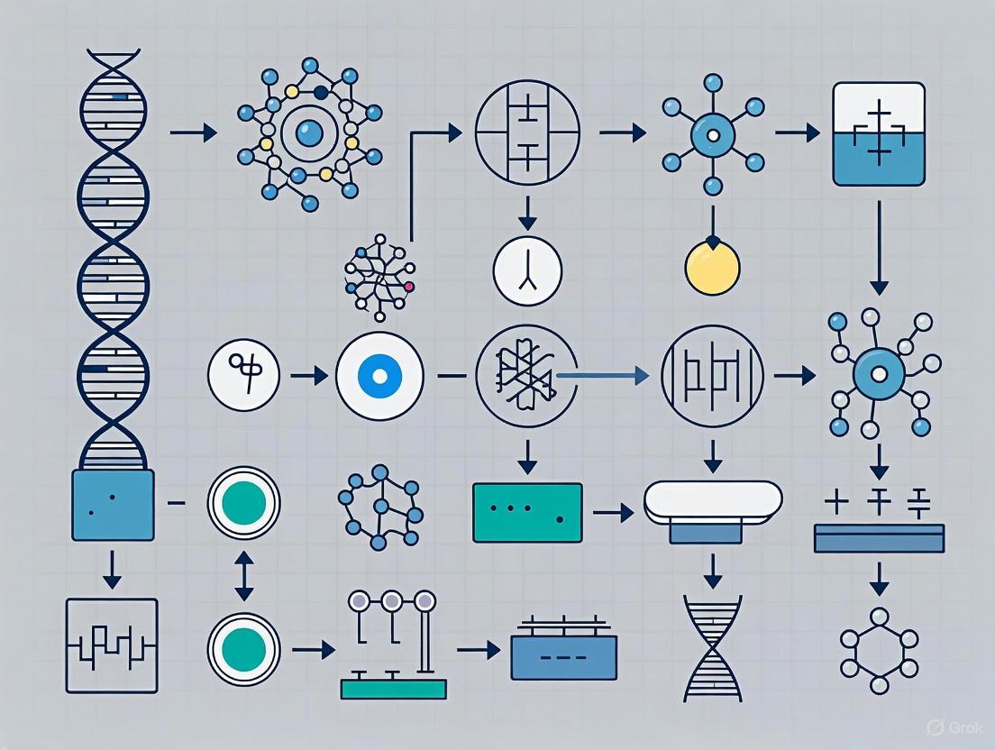
Visualizing Biosensor Workflows and Signaling Pathways
The following diagrams, generated using Graphviz DOT language, illustrate core concepts and workflows in synthetic biology-enabled biosensing.
Figure 1: Core Biosensor Architecture and Information Flow
Figure 2: Synthetic Gene Circuit for Chemical Detection
Figure 3: Amplified Sandwich Immunoassay Workflow
Synthetic biology aims to program living cells with novel functionalities by employing an engineering-driven approach using well-characterized, modular biological parts. The precision control of gene expression is paramount for applications ranging from optimized biosynthetic pathways to sophisticated diagnostic tools [6]. Within this framework, engineered biological parts—specifically riboregulators, allosteric transcription factors (aTFs), and modular genetic circuits—serve as fundamental components for constructing complex systems. These elements are particularly transformative for the development of advanced microscale biosensing technologies, enabling the detection of pathogens, environmental pollutants, and disease biomarkers with unprecedented sensitivity and specificity [2]. This technical guide provides an in-depth analysis of the design principles, experimental methodologies, and integrative strategies for deploying these engineered parts in biosensing research and development.
Riboregulators: Programmable RNA Sensors
Design Principles and Mechanisms
Riboregulators are synthetic RNA molecules that undergo conformational changes upon binding specific input triggers, such as small molecules or nucleic acids, to control translation. Two high-performance, de novo-designed translational repressors are the toehold repressor and the three-way junction (3WJ) repressor [6].
- Toehold Repressor: In its default state (OFF), the switch RNA features an exposed ribosome binding site (RBS) and start codon, allowing active translation. The cognate trigger RNA is complementary to the toehold domain and the stem of the switch RNA. Upon binding, a strand displacement reaction occurs, inducing the formation of a new secondary structure that sequesters the RBS and start codon, thereby repressing translation [6].
- Three-Way Junction (3WJ) Repressor: The switch RNA possesses an unstable hairpin structure that is initially translationally active. The cognate trigger RNA, which contains a hairpin itself, binds to single-stranded domains on the switch RNA via a toehold-mediated interaction. This binding event results in the formation of a stable three-way junction structure that hides the RBS and start codon, effectively repressing translation [6].
Table 1: Performance Characteristics of De Novo-Described Riboregulators [6]
| Riboregulator Type | Number of Devices Tested | Devices with ≥10-fold Repression | Devices with ≥50-fold Repression | Maximum Fold Repression |
|---|---|---|---|---|
| Toehold Repressor | 44 | 21 (48%) | 5 (11%) | >50-fold |
| 3WJ Repressor | 48 | 34 (71%) | 4 (8%) | >50-fold |
Experimental Protocol: Validating Riboregulator Performance
Objective: To quantify the dynamic range and orthogonality of novel riboregulator devices in E. coli.
Strain and Plasmids:
- Use an E. coli strain with genomic T7 RNA polymerase (e.g., BL21 Star DE3) for high-level, inducible expression.
- Clone the switch RNA, regulating a reporter gene (e.g., GFP), into a medium-copy plasmid.
- Clone the trigger RNA under an inducible promoter (e.g., IPTG-inducible) on a high-copy plasmid.
Transformation and Cultivation:
- Co-transform both plasmids into the expression host.
- For each device, inoculate two cultures: one induced with IPTG (to express the cognate trigger, OFF state) and one with a non-cognate trigger RNA (ON state).
Measurement and Analysis:
- Grow cultures to mid-log phase and measure fluorescence intensity via flow cytometry.
- Calculate the fold repression as the ratio of the geometric mean fluorescence in the ON state to the OFF state.
- To assess orthogonality, test all possible non-cognate trigger-switch pairs to ensure minimal crosstalk.
Computational Design and Optimization
The design of high-performance riboregulators can be accelerated by sequence-to-function deep learning frameworks. For instance, the Sequence-based Toehold Optimization and Redesign Model (STORM), a convolutional neural net (CNN), and Nucleic-Acid Speech (NuSpeak), a natural language processing (NLP) model, can predict riboregulator functionality from sequence data alone [7]. These models can identify over-represented nucleotide motifs in high-performing switches (e.g., a preference for 'NUA' at a specific bulge) and can be used to rationally redesign sub-optimal sequences, improving their dynamic range through transfer learning [7].
Figure 1: Riboregulator Mechanisms for Biosensing. Toehold and three-way junction (3WJ) repressors transition from an ON to an OFF state upon binding a specific input trigger, such as a pathogenic RNA sequence, leading to repression of a reporter output.
Allosteric Transcription Factors: Engineering Molecular Switches
Structure and Function of aTFs
Allosteric transcription factors are proteins that regulate gene expression by changing their DNA-binding affinity upon binding a specific small molecule ligand. They typically consist of a DNA-binding domain (DBD) and a signal-sensing domain (SSD). The binding of a ligand induces a conformational change that modulates the protein's interaction with its target operator sequence, thereby acting as a genetic switch [8]. Natural aTFs are widely used as biosensors but are limited to sensing their native ligands.
Engineering aTFs for Novel Ligands
A significant challenge is re-engineering aTFs to respond to non-native ligands, as mutations for new ligand binding can disrupt allosteric communication. Two primary strategies have been successfully employed:
Domain Swapping for Modular Regulators: This strategy involves creating hybrid aTFs by swapping functional domains between homologous regulators within the same protein family (e.g., LacI, TetR). The resulting chimera retains the DNA-binding specificity of the donor DBD and the ligand specificity of the donor SSD. This enables the flexible wiring of new input-output connections, such as controlling different promoters with the same signal or requiring multiple signals to activate a single promoter [8].
High-Throughput Engineering with Sensor-seq: Sensor-seq is a platform for designing aTF biosensors for new ligands with high scale and sensitivity [9].
- Library Design: A promiscuous aTF with a large binding pocket (e.g., TtgR) is chosen as the scaffold. Thousands of variants are generated through phylogeny-guided diversification.
- Barcoded Screening: Each aTF variant is linked to a random DNA barcode in a genetic construct. The library is exposed to a target ligand, and reporter transcript levels are quantified via RNA-seq.
- Functional Scoring: An F-score is calculated for each variant from the normalized ratio of reporter transcripts with and without ligand. F-scores > 1 identify functional, ligand-responsive biosensors [9].
Table 2: Strategies for Engineering Allosteric Transcription Factors
| Engineering Strategy | Key Principle | Advantages | Example Application |
|---|---|---|---|
| Domain Swapping [8] | Swapping DNA- and ligand-binding domains between homologous regulators. | Creates modular, customizable parts; predictable wiring. | Building complex genetic circuits with novel logic. |
| Sensor-seq Screening [9] | High-throughput sequencing to identify functional aTFs from vast mutant libraries. | Sensitive detection of rare functional variants; applicable to diverse ligands. | Developing biosensors for non-native ligands like naltrexone and quinine. |
Experimental Protocol: Sensor-seq for aTF Development
Objective: To identify aTF variants that respond to a target non-native ligand from a large library.
Library Construction:
- Generate a diverse mutant library of an aTF scaffold (e.g., TtgR) targeting the ligand-binding pocket.
- Clone each variant into a screening construct where it regulates a reporter gene. Each construct contains a unique, random DNA barcode.
Pooled Screening and Sequencing:
- Divide the library of E. coli cells into two conditions: one supplemented with the target ligand and a vehicle control.
- Harvest cells during log-phase growth and extract both total RNA and plasmid DNA.
- Prepare cDNA from the RNA and use PCR to link the barcode sequence to its corresponding aTF variant gene in a single amplicon for deep sequencing.
Data Analysis and Hit Identification:
- Map sequencing reads to assign each barcode to its aTF variant.
- For each variant, calculate the F-score:
(normalized cDNA count with ligand) / (normalized cDNA count without ligand). - Select variants with high F-scores for clonal validation using methods like qRT-PCR or flow cytometry.
Figure 2: Sensor-seq Workflow for Engineering aTFs. This high-throughput platform enables the screening of thousands of aTF variants against target ligands to identify functional biosensors, culminating in practical applications like cell-free detection systems.
Modular Genetic Circuit Design
Composing Circuits from Basic Parts
Individual regulatory devices can be integrated to form genetic circuits that perform complex computations, such as logic operations, signal processing, and memory storage. The core engineering principle is modularity—ensuring that devices function predictably and orthogonally when connected [10]. Fundamental circuit architectures include:
- Logic Gates: Boolean operations (AND, OR, NOR, NAND) can be implemented by combining multiple repressors. For example, riboregulators have been integrated to execute universal NAND and NOR logic [6].
- Bistable Switches and Memory Devices: These circuits possess two stable states (ON/OFF) and can "remember" a transient signal. They are often built using mutually repressing transcription factors or site-specific recombinases that create permanent, heritable DNA sequence changes [10].
- Signal Processors: Circuits can be designed to filter noise, amplify signals, or generate temporal pulses.
A Domain Swapping Approach for Flexible Circuit Design
The domain swapping strategy for creating modular aTFs directly enables more flexible and complex circuit topologies [8]. For instance:
- Using a set of hybrid regulators that all respond to the same ligand but bind to different DNA sequences, a single input signal can be wired to control multiple independent promoters.
- Conversely, using regulators that respond to different ligands but all bind the same promoter creates a combinatorial logic gate at that promoter, requiring the presence of multiple inputs for activation.
The Scientist's Toolkit: Research Reagent Solutions
Table 3: Essential Research Reagents and Resources
| Reagent / Resource | Function and Application | Specific Examples |
|---|---|---|
| Riboregulator Plasmid Libraries [6] | Validated collections of toehold and 3WJ repressors for screening and initial circuit construction. | Libraries of 44 toehold and 48 3WJ repressors. |
| Allosteric Transcription Factor Scaffolds [9] [8] | Well-characterized, promiscuous aTFs used as starting points for engineering new biosensors. | TtgR (for Sensor-seq), LacI (for domain swapping). |
| Sensor-seq Platform Components [9] | Specialized plasmids and protocols for high-throughput aTF screening. | Barcoded screening construct, mapping primers, analysis pipeline. |
| Cell-Free Expression Systems [9] | Lysate-based transcription-translation systems for rapid, contained prototyping of biosensors. | Used for deploying newly developed aTF biosensors (e.g., for naltrexone). |
| Computational Design Tools [7] | Software and models for predicting and optimizing the performance of biological parts. | NUPACK (sequence design), STORM & NuSpeak (deep learning for riboregulators). |
The strategic integration of engineered riboregulators, allosteric transcription factors, and modular genetic circuits is fundamentally advancing the capabilities of microscale biosensing technologies. The advent of high-performance, de novo-designed riboregulators and the application of innovative strategies like domain swapping and Sensor-seq for aTF engineering are systematically overcoming previous limitations of part idiosyncrasy and limited ligand specificity. Furthermore, the integration of these components with computational tools and high-throughput methodologies is creating a powerful, streamlined workflow for biosensor development. As these tools continue to mature, their synergy is poised to yield increasingly sophisticated, robust, and field-deployable biosensing systems that will have a profound impact on diagnostic medicine, environmental monitoring, and drug development.
The discovery of Clustered Regularly Interspaced Short Palindromic Repeats (CRISPR) and CRISPR-associated (Cas) proteins has revolutionized molecular biology, providing not only powerful tools for gene editing but also transformative technologies for diagnostic applications. CRISPR systems function as adaptive immune systems in bacteria and archaea, capable of recognizing and cleaving specific nucleic acid sequences with remarkable precision. While CRISPR-Cas9 initially gained prominence for gene editing applications, recent research has unveiled the exceptional potential of other Cas proteins, particularly Cas12 and Cas13, for nucleic acid detection. These proteins exhibit a unique property called "trans-cleavage" or "collateral cleavage" activity, which enables them to become potent nucleic acid detection tools upon recognition of their specific target sequences [11].
The integration of CRISPR technology into diagnostic systems represents a significant advancement in the field of microscale biosensing for synthetic biology research. These systems provide unprecedented specificity and sensitivity, capable of distinguishing single-nucleotide polymorphisms and detecting target nucleic acids at attomolar concentrations. For researchers and drug development professionals, CRISPR-based diagnostics offer rapid, cost-effective alternatives to traditional molecular detection methods like PCR, with the added advantage of point-of-care applicability. Two leading platforms—SHERLOCK and DETECTR—exemplify how CRISPR mechanisms can be harnessed for precise nucleic acid detection, each employing distinct Cas proteins with unique properties and applications [12].
SHERLOCK (Specific High-sensitivity Enzymatic Reporter unLOCKing) and DETECTR (DNA Endonuclease Targeted CRISPR Trans Reporter) technologies have emerged as frontrunners in the CRISPR diagnostic field, demonstrating exceptional performance in detecting various pathogens, genetic mutations, and other nucleic acid targets. These systems combine the programmability of CRISPR with isothermal amplification techniques and innovative reporter systems to create highly sensitive detection platforms that can be deployed in diverse settings, from advanced laboratories to resource-limited environments [13]. Their development marks a significant milestone in synthetic biology, showcasing how fundamental biological mechanisms can be repurposed for practical applications that address pressing challenges in human health, agriculture, and environmental monitoring.
Fundamental Mechanisms of CRISPR-Cas Systems in Diagnostics
Core Molecular Mechanisms
The diagnostic application of CRISPR systems relies on the targeted recognition and collateral cleavage activities of specific Cas proteins. Unlike Cas9, which is primarily used for gene editing and lacks trans-cleavage activity, Cas12 and Cas13 proteins exhibit nonspecific nuclease activity upon target recognition, making them ideal for diagnostic applications. Cas12 proteins (including Cas12a and Cas12b) target DNA sequences, while Cas13 proteins target RNA sequences. This fundamental difference determines their respective applications in detecting DNA or RNA targets [11].
The activation mechanism begins when the Cas protein forms a complex with a designed guide RNA (crRNA or gRNA) that is complementary to the target nucleic acid sequence. For Cas12a, target recognition requires the presence of a protospacer adjacent motif (PAM) sequence adjacent to the target region, while Cas13a recognizes specific protospacer flanking sites. Upon binding to the target sequence, the Cas protein undergoes a conformational change that activates its collateral cleavage activity, enabling it to indiscriminately cleave nearby non-targeted single-stranded DNA (for Cas12) or single-stranded RNA (for Cas13) molecules [14].
This collateral cleavage activity forms the basis for signal generation in CRISPR diagnostics. Reporter molecules—typically single-stranded DNA or RNA oligonucleotides tagged with a fluorophore-quencher pair—are included in the reaction mixture. When the Cas protein is activated by target recognition, it cleaves these reporter molecules, separating the fluorophore from the quencher and generating a detectable fluorescent signal. Alternative reporter systems, including lateral flow strips and electrochemical sensors, have also been developed to broaden the applications and usability of these diagnostic platforms [15].
Key Cas Proteins and Their Properties
The selection of appropriate Cas proteins is critical for optimizing diagnostic applications. Researchers have characterized various Cas proteins with distinct properties that make them suitable for different diagnostic scenarios. The following table summarizes the key characteristics of major Cas proteins used in nucleic acid detection systems:
Table 1: Comparison of Cas Proteins Used in Nucleic Acid Detection
| Cas Protein | Classification | Guide RNA | Trans-Cleavage Activity | Target Molecule | PAM/PFS Requirement | Size (amino acids) |
|---|---|---|---|---|---|---|
| Cas9 | Type II | sgRNA | None | dsDNA | PAM (5'-NGG-3') | ~1,400 |
| Cas12a (Cpf1) | Type V | crRNA | ssDNA | dsDNA, ssDNA | PAM (5'-TTTN-3') | 1,000-1,300 |
| Cas12b | Type V | sgRNA | ssDNA | dsDNA, ssDNA | PAM | 700-1,000 |
| Cas13a | Type VI | crRNA | ssRNA | ssRNA | PFS | 700-1,000 |
| Cas14 | Type V | sgRNA | ssDNA | ssDNA | None | 400-700 |
The compact size of some Cas proteins, particularly Cas14 (400-700 amino acids), offers advantages for diagnostic applications where space constraints exist, such as in microfluidic devices or field-deployable sensors. Additionally, the absence of PAM requirements for Cas14 enhances its flexibility in target selection. The temperature stability of certain Cas variants, such as Cas12b, makes them suitable for applications requiring elevated reaction temperatures or challenging environmental conditions [14].
SHERLOCK Technology Platform
Core Mechanism and Workflow
The SHERLOCK platform utilizes Cas13 enzymes for the detection of specific RNA sequences. Developed by researchers from the Broad Institute, including Dr. Feng Zhang and Dr. James Collins, SHERLOCK leverages the RNA-guided RNA-targeting capability of Cas13a (formerly known as C2c2) to achieve exceptional sensitivity and specificity in nucleic acid detection [16]. Upon recognition and binding to its target RNA sequence, Cas13a exhibits collateral cleavage activity that degrades nearby non-target RNA molecules, including reporter RNA molecules designed to generate a detectable signal.
The standard SHERLOCK protocol involves a two-step amplification and detection process to achieve maximal sensitivity. In the first step, the target nucleic acid is amplified using recombinase polymerase amplification (RPA) or reverse transcription RPA (RT-RPA) for RNA targets. This isothermal amplification step occurs at a constant temperature (typically 37-42°C) and does not require thermal cycling equipment, making it suitable for point-of-care applications. The amplified product then serves as the template for the subsequent CRISPR detection step [13].
In the detection phase, the RPA product is transcribed into RNA using T7 RNA polymerase, and the resulting RNA is detected by the Cas13-crRNA complex. When the Cas13-crRNA complex recognizes its target RNA sequence, the activated Cas13 cleaves the reporter RNA molecules, generating a fluorescent signal that can be quantified using a fluorimeter or visualized on a lateral flow strip. The entire process can be completed in less than two hours, with the actual CRISPR detection step requiring approximately 30 minutes [12].
Figure 1: SHERLOCK Experimental Workflow for RNA Detection
Performance Characteristics and Optimization
The SHERLOCK platform demonstrates exceptional analytical sensitivity, capable of detecting target nucleic acids at attomolar concentrations (10⁻¹⁸ molar). This sensitivity enables the detection of single molecules of target RNA in a sample. The system also exhibits high specificity, with the ability to distinguish targets that differ by only a single nucleotide, making it suitable for detecting single-nucleotide polymorphisms (SNPs) and specific viral strains [16]. The combination of RPA amplification and CRISPR detection provides a dual layer of specificity that minimizes false-positive results.
Further enhancements to the platform led to the development of SHERLOCKv2, which incorporates additional Cas proteins and optimization to improve sensitivity and expand multiplexing capabilities. SHERLOCKv2 can achieve zeptomolar sensitivity (10⁻²¹ molar) through the combined use of Cas13 and Csm6, another CRISPR enzyme that enhances signal amplification. The platform also supports multiplex detection of up to four different targets in a single reaction by utilizing different Cas13 orthologs with distinct crRNAs and reporter combinations [13].
The practical utility of SHERLOCK has been demonstrated in various applications, including detection of Zika and Dengue virus, identification of cancer mutations, and antimicrobial resistance profiling. During the COVID-19 pandemic, researchers adapted SHERLOCK for SARS-CoV-2 detection, creating a rapid test that could provide results in approximately one hour with sensitivity comparable to RT-PCR [13]. The cost-effectiveness of the platform (approximately $0.61 per test) further enhances its potential for widespread deployment in resource-limited settings [12].
DETECTR Technology Platform
Core Mechanism and Workflow
The DETECTR platform employs Cas12 proteins, primarily Cas12a (also known as Cpf1), for the detection of DNA targets. Developed by researchers at the University of California, Berkeley, led by Dr. Jennifer Doudna, DETECTR leverages the DNA-guided DNA-targeting capability of Cas12a, which exhibits collateral cleavage activity against single-stranded DNA upon target recognition [12]. Similar to SHERLOCK, DETECTR combines isothermal amplification with CRISPR-based detection to achieve high sensitivity and specificity.
The DETECTR workflow begins with the extraction of DNA from the sample, followed by isothermal amplification using RPA. The amplified DNA is then incubated with the Cas12a-crRNA complex and single-stranded DNA reporters. When Cas12a recognizes and binds to its target DNA sequence, it becomes activated and cleaves the ssDNA reporters, generating a detectable signal. The entire process can be completed in less than one hour, significantly faster than conventional PCR-based methods [13].
A key advantage of DETECTR is its compatibility with various detection methods. While fluorescence-based detection provides quantitative results, the system can also be adapted for lateral flow assays, enabling visual readouts without specialized equipment. This flexibility makes DETECTR suitable for both laboratory settings and point-of-care testing in field conditions. Mammoth Biosciences, a company co-founded by Dr. Doudna, has commercialized the DETECTR technology and developed user-friendly platforms that integrate sample processing, amplification, and detection [12].
Figure 2: DETECTR Experimental Workflow for DNA Detection
Performance Characteristics and Optimization
DETECTR demonstrates impressive analytical performance, with sensitivity reaching detection limits of 10⁻²¹ molar for DNA targets. This exceptional sensitivity enables the detection of low-abundance targets, such as viral DNA in early infection or tumor DNA in liquid biopsies. The system also shows high specificity, capable of distinguishing between closely related pathogens and identifying single-nucleotide variations [12]. The requirement for a specific PAM sequence (TTTV, where V is A, C, or G) adjacent to the target site contributes to the specificity of Cas12a-based detection.
Optimization of the DETECTR system has focused on improving reaction conditions, crRNA design, and reporter systems to enhance performance and reliability. The use of thermostable Cas12b variants has expanded the temperature range for reactions, increasing flexibility and robustness. Additionally, the development of novel reporter molecules with improved cleavage efficiency and signal-to-noise ratios has further enhanced the sensitivity of the platform [14].
DETECTR has been successfully applied to the detection of various human pathogens, including human papillomavirus (HPV), SARS-CoV-2, and Mycobacterium tuberculosis. In one notable application, researchers developed a DETECTR assay for SARS-CoV-2 that targeted the E and N genes of the virus, achieving results in approximately 45 minutes with 95% positive predictive agreement and 100% negative predictive agreement compared to RT-PCR [13]. The low cost of DETECTR tests (less than $1 per test) positions the technology as a viable alternative to traditional diagnostic methods in both clinical and field settings [12].
Comparative Analysis of SHERLOCK and DETECTR
Technical Performance Comparison
While both SHERLOCK and DETECTR leverage CRISPR collateral cleavage activity for nucleic acid detection, they differ in several key aspects that influence their application suitability. The following table provides a comprehensive comparison of their technical specifications and performance characteristics:
Table 2: Technical Comparison of SHERLOCK and DETECTR Platforms
| Parameter | SHERLOCK | DETECTR | Significance |
|---|---|---|---|
| Core Cas Protein | Cas13 (Type VI) | Cas12a (Type V) | Determines target type (RNA vs. DNA) |
| Primary Target | RNA | DNA | Application-specific selection |
| Amplification Method | RT-RPA + T7 Transcription | RPA | SHERLOCK requires additional transcription step |
| Detection Time | <2 hours | <1 hour | DETECTR typically faster |
| Sensitivity | 10⁻¹⁸ M (attomolar) | 10⁻²¹ M (zeptomolar) | Both highly sensitive |
| Specificity | Single-base mismatch discrimination | Single-base mismatch discrimination | Both highly specific |
| PAM/PFS Requirement | PFS for Cas13 | PAM (TTTV) for Cas12a | Impacts target site selection |
| Cost per Test | ~$0.61 | <$1.00 | Both cost-effective |
| Readout Methods | Fluorescence, lateral flow | Fluorescence, lateral flow | Multiple options available |
| Commercialization | Sherlock Biosciences | Mammoth Biosciences | Both commercially developed |
The choice between SHERLOCK and DETECTR depends primarily on the nature of the target nucleic acid and specific application requirements. SHERLOCK is particularly well-suited for detecting RNA viruses, monitoring gene expression, and identifying RNA biomarkers, while DETECTR excels in detecting DNA viruses, bacterial pathogens, and genetic mutations. Both platforms can be adapted for use with lateral flow strips, enabling visual detection without specialized equipment, though fluorescence-based readouts provide quantitative results with higher sensitivity [13].
Applications in Research and Diagnostics
SHERLOCK and DETECTR have found diverse applications across multiple fields, demonstrating their versatility and utility. In clinical diagnostics, both platforms have been deployed for infectious disease detection, with SHERLOCK used for Zika and Dengue virus identification and DETECTR applied to HPV subtyping and SARS-CoV-2 detection [16] [13]. The ability to distinguish between closely related pathogen strains with single-nucleotide resolution makes these technologies particularly valuable for tracking disease outbreaks and monitoring emerging variants.
In oncology research, CRISPR-based detection systems enable non-invasive cancer monitoring through liquid biopsy applications. SHERLOCK can detect cancer-associated RNA biomarkers and fusion transcripts, while DETECTR can identify tumor-specific DNA mutations in circulating tumor DNA. Both platforms offer the sensitivity required to detect rare mutations in complex biological samples, facilitating early cancer detection and treatment monitoring [17]. The integration of these technologies with microfluidic systems and portable readers further enhances their potential for point-of-care cancer diagnostics.
Beyond human health, SHERLOCK and DETECTR have applications in agricultural biotechnology, food safety, and environmental monitoring. These systems can detect plant pathogens, identify genetically modified organisms, monitor food contamination, and track environmental microbes. The robustness, portability, and cost-effectiveness of these platforms make them particularly valuable for field applications where traditional laboratory methods are impractical [12]. As these technologies continue to evolve, their applications are expected to expand further, driven by ongoing research into novel Cas proteins with improved properties and the development of more integrated and automated systems.
Research Reagent Solutions and Experimental Protocols
Essential Research Reagents
Successful implementation of SHERLOCK and DETECTR protocols requires carefully selected reagents and optimized reaction conditions. The following table outlines the key components and their functions in CRISPR-based detection systems:
Table 3: Essential Research Reagents for CRISPR-Based Detection Systems
| Reagent Category | Specific Components | Function | Example Products |
|---|---|---|---|
| Cas Enzymes | Cas13a, Cas12a, Cas12b | Target recognition and collateral cleavage | LwaCas13a, AapCas12b, AsCas12a |
| Amplification Enzymes | Reverse Transcriptase, Bsu DNA Polymerase | Isothermal amplification of target nucleic acids | Hifair III Reverse Transcriptase, Bsu DNA Polymerase |
| Recombinase Proteins | T4 UvsX, T4 UvsY | Facilitate primer invasion during RPA | T4 UvsX Recombinase, T4 UvsY protein |
| Strand Displacement Polymerases | Bst DNA Polymerase | DNA amplification under isothermal conditions | Hieff Bst Plus DNA Polymerase |
| Reporters | FQ-labeled ssDNA/ssRNA | Signal generation upon collateral cleavage | FAM-TTATT-BHQ1 quenched fluorescent reporters |
| Guide RNAs | crRNAs, sgRNAs | Specific target recognition | Custom-designed crRNAs |
| Buffer Systems | Reaction buffers, magnesium salts | Optimal enzyme activity and stability | NEBuffer, ThermoPol Buffer |
The selection of appropriate Cas enzymes is critical for assay performance. Different Cas orthologs exhibit variations in temperature optimum, cleavage efficiency, and PAM requirements, which can significantly impact detection sensitivity and specificity. For example, LwaCas13a from Leptotrichia wadei is commonly used in SHERLOCK assays due to its high cleavage activity and stability at 37°C, while AapCas12b from Alicyclobacillus acidiphilus is favored in some DETECTR applications because of its thermostability, enabling reactions at higher temperatures that enhance specificity [11].
Guide RNA design represents another crucial factor in assay development. The crRNA or sgRNA must be designed to target unique regions of the pathogen or biomarker of interest while avoiding off-target effects. Software tools are available to assist researchers in selecting optimal target sequences and designing highly specific guide RNAs. Typically, guide RNAs are 20-30 nucleotides in length and should be tested empirically to verify their specificity and efficiency [14].
Detailed Experimental Protocol
The following protocol provides a generalized framework for implementing SHERLOCK and DETECTR assays, with specific adjustments required based on the target and application:
Sample Preparation and Nucleic Acid Extraction
- Collect appropriate samples (serum, saliva, swabs, tissue) in suitable collection tubes
- Extract nucleic acids using commercial kits or established protocols
- For RNA targets: Use RNA-specific extraction methods with RNase inhibitors
- For DNA targets: Use DNA-specific extraction methods, ensuring complete removal of RNA if necessary
- Quantify nucleic acid concentration and quality using spectrophotometry or fluorometry
Isothermal Amplification (RPA/RT-RPA)
- Prepare RPA reaction mix according to manufacturer's instructions
- Components include: reaction buffer, primers, template nucleic acid, recombinase enzymes, strand-displacing polymerase, and nucleotides
- For SHERLOCK (RNA targets): Include reverse transcriptase in the reaction mix
- Incubate at 37-42°C for 25-30 minutes
- Terminate the reaction by heating to 85°C for 5 minutes or using specific termination buffers
CRISPR Detection
- Prepare CRISPR reaction mix containing: Cas enzyme, guide RNA, reporter molecules, and appropriate buffer
- Combine CRISPR reaction mix with amplified product
- Incubate at optimal temperature for the specific Cas enzyme (typically 37°C for Cas13a, 37-60°C for Cas12 variants)
- Monitor fluorescence in real-time or measure endpoint fluorescence after 30-60 minutes
- For lateral flow detection: Apply reaction mixture to lateral flow strip and interpret results after 2-5 minutes
Troubleshooting and Optimization
- Low signal: Optimize guide RNA design, increase amplification time, adjust Cas:guide RNA ratio
- High background: Purify amplification products, optimize reporter concentration, increase reaction temperature
- Inconsistent results: Standardize sample processing, include appropriate controls, ensure reagent quality
Recent advancements have led to the development of streamlined protocols that combine amplification and detection in a single tube, reducing handling steps and the risk of contamination. These integrated approaches are particularly valuable for point-of-care applications where simplicity and robustness are essential [13]. Additionally, the incorporation of lyophilized reagents improves stability and shelf life, enabling distribution and storage without cold chain requirements.
Future Perspectives in Microscale Biosensing
The integration of CRISPR-based detection systems with advanced biosensing platforms represents a promising direction for future development. Electrochemical CRISPR sensors have emerged as particularly attractive formats, offering enhanced portability, lower costs, and compatibility with wearable devices. Recent research has demonstrated the feasibility of CRISPR-driven electrochemical sensors that detect DNA or RNA targets through measurable changes in electrical properties. These systems often incorporate stabilizing polymers such as polyvinyl alcohol (PVA) to protect DNA probes on electrode surfaces, significantly extending shelf life while maintaining sensitivity [15]. The development of such stable, inexpensive sensors aligns with the growing demand for decentralized diagnostics and personalized medicine.
The application of machine learning and artificial intelligence to CRISPR diagnostic systems presents another frontier for innovation. These computational approaches can enhance assay design through improved guide RNA selection, optimize reaction conditions based on historical data, and interpret complex signal patterns for more accurate target quantification. Furthermore, AI-assisted image analysis can simplify result interpretation from lateral flow assays and other visual readouts, reducing subjectivity and enabling quantitative measurements from qualitative tests [15]. The combination of CRISPR diagnostics with smartphone-based readout systems and cloud-based data analysis creates opportunities for real-time disease monitoring and epidemiology.
Multiplexing capabilities continue to expand with the discovery and engineering of novel Cas proteins with distinct properties. The identification of compact Cas variants, such as Cas14 and miniature Cas12 proteins, enables the development of high-density detection arrays and microfluidic systems capable of simultaneously screening for dozens or even hundreds of targets [11]. These advances support the trend toward comprehensive pathogen panels and complex biomarker signatures for precise disease classification. Additionally, the integration of CRISPR detection with other emerging technologies, such as single-molecule analysis and digital quantification, promises to push detection limits to new extremes while providing absolute quantification of target molecules [18].
As these technologies mature, standardization and quality control will become increasingly important for clinical translation. Establishing reference materials, validated protocols, and performance standards will facilitate the transition from research tools to approved diagnostic tests. Regulatory frameworks are evolving to accommodate these novel testing platforms, with the FDA having already issued Emergency Use Authorizations for CRISPR-based SARS-CoV-2 tests [18]. The continued collaboration between academic researchers, commercial developers, and regulatory agencies will be essential for realizing the full potential of CRISPR-based diagnostics in clinical practice and public health.
- Industrial Technology Research Institute (ITRI) Report on CRISPR-Cas Technology (2019)
- Food Industry Journal on Biosensors for Foodborne Pathogens (2021)
- ToloBio Commercial Website on CRISPR Technology (2023)
- Virosin.org on Zhang Feng's CRISPR-Cas System (2017)
- Biotrade Article on CRISPR Platform for SARS-CoV-2 Detection (2020)
- Journal of Semiconductors Review on Advanced Biosensing Technologies (2023)
- Forward Pathway Article on CRISPR-Driven Electrochemical Sensors (2025)
- Yeasen Biotechnology Technical Guide on CRISPR in IVD Applications (2023)
- Forward Pathway Article on CRISPR in Cancer Diagnosis (2025)
- PMC Scientific Review on Viral Diagnostic Technologies (2022)
The integration of biosensing technologies into synthetic biology has traditionally relied on whole-cell systems, which use living microorganisms as the foundational platform for detecting environmental contaminants and disease biomarkers. While these cell-based biosensors leverage natural biological selectivity, they face fundamental limitations including stringent viability requirements, slow response times due to cell-wall transport barriers, and susceptibility to external stressors that can compromise functionality [19] [20]. These constraints have prompted a paradigm shift toward cell-free biosensing platforms that overcome these challenges while offering enhanced versatility and programmability.
Cell-free protein synthesis (CFPS) systems represent a transformative approach that extracts and repurposes the essential biochemical machinery of cells—including ribosomes, transcription and translation factors, energy sources, and cofactors—to produce desired proteins without maintaining cell viability [19] [20]. By eliminating constraints associated with living cells, CFPS provides a highly tunable and efficient platform for biosensing applications across diverse fields, from environmental monitoring to medical diagnostics [21]. This technical guide examines the fundamental advantages of cell-free biosensing platforms, provides detailed experimental methodologies, and explores their integration within microscale synthetic biology research frameworks.
Fundamental Advantages of Cell-Free over Whole-Cell Systems
Cell-free biosensors harness the selectivity of cellular machinery without the constraints of living cells, offering significant technical and practical advantages for both environmental monitoring and diagnostic applications [19] [22]. The removal of cellular barriers enables direct access to biological machinery, fundamentally enhancing biosensor performance.
Table 1: Comparative Analysis of Whole-Cell vs. Cell-Free Biosensing Systems
| Characteristic | Whole-Cell Biosensors | Cell-Free Biosensors |
|---|---|---|
| Response Time | Slow (hours to days) due to transmembrane transport and gene expression delays | Rapid (minutes to hours) due to direct access to transcriptional/translational machinery [21] |
| Tolerance to Toxicity | Limited; viability compromised by toxic samples | High; no viability requirements enable operation in toxic environments [19] |
| Storage Stability | Requires controlled conditions; viability maintenance challenging | Enhanced stability via lyophilization; room temperature storage for months [21] |
| Design Flexibility | Constrained by cellular metabolism and homeostasis requirements | Highly flexible; components can be mixed modularly for custom applications [19] [2] |
| Standardization | Challenging due to biological variability between cell batches | More reproducible; reaction conditions precisely controlled [21] |
| Portability | Limited by need to maintain cell viability | Excellent; paper-based, lyophilized formats enable field deployment [19] [21] |
The absence of cell membranes in CFPS systems avoids the barrier of transmembrane transport of analytes and output signal substances, significantly accelerating response times [21]. This open reaction environment allows direct manipulation of biological components, enabling the design, testing, and optimization of biosensors in a faster, more convenient, and more controlled manner compared to cellular systems [23]. Furthermore, the elimination of viability requirements allows cell-free biosensors to operate in environments that would otherwise be toxic to living cells, significantly expanding their application range for environmental monitoring [19].
Technical Applications and Performance Metrics
Environmental Monitoring Applications
Cell-free biosensors have demonstrated remarkable capabilities in detecting environmental contaminants with sensitivity and specificity often surpassing traditional methods. These systems leverage the flexibility of cell-free approaches to detect diverse analytes while offering practical advantages for field deployment through preservation methods such as lyophilization and paper-based formats [19] [20].
Table 2: Performance Metrics of Cell-Free Biosensors for Environmental Monitoring
| Target Analyte | Detection Mechanism | Limit of Detection | Sample Matrix | Key Features |
|---|---|---|---|---|
| Mercury (Hg²⁺) | merR gene, plasmid DNA with reporter genes | 1 ppb [19], 0.5 nM [19] | Water | Paper-based system with smartphone readout [19] |
| Lead (Pb²⁺) | Engineered allosteric transcription factors (aTFs) | 0.1 nM [19], 50 nM [19] | Water | Detection below WHO guidelines [19] |
| Arsenic | Optimized transcription factors | ≤10 μg/L [19] | Water | Below WHO recommended maximum concentration [19] |
| Tetracyclines | Riboswitch-based, RNA aptamers | 0.4-0.47 μM [19] | Milk samples | Broad-spectrum detection of antibiotic family [19] |
| Pathogens | 16S rRNA detection with retroreflective Janus particles | Femtomolar levels [19] | Clinical samples | Multiplexed detection of biological warfare agents [19] |
Heavy metal detection represents one of the most mature applications of cell-free biosensing technology. Gräwe et al. developed a paper-based system for mercury detection utilizing a dual-filter approach with smartphone readout, achieving detection limits as low as 6 μg/L [19]. Similarly, Zhang et al. created cell-free paper-based biosensors dependent on allosteric transcription factors for on-site detection of Hg²⁺ and Pb²⁺ in water with impressive detection limits of 0.5 nM and 0.1 nM, respectively, demonstrating recoveries ranging from 91% to 123% for actual water samples [19].
Beyond metals, cell-free systems have been adapted for detecting organic pollutants including pesticides and antibiotics. Dong et al. developed a riboswitch-based cell-free biosensor for broad-spectrum detection of tetracyclines using artificially screened tetracycline RNA aptamers to control reporter gene expression [19]. This system achieved detection limits of 0.47, 0.079, 0.084, and 0.43 μM for tetracycline, oxytetracycline, chlortetracycline, and doxycycline, respectively, enabling qualitative detection in milk samples at concentrations as low as 1 μM [19].
Diagnostic and Medical Applications
In medical diagnostics, cell-free biosensors offer potential for rapid, sensitive, and specific detection of biomarkers and pathogens in clinical samples, enabling point-of-care testing with minimal equipment requirements [19] [20]. These systems are particularly valuable for resource-limited settings where traditional laboratory infrastructure may be unavailable.
The development of hormone-responsive biosensors represents a significant advancement in medical applications. Salehi et al. pioneered a cell-free protein synthesis approach for detecting thyroid receptor β-specific endocrine disruptors using an engineered allosterically activated fusion protein [20]. Subsequent work led to the development of the Rapid Adaptable Portable In vitro Detection (RAPID) platform for detecting estrogenic compounds in human blood and urine samples with detection times significantly faster than traditional cellular assays [20].
For pathogen detection, Park et al. developed an innovative cell-free biosensor that transforms the 16S rRNA of targeted pathogens into detectable protein molecules [19]. By integrating retroreflective Janus particles for signal amplification, the system achieved exceptional sensitivity, detecting 16S rRNA at femtomolar levels with remarkable specificity for multiple dangerous pathogens, including B. anthracis, F. tularensis, Y. pestis, B. Pseudomallei, and B. abortus [19].
Experimental Framework and Methodologies
Core Experimental Protocols
The development and implementation of cell-free biosensing platforms requires standardized methodologies to ensure reproducibility and performance. Below are detailed protocols for key processes in cell-free biosensor construction and deployment.
Cell-Free Protein Synthesis System Preparation
Protocol: E. coli-Based CFPS Extract Preparation
- Cell Culture: Grow E. coli strain BL21 Star (DE3) in 1L of 2xYTPG medium at 37°C with vigorous shaking (250 rpm) to mid-exponential phase (OD600 ≈ 3.0).
- Harvesting: Centrifuge cells at 5,000 × g for 15 minutes at 4°C. Wash cell pellet with cold S30 buffer (10 mM Tris-acetate, 14 mM magnesium acetate, 60 mM potassium acetate, 1 mM dithiothreitol, pH 8.2).
- Lysis: Resuspend cells in 25 mL of fresh S30 buffer and disrupt using a French press at 1,500 psi. Centrifuge lysate at 12,000 × g for 30 minutes at 4°C to remove cell debris.
- Run-Off Reaction: Incubate supernatant for 80 minutes at 37°C with gentle shaking to deplete endogenous mRNA.
- Dialysis: Dialyze extract against 1L of fresh S30 buffer for 3 hours at 4°C with one buffer change.
- Aliquoting and Storage: Flash-freeze aliquots in liquid nitrogen and store at -80°C [23].
Paper-Based Biosensor Fabrication
Protocol: Lyophilized Paper-Based Biosensor Preparation
- Reaction Mixture Preparation: Combine CFPS extract with energy sources (creatine phosphate, ATP, GTP), amino acid mixture, DNA template encoding reporter protein under control of inducible promoter, and necessary salts.
- Impregnation: Apply 10-15 μL of reaction mixture onto defined zones of chromatography paper using precision pipetting.
- Lyophilization: Flash-freeze paper sensors at -80°C for 30 minutes, then transfer to freeze-dryer for 4-6 hours until completely dry.
- Packaging: Store dried sensors with desiccant in sealed pouches at room temperature protected from light [19] [21].
Optimization Strategies for Enhanced Performance
Systematic optimization of CFPS systems is crucial for achieving high sensitivity and robust performance. Key parameters requiring optimization include:
- Magnesium Ion Concentration: Titrate Mg²⁺ concentration (typically 5-15 mM) to maximize protein synthesis yield, as magnesium critically influences ribosomal function and RNA stability [23].
- Energy System Optimization: Evaluate different energy sources (phosphoenolpyruvate, creatine phosphate, 3-phosphoglyceric acid) for sustained ATP regeneration during protein synthesis [23].
- DNA Template Design: Optimize plasmid concentration (1-10 nM), ribosomal binding site strength, and promoter selection to balance expression level and background signal [23].
- Codon Optimization: Adapt codon usage to match the source organism of the cell-free extract, particularly for non-E. coli systems [23].
Signaling Pathways and Molecular Mechanisms
Cell-free biosensors employ diverse molecular mechanisms for signal detection and transduction, leveraging synthetic biology principles to create highly specific recognition elements.
Diagram 1: Transcription factor-based detection mechanism. Allosteric transcription factors (aTFs) undergo conformational change upon analyte binding, enabling reporter gene expression in cell-free systems.
Diagram 2: Riboswitch-based detection mechanism. Analyte binding induces structural rearrangement in the RNA aptamer, modulating accessibility of the ribosome binding site and controlling translation of the reporter gene.
Essential Research Reagents and Materials
The successful implementation of cell-free biosensing platforms requires carefully selected reagents and materials that maintain system functionality while enabling practical deployment.
Table 3: Essential Research Reagent Solutions for Cell-Free Biosensing
| Reagent Category | Specific Examples | Function | Optimization Notes |
|---|---|---|---|
| Cell-Free Extracts | E. coli BL21, V. natriegens, B. subtilis extracts | Source of transcriptional/translational machinery | E. coli offers highest yield; B. subtilis has low endotoxins; V. natriegens offers rapid expression [23] |
| Energy Systems | Phosphoenolpyruvate (PEP), Creatine phosphate, 3-PGA | ATP regeneration for sustained protein synthesis | Creatine phosphate generally provides longer-lasting energy [23] |
| Reporter Systems | sfGFP, Luciferase, β-galactosidase, LacZ | Signal generation for detection | Fluorescent proteins enable quantitative measurement; colorimetric enzymes allow visual detection [19] [21] |
| Recognition Elements | Allosteric transcription factors, RNA aptamers, riboswitches | Target analyte recognition | Engineering transcription factors can significantly improve sensitivity and dynamic range [19] |
| Stabilizers | PEG8000, Trehalose, Glycerol | Enhanced protein stability and folding | PEG8000 at 2-4% concentration can significantly improve protein synthesis yields [23] |
| Paper Substrates | Whatman chromatography paper, Nitrocellulose membrane | Immobilization matrix for lyophilization | Porous structure preserves protein synthesis activity after rehydration [19] [21] |
Integration with Microscale Synthetic Biology Research
Cell-free biosensing platforms represent a cornerstone technology in the advancing field of microscale synthetic biology, enabling sophisticated biological programming without cellular constraints. These systems provide an ideal testbed for rapid prototyping of genetic circuits and biosensing elements, dramatically accelerating the design-build-test cycle fundamental to synthetic biology research [2] [23]. The open nature of CFPS systems allows direct manipulation of biological components, facilitating precise control over reaction conditions and composition that is challenging to achieve in living cells.
The integration of synthetic biology approaches has enabled the development of complex signal processing capabilities in cell-free systems, including multiplexed detection, logic operations, and sophisticated sensor designs incorporating riboswitches, split reporter systems, and metabolic sensing modules [19] [20]. These advances have been particularly valuable for creating biosensors that can operate in challenging environments, from monitoring water quality in field settings to diagnosing diseases in point-of-care scenarios with minimal equipment [19] [21].
Future developments in cell-free biosensing are likely to focus on enhanced integration with electronic systems, improved preservation methods for long-term stability, and reduced costs through the development of more efficient extract preparation methods [19] [21]. As these technologies mature, cell-free biosensors are poised to become essential tools for addressing global challenges in healthcare, environmental protection, and biosecurity within the broader framework of synthetic biology innovation.
The Role of Synthetic Biology in Creating Reconfigurable and Intelligent Sensing Architectures
Synthetic biology is fundamentally reshaping the landscape of biosensing by providing an engineering framework to program biological systems for sophisticated detection tasks. This discipline moves beyond simply leveraging biological components to employing a modular engineering paradigm where biological parts are standardized and assembled into complex circuits that can sense, compute, and respond to environmental cues [24]. These engineered systems represent a significant evolution from conventional biosensors, offering unprecedented capabilities for real-time monitoring, multiplexed detection, and autonomous operation in diverse application environments [2].
The core advantage of synthetic biology-based sensing architectures lies in their inherent reconfigurability and intelligence. Unlike traditional biosensors with fixed recognition elements, synthetic biological circuits can be reprogrammed at the genetic level to detect novel targets, implement sophisticated signal processing, and produce tailored outputs [4] [25]. This flexibility is particularly valuable for addressing emerging challenges in healthcare, environmental monitoring, and biomanufacturing where sensing requirements constantly evolve. Furthermore, the integration of synthetic biology with materials science and computational design has enabled the development of intelligent sensing systems capable of adapting their behavior based on complex input combinations and historical context [4] [26].
Framed within the broader context of microscale biosensing technologies, synthetic biology provides the molecular programming language that defines sensor function, while micro- and nanofabrication technologies provide the physical implementation platform. This synergy creates powerful sensing architectures that operate at relevant biological scales while maintaining the programmability and precision of engineered systems [27]. As these technologies mature, they are poised to transform diagnostic paradigms from centralized laboratory testing to distributed, autonomous monitoring systems capable of operating in resource-limited environments and closed-loop control applications [28].
Fundamental Principles of Synthetic Biology Biosensing
Architectural Components of Biological Sensing Systems
Synthetic biology biosensors are constructed from modular biological components organized into a coherent sensing architecture. The fundamental framework consists of three core modules: a sensing interface for target recognition, a processing unit for signal computation and integration, and an output module for generating detectable responses [2]. This modular organization mirrors conventional electronic sensor architectures while leveraging biological mechanisms for molecular-scale operation.
The sensing interface typically employs transcription factors, riboswitches, or membrane receptors that undergo conformational changes upon binding specific target molecules. These recognition events are then transduced into genetic signals that activate the processing unit [4]. The processing unit, often implemented as a genetic logic circuit, performs operations on the input signals using promoters, regulators, and other genetic elements to implement functions such as amplification, filtering, or logic operations (AND, OR, NOT) [2]. Finally, the output module converts the processed signal into a detectable response using reporter proteins (e.g., fluorescent proteins, luciferases), enzymes that produce colorimetric changes, or metabolic pathways that synthesize specific molecules [4] [24].
Table 1: Core Functional Modules in Synthetic Biology Biosensors
| Module Type | Key Components | Function | Examples |
|---|---|---|---|
| Sensing Interface | Transcription factors, Riboswitches, Membrane receptors | Target recognition and signal transduction | ArsR for arsenic detection, LuxR for quorum sensing |
| Processing Unit | Promoters, Operators, Riboregulators | Signal computation and integration | Logic gates, Amplification circuits, Band-pass filters |
| Output Module | Fluorescent proteins, Luciferases, Enzymes | Generation of detectable signals | GFP, LuxAB, β-galactosidase, Chromogenic substrates |
Sensing Mechanisms and Signal Transduction Pathways
Biological sensing mechanisms exploit nature's evolved molecular recognition capabilities while engineering them for enhanced performance and specificity. Transcription factor-based sensors operate through allosteric regulation, where ligand binding induces conformational changes that alter DNA-binding affinity, thereby controlling downstream gene expression [4]. For instance, natural heavy metal resistance systems have been repurposed to create sensors for environmental monitoring, with transcription factors like MerR (mercury), ArsR (arsenic), and ZntR (zinc) providing specific recognition for their respective metal ions [4] [24].
Riboswitch-based sensors employ RNA aptamers that undergo structural rearrangements upon ligand binding, modulating translation initiation or transcription termination. These RNA-based sensors offer advantages in size and design flexibility compared to protein-based systems [29]. More recently, CRISPR-based sensors have emerged as powerful platforms that leverage the programmability of CRISPR-Cas systems for nucleic acid detection, with enzymes like Cas12 and Cas13 providing both specific recognition and signal amplification through their collateral cleavage activities [2].
Signal transduction in these systems follows carefully engineered pathways that convert molecular recognition events into measurable outputs. For example, in the well-characterized arsenic detection system derived from the E. coli arsenic resistance operon, arsenite binding to the ArsR repressor protein causes its dissociation from the ars promoter, allowing transcription of downstream reporter genes [24]. Similar principles apply to engineered sensors for small molecules, ions, and physical parameters like light and temperature, with the specific transduction pathway determined by the sensing mechanism and desired performance characteristics [4].
Engineering Reconfigurable Sensing Platforms
Genetic Circuit Design for Programmable Sensing
The reconfigurability of synthetic biology biosensors stems from the modular nature of genetic circuits, which enables systematic repurposing of sensing platforms for different applications. Standardized biological parts, such as those cataloged in the BioBricks framework, facilitate this modularity by providing well-characterized DNA sequences with defined functions and standardized interfaces [24]. These parts include promoters, ribosome binding sites, coding sequences, and terminators that can be assembled in various configurations to create sensors with customized properties [24].
Advanced circuit designs implement programmable logic that allows sensors to respond to complex combinations of inputs. For instance, AND-gate circuits require the simultaneous presence of two target molecules to activate output expression, increasing specificity by reducing false positives in complex environments [2]. More sophisticated circuits can perform temporal logic, signal processing, and even memory functions that record detection events. The engineering of these circuits is supported by computational modeling tools that predict system behavior before implementation, optimizing parameters such as promoter strength, ribosome binding efficiency, and degradation rates to achieve desired performance characteristics [26].
Table 2: Representative Synthetic Gene Circuits for Environmental Sensing
| Input Signal | Sensor Mechanism | Genetic Circuit | Output Signal | Host Organism | Detection Limit | Stability | Reference |
|---|---|---|---|---|---|---|---|
| Pb²⁺ | Pb²⁺-responsive promoter (Ppbr) | Ppbr→mtagBFP2 | Blue fluorescence | B. subtilis | 0.1 μg/L | >7 days | [4] |
| Cu²⁺ | Cu²⁺-responsive promoter (PcopA) | PcopA→eGFP | Green fluorescence | B. subtilis | 1.0 μg/L | >7 days | [4] |
| Hg²⁺ | Hg²⁺-responsive promoter (Pmer) | Pmer→mCherry | Red fluorescence | B. subtilis | 0.05 μg/L | >7 days | [4] |
| Theophylline | Theophylline riboswitch | PconII→YFP | Yellow fluorescence | S. elongatus | ~0.5 mM | >7 days | [4] |
| Formaldehyde | Formaldehyde-responsive promoter (Pm4) | Pm4→ATF1 | Isoamyl acetate (odor) | E. coli | ~0.12 ppm | >2 months | [4] |
Dynamic Reconfiguration Using External Cues
Beyond genetic reprogramming, some synthetic sensing systems can be dynamically reconfigured using external stimuli such as light, temperature, or chemical inducers. Optogenetic systems use light-sensitive proteins to control circuit activity, enabling spatial and temporal precision in sensor operation [4]. For example, sensors incorporating phytochrome or LOV domain proteins can be switched between active and inactive states using specific light wavelengths, allowing external control over detection thresholds and timing [4].
DNA nanotechnology provides particularly powerful approaches to reconfigurable sensing through toehold-mediated strand displacement [27]. In these systems, detection of a target nucleic acid initiates a cascade of strand displacement reactions that can propagate signals, perform computations, and generate outputs. The programmability of Watson-Crick base pairing makes these systems highly reconfigurable for different targets simply by redesigning nucleotide sequences. Recent advances have demonstrated DNA-based circuits that can classify patterns of multiple biomarkers, implementing sophisticated diagnostic algorithms entirely through molecular computations [27].
The integration of reconfigurable synthetic systems with biological components creates hybrid sensing platforms that combine the programmability of synthetic biology with the sophisticated functionality of natural biological systems. For instance, engineered DNA nanostructures have been interfaced with lipid membranes to create synthetic channels whose permeability can be controlled through structural reconfiguration, enabling regulation of molecular transport across biological barriers [27]. These technologies expand the capabilities of biosensing beyond mere detection to include controlled response and delivery functionalities.
Advanced Sensing Architectures and Material Interfaces
Engineered Living Materials for Distributed Sensing
The integration of synthetic gene circuits with functional materials has given rise to Engineered Living Materials (ELMs) that combine the responsiveness of biological systems with the structural properties of synthetic matrices [4]. These materials typically embed engineered microorganisms within hydrogels, polymers, or other scaffolds that provide mechanical stability while allowing nutrient diffusion and signal transduction [4]. ELMs enable the deployment of synthetic biology sensors in real-world environments by protecting the engineered cells from environmental stresses and preventing their release into the environment.
ELMs can be programmed to sense and respond to diverse inputs including environmental chemicals, light, heat, and mechanical loadings [4]. For example, researchers have developed ELMs that detect heavy metal contamination through fluorescence responses and self-healing materials that repair damage in response to specific triggers. The material properties themselves can be designed to enhance sensing performance through concentration of target analytes, exclusion of interferents, or provision of optimal microenvironments for the engineered organisms [4].
Recent advances have focused on improving the stability and longevity of ELM-based sensors for extended monitoring applications. Strategies include engineering drought tolerance or nutrient synthesis pathways into the host organisms, designing matrices that gradually release nutrients, and implementing kill switches that prevent persistence beyond the intended operational lifetime [4] [28]. These developments are critical for applications in environmental monitoring, smart infrastructure, and biomedical implants where long-term, autonomous operation is required.
Cell-Free Biosensing Systems
As an alternative to whole-cell biosensors, cell-free systems deploy purified transcriptional and translational machinery in vitro to execute genetic circuits without the constraints of maintaining cell viability [2] [28]. These systems offer several advantages for sensing applications, including rapid response (minutes rather than hours), operation in toxic environments, and simplified storage through lyophilization [2]. Cell-free biosensors are particularly suited for point-of-care diagnostics where simplicity, speed, and shelf stability are critical.
Innovative formats have been developed to enhance the deployability of cell-free sensors, including paper-based assays that can be stored at room temperature and activated by simple rehydration [2]. These platforms have been demonstrated for detection of pathogens, biomarkers, and environmental contaminants with sensitivities approaching those of laboratory-based methods. Recent work has focused on increasing the multiplexing capacity of these systems and integrating them with portable readout devices such as smartphones [2].
Despite their advantages, cell-free systems face challenges in long-term operation and complex computation compared to living cells, which can maintain themselves and implement sophisticated behaviors through gene regulatory networks [28]. Hybrid approaches that combine the stability of cell-free systems with the adaptive capabilities of living cells represent a promising direction for next-generation biosensing platforms that can operate autonomously in challenging environments.
Experimental Implementation and Workflow
Standardized Methodology for Biosensor Development
The development of synthetic biology biosensors follows an iterative design-build-test-learn cycle that integrates computational design, genetic assembly, experimental characterization, and model refinement. A standardized workflow for implementing a reconfigurable sensing architecture typically begins with computational design of genetic circuits using tools like Cello for logic circuit design or NUPACK for nucleic acid strand displacement systems [26]. This in silico phase identifies appropriate biological parts, predicts circuit behavior, and optimizes parameters to meet specification requirements.
The implementation phase involves genetic assembly of the designed circuit using techniques such as Golden Gate assembly, Gibson assembly, or CRISPR-based editing [2]. The constructed genetic system is then introduced into an appropriate host organism (bacteria, yeast, or mammalian cells) or configured for cell-free expression. Critical to this process is the careful characterization of individual parts under standardized conditions to ensure predictable system behavior when assembled into larger circuits [24].
Experimental validation proceeds through a sequence of characterization assays beginning with measurement of transfer functions that relate input concentrations to output responses [4]. Sensors are evaluated for key performance metrics including sensitivity (limit of detection), dynamic range, specificity (cross-reactivity with similar molecules), and response time [4] [2]. For reconfigurable systems, additional tests verify the ability to reprogram sensor specificity and response characteristics through appropriate genetic modifications or external stimuli.
Diagram 1: Sensor Development Workflow
Implementation of a Reconfigurable DNA Nanostructure-Based Sensor
A cutting-edge example of reconfigurable sensing architecture employs DNA nanorafts that undergo structural transformations on synthetic cell membranes [27]. The experimental protocol for implementing this system begins with the fabrication of DNA origami structures using the scaffolded origami method. A single-stranded M13mp18 DNA scaffold is combined with approximately 200 staple strands that define the nanoraft structure through thermal annealing in magnesium-containing buffer [27].
The nanorafts are engineered with cholesterol-modified anchor strands that mediate association with lipid membranes. Typically, 12 cholesterol anchors are arranged in patterns that change with nanoraft reconfiguration, transitioning from dispersed anchors in the square conformation (s-DR) to linear arrangements in the elongated rectangular conformation (e-DR) [27]. These anchors are incorporated by including modified staple strands during the origami folding process.
Reconfiguration between structural states is achieved through toehold-mediated strand displacement reactions. To transform s-DRs to e-DRs, unlocking DNA strands are added that hybridize to specific domains on the origami, triggering rearrangement. The reverse process is initiated by adding locking strands that displace the unlocking strands [27]. This reconfiguration changes the nanoraft aspect ratio from approximately 1.3 (70.8 nm × 55 nm) to 9.5 (190 nm × 20 nm), dramatically altering their interactions with the membrane and with each other.
The functional impact of reconfiguration is assessed by monitoring membrane deformation and cargo transport. Giant unilamellar vesicles (GUVs) functionalized with the DNA nanorafts are imaged using fluorescence microscopy to quantify shape changes. During shape recovery, the locally ordered e-DRs collaborate with biogenic pores (e.g., OmpF proteins) to form synthetic channels that enable transport of large cargo (up to ~70 kDa) across the membrane [27]. This transport capability can be regulated by reverting the nanorafts to their original conformation, demonstrating the reconfigurable nature of the sensing and response system.
Table 3: Research Reagent Solutions for DNA Nanostructure-Based Sensing
| Reagent/Category | Specific Examples | Function in Experimental System |
|---|---|---|
| DNA Scaffold | M13mp18 phage DNA | Provides structural framework for DNA origami |
| Staple Strands | ~200 custom oligonucleotides | Define origami shape through complementary binding to scaffold |
| Membrane Anchors | Cholesterol-modified DNA strands | Mediate association between DNA nanostructures and lipid membranes |
| Trigger Strands | Unlocking/Locking DNA sequences | Initiate reconfiguration through strand displacement reactions |
| Lipid Components | DOPC, Atto655-DOPE | Form synthetic cell membranes (GUVs) for interfacing |
| Biogenic Pores | OmpF proteins | Enable collaboration with DNA nanorafts for cargo transport |
| Fluorescent Reporters | Cy3, Cy5 dyes | Enable FRET-based monitoring of structural transitions |
Applications in Advanced Sensing Scenarios
Environmental Monitoring and Biomedical Diagnostics
Synthetic biology biosensors have found particularly valuable applications in environmental monitoring, where they enable detection of pollutants and pathogens in distributed, resource-limited settings [4] [24]. Heavy metal sensors based on natural resistance systems have been deployed for detection of arsenic, mercury, and other contaminants in groundwater, with several systems achieving detection limits below World Health Organization recommended levels [24]. For example, arsenic biosensors derived from the E. coli arsenic resistance operon can detect concentrations as low as 10 ppb, making them suitable for monitoring drinking water safety in affected regions [24].
In biomedical applications, synthetic biology enables novel diagnostic paradigms that go beyond simple detection to include classification and decision-making. CRISPR-based diagnostics like SHERLOCK and DETECTR provide exceptional sensitivity for detection of pathogens and cancer biomarkers, with the potential to discriminate between closely related strains [2]. More advanced systems implement molecular classifiers that can identify specific disease states based on complex biomarker patterns, enabling more accurate diagnosis and personalized treatment selection [2] [30].
The reconfigurability of these systems is particularly valuable for addressing emerging health threats, as sensors can be rapidly repurposed to detect novel pathogens. This capability was demonstrated during the COVID-19 pandemic, where CRISPR-based diagnostics were quickly adapted to detect SARS-CoV-2 variants [2]. Similarly, the programmable nature of nucleic acid detection platforms enables comprehensive surveillance for multiple pathogens using a single, reconfigurable sensing platform rather than requiring separate tests for each target.
Closed-Loop Therapeutic Systems and Autonomous Function
Perhaps the most advanced application of synthetic biology sensing is in closed-loop therapeutic systems that detect disease states and automatically initiate appropriate treatments [28]. These systems typically incorporate sensors that monitor disease biomarkers, processors that interpret these signals, and actuators that produce therapeutic responses. For example, engineered cells have been designed to detect inflammatory markers and produce anti-inflammatory cytokines in response, creating autonomous systems for managing chronic inflammation [28].
The implementation of closed-loop systems requires particularly robust performance characteristics, including high specificity to avoid inappropriate activation, predictable dynamics to ensure proper dosing, and fail-safe mechanisms to prevent harmful overcorrection [28]. Progress has been made on all these fronts, with systems demonstrating clinically relevant performance in animal models of diseases such as diabetes, gout, and cancer [28].
Looking forward, the integration of synthetic biology sensors with wearable devices and implantable platforms will enable continuous health monitoring and intervention. These systems will leverage the molecular specificity of biological sensing with the computational power and connectivity of electronic devices, creating truly intelligent health management systems that can adapt to changing individual needs and provide early warning of disease onset [2] [28].
Challenges and Future Directions
Technical Hurdles in Real-World Implementation
Despite significant advances, synthetic biology biosensors face several technical challenges that must be addressed to enable widespread deployment outside laboratory settings. Long-term stability remains a particular concern, especially for whole-cell systems that must maintain viability and genetic integrity over extended periods [28]. Strategies to address this limitation include engineering robust host organisms, developing improved preservation methods such as lyophilization or encapsulation, and implementing genetic stability elements that prevent evolutionary drift [28].
Standardization and reproducibility present additional challenges, as biological systems inherently exhibit greater variability than their electronic counterparts. The synthetic biology community has responded by developing standardized measurement units, reference materials, and characterization protocols to improve interlaboratory reproducibility [24] [2]. Computational design tools that better predict in vivo performance are also critical for reducing the iterative optimization required for new sensor development [26].
For applications involving environmental release or medical use, biosafety and biocontainment are essential considerations [4] [28]. Physical containment strategies such as encapsulation in semi-permeable materials can prevent escape of engineered organisms, while genetic containment approaches including auxotrophies and toxin-antidote systems provide redundant biological barriers. These safety systems must be carefully designed to ensure reliable containment without compromising sensor function.
Emerging Frontiers in Intelligent Sensing Architectures
The future development of synthetic biology sensing architectures will be shaped by several emerging technological frontiers. The integration of artificial intelligence with synthetic biology is already accelerating the design process, with machine learning models predicting protein structures, optimizing genetic circuits, and suggesting novel biological parts with desired properties [30] [26]. As these tools mature, they will enable more sophisticated sensor designs that incorporate advanced features such as adaptive gain control, signal processing, and learning capabilities.
Another promising direction involves the development of multicellular sensing systems that distribute detection tasks across specialized cell types [2]. These consortia can implement more complex computations than single strains while providing built-in redundancy and robustness. By engineering communication channels between different cell types, researchers can create synthetic ecosystems that mimic the sophisticated sensing capabilities of natural microbial communities.
Finally, the interface between biological and electronic sensing systems will create hybrid intelligence that leverages the strengths of both technologies [2] [28]. Biological components provide molecular specificity and low-power operation, while electronics offer precision, memory, and communication capabilities. These hybrid systems could enable entirely new applications in continuous health monitoring, environmental surveillance, and interactive materials that bridge the digital and biological worlds.
Diagram 2: Autonomous System Operation
Synthetic biology has fundamentally transformed the paradigm of biosensing by providing the tools to program biological systems with reconfigurable and intelligent sensing capabilities. The modular architecture of genetic circuits, coupled with advanced DNA synthesis and assembly techniques, has enabled the development of sensors that can be rationally designed, systematically optimized, and dynamically reprogrammed for different detection tasks. These systems now achieve performance characteristics that rival or exceed conventional biosensing approaches while offering unique capabilities such as molecular computation, autonomous operation, and adaptive responses.
The integration of synthetic biology with materials science, nanotechnology, and computational design has further expanded the potential applications of these sensing architectures, enabling their deployment in challenging environments from the human body to remote ecosystems. As the field continues to advance, key challenges around stability, standardization, and safety are being addressed through multidisciplinary approaches that combine biological engineering with insights from other engineering disciplines.
Looking forward, synthetic biology sensing architectures will play an increasingly important role in addressing global challenges in health, environment, and security. The ability to rapidly develop and deploy sensors for emerging threats, to implement distributed monitoring networks, and to create autonomous systems that detect and respond to changing conditions will transform how we interact with the biological world. As these technologies mature, they will undoubtedly become integral components of our technological infrastructure, working seamlessly alongside electronic systems to create a more responsive, adaptive, and intelligent world.
From Lab to Clinic: Biosensor Implementation in Biomedical Research and Therapeutics
The development of accessible, reliable, and cost-effective diagnostic tools is a critical challenge in global healthcare, particularly in low-resource settings (LRS) where limitations in infrastructure, trained personnel, and financial resources exacerbate health disparities [31]. Point-of-care (POC) diagnostics that can be deployed directly at the site of patient care have demonstrated tremendous potential to save hundreds of thousands of lives annually in these environments [31]. Among the most promising technological advances for POC applications are paper-based and lyophilized cell-free biosensing systems, which represent a convergence of synthetic biology, materials science, and microfluidic engineering.
Traditional diagnostic instrumentation typically requires sophisticated infrastructure, stable electrical power, expensive reagents, and highly trained personnel—resources often unavailable in limited-resource settings [31]. In contrast, cell-free synthetic biology systems bypass the need for maintaining living cells while harnessing their fundamental biochemical machinery. These systems utilize purified cellular components—including transcription and translation factors, ribosomes, and energy sources—to perform complex molecular reactions in vitro [19]. When optimized for low-resource environments, they can be lyophilized (freeze-dried) onto paper matrices, creating stable, portable, and inexpensive diagnostic tests that can be stored without refrigeration and activated with minimal end-user steps [32] [19].
The integration of these systems with paper-based microfluidics has further enhanced their applicability for LRS. Paper substrates provide an ideal platform for housing lyophilized reactions, wicking samples via capillary action, and enabling colorimetric readouts that are interpretable by eye or with simple portable devices [33]. This technical guide explores the fundamental principles, experimental methodologies, and implementation frameworks for these innovative diagnostic systems within the broader context of microscale biosensing technologies in synthetic biology research.
Technical Foundations of Cell-Free Biosensing Systems
Core Principles and Mechanisms
Cell-free protein synthesis (CFPS) systems form the biochemical foundation of these diagnostic platforms. CFPS typically consists of purified ribosomes, transcription and translation factors, energy sources (e.g., ATP, GTP), amino acids, and cofactors that enable protein production independent of cellular growth and viability constraints [19]. For biosensing applications, these systems are engineered to produce reporter proteins only in the presence of specific target analytes, which can include nucleic acids (DNA, RNA), proteins, small molecules, and environmental contaminants [19] [34].
The remarkable versatility of cell-free biosensors stems from their programmability through synthetic biology. Detection specificity is achieved by incorporating engineered biological components that respond to target presence:
- Toehold switches: Synthetic RNA switches that undergo structural changes upon binding specific trigger RNAs, initiating translation of reporter genes [33].
- Allosteric transcription factors (TFs): Proteins that bind specific small molecules or ions, causing conformational changes that activate transcription of reporter genes [35].
- Split enzyme systems: Modular components that reassemble into functional enzymes when brought together by target recognition events [34].
- Riboswitches and aptamers: RNA elements that change conformation upon ligand binding, regulating translation of reporter genes [19].
A key advantage of cell-free systems is their flexibility compared to whole-cell biosensors. They can detect analytes toxic to living cells, operate in complex sample matrices that would inhibit cell growth, and be optimized for speed by focusing metabolic resources solely on the sensing reaction rather than cellular maintenance [19].
Lyophilization for Enhanced Stability
The practical deployment of cell-free systems in LRS requires exceptional stability without refrigeration. Lyophilization (freeze-drying) addresses this challenge by removing water from the reaction mixtures under low temperature and pressure, creating stable powders that can be stored at ambient temperatures for extended periods [32]. Research has demonstrated that properly formulated lyophilized cell-free systems can maintain functionality for over a year without refrigeration, a critical characteristic for distribution and storage in regions with unreliable cold chains [32] [19].
Optimization of lyophilization protocols involves careful consideration of cryoprotectants (e.g., trehalose, polyethylene glycol), buffer composition, and drying parameters to preserve the activity of delicate enzymatic components. Studies have systematically explored storage conditions including treatment time, storage temperature, and component-mixing modes to maximize shelf life and performance reliability [32]. The resulting lyophilized reagents can be integrated with paper substrates through simple absorption and drying processes, creating ready-to-use diagnostic tests that require only the addition of a liquid sample to activate.
Experimental Protocols and Workflows
Development of Paper-Based Diagnostic Tests
The creation of effective paper-based cell-free diagnostics involves a multi-stage process from sensor design to validation. The following workflow outlines the key experimental stages:
Sensor Design and DNA Template Preparation
The initial stage involves designing the genetic circuit that will confer detection specificity. For nucleic acid targets, this typically involves:
- Identification of target-specific sequences: For pathogen detection, unique genomic regions are identified (e.g., specific Zika virus RNA sequences) [33].
- Toehold switch design: Computational tools are used to design RNA switches that remain inactive until bound to target RNA sequences. The Zika virus detection system employed toehold switches designed to unfold upon target binding, initiating translation of a LacZ reporter gene [33].
- DNA template construction: DNA sequences encoding the toehold switches and reporter genes are synthesized and cloned into plasmids or produced as linear fragments using PCR.
For non-nucleic acid targets (proteins, small molecules), the approach utilizes:
- Transcription factor selection: Identification of TFs that naturally bind the target analyte (e.g., MerR for mercury, ArsR for arsenic) [35].
- Promoter engineering: The operator sequences recognized by these TFs are incorporated into promoter regions controlling reporter gene expression.
- Modular sensor assembly: As demonstrated in the modular cell-free protein biosensor platform, split T7 RNA polymerase systems can be configured to detect diverse protein targets by attaching polymerase fragments to analyte-binding proteins [34].
Cell-Free Reaction Assembly and Lyophilization
With genetic components designed, the cell-free system is prepared:
- Cell extract preparation: Escherichia coli cells are cultured, harvested during log-phase growth, and lysed. Cellular debris is removed via centrifugation, preserving the translational machinery [19].
- Reaction mixture formulation: The cell extract is combined with energy sources (ATP, GTP), amino acids, nucleotide triphosphates, and the DNA sensor template. The formulation is optimized with cryoprotectants like trehalose (typically 5-10% w/v) to stabilize enzymes during lyophilization [32].
- Lyophilization protocol: Aliquots of the reaction mixture are transferred to vials or directly applied to paper substrates. Freezing is performed at -80°C for 2-4 hours, followed by primary drying at -40° to -20°C under vacuum (<0.1 mbar) for 24-48 hours, and secondary drying at 4-25°C for 8-12 hours [32].
Paper Integration and Assay Assembly
The lyophilized reactions are integrated into diagnostic formats:
- Paper substrate selection: Chromatography paper or glass fiber membranes with consistent wicking properties are preferred.
- Reagent deposition: Lyophilized pellets are rehydrated with minimal buffer and spotted onto paper, or liquid reactions are directly applied followed by air-drying.
- Device assembly: Paper test zones are incorporated into plastic cassettes or laminated pouches with sample application and detection windows. For multiplexed detection, multiple sensors can be arrayed on a single device [33].
Field Detection Workflow for Viral Pathogens
The complete workflow for pathogen detection in low-resource settings integrates sample preparation, amplification, and detection:
This workflow was successfully implemented for Zika and chikungunya virus detection in a field validation study in Latin America [33]. The specific steps include:
- Sample collection and RNA extraction: 100 μL of patient serum is processed using simple RNA extraction methods (e.g., spin columns, magnetic beads, or direct lysis). For viral targets, studies have demonstrated detection directly from heat-treated samples, bypassing complex extraction [33].
- Isothermal amplification: Nucleic acid sequence-based amplification (NASBA) is performed at 41°C for 90-120 minutes using target-specific primers. This step amplifies pathogen RNA to detectable levels, achieving sensitivities comparable to RT-qPCR (approximately 2 aM for Zika virus) [33].
- Cell-free detection: Amplified products are transferred to paper test strips containing lyophilized cell-free reactions. Incubation at 37°C for 30-90 minutes produces colorimetric signals through enzymatic reactions (e.g., LacZ conversion of chlorophenol red-β-D-galactopyranoside from yellow to purple) [33].
- Result interpretation: Color changes are read visually or using portable readers like the PLUM (Portable, Low-cost, User-friendly, Multimode) reader, which uses computer vision for automated analysis [33].
Performance Data and Applications
Analytical Performance of Representative Systems
Paper-based cell-free diagnostics have demonstrated exceptional performance across diverse applications. The table below summarizes analytical characteristics of representative systems:
Table 1: Performance Metrics of Paper-Based Cell-Free Biosensors
| Target Analyte | Detection Mechanism | Limit of Detection | Sample Matrix | Reference |
|---|---|---|---|---|
| Zika virus | Toehold switch + NASBA | ~2 aM | Human serum | [33] |
| Chikungunya virus | Toehold switch + NASBA | ~5 fM | Human serum | [33] |
| Mercury ions | MerR transcription factor | 0.5 nM (0.1 μg/L) | Water | [19] |
| Lead ions | PbrR transcription factor | 0.1 nM (0.02 μg/L) | Water | [19] |
| SARS-CoV-2 protein | Split T7 RNA polymerase | Clinically relevant | Saliva, serum | [34] |
| Tetracyclines | Riboswitch + aptamer | 0.08-0.47 μM | Milk | [19] |
The field validation of Zika and chikungunya virus tests demonstrated 98.5% accuracy for both targets when compared to CDC RT-qPCR methods, with specificities and sensitivities exceeding 95% in most cases [33]. This performance is comparable to laboratory-based molecular methods while requiring minimal infrastructure.
Applications in Low-Resource Settings
The adaptability of cell-free systems enables their application across multiple domains critical to global health:
- Infectious disease diagnostics: Tests for malaria, HIV, tuberculosis, and arboviruses (dengue, Zika, chikungunya) have been developed using lateral flow and paper-based formats [31]. These tests provide rapid results enabling immediate treatment decisions at the point of care.
- Environmental monitoring: Heavy metal detection systems have been deployed for water quality monitoring in resource-limited communities. Transcription factor-based sensors for mercury, lead, and arsenic achieve detection limits below World Health Organization safety guidelines [19].
- Nutritional status assessment: Cell-free biosensors have been developed to detect micronutrient deficiencies, including tests for zinc deficiency that use a fingertip prick blood sample [34].
- Food safety testing: Platforms for detecting antibiotic residues in milk and other food products help prevent antimicrobial resistance and ensure food safety [19].
Essential Research Reagents and Materials
Successful development of paper-based cell-free diagnostics requires carefully selected reagents and materials. The following table outlines key components and their functions:
Table 2: Research Reagent Solutions for Cell-Free Diagnostic Development
| Reagent/Material | Function | Examples/Specifications |
|---|---|---|
| E. coli cell extract | Provides transcriptional/translational machinery | Prepared from BL21 Star or S30 strains |
| Energy mix | Fuels protein synthesis | ATP, GTP, creatine phosphate, creatine kinase |
| Amino acid mixture | Building blocks for protein synthesis | All 20 standard amino acids |
| Nucleotide triphosphates | Substrates for transcription | ATP, CTP, GTP, UTP |
| DNA template | Encodes sensor and reporter | Plasmid or PCR product with T7/lac promoter |
| Reporter system | Generates detectable signal | LacZ, luciferase, fluorescent proteins |
| Cryoprotectants | Stabilizes during lyophilization | Trehalose, PEG, sucrose |
| Paper substrate | Reaction support and wicking | Whatman chromatography paper, glass fiber |
| Sample processing reagents | Prepares biological samples | RNA extraction kits, lysis buffers |
| Amplification reagents | Isothermal amplification | NASBA or RPA enzymes and primers |
The modular nature of these systems enables researchers to substitute components based on application requirements. For instance, different reporter systems can be selected based on detection needs: colorimetric reporters for visual readout, fluorescent reporters for enhanced sensitivity with portable readers, or luciferase reporters for ultra-sensitive detection [19] [34].
Implementation Considerations for Low-Resource Settings
The successful deployment of paper-based cell-free diagnostics in authentic low-resource settings requires addressing multiple practical considerations beyond technical performance:
- Storage stability and distribution: Lyophilized systems must maintain functionality through distribution chains without refrigeration. Research indicates that properly formulated systems can retain >90% activity after 12 months at 25°C and withstand temperature fluctuations [32]. Strategic packaging with desiccants is essential for humidity protection.
- Cost-effectiveness: To be viable in LRS, diagnostics must be extremely low-cost. Cell-free systems offer potential cost advantages, with research-grade materials costing approximately $0.50-$1.00 per test in bulk, and potential for further reduction through optimized production [36]. The World Health Organization's target of $1-10 per test for point-of-care applications in low-to-middle-income regions is achievable with these systems [36].
- User-friendly operation: Diagnostic workflows must accommodate minimal training and infrastructure. Successful implementations have integrated all reagents into single-step devices and utilized colorimetric readouts interpretable without instruments [33]. The development of portable readers like the PLUM device ($500 versus $10,000+ for conventional plate readers) enables quantitative analysis when needed [33].
- Manufacturing scalability: Transitioning from laboratory prototypes to mass-produced tests requires manufacturing processes compatible with quality-controlled, large-scale production. Roll-to-roll printing of paper-based tests and automated reagent deposition systems show promise for scalable manufacturing [31].
Future Directions and Research Opportunities
The field of paper-based and lyophilized cell-free diagnostics continues to evolve rapidly, with several promising research directions emerging:
- Multiplexed detection platforms: Future systems will simultaneously detect multiple pathogens or biomarkers from single samples. Research has demonstrated prototype arrays detecting up to 14 different targets on a single paper-based device [19].
- Enhanced signal amplification: Novel signal amplification strategies, including CRISPR-based pre-amplification and nanozyme reporters, will push detection sensitivities beyond current limits, potentially enabling direct detection without nucleic acid amplification [2].
- Integration with digital health technologies: Coupling paper-based diagnostics with mobile health platforms and cloud-based data analytics will enable real-time disease surveillance and remote diagnosis consultation [36].
- Expanded application domains: Beyond human health, these platforms are being adapted for veterinary medicine, agricultural monitoring, and environmental surveillance, creating opportunities for cross-disciplinary research [19] [35].
- Improved manufacturing and distribution: Advanced manufacturing techniques including 3D printing and continuous flow production will lower costs and improve quality control, while innovative distribution models will enhance accessibility in remote regions [31].
Paper-based and lyophilized cell-free systems represent a transformative approach to point-of-care diagnostics that effectively addresses the unique constraints of low-resource settings. By combining the programmability of synthetic biology with the practical advantages of paper microfluidics and lyophilization stability, these technologies demonstrate potential to revolutionize healthcare access and disease management in some of the world's most vulnerable populations. Continued research and development focusing on reliability, affordability, and usability will further expand their impact on global health equity.
Biosensor-integrated drug delivery systems represent a transformative advancement in therapeutic management for chronic diseases. These closed-loop systems, often termed "smart" therapeutic platforms, autonomously detect specific physiological biomarkers and respond with precise drug release. By merging diagnostic capability with therapeutic action, they create an internal self-regulating system that significantly improves treatment efficacy and patient compliance for conditions requiring continuous monitoring and intervention, such as diabetes, cancer, and cardiovascular diseases [37] [38] [39]. This technical guide explores the core principles, current technologies, and experimental methodologies underpinning these systems, framed within the advancing field of microscale biosensing and synthetic biology.
The fundamental architecture of a closed-loop system comprises a biosensing element for continuous analyte monitoring and an actuator component that triggers drug release in response to the sensed signal. This monitor/actuator pairing allows for drug release to be activated at or above a specific signal threshold while inhibiting release when levels are within normal ranges [38] [39]. The integration of these components has been greatly facilitated by developments in bio microelectromechanical systems (bioMEMS), stimulus-responsive polymers, and electrochemical sensors [39].
Core Technological Approaches in Biosensor-Integrated Drug Delivery
The convergence of biosensing and drug delivery leverages several distinct technological approaches, each with unique operating principles and applications.
Bio-Responsive Smart Polymers
These "smart" polymeric systems function as both sensor and delivery vehicle, undergoing predictable structural changes—such as swelling, shrinking, or dissolution—in response to specific biological stimuli [39]. A quintessential example is a glucose-responsive hydrogel for insulin delivery. The system incorporates the enzyme glucose oxidase, which catalyzes the oxidation of glucose to gluconic acid, consequently lowering the local pH. This pH drop triggers a physical transformation in the polymer network, enabling the release of encapsulated insulin [39]. The release kinetics are thus directly proportional to the ambient glucose concentration.
Bio Microelectromechanical Systems (BioMEMS)
BioMEMS are miniaturized devices comprising mechanical, electrical, and biological components. In these systems, an implanted bioMEMS device converts a detected physical, chemical, or biological signal into an electrical signal that, in turn, triggers drug release from a micro-reservoir [39]. They offer significant advantages, including short response times, high scalability, and excellent sensitivity [39]. Their implantable nature allows for direct, real-time interaction with physiological environments.
Electrochemical Biosensors in Integrated Systems
While predominantly used for sensing, electrochemical biosensors are being integrated into closed-loop therapeutic platforms. These sensors detect biomolecules like glucose, cholesterol, and lactate by converting a chemical signal into a quantifiable electrical current or potential [39] [40]. In an integrated system, this electrical signal can be processed to activate a separate drug release mechanism, enabling precise control over therapeutic administration based on dynamic physiological needs.
Table 1: Comparison of Core Technological Platforms for Biosensor-Integrated Drug Delivery
| Technology Platform | Mechanism of Action | Key Advantages | Inherent Challenges | Exemplary Applications |
|---|---|---|---|---|
| Bio-Responsive Smart Polymers | Polymer undergoes structural change (e.g., swelling/deswelling) in response to a biological stimulus (e.g., pH, enzyme). | Simplicity; No external power required; Direct coupling of sensing and release. | Diffusion-limited response time; Potential lack of robustness in complex physiological environments. | Glucose-responsive insulin delivery [39]. |
| BioMEMS (Bio Microelectromechanical Systems) | Miniaturized device detects a signal and electrically triggers drug release from a micro-reservoir. | High sensitivity and scalability; Fast response; Precise electronic control. | Complexity of fabrication and power supply; Biocompatibility and long-term stability of implants. | Implantable devices for chronic disease management [39]. |
| Electrochemical Sensor-Triggered Systems | Sensor generates an electrical signal upon analyte detection, which is processed to command a release actuator. | High specificity and accuracy; Well-established sensor technology. | System integration complexity; Requires a dedicated drug release actuator component. | Closed-loop systems for biomarkers like glucose, cholesterol [39] [40]. |
Disease-Specific Applications and Quantitative Outcomes
The application of closed-loop systems is yielding promising results across several major disease domains, with diabetes management being the most advanced.
Diabetes Mellitus
The paradigm for closed-loop systems is the glucose-responsive insulin delivery system, which mimics the function of pancreatic beta cells [39]. These systems have evolved through multiple generations of electrochemical detection technology [39]:
- First-Generation: Utilizes glucose oxidase (GOx); measures amperometric signal from hydrogen peroxide (H₂O₂) oxidation.
- Second-Generation: Employs an artificial metal mediator to shuttle electrons, reducing interference.
- Third-Generation: Enables direct electron transfer between the enzyme's redox center and the electrode, eliminating the need for a mediator.
Clinical implementations include the GlySure CIGM system, an intravascular continuous glucose monitor that uses a diboronic acid-based receptor for precise plasma glucose measurement every 15 seconds. This system has demonstrated high reliability and accuracy in clinical settings for critically ill patients [41]. Another study using an implanted sensor/telemetry system with glucose oxidase and catalase membranes coupled to oxygen electrodes enabled long-term glucose monitoring in diabetic patients, with results correlating well with reference values [41].
Cardiovascular Diseases (CVD)
For CVD, management requires monitoring biomarkers like cholesterol and responding with therapeutics such as statins. Biosensor-integrated systems are being developed for the real-time tracking of cardiovascular parameters, including continuous blood pressure and pulse, directly within the circulatory system [41]. The ability to detect chemical biomarkers indicative of blood clogging or inflammation before a catastrophic event like a stroke is a primary focus. These systems aim to release anticoagulants or other cardioprotective drugs in response to rising biomarker levels, providing proactive intervention [38].
Cancer
The closed-loop approach to oncology focuses on detecting cancer-specific biomarkers for early diagnosis and targeted, localized drug delivery. This can mitigate the severe side effects of conventional chemotherapy [38] [39]. For instance, devices can be engineered to detect volatile organic compounds (VOCs) associated with lung cancer in exhaled breath or specific proteomic signatures from liquid biopsies [38]. Upon confirmation of a malignant signature, the system can release a chemotherapeutic agent locally, minimizing systemic exposure.
Table 2: Key Biomarkers and Sensor Technologies for Chronic Disease Management
| Disease Area | Target Biomarkers | Exemplary Biosensing Technology | Therapeutic Payload | Reported Performance Metrics |
|---|---|---|---|---|
| Diabetes | Blood Glucose | Enzymatic (Glucose Oxidase) Electrochemical Sensor; Fluorescence-based Intravascular Sensor [39] [41] | Insulin | Accuracy: GlySure system demonstrated high clinical accuracy [41]. Response Time: Measurements every 15 seconds [41]. |
| Cardiovascular Diseases | Cholesterol, Blood Pressure, Cardiac Troponins | Electrochemical Sensor for cholesterol; Photonic sensors for BP/HR [41] [40] | Statins, Anticoagulants | Enables real-time tracking of cardiovascular parameters for early intervention [41]. |
| Cancer | Volatile Organic Compounds (VOCs), Specific Proteins (e.g., PSA), Circulating Tumor Cells | Optical, Electrochemical, and Synthetic Biology-based sensors [38] [2] | Chemotherapeutic Agents (e.g., Doxorubicin) | Aims for early detection before tumor manifestation and localized release to reduce side-effects [38]. |
Experimental Protocols for Closed-Loop System Development
Fabrication of a Glucose-Responsive Insulin Delivery Hydrogel
This protocol outlines the creation of a smart polymer system for insulin release triggered by glucose.
Materials:
- Stimulus-Responsive Polymer: Poly(N-isopropylacrylamide) (PNIPAM) or a pH-sensitive polymer like poly(acrylic acid).
- Enzyme: Glucose Oxidase (GOx).
- Cross-linker: N,N'-methylenebis(acrylamide) (BIS).
- Therapeutic Agent: Recombinant human insulin.
- Buffer: Phosphate Buffered Saline (PBS), pH 7.4.
Methodology:
- Polymerization Mixture Preparation: Prepare a solution containing the monomer (e.g., acrylic acid), the cross-linker BIS, and the enzyme GOx in PBS.
- Drug Incorporation: Dissolve the insulin payload uniformly into the polymerization mixture.
- Initiation and Gel Formation: Add polymerization initiators (e.g., ammonium persulfate and tetramethylethylenediamine) to the mixture and transfer it to a mold. Allow polymerization to proceed at room temperature for 24 hours to form a stable hydrogel.
- Washing and Storage: Wash the resulting hydrogel extensively with PBS to remove unreacted monomers and initiators. The hydrogel can be stored hydrated in PBS at 4°C until use.
Validation and Characterization:
- In Vitro Release Testing: Incubate the hydrogel in PBS solutions with varying glucose concentrations (e.g., 100 mg/dL normoglycemic, 400 mg/dL hyperglycemic) at 37°C under constant agitation.
- Insulin Quantification: At predetermined time points, collect release medium samples and quantify insulin concentration using HPLC or a commercial ELISA kit.
- Kinetic Analysis: Plot cumulative insulin release versus time to model release kinetics and establish the correlation between glucose concentration and release rate.
Development and Testing of an Implantable Electrochemical Biosensor
This protocol describes the creation of a sensor for continuous analyte monitoring, a critical component of bioMEMS or triggered systems.
Materials:
- Transducer: Screen-printed or microfabricated electrode system (Working, Counter, and Reference electrodes).
- Biorecognition Element: Enzyme (e.g., Glucose Oxidase), antibody, or aptamer.
- Immobilization Matrix: Nafion, chitosan, or sol-gel.
- Analyte: Target molecule (e.g., glucose, cholesterol).
Methodology:
- Electrode Pretreatment: Clean and precondition the working electrode surface electrochemically (e.g., by cyclic voltammetry in sulfuric acid) to ensure a reproducible surface.
- Bioreceptor Immobilization: Deposit a mixture of the biorecognition element and the immobilization matrix onto the working electrode. For example, create a solution of GOx and Nafion, and drop-cast a precise volume onto the electrode. Allow to dry, forming a thin, stable film.
- Calibration: Connect the functionalized sensor to a potentiostat. Record the amperometric or voltammetric response in standard solutions with known concentrations of the target analyte to generate a calibration curve (signal vs. concentration).
- In Vitro Testing: Challenge the sensor with complex biological fluids (e.g., serum, whole blood) spiked with the analyte to assess sensitivity, selectivity, and potential fouling.
Validation and Characterization:
- Sensitivity: Calculate from the slope of the calibration curve (e.g., current per concentration unit, nA/mM).
- Limit of Detection (LOD): Determine as three times the standard deviation of the blank signal divided by the sensitivity.
- Selectivity: Test the sensor's response in the presence of common interferents (e.g., acetaminophen, ascorbic acid for glucose sensors).
The Scientist's Toolkit: Essential Research Reagents and Materials
The development and testing of biosensor-integrated drug delivery systems rely on a specific set of reagents and materials.
Table 3: Essential Research Reagents and Materials for System Development
| Reagent/Material | Function/Application | Technical Notes |
|---|---|---|
| Glucose Oxidase (GOx) | Key biorecognition element in enzymatic glucose sensors; catalyzes glucose oxidation. | Source and purity affect stability and sensitivity. Often used with a second enzyme (e.g., Catalase) to mitigate H₂O₂ interference [39]. |
| Stimulus-Responsive Polymers (e.g., PNIPAM, pH-sensitive hydrogels) | Form the backbone of "smart" drug delivery systems, undergoing physical change in response to stimuli. | Choice of polymer dictates the triggering stimulus (temperature, pH, light). Biocompatibility and degradation profile are critical for in vivo use [39]. |
| Nafion / Chitosan | Polymer matrices used for immobilizing biorecognition elements (enzymes, antibodies) onto transducer surfaces. | Provide a stable, biocompatible environment for the biological component while allowing analyte diffusion. Can also enhance selectivity by repelling interferents [40]. |
| Screen-Printed Electrodes (SPEs) | Low-cost, disposable transducers for electrochemical biosensing. | Typically consist of a carbon-based working electrode, carbon counter electrode, and silver/silver chloride reference electrode. Enable mass production and portability [39]. |
| Polymerization Initiators (e.g., APS, TEMED) | Catalyze the cross-linking reaction to form hydrogels for drug encapsulation. | Concentration and ratio determine the polymerization rate and the final mesh size/porosity of the hydrogel, which controls drug diffusion [39]. |
Signaling Pathways and Synthetic Biology Circuitry
Synthetic biology provides tools to engineer sophisticated genetic circuits into microbial or cell-free biosensing platforms, creating highly specific and programmable systems.
Advanced synthetic biology approaches are being integrated to create next-generation biosensors. These include:
- CRISPR-Based Diagnostics: Systems like SHERLOCK and DETECTR use CRISPR-Cas enzymes (e.g., Cas12, Cas13) that, upon recognizing a specific pathogen DNA or RNA sequence, exhibit collateral activity, cleaving a reporter molecule to generate a fluorescent or colorimetric signal [2]. This pathway offers ultra-high sensitivity and can be adapted for multiplexed detection.
- Engineered Genetic Circuits: Microorganisms can be reprogrammed with genetic modules containing specific promoters that are activated only in the presence of a target analyte (e.g., a heavy metal, toxin, or disease biomarker). This activation triggers a downstream genetic circuit that results in the production of a easily detectable signal, such as fluorescence or bioluminescence [2].
- Cell-Free Expression Systems: These systems utilize the transcriptional and translational machinery of cells without the intact cell itself. They can be freeze-dried on paper or embedded in hydrogels, creating stable, field-deployable biosensors that activate and produce a colorimetric output upon rehydration with a sample containing the target analyte [2]. This is particularly valuable for point-of-care diagnostics in low-resource settings.
The convergence of biosensing, mobile health (mHealth), and synthetic biology is revolutionizing approaches to personalized medicine and real-time physiological monitoring. Smartphone-based and wearable biosensors represent a paradigm shift in point-of-care (POC) diagnostics, moving analysis from centralized laboratories directly to patients and consumers [42]. These technologies leverage the ubiquitous computing power, connectivity, and imaging capabilities of modern smartphones to create accessible monitoring platforms [42]. Simultaneously, advances in synthetic biology provide programmable biological components—such as genetic circuits, engineered enzymes, and cellular biosensors—that enable the development of highly specific, sensitive, and adaptable detection systems for a wide range of biomarkers [2]. This technical guide examines the integration of these technological streams, focusing on their operational principles, current applications, and implementation frameworks within the broader context of microscale biosensing technologies in synthetic biology research.
Technological Foundations and Operating Principles
Smartphone-Based Biosensing Platforms
Smartphone-based biosensors utilize the device's inherent capabilities as a versatile analytical tool. These systems typically consist of a biosensing interface attached to or communicating with the smartphone, which provides power, processing, connectivity, and user interface [42]. The primary detection modalities include:
- Electrochemical Biosensors: These represent the most common architecture, where the smartphone serves as a potentiostat, data processor, and display. Recent developments have focused on minimizing external hardware, with some designs using the smartphone's audio jack for both power and data transmission [42]. These sensors measure electrical signals (current, potential, or impedance) generated from biochemical reactions at functionalized electrode surfaces.
- Optical Biosensors: These systems utilize the smartphone's camera as a detector for colorimetric, fluorescent, or luminescent signals. Paper-based microfluidic devices (µPADs) are frequently integrated with optical detection to create low-cost, disposable analytical platforms [42]. Advanced implementations use the smartphone's flash as an excitation source and the camera as a spectrometer.
- Paper-Based Microfluidic Analytical Devices (µPADs): First proposed by the Whitesides group in 2007, these devices create hydrophilic/hydrophobic microchannel networks on paper substrates that transport biological samples via capillary action [42]. When combined with smartphone readout, µPADs provide quantitative analysis while addressing challenges of reproducibility and operator subjectivity in visual interpretation.
The appeal of smartphone-based platforms lies in their ability to semi-automate the user interface, minimize additional hardware, and enable operation in non-laboratory settings, offering a cost-effective alternative for decentralized testing [42].
Wearable Biosensor Architectures
Wearable biosensors are characterized by their form factors and continuous monitoring capabilities. Current implementations include skin-worn devices, smart textiles, wrist-worn wearables, and implantable sensors [43]. These platforms monitor physiological parameters through various transduction mechanisms:
- Epidermal Biosensors: These devices adhere directly to the skin for non-invasive biomarker monitoring in biofluids such as sweat and interstitial fluid. Common transduction modes include optical, electrochemical, and mechanical systems that bind to biocatalytic and affinity-based recognition elements [43].
- Flexible and Textile-Integrated Sensors: Advances in printed electronics and materials science enable the integration of miniature sensors into clothing, watches, and other everyday items [43]. These biosensors can measure physiological parameters (skin conductance, heart rate, body temperature) and biochemical markers (pH, glucose, electrolytes).
A key advantage of wearable sensors is their ability to provide continuous, real-time health monitoring, enabling users to conduct scientific health assessment outside clinical settings and receive timely alerts for abnormal conditions [43].
Integration with Synthetic Biology Components
Synthetic biology provides the molecular recognition elements that enable specific detection of target analytes. Engineered biological components can be integrated with electronic readout systems to create hybrid biosensors with enhanced capabilities [2]:
- Cell-Free Biosensing Systems: These utilize synthetic gene circuits and cellular components without living cells, making them suitable for low-resource, point-of-care applications. Cell-free protein expression systems can be lyophilized on paper or embedded in hydrogels to create stable, field-deployable sensors [2].
- CRISPR-Based Diagnostics: Technologies such as SHERLOCK (Specific High-sensitivity Enzymatic Reporter UnLOCKing) and DETECTR (DNA Endonuclease-Targeted CRISPR Trans Reporter) provide ultra-high sensitivity for nucleic acid detection and have been adapted to smartphone-based readout platforms [2].
- Engineered Whole-Cell Biosensors: Genetically modified microorganisms (bacteria, yeast) function as living biosensors, producing detectable signals (colorimetric, fluorescent, bioluminescent) in response to specific environmental stimuli or biomarkers [2].
Table 1: Performance Comparison of Major Biosensor Types Integrated with mHealth Platforms
| Biosensor Type | Key Biomarkers | Sensitivity Range | Response Time | Current Commercial Examples |
|---|---|---|---|---|
| Electrochemical Smartphone Sensors | Glucose, lactate, cholesterol, pathogens | nM-µM range | Seconds to minutes | Commercial glucose monitors (e.g., i-STAT) |
| Optical Smartphone Sensors (Colorimetric) | Proteins, pathogens, small molecules | µM-nM range | Minutes | Paper-based diagnostic tests with smartphone readout |
| Wearable Sweat Biosensors | Metabolites (lactate, urea), electrolytes (Na+, K+), cortisol | mM-µM range | Continuous real-time monitoring | ECG patches, smartwatches with health monitoring |
| Synthetic Biology-Based (Cell-Free) | Nucleic acids, toxins, heavy metals | Potentially single molecule | 5-30 minutes | SHERLOCK, DETECTR diagnostic platforms |
Experimental Implementation and Workflow
Development Pipeline for Transporter-Based Biosensors
The creation of genetically encoded biosensors from transporter proteins illustrates a sophisticated implementation of synthetic biology principles. The following workflow details the development of SweetTrac1, a biosensor derived from Arabidopsis SWEET1 sugar transporter [44]:
Initial Design: A circularly permutated, superfolded green fluorescent protein (cpsfGFP) was inserted between the two pseudosymmetric halves of the Arabidopsis SWEET1 transporter. Six potential insertion sites in the intracellular loop connecting the third and fourth transmembrane helices were selected based on homology modeling with related structures [44].
Linker Optimization: A gene library of chimeras with two- and three-amino-acid-long linkers was generated by PCR amplification of cpsfGFP using primers containing NNK degenerate codons. The resulting DNA fragments were inserted by yeast homologous recombination into a linearized vector containing the AtSWEET1 sequence [44].
High-Throughput Screening: Fluorescence-activated cell sorting (FACS) was employed to screen 450,000 cells expressing biosensor variants, isolating over 900 cells with the highest green fluorescence levels. Cells were regrown and tested for fluorescence changes in response to glucose addition [44].
Variant Analysis: Sequencing of 44 outliers with the largest fluorescence increases revealed only 15 unique linker combinations, suggesting PCR biases and transformation efficiency limitations affected library diversity. Statistical coupling analysis indicated amino acid distributions at each position were independent [44].
Final Biosensor Construction: The optimal variant (SweetTrac1) was created with left and right linker sequences DGQ and LTR, respectively, combining high base fluorescence with substantial dynamic range in response to glucose [44].
Quantitative Analysis Framework
A critical component of biosensor implementation is the mathematical framework for correlating sensor output with target analyte concentration. For the SweetTrac1 biosensor, researchers formulated a mass action kinetics model to explain the fluorescence response relative to glucose transport [44]:
The model assumes SWEETs operate as symmetric transporters that rapidly equilibrate intra- and extracellular sugar concentrations. The transport cycle can be represented as:
[ T{out} + S{out} \rightleftharpoons TS \rightleftharpoons T{in} + S{in} ]
Where:
- ( T{out} ) and ( T{in} ) represent the transporter facing outward and inward conformations
- ( S{out} ) and ( S{in} ) represent substrate outside and inside the cell
- ( TS ) represents the transporter-substrate complex
The fluorescence response (( F )) correlates with substrate binding during the transport cycle, following the relationship:
[ \frac{F - F{min}}{F{max} - F} = K_d \cdot [S] ]
Where:
- ( F{min} ) and ( F{max} ) represent minimum and maximum fluorescence intensities
- ( K_d ) represents the apparent dissociation constant
- ( [S] ) represents substrate concentration
This modeling approach suggests SWEETs are low-affinity, symmetric transporters that can rapidly equilibrate sugar concentrations across membranes [44]. Similar frameworks can be adapted for other transporter biosensors that function via alternative access mechanisms.
Table 2: Research Reagent Solutions for Transporter Biosensor Development
| Reagent/Material | Function/Application | Key Characteristics | Example from SweetTrac1 Development |
|---|---|---|---|
| Circularly Permutated GFP (cpsfGFP) | Fluorescent reporter module | Inserted into transporter structure; fluorescence changes with substrate binding | Superfolded cpsfGFP variant for enhanced stability |
| Transporter Protein | Recognition and transport element | Provides substrate specificity and transport mechanism | Arabidopsis SWEET1 sugar transporter |
| Linker Peptides | Connect protein domains | Length and composition critical for biosensor performance | Optimized linkers DGQ (left) and LTR (right) |
| Yeast Expression System | Host for biosensor expression and testing | Compatible with eukaryotic membrane proteins | Saccharomyces cerevisiae EBY4000 strain |
| FACS System | High-throughput screening | Enables isolation of functional biosensor variants | Fluorescence-activated cell sorting of yeast library |
| Homology Modeling Software | Structural analysis and design | Predicts optimal insertion sites for fluorescent protein | Model based on rice OsSWEET2b structure |
| [14C]-labeled Substrate | Transport assay validation | Quantifies transport functionality independent of fluorescence | [14C]-glucose for influx measurements |
System Integration and mHealth Implementation
Data Acquisition and Processing Architectures
The integration of biosensors with mHealth platforms requires robust data acquisition and processing architectures. A representative remote monitoring system includes these core components [43]:
- Wearable Sensors: Collect physiological data through various transduction mechanisms (electrochemical, optical, mechanical).
- Mobile Device Interface: Receives data via wireless communication protocols (Bluetooth, NFC) and provides preliminary processing.
- Cloud Connectivity: Transmits data to remote servers for storage, advanced analysis, and healthcare provider access.
- Alert System: Generates notifications for abnormal readings or emergency situations to users, caregivers, and medical professionals.
Performance Optimization Considerations
Enhancing biosensor performance for real-world applications requires addressing several technical challenges:
- Sensitivity and Specificity Enhancement: Synthetic biology approaches enable significant improvements in these parameters through engineered biological components. For instance, CRISPR-based systems achieve attomolar sensitivity for nucleic acid detection, while engineered riboregulators and genetic circuits enhance specificity through sophisticated molecular recognition [2].
- Stability and Reliability: In wearable applications, biosensors must maintain performance under varying environmental conditions. Encapsulation strategies, material selection, and stabilization of biological components (e.g., through lyophilization) extend operational lifetime [43].
- Multiplexing Capabilities: Synthetic biology enables multiplexed detection through orthogonal genetic circuits that respond to different inputs without cross-talk. This allows simultaneous monitoring of multiple biomarkers from a single sample [2].
- Power Management: For wearable applications, energy harvesting (body heat, motion) and low-power communication protocols extend battery life and enable continuous monitoring [43].
Table 3: Analytical Performance Metrics for Advanced Biosensing Platforms
| Performance Parameter | Traditional Biosensors | Synthetic Biology-Enhanced Biosensors | Key Enabling Technologies |
|---|---|---|---|
| Detection Sensitivity | µM-nM range | fM-aM range for nucleic acids | CRISPR-Cas systems, engineered aptamers |
| Response Time | Minutes to hours | Seconds to minutes | Cell-free expression systems, optimized genetic circuits |
| Multiplexing Capacity | Limited (typically 1-3 analytes) | High (potentially 10+ analytes) | Orthogonal genetic circuits, spectral encoding |
| Detection Range | ~2-3 orders of magnitude | ~4-6 orders of magnitude | Amplification circuits, nonlinear response design |
| Stability at Room Temperature | Days to weeks | Weeks to months (lyophilized) | Lyophilization, synthetic biology chassis engineering |
| Target Programmability | Requires extensive reengineering | Modular target swapping | Standardized biological parts, modular genetic design |
Future Directions and Research Opportunities
The convergence of smartphone-based biosensors, wearable technology, and synthetic biology presents numerous promising research directions:
- Hybrid Bio-Electronic Systems: Emerging research explores the integration of biological components with semiconductor electronics, creating hybrid systems that leverage the advantages of both domains. These systems could enable direct biological-to-digital signal transduction, enhancing sensitivity and specificity while reducing power requirements [45].
- Advanced Materials Integration: The development of biocompatible, flexible, and self-healing materials will enhance wearable sensor comfort and durability, particularly for long-term monitoring applications [43].
- Artificial Intelligence Integration: Machine learning algorithms can enhance biosensor performance through adaptive calibration, noise reduction, and pattern recognition in complex biological signals. AI-enabled analysis also facilitates predictive health analytics based on continuous monitoring data [43].
- Semi-Synthetic Biosensing Systems: Research in semiconductor synthetic biology (semisynbio) focuses on engineering biological systems for information processing applications, potentially leading to biosensors with embedded computing capabilities for local data analysis and decision-making [45].
Despite significant technical progress, challenges remain in standardization, regulatory approval, clinical validation, and user adoption. Future research should address these translational considerations alongside technical innovation to realize the full potential of integrated biosensing-mHealth platforms [42].
Smartphone-based and wearable biosensors integrated with synthetic biology components represent a transformative approach to real-time health monitoring. These technologies leverage the complementary strengths of electronic readout systems and engineered biological sensing elements to create highly specific, sensitive, and accessible monitoring platforms. As research advances in synthetic biology, materials science, and artificial intelligence, these integrated systems will become increasingly sophisticated, enabling comprehensive health assessment outside traditional clinical settings. The continued convergence of these technological domains holds significant promise for advancing personalized medicine, decentralized diagnostics, and proactive health management.
High-throughput and multiplexed detection technologies represent a transformative advancement in biosensing, enabling the simultaneous measurement of numerous biomarkers from minimal sample volumes. These technologies are reshaping biomarker discovery and pharmacodynamic (PD) studies by providing comprehensive, systems-level insights into biological processes and therapeutic responses. Framed within the broader context of microscale biosensing technologies in synthetic biology research, these platforms leverage engineered biological components to create sensitive, specific, and scalable detection systems that overcome the limitations of traditional single-analyte approaches [2]. The integration of synthetic biology tools with advanced readout platforms has been particularly revolutionary, allowing researchers to move beyond simple concentration measurements to complex pathway activity reporting that captures the dynamic interplay of biological systems in response to therapeutic intervention [46] [47].
The critical advantage of multiplexed assays lies in their ability to maximize information obtained from precious clinical specimens, which is especially valuable in longitudinal PD studies where sample material is often limited [47]. Furthermore, by measuring multiple analytes simultaneously, these technologies provide built-in validation through correlative markers and reduce the likelihood of false positive or negative results that can occur with single-analyte assays. This capability is particularly important for understanding complex drug effects and resistance mechanisms in areas such as cancer therapy and infectious disease monitoring [48] [47].
Technological Platforms for Multiplexed Detection
Proximity-Based Assay Systems
Proximity Extension Assay (PEA) technology exemplifies the innovation in high-throughput multiplexed detection. This method employs antibody pairs labeled with complementary DNA oligonucleotides. When both antibodies bind to their target protein, the oligonucleotides are brought into proximity, hybridize, and form a unique DNA barcode that is subsequently amplified and quantified via quantitative PCR (qPCR) or next-generation sequencing (NGS) [49]. This dual-recognition approach provides exceptional specificity and sensitivity while avoiding cross-reactivity issues that can plague traditional immunoassays. The technology has been successfully implemented in platforms like Olink, which can measure over 5,400 proteins from a single sample, enabling comprehensive proteomic profiling for therapeutic effect evaluation, disease mechanism understanding, and drug target identification [49].
Cell-Free Synthetic Biology Biosensors
Cell-free biosensing systems have emerged as powerful tools for environmental monitoring, medical diagnostics, and biotechnological applications. These systems harness the selectivity of cellular machinery without the constraints of maintaining cell viability, offering advantages in stability, customization, and deployment flexibility [19]. By combining synthetic biology components such as allosteric transcription factors, riboswitches, and CRISPR-based tools with freeze-dried, cell-free (FDCF) reactions, researchers have created highly sensitive biosensors that can be integrated into various formats, including wearable materials [50] [19].
Recent innovations have demonstrated the incorporation of FDCF synthetic circuits into flexible substrates and textiles for wearable biomolecule detection. These systems are activated upon rehydration and can report the presence of specific targets through colorimetric changes or optical signals detectable by smartphone-based readers or integrated fiber optic networks [50]. This approach has been successfully applied for sensitive detection of pathogens, including SARS-CoV-2 in face masks, with detection limits rivaling laboratory methods like quantitative PCR [50].
Bead-Based and Planar Array Platforms
For clinical sample analysis, bead-based multiplexing technologies such as the Luminex xMAP platform and electrochemiluminescent systems like MesoScale Discovery MULTI-ARRAY have become established tools. These platforms utilize capture antibodies immobilized on fluorescently-labeled beads or carbon electrode plates to simultaneously quantify multiple analytes from tissue lysates or blood samples [47]. The reverse phase protein array (RPPA) represents another powerful approach, where protein extracts from tissues or cells are spotted onto slides and probed with validated antibodies to provide functional proteomics analysis of complex signaling pathways [47].
Table 1: Comparison of Major High-Throughput Multiplexed Detection Platforms
| Platform | Mechanism | Multiplexing Capacity | Sample Requirements | Key Applications |
|---|---|---|---|---|
| PEA Technology (e.g., Olink) | Antibody pairs with DNA oligonucleotides; qPCR/NGS readout | 5,400+ proteins | Low sample volume | Comprehensive proteomic profiling, biomarker signature development |
| Cell-Free Biosensors | FDCF synthetic genetic circuits; colorimetric/fluorescent output | Customizable; typically 1-10 targets | <1 μL for some platforms | Point-of-care diagnostics, environmental monitoring, wearable sensors |
| Bead-Based Arrays (e.g., Luminex) | Capture antibodies on fluorescent-coded beads | 10-500 analytes | 50-500 μL serum | Cytokine profiling, clinical biomarker validation, immune monitoring |
| Planar Arrays (e.g., RPPA) | Protein extracts spotted on slides; antibody probing | 100-1,000 proteins | Small tissue biopsies (∼7 mg) | Signaling pathway analysis, pharmacodynamic studies |
Applications in Biomarker Discovery
High-throughput multiplexed technologies have dramatically accelerated biomarker discovery by enabling comprehensive profiling of protein signatures associated with disease progression, treatment response, and clinical outcomes. In a landmark study on Duchenne muscular dystrophy (DMD), researchers employed the 7K SomaScan assay to analyze 702 serum samples from 153 patients, identifying 318 protein biomarkers (294 proteins) significantly associated with motor performance across multiple clinical sites [51]. This large-scale proteomic analysis revealed distinct protein trajectories associated with age and corticosteroid treatment, with 44 proteins identified as potential efficacy biomarkers and 172 as safety biomarkers [51].
The power of multiplexed approaches is particularly evident in their ability to identify biomarker signatures that predict clinically meaningful disease milestones. In the DMD study, 36 proteins were associated with disease milestones including loss of ambulation, with proteins such as RGMA, ART3, ANTXR2, and DLK1 showing particular promise as prognostic biomarkers for patient stratification and clinical trial monitoring [51]. This approach demonstrates how multiplexed protein detection can move beyond single biomarker measurements to identify coordinated protein networks that provide a more comprehensive view of disease biology and progression.
The workflow for biomarker discovery typically begins with sample collection from well-characterized cohorts, followed by protein measurement using high-throughput platforms, statistical analysis to identify associations with clinical parameters, and validation in independent cohorts. The integration of multiplexed protein data with other omics datasets and clinical metadata enables the construction of predictive models that can forecast disease trajectory and treatment response with unprecedented accuracy [51] [52].
Table 2: Experimentally Determined Detection Limits for Cell-Free Biosensors in Environmental and Clinical Applications
| Target Analyte | Detection Method/System | Limit of Detection | Sample Matrix | Reference |
|---|---|---|---|---|
| Mercury | Paper-based, dual-filter, smartphone readout | 6 μg/L | Water | [19] |
| Mercury | Allosteric transcription factors (aTFs) | 0.5 nM | Water | [19] |
| Lead | Allosteric transcription factors (aTFs) | 0.1 nM | Water | [19] |
| SARS-CoV-2 | Face mask with lyophilized CRISPR sensor | Comparable to qPCR methods | Aerosols/splash | [50] |
| Tetracyclines | Riboswitch-based, RNA aptamers | 0.4-0.47 μM | Milk samples | [19] |
| Nerve agents | AChE-choline oxidase-HRP reaction | 4 orders below LD50 | Simulated exposure | [50] |
Applications in Pharmacodynamic Studies
Multiplexed assays have revolutionized pharmacodynamic studies by enabling comprehensive monitoring of drug effects on multiple pathways simultaneously. In clinical trials for targeted cancer therapies, multiplex PD assays provide significantly more information about target engagement and pathway activation than single-biomarker approaches [47]. This capability is particularly valuable for combination therapies with different mechanisms of action, where understanding the interplay between multiple drugs requires simultaneous monitoring of several biomarkers.
A key application involves DNA damage response assessment in cancer trials, where multiplex immunofluorescence assays can measure both DNA damage sensors (such as γH2AX) and DNA repair enzymes (such as Rad51 or ERCC1) in the same tissue section [47]. This approach allows researchers to distinguish between true lack of drug effect and genetic defects that prevent modulation of the primary target. For instance, absent phosphorylation of DNA damage sensor Nbs1 (pS343-Nbs1) could result from either ineffective drug delivery or ATM deficiency, but simultaneous measurement of other DNA repair markers can clarify the mechanism [47].
The development of multiplex apoptosis signaling pathway assays that can be performed on small biopsy samples (approximately 20 mg tissue) has enabled comprehensive PD monitoring even in trials with limited tissue availability [47]. These assays typically divide analytes into multiple panels, allowing researchers to focus on specific pathways of interest while conserving precious sample material for additional analyses.
Diagram 1: Multiplexed Pharmacodynamic Assessment. This pathway illustrates how comprehensive PD monitoring captures drug effects from initial target engagement through downstream cellular responses, measuring multiple biomarkers simultaneously.
Experimental Protocols and Methodologies
Multiplexed Immunoassay Protocol for Protein Biomarkers
The MO-BEAM platform exemplifies the integration of enzymatic signal amplification with smartphone-based detection for high-throughput multiplexed biomarker analysis. The protocol begins with surface preparation using poly-L-lysine coated glass slides patterned with polydimethylsiloxane (PDMS) wells to create an array of reaction chambers [48]. Capture antigens or antibodies are spotted into these wells and incubated overnight at 4°C. After blocking with bovine serum albumin (BSA), diluted serum samples (typically <1 μL volume) are added and incubated for 1 hour to allow biomarker binding [48].
Following sample incubation and washing, enzyme-conjugated detection probes (e.g., HRP-anti-human IgG or IgM) are added and incubated for an additional hour. The key detection step involves enzymatic metallization, where equal volumes of substrate components are sequentially added, converting biomarker binding to localized surface deposition of silver metal spots whose optical density correlates with biomarker concentration [48]. Finally, a custom smartphone application using real-time computer vision detects and quantifies the deposited metal spots, enabling sensitive and reproducible biomarker quantification without specialized laboratory equipment.
Cell-Free Biosensor Implementation for Wearable Detection
The development of wearable biosensors with freeze-dried, cell-free (FDCF) reactions involves several critical steps. First, genetic circuits are designed with appropriate sensing modules (toehold switches, transcription factors, or riboswitches) and output modules (colorimetric, fluorescent, or luminescent reporters). These circuits are then combined with cell-free transcription-translation systems and lyophilized onto porous substrates such as textiles or paper [50].
For integration into wearable formats, the FDCF reactions are embedded within multi-material assemblies that include fluid wicking layers, reaction containment chambers, and signal detection components. For colorimetric detection, devices are typically assembled layer-by-layer with reaction chambers fluidically connected to sample portals that direct fluids toward hydrophilic paper networks containing the FDCF reactions [50]. For fluorescent detection, FDCF reactions are immobilized in hydrophilic threads interwoven with polymeric optic fibers for signal interrogation. These wearable sensors are activated by rehydration from sample exposure, with target recognition triggering output production detectable within 30-90 minutes at ambient temperatures [50].
The Scientist's Toolkit: Essential Research Reagents and Materials
Table 3: Research Reagent Solutions for High-Throughput Multiplexed Detection
| Reagent/Material | Function | Example Applications | Key Characteristics |
|---|---|---|---|
| Olink Panels | Multiplexed protein detection via PEA technology | Biomarker discovery, therapeutic monitoring | 5,400+ protein targets, dual-recognition for specificity |
| Luminex/xMAP Beads | Magnetic bead-based multiplex immunoassays | Cytokine profiling, signaling pathway analysis | 50-500 analyte multiplexing, low sample volume requirements |
| Cell-Free Protein Synthesis Kits | In vitro transcription/translation without living cells | Biosensor development, field-deployable diagnostics | Lyophilization-compatible, customizable genetic circuits |
| Allosteric Transcription Factors | Environmental analyte sensing | Heavy metal detection, small molecule sensing | High specificity, modular design |
| CRISPR-Cas Components | Nucleic acid detection with signal amplification | Pathogen detection, viral RNA sensing | High sensitivity, programmability |
| Polymeric Optic Fibers | Signal transmission in wearable sensors | Fluorescent/luminescent output detection | Flexible, wearable-compatible |
Future Perspectives and Concluding Remarks
The field of high-throughput multiplexed detection continues to evolve rapidly, driven by advances in synthetic biology, materials science, and data analytics. Future developments will likely focus on increasing multiplexing capacity while reducing costs and technical barriers to implementation. The integration of machine learning and artificial intelligence with multiplexed data will enhance pattern recognition and predictive modeling, enabling more accurate biomarker signature identification and clinical outcome prediction [52]. Additionally, the convergence of multiplexed detection with microfluidics and miniaturization technologies will further enhance throughput and reduce sample requirements.
The growing emphasis on point-of-care and decentralized testing will continue to drive innovation in portable and wearable multiplexed sensors, potentially transforming clinical trial monitoring and personalized medicine approaches [50] [19]. As these technologies become more accessible and cost-effective, their implementation is expected to expand beyond specialized research settings into routine clinical practice, ultimately accelerating therapeutic development and improving patient outcomes through more precise biomarker-guided treatment strategies.
In conclusion, high-throughput multiplexed detection technologies represent a cornerstone of modern biosensing in synthetic biology research, offering unprecedented capabilities for comprehensive biomarker discovery and detailed pharmacodynamic assessment. By enabling simultaneous measurement of multiple analytes from minimal samples, these platforms provide the rich, multidimensional data necessary to unravel complex biological systems and therapeutic responses, positioning them as essential tools for 21st-century biomedical research.
The study of subcellular targets and biochemical processes within a living cell is invaluable for biological and medical research. Secretory vesicles, key nanoscale lipid structures capable of storage, transport, and secretion of neurotransmitters, hormones, proteins, and waste products, play an essential role in intercellular communication systems [53]. When deregulated, these communication processes contribute to the pathogenesis of neurodegenerative diseases and diabetes [53]. However, due to their nanometer size and intracellular location, the analysis of single vesicles and their content represents a significant challenge, requiring sensitive techniques, micro/nanoscale tools, and instruments with extreme spatio-temporal resolution [53].
This technical guide explores the cutting-edge developments in nano/microscale electrochemical sensors for single-vesicle analysis, framed within the broader context of microscale biosensing technologies in synthetic biology research. We examine how these technologies provide unprecedented insights into cellular communication mechanisms and discuss their implications for drug development and diagnostic applications.
Theoretical Foundations of Single-Vesicle Analysis
Vesicle Structure and Function in Cellular Communication
Secretory vesicles are fundamental to cellular communication, facilitating the release of chemical messaging agents through a process known as exocytosis. These nanoscale organelles contain varied cargo depending on cell type, including neurotransmitters in neuronal cells, hormones in endocrine cells, and proteins in various secretory cells. The vesicle membrane, composed of a lipid bilayer, contains specialized proteins that enable docking, fusion, and release of contents in response to specific cellular signals.
Electrochemical analysis of single vesicles has revealed heterogeneities in their content and release mechanisms that were obscured in bulk measurements. These findings have profound implications for understanding cellular communication networks and developing targeted therapeutic interventions [53].
Electrochemical Sensing Principles at the Nanoscale
Nano/microscale electrochemical sensors for single-vesicle analysis operate on principles adapted from conventional electrochemistry but optimized for extreme sensitivity and spatial resolution. These sensors typically employ micro/nanoelectrodes that approach the size of individual vesicles (50-1000 nm in diameter), enabling the detection of redox-active molecules released from single vesicles during exocytotic events.
The primary detection mechanisms include:
- Amperometry: Measures Faradaic current resulting from oxidation or reduction of electroactive species at a fixed potential
- Voltammetry: Applies potential sweeps to characterize redox properties of detected molecules
- Impedance spectroscopy: Monchanges in electrical properties at the electrode interface
Quantitative Performance of Nano/Microscale Electrochemical Sensors
Table 1: Performance Characteristics of Nano/Microscale Electrochemical Sensors for Vesicle Analysis
| Analyte Category | Specific Targets | Detection Limit | Spatial Resolution | Temporal Resolution | Key Advantages |
|---|---|---|---|---|---|
| Neurotransmitters | Catecholamines, Glutamate, Serotonin | Zeptomole to attomole levels | <1 μm | Sub-millisecond | Real-time monitoring of exocytotic events |
| Reactive Oxygen/Nitrogen Species | H₂O₂, NO, ONOO⁻ | Nanomolar to picomolar | Nanoscale | Seconds | Monitoring oxidative stress in cellular communication |
| Vesicular Content | Catecholamines, peptides | Single vesicle resolution | Single vesicle (~100 nm) | Millisecond | Reveals heterogeneity in vesicular content |
| Metal Ions | Zn²⁺, Cu²⁺ | Nanomolar range | Subcellular | Seconds | Role in neurotransmission and vesicular storage |
Advanced Biosensing Technologies in Synthetic Biology
Genetically Encoded Fluorescent Biosensors (GEFBs)
The development of genetically encoded fluorescent biosensors (GEFBs) has revolutionized our ability to quantify molecules and molecular events in vivo [54]. These biosensors directly sense analytes and allow measurements at the level of individual cells over time, providing valuable data for mathematical modelers interested in representing biological phenomena in silico [54].
GEFBs typically consist of one or more fluorescent proteins (FPs) whose fluorescent properties change with a given stimulus. They can be categorized as:
- Extrinsic sensors: Rely on a sensory domain separate from the fluorescence domain to respond to a stimulus, such as FRET-based ABACUS sensors for abscisic acid [54]
- Intrinsic sensors: Fluorescence properties of the FP itself are varied by direct interactions with the stimulus, such as roGFP for glutathione redox potential [54]
These biosensors have proven particularly valuable for dissecting complex signaling networks with high spatiotemporal resolution, enabling researchers to simultaneously monitor multiple nodes in signaling pathways [54].
Cell-Free Biosensing Systems
Recent advances in cell-free biosensors harness the selectivity of cellular machinery without the constraints of living cells, offering advantages in sensitivity and practical applicability [19]. These systems utilize purified ribosomes, transcription and translation factors, energy sources, and cofactors that enable protein production independent of cellular growth [19].
Cell-free biosensors have demonstrated exceptional capabilities for detecting diverse analytes including heavy metals, organic pollutants, pathogens, and clinical biomarkers with high sensitivity and specificity [19]. The integration of synthetic biology approaches has enabled complex signal processing, multiplexed detection, and novel sensor designs including riboswitches, split reporter systems, and metabolic sensing modules [19].
Table 2: Research Reagent Solutions for Single-Vesicle Electrochemical Analysis
| Reagent/Material | Function/Application | Key Characteristics |
|---|---|---|
| Carbon nanoelectrodes | Amperometric detection of vesicle release | Nanoscale tip (~100 nm), high sensitivity, fast response |
| Platinum-iridium electrodes | Vesicle impact electrochemical cytometry | Surface functionalization, high conductivity |
| Microfluidic chambers | Single-cell immobilization and solution exchange | Compatible with microscopy, controlled environment |
| Cell-free protein synthesis systems | Biosensor component production | Purified transcriptional/translational machinery, tunable |
| Allosteric transcription factors | Heavy metal detection in cell-free systems | High specificity, engineered for improved sensitivity |
| Riboswitches/RNA aptamers | Detection of small molecules | Conformational change upon ligand binding |
| Supported lipid bilayers | Artificial membrane platforms | Biocompatible interface, controlled composition |
| Hydrogels/artificial cells | Sensor microcompartmentalization | Enhanced stability, signal localization |
Experimental Protocols for Single-Vesicle Analysis
Nanoelectrode Fabrication and Characterization
Protocol 1: Carbon-Fiber Nanoelectrode Fabrication
- Materials: Carbon fibers (7 μm diameter), glass capillaries, pipette puller, epoxy resin, silver paint, electrochemical workstation
- Fabrication Procedure:
- Insert a single carbon fiber into a glass capillary
- Use a pipette puller to heat and pull the capillary to form a sealed nanoelectrode tip
- Back-fill the capillary with epoxy resin to secure the fiber
- Connect the carbon fiber to a copper wire using silver paint
- Electrode Characterization:
- Polish the electrode tip to expose the carbon fiber
- Test electrode response in standard solutions (e.g., 1 mM dopamine)
- Measure background current and noise characteristics
- Confirm spatial resolution using scanning electrochemical microscopy
Single-Cell Vesicular Release Measurements
Protocol 2: Amperometric Detection of Catecholamine Release
- Cell Preparation:
- Culture catecholamine-producing cells (e.g., PC12 cells, chromaffin cells) on glass coverslips
- Incubate for 24-48 hours to ensure adhesion and recovery
- Experimental Setup:
- Mount coverslip in recording chamber with physiological buffer
- Position nanoelectrode within 1-5 μm of cell surface using micromanipulator
- Apply holding potential of +650 mV vs. Ag/AgCl reference electrode
- Stimulation and Recording:
- Stimulate exocytosis using high K⁺ solution (50-100 mM) or specific secretagogues
- Record amperometric currents at 100 kHz sampling rate
- Filter signals at 5 kHz low-pass
- Data Analysis:
- Identify spike events corresponding to vesicular release
- Quantify spike parameters: amplitude, charge, half-width, rise time
- Calculate vesicular content using Faraday's law
Vesicle Impact Electrochemical Cytometry
Protocol 3: Intracellular Vesicle Catecholamine Quantification
- Cell Membrane Permeabilization:
- Treat cells with streptolysin-O or digitonin to create pores in plasma membrane
- Confirm permeability with dye exclusion tests
- Electrochemical Measurements:
- Position carbon-fiber microelectrode inside permeabilized cell
- Apply oxidative potential to detect catecholamines released from vesicles
- Record individual collision events as vesicles rupture on electrode surface
- Data Interpretation:
- Measure spike charge to quantify catecholamine molecules per vesicle
- Construct frequency histograms of vesicular content
- Compare content distributions under different physiological conditions
Signaling Pathways and Experimental Workflows
Vesicle Signaling and Detection Pathway
Integrated Experimental Workflow
Single-Vesicle Analysis Workflow
Future Perspectives and Applications
The integration of nano/microscale electrochemical sensors with advanced biosensing technologies represents a powerful approach for studying cellular communication. Future developments will likely focus on several key areas:
Multiplexed Sensing Platforms: Combining electrochemical detection with complementary techniques such as fluorescence microscopy, patch-clamp electrophysiology, and mass spectrometry will provide multidimensional information about vesicular composition and release dynamics [54] [53].
Advanced Materials: The development of novel nanomaterials including graphene, carbon nanotubes, and metallic nanoparticles will enhance sensor sensitivity, selectivity, and biocompatibility [53] [19].
Point-of-Care Applications: Miniaturization and integration with microfluidic systems will enable translation of these technologies to clinical diagnostics and therapeutic monitoring [19].
Bioinformational Engineering: The convergence of biological systems with information technologies will enable new paradigms in biosensing, including hybrid biological-semiconductor systems for advanced information processing [45].
These advancements will continue to transform our understanding of cellular communication mechanisms and provide new tools for diagnosing and treating diseases characterized by disrupted vesicular signaling.
Overcoming Hurdles: Strategies for Enhancing Biosensor Performance and Reliability
Addressing Sample Matrix Interference and Mitigating Non-Specific Binding
The accurate detection of target analytes within complex biological samples remains a significant hurdle in the development of robust biosensors for synthetic biology and drug development. Sample matrix interference and non-specific binding (NSB) profoundly impact biosensor performance by reducing sensitivity, specificity, and reproducibility [55] [56]. These phenomena introduce false-positive signals, elevate background noise, and compromise detection limits, particularly in microscale systems where the scale of interference molecules approximates that of the sensor elements themselves [55]. For synthetic biology research, where biosensors enable real-time monitoring of metabolic pathways and genetic circuits, matrix effects can obscure critical analytical signals, leading to erroneous biological interpretations [57] [58]. This technical guide examines the fundamental mechanisms of matrix interference and NSB while providing detailed methodologies for their mitigation, specifically framed within the context of advanced microscale biosensing technologies.
Fundamental Mechanisms of Interference and NSB
Non-Specific Adsorption in Biosensing Systems
Non-specific adsorption (NSA) represents a predominant form of interference in biosensors, occurring when molecules adhere to sensor surfaces through physisorption rather than specific biorecognition events [55]. This process is driven primarily by:
- Hydrophobic interactions between non-polar sensor surfaces and hydrophobic protein domains
- Electrostatic attractions between charged surface groups and ionic residues on biomolecules
- Van der Waals forces that facilitate adhesion even between electrically neutral species
- Hydrogen bonding between surface functional groups and biomolecules [55] [59]
In microfluidic biosensors, which offer advantages of minimal sample consumption and portability, NSA presents a particularly challenging problem due to the high surface-to-volume ratio that amplifies surface-related interference effects [55].
Matrix Interference in Complex Samples
Matrix interference extends beyond surface adsorption to encompass various sample-derived effects that compromise analytical accuracy. Blood, serum, and plasma contain numerous interfering components including:
- Heterophilic antibodies that bind indiscriminately to detection antibodies
- Human anti-animal antibodies (HAAA) generated from prior immune exposure
- High-abundance proteins like albumin and fibrinogen that compete for binding sites
- Blood cells and sample viscosity effects in non-separated whole blood [56]
The manifestation of these interference effects varies significantly across detection platforms. In electrochemical aptamer-based (E-AB) biosensors, fouling causes signal drift and passivation, while in surface plasmon resonance (SPR) biosensors, NSB produces reflectivity changes indistinguishable from specific binding events [59].
Figure 1: Mechanisms of Matrix Interference and Non-Specific Binding in Biosensors
Quantitative Analysis of Interference Effects
Impact of Antibody Surface Coverage
Recent systematic studies using microfluidic immunoassay platforms have revealed that antibody surface coverage significantly influences matrix interference effects. Research conducted with hundreds of parallel microfluidic capillary assays demonstrated that serum matrix interference is profoundly affected by the density of immobilized capture antibodies [56].
Table 1: Impact of Antibody Surface Coverage on Serum Matrix Interference in Microfluidic Immunoassays
| Assay Type | Antibody Concentration (μg/mL) | Signal in Buffer (a.u.) | Signal in Serum (a.u.) | Signal Reduction | Matrix Interference |
|---|---|---|---|---|---|
| mIgG/anti-mIgG | 10 | 0.25 | 0.18 | 28% | High |
| mIgG/anti-mIgG | 50 | 0.62 | 0.59 | 5% | Low |
| mIgG/anti-mIgG | 200 | 0.81 | 0.79 | 2% | Minimal |
| IL-1β | 10 | 0.31 | 0.22 | 29% | High |
| IL-1β | 50 | 0.58 | 0.53 | 9% | Moderate |
| IL-1β | 200 | 0.75 | 0.72 | 4% | Low |
| PSA | 10 | 0.28 | 0.19 | 32% | High |
| PSA | 50 | 0.61 | 0.57 | 7% | Moderate |
| PSA | 200 | 0.79 | 0.76 | 4% | Low |
Data adapted from systematic analysis using microfluidic immunoassay fluoropolymer strips [56].
The data indicates that low antibody surface coverage (10 μg/mL) results in significant signal reduction (28-32%) when assays are performed in serum versus buffer, suggesting that insufficient coating allows serum components to compete for binding sites. At optimal coverage (200 μg/mL), this interference is minimized to 2-4%, supporting the hypothesis that matrix effects primarily stem from low-affinity serum components competing with high-affinity antigens for limited antibody binding sites [56].
Microscale Biosensor Performance Metrics
The transition to microscale biosensing platforms introduces unique interference challenges and performance considerations. The table below summarizes key interference-related performance metrics across different biosensor platforms.
Table 2: Performance Metrics of Microscale Biosensing Platforms Affected by Matrix Interference
| Biosensor Platform | Limit of Detection | Dynamic Range | Impact of NSB | Signal-to-Noise Reduction | Reproducibility (%CV) |
|---|---|---|---|---|---|
| Electrochemical Immunosensors | 0.06-100 ng/mL [58] | 3-4 orders of magnitude | High without passivation | Up to 50% in serum | 15-40% without optimization |
| SPR Biosensors | ~1 nM | 2-3 orders of magnitude | Severe without coatings | >60% in blood/serum | 20-50% in complex samples |
| Microfluidic Immunoassays | pg/mL-ng/mL [56] | 3-4 orders of magnitude | Moderate to high | 25-60% without optimization | 10-25% with optimization |
| Cell-Free Biosensors [58] | nM-μM range | 2-3 orders of magnitude | Low (no membranes) | 10-30% | 15-30% |
| Whole-Cell Biosensors [57] | nM-μM range | 2-3 orders of magnitude | Variable | 20-50% | 20-40% |
Synthetic Biology Approaches for Enhanced Specificity
Genetic Circuit Engineering for Biosensing
Synthetic biology offers innovative strategies to circumvent matrix interference through the engineering of highly specific genetic circuits that operate within cellular or cell-free systems. These approaches leverage natural biological recognition elements while enhancing their performance through rational design:
Transcription Factor-Based Sensors: Engineered transcription factors that specifically bind small molecules can trigger reporter gene expression, creating biosensors with inherent biological specificity that function within complex cellular environments [57].
CRISPR-Cas Integrated Systems: CRISPR-based recognition modules provide programmable specificity for nucleic acid targets, with collateral cleavage activity enabling signal amplification that helps overcome matrix-derived background noise [57].
Riboswitches and Toehold Switches: RNA-based sensors undergo conformational changes upon analyte binding, regulating translation of reporter proteins; their operation within cell-free systems eliminates interference from cellular membranes and internal components [58].
Recombinase-Based Circuits: Engineered recombinases enable permanent recording of molecular events, reducing background signal from transient, non-specific interactions [57] [58].
These synthetic biology tools increasingly incorporate logic gate operations that can distinguish target signals from interference through multi-input processing, significantly enhancing specificity in complex samples [57].
Cell-Free Biosensing Platforms
Cell-free transcription-translation (TX-TL) systems represent a particularly promising synthetic biology platform for mitigating matrix effects. These systems offer several advantages for operation in complex matrices:
Elimination of Membrane Barriers: Without cell membranes, these systems experience reduced NSB from hydrophobic interactions with lipid bilayers [58].
Enhanced Reaction Accessibility: The open nature of cell-free systems facilitates direct analyte access to sensing elements, minimizing steric interference [28].
Tunable Operation Conditions: Cell-free systems can be optimized with additives that reduce NSB without concerns about cellular toxicity [58].
High Biosafety: Without self-replicating components, cell-free biosensors can be safely deployed in diverse environments [58].
Recent advances have demonstrated cell-free biosensors capable of detecting clinically relevant biomarkers in blood, serum, and sweat samples with minimal sample preparation, highlighting their robustness against matrix interference [58].
Experimental Protocols for NSB Mitigation
Antibody Surface Coverage Optimization Protocol
Based on findings that antibody surface coverage significantly influences matrix interference [56], the following protocol provides a systematic approach for optimizing capture antibody density in microfluidic immunoassays:
Materials:
- Microfluidic device (e.g., fluoropolymer microcapillary film)
- Capture antibody specific to target analyte
- Phosphate-buffered saline (PBS), pH 7.4
- Blocking solution (3% w/v protease-free BSA in PBS or commercial blocking solution)
- Wash buffer (PBS with 0.05% v/v Tween-20)
- Target antigen in known concentrations
- Detection antibody (biotinylated or enzyme-conjugated)
- Signal generation reagents (e.g., streptavidin-HRP with chromogenic substrate)
Procedure:
- Prepare capture antibody solutions at varying concentrations (0, 10, 50, 100, 200 μg/mL) in PBS.
- Introduce each antibody solution into separate microfluidic channels and incubate for 60 minutes at room temperature.
- Remove antibody solutions and wash channels with 3 volumes of wash buffer.
- Block remaining surfaces with blocking solution for 30 minutes.
- Wash channels with 3 volumes of wash buffer.
- Prepare antigen solutions spiked into both buffer and biological matrix (serum, plasma) at relevant concentrations.
- Introduce antigen solutions into channels and incubate for 5-30 minutes.
- Wash channels with 5 volumes of wash buffer.
- Introduce detection antibody solution and incubate for 30 minutes.
- Wash channels with 5 volumes of wash buffer.
- Add signal generation reagents and measure output after appropriate development time.
- Compare signals between buffer and matrix samples at each antibody concentration to determine optimal coverage that minimizes interference.
Validation:
- Perform dose-response curves in both buffer and matrix to confirm maintained sensitivity and specificity.
- Test with negative controls (matrix without analyte) to assess non-specific signal.
- Repeat with multiple lots of biological matrix to account for sample-to-sample variability.
Assessment of Matrix Interference Using Combined EC-SPR
Coupled electrochemical-surface plasmon resonance (EC-SPR) biosensors provide a powerful platform for evaluating NSA and the efficacy of antifouling strategies. The following protocol details the assessment of matrix effects using this dual-detection approach:
Materials:
- EC-SPR instrument with integrated flow cell
- SPR gold chips (e.g., bare gold or pre-modified surfaces)
- Electrochemical reagents (redox mediators such as ferro/ferricyanide)
- Complex samples (serum, blood, milk)
- Target analytes and specific biorecognition elements
- Antifouling coating materials (e.g., zwitterionic polymers, peptides, hybrid materials)
Procedure:
- Mount SPR chip in instrument and establish baseline in running buffer.
- Introduce redox mediator solution and perform electrochemical characterization (cyclic voltammetry, EIS) to establish baseline electron transfer properties.
- Apply antifouling coating to sensor surface according to manufacturer or established protocol.
- Re-assess electrochemical properties and SPR baseline to confirm proper coating application.
- Introduce complex sample (e.g., 1-10% serum in buffer) and monitor both SPR response and electrochemical signal (charge transfer resistance, capacitance) in real-time.
- Analyze correlation between SPR signal changes (indicating mass adsorption) and electrochemical changes (indicating surface fouling effects on electron transfer).
- Compare antifouling coatings by quantifying both signals during sample exposure.
- Regenerate surface and repeat with specific analyte spiked into matrix to assess specific vs. non-specific response.
Data Analysis:
- Calculate fouling resistance (% signal maintained in complex matrix vs. buffer)
- Determine limit of detection (LOD) shift between buffer and matrix measurements
- Quantify non-specific adsorption rate from initial slope of SPR signal in sample without target analyte
- Assess signal-to-noise ratio degradation in complex matrix
The Scientist's Toolkit: Research Reagent Solutions
Table 3: Essential Reagents for Addressing Matrix Interference and NSB
| Reagent/Category | Specific Examples | Function/Mechanism | Application Notes |
|---|---|---|---|
| Blocking Proteins | BSA, casein, milk proteins [55] | Adsorb to vacant surfaces, reduce NSA | Inexpensive but can introduce variability; optimize concentration for each application |
| Zwitterionic Polymers | Poly(carboxybetaine), poly(sulfobetaine) [59] | Create hydrated surface layer resistant to protein adsorption | Excellent for electrochemical sensors; maintain electron transfer |
| PEG-based Coatings | PEG-SAMs, PEGylated proteins | Steric hindrance prevents macromolecular approach | Thickness and density critical for performance; may oxidize over time |
| Peptide-Based Coatings | EK, ZE, CGG-KE, CGG-EE [59] | Electrostatic and hydration barrier effects | Tunable length and composition; emerging class with high potential |
| Hybrid Materials | Peptide-inorganic composites, polymer hydrogels [59] | Combine multiple antifouling mechanisms | Enhanced stability; customizable properties |
| Whole-Cell Biosensors | Engineered E. coli, P. pastoris [57] [28] | Internal specificity through genetic circuits | Ideal for environmental monitoring; may have limited host compatibility |
| Cell-Free Biosensors | TX-TL systems with riboswitches, toehold switches [58] | Specific recognition without membranes | Rapid development; compatible with toxic compounds |
Integrated Workflow for Comprehensive Interference Management
Figure 2: Integrated Workflow for Addressing Matrix Interference and NSB in Biosensor Development
Emerging Solutions and Future Perspectives
Advanced Antifouling Materials
Recent developments in antifouling coatings show significant promise for addressing NSA in complex matrices:
Peptide-based coatings: Short peptide sequences (e.g., EK, ZE) demonstrate excellent antifouling properties with controllable thickness and functional groups suitable for both EC and SPR detection [59].
Cross-linked protein films: Engineered protein matrices provide reproducible blocking layers with enhanced stability compared to traditional BSA or casein [59].
Hybrid organic-inorganic materials: Composite coatings leverage the antifouling properties of polymers with the mechanical stability of inorganic components, creating durable sensor interfaces [59].
Stimuli-responsive coatings: "Smart" materials that change properties in response to environmental cues (pH, temperature) enable triggered antifouling behavior or simplified regeneration [59].
Synthetic Biology-Enabled Innovations
The integration of synthetic biology with biosensing continues to produce novel solutions to matrix interference:
Multi-analyte logic gates: Genetic circuits that require multiple inputs reduce false positives by ignoring single interferents [57].
Signal amplification systems: CRISPR-based amplification and enzyme-free signal enhancement strategies improve sensitivity without increasing background [57] [58].
Environmental robustness engineering: Stabilization of biological components through protein engineering and formulation enables operation in challenging matrices [28].
Cell-free biosensor stabilization: Lyophilized TX-TL systems maintain functionality for extended periods, enabling point-of-care applications without cold chain requirements [58] [28].
Machine Learning and High-Throughput Approaches
Advanced computational methods are accelerating the development of interference-resistant biosensors:
Machine learning-assisted evaluation: AI algorithms predict antifouling performance of new materials based on chemical structure, reducing experimental optimization time [59].
High-throughput screening platforms: Automated systems enable rapid testing of numerous antifouling coatings and surface modifications under standardized conditions [59].
Molecular dynamics simulations: In silico modeling of protein-surface interactions provides molecular-level insights into NSA mechanisms, guiding rational design of resistant interfaces [59].
These emerging approaches, combined with the fundamental understanding of interference mechanisms and the experimental protocols outlined in this guide, provide a comprehensive toolkit for researchers developing robust biosensing platforms for synthetic biology applications and drug development.
Optimization of Sensitivity and Dynamic Range through Genetic Circuit Tuning and Machine Learning
The pursuit of precise molecular sensing is a cornerstone of synthetic biology, with applications spanning diagnostics, environmental monitoring, and biomanufacturing. The performance of these biosensing systems is fundamentally governed by two critical parameters: sensitivity, which defines the minimal detectable input signal, and dynamic range, the span between the minimal and maximal detectable concentrations of a target analyte [60]. In the context of microscale biosensing technologies, optimizing these parameters is paramount for detecting low-abundance biomarkers, achieving accurate quantitative measurements across physiologically relevant concentration windows, and ensuring robust performance in complex biological matrices.
Genetic circuits provide the foundational framework for biosensing within engineered biological systems. These circuits, composed of interconnected genetic elements, process input signals (e.g., small molecules, proteins, nucleic acids) to produce measurable outputs (e.g., fluorescence, enzymes, electrical signals) [61]. However, the performance of these native or engineered circuits is often suboptimal for applied purposes, exhibiting limited dynamic range, slow response times, or inadequate sensitivity [60]. Traditional optimization, reliant on iterative trial-and-error, is laborious and often fails to navigate the complex, nonlinear design space of biological circuits effectively.
The integration of machine learning (ML) with synthetic biology is transforming this paradigm. ML algorithms can analyze high-dimensional data from circuit characterization experiments, predict the effects of genetic modifications on circuit performance, and identify optimal tuning strategies that would be non-intuitive to human designers [62] [63]. This synergistic approach enables the rational design of biosensors with tailored performance characteristics, accelerating the development of next-generation microscale biosensing technologies for advanced research and therapeutic development.
Genetic Circuit Fundamentals for Biosensing
Core Components and Circuit Types
Genetic circuits for biosensing are engineered to emulate electronic circuits, processing molecular information through programmed logic. The core components include promoters, which initiate transcription in response to specific transcription factors or environmental cues; ribosome binding sites (RBS), which control translation initiation rates; protein-coding sequences for sensors and actuators; and terminators [61]. These parts are assembled to create complex functions from simple operational units.
- Repression-Based Circuits: These circuits, often forming NOT or NOR gates, utilize a repressor protein that binds to a promoter to block transcription. The binding affinity between the repressor and its operator sequence, and the translation efficiency of the repressor, are key tunable parameters that directly impact the circuit's transfer function and dynamic range [64]. For example, a genetic inverter was optimized by mutating the RBS upstream of the cI repressor gene and the OR1 operator sequence, which altered the repressor's production rate and its DNA-binding affinity, respectively [64].
- Activation-Based Circuits: These employ activators that recruit RNA polymerase to a promoter. AND gates can be constructed using activators that require specific chaperones or through split-protein systems that reconstitute only in the presence of two inputs [61].
- Advanced Regulator Classes: Beyond classic transcription factors, newer tools offer greater programmability. CRISPRi/a systems use a catalytically inactive Cas9 (dCas9) fused to repressor or activator domains, which are guided to specific DNA sequences by RNA guides. This allows for the design of highly specific and orthogonal regulators [61]. Invertases, such as serine integrases, enable permanent memory storage by flipping DNA segments, which can be used to build complex logic gates and counters [61].
Key Performance Metrics for Biosensors
The performance of a biosensor is quantitatively evaluated against several key metrics, which are essential for benchmarking and optimization.
Table 1: Key Performance Metrics for Biosensors
| Metric | Definition | Impact on Biosensor Performance |
|---|---|---|
| Dynamic Range | The ratio or difference between the maximum and minimum output signals. | Determines the span of analyte concentrations over which the sensor provides a usable signal. |
| Response Threshold / Sensitivity | The minimal input concentration that produces a statistically significant output signal. | Critical for detecting low-abundance analytes; defines the lower limit of detection. |
| Operating Range | The concentration window of the analyte where the biosensor performs optimally. | Must be matched to the expected physiological or environmental concentrations of the target. |
| Response Time | The time required for the biosensor to reach its maximum output after analyte exposure. | Vital for real-time monitoring and dynamic control applications. |
| Signal-to-Noise Ratio | The ratio of the specific output signal to the background variability. | Affects the resolution and reliability of the measurement; high noise can obscure true signals. |
These metrics are not independent; trade-offs are common. For instance, increasing dynamic range might slow the response time, and tuning for higher sensitivity can narrow the operating range [60]. Therefore, optimization must be guided by the specific requirements of the intended application.
Tuning Genetic Circuits for Enhanced Performance
Molecular Strategies for Parameter Control
Fine-tuning the performance of a genetic circuit involves systematically adjusting the kinetic parameters of its molecular components. This is achieved through a variety of well-established molecular biology techniques.
- Promoter Engineering: The strength of a promoter, which dictates the rate of transcription initiation, can be modulated by mutating its core sequence [-10 and -35 boxes in bacteria], altering its upstream activating sequences, or using synthetic promoter libraries of varying strengths [61]. Weaker promoters can reduce background leakage, while stronger ones can increase output signal magnitude.
- RBS Tuning: The translation initiation rate is a primary control point for protein expression. By altering the sequence and structure of the RBS, particularly the Shine-Dalgarno sequence in bacteria, one can modulate the binding efficiency of the ribosome. This allows for precise control over the production rate of specific circuit proteins, such as repressors or output enzymes, without altering the coding sequence [64].
- Operator Sequence Modification: The affinity between a DNA-binding protein (e.g., a repressor or activator) and its operator site directly influences the sensitivity and threshold of the circuit. Site-directed mutagenesis of the operator sequence can weaken or strengthen this binding, thereby shifting the input concentration required to trigger an output response [64].
- Protein Engineering: The properties of the sensor proteins themselves can be optimized. Directed evolution can be used to enhance the specificity of a transcription factor for a desired analyte, reduce cross-talk, or alter its allosteric properties to change the dynamic range [60] [3].
Global Sensitivity Analysis for Targeted Optimization
In complex circuits, it is challenging to predict a priori which parameters have the greatest influence on a given performance metric. Global Sensitivity Analysis (GSA) is a powerful computational method that addresses this by quantifying how the uncertainty in a circuit's output can be apportioned to uncertainties in its input parameters (e.g., rate constants, binding affinities) across their entire potential range of variation [64].
The Random Sampling-High Dimensional Model Representation (RS-HDMR) algorithm is one such GSA technique that has been successfully applied to genetic circuits. It can identify which genetic components (e.g., the RBS of a specific repressor gene or the operator of a promoter) are the most effective mutation targets for optimizing a particular property, such as inverter gain or output steady-state level [64].
Table 2: Genetic Circuit Tuning Strategies and Their Effects
| Tuning Strategy | Molecular Target | Primary Performance Parameter Affected |
|---|---|---|
| Promoter Engineering | Promoter sequence | Transcription rate, background leakage (noise) |
| RBS Tuning | Ribosome Binding Site | Translation rate, protein expression level |
| Operator Modification | DNA operator site | Repressor/activator binding affinity, response threshold |
| Directed Evolution | Sensor protein (e.g., Transcription Factor) | Specificity, dynamic range, sensitivity |
| CRISPR Guide RNA Design | gRNA sequence for dCas9 | Specificity, orthogonality, dynamic range |
For example, a GSA of a genetic inverter revealed that the output concentration was more sensitive to mutations in the RBS upstream of the repressor gene than to mutations in the operator site, and that the optimal mutation target for adjusting the output level differed from the target for optimizing the circuit's gain [64]. This information directs laboratory efforts to the most impactful modifications, saving significant time and resources.
Machine Learning-Enabled Design and Optimization
ML for Predictive Modeling and Circuit Design
Machine learning offers a data-driven approach to model and design genetic circuits, overcoming the limitations of purely mechanistic models which often struggle with the complexity and context-dependency of biological systems.
- Algorithm Selection for Biosensing: Different ML algorithms are suited to different aspects of biosensor optimization. Supervised learning algorithms, such as regression models and neural networks, can be trained on experimental data to predict circuit output (e.g., fluorescence level) from input parameters (e.g., promoter strength, RBS strength, inducer concentration) [62]. Unsupervised learning methods like clustering can identify hidden patterns in high-dimensional biosensor data, such as classifying different cellular states based on multiparameter sensor readings [62]. Deep learning is particularly powerful for analyzing complex, non-linear relationships and can process raw data from high-throughput experiments to predict circuit behavior [62] [65].
- Computer-Aided Design: ML models can be integrated into computer-aided design (CAD) tools for synthetic biology. These tools can automatically generate genetic circuit designs that meet user-specified performance criteria, such as a desired dynamic range or a specific logic function [66]. This involves searching a vast space of possible component combinations and configurations, a task for which ML is ideally suited.
ML in High-Throughput Experimentation
The generation of large, high-quality datasets is crucial for training robust ML models. This is achieved by coupling ML with high-throughput experimental workflows.
- Closed-Loop Optimization: "Computer-in-the-loop" experimental platforms automate the cycle of design, build, test, and learn. An ML algorithm designs a library of genetic variants (e.g., with different promoter-RBS combinations), these variants are built and tested experimentally in a high-throughput manner (e.g., using flow cytometry or droplet-based microfluidics), and the resulting data is fed back to the ML model to refine its predictions and propose a new, improved set of designs for the next iteration [60].
- Advanced Screening: ML facilitates the analysis of complex readouts from sensor arrays. For instance, a platform using DNA aptamers conjugated to fluorescent tags profiled surface proteins on cells. The resulting multidimensional data was analyzed by ML for precise multi-disease diagnosis, a task difficult for human analysis [63]. Similarly, ML-enhanced electronic noses can distinguish between volatile compounds more effectively, improving pathogen or disease detection [62].
Figure 1: A closed-loop workflow for ML-guided biosensor optimization. The cycle iterates until the model identifies a variant that meets the target specifications.
Integrated Experimental Protocols
This section provides a detailed methodology for a key experiment: optimizing a transcription factor-based biosensor using high-throughput screening and data analysis, a foundational approach in the field.
Protocol: High-Throughput Optimization of a Biosensor's Dynamic Range
Objective: To generate and screen a library of biosensor genetic variants to identify constructs with an improved dynamic range in response to a target metabolite.
Materials:
- Plasmid library of biosensor variants (e.g., with mutated promoters, RBSs, or operator sites).
- Host microbial strain (e.g., E. coli)
- Target metabolite (inducer)
- Microplate reader or flow cytometer equipped with appropriate lasers and filters.
- Liquid handling robotics (optional but recommended).
Procedure:
- Library Construction: Generate the variant library using techniques such as site-saturation mutagenesis of the operator, Golden Gate assembly with a library of promoter/RBS parts, or CRISPR-based multiplexed genome engineering [66] [61].
- Transformation and Cultivation: Transform the plasmid library into the host microbial strain. Plate the transformed cells on selective agar to obtain isolated colonies. Pick hundreds to thousands of individual colonies into 96-well or 384-well microtiter plates containing a defined growth medium.
- Induction and Assay:
- Grow the cultures to mid-exponential phase.
- Using a liquid handler, split each culture into at least two separate wells.
- To one set of wells, add a high concentration of the target metabolite (saturating condition). To another set, add no metabolite (uninduced condition). A third set with a low metabolite concentration can be included for more detailed characterization.
- Continue incubation for a fixed period to allow for full expression of the output reporter (e.g., GFP).
- High-Throughput Measurement: Measure the output signal (e.g., fluorescence intensity) and optical density (OD600) for all wells using a microplate reader. Alternatively, analyze the cultures via flow cytometry, which provides single-cell resolution and can reveal population heterogeneity.
- Data Calculation: For each variant, calculate the key metrics:
- Dynamic Range:
(Mean Fluorescence at High Inducer - Background) / (Mean Fluorescence at No Inducer - Background) - Response Threshold: The lowest inducer concentration that produces a signal significantly above the uninduced baseline.
- Signal-to-Noise Ratio:
(Mean Signal at High Inducer - Mean Signal at No Inducer) / Standard Deviation of Signal at No Inducer.
- Dynamic Range:
- Hit Identification and Validation: Rank variants based on their performance metrics. Select top-performing clones for validation in shake-flask cultures to confirm performance under more physiologically relevant conditions. Sequence the genetic constructs of validated hits to identify the specific mutations responsible for the improvement.
Protocol: Global Sensitivity Analysis of a Genetic Circuit
Objective: To identify the circuit parameters with the greatest influence on a specific performance metric (e.g., gain, response time) using computational GSA.
Materials:
- A well-defined mechanistic ordinary differential equation (ODE) model of the genetic circuit.
- Software capable of implementing GSA (e.g., MATLAB, Python with SALib or SAnDy package).
- High-performance computing resources (for complex models).
Procedure:
- Model Formulation: Develop an ODE model that captures the essential biochemistry of the circuit, including transcription, translation, repression/activation, and degradation. The model should consist of a set of equations:
dX_i/dt = F_i(X, k), whereXis the vector of species concentrations andkis the vector of model parameters (e.g., transcription/translation rate constants, dissociation constants, degradation rates) [64]. - Parameter Sampling: Define a plausible range for each model parameter
k_j(e.g., ±10-fold from a nominal value). Use a random sampling method (e.g., Latin Hypercube Sampling) to generate a large number (N > 1000) of parameter sets from this multidimensional space. - Model Simulation: For each sampled parameter set, simulate the ODE model to compute the target performance metric (e.g., inverter gain, steady-state output level).
- Sensitivity Index Calculation: Apply the RS-HDMR algorithm to the input-output dataset
{k, Metric(k)}. This algorithm decomposes the variance of the output metric into contributions from individual parameters and their interactions, yielding first-order and total-order sensitivity indices for each parameter [64]. - Interpretation: Parameters with high total-order sensitivity indices are the most critical drivers of the circuit's performance. These represent the most promising targets for experimental mutagenesis or tuning. This analysis can reveal, for instance, whether the circuit's gain is more sensitive to the repressor's translation rate or its DNA-binding affinity, guiding rational engineering.
Table 3: Key Research Reagent Solutions for Biosensor Engineering
| Reagent / Resource | Function / Application | Examples / Notes |
|---|---|---|
| Modular Cloning Systems | Standardized assembly of genetic parts into circuits. | MoClo, Golden Gate systems. Enable rapid, hierarchical construction. |
| Plasmid Repositories | Source of well-characterized, standardized genetic parts. | Addgene. Provides promoters, RBSs, coding sequences, and whole circuits. |
| Genome Engineering Tools | For precise chromosomal integration of circuits. | CRISPR-Cas9, MAGE (Multiplex Automated Genome Engineering). |
| Fluorescent Reporters | Quantitative measurement of circuit output and dynamics. | GFP, mCherry, YFP, CFP. Requires plate reader or flow cytometer. |
| Flow Cytometer | Single-cell analysis of circuit performance in populations. | Reveals heterogeneity; essential for characterizing noise and sorting. |
| Microfluidic Droplet Systems | Ultra-high-throughput screening of biosensor libraries. | Allows screening of >10^6 variants by encapsulating cells in droplets. |
| Global Sensitivity Analysis Software | Identifying key parameters for circuit optimization. | Implementations of RS-HDMR, Sobol' method in Python/MATLAB. |
| Machine Learning Platforms | Modeling and design of genetic circuits. | TensorFlow, PyTorch; custom pipelines for biological data. |
The convergence of genetic circuit engineering and machine learning is poised to revolutionize the capabilities of microscale biosensing technologies. Future advancements will likely focus on several key frontiers. The development of de novo designed biosensor components using protein language models and AlphaFold3 will expand the repertoire of detectable analytes beyond those provided by nature [3]. Furthermore, the concept of "bio-informational engineering" envisions seamless two-way communication between biological sensors and digital systems, creating cyber-physical feedback loops for applications in precision medicine and smart biomanufacturing [3]. This will require continued refinement of interfaces that convert biological signals (chemical, optical) into electrical signals and vice versa, such as optogenetics and electrogenetics [3].
Addressing challenges related to data quality and quantity is paramount. The success of ML is contingent on large, well-annotated datasets, necessitating standardized protocols for data collection and sharing within the synthetic biology community [62]. As these technologies mature, ethical considerations surrounding data privacy, biosecurity, and the environmental release of engineered biosensors must be proactively addressed [62].
In conclusion, the optimization of biosensor sensitivity and dynamic range is no longer confined to the traditional domains of molecular biology. By strategically tuning genetic circuits using global sensitivity analysis and leveraging the predictive power of machine learning to navigate the vast design space, researchers can now engineer biosensors with unprecedented performance. This integrated, data-driven approach significantly accelerates the design-build-test cycle, paving the way for the development of highly sensitive, robust, and reliable biosensing systems that will underpin the next generation of diagnostics, therapeutics, and fundamental biological research.
Scalability and Manufacturing Challenges in Micro-Nano Fabrication
The convergence of micro-nano fabrication with synthetic biology has catalyzed a revolution in biosensing technology, enabling unprecedented capabilities in medical diagnostics, environmental monitoring, and biotechnological research [2]. These advanced biosensing platforms leverage engineered biological components—including whole cells, cell-free systems, and biomolecular circuits—integrated with micro-nano fabricated structures to achieve highly specific detection capabilities [2] [19]. However, the transition from laboratory demonstration to commercially viable, large-scale production presents formidable challenges that span multiple domains, including manufacturing throughput, material compatibility, structural complexity, and economic feasibility [67] [68]. The scalability gap represents a critical bottleneck that impedes the widespread adoption of these promising technologies despite their remarkable potential.
Scalability in micro-nano manufacturing is fundamentally defined as the capability to control process throughput without significant loss of manufacturing resources [67]. This definition encompasses not only quantitative expansion of production volume but also the preservation of precision, functionality, and cost-effectiveness across scaling operations. Within the specific context of biosensing technologies, scalability challenges manifest distinctly in the need to maintain biological activity and sensing fidelity while transitioning from prototype to mass production [68]. The inherent fragility of biological components, the dimensional constraints of nanoscale features, and the specialized requirements of hybrid bio-inorganic systems collectively create a complex optimization landscape that conventional manufacturing paradigms are poorly equipped to address [67] [68].
This technical guide examines the core scalability and manufacturing challenges facing micro-nano fabrication for synthetic biology applications, with particular emphasis on biosensing technologies. Through systematic analysis of manufacturing approaches, quantitative performance metrics, experimental methodologies, and emerging solutions, this work aims to provide researchers and development professionals with a comprehensive framework for addressing the critical translation pathway from laboratory innovation to commercially viable biosensing platforms.
Micro-Nano Manufacturing Approaches: Capabilities and Limitations
Micro-nano fabrication technologies encompass diverse methodologies that can be broadly categorized by their fundamental operating principles and scalability characteristics. Each approach offers distinct advantages and limitations for biosensing applications, with significant implications for manufacturing scalability, resolution, material compatibility, and structural complexity [67].
Table 1: Comparative Analysis of Micro-Nano Manufacturing Approaches for Biosensing Applications
| Manufacturing Approach | Process Scale | Resolution | Geometric Freedom | Scalability Potential | Key Limitations |
|---|---|---|---|---|---|
| Top-Down Approaches | |||||
| Photolithography [67] | Micro-nano | * (10s nm) | (2.5D) | * (High) | High equipment cost, limited to 2.5D structures |
| Electron Beam Lithography [67] | Nano | * (<10 nm) | (2.5D) | (Low) | Very slow, ultra-high vacuum required |
| Nanoimprint Lithography [67] [69] | Nano | ** (10s nm) | (2.5D) | ** (Medium-High) | Mold wear, defect management |
| Micro-injection Molding [70] | Micro | (100s nm) | * (3D) | * (High) | High initial tooling cost |
| Bottom-Up Approaches | |||||
| DNA Origami [68] | Nano | * (Molecular) | ** (3D) | * (Very Low) | Low yield, complex purification |
| Self-Assembly [67] | Nano | * (Molecular) | * (3D) | (Low-Medium) | Limited pattern control, defects |
| Hybrid & Emerging Approaches | |||||
| 3D/4D Printing [70] | Micro-nano | (100s nm) | * (Freeform) | * (Medium) | Resolution vs. speed tradeoff |
| Roll-to-Roll NIL [69] | Nano | ** (10s nm) | (2.5D) | * (High) | Limited to flexible substrates |
| Two-Photon Polymerization [70] | Nano-Micro | * (100s nm) | * (Freeform) | (Low) | Very slow, small build volume |
The classification and evaluation of these processes reveals fundamental trade-offs between resolution, throughput, and geometric freedom [67]. No single manufacturing approach optimally addresses all requirements for advanced biosensing applications, necessitating strategic selection and integration of multiple techniques throughout the development lifecycle. Top-down methods generally excel at producing highly precise, reproducible 2.5D structures with established scaling pathways, while bottom-up approaches enable molecular-scale precision and complex 3D architectures at the expense of manufacturing throughput [67] [68]. The emerging category of hybrid approaches seeks to bridge these paradigms by combining multiple techniques within integrated manufacturing platforms [67].
For biosensing applications specifically, manufacturing approach selection must further consider biological compatibility, functionalization requirements, and the preservation of biomolecular activity throughout the fabrication process [68]. Techniques that involve extreme temperatures, harsh chemicals, or high-energy radiation may degrade sensitive biological components, necessitating post-fabrication functionalization strategies that can further complicate scaling operations [2] [19]. The successful translation of biosensing technologies therefore depends not only on overcoming fundamental manufacturing limitations but also on developing biocompatible processes that maintain sensing fidelity across production volumes.
Quantitative Analysis of Fabrication Performance Metrics
The selection of appropriate manufacturing technologies requires rigorous comparison of performance characteristics relative to biosensing application requirements. Quantitative metrics including resolution, throughput, material compatibility, and cost structure collectively determine the scalability potential of specific fabrication approaches [67].
Table 2: Performance Metrics for Micro-Nano Fabrication in Biosensing Applications
| Fabrication Technology | Best Resolution Achieved | Maximum Throughput | Biocompatibility | Relative Cost | Biosensing Integration Complexity |
|---|---|---|---|---|---|
| Photolithography [67] | <100 nm | * (High) | (Medium) | * (Medium) | (Medium) |
| Nanoimprint Lithography [67] [69] | <10 nm | ** (Medium-High) | * (Medium-High) | (Low-Medium) | (Medium) |
| Micro-injection Molding [70] | 100-500 nm | * (High) | * (Medium-High) | * (Low) | * (High) |
| 3D Bioprinting [70] | 50-200 μm | (Low-Medium) | * (High) | ** (High) | * (Low) |
| Two-Photon Polymerization [70] | 100-300 nm | * (Low) | ** (High) | * (Very High) | (Medium) |
| Electrochemical Discharge Processing [69] | 100-300 μm | * (Medium) | (Medium) | (Low-Medium) | * (High) |
| DNA Origami [68] | <10 nm | * (Very Low) | * (High) | * (Very High) | * (Low) |
The quantitative comparison reveals significant disparities between the capabilities of established semiconductor fabrication methods and emerging techniques optimized for biological integration. Conventional high-throughput methods such as photolithography and micro-injection molding offer compelling advantages in manufacturing scalability but face limitations in structural complexity and biocompatibility [67] [70]. Conversely, biologically-compatible approaches such as DNA origami and 3D bioprinting enable sophisticated bio-integration but lack established pathways for mass production [68]. This divergence highlights the critical need for hybrid manufacturing strategies that leverage the complementary strengths of multiple techniques [67].
For synthetic biology biosensing applications, performance metrics must further encompass biological functionality preservation, including the maintenance of protein folding, nucleic acid integrity, and cellular viability where applicable [2] [19]. Manufacturing processes that subject biological components to mechanical stress, temperature extremes, or denaturing chemicals can irreversibly compromise sensing functionality, creating invisible defects that manifest as performance variability in final devices [68]. Advanced process monitoring and quality control measures specifically designed for bio-integrated systems are therefore essential components of scalable manufacturing frameworks [68].
Experimental Protocols for Scalable Biofabrication
Hybrid Manufacturing for Micro-Nano Biosensors
The integration of top-down and bottom-up fabrication approaches represents a promising strategy for overcoming the individual limitations of each paradigm while preserving their respective advantages [67]. This protocol outlines a standardized methodology for fabricating micro-nano biosensors through sequential top-down substrate patterning and bottom-up biological functionalization, enabling high-throughput production of spatially organized biosensing elements with molecular-scale precision [67] [68].
Materials and Equipment:
- Silicon or glass substrates (100 mm diameter)
- Negative photoresist (SU-8 2000 series)
- Photolithography system (UV alignment)
- Plasma etching system (Reactive Ion Etching)
- Atomic Layer Deposition system
- Functionalization reagents: (3-Aminopropyl)triethoxysilane (APTES), glutaraldehyde
- Purified biological components: DNA probes, enzymes, or transcription factors
- Microfluidic patterning system
- Characterization equipment: AFM, SEM, fluorescence microscopy
Procedure:
- Substrate Preparation and Patterning:
- Clean substrates using piranha solution (3:1 H₂SO₄:H₂O₂) for 30 minutes, followed by thorough rinsing with deionized water and nitrogen drying.
- Spin-coat photoresist at 3000 rpm for 30 seconds to achieve 2 μm thickness.
- Soft-bake at 95°C for 2 minutes, then expose through photomask with 150 mJ/cm² UV dose.
- Develop in SU-8 developer for 3 minutes, followed by hard bake at 150°C for 10 minutes.
- Transfer pattern using RIE with CHF₃/O₂ chemistry (50:5 sccm, 100 mTorr, 100 W) for 3 minutes.
Surface Functionalization:
- Deposit 20 nm alumina layer using ALD at 150°C with trimethylaluminum and water precursors.
- Vapor-phase silanization with APTES in vacuum chamber at 80°C for 4 hours.
- Immerse in 2.5% glutaraldehyde in PBS buffer for 2 hours at room temperature.
- Rinse thoroughly with PBS buffer and deionized water.
Biological Element Integration:
- Prepare biological solution containing 50 μM DNA probes or 1 mg/mL protein in appropriate buffer.
- Load solution into microfluidic patterning system and align to substrate features.
- Flow biological solution at 5 μL/min for 30 minutes to achieve specific binding.
- Rinse with stabilization buffer and dry under nitrogen atmosphere.
Quality Control and Validation:
- Verify feature dimensions and alignment using AFM and SEM imaging.
- Confirm biological activity through fluorescence labeling and quantification.
- Perform functional testing with target analytes to validate sensing performance.
This hybrid methodology enables the precise spatial organization of biological sensing elements within microfabricated arrays while maintaining the scalability advantages of photolithographic patterning [67]. The protocol can be adapted for various biosensing modalities through substitution of the biological components, including transcription factors for environmental monitoring [19], riboswitches for small molecule detection [2], or CRISPR-based systems for pathogen identification [2].
Scalable Nanomanufacturing via Roll-to-Roll Nanoimprint Lithography
Roll-to-roll nanoimprint lithography (R2R NIL) represents a high-throughput manufacturing approach capable of producing large-area nanostructured substrates for biosensing applications [69]. This protocol details the fabrication of plasmonic nanostructures for surface-enhanced Raman scattering (SERS) biosensors, enabling sensitive detection of chemical and biological molecules with scalability to industrial production volumes [69].
Materials and Equipment:
- Polymer web material (PET, 250 μm thickness)
- UV-curable resin (NOA81 or equivalent)
- Seamless nickel mold with nanostructured features
- R2R NIL system with UV curing station
- Metal evaporation system (thermal or e-beam)
- Plasmonic metal targets (gold, 99.999% purity)
- Raman spectrometer for validation
Procedure:
- Mold Preparation:
- Clean nickel mold using oxygen plasma treatment (100 W, 100 mTorr, 2 minutes).
- Apply anti-adhesion layer (trichlorosilane) through vapor deposition at 80°C for 1 hour.
R2R NIL Process:
- Mount polymer web on unwinding station and thread through R2R system.
- Apply UV-curable resin using slot-die coating with 5 μm wet thickness.
- Bring nanostructured mold into contact with resin-coated web at 0.5 MPa pressure.
- Cure resin through UV exposure (365 nm, 500 mJ/cm²) while maintaining pressure.
- Separate mold from cured resin to release patterned web.
- Rewind patterned web at 0.5-2.0 m/min processing speed.
Plasmonic Metal Deposition:
- Load patterned web into roll-to-roll evaporation system.
- Deposit 2 nm chromium adhesion layer followed by 50 nm gold at 0.5 Å/s deposition rate.
- Maintain web temperature below 50°C throughout deposition process.
Biosensor Functionalization:
- Functionalize SERS substrate with thiolated capture probes specific to target analytes.
- Incubate in 1 μM probe solution for 12 hours at room temperature.
- Block nonspecific binding sites with 1% BSA solution for 1 hour.
Performance Validation:
- Characterize nanostructure morphology using SEM imaging.
- Verify SERS enhancement factor using benchtop Raman spectrometer with 4-ATP reporter.
- Test biosensing performance with target analytes across production lot.
R2R NIL enables the large-scale manufacturing of SERS biosensor chips with sub-10 nm gaps between plasmonic nanostructures, creating high-density hotspots for sensitive molecular detection [69]. This approach achieves manufacturing scales exceeding 1 square inch with high reliability and reproducibility, addressing critical scalability challenges in plasmonic biosensing platforms [69].
Visualization of Manufacturing Workflows and System Architectures
Hybrid Manufacturing Process for Biosensor Fabrication
Hybrid Biosensor Fabrication - This diagram illustrates the integrated manufacturing workflow combining top-down semiconductor processes with bottom-up biological integration for scalable biosensor production.
Scalable Cell-Free Biosensor Manufacturing Architecture
Cell-Free Biosensor Production - This architecture outlines the manufacturing pathway for field-deployable cell-free biosensors, highlighting stabilization and integration processes that enable scalable production.
Research Reagent Solutions for Micro-Nano Biosensor Fabrication
The successful implementation of scalable manufacturing processes for micro-nano biosensors depends on specialized research reagents and materials that bridge biological and inorganic domains. The following table details essential materials and their functional roles in biosensor fabrication and operation.
Table 3: Essential Research Reagents for Micro-Nano Biosensor Fabrication
| Reagent/Material | Function | Application Example | Scalability Consideration |
|---|---|---|---|
| SU-8 Photoresist [67] | High-aspect ratio patterning | Microfluidic channel fabrication | Excellent scalability through established semiconductor processes |
| PDMS [70] | Flexible substrate, microfluidics | Organ-on-chip devices, soft lithography | Moderate scalability, limited by molding cycle times |
| APTES Silane [19] | Surface functionalization | Covalent immobilization of biomolecules | Highly scalable through vapor-phase deposition |
| Gold Nanoparticles [69] | Plasmonic transduction, SERS enhancement | Colorimetric biosensors, LSPR platforms | High scalability with controlled size distribution |
| CRISPR-Cas Components [2] | Nucleic acid detection | SHERLOCK, DETECTR platforms | Moderate scalability, requires cold chain |
| Allosteric Transcription Factors [19] | Small molecule recognition | Environmental monitoring biosensors | Engineered for stability in cell-free systems |
| Riboswitches [2] | Regulatory elements | Tetracycline detection in milk | Modular design enables target swapping |
| Lyophilization Stabilizers [19] | Preservation of biological activity | Paper-based biosensors for field use | Critical for shelf-stable, distributable biosensors |
| UV-Curable Resins [69] | Rapid patterning | Roll-to-roll nanoimprint lithography | Excellent scalability with fast curing times |
| Conductive Polymers [70] | Flexible electrodes, transducers | Wearable biosensors, electrochemical detection | Moderate scalability with printing techniques |
The selection and optimization of research reagents represents a critical factor in addressing manufacturing scalability challenges. Materials that enable simultaneous compatibility with biological components and high-volume fabrication processes serve as key enablers for translational biosensing platforms [68] [19]. For instance, lyophilization stabilizers permit the long-term preservation of cell-free biosensing systems without refrigeration, dramatically expanding their potential deployment in resource-limited settings [19]. Similarly, advanced functionalization reagents such as silane compounds with specific terminal groups enable robust and reproducible immobilization of biological recognition elements across manufacturing batches [19].
Emerging material innovations continue to expand the scalability frontier for biosensing technologies. Conductive polymers combine the processability of plastics with the electronic functionality of metals, enabling printed biosensor architectures that circumvent the limitations of conventional silicon fabrication [70]. Engineered biological components with enhanced stability characteristics, including thermostable enzymes and chemically-resistant nucleic acid aptamers, withstand harsh processing conditions that would denature their native counterparts [2] [19]. The strategic selection and development of research reagents specifically designed for scalable manufacturing therefore represents a critical research direction with profound implications for the commercial viability of micro-nano biosensing technologies.
Emerging Solutions and Future Directions
The addressing of scalability challenges in micro-nano fabrication for biosensing applications requires integrated approaches that span technological innovations, manufacturing paradigms, and strategic materials development. Several promising directions are emerging that collectively suggest a pathway toward overcoming current limitations.
Hybrid and Bridging Manufacturing Technologies
The integration of multiple fabrication techniques within unified platforms represents perhaps the most promising approach for addressing the divergent requirements of precision, throughput, and biological compatibility [67]. Hybrid manufacturing systems that combine the resolution of bottom-up methods with the scalability of top-down processes enable the creation of biosensing architectures that would be impossible to produce with any single technique [67]. For instance, the combination of photolithographic patterning with DNA-guided assembly allows the creation of precisely positioned nanostructures with molecular-scale feature control [68]. Similarly, the integration of microfluidic functionalization with roll-to-roll substrate handling enables high-throughput biological patterning that maintains the precision of laboratory-scale methods [69].
The concept of "bridging" technologies extends beyond simple process integration to encompass standardized interfaces that permit the modular combination of specialized manufacturing techniques [67]. These interfaces include both physical standards that ensure dimensional compatibility and procedural standards that maintain biological functionality across process boundaries. The development of such bridging technologies requires close collaboration between manufacturing engineers, materials scientists, and biologists to identify and address critical integration challenges. Successful implementation creates manufacturing ecosystems where specialized processes can be selectively combined to address specific biosensing requirements without compromising scalability or functionality [67].
Advanced Materials for Scalable Bio-Integration
Materials innovation represents another critical pathway for addressing scalability challenges in biosensor manufacturing. The development of "drop-in" biocompatible materials that are processable using established high-volume techniques can dramatically simplify the transition from laboratory to production [70]. For example, functionalized polymers that combine the moldability of conventional thermoplastics with specific surface chemistry for biological immobilization enable the use of injection molding for biosensor component production [70]. Similarly, conductive inks incorporating biologically-compatible stabilizers permit the printing of electrode structures directly onto biosensor surfaces using high-throughput methods such as gravure or screen printing [70].
Engineered biological components with enhanced robustness represent another materials-based approach to addressing scalability challenges. The directed evolution of enzymes for thermal stability, protease resistance, and longevity in cell-free systems creates biological recognition elements that withstand manufacturing processes designed for conventional materials [19]. Likewise, the computational design of protein-based sensing elements with defined attachment sites enables oriented immobilization that maximizes sensing density and consistency across production batches [2]. These advanced biological materials blur the distinction between biological and synthetic components, creating hybrid systems that leverage the specific recognition capabilities of biology while accommodating the processing requirements of scalable manufacturing.
AI-Enabled Process Optimization and Standardization
The incorporation of artificial intelligence and machine learning approaches throughout the manufacturing lifecycle offers powerful tools for addressing the multi-variable optimization challenges inherent in biosensor production [68]. AI-driven design tools can simultaneously optimize device layouts for both biological functionality and manufacturing efficiency, identifying configurations that maximize performance while minimizing production complexity [68]. During process development, machine learning algorithms can analyze complex parameter interactions to identify optimal processing conditions that might be non-intuitive to human operators, significantly reducing development timelines [68].
In production environments, AI-enabled monitoring systems can detect subtle process variations that might compromise biosensing performance, enabling real-time corrections that maintain quality across manufacturing batches [68]. These systems are particularly valuable for hybrid manufacturing processes where multiple parameter interactions create complex optimization landscapes. The implementation of comprehensive data standards and sharing frameworks further accelerates process optimization by permitting collective learning across multiple manufacturing facilities and organizations [68]. As these AI tools mature, they are expected to become indispensable components of scalable manufacturing infrastructure for micro-nano biosensing technologies.
The journey from laboratory demonstration to commercially viable manufacturing represents a critical transition point for micro-nano fabricated biosensors. While significant challenges remain in balancing the competing demands of resolution, throughput, biological compatibility, and cost, emerging technological approaches provide clear pathways toward scalable production. The strategic integration of hybrid manufacturing methods, development of advanced bio-compatible materials, and implementation of AI-enabled optimization collectively address the fundamental scalability limitations that have historically impeded the widespread adoption of biosensing technologies.
For researchers and development professionals working at the intersection of synthetic biology and micro-nano fabrication, the conscious consideration of scalability throughout the design process is essential for successful translation. This entails not only the selection of appropriate manufacturing approaches but also the early incorporation of design principles that facilitate subsequent scaling operations. By addressing these challenges through multidisciplinary collaboration and systematic methodology, the field can overcome the "valley of death" between innovation and implementation, ultimately fulfilling the transformative potential of biosensing technologies across healthcare, environmental monitoring, and fundamental biological research.
Abstract The translation of synthetic biology innovations, particularly microscale biosensors, from controlled laboratory settings to real-world applications is a pivotal research frontier. A significant technical challenge in this field is maintaining the stability and functionality of these biological components during storage, transport, and deployment. This technical guide examines preservation and lyophilization as critical enabling technologies. We detail how these methods ensure the long-term viability of cell-based systems and the activity of cell-free biosensors, with a specific focus on their role in advancing microscale biosensing technologies for use in resource-limited and point-of-care diagnostics.
Synthetic biology holds immense promise for addressing global challenges in healthcare, environmental monitoring, and bioproduction [28]. A key innovation in this area is the development of microscale biosensors—engineered systems that use biological components to detect specific analytes. These can be whole-cell biosensors, which utilize living microbes to sense and report, or cell-free biosensors, which employ the core transcriptional and translational machinery without the constraints of maintaining cell viability [46] [19].
However, a major translational bottleneck is the inherent instability of biological moieties. For cell-based sensors, challenges include the need for stringent viability conditions, slow response times, and susceptibility to environmental stressors. Cell-free systems, while bypassing the need for viability, still face degradation of their protein-synthesis machinery and nucleic acids [19]. Effective preservation is therefore not merely a storage concern but a fundamental requirement for enabling the deployment of synthetic biology tools in "outside-the-lab" scenarios, such as point-of-care medical diagnostics in remote areas or environmental monitoring in the field [28]. This guide explores lyophilization, or freeze-drying, as a primary method to overcome these stability challenges, ensuring that biosensors retain their long-term functionality from production to point-of-use.
Lyophilization Fundamentals and Protective Mechanisms
Lyophilization is a dehydration process that involves freezing a product, lowering pressure, and then applying heat to allow the frozen water to sublimate directly from the solid to the gas phase. This method is widely used to preserve thermolabile materials, including proteins, vaccines, and entire cellular systems, by drastically reducing molecular mobility and halting degradative biochemical reactions [71] [72].
The process, however, introduces significant stresses, including ice crystal formation, cryo-concentration of solutes, and dehydration, which can damage lipid bilayers, denature proteins, and disrupt nucleic acids [73] [71]. To mitigate this damage, lyoprotectants—stabilizing excipients—are essential. Their mechanisms of action include:
- Water Replacement: During drying, sugars like trehalose and sucrose can form a rigid, amorphous glassy matrix that immobilizes biological structures. Their hydroxyl groups form hydrogen bonds with polar head groups of lipids and side chains of proteins, replacing the water molecules that are lost and preserving structural integrity [74] [73].
- Vitrification: These excipients form a stable glassy state upon drying. This glass has a very high viscosity, which slows down diffusion and prevents molecular motion, thereby inhibiting chemical degradation pathways and physical aggregation [72].
The protective effect is critically influenced by the formulation of the lyophilization medium. The table below summarizes key excipients and their functions.
Table 1: Key Excipients in Lyophilization Formulations for Biosensing Components
| Excipient Category | Specific Examples | Primary Function & Mechanism |
|---|---|---|
| Disaccharides | Trehalose, Sucrose [74] [73] | Water replacement & vitrification; form a stable glassy matrix to preserve membrane and protein structure. |
| Polyols | Mannitol [73] | Bulking agent; provides structural integrity to the lyophilized cake. |
| Amino Acids | Glycine [73] | Stabilizer and buffering agent; can prevent pH shifts during processing. |
| Polymers | Dextran [73] | Cryoprotectant and lyoprotectant; helps to control ice crystal formation and provides a stabilizing matrix. |
| Biological Media | Cell culture-conditioned media [73] | Complex lyoprotectant; provides a biomimetic environment with native stabilizing factors. |
Quantitative Impact of Storage Conditions on Lyophilized Products
The stability of a lyophilized product is highly dependent on post-lyophilization storage conditions. Temperature and time are the two most critical factors, as demonstrated by studies on lyophilized biological secretomes.
Research on the lyophilized secretome of mesenchymal stromal cells (MSCs) provides a clear quantitative framework for understanding these dependencies. After lyophilization, samples were stored at different temperatures for 3 months and 30 months, and the recovery of key biomolecules was assessed [74].
Table 2: Stability of Lyophilized MSC Secretome Components Under Different Storage Conditions [74]
| Analyte | -80°C (30 months) | -20°C (30 months) | +4°C / RT (3 months) | +4°C / RT (30 months) |
|---|---|---|---|---|
| BDNF | >70% preserved | Significant decrease | ~25% decrease | Significantly reduced |
| bNGF | >70% preserved | Significant decrease | ~45% decrease | Significantly reduced |
| VEGF-A | >70% preserved | Decreased | No significant change | Significantly reduced |
| sVCAM-1 | >70% preserved | No significant change | ~30% decrease | Significantly reduced |
| IL-6 | >70% preserved | No significant change | No significant change | Significantly reduced |
The data underscores that ultra-low temperature storage (-80°C) is the gold standard for long-term (30-month) preservation, with most components maintaining over 70% of their original levels [74]. In contrast, storage at -20°C already shows significant degradation of certain sensitive growth factors like BDNF and bNGF over the long term. Storage at positive temperatures (4°C and Room Temperature) leads to a marked decline in analyte levels after just 3 months, which becomes severe after 30 months. This quantitative evidence highlights that for maximum shelf life, lyophilized biosensor components must be stored at the lowest feasible temperature.
Experimental Protocol: Lyophilization of a Cell-Free Biosensing System
The following is a generalized protocol for the lyophilization of a cell-free biosensor, adaptable for various sensing modalities (e.g., transcription-factor-based or riboswitch-based).
Aim: To produce a stable, lyophilized pellet containing all necessary components for a cell-free biosensing reaction that can be reconstituted with water upon the addition of a sample.
Materials (The Scientist's Toolkit):
- Cell-Free System: S30 or T7-based cell lysate (e.g., from E. coli) [19].
- Sensor Module: Plasmid DNA encoding a reporter gene (e.g., GFP, luciferase) under the control of an inducible promoter/riboswitch specific to the target analyte [46] [19].
- Lyoprotectant Cocktail: A mixture of 100-200mM trehalose and/or 5-10% (w/v) dextran in nuclease-free water [73].
- Energy Solution: Master mix containing amino acids, nucleotides (ATP, GTP, CTP, UTP), and an energy regeneration system (e.g., phosphoenolpyruvate and pyruvate kinase).
- Equipment: Freeze-dryer (lyophilizer), -80°C freezer, vacuum concentrator (optional).
Methodology:
- Formulation: In a sterile microtube, combine the cell-free lysate, energy solution, sensor plasmid DNA, and lyoprotectant cocktail on ice. A typical ratio is 25µL lysate, 10µL energy solution, 5µL plasmid DNA (50ng/µL), and 10µL of 200mM trehalose/5% dextran solution.
- Dispensing: Aliquot the formulated reaction mixture into the vials or tubes intended for lyophilization.
- Flash Freezing: Immediately freeze the aliquots by immersing them in liquid nitrogen or placing them in a -80°C freezer for a minimum of 2 hours. Rapid freezing promotes the formation of small ice crystals, minimizing damage.
- Primary Drying (Lyophilization): Transfer the frozen samples to a pre-cooled (-40°C to -50°C) lyophilizer chamber. Apply a vacuum below 0.150 mBar for 18-48 hours. This step allows for the sublimation of the bulk water.
- Secondary Drying: Gradually increase the shelf temperature to 20-25°C while maintaining the vacuum for an additional 4-8 hours. This step desorbs bound water from the product, resulting in a final moisture content of <5%.
- Storage: Back-fill the vials with an inert gas (e.g., argon or nitrogen) if available, seal them, and store at -20°C or, for optimal long-term stability, at -80°C [74].
Validation: To test the functionality of the lyophilized biosensor, reconstitute a pellet with nuclease-free water containing the target analyte. Incubate at 37°C for 1-2 hours and measure the output signal (e.g., fluorescence, luminescence). Compare the signal to a positive control (freshly mixed reaction) and a negative control (reconstitution with water without the analyte).
Diagram 1: Lyophilization workflow for a cell-free biosensor, from formulation to functional validation.
Application to Microscale Biosensing Technologies
Lyophilization finds critical application in making biosensors field-deployable. A prominent example is the development of paper-based biosensors. In these devices, the lyophilized cell-free reaction components are spotted onto paper, creating a stable, solid-state sensor that can be stored without refrigeration [19]. Upon the addition of a liquid sample (e.g., water, serum), the pellets reconstitute and the biosensing reaction commences, producing a colorimetric or fluorescent signal detectable by a smartphone camera [19].
This approach has been successfully used to detect a wide range of analytes, including:
- Heavy Metals: Mercury and lead sensors using allosteric transcription factors (aTFs) have achieved detection limits in the nanomolar range (e.g., 0.5 nM for Hg²⁺, 0.1 nM for Pb²⁺) [19].
- Antibiotics: Riboswitch-based cell-free biosensors have been developed for tetracyclines in milk samples [19].
- Pathogens: Advanced systems can detect pathogen-specific rRNA sequences from biological warfare agents with femtomolar sensitivity [19].
The protective mechanism of trehalose and other excipients is vital for stabilizing the complex molecular interactions required for sensing, as illustrated below.
Diagram 2: Molecular mechanism of lyoprotectants like trehalose in preserving biomolecular structure during drying.
The path from laboratory validation to real-world application for synthetic biology biosensors is paved with stability challenges. As this guide has detailed, lyophilization is not merely a supplementary technique but a core enabling technology for the field. Through the strategic use of lyoprotectant formulations, controlled process parameters, and appropriate post-lyophilization storage, researchers can ensure the long-term functionality of these sophisticated tools. The successful integration of preservation science with biosensor design is what will ultimately unlock the full potential of synthetic biology for decentralized diagnostics, environmental surveillance, and portable biomanufacturing, truly moving these technologies "outside-the-lab" [28].
The integration of microscale biosensing technologies into mainstream healthcare represents a paradigm shift in diagnostics and personalized medicine. However, the full potential of these advanced biosensors can only be realized through seamless integration with existing healthcare infrastructure, which necessitates solving fundamental challenges in data interoperability and standardization. Biosensors developed through synthetic biology approaches—including engineered living sensors, freeze-dried cell-free systems, and wearable monitoring devices—generate vast amounts of physiological data that must be effectively communicated, interpreted, and incorporated into clinical decision-making systems [50] [75]. Without standardized frameworks for data exchange, even the most technologically advanced biosensors risk becoming isolated data silos with limited clinical utility.
The healthcare sector currently operates amid a complex ecosystem of legacy systems and emerging technologies, creating a landscape where data standardization becomes critical for enabling seamless communication between devices, electronic health records (EHRs), and clinical decision support systems. The fundamental challenge lies in establishing technical and semantic frameworks that allow biosensing technologies to transcend their standalone functionality and become integrated components of comprehensive healthcare delivery systems [76] [77]. This whitepaper examines the current state of healthcare interoperability standards, technical implementation frameworks for biosensing technologies, and practical pathways for researchers developing next-generation biosensing platforms within synthetic biology.
Healthcare Interoperability Standards Landscape
Foundational Interoperability Standards
Healthcare interoperability operates across multiple levels of complexity, from basic data exchange to sophisticated semantic understanding. The foundational interoperability standards governing healthcare data exchange provide the essential framework upon which biosensing integration must be built.
Table 1: Levels of Healthcare Interoperability
| Level | Description | Example in Biosensing Context |
|---|---|---|
| Foundational | Basic connectivity between systems enabling data exchange | A wearable biosensor transmits raw data via Bluetooth to a mobile application [77] |
| Structural | Standardized format and syntax for data organization | Biosensor data formatted according to HL7 standards for incorporation into EHRs [77] |
| Semantic | Shared understanding of data meaning through common vocabularies | Using SNOMED CT codes to represent biosensor measurements in clinical context [77] |
| Organizational | Governance, policy, and social considerations for shared care | Consent management for continuous biosensor data sharing across healthcare organizations [77] |
The structural interoperability layer is particularly critical for biosensing integration, with Health Level Seven (HL7) Fast Healthcare Interoperability Resources (FHIR) emerging as the dominant standard for modern healthcare data exchange. FHIR leverages contemporary web technologies including RESTful APIs, JSON, and XML, making it particularly well-suited for integration with digital health technologies [76] [77]. This standard provides a modular approach to data representation through "resources"—discrete units of exchange that can be combined to represent complex clinical concepts. For biosensing applications, the FHIR Observation resource serves as the primary container for measurement data, while the Device resource represents the physical biosensing hardware [76].
Complementary standards complete the interoperability ecosystem. The Systematized Nomenclature of Medicine Clinical Terms (SNOMED CT) provides comprehensive clinical terminology for semantic interoperability, while Digital Imaging and Communications in Medicine (DICOM) standards enable handling of complex imaging data [77]. For biosensors detecting specific pathogens or conditions, International Classification of Diseases (ICD) codes facilitate integration with diagnostic and billing workflows [77].
Implementation Challenges with Biosensing Technologies
The unique characteristics of synthetic biology-based biosensing platforms present distinctive challenges for interoperability standards implementation. Three key challenges emerge:
Temporal Resolution Mismatch: Traditional healthcare data typically consists of discrete measurements at specific timepoints, whereas many biosensors generate high-frequency continuous data streams (e.g., minute-to-minute biomarker monitoring) [78] [75]. This creates fundamental incompatibilities with data structures designed for periodic observations.
Novel Biomarker Representation: Biosensors frequently detect novel analytical targets without established coding in clinical terminology systems like SNOMED CT or LOINC (Logical Observation Identifiers Names and Codes) [46]. This creates semantic gaps that impede clinical interpretation and actionability.
Unconventional Output Modalities: Many synthetic biology biosensors employ non-traditional output signals such as colorimetric changes, fluorescence, or luminescence [50] [46]. Translating these signals into clinically meaningful information requires additional standardization layers not fully addressed by current frameworks.
Table 2: Biosensing-Specific Interoperability Challenges and Mitigation Approaches
| Challenge | Impact on Integration | Potential Mitigation Strategies |
|---|---|---|
| Continuous data generation | Incompatibility with discrete observation models | FHIR Subscriptions API for real-time data; Data summarization for clinical review |
| Novel biomarker detection | Lack of standardized coding | Provisional local codes with mapping to nearest established concepts; Advocacy for terminology expansion |
| Non-traditional signal outputs | Difficulty in clinical interpretation | Reference standards and calibration frameworks; Secondary validation against gold-standard methods |
| Multi-analyte detection | Complex result representation | FHIR Bundle resources to group related observations; Structured narrative for pattern interpretation |
Technical Implementation Framework
Data Standardization for Biosensor Outputs
Effective integration of biosensing data requires meticulous attention to data normalization and standardized representation. The following methodology provides a structured approach for researchers implementing interoperability standards:
Step 1: Signal Processing and Data Reduction Raw signals from biosensors (optical density, fluorescence intensity, electrical impedance) must be converted to clinically meaningful measurements through calibration curves and normalization procedures. For example, in wearable FDCF (freeze-dried, cell-free) sensors, quantitative output requires establishing a dose-response curve mapping signal intensity to analyte concentration [50] [60]. This process should document the dynamic range, limit of detection, and response time characteristics essential for clinical interpretation.
Step 2: Structured Data Representation Transformed biosensor data should be structured according to FHIR resources, primarily the Observation resource, which includes critical elements:
Observation.code: The type of measurement using standardized coding systems (e.g., LOINC, SNOMED CT)Observation.value[x]: The result value (quantitative, categorical, or string)Observation.device: Reference to the Device resource representing the biosensorObservation.effective[x]: The clinically relevant time associated with the measurementObservation.referenceRange: Context for interpreting the result
Step 3: Semantic Annotation Each observation should be annotated with appropriate terminology codes to ensure semantic interoperability. For novel biomarkers without existing codes, researchers should:
- Identify the closest related concept in standard terminologies
- Document the relationship between the novel measurement and established concepts
- Implement local provisional codes with clear definitions
- Pursue formal terminology expansion through appropriate standards bodies
Experimental Protocol for Interoperability Validation
Validating the interoperability of biosensing technologies requires rigorous testing methodology. The following protocol provides a framework for ensuring robust healthcare system integration:
Objective: Validate end-to-end data flow from biosensor detection to EHR integration using standardized interfaces.
Materials:
- Functional biosensor platform (wearable, implantable, or point-of-care)
- FHIR-compliant data intermediary (custom middleware or commercial platform)
- TEST EHR environment (sandbox or simulated system)
- Reference standards for analyte detection (if applicable)
Procedure:
- Biosensor Activation and Data Generation
- Activate biosensor according to established experimental protocols
- Record raw output signals (colorimetric, fluorescent, electrical, etc.)
- Convert raw signals to quantitative measurements using pre-established calibration curves
- Document any signal processing or normalization steps applied
FHIR Resource Creation
- Instantiate Device resource with manufacturer, model, and device identifier information
- Create Observation resource with appropriate coding
- Populate required elements including value, unit of measure, and timing
- Include method details in
Observation.methodwhen novel detection approaches are used
API Integration Testing
- Transmit FHIR resources to test EHR environment using RESTful API
- Verify successful persistence of transmitted data
- Confirm accurate representation in EHR user interface
- Validate search and retrieval functionality for transmitted observations
Semantic Validation
- Verify proper display of coded concepts in clinical context
- Test data aggregation with related observations from other sources
- Confirm appropriate triggering of clinical decision support rules
Performance Benchmarking
- Measure end-to-end latency from detection to EHR availability
- Assess system reliability under realistic usage scenarios
- Validate security and privacy controls for sensitive health data
Diagram 1: Biosensor Data Integration Workflow
Interoperability-Enabled Biosensing Applications
Case Study: Wearable Synthetic Biology Sensors
Recent advances in wearable biosensing platforms demonstrate both the potential and challenges of healthcare system integration. Researchers have developed lightweight, flexible substrates and textiles functionalized with freeze-dried, cell-free (FDCF) synthetic circuits that can detect metabolites, chemicals, and pathogen nucleic acid signatures [50]. These platforms illustrate the complete pathway from novel detection methodology to clinical integration.
The technical architecture employs modular biosensing components:
- Sensor Module: Detection elements (transcription factors, riboswitches, toehold switches, CRISPR-based tools)
- Processing Module: Genetic circuits that perform computations using input signals
- Output Module: Reporters (colorimetric, fluorescent, luminescent) generating detectable signals
Upon rehydration from exposure events, these wearable FDCF sensors produce outputs measurable through direct visualization or integrated optical fiber networks. For example, a face mask incorporating a lyophilized CRISPR sensor demonstrated noninvasive detection of SARS-CoV-2 at room temperature within 90 minutes, requiring no user intervention beyond a button press [50].
The interoperability implementation for such platforms involves:
- Device Registration: Creating a unique device identifier and corresponding FHIR Device resource
- Result Encoding: Mapping detection events to FHIR Observation resources with LOINC coding
- Context Capture: Including temporal information and measurement conditions
- Secure Transmission: Implementing appropriate security controls for protected health information
Advanced Integration: Towards Closed-Loop Therapeutic Systems
Beyond diagnostic applications, interoperable biosensing enables closed-loop therapeutic systems that dynamically adjust treatment based on physiological measurements. These systems represent the pinnacle of biosensing integration, requiring real-time data processing, sophisticated analytics, and automated intervention.
Diagram 2: Closed-Loop Therapeutic System Architecture
The implementation framework for such advanced systems requires:
- Real-time Data Pipelines: FHIR Subscriptions API for continuous data streaming
- Predictive Analytics: Machine learning models for pattern recognition and forecasting
- Clinical Decision Support: FHIR Clinical Reasoning module for encoded clinical logic
- Inter-device Communication: Device coordination through standardized messaging
The Scientist's Toolkit: Research Reagent Solutions
Successful implementation of interoperability standards requires both biological and informatics components. The following table details essential research reagents and computational tools for developing healthcare-integrated biosensing systems.
Table 3: Essential Research Reagents and Computational Tools for Interoperable Biosensing
| Category | Specific Reagents/Tools | Function in Interoperability Implementation |
|---|---|---|
| Biosensing Modules | Transcription factors (TetR), riboswitches (theophylline), toehold switches | Environmental signal detection and biological computation [50] [46] |
| Output Reporters | lacZ β-galactosidase, sfGFP, Broccoli aptamer, nanoluciferase | Generation of detectable signals (colorimetric, fluorescent, luminescent) [50] |
| Cell-Free Systems | Freeze-dried cell-free (FDCF) transcription-translation systems | Shelf-stable, abiotic sensing platforms for wearable integration [50] |
| Data Standards | FHIR Resources (Device, Observation), HL7v2 messages | Structured data representation for healthcare communication [76] [77] |
| Terminology Systems | SNOMED CT, LOINC, ICD-10 codes | Semantic interoperability through standardized concept representation [77] |
| API Tools | FHIR RESTful API clients, OAuth2 authentication libraries | Secure data exchange between biosensing platforms and healthcare systems [76] [77] |
| Validation Frameworks | FHIR Validation tools, EHR sandbox environments | Testing and verification of interoperability implementation [77] |
Implementation Roadmap and Future Directions
The successful integration of synthetic biology biosensors into healthcare ecosystems requires systematic progression through increasing levels of interoperability sophistication. Researchers should prioritize the following implementation pathway:
Phase 1: Foundational Technical Interoperability (Months 1-6)
- Implement basic data export functionality from biosensing platforms
- Establish FHIR-compliant data structures for sensor outputs
- Develop secure API connectivity to test EHR environments
- Validate basic data transmission and persistence
Phase 2: Semantic Interoperability (Months 7-12)
- Map biosensor measurements to standardized terminology systems
- Implement contextual metadata for proper clinical interpretation
- Develop clinical validation protocols for novel biomarkers
- Establish data quality and calibration documentation
Phase 3: Advanced Integration (Months 13-18)
- Implement real-time data streaming capabilities
- Develop clinical decision support integration
- Create patient-facing data access interfaces
- Establish automated quality monitoring and alerting
Emerging technologies will further transform the interoperability landscape. Artificial intelligence and machine learning algorithms are increasingly capable of interpreting complex biosensor data, potentially bridging semantic gaps through pattern recognition [75]. The integration of Internet of Things (IoT) architectures with healthcare systems enables decentralized processing of biosensor data at the edge, reducing latency and bandwidth requirements [75]. Additionally, blockchain-based approaches for health data exchange are emerging as potential solutions for secure, patient-mediated data sharing across organizational boundaries.
For synthetic biology researchers, proactive engagement with standards development organizations is critical for ensuring that novel biosensing capabilities can be effectively represented in evolving interoperability frameworks. Participation in HL7 FHIR connectathons, IHE (Integrating the Healthcare Enterprise) testing events, and terminology development initiatives provides valuable opportunities to shape standards while validating implementation approaches.
The future of biosensing integration lies in adaptive standardization frameworks capable of accommodating the rapid innovation characteristic of synthetic biology while maintaining the stability required for clinical implementation. By addressing interoperability challenges systematically throughout the research and development lifecycle, synthetic biology biosensors can fulfill their potential as transformative tools in personalized medicine and public health.
Benchmarking Performance: Validation Metrics and Comparative Analysis of Biosensing Platforms
In the field of synthetic biology research, the development of advanced microscale biosensing technologies relies on the rigorous quantification of four fundamental Key Performance Indicators (KPIs): Limits of Detection (LOD), Sensitivity, Specificity, and Response Time. These metrics collectively define the analytical performance and practical utility of biosensors in applications ranging from medical diagnostics to environmental monitoring [79]. Biosensors are analytical devices that utilize biological sensing elements, such as enzymes, antibodies, nucleic acids, or whole cells, to detect target analytes [58]. For synthetic biology applications, where engineered biological systems are designed to detect specific molecular targets, optimizing these KPIs is essential for translating laboratory research into viable real-world solutions [2].
The integration of synthetic biology with biosensing has ushered in engineering approaches to design cells and cell-free expression systems with programmable responses to specific stimuli [2] [58]. These systems offer unprecedented control over biological processes, allowing for the construction of biosensors that respond to a wide range of inputs, including nucleic acids, small molecules, pathogens, and environmental toxins. However, the performance of these sophisticated systems must be quantitatively evaluated through standardized KPIs to assess their clinical, environmental, or industrial applicability [80]. This whitepaper provides a comprehensive technical examination of these core KPIs, with specific emphasis on their measurement, optimization, and significance in synthetic biology-powered microscale biosensing platforms.
Defining the Core Key Performance Indicators
Limit of Detection (LOD) and Sensitivity
The Limit of Detection (LOD) is defined as the lowest concentration of an analyte that can be reliably distinguished from background noise. It represents a critical threshold for determining a biosensor's utility in detecting trace-level biomarkers or contaminants. Sensitivity, often related to but distinct from LOD, refers to the magnitude of the output signal change per unit change in analyte concentration (e.g., slope of the calibration curve) [81] [80]. In clinical diagnostics, achieving low LOD is paramount for early disease detection when biomarker concentrations are minimal.
Synthetic biology has enabled the development of biosensors with dramatically improved LOD. For instance, CRISPR-based diagnostic systems like SHERLOCK and DETECTR have demonstrated attomolar (aM) to femtomolar (fM) sensitivity for nucleic acid detection, capable of identifying single molecules of DNA or RNA [2]. Similarly, protein detection assays incorporating DNA barcodes and spherical nucleic acids (SNAs) have achieved femtomolar to attomolar detection limits for proteins like HIV-1 p24, far exceeding the picomolar sensitivity of conventional ELISA [80].
Table 1: Reported Detection Limits for Various Biosensing Technologies
| Biosensor Technology | Target Analyte | Reported LOD | Application Context |
|---|---|---|---|
| CRISPR-Cas Systems [2] | Nucleic Acids | aM - fM | Pathogen Detection |
| DNA Barcode / SNA Assay [80] | HIV-1 p24 Protein | fM - aM | Medical Diagnostics |
| Electrochemical Aptasensor [81] | Heavy Metals (Cd, Pb) | nM range | Environmental Monitoring |
| Immunosensor [58] | Cardiac Troponin I | 0.06 ng/mL | Cardiac Health Monitoring |
| Cell-free Biosensors [2] | Small Molecules | nM - pM | Point-of-Care Testing |
Specificity
Specificity refers to a biosensor's ability to exclusively recognize and respond to its target analyte while ignoring interfering substances in complex sample matrices. This KPI is crucial for avoiding false-positive results in clinical diagnostics and ensuring accurate detection in environmentally complex samples [79]. Specificity is primarily determined by the molecular recognition element employed in the biosensor design.
Synthetic biology enhances specificity through multiple sophisticated mechanisms. Engineered genetic circuits can incorporate logic gates that require the presence of multiple specific biomarkers to generate a detectable output, dramatically reducing cross-reactivity [2] [58]. CRISPR-Cas systems achieve single-nucleotide specificity through programmable guide RNAs, enabling discrimination between closely related genetic variants [2]. Furthermore, recombinase-based circuits in bacterial biosensors have been developed to improve specificity for heavy metal detection by reducing background signal (leakiness) through promoter engineering [58].
Response Time
Response Time is defined as the duration required for a biosensor to generate a measurable signal after exposure to the target analyte. This KPI is particularly critical for point-of-care diagnostics and real-time environmental monitoring applications where rapid results are essential for timely decision-making [81] [82].
Microfluidic integration has significantly improved response times by reducing diffusion distances and enhancing mass transport of analytes to sensing elements [82] [80]. Cell-free biosensing systems demonstrate particularly rapid response times because they eliminate the diffusion barrier presented by cell membranes, allowing direct access to transcriptional and translational machinery [2] [58]. Synthetic biology approaches have optimized genetic circuit components, such as promoters and ribosome binding sites, to minimize the time between analyte recognition and signal output generation [58].
Table 2: Comparative Response Times Across Biosensor Platforms
| Biosensor Platform | Typical Response Time | Factors Influencing Response Time |
|---|---|---|
| Whole-Cell Biosensors [2] | 30 mins - several hours | Transcription/translation, membrane permeability |
| Cell-Free Biosensors [2] [58] | Minutes - <1 hour | No membrane barrier, pre-existing machinery |
| Electrochemical Sensors [81] [82] | Seconds - minutes | Diffusion to electrode, binding kinetics |
| Optical Immunosensors [58] | 15-30 minutes | Antibody-antigen binding, washing steps |
Experimental Protocols for KPI Quantification
Protocol for Determining Limit of Detection and Sensitivity
Materials Required:
- Serial dilutions of purified target analyte covering expected concentration range
- Biosensor system (whole-cell, cell-free, or electronic)
- Appropriate signal detection instrumentation (plate reader, potentiostat, etc.)
- Negative controls (samples without target analyte)
- Statistical analysis software
Procedure:
- Prepare a minimum of 8-10 serial dilutions of the target analyte in relevant matrix (buffer, serum, etc.)
- For each concentration, including negative controls, perform a minimum of n=6 replicate measurements
- Measure the output signal for each replicate following standard biosensor operation protocol
- Calculate mean signal and standard deviation for each concentration
- Plot calibration curve with analyte concentration on x-axis and response signal on y-axis
- Perform linear regression analysis on the linear portion of the curve; the slope represents sensitivity
- Calculate LOD using the formula: LOD = 3.3 × (SD of blank response) / (slope of calibration curve) [81]
Data Interpretation: The sensitivity is reported as the slope of the calibration curve with appropriate units (e.g., signal intensity/nM). The LOD represents the minimal detectable concentration with signal-to-noise ratio >3.3. For synthetic biology biosensors using fluorescent reporters, sensitivity may be reported as fold-induction over background [58].
Protocol for Assessing Specificity
Materials Required:
- Target analyte at concentration near LOD
- Structurally similar analogs and potential cross-reactants
- Environmentally or clinically relevant sample matrices
- Biosensor system and detection instrumentation
Procedure:
- Measure biosensor response to target analyte at concentration 2-5× LOD (positive control)
- Measure response to blank matrix (negative control)
- Expose biosensor to each potential interferent at physiologically relevant concentrations
- For synthetic biology biosensors, test against compounds that might activate similar regulatory pathways
- For nucleic acid sensors, test against sequences with single-nucleotide polymorphisms
- Calculate cross-reactivity percentage as: (Signal from interferent / Signal from target) × 100%
Data Interpretation: Specificity is demonstrated when the response to interferents is <5% of the response to the target analyte. For clinical applications, acceptable specificity thresholds are typically >95% [79]. Synthetic biology biosensors employing dual-recognition systems or logic gates should show minimal activation with single inputs [2] [58].
Protocol for Measuring Response Time
Materials Required:
- Target analyte at known concentration
- Biosensor system
- Real-time detection system with time resolution appropriate for expected kinetics
- Temperature control system
Procedure:
- Establish baseline signal from biosensor prior to analyte introduction
- Rapidly introduce target analyte while simultaneously beginning time measurements
- Monitor output signal continuously at appropriate intervals (seconds to minutes depending on system)
- Record time points until signal stabilizes (steady-state response)
- Repeat for n≥3 replicates
- Calculate response time as time to reach 90% of maximum signal amplitude (T90)
Data Interpretation: Report response time as mean ± standard deviation of replicates. For comparative purposes, the time to reach 50% maximum response (T50) may also be reported, particularly for systems with sigmoidal response curves common in genetic circuit-based biosensors [2].
Optimization Strategies in Synthetic Biology
Synthetic biology provides powerful tools for systematically optimizing biosensor KPIs through genetic engineering and computational design.
Enhancing Sensitivity and Lowering LOD
Genetic Component Engineering: Modifying promoter strength, ribosome binding sites, and reporter gene stability can significantly enhance signal output per analyte molecule [58]. For example, engineered bacterial biosensors have incorporated signal amplification modules such as phosphorylation cascades or polymerase amplification circuits to increase sensitivity [2].
Advanced Transduction Mechanisms: Incorporating nanomaterials like gold nanoparticles, quantum dots, or graphene into synthetic biology platforms enhances signal transduction. DNA-functionalized gold nanoparticles in bio-barcode assays have enabled attomolar detection limits for proteins by massively amplifying the signal per binding event [80].
Improving Specificity
Dual-Recognition Systems: Synthetic circuits can be designed to require simultaneous detection of two distinct biomarkers, dramatically reducing false positives. Recombinase-based circuits in bacterial biosensors have demonstrated improved specificity by integrating signals from multiple promoters [58].
CRISPR-Mediated Detection: The programmable specificity of CRISPR-Cas systems enables single-nucleotide discrimination, making them ideal for detecting genetic variants or closely related pathogens [2].
Accelerating Response Time
Cell-Free Systems: By eliminating the diffusion barrier of cell membranes and the time required for cell division, cell-free transcription-translation systems can reduce response times from hours to minutes while maintaining high sensitivity [2] [58].
Microfluidic Integration: Combining synthetic biology biosensors with microfluidics reduces analyte diffusion distances and enables rapid sample processing, significantly decreasing overall response time [82] [80].
Diagram 1: Biosensor signal pathway
The Scientist's Toolkit: Research Reagent Solutions
Table 3: Essential Research Reagents for Synthetic Biology Biosensing
| Reagent / Material | Function | Example Applications |
|---|---|---|
| Genetic Circuits [2] [58] | Programmable analyte recognition and signal processing | Customizable biosensors for specific molecular targets |
| Cell-Free Expression Systems [2] [58] | In vitro transcription and translation without cell membranes | Rapid, portable biosensing with minimal biohazard risk |
| CRISPR-Cas Components [2] | Highly specific nucleic acid recognition | Pathogen detection, genetic variant identification |
| Functionalized Nanoparticles [80] | Signal amplification and enhanced detection | Ultrasensitive protein detection via bio-barcode assays |
| Microfluidic Chips [82] [80] | Miniaturized fluid handling and sample processing | Automated, rapid analysis with small sample volumes |
| Riboswitches / Aptamers [81] [2] | Molecular recognition elements for small molecules | Detection of metabolites, toxins, and antibiotics |
| Engineered Reporter Proteins [2] [58] | Signal generation (e.g., fluorescence, luminescence) | Quantitative readout of analyte concentration |
| Specialized Growth Media [58] | Optimized conditions for cellular biosensors | Maintaining biosensor functionality in complex environments |
Diagram 2: KPI optimization strategies
The systematic quantification and optimization of Limits of Detection, Sensitivity, Specificity, and Response Time represent fundamental requirements for advancing microscale biosensing technologies in synthetic biology research. As demonstrated throughout this technical guide, synthetic biology provides powerful engineering principles to enhance these KPIs through genetic circuit design, nanomaterial integration, and microfluidic implementation. The experimental frameworks outlined herein provide standardized methodologies for rigorous KPI assessment, enabling direct comparison across diverse biosensing platforms. For researchers and drug development professionals, mastering these core performance metrics is essential for developing next-generation biosensors capable of addressing complex challenges in medical diagnostics, environmental monitoring, and therapeutic development. Future advancements will likely focus on further integration of artificial intelligence for biosensor design optimization and the development of multi-analyte sensing platforms that maintain high performance across all KPIs simultaneously.
The rapid evolution of diagnostic and monitoring technologies has positioned biosensing at the forefront of medical, environmental, and food safety applications. Traditional methods like enzyme-linked immunosorbent assay (ELISA) and lateral flow assays (LFAs) have long served as cornerstone technologies for biomarker detection. Meanwhile, emerging synthetic biology biosensors represent a paradigm shift toward programmable, living detection systems. This review provides a comparative analysis of these distinct yet complementary approaches, examining their operational principles, performance characteristics, and suitability for different applications within the expanding field of microscale biosensing technologies. As diagnostic needs evolve toward more decentralized, rapid, and multiplexed detection, understanding the relative capabilities and limitations of each platform becomes essential for researchers and developers seeking to address complex sensing challenges.
Technical Foundations and Operational Principles
Traditional Biosensing Platforms
Enzyme-Linked Immunosorbent Assay (ELISA) operates on the principle of antibody-antigen recognition coupled with enzymatic signal amplification. In a typical sandwich ELISA, the workflow involves: (1) immobilization of capture antibodies onto a solid surface (typically polystyrene microplates); (2) sample application and target antigen binding; (3) binding of enzyme-conjugated detection antibodies to the captured antigen; and (4) signal generation through enzyme-substrate reactions, producing measurable colorimetric, chemiluminescent, or fluorescent signals [83]. Conventional ELISA relies on passive adsorption for antibody coating, which often leads to random orientation and partial denaturation, reducing the number of functionally active antibodies. Signal generation depends on enzymatic amplification, typically using horseradish peroxidase (HRP) or alkaline phosphatase (AP), with detection limits generally in the picomolar to nanomolar range [83].
Lateral Flow Assays (LFAs) utilize capillary action to move samples across a series of zones on a nitrocellulose strip. LFAs exist in two primary formats: sandwich assays for larger analytes with multiple epitopes and competitive formats for small molecules or single-epitope targets [84]. In the direct competitive format—commonly used for small molecules—a labeled competitor (e.g., gold nanoparticles) competes with the target analyte for limited binding sites on immobilized capture antibodies at the test line. Higher target concentrations result in less competitor binding and weaker test line signals [84]. The competitive format is inherently insensitive to the hook effect but presents interpretation challenges due to this inverse signal-to-analyte relationship [84].
Synthetic Biology Biosensors
Synthetic biology biosensors employ engineered biological systems as sensing elements, comprising three modular components: (1) a sensor module that detects environmental inputs through transcription factors, riboswitches, or CRISPR systems; (2) a processing module that performs logical operations using genetic circuits; and (3) an output module that produces detectable signals such as fluorescence, color changes, or electrical impulses [46]. These biosensors can be implemented in either whole-cell or cell-free formats, each offering distinct advantages. Whole-cell biosensors utilize engineered microorganisms (e.g., bacteria, yeast) that detect and respond to specific analytes through programmed genetic circuits [2]. Cell-free systems reconstitute purified biological components (e.g., transcription-translation machinery, CRISPR-Cas proteins) in abiotic platforms, bypassing the constraints of maintaining cell viability [63]. These platforms offer exceptional stability, rapid response, and compatibility with lyophilization for shelf-stable diagnostics [63].
Performance Comparison and Quantitative Analysis
The table below summarizes key performance metrics for synthetic biology biosensors compared to traditional methods:
Table 1: Performance comparison of biosensing platforms
| Parameter | Traditional ELISA | Lateral Flow Assays (LFAs) | Synthetic Biology Biosensors |
|---|---|---|---|
| Sensitivity | Picomolar to nanomolar range [83] | Varies; standard: moderate;enhanced: 0.01 pg/mL with nanoparticles [85] | Attomolar to femtomolar with CRISPR [2] [63] |
| Assay Time | 2-5 hours [83] | 5-20 minutes [85] [84] | 30 minutes to several hours [2] [46] |
| Multiplexing Capability | Limited without specialized setups | Possible with multiple test lines;competitive format challenging [84] | High; programmable circuits enable multi-analyte detection [2] |
| Equipment Needs | Plate readers, washers, incubators | Minimal; visual readout or portable readers [85] | Varies; plate readers, flow cytometers, or custom devices [46] |
| Development Timeline | Months (antibody development) | Months (optimization intensive) | Years (circuit design and validation) [28] |
| Storage Stability | Months with refrigeration | 12-24 months at room temperature [85] | Variable; cell-free systems more stable than whole-cell [28] |
Synthetic biology biosensors demonstrate exceptional sensitivity, particularly when incorporating CRISPR-based amplification, achieving detection limits comparable to nucleic acid tests [2]. Their key advantage lies in programmability – sensing elements can be reconfigured for new targets through rational design of guide RNAs, aptamers, or genetic circuits [2] [46]. Furthermore, they enable complex computation through engineered genetic logic, allowing for multi-input sensing and threshold-based responses [46]. However, these systems face challenges in environmental deployment, including maintaining cell viability, genetic stability, and functionality in complex sample matrices [28].
Traditional methods offer well-characterized performance and immediate deployability but face fundamental limitations. ELISA provides quantitative results and high sensitivity but requires lengthy procedures and sophisticated equipment [83]. LFAs deliver rapid, point-of-care results with minimal infrastructure but traditionally struggle with sensitivity and quantitative accuracy, though recent enhancements using nanoparticles and signal amplification have significantly improved their performance [85] [84].
Experimental Methodologies
Enhanced Traditional Assay Protocol
Advanced Trap Lateral Flow Immunoassay (α-trapLFI) for Cortisol Detection [86]
This protocol describes a signal-amplified LFA that automatically implements enhancement without user intervention, achieving a detection limit of 9.1 pg/mL for cortisol in human saliva, serum, and urine.
Materials:
- Nitrate cellulose membrane: Porous substrate for antibody immobilization and capillary flow.
- Sample and conjugate pads: Fiber matrices for sample application and conjugate release.
- Anti-cortisol antibody-gold nanoparticle (AuNP) conjugate: Recognition element and visual reporter.
- Cortisol-BSA conjugate: Immobilized competitor at the test line.
- Goat anti-mouse IgG: Immobilized at control line for process verification.
- Polyvinyl alcohol (PVA) tape: Water-soluble barrier for delayed reagent release.
- Signal amplification pad: Contains gold ions and reducing agents for nanoparticle growth.
- Absorbent pad: Drives continuous capillary flow by wicking fluid.
Procedure:
- Strip assembly: Sequentially overlap sample pad, conjugate pad, membrane, and absorbent pad on a backing card. The signal amplification pad is positioned above the conjugate pad, separated by PVA tape.
- Conjugate application: Spray-disperse the anti-cortisol antibody-AuNP conjugate onto the conjugate pad and dry.
- Membrane preparation: Dispense cortisol-BSA conjugate (test line) and goat anti-mouse IgG (control line) onto the membrane.
- Sample application: Apply 100 µL of sample (saliva, serum, or urine) to the sample pad.
- Automatic signal amplification: Immediately apply signal amplification solution to the dedicated pad. The PVA tape dissolves upon contact, delaying the flow of gold enhancement solution for approximately 5 minutes—allowing the immunoassay to complete before signal amplification begins.
- Signal development: After 15 minutes total, capture the result using a portable strip reader or smartphone-based imaging.
- Quantification: Calculate the ratio of signal intensity between the detection and deletion zones for quantitative analysis.
Table 2: Key research reagents for advanced LFA development
| Reagent Category | Specific Examples | Function in Assay |
|---|---|---|
| Nanoparticle Labels | Gold nanoparticles (AuNPs), quantum dots, carbon nanoparticles | Signal generation and amplification [85] |
| Recognition Elements | Monoclonal/polyclonal antibodies, aptamers | Specific target binding [84] |
| Membrane Materials | Nitrocellulose, polyvinyl alcohol (PVA) tape, asymmetric membranes | Fluid flow control and reagent immobilization [86] |
| Signal Enhancers | Gold ions, reducing agents, enzymes | Post-assay signal amplification [86] |
| Blocking Agents | Bovine serum albumin (BSA), casein, sucrose | Reduction of non-specific binding [83] |
Synthetic Biology Biosensor Implementation
CRISPR-Based Diagnostic Detection [63]
This protocol outlines a cell-free biosensing platform utilizing CRISPR-Cas systems for nucleic acid detection, achieving attomolar sensitivity for pathogen identification.
Materials:
- CRISPR-Cas proteins: Cas12a, Cas13a, or Cas9 nucleases for target recognition.
- Guide RNAs: Sequence-specific nucleic acid guides complementary to the target.
- Cell-free expression system: Reconstituted transcription-translation machinery.
- Nucleic acid reporters: Quenched fluorescent oligonucleotides that are cleaved upon Cas activation.
- Toehold switches (optional): Engineered RNA sensors that control translation initiation upon target binding.
Procedure:
- Sample preparation: Extract and purify nucleic acids from the sample. For DNA targets, an optional recombinase polymerase amplification (RPA) step may be included to enhance sensitivity.
- Reaction assembly: Combine the cell-free reaction mix, CRISPR-Cas proteins, guide RNAs, and nucleic acid reporters in a single tube.
- Target detection: Introduce the prepared sample to the reaction mix. If the target sequence is present, the Cas protein-guide RNA complex binds and becomes activated.
- Signal amplification: The activated Cas protein exhibits collateral activity, non-specifically cleaving nearby reporter molecules and generating a fluorescent signal.
- Signal detection: Monitor fluorescence in real-time using a plate reader or portable fluorescence detector. Alternatively, results can be visualized on a lateral flow strip by incorporating labeled reporters.
Applications and Integration Potential
Each biosensing platform finds optimal utility in different application spaces. Traditional ELISA remains the gold standard for laboratory-based protein quantification in clinical diagnostics and research due to its robustness and quantitative accuracy [83]. LFAs dominate point-of-care testing and rapid screening applications where speed, simplicity, and low cost are paramount, as demonstrated by their massive deployment during the COVID-19 pandemic [85].
Synthetic biology biosensors excel in specialized applications requiring high sensitivity, programmability, or complex computation. Their current and emerging applications include:
- Precision diagnostics: CRISPR-based detection of viral pathogens and cancer DNA with single-base specificity [63].
- Environmental monitoring: Engineered microbial sentinels for detecting contaminants, heavy metals, or specific metabolic activities in soil and water [46].
- Therapeutic delivery: Closed-loop systems that detect disease biomarkers and respond with controlled therapeutic production [28].
The integration of synthetic biology components with traditional platforms represents a particularly promising direction. For instance, expression immunoassays incorporate cell-free protein synthesis into ELISA workflows, generating detection reagents in situ to enhance sensitivity [83]. Similarly, CRISPR-based signal amplification has been successfully integrated with LFA readouts, combining the programmability of synthetic biology with the deployability of lateral flow platforms [63] [85].
Synthetic biology biosensors and traditional methods represent complementary rather than competing approaches in the biosensing landscape. Traditional ELISA and LFA offer well-established, robust platforms for clinical and point-of-care applications, with recent enhancements significantly improving their sensitivity and functionality. Meanwhile, synthetic biology biosensors provide unprecedented programmability, sensitivity, and computational capabilities, albeit with greater development complexity and current limitations in environmental deployment.
The future of biosensing lies not in the dominance of one approach over others, but in strategic integration across platforms. Combining the genetic circuit capabilities of synthetic biology with the practical deployability of LFAs and the quantification robustness of ELISA will enable a new generation of intelligent, responsive biosensing systems. As these technologies continue to converge, they will collectively advance the capabilities of microscale biosensing to address increasingly complex diagnostic and monitoring challenges across healthcare, environmental science, and biotechnology.
Biosensors are analytical devices that combine a biological recognition element with a transducer to convert a biological response into a quantifiable signal. The transduction mechanism represents a critical component that determines the sensitivity, specificity, and practical applicability of biosensing systems in synthetic biology research. Within the context of microscale biosensing technologies, three primary transduction mechanisms have emerged as foundational platforms: optical, electrochemical, and mechanical. Each mechanism offers distinct advantages and limitations for monitoring synthetic biological systems, from gene circuits to metabolic pathways.
The integration of these transduction platforms with synthetic biology has enabled remarkable advances in real-time monitoring of cellular processes, high-throughput screening of metabolic engineering constructs, and development of point-of-care diagnostic tools. This technical evaluation examines the fundamental principles, performance characteristics, and experimental considerations for optical, electrochemical, and mechanical biosensors, with particular emphasis on their applications in synthetic biology research and drug development.
Optical Biosensors
Fundamental Principles and Variants
Optical biosensors transduce biological recognition events into measurable optical signals through various mechanisms including absorbance, fluorescence, luminescence, and surface plasmon resonance. These systems leverage the exquisite sensitivity of light-matter interactions to detect analytes with high specificity and minimal sample preparation.
Fluorescence-based biosensors represent one of the most versatile categories, employing genetically encoded fluorescent proteins (FPs) as reporter elements. The development of genetically encoded fluorescent biosensors (GEFBs) has enabled quantification of molecules and molecular events in vivo with high spatiotemporal resolution [54]. These biosensors typically consist of one or more FPs whose fluorescent properties change with a given stimulus. Two primary designs dominate the field: extrinsic sensors, which rely on a sensory domain separate from the fluorescence domain (e.g., FRET-based sensors), and intrinsic sensors, where the fluorescence properties of the FP itself are altered by direct interactions with the stimulus [54].
Cell-free optical biosensors have emerged as powerful alternatives to cell-based systems, harnessing the selectivity of cellular machinery without the constraints of living cells. These systems typically consist of purified ribosomes, transcription and translation factors, energy sources, and cofactors that enable protein production independent of cellular growth [19]. Recent advances have integrated synthetic biology approaches to create complex signal processing capabilities, including riboswitches, split reporter systems, and metabolic sensing modules [19].
Performance Characteristics and Applications
Optical biosensors demonstrate exceptional performance for synthetic biology applications, particularly in monitoring dynamic cellular processes. FRET-based sensors like ABACUS1-2μ for abscisic acid detection exemplify the ratiometric capability of these systems, where analyte binding induces conformational changes that alter energy transfer between donor and acceptor FPs [54]. Similarly, intrinsic sensors like roGFP enable monitoring of the glutathione redox potential (E_GSH) through reversible disulfide bond formation that alters the absorption spectrum [54].
The quantitative performance of optical biosensors in environmental monitoring demonstrates their sensitivity and specificity. Cell-free paper-based biosensors utilizing allosteric transcription factors (aTFs) have achieved detection limits of 0.5 nM for Hg²⁺ and 0.1 nM for Pb²⁺ in water samples [19]. Riboswitch-based systems have been developed for broad-spectrum detection of tetracyclines with detection limits ranging from 0.079 to 0.43 μM for different analogs [19].
Table 1: Performance Characteristics of Representative Optical Biosensors
| Target Analyte | Detection Mechanism | Limit of Detection | Dynamic Range | Sample Matrix |
|---|---|---|---|---|
| Hg²⁺ [19] | Cell-free, aTF-based | 0.5 nM | Not specified | Water |
| Pb²⁺ [19] | Cell-free, aTF-based | 0.1 nM | Not specified | Water |
| Tetracyclines [19] | Riboswitch-based, aptamer | 0.079-0.43 μM | Not specified | Milk |
| Auxin [54] | DII-VENUS degradation | Not specified | Not specified | Plant tissue |
| Glutathione redox potential [54] | roGFP absorption shift | Not specified | Not specified | Intracellular |
Experimental Protocols
Protocol for FRET-based Hormone Biosensing (Adapted from Jones et al. [54])
Sensor Design: Construct a fusion protein comprising a hormone-binding domain flanked by FRET-compatible FPs (e.g., edCerulean as donor, edCitrine as acceptor) connected with optimized peptide linkers.
Expression System: Clone the biosensor construct into an appropriate expression vector under a constitutive promoter suitable for the target cell type.
Calibration: Perform in vitro calibration using purified biosensor protein with known analyte concentrations in physiological buffer. Generate a standard curve of FRET ratio versus analyte concentration.
Live-Cell Imaging: Express the biosensor in target cells and image using confocal or widefield microscopy with appropriate filter sets for donor and acceptor FPs.
Ratiometric Analysis: Calculate the emission ratio (acceptor/donor) for each measurement point and convert to analyte concentration using the calibration curve.
Controls: Include controls for expression level variability, photobleaching, and non-specific environmental effects on fluorescence.
Protocol for Cell-Free Paper-Based Biosensors (Adapted from Zhang et al. [19])
Genetic Circuit Preparation: Design and clone DNA constructs encoding the appropriate transcription factors and reporter genes (e.g., luciferase, fluorescent proteins) responsive to target analytes.
Cell-Free Extract Preparation: Lyse appropriate host cells (typically E. coli) and clarify the extract to remove cellular debris while retaining functional transcriptional/translational machinery.
Lyophilization: Apply the cell-free reaction mixture containing genetic circuits onto paper substrates and lyophilize for preservation.
Assay Activation: Rehydrate the paper-based sensor with the sample solution containing the target analyte.
Signal Detection: Quantify the output signal using appropriate instrumentation (e.g., smartphone camera for colorimetric assays, luminometer for bioluminescence).
Data Analysis: Correlate signal intensity with analyte concentration using pre-established calibration curves.
Electrochemical Biosensors
Fundamental Principles and Variants
Electrochemical biosensors transduce biological recognition events into measurable electrical signals including current (amperometric), potential (potentiometric), or impedance (impedimetric). These systems typically employ electrode surfaces functionalized with biological recognition elements (enzymes, antibodies, nucleic acids) that generate or modify electrical signals upon analyte binding.
Amperometric biosensors measure current generated by redox reactions at the electrode surface, typically maintained at a constant potential. Recent advances have integrated novel nanomaterials to enhance sensitivity and stability. For instance, graphene-based electrochemical biosensors have demonstrated exceptional performance for pathogen detection through integration with bacteriophages ("graphages"), creating highly specific and responsive detection platforms [87].
Impedimetric biosensors monitor changes in electrical impedance at the electrode-solution interface resulting from binding events. These label-free systems are particularly valuable for monitoring molecular interactions in real-time without requiring reporter molecules. Recent innovations include covalent organic frameworks (COFs) as electrode modifiers that provide high surface area and multiple functionalities for improved sensitivity [87].
Performance Characteristics and Applications
Electrochemical biosensors offer notable advantages for synthetic biology applications, including portability, low cost, and compatibility with miniaturized systems. Their quantitative performance is evident in recent applications across medical diagnostics and environmental monitoring.
Electrochemical immunosensors using COOH-functionalized 3D graphene electrodes have achieved remarkable sensitivity for Alzheimer's disease biomarkers, detecting tau-441 protein with a limit of detection (LOD) of 0.14 fM in human serum, with a dynamic range of 1 fM–1 nM [87]. Similarly, graphene-bacteriophage hybrid sensors have demonstrated specific and rapid detection of pathogenic bacteria with minimal sample processing [87].
Aptamer-conjugated magnetic nanoparticles integrated with surface-enhanced Raman scattering (SERS) have enabled multiplex detection of Salmonella serotypes with a linear range of 10¹ to 10⁸ CFU/mL, showcasing the potential for integration of electrochemical and optical approaches [87].
Table 2: Performance Characteristics of Representative Electrochemical Biosensors
| Target Analyte | Detection Mechanism | Limit of Detection | Dynamic Range | Sample Matrix |
|---|---|---|---|---|
| Tau-441 protein [87] | Immunosensor, graphene electrode | 0.14 fM | 1 fM - 1 nM | Human serum |
| Pathogenic bacteria [87] | Graphene-phage hybrid | Not specified | Not specified | Food samples |
| Salmonella [87] | Aptamer-MNP with SERS | Not specified | 10¹-10⁸ CFU/mL | Food samples |
| Toxins, pesticides, antibiotics [87] | Various electrochemical | Varies by target | Varies by target | Food matrices |
Experimental Protocols
Protocol for Graphene-Based Electrochemical Immunosensing (Adapted from Biosensors [87])
Electrode Preparation: Clean and functionalize graphene foam electrodes with carboxyl groups using appropriate oxidation methods.
Antibody Immobilization: Activate carboxyl groups with EDC/NHS chemistry and covalently immobilize capture antibodies onto the electrode surface.
Blocking: Treat the functionalized electrode with blocking agents (e.g., BSA, casein) to minimize non-specific binding.
Sample Incubation: Incubate the immunosensor with samples containing the target analyte, followed by washing to remove unbound material.
Electrochemical Measurement: Perform differential pulse voltammetry (DPV) or electrochemical impedance spectroscopy (EIS) measurements in appropriate redox solutions.
Signal Quantification: Correlate electrochemical signals (current, impedance change) with analyte concentration using standard curves.
Protocol for Graphene-Bacteriophage Hybrid Sensors (Adapted from Biosensors [87])
Phage Selection: Identify or engineer bacteriophages with specific binding affinity for target bacteria.
Nanomaterial Functionalization: Immobilize phages onto graphene nanomaterials through covalent or physical adsorption methods.
Electrode Modification: Deposit graphene-phage hybrids onto electrode surfaces using appropriate methods (drop-casting, electrodeposition).
Bacteria Capture: Incubate the functionalized electrode with samples containing target bacteria.
Electrochemical Transduction: Measure changes in electrical properties (impedance, capacitance) resulting from bacteria binding.
Signal Amplification: Employ enzymatic or nanomaterial-based amplification strategies to enhance detection sensitivity when necessary.
Mechanical Biosensors
Fundamental Principles and Variants
Mechanical biosensors transduce biological recognition events into measurable mechanical signals, typically through changes in mass, stiffness, or viscoelastic properties. The most common implementations include cantilever-based systems, surface acoustic wave (SAW) devices, and quartz crystal microbalances (QCM).
Cantilever-based biosensors operate on the principle of surface stress-induced deflection. When biomolecular binding occurs on the functionalized surface of a microcantilever, the resulting surface stress causes measurable deflection that can be detected optically or piezoresistively. These systems enable label-free detection with high sensitivity to mass changes.
Liquid metal-based mechanical sensors represent an emerging technology with unique advantages for wearable applications. These systems utilize liquid metals (LMs) like eutectic gallium-indium (EGaIn) embedded in elastomeric matrices to create stretchable, conductive composites that change electrical properties in response to mechanical strain [87].
Performance Characteristics and Applications
While the search results provide limited specific performance data for mechanical biosensors, recent advances in liquid metal-based composites highlight their growing importance in wearable sensing applications. These systems combine high electrical conductivity with mechanical deformability, enabling conformable interfaces with biological tissues [87].
Liquid metal particles (LMPs) encapsulated by native oxide shells have emerged as versatile fillers for stretchable conductive composites. Recent developments in LM-based hybrid composites that combine LMPs with metal, carbon, or polymeric fillers offer enhanced electrical and mechanical properties, enabling the formation of conductive networks without additional sintering processes [87]. These advanced materials impart composites with multiple functionalities including self-healing, electromagnetic interference shielding, and recyclability.
Applications in wearable sensing include physical motion monitoring (strain and pressure sensing) and electrophysiological signal recording (EMG, ECG) [87]. The integration of these mechanical sensing platforms with synthetic biology components creates opportunities for continuous physiological monitoring in real-world environments.
Experimental Protocols
Protocol for Liquid Metal-Based Strain Sensing (Adapted from Biosensors [87])
Composite Formulation: Prepare LM-based composites by mixing liquid metal particles with elastomeric matrices (e.g., PDMS, Ecoflex) using appropriate shear mixing methods.
Sensor Fabrication: Pattern the composite material into desired geometries using stencil printing, microfluidics, or direct writing techniques.
Electrode Integration: Embed or attach electrodes for electrical connectivity using compatible conductive materials.
Calibration: Characterize the electromechanical response by measuring resistance changes under controlled strain conditions using mechanical testers.
Biocompatibility Assessment: Evaluate cytocompatibility for wearable applications through standard cytotoxicity assays when direct skin contact is intended.
Performance Validation: Test sensor performance under realistic use conditions, including stability, hysteresis, and dynamic response characteristics.
Comparative Analysis and Integration Strategies
Performance Comparison Across Transduction Mechanisms
The selection of an appropriate transduction mechanism depends on the specific requirements of the synthetic biology application, including sensitivity, multiplexing capability, portability, and cost considerations.
Table 3: Comparative Analysis of Biosensor Transduction Mechanisms
| Parameter | Optical | Electrochemical | Mechanical |
|---|---|---|---|
| Sensitivity | Excellent (pM-fM) | Excellent (fM) | Good (nM) |
| Multiplexing Capacity | High (spectral encoding) | Moderate (spatial arraying) | Low |
| Portability | Moderate (miniaturization challenges) | High | High |
| Cost | Moderate to High | Low to Moderate | Moderate |
| Real-time Monitoring | Good (seconds-minutes) | Excellent (milliseconds-seconds) | Good (seconds) |
| Sample Throughput | High (plate readers) | Moderate | Low to Moderate |
| Label Requirement | Often required | Often label-free | Typically label-free |
The Scientist's Toolkit: Essential Research Reagents and Materials
Table 4: Essential Research Reagents for Biosensor Development
| Reagent/Material | Function | Example Applications |
|---|---|---|
| Fluorescent Proteins (GFP, RFP, YFP) | Optical reporters | Gene expression monitoring, protein localization |
| Allosteric Transcription Factors | Natural sensing elements | Metabolite detection, environmental monitoring |
| Graphene Nanomaterials | Electrode modification | Enhanced sensitivity, surface area expansion |
| Bacteriophages | Biorecognition elements | Pathogen detection, specific binding |
| Liquid Metal Composites | Stretchable conductors | Wearable sensors, flexible electronics |
| Covalent Organic Frameworks | Porous immobilization matrices | Analyte enrichment, signal enhancement |
| Cell-Free Transcription-Translation Systems | In vitro biosensing | Portable detection, toxic sample analysis |
| Aptamers | Synthetic recognition elements | Small molecule detection, therapeutic monitoring |
Visualization of Biosensor Architectures
Optical Biosensor Signaling Pathway
Electrochemical Biosensor Architecture
Integrated Biosensing Workflow
The evaluation of optical, electrochemical, and mechanical transduction mechanisms reveals complementary strengths that can be strategically leveraged for specific synthetic biology applications. Optical biosensors provide unparalleled spatial resolution and multiplexing capabilities for intracellular monitoring, while electrochemical systems offer superior sensitivity and miniaturization potential for point-of-care diagnostics. Mechanical biosensors present unique advantages for wearable applications and real-time monitoring of physical parameters.
Future developments in biosensing technologies will likely focus on hybrid approaches that combine multiple transduction mechanisms to overcome individual limitations. The integration of synthetic biology with advanced nanomaterials and microfluidic platforms will further enhance the sensitivity, specificity, and practicality of biosensing systems. As these technologies mature, they will play increasingly important roles in drug development, personalized medicine, and fundamental biological research, enabling unprecedented insights into cellular processes and disease mechanisms.
The accurate detection and quantification of analytes within complex biological and environmental matrices represents a fundamental challenge in analytical chemistry, particularly for advancing microscale biosensing technologies in synthetic biology research. Complex samples such as serum, whole blood, and environmental waters contain numerous interfering components that can significantly compromise analytical accuracy, leading to signal suppression, enhancement, or irreproducible results [88] [89]. Understanding and mitigating these matrix effects is crucial for developing reliable biosensors and bioanalytical methods that perform consistently outside controlled laboratory conditions.
Matrix effects manifest differently across analytical platforms. In mass spectrometry, co-eluting matrix components alter ionization efficiency, while in optical and electrochemical biosensors, interferents can cause nonspecific binding or fouling of sensing interfaces [89] [90]. The composition of these matrices is remarkably complex; biological samples like serum and whole blood contain proteins, lipids, salts, and metabolites, while environmental samples can include humic acids, particulate matter, and varied ionic compositions [88] [89]. These components directly interact with both target analytes and sensor surfaces, creating a challenging environment for precise measurements. For synthetic biology research, where engineered biological systems interface with synthetic components, understanding these matrix interactions becomes paramount for developing functional hybrid bio-electronic systems [45].
Composition and Challenges of Specific Matrices
Blood-Derived Matrices: Serum vs. Whole Blood
Serum and whole blood represent fundamentally different matrices with distinct compositional profiles and associated analytical challenges. Serum is the liquid fraction remaining after blood coagulation, devoid of cells and clotting factors, while whole blood contains cellular components (erythrocytes, leukocytes, platelets) suspended in plasma [91]. This compositional difference directly impacts their analysis; cellular components in whole blood can rupture during processing, releasing additional interferents such as hemoglobin, intracellular enzymes, and metal ions [92].
The selection between serum and whole blood depends on analytical objectives and methodological constraints. Research on rapid diagnostic tests for visceral leishmaniasis demonstrated comparable performance between serum and whole blood for immunochromatographic detection, with sensitivities ranging from 94.7-100% for both matrices, facilitating point-of-care testing without centrifugation [91]. However, for elemental analysis, sample preparation must differ significantly; serum digestion requires a balanced nitric acid-hydrochloric acid-hydrogen peroxide mixture, while whole blood necessitates more extensive pre-treatment with concentrated nitric acid followed by hydrogen peroxide addition to decompose cellular materials and organic components [92].
Table 1: Key Differences Between Serum and Whole Blood Matrices
| Characteristic | Serum | Whole Blood |
|---|---|---|
| Cellular Components | Absent | Present (erythrocytes, leukocytes, platelets) |
| Clotting Factors | Absent | Present |
| Sample Preparation | Simpler (clotting, centrifugation) | More complex (anticoagulants, cellular lysis considerations) |
| Typical Volume for Analysis | 250 μL [92] | 250 μL [92] |
| Interference Profile | Primarily proteins, lipids | Cellular constituents, hemoglobin, in addition to serum components |
Environmental Matrices
Environmental samples such as surface water, wastewater, and soil extracts present distinct challenges due to their non-uniform composition and the presence of natural organic matter like humic substances, which can interfere with both spectroscopic and electrochemical detection methods [88]. Soil samples are particularly problematic due to their heterogeneous nature and strong binding of analytes to soil particles, often requiring extraction techniques like pressurized fluid extraction or sonication [88]. The analysis of polycyclic aromatic hydrocarbons (PAHs) in soil has been successfully achieved using online supercritical fluid extraction coupled with supercritical fluid chromatography-mass spectrometry (SFE-SFC-MS), minimizing sample preparation while reducing sample loss and contamination [88].
Technical Approaches for Matrix Effect Mitigation
Sample Preparation Techniques
Effective sample preparation is the first line of defense against matrix effects, serving to concentrate analytes while removing interfering components.
Solid-Phase Extraction (SPE) utilizes cartridges with various sorbent chemistries to selectively retain target analytes or interferents. This technique is particularly valuable for preconcentrating trace analytes from large-volume aqueous environmental samples and for desalting biological samples [88]. For example, SPE has been successfully employed in detecting nonsteroidal anti-inflammatory drugs (NSAIDs) in drinking water, surface water, and wastewater, where analytes are present at low concentrations [88].
Solid-Phase Microextraction (SPME) offers a solvent-free alternative using a fiber coated with stationary phase, suitable for both volatile and non-volatile compounds from liquid or gas matrices [88]. SPME can be implemented in direct immersion or headspace modes and is particularly valuable for field sampling due to its portability and minimal equipment requirements.
Liquid-Liquid Extraction (LLE) partitions analytes between immiscible solvents based on solubility differences, while protein precipitation using organic solvents or acids is commonly employed for biological samples to remove proteins that interfere with analysis [88]. Derivatization chemically modifies analytes to enhance their detectability or volatility, as demonstrated in the analysis of formaldehyde in shale core and produced water samples, where derivatization effectively "trapped" the reactive analyte for subsequent headspace GC-MS analysis [88].
Analytical Technique Selection and Optimization
The choice of analytical platform significantly influences susceptibility to matrix effects. Liquid chromatography (LC) is indispensable for non-volatile or thermally labile analytes but requires thorough sample cleanup to prevent column contamination [88]. Gas chromatography (GC) paired with headspace sampling efficiently analyzes volatiles without extensive matrix clean-up, as demonstrated in ethanol measurement in blood samples [88].
Mass spectrometric detection requires careful optimization to combat matrix effects. Tandem mass spectrometry (MS/MS) with multiple reaction monitoring (MRM) enhances specificity by monitoring specific precursor-product ion transitions [88]. The ionization technique selection is critical; atmospheric pressure chemical ionization (APCI) typically exhibits less susceptibility to ion suppression compared to electrospray ionization (ESI) due to differences in ionization mechanisms [89]. In ESI, matrix components compete for available charges and alter droplet formation processes, while in APCI, gas-phase competition for protons is the primary mechanism for ion suppression [89].
Table 2: Method Validation Parameters for Matrix Effect Assessment
| Validation Parameter | Assessment Method | Acceptance Criteria |
|---|---|---|
| Matrix Effect | Post-extraction spiking vs. neat solutions | Signal suppression/enhancement < ±15% |
| Processed Sample Stability | Analysis of samples after storage in autosampler | Deviation < ±15% from initial |
| Selectivity/Specificity | Analysis of blank matrices from multiple sources | No co-eluting interference at analyte retention time |
| Recovery | Comparison of extracted samples vs. post-extraction spiked samples | Consistent and reproducible |
Advanced Instrumental Approaches
Innovative detection strategies and data processing techniques further combat matrix effects. Vacuum ultraviolet spectroscopic detection for GC enables post-run spectral deconvolution of co-eluting peaks, while stable isotopically labeled internal standards perfectly co-elute with target analytes, compensating for ionization fluctuations [88]. Although deuterated standards are commonly used, they can exhibit chromatographic isotope effects; nitrogen-15 (¹⁵N) and carbon-13 (¹³C) labeled internal standards are often preferred as they eliminate deuterium isotope effects that alter chromatographic retention, particularly in reversed-phase LC [88].
Emerging biosensing technologies incorporate innovative solutions to matrix challenges. Lateral flow assays (LFAs) with optimized conjugate pads and nitrocellulose membranes efficiently handle complex samples like whole blood through built-in filtration and selective binding [90]. Electrochemical biosensors implement advanced electrode modifications and anti-fouling coatings to maintain sensitivity in biological fluids [90]. For synthetic biology applications, the development of hybrid biological-semiconductor systems promises future solutions where biological components themselves become sensing elements, potentially overcoming traditional matrix limitations through biomimetic approaches [45].
Experimental Protocols for Method Validation
Protocol for Mineral Content Analysis in Serum and Whole Blood
This microwave digestion-based protocol enables precise quantification of essential minerals in small volumes (250 μL) of serum and whole blood, with applications in metallomics and mineralomics studies [92].
Sample Preparation:
- Homogenization: Vortex serum or lithium heparin-anticoagulated whole blood samples thoroughly to ensure homogeneity.
- Aliquoting: Precisely transfer 250 μL of sample to acid-washed microwave digestion vessels.
- Digestion:
- For serum: Add 300 μL concentrated HNO₃, 200 μL concentrated HCl, and 100 μL 30% H₂O₂ (non-stabilized). Add DI water to final volume of 2.0 mL.
- For whole blood: Add 1.0 mL concentrated HNO₃, allow 30-minute pre-reaction, then add 750 μL 30% H₂O₂.
- Microwave Program: Digest using a stepped program: ramp to 200°C over 7 minutes, hold for 9 minutes at 200°C, with maximum pressure of 300 PSI (serum) or 400 PSI (whole blood).
- Post-digestion Processing: Cool samples, quantitatively transfer to acid-washed polypropylene tubes, spike with multielement internal standard (¹¹⁵In, ⁴⁵Sc, ¹⁴¹Pr at 10 ng/mL final concentration), and dilute to final volume with DI water.
Instrumental Analysis:
- ICP-OES for Major Elements (Na, K, Ca, Mg): Dilute samples 5-fold and 25-fold with acid matrix-matched blank. Analyze using wavelengths: Ca 393.366 nm, K 766.470 nm, Mg 279.079 nm, Na 588.995 nm.
- ICP-MS for Trace Elements (Fe, Zn, Cu, Se, Mn, Co, Cr, Mo): Analyze appropriate isotopes with collision/reaction cell technology to eliminate polyatomic interferences.
Validation Data: The method demonstrates precision of 0.635-10.1% RSD for serum and 0.348-5.98% RSD for whole blood across the analyzed minerals [92].
Protocol for Assessment of Matrix Effects in LC-MS/MS
This systematic approach evaluates and quantifies matrix effects during bioanalytical method validation.
Experimental Design:
- Post-extraction Spiking: Prepare at least six lots of blank matrix from different sources. Process each matrix through the entire sample preparation procedure.
- Standard Preparation: Spike known concentrations of analytes and internal standards into the processed blank extracts (Set A) and into pure solvent (Set B).
- Sample Analysis: Analyze all samples and compare peak areas.
Calculation: Matrix Effect (%) = (Peak Area Set A / Peak Area Set B) × 100
Ion suppression is indicated by values <100%, while enhancement is indicated by values >100%. Acceptance criteria typically require matrix effects <±15% with RSD <15% across different matrix lots.
Minimization Strategies:
- Optimize chromatographic separation to shift analyte retention away from common matrix component elution regions
- Implement effective sample clean-up procedures specific to matrix type
- Use stable isotope-labeled internal standards for each analyte
- Dilute samples when sensitivity permits
Visualizing Method Validation Workflows
Matrix Effect Assessment Strategy
Biosensor Development Pathway
The Scientist's Toolkit: Essential Research Reagents and Materials
Table 3: Essential Research Reagents for Complex Matrix Analysis
| Reagent/Material | Function/Purpose | Application Examples |
|---|---|---|
| Stable Isotope-Labeled Internal Standards (¹³C, ¹⁵N) | Compensate for matrix effects during ionization; correct for analyte recovery variations | LC-MS/MS quantification of small molecules in biological fluids [88] [89] |
| Mixed-Bed Solid Phase Extraction Cartridges | Simultaneous removal of multiple interference classes (lipids, acids, bases) while retaining analytes | Environmental water sample cleanup prior to pharmaceutical analysis [88] |
| Antifouling Coatings (PEG, zwitterionic polymers) | Prevent nonspecific adsorption of proteins and other biomolecules to sensor surfaces | Electrochemical biosensors for continuous monitoring in biological fluids [90] |
| Molecularly Imprinted Polymers (MIPs) | Synthetic receptors with selective binding cavities for target analytes | Sample enrichment and interference removal in environmental and biological samples [90] |
| Lithium Heparin Anticoagulant | Prevents blood coagulation without interfering with most analytical measurements | Whole blood collection for elemental analysis [92] |
| Colloidal Gold Nanoparticles | Colorimetric labels for lateral flow immunoassays | Point-of-care testing in whole blood without sample processing [90] |
| Enhanced Digestion Mixtures (HNO₃/HCl/H₂O₂) | Complete oxidation of organic matrix components prior to elemental analysis | Microwave digestion of serum and whole blood for metallomics studies [92] |
Successful analysis in complex matrices requires a systematic approach addressing sample preparation, analytical technique selection, and thorough validation. The fundamental principle is that method development must be conducted in matrices representative of actual samples, as performance in clean standards often fails to predict behavior in complex environments. As biosensing technologies advance toward point-of-care applications and synthetic biology continues developing hybrid bio-electronic systems, robust validation in relevant matrices becomes increasingly critical. By implementing the detailed methodologies and validation protocols outlined in this guide, researchers can develop analytical methods that deliver accurate, reproducible results despite the challenges posed by complex sample matrices.
The Role of Atomic Force Microscopy in Characterizing Biosensors and Measuring Molecular Interactions
Atomic Force Microscopy (AFM) has emerged as a pivotal platform in synthetic biology research, enabling the precise characterization of biosensors and the quantification of molecular interactions at the nanoscale. As synthetic biology advances toward engineering complex biological systems, the demand for technologies that can probe and validate designed components—such as synthetic circuits, engineered proteins, and biomaterial interfaces—under physiologically relevant conditions has intensified. AFM meets this demand by providing unparalleled capabilities for high-resolution structural imaging and quantitative force measurements in near-native environments [93] [94]. Unlike conventional microscopy techniques that often require extensive sample preparation, labeling, or conductive coatings, AFM facilitates the direct investigation of biological specimens in liquid and physiological buffers, thereby preserving their native structure and function [95] [96]. This technical guide examines the fundamental principles, methodologies, and applications of AFM as an indispensable tool for developing and validating next-generation microscale biosensing technologies in synthetic biology.
Fundamental Principles of AFM
Core Components and Operational Mechanism
The atomic force microscope operates based on the precise measurement of forces between a sharp probe and the sample surface. Its key components include a piezoelectric scanner that controls fine movement in three dimensions, a flexible cantilever with an atomically sharp tip, a laser and photodiode detector system to monitor cantilever deflection, and sophisticated feedback electronics to maintain set parameters during scanning [97] [95]. The fundamental operating principle involves scanning the probe across the sample surface while monitoring the cantilever's deflection due to tip-sample interactions. These deflections are typically measured using an optical beam deflection system, where a laser beam reflects off the back of the cantilever onto a position-sensitive photodiode [98]. The resulting changes in the laser's position on the photodiode are converted into a voltage difference, enabling the detection of forces with piconewton sensitivity [95].
The force (F) acting on the cantilever is calculated using Hooke's law: F = -k × x where k is the cantilever's spring constant and x is its deflection [95]. Cantilevers with spring constants as low as 0.01 N/m enable the detection of deflections as small as 0.1 nm, corresponding to forces on the order of 1 pN [95]. This exquisite sensitivity allows researchers to probe everything from individual molecular bonds to the mechanical properties of living cells.
Primary Imaging and Spectroscopy Modes
AFM offers multiple operational modes tailored to different biological applications, each with distinct advantages for studying biosensors and molecular interactions:
Contact Mode: The probe remains in continuous contact with the sample surface during scanning. The feedback loop maintains a constant cantilever deflection, thereby keeping the tip-sample interaction force constant [95] [96]. While this mode provides high-resolution imaging, it can generate significant lateral forces that may damage soft biological samples [95].
Tapping Mode (Intermittent-Contact Mode): The cantilever is oscillated at or near its resonance frequency, causing the tip to intermittently "tap" the sample surface at the bottom of each oscillation swing [95] [96]. The feedback system maintains a constant oscillation amplitude. This mode significantly reduces lateral and frictional forces, making it preferable for imaging soft, fragile biological specimens like live cells, proteins, and molecular biosensors [95] [96].
Force Spectroscopy Mode: This mode does not produce images but rather measures force-distance curves at specific locations on a sample [97] [98]. By performing approach-retract cycles, the AFM probes the mechanical properties of the sample and quantifies interaction forces between the tip and surface, enabling the assessment of parameters such as Young's modulus, adhesion forces, and binding strengths [97] [98].
Table 1: Key AFM Operational Modes in Biosensor and Molecular Interaction Studies
| Operational Mode | Key Principle | Primary Applications | Advantages | Limitations |
|---|---|---|---|---|
| Contact Mode | Maintains constant tip-sample contact force | High-resolution topography of immobilized samples | Fast scanning; high resolution on robust samples | High lateral forces may damage soft samples |
| Tapping Mode | Intermittently taps surface with oscillating tip | Imaging soft biological samples (proteins, live cells) | Minimal sample damage; reduced lateral forces | Slower scanning speed than contact mode |
| Force Spectroscopy | Measures force-distance curves at discrete points | Quantifying mechanical properties, binding forces, and molecular interactions | Piconewton force sensitivity; single-molecule resolution | Does not generate topographic images |
AFM Methodologies for Biosensor Characterization and Interaction Analysis
High-Resolution Topographical Characterization
AFM provides exceptional capability for visualizing the structural integrity and surface morphology of fabricated biosensors. Samples must be immobilized on solid substrates such as freshly cleaved mica, highly ordered pyrolytic graphite (HOPG), or functionalized glass coverslips [97] [99]. Mica provides an atomically flat, negatively charged surface ideal for adsorbing biomolecules through electrostatic interactions, often facilitated by divalent cations [97]. For biosensor applications involving live cells, glass substrates are typically chemically treated to enhance cell adhesion [97].
The characterization process involves scanning the biosensor surface with an AFM tip under physiological buffer conditions, which preserves the native state of biological components [95]. The resulting topographical data reveals critical quality attributes including surface roughness, component distribution, structural uniformity, and presence of defects at nanometer resolution [99]. For example, AFM can visualize the formation of self-assembled monolayers on sensor surfaces, characterize the distribution of immobilized biorecognition elements (e.g., antibodies, enzymes, nucleic acids), and identify potential manufacturing defects that could compromise sensor performance [100] [99].
Force Spectroscopy for Molecular Interaction Quantification
AFM-based force spectroscopy (AFM-FS) has revolutionized the study of molecular interactions by enabling the precise measurement of binding forces and kinetics at the single-molecule level [97] [95]. The experimental workflow involves functionalizing the AFM tip with a specific ligand and then measuring the unbinding forces as the tip is retracted from a surface containing the corresponding receptor [97].
Detailed Experimental Protocol:
Cantilever Functionalization: AFM tips are chemically modified to covalently attach ligands of interest (e.g., proteins, small molecules, nucleic acids). Common strategies include using polyethylene glycol (PEG) spacers terminated with reactive groups such as N-hydroxysuccinimide (NHS) esters for amine coupling or maleimide groups for thiol coupling [97] [98]. The PEG spacer serves a critical function by providing molecular flexibility that facilitates specific binding while reducing nonspecific interactions [97].
Sample Preparation: The complementary receptors are immobilized on an atomically flat substrate (typically mica or functionalized glass). Proper surface chemistry is essential to maintain receptor functionality while preventing denaturation [97].
Force Curve Acquisition: The functionalized tip is brought into contact with the surface with a controlled force (typically 100-500 pN) and dwell time (0.1-1 second) to allow bond formation, then retracted at a constant velocity [97] [98]. This approach-retract cycle is repeated hundreds to thousands of times at different locations to obtain statistically significant data.
Data Analysis: The resulting force-distance curves are analyzed to identify specific unbinding events, characterized by a nonlinear force plateau followed by a sharp rupture event [97]. The most probable unbinding force is determined from the peak of the force histogram, while the binding probability is calculated as the percentage of approach-retract cycles that result in a specific binding event [95].
The following diagram illustrates the workflow for AFM-based force spectroscopy studies:
Diagram 1: Workflow for AFM force spectroscopy
Nanomechanical Property Mapping
Beyond topographical imaging and binding studies, AFM can characterize the mechanical properties of biosensor materials and biological interfaces. By performing force spectroscopy measurements at multiple points across a sample, AFM generates spatial maps of mechanical properties such as Young's modulus (elasticity), adhesion forces, and viscoelasticity [95] [98]. This capability is particularly valuable for assessing the structural integrity of polymer-based biosensors, hydrogel matrices used in drug delivery systems, and cellular mechanical changes in response to synthetic biological circuits [95] [98].
Advanced Applications in Synthetic Biology and Drug Development
Characterization of Biosensor Components and Platforms
AFM serves as both a characterization tool and biosensor platform in synthetic biology applications. Conventional biosensors comprising a biological recognition element integrated with a physicochemical transducer can be thoroughly characterized using AFM to assess their morphology, roughness, and nanomechanical properties [100] [95]. Furthermore, the AFM cantilever itself can be transformed into a highly sensitive biosensor by functionalizing its surface with immobilized bioreceptors [100]. These cantilever biosensors operate by detecting mass-sensitive changes in cantilever deflection resulting from specific binding events [100]. When target analytes bind to the functionalized surface, the induced surface stress causes measurable bending of the cantilever, enabling real-time, label-free detection of molecular interactions [100] [95].
Quantitative Analysis of Molecular Interactions
AFM force spectroscopy provides crucial quantitative data on molecular interactions central to synthetic biology and drug development:
Ligand-Receptor Binding Forces: AFM can directly measure the unbinding forces between therapeutic agents and their cellular targets. For example, studies have quantified interactions between serotonin transporters and inhibitors (e.g., MFZ2-12), and between transforming growth factor-β (TGF-β) and its receptor [97].
Protein-DNA Interactions: AFM has been used to investigate the binding mechanisms of transcriptional regulators, such as the ExpG protein in Sinorhizobium meliloti, to specific promoter DNA sequences, with measured interaction forces ranging from 50 to 165 pN [101].
Cell Adhesion Forces: By functionalizing AFM tips with adhesion receptors or their ligands, researchers can quantify the forces mediating cell-cell and cell-matrix interactions, which are critical in cancer metastasis and immune responses [97] [98].
Table 2: Experimentally Measured Molecular Interaction Forces by AFM
| Interaction Pair | Measured Force Range | Biological Context | Significance |
|---|---|---|---|
| Avidin-Biotin | ~100-200 pN | Model high-affinity interaction | Benchmark for single-molecule force spectroscopy |
| Antibody-Antigen | 50-150 pN | Immune recognition | Vaccine and therapeutic antibody development |
| Integrin-Ligand | 50-150 pN | Cell adhesion | Cancer metastasis, inflammatory diseases |
| DNA-Protein | 50-165 pN | Gene regulation | Transcriptional control mechanisms |
| LHRH-Receptor | Not specified | GPCR signaling | Drug target for reproductive disorders and cancers |
Evaluation of Drug Delivery Systems
AFM plays an increasingly important role in characterizing nanoparticles and other drug delivery vehicles for targeted therapeutic applications [95]. The technique enables researchers to determine the size, shape, and morphology of nanocarriers with sub-nanometer precision [95]. Furthermore, by functionalizing AFM tips with specific cellular receptors, researchers can measure the binding probability and interaction forces between drug carriers and their target receptors, enabling the rational selection of optimal targeting agents [95]. This approach facilitates competitive binding studies where different targeting ligands can be evaluated for their efficacy in mediating carrier-receptor interactions, ultimately guiding the design of more effective targeted delivery systems [95].
Essential Research Reagent Solutions
Successful implementation of AFM methodologies in biosensor development and interaction analysis requires specific reagent solutions:
Table 3: Essential Research Reagents for AFM-Based Biosensor Studies
| Reagent Category | Specific Examples | Function in AFM Experiments |
|---|---|---|
| AFM Probes/Cantilevers | Silicon nitride (Si₃N₄) tips, silicon tips, carbon nanotube tips | Physical probe for surface sensing; different stiffnesses (0.01-50 N/m) for various applications |
| Functionalization Reagents | PEG spacers, NHS esters, maleimide groups, biotin, streptavidin | Covalent modification of AFM tips with specific ligands or receptors |
| Immobilization Substrates | Freshly cleaved mica, HOPG, poly-L-lysine coated glass coverslips | Atomically flat surfaces for sample attachment during imaging or force measurements |
| Biological Buffers | Phosphate buffered saline (PBS), HEPES, Tris-EDTA | Maintain physiological conditions during liquid imaging and force spectroscopy |
| Divalent Cations | Mg²⁺, Ni²⁺, Zn²⁺ | Facilitate attachment of negatively charged molecules (e.g., DNA) to mica surfaces |
Integration with Complementary Techniques and Future Outlook
The functionality of AFM is significantly enhanced when integrated with complementary microscopy, nanoscopy, and spectroscopy techniques [93]. These multimodal approaches provide unprecedented insights by correlating structural information from AFM with functional data from other methodologies. For instance, combining AFM with fluorescence microscopy allows simultaneous topographic imaging and specific molecular localization, enabling researchers to link mechanical properties with biochemical signaling events in synthetic biology constructs [94] [101].
Recent advances in high-speed AFM have enabled the real-time visualization of dynamic biological processes, such as protein conformational changes and molecular motor movement, with sub-second temporal resolution [94]. The emergence of fluidic force microscopy (FluidFM), which integrates microfluidic channels within AFM cantilevers, has opened new possibilities for single-cell manipulation, injection, and extraction, greatly expanding the tool's utility in synthetic biology and drug development [101].
Looking ahead, the integration of artificial intelligence with AFM data analysis is poised to revolutionize molecular identification and characterization. Recent developments demonstrate that conditional generative adversarial networks (CGANs) can convert stacks of constant-height AFM images into ball-and-stick molecular depictions, providing complete information on molecular structure and chemical composition [102]. This AI-powered approach represents a significant step toward automated molecular identification, potentially enabling researchers to rapidly characterize synthetic biological components and their interactions with unprecedented efficiency and accuracy [102].
The continued advancement of AFM technology and its integration with complementary analytical methods will further solidify its role as a cornerstone technique in the development and validation of biosensors and the quantitative analysis of molecular interactions within synthetic biology research and drug development pipelines.
Conclusion
The integration of microscale biosensing with synthetic biology represents a paradigm shift, moving from static detection to dynamic, programmable biological analysis. The foundational principles of genetic circuit design and cell-free systems have enabled the development of highly specific and adaptable biosensors. Methodologically, these tools are now being successfully implemented in closed-loop therapeutics, point-of-care diagnostics, and high-throughput drug screening. While challenges in standardization and real-world deployment persist, optimization strategies powered by machine learning and advanced nanomaterials are providing robust solutions. Comparative analyses consistently validate that these next-generation biosensors offer superior sensitivity, specificity, and multiplexing capabilities over conventional methods. The future of this field points toward greater integration with AI, the rise of hybrid bio-electronic systems, and the development of autonomous diagnostic-therapeutic platforms, ultimately paving the way for more personalized, predictive, and accessible healthcare.
References
- 1. Schematic diagram of typical biosensor consisting ... [https://www.biorender.com/template/schematic-diagram-of-typical-biosensor-consisting-of-bioreceptor-transducer-electronic-system]
- 2. Integrating synthetic biology and biosensing [https://www.sciencedirect.com/science/article/abs/pii/S305047592500778X]
- 3. Sensing the future of bio-informational engineering [https://www.nature.com/articles/s41467-020-20764-2]
- 4. Synthetic Gene Circuits Enable Sensing in Engineered Living ... [https://pmc.ncbi.nlm.nih.gov/articles/PMC12467368/]
- 5. A Guide to Quantitative Biomarker Assay Development using ... [https://pmc.ncbi.nlm.nih.gov/articles/PMC5657521/]
- 6. De-Novo-Designed Translation-Repressing Riboregulators for ... [https://pmc.ncbi.nlm.nih.gov/articles/PMC6864284/]
- 7. Sequence-to-function deep learning frameworks for ... [https://www.nature.com/articles/s41467-020-18676-2]
- 8. A domain swapping strategy to create modular ... [https://pmc.ncbi.nlm.nih.gov/articles/PMC11135624/]
- 9. Highly multiplexed design of an allosteric transcription ... [https://www.nature.com/articles/s41467-024-54260-8]
- 10. Genetic circuits in synthetic biology: broadening ... [https://www.frontiersin.org/journals/synthetic-biology/articles/10.3389/fsybi.2025.1548572/full]
- 11. 干货│基因编辑探秘系列之在体外诊断领域的应用 [https://www.yeasen.com/h5/solutiondetail/1133]
- 12. 改寫核酸分子診斷發展的CRISPR-Cas技術 [https://www.moea.gov.tw/MNS/doit/industrytech/IndustryTech.aspx?menu_id=13545&it_id=233]
- 13. 利用CRISPR平台来快速检测新冠病毒 [https://www.ebiotrade.com/newsf/2020-4/2020422143615402.htm]
- 14. 商务合作-吐露港生物-基于CRISPR创新科技 [https://www.tolobio.com/Patent_Cooperation.html]
- 15. CRISPR驱动的新型电化学传感器:引领疾病诊断的平民化时代 [https://www.forwardpathway.com/234551]
- 16. 张锋教授的新CRISPR-Cas系统可用于检测超微量病毒 [https://www.virosin.org/news/news_cn/PT170417000003hNkQn.htm]
- 17. CRISPR技术赋能癌症诊断:液体活检、早期检测与临床转化 ... [https://www.forwardpathway.com/194909]
- 18. Advances in Viral Diagnostic Technologies for Combating ... [https://pmc.ncbi.nlm.nih.gov/articles/PMC8960186/]
- 19. Recent Advances in Developing Cell-Free Protein ... [https://pmc.ncbi.nlm.nih.gov/articles/PMC12385134/]
- 20. Recent Advances in Developing Cell-Free Protein ... [https://www.mdpi.com/2079-6374/15/8/499]
- 21. Advances, Challenges and Future Trends of Cell-Free ... [https://pmc.ncbi.nlm.nih.gov/articles/PMC9138237/]
- 22. "Recent Advances in Developing Cell-Free Protein ... [https://scholarsarchive.byu.edu/facpub/7828/]
- 23. Development and comparison of cell-free protein synthesis ... [https://bioresourcesbioprocessing.springeropen.com/articles/10.1186/s40643-021-00413-2]
- 24. SYNTHETIC BIOLOGY AND THE ART OF BIOSENSOR DESIGN [https://www.ncbi.nlm.nih.gov/books/NBK84465/]
- 25. Chapter 16 - Application of synthetic biology in biosensors [https://www.sciencedirect.com/science/article/pii/B9780443336492000101]
- 26. Revolutionizing Molecular Design for Innovative ... [https://www.mdpi.com/1420-3049/29/19/4626]
- 27. Morphology remodelling and membrane channel formation ... [https://www.nature.com/articles/s41563-024-02075-9]
- 28. Applications, challenges, and needs for employing ... [https://www.nature.com/articles/s41467-021-21740-0]
- 29. Synthetic biology for pharmaceutical drug discovery - PMC [https://pmc.ncbi.nlm.nih.gov/articles/PMC4675648/]
- 30. The Future of Drug Development: AI Meets Synthetic Biology [https://idr.co/blog/the-future-of-drug-development-ai-meets-synthetic-biology]
- 31. Point-of-Care Diagnostics in Low Resource Settings [https://pmc.ncbi.nlm.nih.gov/articles/PMC4600173/]
- 32. Strategy exploration for developing robust lyophilized cell- ... [https://www.sciencedirect.com/science/article/pii/S2665906921000076]
- 33. Field validation of the performance of paper-based tests for ... [https://www.nature.com/articles/s41551-022-00850-0]
- 34. Point-of-Care Test Cracks Code for Cell-Free Protein Detection [https://coe.gatech.edu/news/2025/03/point-care-test-cracks-code-cell-free-protein-detection]
- 35. Innovations from Design to Applications in Synthetic Biology [https://www.mdpi.com/2079-6374/15/4/221]
- 36. Synthetic Biosensors Market Size & Share, Growth ... [https://www.researchnester.com/reports/synthetic-biosensors-market/7173]
- 37. Special Issue : Biosensor-Integrated Drug Delivery Systems [https://www.mdpi.com/si/258560]
- 38. Integration of Biosensors and Drug Delivery Technologies for ... [https://pmc.ncbi.nlm.nih.gov/articles/PMC3715220/]
- 39. Biosensor-Integrated Drug Delivery Systems as New ... [https://pmc.ncbi.nlm.nih.gov/articles/PMC9496590/]
- 40. Biosensors for health applications [https://www.intechopen.com/chapters/16477]
- 41. and Nano-Enhanced Intravascular Biosensors for Real- ... [https://www.mdpi.com/1424-8220/25/15/4855]
- 42. Smartphone-Based Biosensor Devices for Healthcare [https://pmc.ncbi.nlm.nih.gov/articles/PMC8945789/]
- 43. Reshaping healthcare with wearable biosensors [https://www.nature.com/articles/s41598-022-26951-z]
- 44. Development and quantitative analysis of a biosensor ... [https://pmc.ncbi.nlm.nih.gov/articles/PMC8794804/]
- 45. The coming wave of confluent biosynthetic ... [https://www.nature.com/articles/s41467-025-58030-y]
- 46. Translating New Synthetic Biology Advances for Biosensing ... [https://pmc.ncbi.nlm.nih.gov/articles/PMC7901896/]
- 47. Translating Pharmacodynamic Biomarkers from Bench to ... [https://pmc.ncbi.nlm.nih.gov/articles/PMC5065024/]
- 48. Inexpensive High-Throughput Multiplexed Biomarker ... - PMC [https://pmc.ncbi.nlm.nih.gov/articles/PMC9972466/]
- 49. Olink - Scalable, multiplexed biomarkers [https://labs.iqvia.com/olink]
- 50. Wearable materials with embedded synthetic biology ... [https://www.nature.com/articles/s41587-021-00950-3]
- 51. Large-scale serum protein biomarkers discovery ... [https://www.nature.com/articles/s41467-025-64146-y]
- 52. Data-driven synthetic microbes for sustainable future [https://www.nature.com/articles/s41540-025-00556-4]
- 53. Advances in nano/microscale electrochemical sensors and ... [https://pure.psu.edu/en/publications/advances-in-nanomicroscale-electrochemical-sensors-and-biosensors/]
- 54. Looking through the lens of quantitative biology [https://pmc.ncbi.nlm.nih.gov/articles/PMC10095858/]
- 55. Non-Specific Adsorption Reduction Methods in Biosensing [https://pmc.ncbi.nlm.nih.gov/articles/PMC6603772/]
- 56. Antibody Surface Coverage Drives Matrix Interference in ... [https://pmc.ncbi.nlm.nih.gov/articles/PMC8741144/]
- 57. Synthetic Biology-Driven Biosensors for Healthcare ... [https://www.sciencedirect.com/science/article/pii/S0956566325009121]
- 58. Special Issue “Synthetic Biology for Biosensing in Health and ... [https://pmc.ncbi.nlm.nih.gov/articles/PMC10605167/]
- 59. Promising Solutions to Address the Non-Specific ... [https://www.mdpi.com/2227-9040/13/3/92]
- 60. Challenges and Opportunities in Smart Biosensing for ... [https://pmc.ncbi.nlm.nih.gov/articles/PMC12455640/]
- 61. Principles of Genetic Circuit Design - PMC [https://pmc.ncbi.nlm.nih.gov/articles/PMC4230274/]
- 62. Advancement of machine learning algorithms in biosensors [https://www.sciencedirect.com/science/article/abs/pii/S000989812500556X]
- 63. Engineering biosensing platforms for rapid and accurate ... [https://www.sciencedirect.com/science/article/abs/pii/S095656632500822X]
- 64. Optimizing Genetic Circuits by Global Sensitivity Analysis [https://pmc.ncbi.nlm.nih.gov/articles/PMC1304645/]
- 65. Machine-Learning-Aided Advanced Electrochemical ... [https://pubmed.ncbi.nlm.nih.gov/40489203/]
- 66. Review Genetic circuits for metabolic flux optimization [https://www.sciencedirect.com/science/article/pii/S0966842X24000040]
- 67. Micro-/nanomanufacturing systems [https://bio-protocol.org/exchange/minidetail?id=2363620&type=30]
- 68. Precise nanoscale fabrication technologies, the “last mile” ... [https://www.sciencedirect.com/science/article/pii/S2211383525001807]
- 69. Advanced Fabrication Technologies for Micro/Nano Optics ... [https://spie.org/Publications/Proceedings/Volume/13381]
- 70. Polymer Micro/Nanofabrication and Manufacturing II - PMC [https://pmc.ncbi.nlm.nih.gov/articles/PMC12473674/]
- 71. Freeze-drying for the preservation of immunoengineering products - PMC [https://pmc.ncbi.nlm.nih.gov/articles/PMC9576584/]
- 72. Industrial Biotechnology Conservation Processes [https://pmc.ncbi.nlm.nih.gov/articles/PMC9944097/]
- 73. Comparative Studies of Different Preservation Methods ... [https://pmc.ncbi.nlm.nih.gov/articles/PMC10565719/]
- 74. Storage conditions affect the composition of the lyophilized secretome of multipotent mesenchymal stromal cells | Scientific Reports [https://www.nature.com/articles/s41598-024-60787-z]
- 75. Emergence of integrated biosensing-enabled digital ... [https://pubs.rsc.org/th/content/articlehtml/2024/sd/d4sd00017j]
- 76. A Critical Review of Health Data Interoperability Standards: FHIR, HL7, and Beyond - World Scientific News [https://worldscientificnews.com/a-critical-review-of-health-data-interoperability-standards-fhir-hl7-and-beyond/]
- 77. Healthcare Interoperability in 2025: Standards & Solutions [https://purelogics.com/healthcare-interoperability-in-2025/]
- 78. Wearable biosensors in modern healthcare: Emerging ... [https://www.sciencedirect.com/science/article/pii/S2666831925000888]
- 79. Specialty Grand Challenges in Biosensors [https://www.frontiersin.org/journals/sensors/articles/10.3389/fsens.2020.00003/full]
- 80. Advancing the speed, sensitivity and accuracy of biomolecular ... [https://pmc.ncbi.nlm.nih.gov/articles/PMC4472305/]
- 81. Recent developments in biosensing strategies for the ... [https://www.sciencedirect.com/science/article/abs/pii/S092422442300016X]
- 82. Recent progress, challenges, and opportunities for ... [https://pmc.ncbi.nlm.nih.gov/articles/PMC7853653/]
- 83. Enhancing ELISA Sensitivity: From Surface Engineering to ... [https://pmc.ncbi.nlm.nih.gov/articles/PMC12293621/]
- 84. A comprehensive review of competitive lateral flow assays ... [https://pubs.rsc.org/en/content/articlehtml/2025/lc/d4lc01075b]
- 85. Advancements and Applications of Lateral Flow Assays ... [https://www.mdpi.com/1424-8220/25/17/5414]
- 86. Advanced trap lateral flow immunoassay sensor for the ... [https://www.nature.com/articles/s41598-021-02084-7]
- 87. Biosensors, Volume 15, Issue 7 (July 2025) – 74 articles [https://www.mdpi.com/2079-6374/15/7]
- 88. Overview of Methods and Considerations for Handling ... [https://www.chromatographyonline.com/view/overview-methods-and-considerations-handling-complex-samples]
- 89. Biological Matrix Effects in Quantitative Tandem Mass ... [https://pmc.ncbi.nlm.nih.gov/articles/PMC4695332/]
- 90. Transformative biomedical devices to overcome biomatrix ... [https://www.sciencedirect.com/science/article/abs/pii/S0956566325002477]
- 91. Comparative Evaluation of Blood and Serum Samples in ... [https://pmc.ncbi.nlm.nih.gov/articles/PMC3838042/]
- 92. Analysis of Human Serum and Whole Blood for Mineral ... [https://pmc.ncbi.nlm.nih.gov/articles/PMC4091818/]
- 93. Recent advances in the application of atomic force ... [https://pubmed.ncbi.nlm.nih.gov/37044358/]
- 94. -based... | Nature Reviews Materials Atomic force microscopy [https://www.nature.com/articles/natrevmats20178?error=cookies_not_supported&code=30e909de-3d6c-4894-a694-afbeb8f90216]
- 95. Frontiers | Biosensing, Characterization of Biosensors , and Improved... [https://www.frontiersin.org/journals/nanotechnology/articles/10.3389/fnano.2021.798928/full]
- 96. Application in Biological Atomic ... Force Microscopy Research [https://pmc.ncbi.nlm.nih.gov/articles/PMC3700051/]
- 97. Atomic Force Microscopy: A Multifaceted Tool to Study ... [https://pmc.ncbi.nlm.nih.gov/articles/PMC3779510/]
- 98. Force Sensing on Cells and Tissues by Atomic ... [https://www.mdpi.com/1424-8220/22/6/2197]
- 99. AFM image analysis: a comprehensive guide [https://www.digitalsurf.com/blog/demystifying-afm-image-analysis-a-comprehensive-guide/]
- 100. Using Biosensors | SpringerLink Atomic Force Microscopes [https://link.springer.com/rwe/10.1007/978-0-387-48998-8_98]
- 101. AFM and FluidFM Technologies: Recent Applications in ... [https://pmc.ncbi.nlm.nih.gov/articles/PMC6057332/]
- 102. Molecular identification with atomic force microscopy and ... [https://www.nature.com/articles/s41524-023-01179-1]