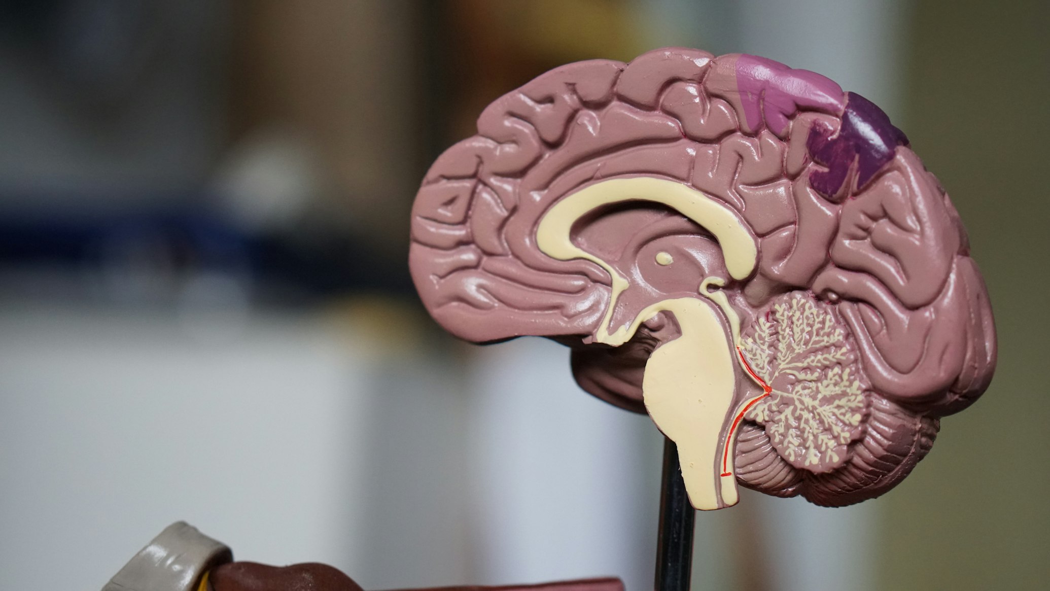
Growing New Body Parts: The Science of Tissue Engineering
Imagine a world where damaged organs can be regrown and injured tissues can be repaired with lab-made materials. This is the promise of tissue engineering, a field that is steadily transforming the future of medicine.
In the mid-1980s, a new scientific discipline emerged with a bold vision: to grow functional human tissues in the lab 2 9 . This field, known as tissue engineering, has since evolved into a powerful force in regenerative medicine. It aims to solve some of medicine's most persistent challenges, such as the shortage of organ donors and the limited ability of some tissues to heal themselves.
At the heart of this endeavor are biomaterial scaffolds—sophisticated three-dimensional structures that act as temporary templates to guide the growth of new tissue 4 5 . This article explores how these ingenious frameworks are engineered to mimic the body's natural environment, supporting cells as they rebuild everything from bone to skin.
The Scaffold Revolution: A Framework for Life
The core principle of tissue engineering is often described as the "tissue-engineering triad," which consists of three key elements: cells, growth-stimulating signals, and biomaterial scaffolds 5 6 . The scaffold is the foundational element, providing the architectural blueprint for new tissue to form.
A well-designed scaffold does more than just fill a gap. Its functions are directly inspired by the body's own extracellular matrix (ECM)—the natural network of proteins and molecules that supports our cells 6 . A successful scaffold must perform several critical roles:
- Provide Structural Support: It offers a physical framework for cells to attach to, grow, and migrate 6 .
- Deliver Bioactive Cues: It can be designed with chemical signals to actively guide cell behavior, encouraging proliferation and differentiation 2 6 .
- Act as a Reservoir: It can hold and release growth factors or pharmaceutical agents in a controlled manner to speed up regeneration 1 6 .
- Define Mechanical Properties: Its intrinsic stiffness and strength provide shape stability to the tissue defect and can influence how cells differentiate 3 6 .
Tissue Engineering Triad
The three essential components that work together to create functional tissue replacements.
Engineering the Perfect Scaffold: A Blueprint for Life
Architectural Mastery: Porosity and Pore Size
The internal architecture of a scaffold is paramount. It requires high porosity and interconnected pores to be successful 3 5 . This design is not arbitrary; it serves vital functions:
- It allows cells to migrate deep into the scaffold.
- It enables the efficient transport of nutrients and oxygen to the cells and the removal of waste products.
- It facilitates the formation of new blood vessels, a process known as vascularization, which is essential for the survival of any thick tissue implant 2 3 .
The ideal pore size depends on the target tissue. For bone regeneration, pore sizes greater than 300 μm are often recommended to support robust bone growth and blood vessel formation 3 . However, a multi-scale approach with hierarchical porosity is increasingly seen as optimal, as it better mimics the complex structure of natural bone 3 .

Microscopic view of a porous scaffold structure
Ideal Scaffold Properties for Different Tissues
| Tissue Type | Target Pore Size | Key Mechanical Property (Young's Modulus) | Primary Function |
|---|---|---|---|
| Bone 3 5 | 200 - 500 μm | 1 - 20 GPa | Load-bearing, structural support |
| Cardiac 5 | N/A | 30 - 400 kPa | Elastic contraction, pumping blood |
| Cartilage 5 | N/A | 10 - 20 kPa | Withstand compression, smooth joint movement |
| Skin 9 | N/A | Information not specific in search results | Protective barrier, wound healing |
The Material World: What Scaffolds Are Made Of
Natural Biomaterials
Natural biomaterials, such as collagen, alginate, and hyaluronic acid, are derived from biological sources. They are prized for their superb biocompatibility, meaning they interact favorably with the body's biological systems and are less likely to cause adverse immune reactions 5 6 .
Their downside is that they often lack the mechanical strength needed for load-bearing applications like bone repair.
Synthetic Polymers
Synthetic polymers, such as Polylactic acid (PLA), offer greater control over physical and mechanical properties, including a controllable degradation rate 5 9 .
Researchers are also developing bioactive materials by decorating these polymers with functional peptides (like RGD) that make them more recognizable to cells, enhancing integration 2 .
Decellularized ECM
A particularly innovative approach uses decellularized ECM 2 . This process involves taking a tissue from an animal or human donor, stripping away all its cellular components, and leaving behind the natural structural scaffold. This ECM scaffold perfectly retains the complex architecture and biological signals of the original tissue, making it an ideal template for regeneration 2 .
The Next Generation: Smart Scaffolds and 3D Bioprinting
The 3D Bioprinting Revolution
3D bioprinting is a form of additive manufacturing that has taken scaffold design to unprecedented levels of precision 8 . Using computer-aided design (CAD) models, bioprinters deposit layers of bioinks—materials containing living cells and biomaterials—to build complex, patient-specific tissue structures layer-by-layer 3 8 .
Bioprinting Technologies
Inkjet Bioprinting
Similar to an office printer, it uses thermal or acoustic forces to eject tiny droplets of bioink, achieving high cell viability (~85%) 8 .
Microextrusion Bioprinting
This method uses mechanical pressure to continuously extrude a filament of bioink, allowing for the creation of high-density tissue structures 8 .
Laser-Assisted Bioprinting
This technique uses a laser to precisely transfer bioink from a donor slide to the printing surface, offering high resolution and cell viability (~95%) 8 .

3D bioprinter creating tissue structures
The Rise of Bio-Responsive and 4D Scaffolds
The frontier of scaffold design lies in creating dynamic, "smart" systems. Researchers are developing bio-responsive scaffolds that can react to physical and chemical stimuli in their environment 7 . Furthermore, the concept of 4D scaffolding has emerged, where 3D-printed constructs can change their shape or function over time in response to specific triggers, more closely mimicking the dynamic nature of living tissue 3 .
A Deeper Dive: The Beta-TCP Experiment in Bone Regeneration
Methodology: Building a Better Bone Graft
A pivotal study focused on repairing a large segmental bone defect in rabbits—a challenging clinical scenario . The researchers followed a meticulous procedure:
1 Scaffold Fabrication
Porous β-Tricalcium Phosphate (β-TCP) scaffolds were created. β-TCP is a ceramic known for its excellent osteoconductivity, meaning it supports bone growth along its surface .
2 Biological Enhancement
The scaffolds were pre-vascularized—a crucial step. Researchers co-cultured rabbit Mesenchymal Stem Cells (rMSCs), which can form bone, with rMSC-derived endothelial cells, which form blood vessels, directly onto the scaffold .
3 Implantation
These cell-seeded, pre-vascularized scaffolds were then implanted into the critical-sized bone defects in the rabbits .
4 Control for Comparison
The results were compared against acellular β-TCP scaffolds or those seeded with cells but not pre-vascularized, to isolate the effect of the enhanced biological strategy.
Bone Regeneration Process
Results and Analysis: A Proof of Concept for Living Implants
The findings were telling. The rabbits that received the pre-vascularized scaffolds showed significantly improved bone repair and integration with the native tissue over 16 weeks . The co-culture of bone-forming and vessel-forming cells created a more complete "tissue unit" from the start, facilitating faster and more robust healing.
This experiment underscores a critical lesson in tissue engineering: simply providing a structural scaffold is often not enough. Successful regeneration requires the simultaneous development of supporting tissues, like blood vessels, to deliver oxygen and nutrients 2 3 . This holistic approach is key to engineering large, functional tissue constructs.
Key Outcomes from the Beta-TCP Bone Regeneration Experiment
| Experimental Group | Key Intervention | Observed Outcome after 16 Weeks |
|---|---|---|
| Pre-vascularized Scaffold | β-TCP scaffold + co-culture of rMSCs & endothelial cells | Enhanced bone repair and tissue integration |
| Cell-Seeded Scaffold | β-TCP scaffold + rMSCs only | Improved bone formation vs. acellular, but less than pre-vascularized |
| Acellular Scaffold | β-TCP scaffold only | Baseline bone formation; insufficient for complete healing |
The Scientist's Toolkit: Essential Reagents in Tissue Engineering
The following table outlines some of the key materials and reagents that are foundational to tissue engineering research, as exemplified in the experiment above and the wider field.
| Reagent / Material | Function / Explanation | Example Applications |
|---|---|---|
| Mesenchymal Stem Cells (MSCs) | Multipotent cells that can differentiate into bone, cartilage, and fat; often used as a primary cell source 5 . | Bone and cartilage regeneration . |
| Growth Factors (e.g., BMP-2, VEGF) | Heterogeneous polypeptides that bind to cell receptors to regulate proliferation and differentiation 5 . | BMP-2 to induce bone formation ; VEGF to promote blood vessel growth 2 . |
| β-Tricalcium Phosphate (β-TCP) | A biodegradable calcium phosphate ceramic with high osteoconductivity . | As a scaffold material for bone defect repair . |
| Hydrogels (e.g., Alginate, Collagen) | Water-swollen polymer networks that mimic the natural extracellular matrix; often used as bioinks 8 . | 3D bioprinting of soft tissues like skin and cartilage 8 . |
| Bioactive Glass | A material that bonds to bone and can release ions (e.g., silicate, borate) with osteogenic potential . | Bone graft substitutes and coatings for implants . |
| Decellularized ECM | The non-cellular component of a tissue that provides biochemical and structural support; used as a native scaffold 2 . | Provides an ideal biomimetic environment for regenerating complex organs 2 . |
The Future of Tissue Engineering
The journey of tissue engineering from a laboratory concept to a clinical reality is well underway. While significant challenges remain—particularly in achieving full vascularization in thick tissues and navigating regulatory pathways—the progress is undeniable 2 . The convergence of advanced biomaterials, 3D bioprinting, and a deeper understanding of cell biology is paving the way for a future where repairing the human body with its own biological materials is not just possible, but routine.