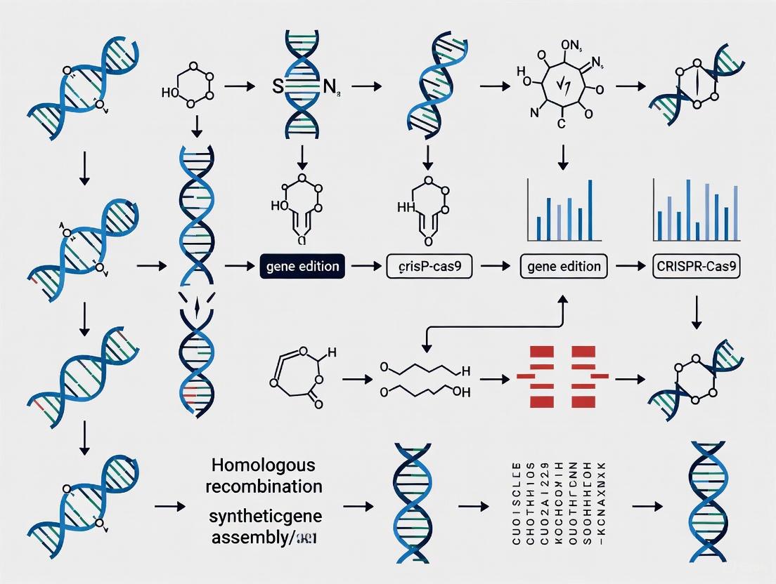
Genome Editing and Synthetic Biology: From Foundational Tools to Clinical Applications
This article provides a comprehensive overview of modern genome editing protocols within synthetic biology, tailored for researchers and drug development professionals.
Genome Editing and Synthetic Biology: From Foundational Tools to Clinical Applications
Abstract
This article provides a comprehensive overview of modern genome editing protocols within synthetic biology, tailored for researchers and drug development professionals. It explores the evolution from foundational CRISPR-Cas systems to advanced editing tools like base and prime editors, detailing their integration with DNA assembly methods such as Golden Gate and Gibson Assembly. The content covers practical delivery strategies, critical troubleshooting for efficiency and specificity, and comparative validation of tools through clinical trial data. By synthesizing cutting-edge research and real-world applications, this guide aims to bridge the gap between laboratory techniques and therapeutic development, offering a roadmap for navigating the rapidly advancing field of programmable genome modification.
The Engine of Change: Core Principles and the Evolution of Editing Tools
The evolution of Clustered Regularly Interspaced Short Palindromic Repeats (CRISPR) technology represents one of the most significant paradigm shifts in modern biological research. What began as the discovery of a bacterial adaptive immune system has rapidly transformed into a versatile molecular toolkit that has revolutionized genome engineering and synthetic biology [1] [2]. The journey from the initial characterization of CRISPR-Cas9 as a programmable DNA-cleaving enzyme to today's expansive CRISPR toolbox exemplifies how fundamental biological research can yield transformative technologies with far-reaching applications across medicine, biotechnology, and basic research [3].
The original CRISPR-Cas9 system, often described as "molecular scissors," introduced unprecedented precision in generating double-strand breaks (DSBs) at predetermined genomic locations [1]. This programmability addressed critical limitations of earlier genome-editing technologies such as zinc finger nucleases (ZFNs) and transcription activator-like effector nucleases (TALENs), which required complex protein re-engineering for each new target site [1]. However, the reliance on DSB formation and subsequent cellular repair mechanisms introduced challenges, including unintended indel mutations, potential off-target effects, and reliance on specific DNA repair pathways that vary in efficiency across cell types [4].
This review chronicles the expansion of the CRISPR toolkit beyond simple cutting toward a multifunctional "Swiss Army knife" capability, enabling precision genome editing, transcriptional regulation, epigenetic modification, and diagnostic applications [5] [6]. We will explore the molecular mechanisms, experimental protocols, and research applications of these advanced CRISPR systems within the context of synthetic biology research, providing both theoretical foundations and practical methodologies for researchers and drug development professionals.
The Foundation: CRISPR-Cas9 as Programmable Molecular Scissors
Historical Context and Mechanism
The CRISPR-Cas9 system originated from fundamental studies of prokaryotic immune systems that protect bacteria from viral infections [1]. The system comprises two key components: the Cas9 nuclease and a guide RNA (gRNA) that directs Cas9 to complementary DNA sequences [3]. The discovery that this system could be reprogrammed to target virtually any DNA sequence by simply modifying the gRNA sequence opened the door to widespread adoption in eukaryotic genome editing [1] [2].
The fundamental mechanism involves the formation of a ribonucleoprotein complex between Cas9 and the gRNA, which scans the genome for protospacer adjacent motifs (PAMs) and complementary sequences to the gRNA spacer region [1]. Upon recognition of the target sequence, Cas9 catalyzes a DSB, which the cell repairs through either non-homologous end joining (NHEJ) or homology-directed repair (HDR) [3]. While NHEJ often results in insertions or deletions (indels) that disrupt gene function, HDR can facilitate precise genetic modifications using an exogenous DNA template [1].
Limitations of First-Generation CRISPR-Cas9
Despite its revolutionary impact, the wild-type CRISPR-Cas9 system presented several limitations for therapeutic applications and precise genetic engineering:
- Off-target effects: Cas9 can cleave DNA at sites with partial complementarity to the gRNA, potentially causing unintended mutations [4]
- DSB-associated risks: The generation of DSBs can lead to large deletions, chromosomal rearrangements, and activation of the p53 pathway, potentially favoring the survival of cells with compromised DNA damage response [7]
- Dependence on cellular repair mechanisms: The efficiency of HDR-mediated precise editing varies significantly across cell types and is generally inefficient in non-dividing cells [4]
- Limited editing scope: Traditional CRISPR-Cas9 is primarily suited for gene disruption or insertion of small fragments via donor templates, with restricted capability for specific nucleotide conversions [4]
These limitations motivated the development of more precise, versatile, and safer CRISPR-based tools that could address the growing demands of synthetic biology and therapeutic genome editing.
Expanding the Toolkit: Precision Editing Without Double-Strand Breaks
Base Editing: Programmable Point Mutations
Base editing represents a significant advancement toward precision genome editing, enabling direct chemical conversion of one DNA base pair to another without inducing DSBs [4]. This technology utilizes catalytically impaired Cas proteins (dCas9 or nickase Cas9) fused to nucleoside deaminase enzymes that mediate specific base transitions [4] [3].
Table: Comparison of Major Base Editing Systems
| Editor Type | Core Components | Base Conversion | Editing Window | Primary Applications |
|---|---|---|---|---|
| Cytosine Base Editor (CBE) | dCas9/nCas9 + cytidine deaminase | C•G to T•A | ~5 nucleotides within target site | Correcting C→T transition mutations, introducing stop codons |
| Adenine Base Editor (ABE) | dCas9/nCas9 + engineered adenosine deaminase | A•T to G•C | ~5 nucleotides within target site | Correcting A→G transition mutations, reverting pathogenic SNVs |
The base editing process involves:
- Target recognition: The gRNA directs the base editor to the target sequence
- DNA strand separation: Cas domain melts the DNA duplex, exposing the single-stranded DNA region
- Nucleoside deamination: The deaminase enzyme converts cytidine to uridine (CBE) or adenosine to inosine (ABE)
- DNA repair: Cellular machinery recognizes the mismatched base and completes the conversion during subsequent DNA replication or repair [4]
Base editors are particularly valuable for correcting single-nucleotide polymorphisms (SNPs) associated with genetic diseases, with clinical applications already in development for various inherited disorders [4].
Prime Editing: Search-and-Replace Genome Editing
Prime editing represents an even more versatile precision editing technology that can mediate all possible base substitutions, small insertions, and small deletions without requiring DSBs or donor DNA templates [7] [3]. The system utilizes a prime editing guide RNA (pegRNA) that both specifies the target site and encodes the desired edit, along with a prime editor protein consisting of a Cas9 nickase fused to a reverse transcriptase [4].
Table: Prime Editing Applications and Specifications
| Edit Type | pegRNA Design Considerations | Efficiency Range | Key Advantages |
|---|---|---|---|
| Point mutations | PBS length: 10-16 nt; RT template: ~10-30 nt | 5-50% | Corrects all 12 possible base substitutions |
| Small insertions | Includes insertion sequence in RT template | 1-20% | Precise sequence insertion without DSBs |
| Small deletions | RT template excludes deleted sequence | 5-40% | Clean deletions without HDR requirement |
| Combination edits | Multiple edits encoded in single RT template | 1-15% | Simultaneous correction of multiple mutations |
The prime editing mechanism occurs through a series of coordinated steps:
- Target binding and nicking: The pegRNA directs the prime editor to the target site, where Cas9 nickase cleaves the PAM-containing strand
- Reverse transcription: The reverse transcriptase uses the pegRNA's extension as a template to synthesize edited DNA
- Flap resolution: Cellular enzymes resolve the resulting DNA flap structure, incorporating the edited sequence into the genome [4] [3]
Prime editing has demonstrated remarkable precision in correcting disease-associated mutations, with substantially reduced off-target effects compared to standard CRISPR-Cas9 approaches [7].
CRISPR Applications Beyond Genome Editing
Transcriptional Control: CRISPRa and CRISPRi
The development of catalytically dead Cas9 (dCas9) created a programmable DNA-binding platform that could be repurposed for gene regulation without altering the underlying DNA sequence [8]. By fusing dCas9 to transcriptional effector domains, researchers have established two powerful technologies: CRISPR activation (CRISPRa) for gene upregulation and CRISPR interference (CRISPRi) for gene repression [4] [8].
The experimental workflow for implementing CRISPRa/i systems typically involves:
Protocol: CRISPR/dCas9-Mediated Transcriptional Regulation in Mammalian Cells
gRNA design: Design gRNAs targeting promoter regions or transcriptional start sites of genes of interest. For CRISPRa, target gRNAs to regions -200 to -50 bp upstream of the transcription start site.
Vector assembly: Clone gRNA sequences into appropriate expression vectors. For stable cell lines, use lentiviral delivery systems.
Effector selection:
- For CRISPRi: Use dCas9-KRAB or dCas9-SRDX fusion constructs
- For CRISPRa: Use dCas9-VP64, dCas9-p300, or SunTag systems for enhanced activation
Delivery: Transfect or transduce cells with dCas9-effector and gRNA constructs using:
- Lipid nanoparticles (LNPs) for primary cells
- Lentiviral transduction for difficult-to-transfect cells
- Electroporation for immune cells
Validation: Assess transcriptional changes 48-72 hours post-delivery using:
- qRT-PCR to measure mRNA levels
- RNA-seq for genome-wide expression profiling
- Western blot or immunofluorescence to detect protein expression changes
Studies in maize protoplasts have demonstrated the efficacy of these systems, with dCas9-SRDX fusions achieving nearly 75% reduction in target gene expression, while dCas9-VP64 and dCas9-TV systems significantly enhanced transcription of endogenous genes [8].
Epigenome Editing
CRISPR-based epigenome editing extends beyond transcriptional control by enabling stable modification of epigenetic marks without changing DNA sequence [4]. By fusing dCas9 to epigenetic effector domains, researchers can write or erase specific DNA methylation or histone modification patterns at targeted genomic loci [5].
Key applications include:
- DNA demethylation: dCas9-TET1 catalytic domain targeted to gene promoters to activate silenced genes
- Histone modification: dCas9-p300 (acetyltransferase) for gene activation or dCas9-LSD1 (demethylase) for gene repression
- Long-term epigenetic memory: Establishing stable transcriptional states through persistent epigenetic modifications
These approaches are particularly valuable for studying the functional consequences of specific epigenetic marks and developing potential therapeutic strategies for diseases with epigenetic components.
Diagnostic Applications: CRISPR-Based Detection
The discovery of collateral cleavage activity in certain Cas proteins (Cas12, Cas13, Cas14) has enabled the repurposing of CRISPR systems for highly sensitive diagnostic applications [6]. These effectors exhibit nonspecific nuclease activity upon target recognition, allowing amplification of detection signals for various pathogens [6].
Table: CRISPR-Cas Diagnostic Systems and Their Applications
| System | Cas Protein | Target | Readout | Detection Limit | Applications |
|---|---|---|---|---|---|
| SHERLOCK | Cas13 | RNA | Fluorescent, colorimetric | aM range | SARS-CoV-2, Zika, Dengue detection |
| DETECTOR | Cas12 | DNA | Fluorescent, electrochemical | aM range | HPV, SARS-CoV-2 DNA detection |
| CRISPR-Chip | dCas9 | DNA | Electrical | fM range | SNP genotyping, rapid diagnostics |
The experimental workflow for CRISPR-based diagnostics typically involves:
- Sample preparation: Nucleic acid extraction from clinical samples (blood, saliva, swabs)
- Amplification (optional): Isothermal amplification (RPA, LAMP) to enhance sensitivity
- CRISPR reaction: Incubation of sample with Cas effector, gRNA, and reporter molecule
- Signal detection: Visual, fluorescent, or electrochemical readout of collateral cleavage activity
These systems enable rapid, field-deployable diagnostics with sensitivity and specificity comparable to conventional PCR-based methods but with significantly reduced processing time and equipment requirements [6].
Advanced Applications and Future Directions
Multiplexed Genome Engineering
The ability to simultaneously target multiple genomic loci represents a powerful approach for studying complex genetic networks and engineering sophisticated biological systems. Multiplexed CRISPR systems enable:
- Large-scale genetic screens: Using pooled gRNA libraries to identify genes involved in specific biological processes or drug resistance mechanisms
- Pathway engineering: Coordinated regulation of multiple genes in metabolic pathways for enhanced production of valuable compounds [5]
- Chromatin engineering: Simultaneous manipulation of multiple genomic loci to study higher-order chromatin organization and its functional implications [3]
Recent advances in CRISPR-associated transposase systems like PASTE (Programmable Addition via Site-specific Targeting Elements) enable insertion of large DNA sequences (up to 36 kb) without DSBs, greatly expanding the scope of genome engineering applications [4].
The Scientist's Toolkit: Essential Research Reagents
Table: Key Research Reagent Solutions for CRISPR Experiments
| Reagent Category | Specific Examples | Function & Applications | Considerations |
|---|---|---|---|
| Cas Effectors | SpCas9, LbCas12a, CasMINI | DNA cleavage, binding, or editing | PAM requirements, size, specificity |
| Base Editors | BE4max, ABE8e | Precision point mutations | Editing window, sequence context |
| Prime Editors | PE2, PEmax, twinPE | Diverse edits without DSBs | pegRNA design, efficiency optimization |
| Delivery Systems | LNPs, AAVs, electroporation | Introducing CRISPR components | Cell type, efficiency, toxicity |
| gRNA Expression | U6 promoters, tRNA arrays | Guide RNA production | Vector design, multiplexing capability |
| Detection Tools | T7E1 assay, NGS, ICE analysis | Edit characterization | Sensitivity, quantification, off-target detection |
Experimental Considerations and Protocol Optimization
Successful implementation of advanced CRISPR tools requires careful consideration of several experimental parameters:
Delivery Methods: The choice of delivery method significantly impacts editing efficiency and cellular viability [9]. Key considerations include:
- RNP delivery: Pre-complexed gRNA-Cas protein ribonucleoproteins offer rapid action, reduced off-target effects, and minimal immunogenicity
- Viral vectors: Lentiviral and AAV systems enable stable expression but have packaging size constraints
- Physical methods: Electroporation and microinjection provide high efficiency but require specialized equipment
- Chemical methods: Lipid nanoparticles (LNPs) have shown promising results for in vivo therapeutic applications [10]
Cell Type Considerations: Editing efficiency varies substantially across different cell types [9]:
- Immortalized cell lines: Generally high editing efficiency, amenable to most delivery methods
- Primary cells: More challenging, often requiring optimized RNP delivery or viral transduction
- Stem cells: Require careful balance between editing efficiency and maintenance of pluripotency
- In vivo applications: Must consider tissue tropism, delivery efficiency, and potential immune responses
The remarkable expansion of the CRISPR toolkit from simple molecular scissors to a versatile multifunctional platform has fundamentally transformed synthetic biology and therapeutic development. The ongoing innovation in CRISPR technology—including base editing, prime editing, epigenetic regulation, and diagnostic applications—continues to push the boundaries of what is possible in genome engineering [5] [6].
As these tools become increasingly sophisticated and accessible, they promise to accelerate both basic research and clinical applications. The integration of artificial intelligence for gRNA design, outcome prediction, and novel enzyme discovery further enhances the precision and capabilities of CRISPR systems [7] [3]. However, responsible development and application of these powerful technologies require ongoing attention to ethical considerations, safety optimization, and equitable access.
The evolution of CRISPR exemplifies how fundamental biological research can yield transformative technologies with profound implications for science and society. As the CRISPR toolkit continues to expand, it will undoubtedly unlock new possibilities for understanding biological systems, treating genetic diseases, and addressing global challenges in health and sustainability.
Synthetic biology aims to program cellular behavior through rational design of genetic components. This discipline leverages molecular regulatory systems that sense specific signals ("sensor") and create defined outputs in response ("effector" or "actuator") [11]. These fundamental regulatory units interface to form complex networks capable of integrating, amplifying, or remembering signals. The core engineering goal involves rewiring natural systems or creating entirely synthetic regulatory systems to achieve predictable cellular functions. As the field matures, increasing emphasis is placed on creating robust, standardized systems through careful characterization of genetic parts, adherence to engineering principles, and computational approaches for automated design.
Fundamental Regulatory Devices
Regulatory devices function as fundamental units within gene regulatory networks, enabling control at multiple levels of gene expression. The synthetic biologist's toolbox now contains a diverse array of these devices, categorized by their mode of action.
DNA-Level Regulatory Devices
Devices acting directly on DNA sequence provide permanent, inheritable control points, making them particularly suitable for implementing stable states like bistable switches or memory devices.
Site-Specific Recombinases: Tyrosine recombinases (e.g., Cre, Flp, FimB/FimE) and serine integrases (e.g., Bxb1, PhiC31) regulate gene expression through DNA inversion or excision. Gene expression is controlled by orienting a promoter with its target gene, creating distinct ON or OFF states [11]. These systems can be made inducible through transcriptional control or fusion to ligand-binding domains (e.g., estrogen receptor) or light-sensitive domains (e.g., LOV2) for optogenetic control [11].
CRISPR-Derived Effectors: RNA-programmable Cas nucleases enable synthetic gene editing without double-strand breaks. Base editors (Cas9 nickase fused to deaminase enzymes) enable targeted single nucleotide changes, while prime editors (Cas9 nickase-reverse transcriptase fusions) allow more complex site-directed edits [11]. Cas1-Cas2 integrase facilitates sequential DNA sequence insertions, valuable for memory devices [11].
Transcriptional Control Devices
Transcriptional regulation represents the most extensively engineered control point in synthetic biology.
Synthetic Transcription Factors: Programmable DNA-binding domains (e.g., engineered zinc fingers, TALEs, CRISPR-dCas9) fused to transcriptional effector domains (activators or repressors) enable precise gene regulation [11]. These systems can be made responsive to various inputs including small molecules, light, or other macromolecules.
Orthogonal Expression Systems: Engineered RNA polymerases and sigma factors that recognize specific promoter sequences provide orthogonal gene expression channels, reducing context dependence in complex circuits [11].
Post-Transcriptional and Translational Control
RNA-level regulation offers faster response times and additional programmability layers.
- Riboswitches and Toehold Switches: Synthetic RNA elements that undergo structural changes upon binding specific ligands or trigger RNAs, controlling translation initiation or transcript stability [11].
- RNA Interference Mechanisms: Engineered RNAi systems provide programmable gene silencing in eukaryotic systems [11].
Post-Translational Control Devices
Protein-level regulation enables rapid response and fine-tuning of circuit behavior.
- Conditional Protein Degradation: Degron tags that render proteins unstable unless stabilized by specific small molecules or conditions [11].
- Protein Relocalization Systems: Light- or chemically-inducible systems controlling protein movement between cellular compartments [11].
- Allosteric Protein Switches: Engineered proteins whose activity is modulated by binding specific ligands or light [11].
Quantitative Analysis of Regulatory Device Performance
The performance of genetic circuits is quantitatively characterized using standardized metrics. The table below summarizes key parameters for various sensing devices implemented in engineered living materials.
Table 1: Performance Parameters of Genetic Sensing Devices in Engineered Living Materials
| Stimulus Type | Input Signal | Output Signal | Host Organism | Threshold | Stability | Reference |
|---|---|---|---|---|---|---|
| Synthetic Inducer | IPTG | RFP (fluorescence) | E. coli | 0.1-1 mM | >72 hours | [12] |
| Synthetic Inducer | aTc | RFP (fluorescence) | E. coli | 50-200 ng/mL | >72 hours | [12] |
| Synthetic Inducer | Theophylline | YFP (fluorescence) | S. elongatus | ~0.5 mM | >7 days | [12] |
| Environmental Chemical | Pb²⁺ | mtagBFP (fluorescence) | B. subtilis | 0.1 μg/L | >7 days | [12] |
| Environmental Chemical | Cu²⁺ | eGFP (fluorescence) | B. subtilis | 1.0 μg/L | >7 days | [12] |
| Environmental Chemical | Hg²⁺ | mCherry (fluorescence) | B. subtilis | 0.05 μg/L | >7 days | [12] |
| Light | 470 nm light | NanoLuc (luminescence) | S. cerevisiae | 470 nm | >7 days | [12] |
| Thermal | Heat | mCherry (fluorescence) | E. coli | >39°C | Not quantified | [12] |
| Mechanical | Compression | IL-1Ra (protein) | Chondrocytes | 15% strain | ≥3 days | [12] |
Standardized Experimental Protocols
Protocol: BreakTag for Characterizing Nuclease Activity
BreakTag provides a scalable method for profiling both on-target and off-target double-strand breaks caused by CRISPR nucleases, compatible with next-generation sequencing workflows [13].
Materials Required:
- CRISPR ribonucleoprotein complexes
- Genomic DNA from target cells
- BreakTag library preparation reagents
- Next-generation sequencing platform
- BreakInspectoR analysis software [13]
Methodology:
- Ribonucleoprotein Complex Formation: Incubate purified Cas nuclease with synthesized guide RNA (30 minutes, room temperature).
- Targeted Digestion: Digest genomic DNA with assembled ribonucleoprotein complexes (2 hours, 37°C).
- Break Collection: Unbiased collection of DNA fragments containing blunt and staggered double-strand breaks using BreakTag adapters.
- Library Preparation: Prepare sequencing libraries (approximately 6 hours hands-on time).
- Sequencing: Perform next-generation sequencing (total protocol time: 3 days including sequencing).
- Data Analysis: Process sequencing data with BreakInspectoR to assess nuclease activity and characterize scission profiles.
- Machine Learning Integration: Utilize XGScission models to predict cleavage behavior at novel genomic targets [13].
Applications: This protocol enables comprehensive assessment of guide RNA specificity, nuclease efficiency, and cleavage dynamics (blunt vs. staggered ends), informing the selection of optimal guides for genetic circuit integration.
Protocol: Implementation of Recombinase-Based Memory Devices
Site-specific recombinases enable stable genetic memory through DNA inversion or excision [11].
Materials Required:
- Expression vector with recombinase (Cre, Flp, Bxb1, or PhiC31)
- Target vector with recombination sites flanking transcriptional terminators or orientation-dependent expression cassettes
- Appropriate inducer molecules (small molecules, light source)
- Host cells (bacterial, yeast, or mammalian)
Methodology:
- Circuit Design: Clone recombination sites (loxP, FRT, attB/attP) in convergent or divergent orientations flanking a promoter or coding sequence.
- Delivery: Co-transfect recombinase expression vector and target vector into host cells.
- Induction: Apply inducer (small molecule for chemically-inducible systems or specific light wavelength for optogenetic systems) for defined duration.
- Verification: Assay for recombination events using PCR, restriction digest, or reporter expression.
- Stability Assessment: Passage cells without inducer and measure retention of genetic state over multiple generations.
Applications: Creates permanent genetic records of transient environmental exposures, implements binary decision-making in cells, and builds complex logic gates through sequential recombination events.
Essential Research Reagent Solutions
Table 2: Key Research Reagents for Genetic Circuit Construction
| Reagent Category | Specific Examples | Function | Applications |
|---|---|---|---|
| Programmable Nucleases | SpCas9, AsCas12f, TnpB | Targeted DNA cleavage, base editing, prime editing | Gene knockouts, precise edits, circuit integration [7] |
| Recombinases | Cre, Flp, Bxb1, PhiC31 | DNA inversion, excision, integration | Memory devices, logic gates, state switching [11] |
| Synthetic Transcription Factors | dCas9-VP64, ZF-TFs, TALE-TFs | Programmable gene activation/repression | Transcriptional logic, multi-gene regulation [11] |
| Reporter Proteins | GFP, mCherry, RFP, Luciferase | Visualizing gene expression, quantifying circuit output | Circuit characterization, biosensor readouts [12] |
| Inducer Molecules | IPTG, aTc, Arabinose, Theophylline | Chemical control of gene expression | Inducible systems, dose-response characterization [12] |
| Engineered Matrices | Hydrogels, Bacterial Cellulose, Curli Fibrils | Structural support for embedded cells | Engineered living materials, biosensing platforms [12] |
Visualization of Genetic Circuit Design Principles
The following diagrams illustrate key concepts, workflows, and relationships in genetic circuit design, created using Graphviz DOT language with specified color palette and contrast requirements.
Figure 1: Genetic Circuit Information Flow
Figure 2: Gene Regulation Levels
Figure 3: BreakTag Workflow
Figure 4: Genetic Memory Mechanism
The standardization of synthetic DNA components and genetic circuit design principles has transformed synthetic biology from an ad hoc discipline to a predictable engineering practice. The comprehensive toolkit of regulatory devices operating at DNA, RNA, protein, and epigenetic levels enables construction of increasingly sophisticated genetic systems. As standardization improves and characterization methodologies like BreakTag become more accessible, the design-build-test-learn cycle accelerates, supporting more reliable implementation of genetic circuits for therapeutic development, bioproduction, and engineered living materials. Continued development of standardized parts, measurement techniques, and predictive models will further enhance our ability to program biological systems with precision and reliability.
In the landscape of synthetic biology and genome editing, the precise manipulation of genetic material relies on foundational enzymatic tools. Restriction enzymes and DNA ligases function as the quintessential "molecular glue," enabling the targeted cutting and rejoining of DNA fragments [14] [15]. These enzymes form the bedrock of recombinant DNA technology, facilitating the construction of novel genetic assemblies essential for advanced research and therapeutic development [16] [17].
While modern gene-editing platforms like CRISPR-Cas9 dominate current discourse, the operational context of many delivery vectors, including gRNA plasmids and viral vectors, is itself a product of restriction-ligation cloning [18] [15]. This article details the latest applications and optimized protocols for these enduring tools, providing critical resources for researchers and drug development professionals.
The Scientific Foundation: Enzyme Mechanics and Classification
Restriction Endonucleases: Precision DNA Scissors
Restriction enzymes are endonucleases that recognize specific DNA sequences and catalyze double-stranded cuts [14]. They are a critical component of the bacterial restriction-modification system, which protects prokaryotes from invading viruses by cleaving non-methylated foreign DNA while protecting host DNA via methylation [19] [17].
Table 1: Classification of Restriction Enzymes
| Type | Recognition Sequence | Cleavage Characteristics | Cofactors | Primary Applications |
|---|---|---|---|---|
| Type I [14] [17] | Bipartite and asymmetric (e.g., EcoKI) | Variable, random cuts at least 1000 bp from recognition site | ATP, AdoMet, Mg²⁺ | Study of DNA translocation and molecular motor mechanisms |
| Type II [16] [14] [17] | Palindromic, short (4-8 bp) (e.g., EcoRI) | Precise cuts within or at fixed positions near recognition site | Mg²⁺ | Molecular cloning, DNA mapping, restriction analysis |
| Type IIS [16] | Asymmetric | Precise cuts outside of recognition site (1-20 bp away) | Mg²⁺ | Golden Gate Assembly, seamless cloning |
| Type III [14] [17] | Asymmetric, short | Cuts at fixed position 24-26 bp downstream of recognition site | ATP, Mg²⁺ (AdoMet stimulatory) | Study of enzyme complex mechanisms |
| Type IV [14] [17] | Variable | Targets modified DNA (methylated, hydroxymethylated) | Mg²⁺ | Epigenetic studies, mapping DNA modifications |
The discovery of Type II restriction enzymes, particularly HindII in 1970, revolutionized molecular biology by enabling reproducible DNA cleavage at specific sequences [17]. Over 3,600 restriction endonucleases have been identified, representing more than 250 different specificities, with more than 800 available commercially [14].
DNA Ligases: Molecular Adhesives
DNA ligases catalyze the formation of phosphodiester bonds between adjacent 3'-hydroxyl and 5'-phosphate termini in DNA, effectively "gluing" DNA fragments together [20] [15]. These enzymes utilize either ATP or NAD⁺ as cofactors and operate through a conserved three-step mechanism [21]:
- Adenylation: A conserved lysine residue attacks the α-phosphate of ATP or NAD⁺, forming a ligase-AMP intermediate and releasing pyrophosphate or NMN.
- AMP Transfer: The AMP moiety is transferred to the 5'-phosphate of the DNA nick, generating an adenylated DNA intermediate.
- Ligation: A phosphodiester bond is formed by nucleophilic attack from the 3'-OH of the adjacent DNA strand, releasing AMP.
Table 2: Properties of Commercially Available DNA Ligases
| Ligase | Cofactor | Recommended Temperature | Primary Applications | Key Features |
|---|---|---|---|---|
| T4 DNA Ligase [20] | ATP | 25°C (4-37°C range) | Sticky-end ligation (>2 base overhangs), nick ligation | Standard workhorse for routine cloning |
| Hi-T4 DNA Ligase [20] | ATP | 25°C (4-50°C range) | High-temperature ligations | Increased thermotolerance maintains activity up to 45°C for extended periods |
| Quick Ligation Kit [20] | ATP | 25°C | Rapid sticky or blunt-end ligation | Contains PEG for enhanced efficiency; not heat-inactivatable |
| Blunt/TA Ligase Master Mix [20] | ATP | 25°C | Fast ligation of blunt or single base overhang substrates | Optimal for T/A cloning and NGS adapter ligation |
| T7 DNA Ligase [20] | ATP | 25°C | Selective nick ligation | High specificity for correctly base-paired nicks; minimal blunt-end activity |
| E. coli DNA Ligase [20] | NAD⁺ | 25°C | Nick ligation in dsDNA | Used in cDNA library preparation protocols |
| Taq DNA Ligase [20] [21] | NAD⁺ | 60°C (37-75°C range) | Ligation detection methods, Gibson Assembly | Thermostable; high discrimination against mismatched bases |
Figure 1: DNA Ligase Three-Step Catalytic Mechanism. The enzyme catalyzes phosphodiester bond formation through an adenylated intermediate [21].
Advanced Applications in Synthetic Biology
DNA Assembly Methodologies
Traditional restriction enzyme cloning using Type IIP enzymes (e.g., EcoRI, HindIII) revolutionized biology but presents limitations including scar sequence introduction and dependence on available restriction sites [16] [15]. Advanced solutions have emerged:
Golden Gate Assembly: This method exploits Type IIS restriction enzymes (e.g., BsaI, BbsI, BsmBI) which cleave outside their recognition sequences [16]. This enables seamless assembly of multiple DNA fragments without introducing scar sequences, as the restriction site is eliminated from the final ligated product [16].
Gibson Assembly: This method uses a combination of 5' exonuclease, DNA polymerase, and DNA ligase (often Taq DNA ligase) to assemble multiple overlapping DNA fragments in a single-tube isothermal reaction [16] [20]. It allows for the simultaneous assembly of several fragments without reliance on restriction sites.
Table 3: Comparison of DNA Assembly Methods
| Method | Principle | Key Enzymes | Fragment Capacity | Scar Sequence | Efficiency |
|---|---|---|---|---|---|
| Traditional Cloning [16] [15] | Restriction digestion and ligation | Type IIP REases, T4 DNA Ligase | 1-2 | Yes (unless compatible ends) | Moderate |
| Golden Gate Assembly [16] | Type IIS digestion and ligation | Type IIS REases, T4 DNA Ligase | High (dozens of fragments) | No | High |
| Gibson Assembly [16] [20] | Exonuclease digestion + homologous recombination | Exonuclease, Polymerase, Taq DNA Ligase | Moderate (multiple fragments) | No | High |
| TA/TOPO-TA Cloning [15] | TA complementarity | Taq Polymerase, Topoisomerase | 1 | Yes | High for PCR products |
Beyond Cloning: Epigenetics and Genome Mapping
Restriction enzymes serve as sensitive detectors of DNA modification status. Enzymes such as MspI and HpaII (both recognizing CCGG) display differential sensitivity to cytosine methylation, enabling identification of 5-methylcytosine (5-mC) patterns [16] [19]. Specialized enzymes like MspJI, FspEI, and LpnPI recognize and cleave DNA at 5-mC and 5-hydroxymethylcytosine (5-hmC) sites, while PvuRts1I preferentially cleaves 5-hmC over 5-mC [16]. These properties are exploited in kits such as the EpiMark 5-hmC and 5-mC Analysis Kit for refined epigenetic marker identification [16].
Restriction Fragment Length Polymorphism (RFLP) analysis uses variations in restriction sites to detect single-nucleotide polymorphisms (SNPs) and insertions/deletions (Indels), enabling applications from genetic disorder diagnosis to parental testing [16] [19].
Essential Protocols for Genome Editing Workflows
Golden Gate Assembly for Modular Vector Construction
This protocol enables seamless, one-pot assembly of multiple DNA fragments, ideal for constructing complex vectors for CRISPR-based editing [16].
Research Reagent Solutions:
- Type IIS Restriction Enzyme: BsaI-HFv2 or BsmBI-v2 (high-fidelity variants minimize star activity)
- DNA Ligase: T4 DNA Ligase or high-concentration variant (e.g., HC T4 DNA Ligase)
- Vector Backbone: Custom plasmid with appropriate Type IIS sites flanking the insertion site
- Insert Fragments: PCR-amplified with appropriate terminal overhangs complementary to adjacent fragments
- Thermostable Ligase (optional): For assemblies performed at elevated temperatures
Procedure:
- Fragment Preparation: Design all DNA fragments with Type IIS recognition sites (e.g., BsaI: GGAGACC) oriented such that cleavage removes the recognition site. Ensure complementary overhangs (typically 4-bp) between adjacent fragments for ordered assembly.
Reaction Setup:
- 50-100 ng vector backbone
- Molar equivalent of each insert fragment (typically 1:2 vector:insert ratio)
- 1× T4 DNA Ligase Buffer
- 5-10 U Type IIS restriction enzyme (e.g., BsaI-HFv2)
- 400 U T4 DNA Ligase (or 2 µL High-Concentration T4 DNA Ligase)
- Nuclease-free water to 20 µL
Thermal Cycling:
- 30-40 cycles: 37°C (2-5 min digestion/ligation) → 16°C (2-5 min digestion/ligation)
- Final digestion: 50°C (5-10 min)
- Enzyme inactivation: 80°C (5-10 min)
Transformation and Verification: Transform 2-5 µL reaction into competent E. coli cells. Screen colonies by colony PCR or analytical restriction digest, followed by Sanger sequencing to verify correct assembly.
Figure 2: Golden Gate Assembly Workflow. Type IIS enzymes cleave outside recognition sites to create unique overhangs for seamless, ordered assembly [16].
Restriction-Based DNA Mapping for Quality Control
Verify plasmid constructs and identify polymorphisms through restriction analysis [19].
Procedure:
- Digestion Reaction: Combine 500 ng plasmid DNA, 1× restriction enzyme buffer, 5-10 U of each restriction enzyme, and water to 20 µL. Incubate at recommended temperature for 30-60 minutes.
Electrophoresis: Load digested DNA alongside uncut control and DNA size standard on 0.8-1.5% agarose gel containing ethidium bromide or alternative DNA stain. Run at 5-10 V/cm until adequate separation.
Pattern Analysis: Visualize under UV light and document fragment pattern. Compare observed fragment sizes to expected pattern using DNA analysis software. Deviations indicate potential mutations, rearrangements, or incorrect assemblies.
High-Efficiency Ligation for Complex Constructs
Maximize transformation efficiency, particularly for low-copy number vectors or large constructs.
Research Reagent Solutions:
- High-Concentration Ligase: M0202T/M T4 DNA Ligase or Quick Ligation Kit
- PEG-Enhanced Buffers: Proprietary formulations containing polyethylene glycol to promote macromolecular crowding
- Electrocompetent Cells: High-efficiency strains (>10⁹ CFU/µg) for challenging constructs
Procedure:
- Sticky-End Ligation: For fragments with >2 bp complementary overhangs, use standard T4 DNA Ligase at 16°C for 1-2 hours or Quick Ligation Kit at room temperature for 5-10 minutes.
Blunt-End/TA Ligation: Use Blunt/TA Ligase Master Mix with proprietary enhancer for highest efficiency. Incubate at room temperature for 15 minutes.
Electroporation-Compatible Ligation: When using PEG-containing master mixes (not heat-inactivatable), purify DNA before electroporation or use Electro Ligase for direct transformation.
Transformation: Use high-efficiency chemically competent cells (>10⁸ CFU/µg) or electrocompetent cells for large constructs. Include positive and negative controls to assess efficiency.
The Scientist's Toolkit: Essential Research Reagents
Table 4: Key Research Reagent Solutions for Restriction-Ligation Workflows
| Reagent Category | Specific Examples | Function & Application Notes |
|---|---|---|
| High-Fidelity (HF) Restriction Enzymes [16] | NEB HF series (e.g., EcoRI-HF, BamHI-HF) | Engineered variants exhibiting minimal star activity under extended incubation or high enzyme concentrations; essential for complex digests |
| Type IIS Restriction Enzymes [16] | BsaI-HFv2, BsmBI-v2, BbsI, Esp3I | Create non-palindromic overhangs outside recognition site; enable Golden Gate Assembly and seamless cloning |
| Thermostable DNA Ligases [20] [21] | Taq DNA Ligase, 9°N DNA Ligase | NAD⁺-dependent enzymes stable at high temperatures; used in Gibson Assembly and ligation detection methods |
| Rapid Ligation Kits [20] | Quick Ligation Kit, Instant Sticky End Ligase Master Mix | PEG-formulated systems enabling 5-15 minute room temperature reactions; ideal for high-throughput workflows |
| Electroporation-Compatible Ligase [20] | Electro Ligase | PEG-free formulation allowing direct transformation by electroporation without intermediate purification |
| Methylation-Sensitive REases [16] [19] | HpaII, DpnI, DpnII | Detect epigenetic modifications; DpnI cleaves only methylated GATC sites, while DpnII cleaves only unmethylated sites |
| Cloning Vectors [15] | pUC19, pBR322 derivatives, Golden Gate destination vectors | Carrier DNA molecules with Multiple Cloning Sites (MCS), selectable markers, and origin of replication |
Despite the emergence of CRISPR-based genome editing systems, restriction enzymes and DNA ligases maintain their foundational role as the essential "molecular glue" in synthetic biology research [18] [15]. Their evolved specificities and engineered enhancements make them indispensable for vector construction, epigenetic analysis, and DNA assembly [16] [19].
The continuing development of high-fidelity restriction enzymes, thermostable ligases, and specialized assembly methodologies ensures these classical tools remain relevant in contemporary genome editing workflows [16] [20]. For researchers building the next generation of therapeutic vectors or engineered biological systems, mastery of these fundamental tools provides the critical capability to precisely manipulate genetic material with reliability and efficiency.
The integration of artificial intelligence (AI) with genome editing represents a paradigm shift in synthetic biology, directly addressing the critical challenge of editor optimization. While CRISPR-based technologies have revolutionized biological research by enabling precise genomic modifications, their efficacy has been hampered by unpredictable editing efficiency, cell-type specific outcomes, and substantial experimental optimization requirements [7] [22]. AI and machine learning (ML) models are now transforming this landscape by extracting complex patterns from massive biological datasets to predict editing outcomes, optimize guide RNA designs, and accelerate the development of novel editing tools [23] [24]. This convergence is particularly impactful in synthetic biology applications where precision, reliability, and throughput are paramount for engineering biological systems.
The optimization challenge stems from the complex relationship between editor components and their cellular context. Traditional approaches required extensive trial-and-error experimentation to identify effective guide RNAs and editing conditions for each new target [22]. AI-powered prediction models bypass this bottleneck by learning from collective experimental data to anticipate editing efficiency and specificity before laboratory validation [7] [25]. This data-driven approach has enabled researchers to move from heuristic design rules to quantitative predictive models that account for sequence features, epigenetic context, and cellular repair mechanisms, substantially accelerating the editor optimization pipeline for synthetic biology applications [23] [26].
Key Machine Learning Applications in Editor Optimization
Predictive Modeling of Editing Efficiency
Machine learning has demonstrated exceptional capability in predicting the on-target efficiency of genome editing tools, a fundamental requirement for experimental success in synthetic biology. Deep learning models, including convolutional and recurrent neural networks, have surpassed earlier statistical approaches by automatically learning relevant features from guide RNA sequences and their genomic context [22]. For prime editing systems, where editing outcomes depend heavily on prime editing guide RNA (pegRNA) design, specialized models like PRIDICT2.0 have achieved remarkable predictive accuracy [25].
The table below summarizes performance metrics for prominent AI prediction tools across different editing platforms:
Table 1: Performance Metrics of AI-Powered Editing Prediction Tools
| Tool Name | Editing Platform | Key Features | Reported Performance | Applications |
|---|---|---|---|---|
| PRIDICT2.0 [25] | Prime Editing | Predicts efficiency for edits up to 15bp; models MMR-deficiency/proficiency | Spearman R: 0.91 (HEK293T), 0.81 (K562) | Multi-base replacements, insertions, deletions |
| DeepHF [22] | High-fidelity Cas9 variants | Specialized for eSpCas9(1.1), SpCas9-HF1; combines RNN with biological features | Outperforms other tools for high-fidelity variants | Editing with reduced off-target effects |
| CRISPRon [22] | SpCas9 | Integrates sequence, thermodynamics, binding energy; deep learning on 23,902 gRNAs | Superior performance on independent datasets | Standard CRISPR-Cas9 editing |
| EasyDesign [22] | Cas12a diagnostics | CNN trained on 11,000 diagnostic-target pairs | Spearman correlation: 0.812 | CRISPR-based diagnostic assays |
Off-Target Effect Prediction and Minimization
The prediction and minimization of off-target effects constitutes another critical application of machine learning in editor optimization. Traditional prediction methods focused primarily on sequence similarity but often failed to capture the complex factors influencing off-target activity [22]. Next-generation models like CRISPR-M employ multi-view deep learning architectures that consider insertions, deletions, and mismatches while incorporating additional features like GC content, melting temperature, and sequence context [22]. These advanced models enable synthetic biologists to select guide RNAs with optimal on-target to off-target activity ratios before experimental validation.
BreakTag technology has further advanced this field by enabling high-throughput profiling of nuclease activity, generating comprehensive datasets of both on-target and off-target editing events [13]. When coupled with machine learning platforms like XGScission, these datasets enable predictive modeling of cleavage dynamics, including the discrimination between blunt and staggered ends—a critical determinant of editing outcomes [13]. This integrated approach provides unprecedented insight into the sequence determinants of nuclease behavior, informing both guide selection and nuclease engineering for enhanced specificity.
Novel Editor Discovery and Engineering
Beyond optimizing existing tools, AI is accelerating the discovery and engineering of novel genome editing systems. Protein language models and structure prediction tools like AlphaFold have enabled computational mining of microbial genomes for rare or ancestral CRISPR systems with unique properties [7] [23]. For instance, AI-guided clustering of terascale sequencing data has identified previously unknown CRISPR-Cas13 variants with distinct targeting capabilities [7]. Similarly, structure-guided protein engineering has yielded compact editors like enAsCas12f that maintain high activity while addressing delivery constraints [7].
The integration of deep mutational scanning with machine learning has proven particularly powerful for editor optimization. By systematically testing thousands of protein variants and training models on the resulting activity data, researchers have evolved editors with expanded PAM compatibility, reduced off-target effects, and altered enzymatic functions [7] [22]. This data-driven engineering approach has produced critical editor variants including base editors with altered sequence preferences and prime editors with enhanced efficiency—both invaluable tools for the synthetic biology toolkit.
Experimental Protocols and Implementation
Protocol: AI-Guided Prime Editing Experiment
This protocol outlines the implementation of prime editing experiments using AI-based prediction tools for pegRNA optimization, suitable for installing precise edits in mammalian cells for synthetic biology applications.
Step 1: Target Selection and Edit Definition
- Identify the genomic target locus and define the precise edit(s) to be introduced (single-base substitution, insertion, or deletion).
- Consider epigenetic context using tools like ePRIDICT, which quantifies how local chromatin environments impact prime editing rates [25].
- Note: Editing efficiency correlates with chromatin accessibility; target open chromatin regions when possible.
Step 2: pegRNA Design Using PRIDICT2.0
- Design candidate pegRNAs according to standard principles (PBS length: 13nt, RTT length: 10-16nt).
- Input candidate pegRNA sequences along with the intended edit into the PRIDICT2.0 model.
- Select the appropriate cell type model (PRIDICT2.0 HEK293T for MMR-deficient cells; PRIDICT2.0 K562 for MMR-proficient cells) [25].
- Rank pegRNAs by predicted efficiency scores and select top 3-5 candidates for experimental validation.
Step 3: Experimental Validation
- Clone selected pegRNAs into appropriate prime editing vectors.
- Transfect target cells (e.g., HEK293T for MMR-deficient, K562 for MMR-proficient) using standard protocols.
- Include controls: non-transfected cells, editor-only negative control.
Step 4: Outcome Assessment
- Harvest genomic DNA 72-96 hours post-transfection.
- Amplify target region by PCR and sequence using next-generation sequencing (preferred) or Sanger sequencing with decomposition analysis (TIDE) [27].
- Quantify editing efficiency as percentage of reads containing the desired edit.
Step 5: Model Refinement (Optional)
- Feed experimental results back into training datasets to improve site-specific prediction accuracy.
- Iterate design if necessary based on empirical results.
Protocol: High-Throughput Editing Characterization with BreakTag
The BreakTag method enables comprehensive profiling of nuclease activity, generating data suitable for training machine learning models of editor optimization [13].
Step 1: Library Design and Preparation
- Design a diverse library of guide RNAs targeting genomic sites of interest.
- Clone guide RNAs into appropriate expression vectors.
Step 2: Cell Transfection and Editing
- Transfect target cells with CRISPR ribonucleoprotein complexes.
- Include appropriate controls (non-targeting guides, editor-only).
Step 3: Genomic DNA Processing and Break Enrichment
- Harvest cells 48-72 hours post-transfection and isolate genomic DNA.
- Fragment DNA and enrich for double-strand breaks using BreakTag adapter ligation.
- Critical: The BreakTag protocol enriches both blunt and staggered ends, providing comprehensive cleavage profiling [13].
Step 4: Sequencing Library Preparation
- Amplify BreakTag-ligated fragments with indexed primers.
- Pool libraries and sequence using high-throughput platforms.
Step 5: Data Analysis with BreakInspectoR
- Process sequencing data with BreakInspectoR to identify and quantify on-target and off-target editing events [13].
- Characterize cleavage profiles (blunt vs. staggered ends).
Step 6: Machine Learning Integration
- Use curated datasets to train predictive models (e.g., XGScission) for anticipating nuclease behavior at novel targets [13].
- Apply trained models to optimize future guide RNA selections.
Visualization of AI-Powered Editor Optimization Workflow
Diagram 1: AI-powered editor optimization workflow with a continuous learning feedback loop.
The Scientist's Toolkit: Research Reagent Solutions
Table 2: Essential Research Reagents for AI-Powered Editor Optimization
| Reagent/Tool | Function | Application Notes |
|---|---|---|
| PRIDICT2.0 [25] | Predicts prime editing efficiency for diverse edit types | Use HEK293T model for MMR-deficient cells; K562 model for MMR-proficient cells |
| BreakTag [13] | High-throughput profiling of nuclease activity | Enriches blunt/staggered breaks; 3-day protocol with integrated analysis |
| DeepCRISPR [22] | Deep learning for guide RNA design | Unsupervised pre-training on billions of sequences; epigenetic integration |
| CRISPR-GPT [22] | Natural language interface for editing design | Three user modes; trained on 11 years of literature and experimental data |
| XGScission [13] | ML model predicting cleavage dynamics | Trained on BreakTag data; predicts blunt vs. staggered ends |
| TIDE [27] | Decomposition analysis of editing outcomes | Rapid quantification from Sanger sequencing; cost-effective for small sets |
The integration of AI with genome editing is progressing beyond single-component optimization toward holistic experimental planning. Emerging platforms like CRISPR-GPT demonstrate the potential of large language models to guide researchers through complex experimental design decisions using natural language interfaces [22]. These systems incorporate decades of published literature, experimental data, and community knowledge to provide contextualized recommendations, effectively democratizing expertise in genome editing optimization.
Looking forward, the field is moving toward virtual cell models that can simulate editing outcomes across diverse cellular contexts [7]. These AI-powered simulations will enable researchers to anticipate functional consequences of edits, optimize multi-locus editing strategies, and account for cell-type specific factors before experimental implementation. For synthetic biology applications, this capability will be transformative, enabling more predictable engineering of complex genetic circuits and metabolic pathways.
In conclusion, AI-powered prediction has fundamentally transformed the genome editing optimization paradigm from empirical testing to computational forecasting. By leveraging machine learning models trained on high-throughput editing data, synthetic biologists can now design optimized editing tools with unprecedented efficiency and precision. As these models continue to incorporate additional layers of biological complexity—from 3D chromatin structure to cellular metabolism—their predictive power will further accelerate the engineering of biological systems for research and therapeutic applications.
The success of genome editing in synthetic biology is fundamentally constrained by the efficacy of delivery systems. Transporting CRISPR-Cas9 components or other editing machinery across multiple cellular barriers—from the plasma membrane to the nuclear envelope—presents a formidable scientific challenge [28] [29]. These barriers have evolved as protective mechanisms, making efficient penetration a key bottleneck in therapeutic applications. The delivery vehicle must navigate extracellular degradation, achieve specific cellular uptake, avoid endosomal entrapment, facilitate cytoplasmic transport, and ultimately achieve nuclear entry [30] [28]. Different delivery strategies—viral, physical, and chemical—have been developed to address these sequential hurdles, each with distinct advantages and limitations for specific research or therapeutic contexts [9] [29].
Viral Delivery Methods
Principle and Workflow
Viral vectors exploit the natural ability of viruses to infiltrate cells and deliver genetic material. In synthetic biology, engineered viral particles are stripped of pathogenic components and repurposed to transport genome-editing machinery such as CRISPR-Cas9 systems [29]. The process involves packaging CRISPR cargo (as DNA, RNA, or donor templates) into viral capsids, which then infect target cells using native viral entry pathways [31] [32]. Common viral vectors include adeno-associated viruses (AAVs), adenoviruses (AdVs), and lentiviruses (LVs), each with distinct infection mechanisms and expression profiles [29].
Key Viral Vectors and Characteristics
Table 1: Comparison of Major Viral Delivery Systems for Genome Editing
| Vector Type | Payload Capacity | Genomic Integration | Immune Response | Primary Applications |
|---|---|---|---|---|
| Adeno-Associated Virus (AAV) | ~4.7 kb [29] | Non-integrative [29] | Low/Moderate [29] | In vivo gene therapy, preclinical models [29] |
| Adenovirus (AdV) | Up to 36 kb [29] | Non-integrative [29] | High [29] | High cargo delivery, both dividing and non-dividing cells [29] |
| Lentivirus (LV) | Large (>10 kb) [29] | Integrative [29] | Moderate [30] | Stable long-term expression, in vitro studies [29] |
| Virus-like Particles (VLPs) | Limited [29] | Non-integrative [29] | Low [29] | Transient delivery, reduced off-target concerns [29] |
Protocol: AAV-Mediated CRISPR Delivery for In Vivo Editing
Application Note: This protocol is optimized for delivering CRISPR components to mouse liver using AAV vectors, suitable for disease modeling and functional genomics studies.
Materials:
- AAV vectors: AAV8-CRISPR (packaging sgRNA) and AAV8-Cas9 (if using dual AAV system) [29]
- Animals: C57BL/6 mice (6-8 weeks old)
- Reagents: Phosphate-buffered saline (PBS), isoflurane anesthesia
- Equipment: Sterile syringes, 29-gauge insulin needles, animal warming pad
Methodology:
- AAV Preparation:
- For single AAV delivery: Use AAV vectors encoding both Cas9 and sgRNA (requires smaller Cas9 variants like Cas12a or engineered compact Cas9) [29].
- For dual AAV delivery: Prepare separate AAVs for sgRNA and Cas9, each with distinct fluorescent or selection markers [29].
- Purify AAV vectors via ultracentrifugation and resuspend in sterile PBS. Determine viral titer (genome copies/mL) using qPCR.
Administration:
- Anesthetize mice using isoflurane (3% induction, 1.5-2% maintenance).
- Administer AAV via tail vein injection at dose of 1×10¹¹ to 1×10¹² genome copies in 100-200 μL PBS [29].
- Maintain mice on warming pad until fully recovered from anesthesia.
Validation:
- Harvest tissue at 2-4 weeks post-injection.
- Extract genomic DNA and assess editing efficiency via T7E1 assay or next-generation sequencing.
- Evaluate protein-level changes via immunohistochemistry or Western blot if knockout is intended.
Troubleshooting:
- Low editing efficiency: Optimize AAV dose; verify sgRNA activity using in vitro cleavage assay.
- Immune response: Consider different AAV serotypes with lower immunogenicity [29].
- Cargo limitation: For larger Cas proteins, utilize dual AAV systems or smaller Cas orthologs [29].
Figure 1: AAV Viral Delivery Pathway - This diagram illustrates the intracellular journey of AAV vectors from cellular binding to genomic editing
Physical Delivery Methods
Principle and Workflow
Physical methods utilize mechanical or electrical forces to create transient openings in the cell membrane, allowing direct passage of genome-editing components into cells [9]. These approaches are particularly valuable for delivering preassembled ribonucleoprotein (RNP) complexes of Cas9 protein and guide RNA, which act immediately upon entry and degrade rapidly, minimizing off-target effects [9]. The primary physical methods include electroporation, nucleofection, and microinjection, each optimized for different cell types and experimental needs.
Physical Transfection Comparison
Table 2: Physical Delivery Methods for Genome Editing Components
| Method | Principle | Efficiency | Optimal Cargo Format | Primary Cell Types | Throughput |
|---|---|---|---|---|---|
| Electroporation | Electrical pulses create membrane pores [9] | High [9] | RNP, mRNA, DNA [9] | Immortalized cells, suspension cells [9] | Medium-High [9] |
| Nucleofection | Electroporation optimized for nuclear delivery [9] | Very High [9] | RNP (direct to nucleus) [9] | Primary cells, stem cells, difficult-to-transfect cells [9] | Medium-High [9] |
| Microinjection | Mechanical injection using microneedle [9] | Highest [9] | RNP, mRNA [9] | Zygotes, oocytes, single cells [9] | Low [9] |
Protocol: RNP Delivery via Nucleofection for Primary T Cells
Application Note: This protocol enables highly efficient gene editing in primary human T cells for immunotherapy research, utilizing preassembled Cas9 RNP complexes for rapid activity and minimal off-target effects.
Materials:
- Cells: Primary human T cells (isolated from PBMCs)
- CRISPR Components: Recombinant Cas9 protein, synthetic sgRNA
- Nucleofection System: Amaxa Nucleofector device, appropriate Nucleofector kit [9]
- Media: RPMI-1640 with 10% FBS, IL-2 (100 U/mL)
- Supplies: Electroporation cuvettes, sterile transfer pipettes
Methodology:
- RNP Complex Assembly:
- Resuspend sgRNA in nuclease-free buffer to 10 µM.
- Combine 3 µg Cas9 protein (10 µL) with 1.5 µg sgRNA (10 µL) in molar ratio ~1:1.2.
- Incubate at room temperature for 10-20 minutes to form RNP complexes.
Cell Preparation:
- Isolate T cells from PBMCs using negative selection kit.
- Activate T cells with CD3/CD28 beads for 24-48 hours in media with IL-2.
- Count cells and collect 1×10⁶ cells per nucleofection condition.
Nucleofection:
- Centrifuge cells at 90 x g for 10 minutes, aspirate supernatant completely.
- Resuspend cell pellet in 100 µL pre-warmed Nucleofector solution.
- Add prepared RNP complexes to cell suspension, mix gently.
- Transfer entire mixture to certified cuvette, avoiding air bubbles.
- Select appropriate program (EH-100 for T cells) and initiate nucleofection.
Post-Transfection Recovery:
- Immediately add 500 µL pre-warmed media to cuvette.
- Transfer cells to 12-well plate with 2 mL pre-warmed complete media with IL-2.
- Incubate at 37°C, 5% CO₂ for 48-72 hours before analysis.
Validation:
- Assess editing efficiency at 72 hours post-nucleofection using flow cytometry analysis of INDELs (via Surveyor assay) or next-generation sequencing.
- Evaluate cell viability using trypan blue exclusion or Annexin V staining.
Troubleshooting:
- Low viability: Optimize cell number, reduce RNP concentration, or try alternative Nucleofector programs.
- Inefficient editing: Verify RNP complex formation via gel shift assay; ensure sgRNA quality and concentration.
- Cell type-specific optimization: Primary cells from different donors may require protocol adjustments.
Figure 2: Physical Delivery by Nucleofection - This workflow illustrates the process of delivering RNP complexes directly to the nucleus using optimized electrical parameters
Chemical and Biomimetic Delivery Methods
Principle and Workflow
Chemical delivery systems utilize synthetic or natural compounds to package genome-editing components and facilitate their cellular uptake through membrane fusion or endocytosis [32] [29]. These include lipid nanoparticles (LNPs), polymers, and biomimetic systems that leverage natural delivery mechanisms. Recent advances have focused on enhancing targeting specificity and endosomal escape capabilities, critical barriers for efficient editing [30] [32]. Biomimetic approaches particularly utilize natural vesicles or engineered viral capsids to evade immune recognition while achieving targeted delivery [32].
Chemical Delivery Systems
Table 3: Chemical and Biomimetic Delivery Vehicles for Genome Editing
| Delivery Vehicle | Composition | Mechanism of Action | Advantages | Limitations |
|---|---|---|---|---|
| Lipid Nanoparticles (LNPs) | Ionizable lipids, phospholipids, cholesterol, PEG-lipids [29] | Endocytosis, pH-dependent endosomal escape [29] | Clinical validation, organ-targeting versions [29] | Endosomal entrapment, liver tropism [30] |
| Extracellular Vesicles (EVs) | Cell-derived lipid bilayers with native membrane proteins [32] [29] | Membrane fusion, natural homing ability [32] | Low immunogenicity, inherent targeting [29] | Heterogeneity, production complexity [29] |
| Polymer-based Nanoparticles | Cationic polymers (PEI, chitosan) [29] | Condense nucleic acids, proton sponge effect [29] | Tunable properties, versatile [29] | Potential cytotoxicity [29] |
| Biomimetic Viral Capsids | Engineered viral proteins [31] | Receptor-mediated entry [31] | High efficiency, evolved entry mechanisms [31] | Immune recognition concerns [30] |
Protocol: LNP Formulation for CRISPR mRNA Delivery
Application Note: This protocol describes the preparation of ionizable lipid nanoparticles for encapsulating and delivering Cas9 mRNA and sgRNA to hepatocytes in vivo, leveraging the natural tropism of LNPs for liver tissue.
Materials:
- Lipids: Ionizable lipid (e.g., DLin-MC3-DMA), DSPC, cholesterol, DMG-PEG2000
- CRISPR Cargo: Cas9 mRNA, modified sgRNA
- Equipment: Microfluidic mixer (NanoAssemblr), syringe pumps, dialysis tubing
- Buffers: Citrate buffer (pH 4.0), PBS (pH 7.4), trehalose solution
Methodology:
- Lipid Solution Preparation:
- Prepare lipid mixture in ethanol at molar ratio 50:10:38.5:1.5 (ionizable lipid:DSPC:cholesterol:DMG-PEG2000).
- Final total lipid concentration should be 10-12 mM in ethanol.
Aqueous Phase Preparation:
- Dilute Cas9 mRNA and sgRNA in citrate buffer (pH 4.0) at 0.2 mg/mL total RNA.
- Include ionizable cationic helper lipid if needed to improve encapsulation.
Nanoparticle Formation:
- Set up microfluidic device with precise flow rate control.
- Mix lipid solution and aqueous solution at 3:1 volume ratio (aqueous:organic).
- Maintain total flow rate of 12 mL/min with turbulent mixing.
- Collect resulting nanoparticles in collection vial.
Buffer Exchange and Characterization:
- Dialyze against PBS (pH 7.4) for 2 hours at 4°C to remove ethanol.
- Concentrate using centrifugal filters if necessary.
- Characterize particle size (should be 60-100 nm) by dynamic light scattering.
- Determine encapsulation efficiency using RiboGreen assay.
In Vivo Administration:
- Administer via intravenous injection at mRNA dose of 1-3 mg/kg.
- For liver targeting, utilize standard LNP compositions; for other tissues, incorporate SORT molecules [29].
Validation:
- Assess editing efficiency in target tissue 7 days post-injection via next-generation sequencing.
- Evaluate potential immune activation by measuring cytokine levels.
Troubleshooting:
- Poor encapsulation: Optimize lipid:RNA ratio; include helper lipids.
- Rapid clearance: Adjust PEG-lipid percentage; optimize particle size.
- Low editing: Verify mRNA integrity; optimize LNP composition for endosomal escape.
Figure 3: LNP Chemical Delivery Mechanism - This diagram shows the pathway of LNP-mediated delivery from cellular uptake to genomic editing, highlighting the critical endosomal escape step
Research Reagent Solutions
Essential Materials for Delivery Experiments
Table 4: Key Research Reagents for Genome Editing Delivery Studies
| Reagent Category | Specific Examples | Function | Application Notes |
|---|---|---|---|
| CRISPR Nucleases | SpCas9, Cas12a, Cas12Max [29] | DNA cleavage at target sites | Smaller variants (Cas12a, Cas12Max) enable AAV packaging [29] |
| Guide RNA Formats | Synthetic sgRNA, crRNA:tracrRNA duplex [33] | Target recognition and nuclease guidance | Chemically modified sgRNAs enhance stability [33] |
| Delivery Vehicles | AAV serotypes, LNPs, Electroporation kits [9] [29] | Transport CRISPR components into cells | Cell-type specific optimization required [9] |
| Editing Detection | T7E1 assay, NGS primers, SURVEYOR assay [33] | Quantify editing efficiency | NGS provides most comprehensive analysis [33] |
| Cell Culture | Primary cell media, cytokines, transfection enhancers [33] | Maintain cell viability during/after editing | Critical for sensitive primary cells [9] |
The navigation of cellular barriers remains a central challenge in genome editing, with physical, chemical, and viral methods offering complementary solutions for different experimental and therapeutic contexts. Viral vectors provide high efficiency but face immunogenicity and cargo size limitations [29]. Physical methods enable direct delivery of RNP complexes with minimal off-target effects but require specialized equipment [9]. Chemical and biomimetic systems offer tunable properties and clinical potential but must overcome endosomal barriers and achieve tissue-specific targeting [30] [32]. The optimal delivery strategy depends critically on the target cell type, desired editing duration, cargo format, and specific application requirements. As synthetic biology advances, emerging approaches that combine the precision of viral vectors with the safety of non-viral systems will likely address current limitations, ultimately expanding the therapeutic potential of genome editing technologies.
Precision in Practice: A Guide to Modern Editing and Assembly Workflows
The advent of clustered regularly interspaced short palindromic repeats (CRISPR) technology has revolutionized synthetic biology, providing researchers with an unprecedented ability to precisely modify genetic material. This application note provides a structured workflow guide for selecting and implementing three foundational CRISPR-based editing tools: CRISPR nucleases, base editors, and prime editors. These technologies form a continuum of precision, allowing scientists to choose the optimal strategy based on their experimental goals, whether for basic research, disease modeling, or therapeutic development.
These gene-editing technologies have moved beyond simple gene knockouts. The field is now advancing through the integration of artificial intelligence (AI) and machine learning, which accelerates the optimization of gene editors, guides the engineering of existing tools, and supports the discovery of novel genome-editing enzymes [7]. For synthetic biologists, this means an ever-expanding toolkit for programming cellular functions. The global market for genome editing, valued at $10.8 billion in 2025 and projected to reach $23.7 billion by 2030, reflects the rapid adoption and commercialization of these technologies across biopharmaceutical and agricultural sectors [34].
Technology Comparison and Selection Guide
Selecting the appropriate gene-editing technology is a critical first step in experimental design. The choice hinges on the desired genetic outcome, the required precision, and the specific context of the target site. The three main classes of editors offer distinct capabilities.
CRISPR Nucleases (e.g., Cas9) are the workhorses of gene editing, primarily used to create double-strand breaks (DSBs) in DNA. The cell's repair of these breaks via error-prone non-homologous end joining (NHEJ) often results in insertions or deletions (indels) that disrupt the target gene, making this technology ideal for gene knockouts [35]. While DSBs can also be repaired via homology-directed repair (HDR) to insert a new sequence, this process is generally inefficient.
Base Editors provide a more precise correction without creating DSBs. They use a catalytically impaired Cas protein fused to a deaminase enzyme to directly convert one DNA base into another—for example, cytosine (C) to thymine (T) or adenine (A) to guanine (G) [36]. This makes them powerful tools for correcting point mutations that account for many genetic diseases, while minimizing the indels and complex rearrangements associated with DSBs [7].
Prime Editors represent the most versatile and precise technology. They combine a Cas9 nickase with a reverse transcriptase enzyme, guided by a specialized prime editing guide RNA (pegRNA) [36]. This system can mediate all 12 possible base-to-base conversions, as well as targeted insertions and deletions, without requiring DSBs or donor DNA templates [7]. Prime editing dramatically expands the scope of "search-and-replace" genome editing, though editing efficiency can vary and requires optimization.
The table below provides a quantitative comparison to guide your selection.
Table 1: Quantitative Comparison of Key Gene-Editing Technologies
| Feature | CRISPR Nuclease | Base Editor | Prime Editor |
|---|---|---|---|
| Core Mechanism | Creates Double-Strand Breaks (DSBs) | Chemical conversion of bases without DSBs | Reverse transcription from pegRNA without DSBs |
| Primary Applications | Gene knockouts, large deletions | Correcting point mutations (e.g., C>T, A>G) | All 12 base conversions, small insertions/deletions |
| Key Components | Cas9 nuclease + sgRNA | Cas9 nickase + Deaminase + sgRNA | Cas9 nickase + Reverse Transcriptase + pegRNA |
| Editing Precision | Low (indels via NHEJ) | High (single-base changes) | Very High (precise sequence writing) |
| Theoretical Editing Types | N/A | 4 (C>T, G>A, T>C, A>G) [36] | All 12 possible base substitutions [36] |
| PAM Flexibility | Moderate (e.g., NGG for SpCas9) | Moderate (dependent on Cas domain) | Moderate (dependent on Cas domain) |
| Key Limitations | Off-target effects, p53 pathway activation, complex rearrangements [7] | Off-target RNA editing, restricted to certain base changes [7] | Lower efficiency, challenges with large pegRNA delivery [36] |
Workflow for Editor Selection
The following decision tree visualizes the pathway for selecting the most appropriate gene-editing technology based on your experimental goal.
Detailed Experimental Protocols
A successful gene-editing experiment requires careful planning and execution across a standardized workflow. The following section outlines a general protocol applicable to various model systems, with specific notes for different editing tools.
General Workflow for Genome Editing
A standard gene-editing experiment progresses through a series of defined stages, from design to validation. The workflow below illustrates this process, which is adaptable for nuclease, base, and prime editing.
Protocol 1: CRISPR Nuclease Knockout in Cultured Cells
This protocol outlines a standard procedure for generating a gene knockout in mammalian cells using Cas9 ribonucleoprotein (RNP) complexes delivered via electroporation, a method recommended for its high efficiency and reduced off-target effects [37].
Step 1: Guide RNA (gRNA) Design and Synthesis
- Design: Use a tool like the TrueDesign Genome Editor [38] or other public software to design a gRNA with a 20-nucleotide spacer sequence that is unique to the target gene and located immediately 5' to a Protospacer Adjacent Motif (PAM), typically NGG for S. pyogenes Cas9 [35]. Select the gRNA with the highest predicted on-target and lowest off-target scores.
- Synthesis: Chemically synthesize the gRNA. Using modified, length-optimized Alt-R CRISPR gRNAs can increase nuclease resistance and reduce innate immune responses [37].
Step 2: RNP Complex Assembly
- Procedure: In a nuclease-free tube, complex the purified Alt-R S.p. Cas9 Nuclease V3 with the synthesized gRNA at a molar ratio of 1:2 (e.g., 5 µg Cas9 to 1.5 µg gRNA) in Opti-MEM medium. Incubate at room temperature for 10-20 minutes to form the RNP complex [37].
Step 3: Cell Preparation and Electroporation
- Cell Culture: Grow the target mammalian cells (e.g., HEK-293, Jurkat) to mid-log phase.
- Preparation: Harvest 2 x 10^5 cells, wash with PBS, and resuspend in the appropriate electroporation buffer specific to your system (e.g., Neon Resuspension Buffer) [37].
- Electroporation: Mix the cell suspension with the pre-assembled RNP complex. Electroporate using a validated system-specific protocol (e.g., for the Neon Transfection System: 1400V, 10ms, 3 pulses for HEK-293 cells) [37].
Step 4: Post-Transfection Culture and Analysis
- Recovery: Immediately transfer the electroporated cells to pre-warmed culture medium and incubate under standard conditions.
- Efficiency Check: 48-72 hours post-transfection, harvest a portion of the cells to assess editing efficiency. Use the T7 Endonuclease I (T7E1) assay or tracking of indels by decomposition (TIDE) analysis to detect induced mutations.
Step 5: Clonal Isolation and Validation
- Isolation: Dilution clone or use fluorescence-activated cell sorting (FACS) to isolate single cells into 96-well plates. Expand clonal populations for 2-3 weeks.
- Genotypic Validation: Extract genomic DNA from clones and amplify the target region by PCR. Confirm the presence of indels via Sanger sequencing or next-generation sequencing (NGS).
- Phenotypic Validation: For knockout confirmation, perform a Western blot to confirm loss of protein expression or a functional assay to assess loss of gene function.
Protocol 2: Prime Editing for Precise Sequence Insertion
This protocol describes the key steps for implementing a prime editing experiment, focusing on the critical aspects of pegRNA design and delivery.
Step 1: Prime Editing Guide RNA (pegRNA) Design
- The pegRNA is a specialized guide with two critical functions: targeting the Cas9 nickase to the DNA site and serving as a template for the new sequence. It consists of four parts [36]:
- Target Sequence: The ~20 nt spacer that binds the target DNA.
- Scaffold: Binds the Cas9 nickase.
- Primer Binding Site (PBS): A 10-15 nt sequence that anneals to the nicked DNA strand to initiate reverse transcription.
- Reverse Transcription Template (RTT): Contains the desired new sequence to be written into the genome.
- Design Tips: The total pegRNA length is typically 120-145 nucleotides, which presents synthesis challenges. Carefully design the PBS and RTT to minimize secondary structures. Online tools and published algorithms are available to assist with optimal pegRNA design.
- The pegRNA is a specialized guide with two critical functions: targeting the Cas9 nickase to the DNA site and serving as a template for the new sequence. It consists of four parts [36]:
Step 2: Delivery of Prime Editing Components
- Challenge: The prime editor is a large protein (Cas9 nickase-reverse transcriptase fusion), and the pegRNA is long, making co-delivery inefficient with standard methods [36].
- Solutions:
- Plasmid Transfection: Co-transfect plasmids expressing the prime editor and the pegRNA. This is common but can lead to prolonged expression and increased off-target effects.
- Viral Vectors: Use all-in-one lentiviral or adenoviral vectors. However, packaging size constraints can be a limitation.
- RNA Delivery: Deliver in vitro transcribed (IVT) mRNA for the prime editor and synthetic pegRNA. This is transient but the large pegRNA can be unstable.
- Advanced Methods: Lipid nanoparticles (LNPs) and engineered viral vectors are being developed specifically for large cargo delivery [36].
Step 3: Enhancing Editing Efficiency and Specificity
- Use the PE5 System: The most advanced prime editor systems (PE5) include additional engineered proteins, such as a dominant-negative mismatch repair protein (MLH1dn), to inhibit the cellular machinery that would otherwise reverse the edit, thereby greatly improving efficiency [36].
- Dual-guide Systems: For higher efficiency, use the PE3 or PE3b system, which involves a second, standard sgRNA that directs a nick on the non-edited strand to encourage the cell to use the edited strand as a repair template [36].
The Scientist's Toolkit: Essential Reagents and Solutions
Successful execution of gene-editing protocols relies on a core set of high-quality reagents. The table below details essential materials and their functions.
Table 2: Key Research Reagent Solutions for CRISPR Experiments
| Reagent / Material | Function / Description | Example Products & Notes |
|---|---|---|
| Cas9 Nuclease | Engineered enzyme that creates a double-strand break at the target DNA site. | Alt-R S.p. Cas9 Nuclease V3 [37]; High-fidelity versions (e.g., Alt-R S.p. HiFi Cas9) reduce off-target effects [35] [37]. |
| Guide RNA (gRNA) | Synthetic RNA that directs the Cas protein to the specific genomic locus. | Alt-R CRISPR-Cas9 guide RNA [37]; Chemically synthesized for higher consistency and reduced immune response compared to in vitro transcribed (IVT) gRNA. |
| Base Editor | Fusion protein (e.g., Cas9 nickase + deaminase) for direct chemical conversion of a single DNA base. | BE4max cytosine base editor; ABE8e adenine base editor [36]. |
| Prime Editor | Fusion protein (Cas9 nickase + reverse transcriptase) for precise DNA sequence writing. | PE2, PE3, PE5 systems [36]. The PE5 system includes MLH1dn to boost efficiency. |
| pegRNA | Specialized guide RNA for prime editing that contains both targeting and template information. | Custom synthesized RNA, typically 120-145+ nucleotides. Design is critical and requires optimization of PBS and RTT [36]. |
| Delivery Reagents | Methods to introduce editing components into cells. | Lipofection reagents (e.g., Lipofectamine CRISPRMAX); Electroporation systems (e.g., Neon, 4D-Nucleofector) [37]; Viral vectors (lentivirus, AAV). |
| Cell Culture Media | Optimized media for the growth and maintenance of specific cell types pre- and post-editing. | Gibco Media; specific formulations are critical for sensitive cells like stem cells. |
| Validation Assays | Tools to confirm the genotype and phenotype of edited cells. | T7E1 Assay or TIDE for initial efficiency check; Sanger Sequencing or NGS for clonal validation; Western Blot for protein-level confirmation. |
The CRISPR toolkit, encompassing nucleases, base editors, and prime editors, provides synthetic biologists with a powerful and versatile suite of technologies for precise genome manipulation. The choice of tool is not one-size-fits-all; it must be strategically aligned with the experimental objective, balancing the need for efficiency with the demand for precision. By following the structured workflow, selection guide, and detailed protocols outlined in this application note, researchers can systematically approach their gene-editing experiments, from initial design to final validation, thereby accelerating discovery and therapeutic development in the field of synthetic biology.
In the field of synthetic biology and advanced therapeutic development, the precision engineering of genetic constructs is a foundational activity. Seamless DNA assembly techniques are critical for building the plasmids and vectors that serve as vehicles for gene editing tools, including CRISPR-Cas systems [15] [7]. Among the most powerful and widely adopted methods are Golden Gate Assembly, Gibson Assembly, and Gateway Cloning. Each technique employs a distinct biochemical principle—restriction-ligation, homologous recombination, and site-specific recombination, respectively—to achieve high-fidelity DNA construction [15] [39] [40]. The selection of an appropriate method directly impacts experimental efficiency, construct fidelity, and success in downstream applications such as recombinant protein production, functional genomics, and gene therapy vector development [15]. This application note provides a structured comparison, detailed protocols, and practical guidance to enable researchers to select and implement the optimal DNA assembly strategy for their specific research goals in genome editing and synthetic biology.
The following table provides a systematic comparison of the key technical characteristics and requirements for the three DNA assembly methods.
Table 1: Technical Comparison of Golden Gate, Gibson Assembly, and Gateway Cloning
| Feature | Golden Gate Assembly | Gibson Assembly | Gateway Cloning |
|---|---|---|---|
| Core Mechanism | Restriction-ligation using Type IIS enzymes [39] | Homologous recombination via enzymatic master mix [41] | Site-specific recombination using bacteriophage λ enzymes [40] |
| Key Enzymes | Type IIS RE (e.g., BsaI, BsmBI), T4 DNA Ligase [39] | T5 Exonuclease, DNA Polymerase, DNA Ligase [42] | BP/LR Clonase enzyme mixes [43] |
| Seamless (Scarless) | Yes [44] [42] | Yes [44] [42] | No (leaves attB site scars) [15] |
| Typical Fragment Number | High (up to 30+ in a single reaction) [42] | Moderate (typically up to 15 fragments) [42] | Standard: 1; Multisite: 2-4 fragments [40] |
| Primary Application | High-throughput, modular assembly of multiple fragments [44] | Flexible assembly of large or complex constructs [44] | High-throughput transfer of genes between vectors [40] |
| Reaction Time | 1-2.5 hours (including thermal cycling) [45] | 15-60 minutes (single temperature) [41] | 1 hour per reaction (BP or LR) [43] |
| Cost Consideration | Cost-effective [42] | Generally more expensive [42] | Requires proprietary enzyme mixes and vectors [15] |
Workflow Diagrams
The following diagrams illustrate the core biochemical mechanisms and standard experimental workflows for each DNA assembly method.
Diagram 1: Golden Gate Assembly Workflow. The process involves careful fragment design followed by a one-pot reaction with simultaneous digestion and ligation driven by thermal cycling.
Diagram 2: Gibson Assembly Mechanism. The one-step isothermal reaction utilizes three enzymatic activities to assemble overlapping DNA fragments.
Diagram 3: Gateway Cloning Process. This two-step recombination process uses BP and LR reactions to shuttle a gene of interest from an entry clone into various destination vectors.
Detailed Experimental Protocols
Golden Gate Assembly Protocol
This protocol is adapted from the NEBridge Ligase Master Mix procedure and is suitable for assembling 2-6 DNA fragments [45].
Table 2: Golden Gate Assembly Reaction Setup
| Component | Volume for 2-6 Fragments | Final Concentration/Amount |
|---|---|---|
| NEBridge Ligase Master Mix (3X) | 5 µL | 1X |
| Each DNA Fragment | Variable | 0.05 pmol each |
| BsaI-HFv2 Restriction Enzyme | 1 µL | As recommended |
| Molecular Water | To a final volume of 15 µL | - |
| Total Reaction Volume | 15 µL | - |
Procedure:
- Fragment Preparation: Calculate the volume of each DNA fragment required to achieve 0.05 pmol per fragment. Use a bioinformatics calculator or the formula:
pmols = (weight in ng) × 1,000 / (base pairs × 650 daltons)[41] [45]. - Reaction Setup: In a PCR tube, combine the components in the order listed in Table 2. Gently pipette the mixture 3-5 times to ensure thorough mixing.
- Thermal Cycling: Place the tube in a thermocycler and run the following program:
- For 3-6 fragment assembly: 30 cycles of (37°C for 1 minute + 16°C for 1 minute) [45].
- Final step: 60°C for 5 minutes, then hold at 4°C indefinitely.
- Transformation: Transform 1-5 µL of the reaction directly into competent E. coli cells following standard laboratory protocols.
Gibson Assembly Protocol
This protocol is based on the Gibson Assembly Cloning Kit (NEB #E5510) and is effective for assembling 2-3 fragments [41].
Table 3: Gibson Assembly Reaction Setup
| Component | Volume/Amount | Notes |
|---|---|---|
| DNA Fragments | 0.02 - 0.5 pmols total | Optimized for 50-100 ng vector with 2-3x molar excess of inserts [41]. |
| Gibson Assembly Master Mix (2X) | 5 µL | 1X final concentration |
| Deionized H₂O | To a final volume of 10 µL | - |
| Total Reaction Volume | 10 µL | - |
Procedure:
- Fragment Preparation: Generate DNA fragments (insert and linearized vector) with 20-40 base pair homologous overlaps at their ends via PCR [42].
- Concentration Measurement: Confirm concentrations and purity of fragments using a spectrophotometer (e.g., NanoDrop) or agarose gel electrophoresis.
- Reaction Assembly: On ice, combine the calculated amounts of DNA fragments with the Gibson Assembly Master Mix and water to a total volume of 10 µL.
- Incubation: Incubate the reaction in a thermocycler at 50°C for 60 minutes [41].
- Transformation: Transform 1-2 µL of the assembly reaction into competent E. coli cells such as NEB 5-alpha. Electroporation can significantly increase transformation efficiency for larger constructs [41].
Gateway Cloning Protocol
Gateway Cloning involves two primary recombination reactions: the BP reaction to create an entry clone, and the LR reaction to create an expression clone [43] [40].
A. BP Clonase Reaction (Creating an Entry Clone)
- Reaction Setup: In a 1.5 mL tube at room temperature, combine:
- 1-7 µL attB-flanked PCR product or gene of interest (~15-150 ng final amount)
- 1 µL Donor Vector (e.g., pDONR vector, 150 ng/µL)
- TE Buffer, pH 8.0, to a final volume of 8 µL
- Enzyme Addition: Thaw BP Clonase II enzyme mix on ice. Vortex briefly and add 2 µL to the reaction tube. Mix well by vortexing twice briefly [43].
- Incubation: Incubate the reaction at 25°C for 1 hour.
- Reaction Termination: Add 1 µL of Proteinase K solution and incubate at 37°C for 10 minutes.
- Transformation: Transform 1 µL of the reaction into competent E. coli and plate on LB plates with kanamycin (or the appropriate selection marker for your donor vector) [43].
B. LR Clonase Reaction (Creating an Expression Clone)
- Reaction Setup: In a 1.5 mL tube at room temperature, combine:
- 1-7 µL Entry Clone (50-150 ng)
- 1 µL Destination Vector (150 ng/µL)
- TE Buffer, pH 8.0, to a final volume of 8 µL
- Enzyme Addition: Thaw LR Clonase II enzyme mix on ice. Vortex briefly and add 2 µL to the reaction tube. Mix well [43].
- Incubation and Termination: Incubate at 25°C for 1 hour. Terminate with 1 µL Proteinase K (37°C for 10 minutes).
- Transformation: Transform 1 µL of the reaction and plate on LB plates with ampicillin (or the appropriate antibiotic for your destination vector) [43].
The Scientist's Toolkit: Essential Research Reagents
Successful implementation of seamless DNA assembly methods requires specific, high-quality reagents. The following table outlines key solutions for these protocols.
Table 4: Essential Reagents for DNA Assembly Methods
| Reagent / Kit | Function / Application | Example Product |
|---|---|---|
| Type IIS Restriction Enzymes | Cleaves DNA outside its recognition site to generate unique, user-defined overhangs for Golden Gate Assembly [39]. | BsaI-HFv2, BsmBI-v2 [39] |
| Gibson Assembly Master Mix | All-in-one cocktail containing exonuclease, polymerase, and ligase for seamless assembly via homologous recombination [41]. | Gibson Assembly Cloning Kit (NEB #E5510) [41] |
| BP & LR Clonase Enzyme Mixes | Proprietary enzyme mixes that catalyze the site-specific recombination reactions in Gateway Cloning [43]. | BP Clonase II, LR Clonase II [43] |
| ccdB Survival Competent Cells | Specialized bacterial strains essential for propagating Gateway vectors containing the toxic ccdB gene before successful recombination [40]. | One Shot ccdB Survival Competent Cells [40] |
| High-Fidelity DNA Polymerase | Amplifies DNA fragments with exceptional accuracy for PCR, critical for generating error-free inserts for all assembly methods. | Phusion DNA Polymerase [42] |
| NEBridge Ligase Master Mix | A proprietary master mix optimized for high-efficiency Golden Gate assemblies with a broad range of Type IIS enzymes [39] [45]. | NEBridge Ligase Master Mix (NEB #M1100) [45] |
Golden Gate, Gibson Assembly, and Gateway Cloning each offer distinct strategic advantages for synthetic biology and therapeutic development workflows. Golden Gate excels in modular, high-throughput construction of complex multi-gene circuits. Gibson Assembly provides unparalleled flexibility for fusing large DNA fragments with precision. Gateway Cloning remains a powerful system for the high-throughput mobilization of genetic elements across standardized vector libraries. The ongoing integration of these methods with advanced AI-driven protein design and optimization tools promises to further accelerate the pace of discovery and therapeutic development in genome editing [7]. By leveraging the comparative data, detailed protocols, and reagent guidance provided herein, researchers can make informed decisions to strategically implement these powerful DNA assembly techniques.
In the field of synthetic biology, precise genome editing is foundational to advancing research and therapeutic development. The efficacy of these manipulations is profoundly influenced by the delivery method used to introduce editing tools into target cells. Electroporation, lipofection, and lipid nanoparticles (LNPs) represent three cornerstone non-viral transfection technologies, each with distinct advantages and optimal applications. This document provides detailed application notes and standardized protocols for these methods, framing them within the context of genome editing workflows. It includes structured quantitative comparisons, step-by-step experimental procedures, and essential reagent solutions to guide researchers in selecting and optimizing delivery strategies for various cell types.
The choice of transfection method is critical and depends on the target cell type, the nucleic acid to be delivered, and the desired outcome, whether for high-throughput screening or therapeutic development. The table below summarizes the key characteristics of electroporation, lipofectamine-based lipofection, and LNPs.
Table 1: Quantitative Comparison of Key Transfection Methods
| Method | Typical Efficiency | Typical Viability | Key Applications | Primary Cell Performance | Key Advantages |
|---|---|---|---|---|---|
| Electroporation | Up to 90%+ [46] | Requires optimization; high with specialized systems [46] | DNA, mRNA, CRISPR RNP delivery [46] | Excellent for primary and stem cells [46] | Versatile payload, high efficiency, clinically adaptable [46] |
| Lipofection (Lipofectamine 2000) | Varies by cell line | Can be cytotoxic at high concentrations | Plasmid DNA, siRNA [47] | Variable; often lower efficiency [48] | Simple protocol, suitable for high-throughput formats [47] |
| Lipid Nanoparticles (LNPs) | High for nucleic acids [49] | Good biocompatibility [49] | mRNA/siRNA therapeutics, in vivo delivery [49] [50] | Effective for primary cells [49] | Low immunogenicity, suitable for in vivo use, high stability [49] |
Detailed Experimental Protocols
Electroporation Protocol for CRISPR RNP Delivery
Electroporation uses an electrical field to create transient pores in the cell membrane, allowing for the direct delivery of molecules like CRISPR ribonucleoproteins (RNPs) into the cytoplasm. This protocol is adapted for high efficiency and viability in difficult-to-transfect cells.
Workflow Diagram: Electroporation for CRISPR RNP Delivery
Materials:
- ExPERT Electroporation System (MaxCyte) or equivalent [46]
- Electroporation Buffer (e.g., MaxCyte Electroporation Buffer, animal-derived component free) [46]
- CRISPR RNP Complex: Pre-assembled Cas9 protein and sgRNA
- Cell-specific electroporation protocol
Procedure:
- Cell Preparation: Harvest actively growing cells and centrifuge. Wash cells once with PBS and resuspend in electroporation buffer at a concentration of 1-2 x 10^7 cells/mL [46].
- Complex Formation: For 100 µL of cell suspension, pre-assemble 5-10 µg of CRISPR RNP complex and incubate at room temperature for 10 minutes.
- Mixing: Combine 100 µL of cell suspension with the pre-assembled RNP complex. Mix gently by pipetting.
- Electroporation: Transfer the cell-RNP mixture to an electroporation cuvette. Place the cuvette in the electroporator and deliver the electrical pulse using a pre-optimized, cell-specific protocol. For many primary cells, this involves a low-voltage, prolonged pulse time for high viability and efficiency [46].
- Recovery: Immediately after pulsing, transfer the cells from the cuvette into a pre-warmed culture plate containing complete medium. Incubate the cells at 37°C in a CO2 incubator.
- Analysis: After 48-72 hours, assay the cells for editing efficiency using next-generation sequencing (NGS) or methods like BreakTag for on- and off-target analysis [13].
Notes: Cell viability can be optimized by adjusting pulse parameters (voltage, duration, number of pulses). Using a specialized electroporation buffer, rather than standard PBS, can significantly enhance both viability and transfection efficiency [46].
Lipofection Protocol for Plasmid DNA
Lipofection utilizes cationic lipids to form complexes with nucleic acids, facilitating cellular uptake through endocytosis. This is a standard protocol for Lipofectamine 2000 with plasmid DNA in a 24-well format [47].
Workflow Diagram: Lipofection with Lipofectamine 2000
Materials:
- Lipofectamine 2000 Transfection Reagent (Thermo Fisher Scientific) [47]
- Gibco Opti-MEM I Reduced Serum Medium (Thermo Fisher Scientific) [47]
- Plasmid DNA (High-quality, endotoxin-free preparation)
Procedure:
- Cell Seeding: One day before transfection, plate 0.5-2 x 10^5 cells in 500 µL of growth medium per well of a 24-well plate. Do not add antibiotics. At the time of transfection, cells should be 70-90% confluent [47].
- Solution A: Dilute 0.8 µg of DNA in 50 µL of Opti-MEM I Medium. Mix gently.
- Solution B: Gently mix Lipofectamine 2000 reagent before use. Dilute 2.0 µL of Lipofectamine 2000 in 50 µL of Opti-MEM I Medium. Mix gently and incubate for 5 minutes at room temperature. Do not exceed 25 minutes [47].
- Complexation: After the 5-minute incubation, combine Solution A (diluted DNA) and Solution B (diluted Lipofectamine 2000). Mix gently by pipetting or inverting the tube. Incubate the mixture for 20 minutes at room temperature; the solution may appear cloudy.
- Transfection: Add the 100 µL of DNA-lipid complexes drop-wise to each well containing cells and medium. Gently rock the plate back and forth to ensure even distribution.
- Incubation and Analysis: Incubate cells at 37°C in a CO2 incubator for 24-48 hours. Medium may be changed after 4-6 hours of transfection. Assay for transgene expression after this period [47].
Notes: For stable cell line generation, passage cells at a 1:10 dilution into fresh growth medium 24 hours after transfection and apply selective medium the following day [47]. Efficiency and cytotoxicity should be optimized by varying the DNA to Lipofectamine 2000 ratio from 1:0.5 to 1:5 [47].
LNP Formulation and Transfection for mRNA Delivery
LNPs are a leading technology for the delivery of fragile nucleic acids like mRNA. Their formulation can be complex, but new AI-driven tools are accelerating their design [50].
Workflow Diagram: LNP Formulation and Transfection
Materials:
- Lipids: Ionizable lipid (e.g., C12-200), helper lipid (e.g., DOPE), cholesterol, PEG-lipid (e.g., C14-PEG) [50]
- mRNA: Purified, modified mRNA of interest.
- Microfluidic mixer (e.g., NanoAssemblr)
Procedure:
- Lipid Solution Preparation: Prepare an ethanol solution containing the ionizable lipid, helper lipid, cholesterol, and PEG-lipid at a specific molar ratio (e.g., 50:10:38.5:1.5). The total lipid concentration is typically 10-20 mM.
- Aqueous Solution Preparation: Dissolve the mRNA in an acidic aqueous buffer (e.g., 10 mM citrate, pH 4.0) at a concentration of 0.1-0.2 mg/mL.
- Nanoparticle Formation: Using a microfluidic device, rapidly mix the ethanol phase (lipid solution) with the aqueous phase (mRNA solution) at a fixed flow rate ratio (e.g., 3:1 aqueous-to-organic ratio). This spontaneous mixing leads to the formation of mRNA-encapsulating LNPs [50].
- Dialysis and Characterization: Dialyze the formed LNP suspension against a large volume of PBS (pH 7.4) for several hours to remove ethanol and adjust the pH. After dialysis, characterize the LNPs for particle size (e.g., 80-120 nm), polydispersity index (PDI < 0.2), and encapsulation efficiency (EE > 90%).
- Transfection: Add the LNP suspension directly to cells in culture. For in vitro transfection, use an mRNA dose of 0.1-0.5 µg per well in a 24-well plate. Quantify protein expression (e.g., via luciferase assay) 24-48 hours post-transfection.
Notes: The LNP composition (lipid structures and molar ratios) is the primary determinant of efficacy and must be optimized for each cell type and application [50]. AI models like COMET can predict the efficacy of LNP formulations, dramatically accelerating this design process [50].
Research Reagent Solutions
The following table lists key reagents and their critical functions in the transfection protocols described above.
Table 2: Essential Research Reagents for Transfection and Genome Editing
| Reagent/Material | Function/Purpose | Example Product/Citation |
|---|---|---|
| Electroporation Buffer | Provides optimal ionic and osmotic conditions for high viability and efficiency during electrical pulsing. | MaxCyte Electroporation Buffer [46] |
| CRISPR RNP Complex | A pre-formed complex of Cas9 protein and guide RNA; enables rapid, high-efficiency editing with reduced off-target effects. | Custom assembly from purified components |
| Cationic Lipid Reagent | Forms complexes with nucleic acids, protecting them and facilitating cellular uptake through endocytosis. | Lipofectamine 2000 [47] |
| Ionizable Lipid | The key functional component of LNPs; promotes endosomal escape and release of nucleic acids into the cytoplasm. | C12-200 [50] |
| Opti-MEM I Medium | A low-serum medium used for diluting lipids and nucleic acids; improves complex formation and reduces cytotoxicity. | Gibco Opti-MEM I [47] |
| Off-Target Analysis Kit | A method for the high-throughput profiling of nuclease activity to identify on-target and off-target editing sites. | BreakTag Kit [13] |
| AI-Based LNP Design Tool | A deep learning model that predicts LNP efficacy from composition, accelerating formulation optimization. | COMET Model [50] |
The transition of genome editing from a research tool to a clinical therapeutic has been revolutionized by the development of two distinct delivery paradigms: ex vivo and in vivo gene editing [51]. Ex vivo editing involves extracting cells from a patient, genetically modifying them in a controlled laboratory setting, and then reinfusing the engineered cells back into the patient [52] [51]. This approach is particularly suited for blood-derived cells, with prominent examples including CAR-T cell therapies for cancer and Casgevy (exagamglogene autotemcel) for sickle cell disease and beta-thalassemia [51]. In contrast, in vivo editing delivers the genome editing machinery directly into the patient's body to target cells within their native physiological environment [53] [51]. This method is indispensable for treating genetic disorders affecting solid, non-transplantable organs such as the brain, liver, and eyes, with approved therapies including Zolgensma (onasemnogene abeparvovec) for spinal muscular atrophy and Luxturna (voretigene neparvovec) for inherited retinal dystrophy [51].
The fundamental distinction between these approaches lies not only in the site of genetic modification but also in their scalability, technical complexity, and therapeutic applications. In vivo therapies are inherently more scalable as they can be manufactured as standardized pharmaceutical products, whereas ex vivo therapies require complex, patient-specific cell processing, making them more resource-intensive [51]. The following sections provide detailed application notes and protocols for implementing both strategies within a synthetic biology research framework, focusing on the CRISPR/Cas9 system as the primary editing platform.
Ex Vivo Genome Editing: Protocols and Applications
Workflow and Experimental Design
Ex vivo genome editing enables precise genetic modification of patient-derived cells under controlled laboratory conditions before reinfusion. This approach is particularly valuable for hematopoietic stem cells (HSCs) and immune cells, where even a small population of successfully edited cells can confer therapeutic benefits through expansion and engraftment in the host [52]. The generalized workflow for ex vivo editing, detailed in the diagram below, involves cell collection, isolation, activation, editing, expansion, and quality control before reinfusion.
Detailed Protocol: CRISPR-Cas9 Editing of T Cells for Immunotherapy
Objective: Generate PD-1 knockout T cells for enhanced antitumor immunity using CRISPR-Cas9 ribonucleoprotein (RNP) electroporation [52] [54].
Materials:
- Human peripheral blood mononuclear cells (PBMCs) from leukapheresis product
- CRISPR-Cas9 components: synthetic sgRNA targeting PD-1 gene, purified Cas9 protein
- Cell culture reagents: X-VIVO 15 serum-free medium, human IL-7 and IL-15 cytokines
- T-cell activation: anti-CD3/CD28 Dynabeads
- Electroporation system (e.g., Lonza 4D-Nucleofector)
- Flow cytometry antibodies for CD3, CD8, CD4, and PD-1
- T7 Endonuclease I assay or next-generation sequencing for editing efficiency confirmation
Procedure:
PBMC Isolation and T-Cell Activation:
- Isolate PBMCs from leukapheresis product using Ficoll density gradient centrifugation.
- Seed PBMCs at 1-2×10^6 cells/mL in X-VIVO 15 medium supplemented with 5% human AB serum, 10 ng/mL IL-7, and 5 ng/mL IL-15.
- Activate T cells using anti-CD3/CD28 Dynabeads at a 3:1 bead-to-cell ratio.
- Culture for 48 hours at 37°C, 5% CO2.
RNP Complex Formation:
- For each reaction, complex 30 µg of purified Cas9 protein with 15 µg of synthetic sgRNA (targeting PD-1) in a sterile tube.
- Incubate at room temperature for 10-20 minutes to allow RNP complex formation.
Electroporation:
- Harvest activated T cells and wash with PBS.
- Resuspend 1×10^7 cells in 100 µL of P3 Primary Cell Nucleofector Solution.
- Mix cell suspension with pre-formed RNP complexes.
- Transfer to a certified cuvette and electroporate using the DS-137 program on the 4D-Nucleofector System.
- Immediately add pre-warmed culture medium and transfer cells to a 12-well plate.
Post-Electroporation Culture and Expansion:
- Culture electroporated cells in complete medium with IL-7 and IL-15.
- Remove activation beads 3-5 days post-electroporation.
- Expand cells for 7-14 days, maintaining cell density between 0.5-2×10^6 cells/mL with regular medium replenishment.
Quality Control and Validation:
- Assess cell viability using trypan blue exclusion.
- Measure editing efficiency via T7E1 assay or next-generation sequencing of the PD-1 target locus.
- Confirm PD-1 knockout by flow cytometry staining.
- Perform sterility testing (bacterial/fungal culture, mycoplasma testing).
Cell Harvest and Formulation:
- Harvest cells when sufficient expansion is achieved (typically 10-14 days post-editing).
- Wash cells and formulate in appropriate infusion medium.
- Transport to clinical site in temperature-controlled shipping container.
Table 1: Key Parameters for T-Cell Electroporation
| Parameter | Specification | Notes |
|---|---|---|
| Cell Number | 1×10^7 cells per reaction | Optimal density for nucleofection |
| Cas9 Concentration | 30 µg per reaction | Higher concentrations may increase toxicity |
| sgRNA:Cas9 Ratio | 1:2 (w/w) | 15 µg sgRNA : 30 µg Cas9 |
| Electroporation Program | DS-137 (Lonza 4D) | Program optimized for primary T cells |
| Post-Editing Expansion Time | 10-14 days | Allows recovery and expansion of edited cells |
| Expected Viability | 50-70% at 24h post-electroporation | Viability drop is normal immediately after electroporation |
| Expected Editing Efficiency | 60-90% | Varies based on sgRNA design and cell donor |
In Vivo Genome Editing: Protocols and Applications
Workflow and Experimental Design
In vivo genome editing delivers CRISPR components directly to target tissues within the patient, bypassing the need for cell extraction and transplantation [53] [54]. This approach is particularly advantageous for targeting organs that cannot be easily removed or manipulated externally, such as the liver, brain, and muscle [51]. The success of in vivo editing hinges on the delivery vehicle's ability to protect the genome editing components from degradation, target specific tissues, and facilitate efficient cellular uptake and nuclear localization [53] [54]. The diagram below illustrates the key considerations and pathways for in vivo genome editing delivery.
Detailed Protocol: LNP-Mediated CRISPR-Cas9 mRNA Delivery to Hepatocytes
Objective: Achieve therapeutic gene editing in hepatocytes via systemic administration of LNP-formulated Cas9 mRNA and sgRNA for the treatment of hereditary transthyretin amyloidosis [55] [54].
Materials:
- CRISPR-Cas9 RNA components: Cas9 mRNA (pseudouridine-modified, codon-optimized), synthetic sgRNA (chemical modifications for stability)
- Lipid nanoparticles: ionizable lipid (e.g., DLin-MC3-DMA), phospholipid, cholesterol, PEG-lipid
- Microfluidic mixer for LNP formation (e.g., NanoAssemblr)
- Animal model: mice or non-human primates
- Analytics: next-generation sequencing, T7E1 assay, immunofluorescence, Western blot
- Clinical chemistry analyzer for liver function tests (ALT, AST)
Procedure:
mRNA and sgRNA Preparation:
- Use chemically modified Cas9 mRNA with 5-methoxyuridine to reduce immunogenicity and enhance stability [55].
- Incorporate 2'-O-methyl-3'-phosphorothioate modifications in sgRNA to improve nuclease resistance.
- Confirm RNA quality and integrity by capillary electrophoresis.
LNP Formulation:
- Prepare lipid mixture: ionizable lipid (50 mol%), phospholipid (10 mol%), cholesterol (38.5 mol%), PEG-lipid (1.5 mol%) in ethanol.
- Prepare aqueous phase: Cas9 mRNA and sgRNA at 0.2 mg/mL total RNA in citrate buffer (pH 4.0).
- Use microfluidic mixer with 3:1 aqueous-to-ethanol flow rate ratio to form LNPs.
- Dialyze LNPs against PBS (pH 7.4) to remove ethanol and adjust tonicity.
- Filter-sterilize through 0.22 µm membrane.
- Characterize LNP size (80-100 nm ideal), polydispersity (<0.2), and encapsulation efficiency (>90%).
In Vivo Administration:
- Administer via intravenous injection in animal model.
- Dose range: 0.5-3.0 mg RNA/kg body weight.
- For non-human primates, use slow bolus injection followed by saline flush.
Efficiency and Safety Assessment:
- Collect tissue samples 7-14 days post-administration.
- Quantify editing efficiency by next-generation sequencing of target locus.
- Assess potential off-target effects using GUIDE-seq or CIRCLE-seq.
- Monitor liver enzymes (ALT, AST) weekly for 4 weeks to assess hepatotoxicity.
- Evaluate immune responses: cytokine levels, anti-Cas9 antibodies.
Table 2: Comparison of In Vivo Delivery Systems for CRISPR-Cas9
| Delivery System | Packaging Capacity | Editing Duration | Immunogenicity | Manufacturing | Best Applications |
|---|---|---|---|---|---|
| Adeno-Associated Virus (AAV) | ~4.7 kb (single) [55] | Long-term (months-years) [55] | Moderate (neutralizing antibodies) [54] | Complex (viral production) | Neurological disorders, retinal diseases [51] |
| Lipid Nanoparticles (LNP) | High (>10 kb) [55] | Transient (days-weeks) [55] | Low to moderate (dose-dependent) | Scalable (chemical synthesis) | Liver-targeted therapies [54] |
| Virus-Like Particles (VLP) | Moderate (~5 kb) [55] | Short-term (days) [55] | Low (non-replicative) | Moderately complex | Transient editing requiring RNP delivery |
The Scientist's Toolkit: Research Reagent Solutions
Successful implementation of ex vivo and in vivo genome editing protocols requires carefully selected reagents and materials. The table below outlines essential components for CRISPR-based therapeutic applications.
Table 3: Essential Research Reagents for Genome Editing Therapeutics
| Reagent Category | Specific Examples | Function | Considerations |
|---|---|---|---|
| CRISPR Editors | Wild-type Cas9, Base editors, Prime editors [54] | Induces targeted DNA breaks or precise nucleotide changes | Cas9 size affects AAV packaging; base editors enable precise single-nucleotide changes |
| Delivery Materials | 4D-Nucleofector, Lipofectamine CRISPRMAX, AAV serotypes, LNPs [9] | Facilitates intracellular delivery of editing components | Electroporation optimal for ex vivo; LNPs preferred for in vivo mRNA delivery |
| Cell Culture Reagents | X-VIVO 15 medium, Human IL-7/IL-15, Anti-CD3/CD28 beads | Supports cell viability, activation and expansion | Serum-free media preferred for clinical applications; cytokine combinations vary by cell type |
| Analytical Tools | T7E1 assay, NGS platforms, Flow cytometry | Confirms editing efficiency and characterizes phenotypes | NGS provides most comprehensive assessment; flow cytometry validates protein-level changes |
| Quality Control Assays | Mycoplasma testing, Endotoxin detection, Sterility testing | Ensures product safety and regulatory compliance | Required for clinical translation; must follow Good Manufacturing Practices |
The development of robust protocols for ex vivo and in vivo therapeutic genome editing represents a cornerstone of synthetic biology's translational potential. Ex vivo editing offers greater control over the editing process and cell product quality, while in vivo editing provides a less invasive approach capable of targeting multiple tissues systemically [51]. Both modalities face shared challenges in optimizing editing efficiency, minimizing off-target effects, and ensuring product safety [56]. As the field advances, the integration of novel delivery platforms, improved gene editing enzymes with enhanced specificity, and more sophisticated patient conditioning regimens will further expand the therapeutic landscape for genetic disorders [55] [54]. The protocols outlined herein provide a foundation for researchers developing next-generation genome editing therapies, with the ultimate goal of accelerating the translation of laboratory innovations to clinical applications that address unmet patient needs.
The transition to a sustainable bioeconomy necessitates the development of efficient cellular factories capable of converting renewable feedstocks into biofuels, biochemicals, and therapeutic molecules. Metabolic pathway engineering in microalgae and microbes represents a cornerstone of synthetic biology, enabling the precise rewiring of cellular metabolism for enhanced bioproduction [57]. Within the broader context of genome editing and modification protocols, these engineering efforts leverage advanced tools like CRISPR-Cas systems, pathway optimization algorithms, and multi-omics integration to overcome natural metabolic limitations [58] [59]. This document provides detailed application notes and standardized protocols for engineering metabolic pathways in these promising biocatalysts, offering researchers a structured framework for developing next-generation production platforms aligned with decarbonization and circular economy goals [60].
Application Notes
Advancing Microalgal Biofuel Production Through Metabolic Engineering
Microalgae have emerged as promising platforms for biofuel production due to their high photosynthetic efficiency, rapid growth rates, and ability to accumulate substantial lipid reserves without competing with food crops [59]. However, inherent metabolic constraints and processing challenges have limited their industrial integration. Metabolic engineering provides a robust toolkit to overcome these barriers.
Key Engineering Targets and Strategies:
- Enhancing Lipid Accumulation: Genetic modifications targeting key nodes in the carbon flux network can redirect photosynthetic carbon toward lipid synthesis. Overexpression of acetyl-CoA carboxylase (ACCase) and malic enzyme provides precursors and reducing power (NADPH) for fatty acid biosynthesis [59]. Simultaneously, downregulating competing pathways such as starch synthesis through RNA interference (RNAi) or CRISPR-Cas9 further amplifies lipid yields.
- Improving Stress Tolerance: Engineering strains for enhanced tolerance to environmental stresses (e.g., high salinity, temperature, light intensity) can extend production cycles and improve biomass productivity. This involves overexpression of protective proteins like heat shock proteins (HSPs) and osmoprotectant synthesis enzymes (e.g., betaine aldehyde dehydrogenase) [59].
- Optimizing Carbon Sequestration and Utilization: Enhancing CO₂ fixation efficiency is critical. Strategies include introducing more efficient carbon-concentrating mechanisms (CCMs) and expressing superior isoforms of RuBisCO (Ribulose-1,5-bisphosphate carboxylase/oxygenase) from other photosynthetic organisms to boost the Calvin cycle flux [59].
Integrated Omics-Guided Workflow: A systematic, omics-guided approach is essential for successful strain development. Figure 1 outlines a high-throughput workflow that integrates genomics, transcriptomics, and proteomics to identify key metabolic targets. This data informs the design of genetic constructs, which are then tested in iterative cycles of transformation, cultivation, and phenotypic analysis. The most promising engineered strains undergo scaled-up cultivation and a comprehensive techno-economic assessment (TEA) to evaluate industrial viability [59].
Figure 1. High-Throughput Workflow for Engineering Microalgal Biofuel Strains. The process integrates multi-omics data for target identification with iterative cycles of genetic engineering and phenotypic screening to develop robust industrial strains.
Table 1: Quantitative Performance Metrics of Engineered Microalgal Strains
| Engineering Trait | Strategy | Performance Improvement | Reference Context |
|---|---|---|---|
| Lipid Productivity | Overexpression of ACCase; Knockdown of starch synthesis | >2-fold increase in lipid content | [59] |
| Biodiesel Conversion | Metabolic engineering for lipid overproduction | Up to 91% conversion efficiency from lipids | [57] |
| Stress Tolerance | Expression of HSPs and osmoprotectant enzymes | Extended production cycles under high salinity/light | [59] |
Engineering Synthetic C1 Metabolism in Non-Model Microbes
The utilization of one-carbon (C1) compounds (e.g., CO₂, methanol, formate) represents a frontier in sustainable biomanufacturing, turning greenhouse gases into valuable products [60]. While model organisms like E. coli and S. cerevisiae are common starting points, non-model microbes often possess superior native traits—such as high substrate tolerance, robustness in industrial conditions, and unique metabolic capabilities—that make them ideal hosts for C1 bioconversion.
Host Selection and Pathway Design:
- Host Selection Criteria: Priority should be given to polytrophic hosts with documented genetic tools, desirable fermentation attributes (e.g., anaerobic growth), and intrinsic resistance to process inhibitors. Physiological and metabolic characterization through omics profiling is a critical first step [60].
- Pathway Implementation: Key synthetic pathways include the Reductive Glycine Pathway (rGlyP), the Serine-Threonine Cycle, and the Calvin Cycle. The rGlyP is particularly attractive for its linearity and theoretical high yield [60]. Pathway choice must consider thermodynamic feasibility, estimated through tools like Minimum-Maximum Driving Force (MDF) analysis, and integration points with the host's native central metabolism [60].
Integrated Bioprocess Design: Engineering a C1-utilizing microbe is inseparable from designing the overall bioprocess. An early-stage Techno-Economic Analysis (TEA) and Life Cycle Assessment (LCA) are crucial to define performance targets (titer, rate, yield) and ensure environmental sustainability. This "begin with the end in mind" approach guides all subsequent engineering decisions [60]. The entire workflow, from strain selection to fermentation optimization, is depicted in Figure 2.
Figure 2. A Roadmap for Engineering Synthetic C1 Metabolism in Non-Model Microbes. The process is circular and guided by techno-economic and life cycle assessments (TEA/LCA), emphasizing iterative strain development based on fermentation performance.
Table 2: Comparison of Promising C1 Assimilation Pathways
| Pathway | Key Features | Energetics (ATP) | Reducing Equivalents | Carbon Efficiency |
|---|---|---|---|---|
| Reductive Glycine (rGlyP) | Linear, orthogonal, simplifies flux control | Moderate | High (requires H₂ or formate) | High |
| Calvin Cycle (CBB) | Natural CO₂ fixation, large enzyme burden | High (per acetyl-CoA) | High | Moderate |
| Serine-Threonine Cycle | Links C1 metabolism to central metabolites | Variable | Moderate | High |
Experimental Protocols
Protocol: CRISPR-Cas9 Mediated Gene Knock-In for Lipid Pathway Enhancement inPhaeodactylum tricornutum
This protocol details a method for integrating a gene cassette for a key lipid biosynthesis enzyme (e.g., a malic enzyme) into the genome of the diatom P. tricornutum using CRISPR-Cas9 ribonucleoprotein (RNP) complexes to enhance lipid production [59].
I. Materials
- Strain: Phaeodactylum tricornutum UTEX 646.
- Growth Medium: Artificial seawater with f/2 nutrients.
- CRISPR Components: Streptococcus pyogenes Cas9 nuclease, T7 RNA polymerase, target-specific gRNA template.
- Donor DNA: A linear dsDNA fragment containing the malic enzyme gene (codon-optimized) driven by an endogenous strong promoter (e.g., fcpA), flanked by ~500 bp homology arms corresponding to the safe-harbor locus.
- Electroporation System.
II. Procedure
Day 1: Pre-culture and RNP Complex Preparation
- Inoculate P. tricornutum in 50 mL of f/2 medium. Incubate at 22°C with constant light (50 µmol photons m⁻² s⁻¹) and shaking (120 rpm) for 3 days to mid-log phase (OD₇₅₀ ~0.5).
- Prepare gRNA: Transcribe gRNA in vitro using T7 polymerase and a synthesized DNA template. Purify using a commercial RNA cleanup kit.
- Form RNP Complexes: In a sterile tube, mix 5 µg of purified Cas9 protein with a 2:1 molar ratio of gRNA. Incubate at 25°C for 15 minutes to form the RNP complex.
Day 2: Transformation and Recovery
- Harvest 4 x 10⁷ cells from the pre-culture by centrifugation (3,000 x g, 5 min). Wash twice with electroporation buffer (0.4 M sucrose, 7 mM MgCl₂, 10 mM HEPES, pH 7.2).
- Resuspend the cell pellet in 100 µL of electroporation buffer.
- Add the following to the cell suspension:
- 10 µL of the pre-formed RNP complex.
- 2 µg of purified donor DNA fragment.
- Transfer the mixture to a 2 mm electroporation cuvette. Perform electroporation with the following parameters: Voltage = 1.2 kV, Capacitance = 25 µF, Resistance = 400 Ω.
- Immediately add 1 mL of fresh f/2 medium to the cuvette and transfer the cells to a 12-well plate containing 2 mL of f/2 medium. Wrap the plate to protect from light and recover for 24 hours under standard growth conditions.
Day 3-21: Selection and Screening
- After 24 hours, transfer the cells to solid f/2 medium containing the appropriate antibiotic (e.g., Zeocin 100 µg/mL). Incubate for 2-3 weeks until colonies appear.
- Pick individual colonies and inoculate into 96-deep well plates containing 1 mL of f/2 medium with antibiotic. Grow for 7 days.
- Screen for positive clones via colony PCR using primers that flank the integration site and bind within the inserted gene.
- Validate positive clones by Sanger sequencing of the PCR product.
III. Validation and Analysis
- Cultivate validated mutants and wild-type control in nitrogen-depleted f/2 medium for 5 days to induce lipid accumulation.
- Quantify total lipid content using the gravimetric Bligh & Dyer method or Nile Red fluorescence assay.
- Measure the Lipid Productivity (mg L⁻¹ day⁻¹) and compare with the wild-type control. A successful engineering round should yield at least a 2-fold increase in lipid content [59].
Protocol: Implementing the Reductive Glycine Pathway in a Non-Model Bacterium
This protocol outlines the steps for constructing and testing the synthetic reductive glycine pathway (rGlyP) in a selected polytrophic host (e.g., Pseudomonas putida) for formate assimilation [60].
I. Materials
- Strain: Pseudomonas putida KT2440.
- Plasmids: A modular plasmid system (e.g., pSEVA) containing the core rGlyP genes: gcvT (aminomethyltransferase), gcvH (lipoylprotein), gcvP (glycine dehydrogenase), lpdA (lipoamide dehydrogenase), folD (methylenetetrahydrofolate dehydrogenase), and purU (formyltetrahydrofolate deformylase).
- Growth Media: M9 minimal medium supplemented with formate (50 mM) as the primary carbon source.
- Fermentation System: Bench-top bioreactor.
II. Procedure
Stage 1: In Silico Pathway Design and Validation
- Reconstruct the host's genome-scale metabolic model.
- Add the stoichiometric reactions of the rGlyP to the model.
- Perform Flux Balance Analysis (FBA) with formate as the carbon source and biomass formation as the objective function to test pathway functionality and predict growth yields.
- Use Minimum-Maximum Driving Force (MDF) analysis to identify potential thermodynamic bottlenecks in the pathway under physiological conditions.
Stage 2: Plasmid Assembly and Transformation
- Assemble the rGlyP gene expression cassette on a pSEVA plasmid, using strong, constitutive promoters native to the host. Codon-optimize all heterologous genes.
- Introduce the assembled plasmid into P. putida via electroporation. Select transformants on LB agar plates with the appropriate antibiotic.
Stage 3: Strain Evaluation and Adaptive Laboratory Evolution (ALE)
- Inoculate a single transformant colony into M9 medium with 50 mM formate and antibiotic. Incubate at 30°C with shaking.
- Monitor growth (OD₆₀₀) and formate consumption over 5-7 days. The first generation may show poor growth.
- Once growth is detected, serially passage the culture into fresh M9-formate medium every 5-7 days for approximately 50 generations to select for adaptive mutations that improve growth on formate.
- Isolate single clones from the evolved population and screen for improved growth rates on formate.
III. Validation and Analysis
- Cultivate the best-performing evolved strain and the plasmid-free control in a controlled bioreactor with M9 + 50 mM formate.
- Measure the maximum growth rate (µₘₐₓ, h⁻¹) and formate consumption rate (mmol gDCW⁻¹ h⁻¹).
- Use ¹³C-formate tracing and LC-MS to quantify the flux through the rGlyP and confirm its activity. Calculate the carbon yield (g biomass per g formate consumed) and compare it with the in silico prediction.
The Scientist's Toolkit: Research Reagent Solutions
Table 3: Essential Reagents and Tools for Metabolic Pathway Engineering
| Category | Item | Function/Application |
|---|---|---|
| Genome Editing Tools | CRISPR-Cas9 Ribonucleoprotein (RNP) Complexes | Enables precise gene knock-out and knock-in without persistent foreign DNA [57] [59]. |
| Base Editors (e.g., ABE, CBE) | Facilitates single-nucleotide changes without double-strand breaks, useful for functional studies and enzyme engineering [7]. | |
| Prime Editing | Allows for targeted insertions, deletions, and all 12 possible base-to-base conversions without donor DNA templates [7]. | |
| Pathway Assembly & Expression | Modular Cloning Systems (e.g., pSEVA, MoClo) | Standardized genetic parts for rapid, parallel assembly of complex metabolic pathways [60]. |
| Native Constitutive & Inducible Promoters | Enables fine-tuned, host-optimized control of heterologous gene expression levels [60]. | |
| Analytical & Computational Tools | Flux Balance Analysis (FBA) Software (e.g., COBRApy) | Constraint-based modeling to predict metabolic fluxes and identify engineering targets [60]. |
| LC-MS/MS with ¹³C Isotope Labeling | Measures intracellular metabolite levels and quantifies absolute metabolic flux [60]. | |
| AI-Driven Protein Design Tools (e.g., AlphaFold 3, CataPro) | Predicts protein structures and catalytic properties to guide enzyme engineering and discovery [7]. |
Navigating Experimental Hurdles: Strategies for Enhancing Efficiency and Specificity
The CRISPR-Cas9 system has revolutionized genome editing by providing researchers with an unprecedented ability to modify genetic information with relative ease. However, a significant challenge that persists is the occurrence of off-target effects—unintended edits at genomic locations that resemble the intended target sequence. These inaccuracies stem from the CRISPR system's ability to tolerate mismatches between the single-guide RNA (sgRNA) and DNA, particularly outside the crucial "seed region" adjacent to the Protospacer Adjacent Motif (PAM) [61] [62]. The consequences of such off-target activity can be severe, including genomic instability, disruption of essential genes, activation of oncogenes, or inhibition of tumor suppressor genes, presenting substantial safety concerns for both basic research and therapeutic applications [61] [63].
Addressing these concerns is paramount for the advancement of CRISPR-based technologies, particularly in synthetic biology and drug development. This application note focuses on two powerful and synergistic strategies to enhance editing precision: the implementation of high-fidelity Cas variants and the utilization of ribonucleoprotein (RNP) complexes for delivery. By integrating these approaches, researchers can significantly mitigate off-target risks while maintaining robust on-target activity, thereby improving the reliability and safety of genome editing experiments [64] [65].
High-Fidelity Cas9 Variants: Mechanisms and Selection
Engineering and Mechanisms of Action
Traditional CRISPR-Cas9 systems, particularly the wild-type Streptococcus pyogenes Cas9 (SpCas9), possess more binding energy than necessary for optimal target recognition. This excess energy enables the nuclease to cleave DNA even at sites with imperfect sgRNA complementarity, leading to off-target effects [64]. High-fidelity variants address this fundamental issue through strategic protein engineering designed to refine DNA binding interactions.
These engineered variants primarily employ two mechanistic strategies to enhance specificity. The first strategy, exemplified by eSpCas9(1.1), strengthens interactions with the DNA non-complementary strand. This enhanced binding destabilizes the heteroduplex (sgRNA-DNA) when mismatches occur, causing the complex to dissociate more readily from off-target sites [65]. The second strategy, implemented in SpCas9-HF1 (High-Fidelity variant #1), takes the opposite approach by strategically weakening key interactions between Cas9 and the DNA target strand. This is achieved through alanine substitutions (N497A, R661A, Q695A, and Q926A) that reduce non-specific DNA contacts, making the complex dependent on near-perfect complementarity for stable binding and cleavage [64]. A more recently developed variant, SpCas9-HiFi, was created through directed evolution to achieve an optimal balance, offering high on-target efficiency coupled with exceptionally low off-target activity, making it particularly suitable for challenging applications in primary cells [65] [66].
Comparative Performance of High-Fidelity Variants
Table 1: Comparison of Key High-Fidelity Cas9 Variants
| Variant | Key Mutations | Mechanism of Action | On-Target Efficiency | Specificity Improvement | Primary Applications |
|---|---|---|---|---|---|
| SpCas9-HF1 | N497A, R661A, Q695A, Q926A | Reduces non-specific DNA contacts | ~70-100% of wtCas9 for 86% of sgRNAs [64] | Renders most or all off-targets undetectable by GUIDE-seq for standard sites [64] | General purpose high-specificity editing |
| eSpCas9(1.1) | K848A, K1003A, R1060A | Enhances non-complementary strand binding | Varies by cell type and target | Reduces off-target activity while maintaining robust on-target cleavage [65] | Applications requiring high on-target efficiency |
| SpCas9-HiFi | R691A | Directed evolution | High in primary cells [65] | Significantly reduced off-target activity in primary cells [66] | Therapeutic applications, primary cell editing |
| HypaCas9 | N692A, M694A, Q695A, H698A | Stabilizes Cas9 in inactive conformation | Comparable to wild-type | Enhanced mismatch discrimination [66] | Sensitive editing applications |
| SuperFi-Cas9 | Non-overlapping with other variants | Recognizes double-stranded DNA structure | Relatively low on-target activity [66] | Can discriminate between on- and off-target DNA without cleavage [66] | Complementary to hyperactive base editors |
Diagram: Engineering Strategies for High-Fidelity Cas Variants. High-fidelity variants address the excess binding energy in wild-type Cas9 through complementary strategies that increase specificity.
Selection Guidelines and Experimental Validation
Choosing the appropriate high-fidelity variant requires consideration of multiple experimental parameters. For applications demanding the highest specificity, such as therapeutic development or disease modeling, SpCas9-HiFi often represents the optimal choice due to its superior performance in primary cells [65]. When working with established cell lines where maximal on-target efficiency is prioritized, eSpCas9(1.1) or HypaCas9 may be preferable. For foundational research exploring specificity mechanisms, SpCas9-HF1 provides well-characterized engineering principles [64].
Critical to implementation is conducting benchmarking experiments to compare selected high-fidelity variants against wild-type Cas9 within the specific experimental system, including the relevant cell type and target loci [65]. This validation should assess both on-target efficiency (via T7 Endonuclease I assays or next-generation sequencing) and off-target profiles (using GUIDE-seq, CIRCLE-seq, or similar comprehensive methods) [66]. The optimal variant achieves an acceptable balance between on-target efficiency and off-target reduction for the specific application.
RNP Delivery: Principles and Optimization
Mechanisms of RNP-Mediated Specificity Enhancement
Ribonucleoprotein (RNP) delivery involves the direct introduction of preassembled complexes of Cas9 protein and sgRNA into cells, bypassing the need for intracellular transcription and translation. This approach offers significant advantages for reducing off-target effects through multiple mechanisms. The most critical is the transient activity of the CRISPR machinery—RNPs are rapidly degraded within cells, creating a short editing window that prevents prolonged exposure to the genome and reduces the probability of off-target cleavage [67] [65].
The dose effect principle underpins the specificity of RNP delivery. When Cas9 and sgRNA are continuously expressed from plasmid DNA, high intracellular concentrations of the components increase the likelihood of binding to low-affinity off-target sites. In contrast, RNP delivery typically results in a defined, lower concentration of active complexes that preferentially bind only to high-affinity on-target sites [65]. Additionally, RNP delivery avoids the risk of random integration of plasmid DNA into the host genome, a concern associated with viral vector delivery, and generally elicits reduced immune responses compared to DNA-based delivery methods [67] [68].
RNP Delivery Methods and Applications
Table 2: Comparison of RNP Delivery Methods
| Delivery Method | Mechanism | Editing Efficiency | Specificity Advantages | Primary Applications | Technical Considerations |
|---|---|---|---|---|---|
| Electroporation | Electrical pulses create temporary pores in cell membrane | High in immune cells, stem cells [67] | Short editing window, controlled dosage | Ex vivo therapeutic editing (e.g., HSCs, T-cells) | Optimized voltage and pulse parameters needed |
| Microinjection | Direct physical injection via glass micropipette | High in embryos and large cells [67] | Quantitative control of injected RNP amount | Embryo editing, zygotic injection | Technically demanding, low throughput |
| Cell-Penetrating Peptides (CPPs) | Fusions with membrane-translocating peptides | Variable (depends on CPP efficiency) | Self-deliverable capability without helpers [68] | Neural cells, hard-to-transfect primary cells | Requires Cas9-CPP fusion protein engineering |
| Nanoparticles | Encapsulation in biodegradable polymeric/inorganic particles | Moderate to high | Protection from nucleases, controlled release | In vivo therapeutic applications, tissues | Complex formulation optimization |
Diagram: RNP Delivery Methods and Their Specificity Advantages. Different delivery approaches offer distinct mechanisms for enhancing editing specificity, from physical methods that enable precise dosage control to advanced engineered systems for specialized applications.
Recent advances in RNP delivery engineering have significantly expanded its applications. The development of self-deliverable Cas9 proteins fused to cell-penetrating peptides (CPPs), such as variants incorporating the A22p peptide derived from human semaphorin-3a, has enabled efficient genome editing in challenging targets like neural progenitor cells and the mouse brain without helper materials [68]. These engineered RNPs maintain robust editing efficiency while leveraging the specificity benefits of protein-based delivery. For in vivo applications, stimuli-responsive nanoparticle systems can provide spatial and temporal control over RNP release, further enhancing specificity by restricting editing to target tissues [67].
Integrated Experimental Protocols
Protocol 1: RNP Assembly and Electroporation for T-Cell Editing
This protocol outlines a standardized procedure for achieving high-efficiency gene editing in primary human T-cells with minimal off-target effects, combining high-fidelity Cas9 variants with RNP electroporation.
Materials Required:
- High-fidelity Cas9 protein (e.g., SpCas9-HiFi)
- Chemically synthesized sgRNA with 3' and 5' phosphorothioate modifications
- Electroporation buffer system (commercial T-cell kits recommended)
- Primary human T-cells from appropriate source
- Nuclease-free water and duplex buffer
Procedure:
- sgRNA Design and Validation: Design sgRNA using computational tools (CRISPOR, Chop-Chop) with strict off-target filtering. Prioritize sgRNAs with out-of-frame scores >60 and minimal predicted off-target sites with >3 mismatches [65].
- RNP Complex Assembly:
- Resuspend sgRNA in nuclease-free water to 160 μM stock concentration.
- Combine 1.5 μL sgRNA (160 μM) with 3.5 μL high-fidelity Cas9 (40 μM) in duplex buffer.
- Incubate at room temperature for 10-20 minutes to allow complete RNP complex formation.
- Cell Preparation:
- Isolate and activate primary T-cells using CD3/CD28 antibodies with IL-2 supplementation for 48-72 hours.
- Wash cells and resuspend in electroporation buffer at 1-2 × 10^6 cells per 100 μL.
- Electroporation:
- Mix 5 μL assembled RNP complex with 100 μL cell suspension.
- Transfer to electroporation cuvette and electroporate using manufacturer-optimized program (typically 1500-1700V, 20ms pulse width).
- Immediately post-electroporation, add pre-warmed culture medium and transfer to culture plates.
- Analysis and Validation:
- Assess editing efficiency 72-96 hours post-electroporation via T7EI assay or next-generation sequencing.
- Evaluate off-target effects at predicted sites and via unbiased methods (GUIDE-seq or CIRCLE-seq) for therapeutic applications [66].
Protocol 2: Self-Deliverable RNP Engineering and Validation
For cell types resistant to standard transfection methods, engineering self-deliverable Cas9-CPP fusions provides an effective alternative. This protocol details the creation and testing of such constructs, with specific application to neural cell types [68].
Materials Required:
- Cas9 expression plasmid with appropriate cloning sites
- Synthetic oligonucleotides encoding selected CPPs (e.g., A22p, Bac7)
- Bacterial expression system (E. coli BL21(DE3) or similar)
- Chromatography purification system (Ni-NTA and Im7-6B resins)
- Target neural progenitor cells (e.g., Ai9 tdTomato NPCs for efficiency tracking)
Procedure:
- CPP-Cas9 Construct Design:
- Fuse selected CPP sequences to Cas9 C-terminus via flexible linkers (e.g., GSG repeats).
- Include N-terminal nuclear localization signals (2xSV40 NLS) and affinity tags (CL7/His6) for purification.
- Protein Expression and Purification:
- Transform expression plasmid into E. coli and induce with 0.5 mM IPTG at 18°C for 16-20 hours.
- Lyse cells and purify fusion proteins using nickel-NTA affinity chromatography followed by Im7-based affinity purification.
- Verify purity (>90%) by SDS-PAGE and concentrate to 5-10 mg/mL.
- RNP Assembly and Delivery:
- Complex purified CPP-Cas9 with sgRNA at 2:1 molar ratio (protein:RNA) for 15 minutes at room temperature.
- Add directly to neural progenitor cell cultures at concentrations ranging from 10-100 nM RNP.
- Incubate 48-72 hours before analysis.
- Efficiency and Specificity Assessment:
- Quantify editing efficiency via flow cytometry (for reporter systems) or next-generation sequencing.
- Compare off-target profiles with standard delivery methods using GUIDE-seq or targeted sequencing of predicted off-target sites.
- Optimize CPP copy number and arrangement based on initial results (e.g., 3x copies of A22p show enhanced efficiency) [68].
The Scientist's Toolkit: Essential Research Reagents
Table 3: Key Reagents for High-Fidelity CRISPR Editing
| Reagent Category | Specific Examples | Function and Utility | Considerations for Use |
|---|---|---|---|
| High-Fidelity Cas Variants | SpCas9-HF1, eSpCas9(1.1), SpCas9-HiFi, HypaCas9 | Reduce off-target editing while maintaining on-target activity | Benchmark in your specific cell system; efficiency varies by variant and cell type [64] [66] |
| Chemically Modified sgRNAs | 3'/5' phosphorothioate bonds, 2'-O-methyl-3'-phosphonoacetate | Enhance nuclease resistance and specificity; reduce off-target effects [66] | Commercial synthetic sgRNAs with proprietary modifications often show improved performance |
| RNP Delivery Systems | Electroporation kits (Neon, Amaxa), cell-penetrating peptides, lipid nanoparticles | Enable transient Cas9 activity; reduce off-target risk [67] [68] | Method choice depends on cell type; primary cells often require optimized protocols |
| Off-Target Detection Kits | GUIDE-seq, CIRCLE-seq, SITE-seq, CHANGE-seq kits | Genome-wide identification of off-target sites; essential for safety validation [61] [66] | Sensitivity varies; use orthogonal methods for comprehensive assessment |
| Bioinformatics Tools | CRISPOR, Cas-OFFinder, Chop-Chop, GuideScan | Predict potential off-target sites during sgRNA design [61] [66] | Incorporate chromatin accessibility data for improved prediction accuracy |
The strategic integration of high-fidelity Cas variants with RNP delivery represents a powerful approach for minimizing off-target effects in CRISPR genome editing. The continued evolution of both components promises further enhancements in specificity. Emerging technologies such as SuperFi-Cas9, which demonstrates an ability to discriminate against off-target substrates without cleavage, and advanced engineering of self-deliverable RNP systems with tissue-specific targeting capabilities, will expand the applications of precise genome editing in both basic research and therapeutic contexts [68] [66].
For researchers implementing these strategies, a systematic approach is essential: begin with comprehensive sgRNA design using multiple bioinformatic tools, select the appropriate high-fidelity variant based on empirical benchmarking, employ RNP delivery with optimized conditions for the target cell type, and rigorously assess editing outcomes using both targeted and genome-wide methods. As the field advances, the integration of artificial intelligence and machine learning for sgRNA design and outcome prediction will further enhance our ability to achieve precise genetic modifications with minimal unintended consequences [7]. Through the diligent application of these refined tools and methodologies, researchers can harness the full potential of CRISPR technology while mitigating the risks associated with off-target effects.
In the field of synthetic biology, precision genome editing serves as a foundational technology for engineering biological systems, from reprogramming cellular functions to developing novel therapeutic interventions [69]. The CRISPR-Cas9 system has revolutionized this domain by providing unprecedented ability to induce targeted double-strand breaks (DSBs) in the genome [70] [71]. However, the cellular repair of these breaks presents a significant challenge for applications requiring precise genetic modifications. While non-homologous end joining (NHEJ) operates efficiently throughout the cell cycle and often introduces random insertions or deletions (indels), homology-directed repair (HDR) offers a pathway for precise gene modifications using exogenous donor templates [70] [33].
The fundamental challenge lies in the natural competition between these repair pathways. NHEJ constitutes the predominant and faster cellular response to DSBs, especially in postmitotic cells, resulting in HDR typically accounting for only a minority of repair outcomes [70] [72]. This imbalance severely limits the efficiency of precise genome editing applications, including targeted gene insertions, corrections, and substitutions—all crucial techniques for advanced synthetic biology research and therapeutic development [33]. This application note outlines current practical strategies to modulate DNA repair pathway balance, enhance HDR efficiency, and provides detailed protocols for researchers seeking to implement these approaches in their experimental workflows.
Understanding the Molecular Basis of DNA Repair Pathway Competition
Key Players in DNA Repair and Their Manipulation
The fate of a CRISPR-induced double-strand break is determined by a complex interplay of DNA repair proteins that compete for the break site [70]. Understanding these molecular mechanisms provides the foundation for rational intervention strategies:
NHEJ Pathway Dynamics: The Ku70-Ku80 heterodimer acts as a first responder, binding rapidly to broken DNA ends and recruiting additional NHEJ factors [70]. The protein 53BP1 reinforces end protection and inhibits BRCA1, a key HDR factor, thereby locking the break into an NHEJ-favored state [70]. This pathway operates throughout the cell cycle, making it particularly dominant in non-cycling or slowly cycling cells.
HDR Pathway Requirements: HDR initiation requires 5' to 3' end resection by the MRN complex (MRE11-RAD50-NBS1) and CtIP, creating 3' single-stranded overhangs [70]. Subsequent long-range resection by Exo1 and Dna2/BLM generates extended 3' ssDNA tails that are stabilized by replication protein A (RPA) before RAD51-mediated strand invasion occurs [70]. This complex process is confined primarily to the S and G2 phases of the cell cycle when sister chromatids are available as repair templates [70].
Alternative Repair Pathways: Beyond the main NHEJ and HDR pathways, microhomology-mediated end-joining (MMEJ) and single-strand annealing (SSA) can also contribute to DSB repair, typically resulting in deletions [70]. MMEJ relies on polymerase theta (Pol θ) and PARP1 to anneal short microhomologous regions (2-20 nucleotides), while SSA requires more extensive homology and RAD52-mediated annealing [70].
The following diagram illustrates the critical decision points in DNA repair pathway choice following a CRISPR-Cas9 induced double-strand break:
Diagram 1: DNA Repair Pathway Competition Following CRISPR-Cas9 Cleavage. This diagram illustrates the key molecular decision points after a double-strand break (DSB) and potential intervention strategies (dashed lines) to bias repair toward HDR.
Quantitative Assessment of HDR Efficiency Barriers
The efficiency limitations of HDR are not merely theoretical but present significant practical challenges across different experimental systems. The following table summarizes key factors contributing to low HDR rates and their biological basis:
Table 1: Key Factors Limiting HDR Efficiency in Genome Editing Applications
| Limiting Factor | Biological Basis | Impact on HDR Efficiency |
|---|---|---|
| Cell Cycle Dependence | HDR requires sister chromatid template, primarily available in S/G2 phases [70] | Can reduce HDR efficiency by >10-fold in G0/G1 arrested cells |
| Kinetic Competition | NHEJ machinery engages DSBs more rapidly than resection factors [70] | NHEJ typically outcompetes HDR, with reported ratios of 10:1 or higher |
| Donor Template Accessibility | Limited nuclear delivery and stability of donor templates [33] | Variable depending on delivery method; single-stranded ODNs typically show 0.5-5% HDR |
| Chromatin Environment | Compact heterochromatin limits access to DSB sites for repair machinery [70] | Can reduce editing efficiency 2-5 fold compared to open chromatin regions |
| Cellular State | Postmitotic cells and certain stem cell types have inherently low HDR activity [70] [73] | HDR rates in iPSCs and HSPCs often <5% without enhancement strategies |
Practical Strategies to Enhance HDR Efficiency
Chemical and Genetic Interventions
Multiple approaches have been developed to modulate the activity of specific pathway components, creating a cellular environment more favorable to HDR. These strategies target key regulatory nodes in the DNA repair network:
NHEJ Pathway Inhibition: Transient suppression of key NHEJ factors through small-molecule inhibitors, RNA interference, or CRISPR-based knockdown represents a well-established strategy [70]. DNA-PKcs inhibitors specifically block a critical kinase required for NHEJ progression, while 53BP1 depletion removes a major barrier to end resection [70]. These interventions can increase HDR efficiency by 2-3 fold in many cell types, though the magnitude of enhancement varies significantly between systems.
HDR Pathway Enhancement: Newly developed reagent systems such as the Alt-R HDR Enhancer Protein (IDT) demonstrate the potential of directly stimulating HDR factors [73]. This proprietary molecule has been shown to facilitate up to a two-fold increase in HDR efficiency in challenging primary cells including induced pluripotent stem cells (iPSCs) and hematopoietic stem and progenitor cells (HSPCs) while maintaining cell viability and genomic integrity [73].
Cell Cycle Synchronization: Forcing cells into HDR-permissive phases through chemical synchronization with compounds such as nocodazole or thymidine can significantly enhance HDR outcomes [70]. When combined with timed delivery of CRISPR components, this approach can improve HDR rates by 3-5 fold in cycling cell populations, though it presents practical challenges for primary cells and in vivo applications.
Table 2: Compounds and Reagents for Modulating DNA Repair Pathways
| Intervention Category | Specific Agents/Approaches | Reported HDR Enhancement | Practical Considerations |
|---|---|---|---|
| NHEJ Inhibitors | DNA-PKcs inhibitors (e.g., NU7441, KU-0060648) | 2-4 fold | Potential cytotoxicity at higher doses; may increase off-target effects |
| HDR Enhancers | Alt-R HDR Enhancer Protein, RAD51 stimulators | 1.5-3 fold | Compatible with various Cas systems; minimal impact on cell viability [73] |
| Cell Cycle Synchronizers | Nocodazole (G2/M arrest), Aphidicolin (S phase arrest), Thymidine (S phase) | 3-5 fold | Can reduce overall cell viability; challenging for in vivo application |
| Small Molecule Enhancers | L755507 (β3-adrenergic receptor agonist), RS-1 (RAD51 stabilizer) | 2-3 fold | Mechanism not fully characterized; cell-type specific effects |
Donor Template Design and Delivery Optimization
The design and delivery of the donor template significantly influences HDR efficiency. Strategic optimization of these components can dramatically improve precise editing outcomes:
Template Format Selection: Single-stranded oligodeoxynucleotides (ssODNs) serve as effective donors for introducing small modifications (<100 bp) and typically yield higher HDR rates than double-stranded DNA templates in many systems [33]. For larger insertions, double-stranded donors with ~800 bp homology arms demonstrate optimal efficiency, with viral vector-based delivery (AAV, lentivirus) often providing superior results for complex modifications [33].
Template Modification and Protection: Chemical modification of donor templates (e.g., phosphorothioate linkages) enhances stability against nucleases and can improve HDR efficiency by 1.5-2 fold [33]. Additionally, incorporating silent mutations in the PAM region or protospacer sequence prevents re-cleavage of successfully edited alleles, thereby increasing the recovery of modified cells [33].
Strategic Homology Arm Design: While traditional targeting vectors employ long homology arms (≥500 bp), recent evidence suggests that optimized shorter arms (50-100 bp for ssODNs; 200-400 bp for dsDNA) can maintain efficiency while simplifying template construction [33]. For ssODN donors, positioning the modification closer to the Cas9 cut site (within 10-30 bp) significantly enhances incorporation rates.
The following workflow illustrates a comprehensive experimental approach for optimizing HDR conditions:
Diagram 2: Comprehensive Workflow for HDR Efficiency Optimization. This experimental roadmap outlines key steps from reagent preparation through analysis, highlighting critical optimization points.
Detailed Protocol: HDR Enhancement in Human Pluripotent Stem Cells
Background and Application Notes
Human pluripotent stem cells (hPSCs), including both embryonic stem cells (ESCs) and induced pluripotent stem cells (iPSCs), represent a critical system for disease modeling, drug screening, and therapeutic development [33]. However, these cells typically exhibit low HDR efficiency, presenting a significant barrier to precise genome engineering. The following protocol outlines a comprehensive approach for enhancing HDR in hPSCs, incorporating both chemical and reagent-based enhancement strategies.
This protocol assumes basic competency in hPSC culture techniques and CRISPR delivery methods. All procedures should be performed under sterile conditions using appropriate biosafety precautions.
Materials and Reagent Setup
Table 3: Essential Reagents for HDR Enhancement in hPSCs
| Reagent Category | Specific Products/Compositions | Purpose | Notes |
|---|---|---|---|
| CRISPR Components | Alt-R S.p. Cas9 Nuclease V3 (IDT), Custom sgRNA | DSB induction at target locus | Validate cleavage efficiency prior to HDR experiments |
| Donor Template | Single-stranded ODNs (100-200 nt) or dsDNA donor with homology arms | Template for precise repair | Incorporate silent PAM-disrupting mutations to prevent re-cutting |
| HDR Enhancers | Alt-R HDR Enhancer Protein (IDT) or 5-10 μM L755507 | Increase HDR:NHEJ ratio | Titrate for optimal performance in specific cell line |
| NHEJ Inhibitors | 1-5 μM DNA-PKcs inhibitor (e.g., NU7441) | Suppress error-prone repair | Monitor cytotoxicity; reduce concentration if viability declines |
| Cell Culture | mTeSR1 medium, Matrigel-coated plates, Accutase | hPSC maintenance | Use low-passage cells for optimal editing efficiency |
| Delivery Reagent | Electroporation system (e.g., Neon) or chemical transfection | Introduction of editing components | Optimization required for specific delivery method |
Step-by-Step Procedure
Day 1: Cell Preparation and Synchronization
Plate hPSCs at 60-70% confluence on Matrigel-coated plates in mTeSR1 medium supplemented with 10 μM ROCK inhibitor (Y-27632). Ensure cells are in log-phase growth.
Initiate cell cycle synchronization 24 hours after plating by adding 2 mM thymidine to culture medium. Incubate for 16-18 hours to enrich for S-phase cells.
Day 2: CRISPR Component Delivery
Prepare ribonucleoprotein (RNP) complex by mixing:
- 5 μg (30 pmol) Alt-R S.p. Cas9 Nuclease
- 3.6 μg (30 pmol) synthetic sgRNA
- 1-5 μg Alt-R HDR Enhancer Protein (or alternative HDR enhancer)
- Incubate at room temperature for 15-20 minutes to form RNP complexes
Add donor template to the RNP mixture:
- For ssODN donors: 2-4 μl of 100 μM stock (final 200-400 pmol)
- For plasmid donors: 5-10 μg of purified DNA
- Mix gently and proceed immediately to delivery
Harvest synchronized hPSCs using Accutase or EDTA to generate single-cell suspension. Count cells and resuspend at 1-2×10^7 cells/ml in appropriate electroporation buffer.
Combine cell suspension with RNP/donor mixture and transfer to electroporation cuvette. Electroporate using optimized parameters (e.g., Neon System: 1400V, 10ms, 3 pulses for hPSCs).
Immediately plate transfected cells onto pre-warmed Matrigel-coated plates in mTeSR1 with ROCK inhibitor. Distribute cells appropriately for clonal isolation or bulk analysis.
Day 2-3: Post-transfection Enhancement
Add NHEJ inhibitor (1-5 μM DNA-PKcs inhibitor) 2-4 hours post-transfection. Incubate for 24-48 hours to suppress competing repair pathways.
Optional: Continue HDR enhancement by maintaining Alt-R HDR Enhancer Protein or small molecule enhancers in culture medium for 48-72 hours post-transfection.
Day 4-7: Recovery and Analysis
Change to fresh mTeSR1 medium without enhancement compounds 72 hours post-transfection. Allow cells to recover for 2-3 days before analysis.
Assess editing efficiency at 5-7 days post-transfection using appropriate methods:
- Flow cytometry for fluorescent reporter knock-in
- T7E1 or Surveyor assay for modification detection
- PCR-based methods followed by sequencing for precise quantification
For clonal isolation, passage cells at appropriate dilution for single-cell cloning. Continue culture for 10-14 days until colonies are visible for picking and expansion.
Troubleshooting and Optimization Guidelines
Low HDR Efficiency: Confirm cell viability post-transfection (>70% for electroporation). Optimize RNP:donor ratio and increase HDR enhancer concentration. Verify cell cycle synchronization efficiency through flow cytometry.
High Cytotoxicity: Reduce NHEJ inhibitor concentration or exposure time. Optimize delivery parameters to minimize cell stress. Ensure ROCK inhibitor is included in post-transfection media.
Poor Cell Recovery Post-transfection: Plate at higher density immediately after transfection. Freshly prepare all enhancement compounds to ensure activity. Use conditioned medium during recovery phase if necessary.
Variable Efficiency Between Replicates: Standardize cell passage number and culture conditions. Use freshly prepared RNP complexes and quality-controlled donor templates. Maintain consistent timing between synchronization and transfection steps.
The Scientist's Toolkit: Essential Reagents for HDR Research
Table 4: Key Research Reagent Solutions for HDR Enhancement Studies
| Reagent/Kit | Supplier | Primary Application | Key Features/Benefits |
|---|---|---|---|
| Alt-R HDR Enhancer Protein | Integrated DNA Technologies | HDR enhancement in challenging cell types | Protein-based formulation; up to 2-fold HDR improvement; maintains cell viability [73] |
| Alt-R CRISPR-Cas9 System | Integrated DNA Technologies | Targeted DSB generation | High-specificity Cas9; modified tracrRNA for enhanced stability; minimal off-target effects |
| Cas9 Electroporation Enhancer | Integrated DNA Technologies | Improvement of RNP delivery | Synthetic single-stranded DNA; enhances editing in hard-to-transfect cells |
| Human Stem Cell Nucleofector Kit | Lonza | Efficient delivery in hPSCs | Optimized reagents for stem cell transfection; maintained pluripotency post-editing |
| ssODN HDR Donor Templates | Custom synthesis (IDT, etc.) | Template for precise edits | Chemical modifications available for enhanced stability; HPLC purification for high purity |
| DNA-PKcs Inhibitors | Multiple suppliers (Tocris, etc.) | NHEJ pathway suppression | Small molecule inhibition; titratable effect on repair pathway balance |
The strategic enhancement of HDR efficiency represents a critical enabling technology for advanced synthetic biology applications requiring precise genome modifications. By understanding the molecular basis of DNA repair pathway competition and implementing rational intervention strategies, researchers can significantly improve the efficiency of precise genome editing across diverse cell types.
The most successful approaches typically combine multiple complementary strategies: optimized donor template design, temporal control of cell cycle status, chemical modulation of repair pathways, and utilization of novel enhancement reagents such as the Alt-R HDR Enhancer Protein [73]. This multi-faceted approach addresses the fundamental biological constraints on HDR efficiency from different angles, yielding synergistic improvements in precise editing outcomes.
As the field advances, emerging technologies including base editing, prime editing, and CRISPR-associated transposase systems offer alternative pathways to precision genome modification that may circumvent traditional HDR limitations [70]. However, for many applications requiring precise insertion of larger DNA fragments or specific allele replacements, HDR-based approaches remain indispensable. The continued development of more effective and less toxic HDR enhancement strategies will undoubtedly accelerate both basic research and translational applications in synthetic biology and therapeutic genome engineering.
In the expanding toolkit of synthetic biology, the CRISPR-Cas9 system has emerged as a paradigm-shifting technology for precise genome modification. However, its transformative potential is often gated by a critical and persistent bottleneck: the efficient delivery of editing components into the cell's interior. This challenge is particularly pronounced when working with therapeutically relevant but hard-to-transfect cells, including primary cells, stem cells, and immune cell lines [74] [75]. The plasma membrane of these cells is naturally impermeable to large macromolecular complexes like CRISPR-Cas9, necessitating the use of external delivery strategies [74].
The choice of delivery method is not trivial; it directly impacts editing efficiency, specificity, cell viability, and ultimately, the experimental or therapeutic outcome [29] [75]. Success hinges on selecting a strategy tailored to the unique biological properties of the target cell type. This application note delves into the current delivery landscape, providing a structured comparison of available technologies, detailed protocols for challenging cells, and a forward-looking perspective on their role in advancing synthetic biology applications.
Delivery Methodologies: A Comparative Landscape
CRISPR cargo can be delivered as DNA plasmid, mRNA, or pre-assembled Ribonucleoprotein (RNP) complexes [29]. The RNP format, comprising the Cas9 protein complexed with guide RNA, is increasingly favored for its rapid activity, reduced off-target effects, and transient presence, which minimizes unintended genetic alterations [29] [76] [75].
Delivery vehicles are broadly categorized into viral, non-viral, and physical methods. The table below summarizes the key characteristics of these platforms.
Table 1: Comparison of Major CRISPR-Cas9 Delivery Methods
| Delivery Method | Mechanism of Action | Ideal Cell Types | Key Advantages | Major Limitations |
|---|---|---|---|---|
| Lentiviral Vectors (LVs) | Viral transduction leading to stable genomic integration [29]. | Hard-to-transfect suspension cells (e.g., THP-1, T-cells), stem cells [77] [78]. | High transduction efficiency; stable expression; infects dividing and non-dividing cells [29]. | Safety concerns due to genomic integration; limited payload size for all-in-one constructs [29]. |
| Adeno-associated Viral Vectors (AAVs) | Viral transduction without genomic integration [29]. | In vivo delivery, neurons, muscle cells. | Favorable safety profile; low immunogenicity; tissue-specific serotypes [29] [10]. | Very small payload capacity (~4.7 kb), too small for standard SpCas9 [29]. |
| Electroporation/Nucleofection | Electrical pulses create transient pores in the cell membrane [76] [74]. | Primary T cells, HSCs, iPSCs, Jurkat cells [76] [79] [75]. | High efficiency for RNP delivery; applicable to a wide range of cells [76]. | Can cause significant cell toxicity and stress; requires parameter optimization [74] [75]. |
| Lipid Nanoparticles (LNPs) | Lipid-based encapsulation and fusion with cell membranes [29] [10]. | In vivo systemic delivery (particularly to liver), primary cells in vitro [29] [10]. | Low immunogenicity; suitable for in vivo redosing; successful clinical use for mRNA [10]. | Can be trapped in endosomes; primarily target liver cells without further engineering [29] [10]. |
The selection of a delivery system must be guided by the experimental context—whether it is in vitro, ex vivo, or in vivo—and the specific requirements for editing efficiency, durability of expression, and safety [29]. For instance, while viral vectors offer high efficiency, their potential for insertional mutagenesis and immunogenicity makes non-viral methods like electroporation or LNPs more attractive for many clinical applications, especially those involving transient RNP delivery [10] [75].
Optimized Protocols for Challenging Cell Types
Protocol 1: Lentiviral Knockout in Hard-to-Transfect THP-1 Cells
Background: THP-1, a human monocytic leukemia cell line, is widely used to study immune function and inflammation but is recalcitrant to standard transfection methods. This protocol details a lentiviral CRISPR-Cas9 approach for generating single-gene knockouts with high efficiency [77] [78].
Key Reagents and Materials:
- Biologicals: THP-1 cells (ATCC TIB-202), Lenti-X 293T cells (Takara).
- Plasmids: LentiCRISPRv2 (Addgene #52961), psPAX2 (packaging plasmid), pMD2.G (envelope plasmid).
- Reagents: Polybrene, Lipofectamine 2000, Puromycin, Lenti-X Concentrator, Lenti-X GoStix.
Step-by-Step Procedure:
sgRNA Design and Cloning:
- Design sgRNAs using online tools (e.g., Synthego CRISPR Design Tool). Target an exon common to all isoforms of the gene of interest (e.g., GSDMD) [78].
- Clone the annealed oligonucleotides into the BsmBI-v2 digested LentiCRISPRv2 vector using T4 PNK and T4 DNA ligase.
- Transform the ligation product into Stbl3 competent E. coli and select with ampicillin. Confirm cloning by colony PCR and Sanger sequencing.
Lentivirus Production:
- Culture Lenti-X 293T cells in DMEM + 10% FBS until 70-80% confluent in a 6-well plate.
- Co-transfect the LentiCRISPRv2 transfer plasmid with the psPAX2 and pMD2.G plasmids using Lipofectamine 2000 and PLUS Reagent in Opti-MEM.
- Replace the medium after 6-8 hours. Collect the virus-containing supernatant at 48 and 72 hours post-transfection.
Virus Concentration and Titration:
- Concentrate the supernatant using Lenti-X Concentrator according to the manufacturer's instructions.
- Determine the viral titer rapidly using Lenti-X GoStix, or via quantitative methods like qPCR.
Cell Transduction and Selection:
- Culture THP-1 cells in RPMI-1640 + 10% FBS. Seed 2x10^5 cells per well in a 12-well plate.
- Add the concentrated lentivirus and polybrene (8 µg/mL) to the cells. Centrifuge the plate at 800 x g for 30 minutes at 32°C to enhance transduction (spinoculation).
- After 24 hours, replace the virus-containing medium with fresh growth medium.
- Begin puromycin selection (dose determined by kill curve) 48 hours post-transduction to select for successfully transduced cells.
Validation of Knockout:
- Genomic DNA Analysis: Isolate genomic DNA after 5-7 days of selection. Amplify the target region by PCR and analyze editing efficiency using T7 Endonuclease I (T7EI) assay or by Sanger sequencing followed by trace decomposition analysis (e.g., using Synthego's ICE tool).
- Protein Level Analysis: Confirm knockout at the protein level by western blotting using an antibody against the target protein (e.g., GSDMD).
Protocol 2: RNP Electroporation in Jurkat T-Cells
Background: Jurkat cells, a model T-cell line, are critical for immunology research but are notoriously difficult to transfect. Electroporation of RNP complexes provides a highly efficient, transient delivery solution that minimizes off-target effects [76].
Key Reagents and Materials:
- Biologicals: Jurkat cells (Clone E6-1, ATCC TIB-152).
- CRISPR Reagents: Alt-R S.p. Cas9 Nuclease 3NLS, Alt-R CRISPR-Cas9 crRNA and tracrRNA (chemically modified for enhanced stability).
- Equipment: Neon Transfection System (Thermo Fisher Scientific).
- Reagents: Neon Resuspension Buffer R, Alt-R Cas9 Electroporation Enhancer.
Step-by-Step Procedure:
RNP Complex Assembly:
- Resuspend the Alt-R crRNA and tracrRNA in nuclease-free buffer to 100 µM. Mix equal volumes to form a 45 µM guide RNA duplex by heating to 95°C for 5 minutes and then cooling slowly to room temperature.
- Complex the guide RNA duplex with the Alt-R Cas9 protein at a molar ratio of 1:1.2 (e.g., 18 µM Cas9 to 21.6 µM RNA). Incubate at room temperature for 10-20 minutes to form the RNP complex.
Cell Preparation and Electroporation:
- Culture Jurkat cells in log-phase growth. Harvest 2x10^5 cells per condition and wash with 1x PBS.
- Resuspend the cell pellet in 10 µL Buffer R. Mix with 1 µL of the prepared RNP complex and 1 µL of Alt-R Cas9 Electroporation Enhancer.
- Load the cell-RNP mixture into a 10 µL Neon Tip.
- Electroporate using optimized parameters for Jurkat E6-1 cells: 1600V, 3 pulses, 10 ms pulse width [76].
- Immediately transfer the electroporated cells into pre-warmed culture medium in a 96-well plate.
Post-Electroporation Analysis:
- Monitor cell viability and density over 72 hours. Expect some initial toxicity.
- After 72 hours, extract genomic DNA and assess editing efficiency at the target locus (e.g., HPRT1) using the T7EI assay or next-generation sequencing.
Table 2: Troubleshooting Common Issues in CRISPR Delivery
| Problem | Potential Cause | Suggested Solution |
|---|---|---|
| Low Editing Efficiency | Inefficient delivery; poor sgRNA design; low RNP viability. | Optimize electroporation voltage/pulse; use predictive algorithms for sgRNA design; use fresh, quality-controlled RNP components. |
| High Cell Death (Electroporation) | Excessive electrical stress. | Lower voltage or pulse width; increase recovery media volume; use a carrier DNA/RNA to reduce charge. |
| Low Viral Titer | Inefficient transfection of packaging cells; poor virus stability. | Use high-quality packaging plasmids and transfection reagents; concentrate virus promptly; use fresh polybrene. |
| Inconsistent Knockout | Inefficient selection; heterogeneous cell population. | Perform a kill curve to determine optimal antibiotic concentration; single-cell clone and validate. |
The Scientist's Toolkit: Essential Reagent Solutions
A successful genome-editing experiment relies on a suite of high-quality reagents. The table below lists key solutions for implementing the protocols described.
Table 3: Research Reagent Solutions for CRISPR Delivery
| Item | Function/Description | Example Products & Sources |
|---|---|---|
| Chemically Modified sgRNAs | Enhanced stability and reduced off-target effects compared to unmodified RNAs. | Alt-R CRISPR-Cas9 crRNA and tracrRNA (IDT) [76]. |
| Cas9 Nuclease (3NLS) | High-purity Cas9 protein with nuclear localization signals for efficient nuclear import. | Alt-R S.p. Cas9 Nuclease 3NLS (IDT) [76]. |
| Lentiviral CRISPR Vectors | All-in-one plasmids for expressing Cas9, sgRNA, and a selection marker (e.g., puromycin). | LentiCRISPRv2 (Addgene) [78]. |
| Electroporation Enhancer | A synthetic carrier DNA that enhances editing efficiency during electroporation by improving RNP delivery. | Alt-R Cas9 Electroporation Enhancer (IDT) [76]. |
| Viral Titer Kits | Rapid, user-friendly immunostrips for semi-quantitative assessment of lentivirus p24 levels. | Lenti-X GoStix (Takara) [78]. |
| p38 Inhibitor | Small molecule added to culture media to improve the fitness and long-term engraftment potential of edited hematopoietic stem cells (HSPCs) by reducing cellular stress [79]. | (Various suppliers) |
Visualizing the Delivery Workflow and Method Selection
The following diagram illustrates the key decision points and workflows for selecting and implementing a CRISPR delivery strategy for hard-to-transfect cells.
Overcoming the delivery bottleneck is paramount for unlocking the full potential of CRISPR-based genome editing in synthetic biology and therapeutic development. As evidenced by the protocols and data herein, there is no universal solution. Success is contingent on a methodical, cell-type-specific strategy that carefully balances efficiency, precision, and cellular health [75].
The future of CRISPR delivery is leaning towards increasingly sophisticated and safer non-viral platforms. Lipid nanoparticles (LNPs), validated by their success in mRNA vaccines, are emerging as a powerful vehicle for in vivo CRISPR therapy, with recent clinical trials demonstrating the feasibility of redosing—a significant advantage over viral vectors [10]. Furthermore, the engineering of virus-like particles (VLPs) and the development of selective organ targeting (SORT) nanoparticles promise to usher in an era of highly specific, cell-targeted delivery with minimal off-target tissue exposure and reduced immunogenicity [29].
For researchers in synthetic biology, mastering these delivery tools is not just a technical necessity but a foundational skill. It enables the precise rewiring of cellular circuitry, the modeling of complex diseases in stem cells, and the engineering of potent cellular therapies, thereby driving innovation from the benchtop to the clinic.
In synthetic biology research, the precise assembly of genetic constructs is a foundational step for advancing genome editing and therapeutic development. Cloning methodologies, which can be broadly categorized into ligation-dependent and ligation-independent techniques, enable this assembly but are frequently hampered by inefficiencies that can stall critical research and drug development pipelines [58]. Ligation-dependent cloning, traditionally relying on restriction enzymes and DNA ligase, often faces challenges such as vector re-ligation and inefficient ligation steps. Meanwhile, ligation-independent cloning (LIC) methods, while circumventing some of these issues, introduce their own unique sets of considerations [80]. This application note provides a structured framework to diagnose and resolve common problems in both cloning paradigms, integrating quantitative data and detailed protocols to expedite the creation of recombinant DNA for synthetic biology applications.
Core Principles and Control Experiments
Essential Controls for Diagnostic Troubleshooting
Implementing a complete set of control experiments is the most effective strategy for pinpointing the source of cloning failure. The table below outlines key controls that should be run alongside your cloning experiment.
Table 1: Essential Control Experiments for Cloning Troubleshooting
| Control | Description | Interpretation of Results |
|---|---|---|
| Uncut Vector [81] | Transform ~0.1-1 ng of undigested parent vector. | Checks cell viability/transformation efficiency. High colony count validates competent cells and antibiotic selection. |
| Cut Vector [82] [81] | Ligate and transform linearized vector alone (no insert). | Determines background from undigested or re-ligated vector. Colony count should be <1% of uncut vector control [81]. |
| Vector + Ligase [82] | Ligate linearized vector without insert, but with ligase. | Differentiates re-ligation (ligase-dependent) from undigested vector background (ligase-independent). |
| Vector - Ligase [82] | Transform linearized vector without ligase. | Reveals background from undigested vector only. |
| Ligation Efficiency [82] [81] | Digest, re-ligate, and transform a control vector. | Tests ligase activity. Ligation with enzyme should yield significantly more colonies (5-10x) than without. |
Workflow for Systematic Cloning Troubleshooting
The following diagram provides a logical pathway for diagnosing common cloning problems based on the outcomes of your experiments and controls.
Troubleshooting Ligation-Dependent Cloning
Common Problems and Solutions
Ligation-dependent cloning remains a workhorse method. The following table catalogs frequent issues, their root causes, and validated solutions.
Table 2: Troubleshooting Guide for Ligation-Dependent Cloning
| Problem | Potential Cause | Recommended Solution |
|---|---|---|
| Few or no transformants | Incompetent cells or poor transformation. | Transform an uncut plasmid to check efficiency; use high-efficiency commercial cells (>1x10⁶ CFU/µg) if needed [82] [81]. |
| Inefficient ligation. | Ensure at least one fragment has a 5' phosphate [81] [83]. Use fresh ligation buffer (ATP degrades) [81]. Vary insert:vector ratio from 1:1 to 1:10 [81]. | |
| Incompatible ends or incorrect phosphorylation. | Verify end compatibility (blunt vs. sticky). Phosphorylate PCR products from proofreading polymerases [83]. | |
| DNA fragment is toxic or too large. | Use specific E. coli strains (e.g., NEB Stable) for large constructs or toxic genes [81]. | |
| PEG or other inhibitors in ligation mix. | Clean up DNA prior to ligation, especially for electroporation. Use recommended reaction volume to dilute inhibitors [81] [83]. | |
| High background (no insert) | Incomplete restriction digestion. | Check methylation sensitivity of enzyme; clean up DNA to remove contaminants; ensure correct buffer and enzyme activity [81]. |
| Vector re-ligation. | Dephosphorylate vector with phosphatase (e.g., rSAP or CIP) to prevent self-ligation [82] [81]. | |
| Inefficient dephosphorylation. | Heat-inactivate or remove restriction enzymes before dephosphorylation. Subsequently, heat-inactivate phosphatase before ligation [81]. | |
| Colonies contain wrong construct | Internal restriction site. | Analyze insert sequence for internal recognition sites using tools like NEBcutter [81]. |
| Recombination in cells. | Use a recA– strain such as NEB 5-alpha or NEB 10-beta [81]. | |
| Mutation in sequence. | Use a high-fidelity DNA polymerase (e.g., Q5) for PCR amplification [81]. |
Optimized Protocol for DNA Ligation
The following steps provide a robust starting point for a standard ligation reaction, with key considerations for optimization.
- Calculate Molar Ratios: Use the formula to calculate a 1:1 molar ratio: ng of insert = (ng of vector × length of insert (bp)) ÷ length of vector (bp) [83]. A 3:1 insert:vector ratio is a good starting point for sticky-end ligations. For blunt-end ligations, use a higher ratio, such as 10:1 [83].
Assemble Reaction:
- Combine the following components in a nuclease-free microcentrifuge tube. The table provides a general guideline; consult specific manufacturer protocols for optimal performance.
Table 3: Recommended Ligation Reaction Setup
Component Sticky-end Ligation Blunt-end Ligation Vector DNA 20-100 ng 20-100 ng Insert DNA Calculated amount (e.g., for 3:1 ratio) Calculated amount (e.g., for 10:1 ratio) 10X Ligation Buffer 2 µL 2 µL 50% PEG 4000 Optional 2 µL (recommended) T4 DNA Ligase 1.0-1.5 Weiss Units 1.5-5.0 Weiss Units Nuclease-free Water to 20 µL to 20 µL - Critical Note: Ligation buffer contains ATP and DTT, which degrade over multiple freeze-thaw cycles. Aliquot the buffer into single-use portions to maintain efficacy [83].
- Incubate: Incubate the reaction mixture at room temperature (22°C) for 10 minutes to 1 hour. Ligation is typically very fast. Overnight incubation is rarely necessary and can be detrimental for some applications [83].
- Transform: Use 1-5 µL of the ligation reaction to transform into competent E. coli cells following the manufacturer's protocol.
Troubleshooting Ligation-Independent Cloning (SLIC)
Sequence and Ligation-Independent Cloning (SLIC) is a powerful method that harnesses homologous recombination in E. coli, bypassing the need for restriction enzymes and ligase [84]. The workflow and common pitfalls are outlined below.
SLIC Troubleshooting Guide
Table 4: Common Issues and Solutions in SLIC Cloning
| Problem | Potential Cause | Recommended Solution |
|---|---|---|
| Low yield of recombinant colonies | Insufficient homology length. | Ensure homology regions are 20-60 bp. For multi-part assemblies, use at least 40 bp overlaps [84]. |
| Low efficiency of T4 polymerase chewing. | Ensure the T4 DNA polymerase reaction is performed in the absence of dNTPs to allow exonuclease activity to dominate [84]. | |
| Low DNA concentration or purity. | Purify PCR products prior to T4 treatment. Use RecA protein to boost efficiency, especially with low DNA amounts (e.g., 3 ng) [84]. | |
| Incorrect assembly | Misannealing due to sequence similarity. | Avoid using multiple fragments with terminal sequence homology in the same reaction. Perform hierarchical assembly if necessary [84]. |
| Stable secondary structures in overhangs. | If overhangs form stable stem-loops (e.g., from terminators), consider an alternative method like Gibson assembly or redesign the overhang sequence [84]. |
Optimized SLIC Protocol
- Generate Inserts and Vector: Perform PCR to amplify the insert and linearize the vector. Primers must include 5' extensions that are homologous to the target vector ends (15-60 bp).
- Generate Single-Stranded Overhangs:
- Purify the PCR products.
- Set up the T4 DNA polymerase reaction:
- 1 µg of PCR product
- 1X T4 DNA polymerase buffer
- 0.5-1 µL T4 DNA polymerase (e.g., 3 U/µL)
- Do not add dNTPs. This allows the 3'→5' exonuclease activity to create single-stranded overhangs.
- Incubate at room temperature for 30 minutes. Heat-inactivate at 75°C for 20 minutes.
- Anneal Components:
- Mix the treated vector and insert in an equimolar ratio. A typical annealing reaction might contain 20-100 ng of vector.
- Incubate the mixture at 37°C for 30 minutes.
- Transform: Transform 1-2 µL of the annealed mixture into competent E. coli cells. While standard cloning strains work, strains with enhanced recombinogenic activity (e.g., with functional RecA) may improve efficiency.
The Scientist's Toolkit: Essential Reagents and Materials
The following table lists key reagents critical for successful cloning experiments, along with their specific functions.
Table 5: Essential Reagents for Cloning Experiments
| Reagent/Material | Function/Application |
|---|---|
| High-Efficiency Competent Cells (>1x10⁶ CFU/µg) | Essential for obtaining sufficient transformants, especially with difficult ligations or large constructs [81]. |
| T4 DNA Ligase | Joins DNA fragments by catalyzing phosphodiester bond formation, crucial for ligation-dependent cloning [83]. |
| T4 DNA Polymerase | Used in SLIC for its 3'→5' exonuclease activity to generate complementary single-stranded overhangs [84]. |
| T4 Polynucleotide Kinase (PNK) | Adds 5' phosphate groups to PCR products generated by proofreading polymerases, a prerequisite for ligation [81] [83]. |
| Phosphatase (e.g., rSAP or CIP) | Removes 5' phosphates from linearized vectors to prevent re-ligation and reduce background colonies [82] [81]. |
| High-Fidelity DNA Polymerase | Reduces error rate during PCR amplification of inserts and vectors, minimizing mutations in the final construct [81]. |
| DNA Cleanup Kits | Critical for removing enzymes, salts, and other inhibitors that can interfere with subsequent digestion, ligation, or transformation steps [81]. |
| Polyethylene Glycol (PEG 4000) | A crowding agent that significantly increases the efficiency of blunt-end ligation reactions [83]. |
The therapeutic application of genome editing technologies, particularly CRISPR-Cas9 systems, represents a frontier in synthetic biology with transformative potential for treating genetic disorders [30]. However, the clinical translation of these therapies faces a significant biological hurdle: unwanted immune responses against the therapeutic agents themselves [85]. These immune reactions can neutralize the therapy, eliminate edited cells, and prevent successful re-administration, fundamentally limiting the long-term therapeutic potential of genome editing interventions [85]. This protocol details evidence-based strategies to counter these immune responses, enabling effective in vivo delivery and subsequent re-dosing of genome editing therapies, with a specific focus on CRISPR-Cas9 systems.
The challenges are twofold. First, pre-existing or newly formed antibodies can neutralize viral vectors before they reach target cells [85]. Second, cell-mediated immune responses can eliminate successfully transduced cells that express bacterial-derived Cas proteins, destroying the therapeutic effect [86]. Overcoming these barriers requires integrated strategies spanning delivery platform engineering, immune modulation, and clinical protocol optimization.
Understanding the Immune Challenges
Immune Mechanisms Against Genome Editing Therapeutics
The immune system recognizes and responds to genome editing therapeutics through multiple mechanisms. Viral vectors, particularly Adeno-associated viruses (AAVs), commonly used for in vivo delivery, can trigger both humoral and cellular immune responses [85]. The protein components of editing systems, such as the bacterial-derived Cas9 nuclease, contain epitopes that can be presented via Major Histocompatibility Complex (MHC) molecules, activating T-cells and leading to the destruction of edited cells [30].
Nanoparticle-based non-viral delivery systems, while exhibiting lower immunogenicity than viral vectors, still face challenges related to immune recognition, though their surface properties can be engineered to minimize this interaction [30]. The development of anti-drug antibodies (ADAs) against both the delivery vector and the therapeutic payload presents a particularly difficult challenge for re-dosing, as these antibodies can rapidly clear subsequent administrations [85].
Comparative Analysis of Delivery Platforms and Immune Profiles
Table 1: Immune Profiles and Redosing Capacity of Genome Editing Delivery Platforms
| Delivery Platform | Key Characteristics | Immune Profile | Redosing Potential | Primary Applications |
|---|---|---|---|---|
| Adeno-Associated Virus (AAV) | Long-term expression; high transduction efficiency | Moderate to high immunogenicity; pre-existing immunity common | Limited; neutralization by antibodies | In vivo gene editing; monogenic disorders |
| Lipid Nanoparticles (LNPs) | Encapsulate CRISPR components; tunable surface properties | Lower immunogenicity; no pre-existing immunity | Favorable with engineering | CRISPR RNP delivery; liver-targeted applications |
| Polymer Nanoparticles | Controllable size; modifiable surface chemistry | Minimal immune activation | Highly promising | Tissue-specific targeting; regenerative medicine |
Strategic Approaches to Counter Unwanted Immunity
Platform Engineering and Vector Design
Nanoparticle Engineering for Immune Evasion: Non-viral delivery systems offer significant advantages for immune evasion and re-dosing. Synthetic lipid and polymer nanoparticles can be engineered with surface properties that minimize immune recognition while promoting target cell specificity [30]. Surface functionalization with polyethylene glycol (PEG) or other "stealth" polymers creates a hydration barrier that reduces protein adsorption and subsequent immune recognition. Additionally, incorporating targeting ligands specific to recipient cells can enhance specificity while reducing off-target immune interactions.
Viral Vector Engineering: For viral vectors, engineering approaches focus on modifying capsid proteins to evade neutralizing antibodies. This can be achieved through rational design or directed evolution to create "shielded" capsids with reduced immunogenicity [85]. Furthermore, developing chimeric or synthetic capsids that combine elements from multiple serotypes can circumvent pre-existing immunity while maintaining transduction efficiency.
Molecular Engineering of Editing Components
CRISPR Component Modification: Reducing the immunogenicity of the CRISPR-Cas system itself is crucial. Strategies include:
- Humanization of Bacterial Proteins: Engineering Cas proteins to replace immunogenic bacterial epitopes with human counterparts while maintaining function [30].
- Delivery of Ribonucleoprotein (RNP) Complexes: Direct delivery of pre-formed Cas protein complexed with guide RNA reduces the duration of Cas expression, potentially limiting immune exposure compared to DNA delivery methods [30].
- Alternative Cas Orthologs: Sourcing Cas proteins from bacterial species with lower human seroprevalence can reduce the impact of pre-existing immunity.
The following diagram illustrates the strategic workflow for developing immune-evading genome editing therapeutics:
Immunomodulation Strategies
Transient Immunosuppression: Combination regimens of immunosuppressive drugs administered peri-procedure can temporarily blunt adaptive immune responses against editing components. Effective protocols often include:
- Corticosteroids: To reduce general immune activation and T-cell responsiveness
- Anti-metabolites: Such as mycophenolate mofetil to inhibit lymphocyte proliferation
- T-cell Costimulation Blockers: To specifically target antigen-specific T-cell activation
Targeted Immunomodulation: Emerging approaches use monoclonal antibodies against specific immune checkpoints or cytokine pathways to create a therapeutic window for gene editing while minimizing broad immunosuppression [87].
Experimental Protocols for Assessing Immune Responses
Protocol 1: Evaluating Pre-existing and Therapy-Induced Humoral Immunity
Purpose: To quantify neutralizing antibodies against delivery vectors and editing components before and after administration.
Materials:
- Patient serum samples (pre-treatment and post-treatment)
- Target delivery vector (e.g., AAV serotype of interest)
- Reporter cell line permissive to vector transduction
- Luciferase or GFP reporter construct
- Cell culture medium and equipment
- Luminescence plate reader or flow cytometer
Procedure:
- Serum Collection: Collect patient serum before therapy and at scheduled intervals post-administration (e.g., days 7, 14, 30, 60).
- Serum-Vector Incubation: Dilute serum samples (1:10 to 1:1000) and incubate with viral vectors encoding a reporter gene (e.g., luciferase) for 1 hour at 37°C.
- Cell Transduction: Add serum-vector mixture to reporter cells and incubate for 48-72 hours.
- Quantification: Measure reporter gene expression (luminescence or fluorescence) and compare to controls without serum.
- Analysis: Calculate neutralizing antibody titers as the highest serum dilution that reduces reporter expression by ≥50% compared to control.
Protocol 2: Assessing T-cell Responses Against Editing Components
Purpose: To detect and quantify antigen-specific T-cell responses against Cas proteins and vector components.
Materials:
- Patient PBMCs (isolated from blood samples)
- Cas9 protein and overlapping peptide pools
- Positive control antigens (e.g., CEF peptide pool)
- ELISpot plates pre-coated with IFN-γ capture antibody
- Cell culture medium and recombinant cytokines
- ELISpot plate reader
Procedure:
- PBMC Isolation: Isolate peripheral blood mononuclear cells (PBMCs) from patient blood samples using density gradient centrifugation [88].
- Antigen Stimulation: Seed PBMCs into ELISpot plates and stimulate with Cas9 peptide pools (2μg/mL), positive control antigens, or medium alone as negative control.
- Incubation: Culture cells for 24-48 hours at 37°C with 5% CO₂.
- Detection: Develop plates according to manufacturer's protocol to visualize IFN-γ-secreting cells.
- Analysis: Count spot-forming units (SFUs) using an automated ELISpot reader. A response is considered positive if the antigen-stimulated well contains at least 2-fold more SFUs than the negative control and ≥10 SFUs per million PBMCs.
Quantitative Assessment of Immune Responses and Editing Efficiency
Table 2: Key Metrics for Assessing Immune Responses and Editing Outcomes
| Parameter | Assessment Method | Timing | Acceptance Criteria | Clinical Implications |
|---|---|---|---|---|
| Neutralizing Antibody Titer | Serum neutralization assay | Pre-dose, Days 14, 30, 60 | <1:50 for re-dosing | Titers >1:50 may require platform switching |
| Cas-specific T-cells | IFN-γ ELISpot | Pre-dose, Days 14, 30 | <50 SFU/million PBMCs | Higher frequencies correlate with loss of edited cells |
| Editing Efficiency | NGS of target locus | Day 30, Day 90 | >20% target modification | Guides need for re-dosing |
| Vector Shedding | qPCR of body fluids | Days 1, 3, 7, 14 | Undetectable by Day 14 | Informs isolation precautions |
Clinical Protocol for Re-dosing
Pre-dosing Evaluation and Patient Stratification
Immune Status Assessment: Prior to initial therapy, evaluate patients for pre-existing immunity to both the delivery vector and therapeutic payload:
- Screen for neutralizing antibodies against the intended delivery vector
- Test T-cell reactivity against Cas proteins using IFN-γ ELISpot
- Consider alternative delivery platforms or immunosuppressive regimens for patients with pre-existing immunity
Patient Stratification: Based on immune status, stratify patients into:
- Low Risk: No pre-existing immunity to vector or payload
- Intermediate Risk: Low-titer antibodies or modest T-cell reactivity
- High Risk: High-titer neutralizing antibodies or strong T-cell responses
Dosing and Immunosuppression Regimen
Initial Dosing:
- Administer genome editing therapy according to established protocols for the specific condition
- For high-risk patients, initiate immunosuppression 3-7 days pre-infusion
- Consider using nanoparticle-based delivery for patients with high pre-existing anti-vector immunity [30]
Immunosuppression Protocol:
- Corticosteroids: Prednisone 0.5-1 mg/kg/day starting 3 days pre-infusion, tapered over 4-8 weeks
- Mycophenolate Mofetil: 1000 mg twice daily starting 7 days pre-infusion, continuing for 4-12 weeks
- Monitoring: Regular assessment for opportunistic infections and adverse effects
Re-dosing Strategy and Assessment
Timing and Indications for Re-dosing: The decision to re-dose should be based on:
- Suboptimal therapeutic effect from initial dose (e.g., <20% editing efficiency in target tissue)
- Waning therapeutic benefit over time (e.g., decline in therapeutic protein levels)
- Absence of sustained immune responses against the therapy
Landmark Clinical Evidence: Groundbreaking research presented in 2024 demonstrated the feasibility of repeat dosing of CRISPR-based therapy. The study showed that patients who initially received a low dose of NTLA-2001 could later be re-administered a higher therapeutic dose once safety and efficacy were established, with encouraging safety and efficacy profiles [89].
Re-dosing Interval Optimization: Evidence from vaccine studies suggests that extended intervals between administrations can enhance immune tolerance and improve outcomes. One study found that extending the dosing interval from 3-4 weeks to 6-14 weeks resulted in higher neutralizing antibody levels and enriched CD4+ T cells expressing IL-2 [90].
The following diagram illustrates the clinical decision pathway for re-dosing patients:
The Scientist's Toolkit: Essential Research Reagents
Table 3: Key Research Reagents for Studying Immune Responses to Genome Editing Therapies
| Reagent Category | Specific Examples | Research Application | Key Considerations |
|---|---|---|---|
| Immune Assay Kits | IFN-γ ELISpot kits; Multiplex cytokine arrays | Quantifying cellular immune responses; profiling inflammatory mediators | Validate with target antigens; establish baseline levels |
| Neutralization Assay Components | Reporter vectors (luciferase/GFP); permissive cell lines | Measuring neutralizing antibody titers | Use clinically relevant vector serotypes; include appropriate controls |
| Delivery Vectors | AAV serotypes; lipid nanoparticles; polymer nanoparticles | Comparing immune profiles across platforms | Source from reputable manufacturers; characterize thoroughly |
| CRISPR Components | Cas9 mRNA/protein; guide RNAs; RNP complexes | Assessing immunogenicity of different formats | Ensure high purity; test functionality before immune assays |
| Immunomodulators | Corticosteroids; mycophenolate mofetil; anti-IL-6 antibodies | Testing immunosuppression regimens | Dose optimization critical; monitor for off-target effects |
The successful in vivo application and re-dosing of genome editing therapies requires a multifaceted approach to counter unwanted immune responses. By integrating advanced delivery platforms, molecular engineering of therapeutic components, and strategic immunomodulation, researchers can overcome the critical barrier of anti-therapeutic immunity. The protocols outlined here provide a framework for assessing and managing immune responses in both preclinical and clinical settings. As evidenced by recent clinical successes with repeat-dose CRISPR therapies [89], these strategies are already enabling a new generation of genome editing interventions that can be safely and effectively re-administered to achieve sustained therapeutic benefits. The continued development of these approaches will be essential for realizing the full potential of synthetic biology in medicine.
Bench to Bedside: Analyzing Efficacy, Safety, and Clinical Translation
The advent of targeted genome-editing technologies, including CRISPR-Cas9, transcription activator-like effector nucleases (TALENs), and zinc-finger nucleases (ZFNs), has revolutionized synthetic biology and therapeutic development [91]. These foundational technologies enable researchers to manipulate virtually any genomic sequence to create isogenic cell lines, animal models, and potential gene therapies [91]. The editing process relies on creating targeted DNA double-strand breaks (DSBs) that activate cellular repair pathways, primarily facilitating either nonhomologous end joining (NHEJ) for gene knockouts or homology-directed repair (HDR) for precise gene correction when a donor template is present [91].
Validation of editing success presents a critical challenge in the research pipeline. As new approaches like the PERT method emerge—which combines gene editing with engineered suppressor tRNAs to overcome nonsense mutations—the need for robust, multi-method validation becomes increasingly important [92]. This application note provides a comprehensive framework for validating genome edits, leveraging orthogonal methodologies from traditional Sanger sequencing to next-generation sequencing (NGS) to ensure accurate characterization of editing outcomes.
Principles of Validation Methodologies
Sanger Sequencing: The Established Gold Standard
Sanger sequencing, or the enzymatic chain termination method, remains the benchmark for validation in most clinical and research laboratories [93]. This method utilizes fluorophore-labeled dideoxynucleotides (ddNTPs) that, when incorporated by DNA polymerase during chain extension, terminate DNA strand elongation [93]. The resulting fragments are separated by capillary gel electrophoresis, generating a chromatogram that reveals the DNA sequence through peak fluorescence detection [93].
The key advantage of Sanger sequencing lies in its high accuracy (over 99%) and ability to generate longer reads (500-700 base pairs) without complicated data analysis requirements [93]. However, its limitation becomes apparent in sensitivity, with a detection limit of approximately 15-20% for minor variants, making it less suitable for identifying mosaic editing events or characterizing editing efficiency in heterogeneous cell populations [93].
Next-Generation Sequencing: The High-Throughpower Powerhouse
Next-generation sequencing technologies evolved to sequence larger volumes of genetic material faster and at lower cost [93]. NGS methods rely on massively parallel processing, enabling simultaneous sequencing of thousands to millions of DNA strands [94]. This provides higher sequencing depth for increased sensitivity (down to 1%), greater mutation resolution, and significantly higher discovery power compared to Sanger sequencing [93].
The limitations of NGS include shorter read lengths (150-300 bps) in platforms like Illumina and more complex data analysis requirements [93]. However, for comprehensive characterization of editing outcomes—including off-target effects, heterogeneous populations, and precise sequence quantification—NGS provides unparalleled capability.
Comparative Performance Characteristics
Table 1: Comparative Analysis of Sequencing Validation Methods
| Parameter | Sanger Sequencing | Targeted NGS |
|---|---|---|
| Accuracy | >99% [93] | Comparable to Sanger [94] |
| Read Length | 500-700 bp [93] | 150-300 bp (Illumina) [93] |
| Sensitivity | ~15-20% detection limit [93] | Down to 1% [93] |
| Throughput | Low (one fragment per reaction) [93] | High (massively parallel) [93] |
| Variant Discovery | Low discovery power [93] | High mutation resolution [93] |
| Data Complexity | Simple analysis [93] | Complex bioinformatics required [93] |
| Best Applications | Single target validation [93]; Plasmid verification [93]; Clinical variant confirmation [95] | Multi-target screening [93]; Off-target assessment; Mosaic editing detection [93] |
Integrated Validation Protocol
This protocol outlines a comprehensive approach to validate genome editing outcomes, utilizing both Sanger and NGS methodologies for orthogonal confirmation.
Sample Preparation and Quality Control
Materials:
- DNA Isolation: Qiagen salting-out method followed by phenol-chloroform extraction using Manual Phase Lock Gel extraction kit [94]
- Quality Assessment: Spectrophotometer (NanoDrop) and fluorometer (Qubit) for DNA quantification and quality check
- PCR Reagents: High-fidelity DNA polymerase, dNTPs, primer sets flanking target region
Procedure:
- Extract genomic DNA from edited cells using optimized isolation methods [94]
- Quantify DNA using both absorbance (260/280 ratio) and fluorescence-based methods
- Design PCR primers to amplify regions of interest with 50-100 bp flanking the expected edit site
- Amplify target regions using high-fidelity polymerase to minimize PCR errors
- Purify amplicons using magnetic bead-based clean-up systems
Sanger Sequencing Validation
Materials:
- Sequencing Kit: BigDye Terminator v3.1 cycle sequencing kit [94]
- Instrumentation: Capillary electrophoresis system (e.g., 3130x Genetic Analyzer) [94]
- Primer Design: Primer3 software for optimized sequencing primer design [94]
Procedure:
- Cycle Sequencing Reaction:
- Set up 10 μL reactions containing 1-10 ng purified PCR product, 1× BigDye Terminator ready reaction mix, and 3.2 pmol sequencing primer
- Use thermal cycling parameters: 96°C for 1 min, followed by 25 cycles of 96°C for 10 s, 50°C for 5 s, 60°C for 2 min
Post-Reaction Clean-up:
- Remove unincorporated dye terminators using column-based or ethanol precipitation methods
- Resuspend purified products in appropriate volume of Hi-Di formamide
Capillary Electrophoresis:
- Inject samples onto capillary array using standard injection parameters
- Perform electrophoresis using POP-7 polymer under standard conditions
Data Analysis:
Next-Generation Sequencing Validation
Materials:
- Library Preparation: SureSelect or TruSeq target enrichment systems [94]
- Sequencing Platform: Illumina HiSeq or comparable system [94]
- Bioinformatics Tools: MPG genotype caller or similar variant calling algorithm [94]
Procedure:
- Library Preparation:
- Fragment genomic DNA to target size of 150-200 bp using acoustic shearing
- End-repair, A-tail, and ligate platform-specific adapters with dual-index barcodes
- Perform target enrichment using hybrid capture-based methods for specific regions of interest
Quality Control and Quantification:
- Assess library quality using Bioanalyzer or TapeStation
- Quantify libraries using qPCR-based methods for accurate cluster estimation
Sequencing:
- Dilute libraries to appropriate concentration for clustering
- Sequence on appropriate platform (e.g., HiSeq 2000) with paired-end reads [94]
- Target minimum coverage of 100× for confident variant calling
Bioinformatic Analysis:
Data Integration and Orthogonal Confirmation
Materials:
- Analysis Software: Next-Generation Confirmation (NGC) tool in cloud-based platforms [95]
- Visualization Tools: Integrative Genomics Viewer (IGV) for manual inspection
Procedure:
- Compare Variant Calls:
Resolve Discrepancies:
- For NGS variants not validated by initial Sanger sequencing, redesign sequencing primers and repeat Sanger analysis [94]
- Investigate NGS variants with low quality scores or supporting read count
Final Reporting:
- Generate comprehensive report of validated edits
- Include metrics on editing efficiency, off-target events, and potential mosaicisms
Experimental Results and Data Interpretation
Validation Performance Metrics
Large-scale systematic evaluations demonstrate the high concordance between NGS and Sanger sequencing. In one comprehensive study of over 5,800 NGS-derived variants compared against Sanger sequencing data, only 19 variants were not initially validated by Sanger [94]. Upon further investigation using newly-designed sequencing primers, 17 of these NGS variants were confirmed by Sanger sequencing, while the remaining two variants had low quality scores from exome sequencing [94]. This resulted in an overall validation rate of 99.965% for NGS variants using Sanger sequencing [94].
Table 2: Validation Rates Between Sequencing Platforms
| Validation Metric | Performance Value | Context |
|---|---|---|
| NGS to Sanger Concordance | 99.965% [94] | Overall validation rate across 5,800+ variants [94] |
| Initial NGS Discrepancy Rate | 0.33% (19/5,800) [94] | Variants not initially validated by Sanger [94] |
| Resolution of Discrepancies | 89.5% (17/19) [94] | NGS variants confirmed with optimized primers [94] |
| Sanger Error Rate | <0.1% [94] | Incorrect refutation of true positive NGS variants [94] |
Factors Influencing Validation Success
Several technical factors significantly impact the success of edit validation:
Primer Design: Optimal primer design is critical for both amplification and sequencing. Using tools like PrimerTile that utilize current dbSNP databases to omit common variants from designed primers improves success rates [94].
Coverage Depth: For NGS validation, sufficient coverage is essential. Studies used minimum read depth thresholds (e.g., 10 reads) and required a minimum MPG score of 10 for variant calling, which estimates the probability of the next most likely genotype being correct at e⁻¹⁰ or 4.54×10⁻⁵ [94].
Region Complexity: Challenges arise in regions with high GC content, repeats, or secondary structures. For problematic areas like AAV inverted terminal repeats (ITRs), proprietary Sanger-dependent sequencing platforms may be required [93].
The Scientist's Toolkit: Research Reagent Solutions
Table 3: Essential Reagents for Genome Editing Validation
| Reagent/Category | Specific Examples | Function in Validation Workflow |
|---|---|---|
| DNA Isolation Kits | Qiagen salting-out method; Phase Lock Gel extraction [94] | High-quality DNA extraction without contaminants that inhibit downstream applications |
| Target Enrichment Systems | SureSelect (Agilent); TruSeq (Illumina) [94] | Capture and amplify specific genomic regions of interest for focused sequencing |
| Sanger Sequencing Kits | BigDye Terminator v3.1 (Applied Biosystems) [94] | Fluorescent dye-terminator chemistry for capillary electrophoresis sequencing |
| NGS Library Prep | TruSeq DNA PCR-Free; SureSelectXT | Prepare sequencing libraries with minimal bias for whole genome or targeted approaches |
| Variant Calling Software | MPG genotype caller; Next-Generation Confirmation (NGC) tool [94] [95] | Identify sequence variants from sequencing data with statistical confidence metrics |
| Alignment Tools | NovoAlign [94] | Map sequencing reads to reference genomes with high accuracy |
| Specialized Applications | Minor Variant Finder Software [95]; Proprietary ITR sequencing [93] | Detect minor variants (down to 5%); sequence through high-GC problematic regions |
Workflow Visualization
Multi-Method Validation Workflow
Variant Confirmation Pathway
The multi-method approach to validating genome editing success leverages the complementary strengths of both Sanger sequencing and NGS technologies. While Sanger sequencing provides cost-effective validation for individual targets with simple data interpretation, NGS offers comprehensive characterization of editing outcomes with higher sensitivity and throughput [93].
Current evidence suggests that routine orthogonal Sanger validation of NGS variants has limited utility, with studies showing that a single round of Sanger sequencing is more likely to incorrectly refute a true positive variant from NGS than to correctly identify a false positive variant from NGS [94]. Therefore, best practice standards should prioritize NGS as the primary validation method for comprehensive editing assessment, with Sanger sequencing reserved for specific applications such as clinical decision-making where orthogonal validation remains essential [95], or for troubleshooting discordant results when primer redesign may resolve technical issues [94].
As genome editing technologies continue to evolve, with new approaches like PERT emerging that combine editing with suppressor tRNAs to address nonsense mutations [92], validation methodologies must similarly advance. The multi-method framework outlined in this application note provides researchers with a robust foundation for confirming editing success across diverse applications in synthetic biology and therapeutic development.
The advent of clustered regularly interspaced short palindromic repeats (CRISPR) and CRISPR-associated (Cas) systems has revolutionized synthetic biology by providing researchers with an unprecedented ability to modify genomic sequences with high precision [96]. These technologies have evolved beyond simple nucleases to include sophisticated precision editing tools such as base editors and prime editors, which offer unique advantages for therapeutic applications and functional genomics [97] [98]. The fundamental mechanisms of these systems rely on programmed targeting using guide RNAs, with different Cas proteins and editor architectures determining the specific editing outcomes, including the editing window, precision, and indel profiles [99].
For synthetic biology research, selecting the appropriate genome-editing tool requires careful consideration of multiple parameters. The editing window—the specific region within the target site where modifications occur—varies significantly between platforms and influences targeting strategy [100]. Precision refers to the accuracy with which the intended edit is introduced without collateral damage, while indel profiles represent the patterns of insertions and deletions that result from DNA repair processes, particularly following double-strand breaks [101]. This application note provides a systematic comparison of current genome-editing technologies, with detailed protocols for their application in synthetic biology workflows.
Tool Specifications and Performance Metrics
Table 1: Comparison of Major Genome Editing Platforms
| Editing Tool | Editing Window | Precision | Primary Indel Profile | Key Editing Outcomes | Therapeutic Considerations |
|---|---|---|---|---|---|
| CRISPR-Cas9 | Determined by sgRNA targeting | Moderate | Broad range of indels (1-100+ bp); pattern varies by cell type [101] | DSBs repaired by NHEJ or HDR [96] | Potential for significant off-target effects; triggers DNA damage response [97] |
| Base Editors | ~5-nucleotide window within protospacer [100] | High | Very low indel rates (<1.5%) [100] | C•G to T•A or A•T to G•C conversions without DSBs [97] | Can exhibit bystander editing; minimal p53 activation [100] [97] |
| Prime Editors | Flexible editing window specified by pegRNA | Very High | Greatly reduced indels (<0.5%) with engineered Cas9 (H840A + N863A) [100] | All 12 possible base-to-base conversions; small insertions/deletions without DSBs [100] [97] | No DSB formation; requires no donor DNA template [100] |
| OpenCRISPR-1 | Cas9-like targeting | Comparable or improved over SpCas9 [102] | Improved specificity relative to SpCas9 [102] | DSB induction with expanded PAM compatibility [102] | AI-designed editor; compatible with base editing [102] |
Table 2: DNA Repair Pathway Utilization Across Cell Types
| DNA Repair Pathway | Division Status Preference | Editing Outcomes | Efficiency in Neurons | Efficiency in Dividing Cells |
|---|---|---|---|---|
| Non-Homologous End Joining (NHEJ) | Active in both dividing and non-dividing cells | Small indels [101] | High (predominant pathway) [101] | Moderate (competes with MMEJ) [101] |
| Microhomology-Mediated End Joining (MMEJ) | Preferentially active in dividing cells | Larger deletions [101] | Low [101] | High (predominant in iPSCs) [101] |
| Homology-Directed Repair (HDR) | Primarily active in dividing cells | Precise edits with template [96] | Very Low [101] | Variable (cell cycle dependent) [96] |
Key Performance Differentiators
The comparative analysis of editing technologies reveals several critical differentiators for synthetic biology applications. CRISPR-Cas9 systems remain the most versatile for gene disruption but produce highly variable indel profiles that depend on both cellular context and target sequence [101]. Base editors offer superior precision for specific transition mutations but operate within constrained editing windows of approximately 5 nucleotides, which can limit targeting flexibility [100]. Prime editors provide the greatest versatility in editing types while maintaining exceptional precision, though efficiency can vary across genomic loci [100] [97].
The cellular context significantly influences editing outcomes, particularly for tools that induce double-strand breaks. Dividing cells utilize microhomology-mediated end joining (MMEJ) pathways that favor larger deletions, while post-mitotic cells like neurons predominantly employ non-homologous end joining (NHEJ) pathways that produce smaller indels [101]. This fundamental difference in DNA repair mechanisms means that the same editing tool can produce markedly different outcomes across biological systems, with important implications for experimental design in synthetic biology.
Experimental Protocols for Tool Evaluation
Protocol 1: Quantitative Assessment of Editing Precision and Indel Profiles
This protocol provides a standardized methodology for comparing the performance of different genome-editing tools at designated genomic loci, enabling systematic evaluation of editing efficiency, precision, and indel patterns.
Materials and Reagents:
- Plasmid constructs encoding editing tools (Cas9, base editors, or prime editors)
- Appropriate guide RNA or pegRNA expression vectors
- Target cell lines (e.g., HEK293T, iPSCs, or iPSC-derived neurons)
- Transfection reagent (e.g., lipofectamine) or VLP packaging components [101]
- Lysis buffer for genomic DNA extraction
- PCR amplification reagents
- Next-generation sequencing library preparation kit
- Agarose gel electrophoresis equipment
Procedure:
- Design and Cloning (3-5 days)
- Select 3-5 target genomic loci with varying sequence contexts and chromatin environments
- Design and synthesize guide RNAs (20 bp targets with NGG PAM for Cas9) or pegRNAs (including primer binding site and RTT) for each locus
- Clone expression constructs for editors and guide RNAs using standard molecular biology techniques
Cell Transfection and Editing (5-7 days)
- Culture target cells to 70-80% confluence in appropriate media
- Transfect with editor and guide RNA plasmids at optimized ratios
- For post-mitotic cells, utilize virus-like particles (VLPs) for efficient RNP delivery [101]
- Include untransfected controls and transfection-only controls
- Harvest cells at multiple time points (e.g., 24h, 48h, 72h) to assess editing kinetics
Editing Analysis (7-10 days)
- Extract genomic DNA using standard protocols
- Amplify target regions by PCR with barcoded primers
- Prepare next-generation sequencing libraries and sequence with sufficient coverage (>1000x)
- Analyze sequencing data using computational tools (e.g., CRISPResso2, BE-Analyzer)
- Quantify editing efficiency, precision, and indel spectra for each tool-locus combination
Troubleshooting:
- Low editing efficiency: Optimize delivery method and editor-to-guide RNA ratio
- High indel rates in base editing: Verify deaminase activity and adjust editing window
- Variable outcomes across loci: Consider chromatin accessibility and sequence context
Protocol 2: Cell-Type Specific DNA Repair Response Characterization
This protocol characterizes how different cell types process the same DNA lesions induced by genome-editing tools, with particular focus on dividing versus non-dividing cells.
Materials and Reagents:
- Isogenic iPSC line and iPSC-derived neurons [101]
- Cas9 RNP complexes with validated sgRNAs
- VLP packaging system for neuronal delivery [101]
- Immunocytochemistry reagents: antibodies for γH2AX, 53BP1, NeuN
- Flow cytometry equipment
- RNA sequencing library preparation kit
Procedure:
- Cell Culture and Differentiation (14-21 days)
Parallel Editing Experiments (5-21 days)
- Deliver identical Cas9 RNP complexes to both iPSCs and iPSC-derived neurons using optimized methods (transfection for iPSCs, VLPs for neurons) [101]
- Use multiple sgRNAs with different predicted repair outcomes
- Harvest cells at multiple time points (1-21 days) to account for different repair kinetics [101]
DNA Repair Kinetics and Outcome Analysis
- Monitor DSB resolution over time by immunostaining for γH2AX and 53BP1 foci [101]
- Quantify indel accumulation weekly for up to 3 weeks using targeted sequencing [101]
- Analyze repair pathway utilization by RNA sequencing of DNA repair genes
- Compare indel size distributions and MMEJ/NHEJ ratios between cell types
Key Considerations:
- Neurons exhibit prolonged indel accumulation (up to 2-3 weeks) compared to dividing cells [101]
- Dividing cells predominantly utilize MMEJ, while neurons favor NHEJ [101]
- Base editing efficiency is comparable between dividing and non-dividing cells [101]
Diagram 1: Genome Editing Outcomes Determination Pathway. This workflow illustrates how editing tools and cellular context jointly determine final editing outcomes, highlighting the divergent repair pathways in dividing versus non-dividing cells.
Advanced Applications in Synthetic Biology
SELECT System for High-Throughput Genome Engineering
The SELECT (SOS Enhanced ProgrammabLE CRISPR-Cas ediTing) system represents an advanced genome editing strategy that integrates CRISPR-Cas with the DNA damage response to achieve high-precision editing in synthetic biology applications [103]. This system employs double-strand break-induced promoters that activate upon successful genome editing, enabling counter-selection to eliminate unedited cells and ensure high-fidelity outcomes [103].
Key Features and Performance:
- Achieves up to 100% editing efficiency for point mutations, iterative knockouts, and insertions [103]
- Maintains up to 94.2% efficiency in high-throughput library editing while preserving library diversity [103]
- Successfully applied in metabolic engineering, resulting in 3.97-fold increase in flaviolin production [103]
- Compatible with machine learning integration for rapid genotype-phenotype mapping [103]
Implementation Protocol:
- Design DSB-induced promoters specific to your host organism (E. coli or S. cerevisiae)
- Integrate promoter elements with selection markers (antibiotic resistance or metabolic selection)
- Clone CRISPR-Cas components and template DNA with homology arms
- Transform using standard protocols and apply selection pressure
- Screen for successful edits via PCR and sequencing validation
Machine Learning-Enhanced Editor Design
Recent advances in artificial intelligence have enabled the design of novel genome editors with optimized properties. Large language models trained on diverse CRISPR-Cas sequences can generate functional editors with minimal sequence similarity to natural proteins [102].
OpenCRISPR-1 Case Study:
- AI-designed Cas9-like effector with comparable or improved activity and specificity relative to SpCas9 [102]
- Sequence divergence of approximately 400 mutations from natural Cas9 proteins [102]
- Demonstrated compatibility with base editing systems [102]
- Represents a 4.8-fold expansion of protein cluster diversity across CRISPR-Cas families [102]
Research Reagent Solutions
Table 3: Essential Reagents for Precision Genome Editing Research
| Reagent Category | Specific Examples | Function | Considerations for Selection |
|---|---|---|---|
| Editor Expression Systems | SpCas9, SaCas9, Base editors (ABE, CBE), Prime editors (PE2, PE3) | Catalyze targeted genetic modifications | Size constraints for delivery; PAM compatibility; editing window requirements [100] [97] |
| Guide RNA Formats | sgRNA, pegRNA, epegRNA with stabilizing motifs | Target specification and editor recruitment | Stability enhancements (epegRNA); RT template inclusion (pegRNA) [100] |
| Delivery Vehicles | Virus-like particles (VLPs), AAV, Lentivirus, Electroporation | Transport editing components into cells | Vehicle size capacity; tropism for target cells; transient vs. stable expression [101] |
| Validation Tools | Next-generation sequencing, T7E1 assay, Flow cytometry, Western blot | Confirm editing efficiency and specificity | Sensitivity to detect low-frequency edits; throughput requirements [101] |
| Cell Culture Models | iPSCs, iPSC-derived neurons, Cardiomyocytes, Primary T cells | Provide biologically relevant editing contexts | Division status; repair pathway activity; disease relevance [101] |
| Bioinformatics Tools | CRISPOR, CRISPResso2, CHOPCHOP | Design guides and analyze editing outcomes | Species-specific optimization; off-target prediction accuracy [99] |
Diagram 2: Decision Framework for Editing Tool Selection. This schematic guides researchers in selecting appropriate genome editing tools based on experimental requirements and constraints, linking design parameters to expected outcomes.
The expanding toolkit of precision genome-editing technologies provides synthetic biologists with increasingly sophisticated options for genetic manipulation. The choice between CRISPR-Cas9 nucleases, base editors, and prime editors depends fundamentally on the required balance between editing versatility, precision, and practical constraints such as delivery limitations and cellular context [101] [97]. As the field advances, several emerging trends warrant attention:
Integration with Single-Cell Multi-Omics: Combining CRISPR screening with single-cell RNA sequencing and epigenomic profiling enables high-resolution functional genomics at unprecedented scale [98]. This approach permits simultaneous readout of editing outcomes and transcriptional consequences within heterogeneous cell populations.
Machine Learning-Optimized Editors: The successful development of AI-designed editors like OpenCRISPR-1 demonstrates the potential of computational approaches to expand the CRISPR toolbox beyond natural diversity [102]. These methods can optimize multiple editor properties simultaneously, including size, specificity, and activity.
Cell-Type Specific Engineering: Growing understanding of how DNA repair pathways differ across cell types enables tailoring of editing strategies to specific biological contexts [101]. This is particularly important for therapeutic applications targeting non-dividing cells such as neurons and cardiomyocytes.
As these technologies mature, systematic comparison of editing parameters as outlined in this application note will remain essential for selecting optimal tools for synthetic biology applications. The protocols provided herein offer standardized methodologies for evaluating new editors as they emerge, ensuring robust characterization of editing windows, precision, and indel profiles across biological contexts.
Application Note: Efficacy and Safety in Genome Editing Trials
This application note provides a detailed analysis of efficacy and safety outcomes from two landmark trials in genome editing: the CLIMB trials for CASGEVY in sickle cell disease (SCD) and transfusion-dependent beta thalassemia (TDT), and the HELIOS-B trial for vutrisiran in hereditary transthyretin amyloidosis (hATTR). The data presented herein are framed within the advancing field of synthetic biology, where programmable tools like CRISPR-Cas9 and RNA interference (RNAi) are enabling precise genomic and transcriptomic modifications for therapeutic intervention.
Quantitative Efficacy and Safety Outcomes
The following tables summarize key efficacy and safety data from the featured clinical trials, providing a structured comparison for research and development professionals.
Table 1: Summary of Efficacy Outcomes from Landmark Trials
| Trial / Therapy | Indication | Primary Endpoint | Efficacy Result | Duration of Follow-up |
|---|---|---|---|---|
| CLIMB (CASGEVY) [104] [105] | Severe Sickle Cell Disease (SCD) | Freedom from severe vaso-occlusive crises (VOCs) for ≥12 consecutive months (VF12) | 29 of 31 (93.5%) evaluable patients met VF12 [105]. In a broader analysis, 43 of 45 (95.6%) evaluable patients were free of VOCs for at least 12 months [104]. | Longest follow-up >5.5 years; mean of 39.4 months [104]. |
| CLIMB (CASGEVY) [104] | Transfusion-Dependent Beta Thalassemia (TDT) | Transfusion-independence for ≥12 consecutive months (TI12) | 54 of 55 (98.2%) evaluable patients achieved TI12 [104]. | Longest follow-up >6 years; mean of 43.5 months [104]. |
| HELIOS-B (Vutrisiran) [106] | ATTR Cardiomyopathy (ATTR-CM) | Composite of all-cause mortality and recurrent cardiovascular events | Vutrisiran demonstrated a statistically significant 32% reduction in the risk of the primary composite endpoint in the censored monotherapy population (Hazard Ratio 0.68) [106]. | Results through 36 months [106]. |
Table 2: Summary of Safety Outcomes from Landmark Trials
| Trial / Therapy | Common Adverse Events | Key Safety Findings | Special Safety Monitoring |
|---|---|---|---|
| CLIMB (CASGEVY) [104] [105] | Low levels of platelet and white blood cells (due to myeloablative conditioning) [105]. | The safety profile was generally consistent with myeloablative conditioning with busulfan and autologous hematopoietic stem cell transplant. No new safety concerns were identified with longer-term follow-up [104]. | Patients require intensive monitoring for bleeding and infection during the period of cytopenia post-infusion [105]. |
| HELIOS-B (Vutrisiran) [106] | Pain in extremity, arthralgia, dyspnea [106]. | Led to a decrease in serum vitamin A levels. A lower rate of gastrointestinal events was observed compared to placebo [106]. | Supplementation with the recommended daily allowance of vitamin A is advised. Patients should be monitored for ocular symptoms suggestive of vitamin A deficiency [106]. |
Detailed Experimental Protocols
Protocol: Ex Vivo CRISPR-Cas9 Gene Editing for Hemoglobinopathies (CASGEVY)
Objective: To manufacture autologous CD34+ hematopoietic stem and progenitor cells edited with CRISPR-Cas9 at the BCL11A erythroid-specific enhancer region, enabling reactivation of fetal hemoglobin (HbF) to treat SCD and TDT [104] [105].
Workflow Diagram: CASGEVY Manufacturing and Treatment Process
Methodology:
- Cell Mobilization and Collection: Patients receive mobilization agents (e.g., plerixafor) to move hematopoietic stem cells from the bone marrow into the peripheral blood. Cells are then collected via apheresis to obtain CD34+ hematopoietic stem and progenitor cells [105].
- Manufacturing (Ex Vivo Editing):
- The collected cells are transported to a manufacturing facility.
- Cells are transfected using a non-viral method to deliver the CRISPR-Cas9 machinery.
- The gene editing system is designed to make a precise double-strand break in the erythroid-specific enhancer region of the BCL11A gene, a key repressor of fetal hemoglobin (HbF) [104].
- The product, CASGEVY (exagamglogene autotemcel), is created, QC tested, and cryopreserved. This process can take up to 6 months [105].
- Patient Conditioning and Infusion: Patients are hospitalized and receive myeloablative conditioning with busulfan to clear bone marrow space. After conditioning, the CASGEVY product is thawed and administered via intravenous infusion [105].
- Engraftment and Monitoring: Patients remain in the hospital for close monitoring until the edited cells engraft and blood cell counts recover to safe levels, typically lasting 4-6 weeks [105].
Protocol: In Vivo RNAi Therapy for hATTR Amyloidosis (Vutrisiran)
Objective: To achieve sustained knockdown of hepatic production of mutant and wild-type transthyretin (TTR) protein through subcutaneous administration of a small interfering RNA (siRNA) therapeutic, vutrisiran, in patients with hATTR amyloidosis [106].
Workflow Diagram: hATTR Pathophysiology and Vutrisiran Mechanism of Action
Methodology:
- Mechanism of Action: Vutrisiran is a double-stranded siRNA therapeutic conjugated to a GalNAc moiety, which facilitates targeted delivery to hepatocytes. The siRNA guide strand is loaded into the RNA-induced silencing complex (RISC). After binding to complementary TTR mRNA, the catalytic component of RISC cleaves the target mRNA, preventing its translation into TTR protein [106].
- Dosing and Administration: In the HELIOS-B Phase 3 trial, vutrisiran was administered as a subcutaneous injection, typically at a dose of 25 mg once every three months [106].
- Efficacy Assessment:
- Safety Monitoring: Key safety assessments include monitoring for reductions in serum Vitamin A levels, which require supplementation at the recommended daily allowance. The incidence of gastrointestinal events and other adverse reactions is also tracked [106].
The Scientist's Toolkit: Research Reagent Solutions
Table 3: Essential Reagents and Materials for Genome Editing and Oligonucleotide Therapeutics
| Research Reagent / Tool | Function in Research and Development |
|---|---|
| CRISPR-Cas9 System [35] | A two-component system (Cas nuclease and guide RNA) that enables targeted double-strand breaks in genomic DNA for gene knockout or, with a donor template, for precise editing. |
| Base Editors (e.g., BE3/4) [107] [7] | Fusion proteins comprising a catalytically impaired Cas nuclease and a deaminase enzyme. They enable precise, single-nucleotide changes without requiring double-strand breaks or donor DNA templates. |
| Guide RNA (gRNA) [35] | A short synthetic RNA that directs the Cas protein to a specific genomic locus via complementary base pairing. The sequence is easily programmable for different targets. |
| Single-Stranded Oligodeoxynucleotides (ssODNs) | Serve as donor DNA templates for introducing specific point mutations or small insertions via the Homology-Directed Repair (HDR) pathway following CRISPR-induced cleavage. |
| Lipid Nanoparticles (LNPs) [108] | A non-viral delivery vehicle used to encapsulate and protect nucleic acids (e.g., mRNA, siRNA, guide RNAs) for efficient in vivo delivery to target cells, particularly the liver. |
| Small Interfering RNA (siRNA) [106] | Short, double-stranded RNA molecules that harness the natural RNA interference (RNAi) pathway to achieve targeted degradation of complementary mRNA sequences, knocking down gene expression. |
| GalNAc Conjugation [106] | A ligand for the asialoglycoprotein receptor, which is highly expressed on hepatocytes. Conjugating siRNA or ASO therapies to GalNAc enables targeted delivery to the liver. |
Functional validation is a critical step in synthetic biology and genome engineering, confirming that genetic modifications yield the intended protein function and phenotypic outcome in engineered cells. This process moves beyond sequencing to verify that edits restore biological function, correct disease phenotypes, and ensure the safety and efficacy of engineered cellular therapies. The integration of advanced tools like CRISPR-Cas systems with sophisticated analytical methods has enabled researchers to quantitatively assess editing outcomes, protein functionality, and therapeutic correction with unprecedented precision. This Application Note provides detailed protocols and frameworks for comprehensive functional validation in engineered mammalian cells, emphasizing standardized quantitative measures and reproducible methodologies for the research and drug development community.
Key Validation Methodologies and Experimental Workflows
Quantitative Framework for Functional Assessment
A robust validation strategy employs multiple complementary assays to quantify editing efficiency, protein function, and phenotypic correction. The table below outlines core quantitative metrics and their significance in validation workflows.
Table 1: Core Quantitative Metrics for Functional Validation
| Validation Tier | Metric | Measurement Tool | Significance |
|---|---|---|---|
| Editing Efficiency | Indel Frequency | NGS Sequencing [56] | Quantifies prevalence of intended genetic modifications at the DNA level. |
| On-target vs. Off-target Ratio | NGS with Cas9-fidelity variants [56] [18] | Assesses specificity of editing; high-fidelity systems reduce off-target effects. | |
| Protein Function | Full-Length Protein Synthesis | Western Blot, Suppressor tRNA Assay [92] | Confirms successful production of the complete, functional protein. |
| Enzymatic/Binding Activity | Cell-based biosensors, Luminescence [109] [110] | Measures the restored biochemical function of the engineered protein. | |
| Phenotypic Correction | Metabolic Marker Level | Amperometric Biosensors [110] | Quantifies correction of disease-related metabolic imbalances. |
| Pathogen Load / Biomarker | Quorum Sensing Biosensors [110] | Measures ability of engineered cells to respond to or neutralize pathogens. | |
| Cell Viability/Proliferation | Luminescence-based Viability Assays [109] | Assesses restoration of normal cellular growth and function. |
Essential Research Reagent Solutions
The successful implementation of validation protocols relies on a toolkit of specialized reagents and genetic tools.
Table 2: Key Research Reagent Solutions for Functional Validation
| Reagent / Tool | Function in Validation | Key Features & Examples |
|---|---|---|
| AI-Designed Editors | High-performance gene editing [102] | OpenCRISPR-1 (comparable/superior activity & specificity to SpCas9) [102]. |
| High-Fidelity Cas Variants | Reduces off-target effects [56] [18] | xCas9, SpCas9-HF1; engineered for enhanced specificity. |
| Suppressor tRNAs | Bypasses nonsense mutations [92] | Enables read-through of premature stop codons for full-length protein production. |
| Cell-based Biosensors | Real-time detection of analytes/metabolites [110] | Engineered prokaryotic/eukaryotic cells with output (e.g., pigment, fluorescence, electrochemical signal). |
| Programmable Probiotics | In vivo diagnosis and phenotypic monitoring [110] | Engineered E. coli Nissle 1917; produces detectable signals in urine reporting on internal state. |
Detailed Experimental Protocols
Protocol 1: Validation of CRISPR-Cas Editing Efficiency and Specificity
This protocol details steps to quantify on-target efficiency and identify off-target effects, critical for confirming the precision of genome editing.
Materials:
- Cells of interest (e.g., HEK293T, primary T-cells)
- CRISPR-Cas9 system (e.g., plasmid, RNP) with target-specific sgRNA
- Nucleofection or transfection reagents
- Lysis buffer for genomic DNA extraction
- PCR purification kit
- Next-Generation Sequencing (NGS) library prep kit
- Bioanalyzer or TapeStation
Procedure:
- Cell Transfection: Introduce the CRISPR-Cas9 system (e.g., 2 µg plasmid or 2 µM RNP complex) into cells using an optimized nucleofection/transfection protocol.
- Genomic DNA Extraction: After 72 hours, harvest cells and extract genomic DNA using a standard lysis and purification protocol.
- On-target Amplification: Design and synthesize PCR primers flanking the target site (amplicon size: 300-500 bp). Perform PCR amplification on 100 ng of genomic DNA.
- NGS Library Preparation: Purify the PCR amplicons and prepare sequencing libraries according to the kit's instructions. Quantify libraries using a Bioanalyzer.
- Sequencing & Data Analysis: Sequence the libraries on an NGS platform. Analyze the resulting data using specialized software (e.g., CRISPResso2) to calculate the percentage of Indels (editing efficiency).
- Off-target Assessment: Use in silico tools (e.g., Cas-OFFinder) to predict potential off-target sites based on sgRNA sequence. Amplify the top 5-10 predicted sites from the genomic DNA and analyze via NGS or T7 Endonuclease I assay to quantify off-target editing [56].
Protocol 2: Assessing Phenotypic Correction Using a Cell-Based Biosensor
This protocol uses engineered biosensor cells to detect functional correction in the supernatant or co-culture with edited cells, ideal for high-throughput screening.
Materials:
- Reporter cells (e.g., E. coli with a metabolite-responsive circuit [110])
- Cell culture supernatant from edited cells
- Microplate reader (for fluorescence/luminescence/colorimetry)
- 96-well or 384-well microplates
Procedure:
- Conditioned Media Collection: Culture your genome-edited cells and control cells for 48-72 hours. Centrifuge the culture media at 1,500 x g for 10 minutes to obtain cell-free supernatant.
- Reporter Assay Setup: In a 96-well plate, mix 50 µL of conditioned supernatant with 50 µL of reporter cell suspension (OD600 ≈ 0.5) in a minimal medium.
- Signal Incubation and Measurement: Incubate the plate at 37°C for 2-6 hours. Measure the output signal (e.g., fluorescence at 485/535 nm for GFP, or luminescence) using a microplate reader.
- Data Analysis: Normalize the signal from the test well to positive (wild-type cell supernatant) and negative (un-edited mutant cell supernatant) controls. A statistically significant increase in signal (e.g., p < 0.05 via unpaired t-test) toward the positive control indicates functional correction of the metabolic phenotype in the edited cells [110].
Protocol 3: Functional Validation of Nonsense Mutation Correction via PERT
This protocol validates the rescue of full-length protein function using the PERT (Programmable Editor for Reset Therapeutics) strategy, which combines prime editing with integrated suppressor tRNAs [92].
Materials:
- Prime editing system (PE2/PE3)
- Plasmid encoding suppressor tRNA targeting the specific premature stop codon
- Antibodies for Western blot targeting the N- and C-termini of the protein of interest
- Functional assay reagents specific to the target protein (e.g., substrate for an enzyme)
Procedure:
- Co-delivery of Editors: Co-transfect cells with the prime editor machinery (PE2 and pegRNA targeting the safe-harbor locus) and the suppressor tRNA donor plasmid.
- Genomic Integration Verification: After 7-10 days, extract genomic DNA and perform PCR on the safe-harbor locus to confirm successful integration of the suppressor tRNA gene.
- Full-Length Protein Detection: Lyse a subset of the edited cells and perform Western blot analysis. Use an antibody against the C-terminus of the target protein to confirm the presence of the full-length protein, which is absent in un-edited negative controls.
- Functional Assay: Perform a protein-specific functional assay. For example, if the protein is an enzyme, measure the conversion of a substrate to a product in lysates from edited cells compared to controls, confirming the restoration of catalytic activity [92].
Workflow Visualization
The following diagram illustrates the logical progression and decision-making process in a comprehensive functional validation pipeline.
Functional Validation Tiered Workflow
Data Analysis and Reporting Standards
Robust statistical analysis and transparent data presentation are fundamental for reliable validation. Employ estimation statistics to report effect sizes with confidence intervals, moving beyond simple significance testing [111]. For data visualization, use superplots to display individual replicate data points, their means, and the overall condition mean, which clearly communicates the experimental design and sample size [111]. All validation reports should include quantitative data on sample size (n), the definition of n (e.g., biological vs. technical replicate), standard deviation, and the exact statistical tests used. This ensures reproducibility and allows for critical assessment of the validation data by the scientific community.
Within the rapidly evolving field of synthetic biology, precise genome editing and modification protocols have become foundational tools for both basic research and therapeutic development. However, the clinical translation of these technologies is contingent upon rigorous safety and biosafety profiling to assess potential oncogenic risks and unintended chromosomal rearrangements. Comprehensive evaluation is essential, as structural variants resulting from chromosomal rearrangements can modify genome architecture with significant clinical consequences [112]. This application note details the critical parameters, experimental protocols, and analytical frameworks required for thorough genotoxic risk assessment of genome-editing technologies, providing a standardized approach for researchers, scientists, and drug development professionals.
Comprehensive Safety Assessment Parameters
A complete biosafety profile for genetically modified cellular products must evaluate multiple interconnected risk domains. The table below summarizes the core principles and their key components based on current regulatory expectations [113].
Table 1: Core Principles for Biosafety Assessment of Genome-Edited Cellular Products
| Assessment Principle | Key Components | Preclinical Approaches |
|---|---|---|
| Oncogenicity/Tumorigenicity | Risk of malignant transformation; teratogenic potential | In vitro transformation assays; in vivo models in immunocompromised animals [113] |
| Chromosomal Rearrangements | Translocations, inversions, insertions without apparent gain/loss of chromatin | Spectral karyotyping (SKY); high-resolution genome copy number analysis; whole-genome sequencing [114] |
| Immunogenicity | Activation of innate immunity (complement, T-cell, NK-cell responses); HLA typing | Cytokine profiling; lymphocyte subset analysis; functional immune tests [113] |
| Biodistribution | Movement and distribution of cells within recipient; long-term persistence | Quantitative PCR; imaging techniques (PET, MRI) [113] |
| Cell Product Quality | Sterility, identity, potency, viability, genetic stability | Sterility testing; flow cytometry; functional potency assays [113] |
Experimental Protocols for Risk Assessment
Protocol for Detecting Balanced Chromosomal Rearrangements
Balanced Chromosomal Rearrangements (BCAs), including translocations and inversions, are structural variants that do not involve cytogenetically apparent gain or loss of chromatin but can disrupt genes or regulatory regions [115]. Their incidence in the general population is estimated between 1/500 to 1/625 [115].
Methodology:
- Sample Preparation: Obtain genomic DNA from edited cells using a commercial DNA extraction kit (e.g., Puregene; Qiagen). Quantify DNA using assays such as the Qubit dsDNA HS Assay Kit [115].
- Whole-Genome Sequencing (WGS): Utilize low-coverage (approximately 8.25-fold physical coverage) paired-end WGS. A minimum of 120 million read-pairs in a small-insert library (400-600 bp) is recommended to minimize false negatives [115].
- Bioinformatic Analysis:
- Event Clustering: Cluster chimeric read-pairs (where ends align to different chromosomes or the same chromosome with a distance >10 kb) by sorting aligned coordinates (GRCh38/hg38) [115].
- Error Filtering: Filter events against a control dataset to remove systematic errors. Apply a cluster property matrix (supporting read-pair amount, average mismatches) to filter random errors [115].
- Orthogonal Validation: Validate putative rearrangements using orthogonal techniques. For microscopic events, employ G-banded chromosome analysis on at least 100 cells and Fluorescence In Situ Hybridization (FISH) with BAC clones [115]. For submicroscopic events, design primers with tools like Primer3 for PCR amplification across breakpoints, followed by Sanger sequencing to map breakpoints at single-nucleotide resolution [115].
Protocol for Comprehensive Genotoxicity Assessment of Site-Directed Integrations
This protocol assesses the genotoxic potential of nuclease-mediated transgene integration, such as phiC31 integrase, in primary human cells [114].
Methodology:
- Integration Site Analysis:
- Sequence integration sites from a clonal population of modified cells.
- Map sites to the reference genome and categorize as exonic, intronic, or intergenic. The majority of safe integrations are expected to be intronic (>50 kb from transcription start sites) [114].
- Use motif analysis tools (e.g., MEME) to identify shared sequences at integration sites [114].
- Transcriptome Analysis:
- Perform RNA sequencing (RNA-seq) on modified versus wild-type cells.
- Extract total RNA with TRIzol, perform mRNA enrichment, and prepare libraries for paired-end sequencing (e.g., PE50 on a BGISeq-500 platform) [115].
- Align reads to the human genome and transcriptome (e.g., using HISAT and Bowtie). Analyze for significant expression changes (>2-fold) in genes, especially oncogenes or tumor suppressors, within a 1 Mb window of integration sites [114].
- Genomic Integrity Assessment:
- Spectral Karyotyping (SKY): Perform SKY on metaphase spreads to detect nonrecurrent chromosomal translocations [114].
- Copy Number Variant (CNV) Analysis: Use high-resolution genome-wide techniques like array-based Comparative Genomic Hybridization (array-CGH) or SNP arrays to identify any genomic imbalances introduced during editing [114].
- In Vivo Tumorigenicity:
- Administer integrase-modified cells to immunocompromised mice (e.g., NOD/SCID).
- Monitor for at least 4 months for any evidence of tumor formation [114].
Workflow Visualization for Biosafety Profiling
The following diagram illustrates the integrated experimental workflow for systematic biosafety assessment, from cell modification to final validation.
Integrated Workflow for Biosafety Profiling
The Scientist's Toolkit: Research Reagent Solutions
The table below catalogues essential materials and tools for executing the biosafety protocols described herein.
Table 2: Essential Research Reagents and Tools for Oncogenic Risk Assessment
| Item Name | Function/Application | Example/Specification |
|---|---|---|
| phiC31 Integrase System | Facilitates site-directed transgene integration for genotoxicity studies [114]. | Bacteriophage-derived recombinase system with attB-bearing plasmid. |
| Oligonucleotide Arrays | Genome-wide screening for copy number variations and structural rearrangements [112]. | Commercial CGH or SNP arrays (e.g., from Affymetrix, Agilent, Illumina). |
| FISH Probes (BAC Clones) | Validation of chromosomal rearrangements via fluorescence in situ hybridization [115]. | BAC clones selected from UCSC Genome Browser, labeled with SpectrumOrange/Red/Green. |
| Next-Generation Sequencer | Performing WGS for BCA detection and RNA-seq for transcriptome analysis [115] [112]. | Platforms enabling paired-end sequencing (e.g., BGISeq-500, Illumina systems). |
| CRISPR-GPT | An LLM-powered multi-agent system that automates the design, execution, and analysis of CRISPR gene-editing workflows, integrating safety checks [116]. | AI co-pilot with Planner, Task Executor, and User-Proxy agents for guided or autonomous experimentation. |
Emerging Technologies and Future Directions
The field of biosafety profiling is being transformed by the integration of artificial intelligence (AI) and advanced molecular cytogenetics. AI tools like CRISPR-GPT represent a significant advancement, serving as an agentic AI co-pilot that automates gene-editing workflow design and embeds critical dual-use risk mitigation checks, such as blocking requests related to human germline editing [116]. Furthermore, array-based techniques continue to evolve, with CGH and SNP arrays now capable of detecting imbalances down to a few kilobases in size, while next-generation sequencing analytical methods (read depth, read pair, and split read) allow for the extensive characterization of rearrangements at nucleotide resolution [112]. The combination of these sophisticated computational and molecular tools will be paramount in establishing the robust safety frameworks required for the clinical application of synthetic biology.
Conclusion
The integration of advanced genome editing with synthetic biology principles has created a powerful, versatile toolkit that is fundamentally changing therapeutic development. The progression from foundational nucleases to precise base and prime editors, combined with robust DNA assembly and delivery strategies, provides researchers with an unprecedented ability to reprogram biological systems. Current clinical successes, such as the approved therapy for sickle cell disease and promising trials for hATTR amyloidosis, underscore the transformative potential of these technologies. Future progress will be driven by overcoming persistent challenges in delivery efficiency and tissue targeting, further minimizing off-target effects, and leveraging AI for predictive tool design. As the field matures, the convergence of these disciplines promises a new era of personalized, on-demand genetic medicines and sustainable biomanufacturing solutions, solidifying synthetic biology's role as a cornerstone of modern biomedical innovation.
References
- 1. The CRISPR tool kit for genome editing and beyond [https://www.nature.com/articles/s41467-018-04252-2]
- 2. Expanding the Biologist's Toolkit with CRISPR-Cas9 [https://www.sciencedirect.com/science/article/pii/S1097276515001641]
- 3. Revolution of Biotechnology with CRISPR [https://www.nature.com/articles/s12276-025-01452-x]
- 4. Beyond the Double-Stranded Cut: Evolution of CRISPR ... [https://www.synthego.com/blog/beyond-crispr-methods/]
- 5. Beyond Cutting: CRISPR-Driven Synthetic Biology Toolkit for ... [https://pmc.ncbi.nlm.nih.gov/articles/PMC12347677/]
- 6. Expanding horizons of CRISPR applications beyond ... [https://www.sciencedirect.com/science/article/abs/pii/S0168952525001337]
- 7. Harnessing artificial intelligence to advance CRISPR ... [https://www.nature.com/articles/s41576-025-00907-1]
- 8. A CRISPR/dCas9 toolkit for functional analysis of maize genes [https://plantmethods.biomedcentral.com/articles/10.1186/s13007-020-00675-5]
- 9. CRISPR Transfection Protocols Guide: How To Select The ... [https://www.synthego.com/guide/how-to-use-crispr/transfection-protocols/]
- 10. CRISPR Clinical Trials: A 2025 Update [https://innovativegenomics.org/news/crispr-clinical-trials-2025/]
- 11. Genetic circuits in synthetic biology: broadening ... [https://www.frontiersin.org/journals/synthetic-biology/articles/10.3389/fsybi.2025.1548572/full]
- 12. Synthetic Gene Circuits Enable Sensing in Engineered Living ... [https://pmc.ncbi.nlm.nih.gov/articles/PMC12467368/]
- 13. BreakTag: A New Lens on Genome Editing Activity [https://bioengineer.org/breaktag-a-new-lens-on-genome-editing-activity/]
- 14. Restriction enzyme [https://en.wikipedia.org/wiki/Restriction_enzyme]
- 15. A comparative review of DNA assembly strategies [https://www.sciencedirect.com/science/article/pii/S2452014425002493]
- 16. Restriction Endonucleases: Molecular Cloning and Beyond [https://www.neb.com/en/tools-and-resources/feature-articles/restriction-endonucleases-molecular-cloning-and-beyond?srsltid=AfmBOoqfcgCaZomh7hYSdbmCHAwHgGo_YTS4laHPHWbF9_QE4Osgd6P0]
- 17. Highlights of the DNA cutters: a short history of the restriction ... [https://pmc.ncbi.nlm.nih.gov/articles/PMC3874209/]
- 18. Comparing Gene Editing Platforms: CRISPR vs. Traditional ... [https://blog.crownbio.com/comparing-gene-editing-platforms-crispr-vs-traditional-methods]
- 19. Restriction Enzyme - an overview | ScienceDirect Topics [https://www.sciencedirect.com/topics/agricultural-and-biological-sciences/restriction-enzyme]
- 20. Properties of DNA and RNA Ligases [https://www.neb.com/en-us/tools-and-resources/selection-charts/properties-of-dna-and-rna-ligases?srsltid=AfmBOoqICXEDVpPQ4IzX4_Q3p2oOazuBiKWB44Q0ciu5fj6zjHYnF6ZN]
- 21. Decoding protein dynamicity in DNA ligase activity through ... [https://pmc.ncbi.nlm.nih.gov/articles/PMC11581005/]
- 22. AI & CRISPR 2025: How AI Is Revolutionizing Genome Editing [https://aichronicle.co/how-artificial-intelligence-is-revolutionizing-precision-genome-editing-in-2025/]
- 23. From Code to Life: The AI‐Driven Revolution in Genome ... [https://pmc.ncbi.nlm.nih.gov/articles/PMC12376515/]
- 24. The AI-Driven Revolution in Genome Editing [https://pubmed.ncbi.nlm.nih.gov/40538131/]
- 25. Machine learning prediction of prime editing efficiency ... [https://pmc.ncbi.nlm.nih.gov/articles/PMC7617539/]
- 26. Integration of Artificial Intelligence with Genome Editing [https://www.sciencedirect.com/science/article/abs/pii/S0885576525003911]
- 27. Easy quantitative assessment of genome editing by ... [https://pmc.ncbi.nlm.nih.gov/articles/PMC4267669/]
- 28. Overcoming cellular barriers for viral and nonviral gene ... [https://pubmed.ncbi.nlm.nih.gov/40112901/]
- 29. CRISPR Delivery Methods: Cargo, Vehicles, and Challenges [https://www.synthego.com/blog/delivery-crispr-cas9/]
- 30. Challenges in delivery systems for CRISPR-based genome ... [https://pmc.ncbi.nlm.nih.gov/articles/PMC8316527/]
- 31. Physical, Chemical and Synthetic Virology: Reprogramming ... [https://pmc.ncbi.nlm.nih.gov/articles/PMC6461522/]
- 32. Recent progress in targeted delivery vectors based on ... [https://www.nature.com/articles/s41392-021-00631-2]
- 33. Comprehensive protocols for CRISPR/Cas9-based gene ... [https://pmc.ncbi.nlm.nih.gov/articles/PMC4988528/]
- 34. Genome Editing Market Report 2025: CRISPR and Beyond [https://finance.yahoo.com/news/genome-editing-market-report-2025-151500990.html]
- 35. CRISPR Guide [https://www.addgene.org/guides/crispr/]
- 36. Prime Editing as a Precision Gene Editing Tool [https://www.synthego.com/guide/crispr-methods/prime-editing/]
- 37. CRISPR Genome Editing Protocols | IDT [https://www.idtdna.com/page/support-and-education/decoded-plus/protocols-for-crispr-genome-editing-in-your-model-system/]
- 38. Gene Editing | Thermo Fisher Scientific - IL [https://www.thermofisher.com/il/en/home/life-science/genome-editing.html]
- 39. NEBridge® Golden Gate Assembly [https://www.neb.com/en-us/applications/cloning-and-synthetic-biology/dna-assembly-and-cloning/golden-gate-assembly?srsltid=AfmBOopAfAnWw4fjLklT_y8ToF0SqCKntDzZkefHEJ9jyuYhMwueyTc_]
- 40. Gateway Cloning Technique [https://www.snapgene.com/guides/gateway-cloning]
- 41. Gibson Assembly® Protocol (E5510) Milena 07/ 2025 [https://www.protocols.io/view/gibson-assembly-protocol-e5510-milena-07-2025-g385byry7.html]
- 42. Gibson Assembly vs Golden Gate: A Complete Guide to ... [https://www.clyte.tech/post/gibson-assembly-vs-golden-gate-a-complete-guide-to-choosing-the-right-cloning-method?srsltid=AfmBOoqa4ewa8iq4ge9gCoQOfIn5EuzuYVLrvUGGom4ULgS60JpHZoUy]
- 43. Gateway Cloning Protocols [https://www.thermofisher.com/us/en/home/life-science/cloning/gateway-cloning/protocols.html]
- 44. Golden Gate Assembly vs. Gibson Assembly: DNA Cloning ... [https://synapse.patsnap.com/article/golden-gate-assembly-vs-gibson-assembly-dna-cloning-methods-compared]
- 45. Golden Gate Assembly with NEBridge Ligase Master Mix [https://2025.igem.wiki/ubc-vancouver/protocols/golden-gate-assembly-with-nebridge-ligase-master-mix]
- 46. Electroporation Process [https://maxcyte.com/our-technology/guide-to-electroporation/]
- 47. Lipofectamine 2000 | Thermo Fisher Scientific - US [https://www.thermofisher.com/us/en/home/references/protocols/cell-culture/transfection-protocol/lipofectamine-2000.html]
- 48. Transfection types, methods and strategies: a technical review [https://pmc.ncbi.nlm.nih.gov/articles/PMC8067914/]
- 49. Transfection Technologies for Next-Generation Therapies [https://pubmed.ncbi.nlm.nih.gov/40807133/]
- 50. Designing lipid nanoparticles using a transformer-based ... [https://www.nature.com/articles/s41565-025-01975-4]
- 51. In vivo and ex vivo gene therapies explained [https://www.genomicseducation.hee.nhs.uk/blog/in-vivo-and-ex-vivo-gene-therapies-explained/]
- 52. Ex Vivo Cell-Based CRISPR/Cas9 Genome Editing for ... [https://pmc.ncbi.nlm.nih.gov/articles/PMC7035593/]
- 53. Review Therapeutic in vivo delivery of gene editing agents [https://www.sciencedirect.com/science/article/pii/S0092867422003956]
- 54. In vivo delivery of CRISPR-Cas9 genome editing components ... [https://pmc.ncbi.nlm.nih.gov/articles/PMC10018374/]
- 55. Unlocking mRNA-driven CRISPR-Cas9 gene therapy via ... [https://pmc.ncbi.nlm.nih.gov/articles/PMC12594903/]
- 56. The application of CRISPR /Cas mediated gene editing in ... [https://www.frontiersin.org/journals/bioengineering-and-biotechnology/articles/10.3389/fbioe.2022.890155/full]
- 57. Synthetic biology and metabolic engineering paving the way ... [https://pubs.rsc.org/en/content/articlehtml/2025/ya/d5ya00118h]
- 58. Applications of genome synthesis and editing in synthetic ... [https://www.sciencedirect.com/science/article/pii/B9780443336492000137]
- 59. Recent advances in biotechnology and bioengineering for ... [https://www.sciencedirect.com/science/article/pii/S0378382025000232]
- 60. A blueprint for designing the next-generation of synthetic C ... [https://www.nature.com/articles/s41467-025-64483-y]
- 61. CRISPR Off-Target Effects: Mechanisms and Solutions | Danaher Life Sciences [https://lifesciences.danaher.com/us/en/library/crispr-off-target-effects.html]
- 62. Off-target interactions in the CRISPR-Cas9 Machinery: mechanisms and outcomes - PMC [https://pmc.ncbi.nlm.nih.gov/articles/PMC12272595/]
- 63. The hidden risks of CRISPR/Cas: structural variations and ... [https://pmc.ncbi.nlm.nih.gov/articles/PMC12325979/]
- 64. High-fidelity CRISPR-Cas9 variants with undetectable genome-wide off-targets - PMC [https://pmc.ncbi.nlm.nih.gov/articles/PMC4851738/]
- 65. How to Reduce CRISPR Off-Target Effects [https://www.cd-genomics.com/biomedical-ngs/resource/crispr-cas9-off-target-reduction.html]
- 66. evaluating and mitigating off-target effects in CRISPR gene ... [https://www.frontiersin.org/journals/bioengineering-and-biotechnology/articles/10.3389/fbioe.2023.1339189/full]
- 67. Strategies in the delivery of Cas9 ribonucleoprotein for ... [https://pmc.ncbi.nlm.nih.gov/articles/PMC7738854/]
- 68. Engineering self-deliverable ribonucleoproteins for ... [https://www.nature.com/articles/s41467-024-45998-2]
- 69. sciencedirect.com/topics/engineering/ synthetic - biology [https://www.sciencedirect.com/topics/engineering/synthetic-biology]
- 70. Fine-Tuning Homology-Directed Repair (HDR) for ... [https://pmc.ncbi.nlm.nih.gov/articles/PMC12071731/]
- 71. CRISPR-Cas9: Tool for Qualitative and Quantitative Plant ... [https://www.frontiersin.org/journals/plant-science/articles/10.3389/fpls.2016.01740/full]
- 72. Fine-Tuning Homology-Directed Repair (HDR) for ... [https://pubmed.ncbi.nlm.nih.gov/40362308/]
- 73. Boost HDR Efficiency with new HDR Enhancer Protein | IDT [https://www.idtdna.com/pages/about/news/2025/09/25/integrated-dna-technologies-launches-new-enhancer-protein-to-accelerate-crispr-based-therapies-from-translational-research-to-clinical-breakthrough]
- 74. Recent Advances in CRISPR/Cas9 Delivery Approaches for ... [https://pmc.ncbi.nlm.nih.gov/articles/PMC10661828/]
- 75. Optimizing CRISPR delivery for drug discovery [https://www.drugdiscoverynews.com/optimizing-crispr-delivery-for-drug-discovery-16487]
- 76. Successful CRISPR genome editing in hard-to-transfect ... [https://www.idtdna.com/page/support-and-education/decoded-plus/successful-crispr-genome-editing-in-hard-to-transfect-cells/]
- 77. Protocol for Generation of Single-Gene Knockout in Hard- ... [https://pubmed.ncbi.nlm.nih.gov/40655415/]
- 78. Protocol for Generation of Single-Gene Knockout in Hard- ... [https://en.bio-protocol.org/en/bpdetail?id=5365&type=0]
- 79. Protocol for optimizing culture conditions for ex vivo ... [https://www.sciencedirect.com/science/article/pii/S2666166725001285]
- 80. A Practical Comparison of Ligation-Independent Cloning ... [https://journals.plos.org/plosone/article?id=10.1371/journal.pone.0083888]
- 81. Troubleshooting Guide for Cloning [https://www.neb.com/en-us/tools-and-resources/troubleshooting-guides/troubleshooting-guide-for-cloning?srsltid=AfmBOopo2OE3pILOdppw7ttrtESof5NaUSFWIYo2-q-PLSXnZ0rO8Aho]
- 82. Troubleshooting DNA Ligation Problems [https://bitesizebio.com/10273/pin-pointing-dna-ligation-problems/]
- 83. Optimize Your DNA Ligation with 7 Must-Have Tips [https://www.thermofisher.com/us/en/home/brands/thermo-scientific/molecular-biology/molecular-biology-learning-center/molecular-biology-resource-library/spotlight-articles/optimize-dna-ligase.html]
- 84. Sequence and Ligation Independent Cloning (SLIC) [https://blog.addgene.org/plasmids-101-sequence-and-ligation-independent-cloning]
- 85. Nonclinical strategies and considerations to enable the ... [https://www.sciencedirect.com/science/article/pii/S2329050125001159]
- 86. In vivo delivery of CRISPR-Cas9 therapeutics [https://www.sciencedirect.com/science/article/pii/S2211383521001866]
- 87. Synthetic Immunology: Hacking Immune Cells to Expand ... [https://pmc.ncbi.nlm.nih.gov/articles/PMC5555230/]
- 88. Differences in the immune response elicited by two ... [https://pmc.ncbi.nlm.nih.gov/articles/PMC9596164/]
- 89. Richmond CEO Presents Landmark Data on Repeat-Dose ... [https://www.richmondpharmacology.com/news/richmond-ceo-presents-landmark-data-on-repeat-dose-gene-editing]
- 90. Immunogenicity of standard and extended dosing intervals ... [https://www.sciencedirect.com/science/article/pii/S0092867421012216]
- 91. Genome-Editing Technologies: Principles and Applications [https://pmc.ncbi.nlm.nih.gov/articles/PMC5131771/]
- 92. How to fix genetic 'nonsense': versatile gene-editing tool ... [https://www.nature.com/articles/d41586-025-03770-6]
- 93. Sanger Sequencing vs. Next-Generation Sequencing (NGS) [https://www.genscript.com/gene-news/sanger-sequencing-vs-next-generation-sequencing.html]
- 94. Systematic Evaluation of Sanger Validation of NextGen ... [https://pmc.ncbi.nlm.nih.gov/articles/PMC4878677/]
- 95. Next-Generation Sequencing Validation [https://www.thermofisher.com/us/en/home/life-science/sequencing/sanger-sequencing/applications/next-generation-sequencing-validation.html]
- 96. Precision gene editing: The power of CRISPR-Cas in ... [https://pmc.ncbi.nlm.nih.gov/articles/PMC12590234/]
- 97. Precise genome-editing in human diseases [https://www.nature.com/articles/s41392-024-01750-2]
- 98. Unraveling the future of genomics: CRISPR, single-cell omics ... [https://pmc.ncbi.nlm.nih.gov/articles/PMC12021818/]
- 99. Unlocking the potential of CRISPR tools and databases for ... [https://www.frontiersin.org/journals/plant-science/articles/10.3389/fpls.2025.1563711/full]
- 100. Emerging trends in prime editing for precision genome ... [https://www.nature.com/articles/s12276-025-01463-8]
- 101. Characterizing and controlling CRISPR repair outcomes in ... [https://pmc.ncbi.nlm.nih.gov/articles/PMC12623481/]
- 102. Design of highly functional genome editors by modelling ... [https://www.nature.com/articles/s41586-025-09298-z]
- 103. high-precision genome editing strategy via integration of ... [https://pubmed.ncbi.nlm.nih.gov/40568938/]
- 104. Vertex Presents Longer-Term Data at the 2025 European ... [https://investors.vrtx.com/news-releases/news-release-details/vertex-presents-longer-term-data-2025-european-hematology]
- 105. CASGEVY® Clinical Trial and Results [https://www.casgevy.com/sickle-cell-disease/study-information]
- 106. New Data from HELIOS-B Phase 3 Study Demonstrate Lower ... [https://investors.alnylam.com/press-release?id=29316]
- 107. Methods and applications of CRISPR-mediated base editing ... [https://pmc.ncbi.nlm.nih.gov/articles/PMC5997582/]
- 108. Press Release [https://ir.crisprtx.com/news-releases/news-release-details/crispr-therapeutics-provides-business-update-and-reports-third-6]
- 109. Engineering cell-free systems by chemoproteomic-assisted ... [https://pmc.ncbi.nlm.nih.gov/articles/PMC10989505/]
- 110. Synthetic biology-inspired cell engineering in diagnosis ... [https://www.nature.com/articles/s41392-023-01375-x]
- 111. SuperPlotsOfData—a web app for the transparent display ... [https://pmc.ncbi.nlm.nih.gov/articles/PMC8101441/]
- 112. Characterising chromosome rearrangements: recent ... [https://www.nature.com/articles/hdy2011100]
- 113. Safety Assessment of Stem Cell-Based Therapies [https://pmc.ncbi.nlm.nih.gov/articles/PMC12609812/]
- 114. Biosafety Assessment of Site-directed Transgene ... [https://pmc.ncbi.nlm.nih.gov/articles/PMC2911251/]
- 115. Identification of Balanced Chromosomal Rearrangements ... [https://pmc.ncbi.nlm.nih.gov/articles/PMC5932280/]
- 116. CRISPR-GPT — The Future of Bioengineering [https://ersgenomics.com/crispr-gpt-the-future-of-bioengineering/]