Engineering Life: Core Principles of Synthetic Biology Revolutionizing Biomedical Engineering
This article provides a comprehensive exploration of synthetic biology principles and their transformative impact on biomedical engineering.
Engineering Life: Core Principles of Synthetic Biology Revolutionizing Biomedical Engineering
Abstract
This article provides a comprehensive exploration of synthetic biology principles and their transformative impact on biomedical engineering. Tailored for researchers, scientists, and drug development professionals, it systematically examines foundational concepts, from genetic circuit design to synthetic biomaterials. The scope spans methodological applications in therapeutic development, troubleshooting of real-world deployment challenges, and validation through computational modeling and automation. By integrating the latest advances in AI-driven biodesign, automation, and cell-free systems, this review offers a practical framework for developing next-generation biomedical solutions, including living therapeutics, targeted drug delivery platforms, and diagnostic biosensors, while addressing critical optimization and validation requirements for clinical translation.
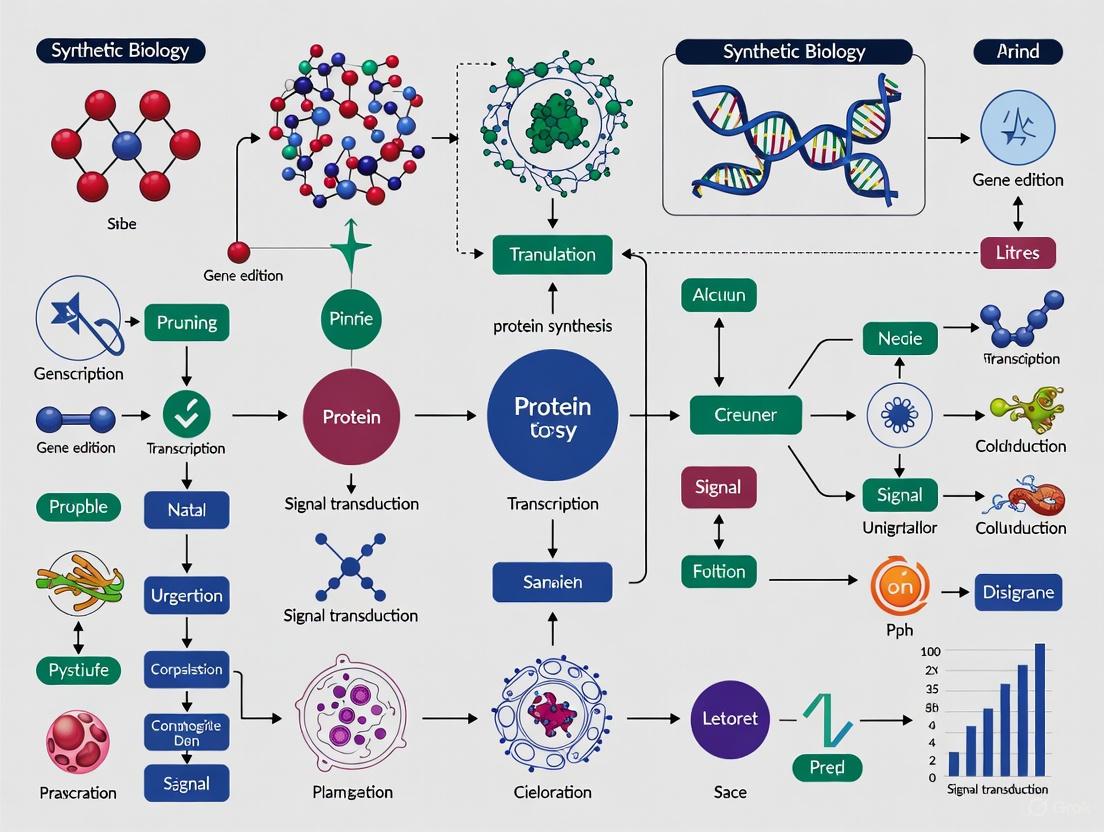
The Engineering Paradigm: Foundational Principles and Core Concepts of Synthetic Biology
The field of biological engineering has undergone a fundamental transformation, evolving from the precise but limited scope of genetic engineering to the comprehensive, systems-level approach of synthetic biology. While genetic engineering refers to the direct manipulation of an organism's DNA, historically limited to altering one or a few genes at a time, synthetic biology represents an entirely new paradigm founded on engineering principles [1]. This approach enables the design and construction of novel, nucleic-acid-encoded biological parts, devices, systems, and organisms for useful purposes by applying a rigorous Design-Build-Test-Learn framework [1]. The conceptual foundation comes from electrical engineering, where defined components with known structures and behaviors can be systematically assembled, much like capacitors and resistors on a circuit board [1].
This shift has been particularly transformative in biomedical engineering research, where synthetic biology now enables the programming of biological systems for advanced therapeutic applications. Unlike traditional genetic engineering, which modifies existing genetic blueprints, synthetic biology aims to create novel biological functions not found in nature through standardized, modular component design [1]. The advent of powerful gene-editing technologies, particularly CRISPR/Cas9, has accelerated this transition by providing researchers with the ability to quickly and efficiently make wholesale changes to an organism's DNA [1]. This technological revolution has created unprecedented opportunities for engineering biological systems to address complex challenges in drug development, diagnostic tools, and therapeutic interventions.
Core Principles of the Synthetic Biology Approach
The Engineering Framework for Biological Systems
Synthetic biology is distinguished from traditional genetic engineering by its foundational commitment to engineering principles. The field operates on a cyclic framework of Design, Build, Test, and Learn that enables iterative refinement of biological systems [1]. In the design phase, biological components are specified with standardized characteristics and interfaces. The build phase implements these designs using DNA construction techniques. The test phase rigorously characterizes system performance, and the learn phase extracts principles to inform subsequent design cycles. This systematic approach allows synthetic biologists to treat biological components as predictable, standardized parts that can be assembled into complex systems with defined functions.
The synthetic biology framework enables predictable system behavior through abstraction hierarchies that separate biological complexity into well-defined layers. At the lowest level, DNA parts (promoters, coding sequences, terminators) combine to form devices (logic gates, sensors, actuators), which in turn integrate into systems capable of complex functions. This hierarchical abstraction allows researchers to work at appropriate complexity levels without needing to manage all molecular details simultaneously. The resulting biological systems exhibit programmable functionality that can be directed to perform specific tasks, from environmental sensing to therapeutic production in response to disease biomarkers.
Foundational Technologies Enabling Synthetic Biology
Several key technological advances have enabled the transition from genetic engineering to synthetic biology. The development of high-throughput DNA synthesis and sequencing technologies has dramatically reduced the cost and time required to build and characterize genetic constructs. Automated laboratory platforms allow for parallel construction and testing of thousands of genetic designs, generating the data necessary to inform predictive models. These advances have been complemented by the development of computational tools for designing biological systems, including bioinformatics algorithms, molecular modeling software, and computer-aided design platforms specifically tailored for biological engineering.
The most transformative technology enabling modern synthetic biology has been the CRISPR/Cas9 system, which functions as programmable molecular scissors [1]. Unlike previous gene-editing technologies that required custom protein engineering for each target site, the CRISPR/Cas9 system uses guide RNA molecules to direct DNA cleavage, making genome editing dramatically faster, cheaper, and more precise [1]. This technology has opened up new possibilities for genetic modification, including in biomanufacturing, where comprehensive changes to an organism's DNA can substantially alter which chemical reactions that organism can perform and what it can produce [1]. The CRISPR system has evolved beyond simple gene editing to include advanced applications such as base editing, prime editing, and multiplexed genome regulation, enabling sophisticated genome-scale engineering that defines the synthetic biology approach [2].
Key Methodologies and Experimental Platforms
Computational and AI-Driven Design Tools
The synthetic biology approach leverages advanced computational tools throughout the Design-Build-Test-Learn cycle. Generative Artificial Intelligence has recently transformed enzyme design from structure-centric strategies toward function-oriented paradigms [2]. These emerging computational frameworks now span the entire design pipeline, including active site design, backbone generation, inverse folding, and virtual screening. For instance, density functional theory calculations can define the geometry of key catalytic components to guide the design of active sites (theozymes) that stabilize transition states [2]. Guided by these theozymes, GAI approaches such as diffusion and flow-matching models enable the generation of protein backbones pre-configured for catalysis.
Additional computational methods include inverse folding algorithms such as ProteinMPNN and LigandMPNN, which incorporate atomic-level constraints to optimize sequence-function compatibility [2]. To assess and optimize catalytic performance, virtual screening platforms such as PLACER allow evaluation of protein-ligand conformational dynamics under catalytically relevant conditions [2]. These computational advances are complemented by molecular dynamics simulations and quantum mechanical calculations, which serve as essential tools for investigating enzyme conformational dynamics and reaction mechanisms [2]. Through representative case studies, researchers have demonstrated how GAI-driven frameworks facilitate the rational creation of artificial enzymes with architectures distinct from natural homologs, thereby enabling catalytic activities not observed in nature [2].
Cell-Free Expression Systems for Rapid Prototyping
Ideal cell-free expression systems can theoretically emulate an in vivo cellular environment in a controlled in vitro platform, providing a versatile prototyping environment for synthetic biology [3]. These systems are particularly valuable because they offer a simplified in vitro representation of cellular processes without the complexity of growth and metabolism. The preparation and execution of an efficient endogenous E. coli based transcription-translation cell-free expression system can produce equivalent amounts of protein as T7-based systems at a 98% cost reduction to similar commercial systems [3].
Table 1: Key Components of TX-TL Cell-Free Expression System
| Component | Function | Specification |
|---|---|---|
| Crude Cell Extract | Provides transcriptional and translational machinery | BL21-Rosetta2 strain, 27-30 mg/ml protein concentration [3] |
| Energy Source | Fuels protein synthesis | 3-phosphoglyceric acid (3-PGA) - superior to creatine phosphate and phosphoenolpyruvate [3] |
| Reaction Buffer | Maintains optimal ionic conditions | Mg- and K- glutamate for increased efficiency [3] |
| DNA Template | Encodes desired genetic circuit | Compatible with both endogenous and T7 promoters [3] |
The entire protocol takes five days to prepare and yields enough material for up to 3000 single reactions in one preparation [3]. Once prepared, each reaction takes under 8 hours from setup to data collection and analysis. Mechanisms of regulation and transcription exogenous to E. coli, such as lac/tet repressors and T7 RNA polymerase, can be supplemented, while endogenous properties such as mRNA and DNA degradation rates can also be adjusted [3]. The resulting system has unique applications in synthetic biology as a prototyping environment or "TX-TL biomolecular breadboard" that allows for rapid testing of genetic designs before implementation in living cells [3].
Advanced Genome Editing Platforms
Modern synthetic biology leverages sophisticated multiplex genome editing platforms that enable coordinated modification of multiple genetic loci simultaneously. This approach is emerging as an ideal method for trait stacking to improve crops, functional genomics, and complex metabolic engineering in various biological systems [2]. The engineering and optimization of the latest CRISPR effectors for scalable and precise multiplex editing includes well-known systems like Cas9 and Cas12 variants, as well as newer, smaller variants such as CasMINI, Cas12j2, and Cas12k that offer advantages for delivery and packaging [2].
Table 2: CRISPR Systems for Multiplexed Genome Engineering
| CRISPR System | Editing Type | Key Applications |
|---|---|---|
| Cas9 | DSB, Nickase | Gene knockouts, large deletions |
| Cas12 Variants | DSB, Nickase | Multiplex editing, diagnostics |
| Base Editors | Chemical conversion | Point mutations without DSBs |
| Prime Editors | Reverse transcription | Precise insertions, deletions, substitutions |
| CasMINI | DSB | Delivery-constrained applications |
Central to any multiplexing approach are the expression and processing strategies of crRNA arrays, which include tRNA-based and ribozyme-mediated methods, synthetic modular designs, and AI-optimized guide RNAs tailored to diverse systems [2]. These editing technologies are complemented by next-generation delivery platforms such as lipid nanoparticles, virus-like particles, and metal-organic frameworks that overcome conventional barriers in in vivo applications [2]. Together, these advances enable precise, high-throughput, and programmable multiplex genome editing across biological systems, setting the foundation for future innovations in synthetic biology, crop improvement, and therapeutic intervention in multigene diseases [2].
Applications in Biomedical Engineering
Immunotherapy and Immune Cell Engineering
The application of synthetic biology principles to the immune system presents new mechanism-based avenues for reengineering immune responses, enabling precise control over temporally encoded cell-cell interactions, performing state-specific modulation of gene expression, and recording and responding to cellular experiences over time via programmable effector functions [4]. These capabilities represent major pillars for achieving the vision of modulating immune cell tropism, evading immune detection by engineered cells, and developing next-generation cell-based immunotherapies [4]. These advances are pivotal for tackling a large number of diseases and processes where the immune system plays a pivotal role in maintaining or disrupting organ homeostasis, from cancer to neurodegeneration to fibrosis and senescence [4].
Specific applications in immunological synthetic biology include engineering immune cells with enhanced specificity, functionality, and controllability, including improved sensing, homing, and effector capabilities [4]. Researchers are designing and developing synthetic bio-circuits to direct cell behavior, such as controlling immune cell tropism or tissue localization, supporting in situ detection of complex, multi-ligand patterns for the detection of emerging or pre-symptomatic diseases, and regulating immune cell states and responses in relation to T cell exhaustion, maintenance of self-tolerance, or T cell activation thresholds [4]. Additional approaches focus on constructing artificial or semi-synthetic immune systems for disease modeling, mechanistic discovery, and therapeutic screening, as well as developing modular systems for immune surveillance and non-invasive reporting, allowing engineered cells to communicate what they've sensed or responded to in real time [4].
Sustainable Bioproduction and Biomaterials
Synthetic biology enables the engineering of microbial platforms for sustainable bioproduction of valuable compounds. For instance, Bacillus methanolicus MGA3 is a methylotrophic bacterium with high potential as a production host in the bioeconomy, particularly with methanol as a feedstock [2]. Recent acceleration in strain engineering technologies through advances in transformation efficiency, the development of CRISPR/Cas9-based genome editing, and the application of genome-scale models for strain design have broadened the biotechnological potential of this thermophilic methylotroph [2]. With its expanding product portfolio, B. methanolicus demonstrates its potential as a microbial cell factory for the production of tricarboxylic acid cycle and ribulose monophosphate cycle intermediates and their derivatives [2].
In the realm of biomaterials, synthetic biology approaches are being applied to produce recombinant collagen that avoids the risks of pathogen transmission associated with animal-derived collagen [2]. Advances in AI-assisted protein engineering are accelerating the design of synthetic collagens and their applications in biomaterials [2]. These engineered biomaterials have crucial applications in biomedical fields such as drug delivery systems, cell culture matrices, and tissue engineering scaffolds [2]. The integration of computational design with biological production systems represents a hallmark of the synthetic biology approach to biomaterial development.
Essential Research Reagent Solutions
The implementation of synthetic biology methodologies requires specialized research reagents and tools. The following table summarizes key solutions essential for experimental work in this field.
Table 3: Essential Research Reagent Solutions for Synthetic Biology
| Category | Specific Solution | Research Application |
|---|---|---|
| Genome Editing | CRISPR-Cas9 systems [1], Base editors [2], Prime editors [2] | Targeted gene knock-out, knock-in, and precise sequence alteration |
| Delivery Platforms | Lipid nanoparticles [2], Virus-like particles [2], Metal-organic frameworks [2] | Efficient in vivo delivery of genetic constructs |
| Cell-Free Systems | E. coli TX-TL system [3], 3-PGA energy source [3] | Rapid prototyping of genetic circuits without cellular constraints |
| Computational Design | ProteinMPNN [2], PLACER [2], Molecular dynamics simulations [2] | In silico design and optimization of biological parts and systems |
| Model Organisms | Bacillus methanolicus [2], Escherichia coli [3] | Engineered microbial hosts for bioproduction and circuit implementation |
Visualizing Synthetic Biology Workflows
Design-Build-Test-Learn Cycle
The core engineering framework of synthetic biology follows an iterative Design-Build-Test-Learn cycle that enables continuous refinement of biological systems.
CRISPR-Mediated Multiplex Genome Editing
Modern synthetic biology utilizes advanced CRISPR systems for coordinated editing of multiple genetic loci.
Cell-Free Transcription-Translation System
Cell-free expression platforms provide a controlled environment for prototyping genetic circuits.
The transition from genetic engineering to biological systems engineering represents a fundamental shift in how we approach biological design. Synthetic biology has established itself as a distinct discipline characterized by its engineering-based framework, standardized biological parts, and systematic design principles. As the field continues to mature, several emerging trends are likely to shape its future development. The integration of artificial intelligence and machine learning throughout the Design-Build-Test-Learn cycle will accelerate the design of biological systems with increasingly complex functions [2]. The expansion of cell-free expression systems as prototyping platforms will enable more rapid characterization and optimization of genetic circuits before implementation in living cells [3]. Additionally, advances in multiplex genome editing will facilitate engineering of complex traits and metabolic pathways for therapeutic and industrial applications [2].
For biomedical engineering research, synthetic biology offers unprecedented opportunities to program biological systems for improved healthcare outcomes. The application of synthetic biology principles to immunology is already yielding novel approaches to disease detection and treatment [4]. As these technologies continue to develop, they will enable increasingly sophisticated therapeutic strategies, from engineered immune cells for cancer treatment to synthetic genetic circuits for managing metabolic diseases. The ongoing refinement of the synthetic biology toolkit—including more precise genome editors, improved delivery vehicles, and better computational models—will further enhance our ability to design and implement biological systems that address complex challenges in medicine and biotechnology. Through the continued application of engineering principles to biological design, synthetic biology promises to transform how we understand, manipulate, and ultimately utilize biological systems for human benefit.
Synthetic biology represents an interdisciplinary field that applies core engineering principles to biological systems, enabling the design and construction of new biological entities or the modification of existing ones [5]. This engineering-based approach aims to transform biology from a purely investigative science into a predictable engineering discipline, facilitating the rational design of biological systems with predefined and reliable functions. The foundational principles of standardization, modularity, and abstraction form the cornerstone of this paradigm shift, providing the necessary framework for managing biological complexity and accelerating the development of innovative solutions in biomedical engineering and therapeutic development [6].
These principles directly address the inherent complexity of biological systems, which has traditionally hindered predictable engineering outcomes. Standardization establishes universal measurement and assembly techniques, modularity enables the decomposition of complex systems into functional units, and abstraction creates hierarchical design layers that allow engineers to focus on specific system levels without being overwhelmed by underlying biological details [7]. The integration of these principles has created a powerful foundation for advancing biomedical applications, including engineered cellular therapies, diagnostic biosensors, and sustainable biomanufacturing platforms [8]. This whitepaper examines the technical implementation, experimental validation, and practical application of these core principles in synthetic biology research for biomedical contexts.
The Principle of Standardization
Standardization in synthetic biology establishes consistent frameworks for quantifying, assembling, and characterizing biological components. This principle is fundamental for enabling reproducible research, facilitating collaboration across laboratories, and creating predictable design workflows essential for biomedical applications [6].
Quantitative Measurement Standards
A critical aspect of standardization involves developing universal measurement units for biological components. The Relative Promoter Unit (RPU) system exemplifies this approach by quantifying promoter activity relative to a standardized reference promoter measured under identical experimental conditions [6]. This methodology enables meaningful comparisons of genetic part performance across different laboratories and experimental setups. Experimental data demonstrates that when properly implemented, the RPU approach maintains reasonable consistency, with promoters characterized via different measurement systems showing an average coefficient of variation (CV) of just 9% among technical replicates [6].
The experimental protocol for RPU measurement involves transforming a standardized genetic construct containing the test promoter into an appropriate host strain (commonly E. coli TOP10), growing cultures in defined media under specific conditions (typically 37°C with aeration), and measuring reporter gene output (e.g., fluorescence) during exponential growth phase using plate readers or flow cytometry. Data normalization is performed against both the reference promoter and appropriate blank controls to ensure accurate relative measurements.
Physical Assembly Standards
Standardized physical assembly methods create consistent techniques for constructing composite genetic systems. BioBrick assembly represents one such standardized framework that enables hierarchical construction of genetic devices from basic parts using a uniform assembly process [6]. This physical standardization allows researchers to share components and reliably combine them into larger functional systems. The table below summarizes key standardization frameworks in synthetic biology:
Table 1: Standardization Frameworks in Biological Design
| Standard Type | Implementation | Application | Experimental Consideration |
|---|---|---|---|
| Measurement Standard | Relative Promoter Units (RPU) | Promoter characterization | Requires reference promoter & controlled growth conditions |
| Physical Assembly | BioBrick assembly | Hierarchical DNA construction | Uses standardized prefix/suffix sequences |
| Data Exchange | Systems Biology Markup Language (SBML) | Computational model sharing | Ensures model reproducibility & interoperability |
| Functional Characterization | Standard biological parts registry | Part performance documentation | Must specify measurement conditions & host chassis |
The Principle of Modularity
Modularity in synthetic biology enables the creation of complex biological systems through the composition of functionally self-contained units or modules [6]. This principle allows engineers to design systems using well-characterized components whose behavior remains predictable when interconnected, mirroring the modular design approaches that have proven successful in other engineering disciplines.
Experimental Assessment of Modularity
Rigorous experimental studies have quantified the challenges and limitations of biological modularity. Research examining promoter activity when characterized via different biological measurement systems (varying plasmids, reporter genes, and ribosome binding sites) revealed that while most promoters showed consistent activity, some exhibited statistical differences (P<0.05) with coefficients of variation up to 22% across measurement systems [6]. This highlights the context-dependent nature of biological components and the importance of standardized measurement conditions.
Further investigations into modularity limitations tested promoter activity variation when independent expression modules were physically combined in the same system. Experiments constructing composite systems with two expression cassettes (GFP and RFP reporters) demonstrated that promoter activity could vary significantly (up to 35% CV) depending on contextual factors such as relative position and flanking sequences [6]. These findings underscore the importance of developing design rules that account for context-dependence when creating modular biological systems.
Functional Interconnection Testing
The modularity of input devices driving a common output device represents a crucial test for biological modularity. Experimental approaches have tested this principle by connecting different input modules (constitutive and regulated promoters) to a fixed output device (a genetic logic inverter) expressing GFP [6]. In a truly modular system, identical transcriptional input signals should produce identical GFP outputs regardless of the specific input device generating the signal. Experimental results revealed significant variability, with output variations up to 44% depending on the specific input device used, highlighting the challenges in achieving perfect modularity in biological systems [6].
Figure 1: Modularity Test Framework for Biological Devices. Different input devices (X1-XN) generating identical transcriptional signals should produce identical outputs when connected to a fixed output device in a truly modular system.
Abstraction creates hierarchical design layers that enable engineers to work at appropriate complexity levels without needing to manage all biological details simultaneously. This principle, borrowed from computer engineering, allows for the encapsulation of biological complexity within well-defined interfaces, dramatically accelerating the design process for sophisticated biological systems [7].
The abstraction hierarchy in synthetic biology typically follows multiple layers, from DNA sequences at the lowest level to full systems at the highest level. At each layer, specific design tools and methodologies facilitate the creation and integration of biological components. The emerging field of Bio-Design Automation (BDA) applies computational tools to streamline this hierarchical design process, creating software solutions that support the specification, design, building, testing, and learning phases of biological engineering [7].
Figure 2: Abstraction Hierarchy in Biological Design. This hierarchy enables engineers to work at appropriate complexity levels, with standardized interfaces between layers.
Bio-Design Automation Tools
Abstraction is implemented through specialized software tools that support biological design at different hierarchy levels. These Bio-Design Automation (BDA) tools create formal computational environments for designing, simulating, and optimizing biological systems [7]. The table below summarizes key BDA tools and their applications in the design workflow:
Table 2: Bio-Design Automation Tools for Abstract Biological Design
| Tool Name | Abstraction Level | Function | Application Example |
|---|---|---|---|
| Cello | Device/System | Compiles Verilog code to DNA sequences | Genetic logic circuit design [7] |
| Eugene | Part/Device | Rule-based specification language | Automated generation of biological devices [7] |
| GenoCAD | Part/Device | Gene design with grammatical rules | Constraint-based biological design [7] |
| Antimony | Device/System | Text-based model definition | Biochemical network modeling & simulation [7] |
| RBS Calculator | Part | Predicts translation initiation rates | Translation efficiency optimization [7] |
Integrated Experimental Framework
The synergistic application of standardization, modularity, and abstraction creates a powerful framework for engineering biological systems. This integrated approach enables the design and construction of sophisticated genetic devices with predictable behaviors, accelerating the development of biomedical solutions.
The Design-Build-Test-Learn Cycle
The core engineering workflow in synthetic biology follows an iterative Design-Build-Test-Learn (DBTL) cycle, where each iteration refines the biological design based on experimental data [7]. This cyclical process depends fundamentally on all three core principles: standardization ensures consistent experimental results, modularity enables the reuse and recombination of components, and abstraction facilitates model-based design and simulation. Advanced platforms like Phoenix now guide users through automated, iterative DBTL cycles, incorporating machine learning to continuously improve designs based on empirical data [7].
Experimental Protocol for Genetic Device Characterization
A standardized experimental protocol for characterizing genetic devices demonstrates the practical integration of all three principles:
Device Design: Create genetic constructs using standardized parts (Standardization) from modular libraries (Modularity) through abstract design tools (Abstraction).
Construct Assembly: Assemble devices using standardized assembly methods (Standardization) such as BioBrick assembly.
Transformation and Culturing: Introduce constructs into host chassis via electroporation or chemical transformation. Grow cultures in defined media (e.g., LB with appropriate antibiotics) at specified temperatures (e.g., 37°C for E. coli) with continuous shaking (250 rpm).
Measurement and Data Collection: Measure reporter outputs (fluorescence, absorbance) during exponential growth phase using plate readers or flow cytometry. Include appropriate controls (empty vector, reference standards).
Data Normalization and Analysis: Normalize data using the RPU framework (Standardization) and compare to model predictions (Abstraction). Evaluate device performance across different contexts (Modularity).
This integrated approach enables rigorous characterization of genetic devices while facilitating comparison and reuse across different research contexts.
Research Reagent Solutions
The practical implementation of synthetic biology principles requires specialized research reagents and tools. The following table details essential materials and their functions in biological design experiments:
Table 3: Essential Research Reagents for Biological Design Experiments
| Reagent/Category | Function | Example Implementation |
|---|---|---|
| Standard Biological Parts | Basic functional units for genetic construction | BioBrick parts in Registry of Standard Biological Parts [6] |
| Standardized Vectors | DNA backbones for part assembly | Plasmid systems with standardized prefixes/suffixes [6] |
| Reporter Proteins | Quantitative measurement of part activity | GFP, RFP with standardized measurement protocols [6] |
| Genome Engineering Tools | Precise genetic modification | CRISPR-Cas9, TALENs, ZFNs for chassis engineering [5] |
| Host Chassis | Cellular environment for device operation | E. coli strains (TOP10, KRX) with characterized behavior [6] |
| Characterization Tools | Quantitative measurement of system performance | Flow cytometers, plate readers, RPU measurement systems [6] |
Biomedical Applications and Future Directions
The application of standardization, modularity, and abstraction principles enables transformative biomedical applications, including engineered cellular therapies, diagnostic biosensors, and programmable living medicines [8]. These principles facilitate the development of sophisticated biological systems that can detect disease states, produce therapeutic responses, and interface with human physiology in precisely controlled ways.
Emerging trends include the development of virtual cells or digital cellular twins that create integrative computational models of cellular processes [9]. These models represent the ultimate expression of abstraction in biological design, enabling in silico prediction of cellular behavior before physical implementation. The integration of artificial intelligence with synthetic biology further enhances these capabilities, allowing for more accurate predictions and optimized designs [10].
As synthetic biology continues to mature, the core principles of standardization, modularity, and abstraction will remain fundamental to advancing biomedical engineering research. Through their consistent application, researchers can develop increasingly sophisticated biological systems that address complex challenges in therapeutics, diagnostics, and sustainable biomedicine.
The engineering of biological systems relies on a foundational hierarchy that organizes biological functionality into manageable levels of complexity: Parts, Devices, and Systems [11]. This structured framework allows researchers to apply engineering principles to biology, enabling the predictable design and construction of new biological functions. In the context of biomedical engineering, this hierarchy provides the toolbox for programming cells to perform therapeutic tasks, diagnose diseases, and produce valuable pharmaceuticals. Synthetic biology merges biology, engineering, and computer science to modify and create living systems, developing novel biological functions served by amino acids, proteins, and cells not found in nature [11]. This field creates reusable biological "parts," streamlining design processes and reducing the need to start from scratch, thus advancing biotechnology's capabilities and efficiency for biomedical applications [11].
Biological Parts: The Fundamental Building Blocks
Biological parts are the most basic functional units in synthetic biology. These standardized DNA-encoded components perform discrete biological functions and can be combined to form more complex structures.
- Promoters: DNA sequences where RNA polymerase binds to initiate transcription. Engineered promoters allow precise control over the timing and level of gene expression. In endogenous E. coli TX-TL systems, sigma70-based promoters with phage operators enable regulated expression [3].
- Protein Coding Sequences: DNA sequences that encode proteins, which can be endogenous or heterologous. These can be optimized for expression in different chassis organisms. For example, the pBEST-OR2-OR1-Pr-UTR1-deGFP-T500 plasmid expresses deGFP and can produce up to 0.75 mg/ml of reporter protein in cell-free systems [3].
- Ribosome Binding Sites (RBS): Sequences that facilitate the binding of ribosomes to mRNA, controlling translation initiation rates. RBS engineering is crucial for optimizing protein expression levels.
- Terminators: DNA sequences that signal the end of transcription, preventing read-through and ensuring proper transcription termination.
- Aptamers: Short nucleic acid sequences that bind specific ligands, providing a modular strategy for transcriptional regulation. When combined with switchable transcription terminators (SWTs), aptamers create ligand-responsive systems with improved precision and dynamic range [2].
Table 1: Characterization of Core Biological Parts
| Part Type | Primary Function | Key Parameters | Example Applications |
|---|---|---|---|
| Promoter | Transcription initiation | Strength, inducibility, specificity | Sigma70-based promoters for endogenous expression [3] |
| RBS | Translation initiation | Efficiency, strength | Optimizing protein expression levels |
| Aptamer | Ligand binding | Affinity, specificity | Ligand-responsive transcriptional regulation [2] |
| Terminator | Transcription termination | Efficiency | Switchable Transcription Terminators (SWTs) [2] |
| Protein Coding Sequence | Protein specification | Codon usage, stability | deGFP reporter (pBEST plasmid) [3] |
Devices: Functional Units for Biological Circuitry
Devices are functional assemblies of multiple biological parts that perform defined operations. They process inputs and generate outputs, creating basic logical functions within cells.
- Logic Gates: Genetic circuits that perform Boolean operations. SWTs have been used to build biological logic gates in vitro, including AND, NOT, NAND, and NOR gates [2]. These gates enable decision-making capabilities in engineered cells.
- Switchable Systems: Regulatory systems that toggle between states. The combination of aptamers with SWTs creates heterologous input logic gates that improve transcription activation by up to 3.3-fold for SWT and 7.84-fold for aptamer regulation compared to their individual performance [2].
- Sensors: Devices that detect environmental or intracellular signals. Quorum sensing (QS) systems, characterized by pathway-independence and autonomous control, can be engineered for population-level sensing and coordination in microbial communities [2].
- Expression Controllers: Devices that regulate gene expression output. Modular transcriptional regulation systems using SWTs and aptamers provide flexible and programmable strategies for fine-tuning gene expression [2].
The development of CRISPR-based technologies has transformed device engineering by enabling programmable regulation. Engineering and optimization of the latest CRISPR effectors, including well-known systems like Cas9 and Cas12 variants, to smaller variants such as CasMINI, Cas12j2, and Cas12k, have expanded the toolbox for creating sophisticated genetic devices [2].
Systems: Integrated Networks for Complex Functions
Systems represent the highest level of the hierarchy, integrating multiple devices to execute complex, coordinated behaviors with applications in therapeutic, diagnostic, and biomanufacturing contexts.
- Metabolic Pathways: Engineered systems for bioproduction. Bacillus methanolicus MGA3 has been engineered as a versatile thermophilic platform for sustainable bioproduction from methanol and alternative feedstocks [2]. The central carbon metabolism of B. methanolicus has been harnessed for various native and non-native carbon sources, expanding its product portfolio to include tricarboxylic acid (TCA) cycle and ribulose monophosphate (RuMP) cycle intermediates and their derivatives [2].
- Therapeutic Production Systems: Engineered organisms for pharmaceutical synthesis. Distributed biomanufacturing offers unprecedented production flexibility in both location and timing, enabling swift responses to sudden demands like disease outbreaks requiring specific medications [11].
- Biosensing Systems: Integrated circuits for diagnostic applications. The combination of aptamers with switchable transcription terminators creates modular, ligand-responsive systems with applications in biosensing and nucleic acid therapy [2].
- Electrocatalytic-Biosynthetic Hybrid Systems: Coupled systems for CO2 fixation. These systems synergize electrocatalytic CO2 reduction (which achieves C1/C2 products with high formation rates) with biosynthesis (which utilizes these C1/C2 species for carbon chain elongation) for efficient CO2 upcycling [2].
Table 2: Representative Synthetic Biology Systems and Applications
| System Type | Components | Function | Research Context |
|---|---|---|---|
| TX-TL Cell-Free System | Endogenous E. coli transcription-translation machinery, energy source, nucleotides | Biomolecular "breadboard" for circuit prototyping [3] | Protein expression, circuit characterization |
| Quorum Sensing System | AHL synthases, receptors, promoter elements | Population-density dependent gene regulation [2] | Coordinated behaviors, population control |
| CRISPR Multiplex Editing | Cas effectors, crRNA arrays, repair templates | Programmable genome editing at multiple loci [2] | Metabolic engineering, functional genomics |
| CO2 Fixation System | CO2-fixing enzymes, regeneration systems | Carbon conversion to valuable chemicals [2] | Sustainable biomanufacturing |
Experimental Protocols for Implementation
Protocol for Endogenous E. coli-Based TX-TL Cell-Free Expression
This protocol creates an efficient endogenous E. coli based transcription-translation (TX-TL) cell-free expression system that preserves native regulatory mechanisms while maintaining high protein expression capability [3].
Day 1: Preparation of Bacterial Culture
- Prepare bacterial culture media, culture plate, and media supplements as described.
- Streak BL21-Rosetta2 strain from -80°C onto a 2xYT+P+Cm agar plate.
- Incubate for at least 15 hours at 37°C or until colonies are visible.
Day 2: Culture Expansion and Reagent Preparation
- Prepare buffers and supplements including S30A buffer.
- Sterilize materials required for day 3.
- Prepare mini-culture 1: Add 4 ml of 2xYT+P media and 4 μl of Cm to a 12 ml sterile culture tube, pre-warm to 37°C for 30 minutes.
- Inoculate mini-culture 1 with a colony from the agar plate, incubate at 220 rpm, 37°C for 8 hours.
- 7.5 hours later, prepare mini-culture 2: Add 50 ml of 2xYT+P media and 50 μl of Cm to a sterile 250 ml Erlenmeyer flask, pre-warm to 37°C for 30 minutes.
- Inoculate mini-culture 2 with 100 μl of mini-culture 1, incubate at 220 rpm, 37°C for 8 hours.
Day 3: Cell Growth and Lysis
- Weigh four empty sterile 50 ml Falcon tubes and chill on ice.
- 7.5 hours after previous step, prepare final bacterial culture media: Transfer 660 ml of 2xYT+P media into each of six 4 L Erlenmeyer flasks, pre-warm to 37°C for 30 minutes.
- Add 6.6 ml of mini-culture 2 into each 4 L Erlenmeyer flask.
- Incubate at 220 rpm, 37°C until culture reaches OD₆₀₀ of 1.5-2.0 (mid-log growth phase).
- Transfer all cultures evenly into four 1 L centrifuge bottles, centrifuge at 5000 × g for 12 minutes at 4°C.
- Complete S30A buffer preparation by adding 4 ml of 1 M DTT to 2 L of previously prepared S30A buffer, maintain on ice.
- Remove supernatant, add 200 ml of S30A buffer at 4°C to each bottle, shake vigorously until pellet is completely solubilized.
- Centrifuge again at 5,000 × g for 12 minutes at 4°C.
- Remove supernatant, add 200 ml of S30A buffer, repeat centrifugation.
- After final centrifugation, estimate pellet weight and add 0.9 ml of S30A buffer per gram of pellet.
- Transfer suspension to bead-beater chamber, add 100 g of 0.1 mm glass beads.
- Lyse cells by bead-beating for 3 minutes, with 30-second intervals to prevent overheating.
- Centrifuge lysate at 12,000 × g for 10 minutes at 4°C.
- Transfer supernatant to fresh tubes, centrifuge again at 12,000 × g for 10 minutes at 4°C.
Day 4: Extract Clarification and Dialysis
- Transfer supernatant (crude extract) to dialysis cassettes.
- Dialyze against 2 L of S30B buffer for 3 hours at 4°C with stirring.
- Transfer dialyzed extract to fresh tubes, centrifuge at 12,000 × g for 10 minutes at 4°C.
- Aliquot supernatant, flash-freeze in liquid nitrogen, store at -80°C.
Day 5: TX-TL Reaction Assembly
- Prepare TX-TL reaction master mix on ice.
- For each 10 μl reaction: 4.5 μl crude cell extract, 1.5 μl of 10× reporter DNA (10-20 nM), 4 μl of reaction mix.
- Incubate reaction at 29°C for 8-14 hours, monitoring protein expression.
- The entire protocol yields enough material for approximately 3,000 single reactions with material costs of approximately $0.11 per 10 μl reaction [3].
Protocol for Engineering Bacillus methanolicus
The methylotrophic bacterium Bacillus methanolicus MGA3 represents a versatile thermophilic platform for sustainable bioproduction [2].
Genetic Tool Development:
- Implement advanced transformation protocols optimized for this thermophilic methylotroph.
- Apply CRISPR/Cas9-based genome editing systems specifically adapted for B. methanolicus.
- Utilize genome-scale models (GSMs) for informed strain design.
Metabolic Engineering:
- Engineer central carbon metabolism for expanded substrate utilization including methanol, mannitol, arabitol, glucose, starch, and xylose.
- Modify metabolic pathways for production of target compounds: TCA cycle intermediates, RuMP cycle intermediates, and their derivatives.
- Optimize expression of heterologous proteins leveraging the organism's capacity for producing thermostable proteins.
Cultivation and Characterization:
- Cultivate engineered strains under controlled conditions with temperature optimization for thermophilic growth.
- Monitor biomass accumulation and product formation.
- Analyze metabolic fluxes and pathway performance using appropriate analytical methods.
Visualization of Synthetic Biology Workflows
The Scientist's Toolkit: Essential Research Reagents and Materials
Table 3: Essential Research Reagents for Synthetic Biology
| Reagent/Material | Function | Example Application | Technical Notes |
|---|---|---|---|
| BL21-Rosetta2 E. coli Strain | Protein expression host with rare tRNA supplementation | Cell-free extract preparation for TX-TL systems [3] | Contains plasmid encoding rare tRNAs; selected with chloramphenicol |
| S30A and S30B Buffers | Cell lysis and dialysis buffers | Crude cell extract preparation and dialysis [3] | Contains Tris-acetate, magnesium and potassium glutamate, DTT |
| 2xYT+P Media | Rich bacterial growth medium | Culture growth for cell extract preparation [3] | Contains yeast extract, tryptone, NaCl, phosphate buffer |
| 3-Phosphoglyceric Acid (3-PGA) | Energy source for cell-free reactions | ATP regeneration in TX-TL systems [3] | Superior protein yields compared to creatine phosphate and phosphoenolpyruvate |
| Mg-/K-Glutamate | Salt components in reaction buffer | Enhancing efficiency in TX-TL systems [3] | Replacement for Mg-/K-acetate in previous protocols |
| Switchable Transcription Terminators (SWTs) | Programmable transcriptional regulation | Construction of logic gates and regulatory circuits [2] | Low leakage expression, high ON/OFF ratios |
| Aptamers | Ligand-binding nucleic acid elements | Modular transcriptional regulation [2] | Can be combined with SWTs for improved regulation |
| CRISPR Effectors (Cas9, Cas12 variants) | Genome editing tools | Multiplex genome editing, metabolic engineering [2] | Includes newer, smaller variants (CasMINI, Cas12j2, Cas12k) |
| Plasmid DNA Templates | Genetic information carriers | Protein expression in cell-free and cellular systems [3] | e.g., pBEST-OR2-OR1-Pr-UTR1-deGFP-T500 for deGFP expression |
Future Directions and Concluding Remarks
The field of synthetic biology continues to evolve with several emerging trends shaping its future. Generative Artificial Intelligence (GAI) is transforming enzyme design from structure-centric strategies toward function-oriented paradigms [2]. Computational frameworks now span the entire design pipeline, including active site design, backbone generation, inverse folding, and virtual screening. GAI approaches such as diffusion and flow-matching models enable the generation of protein backbones pre-configured for catalysis, while inverse folding methods incorporate atomic-level constraints to optimize sequence-function compatibility [2].
Multiplex genome editing technologies are advancing rapidly, enabling precise, high-throughput editing across biological systems [2]. The development of base editors and prime editors allows efficient editing across multiple loci without double-strand breaks, setting the foundation for future innovations in synthetic biology, crop improvement, and therapeutic intervention in multigene diseases [2].
The integration of electrocatalysis and biotransformation represents another promising direction for CO2-based biomanufacturing [2]. These hybrid systems synergize the advantages of electrocatalytic CO2 reduction (which achieves C1/C2 products with high formation rates) with biosynthesis (which utilizes these C1/C2 species for carbon chain elongation) for efficient CO2 upcycling [2].
As synthetic biology matures, the parts-devices-systems hierarchy continues to provide a foundational framework for engineering biology. The ongoing development of standardized biological components, coupled with advanced computational tools and experimental methodologies, promises to accelerate the design-build-test-learn cycle and expand the applications of synthetic biology in biomedical research and therapeutic development.
Synthetic biology has emerged as a formal engineering discipline that utilizes concepts from genetics, biophysics, and molecular biology to repurpose natural biological systems for applications in biomedicine and biotechnology [12] [13]. At the core of this field lie genetic circuits—sophisticated assemblies of biological components that process and manipulate information within living cells to create novel, useful biological functions [14]. These circuits enable researchers to program cells with new capabilities, much like electronic circuits enable computers to perform complex operations. The engineering of these circuits has progressed from simple proof-of-concept designs to complex platforms capable of Boolean logic, memory storage, and oscillatory behavior, with significant implications for therapeutic development, diagnostic tools, and biomanufacturing [15] [13].
The design-build-test-learn cycle represents the fundamental engineering framework in synthetic biology, though it faces unique challenges when applied to biological systems. Unlike conventional engineering substrates, biology presents distinctive challenges stemming from incomplete understanding of natural systems and limitations in manipulation tools [13]. Furthermore, circuit functionality is profoundly influenced by cellular context, including signal noise, metabolic dynamics, and stability considerations [15]. Despite these challenges, synthetic biology is advancing toward rational and high-throughput biological engineering through developing core platforms that span the entire biological design cycle, including DNA construction, parts libraries, computational design tools, and interfaces for manipulating and probing synthetic circuits [13].
This technical guide examines the fundamental principles of three core genetic circuit types—switches, oscillators, and Boolean logic systems—within the context of biomedical engineering research. By providing both theoretical foundations and practical implementation methodologies, we aim to equip researchers and drug development professionals with the knowledge necessary to leverage these powerful tools in their work.
Boolean Logic Systems in Genetic Circuits
Fundamental Principles and Implementation
Boolean logic gates form the computational foundation of genetic circuits, enabling cells to perform decision-making operations based on molecular inputs. These gates process one or more input signals to produce specific outputs according to logical rules, with the NOR gate being particularly significant as any logic function can be implemented through NOR gates alone [15]. In biological terms, inputs typically consist of small molecule inducers or environmental signals, while outputs are often reporter proteins or metabolic changes.
The implementation of Boolean logic in biological systems differs fundamentally from electronic computing. Genetic logic gates operate through biochemical reactions of gene expression that simulate on and off states via protein concentrations, using DNA-binding proteins and transcription factors to express protein concentration for specific logical functions [16]. A typical genetic NOR gate, for instance, might be designed with inputs such as arabinose (Ara) and anhydrotetracycline (aTc), with yellow fluorescent protein (YFP) serving as the output reporter [15]. The gate produces output (YFP expression) only when both inputs are absent, following the NOR truth table.
The mathematical foundation for modeling these systems relies heavily on nonlinear Hill dynamics, which describe the binding of transcription factors to DNA promoter regions. For a genetic NOT gate, the promoter activity function can be described as:
$$f_{\text{NOT}}(u) = \frac{1}{1 + \left(\frac{u}{K}\right)^n}$$
where $u$ represents the concentration of repressor transcription factor, $K$ is the Hill constant, and $n$ is the Hill coefficient representing cooperativity [16]. Similarly, an AND gate with two inputs follows:
$$f{\text{AND}}(u1, u2) = \frac{\left(\frac{u1}{K1}\right)^{n1} \left(\frac{u2}{K2}\right)^{n2}}{1 + \left(\frac{u1}{K1}\right)^{n1} + \left(\frac{u2}{K2}\right)^{n2} + \left(\frac{u1}{K1}\right)^{n1} \left(\frac{u2}{K2}\right)^{n_2}}$$
More complex gates can be constructed through modular combinations. A NAND gate, for example, can be implemented by cascading AND and NOT gates, with the dynamic equation:
$$\begin{aligned} \dot{p}{\text{AND}} &= \alphaP f{\text{AND}}(u1, u2) - \gammaP p{\text{NAND}} + \alpha{P0, \text{AND}}, \ \dot{p}{\text{NOT}} &= \alphaP f{\text{NOT}}(p{\text{AND}}) - \gammaP p{\text{NOT}} + \alpha{P0, \text{NOT}}, \ p{\text{AND}}(u1, u2) &= p_{\text{NOT}} \end{aligned}$$
where $p$ represents protein concentrations, $\alpha$ denotes production rates, and $\gamma$ signifies degradation rates [16].
Advanced Boolean Circuit Designs
Recent research has demonstrated increasingly sophisticated Boolean implementations. A 2019 study successfully constructed 12 circuit logic gate modules in Escherichia coli, including "AND," "NAND," "OR," and "NOR" gates validated through reporter gene expression [17]. These circuits converted inputs into outputs via intermediate products of host metabolism, showcasing the potential for integrating synthetic circuits with native cellular processes.
Plant synthetic biology has also seen significant advances, with researchers establishing a predictive framework for genetic circuit design in both Arabidopsis thaliana and Nicotiana benthamiana [14]. This platform enabled the construction of 21 two-input genetic circuits with various logic functions (14 types) achieving high prediction accuracy (R² = 0.81), demonstrating the scalability of Boolean approaches in complex eukaryotic systems.
Table 1: Performance Characteristics of Genetic Boolean Gates
| Gate Type | Organism | Input Signals | Output Signal | Fold Change | Response Time | Reference |
|---|---|---|---|---|---|---|
| NOR | E. coli | Ara, aTc | YFP | ~50 | ~60 min | [15] |
| AND | E. coli | Multiple TF | Fluorescence | ~40 | ~90 min | [16] |
| NAND | E. coli | PhlF, IcaR | Luciferase | ~30 | ~120 min | [16] |
| NOT | Plant | Auxin | Luciferase | ~40 | ~10 hours | [14] |
| AND | Plant | CK, Auxin | Luciferase | ~25 | ~12 hours | [14] |
Figure 1: Genetic NOR Gate Circuit. The circuit outputs YFP only when both Arabinose and aTc inputs are absent, implementing Boolean NOR logic [15].
Genetic Switches and Oscillators
Bistable Switches and Memory Systems
Genetic toggle switches represent a fundamental class of bistable systems that can maintain one of two stable states indefinitely, functioning as biological memory storage devices. These switches typically comprise two mutually repressing genes, creating a system that can be toggled between states by transient external signals. Once switched, the circuit maintains its state even after the inducing signal is removed, enabling permanent memory storage at the cellular level [15] [13].
The mathematical foundation for bistable switches builds upon the dynamics of mutually inhibitory genes. The system can be described by coupled differential equations representing the production and degradation of the two repressor proteins:
$$\begin{aligned} \frac{dp1}{dt} &= \alpha1 \cdot f{\text{NOT}}(p2) - \gamma1 p1 \ \frac{dp2}{dt} &= \alpha2 \cdot f{\text{NOT}}(p1) - \gamma2 p2 \end{aligned}$$
where $p1$ and $p2$ represent the concentrations of the two repressor proteins, $\alphai$ are their production rates, $\gammai$ are degradation rates, and $f_{\text{NOT}}$ represents the repression function [16]. Bistability occurs when specific parameter combinations yield two stable steady states separated by an unstable equilibrium.
Implementation of robust toggle switches requires careful balancing of kinetic parameters and consideration of cellular context. Key design challenges include minimizing metabolic burden, ensuring orthogonal components to prevent crosstalk with native systems, and maintaining stability across cell divisions. Successful implementations have been demonstrated in various host organisms including bacteria, yeast, and mammalian cells, with applications ranging from long-term cellular memory to lineage tracing in development [15].
Oscillatory Systems and Biological Clocks
Genetic oscillators generate periodic waveforms in protein concentrations, enabling biological timing functions comparable to electronic clocks. The repressilator, first synthesized by Elowitz and Leibler in 2000, represents a landmark achievement in synthetic biology—a three-gene ring network where each gene represses the next in the cycle [16]. This circular topology creates inherent time delays in transcription, translation, and degradation that can sustain oscillations under appropriate parameter conditions.
The dynamics of oscillatory systems are typically modeled using extended versions of the basic gene expression equations:
$$\begin{aligned} \dot{m}i &= \alphai fi(p{i-1}) - \gamma{mi} mi + \alpha{i,0} \ \dot{p}i &= \betai mi - \gamma{pi} pi \end{aligned}$$
where $mi$ and $pi$ represent mRNA and protein concentrations respectively for gene $i$, $\alphai$ and $\betai$ are production rates, $\gamma{mi}$ and $\gamma{pi}$ are degradation rates, $\alpha{i,0}$ represents basal expression, and $fi$ describes the repression function [16].
More recent oscillator designs have incorporated additional regulatory layers such as post-translational control, phosphorylation cascades, and intercellular signaling to enhance robustness and tunability. Applications include programmed drug delivery systems that release therapeutics at specific times, synthetic cell cycle controllers, and synchronization devices for coordinating population-level behaviors [13] [16].
Table 2: Genetic Switch and Oscillator Performance Parameters
| Circuit Type | Host Organism | Number of Components | Switching Time | Stability | Applications |
|---|---|---|---|---|---|
| Toggle Switch | E. coli | 2 repressors, 2 promoters | 2-3 hours | >50 generations | Cellular memory, decision making |
| Repressilator | E. coli | 3 repressors, 3 promoters | 2-4 hour period | ~10 cycles | Gene expression timing, rhythm generation |
| Dual-feedback Oscillator | E. coli | 2 repressors, activators | 1-3 hour period | >40 cycles | Programmable drug delivery |
| CRISPRi Switch | Mammalian cells | 1 dCas9, sgRNA | 12-24 hours | >2 weeks | Gene therapy, cell state control |
Figure 2: Repressilator Oscillator Circuit. A three-gene ring network where each gene represses the next, creating sustained oscillations in protein concentrations [16].
Experimental Methodology and Characterization
Quantitative Characterization Framework
Robust quantitative characterization represents a critical requirement for predictable genetic circuit design. In plant systems, researchers have established a rapid (~10 days), reproducible framework based on the concept of relative promoter units (RPUs) to normalize measurements across experimental batches [14]. This approach selects a reference promoter (e.g., the 200-bp 35S promoter) and defines its activity as 1 RPU within each protoplast batch, converting raw measurements to standardized units that enable comparative analysis across setups.
The experimental pipeline typically incorporates a normalization module featuring a constitutively expressed reference protein (e.g., β-glucuronidase, GUS) alongside the circuit output reporter (e.g., firefly luciferase, LUC). The LUC/GUS ratio provides normalized values that significantly reduce variation. For a genetic circuit with output $O$ and reference signal $R$, the RPU value is calculated as:
$$\text{RPU} = \frac{O{\text{sample}} / R{\text{sample}}}{O{\text{reference}} / R{\text{reference}}}$$
This normalization strategy has demonstrated substantial improvement in measurement reproducibility, enabling predictive circuit design with high accuracy (R² = 0.81 for plant circuits) [14].
Modular Part Design and Engineering
Expanding the repertoire of well-characterized biological parts is essential for sophisticated circuit construction. Modular synthetic promoters represent a key advancement, enabling predictable integration of regulatory functions. A common design approach uses a strong constitutive promoter (e.g., the 200-bp 35S promoter in plants) as a backbone, with specific operator sequences inserted at strategic positions to create repressible promoters [14].
Design optimization involves systematic testing of operator positions to maximize dynamic range while maintaining sufficient basal expression. Research has shown that placing operators between CAAT boxes and the transcription start site typically achieves improved fold-repression [14]. A library of synthetic promoter-repressor pairs using TetR family repressors (LmrA, PhlF, IcaR, BM3R1, SrpR, and BetI) demonstrated fold-repression ranging from 4.3 (IcaR) to 847 (PhlF), with high orthogonality minimizing crosstalk between components [14].
Table 3: Research Reagent Solutions for Genetic Circuit Implementation
| Reagent/Category | Specific Examples | Function/Application | Key Characteristics |
|---|---|---|---|
| Repressor Proteins | TetR, PhlF, IcaR, LmrA, BM3R1 | Transcriptional regulation of synthetic circuits | Orthogonal, high dynamic range, minimal crosstalk |
| Synthetic Promoters | 35S-derived promoters with operator inserts | Context-dependent gene expression control | Modular, tunable strength, inducible/repressible |
| Reporter Systems | YFP, Luciferase, GUS | Quantitative circuit output measurement | High sensitivity, broad dynamic range, minimal toxicity |
| Sensor Systems | Auxin sensor (GH3.3), cytokinin sensor (TCSn) | Detection of small molecule inputs | High sensitivity, specific ligand recognition |
| DNA Assembly Systems | Golden Gate, Gibson Assembly | Physical construction of genetic circuits | High efficiency, modular, standardized |
Network Analysis Approaches for Circuit Design
Network Visualization and Analysis
As genetic circuits increase in complexity, network approaches provide powerful methods for structuring, analyzing, and visualizing design data. Converting circuit designs into network representations creates dynamic structures that can be interactively shaped into subnetworks based on specific requirements such as biological part hierarchy or molecular interactions [15]. This approach enables automatic scaling of abstraction levels, tailoring visualizations to particular analysis needs through coloring or clustering nodes based on types (e.g., genes, promoters, proteins).
A significant advantage of network representations is their ability to integrate diverse data types beyond genetic sequences alone, including circuit modularity, functional details, implementation instructions, dynamical predictions, and validation strategies [15]. Knowledge graphs—structured directed graphs where nodes and edges contain semantic labels—have proven particularly valuable for biological applications, enabling complex control over underlying data and arrangement into multiple abstraction layers.
Standardized data formats like Synthetic Biology Open Language (SBOL) facilitate this network-based approach by describing both structural (e.g., DNA sequences) and functional (e.g., regulation interactions) information [15]. The transformation of design files into networks follows a systematic process: initial conversion into an intermediate data structure compatible across formats, followed by application of graph theory methods to calculate shortest paths between entities, identify clusters, and find intersections within the design [15].
Design Principles for Biological Network Figures
Effective visualization of genetic circuits as networks requires careful consideration of design principles. First, determining the figure's purpose represents a critical initial step—whether to convey network functionality, structure, or specific interactions [18]. For functional relationships, data flow encodings with nodes connected by arrows effectively illustrate interaction cascades, while undirected edges better represent structural associations.
Alternative layouts beyond conventional node-link diagrams may enhance clarity for specific data types. Adjacency matrices excel with dense networks, enabling clear visualization of edge attributes through cell coloring and readable node labels without clutter [18]. For hierarchical circuits, fixed layouts or circular arrangements may provide more intuitive representations.
Visual encoding choices significantly impact interpretation. Quantitative color schemes (e.g., yellow to green gradations) effectively represent continuous values like expression variance, while divergent color schemes (e.g., red to blue) emphasize extreme values of differential expression [18]. Consistent application of these principles ensures network visualizations effectively communicate the intended story of the genetic circuit design.
Figure 3: Network Abstraction Hierarchy. Genetic circuit designs can be represented at multiple abstraction levels, from complete networks with all metadata to simplified input/output relationships [15].
Applications in Biomedical Engineering and Therapeutics
Genetic circuits offer transformative potential for biomedical applications, particularly in therapeutic development and delivery. In diagnostic applications, biosensing circuits programmed at transcriptional, translational, and post-translational levels can detect disease-specific biomarkers and trigger appropriate responses [12]. These systems enable identification of disease mechanisms and drug targets, screening for new therapeutics, and developing improved delivery methods.
Gene therapies represent a particularly promising application area, with circuits designed to correct disease-causing genetic mutations [12]. Research has demonstrated innovative approaches for conditions like Duchenne Muscular Dystrophy, where synthetic circuits could potentially restore normal function. Additionally, synthetic biology is expanding therapeutic platforms by creating biological devices that function as therapies themselves, moving beyond traditional small-molecule approaches [12].
Biocomputing represents another frontier, with researchers working toward complete biological computers through integration of control units (CU) with arithmetic/logic units (ALU) implemented via genetic circuits [16]. These systems adapt the fetch-decode-execute cycle of conventional computers to biological contexts, using genetic logic gates to process information and direct cellular operations. While still emerging, this approach could enable sophisticated decision-making capabilities within therapeutic cells.
The future of genetic circuits in biomedical engineering will likely focus on enhancing predictability across biological contexts, improving scalability, and developing standardized frameworks for clinical translation. As the field addresses these challenges, genetic circuits will play an increasingly important role in creating next-generation biomedical solutions.
The field of synthetic biomaterials represents a paradigm shift in biomedical engineering, merging the programmability of synthetic biology with the functional sophistication of materials science. This convergence has enabled the creation of intelligent, self-assembling systems that operate within biological environments to direct cellular behavior, deliver therapeutics, and regenerate tissues. These systems mark a significant evolution from static, inert biomaterials to dynamic, responsive interfaces that engage in reciprocal communication with biological systems [19] [20]. The foundational principle hinges on engineering biomolecules that spontaneously organize into predetermined nanostructures and macroscopic materials based on encoded information within their building blocks.
This technical guide examines the core principles, design methodologies, and applications of synthetic self-assembling biomaterials, framing them within the broader context of synthetic biology. For researchers and drug development professionals, mastering this interface offers unprecedented control over biological interactions, paving the way for next-generation diagnostic and therapeutic platforms. The materials discussed herein are characterized by their bioactivity, adaptability, and often, their biomimetic nature, drawing inspiration from the self-assembling structures ubiquitous in biology, such as the extracellular matrix, bacterial pili, and viral capsids [20] [21].
Fundamental Principles of Molecular Self-Assembly
Self-assembly in biological and synthetic contexts is governed by the spontaneous organization of molecular components into stable, ordered structures through non-covalent interactions. The engineering of these systems requires a deep understanding of the forces and rules that dictate this process.
Key Driving Forces and Molecular Interactions
The stability and structural fidelity of self-assembled biomaterials arise from a delicate balance of several weak, non-covalent interactions. The table below summarizes these fundamental forces.
Table 1: Fundamental Molecular Interactions in Biomaterial Self-Assembly
| Interaction Type | Energy Range (kJ/mol) | Role in Self-Assembly | Examples in Biomaterials |
|---|---|---|---|
| Hydrophobic Effect | <5 to >50 | Drives amphiphilic molecules to sequester hydrophobic domains away from water, a primary driver for micelle, vesicle, and fibril formation. | Peptide amphiphiles, phospholipid bilayers [21]. |
| Electrostatic Interactions | 5-250 | Occurs between charged amino acid side chains (e.g., Lys/Arg vs Asp/Glu); enables responsiveness to pH and ionic strength. | Self-complementary ionic peptides (e.g., EAK16, RADA16) [20] [21]. |
| Hydrogen Bonding | 4-60 | Creates directional bonds between carbonyl and amide groups, critical for forming secondary structures (β-sheets, α-helices) and fibrillar networks. | β-sheet fibrils in peptide hydrogels [21]. |
| π-π Stacking | 0-50 | Contributes to the self-assembly of aromatic residues (e.g., F, F, W); often works cooperatively with other forces. | Diphenylalanine (FF) peptide nanotubes [21]. |
| Van der Waals Forces | 0.5-5 | Weak, non-specific attractions between electron clouds of adjacent atoms; contribute to packing within assemblies. | Molecular packing in crystalline domains of materials. |
Biomimicry as a Design Framework
A powerful strategy in designing self-assembling systems is to emulate structures and processes found in nature. Natural biomolecular systems, such as the extracellular matrix (ECM), provide a blueprint for creating synthetic materials that can effectively interact with biology [22]. The native ECM is a highly dynamic and complex network that provides not only structural support but also biochemical signals to cells. Modern biomaterial design seeks to replicate this multifunctionality. Key biomimetic approaches include:
- Architectural Mimicry: Designing peptides that form fibrillar networks resembling collagen or fibronectin matrices to support cell adhesion and growth [20] [22].
- Sequential Mimicry: Incorporating short peptide sequences (e.g., RGD for cell adhesion) from native ECM proteins into synthetic polymers to confer specific bioactivity [22].
- Dynamic Mimicry: Engineering materials that can sense and respond to environmental cues (e.g., enzymes, pH, light) much like living tissues, using mechanisms such as reversible cross-linking or programmable degradation [20].
Engineering Strategies for Synthetic Biomaterials
The creation of advanced biomaterials leverages a diverse toolkit from both synthetic biology and materials science, enabling precise control from the molecular to the macroscopic scale.
Genetic Engineering of Cellular Systems
Synthetic biology provides the tools to reprogram living cells as production factories for protein-based biomaterials. This involves engineering genetic circuits to control protein expression, folding, and even secretion.
Figure 1: Genetic circuit for biomaterial production.
Cells can be engineered to perceive specific inputs (biological, chemical, or physical) using synthetic receptors and optogenetic tools [20]. This information is processed by synthetic gene networks—ranging from simple Boolean logic gates to complex CRISPR-based recording systems—which then trigger the expression of output proteins. These outputs can be structural proteins (e.g., engineered silk, elastin) or enzymes that catalyze the formation of biomaterials [20] [8]. This approach allows for the production of complex, multifunctional materials directly from engineered cellular systems.
Peptide and Polymer-Based Self-Assembly
A bottom-up approach involves the de novo design and synthesis of self-assembling peptides and synthetic polymers. This field has expanded significantly, exploring chemical and sequence space beyond that used by biology to create novel functional materials [21].
- Self-Complementary Peptides: These peptides are designed with alternating polar and non-polar amino acids to form stable β-sheet membranes in salt solutions [21]. A classic example is the RADA16-I peptide (Ac-(RADA)4-CONH2), which forms a nanofibrous hydrogel that supports cell attachment and proliferation.
- Peptide Amphiphiles: Molecules consisting of a hydrophobic alkyl tail covalently linked to a hydrophilic peptide head. In water, they self-assemble into cylindrical nanofibers, with the peptide segments displayed on the surface to present bioactive epitopes [21].
- Stimuli-Responsive Peptides: These are engineered to undergo self-assembly in response to specific environmental triggers, such as a change in pH, the presence of a specific enzyme, or light exposure [20]. This allows for spatiotemporally controlled material formation in situ.
Experimental Protocols and Characterization
Robust experimental methodologies are essential for the design, fabrication, and validation of self-assembling biomaterials.
Protocol: Fabrication of a β-Sheet Peptide Hydrogel
This protocol outlines the standard procedure for creating a self-supporting hydrogel from a β-sheet forming peptide, such as RADA16-I [21].
- Peptide Solution Preparation: Dissolve the purified peptide in deionized water at a concentration of 1% (w/v). The solution will have a low viscosity and acidic pH.
- Sonication: Sonicate the solution for 10-30 minutes to break up any pre-existing aggregates and ensure a monomeric or oligomeric starting state.
- Gelation Induction:
- Ionic Trigger: Add a calculated volume of phosphate-buffered saline (PBS) or cell culture medium to the peptide solution to achieve a final salt concentration of ~150 mM. Gently pipette to mix. The gel will form within seconds to minutes.
- pH Trigger: Alternatively, gently add an equimolar amount of NaOH or buffer to neutralize the acidic peptide solution.
- Incubation: Allow the gel to incubate at room temperature or 37°C for 1 hour to complete the assembly process and stabilize the hydrogel network.
- Characterization:
- Microscopy: Analyze the gel's nanofibrous morphology using Scanning Electron Microscopy (SEM) or Atomic Force Microscopy (AFM).
- Spectroscopy: Confirm β-sheet formation using Fourier-Transform Infrared Spectroscopy (FTIR), specifically monitoring the amide I band for a shift to ~1630 cm⁻¹.
- Rheology: Perform oscillatory rheology to measure the mechanical properties (storage modulus G' and loss modulus G") of the hydrogel.
Quantitative Analysis of Biomaterial Properties
The in vivo performance of a biomaterial is dictated by its physico-chemical properties. The following table synthesizes quantitative data linking specific material characteristics to the biological outcome of bone formation, as identified in an empirical model [23].
Table 2: Empirical Model Linking Biomaterial Properties to Intra-Oral Bone Formation
| Biomaterial Property | Measurement Technique | Optimal Range for Bone Formation | Impact on Biological Response |
|---|---|---|---|
| Surface Roughness | Atomic Force Microscopy (AFM) | Higher roughness (Sa > 1.2 µm) | Significantly enhances osteointegration and bone deposition compared to smooth surfaces [23]. |
| Chemical Composition | X-ray Diffraction (XRD) | Hydroxyapatite (HAp) & Biphasic Calcium Phosphate (BCP) | HAp promotes osteoconductivity; BCP (HAp/β-TCP) offers tunable degradation and bioactivity [23]. |
| Macroporosity | Micro-Computed Tomography (μCT) | Pore diameter > 100 µm, Interconnected | Critical for facilitating osteogenesis and angiogenesis by allowing cell migration and nutrient diffusion [23]. |
| Microporosity | Mercury Porosimetry | Pore diameter < 10 µm | Increases specific surface area, enhancing protein adsorption and influencing early inflammatory response [23]. |
Applications in Therapeutics and Diagnostics
The programmability and dynamic nature of synthetic biomaterials unlock a wide array of applications in biomedicine, particularly as therapeutic and imaging agents [19].
Therapeutic Agents
- Targeted Drug Delivery: Self-assembling systems can be designed to encapsulate therapeutic molecules (e.g., chemotherapeutics, antibiotics) and release them in response to disease-specific stimuli, such as elevated enzyme activity or acidic pH in the tumor microenvironment [19] [21].
- Regenerative Medicine: Peptide and protein hydrogels serve as synthetic extracellular matrices (ECMs) for 3D cell culture and tissue engineering. These materials can be functionalized with adhesive ligands and growth factors to direct stem cell differentiation and promote the repair of bone, cartilage, and neural tissues [20] [23] [8].
- Immunotherapy: Biomaterials can be used to deliver immunomodulatory signals (e.g., cytokines, antigens) to immune cells in vivo. For instance, self-assembling peptide vaccines display tumor antigens to efficiently activate the immune system [8].
Imaging and Diagnostics
- Contrast Agents: Supramolecular assemblies can carry high payloads of imaging agents (e.g., gadolinium for MRI, fluorescent dyes for optical imaging). Their ability to accumulate at disease sites through enhanced permeability and retention (EPR) or active targeting improves imaging sensitivity and specificity [19].
- Biosensors: Engineered biomaterials that undergo a macroscopic change (e.g., sol-gel transition, fluorescence emission) in the presence of a specific disease biomarker (e.g., protease, mRNA) can be used for point-of-care diagnostics and monitoring disease progression [8].
The Scientist's Toolkit: Essential Research Reagents
The following table catalogues key reagents and their functions essential for research in synthetic biomaterials.
Table 3: Essential Research Reagents for Engineering Self-Assembling Biomaterials
| Reagent / Material | Function and Utility in Research |
|---|---|
| BioBricks (iGEM Registry) | Standardized, characterized DNA parts for assembling genetic circuits in engineered cells to produce protein-based biomaterials [24]. |
| RADA16-I Peptide | A well-characterized self-complementary peptide that forms a nanofibrous hydrogel under physiological conditions; used as a scaffold for 3D cell culture [21]. |
| Chitosan | A natural polysaccharide derived from chitin; used for its biodegradability, low toxicity, and innate immunomodulatory properties [22]. |
| Cell-Free Expression System | A reconstituted transcription-translation system for rapid prototyping of genetic circuits and synthesis of proteins or biomaterials without the complexity of a living cell [20]. |
| Dynamic Cross-linkers | Chemical cross-linkers (e.g., NHS-ester, Maleimide) or physical interactions (e.g., host-guest bonds) used to stabilize hydrogels and control their mechanical properties [20]. |
| Optogenetic Tools | Light-sensitive proteins (e.g., Cry2, LOV domains) used to control protein-protein interactions and material assembly with high spatiotemporal precision [20]. |
Future Directions and Challenges
The field of synthetic biomaterials is rapidly advancing, driven by several key technological trends. The integration of machine learning and lab automation is accelerating the design-build-test-learn cycle, enabling the prediction of peptide sequences for desired assembly and the high-throughput screening of material properties [20]. Furthermore, the development of synthetic cells from the bottom-up using cell-free expression systems offers a path to creating minimal, programmable chassis for biomaterial production and function [20].
Despite the progress, significant challenges remain. The long-term stability, biocompatibility, and potential immunogenicity of these materials must be thoroughly evaluated in vivo. Scaling up the production of high-purity peptides and polymers for clinical applications presents another major hurdle. Finally, achieving robust and predictable integration of synthetic biomaterials with complex native tissues requires a deeper understanding of the host biological response. Addressing these challenges will be crucial for translating these powerful technologies from the laboratory to the clinic.
Synthetic biology applies engineering principles to design and construct novel biological systems, with the core principle being the use of standardized, well-characterized biological parts to program living cells for predictable functions [25]. The selection and engineering of appropriate host organisms—termed chassis cells—is fundamental to this endeavor, serving as the foundational platform into which synthetic genetic circuits and pathways are installed [26]. These engineered hosts are transforming biomedical research and drug development by enabling the production of complex therapeutics, functioning as living diagnostics, and serving as targeted cellular therapies.
The paradigm of chassis cell engineering extends across biological complexity, from relatively simple prokaryotic systems to sophisticated mammalian cells. Microbial chassis, primarily bacteria and yeast, offer advantages of rapid growth, well-understood genetics, and extensive toolkits for manipulation. In contrast, mammalian cell chassis provide the essential post-translational modification machinery and regulatory systems necessary for producing complex human therapeutics, including monoclonal antibodies and recombinant proteins [27]. This technical guide examines the key host organisms advancing biomedical engineering, detailing their engineering principles, experimental methodologies, and applications within a synthetic biology framework.
Microbial Chassis Cells: Engineering and Applications
Prokaryotic Systems: E. coli and Engineered Probiotics
Escherichia coli remains a cornerstone chassis due to its rapid doubling time, genetic tractability, and extensive characterization. Beyond traditional laboratory strains, synthetic biology has engineered probiotic strains like E. coli Nissle 1917 for diagnostic and therapeutic applications. These engineered bacteria can be programmed with synthetic genetic circuits that sense disease biomarkers and produce measurable outputs.
Key Engineering Strategies:
- Biosensor Integration: Engineered bacteria incorporate two-component systems (TCS) and quorum sensing (QS) modules to detect pathological signals. For instance, researchers have developed E. coli biosensors using the NarX-NarL TCS to sense nitrate, a biomarker of gut inflammation, enabling non-invasive diagnosis [28].
- Memory Circuits: Phage lambda cI/Cro-based genetic memory systems allow engineered bacteria to record exposure to pathological stimuli, such as tetrathionate in inflamed guts, creating a permanent, readable record of disease state [28].
- Therapeutic Delivery: Engineered probiotic systems like PROP-Z utilize synthetic gene circuits that produce detectable urinary signals in response to hepatic tumors, enabling in vivo diagnosis [28].
Table 1: Engineering Applications of Prokaryotic Chassis Cells
| Chassis Organism | Engineering Strategy | Application in Biomedicine | Key Output/Function |
|---|---|---|---|
| Escherichia coli (Nissle 1917) | Synthetic gene circuits with lacZ reporter | Diagnosis of hepatic tumors | Production of detectable signals in urine [28] |
| Escherichia coli | Nitrate-responsive NarX-NarL two-component system | Diagnosis of gut inflammation | Detection of inflammation biomarkers [28] |
| Lactococcus lactis | Chimeric CqsS-NisK quorum sensing block | Detection of Vibrio cholerae | Sensing of cholera autoinducer 1 (CAI-1) [28] |
| Lactobacillus reuteri | agr quorum sensing (agrQS) biosensor | Detection of Staphylococcus pathogens | Identification of autoinducer peptide-I (AIP-I) [28] |
Eukaryotic Microbes: Yeast as a Versatile Chassis
Yeast chassis cells, particularly Saccharomyces cerevisiae, offer the advantages of microbial simplicity combined with eukaryotic protein processing capabilities. These hosts are increasingly engineered for biosensing and bioproduction applications in biomedicine.
Advanced Engineering Applications:
- Electrochemical Biosensors: Engineered yeast biosensors utilize electrochemical elements (amperometry, potentiometry) rather than optical outputs, enabling detection of metabolites like d-lactate through enzyme-catalyzed oxidation-reduction reactions [28].
- Portable Diagnostic Platforms: Integration of engineered yeast with paper-based platforms creates biological paper analytical devices (bioPADs). These embed fluorescent yeast biosensors into portable paper substrates for detecting compounds like doxycycline in complex clinical samples such as urine and serum [28].
- GPCR Engineering: Yeast systems functionally express human G-protein-coupled receptors (GPCRs), creating valuable platforms for screening pharmaceutical compounds that target this important receptor class [28].
Engineering Microbial Consortia for Complex Functions
Beyond single-strain engineering, synthetic biology now programs microbial consortia where different strains perform specialized functions and communicate through engineered signaling pathways. This division of labor enables more complex operations than possible with single engineered strains [29]. Such systems can be designed to detect multiple disease biomarkers simultaneously or to sequentially process therapeutic compounds in vivo.
Mammalian Cell Engineering for Biomanufacturing and Therapeutics
Engineering Mammalian Cell Growth Dynamics
Precise control over mammalian cell growth is critical for biopharmaceutical manufacturing, where optimal cell density and viability directly impact product yield. Recent advances employ multi-level engineering strategies to create tunable regulation systems for mammalian cell bioprocessing.
Breakthrough Growth Control Systems:
- Apoptosis Engineering: CRISPR/Cas9-mediated knockout of pro-apoptotic proteins Bax and Bak significantly attenuates apoptosis, improving cell viability and extending culture lifespan during bioproduction [30].
- Growth Acceleration System: An "accelerator pedal" system based on an abscisic acid-inducible mechanism regulates cMYC gene expression, enabling rapid increases in cell density and precise cell cycle control [30].
- Growth Arrest System: A "brake pedal" system employing a tetracycline-inducible genetic circuit based on BLIMP1 gene expression induces controlled growth cessation and cell cycle arrest [30].
- Dual Control System: Integration of both accelerator and brake systems enables dynamic, precise orchestration of mammalian cell growth, allowing manufacturers to optimize production phases [30].
Table 2: Mammalian Cell Engineering Strategies for Biomanufacturing
| Engineering Approach | Genetic Components | Mechanism of Action | Impact on Biomanufacturing |
|---|---|---|---|
| Apoptosis Attenuation | CRISPR/Cas9 knockout of Bax/Bak | Reduces programmed cell death | Extends culture lifespan and improves viability [30] |
| Growth Acceleration ("Gas Pedal") | Abscisic acid-inducible cMYC expression | Promotes cell proliferation | Enables rapid cell density increase [30] |
| Growth Arrest ("Brake Pedal") | Tetracycline-inducible BLIMP1 circuit | Halts cell cycle progression | Allows controlled growth cessation [30] |
| Dual Control System | Combined accelerator and brake circuits | Enables dynamic growth regulation | Provides precise orchestration of cell growth [30] |
Advanced Bioreactor Systems for Mammalian Cell Culture
The engineering of mammalian cells is complemented by advances in bioprocessing technology. Disposable bioreactor systems using disposable culture ware have transformed pilot plant manufacturing, providing unique flexibility for producing investigational medicinal products [27].
Key Bioreactor Technologies:
- Hollow-Fiber Bioreactors: Systems like the AcuSystXCELL use pressure-driven perfusion through hollow-fiber cartridges with membrane cut-offs below 10 kD, separating cells and proteins from the media stream. This technology achieves high protein concentrations (up to 1 g/L) and significantly reduces processing time and product losses downstream [27].
- Wave Bioreactors: Bag-based systems that use rocking motion for oxygenation and mixing, suitable for batch and repeated batch operations. These portable systems require minimal infrastructure and have reduced validation requirements [27].
Cell Engineering for Therapeutic Applications
Beyond biomanufacturing, mammalian cells are engineered as therapeutic agents themselves. Chimeric Antigen Receptor (CAR) T-cells represent a landmark application, where autologous T-cells are genetically engineered to target specific tumor antigens [31]. This approach has been expanded to other immune cells, including natural killer cells and macrophages, broadening therapeutic capabilities against various diseases.
Advanced Therapeutic Engineering Strategies:
- Genetic Engineering: CRISPR technology and viral/non-viral vectors enable stable expression of therapeutic proteins, controllable expression systems, and CAR introduction [31].
- Preconditioning: Ex vivo treatment with chemical or physical cues primes cells for hostile in vivo environments. Hypoxic priming upregulates pro-survival factors HIF-1α, while cytokine pre-treatment enhances communication with the immune system [31].
- Biomaterial Encapsulation: Materials like alginate microgels shield cells from immune attack and mechanical shear, extending half-life after systemic administration [31].
- Cell Surface Modification: Covalent conjugation, electrostatic interactions, and "cellular backpacks" reengineer cell surfaces to enhance targeting and therapeutic persistence [31].
Experimental Protocols for Host Organism Engineering
Protocol: Engineering a Tunable Growth Control System in Mammalian Cells
This protocol details the creation of a dual-control ("gas and brake pedal") system for regulating mammalian cell growth in biomanufacturing applications [30].
Materials Required:
- Mammalian cell line (e.g., CHO, HEK293)
- CRISPR/Cas9 system for Bax/Bak knockout
- Plasmid vectors containing:
- Abscisic acid-inducible cMYC expression cassette
- Tetracycline-inducible BLIMP1 expression cassette
- Transfection reagent
- Selection antibiotics (e.g., puromycin, hygromycin)
- Inducers: abscisic acid, tetracycline
- Cell culture media and supplements
Methodology:
- Apoptosis Gene Knockout:
- Design sgRNAs targeting Bax and Bak genes.
- Transfect cells with CRISPR/Cas9-sgRNA ribonucleoprotein complexes.
- Isolate single-cell clones and validate Bax/Bak knockout via Western blot.
- Select high-viability clones for further engineering.
Growth Accelerator Installation:
- Transfect Bax/Bak knockout cells with the abscisic acid-inducible cMYC plasmid.
- Select stable transfectants using appropriate antibiotics.
- Validate cMYC induction by treating with abscisic acid (0-10 μM) and measuring cell proliferation rates.
Growth Brake Installation:
- Transfect accelerator-engineered cells with the tetracycline-inducible BLIMP1 plasmid.
- Select double-stable transfectants.
- Validate BLIMP1-mediated growth arrest by tetracycline induction (0-1 μg/mL) and cell cycle analysis.
Dual System Characterization:
- Optimize inducer concentrations and timing for precise growth control.
- Measure culture longevity, peak cell density, and recombinant protein production compared to parental cells.
- Establish process parameters for biomanufacturing applications.
Protocol: Engineering a Bacterial Biosensor for Diagnostic Applications
This protocol describes the creation of a bacterial biosensor for detecting disease biomarkers, using nitrate detection for gut inflammation diagnosis as an example [28].
Materials Required:
- E. coli chassis strain
- Plasmid vectors containing:
- NarX-NarL two-component system
- Promoter responsive to phosphorylated NarL
- Reporter gene (e.g., GFP, lacZ)
- Luria-Bertani (LB) media and antibiotics
- Nitrate standards (0-10 mM)
- Microplate reader or flow cytometer for reporter detection
Methodology:
- Genetic Circuit Assembly:
- Clone the NarX-NarL two-component system into an appropriate expression vector.
- Insert a NarL-responsive promoter upstream of a reporter gene.
- Transform the construct into the E. coli chassis strain.
Biosensor Validation:
- Culture engineered bacteria to mid-log phase in minimal media.
- Expose to nitrate standards (0-10 mM) for 2-4 hours.
- Measure reporter output (fluorescence, absorbance, or luminescence).
- Generate a standard curve of reporter signal versus nitrate concentration.
Performance Optimization:
- Determine optimal induction time, temperature, and cell density.
- Test specificity against similar compounds (nitrite, ammonium).
- Measure detection limit and dynamic range in relevant biological samples (e.g., fecal extracts).
Application Testing:
- Validate biosensor performance in simulated or actual clinical samples.
- Compare results with standard diagnostic methods for correlation.
The Scientist's Toolkit: Essential Research Reagents
Table 3: Essential Research Reagents for Host Organism Engineering
| Reagent/Category | Specific Examples | Function in Engineering | Applications |
|---|---|---|---|
| Gene Editing Tools | CRISPR/Cas9, TALENs, ZFNs | Precise genome modification | Knockout of apoptotic genes (Bax/Bak) [30], CAR insertion [31] |
| Inducible Systems | Tetracycline-inducible, Abscisic acid-inducible | Controlled gene expression | Growth regulation circuits [30], Therapeutic protein control [31] |
| Reporter Systems | GFP, LacZ, Luciferase | Visualizing gene expression, Biosensor outputs | Bacterial biosensor readouts [28], Circuit validation |
| Synthetic Biology Tools | BioBricks, Gibson Assembly, Golden Gate | Modular genetic construction | Pathway engineering, Genetic circuit installation [25] |
| Selection Markers | Antibiotic resistance, Fluorescent proteins | Identifying successfully engineered cells | Stable cell line development [30] |
Computational and Multi-Omics Approaches in Chassis Development
The development of advanced chassis cells increasingly relies on computational tools and multi-omics analyses. Systems biology and multi-omics approaches enable profound understanding of metabolic networks and regulatory mechanisms in bioproduct-producing strains [26]. These computational methods guide engineering strategies by predicting metabolic bottlenecks, optimizing flux through engineered pathways, and identifying non-obvious targets for genetic modification.
Machine learning and artificial intelligence are accelerating chassis development by predicting optimal genetic configurations, guiding protein engineering, and simulating the behavior of complex genetic circuits before physical construction [26]. The integration of these computational approaches with high-throughput experimental methods creates a virtuous cycle of design-build-test-learn that accelerates the development of high-performance chassis cells.
The engineering of host organisms—from microbial chassis to mammalian production systems—represents a cornerstone of synthetic biology's application in biomedical engineering. Through increasingly sophisticated genetic tools, engineering strategies, and computational approaches, researchers can now program biological systems with unprecedented precision. The continued development of chassis cells with enhanced capabilities, more predictable behavior, and specialized functions will accelerate the development of novel diagnostics, therapeutics, and biomanufacturing platforms. As these technologies mature, they promise to transform biomedical research and drug development by providing powerful new ways to address human health challenges.
From Bench to Bedside: Methodological Approaches and Biomedical Applications
The field of synthetic biology for biomedical engineering is being revolutionized by the integration of three powerful technological platforms: CRISPR-Cas systems for precise genetic manipulation, synthetic receptors for engineered cellular sensing and response, and advanced biosensors for monitoring biological and chemical analytes. These toolkits enable researchers to program biological systems with unprecedented precision, creating engineered cellular devices that can diagnose diseases, deliver therapeutic payloads, and continuously monitor physiological states. This technical guide examines the core principles, mechanisms, and experimental methodologies underlying these technologies, providing researchers and drug development professionals with a comprehensive framework for their application in biomedical research and therapeutic development.
The convergence of these platforms represents a paradigm shift in biomedical engineering. CRISPR-Cas systems provide the targeting specificity for genomic and transcriptomic manipulations, synthetic receptors enable custom-designed cellular input-output behaviors, and biosensors facilitate the real-time monitoring of biological processes. When integrated, these systems create powerful feedback-controlled circuits that can sense disease states, process biological information, and execute therapeutic actions with high precision, paving the way for next-generation diagnostic and therapeutic applications [32] [33].
CRISPR-Cas Systems: Mechanisms and Applications
Classification and Molecular Mechanisms
CRISPR-Cas systems function as adaptive immune systems in prokaryotes and have been repurposed as highly programmable genome engineering tools. These systems are broadly classified into two classes based on their effector architecture: Class 1 (types I, III, and IV) utilize multi-protein effector complexes, while Class 2 (types II, V, and VI) employ single effector proteins [34] [35]. The distribution of these systems varies across organisms, with analysis of 315 cyanobacterial genomes revealing that 62.5% contain at least one CRISPR-Cas system, with Type I being most prevalent (48.3%), followed by Type III (37.1%) and Type V (14.0%) [35].
Table 1: Key CRISPR-Cas Effector Proteins and Their Properties
| Effector Protein | Type | Molecular Size | Target | PAM/PFS Requirement | Cleavage Activity | Sensitivity |
|---|---|---|---|---|---|---|
| Cas9 | II | 1000-1400 aa | dsDNA | 5'-NGG | blunt ends | Attomolar |
| Cas12a | V | 1100-1300 aa | ds/ssDNA | 5'-TTTN | 5' staggered cut | Attomolar |
| Cas12b | V | 1100-1300 aa | ds/ssDNA | 5'-TTN | 5' staggered cut | Attomolar |
| Cas13a | VI | 900-1000 aa | ssRNA | PFS 3' non-G | near U or A | Attomolar |
| Cas14a | V | 400-700 aa | ssDNA | Not for ssDNA | staggered | Attomolar |
The natural function of CRISPR-Cas systems involves three distinct stages: adaptation, crRNA biogenesis, and interference. During adaptation, Cas1-Cas2 complexes capture and integrate short DNA fragments from invaders into the host's CRISPR array as spacers. The CRISPR array is then transcribed and processed into mature CRISPR RNAs (crRNAs) that guide Cas effector proteins to recognize and cleave complementary nucleic acid sequences during the interference stage [35]. This fundamental mechanism has been engineered for diverse biomedical applications beyond adaptive immunity.
CRISPR-Cas Systems in Biosensing
The discovery of collateral cleavage activities in Cas12 and Cas13 effectors has enabled the development of highly sensitive diagnostic platforms. Upon recognition and cleavage of their target DNA or RNA (cis-cleavage), these effectors unleash non-specific nuclease activity (trans-cleavage) that degrades surrounding single-stranded DNA or RNA molecules. This collateral activity forms the basis for several detection platforms [34] [36]:
- SHERLOCK (Specific High-sensitivity Enzymatic Reporter Unlocking): Utilizes Cas13a collateral cleavage of RNA reporters after target recognition, enabling detection of DNA or RNA targets with attomolar sensitivity following isothermal pre-amplification via RPA or RT-RPA [34] [36].
- DETECTR (DNA Endonuclease-Targeted CRISPR Trans Reporter): Employs Cas12a collateral cleavage of single-stranded DNA reporters for detection of DNA targets with similar sensitivity following pre-amplification [34] [36].
- HOLMESv2: Uses Cas12b with LAMP pre-amplification to achieve attomolar sensitivity for DNA detection [36].
These systems can detect various disease biomarkers, including viral nucleic acids, cancer-associated mutations, and single-nucleotide polymorphisms, with single-base specificity and attomolar sensitivity [34] [37].
Diagram 1: CRISPR-based biosensing workflow for molecular detection.
Experimental Protocol: CRISPR-Based Nucleic Acid Detection
Materials:
- Target DNA/RNA sample
- Cas effector protein (Cas12a/b for DNA, Cas13 for RNA)
- crRNA designed for target sequence
- Fluorescent reporter (ssDNA for Cas12, ssRNA for Cas13)
- Pre-amplification reagents (RPA/LAMP primers and enzymes)
- Reaction buffer
- Fluorescence detector or lateral flow strip
Procedure:
- Sample Preparation: Extract nucleic acids from biological samples (blood, saliva, tissue).
- Pre-amplification (if required):
- For DNA targets: Set up RPA reaction with specific primers (25-45 minutes at 37-42°C)
- For RNA targets: Perform RT-RPA with reverse transcriptase
- CRISPR Reaction Assembly:
- Prepare master mix containing:
- 10 μL Cas effector protein (100-200 nM)
- 5 μL crRNA (100 nM)
- 5 μL fluorescent reporter (500 nM)
- 3 μL pre-amplified product
- 27 μL reaction buffer
- Incubate at 37°C (Cas12/Cas13) or 55-60°C (Cas12b variants) for 10-60 minutes
- Prepare master mix containing:
- Signal Detection:
- Measure fluorescence intensity using a plate reader or portable detector
- Alternatively, apply reaction to lateral flow strip for visual readout
Validation: Include positive and negative controls. Determine limit of detection using serial dilutions of target nucleic acid [34] [36] [37].
Synthetic Receptors: Engineering Cellular Sensing and Response
Design Principles and Classification
Synthetic receptors are engineered proteins that enable custom-designed cellular input-output relationships by rewiring how cells sense and respond to molecular signals. These receptors typically comprise two core domains: an extracellular sensor domain that binds specific input signals (ligands), and an intracellular actuator domain that transduces sensor activation into predefined cellular outputs [32]. The modular architecture of synthetic receptors allows researchers to mix and match sensing and actuation domains to create custom cellular behaviors.
Table 2: Major Synthetic Receptor Platforms and Their Applications
| Receptor Type | Ligand/Signal | Key Components | Primary Applications | Advantages | Limitations |
|---|---|---|---|---|---|
| CAR (Chimeric Antigen Receptor) | Surface antigens | scFv, transmembrane domain, CD3ζ, costimulatory domains | Cancer immunotherapy, autoimmune diseases | High specificity, potent activation | On-target/off-tumor toxicity, CRS risk |
| synNotch (Synthetic Notch) | Surface antigens | Notch regulatory domain, transcriptional activator | Cell patterning, multi-antigen sensing, conditional CAR expression | Precise control, modular signaling | Limited endogenous signaling integration |
| MPR (Modular Extracellular Sensor Architecture) | Soluble ligands | Extensory sensor, transmembrane domain, intracellular effector | Therapeutic protein delivery in response to biomarkers | Customizable sensing, dose-response | Potential immunogenicity |
| RASER (Rewiring of Aberrant Signaling to Effector Release) | Oncogenic signaling | ERBB-responsive promoter, effector gene | Cancer therapy, targeting hyperactive signaling pathways | Context-dependent activation, compact size | Limited to specific signaling contexts |
In a engineered "designer cell," synthetic receptors function within a comprehensive signal processing system comprising three integrated modules: (1) a sensing module containing synthetic receptors that detect environmental cues; (2) a processing module consisting of rewired endogenous signaling pathways and synthetic genetic circuits that integrate multiple signals; and (3) a response module that executes user-defined outputs such as therapeutic payload secretion [32].
Advanced Synthetic Receptor Architectures
The field has evolved beyond first-generation receptors to sophisticated architectures with enhanced functionalities:
Chimeric Antigen Receptors (CARs) have progressed through multiple generations with improved performance characteristics. Second-generation CARs incorporate one costimulatory domain (e.g., CD28 or 4-1BB) that enhances T-cell proliferation and cytotoxicity. Third-generation CARs contain two costimulatory domains that further increase persistence and proliferation. Fourth-generation CARs (TRUCKs) constitutively or inducibly secrete immunomodulatory molecules like cytokines to enhance anti-tumor activity. Fifth-generation CARs incorporate additional signaling domains (e.g., IL-2Rβ) to induce endogenous cytokine signaling without cytokine secretion [32].
Tuning and Control Mechanisms have been implemented to enhance safety and efficacy. Logic-gated receptors including AND-gate (requiring multiple antigens for activation), OR-gate (responsive to any of several antigens), and NOT-gate (inhibited by specific antigens) enable more precise targeting. Switchable CAR systems utilize bispecific adaptors to redirect universal CARs to different antigens, providing safety control through dose-titratable activation [32].
Diagram 2: Modular architecture of synthetic receptors showing core domains.
Experimental Protocol: Synthetic Receptor Engineering and T-Cell Programming
Materials:
- Primary human T-cells
- Synthetic receptor construct (lentiviral or retroviral vector)
- Packaging plasmids (psPAX2, pMD2.G for lentivirus)
- HEK293T packaging cell line
- Polybrene or other transduction enhancers
- IL-2 and T-cell culture media
- Target cells expressing antigen of interest
- Flow cytometry antibodies for validation
Procedure:
- Receptor Design and Cloning:
- Select appropriate extracellular binding domain (scFv, nanobody, ligand-binding domain)
- Choose transmembrane domain (CD28, CD8, or synthetic sequences)
- Design intracellular signaling domains based on application needs (CD3ζ alone for 1G CAR, plus costimulatory domains for 2G/3G CARs)
- Clone synthetic receptor construct into appropriate viral vector
Viral Vector Production:
- Transfect HEK293T cells with receptor vector and packaging plasmids using PEI or calcium phosphate
- Collect viral supernatant at 48 and 72 hours post-transfection
- Concentrate virus if necessary by ultracentrifugation or precipitation
T-Cell Transduction:
- Isolate PBMCs from donor blood and activate T-cells with CD3/CD28 beads
- Transduce activated T-cells with viral supernatant in retronectin-coated plates with polybrane
- Add IL-2 (50-100 IU/mL) to support T-cell expansion
- Culture transduced T-cells for 7-14 days with periodic media changes
Functional Validation:
- Confirm receptor expression by flow cytometry
- Measure cytokine production (IFN-γ, IL-2) upon coculture with target cells
- Assess cytotoxic activity using real-time cell killing assays
- Evaluate specificity using antigen-positive and antigen-negative target cells [32]
Advanced Biosensing Platforms
Principles and Technological Innovations
Biosensors are analytical devices that integrate biological recognition elements with transducers to detect specific analytes and convert their presence into measurable signals. The core components include: (1) a bioreceptor that specifically binds the target analyte (antibodies, enzymes, aptamers, cells); (2) a transducer that converts the biological interaction into a measurable signal (electrochemical, optical, thermal, piezoelectric); and (3) electronics that process and display the signal [33].
Recent innovations have substantially advanced biosensing capabilities:
Nanomaterial-Enhanced Biosensors leverage the unique properties of nanostructures to improve sensitivity and functionality. Plasmonic nanomaterials, particularly gold and silver nanoparticles, enhance signals through surface plasmon resonance, enabling detection of low-abundance biomarkers. Silicon nanowire-based sensors, such as those developed by Advanced Silicon Group, functionalized with specific antibodies can detect protein concentrations with 15-fold faster results and significantly lower costs compared to conventional ELISA tests [36] [38].
Bioinspired Sensor Designs mimic natural systems to overcome limitations of conventional biosensors. The SENSBIT (Stable Electrochemical Nanostructured Sensor for Blood In situ Tracking) system draws inspiration from the intestinal mucosa, employing a 3D nanoporous gold surface that emulates intestinal microvilli to protect molecular switches. An additional coating of hyperbranched polymer molecules mimics mucosal glycans, providing protection against degradation and fouling. This bioinspired design enables continuous monitoring of drug concentrations in live rats for up to one week—significantly longer than previous intravenous biosensors [39].
Integration with CRISPR and Synthetic Biology
The convergence of biosensing with CRISPR technologies and synthetic receptors creates powerful closed-loop systems for biomedical applications:
CRISPR-Enhanced Biosensors combine the programmability of CRISPR systems with the sensitivity of advanced transducers. Integration of CRISPR-Cas systems with plasmonic nanomaterials creates nanobiosensors capable of detecting trace cancer-related nucleic acid biomarkers with attomolar sensitivity. These systems address the challenge of detecting low-abundance nucleic acids in complex biological samples, enabling non-invasive cancer detection through liquid biopsy [36] [37].
Implantable Biosensor Systems enable continuous in vivo monitoring of therapeutic drugs and biomarkers. The SENSBIT system demonstrates remarkable stability, retaining >70% signal after one month in undiluted human serum and >60% after one week implanted in live rat blood vessels. This extended stability enables real-time molecular monitoring in flowing blood, opening possibilities for personalized dosing and early disease detection [39].
Table 3: Performance Comparison of Advanced Biosensing Platforms
| Biosensor Platform | Target Analytes | Sensitivity | Response Time | Stability/Duration | Key Advantages |
|---|---|---|---|---|---|
| SENSBIT System | Drugs, small molecules | Not specified | Real-time continuous | >1 week in vivo; >70% signal after 1 month in serum | Extended in vivo stability, bioinspired antifouling |
| CRISPR-Nanoplasmonic | Nucleic acid biomarkers | Attomolar | 30-60 minutes | Single-use | Single-base specificity, high sensitivity |
| ASG Silicon Nanowire | Proteins, host cell proteins | Comparable to ELISA | <15 minutes | Reusable | Multiplexing capability, low cost |
| Electrochemical CRISPR | Viral nucleic acids | 10 aM | ~1 hour | Single-use | Portability, minimal equipment needs |
Experimental Protocol: Nanomaterial-Enhanced CRISPR Biosensor Development
Materials:
- Plasmonic nanoparticles (gold nanospheres, silver nanoplates)
- CRISPR-Cas effector protein (Cas12a, Cas13a)
- crRNAs designed for target biomarkers
- Fluorescent or colorimetric reporter molecules
- Functionalization reagents (thiol-modified DNA, EDC-NHS chemistry)
- Microfluidic chip or lateral flow platform
- Spectrophotometer or fluorescence reader
Procedure:
- Nanomaterial Functionalization:
- Synthesize or obtain plasmonic nanoparticles with controlled size and shape
- Functionalize nanoparticle surface with thiol-modified DNA capture probes
- Block non-specific binding sites with BSA or PEG-based blocking agents
CRISPR Component Preparation:
- Design crRNAs complementary to target nucleic acid sequences
- Express and purify Cas effector proteins
- Pre-complex Cas proteins with crRNAs to form ribonucleoprotein complexes
Assay Assembly:
- Immobilize functionalized nanoparticles on sensor substrate or incorporate into lateral flow system
- Integrate CRISPR RNP complexes with signal amplification components
- Optimize reaction conditions (buffer composition, temperature, incubation times)
Detection and Signal Readout:
- Apply sample to biosensor platform
- Incubate to allow target recognition and collateral cleavage activation
- Measure signal enhancement via:
- Metal-enhanced fluorescence for fluorescence-based detection
- Colorimetric changes for visual readout
- Electrochemical signal changes for electronic detection
Validation and Optimization:
Research Reagent Solutions
Table 4: Essential Research Reagents for Genetic Toolkit Applications
| Reagent Category | Specific Examples | Function/Application | Key Considerations |
|---|---|---|---|
| CRISPR-Cas Effectors | Cas9, Cas12a, Cas12b, Cas13a | Genome editing, nucleic acid detection | PAM requirements, temperature sensitivity, collateral activity |
| Guide RNA Components | crRNA, tracrRNA, sgRNA | Target recognition and Cas protein guidance | Specificity, off-target potential, chemical modifications |
| Synthetic Receptor Components | scFv domains, signaling domains (CD3ζ, CD28, 4-1BB) | Engineering cellular sensing and response | Immunogenicity, signaling strength, oligomerization state |
| Biosensor Transducers | Silicon nanowires, gold nanoparticles, graphene electrodes | Signal generation and amplification | Biocompatibility, fouling resistance, functionalization chemistry |
| Nucleic Acid Amplification | RPA, LAMP, PCR reagents | Signal pre-amplification for detection | Isothermal capabilities, reaction speed, inhibitor tolerance |
| Reporter Systems | Fluorescent probes, electrochemical reporters, lateral flow tags | Signal detection and readout | Stability, sensitivity, multiplexing capability |
| Delivery Vehicles | Lentiviral vectors, AAV, nanoparticles | Introducing genetic components into cells | Tropism, payload capacity, safety profile |
The integration of CRISPR-Cas systems, synthetic receptors, and advanced biosensors represents a powerful technological foundation for the next generation of biomedical innovations. These genetic toolkits enable researchers to program biological systems with increasingly sophisticated capabilities—from precisely edited genomes and smart cellular therapeutics to continuous monitoring devices that provide real-time physiological data.
Future developments will likely focus on enhancing the precision, safety, and clinical translatability of these technologies. For CRISPR systems, this includes improving editing accuracy through novel editors and delivery methods, expanding the targeting scope through engineered variants with relaxed PAM requirements, and developing more sophisticated control systems to regulate activity. Synthetic receptors will evolve toward more complex logic-gated systems capable of processing multiple inputs and generating precisely tuned therapeutic outputs. Biosensors will continue advancing toward longer-term in vivo stability, wireless connectivity, and closed-loop integration with therapeutic delivery systems.
The convergence of these platforms with artificial intelligence, machine learning, and nanotechnology will further accelerate their capabilities, enabling the development of autonomous cellular devices that can diagnose, monitor, and treat diseases with minimal external intervention. As these technologies mature, they hold tremendous promise for addressing some of the most challenging problems in medicine, from cancer to genetic disorders to infectious diseases, ultimately realizing the full potential of synthetic biology for biomedical engineering.
Synthetic biology applies engineering principles to biology, aiming to create functional devices, systems, and organisms with novel and useful functions based on catalogued and standardized biological building blocks [40]. This multidisciplinary field provides the foundational framework for engineering living therapeutics, which represents a frontier in treating complex diseases like cancer. By treating biological components as programmable elements, researchers can design high-precision control devices that couple sensing and delivery mechanisms to address key biomedical challenges, including drug-target specificity, precise dosing regimes, and minimizing side effects [40]. The integration of synthetic biology with biomedical engineering has enabled the development of sophisticated therapeutic platforms that operate with unprecedented specificity and predictive functionality.
The convergence of immunotherapy and microbial engineering exemplifies this paradigm, particularly in addressing the longstanding challenges of solid tumor treatment. Traditional immunotherapies like chimeric antigen receptor (CAR)-T cells have demonstrated remarkable success in hematological malignancies but face significant obstacles in solid tumors, including heterogeneous antigen expression, immunosuppressive tumor microenvironments (TME), and inadequate tumor infiltration [41] [42]. Synthetic biology approaches now pioneer combinatorial strategies that integrate engineered immune cells with microbial systems to overcome these barriers, creating synergistic therapeutic platforms with enhanced anti-tumor capabilities [43].
Engineered CAR T-Cell Therapy: Advances and Limitations
CAR T-cell therapy involves genetically modifying T-cells to express synthetic receptors that target specific tumor antigens, enabling precise tumor cell destruction [43]. Since the first FDA approval for CD19-targeting CAR T-cells in B-cell malignancies, the development of CAR T-cell therapy has progressed significantly [43]. However, its application in solid tumors, including gastrointestinal (GI) cancers, has been limited by several factors: identification of suitable target antigens that are uniformly expressed on heterogeneous solid tumors; the suppressive TME that impedes T-cell function; off-target effects leading to toxicity; and inadequate tumor infiltration [41] [42] [43].
Recent research has focused on optimizing CAR T-cell designs to enhance their efficacy against solid tumors. Innovations include "armored" CAR T-cells engineered to resist immunosuppressive signals from the TME [43]. Additionally, dual-targeting strategies, where CAR T-cells recognize multiple antigens, help address antigen heterogeneity [43]. For GI cancers, target antigens such as guanylyl cyclase C (GUCY2C) in colorectal cancer and claudin 18.2 (CLDN18.2) in gastric and pancreatic cancers have shown promise due to their restricted expression in normal tissues and consistent overexpression in tumors [43].
Probiotic and Bacterial Vectors for Tumor Targeting
Unlike CAR T-cells, certain bacterial species possess a natural ability to selectively colonize the hostile, hypoxic microenvironments of immune-privileged tumor cores [41] [42]. This intrinsic tropism has been leveraged to engineer bacteria as antigen-independent platforms for therapeutic delivery. Both pathogenic and non-pathogenic strains have been investigated for this purpose, with non-pathogenic Escherichia coli Nissle 1917 being particularly notable in recent advancements [44].
Engineered bacterial strains can be systematically designed to enhance their therapeutic potential through precise genetic modifications. These modifications improve tumor targeting, immune response modulation, and therapeutic agent delivery [43]. The functional classification of engineered bacteria reveals their diverse therapeutic applications, as shown in Table 1.
Table 1: Classification and Functions of Engineered Bacteria in Cancer Therapy
| Classification Basis | Category | Representative Strains/Examples | Key Functions & Mechanisms |
|---|---|---|---|
| Pathogenicity & Safety | Non-pathogenic | Escherichia coli Nissle 1917 [44] | Safe clinical profile; engineered to deliver tumor antigens & β-glucan [44] |
| Attenuated pathogenic | Salmonella typhimurium VNP20009 [43] | Tumor colonization & immune activation via TNF-α/IL-1β induction [43] | |
| Genetic Modification Type | CRISPR-Cas modified | Various strains [43] | Precision genome editing for enhanced tumor targeting & immune modulation |
| Synthetic gene circuits | Probiotic-guided CAR platforms [41] | Cyclic release of synthetic CAR targets & chemokines in tumor core | |
| Therapeutic Function | Immune modulator | E. coli Nissle with β-glucan [44] | Induces trained immunity in macrophages; promotes DC recruitment & T cell activation |
| Drug deliverer | Salmonella enterica A1-R [43] | Direct tumor lysis via matrix metalloproteinase secretion | |
| Combination enhancer | Multifunctional probiotics [41] | Co-release chemokines to enhance CAR-T cell recruitment & therapeutic response |
The versatility of engineered bacteria allows for developing personalized cancer therapies that adapt to specific tumor characteristics [43]. Their ability to selectively proliferate in hypoxic tumor regions enhances therapeutic potential, as they can compete for nutrients with tumor cells while simultaneously releasing therapeutic agents [43].
Integrated Platforms: Probiotic-Guided CAR T-Cell Systems
ProCAR Platform Design and Mechanism
The probiotic-guided CAR-T cell (ProCAR) platform represents a groundbreaking integration of bacterial and immune cell therapies [41] [42]. This system addresses the fundamental challenge of identifying suitable, uniformly expressed tumor antigens by decoupling the targeting mechanism from native tumor biology. Instead of relying on inherently heterogeneous tumor antigens, the ProCAR platform uses tumor-colonizing probiotics to release synthetic CAR targets that label tumor tissue for CAR-mediated lysis in situ [41].
The platform operates through a coordinated two-stage mechanism. First, engineered probiotics, typically based on well-characterized non-pathogenic E. coli strains, infiltrate and colonize tumor cores [42]. These probiotics are equipped with synthetic gene circuits that enable cyclic release of synthetic CAR targets directly to the tumor microenvironment [41]. Second, CAR-T cells are generated and programmed to recognize these probiotic-delivered synthetic antigen tags, enabling them to "home in" on tagged solid tumors and destroy tumor cells [42]. This approach effectively creates a universal CAR-T system that can be deployed against multiple cancer types regardless of their native antigen expression profiles.
Table 2: Quantitative Outcomes of Engineered Living Therapeutic Platforms in Preclinical Models
| Therapeutic Platform | Tumor Models | Key Efficacy Metrics | Safety Profile |
|---|---|---|---|
| ProCAR Platform [41] [42] | Xenograft & syngeneic models of human & mouse cancers (leukemia, colorectal, breast) | Safe & effective tumor volume reduction; CAR-T cell activation & antigen-agnostic cell lysis | Demonstrated safe profile across multiple models |
| BG/OVA@EcN Vaccine [44] | Prophylactic & therapeutic tumor models | Strong inhibition of tumor growth; potent adaptive antitumor immunity & long-term immune memory; prevented postoperative recurrence | N/A |
| Engineered Bacteria-Assisted CAR-T [43] | Gastrointestinal cancer models | Enhanced CAR-T cell infiltration & functionality in suppressive TME | Reduced off-target effects |
Experimental Protocol for ProCAR Platform Validation
The methodology for developing and validating the ProCAR platform involves sequential engineering and testing phases:
Stage 1: Probiotic Engineering and Characterization
- Bacterial Strain Selection: Use well-characterized non-pathogenic E. coli Nissle 1917 as the foundational chassis [44].
- Genetic Circuit Design: Implement synthetic gene circuits using standardized biological parts [40]. The core circuit includes:
- Tumor-inducible promoters activated specifically in the tumor microenvironment
- Secretory modules for efficient release of synthetic CAR targets
- Chemokine expression cassettes (e.g., CXCL16) to enhance T-cell recruitment [41]
- In Vitro Validation: Confirm circuit functionality using tumor-condition media versus control media. Quantify antigen and chemokine production via ELISA.
Stage 2: CAR-T Cell Engineering
- CAR Construct Design: Develop CARs targeting the synthetic antigens produced by engineered probiotics [41].
- T-Cell Transduction: Use lentiviral or retroviral vectors to transduce primary human T-cells with CAR constructs [43].
- Functional Validation: Verify CAR-T cell activation and cytotoxicity in response to probiotic-released antigens via co-culture assays.
Stage 3: In Vivo Testing
- Animal Models: Implement humanized and immunocompetent mouse models of leukemia, colorectal cancer, and breast cancer [41] [42].
- Treatment Protocol:
- Administer engineered probiotics intravenously or intratumorally
- Allow 48-72 hours for tumor colonization and antigen production
- Infuse CAR-T cells systemically
- Assessment Metrics:
- Tumor volume measurement via calipers or imaging
- Immune cell infiltration analysis by flow cytometry
- Cytokine profiling
- Safety evaluation for on-target, off-tumor toxicity
Molecular Mechanisms: Engineered Probiotic Cancer Vaccines
Another innovative approach involves engineered probiotic-based personalized cancer vaccines that initiate trained immunity to potentiate antitumor immunity [44]. These systems utilize inactivated probiotic Escherichia coli Nissle 1917 engineered to load tumor antigens and β-glucan, a trained immunity inducer [44].
After subcutaneous injection, the cancer vaccine is highly accumulated and phagocytosed by macrophages at injection sites, inducing trained immunity [44]. The trained macrophages subsequently recruit dendritic cells (DCs) to facilitate vaccine phagocytosis and DC maturation, ultimately activating T cells [44]. Additionally, these vaccines enhance circulating trained monocytes/macrophages, promoting differentiation into M1-like macrophages in tumor tissues [44].
This platform generates strong prophylactic and therapeutic efficacy by inducing potent adaptive antitumor immunity and long-term immune memory. Importantly, when delivering autologous tumor antigens, it efficiently prevents postoperative tumor recurrence [44]. The approach successfully integrates trained immunity with adaptive immunity for personalized cancer immunotherapy.
Diagram Title: Engineered Probiotic Vaccine Mechanism
The Scientist's Toolkit: Essential Research Reagents
The development and implementation of engineered living therapeutics require specialized research reagents and tools. Table 3 details essential materials and their functions for working with probiotic-guided CAR-T cell systems.
Table 3: Essential Research Reagents for Probiotic-Guided CAR-T Cell Research
| Reagent/Category | Specific Examples | Function/Application |
|---|---|---|
| Bacterial Engineering | Non-pathogenic E. coli Nissle 1917 [44] | Safe probiotic chassis for tumor colonization & therapeutic delivery |
| Tumor-inducible promoters [41] | Enable targeted transgene expression specifically in tumor microenvironment | |
| Secretory modules [41] | Facilitate release of synthetic CAR targets & chemokines from engineered bacteria | |
| CAR-T Cell Engineering | Lentiviral/retroviral vectors [43] | Delivery of CAR constructs into primary human T-cells |
| CAR constructs targeting synthetic antigens [41] | Program T-cells to recognize probiotic-delivered tags rather than native tumor antigens | |
| Analytical & Screening Tools | Flow cytometry antibodies [44] | Analyze immune cell infiltration (macrophages, DCs, T-cells) & activation status |
| ELISA kits for cytokine/antigen quantification [41] | Measure production of synthetic CAR targets & chemokines from engineered probiotics | |
| Tumor cell lines (leukemia, colorectal, breast) [41] [42] | In vitro & in vivo models for testing platform efficacy | |
| Animal Models | Humanized mouse models [41] [42] | Evaluate therapeutic efficacy & safety in in vivo settings with human immune components |
| Immunocompetent syngeneic models [41] | Study therapy in context of intact immune system |
Market Context and Future Directions
The synthetic biology market, valued at $16.22 billion in 2024, is projected to grow at a compound annual growth rate (CAGR) of 17.30% from 2025 to 2030 [45]. Another estimate places the 2024 market value at $16.94 billion, projecting growth to $167.98 billion by 2035, reflecting a robust CAGR of 23.20% between 2025 and 2035 [46]. The healthcare sector leads this market, accounting for more than 50% of market share, with significant applications in drug development, vaccines, and diagnostics [46].
North America dominates the synthetic biology market, with the United States as a major contributor due to significant biotechnology investments [46]. Europe follows closely, with countries like the UK, Germany, and France leading in academic research and industry advancements. The Asia-Pacific region is expected to see the fastest growth, driven by increased R&D investments and demand for sustainable solutions [46].
Key technological innovations propelling this field include advances in CRISPR-Cas9 and other gene-editing technologies that have revolutionized the ability to precisely manipulate genetic material [45]. Additionally, the application of artificial intelligence in synthetic biology is emerging as a powerful combination for designing and optimizing biological systems [45]. These technologies enable the engineering of complex, high-precision control devices that can meet current biomedical challenges in novel ways [40].
Despite promising advancements, several challenges remain in the clinical translation of engineered living therapeutics. The synthetic biology market faces constraints due to a shortage of skilled professionals, which hampers research and development efforts [47]. Additionally, ethical, safety, and regulatory challenges continue to pose hurdles for market growth, requiring companies to navigate complex issues [46]. For engineered bacteria-assisted CAR-T cell therapies specifically, limitations include potential immune responses against bacterial vectors, control over bacterial proliferation, and optimization of the timing between probiotic administration and CAR-T cell infusion [43].
Future directions focus on enhancing the precision and controllability of these systems. Next-generation designs incorporate feedback-controlled gene circuits that automatically regulate therapeutic activity in response to disease biomarkers [40]. The integration of multiple therapeutic modalities—combining CAR-T cells, engineered probiotics, and conventional treatments—represents a promising strategy to address tumor heterogeneity and prevent treatment resistance [43]. As these technologies mature, they will likely transition from monotherapies to components of comprehensive, personalized treatment regimens that can adapt to dynamic disease states.
Synthetic biology and nanotechnology fusion represent a transformative approach that is fundamentally advancing biomedical science. In SynBioNanoDesign, biological systems are reimagined as dynamic and programmable materials to yield engineered nanomaterials (ENMs) with emerging and specific functionalities [48]. This interdisciplinary synergy leverages the engineering principles of synthetic biology to design and construct novel biological components, which are then integrated with nanoscale materials to create advanced drug delivery systems. These systems are characterized by their biological efficacy, low toxicity, and reconfigurability, offering unprecedented control over therapeutic agent delivery [48]. The core objective is to develop nanomaterials that can perform complex functions such as sensing disease biomarkers, processing biological information, and responding with precise therapeutic actions in a spatially and temporally controlled manner.
The field stands at the intersection of multiple disciplines, utilizing genetic engineering to advance biomedical applications including targeted drug delivery, diagnosis, and therapy [48]. Recent advancements have seen the construction of genetic circuits capable of complex decision-making, which are now being integrated with nanomaterials to create hybrid systems with enhanced capabilities [48]. These developments are particularly crucial for addressing the challenges of conventional drug delivery, such as non-specific distribution, systemic toxicity, and limited therapeutic efficacy.
Foundational Principles and Design Frameworks
Synthetic Biology Tools for Nanomaterial Design
Synthetic biology provides a powerful toolkit for engineering biological systems and materials with specific functions. At the core of this discipline is genetic engineering, which entails the manipulation of DNA to construct new genetic sequences. These sequences can be inserted into host cells, directing them to synthesize particular proteins or peptides that either self-assemble into nanomaterials or alter existing materials [48].
Gene circuits, composed of networks of engineered genes, can process inputs and generate outputs under defined conditions. Such circuits allow for the dynamic regulation of nanomaterials, with varying inputs eliciting corresponding outputs. As sensors for external stimuli, nanoparticles can transform signals from magnetic, light, or ultrasound sources into inputs that activate gene switches, leading to controlled gene expression and responses [48]. Mammalian cells and bacteria can be engineered with artificial gene circuits to detect tumors, initiate complex signaling pathways, and produce tailored therapeutic effects [48].
The CRISPR-Cas system has emerged as a transformative tool in genome editing and is crucial in synthetic biology for regulating gene expression and constructing gene circuits, owing to its specificity, efficiency, and adaptability [48]. Within the CRISPR framework, single guide RNA (sgRNA) targets specific DNA sequences, guiding the Cas protein to cut at precise locations. Ongoing refinements to CRISPR-Cas systems are enhancing their targeting accuracy, endonuclease activity, and delivery methods, facilitating exact DNA modifications, cellular behavior customization, and the development of nanomaterials with improved function [48].
Nanomaterial Platforms in Drug Delivery
Nanomaterials, characterized by at least one dimension within the 1-100 nm size range, have become prominent in drug delivery, leveraging their distinctive physical, chemical, and biological attributes [48]. They encapsulate pharmaceuticals and facilitate their transport and release into cells via endocytosis, enhancing the treatment of diseases [48].
Table 1: Characteristics of Major Nanomaterial Platforms for Drug Delivery
| Nanomaterial Type | Key Characteristics | Applications in Drug Delivery | References |
|---|---|---|---|
| Liposomes | Spherical vesicles with phospholipid bilayers, biocompatible | Encapsulation of hydrophilic and hydrophobic drugs, enhanced permeability and retention effect | [49] |
| Polymeric Nanoparticles | Biodegradable polymers (e.g., PLGA, chitosan), controlled release | Sustained drug delivery, surface functionalization for targeting | [49] [50] |
| Inorganic Nanoparticles | Unique optical, magnetic properties (e.g., gold, magnetic NPs) | Hyperthermia therapy, imaging, stimulus-responsive delivery | [49] |
| Dendrimers | Highly branched, monodisperse structures, multifunctional surface | Drug conjugation, gene delivery, solubility enhancement | [49] |
| Micelles | Amphiphilic block copolymers, core-shell structure | Delivery of hydrophobic drugs, reduced renal clearance | [49] |
Several key characteristics render nanomaterials effective as drug-delivery agents [48]. Their high surface-to-volume ratios improve drug delivery reactivity and capacity, allowing for multiple therapeutic agents and targeting moieties to be attached. Nano-sized carriers also offer improved permeability and retention, targeting drugs more accurately to specific tissues, optimizing pharmacokinetics, and minimizing off-target side effects [48]. The following diagram illustrates the sequential integration of synthetic biology with nanotechnology to create functional drug delivery systems:
Diagram 1: SynBioNanoDesign Integration Workflow
Stimuli-Responsive Nanomaterials: Mechanisms and Applications
Design Principles for Responsive Systems
Stimuli-responsive "smart" nanomaterials represent a significant advancement in targeted drug delivery, capable of altering their properties in response to specific biological cues [48]. Designing these nanomaterials involves incorporating moieties that can recognize and respond to pathological stimuli, enabling controlled drug release with precise timing and location [48]. The responsiveness can be engineered to target various pathological conditions, including the tumor microenvironment, inflammatory sites, and specific intracellular compartments.
A key advantage of stimuli-responsive systems is their ability to minimize off-target effects by maintaining therapeutic agents in an inactive state until they reach the target site. This is particularly important for potent chemotherapeutic drugs and immunotherapies, where systemic exposure can cause severe side effects. The design parameters for these systems include sensitivity to the specific stimulus, response kinetics, and the magnitude of property change upon activation [51].
pH-Responsive Nanomaterials
pH-responsive nanomaterials exploit the pH variations in different tissues and cellular compartments. For instance, the tumor microenvironment and inflamed tissues often exhibit slightly acidic pH (6.5-7.0) compared to normal tissues (pH 7.4), while endosomes and lysosomes have even lower pH (4.5-6.0) [52]. This pH gradient provides a biological trigger for targeted drug delivery.
Recent research has developed "charge-reversible" graphene materials for enhanced tumor targeting [52]. In this approach, graphene oxide is functionalized with a hyperbranched polymer (amino-rich polyglycerol, hPGNH₂) and then modified with a dimethylmaleic anhydride (DMMA) moiety to create a pH-responsive surface. "When the material is in the neutral pH of the bloodstream, its surface remains negatively charged, avoiding detection by the immune system," explains Prof. Nishina. "But when it enters the slightly acidic environment of a tumor, its surface becomes positively charged, helping it bind to and enter cancer cells" [52].
The optimization of these materials involves balancing surface charge density with biocompatibility. In the GOPG-DMMA system, the GOPGNH60-DMMA variant achieved the optimal balance, providing sufficient positive charge in the tumor environment for efficient cellular uptake while maintaining stealth properties in circulation [52]. This balance allowed the material to reach and enter tumor cells more efficiently while avoiding binding to healthy cells and blood proteins, leading to higher accumulation at tumor sites with fewer side effects.
Table 2: Stimuli-Responsive Nanomaterial Systems and Their Applications
| Stimulus Type | Response Mechanism | Target Application | Key Findings | |
|---|---|---|---|---|
| pH-Responsive | Charge reversal, bond cleavage | Tumor microenvironment (pH 6.5-7.0) | GOPGNH60-DMMA showed optimal tumor accumulation with 5.2x increase in cellular uptake at pH 6.8 vs 7.4 | [52] |
| Enzyme-Responsive | Substrate cleavage by disease-associated enzymes | Cancer, inflammatory diseases | MMP-responsive systems show 10-fold increased cellular association in tumors with high MMP-2/9 expression | [51] |
| Redox-Responsive | Disulfide bond cleavage in high glutathione | Intracellular delivery | 3.8x faster drug release in reducing environments mimicking cytoplasm | [50] |
| Temperature-Responsive | Polymer phase transition | Hyperthermia-assisted therapy | Drug release increased by 70% at 42°C compared to 37°C | [48] |
Enzyme-Responsive Nanomaterials
Enzyme-responsive nanomaterials represent a sophisticated approach to targeted drug delivery, exploiting the dysregulation of specific enzymes in disease microenvironments [51]. Enzymes offer exceptional advantages as triggers due to their high substrate specificity and catalytic efficiency under physiological conditions. The design of these systems involves incorporating enzyme-specific substrates or cleavable linkers that undergo structural or property changes upon enzymatic action.
Protease-responsive systems have been extensively developed, particularly for cancer therapy. Matrix metalloproteinases (MMPs), which are overexpressed in the tumor microenvironment and play crucial roles in cancer invasion and metastasis, serve as key triggers [51]. One innovative approach involves activatable cell-penetrating peptides (ACPPs) composed of a polycationic cell-penetrating peptide sequence connected through an MMP-cleavable linker (XPLGLAG) to a polyanionic inhibitory domain [51]. Upon MMP cleavage, the inhibitory domain dissociates, activating the cell-penetrating function and facilitating cellular uptake specifically in MMP-rich environments.
Phospholipase-responsive systems have gained attention for targeting inflammatory diseases and cancers where phospholipase A2 (PLA2) is upregulated. For instance, in prostate cancers, Group IIa sPLA2 is reported to be expressed at levels 22-fold greater than disease-free controls [51]. Masked antitumor ether lipids (AELs) have been designed as prodrugs that are activated by secretory phospholipase A2, offering increased cytotoxicity through enhanced membrane permeability upon enzymatic activation [51].
The following diagram illustrates the mechanism of enzyme-responsive drug release using protease-based systems as an example:
Diagram 2: Enzyme-Responsive Drug Release Mechanism
Experimental Protocols and Methodologies
Protocol: Development of pH-Responsive Graphene Oxide Nanomaterials
This protocol details the synthesis and characterization of pH-responsive graphene oxide (GO) based nanomaterials for tumor-targeted drug delivery, based on the recent work of Zou et al. [52].
Materials and Reagents:
- Graphene oxide (GO) sheets (commercially available)
- Amino-rich hyperbranched polyglycerol (hPGNH₂)
- Dimethylmaleic anhydride (DMMA)
- N,N'-Dicyclohexylcarbodiimide (DCC) and N-Hydroxysuccinimide (NHS) for activation
- Anhydrous dimethylformamide (DMF) or dimethyl sulfoxide (DMSO)
- Phosphate buffered saline (PBS) at various pH values (7.4, 6.8, 5.5)
- Dialysis membranes (MWCO 10-50 kDa)
- Centrifugal filter devices
Synthesis Procedure:
Functionalization of GO with hPGNH₂:
- Disperse 50 mg of GO in 50 mL of anhydrous DMF by sonication for 60 minutes
- Add 500 mg of hPGNH₂ (varying the amino group density for different variants: GOPGNH115, GOPGNH60, GOPGNH30)
- Activate carboxyl groups on GO using DCC (100 mg) and NHS (60 mg) for 4 hours at room temperature with stirring
- React the activated GO with hPGNH₂ for 24 hours at room temperature under nitrogen atmosphere
- Purify by dialysis against deionized water for 48 hours and lyophilize
DMMA Conjugation for pH-Responsiveness:
- Disperse 20 mg of GOPGNH in 20 mL of PBS (pH 7.4)
- Dissolve 50 mg of DMMA in 5 mL of DMSO and add dropwise to the GOPGNH suspension
- Stir the reaction mixture for 12 hours at room temperature
- Purify the GOPG-DMMA product by dialysis against PBS (pH 7.4) for 24 hours
- Characterize by FTIR, XPS, and zeta potential measurements
Characterization Methods:
Surface Charge Analysis:
- Measure zeta potential in buffers at pH 7.4, 6.8, and 5.5 using dynamic light scattering
- Confirm charge reversal from negative at pH 7.4 to positive at acidic pH
Drug Loading and Release Studies:
- Load therapeutic agent (e.g., doxorubicin) via incubation at room temperature for 24 hours
- Determine loading efficiency by UV-Vis spectroscopy of supernatant after centrifugation
- Perform release studies in PBS at different pH values (7.4, 6.8, 5.5) using dialysis method
- Sample at predetermined time points and analyze by HPLC or fluorescence spectroscopy
Cellular Uptake and Cytotoxicity:
- Evaluate cellular uptake in cancer cell lines using fluorescence microscopy and flow cytometry
- Compare uptake at different pH conditions (7.4 vs 6.8)
- Assess cytotoxicity using MTT or similar viability assays
Protocol: Engineering Enzyme-Responsive Nanoparticles
This protocol describes the fabrication of enzyme-responsive nanoparticles triggered by matrix metalloproteinases (MMPs) for targeted drug delivery [51].
Materials and Reagents:
- Poly(ethylene glycol)-b-poly(ε-caprolactone) (PEG-PCL) block copolymer
- MMP-cleavable peptide (e.g., GPLGVRGK)
- N-succinimidyl S-acetylthioacetate (SATA)
- Hydroxylamine hydrochloride
- Drug payload (e.g., chemotherapeutic agents)
- MMP-2 or MMP-9 enzyme
- Fluorescence labeling dyes (e.g., Cy5, FITC)
Fabrication Procedure:
Peptide Functionalization:
- Synthesize or purchase MMP-cleavable peptide with terminal functional groups
- Modify peptide with SATA to introduce protected thiol groups
- Deprotect thiol groups using hydroxylamine hydrochloride (0.5 M, pH 7.5)
Nanoparticle Preparation:
- Prepare PEG-PCL nanoparticles using nanoprecipitation or emulsion-solvent evaporation
- Incorporate drug payload during nanoparticle formation
- Conjugate thiol-containing peptide to maleimide-functionalized nanoparticles
- Purify by centrifugation or gel filtration
Enzyme-Responsiveness Validation:
- Incubate nanoparticles with MMP-2/MMP-9 enzyme in appropriate buffer
- Analyze cleavage products by SDS-PAGE or MALDI-TOF MS
- Monitor drug release in the presence and absence of enzyme
- Evaluate cellular uptake in MMP-positive and MMP-negative cell lines
Research Reagent Solutions Toolkit
Table 3: Essential Research Reagents for SynBioNanoDesign Studies
| Reagent/Category | Specific Examples | Function/Application | Key Considerations | |
|---|---|---|---|---|
| Genetic Engineering Tools | CRISPR-Cas systems, plasmid vectors, promoter libraries | Programming cellular behavior, constructing genetic circuits | Specificity, efficiency, delivery method, off-target effects | [48] |
| Nanomaterial Scaffolds | Graphene oxide, poly(lactic-co-glycolic acid), liposomes, dendrimers | Drug carrier platforms, functionalization substrates | Biocompatibility, biodegradability, surface chemistry, scalability | [48] [49] [52] |
| Stimuli-Responsive Elements | pH-sensitive linkers (DMMA), enzyme-cleavable peptides (MMP substrates), redox-sensitive bonds (disulfides) | Triggering drug release in response to biological cues | Sensitivity, selectivity, response kinetics, stability in circulation | [52] [51] |
| Characterization Reagents | Fluorescent dyes (Cy5, FITC), zeta potential standards, enzyme substrates | Material characterization, functional validation, imaging | Sensitivity, stability, compatibility with biological systems | [52] [51] |
| Biological Validation Tools | Cell lines (cancer, immune), enzyme preparations (MMPs, PLA2), animal disease models | Functional testing in biologically relevant systems | Relevance to human disease, reproducibility, ethical considerations | [51] [50] |
Challenges and Future Perspectives
Despite significant progress, several challenges remain in the clinical translation of SynBioNanoDesign approaches. The complexity of biological systems presents hurdles in predicting the behavior of synthetic biology components in vivo. Nanomaterial stability, potential toxicity, delivery efficacy, and precise control over drug release pose significant challenges that require advanced design strategies [48]. The protein corona effect, where proteins adsorb onto nanoparticle surfaces in biological environments, can alter targeting capabilities and biological identity, necessitating careful surface engineering [53].
Future developments in SynBioNanoDesign will likely focus on increasing sophistication of genetic circuits integrated with nanomaterials, enabling more complex decision-making capabilities. The incorporation of multiple responsiveness into single platforms will enhance targeting precision, while advances in computational modeling and artificial intelligence will accelerate design optimization [48] [54]. Personalized approaches using patient-specific biomarkers represent another promising direction, potentially revolutionizing precision medicine in oncology, autoimmune diseases, and genetic disorders [50].
The integration of diagnostic and therapeutic functions into theranostic platforms will enable real-time monitoring of treatment efficacy and adaptation of therapeutic strategies. As noted by researchers developing pH-responsive nanomaterials, "The success of this precise control could open new avenues for 'theranostics' that integrates both cancer diagnosis and treatment" [52]. These advancements, combined with ongoing efforts to address manufacturing, regulatory, and ethical considerations, position SynBioNanoDesign as a transformative approach in biomedical engineering with the potential to significantly impact patient care.
The integration of synthetic biology with biosensor technology is revolutionizing biomedical engineering by creating powerful tools for disease detection and diagnostics. This whitepaper explores the engineering of cellular and biomolecular systems to construct sophisticated biosensors capable of detecting diseases with exceptional sensitivity and specificity. By leveraging synthetic biology principles—including standardized biological parts, rational design, and design-build-test-learn cycles—researchers are programming living cells and cell-free systems to function as analytical devices. These engineered biosensors translate specific biological responses into quantifiable signals, enabling applications ranging from point-of-care diagnostics to continuous health monitoring. We examine the core operating principles, detailed experimental methodologies, and current applications of these synthetic biology-enabled biosensors, with a particular focus on their implementation for cancer, neurological disorders, and infectious disease diagnostics. The convergence of synthetic biology with biosensor design promises to transform diagnostic paradigms through enhanced precision, miniaturization, and multifunctional sensing capabilities.
Synthetic biology provides the foundational framework for engineering biological systems with novel functionalities, making it particularly suited for advancing biosensor technology. A biosensor is an integrated device that uses a biological recognition element (bioreceptor) coupled to a transducer to detect specific analytes and convert biological responses into measurable signals [55]. The synergy with synthetic biology enables the deliberate design of these biological components for enhanced diagnostic applications.
Biosensors have evolved through three distinct generations, with current third-generation systems featuring direct electron transfer between immobilized redox enzymes and electrode surfaces, eliminating the need for mediators and enabling more efficient detection [56] [55]. This progression has been accelerated by synthetic biology approaches that allow for the precise engineering of biological components.
The fundamental architecture of a biosensor comprises several key components: (1) a bioreceptor that specifically interacts with the target analyte; (2) a transducer that converts the biorecognition event into a measurable signal; (3) electronics that process the signal; and (4) a display that presents the results in a user-interpretable format [55]. Synthetic biology enhances each of these components through rational design principles and standardized biological parts.
Table: Core Components of Synthetic Biology-Enabled Biosensors
| Component | Function | Synthetic Biology Enhancements |
|---|---|---|
| Bioreceptor | Recognizes and binds target analyte | Engineered enzymes, nucleic acids, or whole cells with improved specificity and affinity |
| Transducer | Converts biological interaction to measurable signal | Genetic circuits that amplify signals or produce detectable outputs |
| Interface | Links biological and electronic components | Synthetic biological materials with enhanced stability and signal transduction |
| Output System | Presents data in interpretable format | Genetic programming for visual, fluorescent, or electrochemical outputs |
Synthetic biology principles applied to biosensor design include the use of standardized biological parts (BioBricks), modular design approaches, and the implementation of genetic circuits that perform logical operations. These principles enable the creation of biosensors with predictable behaviors and customizable functionalities for specific diagnostic applications [57]. The design-build-test-learn cycle, fundamental to synthetic biology, allows for rapid optimization of these systems through iterative prototyping and performance characterization.
Operating Principles and Biological Design
Biorecognition Elements
The specificity of engineered biosensors derives from their biorecognition elements, which synthetic biology tools can precisely tailor. These elements include enzymes, antibodies, nucleic acids, aptamers, and whole cells, each offering distinct advantages for different diagnostic scenarios [55]. Synthetic biology enhances these natural recognition mechanisms through protein engineering, directed evolution, and rational design approaches.
Whole-cell biosensors represent a particularly powerful application of synthetic biology, where engineered microorganisms or human cells are programmed to detect specific disease markers. These systems typically incorporate synthetic genetic circuits that trigger measurable responses upon target detection. For instance, engineered probiotic yeasts like Saccharomyces boulardii and Saccharomyces cerevisiae can be designed as diagnostic tools for gastrointestinal diseases, leveraging their natural tropism to specific physiological environments [57].
Signal Transduction Mechanisms
Transduction mechanisms convert biorecognition events into quantifiable signals. The principal transduction methods include:
Electrochemical transducers measure electrical changes resulting from biological interactions, offering cost-effectiveness, portability, and notable sensitivity [56]. These systems can detect current (amperometric), potential (potentiometric), or impedance (impedimetric) changes.
Optical transducers utilize light-based detection methods including fluorescence, luminescence, and surface plasmon resonance. Optical biosensors are experiencing rapid growth due to their ability to determine affinity and kinetics of molecular interactions in real time without requiring molecular tags [58] [59]. They are particularly valuable for applications in drug discovery, including target identification, ligand fishing, assay development, and quality control.
Other transduction methods include thermal detection, which measures enthalpy changes from biochemical reactions; piezoelectric systems, which detect mass changes; and nanomechanical sensors that identify surface stress variations [58].
Table: Comparison of Biosensor Transduction Mechanisms
| Transduction Method | Detection Principle | Advantages | Common Applications |
|---|---|---|---|
| Electrochemical | Measures electrical properties changes | Cost-effective, portable, highly sensitive | Glucose monitoring, point-of-care testing |
| Optical | Detects light interactions | Real-time, label-free, high sensitivity | Drug discovery, protein interaction analysis |
| Thermal | Measures heat changes | Label-free, works in turbid media | Enzyme activity, metabolic studies |
| Piezoelectric | Detects mass changes | High sensitivity, real-time monitoring | Gas detection, pathogen identification |
Biomolecular Motor Systems
A cutting-edge application of synthetic biology in biosensing involves biomolecular motors—protein-based nanomachines that perform mechanical work by converting chemical energy from ATP hydrolysis into directional motion [56]. These systems, including kinesins, dyneins, myosins, DNA polymerases, FoF1-ATPase, and flagellar motors, achieve remarkable energy conversion efficiencies exceeding 40% with high substrate specificity.
In biosensor applications, biomolecular motors can be engineered for in vitro assays that enable target-specific analyte capture, proton transport, and energy conversion, thereby amplifying detection signals with notable sensitivity [56]. For example, microtubule-kinesin systems can be reconstituted for transport-based detection mechanisms, where the movement of motor proteins along immobilized tracks serves as a readout for target analyte presence.
Experimental Protocols and Methodologies
Design and Construction of Whole-Cell Biosensors
Protocol 1: Engineering Genetic Circuits for Analytic Detection
- Promoter Selection: Identify and isolate inducible promoters responsive to target analytes (e.g., disease-specific biomarkers). Synthetic promoters can be designed de novo using computational tools.
- Genetic Circuit Assembly: Construct genetic circuits using standardized biological parts (BioBricks) through hierarchical assembly methods such as Golden Gate or Gibson Assembly.
- Reporter Integration: Incorporate reporter genes (e.g., fluorescent proteins, luciferases, enzymatic reporters) under control of the engineered circuit.
- Host Transformation: Introduce constructed circuits into appropriate host cells (bacteria, yeast, or mammalian cells) via transformation, electroporation, or viral transduction.
- Circuit Validation: Validate circuit functionality using control analytes and measure dose-response characteristics.
The Duke iGEM team's approach to developing ATLAS (Antigen-Triggered Loop Activation System) for gastrointestinal disease treatment exemplifies this methodology, utilizing probiotic yeasts with engineered genetic circuits for targeted diagnostic and therapeutic applications [57].
Protocol 2: Biomolecular Motor-Based Detection System
- Motor Protein Purification: Express and purify recombinant biomolecular motors (e.g., kinesin) from appropriate expression systems.
- Track Preparation: Immobilize microtubules or other molecular tracks on functionalized surfaces.
- Assay Assembly: Combine motor proteins, tracks, and ATP regeneration systems in microfluidic devices.
- Cargo Functionalization: Conjugate detection elements (antibodies, aptamers) to motor proteins or cargo structures.
- Readout Establishment: Implement microscopy or optical systems to track motor movement and cargo delivery as detection readout.
These systems enable highly sensitive detection of pathogens, with some configurations capable of detecting individual viral particles or bacteria through motion-based signal amplification [56].
Sensor Characterization and Validation
Protocol 3: Performance Parameter Quantification
- Sensitivity Analysis: Measure the biosensor's response to varying analyte concentrations to determine the limit of detection (LOD) and limit of quantification (LOQ).
- Specificity Testing: Evaluate cross-reactivity with similar compounds to establish assay specificity.
- Dynamic Range Assessment: Determine the concentration range over which the sensor provides a linear response.
- Response Time Measurement: Quantify the time from analyte introduction to signal detection.
- Stability Testing: Evaluate sensor performance over time and under various storage conditions.
For wearable biosensors, additional validation in real-world conditions is essential to assess performance during physical activity, environmental variations, and extended use periods [60].
Research Reagent Solutions
Table: Essential Research Reagents for Engineered Biosensor Development
| Reagent Category | Specific Examples | Research Application |
|---|---|---|
| Engineered Biological Parts | Standardized promoters, ribosome binding sites, reporter genes | Modular construction of genetic circuits for cellular biosensors |
| Molecular Probes | Fluorescent antibodies, DNA aptamers, quantum dot conjugates | Target recognition and signal generation in various biosensor formats |
| Nanomaterials | Gold nanoparticles, carbon nanotubes, graphene, quantum dots | Enhanced signal transduction and bioreceptor immobilization |
| Specialized Enzymes | Polymerases, recombinases, CRISPR-associated proteins | Signal amplification and specific target recognition in nucleic acid detection |
| Immobilization Matrices | Functionalized hydrogels, self-assembled monolayers, polymer films | Stabilization of biological components on transducer surfaces |
| Cell Culture Systems | Engineered probiotic strains, human cell lines, organoids | Development of whole-cell biosensors for complex analyte detection |
Applications in Disease Detection and Diagnostics
Cancer Diagnostics
Engineered biosensors are revolutionizing cancer detection through multiple approaches. For protein kinase activity monitoring, biosensors can detect enzymatic activity crucial in cancer signaling pathways, enabling early-stage cancer detection, treatment efficacy monitoring, and individualized therapy selection [61]. Quantum biosensors show particular promise in this area, offering ultrahigh sensitivity and rapid detection times for cancer biomarkers.
Liquid biopsy applications represent another significant advancement, where biosensors detect circulating tumor cells, cell-free DNA, or exosomes from blood samples. These minimally invasive approaches enable cancer monitoring without tissue biopsies, facilitating regular assessment of disease progression and treatment response.
Neurological Disorders
Biosensors engineered for neurological disease applications can detect subtle changes in biomarkers associated with conditions like Alzheimer's and Parkinson's diseases. These systems can monitor neurotransmitter imbalances in real-time, detect protein aggregations, identify subtle changes in brain activity patterns, and measure biomarkers for traumatic brain injuries [61].
The integration of wearable biosensors with artificial intelligence has created new opportunities for monitoring mental health conditions including depression, stress, and anxiety through physiological biomarkers like heart rate variability, electrodermal activity, and sleep patterns [60].
Infectious Diseases
Synthetic biology-enabled biosensors provide powerful tools for rapid pathogen detection. Biomolecular motor-based systems can detect viruses, bacteria, and small molecules with high sensitivity and specificity [56]. These systems are particularly valuable for point-of-care testing in resource-limited settings, where traditional laboratory methods may be unavailable.
Whole-cell biosensors engineered to detect specific pathogens can be deployed for environmental monitoring or clinical diagnostics, providing cost-effective and scalable solutions for infectious disease surveillance.
Visualizing Biosensor Workflows
Biosensor Signal Pathway
Design-Build-Test-Learn Cycle
Future Perspectives and Challenges
The field of synthetic biology-enabled biosensors faces several significant challenges that must be addressed to realize its full potential. Regulatory hurdles present substantial barriers, with lengthy certification and approval cycles delaying clinical implementation [58]. The U.S. Food and Drug Administration and other regulatory bodies require extensive performance data demonstrating that devices can be reliably used by healthcare professionals and patients while providing results equivalent to laboratory tests.
Technical challenges include ensuring stability of biological components, achieving reproducible manufacturing at scale, and integrating complex systems into user-friendly devices. For wearable biosensors, additional considerations include power management, data security, and user compliance [60].
Despite these challenges, the future of biomedical biosensors is promising. Advancements in nanomaterial integration, artificial intelligence, and microfabrication are driving improvements in sensitivity, specificity, and form factor [55]. The global biosensor market reflects this growth, projected to expand from USD 34.5 billion in 2025 to USD 54.4 billion by 2030, at a compound annual growth rate of 9.5% [58] [59].
Emerging trends include the development of multiplexed systems capable of detecting multiple biomarkers simultaneously, closed-loop therapeutic devices that both monitor and respond to physiological changes, and distributed diagnostics that democratize access to healthcare monitoring through affordable, portable systems.
The continued integration of synthetic biology principles with biosensor engineering will undoubtedly yield increasingly sophisticated tools for disease detection and diagnostic applications, ultimately transforming how we monitor health and manage disease through precise, personalized, and accessible diagnostic technologies.
Metabolic engineering represents a cornerstone of synthetic biology, applying genetic engineering principles to reprogram the metabolic networks of organisms for the efficient production of target compounds [62]. In the biomedical field, this discipline has evolved from simple pathway optimizations to the sophisticated design of synthetic circuits that enable microbes to function as living therapeutics, diagnostics, and production platforms for complex molecules [63] [64]. The convergence of advanced genome editing tools, computational design, and systems biology has propelled metabolic engineering from laboratory curiosity to a viable approach for addressing challenging therapeutic production needs [65] [66]. This technical guide examines the current platforms, methodologies, and applications of metabolic engineering for therapeutic production, providing researchers with a comprehensive framework for designing and implementing microbial systems for biomedical applications.
Foundational Principles and Platforms
Core Engineering Paradigms
The engineering of microbial metabolic pathways for therapeutic production operates through several distinct paradigms, each with increasing complexity and customization. At its most fundamental level, the "copy, paste and fine-tuning" approach adapts existing natural pathways to new hosts or optimizes their flux through traditional engineering [65]. This method remains valuable for producing well-characterized natural products but is constrained by the structure of evolved pathways. More flexibility is achieved through "mix and match" approaches that freely recombine enzymes from diverse biological sources to create synthetic metabolic networks that can outcompete natural pathways or redirect flux toward non-natural products [65]. The most advanced paradigms involve the creation of completely "novel enzyme chemistries" through de novo enzyme design, breaching natural catalytic capabilities to access new-to-nature biochemical transformations [65].
Host Organisms and Chassis Selection
The choice of microbial host organism constitutes a critical decision point in therapeutic metabolic engineering, with distinct advantages and limitations characterizing different platforms:
Escherichia coli: As a model organism with extensively characterized genetics, E. coli offers rapid growth, high-density fermentation capability, and well-developed genetic tools [63] [67]. Its GRAS (Generally Recognized as Safe) status for specific strains like the probiotic Escherichia coli Nissle 1917 (EcN) makes it particularly valuable for therapeutic applications [63]. Engineering projects utilizing EcN have demonstrated success in producing therapeutic compounds directly within the human gut, leveraging its inherent colonization capabilities [63].
Saccharomyces cerevisiae: The robustness and eukaryotic protein processing machinery of this yeast species make it ideal for producing complex eukaryotic therapeutics [68]. Its long history in industrial fermentation translates to well-established scale-up protocols, while its GRAS status ensures regulatory acceptance [68]. Recent advances have further enhanced its capabilities, such as the development of a CRISPR-mediated method for transcriptional fine-tuning that allows simultaneous targeting of eight pathway genes, demonstrating optimized production of compounds like squalene and heme [66].
Non-Model and Native Producers: Increasingly, non-model organisms and native producers are gaining attention for their specialized metabolic capabilities [69]. Species from genera including Pseudomonas, Streptomyces, and Cupriavidus offer unique biosynthetic pathways and often greater tolerance to toxic intermediates or products [70] [69]. For instance, Streptomyces species have been engineered for enhanced production of secondary metabolites through a universal control system and multitarget optimization framework that enables dynamic and synchronized expression of multiple genes [64].
Enabling Technologies and Methodologies
Genetic Toolkits and Editing Platforms
Precise genome manipulation forms the foundation of modern metabolic engineering, with CRISPR-Cas systems emerging as the technology of choice for multiplexed, high-efficiency editing across diverse microbial hosts [63] [70]. The adaptation of CRISPR-Cas12a for gut commensals like Bacteroides species has enabled targeted gene knockouts and insertions in previously genetically intractable organisms [63]. Beyond simple gene disruption, advanced editing strategies include:
- CRISPR-mediated gene activation and repression: Utilizing catalytically dead Cas9 (dCas9) fused to transcriptional effectors for fine-tuning gene expression without altering DNA sequence [66].
- Multiplexed genome engineering: Simultaneous modification of multiple genomic loci to reconfigure complex metabolic networks in a single step [66].
- CRISPRi-based metabolic regulation: Employing CRISPR interference to dynamically control flux through competing pathways in response to metabolic cues [70].
Complementing CRISPR technologies, recombineering methods enable markerless modifications, while synthetic biology approaches provide libraries of standardized genetic parts—promoters, ribosome binding sites, terminators—for predictable pathway control [63].
Pathway Design and Optimization Strategies
The construction of efficient therapeutic production pathways requires both computational design and experimental optimization:
- Retrobiosynthesis Algorithms: Computational tools that propose novel biosynthetic routes to target molecules by drawing from enzymatic reaction databases, identifying potential pathways based on thermodynamic feasibility and metabolite connectivity [66].
- Dynamic Metabolic Control: Implementing regulatory circuits that automatically adjust pathway flux in response to metabolic status, preventing toxic intermediate accumulation and maximizing yield [63]. This includes metabolite-responsive biosensors that couple product concentration to expression of bottleneck enzymes.
- Modular Pathway Engineering: Dividing complex pathways into functional modules that can be independently optimized before integration, simplifying the debugging process for multi-enzyme cascades [65].
- Cofactor Engineering: Balancing redox cofactors (NADH/NAD+, NADPH/NADP+) to eliminate thermodynamic bottlenecks, sometimes through enzyme engineering to alter cofactor specificity [66].
Table 1: Key Research Reagent Solutions for Metabolic Engineering
| Reagent Category | Specific Examples | Function in Metabolic Engineering |
|---|---|---|
| Genome Editing Systems | CRISPR-Cas9, CRISPR-Cas12a | Targeted gene knockouts, knockins, and transcriptional regulation in diverse bacterial hosts [63] [70] |
| Vector Systems | Stable plasmid vectors, Anaerobic promoters | Maintain genetic constructs in competitive gut environments; control gene expression under low-oxygen conditions [63] |
| Biosensors | Metabolite-responsive regulators | Dynamic pathway control; high-throughput screening of production strains [63] |
| Enzyme Engineering Tools | Site-saturation mutagenesis, DNA shuffling | Improve catalytic efficiency, substrate specificity, and stability of heterologous enzymes [63] [65] |
| Pathway Assembly Methods | Golden Gate assembly, Gibson assembly | Rapid construction of multi-gene pathways from standardized genetic parts [63] |
Case Studies in Therapeutic Production
Engineering Gut Microbiome for In Situ Therapeutic Production
The engineering of commensal gut bacteria to produce therapeutics directly within the gastrointestinal tract represents a paradigm shift in treatment strategies for metabolic disorders. A prominent case study involves the engineering of Escherichia coli Nissle 1917 (EcN) for the treatment of phenylketonuria (PKU), a metabolic disorder characterized by phenylalanine accumulation [63].
Therapeutic Challenge: PKU patients lack functional phenylalanine hydroxylase, leading to toxic phenylalanine buildup that causes severe neurological damage. Traditional management requires a highly restrictive diet, presenting significant compliance challenges and reduced quality of life [63].
Engineering Solution: EcN was engineered to express two phenylalanine-degrading enzymes: phenylalanine ammonia-lyase (PAL) and L-amino acid deaminase (LAAD) [63]. This synthetic pathway enables the engineered strain to convert excess phenylalanine to trans-cinnamic acid and phenylpyruvic acid, which are safely excreted or metabolized.
Implementation Protocol:
- Gene Selection and Optimization: PAL was selected from yeast and plant sources, while LAAD was sourced from proteobacteria. Codon optimization was performed for high expression in EcN.
- Circuit Design and Control: Enzyme expression was placed under the control of anaerobic promoters activated in the gut environment, ensuring production only at the site of action.
- Safety Modifications: Auxotrophic mutations were introduced to prevent survival outside the gastrointestinal tract, creating a biological containment strategy.
- Fermentation and Formulation: Engineered strains were cultured under controlled fermentation conditions and formulated for oral delivery with protective coatings to survive gastric passage.
The engineered EcN strain successfully reduced phenylalanine levels in murine models by over 50% within 24 hours of administration, demonstrating the potential of engineered microbes as living therapeutics [63].
Microbial Production of Therapeutic Oligosaccharides
Human milk oligosaccharides (HMOs) represent another compelling case study, showcasing metabolic engineering for complex carbohydrate synthesis. Lacto-N-triose II (LNT II), a core structural element of HMOs, has been successfully produced in engineered E. coli [63].
Pathway Engineering Strategy:
- Precursor Enhancement: The UDP-GlcNAc precursor supply was augmented through glycerol feeding and engineering of nucleotide sugar metabolism.
- Key Enzyme Overexpression: The lgtA glycosyltransferase was overexpressed to catalyze LNT II synthesis from lactose.
- Competitive Pathway Elimination: The wecB gene was disrupted to reduce flux toward competing cell wall components.
- Fermentation Optimization: Fed-batch fermentation with controlled feeding strategies achieved impressive titers of 46.2 g/L LNT II [63].
This case exemplifies how microbial systems can be engineered to produce complex therapeutic carbohydrates that are otherwise difficult or expensive to synthesize chemically.
Table 2: Quantitative Performance Metrics of Engineered Therapeutic Production Systems
| Therapeutic Product | Host Organism | Engineering Strategy | Maximum Titer | Key Therapeutic Application |
|---|---|---|---|---|
| Lacto-N-triose II (LNT II) | E. coli Nissle 1917 | Pathway reconstruction, precursor enhancement | 46.2 g/L [63] | Infant nutrition, gut health modulator |
| Phenylalanine-degrading enzymes | E. coli Nissle 1917 | Heterologous enzyme expression | >50% plasma Phe reduction [63] | Phenylketonuria (PKU) treatment |
| Interleukin-10 | Lactococcus lactis | Secretion pathway engineering | Colitis amelioration in murine models [63] | Inflammatory bowel disease |
| Pseudouridine (Ψ) | E. coli | Streamlined designer pathway | Not specified | mRNA vaccine component [66] |
| Amentoflavone | Engineered microbes | Gymnosperm-specific CYP90J expression | Not specified | Antiviral, anti-inflammatory agent [66] |
Experimental Protocols and Workflows
Standardized Workflow for Therapeutic Strain Development
The development of engineered microbial strains for therapeutic production follows an iterative "Design-Build-Test-Learn" cycle that systematically optimizes strain performance [65]. The diagram below illustrates this core metabolic engineering workflow:
Design Phase Protocol:
- Pathway Identification: Utilize retrobiosynthesis tools (e.g., BioPKS pipeline) to identify potential routes to target therapeutic [66].
- Enzyme Selection: Mine sequence databases for candidate enzymes with desired activities; prioritize thermostable variants when available.
- Circuit Design: Incorporate appropriate regulatory elements—inducible promoters, riboswitches, or biosensors—to control expression timing and level.
- Modeling and Simulation: Apply constraint-based modeling (e.g., FBA) to predict flux distributions and identify potential bottlenecks.
Build Phase Protocol:
- DNA Assembly: Employ modular cloning techniques (Golden Gate, MoClo) to assemble genetic constructs from standardized parts.
- Host Transformation: Introduce constructs into selected chassis via electroporation or conjugation.
- Genome Integration: Utilize CRISPR-mediated homologous recombination for stable chromosomal integration of pathway genes.
- Sequence Verification: Confirm construct integrity through full-length sequencing and restriction analysis.
Test Phase Protocol:
- High-Throughput Screening: Implement robotic systems or FACS-based methods to screen thousands of variants [66].
- Shake Flask Characterization: Evaluate strain performance in small-scale cultures with appropriate media.
- Bioreactor Validation: Scale up to controlled bioreactors (1-5L) with monitoring of dissolved oxygen, pH, and feeding strategies.
- Product Quantification: Employ HPLC-MS, GC-MS, or enzymatic assays to quantify therapeutic product and byproducts.
Learn Phase Protocol:
- Multi-Omics Analysis: Collect transcriptomic, proteomic, and metabolomic data from high-performing strains.
- Data Integration: Correlate omics data with production metrics to identify unforeseen bottlenecks or regulatory interactions.
- Model Refinement: Update metabolic models with experimental data to improve predictive capability.
- Design Iteration: Implement informed modifications for subsequent engineering cycles.
Analytical Methods for Strain Characterization
Rigorous characterization of engineered strains is essential for understanding metabolic performance and guiding optimization:
- Metabolite Profiling: LC-MS/MS methods for quantifying pathway intermediates, products, and byproducts in culture supernatants and cell extracts.
- Flux Analysis: ¹³C metabolic flux analysis to quantify in vivo reaction rates through central carbon metabolism.
- Enzyme Assays: Kinetic characterization of key pathway enzymes to identify catalytic limitations.
- RNA Sequencing: Transcriptomic analysis to verify expression patterns and identify stress responses or unintended regulatory effects.
Emerging Frontiers and Future Directions
The field of metabolic engineering for therapeutic production continues to evolve rapidly, with several emerging frontiers promising to expand capabilities:
AI-Driven Strain Optimization: Machine learning algorithms are increasingly being deployed to predict gene editing targets, optimize cultivation conditions, and identify non-intuitive engineering strategies [66] [68]. For instance, AI-powered high-throughput digital colony picker platforms now enable single-cell-resolved, contactless screening and export of microbial strains based on multi-modal phenotypes [66].
Consortia Engineering: Rather than engineering single strains to perform all required functions, researchers are developing synthetic microbial consortia where different community members specialize in distinct metabolic tasks [65]. This division of labor can reduce metabolic burden, mitigate toxicity issues, and enable more complex transformations.
Non-Model Chassis Development: While E. coli and S. cerevisiae dominate current applications, non-model organisms with innate capabilities for specific therapeutic productions are being genetically domesticated through the development of species-specific genetic tools [69].
Cell-Free Metabolic Engineering: In vitro transcription-translation systems are emerging as complementary platforms for rapid prototyping of metabolic pathways without cellular constraints, accelerating the design-build-test cycle [65].
The integration of these advanced approaches with established metabolic engineering principles promises to further expand the therapeutic landscape, enabling more efficient production of both natural and novel compounds for biomedical applications.
Metabolic engineering has established itself as a powerful platform for therapeutic production, bridging synthetic biology and biomedical engineering. Through the strategic redesign of microbial metabolism, researchers can now program cells to produce a diverse array of therapeutic molecules, from simple organic acids to complex natural products and therapeutic proteins. The continued refinement of genome editing tools, computational design algorithms, and high-throughput screening methods will further accelerate the development cycle, reducing the time from concept to clinical application. As the field advances, the integration of systems-level understanding with precision genetic control will unlock increasingly sophisticated therapeutic production capabilities, ultimately expanding the toolbox available for addressing human disease and improving health outcomes.
Cell-free synthetic biology represents a fundamental shift in bioengineering, moving biological design from the complex and often constrained environment of living cells to a controllable, open in vitro system. These systems, which can be thought of as programmable liquids, consist of the core molecular machinery of the cell—including enzymes for transcription, translation, and metabolism—extracted from cells and maintained in functionally active states [71]. This approach has emerged as a powerful enabling technology that provides simpler and faster engineering solutions with unprecedented freedom of design compared to traditional cell-based systems [72]. For biomedical engineering research, cell-free biology offers a platform that bypasses the cell membrane barrier, allowing direct access to and manipulation of biological processes without the confounding variables of cellular viability, division, and complex regulatory networks [73].
The fundamental advantage of this paradigm lies in its open nature. Without physical cell barriers, researchers can directly control the reaction environment, adding DNA templates, energy sources, amino acids, and other components at precise concentrations and stoichiometries [71]. This direct access enables rapid prototyping of genetic circuits and biological systems that would be difficult or impossible to engineer in living cells, including those involving toxic components or requiring precise stoichiometric balances [72]. As the field advances, cell-free systems are accelerating the development of innovative diagnostic tools, therapeutic production platforms, and fundamental research capabilities that align with the broader principles of synthetic biology for biomedical applications.
Core Principles and Advantages of Cell-Free Systems
System Configurations and Characteristics
Cell-free synthetic biology platforms typically fall into three primary configurations, each with distinct advantages for specific applications. Extract-based systems utilize crude lysates from organisms such as Escherichia coli, wheat germ, or rabbit reticulocytes, containing the basic transcription and translation machinery along with endogenous enzymes and cofactors [72]. These systems offer high productivity at relatively low cost. Purified systems, such as the PURE (Protein synthesis Using Recombinant Elements) system, consist of individually purified components required for translation [72]. While more expensive, they offer precise control and reduced background activity. Synthetic enzymatic pathway systems comprise numerous purified enzymes for implementing specific bioreactions with high efficiency [72].
The selection of source organism for cell-free extracts depends on the application requirements. Prokaryotic systems like E. coli offer cost-effectiveness and high yield, while eukaryotic systems (wheat germ, insect, or mammalian cell extracts) provide superior capabilities for complex protein folding and post-translational modifications [74].
Table 1: Comparison of Cell-Free System Types and Their Characteristics
| System Type | Key Components | Advantages | Limitations | Ideal Applications |
|---|---|---|---|---|
| Crude Extract | Cellular lysate containing transcription/translation machinery, enzymes, cofactors | Cost-effective, high protein yield, includes natural metabolism | Background activity, batch variability | High-throughput screening, metabolic engineering, educational kits |
| PURE System | ~40 purified components (ribosomes, tRNAs, enzymes, factors) | Precise control, minimal background, flexible genetic code manipulation | High cost, lower total protein yield | Incorporation of unnatural amino acids, fundamental studies of translation |
| Synhetic Enzymatic Pathway | Defined enzyme mixtures for specific bioconversions | High product yield, minimal side reactions | Complex preparation, limited to known pathways | Metabolic engineering, biochemical production |
Advantages Over Cell-Based Systems
Cell-free systems provide several distinct advantages that make them particularly valuable for biomedical engineering applications. The most significant is the reduction in design-build-test cycles. While traditional cell-based engineering requires weeks for a single iteration, cell-free systems enable cycle times of just days by eliminating the need for molecular cloning, transformation, and cell division [72]. This acceleration comes from the direct programming of extracts with genetic templates, bypassing the requirement for laborious genetic encoding into living cells [71].
Additional advantages include:
- Direct Environmental Control: Researchers can freely adjust reaction conditions, energy sources, and substrate concentrations without concerns about cellular homeostasis or membrane permeability [72].
- Elimination of Viability Constraints: Systems can produce proteins toxic to cells and operate under conditions that would not support cell growth [72].
- Focus on Specific Pathways: Metabolism can be directed exclusively toward a desired product without competition from native cellular processes [72].
- Enhanced Safety: The absence of self-replicating cells creates biosafe systems that can be deployed outside laboratory settings [71].
- Stability and Portability: Lyophilized cell-free reactions remain stable at room temperature for extended periods (up to a year), enabling distribution and use in resource-limited settings [71].
Technical Implementation: Experimental Framework
Essential Research Reagents and Materials
Successful implementation of cell-free synthetic biology requires careful preparation and selection of core components. These systems integrate three essential modules: the lysate (providing proteins and machinery), the energy module (supplying ATP and regeneration), and the DNA module (containing genetic instructions) [74].
Table 2: Essential Research Reagent Solutions for Cell-Free Systems
| Reagent Category | Specific Components | Function | Notes for Experimental Optimization |
|---|---|---|---|
| Lysate/Extract | E. coli S30 extract, wheat germ extract, PURE system components | Provides core transcriptional and translational machinery | Choice impacts yield, cost, and PTM capabilities; pre-treating extracts can improve disulfide bond formation [72] |
| Energy Source | Phosphoenolpyruvate (PEP), creatine phosphate, polyphosphate | Fuels ATP regeneration for transcription/translation | System lifespan correlates with energy regeneration efficiency; recent advances enable more economical sources [71] |
| DNA Template | Plasmid DNA or Linear Expression Templates (LETs) | Encodes genetic program for execution | LETs bypass cloning steps, accelerating prototyping [74]; concentration optimization is essential |
| Amino Acids | 20 standard amino acids, unnatural amino acids (uAAs) | Building blocks for protein synthesis | Global replacement or stop-codon suppression enables uAA incorporation [72] |
| Nucleotides | NTPs (ATP, GTP, CTP, UTP) | Substrates for RNA polymerization | Balanced mixtures prevent premature termination |
| Cofactors | Mg²⁺, K⁺, folinic acid, NAD, CoA | Enzyme cofactors for metabolic functions | Mg²⁺ concentration critically impacts ribosome function |
| Detection Components | Split-protein complements [74], fluorescence dyes [74] | Enable monitoring of reaction output | Permit real-time monitoring or endpoint quantification |
Core Methodologies and Workflows
Basic Protein Synthesis Protocol
The foundational protocol for cell-free protein synthesis involves careful assembly and optimization of reaction components:
Reaction Assembly:
- Combine 12 µL of extract (E. coli, wheat germ, or other source)
- Add 8 µL of reaction mixture containing: 10-20 mM magnesium glutamate, 100-150 mM potassium glutamate, 1.5-2.0 mM of each amino acid, 15-35 mM phosphoenolpyruvate, 0.5-2.0 mM NTPs, 0.1-0.2 mg/mL tRNA, and cofactors
- Include DNA template (10-50 ng/µL plasmid or 2-10 nM linear DNA)
- Add supplemental components as needed (redox buffers, surfactants, chaperones)
Incubation Conditions:
- Incubate at 30-37°C for E. coli systems or 25-30°C for eukaryotic systems
- Typical reaction times range from 2-10 hours depending on the system
- Gentle mixing (if available) can improve oxygen transfer and yields
Output Analysis:
- Quantify protein yield via fluorescence, radioactive labeling, or immunoassay
- Assess functionality through activity assays or binding studies
- For membrane proteins, add surfactants or preformed liposomes during synthesis to enhance solubility and folding [72]
This basic protocol serves as a foundation that can be modified for specific applications, including high-throughput screening, metabolic engineering, or diagnostic development.
Diagnostic Sensor Implementation
For diagnostic applications, the workflow incorporates additional steps for sample processing and signal detection:
Diagnostic Workflow Using Cell-Free Systems
The diagnostic implementation leverages the programmability of cell-free systems with specialized genetic circuits such as toehold switches [71]. These riboregulators can be designed to detect specific nucleic acid sequences with high specificity. When combined with an upstream isothermal amplification step, this approach achieves clinically relevant sensitivity, as demonstrated by detection of Zika virus RNA at femtomolar concentrations from patient samples [71].
Biomedical Applications and Case Studies
Diagnostic System Development
Cell-free synthetic biology has enabled a new generation of portable, low-cost diagnostic platforms that function outside traditional laboratory settings. The freeze-dried, paper-based format of these systems allows for room-temperature storage and distribution without refrigeration, addressing critical limitations of conventional diagnostics [71].
Notable implementations include:
- Infectious Disease Detection: Toehold switch-based sensors have been developed for strain-specific detection of Ebola and Zika viruses, achieving single-base-pair resolution to distinguish between viral genotypes [71]. This approach has been extended to detect ten gut bacterial species from patient samples at clinically relevant concentrations, with performance comparable to RT-qPCR [71].
- Antimicrobial Resistance: Paper-based cell-free systems have demonstrated detection of genes responsible for antibiotic resistance, providing a potential platform for point-of-care determination of appropriate treatment regimens [71].
- Cancer Diagnostics: The 2020 CSMU iGEM team developed a cell-free system for detecting oral cancer using saliva, while other researchers have created sensors for endocrine-disrupting compounds with nanomolar sensitivity [75] [71].
The typical diagnostic workflow begins with sample collection, followed by nucleic acid extraction and isothermal amplification to increase sensitivity. The amplified product is then added to the freeze-dried cell-free reaction containing the designed genetic circuit, which produces a detectable output (colorimetric or fluorescent) in the presence of the target sequence.
Therapeutic Protein Production and Engineering
Cell-free protein synthesis has emerged as a powerful platform for producing pharmaceutical proteins, with several candidates progressing to human clinical trials [74]. Key applications in therapeutic development include:
- Antibody Production: CFPS systems have been optimized for rapid antibody discovery and production. Accelerated workflows can determine binding capacity for targets like the SARS-CoV-2 spike protein through high-throughput linear DNA assembly and expression in disulfide-bond-optimized systems [74].
- Membrane Protein Synthesis: G protein-coupled receptors (GPCRs), which represent 34% of all FDA-approved drug targets, have been successfully synthesized in various CFPS systems [74]. The addition of surfactants or the use of endogenous microsomes in insect cell lysates provides the membrane environment necessary for proper folding and function.
- Antibody-Drug Conjugates (ADCs): Site-specific incorporation of unnatural amino acids enables precise conjugation of cytotoxic payloads to antibodies, creating homogeneous ADC populations with controlled drug-to-antibody ratios [72]. This represents a significant advancement over conventional heterogeneous conjugation methods.
- Vaccine Development: CFPS platforms have been used to produce virus-like particles (VLPs) and glycoconjugate vaccines. These systems allow rapid screening of stabilized protein variants and site-specific conjugation of adjuvants to enhance immune responses [74].
Table 3: Therapeutic Proteins Produced in Cell-Free Systems
| Therapeutic Category | Example Molecules | Production Scale | Key Advantages |
|---|---|---|---|
| Cytokines | Human granulocyte-macrophage colony-stimulating factor (rhGM-CSF) [73] | 100L (700 mg/L yield) [73] | Correct folding with disulfide bonds |
| Antibody Fragments | Antigen-binding fragments (sdFabs) against SARS-CoV-2 [74] | Microtiter plate scale | Rapid screening of binding capacity |
| Virus-Like Particles | HBc variants for vaccine development [74] | Laboratory scale | Easy incorporation of unnatural amino acids for conjugation |
| Protein-Drug Conjugates | Antibody-drug conjugates with site-specific payload attachment [72] | 0.1-250 mL scale [74] | Homogeneous product with controlled drug-to-antibody ratio |
| GPCRs | Human histamine 2 receptor (H2R) [74] | Laboratory scale | Proper folding enabled by surfactant supplements |
Expanding Chemistry Through Unnatural Amino Acids
A particularly powerful application of cell-free systems is the site-specific incorporation of unnatural amino acids (uAAs) into proteins, expanding the natural repertoire of 20 amino acids. This capability enables the creation of protein-based therapeutics with enhanced properties [72].
Two primary methodological approaches have been developed:
Global Residue Replacement: In this method, a specific natural amino acid in the cell-free reaction is completely replaced by its uAA analog. This approach was used to synthesize decorated virus-like particles functioning as potential vaccines and imaging agents [72].
Stop Codon Suppression: This strategy uses orthogonal tRNA–aminoacyl-tRNA synthetase pairs to incorporate uAAs at specific amber stop codon positions in mRNAs. The Swartz lab demonstrated this approach by efficiently incorporating p-azido-L-phenylalanine (pAz) to produce active chlorampol acetyltransferase and dihydrofolate reductase at yields of 400-600 mg/L [73].
The PURE system offers particular advantages for uAA incorporation because it allows selective omission of specific components and engineering of the translation machinery without background native components [73]. This flexibility has enabled the synthesis of backbone-cyclized peptides and the initiation of protein synthesis with short peptides composed of D-amino acids, β-amino acids, and N-methyl amino acids [73].
Implementation Considerations and Future Directions
Current Challenges and Limitations
Despite significant advances, cell-free synthetic biology faces several challenges that impact widespread adoption. Cost considerations remain significant, particularly for purified systems like PURE, which are prohibitively expensive for large-scale applications compared to crude extracts [73]. While crude extract systems are more cost-effective, they can exhibit batch-to-batch variability that complicates standardization. Yield limitations also present challenges, though substantial progress has been made with recent reports of up to 1.7 g/L of protein in batch reactions [73]. Regulatory pathways for cell-free produced biologics are still evolving, requiring careful attention to Good Manufacturing Practice (GMP) guidelines for clinical applications [74].
Integration with Emerging Technologies
The future advancement of cell-free synthetic biology hinges on integration with other disruptive technologies:
- Machine Learning and AI: The open nature of CFPS systems generates rich, multidimensional data sets ideal for machine learning optimization. ML algorithms have already been applied to maximize protein yields and guide protein engineering efforts [74]. The "deep screening" approach combines CFPS with ML for antibody discovery, achieving 5200-fold improvements in binding affinity with relatively small library sizes [74].
- High-Throughput Automation: CFPS systems are increasingly compatible with automated liquid handling and microfluidic systems, enabling massive parallelization of protein synthesis and screening experiments [74]. This compatibility significantly accelerates the design-build-test-learn cycles fundamental to synthetic biology.
- Hybrid Approaches: Combining cell-free and cell-based systems offers unique advantages. One innovative platform uses prefabricated light chains produced in cells combined with heavy chains containing uAAs incorporated via CFPS, resulting in 50-100% increases in monoclonal antibody titers [74].
ML-Enhanced DBTL Cycle for Cell-Free Systems
Future Outlook in Biomedical Engineering
Cell-free synthetic biology is poised to become an increasingly central technology in biomedical engineering. Several developing trends suggest its expanding impact:
- Personalized Medicine: CFPS platforms will enable on-demand production of patient-specific proteins, such as tumor antigens for personalized cancer immunotherapy [76]. This approach supports the growing emphasis on precision medicine through rapid, customized therapeutic production.
- Decentralized Biomanufacturing: The stability of freeze-dried cell-free systems at room temperature enables distribution to resource-limited settings, potentially revolutionizing access to advanced diagnostics and therapeutics [71] [74]. This capability could transform global health by making sophisticated biological tools available outside traditional healthcare infrastructure.
- Educational Applications: Initiatives like BioBits and Educational Cell-Free Kits (ECFK) are making synthetic biology more accessible for education, inspiring future generations of biomedical engineers [75].
- Expanded Genetic Codes: Continued advances in genetic code reprogramming will enable more sophisticated incorporation of multiple uAAs, creating protein therapeutics with novel properties and functions not found in nature [73].
As these trends develop, cell-free synthetic biology will solidify its position as an essential platform technology, accelerating both basic research and translational applications in biomedical engineering. The unique advantages of speed, control, and flexibility position cell-free systems to address increasingly complex challenges in therapeutic development, diagnostic implementation, and fundamental biological discovery.
Overcoming Implementation Hurdles: Troubleshooting and Workflow Optimization Strategies
A central objective in synthetic biology is to program cells with predictable and stable functions for biomedical applications, ranging from living therapeutics to diagnostic biosensors [77]. However, the long-term performance of synthetic gene circuits is often compromised by the emergence of mutant cells that have lost circuit function [78]. This genetic instability stems primarily from the metabolic burden imposed by synthetic circuits on host cells, which diverts essential resources like ribosomes and amino acids from native cellular processes, thereby reducing growth rate [79]. Consequently, mutants that inactivate costly circuit functions gain a selective advantage and can eventually dominate the population, leading to circuit failure [78] [79]. This fundamental challenge represents a significant roadblock for clinical and industrial applications where sustained functionality is crucial. This whitepaper, framed within a broader thesis on synthetic biology principles for biomedical engineering, examines the sources of genetic instability and presents engineered strategies to enhance the evolutionary longevity of synthetic gene circuits, with a particular focus on quantitative metrics and practical implementation guidelines for researchers and drug development professionals.
Modes of Circuit Failure: Understanding the Vulnerabilities
Synthetic gene circuits can fail through several distinct mechanisms, each presenting unique vulnerabilities that researchers must address during the design phase. Understanding these modes of failure is essential for developing effective stabilization strategies.
- Plasmid Loss: Circuits encoded on plasmids are vulnerable to segregation errors during cell division, resulting in daughter cells that lack the plasmid entirely [78].
- Recombination-Mediated Deletion: Repeated genetic sequences, such as specific promoters or terminators, create hotspots for homologous recombination, leading to partial or complete deletion of circuit elements [78].
- Transposable Element Disruption: Insertion sequences (IS) and other mobile genetic elements can integrate into circuit components or essential host functions, directly or indirectly disrupting circuit operation [78].
- Point Mutations and Indels: Spontaneous mutations in circuit components (e.g., promoters, ribosome binding sites) or host genes supporting circuit function can reduce circuit activity while relieving the host of metabolic burden [78].
Table 1: Major Vulnerabilities Leading to Synthetic Gene Circuit Failure
| Vulnerability Type | Mechanism | Impact on Circuit Function |
|---|---|---|
| Plasmid Segregation Loss | Uneven distribution of plasmids during cell division | Complete loss of circuit |
| Sequence Deletion | Homologous recombination between repeated sequences | Partial or complete circuit inactivation |
| Mobile Element Insertion | Transposable elements disrupting circuit or host genes | Disruption of circuit components or essential host functions |
| Point Mutations/Indels | Small genetic changes in circuit components | Reduced expression or complete loss of function |
A simple population dynamics model illustrates the interplay between functional (wild-type) and mutant cells [78]:
Diagram 1: Population Dynamics Model of Circuit Failure
In this model, the wild-type population (W) carrying the functional circuit and the mutant population (M) compete within the same environment. The rate of mutant emergence (η) and their relative fitness advantage (α = (μM - δM)/(μW - δW)) determine how quickly the circuit function is lost from the population [78].
Quantitative Framework for Measuring Evolutionary Longevity
To systematically evaluate the success of stabilization strategies, researchers have established specific metrics for quantifying evolutionary longevity. These metrics enable direct comparison between different circuit architectures and stabilization approaches under controlled conditions.
Key Stability Metrics
- P₀ (Initial Output): The total functional output of the ancestral population prior to any mutation, serving as a baseline for performance measurement [79].
- τ±10 (Functional Stability Time): The time required for the total circuit output to fall outside the range of P₀ ± 10%, indicating the duration of near-optimal performance [79].
- τ₅₀ (Functional Half-Life): The time taken for the total circuit output to decline to half of its initial value (P₀/2), representing the "persistence" of circuit function [79].
Table 2: Quantitative Metrics for Assessing Circuit Longevity
| Metric | Definition | Interpretation | Measurement Approach |
|---|---|---|---|
| P₀ | Initial circuit output before mutation | Baseline performance level | Total functional output (e.g., protein molecules) across population |
| τ±10 | Time until output deviates >10% from P₀ | Duration of stable, near-optimal function | Time-series monitoring of output in serial passage experiments |
| τ₅₀ | Time until output declines to 50% of P₀ | Functional half-life ("persistence") | Time-series monitoring until output reaches P₀/2 |
These metrics can be applied to various circuit outputs, including fluorescent protein levels, production of therapeutic compounds, or activation of reporter genes, providing a standardized framework for comparing circuit stability across different experimental systems.
Engineering Strategies to Suppress Mutant Emergence
The first strategic approach focuses on reducing the rate at which circuit-inactivating mutations occur (decreasing η in the population model). Several methods have demonstrated effectiveness in suppressing mutant emergence.
Genomic Integration and Reduced Mutation Hosts
- Chromosomal Integration: Unlike plasmid-based systems, chromosomally integrated circuits avoid segregation loss and typically exhibit more stable inheritance [78]. This approach is particularly valuable in industrial bioprocessing where continuous antibiotic selection is impractical [78].
- Reduced-Genome Strains: Engineering host strains with deleted transposable elements and insertion sequences significantly reduces the background mutation rate [78]. For example, using reduced-genome E. coli strains curtailed IS-mediated circuit failure by 10³-10⁵ fold in a toxin-mediated biocontainment system [78].
- Directed Evolution of Stable Hosts: Selecting for host variants that maintain both high circuit output and rapid growth can identify mutations that enhance genetic reliability. One study identified E. coli mutations in DNA polymerase I and RNase E that reduced plasmid mutation rates by 6- to 30-fold [78].
Population Control and Ecological Interventions
- Compartmentalization: Distributing engineered populations into spatially segregated compartments (e.g., microfluidic devices or microcapsules) confines emergent mutants to local communities and prevents them from overtaking the entire population [78].
- Rock-Paper-Scissors Logic: Implementing three-engineered populations with cyclic elimination dynamics (where population P kills R, S kills P, and R kills S) enables periodic resetting of the system before any single population is taken over by mutants [78].
Engineering Strategies to Reduce Mutant Fitness Advantage
The second strategic approach aims to minimize the selective advantage of mutants once they emerge (reducing α in the population model). This involves engineering dependencies that link circuit function to cellular fitness.
Genetic Feedback Controllers
Recent computational and experimental work has explored "host-aware" design frameworks that model interactions between host and circuit expression, mutation, and mutant competition [79]. These models have identified several promising controller architectures:
- Negative Autoregulation: Circuits with negative feedback on expression levels can reduce metabolic burden and prolong short-term functional stability [79].
- Growth-Based Feedback: Controllers that couple circuit function to growth rate can extend functional half-life by making circuit inactivation less advantageous during rapid growth phases [79].
- Post-Transcriptional Control: Implementing control at the RNA level using small RNAs (sRNAs) provides an amplification step that enables strong regulation with reduced controller burden compared to transcriptional regulation [79].
Diagram 2: Genetic Controller Architectures for Enhancing Stability
Multi-input controllers that combine these approaches show particular promise, with some designs projected to improve circuit half-life over threefold without requiring coupling to essential genes [79].
Transcriptional Condensates via Phase Separation
An emerging strategy borrows directly from nature by using liquid-liquid phase separation to form membraneless organelles that concentrate circuit components [80].
- Mechanism: Fusing transcription factors (TFs) to intrinsically disordered regions (IDRs) drives the formation of transcriptional condensates that concentrate TFs at their target promoters [80].
- Function: These droplet-like compartments act as molecular safe zones, buffering against growth-mediated dilution of key circuit components and maintaining consistent gene expression under dynamic growth conditions [80].
- Application: In a cinnamic acid biosynthesis pathway, this approach demonstrated enhanced production efficiency and circuit resilience across different growth conditions [80].
Diagram 3: Transcriptional Condensate Formation via Phase Separation
Experimental Protocols for Stability Assessment
Standardized experimental approaches are essential for reliably evaluating the evolutionary longevity of synthetic gene circuits. The following protocol outlines a method for quantifying circuit stability through serial passaging.
Serial Passaging with Periodic Output Measurement
This protocol assesses the genetic stability of engineered circuits by monitoring functional output over multiple generations in batch culture conditions.
- Culture Conditions: Grow engineered cells in repeated batch conditions with nutrients replenished every 24 hours, mirroring industrial and laboratory growth scenarios [79].
- Output Monitoring: Regularly sample the population and measure circuit output using flow cytometry (for fluorescent reporters), HPLC (for metabolic products), or other appropriate analytical methods [79].
- Mutation Tracking: Sequence circuit elements from population samples at regular intervals to identify specific mutations leading to loss of function [78].
- Data Analysis: Calculate stability metrics (P₀, τ±10, τ₅₀) from time-series output data to quantify evolutionary longevity [79].
Key Research Reagent Solutions
Table 3: Essential Research Reagents for Genetic Stability Experiments
| Reagent/Cell Line | Function/Application | Key Features |
|---|---|---|
| Reduced-Genome E. coli Strains | Host with minimized mobile genetic elements | Lower background mutation rate; enhanced genetic reliability |
| Chromosomal Integration Systems | Stable circuit implementation | Avoids plasmid loss; more stable inheritance |
| Fluorescent Reporter Proteins | Quantitative circuit output measurement | Enables tracking of functional stability over time |
| Small RNA (sRNA) Libraries | Post-transcriptional regulation | Reduced controller burden; amplification capability |
| Intrinsically Disordered Regions (IDRs) | Phase separation induction | Enables transcriptional condensate formation |
Addressing genetic instability requires a multifaceted approach that combines strategic circuit design, host engineering, and implementation of control systems. The most effective stabilization strategies will likely integrate multiple approaches: genomic integration in reduced-genome hosts to minimize mutation rates, coupled with intelligent feedback controllers that reduce the selective advantage of any mutants that do emerge. Emerging techniques like phase separation offer promising new avenues for maintaining circuit function against evolutionary pressures. As these stabilization technologies mature, they will enable more reliable biomedical applications of synthetic biology, from consistent bioproduction to long-term therapeutic interventions, ultimately fulfilling the promise of programmable cellular functions for human health.
The integration of advanced automation technologies is fundamentally accelerating the pace of discovery and application in synthetic biology. This technical guide examines the core principles, methodologies, and applications of two pivotal automated platforms: high-throughput screening (HTS) and automated colony picking. Framed within the context of biomedical engineering research, this review details how these systems overcome traditional bottlenecks in manual workflows, enhance data integrity, and enable the scalable, reproducible engineering of biological systems for therapeutic and diagnostic applications. Specific technical protocols, performance metrics, and emerging trends, including the convergence of artificial intelligence (AI) with biological automation, are discussed to provide researchers and drug development professionals with a comprehensive resource.
Synthetic biology represents an interdisciplinary frontier that applies engineering principles to design and construct novel biological systems [77]. Within biomedical engineering, this involves reprogramming cellular machinery to perform customized functions, such as sensing disease markers, producing therapeutic agents, or executing logical computations [8]. The traditional design-build-test-learn (DBTL) cycle in synthetic biology, however, is often hampered by manual, low-throughput processes that are time-consuming, prone to human error, and difficult to scale.
Automation addresses these limitations by introducing robotics, sophisticated software, and integrated instrumentation. High-throughput screening (HTS) allows for the rapid testing of millions of chemical, genetic, or pharmacological conditions to identify "hits" that modulate a biological pathway of interest [81] [82]. Automated colony picking, exemplified by systems like the QPix FLEX, streamlines the selection and isolation of microbial colonies—a critical step in strain engineering and microbiome research [83] [84]. By integrating these automated platforms, biomedical engineers can achieve unprecedented levels of precision, throughput, and data traceability, thereby accelerating the development of living therapeutics, diagnostic biosensors, and sustainable biomanufacturing processes.
High-Throughput Screening (HTS) in Synthetic Biology
Core Principles and System Components
High-throughput screening (HTS) is a method for scientific discovery that leverages robotics, data processing software, liquid handling devices, and sensitive detectors to rapidly conduct millions of chemical, genetic, or pharmacological tests [82]. The primary goal is to efficiently identify active compounds, antibodies, or genes that modulate a specific biomolecular pathway, providing crucial starting points for drug design and understanding biological function.
A typical HTS system consists of several integrated components:
- Microtiter Plates: These are the foundational labware, featuring a grid of wells (96, 384, 1536, or more) that hold assay reagents and test compounds [82].
- Robotics and Automation: An integrated robot system transports assay plates between stations for reagent addition, mixing, incubation, and final readout, enabling the simultaneous processing of many plates [82].
- Liquid Handling Devices: These instruments precisely pipette nanoliter to microliter volumes from stock plates to create assay plates, ensuring accuracy and reproducibility [82].
- Detection Systems: Specialized detectors (e.g., fluorometers, luminometers) measure the signal output from each well. Recent advances include using silicon lenses with microfluidic arrays to simultaneously analyze hundreds of thousands of samples [82].
Experimental Protocol and Workflow
A standard HTS protocol involves a sequence of carefully orchestrated steps, as visualized in the workflow below.
- Assay Plate Preparation: An assay plate is created by pipetting small volumes (often nanoliters) from a carefully catalogued stock plate library into the wells of a new microtiter plate [82].
- Reagent/Biological Entity Addition: Each well of the assay plate is filled with the biological target of interest, such as a purified protein, cells, or an animal embryo [82].
- Incubation: The plate is incubated for a predetermined time to allow the biological material to react with the compounds in the wells [82].
- Reaction Observation & Measurement: Measurements are taken from all wells, either manually (e.g., via microscopy for complex phenotypes) or automatically using specialized detectors. An automated analysis machine can generate a grid of numeric values, with each number corresponding to the result from a single well [82].
- Data Analysis & Hit Selection: The resulting data is processed using statistical methods and quality control (QC) metrics to identify initial "hits"—compounds with a desired effect size. Methods like z-score, SSMD (Strictly Standardized Mean Difference), or t-statistic are employed depending on whether the screen includes replicates [82].
- Hit Confirmation (Cherrypicking): Liquid from wells identified as hits is transferred into new assay plates for follow-up experiments to confirm and refine the initial observations [82].
Quantitative Data and HTS Formats
Table 1: Common Microtiter Plate Formats Used in HTS
| Well Count | Well Spacing (mm) | Typical Assay Volume | Primary Use Case |
|---|---|---|---|
| 96 | 9.0 | 50-200 µL | Low-complexity assays, pilot studies |
| 384 | 4.5 | 10-50 µL | Standard HTS campaigns |
| 1536 | 2.25 | 2-10 µL | High-density screening |
| 3456 | 1.5 | 1-3 µL | Ultra-high-throughput screening (uHTS) |
| 6144 | 1.0 | < 1 µL | uHTS, specialized applications |
Table 2: Key Quality Control Metrics for HTS Data Assessment
| Metric | Formula/Principle | Interpretation | Use Case | ||
|---|---|---|---|---|---|
| Z-factor | ( Z = 1 - \frac{3(\sigmap + \sigman)}{ | \mup - \mun | } ) | Measures assay quality; Z'>0.5 is excellent. | Overall assay quality assessment |
| Signal-to-Noise Ratio (S/N) | ( \frac{ | \mup - \mun | }{\sqrt{\sigmap^2 + \sigman^2}} ) | Higher values indicate better signal detection. | Basic signal differentiation |
| Strictly Standardized Mean Difference (SSMD) | ( \frac{\mup - \mun}{\sqrt{\sigmap^2 + \sigman^2}} ) | Directly assesses effect size; superior for hit selection. | Hit selection in screens with/without replicates |
Recent innovations have further expanded HTS capabilities. Quantitative HTS (qHTS) involves screening compounds at multiple concentrations to generate full concentration-response curves, yielding parameters like EC50 and maximal response for the entire library [82]. Microfluidic-based HTS uses picoliter droplets separated by oil to replace wells, enabling 100 million reactions in 10 hours at a fraction of the cost and reagent volume of conventional techniques [82].
Automated Colony Picking Systems
Automated colony picking addresses a critical bottleneck in microbiology and synthetic biology: the selection and isolation of specific microbial colonies from a mixed population on an agar plate [84]. Traditional manual methods are labor-intensive, subjective, and prone to contamination and tracking errors, especially at large scales.
These systems operate on a core principle:
- Imaging: A high-resolution camera captures an image of the source agar plate.
- Image Analysis: Advanced software analyzes the image to identify colonies based on user-defined criteria such as size, shape, color, and morphology. The software converts the selected colonies into picking coordinates [84].
- Robotic Picking: A robotic arm, equipped with sterile pins or tips, moves to the designated coordinates, picks the selected colony, and transfers it to a destination well containing fresh growth medium (e.g., a microtiter plate with liquid broth or agar) [83] [85].
Modern systems, like the QPix FLEX Microbial Colony Picking System, incorporate multimodal imaging and ultrasonic agar sensing to achieve picking accuracy of nearly 100% and efficiency exceeding 95% [83]. Their compact, modular design allows them to fit into constrained spaces, including standard lab benchtops or anaerobic chambers, making this technology accessible to labs of various sizes [83] [85].
Detailed Colony Picking Protocol
The standard operating procedure for automated colony picking is outlined below, with its workflow and key technology features visualized in the subsequent diagram.
- Plate Imaging & Analysis: The source agar plate, containing grown microbial colonies, is loaded into the instrument. The system acquires a high-resolution image [83] [84].
- Software-Based Colony Selection: Image analysis software identifies all colonies and allows the user to set selection parameters. This can include filters for size, circularity, and color intensity to select for specific phenotypes [83] [84].
- Tip Sterilization: Before picking, the picking tips are sterilized using integrated UV decontamination and ultrasonic washing to prevent cross-contamination. Optional HEPA filtration can maintain a sterile internal environment [83] [85].
- Robotic Colony Picking: The robot arm uses the calculated coordinates to pick selected colonies. Advanced systems use ultrasonic sensors to detect the agar surface and ensure gentle, precise contact [83].
- Inoculation into Destination Plate: The picked colonies are inoculated into a destination platform, typically a 96- or 384-well microtiter plate filled with liquid broth or solid agar medium [84].
- Incubation & Growth: The destination plate is incubated under optimal growth conditions to generate pure cultures for downstream applications like DNA extraction, protein expression, or screening [84].
- Sample Tracking: Integrated barcode readers label and track samples throughout the process, ensuring complete data integrity and traceability from the original colony to the final culture [83].
Performance Metrics and System Capabilities
Table 3: Key Performance Indicators for Automated Colony Pickers
| Parameter | Typical Performance | Impact on Workflow |
|---|---|---|
| Picking Efficiency | >95% [83] | Maximizes yield of desired clones, reduces need for re-screening. |
| Picking Accuracy | Nearly 100% [83] | Ensures intended colony is picked, critical for data integrity. |
| Throughput | Hundreds to thousands of colonies per hour [84] | Dramatically reduces time compared to manual picking. |
| Sterility Measures | UV decontamination, ultrasonic washing, HEPA filter [83] | Minimizes cross-contamination, essential for reliable results. |
Table 4: Comparison of Manual vs. Automated Colony Picking
| Aspect | Manual Picking | Automated Picking |
|---|---|---|
| Speed | Slow (limited by human speed and endurance) | High (robotic, continuous operation) |
| Accuracy & Objectivity | Subjective, prone to human error and bias | Consistent, software-defined criteria |
| Contamination Risk | Higher (manual handling) | Lower (enclosed, automated sterilization) |
| Data Traceability | Manual logging, prone to errors | Automated, barcode-based tracking |
| Labor Requirement | High (tedious, skilled labor) | Low (one operator can run multiple systems) |
The Scientist's Toolkit: Essential Research Reagents and Materials
Table 5: Key Research Reagent Solutions for Automated Workflows
| Item | Function in the Workflow | Specific Examples & Notes |
|---|---|---|
| Microtiter Plates | The core vessel for HTS assays and culture growth in colony picking. | 96-, 384-, 1536-well plates; choice depends on required throughput and assay volume [82]. |
| Stock Compound Libraries | Collections of defined chemicals or genetic elements for HTS. | Used to create assay plates for screening; sources can be commercial or custom-made [82]. |
| Agar Plates (Source Plates) | Solid growth medium for microbial colony formation. | Required for the initial growth of colonies to be picked by automated systems [84]. |
| Liquid Growth Media | Broth for cell culture expansion in destination plates. | Used in the destination plates of colony pickers to grow inoculated cultures [84]. |
| Assay-Specific Reagents | Chemical or biological components that generate a detectable signal. | Fluorogenic or chromogenic substrates, antibodies, viability probes; essential for HTS readouts [81]. |
| Sterile Picking Tips | Disposable or washable tools for colony transfer. | Used by the robotic arm to pick and inoculate colonies; often feature integrated sterilization [83]. |
Integration with Biomedical Engineering Research
The automation of HTS and colony picking is a powerful enabler for numerous biomedical engineering research areas. These technologies provide the foundational throughput and precision required to engineer complex biological systems.
- Engineered Immune Cells for Cancer Therapy: HTS is instrumental in identifying lead compounds or constructs that enhance the efficacy of therapies like CAR T-cells. Automated systems can screen vast libraries to find molecules that improve T-cell targeting, persistence, or tumor-killing ability [77].
- Biosensors and Diagnostic Tools: Synthetic biologists engineer bacteria or mammalian cells to detect disease markers. Automated colony picking is crucial for screening thousands of engineered microbial colonies to identify clones with optimal sensor sensitivity and specificity [77] [8].
- Living Therapeutics and Microbiome Engineering: The development of engineered bacterial strains that can colonize the gut or tumors and perform therapeutic functions relies on automated systems to isolate and validate high-performing clones from complex microbial communities [83] [77].
- Synthetic Biological Circuits: Building predictable genetic circuits within cells requires iterative testing of many genetic variants. The design-build-test-learn cycle is accelerated by using HTS to characterize circuit performance and colony picking to isolate correctly functioning clones [77] [8].
A key frontier is the convergence of AI with automated biology. AI and machine learning are being used to analyze the massive datasets generated by HTS to predict protein structure, design novel genetic sequences, and optimize experimental conditions [86]. Projects like BioAutomata aim to create fully automated bioengineering pipelines, where AI guides the DBTL cycle with minimal human supervision, dramatically accelerating the pace of discovery and application in biomedical synthetic biology [86].
Automation in synthetic biology, particularly through high-throughput screening and automated colony picking, has evolved from a luxury to a necessity for cutting-edge biomedical engineering research. These technologies provide the speed, accuracy, and reproducibility required to tackle the inherent complexity of biological systems. As these platforms continue to advance, integrating more sophisticated imaging, data analytics, and especially artificial intelligence, they will further democratize access and amplify our ability to program biology for human health. For researchers and drug development professionals, mastering these automated tools is no longer optional but fundamental to driving the next wave of breakthroughs in synthetic biology-based therapeutics and diagnostics.
The Design-Build-Test-Learn (DBTL) cycle represents a core engineering framework in synthetic biology, enabling the systematic and iterative development of biological systems. This disciplined approach is fundamental to engineering organisms for specific functions, including the production of bio-therapeutics, vaccines, and other valuable compounds [87]. The cycle involves designing biological components, building DNA constructs, testing their functionality in a host system, and learning from the data to inform the next design iteration [88]. The power of this framework lies in its iterative nature, which is crucial for navigating the complexity and inherent unpredictability of biological systems. The implementation of DBTL cycles, particularly within automated facilities known as biofoundries, is transforming synthetic biology research by increasing throughput, standardizing processes, and accelerating the pace of discovery toward a sustainable bioeconomy [88].
The Four Phases of the DBTL Cycle
Design Phase
The DBTL cycle begins with the Design phase, where researchers define the objectives for the desired biological function and create a blueprint for the genetic construction [89]. This phase encompasses several crucial activities:
- Protein Design: Selecting natural enzymes or designing novel proteins to achieve the desired catalytic function [90].
- Genetic Design: Translating amino acid sequences into optimized coding sequences (CDS), designing regulatory elements like ribosome binding sites (RBS), and planning operon architecture [90].
- Assembly Design: Strategically breaking down plasmids into fragments for assembly, considering critical factors such as restriction enzyme sites, overhang sequences, and GC content [90].
Precision in this phase is critical, as traditional manual design methods are prone to errors that can lead to failed experiments. Automation through advanced software platforms can generate detailed DNA assembly protocols, enhancing precision and efficiency, especially for large, complex combinatorial libraries [90]. The design phase increasingly leverages machine learning (ML) and artificial intelligence (AI) for predictive modeling, including protein language models like ESM and ProGen for sequence design, and structure-based tools like ProteinMPNN and MutCompute for stability engineering [89].
Build Phase
The Build phase involves the physical construction of the biological designs through DNA synthesis and assembly. This phase transforms digital designs into tangible genetic constructs that can be introduced into host chassis such as bacteria, yeast, or mammalian cells [89]. Key aspects include:
- DNA Construction: Assembling DNA constructs with high accuracy through methods such as Gibson Assembly or Golden Gate Cloning [90].
- Automation Integration: Utilizing automated liquid handlers from companies like Tecan, Beckman Coulter, and Hamilton Robotics for high-precision pipetting, PCR setup, DNA normalization, and plasmid preparation [90].
- Inventory Management: Tracking reagents and components through laboratory information management systems (LIMS) to ensure optimal resource utilization [90].
A significant bottleneck in synthetic biology has been the Build phase, particularly the procurement of high-quality, gene-length DNA sequences. Innovations such as benchtop DNA printers are emerging to provide laboratories with more control over DNA synthesis, maintain confidentiality of proprietary sequences, and improve project timelines [91]. Partnerships with DNA synthesis providers (Twist Bioscience, IDT, GenScript) further streamline the integration of custom DNA sequences into automated workflows [90].
Test Phase
In the Test phase, the functionally assembled constructs are experimentally evaluated to measure performance against the objectives set during the Design stage [89]. This phase relies heavily on high-throughput technologies:
- High-Throughput Screening (HTS): Automated liquid handling systems (e.g., Beckman Coulter Biomek, Tecan Freedom EVO) and plate readers (e.g., PerkinElmer EnVision, BioTek Synergy HTX) enable rapid and precise assay setups and analysis [90].
- Omics Technologies: Next-Generation Sequencing (NGS) platforms like Illumina's NovaSeq provide genotypic analysis, while automated mass spectrometry (Thermo Fisher's Orbitrap) and NMR facilitate proteomic and metabolomic profiling [90].
- Cell-Free Systems: These platforms use cellular machinery from cell lysates to express proteins without time-intensive cloning, enabling extremely rapid (>1 g/L protein in <4 h) and scalable testing (pL to kL scale) of protein variants and pathways [89].
The Test phase generates vast amounts of data, necessitating robust bioinformatics tools and platforms for data management, analysis, and integration with the design-build process [90].
Learn Phase
The Learn phase completes the cycle, where data from the Test phase is analyzed to extract insights and inform the next design iteration. This phase is being transformed by machine learning and AI:
- Data Analysis: ML algorithms analyze complex datasets to uncover patterns and relationships between genetic designs and observed phenotypes that are difficult to detect manually [90].
- Predictive Modeling: Training ML models on experimental data enables accurate genotype-to-phenotype predictions, guiding subsequent engineering strategies. For example, ML has been used to optimize tryptophan metabolism in yeast and engineer enantioselective enzymes [90] [89].
- Hybrid Learning: Integrating AI with computational models enables hybrid learning, which combines predictive power with explanatory strength from biological first principles, dramatically increasing the speed and success rate of the DBTL cycle [92].
The learning process may involve creating digital twins that mimic cellular and process levels, allowing for in silico testing and optimization before physical implementation [93].
Quantitative Performance of DBTL-Optimized Strains
Table 1: Performance Metrics from a DBTL-Optimized Dopamine Production Strain in E. coli
| Optimization Parameter | Pre-DBTL Performance | Post-DBTL Performance | Fold Improvement |
|---|---|---|---|
| Dopamine Titer | 27 mg/L | 69.03 ± 1.2 mg/L | 2.6-fold |
| Specific Dopamine Production | 5.17 mg/gbiomass | 34.34 ± 0.59 mg/gbiomass | 6.6-fold |
| Key Engineering Strategy | N/A | Knowledge-driven DBTL with high-throughput RBS library screening | N/A |
| Host Strain | N/A | Engineered E. coli FUS4.T2 for high L-tyrosine production | N/A |
The quantitative impact of implementing a DBTL cycle is exemplified by the development of an optimized dopamine production strain in Escherichia coli [94]. Through a knowledge-driven DBTL approach that included upstream in vitro investigation in crude cell lysate systems, researchers gained mechanistic insights into the pathway. This knowledge was then translated in vivo via high-throughput RBS engineering to fine-tune the expression of the enzymes HpaBC and Ddc, ultimately resulting in the significant performance improvements detailed in Table 1 [94].
Advanced DBTL Methodologies and Experimental Protocols
Case Study: Knowledge-Driven DBTL for Dopamine
Objective: To develop and optimize an E. coli strain for high-yield dopamine production [94]. Background: Dopamine is a valuable compound with applications in emergency medicine, cancer diagnosis/treatment, and bio-compatible polymers. Its biological production proceeds from the precursor L-tyrosine to L-DOPA, catalyzed by the monooxygenase HpaBC, and then to dopamine, catalyzed by the decarboxylase Ddc [94].
Experimental Protocol:
- Host Strain Engineering:
- Use an E. coli production strain (e.g., FUS4.T2) engineered for high L-tyrosine production. Critical genomic modifications include depleting the transcriptional dual regulator TyrR and mutating the feedback inhibition of the chorismate mutase/prephenate dehydrogenase (TyrA) [94].
- In Vitro Pathway Prototyping (Knowledge-Driven Step):
- Create single-gene plasmids (pJNTNhpaBC, pJNTNddc) for the dopamine pathway enzymes [94].
- Utilize a crude cell lysate system for cell-free protein synthesis. This system supplies essential metabolites and energy equivalents, bypassing whole-cell constraints [94].
- Prepare a reaction buffer (50 mM phosphate buffer, pH 7) supplemented with 0.2 mM FeCl₂, 50 µM vitamin B6, and 1 mM L-tyrosine or 5 mM L-DOPA [94].
- Test different relative enzyme expression levels in vitro to assess their impact on dopamine production and inform the initial in vivo library design [94].
- In Vivo Library Construction and Screening (Build-Test):
- Design a bi-cistronic construct for the hpaBC and ddc genes.
- Use RBS engineering to fine-tune the translation initiation rates of the genes. A library of RBS variants is generated, focusing on modulating the Shine-Dalgarno sequence while avoiding disruptive secondary structures [94].
- Assemble the library into an appropriate expression vector (e.g., pET system) and transform into the engineered E. coli host [94].
- Cultivate strains in a defined minimal medium (e.g., containing glucose, MOPS, trace elements, and appropriate antibiotics) and induce expression with IPTG [94].
- Quantify dopamine production using high-throughput analytics (e.g., HPLC). Screen for strains with high titer (mg/L) and high specific production (mg/gbiomass) [94].
- Learning and Re-iteration:
- Analyze the performance data of the RBS library to understand the relationship between RBS sequence strength (e.g., GC content in the Shine-Dalgarno sequence) and pathway flux [94].
- Use these insights to select lead candidates for further characterization or to design a more focused library for a subsequent DBTL cycle.
The LDBT Paradigm: Integrating Machine Learning at the Forefront
A significant evolution of the classic cycle is the proposed LDBT (Learn-Design-Build-Test) paradigm. This approach leverages pre-trained machine learning models on large biological datasets to make zero-shot predictions, effectively placing "Learning" at the beginning of the cycle [89].
- Learn: Utilize foundational AI models trained on vast datasets (e.g., protein sequences, structures, fitness landscapes) to gain initial predictive power. Examples include using ESM for predicting beneficial mutations or Stability Oracle for predicting protein stability changes (ΔΔG) [89].
- Design: Create designs based on the predictions of these models, such as generating novel protein sequences with ProteinMPNN or optimizing metabolic pathways with iPROBE [89].
- Build & Test: Rapidly synthesize and test these designs using high-throughput methods, notably cell-free expression systems, which can produce proteins in hours without cloning, enabling megascale data generation [89].
This paradigm aims to reduce reliance on empirical iteration, moving synthetic biology closer to a "Design-Build-Work" model used in more established engineering disciplines [89].
Workflow Visualization of DBTL Cycles
The Classic and Modern DBTL Cycles
Diagram 1: Classic DBTL vs. Modern LDBT Cycle
Knowledge-Driven DBTL with In Vitro Prototyping
Diagram 2: Knowledge-Driven DBTL with In Vitro Prototyping
Essential Research Reagent Solutions
Table 2: Key Research Reagents and Tools for DBTL Cycle Implementation
| Reagent/Tool Category | Specific Examples | Function in DBTL Workflow |
|---|---|---|
| DNA Synthesis Providers | Twist Bioscience, IDT (Integrated DNA Technologies), GenScript | Supplies high-quality custom DNA fragments and genes for the Build phase. |
| Cloning & Assembly Kits | Gibson Assembly, Golden Gate Cloning | Enables seamless assembly of multiple DNA fragments into a functional vector. |
| Expression Vectors | pET plasmid system, pJNTN | Provides a backbone for gene expression in host chassis (e.g., E. coli). |
| Automated Liquid Handlers | Tecan Freedom EVO, Beckman Coulter Biomek, Hamilton Robotics | Automates high-throughput pipetting, PCR setup, and plasmid prep in the Build/Test phases. |
| Cell-Free Protein Synthesis System | Crude E. coli lysate systems | Enables rapid in vitro prototyping and testing of enzyme pathways without cloning. |
| High-Throughput Assay Platforms | PerkinElmer EnVision, BioTek Synergy HTX Multi-Mode Reader | Facilitates rapid, automated screening of thousands of variants in the Test phase. |
| NGS Platforms | Illumina NovaSeq, Thermo Fisher Ion Torrent | Provides genotypic verification and analysis of constructed strains. |
| AI/ML Software Tools | TeselaGen, Cello, ProteinMPNN, ESM | Aids in the Design and Learn phases through predictive modeling and data analysis. |
The DBTL cycle is a foundational framework that brings engineering discipline to the complexity of biological systems. Its implementation, enhanced by automation, biofoundries, and artificial intelligence, is crucial for advancing biomedical engineering research. The iterative process of designing genetic constructs, building them efficiently, testing their function at high throughput, and learning from the resulting data to fuel the next cycle, dramatically accelerates the development of novel cell factories for drug discovery and biomanufacturing. Emerging paradigms like LDBT and methodologies such as knowledge-driven DBTL with in vitro prototyping represent the cutting edge, promising to further increase the speed, predictability, and success of engineering biology for biomedical applications.
The transition of synthetic biology applications from controlled laboratory settings to real-world, resource-limited environments presents a distinct set of complex engineering and biological challenges. Within the broader thesis on principles of synthetic biology for biomedical engineering, this guide addresses the critical gap between in vitro validation and in vivo efficacy, particularly in off-the-grid scenarios where stability, infrastructure, and control are limited. Synthetic biology provides the tools to genetically reprogram cells and biomolecules for diagnostic and therapeutic purposes [95] [96], yet these engineered systems often face unforeseen operational failures outside ideal laboratory conditions. The convergence of synthetic biology with nanotechnology promises transformative drug delivery systems and diagnostic tools [97], but their practical deployment is constrained by environmental instability, resource requirements, and complex preservation needs. This document systematically outlines the primary deployment challenges, quantitative performance comparisons, detailed mitigation protocols, and essential toolkits required to advance robust, field-ready synthetic biology solutions for global health and point-of-care applications.
Core Technical Challenges and Performance Limits
Deploying synthetic biology systems in resource-limited settings encounters four primary technical domains where performance degradation commonly occurs. The table below summarizes these critical challenges and their specific manifestations in field environments.
Table 1: Core Technical Challenges in Outside-the-Lab Deployment
| Challenge Domain | Specific Manifestations in Field Environments | Impact on System Performance |
|---|---|---|
| Stability & Preservation | Degradation of protein-based biosensors [95]; Liposome destabilization in temperature fluctuations [97]; Loss of genetic circuit function in engineered cells [96] | Reduced detection sensitivity (>50% signal loss); Premature drug release; Complete system failure |
| Power & Infrastructure | Dependency on refrigeration for reagents [95]; Need for specialized equipment (e.g., flow cytometers) for readouts [96]; Lack of controlled sterilization | Inability to perform assays; Qualitative rather than quantitative results; Contamination risks |
| Environmental Interference | Host microbiome outcompeting probiotic biosensors [95]; Complex biomolecular backgrounds in patient samples [97]; Variable pH and metabolites affecting circuit triggering | False positives/negatives in diagnostics; Off-target drug activation; Reduced signal-to-noise ratio |
| Control & Readability | Difficulties in real-time monitoring of therapeutic agent release in vivo [97]; Reliance on fluorescent reporters requiring expensive optics [95] | Inability to dose precisely; Subjective colorimetric interpretation; Lack of pharmacokinetic data |
Quantitative Performance Limitations
The performance gap between laboratory validation and field deployment can be quantified through specific metrics. Engineered whole-cell biosensors frequently exhibit a >60% reduction in signal output when moved from purified buffer to complex biological samples like blood or stool, primarily due to host-induced noise and non-specific binding [95]. Regarding stability, many freeze-dried synthetic biology reagents critical for field use, including CRISPR-based diagnostics and cell-free expression systems, demonstrate variable shelf-lives from 2 weeks to 6 months without continuous cold-chain infrastructure, directly impacting assay reliability and diagnostic accuracy [96]. Furthermore, complex genetic circuits requiring significant metabolic host resources often show a >40% reduction in operational output due to competition from endogenous cellular processes in non-optimized, real-world environments [97].
Experimental Protocols for Validating Field Readiness
Protocol: Stress Testing Biosensor Stability
This protocol evaluates the stability of synthetic biology-based biosensors (e.g., engineered bacterial sensors, freeze-dried cell-free systems) under simulated field conditions of temperature variation and extended storage.
- Sample Preparation: Divide the biosensor reagent (e.g., lyophilized engineered E. coli pellets, RNA-based switches) into aliquots sufficient for single tests. Seal aliquots in sterile vials under inert gas if necessary.
- Stress Chambers Setup: Create three environmental challenge groups:
- Thermal Cycling: Expose aliquots to cycles of 12 hours at 37°C and 12 hours at 4°C.
- Elevated Temperature: Maintain aliquots at a constant 40°C.
- Control: Store aliquots at the recommended -20°C or 4°C.
- Periodic Functionality Testing: At defined timepoints (e.g., day 0, 7, 14, 30, 60), reconstitute one aliquot from each group with the target analyte at a known concentration (e.g., 100 nM CAI-1 for V. cholerae detection [95]). Incubate under standard conditions.
- Output Measurement: Quantify the reporter output (e.g., fluorescence intensity, colorimetric signal, luminescence) using both laboratory equipment (plate reader) and field-relevant devices (lateral flow strip, portable luminometer, visual color chart).
- Data Analysis: Calculate the percentage of initial signal remaining for each group. Plot signal degradation over time to determine the functional shelf-life under stress conditions. A >30% loss from the initial control signal is typically considered a failure threshold for quantitative assays.
Protocol: Assessing Interference in Complex Matrices
This methodology tests the specificity and sensitivity of a diagnostic or triggerable therapeutic system in the presence of complex, real-world biological samples.
- Matrix Collection & Spiking: Obtain relevant biological matrices (e.g., stool, blood serum, sputum). Centrifuge samples to remove large particulates. Spike a dilution series of the target biomarker (e.g., heme for gut bleeding, nitrate for inflammation [95]) into the matrix. Include a no-analyte control (matrix only) and a buffer control (analyte in pure buffer).
- Assay Execution: Apply the spiked samples to the synthetic biology system (e.g., probiotic biosensor, nucleic acid amplifier, protein-based switch). Perform the assay according to the standard protocol.
- Signal Quantification and Comparison: Measure the output signal for each sample. Compare the signal from the matrix-spiked samples to the buffer controls.
- Interference Calculation: Determine the Limit of Detection (LOD) in the matrix versus buffer. Calculate the signal-to-noise ratio (SNR) for each analyte concentration:
SNR = (Mean Signal of Spiked Sample) / (Standard Deviation of Negative Control). A significant drop in SNR or a >10x increase in LOD within the matrix indicates substantial interference that must be mitigated [95].
Visualization: Pathways and Workflows for Field Deployment
The following diagrams, generated using Graphviz DOT language, illustrate key signaling pathways and experimental workflows for designing and validating field-ready synthetic biology systems. The color palette adheres to the specified brand guidelines to ensure accessibility and visual consistency.
Diagram 1: Biosensor signaling pathway for field diagnostics.
Diagram 2: DBTL cycle for field-ready systems.
The Scientist's Toolkit: Essential Research Reagents and Materials
The development of robust synthetic biology tools for off-grid deployment relies on a specific set of reagents and materials. The table below details these essential components, their functions, and their relevance to overcoming deployment challenges.
Table 2: Research Reagent Solutions for Field-Deployable Synthetic Biology
| Research Reagent / Material | Function in Development | Role in Overcoming Deployment Challenges |
|---|---|---|
| Two-Component Systems (TCS) [95] | Engineered bacterial sensors for detecting environmental biomarkers (e.g., thiosulfate, nitrate, heme). | Enables specific detection of disease biomarkers in complex gut or tissue environments without lab equipment. |
| Quorum Sensing (QS) Modules [95] | Allows engineered probiotic bacteria (e.g., Lactococcus lactis) to sense pathogen-specific signals (e.g., CAI-1, AIP-I). | Facilitates pathogen diagnosis and targeted therapeutic response directly in the host intestine. |
| Cell-Free Expression Systems | Lyophilized, portable transcription-translation machinery for protein synthesis without living cells. | Eliminates cold-chain needs; allows shelf-stable, on-demand production of therapeutics or diagnostic reporters. |
| CRISPR-Cas Systems [97] | Provides highly specific genome editing and regulation for constructing sophisticated genetic circuits in host cells. | Enables development of "smart" therapeutics that can sense and respond to intracellular disease markers with high accuracy. |
| Engineered Nanomaterials [97] | Serves as drug carriers (e.g., liposomes, polymeric nanoparticles) with surfaces modified for targeting. | Protects therapeutic payloads, enhances stability in bloodstream, and allows targeted release at disease sites using external triggers. |
| Magnetic Nanoparticles [97] | Integrated with engineered cells (e.g., E. coli biohybrids) for external guidance and control. | Enables physical guidance of therapeutic systems to target sites using magnets, overcoming biological targeting limitations. |
Cross-Contamination Prevention and Data Management in High-Throughput Workflows
In the realm of synthetic biology and biomedical engineering research, the pursuit of replicable, scalable, and reliable results is paramount. High-throughput workflows, which are essential for accelerating the design-build-test-learn (DBTL) cycle, introduce significant challenges in maintaining sample integrity and data fidelity. Cross-contamination and poor data management can compromise years of research, leading to erroneous conclusions, failed therapeutic development, and substantial financial losses. The application of systematic engineering principles—rooted in rigorous contamination control and robust data traceability—is not merely a best practice but a foundational requirement for advancing biomedical innovations from the benchtop to the clinic. This guide details the protocols and systems necessary to safeguard both biological samples and the data they generate in high-throughput synthetic biology environments [98] [99].
Understanding and Preventing Cross-Contamination
Cross-contamination is the unintentional transfer of biological materials, chemicals, or other analytes between samples, reagents, or surfaces. In high-throughput workflows, where thousands of reactions are processed in parallel, the risks are magnified. The consequences range from false positives and negatives in diagnostic assays to the complete invalidation of experimental data, potentially derailing drug development pipelines [100] [101].
Common sources include:
- Airborne Transfer: Aerosols and fine powders can disperse via ventilation systems or rapid pipetting.
- Shared Equipment: Residues in liquid handlers, centrifuges, or purification systems can carry over between batches.
- Personnel: Improper gowning or hygiene can introduce contaminants via skin cells, hair, or clothing.
- Reagents and Consumables: Contaminated raw materials or improperly stored reagents can be a source [100] [101].
A Proactive Engineering Approach to Contamination Control
A reactive approach is insufficient; a proactive, engineered strategy is required.
- Facility and Workflow Design: The laboratory layout should enforce a unidirectional workflow, separating pre- and post-amplification areas. Physical barriers, dedicated rooms for sensitive procedures (e.g., nucleic acid amplification), and segregated HVAC systems with HEPA filtration are critical. Maintaining proper pressure differentials (positive pressure in clean areas, negative in dirty areas) prevents the ingress of contaminants [102] [100] [101].
- Automation and Closed Systems: Integrating full automation is a powerful tool for contamination prevention. A prime example is a high-throughput automated molecular detection system that integrates sample preprocessing, nucleic acid extraction, PCR setup, and amplification detection into a fully automated, closed-loop platform. Such systems incorporate advanced biosafety mechanisms like physical partitioning, dual isolation doors, gradient negative pressure control, HEPA filtration, and UV disinfection, ensuring contamination-free operation even under continuous high-throughput conditions [102].
- Validated Cleaning and Decontamination: Establish and rigorously validate cleaning protocols for shared equipment. This involves determining Health-Based Exposure Limits (HBEL) for active pharmaceutical ingredients and setting Acceptable Carryover Limits (ACL). Techniques like swab testing and rinse sampling are used to verify the effectiveness of cleaning procedures between production campaigns [100].
- Personal Hygiene and Rigorous Technique: Personnel must be thoroughly trained in aseptic techniques and proper gowning procedures. The use of color-coded lab coats for different zones and strict adherence to hand hygiene protocols are simple yet effective measures [100] [101].
Table 1: Strategic Comparison of Contamination Control Measures
| Control Strategy | Specific Methods | Primary Application | Key Benefit |
|---|---|---|---|
| Facility Design | Segregated suites, unidirectional airflow, HEPA filtration, pressure differentials | Laboratory layout and infrastructure | Prevents airborne transfer and mix-ups at the source |
| Engineering Controls | Automated closed systems (e.g., PANA HM9000), isolators, RABS | High-throughput sample processing | Eliminates human error and exposure to the open environment |
| Procedural Controls | Validated cleaning protocols (HBEL, ACL), line clearance, gowning procedures | Equipment use and personnel activity | Provides a reproducible and auditable method to eliminate residues |
| Material Controls | Single-use consumables (tips, tubes), USP/EP-compliant raw materials | Reagents and supplies | Eliminates risk from reusable equipment and ensures material quality |
Experimental Protocol: Validation of Cleaning Procedures
This protocol is adapted from Good Manufacturing Practice (GMP) guidelines and is essential for validating the cleaning of shared equipment in a research or pilot-scale setting [100].
- Define the Acceptable Carryover Limit (ACL): Calculate the ACL based on a health-based exposure limit (e.g., 1/1000 of the minimum therapeutic dose) or a default safe threshold (e.g., 10 ppm).
- Spike the Equipment: Contaminate the surface of the equipment (e.g., a pipettor head or reaction chamber) with a known quantity of the substance to be removed (the "challenge" material).
- Perform Cleaning Procedure: Execute the standard cleaning protocol (e.g., chemical decontamination, UV irradiation, autoclaving).
- Sample the Surface: Use a swab moistened with an appropriate solvent to wipe a defined surface area. Alternatively, collect a rinse sample if the equipment permits.
- Analyze the Sample: Use a sensitive analytical technique (e.g., HPLC, mass spectrometry, or qPCR for nucleic acids) to detect and quantify any residual challenge material.
- Validate and Document: The cleaning is validated if the detected residue is below the ACL. This entire process, including all parameters and results, must be thoroughly documented for regulatory compliance and internal quality assurance.
Data Management for Traceability and Reproducibility
The Data Challenge in Synthetic Biology
The synthetic biology DBTL cycle generates vast amounts of heterogeneous data, from nucleotide sequences and construction histories to phenotypic readouts and modeling parameters. A significant challenge is the frequent lack of traceable metadata for biological parts. For instance, the reason for choosing a specific promoter variant (e.g., hEF1A) over another is often lost, hindering reproducibility and optimization [98]. Effective data management is the framework that connects these disparate data points, ensuring that every biological design is fully characterized and its performance is understood.
Foundational Elements of a Data Management Platform
A robust data management strategy should incorporate the following elements:
- Centralized Digital Repository: A single source of truth for all biological designs, parts, and associated metadata. This replaces decentralized and error-prone methods like spreadsheets and paper notebooks.
- Standardized Data Formats: Employing community-developed standards like the Synthetic Biology Open Language (SBOL) is critical. SBOL provides a machine-tractable, ontology-backed representation that captures knowledge across multiple scales—from DNA components to multicellular systems—and throughout the entire DBTL workflow. This facilitates seamless data exchange between different software tools and repositories [103].
- Integration with Laboratory Workflows: The data platform should be integrated with laboratory instruments and operational workflows. This allows for the automatic capture of data and metadata as experiments are performed, reducing manual transcription errors and ensuring data is captured in real-time [98].
- Project and User Management: The platform must support collaborative research by allowing controlled access, tracking contributions, and managing different versions of biological designs and datasets.
Commercial Data Management Solutions
Several commercial off-the-shelf (COTS) platforms have emerged to address these needs, offering configurable solutions for synthetic biology and therapeutic development [98].
Table 2: Overview of Synthetic Biology Data Management Platforms
| Platform Name | Primary Function | Key Features | Integration & Standards |
|---|---|---|---|
| Benchling | Integrated R&D Cloud | Electronic Lab Notebook (ELN), Bioregistry, molecular biology design tools, task management | Integrates with Addgene; supports academic/non-profit use |
| Teselagen | End-to-end platform | AI-powered DNA design, assembly planning, data analytics, and workflow management | A single platform for the entire design and assembly process |
| Catalytic Data Science | Cloud-based collaborative platform | Extensive public data repository (~30M articles), AI-driven search, resource dashboard, chat messaging | Links to NCBI, UniProt; allows adding external tools (e.g., Geneious) |
| Genome Compiler (Twist Bioscience) | Online design platform | Intuitive sequence design and analysis, direct quoting from DNA synthesis companies | Embedded links to synthetic DNA providers; acquired by Twist Bioscience |
Experimental Protocol: Annotating a Genetic Construct with SBOL
Creating a machine-readable record of a biological design is fundamental for reproducibility and tool interoperability. The following protocol outlines the process using the Synthetic Biology Open Language (SBOL) [103].
- Identify Components: Define all basic biological parts in the construct (e.g., promoter, RBS, coding sequence, terminator). Each part should be given a unique and persistent identifier.
- Define Sequence and Features: For each component, specify its DNA sequence and the precise location of its functional boundaries.
- Assemble the Composite Device: Create a parent
Componentobject that represents the entire genetic construct. Link this parent object to its child components using thehasFeatureproperty, specifying their order and orientation. - Attach Functional Annotations: Link each component to terms from standard ontologies (e.g., Sequence Ontology SO, Systems Biology Ontology SBO) to define its biological function (e.g.,
SO:0000167for promoter,SO:0000139for terminator). - Link to External Resources: Attach provenance data, such as publications (via PubMed IDs) or source organisms (via NCBI Taxonomy IDs).
- Store and Share: The final SBOL document can be uploaded to a repository like SynBioHub, making it accessible and reusable by the broader research community.
The Scientist's Toolkit: Essential Research Reagent Solutions
The following table catalogues key reagents, consumables, and systems that form the backbone of a robust, high-throughput synthetic biology workflow, integrating both physical and digital tools [98] [102] [100].
Table 3: Key Research Reagent and Solution Toolkit
| Item/Solution | Function & Application | Technical Considerations |
|---|---|---|
| High-Throughput Automated Molecular Detection System (e.g., PANA HM9000) | Fully integrated, closed-system platform for automated nucleic acid extraction, PCR setup, and detection. | Enables "sample-in, result-out" workflow; essential for large-scale pathogen screening or routine clinical nucleic acid testing with minimal contamination risk [102]. |
| SBOL-Compliant Data Platform (e.g., Benchling, Teselagen) | A centralized informatics platform for biological design, data management, and collaboration. | Supports the SBOL standard for unambiguous data exchange; integrates ELN, design tools, and inventory management to ensure data traceability [98] [103]. |
| Single-Use Consumables | Disposable pipette tips, reaction tubes, and microplates. | Eliminates the need for cleaning validation and prevents cross-contamination between runs; essential for handling sensitive amplification reactions [101]. |
| Validated Nucleic Acid Extraction Kits | Reagents for purifying DNA/RNA from various sample matrices. | Critical for achieving high yield and purity; performance must be validated for the specific sample type and integrated automated system [102]. |
| Synthetic DNA from Biosecure Providers | Gene fragments, oligos, and assembled constructs for genetic engineering. | Providers should adhere to consortium biosecurity guidelines; sequence verification and quality control documentation are mandatory [98]. |
| HEPA Filtration System | High-efficiency particulate air filtration for cleanrooms and biosafety cabinets. | Maintains ISO-classified air quality by removing airborne particulates and microorganisms, a primary defense against airborne contamination [100] [101]. |
| Reference Standards (WHO, National) | Certified reference materials for assay validation and calibration. | Used to determine accuracy, linearity, and limit of detection (LoD) for quantitative assays, ensuring results are comparable across labs and over time [102]. |
The convergence of rigorous contamination control and sophisticated data management represents the pinnacle of engineering discipline applied to biological research. By implementing the integrated strategies outlined in this guide—from automated closed systems and validated cleaning protocols to SBOL-driven data traceability—synthetic biology researchers can construct a foundation of unparalleled reliability. This synergy between physical and digital workflows is what will ultimately power the high-throughput, reproducible, and scalable engineering of biological systems, accelerating the translation of groundbreaking discoveries into transformative biomedical therapies.
Biocompatibility and Toxicity Considerations for Clinical Translation
The clinical translation of synthetically engineered biological systems demands rigorous assessment of their biocompatibility and toxicity. Within synthetic biology, biocompatibility is defined as the ability of a material or engineered biological system to perform with an appropriate host response in a specific application [104]. This concept extends beyond traditional biomaterials to include genetically modified organisms, engineered biological circuits, and biohybrid devices. The evaluation process is critical for ensuring patient safety and therapeutic efficacy, as synthetic biology introduces novel components such as genetically engineered cells, programmable biological circuits, and bio-derived materials that interact with human physiology in complex ways [105] [106]. The framework for biocompatibility assessment must therefore evolve to address both the biological activity of these systems and their material properties.
The integration of engineering principles into biological system design represents a fundamental shift from conventional genetic engineering [105]. Synthetic biology aims to apply standardization, modularization, and abstraction to biological components, creating a structured framework for designing predictable biological systems [105]. This approach necessitates equally standardized biocompatibility assessment protocols that can be integrated throughout the biological design cycle—from initial specification to final implementation. As the field advances toward more complex therapeutic applications, including engineered microbes for targeted drug delivery and programmable gene circuits for disease detection, robust evaluation frameworks become increasingly essential for clinical translation [106] [11].
Fundamental Principles of Biocompatibility Evaluation
Definitions and Regulatory Framework
Biocompatibility evaluation for synthetic biology products builds upon established medical device regulations while addressing unique characteristics of living engineered systems. According to the International Organization for Standardization (ISO 10993), biocompatibility assessment must consider the material type, structural characteristics, manufacturing methodologies, sterilization techniques, nature of contact with cells or tissues, and potential interferences between the implant and host [104]. This standardized approach provides a critical foundation for evaluating the safety of synthetically engineered biological systems intended for clinical use.
The evaluation process follows a structured, phased approach outlined by regulatory bodies such as the FDA and ISO [104]. The initial phase involves chemical, physical, and biological characterization of all material components. Subsequent phases address biocompatibility testing based on the intended use and contact duration, followed by product and process validation, and finally release and audit testing procedures [104]. For synthetic biology applications, this framework must be adapted to account for the dynamic nature of living engineered systems, including their potential for evolution, programmed behaviors, and interactions with the host's biological processes.
Testing Matrices for Medical Products
The specific tests required for biocompatibility evaluation depend on the nature and duration of body contact, as outlined in ISO 10993-1. The following table summarizes the testing matrix based on device categorization and contact duration:
Table 1: Biocompatibility Testing Matrix Based on ISO 10993-1 and FDA Modifications
| Device Category | Body Contact | Contact Duration | Cytotoxicity | Sensitization | Irritation | Systemic Toxicity | Genotoxicity | Implantation |
|---|---|---|---|---|---|---|---|---|
| Surface Devices | Skin | A (Limited) | ✓ | ✓ | ✓ | |||
| Skin | B (Prolonged) | ✓ | ✓ | ✓ | ||||
| Skin | C (Permanent) | ✓ | ✓ | ✓ | ||||
| External Communicating Devices | Tissue/Bone | A (Limited) | ✓ | ✓ | ✓ | O | ||
| Tissue/Bone | B (Prolonged) | ✓ | ✓ | O | O | ✓ | ✓ | |
| Tissue/Bone | C (Permanent) | ✓ | ✓ | O | ✓ | ✓ | ✓ | |
| Implant Devices | Tissue/Bone | A (Limited) | ✓ | ✓ | ✓ | O | ||
| Tissue/Bone | B (Prolonged) | ✓ | ✓ | O | O | ✓ | ✓ |
| Tissue/Bone | C (Permanent) | ✓ | ✓ | O | O | ✓ | ✓ |
Key: A = Limited (≤24 hours), B = Prolonged (24 hours to 30 days), C = Permanent (>30 days), ✓ = Required, O = Required if scientifically justified
For synthetic biology products, additional considerations include horizontal gene transfer potential, immune activation by foreign genetic elements, and off-target effects of engineered biological circuits [105] [11]. The testing strategy must be tailored to the specific product characteristics, whether it involves engineered bacteria, gene therapy vectors, or biologically synthesized materials.
Methodologies for Biocompatibility Assessment
In Vitro Testing Strategies
In vitro testing provides a crucial first line of assessment for biocompatibility, offering controlled conditions for evaluating specific biological interactions. These methods are particularly valuable in synthetic biology for rapid screening of engineered biological components during the design-build-test cycle [105]. Standardized in vitro approaches include:
Cytotoxicity Testing: Evaluating material effects on cell viability using assays such as ATP activity, DNA synthesis, and protein synthesis measurements [104] [107]. These assays employ established cell lines (e.g., CCL1, 74, 76, 131) or primary cells (human lymphocytes, polymorphonuclear leukocytes) to model biological responses [107].
Hemocompatibility Assessment: Analyzing interactions with blood components, particularly important for delivery systems that circulate through the bloodstream [104].
Genotoxicity Screening: Assessing potential damage to genetic material using bacterial reverse mutation tests (Ames test) or mammalian cell assays [104].
Advanced in vitro models increasingly incorporate microfluidic systems that better mimic human physiology, allowing for more predictive assessment of synthetic biology products [108]. These systems can create tissue-like environments that provide more relevant data on the behavior of engineered biological systems before proceeding to animal studies.
Table 2: Extraction Conditions for In Vitro Biocompatibility Testing
| Temperature | Duration | Application Context |
|---|---|---|
| 37 ± 1°C | 24 ± 2 hours | Simulates normal body temperature for devices with brief contact |
| 37 ± 1°C | 72 ± 2 hours | Standard condition for most device evaluations |
| 50 ± 2°C | 72 ± 2 hours | Accelerated extraction for robust materials |
| 70 ± 2°C | 24 ± 2 hours | Aggressive extraction for highly stable materials |
| 121 ± 2°C | 1 ± 0.2 hours | Extreme condition for materials requiring sterilization |
In Vivo Testing Protocols
In vivo evaluation remains essential for understanding the integrated biological response to synthetic biology products, particularly for complex engineered systems. These tests provide critical data on systemic toxicity, immunogenicity, and long-term effects that cannot be fully captured in vitro [104]. Standardized in vivo methodologies include:
Sensitization Assays: Evaluating potential allergic responses using guinea pig maximization tests or local lymph node assays [104].
Irritation Studies: Assessing localized inflammatory responses through intracutaneous injection or implantation of material extracts [104].
Systemic Toxicity Evaluation: Monitoring for acute, subacute, and chronic effects following exposure via relevant routes [104].
Implantation Studies: Examining the local tissue response to materials or devices through subcutaneous, intramuscular, or site-specific implantation for periods ranging from 1-12 weeks [104].
For synthetic biology applications involving engineered microorganisms, additional considerations include biodistribution, persistence, and horizontal gene transfer potential [11]. These studies require specialized containment protocols and detection methods to track engineered biological components in vivo.
Advanced Assessment Techniques
Emerging technologies are enhancing the precision and predictive power of biocompatibility assessment for synthetic biology products:
Microfluidic Biosensors: These systems enable highly sensitive detection of biomarkers indicating toxic responses, leveraging nanomaterials such as gold nanoparticles, carbon nanotubes, and quantum dots to enhance detection capabilities [108]. When integrated with microfluidic platforms, they allow real-time monitoring of cellular responses to engineered biological systems.
Fluorescent Nanomaterials: Quantum dots, metal nanoclusters, and carbon dots enable sophisticated tracking of biodistribution and cellular interactions through mechanisms such as fluorescence resonance energy transfer (FRET) and photoinduced electron transfer (PET) [109].
High-Throughput Screening Platforms: Automated systems combining liquid-handling robots with advanced readouts enable rapid assessment of multiple biocompatibility parameters simultaneously, aligning with the design-build-test paradigm of synthetic biology [105].
The following diagram illustrates the integrated biocompatibility testing workflow for synthetic biology products:
Integrated Testing Workflow for Synthetic Biology Products
Synthetic Biology-Specific Considerations
Engineered Biological Systems
Synthetic biology introduces unique biocompatibility challenges that extend beyond traditional biomaterials. Engineered biological systems exhibit dynamic behaviors and evolutionary potential that must be addressed in safety assessments:
Genetic Circuit Stability: Ensuring that engineered genetic circuits function predictably without unwanted mutations or evolutionary changes that could alter their safety profile [105]. This includes assessing the potential for horizontal gene transfer to host cells or microbiota [11].
Chassis Selection: Choosing appropriate host organisms (chassis) that minimize unwanted interactions with human physiology [105]. Common chassis include engineered strains of E. coli, B. subtilis, and yeast that have been modified to reduce pathogenicity and improve containment.
Programmable Drug Delivery Systems: Designing biologically controlled release mechanisms that respond to specific physiological cues, such as pH changes in different body compartments [106]. For example, cellulose-based systems can be engineered to release drugs selectively in the intestines (pH ~6-7.5) while remaining stable in the stomach (pH ~1.5-3.5) [106].
Biohybrid Materials and Systems
The integration of biological components with synthetic materials creates biohybrid systems with unique biocompatibility profiles:
Engineered Cellulose Materials: Bacterial cellulose can be genetically modified to carry functional peptides or binding motifs for targeted drug delivery [106]. These materials offer advantages including biodegradability, mechanical strength, and customizable surface properties [106].
Polymeric Nanoparticles: Engineered polymeric nanoparticles for drug delivery must be evaluated for payload protection, controlled release profiles, and surface functionalization [110]. Both conventional wet chemistry methods and emerging dry plasma technologies are used in their synthesis, with plasma polymerized nanoparticles (PPNs) showing particular promise for biomedical applications [110].
Stimuli-Responsive Systems: Materials engineered to respond to biological signals such as pH, enzyme activity, or metabolic markers require validation of their specificity and response thresholds in physiological environments [106] [110].
Implementation Framework
Integration with Synthetic Biology Design Cycle
Biocompatibility assessment must be integrated throughout the synthetic biology design cycle to enable rapid iteration and optimization of safety parameters:
Biocompatibility Integration in Design Cycle
Research Reagent Solutions
The following table outlines essential materials and reagents used in biocompatibility assessment of synthetic biology products:
Table 3: Research Reagent Solutions for Biocompatibility Assessment
| Reagent/Material | Function | Application Examples |
|---|---|---|
| Cell Culture Lines (CCL1, 74, 76, 131) | In vitro toxicity screening | Cytotoxicity assessment via ATP activity, DNA/protein synthesis [107] |
| Primary Cells (human lymphocytes, polymorphonuclear leukocytes) | Specialized response evaluation | Immune compatibility, inflammatory response assessment [107] |
| Extraction Media (physiological saline, vegetable oil, DMSO, ethanol) | Leachable compound extraction | Simulating physiological interaction with materials [104] |
| Fluorescent Nanomaterials (quantum dots, carbon dots, metal nanoclusters) | Tracking and imaging | Biodistribution studies, cellular uptake evaluation [109] |
| Microfluidic Biosensors | Sensitive biomarker detection | Real-time monitoring of cellular responses to engineered systems [108] |
| ISO 10993 Reference Materials | Standardization and validation | Method verification, interlaboratory comparison [104] |
Risk Assessment and Mitigation
A comprehensive risk assessment framework for synthetic biology products should address:
Genetic Containment: Implementing multiple layers of containment including auxotrophies, kill switches, and environmental dependencies to prevent persistence or spread of engineered organisms [11].
Immune Recognition: Evaluating and potentially modifying engineered systems to avoid unwanted immune activation while maintaining therapeutic efficacy [105].
Evolutionary Stability: Assessing and improving the genetic stability of engineered circuits to prevent loss of function or emergence of hazardous behaviors over time [105] [11].
Environmental Impact: Evaluating potential ecological consequences should engineered organisms escape containment, including transfer of genetic elements to environmental species [11].
The clinical translation of synthetic biology innovations requires a sophisticated, tailored approach to biocompatibility and toxicity assessment. By integrating established evaluation frameworks with emerging technologies and synthetic biology-specific considerations, researchers can effectively address the unique safety challenges posed by engineered biological systems. The continued development of standardized, predictive assessment methods will be crucial for realizing the full therapeutic potential of synthetic biology while ensuring patient safety and regulatory compliance. As the field advances toward increasingly complex applications, including programmable therapeutics and biohybrid devices, robust biocompatibility evaluation will remain foundational to successful clinical translation.
Ensuring Efficacy: Validation Methodologies and Comparative Analysis of Platforms
Within the forward-engineering paradigm of synthetic biology, computational modeling serves as an indispensable tool for predicting system behavior before physical construction [111]. For biomedical engineers, these models bridge the gap between cellular genotype and phenotype, enabling the rational design of biological systems for therapeutic applications, including engineered immune cells, diagnostic bacteria, and synthetic biological circuits [77]. The modeling spectrum ranges from ordinary differential equations (ODEs), which describe the deterministic dynamics of biochemical networks, to whole-cell models (WCMs), which aim to represent the function of every gene, gene product, and metabolite in a cell [112] [113]. This technical guide examines the core principles, methods, and tools for deploying these computational approaches in biomedical research, with a focus on enhancing the design of synthetic biological systems for drug development and therapeutic intervention.
Foundational Models: Ordinary Differential Equations
Principles and Applications
ODE models are a cornerstone of quantitative systems biology, representing the concentration changes of molecular species (e.g., proteins, mRNAs) over time through rate equations derived from biochemical reaction networks [114]. For synthetic biology, ODEs are particularly valuable for simulating the behavior of engineered genetic circuits, such as logic gates, oscillators, and switches, and for predicting their response to genetic and environmental perturbations [115]. Their deterministic nature makes them well-suited for modeling well-mixed systems where stochastic effects are minimal.
Numerical Integration and Solver Benchmarking
The reliability of ODE models depends critically on the appropriate selection of numerical integration methods and their hyperparameters. A comprehensive benchmark study of 142 published biological models provides evidence that most ODEs in computational biology are stiff, meaning they exhibit dynamics operating on widely different timescales [114]. Stiffness necessitates the use of implicit integration methods to avoid numerical instability.
Key Findings from ODE Solver Benchmarking [114]:
| Hyperparameter | Recommended Choice | Rationale |
|---|---|---|
| Integration Algorithm | BDF (Backward Differentiation Formula) | Superior performance on stiff biological systems |
| Non-linear Solver | Newton-type | Lower failure rate compared to functional iteration |
| Linear Solver | Sparse LU (e.g., KLU) | Efficient handling of the typical sparsity in biological networks |
| Error Tolerances | rtol=1e-3, atol=1e-6 |
A balanced setting for accuracy and computational efficiency |
The study found that the combination of a BDF algorithm with a Newton-type non-linear solver and a sparse linear solver (KLU) achieved the highest reliability across a wide range of models [114]. Furthermore, the choice of error tolerances (rtol and atol) significantly impacts solution accuracy and computation time; overly strict tolerances can lead to prohibitive computational costs without meaningful gains in predictive value.
Experimental Protocol: ODE Model Simulation Workflow
The following workflow outlines the steps for robust simulation of ODE-based biological models.
- Model Formulation: Translate the biochemical reaction network (e.g., a genetic circuit) into a system of ODEs. For a reaction ( \ce{A + B -> C} ), the rate of change of species C might be ( \frac{d[C]}{dt} = k [A] [B] ), where ( k ) is a kinetic parameter.
- Solver Selection: Based on the expected stiffness of the system, select an appropriate solver. For a standard ODE model of a genetic circuit, begin with a BDF-based solver.
- Hyperparameter Configuration: Set the solver hyperparameters. Use the recommended values from the table above as a starting point (
Algorithm: BDF,Non-linear Solver: Newton,Linear Solver: KLU,rtol=1e-3,atol=1e-6). - Simulation Execution: Run the simulation over the desired time course.
- Result Validation: Verify the results. Ensure that concentrations do not become negative and that the simulation converges without errors. Perform a sensitivity analysis on key parameters to assess the robustness of the model's predictions.
Advanced Paradigm: Whole-Cell Models
Definition and Grand Challenge
Whole-cell modeling represents the "ultimate goal" of computational systems biology and "a grand challenge for the 21st century" [112] [116]. Unlike pathway-specific models, a WCM aims to predict cellular phenotypes from genotype by representing the entire genome, the structure and concentration of every molecular species, and every molecular interaction within a cell [113]. These models serve as comprehensive knowledgebases for a biological system, enabling in silico experiments that can identify novel biological phenomena, reveal gaps in existing knowledge, and guide the design of new wet-lab experiments [112].
Core Modeling Principles and Methods
Achieving a whole-cell model requires the integration of multiple modeling approaches, as no single mathematical formalism is sufficient to capture all cellular processes.
Key Principles of Whole-Cell Modeling:
- Mechanistic Representation: The model must account for the function of each individual gene, RNA, protein, and metabolite [112] [113].
- Multi-Algorithmic Simulation: A hybrid modeling approach is used where the most appropriate mathematical method is applied to each subcellular process. These disparate modules are then integrated and simulated in a coordinated fashion [112] [113]. For instance, metabolism might be modeled using constraint-based methods like Flux Balance Analysis (FBA), while gene regulation is modeled stochastically.
- Single-Cell Focus: WCMs aim to predict the behavioral trajectories of single cells over their life cycles, capturing inherent stochasticity and phenotypic variation within clonal populations [113].
The first WCM to encompass nearly all molecular species was developed for the small bacterium Mycoplasma genitalium [112]. This monumental effort consolidated data from over 900 publications into a model that divides cellular activity into 28 subcellular processes, resulting in nearly 3000 pages of code [112].
Experimental Protocol: Workflow for Whole-Cell Modeling
Building a whole-cell model is a massive undertaking. The following protocol outlines the key stages.
- Data Aggregation and Curation: Assemble a comprehensive dataset for the target organism. This includes genome sequence; protein structures and localizations; metabolite and protein concentrations; and kinetic parameters of interactions. Public repositories like UniProt, BioCyc, and SABIO-RK are essential resources [113].
- Model Design and Representation: Decompose the cell into its fundamental functional modules (e.g., metabolism, transcription, translation). Use structured languages like SBML (Systems Biology Markup Language) or rule-based languages like BioNetGen to represent the model in a computable format [113].
- Multi-Algorithmic Integration: For each module, select the optimal simulation strategy. The table below summarizes common methods.
- Model Calibration and Validation: Use parameter estimation tools to fit the integrated model against experimental data. Validate the model by comparing its predictions to observed phenotypes not used during calibration [113].
- Simulation and Analysis: Execute the whole-cell simulation, which often requires high-performance computing (HPC) platforms due to the immense computational load. Use specialized dashboards (e.g., WholeCellViz) to visualize and analyze the complex simulation outputs [113].
The Scientist's Toolkit: Reagents, Data, and Software
Key Research Reagent Solutions
The following table details essential materials and data resources critical for constructing and constraining computational models in synthetic biology.
| Item | Function in Modeling | Example Application |
|---|---|---|
| Engineered Genetic Parts | Functional components for building circuits; characterized to provide kinetic parameters for models. | Promoters, transcripts, and terminators used in the EuGeneCiD tool to design genetic circuits in Arabidopsis thaliana [115]. |
| Fluorescent Reporters | Enable quantitative measurement of gene expression and protein localization dynamics in live cells. | Green Fluorescent Protein (GFP) used as a state reporter in synthetic heavy metal sensor circuits [115]. |
| Public Data Repositories | Provide essential quantitative data for model parameterization and validation. | UniProt (proteins), BioCyc (interactions), ECMDB (metabolites), PaxDb (protein abundances) [113]. |
| Synthetic Biological Circuits | Testbeds for validating model predictions and refining modeling frameworks. | Repressilator (oscillator) circuit used to demonstrate the dynamic modeling capabilities of the EuGeneCiM tool [115]. |
Computational Tools for Model Design and Simulation
A robust software ecosystem is vital for progressing through the synthetic biology design cycle. The table below categorizes key tools and their applications.
| Tool Name | Primary Function | Key Features |
|---|---|---|
| COPASI | Simulation and analysis | Supports deterministic, stochastic, and hybrid simulation of biochemical networks [113]. |
| E-CELL | Multi-algorithmic simulation | Software environment for whole-cell simulation [113] [116]. |
| Virtual Cell | Modeling and simulation | Facilitates model design from databases and supports spatial simulation [113]. |
| WholeCellKB | Data organization | Tool designed specifically to organize heterogeneous data for whole-cell modeling [113]. |
| EuGeneCiD / EuGeneCiM | Genetic circuit design & modeling | Optimization-based tools for designing eukaryotic genetic circuits and modeling their dynamics [115]. |
| SimBiology (MATLAB) | Modeling, simulation, analysis | Graphical environment with ODE and stochastic solvers; used by iGEM teams [117]. |
Applications in Biomedical Engineering and Drug Development
The integration of computational modeling with synthetic biology holds profound implications for biomedical engineering, paving the way for more predictable and personalized therapies.
- Personalized and Predictive Healthcare: Whole-cell models of human cells could ultimately allow physicians to simulate a patient's specific cellular responses to drugs, enabling the design of personalized therapeutic regimens [113] [8].
- Rational Design of Cell-Based Therapies: Modeling is crucial for engineering immune cells for therapies like CAR T-cell cancer treatment. Models can help optimize the design of chimeric antigen receptors and synthetic receptors to enhance tumor targeting and efficacy while minimizing off-target effects [77].
- Engineering of Diagnostic and Therapeutic Bacteria: Computational models guide the design of bacteria that can sense disease markers in the gut or within tumors. For example, models can simulate how engineered bacteria colonize a tumor, express a synthetic fluorescent antigen, and subsequently attract engineered CAR T-cells to destroy the cancer [77].
- Accelerated Therapeutic Protein Production: RNA-based technologies that turn cells into drug factories can be optimized using ODE models. These models can simulate the dynamics of therapeutic protein synthesis and secretion, driven by engineered signal peptides, to improve yields and efficacy for conditions like psoriasis and cancer [77].
Computational modeling and simulation, spanning from ODEs to whole-cell models, provide the foundational framework for a predictive and quantitative synthetic biology. For biomedical engineers, mastering these tools is no longer optional but essential for advancing the next generation of biomedical innovations. As measurement technologies continue to generate more comprehensive data and computational methods become more powerful, the vision of using whole-cell models to guide drug development and create personalized in silico avatars for patients moves closer to reality [112] [113]. Embracing these computational approaches will be key to unlocking transformative new therapies and diagnostic tools in the coming decades.
The engineering of biological systems requires sophisticated computer-aided design (CAD) tools to bridge the gap between computational modeling and physical construction of genetic circuits. These platforms enable researchers to apply engineering principles such as standardization, abstraction, and modularity to biological design, forming a critical foundation for advances in biomedical engineering and therapeutic development [118] [119]. This technical guide examines three prominent biological CAD platforms—TinkerCell, GenoCAD, and BioNetCAD—within the broader context of synthetic biology principles for biomedical research. These tools help researchers design, simulate, and analyze biological systems before laboratory implementation, potentially accelerating development timelines for therapeutic interventions and drug development pipelines.
Biological CAD tools have evolved to support the entire design workflow in synthetic biology, from conceptual design and simulation to physical DNA sequence generation. These platforms provide environments for constructing biological models using standardized components, analyzing system dynamics through computational methods, and generating genetic constructs for experimental implementation.
Table 1: Core Platform Architectures and Features
| Platform | Primary Architecture | Core Modeling Approach | Key Analysis Capabilities | Synthetic Biology Focus |
|---|---|---|---|---|
| TinkerCell | Desktop application with plugin system | Component-based hierarchical modeling | Deterministic/stochastic simulation, FBA, MCA, structural analysis | Visual design of genetic circuits using biological "parts"; modular network design [118] [120] |
| GenoCAD | Web-based application with database | Rule-based grammatical design | Sequence validation, design verification | Synthetic genetic construct design using formal grammars; parts management [121] |
| BioNetCAD | Information not available in search results | Information not available in search results | Information not available in search results | Information not available in search results |
Table 2: Technical Specifications and Interoperability
| Platform | License Model | Supported Formats | Scripting/Extension | Parts Management |
|---|---|---|---|---|
| TinkerCell | BSD open source | SBML, others | C, Python API; third-party plugin integration | XML-based catalog; future database connectivity planned [118] [120] |
| GenoCAD | Apache open source | Proprietary format with import/export capabilities | Web-based interface | Database-driven parts library with user cart system [121] |
| BioNetCAD | Information not available in search results | Information not available in search results | Information not available in search results | Information not available in search results |
TinkerCell: Modular CAD for Synthetic Biology
Core Architecture and Design Principles
TinkerCell functions as a flexible visual modeling environment specifically created for synthetic biology applications. Unlike applications that enforce a specific modeling methodology, TinkerCell employs a generic network representation that allows various interpretations through its plugin architecture. This design intentionally accommodates the evolving nature of synthetic biology, where best practices for modeling and characterization continue to develop [118].
The software implements a component-based modeling approach where users construct models by selecting and connecting biological components from a hierarchical parts catalog. This catalog includes fundamental biological entities such as promoters, proteins, coding regions, and small molecules, each with structured definitions describing their properties and permissible interactions [120]. This structure captures biological ontology, enabling TinkerCell to automatically derive appropriate kinetic equations based on network topology—for example, automatically assigning transcription and translation rate equations when a user connects promoter, RBS, and coding sequence components in sequence [120].
Implementation and Workflow
The TinkerCell workflow begins with visual model construction using biological parts from its catalog. Researchers can then analyze models using various built-in or third-party analysis functions, including deterministic simulation using ordinary differential equations, stochastic simulation, flux balance analysis, metabolic control analysis, and structural analysis [118] [120].
A key innovation in TinkerCell is its extensible plugin architecture, which allows researchers to add custom C/C++ libraries or Python scripts that integrate seamlessly with the visual interface. These plugins can perform specialized analyses—such as searching for restriction enzyme sites within genetic constructs—and can even modify the visual representation to highlight relevant components [118] [120]. This architecture fosters community development and knowledge sharing, though the search results indicate that sharing capabilities were still under development at the time of publication.
Diagram 1: TinkerCell's iterative design workflow featuring automated equation generation and plugin-based analysis.
GenoCAD: Rule-Based Genetic Design
Grammatical Framework for Biological Design
GenoCAD implements a formal grammatical approach to biological design, treating DNA sequences as sentences composed according to specific syntactic rules. This methodology provides a structured framework for designing genetic constructs that conform to biological constraints and design principles [121]. The platform includes multiple "grammars" tailored for different biological contexts, such as E. coli expression grammar that defines valid combinations of genetic parts for microbial systems.
The grammatical approach enforces design rules that help researchers avoid biologically non-functional combinations of genetic elements. For example, the rules might specify that a promoter must be followed by a coding region, which must then be followed by a terminator—preventing nonsensical designs that would fail to function in biological systems [121]. This structured design process is particularly valuable for ensuring the biological validity of complex genetic constructs.
Parts Management and Design Workflow
GenoCAD features a comprehensive parts management system with thousands of standardized genetic components categorized across multiple libraries. Users can browse public parts libraries, search for components with specific attributes, and create personal libraries of frequently used parts [121]. The "shopping cart" interface allows researchers to collect parts of interest before organizing them into project-specific libraries.
The design process in GenoCAD involves selecting parts from personal libraries and assembling them according to the rules of the chosen grammar. The web-based interface provides immediate feedback on valid and invalid part combinations, guiding users toward biologically feasible designs [121]. Once completed, designs can be stored in the system and potentially shared with collaborators, though the search results note that collaboration features were planned for future implementation.
Diagram 2: GenoCAD's rule-based design process with automatic validation against biological grammars.
Experimental Protocols and Methodologies
Protocol for Genetic Circuit Design and Analysis Using TinkerCell
This protocol outlines the complete workflow for designing and analyzing a synthetic genetic circuit in TinkerCell, from initial construction to simulation and refinement.
Network Construction Phase
- Launch TinkerCell and select "New Model" to begin a blank canvas
- Access the biological parts catalog from the top toolbar
- Select appropriate promoters, coding sequences, and terminators from the DNA parts category
- Arrange parts spatially on the canvas and connect them using the interaction tools to establish transcriptional relationships
- Add regulatory components such as repressor proteins and their corresponding inducers
Parameter Configuration
- Right-click on each component to access parameter settings
- Define kinetic parameters for each reaction (transcription rates, translation rates, degradation rates)
- Set initial concentrations for molecular species
- Import parameter sets from database sources when available
Model Analysis and Simulation
- Access the "Functions" menu to view available analysis plugins
- Select "Deterministic Simulation" to run ODE-based time-course analysis
- Configure simulation parameters (time span, numerical solver method)
- Execute simulation and visualize results in the integrated plot window
- Run "Parameter Scan" to analyze system behavior across parameter variations
Model Refinement and Export
Protocol for Genetic Construct Design Using GenoCAD
This protocol details the process of designing synthetic genetic constructs using GenoCAD's rule-based design environment.
Project Setup and Parts Selection
- Log into GenoCAD web interface and navigate to the "Design" tab
- Select appropriate design grammar for your target system (e.g., "E. coli Expression Grammar")
- Choose parts library (public library or personal library)
- Browse or search for required genetic parts using attribute filters
- Add selected parts to personal cart for project organization
Construct Assembly
- Initiate new design project from personal cart interface
- Assemble parts according to grammatical rules guided by the system
- Review design validation feedback provided by the grammatical parser
- Adjust part combinations based on validation errors or warnings
- Save intermediate designs during the assembly process
Design Verification and Export
- Review complete construct design using sequence visualization tools
- Verify regulatory element compatibility and reading frame maintenance
- Export final design in appropriate format for synthesis
- Document design decisions using integrated annotation features [121]
Table 3: Research Reagent Solutions for Biological CAD Implementation
| Reagent/Resource | Function in Workflow | Implementation Example |
|---|---|---|
| Standard Biological Parts | Basic functional elements for circuit design | Registry of Standard Biological Parts; Promoters, RBS, coding sequences, terminators [118] |
| Kinetic Parameter Sets | Quantitative modeling of circuit dynamics | Transcription rates, translation rates, degradation constants from BioNumbers database [120] |
| SBML Models | Model exchange and repository | Import/export of models in Systems Biology Markup Language format [122] [118] |
| Genetic Grammars | Rule-based design validation | E. coli expression grammar, yeast grammar, mammalian cell grammar [121] |
| DNA Synthesis Services | Physical implementation of designs | Outsourcing synthesized genetic constructs based on CAD designs [119] |
Integration with Biomedical Research Workflows
The convergence of biological CAD tools with AI technologies is creating new opportunities for accelerating therapeutic development pipelines. Machine learning algorithms can leverage the structured data and models generated by these platforms to predict biological behavior, optimize designs in silico, and reduce experimental iteration cycles [86]. This integration is particularly valuable for biomedical applications such as engineered immune cells, synthetic genetic circuits for diagnostic applications, and optimized metabolic pathways for therapeutic compound production.
Biological CAD platforms enable high-throughput in silico testing of genetic designs before laboratory implementation, potentially reducing development costs and timeframes for biomedical interventions. For drug development professionals, these tools provide a systematic framework for designing and characterizing synthetic biological systems with therapeutic potential, including engineered bacteria for drug delivery, programmable gene circuits for cancer treatment, and optimized microbial systems for biopharmaceutical production [11] [119].
The future development of biological CAD tools will likely focus on enhanced predictive capabilities through AI integration, improved database connectivity for parts characterization, and more sophisticated multi-scale modeling approaches that span from molecular interactions to population-level dynamics [86]. These advances will further strengthen the role of computational design in biomedical engineering research and therapeutic development.
TinkerCell and GenoCAD represent complementary approaches to biological computer-aided design, each with distinct strengths for different stages of the synthetic biology workflow. TinkerCell's flexible visual interface and extensible plugin architecture support exploratory modeling and analysis of genetic circuit dynamics, while GenoCAD's rule-based framework ensures biological validity through grammatical constraints. For biomedical researchers and drug development professionals, these platforms provide essential capabilities for designing, simulating, and optimizing synthetic biological systems before laboratory implementation, potentially accelerating the development of novel therapeutic interventions and biotechnological solutions. As the field advances, the integration of these tools with AI-driven design algorithms and automated experimental workflows will further enhance their utility in biomedical engineering research.
The field of synthetic biology for biomedical engineering research operates primarily within two distinct yet complementary platforms: cell-based and cell-free systems. Cell-based systems, the traditional workhorse of biological engineering, utilize living cells as hosts for executing complex functions, from producing therapeutic proteins to implementing genetic circuits [71]. In contrast, cell-free systems represent a transformative approach that harnesses a cell's native transcription and translation machinery in an open, in vitro environment, freed from the constraints of maintaining cellular viability and growth [123] [124]. This technical analysis provides a comprehensive comparison of these platforms, examining their fundamental principles, performance characteristics, and optimal applications within biomedical research and development. The evolution of synthetic biology has been significantly influenced by the tension between the rich complexity of living systems and the controlled simplicity of engineered environments, a theme that underpins the ongoing development and adoption of both approaches [9].
Fundamental Principles and System Characteristics
Core Architecture and Operational Environment
Cell-Based Systems rely on the intricate, self-regulating environment of living cells. These systems maintain homeostasis through active regulation at multiple levels of organization, from molecular to network-scale processes [125]. The presence of a physical cell membrane creates a compartmentalized environment that enables spatial organization, membrane-associated processes, and intrinsic stochasticity in biochemical reactions [125]. This architecture supports complex, multi-layered regulatory mechanisms and maintains the system in a homeostatic, non-equilibrium steady state through constant flux of energy and metabolites [125].
Cell-Free Systems fundamentally differ by operating in an open, non-living environment. These systems typically consist of molecular machinery extracted from cells, containing enzymes necessary for transcription and translation, and can be derived from prokaryotic or eukaryotic sources either as purified components or semi-processed cellular extracts [71]. Without physical barriers, all system components are directly accessible for observation and manipulation [125]. This open nature means cell-free reactions are well-mixed and dilute compared to cellular interiors, with reduced macromolecular crowding and a lack of spatial organization unless deliberately engineered [125]. Unlike living cells, cell-free systems relax toward biochemical equilibrium, setting a finite lifetime for reactions unless supplemented with continuous energy and metabolite exchange [125].
Key Technological Advancements
Recent innovations have significantly enhanced the capabilities of both platforms. For cell-free systems, integration with automated biofoundries has dramatically accelerated the Design-Build-Test-Learn (DBTL) cycle, facilitating applications in enzyme engineering, metabolic pathway prototyping, biosensor development, and remote biomanufacturing [123]. Advances in liquid-handling robotics and digital microfluidics have further improved the scalability and reproducibility of cell-free workflows [123]. The convergence of cell-free systems with machine learning has enabled predictive optimization of genetic constructs and biosynthetic systems, enhancing their utility as programmable biological engineering platforms [123].
Cell-based systems have similarly benefited from computational advances, particularly through the development of "Virtual Cell" technologies. These computational models integrate multi-omics data to create digital twins of living cells, allowing researchers to predict how biological systems respond to genetic alterations, environmental perturbations, or pharmacological treatments prior to laboratory testing [9]. This capability not only accelerates discovery but also substantially reduces the costs and resources traditionally required for experimental validation [9].
Table 1: Fundamental Characteristics of Cell-Based vs. Cell-Free Systems
| Characteristic | Cell-Based Systems | Cell-Free Systems |
|---|---|---|
| Architectural Principle | Compartmentalized, self-regulating living cells | Open, non-living biochemical environment |
| Key Components | Living cells (prokaryotic/eukaryotic) with intact membranes | Cellular extracts or purified enzymes (e.g., S30 extracts, PURE system) |
| Regulatory Complexity | Multi-layered active regulation (transcriptional, translational, post-translational) | Minimal to no active regulation; component behavior more predictable |
| Spatial Organization | Native spatial organization with organelles and membranes | Lacks innate spatial organization unless engineered (e.g., through encapsulation) |
| Resource Management | Resources divided between engineered function and cellular maintenance | All energy dedicated to the engineered function |
| Lifespan/Reaction Duration | Indefinite through cell division and growth | Finite (hours to days), limited by resource depletion |
Comparative Performance Analysis
Technical Capabilities and Limitations
The fundamental differences in architecture between cell-based and cell-free systems translate to distinct performance profiles with complementary strengths and limitations.
Cell-Based Systems excel at sustaining complex, long-term processes due to their self-renewing nature. Their ability to maintain homeostasis makes them ideal for applications requiring continuous production or ongoing environmental sensing. The native cellular environment supports proper folding of complex proteins and can perform sophisticated post-translational modifications that are challenging to replicate in vitro [71]. However, this complexity comes with significant constraints, including the metabolic burden imposed by engineered functions, which can interfere with host cell viability and growth [126]. The laborious process of genetically encoding designs into living cells significantly slows design iterations, and concerns over biosafety have restricted the use of engineered cells largely to laboratory settings [71].
Cell-Free Systems offer distinct advantages in control, speed, and safety. Their open nature enables direct manipulation of reaction conditions and direct observation of molecular processes in real-time [124]. The absence of cell walls permits the expression of cytotoxic products that would be impossible to produce in living cells [126]. Perhaps most significantly, cell-free systems dramatically accelerate design cycles by eliminating the need for laborious cloning steps; genetic instructions can be added directly to the system at desired concentrations and stoichiometries using linear or circular DNA formats [71]. This simplicity allows for rapid prototyping of molecular tools, with conceptual designs moving from computational instructions to functional testing within days rather than weeks. Furthermore, cell-free systems can be made biosafe through simple filtration and can be freeze-dried for room-temperature storage and distribution, enabling deployment outside laboratory settings [71].
Table 2: Performance Comparison for Biomedical Applications
| Performance Metric | Cell-Based Systems | Cell-Free Systems |
|---|---|---|
| Development Timeline | Weeks to months (requires cloning, selection) | Days (direct DNA addition) |
| Throughput Capability | Limited by cell growth and transformation efficiency | High (compatible with microfluidics and automation) |
| Protein Yields | High for many proteins (g/L scale in bioreactors) | Variable; typically lower but improving (mg/mL scale) |
| Biosafety Profile | Moderate to low (risk of escape, contamination) | High (can be sterilized, non-replicating) |
| Environmental Robustness | Requires specific conditions to maintain viability | High (can be freeze-dried, stored at room temperature) |
| Cost Considerations | Lower reagent cost but higher time investment | Higher reagent cost but faster iteration and lower overall development cost |
Applications and Use Cases
The complementary strengths of cell-based and cell-free systems make them suitable for different phases of biomedical research and development.
Cell-Based Systems remain the platform of choice for sustained bioproduction of therapeutic proteins, cell and gene therapies (including CAR-T cells), and complex natural products requiring multi-step biosynthesis [71]. Their ability to perform sophisticated post-translational modifications makes them essential for producing complex biologics such as monoclonal antibodies and recombinant hormones. In therapeutic applications, engineered cells themselves serve as the active pharmaceutical ingredient, particularly in regenerative medicine and advanced immunotherapies [71].
Cell-Free Systems have found particularly valuable niches in several biomedical applications. They serve as exceptional platforms for rapid prototyping of genetic circuits, regulatory elements, and metabolic pathways before implementation in cells [124] [125]. Their programmability and biosafety make them ideal for field-deployable diagnostics, exemplified by freeze-dried, paper-based biosensors for detecting pathogens like Zika virus and antibiotic resistance genes [71]. In biomanufacturing, cell-free systems enable production of proteins and small molecules that are toxic to cells, with recent demonstrations reaching industrial scales of 100-1000 liters [71]. They also show promise for on-demand biomanufacturing of therapeutics, particularly personalized medicines and niche drugs where traditional fermentation-based production would be economically unviable [124] [127].
Experimental Framework and Methodologies
Core Workflows and Protocols
The experimental approaches for cell-based and cell-free systems reflect their fundamental operational differences. The diagram below illustrates the key procedural distinctions.
The Scientist's Toolkit: Essential Research Reagents
Table 3: Core Reagents for Cell-Based and Cell-Free Experimentation
| Reagent/Material | Function | Cell-Based Examples | Cell-Free Examples |
|---|---|---|---|
| DNA Template | Encodes genetic program | Circular plasmid with selection marker | PCR-amplified linear DNA or circular DNA |
| Cellular Machinery | Executes transcription and translation | Living cells (E. coli, CHO, HEK293) | Cellular extracts (S30, PURE system) |
| Energy Source | Powers biochemical reactions | Carbon sources (glucose, glycerol) | Phosphoenolpyruvate (PEP), creatine phosphate |
| Building Blocks | Macromolecule synthesis | N/A (provided by cellular metabolism) | Amino acids, nucleotides (NTPs) |
| Cofactors | Enzyme function | N/A (provided by cellular metabolism) | Mg²⁺, K⁺, cyclic AMP |
| Detection System | Output measurement | Fluorescent proteins, antibiotic resistance | Luciferase, colorimetric enzymes, fluorescent reporters |
Detailed Experimental Protocols
Protocol 1: Cell-Free Protein Synthesis for Rapid Prototyping
Principle: This protocol leverages the open nature of cell-free systems to express genetic designs without the time-consuming cloning and transformation steps required in cell-based systems [71] [124].
Materials:
- Commercially available E. coli S30 extract system or PURE system
- DNA template (PCR-amplified linear DNA or plasmid, 10-50 ng/μL)
- Energy solution (2 mM ATP, 2 mM GTP, 1 mM CTP, 1 mM UTP, 20 mM phosphoenolpyruvate)
- Amino acid mixture (1 mM each)
- Reaction buffer (10 mM magnesium glutamate, 100-150 mM potassium glutamate, 50 mM HEPES pH 8.0)
Procedure:
- Prepare the master mix on ice by combining in order: 10 μL S30 extract, 8 μL energy solution, 4 μL amino acid mixture, 12 μL reaction buffer, and 4 μL nuclease-free water [124] [126].
- Add 2 μL DNA template (final concentration 5-10 ng/μL) and mix gently by pipetting.
- Incubate at 30-37°C for 2-8 hours depending on the application.
- Monitor protein synthesis in real-time if using fluorescent reporters, or analyze endpoints via SDS-PAGE, western blot, or functional assays.
- For field applications, lyophilize complete reactions (excluding DNA) and activate by adding DNA solution and water [71].
Troubleshooting Notes: Low yield may indicate energy depletion; consider energy regeneration systems [126]. Rapid fluorescence plateau suggests resource limitation; reduce DNA concentration or increase energy regeneration components [125].
Protocol 2: Cell-Based Circuit Implementation and Validation
Principle: This protocol utilizes living cells to implement and validate genetic circuits in a more biologically relevant context, accounting for cellular interactions and long-term stability [71].
Materials:
- Competent E. coli cells (DH10B, BL21, or other appropriate strains)
- Plasmid DNA with circuit of interest and selection marker
- LB broth and agar plates with appropriate antibiotics
- Induction agents (aTc, IPTG, arabinose) if using inducible systems
- Flow cytometry buffers or plate reader compatible with fluorescent proteins
Procedure:
- Transform competent cells with plasmid DNA according to standard heat-shock or electroporation protocols.
- Plate transformed cells on selective agar plates and incubate overnight at 37°C.
- Pick single colonies and inoculate liquid cultures with appropriate antibiotics.
- Grow cultures to mid-exponential phase (OD600 ≈ 0.4-0.6) at 37°C with shaking.
- If using inducible systems, add inducer at appropriate concentration.
- Monitor cell growth and circuit function over time through fluorescence measurements, plating assays, or other relevant functional readouts.
- For extended studies, passage cultures to assess circuit stability over multiple generations.
Troubleshooting Notes: Poor transformation efficiency may require plasmid purification optimization or different competent cell strains. Circuit failure may indicate metabolic burden; consider lower copy number vectors or circuit refactoring [71].
Application-Specific Implementation
Biosensor Development Pathways
Biosensors represent a key application where both cell-based and cell-free systems offer distinct advantages. The development pathway differs significantly between the two platforms, as illustrated below.
Therapeutic Production Workflows
The production of therapeutic molecules follows fundamentally different pathways in cell-based versus cell-free systems, each with characteristic timelines and applications.
Emerging Trends and Future Directions
The convergence of cell-based and cell-free approaches with advanced computational and engineering methodologies represents the cutting edge of synthetic biology for biomedical applications. Several emerging trends are particularly noteworthy:
Integration with Automation and Machine Learning: Both platforms are increasingly being integrated with automated biofoundries, which combine liquid-handling robotics, high-throughput analytics, and machine learning algorithms to accelerate the DBTL cycle [123]. For cell-free systems specifically, this integration has enabled predictive optimization of genetic constructs and biosynthetic systems [123]. Machine learning approaches are being used to model the complex relationships between reaction components and system outputs, guiding optimization efforts that would be impractical through manual experimentation alone [124].
Advanced Modeling Through Virtual Cells: The development of computational "Virtual Cell" models creates digital twins of both cell-based and cell-free systems, enabling in silico prediction of system behavior before physical implementation [9]. These models integrate multi-omics data to simulate cellular processes, allowing researchers to predict how biological systems respond to genetic alterations, environmental perturbations, or pharmacological treatments [9]. This capability not only accelerates discovery but also substantially reduces the costs and resources traditionally required for experimental validation.
Hybrid Approaches: Future biomedical applications will likely leverage the complementary strengths of both platforms through hybrid approaches. For instance, cell-free systems can serve as rapid prototyping platforms for genetic circuits that are subsequently implemented in cell-based systems for sustained operation [71] [125]. Similarly, cell-free synthesized components can be integrated into therapeutic cells to enhance their functionality, creating advanced cell therapies with precisely controlled behaviors.
Expanding Therapeutic Applications: Both platforms are enabling new therapeutic modalities. Cell-free systems show particular promise for point-of-care diagnostic applications and personalized medicine, where their programmability and stability enable deployment in diverse settings [71] [127]. Cell-based systems continue to advance as platforms for sophisticated therapies, including CAR-T cells and regenerative medicine applications [71]. The ongoing optimization of both platforms will undoubtedly expand their respective roles in addressing diverse biomedical challenges.
The comparative analysis of cell-based and cell-free systems reveals a complementary relationship rather than a competitive one in advancing biomedical engineering research. Cell-based systems offer the unparalleled advantage of biological relevance, supporting complex processes that require the full machinery of living cells, including proper protein folding, post-translational modifications, and sustained operation. Conversely, cell-free systems provide unmatched speed, control, and flexibility for rapid prototyping, toxic molecule production, and field-deployable applications. The optimal choice between these platforms depends fundamentally on the specific requirements of the biomedical application—whether the priority lies in biological fidelity or engineering control, sustained operation or rapid iteration, laboratory precision or field robustness. As both technologies continue to advance through integration with automation, machine learning, and computational modeling, their synergistic application will undoubtedly accelerate the development of novel biomedical solutions, from diagnostic tools to therapeutic interventions, ultimately advancing the core mission of synthetic biology to engineer biological systems for the benefit of human health.
Validation frameworks in synthetic biology provide the critical foundation for translating laboratory research into reliable biomedical applications, ensuring that engineered biological systems are safe, effective, and reproducible. As synthetic biology transitions toward more complex therapeutic interventions and diagnostic tools, rigorous validation has become increasingly essential for mitigating risks associated with biological variability, functional unpredictability, and manufacturing inconsistencies. These frameworks encompass standardized methodologies for experimental testing, comprehensive quality control measures, and quantitative performance metrics that collectively enable researchers to characterize, verify, and confirm that synthetic biological systems behave as intended under specified conditions.
The validation paradigm in synthetic biology operates across multiple dimensions, addressing everything from molecular component characterization to system-level performance assessment. In biomedical contexts, where engineered biological systems interface with human physiology, validation becomes particularly crucial for preventing unintended immune responses, off-target effects, or variable therapeutic outcomes. This technical guide examines current methodologies, standards, and best practices for validating synthetic biological systems, with emphasis on applications in drug development and biomedical innovation. By establishing robust validation frameworks, researchers can accelerate the translation of synthetic biology from foundational research to clinical applications while maintaining stringent safety and efficacy standards.
Analytical Validation for Sequencing and Omics Technologies
High-throughput sequencing technologies have become indispensable tools for characterizing and validating synthetic biological systems, yet they introduce significant analytical challenges that must be addressed through standardized validation frameworks. The Sequencing Quality Control 2 (SEQC2) project, a FDA-led consortium involving over 300 scientists, has established community standards for evaluating next-generation sequencing (NGS) performance in precision medicine applications [128]. This initiative provides critical guidance for validating sequencing-based methods across diverse synthetic biology applications.
A primary challenge in sequencing validation is managing the analytical variability that arises from complex, multi-step sequencing workflows. As identified by RNASeq analysis pipelines, this variability stems from inconsistencies in processing pipelines, tool parameterization, and bioinformatic analyses [129]. The SEQC2 project addresses these challenges through reference materials and standardized benchmarking that enable cross-platform performance assessment. For synthetic biology applications, several key validation considerations emerge:
Germline Variant Detection: SEQC2 findings indicate that bioinformatic workflows, particularly alignment and variant-calling tools, have the largest impact on reproducibility between laboratories, with most errors representing false negatives missed by variant callers [128]. Insertion and deletion (indel) detection proves particularly challenging, with larger structural variants routinely missed.
Synthetic Controls Development: To address limitations of natural reference genomes, SEQC2 developed synthetic controls containing unambiguous representations of difficult sequences, including complex variants, viral insertions, duplications, and translocations [128]. These controls enable benchmarking of sequencing technologies in resolving challenging genomic regions.
Troubleshooting Guide: Table 1 outlines common sequencing validation issues and recommended solutions based on SEQC2 findings.
Table 1: Troubleshooting Guide for Sequencing Validation in Synthetic Biology
| Issue | Potential Cause | Recommended Solution |
|---|---|---|
| High false negative variant rates | Suboptimal variant calling parameters | Implement ensemble calling approaches; use synthetic controls for benchmarking |
| Poor indel detection | Alignment artifacts in repetitive regions | Combine multiple alignment algorithms; target enriched sequencing |
| Inconsistent results across replicates | Library preparation variability | Standardize input DNA quantification; implement robotic liquid handling |
| Low sensitivity in ctDNA detection | Limited input material; sequencing errors | Use unique molecular identifiers; increase sequencing depth to appropriate levels |
For circulating tumor DNA (ctDNA) assays—increasingly relevant for synthetic biology-based diagnostics—SEQC2 evaluations demonstrated that detection of somatic mutations at frequencies lower than 0.5% becomes increasingly unreliable across all assays, a limitation imposed primarily by low ctDNA input amounts rather than sequencing depth [128]. This finding has profound implications for validating synthetic biology systems designed for ultra-sensitive detection of biomarkers.
Experimental protocols for sequencing validation should incorporate modular infrastructure that captures tool versions, parameters, and provenance. As demonstrated in reproducible RNASeq pipelines, this infrastructure typically includes containerized applications, structured metadata tracking, and automated processing workflows that ensure consistent analytical reproducibility across experiments and laboratories [129].
Genetic Part and Circuit Validation
Validating genetic parts and circuits requires methodologies that confirm both intended function and absence of unintended interactions within host systems. The Protein Quality Control (ProQC) system exemplifies an advanced validation approach for ensuring translation of full-length proteins from synthetic constructs [130]. This system addresses a fundamental validation challenge in bacterial expression systems: the indiscriminate translation of both intact and truncated mRNAs, which generates nonfunctional polypeptides and reduces overall system efficiency.
The ProQC validation mechanism enables translation only when both ends of mRNAs are present, followed by circularization based on sequence-specific RNA–RNA hybridization. Implementation and validation of this system involves:
Experimental Protocol: The ProQC system is validated through fluorescent reporter assays comparing full-length versus truncated protein production. Researchers clone target genes into ProQC vectors containing terminal hybridization sequences, then transform into production hosts (e.g., E. coli). Validation involves:
- Inducing expression of both ProQC-equipped and control vectors
- Measuring total protein production and functionality
- Quantifying the fraction of full-length proteins via Western blot or mass spectrometry
- Comparing production yields for target compounds (e.g., 3-hydroxypropionic acid, violacein, lycopene)
Performance Metrics: Successful validation demonstrates increased full-length protein production (ProQC systems show up to 2.5-fold improvement) and enhanced biochemical production (1.6- to 2.3-fold greater), without changing transcription or translation efficiency [130].
For DNA assembly validation—a fundamental process in constructing synthetic genetic circuits—homology-based methods have emerged as best practices. Experimental tests demonstrate that personnel with no specialized training successfully use homology-based assembly methods like Gibson assembly on their first attempt with high probability (96% overall success across 192 tests) [131]. The validation workflow for DNA assembly includes:
- Assembling DNA fragments with homologous ends (30-40 bp or 200+ bp overlaps)
- Transforming assembled constructs into appropriate hosts
- Screening colonies for all designed functional elements
- Sequencing validated constructs to confirm assembly fidelity
Table 2: Validation Metrics for DNA Assembly Methods
| Method | Success Rate (All Tests) | Success Rate (20ng DNA) | Key Applications |
|---|---|---|---|
| Gibson Assembly | 81% | 100% (with long homologies) | Multi-fragment assembly; high-efficiency cloning |
| Seamless Assembly | 73% | 83% | Standard cloning; variant library construction |
| PCR Assembly | 56% | 75% | Site-directed mutagenesis; simple fusions |
| Homologous Recombination (in vivo) | 44% | 63% | Yeast assembly; large DNA construction |
Validation of genetic circuits increasingly incorporates modular transcriptional regulation systems using switchable transcription terminators (SWTs) and aptamers. These systems are validated through in vitro transcription assays measuring ON/OFF ratios, leakage expression, and ligand responsiveness [2]. Successful validation demonstrates high-performance regulation with low leakage and significantly improved transcription activation (up to 7.84-fold enhancement when combining aptamers with SWTs) [2].
Host System and Metabolic Engineering Validation
Validating engineered host systems requires methodologies that confirm robust performance across scales and conditions—a particular challenge in metabolic engineering where environmental adaptability often compromises process robustness. The two-stage dynamic deregulation approach addresses this validation challenge by decoupling growth from production phases, creating more predictable and consistent system performance [132].
This validation framework employs synthetic metabolic valves that combine proteolysis (using C-terminal degron tags) and gene silencing (via CRISPR Cascade systems) to dynamically reduce levels of key metabolic enzymes. Implementation and validation involves:
Experimental Protocol:
- Engineer production hosts (e.g., E. coli) with metabolic valves targeting central metabolism enzymes (Zwf, GltA, FabI, UdhA)
- Cultivate strains in two-stage, phosphate-limited processes
- Measure metabolic fluxes, product titers, and growth parameters across scales (microfermentations to bioreactors)
- Compare performance with and without dynamic deregulation under varying environmental conditions
Validation Metrics: Successful implementation demonstrates reduced regulation of central metabolism, leading to:
- Alleviation of metabolite-mediated inhibition (e.g., alpha-ketoglutarate inhibition of glucose uptake)
- Improved cofactor balancing (NADPH fluxes)
- Enhanced pathway fluxes despite environmental perturbations
The critical validation outcome for metabolic engineering is process robustness—consistent performance despite changes in process variables. Studies demonstrate that two-stage dynamic regulation enables successful scale-up without traditional process optimization, achieving high titers (∼200 g/L for xylitol; ∼125 g/L for citramalate) across scales [132]. This represents a significant validation milestone in metabolic engineering, where lack of robustness typically impedes translation from screening systems to production scales.
Diagram 1: Two-stage dynamic regulation workflow for validating robust metabolic engineering.
For host system validation more broadly, the National Institute of Standards and Technology (NIST) provides critical reference materials and measurement standards, including:
- Living cell reference materials for accurate microbial counting during culturing
- NISTCHO cell line for biopharmaceutical process development
- Genome in a Bottle (GIAB) Consortium references for DNA sequencing and genome engineering validation [133]
These reference materials enable standardized validation across laboratories and manufacturing facilities, addressing a critical need in synthetic biology commercialization.
AI-Driven Design and Model Validation
Artificial intelligence has transformed synthetic biology design, but introduces unique validation challenges requiring specialized frameworks to ensure model predictions translate to biological reality. The convergence of AI and synthetic biology enables unprecedented capabilities in protein engineering, pathway optimization, and system design, yet demands rigorous validation of in silico predictions [86].
Generative Artificial Intelligence (GAI) for de novo enzyme design exemplifies both the promise and validation challenges of AI-driven synthetic biology. These frameworks span the entire design pipeline, including active site design (theozyme construction), backbone generation, inverse folding, and virtual screening [2]. Validation of AI-generated biological designs requires:
Experimental Protocol for AI Validation:
- Generate enzyme designs using GAI models (diffusion models, flow-matching models)
- Use inverse folding methods (ProteinMPNN, LigandMPNN) to optimize sequences
- Screen designs virtually using platforms like PLACER to evaluate protein-ligand dynamics
- Synthesize and express top candidate designs
- Measure catalytic activity, specificity, and stability compared to natural homologs
- Iteratively refine models based on experimental discrepancies
Performance Metrics: Successful validation demonstrates AI-designed enzymes with:
- Architectures distinct from natural homologs
- Customized catalytic activities not observed in nature
- Experimental performance metrics matching in silico predictions
The explainable AI approach advocated by NIST addresses a critical validation need: enabling researchers and regulators to understand and trust AI model predictions [133]. For synthetic biology applications, this involves developing transparent models whose biological predictions can be interpreted and validated against known mechanisms.
Table 3: Validation Framework for AI in Synthetic Biology
| Validation Stage | Methodology | Success Criteria |
|---|---|---|
| Model Training | Cross-validation; synthetic data generation | Generalization to unseen data; biological plausibility |
| Virtual Screening | Molecular dynamics; docking simulations | Accurate prediction of binding affinities; stability |
| Experimental Testing | High-throughput characterization; multi-parameter assays | Statistical significance between predicted and observed functions |
| Process Integration | Control experiments; comparative studies | Improved performance over traditional design methods |
A key consideration in AI validation is data quality and availability, as biological datasets are often limited, noisy, and heterogeneous [134]. Validation frameworks must address these limitations through robust statistical approaches, uncertainty quantification, and appropriate benchmarking against experimental gold standards.
Quality Control Standards and Reference Materials
Standardized quality control measures provide the foundation for reproducible synthetic biology research and translation. Multiple initiatives have established reference materials, measurement standards, and best practices that form essential components of validation frameworks for biomedical applications.
NIST leads development of critical reference materials that enable validation across laboratories and manufacturing platforms [133]. These include:
- NISTmAb (monoclonal antibody reference material): Allows biopharmaceutical industry to verify and improve quality control methods, accelerating innovation in biomanufacturing
- Genome editing reference materials: Developed through the NIST Genome Editing Consortium to ensure accurate measurement of altered DNA
- Flow cytometry standards: Enable quantitative measurement of cell characteristics through the Flow Cytometry Standards Consortium
The Global Biofoundry Alliance represents another critical initiative establishing standardized protocols and measurement solutions for synthetic biology validation [133]. This international collaboration addresses the pressing need for reproducibility across distributed manufacturing centers and research facilities.
For DNA assembly validation, the Registry of Standard Biological Parts has established sequence constraints to enable standardized assembly methods, though recent evidence supports transitioning to homology-based assembly as a best practice that reduces sequence constraints while maintaining quality [131]. This transition highlights how validation frameworks evolve with technological advancements.
Quality control in synthetic biology also extends to cyberbiosecurity—protecting valuable biological data and intellectual property while preventing malicious use of engineering biological systems [133]. Validation frameworks must incorporate appropriate data security measures, particularly as AI-enabled design tools become more prevalent and accessible.
Emerging Challenges and Future Directions
Validation frameworks for synthetic biology continue to evolve in response to emerging technologies and applications. Several key challenges and future directions warrant attention from the research community:
Real-time Validation: As synthetic biology systems incorporate real-time monitoring and control capabilities, validation frameworks must expand to address dynamic performance in living systems. RNA sensors embedded in cell factories exemplify this direction, enabling nondestructive, real-time monitoring of cell function during biomanufacturing [133].
Standardized Validation Frameworks: The field requires continued development of standardized testing frameworks and benchmarks to facilitate comparison and evaluation of synthetic biological systems across laboratories and applications [134]. Initiatives like the SEQC2 project provide important models for such standardization efforts.
Ethical and Safety Validation: As synthetic biology capabilities expand, validation frameworks must incorporate rigorous ethical and safety assessment, particularly for biomedical applications. This includes evaluating potential misuse scenarios and implementing appropriate safeguards [86].
Automated Validation Pipelines: Projects like BioAutomata demonstrate the potential for automated design-build-test-learn cycles with limited human supervision [86]. Future validation frameworks will need to integrate with these automated pipelines while maintaining appropriate human oversight and ethical considerations.
The integration of AI with synthetic biology represents both a transformative opportunity and a validation challenge. Future frameworks must balance the accelerating pace of AI-driven design with the fundamental need for rigorous experimental validation, ensuring that computational predictions translate reliably to biological function in clinically relevant applications.
Robust validation frameworks provide the essential foundation for translating synthetic biology innovations into clinically viable applications. By implementing comprehensive experimental testing, quality control measures, and standardized performance metrics, researchers can bridge the gap between laboratory demonstrations and reliable biomedical solutions. The methodologies, standards, and reference materials discussed in this guide offer a pathway for validating synthetic biological systems across multiple dimensions—from genetic parts and circuits to integrated host systems and AI-generated designs.
As synthetic biology continues to advance toward more complex therapeutic and diagnostic applications, validation frameworks will play an increasingly critical role in ensuring safety, efficacy, and reproducibility. By adopting these best practices and contributing to ongoing standardization efforts, the research community can accelerate the responsible development of synthetic biology solutions that address pressing challenges in biomedicine and human health.
The field of synthetic biology is undergoing a transformative shift with the integration of artificial intelligence (AI) and machine learning (ML). These technologies are moving beyond traditional analysis to enable the predictive modeling and de novo design of biological systems with unprecedented precision. At its core, this paradigm leverages computational algorithms to decode the fundamental "language" of biology—DNA, RNA, and protein sequences—to understand, predict, and engineer biological function [135] [136]. This approach is particularly valuable in biomedical engineering, where the complexity of biological systems often defies simple human intuition and traditional modeling approaches. Machine learning excels in these environments because it can identify subtle, non-linear patterns within high-dimensional biological data without relying on pre-defined assumptions about the underlying system [136].
The application of ML in biology is multifaceted. It is used not only for accurate prediction of biological outcomes—such as gene expression levels or the functional impact of a mutation—but also as a critical benchmark for evaluating human-generated models. When a model based on human intuition performs close to an ML benchmark, it increases confidence that the model's principles are meaningful [136]. Furthermore, the explosion of large-scale biological datasets, from whole genomes to high-throughput experimental results, has made ML a necessity rather than an option, as it can model complex, recurrent, and non-linear interactions that are intractable for human researchers to conceptualize fully [136]. This technical guide explores the core principles, methodologies, and applications of AI and ML in the context of synthetic biology for biomedical engineering research.
Core AI Methodologies and Models
Foundational Large Language Models for Genetics
A groundbreaking development in the field is the creation of large-scale AI models trained on the vast corpus of evolutionary data encoded in genomic sequences. A premier example is Evo 2, the largest AI model in biology to date [137] [138]. Evo 2 is a foundational model that was trained on over 9.3 trillion nucleotides from more than 128,000 whole genomes, spanning the entire tree of life, including bacteria, archaea, plants, and humans [137] [138]. Its architecture, StripedHyena 2, allows it to process genetic sequences of up to 1 million nucleotides at once, enabling it to understand long-range interactions within a genome [138].
Evo 2 demonstrates a "generalist understanding of the tree of life" and is capable of multiple tasks, from identifying disease-causing mutations to designing entirely new genomes [138]. For instance, in tests with the breast cancer gene BRCA1, Evo 2 achieved over 90% accuracy in classifying mutations as benign or pathogenic [138]. Its design allows it to function as an "operating system kernel" upon which more specialized applications can be built, making it a versatile tool for the research community [138].
Specialized Models for Targeted Tasks
In contrast to generalist models, other AI approaches are designed for specific synthetic biology tasks. These often employ a pre-training and fine-tuning paradigm, which is highly effective when labeled experimental data is limited [139]. For example, the DNABERT model is pre-trained on a massive corpus of DNA sequences to learn fundamental genetic syntax [139]. This model can then be fine-tuned for specific prediction tasks, such as forecasting promoter expression levels.
Building on DNABERT, researchers developed Pymaker, a model specialized in predicting promoter expression in yeast (Saccharomyces cerevisiae) [139]. This specialized approach consistently outperformed non-pre-trained models. When experimentally validated, promoters selected by Pymaker yielded a three-fold increase in protein expression compared to traditional promoters, demonstrating the power of targeted AI models [139].
Another approach focuses on RNA-based tools like toehold switches. Here, researchers have used diverse ML techniques, including:
- Computer Vision: Treating RNA folding states as 2D images to allow a model to recognize patterns indicative of high-quality switches [135].
- Natural Language Processing (NLP): Treating nucleotide sequences as "phrases" to learn the grammatical rules of functional sequence design [135]. These methods led to optimization tools like STORM (Sequence-based Toehold Optimization and Redesign Model) and NuSpeak (Nucleic Acid Speech), which successfully improved the performance of SARS-CoV-2 viral RNA sensors by up to 28 times [135].
Table 1: Key AI Models in Biological Design and Their Applications
| Model Name | Model Type | Primary Application | Key Performance Metric |
|---|---|---|---|
| Evo 2 [137] [138] | Foundational Large Language Model | Pan-genomic analysis & design, variant effect prediction | >90% accuracy classifying pathogenic BRCA1 variants |
| DNABERT [139] | Pre-trained DNA Model | General-purpose DNA sequence understanding | Base for specialized models; outperforms non-pre-trained models |
| Pymaker [139] | Fine-tuned Predictive Model | Yeast promoter expression prediction | 3x increase in protein expression over traditional promoters |
| STORM / NuSpeak [135] | Optimization Models | Toehold switch sensor design | Up to 28x performance improvement in SARS-CoV-2 sensors |
Experimental Protocols and Workflows
Workflow for AI-Driven Protein Design and Validation
The process of turning an AI-designed protein sequence into a functional, validated biologic is a multi-stage pipeline that integrates computational and experimental biology. The following workflow diagram outlines the key stages from in silico design to functional testing.
Diagram 1: AI-Driven Protein Design Workflow
Detailed Protocol Steps
Design the Protein
- Functional Site Identification: The process begins by defining the target function and identifying the structural motif or active site responsible for that biochemical activity (e.g., a binding interface or catalytic pocket) [140].
- Sequence Design: Deep learning algorithms, such as those developed by David Baker's team or AlphaFold, are used to design an amino acid sequence that will fold into a stable 3D structure containing the desired functional site. This is a non-trivial task that requires optimizing the sequence for both stability and function [140].
Validation and Optimization
- Computational Validation: Before moving to the lab, the designed protein structure is computationally validated. This involves checks for stability (e.g., via molecular dynamics simulations), correctness of the functional site geometry, and prediction of interactions with target molecules [140].
- Optimization: The initial design often undergoes multiple rounds of in silico optimization. The AI model may generate numerous variants, which are ranked based on predicted stability, expressibility, and functional efficacy. This iterative loop improves the likelihood of experimental success [135] [139].
Synthesis and Expression
- Gene Synthesis: The optimized DNA sequence encoding the protein is chemically synthesized. Platforms like Synbio Technologies' Syno GS can produce 100% accurate gene sequences cloned into specified vectors [140].
- Expression: The synthesized gene is transfected into a suitable host organism (e.g., bacteria, yeast, or mammalian cells) for protein expression. The choice of host and expression system (e.g., bacterial, yeast, insect, or mammalian) is critical and depends on the nature of the protein [140].
Purification and Characterization
- Purification: The expressed protein is extracted from the host cells and purified using chromatographic techniques to isolate it from contaminants [140].
- Biophysical Characterization: The purified protein is analyzed using techniques like mass spectrometry, circular dichroism, and spectroscopy to confirm its molecular weight, secondary structure, and overall folding integrity [140].
Functional Testing
- Biological Activity Assays: The final and most critical step is to test the protein's function. This involves specific assays such as enzymatic activity measurements, binding affinity tests (e.g., Surface Plasmon Resonance), or cell-based assays to confirm that the AI-designed protein performs the intended task [140].
Protocol for Toehold Switch Optimization using STORM
The STORM framework provides a methodology for the complete redesign of genetic components from the ground up.
- Massive Dataset Generation: A foundational step is the creation of a large dataset for training. Researchers synthesized a library of nearly 100,000 toehold switches by sampling trigger regions across viral genomes and human transcription factors [135].
- Model Training: A convolutional neural network (CNN) and multi-layer perceptron (MLP) are trained on this dataset. The model treats sequences as 1D images, learning patterns of bases and their interactions to distinguish high-performing toeholds from low-performing ones [135].
- Sequence Optimization and Redesign: For a given target (e.g., a SARS-CoV-2 RNA sequence), the trained STORM model is used to generate entirely new toehold switch sequences optimized for high dynamic range and low leakiness [135].
- Experimental Validation: The top-ranked designed sequences are synthesized and tested experimentally in vitro or in cell-based assays to measure their performance as sensors, comparing their activation in the presence and absence of the target trigger [135].
The Scientist's Toolkit: Research Reagent Solutions
Translating AI designs into tangible biological results requires a suite of reliable reagents and platforms. The following table details key materials and their functions in the workflow.
Table 2: Essential Research Reagents and Platforms for AI-Driven Biology
| Item / Platform | Function in the Workflow | Application Context |
|---|---|---|
| Syno GS Platform [140] | Gene synthesis; delivers 100% accurate DNA sequences cloned into a specified vector. | Critical for obtaining the physical DNA that corresponds to the AI-designed protein or genetic circuit. |
| NG Codon Optimization [140] | Algorithmically optimizes codon usage for a chosen host organism to maximize protein expression. | Used during the gene synthesis step to improve protein yield in bacterial, yeast, insect, or mammalian systems. |
| Syno Ab Platform [140] | Simulates antigen-antibody docking using predicted structures to accelerate antibody development. | Applied in therapeutic antibody design, building on structures predicted by tools like AlphaFold. |
| Specialized Expression Systems [140] | Protein production in hosts like E. coli (bacterial), yeast, insect, or mammalian cells. | The choice of system depends on the complexity of the AI-designed protein (e.g., need for post-translational modifications). |
| AI-TAT [140] | An AI-powered tool for predicting and optimizing other sequence-based properties. | Can be used for tasks like optimizing protein solubility or subcellular localization. |
Quantitative Performance of AI Models
The efficacy of AI models in biological design is ultimately measured by quantitative benchmarks. The table below summarizes key performance metrics from recent studies, highlighting the significant advancements achieved.
Table 3: Quantitative Performance Metrics of AI Models in Biological Design
| Model / Study | Task | Performance Outcome | Impact |
|---|---|---|---|
| Evo 2 [138] | Pathogenic variant classification | >90% accuracy on BRCA1 variants | Rapid, accurate identification of disease-causing mutations, accelerating diagnostic and drug target discovery. |
| Pymaker [139] | Promoter-driven protein expression | 3x increase in LTB protein expression vs traditional promoters | Enables high-yield production of protein-based drugs and reagents in yeast. |
| STORM/NuSpeak [135] | Toehold switch sensor optimization | Up to 160% (NuSpeak) and 28x (STORM) improvement in sensor performance | Creates highly sensitive and specific diagnostic sensors for pathogens and biomarkers. |
| Computer Vision for Toeholds [135] | Predicting functional RNA elements | High accuracy in classifying effective toehold switches (precise metric not given) | Identifies subtle structural features influencing performance, providing new RNA design insights. |
The integration of AI and machine learning into biological design marks a paradigm shift in synthetic biology and biomedical engineering. The ability of models like Evo 2 to read and write the language of nucleotides, and of specialized tools like Pymaker and STORM to optimize specific genetic components, is moving the field from a trial-and-error approach to a principled, predictive engineering discipline [135] [138] [139]. This is critically important for addressing complex challenges in drug development, where AI can systematically analyze difficult-to-drug targets and design novel protein therapeutics, thereby increasing the overall efficiency and success rate of discovery [140].
The future of this field lies in the continued development and broader adoption of these foundational models. As noted by researchers, the goal is to create tools that are as accessible and interoperable as an operating system, enabling a wide community of researchers to build specialized applications for targeted problems [138]. Furthermore, the concept of "generative biology"—where AI does not just predict but actively designs novel, functional biological parts and systems—is now a reality [137]. As these models evolve, they will undoubtedly unlock new frontiers in personalized medicine, gene therapy, and sustainable biomaterial production, solidifying AI's role as an indispensable partner in the biomedical engineering toolkit.
Synthetic biology aims to apply engineering principles to biological systems, with predictability and reproducibility being fundamental to any engineering discipline [141]. However, the field faces significant replication challenges; one study reported that only 11% of landmark cancer studies could be reproduced, and biologists estimate that only 59% of published results in their field are reproducible [141]. These issues often originate from the infinite complexity of living organisms and the unpredictable complex behavior of biological systems, which make downstream processing and automated workflows particularly challenging [142]. Furthermore, inconsistencies in measurement protocols, equipment calibration, and operator technique can account for substantial data discrepancies, with one analysis of a multi-center study finding that patient demographics and environmental factors accounted for 37% of data variations [143]. Establishing robust validation protocols is therefore not merely optional but constitutes the essential backbone of credible scientific advancement in synthetic biology for biomedical applications.
Core Principles of Validation: Validity and Reliability
Robust validation protocols are built upon two cornerstone concepts: validity and reliability. Validity refers to whether a tool or assay accurately measures what it claims to measure, while reliability focuses on the consistency of measurements across repeated tests and different operators [143]. The distinction is critical; a measurement can be reliable (consistent) without being valid (accurate), but cannot be valid without first being reliable.
In practical terms, several factors threaten measurement accuracy in biological research. Systematic bias from equipment miscalibration, random errors from environmental fluctuations, and operator variability in technique represent the primary sources of error [143]. A 2023 analysis of validation processes demonstrated that systematic approaches to quality control reduce variability by 41% compared to traditional methods [143]. Properly trained teams achieve 58% higher consistency rates, and monthly calibration cycles improve instrument accuracy by 33% [143]. These quantitative improvements underscore why methodological precision serves as the cornerstone of trustworthy science in biomedical applications of synthetic biology.
Table 1: Common Measurement Errors and Mitigation Strategies in Biological Research
| Error Type | Definition | Common Factors | Impact on Data | Mitigation Strategies |
|---|---|---|---|---|
| Systematic Bias | Consistent, directional inaccuracy | Equipment miscalibration, measurement drift | Undermines accuracy; distorts outcomes systematically | Monthly calibration cycles, validated equipment |
| Random Errors | Unpredictable, non-directional variations | Environmental fluctuations, reagent lot differences | Increases variability; obscures true patterns | Controlled environmental conditions, standardized reagents |
| Operator Variability | Inconsistencies between different users | Training gaps, interpretation differences, technique | Intra-observer variability affects 23% of studies; inter-observer differences affect 41% | Standardized training (reduces discrepancies by 58%), blinded measurements |
Establishing Robust Validation Frameworks
Quality Control Systems and Standard Operating Procedures (SOPs)
Implementation of comprehensive quality control systems is fundamental to reproducible research. The Good Laboratory Practice (GLP) framework provides a structured approach to managing laboratory processes, ensuring data is trustworthy, reproducible, and aligned with global standards [144]. Research indicates that data integrity issues like missing original data and inadequate system controls were cited in 61% of FDA warning letters in 2021, highlighting the critical importance of these frameworks in regulatory contexts [144].
Key components of an effective validation framework include:
- Study Protocols: Clearly defined objectives, methodologies, and evaluation criteria that ensure every experiment follows a systematic approach, reducing variability and aligning research activities with specific goals [144].
- Standard Operating Procedures (SOPs): Detailed written instructions that standardize laboratory processes to minimize errors and variability, leading to more reliable and reproducible results [144].
- Traceability and Archiving: Robust systems for archiving data and records ensure long-term traceability, guaranteeing that study data remains accessible for audits or future research [144].
- Quality Assurance Units: Independent reviews and audits that verify adherence to GLP principles, with dedicated teams monitoring processes, identifying deviations, and ensuring corrective actions are taken promptly [144].
Standardization and Characterization of Biological Parts
A foundational challenge in synthetic biology is the lack of standardized, well-characterized biological parts. Despite dwindling technical and resource constraints in biological experimentation, standardization of the synthetic biology workflow has not kept abreast, leading to the collection of large multi-omics data sets that often remain disconnected or unexploited [145]. This standardization gap hinders the predictable forward engineering of biological systems.
The implementation of standardized data formats such as the Synthetic Biology Open Language (SBOL) facilitates sharing designs and results across different tools and platforms [146]. Additionally, APIs enable interoperability between design software, laboratory automation systems, and data analysis tools, creating a cohesive workflow essential for reproducible research [146]. Leading professional organizations have developed consensus-driven guidelines through collaborative efforts that address measurement precision, operator training, and documentation practices [143]. A 2023 review showed teams using such standards achieved 73% higher data agreement rates [143].
Table 2: Quantitative Impact of Standardized Validation Protocols on Research Reproducibility
| Validation Approach | Consistency Rate | Error Reduction | Data Agreement Rates | Clinical Impact |
|---|---|---|---|---|
| Traditional Methods | 64% | 12% | N/A | Moderate |
| Standardized Protocols | 91% | 41% | 73% higher | High |
| Monthly Calibration | N/A | 33% improvement in instrument accuracy | N/A | Significant |
| Operator Training | 58% higher consistency rates | N/A | N/A | Substantial |
Experimental Protocols for Validation
Protocol for Validating Genetic Circuit Function
Objective: To establish a standardized methodology for validating the performance of synthetic genetic circuits in host organisms, ensuring predictable input-output relationships and characterizing context-dependent effects.
Materials and Reagents:
- Cloning technology kits [142]
- Validated chassis organisms [142]
- Synthetic DNA/gene fragments [142]
- Measurement and modeling tools [142]
- Microfluidic systems or multi-well plates for high-throughput screening [142]
Methodology:
- Characterization of Genetic Parts: Individually characterize all promoters, RBS sequences, coding sequences, and terminators using standardized measurement conditions [99].
- Circuit Assembly: Assemble complete genetic circuits using standardized DNA assembly methods (e.g., Golden Gate, Gibson Assembly) with documentation of all parts in SBOL format [146].
- Host Transformation: Introduce constructs into validated chassis organisms using reproducible transformation protocols, including appropriate control strains [99].
- Controlled Cultivation: Grow strains under defined conditions in biological replicates (minimum n=3) with monitoring of growth parameters [141].
- Functional Measurement: Quantify circuit performance using calibrated instruments (e.g., plate readers, flow cytometers) with standard curves for absolute measurements [143] [144].
- Data Documentation: Record all raw data with metadata including host strain background, cultivation conditions, measurement timepoints, and instrument calibration status [144].
Validation Metrics:
- Calculate output variation between biological replicates (target <15% coefficient of variation)
- Determine dynamic range and response function to inputs
- Assess context-dependency by testing in multiple strain backgrounds
- Evaluate long-term stability over multiple generations [99]
Protocol for Instrument Calibration and Validation
Objective: To ensure measurement equipment produces accurate, consistent data across multiple instruments and timepoints.
Materials and Reagents:
- Certified reference standards for all measured analytes
- Calibration verification materials with known values
- Documentation system for tracking calibration status [144]
Methodology:
- Establish Calibration Schedule: Implement monthly calibration cycles for critical instruments, with more frequent verification for high-use equipment [143].
- Documentation Protocol: Maintain calibration records including date, technician, reference standards used, adjustments made, and verification results [144].
- Inter-instrument Comparison: Periodically measure identical samples across multiple instruments to identify systematic biases [143].
- Operator Training: Conduct quarterly competency assessments for all personnel using calibrated equipment [143].
Visualization of Validation Workflows
Design-Build-Test-Learn Cycle with Validation Checkpoints
Quality Control Framework for Experimental Data
The Researcher's Toolkit: Essential Reagents and Materials
Table 3: Essential Research Reagents and Platforms for Validation in Synthetic Biology
| Tool Category | Specific Examples | Function in Validation | Key Features |
|---|---|---|---|
| DNA Assembly Systems | Cloning technology kits, Golden Gate assembly systems [142] | Standardized construction of genetic constructs | Reproducible efficiency, high fidelity, modular design |
| Gene Editing Tools | CRISPR/Cas9 systems, TALENs, ZFNs [99] | Precise genomic modifications | High specificity, design success rate, minimal off-target effects |
| Chassis Organisms | Standardized microbial strains (e.g., E. coli K-12, B. subtilis) [142] | Reproducible host context for circuit validation | Well-characterized genetics, predictable behavior |
| Measurement Platforms | Plate readers, flow cytometers, microfluidic systems [141] [142] | Quantitative characterization of system performance | Calibration traceability, precision, appropriate dynamic range |
| Data Management Tools | Laboratory Information Management Systems (LIMS) [141] | Ensuring data integrity and traceability | Audit trails, metadata capture, version control |
| Automation Platforms | Liquid handling robots (e.g., Opentrons OT-2), biofoundries [141] | Reducing human variability in repetitive tasks | Protocol standardization, minimal dead volumes, scheduling |
The establishment of robust validation protocols is not merely a regulatory formality but a fundamental requirement for advancing synthetic biology in biomedical engineering. Through the implementation of systematic quality control, standardized biological parts, comprehensive documentation, and automated workflows, researchers can significantly enhance the reproducibility and reliability of their findings. The quantitative evidence demonstrates that standardized protocols can improve consistency rates from 64% to 91% and reduce errors by 41% compared to traditional methods [143]. Furthermore, the adoption of shared standards, automated facilities, and rigorous validation checkpoints throughout the Design-Build-Test-Learn cycle will be crucial for overcoming the current reproducibility challenges. As the field continues to mature, these practices will enable synthetic biology to fulfill its promise of delivering predictable, engineered biological systems for transformative biomedical applications.
Conclusion
The integration of synthetic biology principles into biomedical engineering represents a paradigm shift in how we approach healthcare challenges. The foundational engineering mindset of standardization, modularity, and abstraction enables systematic biological design, while advanced methodologies in genetic circuit engineering and synthetic biomaterials have yielded transformative applications from living therapeutics to targeted drug delivery systems. Critical to successful implementation are robust troubleshooting frameworks that address stability and deployment challenges through automation and optimized DBTL cycles. Furthermore, rigorous validation using computational modeling, CAD tools, and comparative analysis ensures reliability and efficacy. Looking forward, the convergence of AI with synthetic biology promises to accelerate biological design, with deep learning for DNA sequence optimization and whole-cell simulations potentially revolutionizing predictive design. However, realizing the full potential of these advances will require addressing ethical considerations, establishing regulatory frameworks, and developing scalable manufacturing processes. As the field matures, synthetic biology is poised to deliver increasingly sophisticated biomedical solutions, ultimately enabling personalized, predictive, and precision medicine approaches that could fundamentally transform patient care and therapeutic development.
References
- 1. From Genetic Synthetic Engineering - Australian Institute... to Biology [https://aibn.uq.edu.au/genetic-engineering-synthetic-biology]
- 2. and Synthetic - Journal - SCIEPublish Biology Engineering [https://www.sciepublish.com/journals/sbe]
- 3. for Implementing an Escherichia coli Based TX-TL Cell-Free... Protocols [https://www.jove.com/t/50762/protocols-for-implementing-an-escherichia-coli-based-tx-tl-cell-free]
- 4. Immunology - Chan Zuckerberg Initiative Synthetic Biology Principles [https://chanzuckerberg.com/rfa/synthetic-biology-principles-immunology/]
- 5. Unlocking Synthetic Biology [https://www.numberanalytics.com/blog/ultimate-guide-synthetic-biology-biophysics]
- 6. Bottom-Up Engineering of Biological Systems through Standard... [https://pmc.ncbi.nlm.nih.gov/articles/PMC3401228/]
- 7. Design Automation in Synthetic Biology - PMC [https://pmc.ncbi.nlm.nih.gov/articles/PMC5378053/]
- 8. | GT Synthetic Biology Biomedical Engineering [https://bme.gatech.edu/research/areas-research/synthetic-biology]
- 9. Virtual Cells: From Conceptual Frameworks to Biomedical Applications [https://arxiv.org/html/2509.18220v1]
- 10. Top 5 Emerging Trends in Life Science and Biotech for [https://www.azolifesciences.com/article/Top-5-Emerging-Trends-in-Life-Science-and-Biotech-for-2025.aspx]
- 11. Biotechnology and Synthetic Biology | Stanford Emerging Technology Review [https://setr.stanford.edu/technology/biotechnology-synthetic-biology/2025]
- 12. Synthetic Bioengineering | Bioengineering | Illinois [https://bioengineering.illinois.edu/research/technical-research-areas/synthetic]
- 13. Synthetic biology: an emerging engineering discipline - PubMed [https://pubmed.ncbi.nlm.nih.gov/22577777/]
- 14. Predictive genetic design for... | Nature Communications circuit [https://www.nature.com/articles/s41467-025-56042-2?error=cookies_not_supported&code=0d9a4da8-2553-4b98-9f09-e58a0fe43cb2]
- 15. A Network Approach to Genetic Circuit Designs - PMC [https://pmc.ncbi.nlm.nih.gov/articles/PMC9486963/]
- 16. Synthesis of control unit for future biocomputer | Journal of Biological... [https://jbioleng.biomedcentral.com/articles/10.1186/s13036-018-0109-4]
- 17. Construction of Boolean gates based on dual-vector logic of... circuits [https://pubmed.ncbi.nlm.nih.gov/30374564/]
- 18. Ten simple rules to create biological network figures for communication | PLOS Computational Biology [https://journals.plos.org/ploscompbiol/article?id=10.1371/journal.pcbi.1007244]
- 19. - Synthetic Materials in Self Environments Assembled Biological [https://pubmed.ncbi.nlm.nih.gov/27042774/]
- 20. The living interface between synthetic biology and biomaterial design - PMC [https://pmc.ncbi.nlm.nih.gov/articles/PMC10265650/]
- 21. Biomimetic peptide self - assembly for... | Nature Reviews Chemistry [https://www.nature.com/articles/s41570-020-0215-y?error=cookies_not_supported&code=361736fd-a619-462a-bcee-0f29ca24a902]
- 22. Bioactive potential of natural biomaterials: identification, retention and assessment of biological properties | Signal Transduction and Targeted Therapy [https://www.nature.com/articles/s41392-021-00512-8]
- 23. An Empirical Model Linking Physico-Chemical Biomaterial Characteristics to Intra-Oral Bone Formation [https://www.mdpi.com/2079-4983/14/7/388]
- 24. Lab Manual: Synthetic Biology Protocols - Biology LibreTexts [https://bio.libretexts.org/Bookshelves/Biotechnology/Lab_Manual:_Synthetic_Biology_Protocols]
- 25. approaches in Synthetic discovery and pharmaceutical... biology drug [https://pmc.ncbi.nlm.nih.gov/articles/PMC2872025/]
- 26. Frontiers | Innovative microbial transforming chassis -based... cells bio [https://www.frontiersin.org/research-topics/74937/innovative-microbial-chassis-cells-transforming-bio-based-product-synthesis]
- 27. Boosting Mammalian Cell-line Manufacturing Pilot Plant—A Case Report | BioPharm International [https://www.biopharminternational.com/view/boosting-mammalian-cell-line-manufacturing-pilot-plant-case-report]
- 28. Synthetic biology-inspired cell engineering in diagnosis, treatment and drug development | Signal Transduction and Targeted Therapy [https://www.nature.com/articles/s41392-023-01375-x]
- 29. Frontiers | Synthetic Biology Tools for Engineering Microbial Cells to Fight Superbugs [https://www.frontiersin.org/journals/bioengineering-and-biotechnology/articles/10.3389/fbioe.2022.869206/full]
- 30. growth dynamics for biomanufacturing Engineering mammalian cell [https://pubmed.ncbi.nlm.nih.gov/38325641/]
- 31. Cell engineering techniques improve pharmacology of cellular therapeutics - PMC [https://pmc.ncbi.nlm.nih.gov/articles/PMC9934495/]
- 32. Programmable synthetic receptors: the next-generation of cell and gene therapies - PMC [https://pmc.ncbi.nlm.nih.gov/articles/PMC10761793/]
- 33. Innovations in Biosensor Technologies for Healthcare Diagnostics and Therapeutic Drug Monitoring: Applications, Recent Progress, and Future Research Challenges - PMC [https://pmc.ncbi.nlm.nih.gov/articles/PMC11360123/]
- 34. Recent Advances in CRISPR/Cas System-Based Biosensors for the Detection of Foodborne Pathogenic Microorganisms - PMC [https://pmc.ncbi.nlm.nih.gov/articles/PMC11596558/]
- 35. Frontiers | Cyanobacterial type I CRISPR - Cas : distribution... systems [https://www.frontiersin.org/journals/bioengineering-and-biotechnology/articles/10.3389/fbioe.2025.1552030/full]
- 36. CRISPR/Cas-Based Nanobiosensor Using Plasmonic Nanomaterials to Detect Disease Biomarkers | BioChip Journal [https://link.springer.com/article/10.1007/s13206-024-00183-x]
- 37. Nanotechnology-leveraged CRISPR/Cas systems: icebreaking in trace cancer-related nucleic acids biosensing | Molecular Cancer | Full Text [https://molecular-cancer.biomedcentral.com/articles/10.1186/s12943-024-02222-5]
- 38. Startup’s biosensor makes drug development and manufacturing cheaper | MIT News | Massachusetts Institute of Technology [https://news.mit.edu/2025/startup-biosensor-makes-drug-development-and-manufacturing-cheaper-0615]
- 39. Synthetic Biosensor Monitors Drugs In Vivo Over Time [https://www.genengnews.com/topics/translational-medicine/synthetic-biosensor-monitors-drug-molecules-in-vivo-over-extended-time-period/]
- 40. Emerging biomedical of applications - PMC synthetic biology [https://pmc.ncbi.nlm.nih.gov/articles/PMC7097403/]
- 41. -guided Probiotic - CAR for solid T cells tumor targeting [https://pubmed.ncbi.nlm.nih.gov/37824640/]
- 42. guide Engineered - bacteria to poorly infiltrated... - ecancer CAR T cells [https://ecancer.org/en/news/23828-engineered-bacteria-guide-car-t-cells-to-poorly-infiltrated-solid-tumours]
- 43. Frontiers | Prospects of engineered -assisted bacteria ... CAR T Cell [https://www.frontiersin.org/journals/oncology-reviews/articles/10.3389/or.2025.1581856/full]
- 44. Engineered Probiotic-Based Personalized Cancer Vaccine Potentiates Antitumor Immunity through Initiating Trained Immunity - PubMed [https://pubmed.ncbi.nlm.nih.gov/38009498/]
- 45. Promising Genomics & Synthetic Biology Stocks to Consider in 2025 | Nasdaq [https://www.nasdaq.com/articles/promising-genomics-synthetic-biology-stocks-consider-2025]
- 46. Medhealthreview [https://www.medhealthreview.com/2025/04/17/synthetic-biology-market-projected-to-grow-significantly-by-2035/]
- 47. Synthetic Biology Market Research 2024-2034: Growth in Research on Synthetic Drugs and Vaccines, and Increasing Demand for Fuel Alternatives Fueling Opportunities [https://www.globenewswire.com/news-release/2025/01/07/3005202/28124/en/Synthetic-Biology-Market-Research-2024-2034-Growth-in-Research-on-Synthetic-Drugs-and-Vaccines-and-Increasing-Demand-for-Fuel-Alternatives-Fueling-Opportunities.html]
- 48. : pioneering SynBioNanoDesign with... targeted drug delivery [https://pmc.ncbi.nlm.nih.gov/articles/PMC11884119/]
- 49. Nano based drug delivery systems: recent developments and future prospects | Journal of Nanobiotechnology | Full Text [https://jnanobiotechnology.biomedcentral.com/articles/10.1186/s12951-018-0392-8]
- 50. Biodegradable and Stimuli-Responsive Nanomaterials for Targeted Drug Delivery in Autoimmune Diseases - PMC [https://pmc.ncbi.nlm.nih.gov/articles/PMC11766201/]
- 51. Enzyme-Responsive Nanomaterials for Controlled Drug Delivery - PMC [https://pmc.ncbi.nlm.nih.gov/articles/PMC4425417/]
- 52. New pH- responsive enhance precision nanomaterials ... drug delivery [https://www.news-medical.net/news/20250807/New-pH-responsive-nanomaterials-enhance-precision-drug-delivery-to-tumors.aspx]
- 53. A framework for designing delivery systems | Nature Nanotechnology [https://www.nature.com/articles/s41565-020-0759-5?error=cookies_not_supported&code=10fdba18-5b54-46a6-b259-d89f9b3d5f44]
- 54. Unlocking Synthetic Biology with Data Analysis [https://www.numberanalytics.com/blog/ultimate-guide-data-analysis-synthetic-biology]
- 55. A Review on Biosensors and Recent Development of Nanostructured Materials-Enabled Biosensors - PMC [https://pmc.ncbi.nlm.nih.gov/articles/PMC7915135/]
- 56. The advancement of biosensor design and construction utilizing biomolecular motors - PMC [https://pmc.ncbi.nlm.nih.gov/articles/PMC11908458/]
- 57. for Human Health and Society ( Synthetic -2026) Biology 2025 [https://bassconnections.duke.edu/project/duke-igem-synthetic-biology-human-health-and-society-2025-2026/]
- 58. Biosensors Market Size, Share, Trends & Growth 2030 [https://www.marketsandmarkets.com/Market-Reports/biosensors-market-798.html]
- 59. Biosensors Industry worth $54.37 billion by 2030 [https://www.marketsandmarkets.com/PressReleases/biosensors.asp]
- 60. Fusing Wearable Biosensors with Artificial Intelligence for Mental Health Monitoring: A Systematic Review [https://www.mdpi.com/2079-6374/15/4/202]
- 61. How Accurate are Quantum Biosensors ? 2025 (Clinical Data) [https://www.linkedin.com/pulse/how-accurate-quantum-biosensors-2025-clinical-data-wardah-jb-wwg9f]
- 62. and Metabolic Engineering Synthetic Biology [https://pubmed.ncbi.nlm.nih.gov/30382570/]
- 63. of the gut microbiome using synthetic... Therapeutic engineering [https://pmc.ncbi.nlm.nih.gov/articles/PMC12328466/]
- 64. - Latest research and news | Nature Metabolic engineering [https://www.nature.com/subjects/metabolic-engineering?error=cookies_not_supported&code=296f6386-451c-4654-906d-c199dcfec53e]
- 65. research metabolic engineering: Topics by Science.gov [https://www.science.gov/topicpages/r/research+metabolic+engineering]
- 66. Metabolic engineering | Nature Communications [https://www.nature.com/subjects/metabolic-engineering/ncomms]
- 67. Frontiers | Advancements in metabolic engineering: unlocking the potential of key organic acids for sustainable industrial applications [https://www.frontiersin.org/journals/bioengineering-and-biotechnology/articles/10.3389/fbioe.2025.1556516/full]
- 68. and Synthetic paving the way for... biology metabolic engineering [https://pubs.rsc.org/en/content/articlehtml/2025/ya/d5ya00118h]
- 69. Metabolic engineering of strains: from industrial-scale to lab-scale chemical production - PubMed [https://pubmed.ncbi.nlm.nih.gov/25413209/]
- 70. Pathway Metabolic for Enhanced PHA... | SpringerLink Engineering [https://link.springer.com/chapter/10.1007/978-981-96-2022-7_6]
- 71. Synthetic Biology Goes Cell-Free | BMC Biology | Full Text [https://bmcbiol.biomedcentral.com/articles/10.1186/s12915-019-0685-x]
- 72. - Cell : free in an open world - PMC synthetic biology Engineering [https://pmc.ncbi.nlm.nih.gov/articles/PMC5625795/]
- 73. Cell-free biology: exploiting the interface between synthetic biology and synthetic chemistry - PMC [https://pmc.ncbi.nlm.nih.gov/articles/PMC4038125/]
- 74. Cell-free protein synthesis platforms for accelerating drug discovery - PMC [https://pmc.ncbi.nlm.nih.gov/articles/PMC11929937/]
- 75. Celebrating the Rise of Cell - free in iGEM and Systems ... Synthetic [https://blog.igem.org/blog/2024/4/24/celebrating-the-rise-of-cell-free-systems-in-igem-and-synthetic-biology]
- 76. - Cell Protein free in the Real World: 5 Uses You'll Actually... Synthesis [https://www.linkedin.com/pulse/cell-free-protein-synthesis-real-world-5-uses-youll-actually-iqw6e]
- 77. Learn about the different types of synthetic and how it is... biology [https://www.nibib.nih.gov/science-education/science-topics/synthetic-biology]
- 78. Design patterns for engineering genetic stability - PMC [https://pmc.ncbi.nlm.nih.gov/articles/PMC8294169/]
- 79. Genetic controllers for enhancing the evolutionary longevity of synthetic gene circuits in bacteria - PMC [https://pmc.ncbi.nlm.nih.gov/articles/PMC12479964/]
- 80. Stabilizing Synthetic Gene Circuits Using Transcriptional Condensates Transcriptional condensates stabilize synthetic gene circuits [https://www.genengnews.com/topics/bioprocessing/stabilizing-synthetic-gene-circuits-using-transcriptional-condensates/]
- 81. High-throughput screening - Latest research and news | Nature [https://www.nature.com/subjects/high-throughput-screening]
- 82. High-throughput screening - Wikipedia [https://en.wikipedia.org/wiki/High-throughput_screening]
- 83. QPix FLEX Microbial Colony Picker Launch | Molecular Devices [https://www.moleculardevices.com/newsroom/news/qpix-flex-microbial-colony-picking-system-launch]
- 84. - Colony Robots: Accelerating Picking Research Synthetic Biology [https://hudsonlabautomation.com/colony-picking-robots-accelerating-synthetic-biology-research-and-genetic-engineering/]
- 85. Molecular Devices Launches Automated QPix FLEX Microbial Colony Picking System [https://www.technologynetworks.com/immunology/product-news/molecular-devices-launches-automated-qpix-flex-microbial-colony-picking-system-399662]
- 86. The convergence of AI and synthetic biology: the looming deluge | npj Biomedical Innovations [https://www.nature.com/articles/s44385-025-00021-1]
- 87. Approach in DBTL | Molecular Devices Synthetic Biology [https://www.moleculardevices.com/applications/synthetic-biology/dbtl-approach]
- 88. Frontiers | Frontiers in biofoundry: opportunities and challenges [https://www.frontiersin.org/journals/synthetic-biology/articles/10.3389/fsybi.2025.1630026/full]
- 89. LDBT instead of DBTL : combining machine... | Nature Communications [https://www.nature.com/articles/s41467-025-65281-2?error=cookies_not_supported&code=e88cbede-8318-48ef-9a1c-3e2e5aaf4c77]
- 90. DBTL Cycle Advancement: Software Elevates Biotech R&D [https://teselagen.com/blog/revolutionizing-rd-dbtl-cycle/]
- 91. Optimizing the DBTL cycle to scale engineering biology [https://www.evonetix.com/white-papers/optimizing-the-design-build-test-learn-cycle-in-synthetic-biology]
- 92. : The BIOS -intelligent bio , a key enabler catalysing the... DBTL cycle [https://cordis.europa.eu/project/id/101070281]
- 93. The bio -intelligent DBTL , a key enabler catalysing the industrial... cycle [https://cris.vtt.fi/en/projects/the-bio-intelligent-dbtl-cycle-a-key-enabler-catalysing-the-indus/]
- 94. The knowledge driven DBTL provides mechanistic insights while... cycle [https://microbialcellfactories.biomedcentral.com/articles/10.1186/s12934-025-02729-6]
- 95. -inspired cell Synthetic in diagnosis, treatment... biology engineering [https://pmc.ncbi.nlm.nih.gov/articles/PMC10007681/]
- 96. Harnessing Synthetic for... | Technology Networks Biology Principles [https://www.technologynetworks.com/drug-discovery/articles/harnessing-synthetic-biology-principles-for-pharmaceutical-development-342660]
- 97. SynBioNanoDesign: pioneering targeted drug delivery with... [https://jnanobiotechnology.biomedcentral.com/articles/10.1186/s12951-025-03254-9]
- 98. Solutions Enabling Early Gene... Synthetic Biology Data Management [https://www.cellandgene.com/doc/synthetic-biology-data-management-solutions-enabling-early-gene-therapy-discovery-0001]
- 99. sciencedirect.com/topics/ engineering / synthetic - biology [https://www.sciencedirect.com/topics/engineering/synthetic-biology]
- 100. Contamination, Cross - Contamination , And Mix-Ups... | GMP Insiders [https://gmpinsiders.com/contamination-cross-contamination-and-mix-ups-in-pharmaceutical-manufacturing/]
- 101. Identifying & Preventing Lab Contamination [https://www.kewaunee.in/blog/laboratory-contamination-identifying-and-mitigating-sources/]
- 102. Objective [https://www.frontiersin.org/journals/microbiology/articles/10.3389/fmicb.2025.1609142/xml]
- 103. Frontiers | The Synthetic Open Language (SBOL) Version... Biology [https://www.frontiersin.org/journals/bioengineering-and-biotechnology/articles/10.3389/fbioe.2020.01009/full]
- 104. Biocompatibility Evaluation - an overview | ScienceDirect Topics [https://www.sciencedirect.com/topics/engineering/biocompatibility-evaluation]
- 105. Developments in the Tools and Methodologies of Synthetic Biology - PMC [https://pmc.ncbi.nlm.nih.gov/articles/PMC4244866/]
- 106. innovations in designing... :: BioResources Synthetic biology [https://bioresources.cnr.ncsu.edu/resources/synthetic-biology-innovations-in-designing-cellulose-based-smart-drug-delivery-systems/]
- 107. Biocompatibility test procedures for materials evaluation in vitro. II. Objective methods of toxicity assessment - PubMed [https://pubmed.ncbi.nlm.nih.gov/4066724/]
- 108. Microfluidic biosensors for biomarker in body fluids: a key... detection [https://biomarkerres.biomedcentral.com/articles/10.1186/s40364-024-00697-4]
- 109. Recent advances in fluorescent nanomaterials designed for biomarker ... [https://pmc.ncbi.nlm.nih.gov/articles/PMC12053759/]
- 110. Polymeric nanoparticle synthesis for biomedical applications... [https://pubs.rsc.org/en/content/articlehtml/2025/nr/d5nr00436e]
- 111. for Applying Multi-level Computational to Tools ... Models Synthetic [https://link.springer.com/chapter/10.1007/978-981-10-8693-9_7]
- 112. Multi-scale models of whole : progress and challenges - PMC cells [https://pmc.ncbi.nlm.nih.gov/articles/PMC10661945/]
- 113. Emerging whole-cell modeling principles and methods - PMC [https://pmc.ncbi.nlm.nih.gov/articles/PMC5997489/]
- 114. Benchmarking of numerical integration methods for ODE models of biological systems | Scientific Reports [https://www.nature.com/articles/s41598-021-82196-2]
- 115. Optimization-based Eukaryotic Genetic Circuit Design (EuGeneCiD)... [https://pmc.ncbi.nlm.nih.gov/articles/PMC8479143/]
- 116. Emerging whole-cell modeling principles and methods - PubMed [https://pubmed.ncbi.nlm.nih.gov/29275251/]
- 117. Modeling Biology with SimBiology: An Introduction for iGEM Teams - MATLAB [https://www.mathworks.com/videos/modeling-biology-with-simbiology-an-introduction-for-igem-teams-81817.html]
- 118. TinkerCell: modular CAD tool for synthetic biology | Journal of Biological Engineering | Full Text [https://jbioleng.biomedcentral.com/articles/10.1186/1754-1611-3-19]
- 119. Synthetic biology and application areas | Discover Biotechnology [https://link.springer.com/article/10.1007/s44340-025-00010-5]
- 120. Computer-aided design of biological circuits using TinkerCell - PMC [https://pmc.ncbi.nlm.nih.gov/articles/PMC3026467/]
- 121. A Step-by-Step Introduction to Rule-Based Design of Synthetic Genetic Constructs Using GenoCAD - ScienceDirect [https://www.sciencedirect.com/science/article/abs/pii/B9780123851208000085]
- 122. Computational Tools for Metabolic Engineering - PMC [https://pmc.ncbi.nlm.nih.gov/articles/PMC3361690/]
- 123. Automated and Programmable Cell-Free Systems for Scalable Synthetic Biology with a Focus on Biofoundry Integration - PubMed [https://pubmed.ncbi.nlm.nih.gov/40967915/]
- 124. Biotechnology Applications of Cell - Free Expression Systems - PMC [https://pmc.ncbi.nlm.nih.gov/articles/PMC8705439/]
- 125. Cell-Free Systems: A Proving Ground for Rational Biodesign - PMC [https://pmc.ncbi.nlm.nih.gov/articles/PMC7393481/]
- 126. Systems biology-based analysis of cell-free systems - PMC [https://www.ncbi.nlm.nih.gov/pmc/articles/PMC9177627/]
- 127. - CELL GENE SYNTHESIS - Eliminating Critical Bottlenecks in... FREE [https://drug-dev.com/cell-free-gene-synthesis-eliminating-critical-bottlenecks-in-genetic-medicine-development/]
- 128. The Sequencing Quality Control 2 study: establishing community standards for sequencing in precision medicine | Genome Biology | Full Text [https://genomebiology.biomedcentral.com/articles/10.1186/s13059-021-02528-3]
- 129. A toolkit for enhanced reproducibility of RNASeq analysis for synthetic ... [https://pmc.ncbi.nlm.nih.gov/articles/PMC9408027/]
- 130. Synthetic protein quality control to enhance full-length translation in bacteria | Nature Chemical Biology [https://www.nature.com/articles/s41589-021-00736-3]
- 131. No training required: experimental support homology-based... tests [https://jbioleng.biomedcentral.com/articles/10.1186/s13036-015-0006-z]
- 132. Two-stage dynamic deregulation of metabolism improves process robustness & scalability in engineered E. coli. - ScienceDirect [https://www.sciencedirect.com/science/article/abs/pii/S109671762100149X]
- 133. Engineering / synthetic biology | NIST [https://www.nist.gov/engineering-synthetic-biology]
- 134. for AI in Testing - DEV Community Synthetic Biology [https://dev.to/kljkljj_dytgh_3ce740a137cf15ad/testing-for-ai-in-synthetic-biology-gg5]
- 135. Analyzing toehold sequences for synthetic — Harvard Gazette biology [https://news.harvard.edu/gazette/story/2020/10/analyzing-toehold-sequences-for-synthetic-biology/]
- 136. The Roles of Machine Learning in Biomedical Science - Frontiers of Engineering - NCBI Bookshelf [https://www.ncbi.nlm.nih.gov/books/NBK481619/]
- 137. New AI breakthrough can model and design genetic code across all... [https://engineering.berkeley.edu/news/2025/02/new-ai-breakthrough-can-model-and-design-genetic-code-across-all-domains-of-life/]
- 138. can now AI and model the genetic code for all... | Arc Institute design [https://arcinstitute.org/news/evo2]
- 139. Leveraging pre-trained AI models for robust promoter sequence design... [https://www.biophysics-reports.org/en/article/doi/10.52601/bpr.2025.240033]
- 140. Bringing AI-designed Proteins to Life_Synbio Technologies [https://synbio-tech.com/bringing-ai-designed-proteins-to-life]
- 141. Frontiers | Improving Reproducibility in Synthetic Biology [https://www.frontiersin.org/journals/bioengineering-and-biotechnology/articles/10.3389/fbioe.2019.00018/full]
- 142. Market Growth Analysis - Size and... | Technavio Synthetic Biology [https://www.technavio.com/report/synthetic-biology-market-industry-analysis]
- 143. Your Methods: Quality Control Techniques That Ensure... Validate [https://editverse.com/validate-your-methods-quality-control-techniques-that-ensure-reproducible-results/]
- 144. 10 Examples of Good Laboratory Practice (GLP) | MaterialsZone [https://www.materials.zone/blog/examples-of-good-laboratory-practice-glp]
- 145. Standardization in synthetic : an biology discipline... engineering [https://pubmed.ncbi.nlm.nih.gov/28954542/]
- 146. How Synthetic Technology Works In One Simple Flow ( Biology ) 2025 [https://www.linkedin.com/pulse/how-synthetic-biology-technology-works-one-simple-owqgc]