Droplet-Based Microfluidics: Foundational Principles and Advanced Applications in Single-Cell Analysis
Droplet-based microfluidics has emerged as a transformative platform for high-throughput single-cell analysis, addressing critical limitations of traditional bulk methods.
Droplet-Based Microfluidics: Foundational Principles and Advanced Applications in Single-Cell Analysis
Abstract
Droplet-based microfluidics has emerged as a transformative platform for high-throughput single-cell analysis, addressing critical limitations of traditional bulk methods. This technology enables the precise encapsulation of individual cells within picoliter to nanoliter droplets, functioning as isolated microreactors for genomic, transcriptomic, and proteomic analysis. By combining principles of fluid dynamics with microfabrication, it facilitates unprecedented resolution in studying cellular heterogeneity, drug responses, and rare cell populations. This article provides a comprehensive examination of its core principles, methodological applications across biomedical research, optimization strategies for enhanced data fidelity, and comparative validation frameworks—offering researchers and drug development professionals an essential guide to leveraging this powerful technology in oncology, immunology, and therapeutic discovery.
Core Principles and Technological Foundations of Droplet Microfluidics
Fundamental Principles of Compartmentalization and Single-Cell Isolation
Single-cell analysis has revolutionized our understanding of cellular heterogeneity, revealing profound differences in genomes, transcriptomes, and proteomes even within clonally identical populations [1]. This technical guide examines the fundamental principles underpinning compartmentalization and single-cell isolation technologies, with particular emphasis on droplet-based microfluidics as a transformative platform for high-throughput single-cell research. We detail core methodologies, experimental protocols, and technical considerations essential for researchers investigating cellular heterogeneity, drug screening, and therapeutic development. By providing a comprehensive framework for single-cell isolation and analysis, this whitepaper serves as both an introduction and reference for scientists implementing these powerful techniques in their research programs.
Conventional cell-based assays typically measure population averages, masking potentially critical biological information from rare or distinct cell subpopulations [1]. This averaging effect can obscure meaningful cellular heterogeneity that drives developmental processes, disease progression, and therapeutic responses [2]. Single-cell isolation and compartmentalization technologies address this limitation by enabling researchers to investigate individual cells, uncovering diversity that would otherwise be lost in ensemble measurements.
The cell is the fundamental unit of biological organisms, and despite apparent synchrony in cellular systems, analyzed single-cell results show that even the same cell line or tissue can present different genomes, transcriptomes, and epigenomes during cell division and differentiation [1]. This heterogeneity is particularly critical in fields such as cancer research, immunology, stem cell biology, and neuroscience, where small subpopulations can determine overall system behavior [1] [3].
Compartmentalization refers to the physical separation of individual cells into discrete microenvironments where they can be manipulated, analyzed, and tracked independently. This approach provides several key advantages over bulk analysis: (1) dramatically increased detection sensitivity for secreted or low-abundance molecules, (2) maintenance of linkage between genotype and phenotype, (3) ability to perform high-throughput screening of cellular functions, and (4) preservation of temporal information about cellular processes [4] [5].
Core Compartmentalization Platforms
Microfluidic technologies have emerged as powerful tools for single-cell compartmentalization, offering precise fluid control, reduced reagent consumption, and high-throughput capabilities [2] [6]. These systems operate at a length scale comparable to individual cells (approximately 10 µm in diameter and 1 pL in volume), making them ideal for single-cell manipulation [2]. Three primary microfluidic platforms have been developed for single-cell compartmentalization, each with distinct advantages and limitations.
Table 1: Comparison of Microfluidic Compartmentalization Platforms
| Platform | Throughput | Reaction Volume | Key Advantages | Primary Limitations |
|---|---|---|---|---|
| Microvalves | ~100 cells/run [2] | Nanoliter scale [2] | Precise fluid control, integrated multi-step processing [2] | Complex fabrication, limited throughput [2] |
| Microwells | 10,000-100,000 cells [2] | Nanoliter scale [2] | Simple operation, compatible with microscopy | Manual cell retrieval, limited manipulation options [4] |
| Microdroplets | Millions of cells [4] | Picoliter scale (e.g., 50 pL) [4] | Ultra-high throughput, minimal reagent consumption | Poisson loading statistics, limited assay flexibility [4] [5] |
Microvalve Systems
Microvalve platforms utilize pneumatic valves to control fluid flow in microchannels, enabling active manipulation of single cells and reagents [2]. Pioneered by the Quake research group and commercialized by Fluidigm, these systems employ multilayer soft lithography to create valves consisting of flow channels separated by thin membranes from control channels [2]. When pressure is applied to control channels, the membrane deforms to block flow in the underlying channels, providing precise fluidic switching.
The Fluidigm C1 Integrated Fluidic Circuit (IFC) system represents the most widely adopted microvalve platform, capable of processing up to 96 individual cells in parallel through automated capture, lysis, and molecular analysis [2]. These systems excel at complex, multi-step processing workflows including cell staining, stimulation, and nucleic acid amplification. However, their throughput remains limited compared to other compartmentalization methods, typically processing hundreds of cells per run rather than thousands or millions [2].
Microwell Arrays
Microwell platforms consist of arrays of physical chambers that trap individual cells in nanoliter volumes [2]. These systems offer simplicity of operation and compatibility with live-cell imaging, making them suitable for longitudinal studies of cellular behavior [4]. Cells are typically loaded into microwells by gravity or flow, with well dimensions designed to maximize single-cell occupancy.
While microwell systems can process tens of thousands of cells in parallel, they offer limited capabilities for fluid exchange or multi-step assays compared to microvalve systems [4]. Cell retrieval from specific wells can be challenging, often requiring micromanipulation techniques that limit throughput [4]. Despite these limitations, microwell platforms remain valuable for applications requiring microscopic monitoring of single cells over extended time periods.
Microdroplet Systems
Droplet-based microfluidics represents the highest-throughput approach to single-cell compartmentalization, enabling the analysis of millions of individual cells in a single experiment [4]. These systems generate highly monodisperse aqueous droplets (typically 1-100 picoliters) flowing in an inert carrier oil, with each droplet functioning as an independent microreactor [4]. The extremely small volume of these compartments significantly enhances detection sensitivity for secreted molecules and reduces reagent consumption by several orders of magnitude compared to conventional methods [4].
A key advantage of droplet microfluidics is the ability to link genotype with phenotype through compartmentalization, as cells and molecules they secrete remain trapped together throughout analysis and sorting steps [4]. This feature is particularly valuable for screening applications such as antibody discovery, where cells producing molecules of interest can be identified and recovered [4]. Commercial droplet microfluidics platforms, such as the 10x Genomics Chromium system, have made this technology widely accessible for single-cell transcriptomics and other omics applications [7].
Fundamental Principles of Droplet-Based Microfluidics
Droplet Generation and Physics
Droplet microfluidics typically utilizes flow-focusing geometries to generate highly monodisperse droplets [4]. In this configuration, an aqueous stream containing cells is focused by two opposing streams of carrier oil, resulting in periodic pinch-off of droplets at the flow-focusing junction [4]. The size and frequency of droplet generation are controlled by adjusting the flow rates of the aqueous and oil phases, as well as the geometry of the microfluidic channels.
Droplet stability is maintained through the use of surfactants that populate the water-oil interface, preventing uncontrolled coalescence [4]. Fluorinated oils with dissolved surfactants are commonly used, as they provide excellent oxygen permeability for maintaining cell viability and are poor solvents for organic molecules, minimizing analyte loss from the aqueous phase [4]. Proper surfactant selection and concentration are critical for maintaining droplet integrity while avoiding cytotoxicity or interference with biochemical assays.
Single-Cell Encapsulation
Single-cell encapsulation in droplets follows Poisson statistics, where the probability of encapsulating k cells in a droplet is given by:
[ P(k) = \frac{\lambda^k e^{-\lambda}}{k!} ]
where λ is the average number of cells per droplet [5]. To maximize the fraction of droplets containing exactly one cell while minimizing empty droplets and multiplets, λ is typically maintained at approximately 0.1-0.2 [5]. At λ = 0.1, about 90% of droplets are empty, 9% contain single cells, and 1% contain multiple cells.
Several strategies have been developed to improve single-cell encapsulation efficiency beyond Poisson statistics. Active sorting approaches use fluorescence or impedance measurements to identify cell-containing droplets followed by dielectrophoretic sorting [5]. Passive methods utilize hydrodynamic effects such as cell-triggered Rayleigh-Plateau instability or self-organization in high-density suspensions to achieve encapsulation efficiencies up to 80% [5].
Diagram 1: Single-Cell Analysis Workflow
Droplet Manipulation and Analysis
Following encapsulation, droplets can be manipulated in various ways to enable complex experimental workflows. Incubation can occur on-chip in delay lines or off-chip in reservoirs for extended time periods [4]. Additional reagents can be added to pre-formed droplets through passive droplet fusion, electrocoalescence, or picoinjection [4] [5]. Droplet splitting enables parallel assays on the same cellular content, while sorting based on fluorescence or other properties allows selection of droplets containing cells with desired characteristics [5].
The most common readout for droplet-based assays is fluorescence, including intensity, polarization, and resonance energy transfer (FRET) measurements [4]. These detection modalities enable quantification of enzyme activities, protein secretion, and other cellular functions. Following analysis, droplets of interest can be selectively sorted using dielectrophoresis or acoustic waves, with the encapsulated cells recovered for further culture or analysis [4] [5].
Experimental Protocols for Droplet-Based Single-Cell Analysis
Secreted Protein Detection Protocol
This protocol describes a binding assay for detecting antibodies secreted from single mouse hybridoma cells, adaptable to other secreted proteins [4].
Materials and Reagents
- Microfluidic device with droplet generation, incubation, and sorting capabilities
- Mammalian cells in single-cell suspension
- Fluorinated carrier oil with appropriate surfactants
- Fluorescent probe for target detection
- Capture beads coated with anti-mouse IgG antibodies
Procedure
Device Preparation: Fabricate PDMS microfluidic devices via soft lithography [4]. Treat channel surfaces to ensure proper wettability.
Cell Preparation: Prepare single-cell suspension at appropriate density (typically 1-5×10^6 cells/mL) in culture medium or buffer.
Droplet Generation: Co-flow cell suspension, fluorescent probe, and capture beads at the flow-focusing junction with carrier oil [4]. Typical flow rates:
- Aqueous phase: 100-500 µL/h
- Oil phase: 300-1000 µL/h
- Droplet generation frequency: ~1 kHz
Droplet Incubation: Collect droplets off-chip or incubate in on-chip delay lines for 15 minutes to several hours [4]. Secreted antibodies bind to capture beads and fluorescent probes.
Fluorescence Detection and Sorting: Reinject droplets into sorting device. Detect fluorescence using laser-induced fluorescence. Sort positive droplets using dielectrophoresis at ~200 Hz [4].
Cell Recovery: Break sorted droplets to recover viable cells for further culture or analysis [4].
Table 2: Key Research Reagent Solutions
| Reagent | Function | Example Formulation | Application Notes |
|---|---|---|---|
| Fluorinated Carrier Oil | Continuous phase for droplet generation | HFE-7500 with 1-2% PEG-PFPE surfactant [4] | High oxygen permeability, biocompatible |
| Capture Beads | Immobilization and detection of secreted molecules | Polystyrene beads coated with anti-mouse IgG [4] | Enable signal localization through fluorescence accumulation |
| Fluorescent Probe | Detection of target analyte | Antigen-conjugated fluorophore [4] | Binding generates localized signal on beads |
| Aqueous Buffer | Cell suspension and reagent medium | Cell culture medium with serum substitutes [4] | Maintains cell viability during assay |
Single-Cell RNA Sequencing Sample Preparation
Droplet-based single-cell RNA sequencing has become a widely adopted method for transcriptional profiling at single-cell resolution [7].
Materials and Reagents
- Single-cell suspension (viability >90%)
- Reverse transcription master mix
- Barcoded beads with oligo(dT) primers
- Droplet generation oil
- Cell lysis buffer
Procedure
Encapsulation: Co-encapsulate single cells with barcoded beads and lysis/reaction reagents in droplets [7].
Cell Lysis and Barcoding: Lysed cells release mRNA, which is captured by barcoded oligo(dT) primers on beads [7].
Reverse Transcription: Perform reverse transcription inside droplets to create barcoded cDNA [7].
Droplet Breaking: Pool and break droplets to recover barcoded beads.
Library Preparation: Amplify cDNA and prepare sequencing libraries following standard protocols.
Sequencing and Analysis: Sequence libraries and demultiplex data based on cell barcodes and UMIs [7].
Technical Considerations and Optimization
Cell Viability and Physiology
Maintaining cell viability and normal physiology during compartmentalization is critical for meaningful experimental results. Several factors must be considered:
- Oxygen Permeability: Fluorinated oils provide excellent oxygen transport, supporting cell viability for extended periods [4].
- Shear Stress: Laminar flow in microfluidic channels minimizes shear stress, but droplet generation and sorting can potentially affect sensitive cell types [6].
- Biocompatibility: Surfactants and other system components must be evaluated for cytotoxicity [4].
- Incubation Time: Depending on the application, cells may be incubated in droplets for minutes to hours, with viability typically maintained for several hours [4].
Multiplet Management
Cell multiplets (droplets containing more than one cell) represent a significant challenge in single-cell experiments. Several strategies can mitigate this issue:
- Loading Concentration: Optimize cell concentration to balance single-cell yield and multiplet rate [5].
- Doublet Detection: Species-mixing experiments or sample barcoding approaches (e.g., Cell Hashing) enable doublet identification [7].
- Computational Correction: Bioinformatics tools can identify and remove putative multiplets based on gene expression patterns [7].
Signal Detection and Analysis
The small volume of droplets (typically 50 picoliters) dramatically enhances detection sensitivity for secreted molecules by increasing effective concentration [4]. However, several factors influence signal quality:
- Background Reduction: Compartmentalization reduces background noise by isolating reactions [2].
- Detection Modalities: Fluorescence is most common, but other approaches including electrochemical detection and mass spectrometry are emerging [4] [3].
- Quantification: Proper controls and calibration are essential for quantitative measurements.
Diagram 2: Secreted Protein Detection
Applications in Research and Drug Development
Single-cell compartmentalization technologies have enabled diverse applications across biological research and drug development:
- Antibody Discovery: High-throughput screening of hybridoma or B-cell populations for antigen-specific antibodies [4].
- Cancer Heterogeneity: Characterization of genetic and functional diversity in tumor populations [1].
- Immunology: Analysis of immune cell function and cytokine secretion at single-cell resolution [3].
- Stem Cell Biology: Investigation of differentiation pathways and regulatory networks [3].
- Enzyme Engineering: Directed evolution through high-throughput screening of catalytic activities [4].
- Neuroscience: Classification of neuronal cell types and their transcriptional signatures [1].
- Microbiome Research: Functional analysis of uncultivable microbial species [1].
In drug development, droplet-based microfluidics enables high-throughput screening of compound libraries against cellular targets, identification of therapeutic antibodies, and assessment of drug effects on heterogeneous cell populations [8]. The technology's ability to profile thousands to millions of individual cells provides unprecedented resolution for understanding drug mechanisms and identifying predictive biomarkers.
Future Perspectives
The field of single-cell compartmentalization continues to evolve rapidly, with several emerging trends shaping its future development:
- Multi-omics Integration: Simultaneous measurement of multiple molecular modalities (transcriptome, proteome, epigenome) from the same single cells [7].
- Spatial Context Preservation: Integration of spatial information with single-cell molecular profiles [3].
- Longitudinal Monitoring: Technologies for tracking single-cell behavior over extended time periods [2].
- Automated Analysis: Integration of artificial intelligence for high-throughput data interpretation [8].
- Clinical Translation: Development of robust, standardized platforms for diagnostic applications [8].
As these technologies mature, they are poised to transform our understanding of cellular biology and accelerate the development of novel therapeutics for complex diseases.
Compartmentalization and single-cell isolation technologies represent fundamental tools for modern biological research, enabling investigators to probe cellular heterogeneity with unprecedented resolution. Among these approaches, droplet-based microfluidics offers unique advantages in throughput, sensitivity, and functionality, making it particularly valuable for applications requiring analysis of large cell numbers. By understanding the fundamental principles, methodological considerations, and application landscapes of these technologies, researchers can effectively implement them in diverse research programs spanning basic science to therapeutic development.
As the field continues to advance, ongoing innovations in microfluidic design, detection modalities, and computational analysis will further expand the capabilities of single-cell technologies, opening new frontiers in our understanding of cellular biology and disease mechanisms.
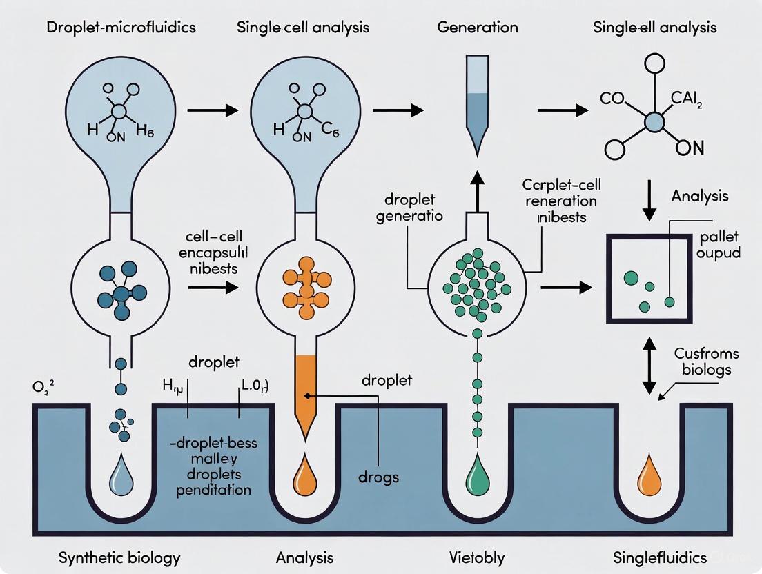
Droplet-based microfluidics has emerged as a transformative technology in single-cell analysis, enabling high-throughput, precise manipulation of individual cells by encapsulating them within picoliter to nanoliter droplets [9] [10]. These droplets act as isolated micro-reactors, eliminating cross-contamination and allowing for the study of cellular heterogeneity, which is often obscured in bulk population analyses [11] [12]. The foundation of this technology lies in the design of microfluidic channels that generate highly monodisperse droplets. Among the various architectures, T-junction, Flow-Focusing, and Co-flow designs stand as the three principal geometries that facilitate droplet formation through controlled fluid dynamics [9] [13]. This guide provides an in-depth technical examination of these core channel architectures, detailing their operating principles, performance characteristics, and experimental protocols, framed within the critical context of advancing single-cell research.
Fundamental Physics of Droplet Generation
Droplet generation in microfluidic devices relies on the interplay between two immiscible fluids—typically a dispersed aqueous phase and a continuous oil phase. The process is governed by the balance between interfacial tension, which strives to minimize the surface area between the two fluids, and viscous shear forces, which act to deform the interface [12] [13]. This force balance is characterized by key dimensionless numbers, most notably the Capillary number (Ca), which represents the ratio of viscous forces to interfacial tension forces [12] [13]. The flow regimes—squeezing, dripping, and jetting—are determined by this balance and the specific geometry of the microchannel, ultimately controlling the size, monodispersity, and generation frequency of the droplets [12] [13].
Core Channel Architectures
The three primary channel designs for droplet generation are T-junction, Flow-Focusing, and Co-flow. Each offers distinct mechanisms and advantages for forming droplets.
T-Junction Design
The T-junction is one of the earliest and simplest geometries for droplet generation. In this design, the channel containing the dispersed phase intersects perpendicularly with the main channel carrying the continuous phase [9] [13].
- Operating Principle: The dispersed phase is introduced into the continuous phase stream, where it is deformed and periodically pinched off by the shear force exerted by the continuous phase. At low capillary numbers, the mechanism is often dominated by the "squeezing" regime, where the dispersed phase temporarily blocks the main channel, leading to droplet break-off once the upstream pressure surpasses the capillary pressure at the junction [13].
- Advantages and Disadvantages: The T-junction is valued for its simple fabrication and operation. A key drawback is its inherent asymmetry, which can lead to less stable droplet formation compared to other designs [13].
Flow-Focusing Design
The flow-focusing geometry is widely used for producing highly monodisperse droplets at high frequencies. Here, the dispersed phase flows through a central channel, which is symmetrically "focused" by two opposing channels carrying the continuous phase [9] [13].
- Operating Principle: The two streams of the continuous phase hydrodynamically compress the dispersed phase, forcing it through a narrow constriction. This creates a thin, unstable jet that breaks up into droplets either within or immediately downstream of the constriction. This design offers excellent control over droplet size [9] [13].
- Advantages and Disadvantages: Flow-focusing devices generate very uniform droplets and can achieve high production rates (kHz). A potential challenge is the occurrence of the "jetting" regime at high capillary numbers, which can produce less monodisperse droplets and require careful control of flow parameters and surfactant concentrations [13].
Co-flow Design
In a co-flow configuration, the dispersed and continuous phases are arranged concentrically. Typically, a capillary tube carrying the dispersed phase is aligned along the axis of a larger channel through which the continuous phase flows [9] [13].
- Operating Principle: Droplets are formed at the tip of the inner capillary due to the shear stress exerted by the co-flowing continuous phase. Similar to other geometries, the process can occur in either "dripping" or "jetting" regimes, depending on the continuous phase velocity [13].
- Advantages and Disadvantages: Co-flow systems are effective for generating droplets from viscous fluids. However, devices based on concentric capillaries can be more challenging to fabricate with high reproducibility and may produce somewhat polydisperse droplets compared to planar lithographic methods [11] [13].
Table 1: Comparative Analysis of Microfluidic Droplet Generation Architectures
| Feature | T-Junction | Flow-Focusing | Co-flow |
|---|---|---|---|
| Basic Geometry | Perpendicular intersection of channels [13] | Continuous phase focuses dispersed phase from two sides [13] | Concentric flow of dispersed phase within continuous phase [9] [13] |
| Primary Formation Mechanism | Squeezing/Pinching by continuous phase flow [9] [13] | Hydrodynamic compression and focusing [9] [13] | Shear stress from co-flowing continuous phase [13] |
| Typical Droplet Monodispersity | Good | Excellent [13] | Moderate [11] [13] |
| Key Controlling Parameters | Flow rate ratio, capillary number [13] | Flow rate ratio, constriction geometry, capillary number [9] [13] | Velocity of continuous phase, viscosity ratio, capillary diameter [13] |
| Throughput | Moderate | High (can reach kHz) [13] | Moderate to High |
| Ease of Fabrication | Simple | Moderate (requires precise constriction) | More complex (capillary alignment) [11] |
| Common Applications | Basic encapsulation, chemical reactions [13] | High-throughput single-cell analysis, synthetic biology [9] [14] | Generation of droplets with viscous fluids [13] |
Quantitative Performance Data
Understanding the relationship between flow parameters and droplet properties is essential for experimental design.
Table 2: Impact of Flow Parameters on Droplet Characteristics
| Parameter | Effect on Droplet Size | Effect on Generation Frequency |
|---|---|---|
| Increase in Dispersed Phase Flow Rate (Qd) | Increases [13] | Increases |
| Increase in Continuous Phase Flow Rate (Qc) | Decreases [13] | Increases |
| Increase in Flow Rate Ratio (Qd/Qc) | Increases | Varies |
| Increase in Capillary Number (Ca) | Generally decreases, but regime-dependent [12] | Increases |
Experimental Protocol: Single-Cell Encapsination via Flow-Focusing
This protocol details a standard methodology for encapsulating single cells using a flow-focusing droplet microfluidic device.
Materials and Reagents
- Microfluidic Chip: PDMS-based chip with a flow-focusing geometry (channel height: 50-100 µm).
- Continuous Phase: Fluorinated oil (e.g., HFE 7500) supplemented with 1-2% w/w biocompatible surfactant (e.g., PFPE-PEG block copolymer) to stabilize droplets against coalescence [9].
- Dispersed Phase: Cell suspension in appropriate growth medium or buffer.
- Equipment: Two precision syringe pumps, tubing and connectors, microscope for observation, and a collection vial.
Procedure
- Chip Priming: Load the continuous phase (fluorinated oil with surfactant) into a syringe and connect it to the chip's oil inlets. Place the syringe on a pump and prime the device's channels with the continuous phase to ensure they are filled and air bubbles are eliminated.
- Sample Preparation: Prepare a suspension of the target cells (e.g., bacteria or mammalian cells) at an optimal concentration. The cell density should be titrated to maximize the number of droplets containing exactly one cell, following Poisson statistics. A typical starting concentration is ~1-2 million cells/mL [11] [12].
- Droplet Generation:
- Load the cell suspension into a separate syringe and place it on a second pump.
- Connect the syringe to the aqueous phase inlet of the chip.
- Start both pumps simultaneously. Typical flow rates are:
- Continuous Phase (Oil): 1000-5000 µL/h
- Dispersed Phase (Aqueous Cell Suspension): 100-500 µL/h
- Adjust the flow rates to achieve the desired droplet size and generation frequency. Monitor droplet formation in real-time using the microscope.
- Droplet Collection: Guide the emerging droplets from the outlet channel into a collection vial for subsequent incubation or analysis.
- Incubation and Analysis: Incubate the collected droplets under conditions suitable for the encapsulated cells (e.g., 37°C for mammalian cells). Downstream analysis can include fluorescence microscopy, PCR, or sequencing to assess single-cell behavior [9] [10].
The Scientist's Toolkit: Essential Research Reagents
Table 3: Key Reagent Solutions for Droplet Microfluidics
| Reagent/Material | Function | Key Considerations |
|---|---|---|
| Fluorinated Oil (e.g., HFE 7500) | Serves as the continuous phase; immiscible with aqueous samples [9]. | High oxygen and CO2 permeability beneficial for cell viability; requires compatible surfactants [9]. |
| PFPE-PEG Surfactant | Prevents droplet coalescence during and after generation by stabilizing the oil-water interface [9]. | Critical for long-term stability of droplets; biocompatibility is essential for cell culture applications [9]. |
| Biocompatible Hydrogel (e.g., Alginate) | Forms gel microdroplets for 3D cell culture, providing a matrix that mimics the extracellular environment [12]. | Enables long-term cell culture and differentiation studies within droplets [12]. |
| Cell Staining Dyes (e.g., Calcein AM, Propidium Iodide) | Used for live/dead assays and monitoring cell viability and function inside droplets. | Must be compatible with the droplet system and not interfere with the surfactant or oil phase. |
| Lysis Buffer & RT Reagents | For downstream single-cell omics; lyses cells and enables reverse transcription within droplets for transcriptomic analysis [10]. | Formulations must be optimized for function in picoliter volumes and the specific microfluidic environment. |
Visualizing Microfluidic Workflows and Relationships
The following diagrams illustrate the core architectures and an integrated workflow for single-cell droplet analysis.
Diagram 1: Three Core Droplet Generation Architectures
Diagram 2: Single-Cell Analysis Workflow via Droplet Microfluidics
T-junction, Flow-Focusing, and Co-flow microfluidic architectures provide the foundational toolkit for droplet generation in single-cell research. The choice of architecture involves a trade-off between simplicity, control, monodispersity, and throughput. As the field advances, the integration of these designs with automated control systems and sophisticated downstream analytical techniques will continue to unlock deeper insights into cellular heterogeneity and function, solidifying droplet microfluidics as a cornerstone technology in life science research and drug development.
Droplet-based microfluidics has emerged as a powerful tool in single-cell analysis, enabling high-throughput, precise manipulation of individual cells within picoliter to nanoliter aqueous compartments surrounded by an immiscible carrier oil [9] [10]. The physics underlying droplet generation—primarily the interplay between interfacial tension and shear forces—directly determines the monodispersity, stability, and size of the droplets, which are critical parameters for reliable single-cell research [15] [16]. Controlling these physical forces allows researchers to create highly uniform microenvironments for applications ranging from single-cell genomics and proteomics to drug screening and cell culture [9] [10]. This technical guide explores the fundamental principles, quantitative relationships, and experimental methodologies governing droplet formation, with a specific focus on their implications for single-cell analysis research.
Fundamental Forces in Droplet Formation
Interfacial Tension
Interfacial tension (γ) is the contractive force per unit length at the interface between two immiscible fluids, acting to minimize the interfacial area and promote a spherical droplet shape [17] [16]. In microchannel emulsification, it serves as the dominant driving force for droplet formation when capillary forces overcome viscous forces [18] [16]. The Laplace pressure (Δp), which represents the pressure difference across a curved interface, is governed by the Young-Laplace equation:
$$ {\rm{\Delta }}p=\gamma (\frac{1}{{r}{1}}+\frac{1}{{r}{2}})\cos (\theta ) $$
where r₁ and r₂ are the principal radii of curvature, and θ is the contact angle between the fluids and channel walls [15]. In spontaneous droplet generation systems, the Laplace pressure at the nozzle opposes the applied dispersed phase pressure [15]. As a droplet grows, its radius of curvature increases, thereby decreasing the Laplace pressure (Δpₐ = 2γ/rₐ(t)) and enabling further phase inflow until neck collapse and droplet pinch-off occur [15].
Shear Forces
Shear forces arise from the viscous drag exerted by the continuous phase flowing past the dispersed phase. These forces act to deform the interface and detach forming droplets [9] [19]. The balance between interfacial tension and viscous shear forces is quantitatively expressed through the Capillary number (Ca):
$$ Ca=\frac{\etac \dot{\varepsilon} R0}{\sigma} $$
where ηc is the continuous phase viscosity, \dot{\varepsilon} is the strain rate, R₀ is the characteristic length (e.g., initial droplet radius), and σ is the interfacial tension [17]. At low Ca values (~10⁻⁴ to 10⁻³), capillary forces dominate, leading to monodisperse droplet generation [18]. Conversely, at higher Ca values, viscous forces dominate, which can result in unstable droplet formation and polydisperse emulsions [16].
Achieving Monodispersion
Monodisperse droplets, characterized by a uniform size distribution (typically with a coefficient of variation <5%), are essential for quantitative single-cell analyses as they ensure consistent reactor volumes and minimize experimental variability [15] [16]. Monodispersity is achieved when droplet breakup is driven by a spontaneous transformation mechanism governed by interfacial tension, rather than by turbulent or uncontrolled shear [16]. The transition from monodisperse to polydisperse regimes occurs at a critical capillary number (Ca*), beyond which viscous forces prevent the interface from stabilizing the neck, leading to irregular breakup [15].
Table 1: Key Parameters Influencing Droplet Monodispersity
| Parameter | Effect on Monodispersity | Optimal Range for Monodispersion |
|---|---|---|
| Capillary Number (Ca) | Determines breakup regime; low Ca promotes uniformity [18] [16] | 10⁻⁴ to 10⁻³ [18] |
| Viscosity Ratio (ηd/ηc) | Affects droplet deformation and relaxation [17] | Varies by geometry; often near 1 [17] |
| Surfactant Concentration | Reduces dynamic interfacial tension, stabilizes against coalescence [20] [16] | Above Critical Micelle Concentration (CMC) [16] |
| Channel Geometry | Controls flow profile and pressure gradients [18] [15] | Nozzle/constriction dimensions comparable to target droplet size [18] [15] |
Quantitative Analysis of Droplet Formation
The following table summarizes key quantitative relationships and values critical for predicting and controlling droplet formation.
Table 2: Quantitative Relationships in Droplet Formation Physics
| Relationship/Parameter | Mathematical Expression | Typical Values/Impact |
|---|---|---|
| Laplace Pressure | (\Delta p = \gamma \left( \frac{1}{r1} + \frac{1}{r2} \right) \cos(\theta)) [15] | Dominant at microscale; driving force in spontaneous emulsification [18] [16] |
| Droplet Deformation | (D = \frac{l - b}{l + b} \sim Ca \frac{19\etad + 16\etac}{16\etad + 16\etac}) [17] | Quantifies droplet stretch in flow; crucial for IFT measurement [17] |
| Critical Capillary Number (Ca*) | (Ca^* = \frac{\etad Ud^*}{\gamma}) [15] | Threshold for transition from monodisperse to polydisperse regime [15] |
| Confined Droplet Size | Influenced by terrace length and channel depth [16] | Droplet diameter ~ terrace length in MC emulsification [16] |
| Dynamic IFT Effect | IFT exceeds equilibrium value at low surfactant conc. [20] [16] | Leads to shorter detachment time, stabilizing formation [20] |
Experimental Protocols for Droplet Analysis
Protocol: Measuring Interfacial Tension via Droplet Deformation
This protocol uses a cross-slot microfluidic device to trap a single droplet and measure its deformation under a quasi-static extensional flow to calculate the interfacial tension (IFT) [17].
- Device Fabrication: Fabricate a cross-junction microfluidic device featuring a flow-focusing droplet generation region upstream of a cross-slot analysis region using standard soft lithography techniques [17].
- Droplet Generation: Introduce the continuous (e.g., oil with surfactant) and dispersed (e.g., aqueous sample) phases into the flow-focusing junction at controlled flow rates (Qc and Qd) using precision syringe pumps. Generate monodisperse droplets of the desired size [17].
- Droplet Trapping and Stretching: Guide a single droplet to the stagnation point at the center of the cross-junction. Apply a controlled extensional flow by simultaneously pumping the continuous phase from opposite inlet channels toward the center and out through the opposing outlets. The strain rate, (\dot{\varepsilon}), is proportional to the applied flow rate [17].
- Image Acquisition and Analysis: Use a high-speed camera mounted on an inverted microscope to capture images of the deformed droplet at steady state. Measure the droplet's major (l) and minor (b) axes [17].
- IFT Calculation:
- For confined droplets (where the droplet size is comparable to the channel height, δ = 2R₀/h ≈ 1), use the 2D Darcy approximation model to calculate the IFT, σ [17].
- For unconfined droplets (δ < 1), use the 3D small deformation model (based on Taylor's theory) for a more robust estimate [17].
- The capillary number is calculated as (Ca = \etac \dot{\varepsilon} R0 / \sigma), and the deformation parameter is (D = (l - b)/(l + b)). The IFT is then derived from the theoretical relationship between Ca and D [17].
Protocol: On-Demand Droplet Generation at a T-Junction
This protocol outlines a pressure-driven method for generating individual droplets on demand, providing precise temporal control for single-cell encapsulation studies [19].
- Device Setup: Use a PDMS-based microfluidic device with a T-junction where the oil phase (continuous) enters from the side channel and the aqueous phase (dispersed) flows through the main channel. Connect both inlets to programmable pressure pumps [19].
- Pressure Calibration:
- Upper Pressure Threshold (Pmax): For a fixed aqueous pressure (Paq), slowly increase the oil pressure (Poil) until the oil-water meniscus is static at the junction and the aqueous phase cannot enter the outlet channel. This pressure, just below the point of no droplet formation, is Pmax. It can be estimated by the Laplace pressure balance at the junction: (\Delta P1 = \gamma \left( -\frac{\cos \pi}{ww} - \frac{\cos \theta1}{ww} - \frac{2\cos \theta_1}{h} \right)) [19].
- Lower Pressure Threshold (Pmin): For the same Paq, decrease Poil until the aqueous phase forms a continuous stream into the outlet without breaking into droplets. The pressure just above this point is Pmin [19].
- On-Demand Operation: To generate a single droplet, set Poil to a value between Pmin and Pmax for the desired Paq. The system is stable with no droplet formation. Briefly apply a pressure pulse to the aqueous inlet (or a corresponding negative pulse to the oil inlet) that temporarily shifts the system into the droplet generation regime, creating one droplet before returning to a stable state [19].
- Validation and Optimization: Monitor droplet generation in real-time with a CCD camera. Use the observed frequency of passive generation at given pressures to fine-tune the pressure thresholds for reliable on-demand operation without trial and error [19].
Droplet Generation Techniques and Single-Cell Applications
The workflow from device design to biological insight relies on the precise manipulation of physical forces. The following diagram illustrates the logical pathway from core physics principles to single-cell research applications.
The Scientist's Toolkit: Essential Reagents and Materials
Table 3: Key Research Reagent Solutions for Droplet Microfluidics
| Item | Function | Example Applications |
|---|---|---|
| Fluorinated Oils (e.g., HFE-7500) | Biocompatible continuous phase; low interfacial tension with aqueous solutions, high oxygen permeability [18] [9]. | Long-term single-cell culture, digital droplet PCR [18] [9]. |
| PFPE-PEG Surfactants | Stabilize droplets against coalescence; reduce interfacial tension; biocompatible [9]. | Maintaining droplet integrity in high-throughput incubations, single-cell sequencing [9]. |
| PDMS (Polydimethylsiloxane) | Elastomeric material for device fabrication; optically clear, gas-permeable [19] [21]. | Rapid prototyping of microfluidic chips for cell culture [21]. |
| PTFE (Polytetrafluoroethylene) | Hydrophobic channel material; enables spontaneous capillary flow of fluorinated oils [18]. | Open-channel droplet microfluidics for direct pipette access [18]. |
| SDS (Sodium Dodecyl Sulfate) | Anionic surfactant; reduces interfacial tension in oil-in-water emulsions [16]. | Fundamental studies on IFT effects in model systems [16]. |
The precise control of droplet formation in microfluidics, governed by the fundamental physics of interfacial tension and shear forces, is a cornerstone of modern single-cell analysis. Understanding and manipulating these forces through tailored device geometries, surfactant chemistry, and operational parameters enables the generation of highly monodisperse droplets essential for quantitative biological research. As the field advances, the integration of more sophisticated active control systems and the development of novel, biocompatible materials will further solidify droplet microfluidics as an indispensable platform for unraveling cellular heterogeneity and driving discoveries in drug development and precision biology.
High-Throughput Operation, Reagent Minimization, and Cross-Contamination Prevention
Droplet-based microfluidics has emerged as a transformative technology for single-cell analysis, enabling unprecedented resolution in studying cellular heterogeneity. This approach involves compartmentalizing individual cells within picoliter to nanoliter aqueous droplets dispersed in an immiscible carrier oil, effectively creating millions of isolated microreactors. The fundamental principles of this technology make it particularly suited for addressing the key challenges of high-throughput operation, dramatic reagent minimization, and stringent cross-contamination prevention. These capabilities are revolutionizing fields from functional genomics to drug discovery by allowing researchers to capture and analyze cellular heterogeneity at unprecedented scale and resolution, moving beyond the limitations of bulk analysis that only provides population-averaged data [10] [5].
For single-cell analysis, droplet microfluidics represents a paradigm shift. Cellular heterogeneity is a fundamental characteristic of biological systems, where genetically identical cells can exhibit significant variations in morphology, gene expression, protein secretion, and metabolic activity [10]. Traditional bulk analysis methods mask these differences, potentially overlooking rare but biologically critical cell subtypes. Droplet microfluidics addresses this limitation by enabling the systematic high-throughput analysis of individual cells in a highly controlled manner [5]. The technology's core advantages align perfectly with the needs of modern biological research and drug development, particularly as the field moves toward more precise and personalized therapeutic approaches.
Technological Fundamentals of Droplet Generation and Manipulation
Droplet Generation Mechanisms
The formation of monodisperse droplets is achieved through precisely engineered microfluidic geometries that harness immiscible fluid interactions. Three primary configurations dominate droplet generation:
- Flow Focusing: A dispersed phase (aqueous cell suspension) is injected into a cross-junction where it is pinched by a continuous phase (oil), forming highly uniform droplets through controlled shear forces [22].
- T-Junction: Two immiscible phases intersect perpendicularly, with shear forces at the interface leading to droplet breakup in this simple yet effective design [22].
- Co-Flow: Droplets form within a continuous phase due to controlled shear stress along a coaxial geometry, where both fluids flow in the same direction [22].
The formation process is governed by the balance between interfacial tension and shear forces, with the Capillary number (Ca = ηU/σ, where η is viscosity, U is characteristic velocity, and σ is interfacial tension) serving as a key dimensionless parameter predicting droplet behavior [22]. Channel geometry, flow rate ratios, and surface wettability are carefully optimized to achieve stable dripping or jetting regimes appropriate for specific applications.
Advanced Droplet Manipulation Techniques
Once generated, droplets undergo sophisticated manipulation to enable complex experimental workflows:
- Pico-injection: Allows precise addition of reagents into existing droplets using external electric fields to temporarily destabilize the oil-water interface for controlled reagent introduction [23].
- Droplet Merging: Facilitates combinatorial assays by bringing two droplets into contact through decompression merging, electrocoalescence, or thermal activation strategies [22].
- Droplet Splitting: Enables sample dilution or parallel processing through geometric modifications to microfluidic channels that divide parent droplets into daughter droplets [23] [22].
- Incubation: Maintains droplets under specific environmental conditions for extended periods, either on-chip in delay lines or off-chip in bulk collection vessels, to support biological processes requiring minutes to days [23] [5].
- Sorting: Employs dielectrophoresis, acoustic waves, or magnetic fields to selectively isolate droplets of interest based on their content or assay results, with fluorescence-activated droplet sorting being particularly widespread [23] [22].
Table 1: Key Droplet Manipulation Functions and Their Implementation
| Function | Implementation Methods | Typical Applications |
|---|---|---|
| Reagent Addition | Pico-injection, droplet merging | Multi-step assays, stimulus-response studies |
| Incubation | On-chip delay lines, off-chip storage | Cell culture, enzymatic reactions, PCR amplification |
| Sorting | Dielectrophoresis, acoustic waves, magnetic fields | Isolation of rare cells, hit selection in screening |
| Splitting | Channel bifurcations, electrostatic forces | Sample replication, parallel testing, dilution series |
| Detection | Fluorescence, absorbance, electrochemical, mass spectrometry | Real-time monitoring, end-point analysis |
High-Throughput Operation
Quantitative Throughput Capabilities
Droplet microfluidics operates at scales and rates unachievable by conventional laboratory workflows, with sample processing speeds orders of magnitude beyond robotic liquid handling systems. The technology enables the generation and analysis of thousands to millions of droplets per hour, each serving as an independent microreactor [23]. Throughput capabilities include:
- Droplet Generation: Production rates exceeding 500 Hz for droplet formation and manipulation, dwarfing the approximately 5 Hz rate of robotic "high throughput" liquid handling systems [23].
- Cell Encapsulation: Successful processing of approximately 100,000 cells per second, making it the most effective tool for high-throughput screening of different biological libraries [24].
- Single-Cell Analysis: Profiling of thousands of individual cells simultaneously, with techniques like Drop-Seq enabling a single researcher to create 10,000 single-cell libraries per day [25].
- Screening Scale: Capability to screen up to 10^7 mutants in a single experiment, with demonstrated examples of identifying improved enzyme variants from libraries containing 2×10^7 mutants through only 6 rounds of screening [24].
Table 2: Throughput Comparison Between Droplet Microfluidics and Conventional Methods
| Method | Throughput Scale | Time Requirement | Practical Applications |
|---|---|---|---|
| Droplet Microfluidics | 10^3-10^7 samples per day | Minutes to hours | Single-cell RNA-seq, enzyme evolution, antibody discovery |
| Robotic Liquid Handling | 10^3-10^4 samples per day | Hours to days | Compound screening, plate-based assays |
| Manual Processing | 10^1-10^2 samples per day | Days to weeks | Small-scale research, pilot studies |
| Flow Cytometry | 50,000-100,000 cells analyzed | Minutes | Immunophenotyping, cell sorting |
Enabling High-Throughput Applications
The remarkable throughput of droplet microfluidics has enabled previously impractical applications across biological research and drug discovery:
Single-Cell Omics: Technologies like Drop-Seq, inDrop, and CytoSeq leverage high-throughput droplet encapsulation to barcode transcripts from thousands of individual cells in parallel, enabling comprehensive tissue atlas construction and rare cell population identification [26] [25]. These methods use molecular barcoding strategies where each bead contains primers with identical cell barcodes but different unique molecular identifiers (UMIs), allowing precise attribution of sequencing reads to their cell of origin while counting transcripts digitally [25].
Directed Evolution: High-throughput screening of enzyme libraries containing millions of variants, identifying mutants with significantly improved catalytic properties through iterative screening cycles at unprecedented scales [24].
Functional Genomics: Combining CRISPR perturbations with droplet-based single-cell RNA sequencing enables large-scale mapping of gene regulatory networks and systematic functional interrogation of non-coding elements across thousands of cells simultaneously [27].
Antibody Discovery: Screening of single B-cells for antigen-specific antibody production, enabling rapid identification of therapeutic candidates from immune repertoires [5].
The scalability of these approaches was demonstrated in a landmark study that measured 90 cytokine perturbations across 12 donors and 18 immune cell types, resulting in nearly 20,000 observed perturbations and generating a 10 million cell dataset with 1,092 samples in a single run [27].
Reagent Minimization
Volume Reduction and Cost Implications
Droplet microfluidics achieves remarkable reagent conservation by scaling reaction volumes from microliters in conventional well plates to picoliters or nanoliters in droplets, representing 10^3-10^6 fold volume reductions [23]. This miniaturization translates to substantial practical benefits:
- Consumption Reduction: When screening 10^7 mutants using droplet microfluidics, the total reagent volume consumed is 10^6 times lower than that required by the microtiter plate method [24].
- Volume Range: Typical droplet volumes range from picoliters (10^-12 L) to nanoliters (10^-9 L), with aqueous compartments often measuring 1-300 μm in diameter [22].
- Cost Efficiency: Significant reduction in reagent expenses, particularly valuable for expensive biochemicals, antibodies, or enzymes used in screening campaigns [24].
- Waste Minimization: Drastic decrease in chemical waste production, supporting more sustainable laboratory practices [24].
The economic impact of these savings becomes substantial in large-scale screening applications. For drug discovery pipelines requiring thousands to millions of parallel assays, the cost differential between microliter and nanoliter reaction scales can determine project feasibility.
Concentration Advantages in Small Volumes
Beyond direct volume reduction, the small dimensions of droplet microfluidics confer additional biochemical benefits:
- Rapid Mixing: Molecular diffusion times within picoliter volumes are short, significantly accelerating chemical reactions and reducing incubation periods [26].
- Enhanced Effective Concentrations: The small volumes raise relative concentrations of cellular analytes, improving detection sensitivity for low-abundance molecules [26].
- Contaminant Reduction: Minimal volumes limit potential contamination from environmental sources, improving assay reliability [26].
These factors collectively enhance the information content obtainable from limited biological samples, enabling comprehensive multi-parametric analysis from specimen volumes previously considered insufficient for meaningful analysis.
Cross-Contamination Prevention
Physical Isolation Mechanisms
The fundamental architecture of droplet microfluidics provides inherent protection against cross-contamination through physical compartmentalization:
- Complete Encapsulation: Individual cells are confined within their own picoliter-scale aqueous environments, physically separated from other droplets by a continuous oil phase and surfactant molecules [10] [5].
- Surfactant Stabilization: Specialized surfactants (e.g., FluoSurf formulations for fluorinated oils) accumulate at the oil-water interface, forming protective monolayers that prevent droplet coalescence and content exchange [22].
- Barrier Function: The combination of immiscible phases and surfactant layers creates effective barriers against molecular diffusion between droplets, even during prolonged incubation or storage [23].
This physical isolation is maintained throughout complex workflow operations, including droplet generation, incubation, reinjection, sorting, and analysis. The stability of these emulsion systems has been demonstrated under various challenging conditions, including thermocycling for PCR amplification and extended culture periods for cellular assays [22].
Impact on Assay Quality and Biological Discovery
The prevention of cross-contamination has profound implications for data quality and biological insight:
- Elimination of Signal Crosstalk: In single-cell secretion assays, captured analytes remain concentrated within the droplet of origin rather than diffusing throughout the bulk solution, enabling accurate attribution of secreted factors to their producer cells [5].
- Preservation of Rare Events: Rare cells (e.g., circulating tumor cells or stem cells) can be analyzed without their signals being diluted or overwhelmed by abundant cell types, maintaining detection sensitivity for biologically significant but numerically minor populations [10].
- Accurate Single-Cell Representation: The transcriptomes, genomes, or proteomes of individual cells remain distinct throughout analysis, enabling true single-cell resolution without artificial averaging effects [26] [25].
The importance of this compartmentalization is particularly evident in single-cell genomics and transcriptomics, where even minute cross-contamination between cells could generate misleading chimeric sequences or falsely suggest intermediate cellular states that don't actually exist biologically.
Experimental Protocols and Implementation
Drop-Seq Method for Single-Cell RNA Sequencing
Drop-Seq represents a powerful implementation of droplet microfluidics for high-throughput single-cell transcriptomics [25]. The detailed methodology involves:
Microfluidic Device Preparation
- Utilize a custom-designed microfluidic device that joins two aqueous flows (cells and barcoded beads) before their compartmentalization into discrete droplets.
- Implement channel designs that maintain laminar flow to prevent mixing of the two aqueous inputs prior to droplet formation.
Bead Preparation
- Synthesize barcoded magnetic beads (approximately 20μm in diameter) functionalized with oligonucleotide primers containing:
- A universal PCR handle sequence for amplification
- A cell barcode (identical across all primers on a single bead)
- Unique Molecular Identifiers (UMIs) for digital counting
- A 30-bp oligo-dT sequence for mRNA capture
- Generate cell barcodes through split-pool synthesis involving 12 cycles, creating 4^12 (16,777,216) possible barcode sequences.
- Add UMIs through eight rounds of degenerate synthesis with all four DNA bases, yielding 4^8 (65,536) possible UMI sequences per bead.
Cell Preparation
- Dissociate tissue into single-cell suspension using standard protocols.
- Adjust cell concentration to optimize single-cell encapsulation while minimizing multiple cells per droplet.
Droplet Generation and mRNA Capture
- Co-flow cell suspension and barcoded beads in lysis buffer at specific flow rates (e.g., 4,000 μL/hour each) with oil phase (15,000 μL/hour).
- Immediately lyse cells within droplets, releasing mRNAs that hybridize to primers on companion beads.
- Break droplets by adding reagent to destabilize the oil-water interface.
- Collect beads and wash to obtain STAMPs (Single-cell Transcriptomes Attached to MicroParticles).
Library Preparation and Sequencing
- Reverse transcribe captured mRNAs to cDNA on beads.
- Amplify cDNA pools by PCR for high-throughput sequencing.
- Sequence resulting molecules and align to reference genome.
- Organize reads by cell barcodes and digitally count mRNA transcripts using UMIs.
High-Throughput Screening Protocol for Enzyme Evolution
Droplet microfluidics enables ultra-high-throughput screening for directed enzyme evolution [24]:
Library Creation
- Generate mutant library through error-prone PCR or DNA shuffling.
- Transform into expression host if working with microbial systems.
Droplet Compartmentalization
- Encapsulate single cells or lysates containing enzyme variants into droplets with fluorogenic or chromogenic substrates.
- Include necessary cofactors and reagents for enzymatic reaction.
Incubation
- Incubate droplets on-chip in delay lines or off-chip to allow enzymatic reactions to proceed.
- Maintain stable emulsion throughout incubation period.
Detection and Sorting
- Analyze droplet fluorescence or absorbance as they flow through detection region.
- Implement fluorescence-activated droplet sorting (FADS) to selectively deflect droplets containing improved enzyme variants.
- Apply sorting at rates of up to several kHz.
Recovery and Validation
- Break sorted droplets to recover enriched variants.
- Replate for further rounds of screening or validation.
- Typically require multiple iterative rounds (e.g., 6 rounds to identify isomerase with 17-fold increased catalytic efficiency).
Research Reagent Solutions and Materials
Successful implementation of droplet microfluidics requires specific reagents and materials optimized for the unique demands of the technology:
Table 3: Essential Reagents and Materials for Droplet Microfluidics
| Category | Specific Examples | Function and Importance |
|---|---|---|
| Surfactants | FluoSurf-C, FluoSurf-O, FluoSurf-S | Stabilize droplets against coalescence; reduce interfacial tension; ensure emulsion stability during thermocycling |
| Carrier Oils | Fluo-Oil 135 (Novec 7500 alternative), Fluo-Oil 200 (Fluorinert FC-40 alternative) | Form continuous phase; compatible with biological systems; appropriate viscosity and surface tension |
| Chip Materials | Polydimethylsiloxane (PDMS), Glass, Flexdym | Provide optical clarity; enable precise channel fabrication; appropriate surface properties |
| Surface Treatments | Fluo-ST1, Fluo-ST3 | Covalently bond to channel walls; control wettability; prevent nonspecific adsorption |
| Barcoding Beads | Functionalized magnetic beads, Hydrogel microparticles | Capture cellular analytes; provide molecular barcoding; enable sample multiplexing |
Integration in Drug Discovery and Development
Droplet microfluidics is reshaping pharmaceutical research by providing powerful tools across the drug development pipeline:
Target Identification and Validation: Single-cell RNA sequencing reveals cell-type-specific expression of potential drug targets in disease-relevant tissues, with studies demonstrating that targets with cell-type-specific expression in disease-relevant tissues show higher progression rates from Phase I to Phase II clinical trials [27].
High-Throughput Screening: Miniaturized assays enable screening of compound libraries against cellular targets with dramatically reduced reagent requirements and increased throughput compared to conventional plate-based approaches [23] [24].
Toxicity Assessment: Single-cell responses to drug candidates can be profiled at scale, identifying potential toxicity mechanisms and subpopulations with differential sensitivity [27].
Biomarker Discovery: Comprehensive characterization of cell subtypes and states in diseased versus healthy tissues reveals potential diagnostic, prognostic, or predictive biomarkers with higher specificity than bulk tissue analysis [27].
Immunotherapy Development: Particularly valuable for characterizing immune cell repertoires, identifying rare antigen-specific cells, and profiling complex immune responses to therapeutic interventions [26].
The technology's impact is magnified when integrated with artificial intelligence approaches, where large-scale single-cell datasets train predictive models for drug response and target validation [28].
Diagram 1: Comprehensive droplet microfluidics workflow encompassing generation, processing, and analysis stages with key operational modules.
Diagram 2: Cross-contamination prevention mechanisms in droplet microfluidics and their impact on data quality.
Droplet microfluidics represents a foundational technology that effectively addresses the intertwined challenges of high-throughput operation, reagent minimization, and cross-contamination prevention in single-cell analysis. By compartmentalizing biological reactions in picoliter droplets, researchers can now conduct experiments at scales and resolutions previously impossible, driving advances in our understanding of cellular heterogeneity and accelerating therapeutic discovery. As the technology continues to mature through improvements in chip design, surfactant chemistry, and analytical methods, its integration into mainstream biological research and drug development pipelines promises to further transform our approach to studying and harnessing biological complexity.
Droplet-based microfluidics has emerged as a transformative technology for high-throughput single-cell analysis, enabling researchers to encapsulate individual cells in picoliter droplets that function as discrete microreactors. This platform permits the analysis of proteins released from or secreted by cells, thereby overcoming a major limitation of traditional flow cytometry and fluorescence-activated cell sorting [4]. The core functionality of these systems depends on three fundamental components: surfactant chemistry for droplet stabilization, carrier oils as the continuous phase, and specialized materials for device fabrication. Together, these elements create a controlled environment for manipulating millions of individual cells in compartmentalized droplets, facilitating applications from single-cell genomics and proteomics to drug screening and directed evolution [4] [10]. This technical guide examines the principles, selection criteria, and practical considerations for these essential components within the broader context of single-cell analysis research.
Surfactant Chemistry
Role and Mechanism of Surfactants
Surfactants are amphiphilic compounds essential for stabilizing emulsions in droplet-based microfluidics. Their molecular structure comprises a hydrophilic (water-attracting) head and a hydrophobic (water-repelling) tail, allowing them to adsorb at the interface between immiscible fluids, such as water and carrier oil. By reducing interfacial tension, surfactants facilitate the formation of uniform droplets and prevent coalescence by creating a physical barrier between adjacent droplets [29] [30]. The stabilization mechanism involves both steric hindrance and, in the case of charged surfactants, electrostatic repulsion [31]. Without surfactants, droplets would rapidly coalesce upon contact, rendering high-throughput applications impossible. The choice of surfactant is therefore critical and depends primarily on the type of emulsion (water-in-oil or oil-in-water) and the specific biological application, particularly when working with living cells [30].
Surfactant Selection by Oil Type
The effectiveness of a surfactant is intrinsically linked to the chemical nature of the carrier oil. The Hydrophilic-Lipophilic Balance (HLB) value, which ranges from 0 to approximately 20, provides a guideline for selection: lower HLB values (below 7) indicate a preference for stabilizing water-in-oil (W/O) emulsions, while higher HLB values (above 8-9) favor oil-in-water (O/W) emulsions [31]. Single-cell analysis primarily utilizes W/O emulsions, where aqueous droplets containing cells and reagents are dispersed in a continuous oil phase.
Table 1: Common Surfactants and Their Compatible Oils for Single-Cell Analysis
| Oil Category | Example Oils | Recommended Surfactants | Typical Applications in Single-Cell Analysis |
|---|---|---|---|
| Fluorinated Oil | HFE-7500, FC-40, Novec | FluoSurf, PFPE-PEG, PFPECOO-NH₄ | Cell encapsulation, droplet digital PCR (ddPCR), single-cell sequencing, long-term cell culture [30] [31] |
| Hydrocarbon Oil | Mineral Oil, Hexadecane | Span 80, ABIL EM90 | Directed evolution, chemical reactions, protein expression [30] |
| Silicone Oil | DC200, PDMS-based oils | Triton X-100, ABIL EM90 | PCR, directed evolution [30] |
Specialized fluorinated surfactants, such as the FluoSurf series, are particularly critical for biotechnology applications. FluoSurf is a non-ionic, fluorinated surfactant designed specifically to stabilize aqueous droplets in fluorinated oils. Its key attributes include high biocompatibility (supporting cell viability), excellent stability during thermocycling (essential for ddPCR), and high purity to minimize background interference in sensitive detection assays [31]. Different formulations, such as FluoSurf-O with ultra-low autofluorescence, are optimized for specific experimental needs like fluorescence-activated droplet sorting.
Carrier Oils
Oil Types and Properties
The continuous phase oil is a primary determinant of the physical and chemical environment within a microfluidic device. The three main categories of oils used are fluorinated oils, hydrocarbon oils, and silicone oils, each with distinct properties that dictate their application suitability [30].
Fluorinated oils (e.g., HFE-7500, FC-40, Novec) are generally the most suitable for live-cell applications. Their key advantages include high oxygen and gas solubility, which is critical for cell viability during long-term incubations, and poor solubility for most organic molecules, which minimizes the leakage of hydrophobic reagents and biomolecules from the aqueous droplets into the oil phase [4] [30]. Hydrocarbon oils (e.g., mineral oil, hexadecane) are more traditional but are often incompatible with cells for extended periods due to their tendency to dissolve organic compounds, leading to potential reagent loss and limited gas exchange. Silicone oils are used less frequently, partly due to their incompatibility with the common device material PDMS, which can cause swelling and device failure [30].
Impact of Oil Viscosity and Material Compatibility
The viscosity of the carrier oil is a critical physical parameter that directly influences droplet generation. Higher oil viscosity typically results in smaller droplet sizes and lower droplet generation rates under the same flow pressure conditions [30].
Table 2: Impact of Mineral Oil Viscosity on Droplet Generation (Adapted from Yao et al., 2019)
| Water:Oil Pressure (mbar) | 5 cSt Oil Droplet Size (µm) | 10 cSt Oil Droplet Size (µm) | 15 cSt Oil Droplet Size (µm) | 5 cSt Generation Rate (drops/s) | 15 cSt Generation Rate (drops/s) |
|---|---|---|---|---|---|
| 30:40 | 68.3 ± 2.0 | 51.0 ± 1.7 | 46.3 ± 1.8 | 76 ± 1 | 45 ± 0 |
| 90:120 | 37.1 ± 1.8 | 31.0 ± 1.0 | 28.9 ± 0.9 | 239 ± 8 | 149 ± 1 |
| 150:200 | 32.1 ± 1.0 | 26.9 ± 1.1 | 25.1 ± 1.3 | 581 ± 12 | 305 ± 8 |
Compatibility between the oil and the chip material is also essential to ensure device integrity and proper droplet formation. For instance, fluorinated oils are compatible with most materials, including plastics, glass, and elastomers. In contrast, silicone oils should only be used with silicone-based devices, and hydrocarbon oils are often paired with polycarbonate devices [30].
Device Fabrication Materials
PDMS and Soft Lithography
Polydimethylsiloxane (PDMS), specifically the two-part Sylgard 184 formulation, is the most ubiquitous material for fabricating microfluidic devices for research [32]. Its popularity stems from its ease of use, economy, optical transparency (allowing for microscopic observation), gas permeability (beneficial for cell viability), and flexibility, which enables the integration of features like pneumatic valves [4] [32]. The standard fabrication method is soft lithography, a multi-step replication process.
A key step in PDMS device preparation is bonding the molded PDMS slab to a glass substrate to form sealed channels. This is most commonly achieved through oxygen plasma treatment, which activates the PDMS and glass surfaces, allowing them to form a permanent, irreversible bond when brought into contact immediately after treatment [32].
Alternative Fabrication Materials
While PDMS dominates research settings, other materials are important for specific applications. Glass and silicon were used in first-generation microfluidic devices. Glass offers excellent optical transparency, chemical resistance, and high-pressure tolerance, making it suitable for electrophoresis and separations combined with mass spectrometry [33]. However, the fabrication of glass devices is complex, often requiring high-temperature bonding and cleanroom facilities [33]. Thermoplastics (e.g., cyclic olefin copolymer, or COC) are rigid polymers that are increasingly used for commercial and point-of-care devices due to their scalability via injection molding and generally lower cost for mass production [33]. A comparative overview of these material properties is summarized in the following diagram.
Integrated Experimental Protocol: Single-Cell Secretion Analysis
To illustrate how these components work in concert, consider a protocol for detecting antibodies secreted from single mouse hybridoma cells, a common application in drug development [4].
The experimental process begins with device fabrication and cell preparation, culminates in droplet generation and incubation for secretion detection, and ends with sorting and recovery of cells of interest.
Key System Components and Reagents
The success of this protocol hinges on the precise integration of the core components discussed in this guide.
Table 3: Research Reagent Solutions for Single-Cell Secretion Assay
| Component | Specific Example / Property | Function in the Experiment |
|---|---|---|
| Carrier Oil | Fluorinated Oil (HFE-7500) | Serves as the continuous, oxygen-permeable phase for aqueous droplet generation and flow [4] [30]. |
| Surfactant | PFPE-PEG (e.g., FluoSurf) | Stabilizes droplets against coalescence, ensuring compartment integrity during incubation and sorting [4] [31]. |
| Device Material | PDMS (Sylgard 184) | Forms the microchannels for droplet generation, manipulation, and sorting; bonded to a glass substrate [32]. |
| Probe & Beads | Fluorescent Antigen & IgG-coated Beads | Beads capture secreted antibodies; fluorescence localizes upon probe binding, enabling detection [4]. |
| Lysis Additive | n-Dodecyl-β-D-maltoside (DDM) | A non-ionic, MS-compatible surfactant that aids in cell lysis and prevents surface adsorption in downstream proteomic analysis [34]. |
The robust performance of droplet-based microfluidics in high-throughput single-cell analysis is fundamentally governed by the careful selection and integration of surfactants, carrier oils, and fabrication materials. Fluorinated oils paired with specialized surfactants like FluoSurf provide a biocompatible and stable environment for single-cell encapsulation and assay execution. PDMS remains the material of choice for prototyping due to its versatility, though glass and thermoplastics offer advantages for specific commercial and analytical applications. As the field advances toward more quantitative and comprehensive single-cell multi-omics, the continued refinement of these core components—particularly in reducing analyte loss and improving detection sensitivity—will be crucial for unlocking deeper biological insights and broadening the impact of this powerful technology in biomedical research and therapeutic development.
Methodological Implementations and Transformative Applications in Biomedical Research
Single-Cell Encapsination Strategies and Poisson Distribution Considerations
Single-cell analysis has emerged as a transformative paradigm in biological research, enabling the investigation of cellular heterogeneity that is obscured in bulk population studies [12] [35]. Droplet-based microfluidics represents one of the most powerful platforms for single-cell analysis, where individual cells are compartmentalized within picoliter-volume droplets that function as isolated microreactors [12] [36]. The critical first step in this pipeline—reliably encapsulating individual cells into separate droplets—presents a significant technical challenge governed by statistical principles, predominantly the Poisson distribution [37].
The inherent limitation of random encapsulation necessitates advanced strategies to overcome Poisson statistics, which theoretically limit single-cell encapsulation efficiency to approximately 37% under optimal conditions [12] [37]. This technical guide examines the fundamental principles of single-cell encapsulation, quantitative performance of various strategies, and detailed methodological protocols, providing researchers with a comprehensive framework for implementing these technologies in drug development and basic research contexts.
Poisson Distribution: Theoretical Framework and Practical Limitations
Mathematical Foundations of Random Encapsulation
In droplet microfluidics, when cells are randomly distributed and introduced into droplets at a fixed rate, the process follows Poisson statistics. The probability P(k) of finding k cells in a given droplet is described by the equation:
P(k) = (λ^k × e^(-λ)) / k!
where λ represents the average number of cells per droplet [37]. This statistical framework predicts that the maximum proportion of droplets containing exactly one cell occurs at λ = 1, where approximately 37% of droplets contain a single cell, 37% remain empty, and 26% contain multiple cells [12] [37]. This distribution results in substantial waste of reagents and analytical effort on empty or multiply-occupied droplets.
Practical Implications for Experimental Design
The Poisson limitation necessitates careful consideration of cell suspension density during experimental design. Higher cell concentrations (λ > 1) increase the proportion of multiplets (droplets with >1 cell), while lower concentrations (λ < 1) increase the number of empty droplets [38] [37]. This tradeoff fundamentally constrains the efficiency of random encapsulation approaches, motivating the development of advanced active and passive encapsulation strategies that overcome these statistical limitations.
Table 1: Theoretical Encapsulation Efficiencies According to Poisson Statistics
| Average Cells per Droplet (λ) | Empty Droplets (%) | Single-Cell Droplets (%) | Multiple-Cell Droplets (%) |
|---|---|---|---|
| 0.1 | 90.5 | 9.0 | 0.5 |
| 0.5 | 60.7 | 30.3 | 9.0 |
| 1.0 | 36.8 | 36.8 | 26.4 |
| 1.5 | 22.3 | 33.5 | 44.2 |
Advanced Encapsulation Strategies Overcoming Poisson Limitations
Hydrodynamic Focusing and Cell Ordering
Hydrodynamic focusing techniques utilize specially designed microchannel geometries to arrange cells into orderly streams before encapsulation, significantly improving single-cell encapsulation efficiency. Spiral microchannels exploit inertial forces to focus cells into a narrow stream, with the Dean drag force and wall-effect lift force working in concert to position cells near the channel inner wall [38]. One implementation features an 8-loop double spiral unit with equally spaced pillars (100μm width, 60μm depth), successfully focusing cells into a near-equidistant linear arrangement at flow rates of 40-80 μL/min [38]. This approach achieved single-cell encapsulation rates of 72.2% for MDA-MB-231 and MKN-45 cell lines, substantially exceeding Poisson limitations [38].
Active Encapsulation Using Micro-Vortices
Micro-vortex based trapping represents an innovative interfacial hydrodynamic technique where cells are initially trapped in micro-vortices and subsequently released one-to-one into droplets [36]. This method controls encapsulation through the width of the outer streamline (dgap) that separates the vortex from the main flow. When dgap is approximately equal to the cell radius, one-to-one encapsulation is achieved, while complete cell trapping occurs when d_gap is smaller than the cell radius [36]. This approach demonstrated 50% single-cell encapsulation efficiency even at low cell loading densities and enabled size-selective capture of blood cells (platelets, RBCs, WBCs) with 70% WBC capture efficiency from diluted blood samples [36].
On-Chip Sample Enrichment
Integrated sample enrichment modules address the tradeoff between cell suspension density and focusing performance by removing excess aqueous phase after cell focusing but before droplet generation [38]. One implementation features a flow resistance-based enrichment module with five identical serpentine units (700μm length, 100μm width each), allowing over 50% of the aqueous phase to be removed while maintaining focused cells in sequence [38]. This approach enables the use of lower initial cell concentrations (2×10^6 objects/mL), reducing cell-cell interactions and improving focusing performance while avoiding channel clogging [38].
Microfluidics-Free Templated Emulsification
Recent innovations have introduced microfluidics-free approaches such as Particle-templated Instant Partition Sequencing (PIP-seq), which uses bulk emulsification with barcoded hydrogel templates [39]. This method employs particle templating to compartmentalize cells, barcoded hydrogel templates, and lysis reagents in monodispersed water-in-oil droplets through vortexing rather than microfluidic confinement [39]. The approach accommodates various formats including microwell plates and large-volume conical tubes, enabling thousands of samples or millions of cells to be processed in minutes while maintaining low doublet rates (<5%) and high transcriptome purity (<3% cross-contamination in mouse-human mixing studies) [39].
Table 2: Performance Comparison of Single-Cell Encapsulation Strategies
| Encapsulation Strategy | Single-Cell Efficiency | Throughput | Technical Complexity | Key Applications |
|---|---|---|---|---|
| Poisson (Random) Encapsulation | ~37% (theoretical max) | Very High | Low | Standard droplet workflows |
| Hydrodynamic Focusing | Up to 79.2% | High | Medium | High-efficiency single-cell assays |
| Micro-Vortex Trapping | ~50% | Medium | High | Size-selective cell encapsulation |
| On-Chip Sample Enrichment | 72.2% (cells) | High | High | Viscous or complex samples |
| Templated Emulsification (PIP-seq) | >65% (cell capture) | Very High (millions of cells) | Medium | Large-scale studies, clinical applications |
Experimental Protocols and Methodological Guidelines
Hydrodynamic Focusing Encapsulation Protocol
Device Fabrication: The microfluidic platform is fabricated using standard soft lithography with a three-unit design: (1) double spiral focusing unit (8-loop, 100μm width, 60μm depth) with half-circle pillars (70μm) on the inner channel side; (2) flow resistance-based sample enrichment module with 5 serpentine units; (3) crossflow droplet generation unit [38]. The SU-8 mold is fabricated via photolithography, and PDMS chips are cast at 10:1 base-to-crosslinker ratio, followed by oxygen plasma bonding and hydrophobic treatment with fluorosilane [38].
Cell Preparation: Culture MDA-MB-231 or MKN-45 cells to 90% confluence, digest with TrypLE Express, centrifuge at 200×g for 3 minutes, and resuspend in culture media at desired concentrations (1-4×10^6 cells/mL) [38]. For viability assessment, use Trypan blue staining after droplet merging induced by anti-static gun treatment [38].
Encapsulation Procedure:
- Inject cell suspension at 80μL/min and oil phase (Novec 7500 with 2% Pico-Surf surfactant) at 70μL/min using separate syringe pumps.
- Monitor cell focusing through microscope with high-speed camera (2000 fps).
- Adjust flow resistance at enrichment module by punching different outlets to control aqueous phase removal.
- Collect droplets generated at approximately 4000 droplets per second for downstream analysis [38].
Visualization and Analysis: Use lab-developed Python codes for standard deviation analysis based on 1000 stacked high-speed camera frames to visualize cell/bead trajectories [38]. Implement YOLOv8n-based droplet detection algorithms for counting cells/beads in droplets and statistical analysis of encapsulation rates [38].
Micro-Vortex Encapsulation Protocol
Device Design: Implement a microfluidic design incorporating expansion regions to generate controlled micro-vortices adjacent to the main flow channel. Precisely tune the channel geometry to achieve a d_gap (width of the outer streamline) approximately equal to the radius of target cells [36].
Cell Separation: For blood cell separation, dilute whole blood 10× in appropriate buffer. Hydrodynamic separation occurs based on cell size, with platelets, RBCs, and WBCs selectively trapped in vortices based on their differential size and hydrodynamic properties [36].
Encapsulation Process:
- Introduce cell suspension at optimized concentration and flow rate to ensure single-cell occupancy in vortices.
- Control release of trapped cells into droplets by maintaining d_gap at cell radius dimension.
- Collect droplets with size-sorted cells for downstream analysis [36].
The Scientist's Toolkit: Essential Research Reagents and Materials
Table 3: Key Research Reagent Solutions for Single-Cell Encapsulation
| Reagent/Material | Function | Example Products/Specifications |
|---|---|---|
| Fluorocarbon Oil | Continuous phase for water-in-oil emulsions | Novec 7500 (3M) |
| Surfactants | Stabilize droplets, prevent coalescence | Pico-Surf (Sphere Fluidics, 2% w/w) |
| PDMS | Chip fabrication material | Sylgard 184 (Dow Corning, 10:1 ratio) |
| Hydrophobic Treatment | Modify channel surface properties | MesoPhobic-2000 (MesoBioSystem) |
| Barcoded Hydrogel Beads | mRNA capture and barcoding in droplet workflows | 10× Genomics Gel Beads, PIP-seq templates |
| Cell Suspension Buffer | Maintain cell viability and integrity during encapsulation | PBS with BSA (0.5-1.0%) |
| Lysis Reagents | Release cellular contents within droplets | Proteinase K, Triton X-100 (0.1-1%) |
Visualization of Encapsulation Strategies
Diagram 1: Strategic workflow for single-cell encapsulation methodologies, illustrating efficiency metrics and sequential development of advanced approaches.
Diagram 2: Comparison of random Poisson encapsulation versus active encapsulation strategies, demonstrating efficiency improvements through hydrodynamic focusing and cell ordering techniques.
Single-cell encapsulation technologies have evolved substantially beyond the limitations of Poisson statistics, with advanced hydrodynamic, active sorting, and templated approaches now enabling single-cell encapsulation efficiencies exceeding 70% [38] [36] [39]. These improvements have direct implications for drug development pipelines, where enhanced encapsulation efficiency translates to reduced reagent costs, increased throughput, and improved detection of rare cell populations [35] [40].
Future developments will likely focus on integrating single-cell encapsulation with multi-omics measurements, including combined transcriptomic, epigenomic, and proteomic profiling within unified workflow platforms [41] [39]. Additionally, microfluidics-free approaches such as PIP-seq demonstrate promising directions toward increased accessibility and scalability of single-cell technologies for clinical applications and large-scale drug screening initiatives [39]. As these technologies continue to mature, they will undoubtedly expand our understanding of cellular heterogeneity and accelerate the development of targeted therapeutic interventions.
High-Throughput Screening Platforms for Drug Discovery and Antibiotic Susceptibility Testing
High-Throughput Screening (HTS) represents a foundational methodology in modern drug discovery, enabling the rapid empirical testing of thousands to millions of chemical or biological compounds against disease-associated targets [42]. Traditional HTS platforms rely heavily on robotics, sensitive detectors, and data processing software to conduct automated, miniaturized assays in microtiter plates (typically 96-, 384-, and 1536-well formats) [43] [42]. While effective, these conventional systems face limitations regarding reagent consumption, assay speed, and cost, particularly when scaling to ultra-high-throughput screening (uHTS), which aims to process millions of compounds daily [42].
Droplet-based microfluidics has emerged as a transformative technology poised to address these limitations. This approach utilizes two immiscible fluids to generate vast arrays of monodisperse droplets at rates of thousands to millions per second, creating picoliter- to nanoliter-volume isolated compartments that function as individual microreactors [44] [43] [45]. When applied to single-cell analysis, each droplet can encapsulate a single cell, allowing for high-resolution investigation of cellular heterogeneity, genotype-phenotype linkages, and responses to chemical compounds or antibiotics within a format that is both high-throughput and highly relevant to industrial fermentation and physiological conditions [46] [47] [45]. The principles of droplet-based microfluidics thus provide the technological foundation for next-generation HTS platforms, offering unparalleled throughput, minimal sample and reagent consumption, and the ability to perform large-scale genotypic and phenotypic screens at the single-cell level [44].
Core Principles of Droplet-Based Microfluidics for Single-Cell Analysis
Droplet microfluidics operates on the principle of creating discrete aqueous compartments within an immiscible continuous oil phase, facilitated by precisely engineered microchannel geometries. The fundamental workflow involves droplet generation, incubation, manipulation, and analysis, with each step being critical for successful HTS applications.
Droplet Generation and Single-Cell Encapsulation
The formation of monodisperse droplets is typically achieved using passive techniques that leverage fluid flow dynamics at the interface between the aqueous (dispersed) and oil (continuous) phases. Common microfluidic channel designs for droplet generation include:
- T-junctions: The aqueous phase is introduced orthogonally into a main channel carrying the oil phase. Droplets are pinched off by the shear force exerted by the continuous phase [45].
- Flow-Focusing Devices: The aqueous stream is hydrodynamically focused by two symmetric streams of the continuous phase, forcing it through a narrow constriction where droplets break off due to pressure imbalances [45].
- Co-flowing Devices: The dispersed and continuous phases flow in the same direction through concentric channels, with droplet formation occurring at the tip of the inner capillary [45].
The resulting droplet size and generation frequency are precisely controlled by adjusting the flow rates of the two phases and the channel geometry [47] [45]. For single-cell encapsulation, a diluted cell suspension is used as the aqueous phase. Under optimal dilution conditions, the statistical Poisson distribution ensures that a significant proportion of droplets contain exactly one cell [47] [45]. Surfactants are added to the continuous phase to stabilize the droplets against coalescence, ensuring their integrity during subsequent incubation and manipulation steps [45].
Key Microfluidic Operations for HTS
Beyond generation, robust HTS platforms require a suite of integrated operations to be performed on droplets:
- Droplet Sorting: Fluorescence-Activated Droplet Sorting (FADS) is a widely used method where droplets are analyzed based on an optical signal (e.g., fluorescence from a cellular reaction or reporter gene) and subsequently deflected into a collection channel using an electric field or other external force [43] [47]. Label-free sorting techniques based on impedance or acoustic properties are also emerging [43].
- Droplet Merging (Fusion): This allows for the addition of reagents, such as substrates for enzymatic assays or antibiotic solutions, to pre-formed cell-containing droplets. Techniques include picoinjection, where a controlled volume of reagent is injected into passing droplets using an electric field [48].
- Incubation: Droplets can be stored on-chip in delay lines, chambers, or hydrodynamic traps, or off-chip in bulk reservoirs, for extended periods to allow for cell growth, expression, or biochemical reactions [47] [48].
- Library Generation: Creating droplet libraries with varying chemical concentrations is crucial for dose-response studies. This can be achieved using active methods like surface acoustic wave (SAW)-based mixing or induced charge electroosmosis to precisely control the composition of the dispersed phase before droplet formation [43].
The following diagram illustrates a generalized workflow for a droplet-based microfluidic HTS platform, integrating these core operations from droplet generation to final analysis.
Comparative Advantages of Droplet Microfluidics
The power of droplet microfluidics for HTS stems from several key advantages over traditional platforms:
Table 1: Comparison of High-Throughput Screening Platforms
| Attribute | Microtiter Plates | Flow Cytometry (FACS) | Droplet Microfluidics |
|---|---|---|---|
| Throughput | ~10^5 variants/day [48] | ~50,000 cells/sec [48] | Up to 10^7 variants/day [48] |
| Reaction Volume | Microliters (100-200 µL) [48] | N/A (in stream) | Picoliters to Nanoliters (0.5 pL) [48] |
| Single-Cell Resolution | Limited | Yes | Yes |
| Extracellular Secretion Analysis | Yes, but diluted | Difficult or impossible [47] | Yes, concentrated locally [47] |
| Genotype-Phenotype Linkage | N/A (clonal populations) | Possible for intracellular products [48] | Excellent, via compartmentalization [48] |
| Reagent Consumption | High | Medium | Very Low [43] |
| Droplet/Cell Uniformity | N/A | High | High (CV < 3%) [48] |
As shown in Table 1, droplet microfluidics offers a unique combination of high throughput, minimal volume, and the ability to assay both intracellular and extracellular products, effectively bridging a critical gap between microtiter plate and FACS-based methods [47] [48].
Applications in Drug Discovery and Antibiotic Screening
The implementation of droplet microfluidics has led to significant advancements in specific application areas, including small-molecule drug discovery and the tackling of antimicrobial resistance (AMR).
Drug Discovery and Directed Evolution
In drug discovery, droplet microfluidics excels in screening vast combinatorial libraries for novel lead compounds and in the directed evolution of enzymes and biologics.
- Strain Improvement for Natural Products: A prominent example is the screening of Streptomyces, a genus of bacteria responsible for producing a majority of clinical antibiotics. Traditional screening methods using spores or protoplasts do not represent the filamentous, stationary-phase mycelium used in industrial fermentation. A droplet-based platform was developed to encapsulate and cultivate single Streptomyces lividans spores into mycelia within droplets, enabling fluorescence-activated sorting based on cellulase production. This method achieved an enrichment ratio of up to 334.2 for hyperproducers and identified mutants with 69.2–111.4% greater production than the wild type, all at a throughput of ~10,000 variants per hour [47].
- Directed Enzyme Evolution: Droplet microfluidics facilitates the screening of entire mutant libraries created via directed evolution. A key advantage is in vitro compartmentalization (IVC), where a single gene, the enzyme it encodes, and a fluorescent substrate are co-encapsulated in a droplet. This creates a direct link between the genotype (DNA) and phenotype (catalytic activity), allowing for the high-throughput selection of improved enzyme variants without the need for cloning after each round of mutagenesis [48]. This cell-free approach is faster and can access a larger sequence space.
Antimicrobial Susceptibility Testing (AST)
Droplet-based methods are revolutionizing antimicrobial susceptibility testing (AST) by enabling rapid, high-throughput analysis at the single-cell level.
- Single-Cell Minimum Inhibitory Concentration (scMIC): Conventional AST determines the Minimal Inhibhibitory Concentration (MIC) for a population, masking heteroresistance (where a sub-population shows elevated resistance). Droplet-based AST can encapsulate single bacteria with graded concentrations of antibiotics across a droplet library. After incubation, cell viability in each droplet is assessed via fluorescence (e.g., from metabolic activity or membrane integrity dyes), allowing for the determination of inhibition thresholds for individual cells within a population [49].
- High-Throughput Combination Screening: The platform's ability to generate droplets with controlled chemical compositions makes it ideal for screening vast matrices of drug combinations against resistant pathogens, identifying synergistic pairs that could overcome existing resistance mechanisms [49].
The experimental workflow for these applications, from preparation to analysis, is detailed in the following diagram.
Experimental Protocol: High-Throughput Screening of Streptomyces Mycelium
The following detailed protocol is adapted from a study that established a droplet-based microfluidic platform for screening Streptomyces mycelium [47].
Objective: To screen a library of Streptomyces lividans 66 for hyperproducers of a secreted enzyme (e.g., cellulase) using fluorescence-activated droplet sorting (FADS).
Materials:
- Monodispersed spore suspension of the S. lividans 66 library (wild-type and mutants).
- Nutrient-rich R2YE liquid medium.
- Fluorogenic substrate for the target enzyme (e.g., a cellulase substrate that yields a fluorescent product upon hydrolysis).
- Continuous phase: Fluorinated oil with a biocompatible surfactant (e.g., 2-3% PFPE-PEG block copolymer).
- PDMS or glass microfluidic droplet generation and sorting chips.
Method:
- Spore Encapsulation and Cultivation:
- Dilute the spore suspension to a concentration of approximately 10^6 spores/mL in R2YE medium. This concentration is chosen to maximize the number of droplets containing a single spore based on Poisson statistics.
- Generate water-in-oil droplets using a flow-focusing microfluidic device. Optimized flow rates (e.g., 1000 µL/h for oil, 650 µL/h for aqueous phase) yield monodisperse droplets of ~92 µm in diameter at a rate of ~443 droplets per second.
- Collect the droplets and incubate them off-chip at 30°C for 24 hours to allow for spore germination and mycelial growth.
Droplet Re-injection and Substrate Addition:
- After incubation, re-inject the droplets into a second microfluidic chip designed for picoinjection.
- Merge the cell-containing droplets with a stream of the fluorogenic enzyme substrate solution using picoinjection. An electric field applied at the picoinjection junction destabilizes the droplet interface, allowing for the controlled incorporation of the substrate.
Incubation and Sorting:
- Pass the droplets through an on-chip incubation delay line (or incubate off-chip) for a defined period (e.g., 30-60 minutes) to allow the enzymatic reaction to proceed and the fluorescent signal to develop.
- Re-inject the droplets into the FADS chip. As droplets pass through a laser spot, their fluorescence intensity is measured.
- Apply a sorting trigger (e.g., 700 V deflection voltage) to the top 1% of droplets with the highest fluorescence intensity, deflecting them into a collection channel. Untargeted droplets are routed to waste.
Downstream Analysis:
- Break the collected emulsion to recover the sorted hyperproducer mycelia.
- Spread the contents on R2YE agar plates for regeneration and isolation of single colonies.
- Validate the cellulase production of the sorted clones using shake-flask fermentation and benchmark assays against the wild-type strain.
The Scientist's Toolkit: Essential Research Reagent Solutions
The successful implementation of a droplet microfluidics HTS platform relies on a suite of specialized reagents and materials. The following table details key components and their functions.
Table 2: Essential Reagents and Materials for Droplet Microfluidic HTS
| Category | Specific Examples | Function | Key Considerations |
|---|---|---|---|
| Continuous Phase (Oil) | Fluorinated Oil (e.g., HFE-7500), Hydrocarbon Oil | The immiscible carrier fluid that surrounds the aqueous droplets. | Fluorinated oils offer high oxygen permeability and biocompatibility [45]. |
| Surfactants | PFPE-PEG block copolymers, Span 80 | Stabilizes droplets against coalescence and prevents biofouling. | Critical for long-term droplet stability; choice depends on oil phase and application [45]. |
| Fluorogenic Substrates | Fluorescein-di-β-D-galactopyranoside (FDG), Cellulase-specific substrates | Enzyme substrates that yield a fluorescent product upon cleavage. | Enables detection of enzymatic activity for sorting (FADS) [47]. |
| Microfluidic Chip Material | Polydimethylsiloxane (PDMS), Glass | The substrate for fabricating microchannels. | PDMS is common and oxygen-permeable but can absorb small molecules; glass is inert but more expensive [48]. |
| Biological Reagents | Single-cell suspensions, DNA libraries, PCR/LAMP reagents, Viability dyes (e.g., resazurin) | The biological entities and assays to be screened. | Must be compatible with surfactants and oils; cells may require pre-treatment for monodispersity [47] [45]. |
Droplet-based microfluidics has firmly established itself as a powerful platform for high-throughput screening, offering transformative potential for drug discovery and antibiotic susceptibility testing. By compartmentalizing reactions into picoliter droplets, this technology enables researchers to conduct large-scale genotypic and phenotypic screens with unprecedented speed, efficiency, and single-cell resolution [44] [46]. The ability to work with industry-relevant microbial forms, such as Streptomyces mycelium, and to screen based on extracellular secretion, addresses critical limitations of previous flow cytometry-based methods [47].
Future developments in this field are likely to focus on increasing integration and complexity. This includes the creation of fully automated, end-to-end systems that perform all steps—from library generation and droplet incubation to sorting and hit validation—on a single, interconnected platform [43] [48]. Furthermore, the integration of label-free sorting techniques, such as those based on electrical impedance or acoustic properties, will expand the range of applicable assays, particularly for targets where fluorescent labeling is challenging or alters native function [43]. Finally, coupling droplet-based screening with downstream single-cell 'omics' technologies, like next-generation sequencing (NGS), will provide a direct and comprehensive readout of the genotypes underlying the selected phenotypes, closing the loop on the directed evolution cycle [50] [48].
In conclusion, the principles of droplet-based microfluidics provide a robust and versatile foundation for next-generation HTS platforms. As the technology continues to mature and become more accessible, it is poised to accelerate the pace of discovery across biomedical research, from the identification of novel therapeutics to the ongoing battle against antimicrobial resistance.
Single-cell omics technologies have fundamentally transformed biological research by enabling the characterization of individual cells, thereby revealing diverse cell types, dynamic cellular states, and rare cell populations that are concealed within ensemble bulk measurements [51]. This cellular heterogeneity exists not only between different cell groups but also among cells from the same population due to genetic mutations, variations in genetic transcription and translation levels, enzyme activities, and metabolic levels [52]. The advent of droplet-based microfluidics has opened a new chapter for single-cell analysis by allowing the generation of nanoscale monodisperse droplets that function as isolated micro-laboratories for various high-throughput, precise single-cell analyses [45]. This in-depth technical guide explores the principles, methodologies, and applications of single-cell RNA sequencing, proteomics, and multi-modal analysis within the framework of droplet-based microfluidics, providing researchers and drug development professionals with a comprehensive resource for advancing their investigative capabilities.
Principles of Droplet-Based Microfluidics for Single-Cell Analysis
Droplet-based microfluidics represents a vital branch of microfluidic technology centered on generating and manipulating discrete microdroplets, with volumes ranging from nanoliters to femtoliters, using immiscible fluid systems [11]. The fundamental principle involves creating highly monodisperse aqueous droplets that flow in an inert carrier oil within microfluidic channels on a chip, with each droplet functioning as an independent microreactor [4]. This approach offers four key advantages for single-cell analysis: (1) Single-cell isolation: Individual cells are encapsulated in separate droplets, eliminating competition and enabling true single-cell assays; (2) Compartmentalization: Each droplet provides an isolated environment that prevents cross-contamination and confines cellular secretions; (3) High-throughput operation: Droplets can be generated and manipulated at kHz frequencies, allowing the processing of millions of individual cells; (4) Reagent minimization: The ultra-small volumes (picoliter scale) significantly reduce reagent consumption and cost [4] [45] [11].
Droplet Generation and Single-Cell Encapsulation Methods
Droplet generation is achieved through precisely designed microfluidic channels that leverage hydrodynamic forces to create uniform droplets. The most common channel designs include:
- T-junction: The dispersed phase flows perpendicularly into the continuous phase stream, where shear forces create uniform droplets [11].
- Flow-focusing: The continuous phase fluid squeezes from both sides at a cross-structure, yielding more stable and uniform droplets than T-junctions [4] [11].
- Co-flow-focusing: Features two microchannels in a coaxial nested configuration where the continuous phase surrounds the dispersed phase concentrically [45] [11].
The encapsulation of single cells in droplets follows Poisson distribution statistics. To maximize single-cell encapsulation efficiency (typically 30-40% under optimized conditions), cell concentration and droplet size must be carefully controlled [11]. Surfactants are crucial for stabilizing droplets against uncontrolled coalescence by populating the water-oil interface [4]. Common surfactants include Span80 for hydrocarbon oils and PFPE for fluorocarbon oils, with fluorinated oils offering better oxygen transport and higher biocompatibility for cell-based assays [4] [45].
Advanced Droplet Manipulation Techniques
Beyond generation, droplet microfluidic platforms incorporate various manipulation modules:
- Picoinjection: Enables the addition of new reagents to preformed droplets at defined times [4] [53].
- Droplet sorting: Fluorescence-activated droplet sorting (FADS) uses dielectrophoresis or acoustic waves to selectively sort droplets based on optical signals [4] [53].
- Droplet splitting and merging: Allows for dividing droplets or combining contents for multi-step assays [4] [11].
The spinDrop workflow exemplifies advanced droplet microfluidics, combining FADS to enrich droplets containing single viable cells with picoinjection to add an improved reverse transcription formulation, significantly increasing gene detection rates while reducing background noise [53].
Single-Cell RNA Sequencing Technologies and Methodologies
Single-cell RNA sequencing (scRNA-seq) enables detailed exploration of genetic information at the cellular level, capturing inherent heterogeneity across various tissues and diseases [54]. Since its inception in 2009 [54], scRNA-seq has evolved into numerous methodological variants with distinct advantages and applications.
Core scRNA-seq Methodologies
Table 1: Comparison of Major Single-Cell RNA Sequencing Technologies
| Technology | Principle | Throughput | Transcript Coverage | Key Applications | References |
|---|---|---|---|---|---|
| SMART-seq2 | Full-length transcript sequencing with template switching | Low to moderate | Full-length | Alternative splicing analysis, mutation detection | [51] [54] |
| CEL-seq2 | 3' end counting with in vitro transcription | High | 3' end | High-throughput profiling, differential expression | [51] |
| MARS-seq | 3' end counting with combinatorial indexing | High | 3' end | Large-scale atlas projects | [51] |
| 10X Genomics Chromium | Droplet-based 3' end counting | Very high | 3' end | Large-scale cellular heterogeneity studies | [51] [54] |
| Drop-seq | Droplet-based with barcoded beads | High | 3' end | Cost-effective large-scale profiling | [51] [54] |
| SPLiT-seq | Combinatorial barcoding in fixed cells | Very high | 3' end | Fixed tissue analysis, high-throughput screens | [51] |
| inDrop | Droplet-based with hydrogel microwells | High | 3' end | High-throughput single-cell analysis | [53] |
Experimental Protocol: Droplet-Based scRNA-seq
A standard workflow for droplet-based scRNA-seq involves the following key steps:
Single-Cell Suspension Preparation:
- Dissociate tissue using appropriate enzymatic cocktails (e.g., collagenase, trypsin)
- Filter through 30-40μm strainers to remove cell clumps
- Assess viability and concentration using trypan blue exclusion or automated cell counters
- Adjust concentration to 700-1,200 cells/μl for optimal droplet encapsulation [51] [54]
Droplet Generation and Cell Encapsulation:
- Load cell suspension into the dispersed phase inlet
- Use appropriate oil phase with surfactant (e.g., fluorocarbon oil with 2% PFPE-PEG-PFPE surfactant)
- Adjust flow rates to achieve desired droplet size (typically 50-100μm diameter, 1-10nL volume)
- Monitor droplet quality and single-cell encapsulation rate [4] [45]
Cell Lysis and mRNA Capture:
Reverse Transcription and cDNA Amplification:
Library Preparation and Sequencing:
Figure 1: Single-Cell RNA Sequencing Workflow
Single-Cell Proteomics and Multi-modal Analysis
While transcriptomics provides crucial insights into cellular states, proteins represent the functional effectors of cellular processes. Single-cell proteomics has emerged as a powerful complement to transcriptomic approaches, enabling the quantification of protein abundance and post-translational modifications at single-cell resolution [55] [56].
Technologies for Single-Cell Proteomics and Multi-modal Analysis
The integration of proteomic measurements with transcriptomic profiling represents a significant advance in single-cell multi-omics. Key technologies include:
CITE-seq (Cellular Indexing of Transcriptomes and Epitopes by Sequencing): Uses oligonucleotide-labeled antibodies to simultaneously quantify surface protein abundance and transcriptome in single cells [55] [54]. This approach enables the measurement of >100 surface proteins alongside whole transcriptome profiling.
REAP-seq: Similar to CITE-seq, allows for coupling cellular protein detection with transcriptome sequencing using nucleotide-labeled antibodies [55].
ECCITE-seq: Extends CITE-seq capability to include enhanced analysis of CRISPR perturbations, immune repertoire sequencing, and transcriptome profiling [55].
Perturb-CITE-seq: Combines CRISPR screening with protein and transcriptome measurement, enabling high-content functional genomics [55].
Experimental Protocol: CITE-seq for Multi-modal Analysis
The CITE-seq protocol enables simultaneous transcriptome and surface protein profiling:
Antibody Staining:
Cell Encapsulation and Library Preparation:
- Combine antibody-labeled cells with barcoded beads in droplets
- Perform cell lysis and mRNA capture alongside antibody-derived tag (ADT) capture
- Generate separate libraries for transcriptome and ADTs
- Use different PCR amplification cycles for cDNA (12-14 cycles) and ADT (10-12 cycles) libraries [55] [54]
Sequencing and Data Integration:
- Pool libraries and sequence with appropriate read lengths (Transcriptome: 28bp read1, 90bp read2; ADTs: 20-25bp)
- Demultiplex data and generate feature-barcode matrices
- Normalize ADT counts using centered log-ratio (CLR) transformation
- Integrate transcriptomic and proteomic data for joint analysis [55] [54]
Advanced Multi-modal Omics Technologies
Beyond transcriptome-proteome integration, advanced methods now enable the joint profiling of additional molecular modalities:
Genome + Transcriptome: G&T-seq and DR-seq enable parallel DNA and RNA sequencing from the same single cell, revealing relationships between genomic variations and transcriptomic patterns [55].
Transcriptome + Epigenome: Technologies such as scTrio-seq, scNMT-seq, and SNARE-seq enable the joint profiling of transcriptome with DNA methylation or chromatin accessibility, providing insights into gene regulatory mechanisms [55].
Multi-omics Integration: Computational methods for integrating single-cell multi-omics data have rapidly developed, including tools like Seurat, Harmony, and others that enable the joint analysis of multiple molecular modalities from the same cells [55] [54].
Table 2: Single-Cell Multi-modal Omics Technologies
| Technology | Molecular Modalities | Principle | Key Applications | References |
|---|---|---|---|---|
| CITE-seq | Transcriptome + Surface Proteins | Oligonucleotide-labeled antibodies | Immune cell profiling, cell type identification | [55] [54] |
| REAP-seq | Transcriptome + Surface Proteins | Antibody-derived tags with transcriptome | Cellular phenotyping, activation states | [55] |
| ECCITE-seq | Transcriptome + Proteins + CRISPR | Extended CITE-seq with perturbation readout | Functional genomics, immune repertoire | [55] |
| G&T-seq | Genome + Transcriptome | Physical separation of DNA and RNA | Genotype-phenotype relationships | [55] |
| scTrio-seq | Genome + Transcriptome + Methylome | Sequential analysis of multiple layers | Cancer evolution, cellular differentiation | [55] |
| SNARE-seq | Transcriptome + Chromatin Accessibility | Nuclei processing for simultaneous profiling | Gene regulatory networks, epigenetics | [55] |
The Scientist's Toolkit: Essential Research Reagents and Materials
Successful single-cell omics experiments require carefully selected reagents and materials optimized for droplet-based microfluidic platforms.
Table 3: Essential Research Reagents for Droplet-Based Single-Cell Omics
| Reagent Category | Specific Examples | Function | Application Notes | References |
|---|---|---|---|---|
| Surfactants | PFPE-PEG-PFPE (2%), Span 80 | Stabilize droplets, prevent coalescence | Fluorinated surfactants preferred for oxygen permeability | [4] [45] |
| Carrier Oils | Fluorocarbon oil (HFE-7500), Mineral oil | Continuous phase for droplet generation | Fluorocarbon oils offer better oxygen transport | [4] [45] |
| Cell Viability Stains | Calcein-AM, Ethidium homodimer | Viability assessment for sorting | Essential for fluorescence-activated droplet sorting | [53] [11] |
| Lysis Reagents | Triton X-100, NP-40, Proteinase K | Cell membrane disruption | Compatibility with downstream enzymes critical | [53] [54] |
| Reverse Transcription Enzymes | Maxima H-, SuperScript IV | cDNA synthesis from mRNA | High-temperature enzymes reduce secondary structure | [51] [54] |
| Barcoded Beads | 10X Gel Beads, inDrop microgels | mRNA capture and barcoding | Different chemistries for various platforms | [51] [53] |
| Oligo-Labeled Antibodies | TotalSeq antibodies (BioLegend) | Protein detection in CITE-seq | Require specific buffer conditions | [55] [54] |
Applications in Drug Discovery and Development
Single-cell omics technologies are revolutionizing drug discovery by providing unprecedented resolution into cellular heterogeneity, drug mechanisms, and resistance patterns. Applications span the entire drug development pipeline from target identification to clinical trials [56] [28].
Target Identification and Validation
Single-cell technologies enable the identification of novel drug targets by:
- Revealing rare cell populations that drive disease pathogenesis
- Identifying cell-type specific expression of potential target genes
- Uncovering signaling pathways active in specific cellular contexts [56] [28]
For example, single-cell analyses of tumor microenvironments have identified rare cell populations with stem-like properties that contribute to therapy resistance and disease recurrence, providing new targets for therapeutic intervention [28].
Compound Screening and Mechanism of Action Studies
Droplet-based microfluidics enables high-throughput screening of compounds at the single-cell level:
- Antibiotic susceptibility testing: Single-bacterium encapsulation allows for rapid assessment of antibiotic effects on bacterial viability and growth [11].
- Functional genomics: Perturb-seq combines CRISPR-based genetic perturbations with single-cell RNA sequencing to identify genetic modifiers and mechanisms of drug action [55] [28].
- Immunotherapy development: CITE-seq profiling of immune cells enables comprehensive characterization of immune responses to therapeutic candidates [55] [28].
Clinical Trial Optimization and Biomarker Discovery
Single-cell multi-omics approaches are enhancing clinical trial design and biomarker discovery through:
- Identification of patient subpopulations based on cellular heterogeneity
- Discovery of predictive biomarkers of treatment response
- Monitoring of cellular responses to therapies in clinical trials [56] [28]
The integration of single-cell technologies with artificial intelligence and machine learning approaches further enhances drug discovery by enabling the prediction of drug responses and the identification of novel therapeutic combinations [28].
Droplet-based microfluidics has emerged as a transformative platform for single-cell omics analysis, enabling high-resolution molecular profiling that reveals the profound heterogeneity within biological systems. The integration of RNA sequencing, proteomics, and other molecular modalities within unified experimental frameworks provides unprecedented insights into cellular states and functions. As these technologies continue to evolve, they promise to deepen our understanding of biology and disease mechanisms, accelerate drug discovery, and ultimately pave the way for more effective personalized therapeutic strategies. The ongoing development of more sensitive, scalable, and multi-modal approaches will further expand the capabilities of single-cell analysis, opening new frontiers in biomedical research and clinical application.
Circulating Tumor Cell (CTC) Analysis and Rare Cell Population Isolation
Circulating tumor cells (CTCs) represent a rare population of cells shed from primary tumors into the bloodstream, playing a crucial role in cancer metastasis. Their isolation and analysis present significant technical challenges due to their extreme rarity and heterogeneity. This whitepaper provides an in-depth examination of current CTC isolation technologies, with particular focus on droplet-based microfluidic platforms that enable high-throughput single-cell analysis. We detail experimental methodologies, technical specifications of isolation platforms, and essential research reagents, providing a comprehensive resource for researchers and drug development professionals working at the intersection of microfluidics and cancer biology.
Circulating tumor cells are cancer cells that detach from primary or metastatic tumors and enter the bloodstream, where they circulate at extremely low concentrations—approximately 1 CTC per billion blood cells [57]. These cells are fundamental to the metastatic cascade, as they can intravasate, survive in circulation, and eventually extravasate to form secondary tumors. CTCs exhibit remarkable heterogeneity, with phenotypic and genotypic variations across tumor types and even within individual patients [57]. This heterogeneity extends to physical properties, including cell size (typically 12-25 μm in diameter, compared to 8-12 μm for leukocytes), deformability, and surface charge [58] [57].
The study of CTC clusters has gained increasing attention, as these multicellular aggregates are associated with significantly higher metastatic potential (estimated 20-50 fold greater) compared to single CTCs [59]. CTC clusters can include homotypic cancer cell groupings or heterotypic clusters incorporating immune cells (particularly neutrophils), platelets, and other blood components that provide protection during transit [59]. These clusters display distinct gene expression profiles, often exhibiting partial epithelial-to-mesenchymal transition (EMT) while retaining intercellular junctions that confer physical stability and suppress anoikis [59].
The technical challenges in CTC isolation and analysis are substantial, stemming from three primary sources: the complexity of CTCs themselves, the complexity of blood as a medium, and limitations in detection assay performance [57]. These challenges include the extreme rarity of CTCs, phenotypic heterogeneity that complicates marker-based isolation, preservation of cell viability during processing, and the need for specialized equipment with sufficient sensitivity and specificity.
CTC Isolation and Analysis Technologies
CTC isolation strategies can be broadly categorized into three approaches: positive selection, negative selection, and selection-free methods [58]. Positive selection enriches for cells with CTC-like properties not exhibited by other blood components, while negative selection depletes objects with WBC-like properties. Selection-free approaches use high-throughput imaging and bulk methods without prior enrichment.
Table 1: Comparison of Major CTC Isolation Technologies
| Technology | Principle | Markers/Properties | Viability | Throughput | FDA Status |
|---|---|---|---|---|---|
| CellSearch [58] [60] | Immunomagnetic positive selection | EpCAM, Cytokeratins | Non-viable (fixed cells) | 7.5 mL blood sample | Approved for metastatic breast, prostate, colorectal cancer |
| Parsortix PC1 [60] | Size-based microfluidic capture | Size, deformability | Preserved | N/A | FDA-cleared |
| ApoStream [58] [61] | Dielectrophoresis | Dielectric properties | Preserved | Continuous processing | Research use |
| Microfluidic DLD [62] | Deterministic lateral displacement | Size, deformability | Preserved | ~1 mL/min | Research use |
| Filtration-based [58] | Size exclusion | Size | Variable | Device-dependent | Research use |
| Droplet Microfluidics [5] | Encapsulation & analysis | Various | Preserved | High (thousands of droplets) | Research use |
Label-Based Isolation Methods
Label-based approaches rely on biological markers, primarily surface proteins, to distinguish CTCs from hematological cells. The CellSearch system, the first FDA-approved CTC technology, uses anti-EpCAM antibody-coated magnetic beads for positive selection [58] [60]. Following immunomagnetic capture, cells are stained with fluorescent markers (cytokeratins for epithelial cells, CD45 for leukocytes, and DAPI for nuclear material) for identification and enumeration [60]. While highly standardized and reproducible, this approach may miss CTC populations with low or absent EpCAM expression, particularly those undergoing epithelial-to-mesenchymal transition [58].
Label-Free Isolation Methods
Label-free technologies exploit physical differences between CTCs and blood cells, including size, deformability, density, and dielectric properties [62]. These approaches offer the advantage of capturing CTCs independent of biomarker expression, potentially recovering a more heterogeneous population.
Size-based techniques include microfiltration systems and deterministic lateral displacement (DLD) chips that leverage the typically larger size of tumor cells compared to blood cells [62]. The Parsortix system captures cells based on size and deformability in a microfluidic cassette, preserving cell viability for downstream molecular analyses [60].
Dielectrophoretic systems like ApoStream exploit differences in the dielectric properties (polarizability) of cells using a process called dielectrophoresis (DEP) field-flow assist [61]. A cell's dielectric properties depend on multiple biophysical characteristics including cell diameter, membrane surface area, cellular density, chromatin density, and membrane protein composition [61].
Droplet-based microfluidics represents an emerging approach for CTC analysis, enabling high-throughput manipulation of individual cells compartmentalized in picoliter droplets [5]. These aqueous droplets in an oil continuum function as "nanolabs" that maintain secreted factors in proximity to the source cell, allowing for various analytical approaches including fluorescence measurements, UV-visible spectroscopy, and electrochemical detection [5].
Experimental Protocols and Workflows
CTC Isolation Using Microfluidic Platforms
Sample Preparation:
- Collect peripheral blood (typically 7.5-10 mL) in specialized blood collection tubes containing preservatives (e.g., EDTA, CellSave) to prevent coagulation and preserve cell integrity [57].
- Process samples within specific timeframes (usually 24-96 hours depending on preservative) to maintain CTC viability and prevent degradation.
- For density-based pre-processing, use Ficoll-Paque or similar density gradient media to separate nucleated cells from erythrocytes and plasma [58].
Microfluidic Processing:
- For label-based chips (e.g., LIPO-SLB platform): Functionalize microchannels with capture antibodies (e.g., anti-EpCAM). Introduce blood sample at optimized flow rates (typically 1-3 mL/hour) to maximize CTC binding while minimizing non-specific adhesion [63].
- For size-based devices: Adjust flow rates and pressure differentials to ensure larger CTCs are retained while smaller blood cells pass through filters or DLD arrays [62].
- For dielectrophoretic systems: Apply appropriate frequency and amplitude of alternating current electric fields to create DEP forces that differentially act on CTCs versus blood cells [61].
Post-Processing and Validation:
- Release captured cells using enzymatic (e.g., trypsin) or mechanical methods depending on capture mechanism.
- Fix cells for immunocytochemical analysis or maintain in viable condition for culture.
- Identify CTCs using multi-parameter staining: typically PanCK⁺/CD45⁻/DAPI⁺ for epithelial CTCs [63].
- For molecular analysis, extract DNA/RNA using single-cell whole genome amplification or RNA sequencing protocols.
CTC-Derived Spheroid Culture and Drug Screening
CTC-derived spheroids provide valuable ex vivo models for drug sensitivity testing when tissue is unavailable [63]. The following protocol outlines the process for establishing and utilizing these cultures:
CTC Spheroid Formation:
- Isolate CTCs using preferred methodology (e.g., LIPO-SLB microfluidic platform functionalized with anti-EpCAM antibodies) [63].
- Resuspend CTCs in specialized 3D culture medium supplemented with growth factors (e.g., EGF, bFGF) and extracellular matrix components (e.g., Matrigel).
- Seed cells in low-adhesion plates to promote aggregate formation.
- Culture for 7-14 days, monitoring spheroid morphology and growth.
Drug Sensitivity Testing:
- Transfer formed spheroids to 384-well low-adhesion plates.
- Treat with therapeutic agents across a concentration range (typically 5-8 concentrations in duplicate).
- Include controls: vehicle control (DMSO typically <0.1%), positive control for cell death (e.g., staurosporine).
- Incubate for predetermined time (typically 3-7 days) under standard culture conditions.
- Assess viability using RealTime-Glo Cell Viability Assay or similar non-destructive method [63].
- Define response threshold (e.g., cell viability below 30% of control) [63].
- Image spheroids before and after treatment to document morphological changes.
Droplet Microfluidics for Single CTC Analysis
Droplet-based microfluidics enables high-throughput single-cell analysis by encapsulating individual cells in picoliter-sized aqueous droplets [5]:
Droplet Generation:
- Prepare cell suspension at optimized concentration (typically ~10⁶ cells/mL) to maximize single-cell encapsulation while minimizing empty droplets and multiple cell encapsulations.
- Use microfluidic droplet generator with appropriate geometry (flow-focusing or T-junction) to produce monodisperse emulsion.
- Match density of aqueous and oil phases to reduce sedimentation during encapsulation.
- Include surfactants in oil phase (typically 1-2% fluorosurfactants for fluorocarbon oils) to stabilize droplets against coalescence.
Single-Cell Encapsulation:
- Achieve single-cell encapsulation through Poisson distribution statistics or active sorting methods.
- For fluorescence-activated droplet sorting (FADS): Use fluorescently labeled cells and detect fluorescence to trigger sorting via dielectrophoresis [5].
- For passive methods: Employ cell-triggered Rayleigh-Plateau instability or osmotic droplet shrinking to increase single-cell encapsulation efficiency [5].
On-Droplet Analysis:
- Perform assays within droplets including enzyme activity measurements, antibody secretion profiling, or PCR-based detection.
- Incubate droplets on-chip in delay lines or trap devices, or off-chip with subsequent reinjection.
- Add reagents via picoinjection or droplet fusion at defined timepoints.
- Sort droplets based on assay results using FADS or size-based separation.
Recovery and Downstream Processing:
- Break emulsion to recover cells or contents using droplet destabilization methods (electrocoalescence, surfactant addition).
- Process released cells for single-cell RNA sequencing or other omics analyses.
The Scientist's Toolkit: Essential Research Reagents and Materials
Table 2: Key Research Reagents for CTC Isolation and Analysis
| Reagent/Material | Function | Application Examples | Considerations |
|---|---|---|---|
| Anti-EpCAM antibodies | CTC capture via epithelial marker | CellSearch, microfluidic immunocapture | May miss EMT-type CTCs |
| Anti-cytokeratin antibodies | CTC identification | Immunofluorescence staining | Pan-keratin cocktails increase detection |
| CD45 antibodies | Leukocyte exclusion | Negative selection, staining control | Critical for specificity |
| DAPI/Nuclear dyes | Nuclear staining | Cell identification, viability assessment | Distinguishes nucleated cells |
| Matrigel/ECM components | 3D culture support | Spheroid formation, invasion assays | Lot-to-lot variability |
| Density gradient media | Blood fractionation | Ficoll-Paque separation | May lose small/dense CTCs |
| Fluorosurfactants | Droplet stabilization | Droplet microfluidics | Biocompatibility essential |
| Viability assay reagents | Cell health assessment | Drug screening, functional assays | RealTime-Glo for kinetics |
| Cell fixation reagents | Cell preservation | Immunocytochemistry, storage | Impacts downstream molecular assays |
| Single-cell RNAseq kits | Molecular profiling | CTC heterogeneity studies | Requires amplification |
Integration with Droplet-Based Microfluidics Principles
Droplet-based microfluidics provides a powerful framework for CTC analysis, enabling high-throughput single-cell manipulation under precisely defined conditions [5]. The fundamental principle involves compartmentalizing individual cells in picoliter droplets that function as isolated microreactors, preventing cross-contamination and maintaining secreted factors in proximity to their source cells.
The workflow encompasses several key operations that can be integrated into automated platforms:
- Droplet generation using flow-focusing or T-junction geometries to create monodisperse emulsions
- Incubation in delay lines, traps, or off-chip to allow biological processes to occur
- Reagent addition via picoinjection or droplet fusion at defined timepoints
- Droplet sorting based on fluorescence, electrical properties, or size to select populations of interest
- Detection through fluorescence, UV-visible spectroscopy, or electrochemical measurements
- Emulsion breaking to recover cells or contents for downstream analysis
These capabilities align with the needs of CTC research, particularly for functional characterization of these rare cells. For example, droplet platforms can be used to profile secretion patterns from individual CTCs, assess drug responses at single-cell resolution, or perform single-cell sequencing to unravel CTC heterogeneity [5].
Recent advances aim to overcome limitations in single-cell encapsulation efficiency, which typically follows Poisson distribution statistics resulting in many empty droplets. Active sorting using dielectrophoresis or passive methods based on hydrodynamic effects can increase the proportion of cell-containing droplets, improving overall throughput and efficiency [5].
CTC analysis represents a rapidly advancing field with significant implications for cancer biology, diagnostics, and therapeutic development. The integration of microfluidic technologies, particularly droplet-based platforms, has dramatically enhanced our ability to isolate and characterize these rare cells. Current methodologies span label-based and label-free approaches, each with distinct advantages and limitations. The emergence of functional assays including CTC-derived spheroid culture and drug sensitivity testing further expands the translational potential of CTC analysis.
Future directions will likely focus on increasing throughput and sensitivity, improving viability maintenance during processing, integrating multi-omic analyses, and standardizing protocols for clinical application. As these technologies mature, CTC-based liquid biopsies are poised to become increasingly important tools for personalized cancer management, offering real-time insights into tumor dynamics, heterogeneity, and treatment resistance.
The vast majority of environmental microorganisms—often cited as over 99%—resist cultivation using conventional laboratory methods, creating a significant bottleneck in microbiological research and drug discovery [11] [64]. This "microbial dark matter" represents an enormous reservoir of unexplored biological diversity and genetic potential, particularly relevant in an era of rising antibiotic resistance [65]. Traditional culture-based methods inherently struggle to resolve microbial heterogeneity, isolate slow-growing organisms, and recreate the complex environmental conditions many bacterial species require for growth [11]. This limitation fundamentally constrains our ability to fully characterize bacterial physiology, ecological functions, and pathological potential.
Droplet-based microfluidics has emerged as a transformative platform that operates on the principles of single-cell analysis, effectively addressing these long-standing limitations [11] [66]. This technology enables the precise manipulation of fluids at the microscale, allowing individual bacterial cells to be physically isolated within monodisperse water-in-oil microdroplets that function as independent micro-reactors [11]. By combining high-throughput operation (generating thousands of droplets per second) with precise compartmentalization, droplet-based microfluidics provides the technical foundation for cultivating previously unculturable species and performing sophisticated antibiotic resistance analyses at single-cell resolution [11] [65]. This technical guide explores the core principles, methodologies, and applications of this rapidly advancing field, framed within the broader context of single-cell analysis research.
Principles of Droplet-Based Microfluidics for Single-Cell Analysis
Droplet-based microfluidics focuses on generating and manipulating discrete microdroplets with volumes ranging from nanoliters to femtoliters using immiscible fluid systems [11]. This approach offers four distinct advantages essential for advanced microbial studies: (1) True single-cell isolation: Individual bacterial cells are encapsulated in separate droplets, eliminating competition from fast-growing strains that often dominate traditional agar plates [11]. (2) Native microenvironment emulation: Parameters including temperature, oxygen concentration, and nutrient gradients can be dynamically tuned within each droplet, recreating critical aspects of native microhabitats to support the growth of fastidious organisms [11]. (3) High-throughput operation: Rapid droplet generation supports the parallel cultivation and analysis of millions of single bacterial cells within dramatically reduced timeframes [11]. (4) Integrated workflows: The entire process—from encapsulation and cultivation to detection and sorting—can be combined on a single microfluidic device, streamlining experimental workflows and enabling automated, highly reproducible microbiological assays [11].
A typical droplet-based microfluidic platform consists of several functional modules that can be used individually or integrated into a single chip. Table 1 summarizes the core manipulations and their applications in microbial studies.
Table 1: Core Manipulations in Droplet-Based Microfluidic Platforms for Microbial Studies
| Manipulation | Technical Approaches | Applications in Microbial Studies |
|---|---|---|
| Generation | Creating monodisperse droplets through interaction of continuous (oil) and dispersed (aqueous) phases | Fundamental encapsulation of single bacteria within picoliter to nanoliter droplets [11] |
| Splitting | Dividing a single parent droplet into multiple daughter droplets | In-droplet cell concentration; single-cell protein profiling [11] |
| Merging | Combining multiple droplets into a single droplet | Single-cell pairing studies; targeted lysis of bacterial hosts; introducing antibiotics or other compounds to cultured droplets [11] |
| Sorting | selectively extracting target droplets from a population | Isolation of droplets containing cells with desired traits (e.g., antibiotic susceptibility, enzyme production) [11] |
| Incubation | Maintaining droplets under controlled temperature and gas conditions | Long-term cultivation and monitoring of single bacterial cells; antibiotic susceptibility testing [11] |
Droplet Generation and Single-Cell Encapsulation Methods
The encapsulation of single bacteria in droplets is achieved through precise manipulation of fluid dynamics in microfluidic systems. Bacterial encapsulation inherently follows Poisson statistics in traditional passive systems, with maximum efficiency typically reaching 30-40% under optimized conditions [11]. This limitation results in a high proportion of empty droplets and droplets containing multiple cells, presenting a significant bottleneck for applications requiring high purity and throughput.
Traditional droplet generation methods rely on passive hydrodynamic principles:
- T-junction: The dispersed phase flows perpendicularly into the continuous phase stream, where shear forces create uniform droplets [11]. Applications include encapsulating single Escherichia coli cells by adjusting initial bacterial concentrations according to Poisson distribution statistics [11].
- Flow-focusing: This method forms droplets at cross-structures by squeezing the continuous phase fluid from both sides, yielding more stable and uniform droplets than T-junctions [11]. Researchers have employed this approach to co-encapsulate fluorescent dyes with single Salmonella within homogeneous picoliter droplets for rapid pathogen detection [11].
- Co-flow-focusing: This configuration utilizes two microchannels in a coaxial nested arrangement, where the continuous phase surrounds the dispersed phase concentrically [11]. This technique has been used to construct water-in-oil emulsions for encapsulating individual bacterial cells, creating microculture environments that maintain bacterial viability for screening applications [11].
Emerging droplet generation techniques overcome the constraints of stochastic encapsulation through structural innovations or actively controlled strategies, contributing to enhanced efficiency and precision in single-bacterium encapsulation [11]. These advanced methods include systems with additional pneumatic valves, electrical fields, or other external forces that provide more deterministic control over cell loading, significantly improving single-cell encapsulation rates.
Figure 1: Workflow of single-bacterium encapsulation in droplet-based microfluidics, highlighting the two principal encapsulation modes.
Cultivating the "Unculturable": Methodologies and Protocols
Understanding Cultivation Limitations
Conventional cultivation methods fail for most environmental microorganisms for several key reasons: (1) Inability to replicate native environments: Standard laboratory media cannot mimic the complex physicochemical conditions of natural habitats [64]. (2) Dependence on microbial communities: Many species require signaling molecules, metabolic cross-feeding, or other interactions provided by neighboring organisms [64]. (3) Oligotrophic nature: Most environmental bacteria are adapted to nutrient-scarce conditions and are overwhelmed by rich laboratory media [64]. (4) Fast-growing competitors: Slow-growing species are outcompeted on standard media [11].
Droplet microfluidics addresses these limitations by enabling single-cell isolation in picoliter volumes that can be tuned to mimic natural conditions, preventing competition and allowing researchers to systematically optimize cultivation parameters [11].
Experimental Protocol: High-Throughput Cultivation of Uncultured Species
Objective: To isolate and cultivate previously uncultured bacterial species from complex environmental samples using droplet-based microfluidics.
Materials and Reagents:
- Microfluidic device: Fabricated with appropriate geometry (typically flow-focusing or co-flow design)
- Continuous phase: Fluorinated oil with 1-5% biocompatible surfactant (e.g., Pico-Surf)
- Dispersed phase: Filter-sterilized environmental sample diluted in appropriate buffer
- Cultivation media: Dilute nutrient media tailored to sample origin (soil, marine, etc.)
- Incubation system: Temperature-controlled environment with humidity control
- Detection system: Microscopy setup with fluorescence capabilities
Procedure:
- Sample Preparation:
- Dilute environmental sample (soil extract, marine water, etc.) to approximately 10^6 cells/mL in a suitable buffer.
- Mix with pre-filtered cultivation media at a ratio determined by preliminary optimization.
Droplet Generation:
- Load bacterial suspension into syringe for dispersed phase.
- Load oil with surfactant into syringe for continuous phase.
- Connect syringes to microfluidic device using appropriate tubing.
- Adjust flow rates (typically 100-500 µL/h for dispersed phase, 300-800 µL/h for continuous phase) to generate monodisperse droplets of 20-100 µm diameter.
- Collect emulsion in sterile container.
Incubation and Monitoring:
- Transfer droplets to temperature-controlled environment matching sample origin.
- Incubate for periods ranging from hours to weeks, with periodic monitoring.
- Use time-lapse microscopy or endpoint staining (e.g., with fluorescent viability markers) to identify droplets containing growing bacteria.
Recovery of Target Droplets:
- Use droplet sorting techniques (e.g., fluorescence-activated droplet sorting) to isolate droplets showing bacterial growth.
- Break sorted emulsion to recover viable cells for further cultivation and identification.
Technical Considerations: Success depends critically on appropriate media formulation, which may require supplementation with signaling molecules from the native environment or specific growth factors [64]. The extremely high surface-to-volume ratio in microdroplets necessitates careful surfactant selection to maintain biocompatibility while preventing droplet coalescence.
Table 2: Key Research Reagent Solutions for Microbial Cultivation in Droplets
| Reagent/Category | Specific Examples | Function in Experimental Workflow |
|---|---|---|
| Microfluidic Chip Materials | PDMS, PMMA, Glass | Device fabrication; provides microchannels for droplet generation and manipulation [11] [67] |
| Continuous Phase | Fluorinated oils (e.g., HFE-7500) with surfactants | Forms immiscible phase that shears aqueous stream into droplets; prevents coalescence [11] |
| Biocompatible Hydrogels | Alginate, GelMA, HAMA | Creates structured 3D microenvironments within droplets for enhanced cell growth [68] |
| Viability Indicators | Fluorescent dyes (resazurin, CFDA), ATP luciferase | Reports on metabolic activity and cell viability within droplets [11] |
| Culture Media Components | Dilute nutrient broths, environmental supplements | Supports growth of fastidious organisms; recreates native microenvironments [64] |
Antibiotic Resistance Analysis at Single-Cell Resolution
The Antibiotic Resistance Threat and Analysis Challenges
Antimicrobial resistance (AMR) represents a mounting global health crisis that undermines the effectiveness of life-saving treatments [69] [65]. The World Health Organization's GLASS program reports data from over 23 million bacteriologically confirmed infections across 110 countries, demonstrating the alarming scale of this problem [69]. Traditional antibiotic susceptibility testing (AST) methods provide population-level data but obscure cellular heterogeneity in resistance mechanisms, potentially missing important subpopulations with resistance or persistence characteristics [11].
Droplet microfluidics enables researchers to move beyond population-level analysis to investigate resistance mechanisms, heteroresistance, and persistence at single-cell resolution [11] [65]. This approach is particularly valuable for studying the complex dynamics of antibiotic resistance gene (ARG) transfer between bacterial strains, which has significant implications for clinical and environmental settings [70] [71].
Experimental Protocol: Single-Cell Antibiotic Susceptibility Testing
Objective: To determine antibiotic susceptibility profiles of bacterial isolates at single-cell resolution and identify heteroresistant subpopulations.
Materials and Reagents:
- Bacterial strain: Pure culture of the isolate to be tested
- Antibiotic solutions: Series of concentrations prepared in appropriate solvent
- Viability indicator: Fluorescent cell viability marker (e.g., SYTOX Green, propidium iodide)
- Growth medium: Appropriate broth for the bacterial strain
- Microfluidic device: Integrated device with droplet generation and sorting capabilities
Procedure:
- Sample Preparation:
- Grow bacterial culture to mid-log phase.
- Wash and resuspend in fresh medium at approximately 10^6 cells/mL.
- Mix bacterial suspension with viability indicator and antibiotic solutions.
Droplet Generation and Exposure:
- Generate droplets containing single bacterial cells with varying antibiotic concentrations.
- Include control droplets without antibiotics for reference growth rates.
- Incubate emulsion at appropriate temperature for predetermined time (typically 2-8 hours).
Monitoring and Detection:
- Monitor droplet fluorescence at regular intervals using integrated detection system.
- Track growth kinetics through changes in optical density or fluorescence intensity.
- Identify dead/dying cells through viability indicator fluorescence.
Data Analysis:
- Determine minimum inhibitory concentration (MIC) for individual cells.
- Identify heteroresistant subpopulations that show differential susceptibility.
- Calculate killing kinetics for different antibiotic concentrations.
Technical Considerations: This protocol can be adapted to study combination therapies by merging droplets containing different antibiotics, enabling high-throughput screening of synergistic effects [11]. Additionally, the integration with mass spectrometry allows for direct correlation of susceptibility profiles with metabolic changes in individual cells [65].
Figure 2: Experimental workflow for single-cell antibiotic susceptibility testing in droplet-based microfluidics.
Protocol for Studying Horizontal Gene Transfer of ARGs
Objective: To investigate the transfer of antibiotic resistance genes between bacterial strains within confined microenvironments.
Materials and Reagents:
- Bacterial strains: Donor and recipient strains with selectable markers
- Selection agents: Appropriate antibiotics for selecting transconjugants
- Fluorescent markers: Differential labeling for donor and recipient populations
- Microfluidic device: System with droplet generation and incubation capabilities
Procedure:
- Strain Preparation:
- Grow donor and recipient strains separately to appropriate density.
- Label strains with different fluorescent markers if possible.
- Mix strains at desired ratio for encapsulation.
Droplet Generation and Co-culture:
- Generate droplets containing both donor and recipient cells.
- Incubate droplets to allow conjugation events.
- Include control droplets with each strain alone.
Selection and Detection:
- Apply selection pressure to identify transconjugants.
- Use fluorescence-activated droplet sorting to isolate droplets containing successful transconjugants.
- Break sorted droplets and plate on selective media for validation.
Analysis:
- Quantify transfer frequency under different conditions.
- Identify genetic elements involved in transfer through subsequent analysis.
Technical Considerations: This approach is particularly powerful for studying the transfer of ARGs associated with mobile genetic elements, which research has shown to be prevalent in various environments—including 15% of ARGs in human oral microbiomes found to be associated with plasmids or other mobile elements [70].
Table 3: Quantitative Analysis of Antibiotic Resistance Gene Distribution in Environmental Settings
| Environment/Setting | Dominant Bacterial Phyla | ARG Classes Identified | Noteworthy Findings |
|---|---|---|---|
| Hospital Obstetric Outpatient (Dust Samples) [71] | Actinobacteria, Proteobacteria, Firmicutes (Cumulative abundance: 97.66%) | 182 ARG types, 91 antibiotic classes | Vancomycin & tetracycline resistance genes comprised 44.95% of total ARG abundance |
| Healthy Human Oropharynx [70] | Not specified in detail | Multiple ARG classes | 15% of identified ARGs were physically linked to mobile genetic elements (plasmids) |
| General Trends [69] | Varies by region and setting | Increasing resistance to 3rd-gen. cephalosporins, carbapenems, fluoroquinolones | 104 countries reported surveillance data in 2023, showing global spread |
Integrated Applications in Drug Discovery and Microbial Ecology
Droplet microfluidics serves as a powerful engine for antibiotic discovery by addressing long-standing limitations in cultivation, screening, and compound identification [65]. Key applications in this domain include:
Accessing Microbial Dark Matter
The technology enables high-throughput cultivation of previously unculturable microbes, expanding the accessible microbial diversity for drug discovery screens [65]. By encapsulating single cells from environmental samples in droplets with tailored growth conditions, researchers can successfully cultivate strains that resist traditional methods, unlocking their biosynthetic potential.
Activating Cryptic Biosynthetic Gene Clusters (BGCs)
Many bacteria possess silent BGCs that encode novel antimicrobial compounds but remain unexpressed under standard laboratory conditions [65]. Droplet-based coculture systems allow researchers to study microbial interactions in controlled microenvironments, potentially activating these cryptic pathways through interspecies signaling.
Accelerating Compound Discovery and Dereplication
The integration of droplet microfluidics with mass spectrometry enables rapid screening of droplet contents for novel metabolites [65]. This approach facilitates early identification of previously discovered compounds (dereplication), streamlining the discovery pipeline for truly novel antibiotics.
Elucidating Resistance Mechanisms
By linking antibiotic resistance genes to individual microbial hosts and single-cell transcriptomes, droplet microfluidics provides unprecedented insights into resistance and persistence mechanisms [65]. This knowledge is critical for developing strategies to combat existing and emerging resistance threats.
Droplet-based microfluidics represents a paradigm shift in microbial studies, enabling researchers to overcome fundamental limitations of traditional cultivation methods and analyze antibiotic resistance mechanisms with unprecedented resolution. The technology's capacity to isolate individual cells in controlled microenvironments, perform high-throughput experimentation, and integrate complex workflows on a single platform positions it as an indispensable tool for modern microbiology.
Future developments will likely focus on enhancing automation, improving integration with analytical techniques like mass spectrometry, and developing more sophisticated biomaterials for advanced cell culture within droplets [68]. As the global AMR crisis continues to escalate [69] [65], and our understanding of microbial complexity deepens, droplet-based approaches will play an increasingly critical role in both fundamental research and applied biotechnology. The continued innovation in this field promises to illuminate the vast diversity of microbial dark matter and provide novel solutions to one of the most pressing public health challenges of our time.
Functional Co-encapsulation Assays for Cell-Cell and Cell-Microbe Interaction Studies
Functional co-encapsulation assays represent a transformative approach in single-cell analysis, enabling the detailed investigation of cell-cell and cell-microbe interactions within spatially confined microenvironments. By leveraging droplet-based microfluidics, researchers can compartmentalize individual cells or defined cellular communities into picoliter-volume droplets, facilitating high-throughput, multi-parametric analysis of interaction mechanisms. This technical guide explores the core principles, methodological frameworks, and applications of co-encapsulation technologies, with emphasis on their integration within broader droplet-based microfluidics research. We provide detailed experimental protocols, quantitative comparisons of system parameters, and visualization of key workflows to establish a comprehensive resource for researchers and drug development professionals advancing single-cell analysis.
Droplet-based microfluidics enables the precise manipulation of fluids at the microscale to generate monodisperse water-in-oil emulsions, where each droplet functions as an isolated microreactor [11] [10]. The fundamental capability of this technology for single-cell analysis is single-cell trapping and encapsulation, which allows individual cells to be isolated from a population and analyzed without population-level averaging effects that obscure cellular heterogeneity [10]. Functional co-encapsulation extends this principle by deliberately packaging multiple biological entities—such as different cell types, or cells combined with microbes, particles, or specific molecular components—within individual droplets to study their interactions under controlled conditions.
The significance of co-encapsulation assays lies in their capacity to emulate native microenvironments while maintaining single-cell resolution. Within each droplet, encapsulated entities experience proximity-induced interactions that mimic natural physiological conditions, whether studying immune cell recognition, host-pathogen interactions, or microbial community dynamics [72] [73]. The technology offers four key advantages: (1) Single-cell isolation eliminates competition from fast-growing populations that often suppress slower counterparts in bulk cultures [11]; (2) Native microenvironment emulation allows researchers to recreate otherwise-inaccessible microhabitats through dynamic tuning of parameters like temperature, oxygen concentration, and nutrient gradients [11]; (3) High-throughput operation supports rapid parallel analysis of thousands to millions of individual cellular encounters within short timeframes [11] [10]; (4) Integrated workflows combine encapsulation, cultivation, and detection on a single microfluidic device, streamlining experimental workflows and enabling automated, highly reproducible assays [11].
Principles of Droplet-Based Microfluidics for Co-encapsulation
Fundamental Mechanisms of Droplet Generation
Droplet formation in microfluidic devices occurs through precise manipulation of fluid dynamics using immiscible phases—typically an aqueous dispersed phase and an oil-based continuous phase containing surfactants for stabilization [11] [10]. The generation of monodisperse droplets relies on the interplay between interfacial tension, which promotes droplet formation, and viscous shear forces, which control droplet size and frequency [10]. Depending on the specific application requirements, these manipulations can be used individually or integrated into a single chip, affording programmable control over droplet size, morphology, and chemical composition [11].
Traditional droplet generation methods include three primary geometries:
- T-junction: The dispersed phase flows perpendicularly into the continuous phase stream, where shear forces create uniform droplets [11]. This method was used by Dong et al. to encapsulate single Escherichia coli cells by adjusting initial cell concentrations according to Poisson distribution statistics [11].
- Flow-focusing: Forms droplets at cross-structures by squeezing the continuous phase fluid from both sides, yielding more stable and uniform droplets than a single-sided T-junction [11]. An et al. employed flow-focusing microchannels to co-encapsulate fluorescent dyes with single Salmonella within homogeneous picoliter droplets for pathogen detection [11].
- Co-flow-focusing: Features coaxial nested configuration where the continuous phase surrounds the dispersed phase concentrically [11]. Terekhov et al. employed this method to construct water-in-oil emulsions for encapsulating individual bacterial cells, forming a microculture environment capable of maintaining bacterial viability [11].
Emerging actively controlled generation techniques overcome the constraints of stochastic encapsulation inherent to passive methods, contributing to enhanced efficiency and precision in single-cell and co-encapsulation applications [11].
Encapsulation Statistics and Efficiency
The encapsulation of single cells or multiple entities in droplets follows Poisson distribution statistics, where cells are randomly distributed into droplets along with reagents [11]. According to this statistical framework, the probability (P(k)) of finding (k) cells in a given droplet is described by:
[ P(k) = \frac{\lambda^k e^{-\lambda}}{k!} ]
where (\lambda) represents the average number of cells per droplet. For single-cell encapsulation, (\lambda) is typically maintained below 1 to maximize the proportion of droplets containing exactly one cell. However, this stochastic process inherently produces empty droplets and droplets containing multiple cells, with maximum theoretical efficiency for single-cell encapsulation reaching approximately 30-40% under optimized conditions [11]. For co-encapsulation applications requiring two or more specific entities per droplet, efficiencies are substantially lower, often necessitating advanced sorting or active encapsulation approaches.
Table 1: Comparison of Droplet Generation Methods for Co-encapsulation
| Method | Droplet Uniformity | Throughput | Encapsulation Efficiency | Typical Applications |
|---|---|---|---|---|
| T-junction | Moderate | High | ~10% for single cells [11] | Bacterial encapsulation, chemical assays |
| Flow-focusing | High | Very high | ~30-40% maximum for single cells [11] | Pathogen detection, single-cell analysis |
| Co-flow-focusing | Moderate to low | Moderate | Varies with configuration | Microbial culture, screening applications |
| Active methods | High | Configurable | Significantly enhanced over passive | High-precision co-encapsulation, rare cell studies |
Advanced Co-encapsulation Methodologies
scPAIR-seq for Host-Pathogen Interactions
The scPAIR-seq (single-cell pathogen mutant-specific immune response sequencing) platform represents a sophisticated co-encapsulation approach for analyzing host-pathogen interactions at single-cell resolution [72]. This method enables functional analysis of the effects of bacterial mutants on host immunity by capturing both the infected host cell transcriptome and the identity of the intracellular bacterial mutant within individual infected cells.
The core innovation involves engineering bacterial pathogens with unique molecular identifiers of bacterial mutants (MIBs)—constitutively expressed barcodes containing a fixed sequence followed by a polyA tail [72]. When host cells are infected with a pooled library of these MIB-tagged bacterial mutants, scRNA-seq protocols capture both host transcripts and the MIB sequences, enabling correlation of host response profiles with specific bacterial genotypes [72].
Table 2: Key Reagents for scPAIR-seq Implementation
| Reagent/Component | Function | Technical Considerations |
|---|---|---|
| MIB-tagged bacterial library | Pooled mutants with unique barcodes | Each mutant contains a unique polyadenylated barcode sequence for detection |
| Modified scRNA-seq reagents | Capture of both host and bacterial transcripts | Optimized for low-abundance bacterial RNA within host cells |
| Custom sequencing primers | Specific amplification of MIB sequences | Targets sequence upstream of MIB to allow exclusive detection |
| GFP-expressing plasmid | Fluorescent detection of infected cells | Enables sorting of infected populations before analysis |
The scPAIR-seq workflow involves:
- Library Construction: Generating a pooled library of MIB-tagged bacterial mutants, each expressing a unique barcoded sequence [72].
- Infection: Incubating host cells (e.g., macrophages) with the pooled bacterial mutant library at appropriate multiplicity of infection [72].
- Single-Cell Encapsulation: Loading infected cells into droplet-based scRNA-seq systems.
- Sequencing and Analysis: Using customized bioinformatic pipelines to deconvolute MIB identities and correlate with host transcriptional profiles [72].
This approach was successfully applied to macrophages infected with a library of 25 Salmonella Typhimurium SPI-2 secretion system effector mutants, identifying specific host targets, gene signatures, and cell states affected by individual SPI-2 mutants [72].
Microdroplet-Based Multiplex RT-PCR
Microdroplet-based one-step RT-PCR provides an ultrahigh-throughput platform for multiplex gene expression analysis in single cells, enabling detection of multiple targets simultaneously in over 100,000 single cells in a single experiment [74]. This approach addresses key limitations of conventional single-cell RNA sequencing, including cost, throughput, and technical complexity.
A critical technical challenge overcome in this methodology is cell-lysate-mediated inhibition of PCR in nanoliter droplets. Despite encapsulating only a single cell in each 1 nL microdroplet, the effective cell lysate concentration is approximately 1000 cells/μL, which severely inhibits conventional RT-PCR reactions [74]. The innovative solution incorporated bacteriophage T7 gene 2.5 protein (gp2.5), a single-stranded DNA binding protein that alleviates inhibitory effects of enzymes and other proteins on PCR reactions [74]. This optimized reagent mix enables quantification of mRNA transcripts directly across a range of 1 to 10,000 cells in 10 μL RT-qPCR reactions [74].
The workflow involves:
- Droplet Generation: Encapsulating single cells with RT-PCR reagents and lysis buffer in nanoliter droplets using a flow-focusing microfluidic device [74].
- On-chip Lysis and RT-PCR: Cells rapidly lyse within droplets, followed by one-step reverse transcription and PCR amplification with fluorescent probes.
- Droplet Imaging: Transferring droplets to microchambers for monolayer trapping and automated fluorescence microscopy.
- Image Analysis: Using CellProfiler software to identify droplets, measure fluorescence signals, and classify droplets as positive or negative for target genes [74].
This platform demonstrated detection of rare cell populations at frequencies as low as 0.1%, with simultaneous amplification of three targets in single encapsulated cells [74].
Probiotic-Functional Component Co-encapsulation
Co-encapsulation of probiotics with functional components represents a promising application in biomedical and nutritional sciences, enhancing probiotic survival and functionality through synergistic relationships [75]. This strategy involves packaging live probiotic microorganisms with complementary bioactive compounds such as metabolites, prebiotics, or polyphenols within protective matrices.
Advanced manufacturing technologies enable precise design of these co-encapsulation systems:
- Microfluidics: Allows precise control over droplet size and composition for consistent co-encapsulation [75].
- Layer-by-layer (LbL) encapsulation: Constructs complex encapsulation systems with multi-level responsive mechanisms and sequential release characteristics by controlling parameters such as carrier geometry, pore distribution, and wall material composition [75].
- 3D printing and Electrospinning/Electrospraying: Facilitate fabrication of sophisticated carrier structures with enhanced protective properties and controlled release profiles [75].
Functional components provide multiple benefits when co-encapsulated with probiotics:
- Metabolites (e.g., short-chain fatty acids, tryptophan metabolites) activate key signaling pathways (GPR41/43, AhR) while promoting probiotic growth and enhancing colonization capacity [75].
- Prebiotics (e.g., inulin, fructo-oligosaccharides) serve as selective growth substrates, improving probiotic viability during gastrointestinal transit.
- Polyphenols (e.g., gallic acid, quercetin) improve local redox status and assist energy supply while being metabolized by probiotics [75].
Experimental Protocols
Protocol 1: Basic Co-encapsulation for Interaction Studies
This protocol outlines a foundational approach for co-encapsulating two cell types to study cell-cell interactions.
Materials:
- Microfluidic droplet generation device (flow-focusing geometry recommended)
- Aqueous buffer (PBS or culture medium compatible with both cell types)
- Oil phase (fluorinated oil with appropriate surfactants, 1-5% w/w)
- Cell suspensions for both cell types (concentration optimized for Poisson distribution)
- Collection tube (PCR tubes or glass vials)
Procedure:
- Cell Preparation: Prepare suspensions of both cell types in appropriate media. Adjust concentrations based on desired encapsulation statistics. For 1:1 co-encapsulation targeting 10% of droplets containing one cell of each type, calculate concentrations using Poisson distribution.
- Device Priming: Prime microfluidic channels with oil phase to remove air bubbles and ensure stable surface properties.
- Droplet Generation: Mix cell suspensions and introduce through aqueous inlet(s) while maintaining oil phase flow. Precisely control flow rates using pressure- or syringe-pump systems.
- Typical flow rate ratios: Aqueous:Oil = 1:3 to 1:10
- Monitor droplet formation visually; adjust flow rates to achieve stable, monodisperse droplet generation.
- Droplet Collection: Collect emulsion in tube for 10-30 minutes depending on desired droplet number. Maintain collection tube on ice if viability is concern.
- Incubation: Incubate droplets at appropriate temperature (e.g., 37°C for mammalian cells) for desired interaction period. Consider gas permeability requirements for aerobic cultures.
- Analysis: Proceed to downstream analysis based on experimental objectives (microscopy, sorting, lysis, etc.).
Troubleshooting:
- If droplet stability is insufficient, increase surfactant concentration or try alternative surfactants.
- If cell viability is low post-encapsulation, optimize osmolarity of aqueous phase and check for cytotoxic components in oil phase.
- If co-encapsulation efficiency is too low, implement droplet sorting or active encapsulation approaches.
Protocol 2: scPAIR-seq for Host-Pathogen Interaction Mapping
This specialized protocol details implementation of scPAIR-seq for correlating bacterial genotype with host transcriptional response.
Materials:
- MIB-tagged bacterial mutant library (validated for unique barcode detection)
- Target host cells (e.g., bone marrow-derived macrophages)
- scRNA-seq platform (10x Genomics or similar)
- Modified scRNA-seq reagents for MIB capture
- FACS sorter (if pre-enrichment of infected cells is required)
Procedure:
- Library Validation: Confirm specific detection of each MIB in the bacterial library by generating control MIB libraries from individual mutant cultures [72].
- Infection: Infect host cells with pooled MIB-tagged bacterial library at optimized MOI (e.g., 5:1 for macrophages) [72]. Include appropriate controls (uninfected, single mutant infections).
- Single-Cell Suspension Preparation: Harvest infected cells, ensure single-cell suspension, and assess viability (>80% recommended).
- Single-Cell Partitioning: Load cells into appropriate scRNA-seq system following manufacturer's protocols with modifications for MIB detection.
- Library Preparation and Sequencing: Perform scRNA-seq library preparation with custom primers for MIB amplification [72]. Sequence with sufficient depth to detect low-abundance bacterial transcripts.
- Computational Analysis:
- MIB Identification: Process sequencing data to identify MIB sequences associated with each cell barcode.
- Host Transcriptome Analysis: Perform standard scRNA-seq analysis to identify cell types and states.
- Integration: Correlate MIB identities (bacterial genotype) with host transcriptional profiles using customized computational pipelines [72].
Validation:
- Verify minimal impact on host gene expression between pooled and unpooled infection conditions [72].
- Confirm high signal-to-noise ratio for individual MIB detection [72].
- Validate key findings using orthogonal methods (e.g., FISH, cytokine measurements).
Visualization of Workflows and Signaling Pathways
scPAIR-seq Experimental Workflow
Diagram 1: scPAIR-seq Workflow. The experimental phase involves creating MIB-tagged bacterial libraries and infecting host cells, while the analytical phase encompasses single-cell RNA sequencing and computational integration of bacterial genotype with host transcriptome.
Probiotic Co-encapsulation Synergistic Mechanisms
Diagram 2: Probiotic Co-encapsulation Synergy. Functional components (metabolites, prebiotics, polyphenols) are co-encapsulated with probiotics to create synergistic effects that enhance survival, colonization, and targeted release, ultimately improving health benefits.
The Scientist's Toolkit: Essential Research Reagents
Table 3: Essential Reagents and Materials for Functional Co-encapsulation Assays
| Category | Specific Reagents/Materials | Function/Purpose | Technical Notes |
|---|---|---|---|
| Microfluidic Supplies | Fluorinated oils (HFE-7500, FC-40) | Continuous phase for water-in-oil emulsions | Compatible with biological systems; low viscosity preferred |
| PEG-PFPE amphiphilic block copolymers | Surfactants for droplet stabilization | Prevent droplet coalescence during thermal cycling | |
| PDMS chips or glass microfluidic devices | Droplet generation platforms | PDMS offers gas permeability for extended cell culture | |
| Molecular Biology Reagents | Bacteriophage T7 gene 2.5 protein (gp2.5) | Overcomes cell lysate-mediated PCR inhibition | Critical for direct RT-PCR in nanoliter droplets [74] |
| Unique molecular identifiers (MIBs) | Barcoding for pooled mutant screening | Polyadenylated sequences for scRNA-seq capture [72] | |
| One-step RT-PCR kits with customized formulations | Nucleic acid amplification within droplets | Optimized for high cell lysate concentrations [74] | |
| Cell Culture Components | Viability-enhancing additives (e.g., alginate, chitosan) | Improve cell survival during encapsulation | Particularly important for sensitive primary cells |
| Functional components (metabolites, prebiotics) | Synergistic effects in co-encapsulation systems | Enhance probiotic functionality and survival [75] | |
| Analysis Tools | CellProfiler software | Automated image analysis of droplet content | Identifies droplets, measures fluorescence, classifies positive signals [74] |
| Custom computational pipelines (e.g., for scPAIR-seq) | Correlation of genotype with phenotypic outcomes | Integrates bacterial MIB identity with host transcriptome [72] |
Functional co-encapsulation assays represent a powerful technological paradigm within droplet-based microfluidics, enabling unprecedented resolution in studying cell-cell and cell-microbe interactions. The methodologies detailed in this guide—from foundational encapsulation principles to advanced applications like scPAIR-seq and probiotic co-encapsulation—provide researchers with versatile tools to investigate complex biological interactions with high throughput and precision. As these technologies continue to evolve, integration with emerging computational methods, expanded multimodal readouts, and improved encapsulation efficiencies will further enhance their transformative potential in basic research and therapeutic development.
3D Cell Culture within Droplets for Tumor Microenvironment Modeling
The pursuit of physiologically relevant in vitro models has led to the rapid adoption of three-dimensional (3D) cell culture systems in cancer research. Unlike traditional two-dimensional (2D) monolayer cultures, 3D models more accurately mimic the complex architecture, cell-cell interactions, and cell-extracellular matrix (ECM) dynamics of in vivo tumors [76] [77]. When integrated with droplet-based microfluidics—a high-throughput platform for single-cell analysis—these 3D cultures become powerful tools for dissecting tumor heterogeneity and screening therapeutic agents [4] [45]. This technical guide explores the confluence of these technologies, detailing how 3D cell cultures encapsulated within microfluidic droplets are revolutionizing tumor microenvironment (TME) modeling within the broader principles of single-cell analysis research.
The Limitation of Traditional Models and the Rise of 3D Systems
Traditional 2D cell culture, while cost-effective and straightforward, fails to recapitulate the TME's spatial organization. This leads to altered gene expression, metabolism, and drug response profiles that poorly predict clinical outcomes [76] [78]. Animal models, though valuable, are expensive, time-consuming, and raise ethical concerns, making them unsuitable for high-throughput screening [77]. 3D cell culture systems bridge this gap by providing a more physiologically relevant context that includes gradients of nutrients and oxygen, the presence of ECM, and realistic cell-cell signaling [78]. These models manifest as multicellular spheroids, organoids, and scaffold-based cultures, which better simulate the pathophysiological conditions of a tumor, including the presence of necrotic, hypoxic, and proliferative cell zones [77] [78].
The Role of Droplet Microfluidics in Single-Cell Analysis
Droplet-based microfluidics involves compartmentalizing single cells into picoliter-to-nanoliter aqueous droplets within an immiscible carrier oil [4] [45]. Each droplet acts as an isolated microreactor, enabling the high-throughput analysis of thousands to millions of individual cells. This platform is particularly powerful for single-cell research because it:
- Overcomes Major Limitations of Flow Cytometry: It allows for the analysis of proteins secreted by cells, not just intracellular or surface markers [4].
- Enables High-Throughput Screening: Droplets can be generated and manipulated at kilohertz frequencies, facilitating the screening of immense cellular libraries [4].
- Links Genotype to Phenotype: Cells and their secreted molecules remain trapped together, allowing for the direct correlation of cellular function with omics data [4].
The integration of 3D cell cultures with this droplet-based platform creates a unique and powerful paradigm for modeling the TME with single-cell resolution.
Principles of Droplet-Based Microfluidics for Cell Culture
The core of this technology lies in the generation and manipulation of monodisperse droplets containing single cells and the necessary components for 3D culture.
Droplet Generation and Single-Cell Encapsulation
Droplets are typically formed by introducing two immiscible fluids—an aqueous phase containing the cells and an oil phase with surfactants—into a microscale channel design. Surfactants are critical for stabilizing the droplets against uncontrolled coalescence [4] [45]. Common channel designs include:
- T-Junction: The aqueous phase is introduced orthogonally into a stream of the continuous oil phase, and droplets are pinched off by shear force [45].
- Flow-Focusing: The aqueous stream is focused from two sides by the oil phase, forcing it through a narrow constriction where droplets break off. This design produces highly monodisperse droplets [4] [45].
The encapsulation of cells into droplets is a Poisson process. To maximize the number of droplets containing exactly one cell, the cell concentration in the aqueous phase must be carefully optimized [45].
Foundations of 3D Cell Culture in Droplets
Once encapsulated, cells can be cultured in a 3D format within the droplet. This is primarily achieved through two approaches:
- Scaffold-Based Culture: The aqueous phase includes a biocompatible hydrogel precursor, such as Matrigel, collagen, or alginate. After encapsulation, the hydrogel solidifies, providing a 3D ECM-like scaffold that supports cell adhesion, proliferation, and the formation of complex structures [76] [78]. This method is used for organoid culture and 3D bioprinting.
- Scaffold-Free Culture: Cells are encapsulated in a standard medium without a scaffold. Under appropriate conditions, they can self-assemble into multicellular spheroids through intrinsic cellular interactions [76].
The small volume of droplets (picoliters) means that molecules secreted by a single cell quickly reach detectable concentrations, enabling rapid functional assays [4]. Furthermore, fluorinated oils with high oxygen permeability help maintain cell viability over extended culture periods [4].
Applications in Tumor Microenvironment Research
The combination of 3D culture and droplet microfluidics offers a robust platform for various advanced applications in oncology.
Drug Sensitivity Testing and High-Throughput Screening
Patient-derived tumor organoids (PDTOs) encapsulated in droplets retain the genetic and transcriptomic characteristics of the original tumor [76]. This platform allows for high-throughput drug screening on a patient-specific basis. Studies consistently show that cells in 3D culture, whether as spheroids or organoids, demonstrate higher resistance to chemotherapeutic agents (e.g., paclitaxel, doxorubicin) compared to 2D-cultured cells, more accurately mimicking in vivo drug responses [77] [78]. For instance, ovarian cancer cells (e.g., HEY, A2780) and breast cancer cells (e.g., MCF-7) cultured in 3D formats showed significantly reduced sensitivity to taxol and tamoxifen, respectively [77].
Single-Cell Analysis and Tumor Heterogeneity
Tumors are highly heterogeneous. Droplet microfluidics enables the profiling of individual cells within a 3D culture, revealing subpopulations with distinct genotypes, phenotypes, and functional states [45] [79]. By integrating single-cell RNA sequencing (scRNA-seq) with spatial transcriptomics, researchers can map the location of these subpopulations within the architectural context of the 3D TME [79]. For example, integrated analysis of breast cancer tissues has identified rare "boundary cells" at the interface between ductal carcinoma in situ (DCIS) and the myoepithelial layer, which may play a critical role in cancer invasion [79].
Modeling Cell-Cell and Cell-ECM Interactions
The TME is composed of cancer cells, stromal cells, immune cells, and the ECM. Droplet-based systems allow for the co-encapsulation of different cell types to study their interactions. Furthermore, by using ECM-derived hydrogels (e.g., from decellularized tumor tissues), researchers can create a biomimetic scaffold that influences cellular behavior. For instance, studies have shown that the composition of the tumor ECM can significantly alter the metabolic state of cells, promoting a more glycolytic phenotype compared to normal ECM [78].
Experimental Protocols and Methodologies
This section provides detailed methodologies for key experiments in establishing and analyzing 3D cell cultures within droplets.
Protocol: Generating 3D Tumor Spheroids in Droplets
This protocol describes a scaffold-free method for forming multicellular spheroids.
- Cell Preparation: Prepare a single-cell suspension of the tumor cell line (e.g., MCF-7, HepG2) in culture medium at a concentration of 1-5 x 10^6 cells/mL.
- Droplet Generation:
- Load the cell suspension into a syringe and connect it to the aqueous inlet of a flow-focusing microfluidic chip.
- Load fluorocarbon oil (e.g., HFE 7500) containing 2% (w/w) PEG-PFPE surfactant into another syringe connected to the oil inlets.
- Using syringe pumps, set the flow rates to achieve stable droplet generation. A typical ratio is Aqua:Oil = 1:3, with a total flow rate of 1000 µL/h.
- Collect the resulting emulsion (containing cells in droplets) in a sterile microcentrifuge tube.
- Spheroid Culture: Incubate the tube at 37°C with 5% CO₂ for 3-7 days. The confined environment within the droplet will promote cell aggregation and spheroid formation.
- Spheroid Retrieval (Optional): To recover spheroids for downstream analysis, break the emulsion by adding a destabilizing agent like 1H,1H,2H,2H-Perfluoro-1-octanol (PFO) and centrifuge.
Protocol: High-Throughput Drug Screening on Tumor Organoids
This protocol leverages droplet microfluidics for screening anti-tumor drugs.
- Organoid Generation: Establish patient-derived tumor organoids (PDTOs) in a Matrigel-based 3D culture according to established protocols [76].
- Droplet Encapsulation and Drug Exposure:
- Dissociate organoids into small clusters or single cells.
- Co-encapsulate cells with a drug candidate and a fluorescent viability probe (e.g., Calcein AM for live cells, Ethidium homodimer-1 for dead cells) into droplets using a microfluidic device.
- Incubate the droplets on-chip or off-chip for 24-72 hours.
- Fluorescence Detection and Sorting:
- Flow the droplets through a laser-based fluorescence detection system.
- Based on the fluorescence ratio (live/dead), sort droplets using a dielectrophoresis (DEP) or acoustic wave sorter [4].
- Analysis: Break the sorted droplets to recover the viable organoids for further expansion or molecular analysis (e.g., genomics, transcriptomics).
Essential Research Tools and Reagents
The following table details key reagents and materials essential for conducting 3D cell culture within droplet microfluidics.
Table 1: Research Reagent Solutions for Droplet-Based 3D Cell Culture
| Category | Item | Function & Explanation |
|---|---|---|
| Microfluidic Setup | Flow-Focusing Chip (PDMS) | Standard device for generating highly monodisperse droplets. |
| Syringe Pumps | Provide precise, stable flow rates for aqueous and oil phases. | |
| Fluorocarbon Oil (HFE-7500) | Biocompatible continuous phase with high oxygen permeability [4]. | |
| Surfactants | PFPE-PEG Block Copolymer | Prevents droplet coalescence; crucial for emulsion stability during culture and analysis [4] [45]. |
| 3D Scaffolds | Matrigel | Basement membrane extract; a gold-standard natural hydrogel for organoid culture. |
| Type I Collagen | Natural polymer providing a biomimetic ECM for many cancer types. | |
| Alginate | Synthetic polymer allowing for tunable stiffness and chemical modification. | |
| Cell Analysis | Viability Dyes (e.g., Calcein AM) | Fluorescent probes for assessing cell viability within droplets. |
| Barcoded Beads | For single-cell RNA-sequencing; uniquely labels mRNA from each cell/droplet. | |
| Biologicals | Patient-Derived Cells | Primary tumor cells that best recapitulate the original TME for PDTO models [76]. |
| Cancer-Associated Fibroblasts (CAFs) | Co-culture component to model tumor-stroma interactions. |
Data Presentation and Analysis
The quantitative differences between 2D and 3D culture systems are critical for understanding their respective applications.
Table 2: Quantitative Comparison of 2D vs. 3D Cell Culture Models [76] [77]
| Parameter | 2D Culture | 3D Culture |
|---|---|---|
| Cell Morphology | Flat, stretched | In vivo-like, rounded |
| Proliferation Rate | Rapid, contact-inhibited | Slower, more physiologically relevant |
| Gene Expression | Altered; functional simplification | Closer to in vivo profiles |
| Cell Communication | Limited cell-cell contact | Robust cell-cell and cell-ECM interactions |
| Drug Response | Often hypersensitive | More resistant, clinically predictive |
| Gradient Formation | No | Yes (Oxygen, nutrients, metabolites) |
Visualizing Workflows and Signaling
The following diagrams, created using the specified color palette, illustrate core experimental workflows and signaling pathways in the TME.
Workflow for 3D Spheroid Formation in Droplets
Key Signaling Pathways in the 3D Tumor Microenvironment
Current Challenges and Future Perspectives
Despite its promise, the field faces several challenges. Technical hurdles include the difficulty of retrieving specific 3D structures from droplets for further expansion and the complexity of integrating long-term culture with on-chip analysis. Biological complexities involve fully recapitulating the immune component of the TME and the vascular network within a microfluidic device [45].
Future development will focus on:
- Multi-omics Integration: Combining droplet-based functional screens with single-cell genomics, proteomics, and metabolomics.
- Advanced Biomaterials: Developing synthetic ECMs with tunable mechanical and biochemical properties to better mimic specific tumor types [78].
- Spatial Transcriptomics: Increasingly integrating droplet-based cultures with high-resolution spatial analysis technologies like Xenium to map gene expression within the 3D structure [79].
- Standardization and Automation: Creating standardized protocols and automated platforms to make this technology more accessible for clinical drug screening and personalized medicine.
In conclusion, 3D cell culture within droplet-based microfluidic devices represents a transformative approach in cancer research. By providing a high-throughput, physiologically relevant platform that preserves cellular heterogeneity, this methodology is poised to accelerate our understanding of tumor biology and improve the efficacy of preclinical drug development.
Technical Challenges, Optimization Strategies, and Quality Control Measures
Droplet-based microfluidics has emerged as a foundational technology for single-cell analysis, enabling the high-throughput partitioning of individual cells into picoliter-scale reaction vessels. However, a fundamental challenge persists: the random statistical nature of conventional encapsulation, which follows Poisson distribution principles. This statistical limitation dictates that even under optimal conditions, the theoretical maximum for single-cell encapsulation efficiency is approximately 37%, with the majority of droplets remaining empty and a significant portion containing multiple cells [80] [81]. This inefficiency leads to substantial reagent waste, reduced experimental throughput, and potential data ambiguity from multi-cell encapsulations [82] [81].
Within the broader context of droplet-based microfluidics for single-cell analysis research, overcoming the Poisson barrier has become a critical focus. The limitations of passive encapsulation methods have stimulated the development of advanced active microfluidic technologies that leverage external force fields and sophisticated microengineering to achieve deterministic single-cell placement. These innovative approaches include acoustic manipulation, inertial ordering, and hydrodynamic focusing, which collectively enable encapsulation efficiencies previously considered unattainable [80] [82] [3]. This technical guide examines the principles, methodologies, and applications of these emerging active encapsulation technologies, providing researchers with comprehensive insights into their implementation for enhanced single-cell analysis.
Fundamental Principles of Active Encapsulation
Active encapsulation methods share a common objective: to replace stochastic cell distribution with deterministic single-cell placement through the application of precisely controlled external forces or sophisticated microfluidic engineering. These approaches fundamentally differ from passive methods by incorporating real-time detection and response systems or by creating microenvironments that physically constrain cell movement into ordered arrays prior to encapsulation.
The core innovation lies in circumventing the Poisson distribution through deterministic positioning rather than statistical probability. Where passive encapsulation relies on adjusting cell concentration and droplet size to optimize Poisson probabilities, active methods directly manipulate individual cells using acoustic, electrical, magnetic, optical, or highly tuned hydrodynamic forces [3]. These forces enable precise spatiotemporal control over cell positioning, ensuring that individual cells arrive at the droplet generation junction in a synchronized manner with the droplet formation cycle.
A particularly elegant approach involves the integration of real-time detection systems with on-demand droplet generation. In such systems, cells are detected immediately upstream of the droplet generator using optical, electrical, or other sensing mechanisms. This detection triggers precisely timed actuation mechanisms that initiate droplet formation exactly when a single cell is properly positioned at the junction [80]. This closed-loop control system represents the pinnacle of active encapsulation technology, achieving near-perfect encapsulation efficiencies while virtually eliminating empty droplets and multi-cell events.
Theoretical Framework: From Poisson Statistics to Deterministic Loading
The mathematical foundation underlying conventional encapsulation methods follows the Poisson distribution formula:
[ P(X=k) = \frac{\lambda^k e^{-\lambda}}{k!} ]
where ( \lambda ) represents the average number of cells per droplet, and ( P(X=k) ) is the probability of finding exactly ( k ) cells in a droplet. According to this distribution, maximizing single-cell encapsulation (( k=1 )) requires setting ( \lambda = 1 ), which theoretically yields only 36.8% single-cell droplets, with 36.8% empty droplets and 26.4% containing multiple cells [80] [82].
Active encapsulation methods fundamentally break this statistical constraint by ensuring that each droplet formation event is precisely triggered by the presence of a single cell. This approach effectively decouples the encapsulation process from Poisson statistics, enabling efficiencies that approach 100% regardless of cell concentration [80]. The transition from statistical to deterministic encapsulation represents a paradigm shift in droplet microfluidics, with profound implications for reagent conservation, data quality, and experimental design in single-cell research.
Active Encapsulation Methodologies
Acoustic Methods
Surface Acoustic Wave (SAW) technology has emerged as a particularly powerful approach for active single-cell encapsulation, achieving remarkable efficiencies of 97.9 ± 2.1% at droplet formation rates exceeding 15 Hz [80] [83]. The method employs interdigital transducers (IDTs) patterned on a piezoelectric substrate to generate high-frequency acoustic waves that propagate along the chip surface, creating precise pressure fields capable of manipulating individual cells within microfluidic channels.
The SAW encapsulation workflow begins with a standard T-junction droplet generator, where the continuous oil phase and dispersed cell suspension converge. The critical innovation is the integration of a focused acoustic field immediately upstream of the junction, which acts as a micropump to regulate cell movement [80]. When a cell is detected approaching the junction via an integrated optical sensor, the SAW device is briefly activated, generating acoustic streaming forces that push the cell into the forming droplet with precise timing. This synchronized actuation ensures single-cell encapsulation while preventing empty droplets or multiple cell loading.
Key advantages of the SAW method include its high biocompatibility (minimizing cellular stress), compatibility with diverse cell types including delicate primary cells, and the ability to maintain cell viability post-encapsulation [80] [3]. Furthermore, the acoustic components can be miniaturized to a footprint of approximately 4 mm², facilitating integration with complex microfluidic networks for downstream processing and analysis [80].
Table 1: Key Components for SAW-Based Active Encapsulation
| Component | Specification | Function |
|---|---|---|
| Interdigital Transducer (IDT) | Tapered design, piezoelectric substrate | Generates traveling surface acoustic waves for precise cell manipulation |
| Microfluidic Chip | PDMS, T-junction geometry | Creates water-in-oil emulsion droplets at specific junction |
| Optical Detection System | Laser-based cell detection | Identifies cell presence and triggers SAW actuation |
| Continuous Phase | HFE-7500 oil with 1.8% FluoSurf surfactant | Forms stable, monodisperse emulsion droplets |
| Dispersed Phase | Cell suspension in PBS or culture media | Contains cells to be encapsulated at appropriate concentration |
| Acoustic Coupling Layer | Air gap (10μm height) | Prevents damping of acoustic waves in PDMS |
Inertial and Hydrodynamic Ordering Methods
Inertial microfluidics leverages precisely engineered channel geometries to focus cells into a single-file stream with regular spacing before encapsulation. This approach utilizes a combination of lift forces (wall-effect and shear-gradient) and Dean drag forces in curved channels to position cells at specific equilibrium points within the flow [82]. Unlike acoustic methods that actuate on individual cells, inertial focusing creates continuous, self-organizing cell streams that enable high-throughput encapsulation without active sensing components.
The Bead Ordered Arrangement Droplet (BOAD) system represents an advanced implementation of this principle, incorporating a three-stage architecture: (1) a pre-focusing region where sheath flow and Dean vortices concentrate particles; (2) a particle ordering channel with unilateral constrictive structures that refine particle positioning; and (3) a droplet generation zone where ordered particles are sequentially encapsulated [82]. This system achieves single-bead encapsulation efficiencies of approximately 79.2% while significantly shortening the required channel length to just 1 cm through optimized 3D hydrodynamic focusing [82].
A related innovation employs double-spiral microchannels with integrated sample enrichment modules to enhance single-cell encapsulation. This design focuses cells using inertial forces while simultaneously removing excess aqueous phase through tunable outlet resistance, effectively increasing the effective cell concentration immediately before encapsulation [38]. Experimental results demonstrate single-cell encapsulation rates of 72.2% using this approach, significantly surpassing Poisson limitations without requiring complex external actuation systems [38].
Table 2: Performance Comparison of Active Encapsulation Methods
| Method | Encapsulation Efficiency | Throughput | Key Advantages | Limitations |
|---|---|---|---|---|
| Surface Acoustic Waves (SAW) | 97.9 ± 2.1% [80] | 15 Hz [80] | Highest efficiency, biocompatible, minimal empty droplets | Lower throughput, complex instrumentation |
| Inertial Focusing (BOAD System) | 79.2% (beads) [82] | High (4000 droplets/sec) [38] | Compact design (1cm channel), simple operation, high speed | Efficiency lower than SAW, requires specific channel geometry |
| Double-Spiral with Sample Enrichment | 72.2% (cells) [38] | High (4000 droplets/sec) [38] | Reduced clogging, works with low-density samples | Requires sheath flow, multiple optimization parameters |
| Passive Encapsulation (Poisson) | Theoretical max: 37% [80] | Very high (>10 kHz) | Simple implementation, established protocols | High waste, many empty droplets |
Experimental Protocols
SAW-Based Active Encapsulation Protocol
Materials and Equipment:
- Piezoelectric substrate with patterned IDT (tapered design)
- PDMS microfluidic chip with T-junction geometry
- Surface acoustic wave generator and controller
- Laser-based optical detection system
- Precision pressure pumps for fluid handling
- HFE-7500 oil with 1.8% (w/w) FluoSurf surfactant
- Phosphate-buffered saline (PBS) for cell suspension
Device Fabrication:
- Fabricate SU-8 master mold using standard photolithography techniques
- Replicate microchannel in PDMS (Sylgard 184, 10:1 ratio) via soft lithography
- Create air gap (10μm height) above IDT to prevent acoustic damping in PDMS
- Bond PDMS mold to piezoelectric substrate using oxygen plasma treatment
- Align tapered IDT with microchannel (10μm × 10μm cross-section) for optimal acoustic coupling
Experimental Procedure:
- Prepare cell suspension at appropriate concentration (typically 1-5×10⁶ cells/mL) in PBS or culture medium
- Load continuous phase (surfactant-stabilized HFE-7500 oil) into oil inlet reservoir
- Apply precise pressure to both inlets (typically 50-200 mbar) to establish stable flow
- Calibrate optical detection system to trigger upon cell arrival at detection point
- Program delay time between cell detection and SAW actuation based on flow velocity
- Activate SAW system with optimized power and pulse duration (1-10μs) for droplet pinch-off
- Collect droplets from outlet reservoir for downstream analysis or cultivation
Critical Optimization Parameters:
- SAW power and duration must be tuned to achieve reliable droplet pinch-off without damaging cells
- Delay time between cell detection and actuation must be precisely calibrated based on flow velocity
- Cell suspension concentration should be optimized to match droplet generation rate
- Continuous and dispersed phase pressures must be balanced for stable droplet formation [80]
Inertial Ordering Encapsulation Protocol
Materials and Equipment:
- PDMS microfluidic chip with double-spiral channel design (100μm width)
- Precision syringe pumps for aqueous and oil phases
- Fluorinated oil (Novec 7500) with 2% Pico-Surf surfactant
- Cell culture media for suspension preparation
- High-speed camera for monitoring encapsulation efficiency
Device Design Specifications:
- Double-spiral focusing unit: 8-loop design with 100μm channel width
- Sample enrichment module: 5 serpentine units (700μm length each) with adjustable outlets
- Droplet generation: Cross-flow geometry with 60μm channel height
- Incorporate equally spaced pillars (70μm half-circles) on inner channel side every 30°
Experimental Procedure:
- Prepare cell suspension at 2×10⁶ cells/mL in appropriate culture medium
- Treat microchannel walls with fluorosilane to enhance hydrophobicity
- Set aqueous phase flow rate to 80μL/min and oil phase to 70μL/min
- Focus cells through double-spiral unit, observing single-file formation
- Remove excess aqueous phase through sample enrichment module (up to 50% removal)
- Encapsulate sequentially ordered cells at cross-flow junction (~4000 droplets/second)
- Monitor encapsulation efficiency using high-speed camera and YOLOv8n-based detection algorithm
Optimization Guidelines:
- Adjust flow rate ratio to balance focusing efficiency and droplet stability (40-80μL/min aqueous)
- Fine-tune sample enrichment by punching different outlets to control flow resistance
- Validate cell viability post-encapsulation using Trypan blue exclusion assay
- Optimize bead/cell concentration (1-4×10⁶ objects/mL) to maximize single-cell encapsulation rate [38]
The Scientist's Toolkit: Essential Research Reagents and Materials
Table 3: Essential Research Reagent Solutions for Active Encapsulation
| Category | Specific Product/Model | Application Function | Key Considerations |
|---|---|---|---|
| Microfluidic Chips | PDMS-based (Sylgard 184) | Chip fabrication via soft lithography | 10:1 basecrosslinker ratio, oxygen plasma bonding |
| Continuous Phase Oil | HFE-7500 (3M Novec) or Novec 7500 | Fluorinated oil for emulsion stability | Low viscosity, biocompatible, high oxygen permeability |
| Surfactants | FluoSurf (1.8 w/w%) or Pico-Surf (2% w/w) | Stabilizes droplets against coalescence | Critical for long-term droplet stability |
| Cell Suspension Media | Phosphate-buffered saline (PBS) or culture media | Maintains cell viability during encapsulation | Serum-free reduces surfactant interference |
| Bead Materials | Polystyrene beads (10-15μm) | System validation and optimization | Size-matched to target cells for protocol development |
| Surface Treatment | Aquapel or fluorosilane solutions | Channel hydrophobization for W/O droplets | Essential for stable droplet formation |
| Detection Reagents | Trypan blue solution (4%) | Cell viability assessment post-encapsulation | Validates biocompatibility of process |
Applications in Single-Cell Research
The implementation of active encapsulation technologies has transformative implications across diverse domains of single-cell research. In single-cell genomics, these methods significantly enhance the efficiency of barcoding reactions by maximizing the proportion of droplets containing exactly one cell and one barcoded bead [7] [84]. This improvement directly translates to increased usable data yield per sequencing run and reduced reagent costs, particularly critical for large-scale atlas projects like the Human Cell Atlas [84].
In cancer research, active encapsulation enables comprehensive analysis of circulating tumor cells (CTCs) and tumor heterogeneity by ensuring that rare cell subpopulations are efficiently captured and analyzed without being overwhelmed by empty droplets or multi-cell aggregates [84]. The ability to reliably encapsulate individual CTCs (which typically exhibit capture efficiencies ranging from 0.004% to 69.5% using conventional methods) represents a substantial advancement for liquid biopsy applications and minimal residual disease detection [84].
For immunology and drug discovery, active methods facilitate precise cell-pairing applications, such as T-cell and antigen-presenting cell interactions, by ensuring that droplets contain the desired cellular combinations without empty droplets that reduce screening efficiency [80] [81]. This capability is particularly valuable for functional antibody discovery and immune receptor profiling, where the accurate pairing of heavy and light chains depends on efficient single-cell encapsulation [7].
The integration of active encapsulation with emerging multi-omic technologies—including CITE-seq (cellular indexing of transcriptomes and epitopes by sequencing), ASAP-seq (assay for transposase-accessible chromatin with sequencing), and DOGMA-seq—further extends its utility by ensuring that precious single-cell samples are utilized with maximal efficiency across multiple molecular profiling dimensions [7] [3].
Active encapsulation methodologies represent a paradigm shift in droplet-based microfluidics, effectively overcoming the fundamental limitations imposed by Poisson statistics. The development of SAW-based acoustic manipulation, inertial focusing systems, and integrated enrichment technologies has enabled single-cell encapsulation efficiencies exceeding 70-97%, dramatically improving the utilization efficiency of cells, reagents, and experimental resources [80] [82] [38].
As the field advances, several promising directions are emerging. The integration of machine learning algorithms for real-time droplet monitoring and quality control offers the potential for further optimization of encapsulation parameters [81]. Similarly, the development of highly parallelized acoustic arrays or multi-stage inertial focusing systems may bridge the gap between the exceptional efficiency of SAW methods and the high throughput of inertial approaches [3]. The ongoing miniaturization of active components and their integration with downstream analysis modules points toward fully automated single-cell analysis platforms that seamlessly process samples from encapsulation to molecular profiling [3].
For researchers implementing these technologies, the selection of appropriate active encapsulation methods should be guided by specific application requirements: SAW-based systems for maximal efficiency with moderate throughput, inertial focusing for high-throughput applications with good efficiency, and hybrid approaches for specialized applications requiring precise temporal control. As these active microfluidic technologies continue to mature and become more accessible, they are poised to fundamentally transform the landscape of single-cell analysis, enabling more comprehensive, efficient, and reproducible investigation of cellular heterogeneity across diverse biological systems and research domains.
Droplet-based microfluidics has fundamentally transformed single-cell research by enabling the high-throughput profiling of thousands to millions of individual cells. This revolutionary approach partitions single cells into nanoliter-scale droplets that function as isolated reaction chambers, allowing for parallel barcoding and sequencing of cellular transcriptomes. The core innovation lies in the integration of barcoded gel beads within a water-in-oil emulsion system, where each bead carries oligonucleotides designed for specific mRNA capture and molecular labeling [84]. Despite its transformative impact, the methodology is accompanied by persistent technical challenges that can confound data interpretation if not properly addressed.
Cell doublets, barcode collisions, and ambient RNA contamination represent three fundamental artifacts that compromise data quality in droplet-based single-cell RNA sequencing (scRNA-seq). These artifacts arise from the physical and biochemical principles of the platform itself. Cell doublets occur when multiple cells are co-encapsulated in a single droplet, creating artificial transcriptional profiles that can be misinterpreted as novel biological states. Barcode collisions emerge from errors in the oligonucleotide-based indexing system that uniquely tags each cell's transcripts, leading to incorrect cell identity assignments. Ambient RNA contamination stems from nucleic acids released by lysed cells that persist in the suspension and are subsequently incorporated into droplets, blurring the distinction between true cell-specific expression and background noise [84] [53] [7]. Understanding and mitigating these artifacts is essential for generating accurate, reliable single-cell data, particularly as the field advances toward clinical and translational applications.
Understanding and Addressing Cell Doublets
Origins and Detection of Cell Doublets
Cell doublets form primarily through the co-encapsulation of two or more cells within a single droplet during the microfluidic partitioning process. The probability of doublet formation is governed by Poisson statistics, where the number of cells per droplet follows a Poisson distribution based on the cell loading concentration [7]. In standard operations, droplet-based systems like the 10x Genomics Chromium platform report multiplet rates ranging from 5.4% when loading 7,000 target cells to 7.6% when loading 10,000 cells [85]. These multiplets can be categorized as homotypic (cells of the same type) or heterotypic (cells of distinct types), with the latter being particularly problematic as they can generate artificial cell populations that do not exist biologically.
The gold-standard experimental approach for doublet detection is the species-mixing experiment, where cells from different species (e.g., human and mouse) are mixed in equal proportions before encapsulation [7]. After sequencing, bioinformatic tools can readily identify heterotypic doublets through their mixed-species expression profiles visualized in "barnyard plots" [7]. In studies involving single species, computational methods have been developed to detect doublets by identifying cells that co-express marker genes from distinct lineages or exhibit abnormally high gene or UMI counts. Performance benchmarks of these tools reveal varying effectiveness, with Scrublet offering scalability for large datasets, doubletCells providing statistical stability across varying cell numbers, and DoubletFinder demonstrating superior accuracy for downstream analyses like differential gene expression and trajectory inference [85].
Strategies for Doublet Mitigation
Table 1: Approaches for Mitigating Cell Doublets
| Approach | Mechanics | Advantages | Limitations |
|---|---|---|---|
| Experimental: Cell Loading Optimization | Adjusting cell concentration to control Poisson distribution | Simple, cost-effective | Reduces cell throughput, does not eliminate doublets |
| Experimental: Fluorescence-Activated Droplet Sorting (FADS) | Enriches droplets containing single viable cells via fluorescence [53] | Dramatically reduces doublets (96.1% purity achieved); enables processing of challenging samples | Requires specialized equipment; adds protocol complexity |
| Experimental: Cell Hashing/MULTI-seq | Labels cells from different samples with distinct oligonucleotide barcodes [7] | Identifies doublets across samples; increases throughput | Additional reagent cost; requires sample multiplexing |
| Computational: Algorithm-Based Detection | Identifies doublets via gene expression patterns and UMI counts [85] | No protocol modification; works post-sequencing | Variable accuracy (0.537 highest in benchmarks); may remove biologically relevant transitional states |
Advanced microfluidic techniques like fluorescence-activated droplet sorting (FADS) integrated into platforms such as spinDrop can achieve remarkable enrichment for droplets containing single viable cells, with demonstrations reaching 96.1% purity [53]. This approach uses viability dyes (e.g., Calcein-AM) or DNA stains to identify and sort droplets containing intact cells, simultaneously reducing doublets and eliminating empty droplets or those containing debris. For computational removal, a combination of automated tools and manual inspection is recommended, with careful scrutiny of cells co-expressing well-established markers of distinct cell types to differentiate true transitional states from technical artifacts [85].
Figure 1: Comprehensive workflow for doublet detection and mitigation in droplet-based scRNA-seq.
Preventing Barcode Collisions and Errors
Understanding Barcode Multiplets
Barcode collisions, also termed barcode multiplets, occur when transcripts from multiple cells are labeled with the same cellular barcode, leading to incorrect attribution of gene expression to a single cell. This artifact can arise through two primary mechanisms: multiple gel beads being encapsulated in a single droplet with one cell, or sequence heterogeneity on individual beads [7]. The commercial 10x Genomics Chromium system maintains barcode collision probabilities below 0.1% through optimized microfluidics and barcoding chemistry [84]. Early droplet-based methods like inDrop utilized a library of 147,456 barcodes to randomly label thousands of cells with 99% unique labeling efficiency [86].
Barcode multiplets present a particularly insidious challenge because they are not easily distinguishable from genuine single-cell profiles through standard bioinformatic quality control metrics. However, purposefully overloading beads in microfluidic reactions can leverage barcode multiplets to increase cell throughput, though this requires sophisticated computational deconvolution [7]. The integrity of the barcoding system can be validated through mixing experiments with distinct cell lines (e.g., mouse and human), where high purity (e.g., 96% of barcodes mapping to either species with >99% purity) confirms minimal barcode sharing between droplets [86].
Experimental and Computational Solutions
Table 2: Strategies for Addressing Barcode-Related Artifacts
| Strategy Type | Specific Methods | Key Features | Impact on Data Quality |
|---|---|---|---|
| Bead Design | Hydrogel beads (inDrop, 10x) vs. hard resin beads (Drop-seq) | Sub-Poisson loading improves single-bead encapsulation [7] | Reduces multiple-bead encapsulation events |
| Barcode Pool Diversity | Large barcode libraries (e.g., 147,456 barcodes in inDrop) [86] | Enables random labeling of thousands of cells with 99% uniqueness | Minimizes probability of identical barcodes labeling different cells |
| Microfluidic Optimization | Precision channel design and flow control | Ensures monodisperse droplet generation with consistent bead loading [86] | Reduces technical variability in cell-barcode pairing |
| Bioinformatic Filtering | Knee plot analysis, empty droplet identification [7] | Removes barcodes associated with low-quality cells or ambient RNA | Improves signal-to-noise ratio in final count matrix |
Modern approaches address barcode collisions through both experimental and computational refinements. Experimentally, using hydrogel beads that allow for sub-Poisson loading distributes beads more evenly into droplets, concentrating the distribution closer to one bead per droplet and thus increasing cell throughput while minimizing multiple-bead encapsulation [7]. The spinDrop platform further enhances barcoding accuracy by implementing fluorescence-activated droplet sorting to exclusively extract droplets containing single viable cells with barcoded microgels, significantly reducing background noise and improving the fidelity of cell-to-barcode pairing [53].
Computationally, analyzing "knee plots" that display barcodes ranked by UMI counts helps distinguish barcodes associated with genuine cells from those representing empty droplets or barcode collisions [7]. Sophisticated background correction models that explicitly account for ambient molecules from empty droplets can create high-quality transformations of raw count data, effectively mitigating the impact of barcode collisions on downstream analyses [7].
Managing Ambient RNA Contamination
Ambient RNA contamination represents a pervasive technical artifact in droplet-based scRNA-seq, originating from nucleic acids released by lysed or apoptotic cells into the suspension medium during sample preparation. These free-floating RNA molecules are indiscriminately encapsulated into droplets alongside intact cells, creating a background contamination that blurs cell-type-specific expression signatures [53]. The degree of contamination is particularly pronounced when processing challenging samples with high rates of cell death or when analyzing rare cell types whose subtle expression profiles can be overwhelmed by background noise.
The impact of ambient RNA is twofold: it increases technical noise, reducing the ability to detect differentially expressed genes, and can lead to misclassification of cell types through the false detection of marker genes in cell types that don't actually express them. Studies have noted that contamination can be especially problematic in certain contexts, with tools like SoupX performing better on single-nucleus data compared to single-cell data [85]. The presence of ambient RNA is often revealed by the detection of cell-type-specific markers in inappropriate cell types, particularly those derived from abundant cell populations in the tissue, or from cells displaying elevated levels of mitochondrial genes indicating compromised viability [85].
Comprehensive Mitigation Approaches
Effective management of ambient RNA requires integrated experimental and computational strategies. Experimental approaches focus on preserving cell integrity throughout the workflow. The spinDrop platform addresses this through fluorescence-activated droplet sorting (FADS) to selectively enrich droplets containing single viable cells while excluding those containing cell debris or free RNA [53]. This process can reduce background noise by up to half while increasing gene detection rates fivefold compared to standard inDrop protocols [53]. Additionally, optimizing tissue dissociation protocols to minimize cell stress and damage, using viability-preserving staining methods, and implementing rapid processing of samples can significantly reduce the initial release of ambient RNA.
Computational methods have been developed to estimate and subtract the ambient RNA signal from single-cell data. SoupX operates without requiring precise pre-annotation but benefits from user knowledge of marker genes to estimate contamination fractions [85]. CellBender employs a deep generative model to learn and remove technical artifacts, including ambient RNA molecules, and has demonstrated superior performance in providing accurate estimation of background noise compared to other tools [85]. These computational approaches are particularly valuable for reanalyzing existing datasets where experimental controls for ambient RNA were not implemented.
Figure 2: Ambient RNA contamination sources, effects, and mitigation strategies in droplet-based scRNA-seq.
Integrated Experimental Protocols
Species-Mixing Experiment for Doublet Detection
The species-mixing experiment remains the gold-standard protocol for validating single-cell assays and quantifying doublet rates. This procedure involves:
Cell Preparation: Select distinguishable cell lines from different species (typically human and mouse). Culture each line separately under standard conditions until 70-80% confluency. Harvest cells using gentle dissociation methods to preserve viability.
Cell Staining and Mixing: Stain cells with viability dyes (e.g., Calcein-AM) if assessing viability. Mix human and mouse cells in approximately equal proportions (50:50 ratio) to maximize the probability of detecting heterotypic doublets. The total cell concentration should be adjusted based on the target cell recovery for the specific platform.
Sample Processing: Process the mixed cell suspension through the standard droplet microfluidic workflow for your platform (10x Genomics, inDrop, or Drop-seq). Follow manufacturer protocols for cell loading concentrations to avoid excessive doublet formation while maintaining throughput.
Sequencing and Analysis: Sequence the libraries following standard protocols. Process the data through alignment pipelines that separate reads by species. Generate a "barnyard plot" with human transcripts on one axis and mouse transcripts on the other to visually identify heterotypic doublets as cells with substantial contributions from both species.
Doublet Rate Calculation: Calculate the observed doublet rate as the percentage of barcodes with significant expression from both species. Apply the formula to estimate the total doublet rate, considering that homotypic doublets (human-human or mouse-mouse) cannot be distinguished from singlets in this design [7].
spinDrop Protocol for Enhanced Viability and Reduced Ambient RNA
The spinDrop protocol represents an advanced integrated approach that addresses multiple artifacts simultaneously through fluorescence-activated droplet sorting and picoinjection:
Cell Staining: Stain cell suspension with viability dye (Calcein-AM for live cells) or DNA stain (Vybrant Green for fixed cells/nuclei). For cell-type-specific enrichment, use fluorescently labeled antibodies targeting surface markers.
Droplet Generation and Encapsulation: Channel cells through a flow-focusing microfluidic device along with barcoded polyacrylamide microgels (inDrop v3 barcoding scheme) and lysis mixture to generate water-in-oil emulsions.
Fluorescence-Activated Droplet Sorting (FADS): As droplets pass the sorting junction, excite with a blue-laser optical emission fiber. Detect fluorescence via collection fiber; viable cells will show strong fluorescence from cleaved Calcein. Trigger electrical pulses via electrodes filled with 5M NaCl solution to divert viable cell-containing droplets from empty droplets or those containing debris through dielectrophoresis.
Picoinjection: Route sorted droplets to a picoinjector where reverse transcription mixture is added through electrical disruption of droplet interfaces. This decouples cell lysis from reverse transcription, enhancing mRNA capture efficiency.
Incubation and Library Preparation: Collect droplets and incubate for reverse transcription. Break emulsions and proceed with standard library preparation protocols. This integrated approach has demonstrated fivefold higher gene detection rates compared to standard inDrop while reducing background noise by up to half [53].
The Scientist's Toolkit: Essential Research Reagents
Table 3: Key Research Reagents for Artifact Mitigation in Droplet-Based scRNA-seq
| Reagent Category | Specific Examples | Function in Artifact Mitigation | Protocol Applications |
|---|---|---|---|
| Viability Stains | Calcein-AM, Ethidium homodimer-1 | Distinguishes live/dead cells for selective encapsulation or sorting [53] | spinDrop, FADS-integrated protocols |
| Cell Labeling Reagents | Cell Hashing antibodies, MULTI-seq lipids [7] | Labels cells with sample-specific barcodes for multiplet identification | Sample multiplexing, doublet detection |
| Barcoded Beads | 10x Gel Beads, inDrop hydrogel beads [84] [86] | Provides cell barcodes and UMIs for single-cell tracking | All droplet-based scRNA-seq protocols |
| Surface Markers | Fluorescently labeled antibodies [53] | Enables cell-type-specific enrichment during sorting | Target cell population isolation |
| Droplet Stabilizers | PFPE, Span80 surfactants [45] | Maintains droplet integrity during processing | All droplet-based workflows |
| Nucleic Acid Additives | Proteinase K, RNase inhibitors [53] | Enhances RNA capture efficiency; reduces degradation | Protocols with multi-step enzymatic processing |
Technical artifacts including cell doublets, barcode collisions, and ambient RNA contamination present significant challenges in droplet-based single-cell RNA sequencing, but integrated experimental and computational strategies can effectively mitigate their impact. As the field advances, emerging technologies like fluorescence-activated droplet sorting, picoinjection, and sophisticated bioinformatic tools are progressively enhancing our ability to distinguish biological signal from technical noise. The ongoing development of both experimental and computational methods continues to improve the accuracy and reliability of single-cell data, supporting more robust biological discoveries and accelerating the translation of single-cell genomics into clinical applications. By implementing the comprehensive mitigation strategies outlined in this technical guide, researchers can significantly enhance data quality and confidently interpret the complex biological heterogeneity revealed through single-cell analysis.
Optimizing Cell Viability and Biomolecular Integrity within Droplet Environments
Droplet-based microfluidics has emerged as a transformative technology for high-throughput single-cell analysis, enabling the compartmentalization of individual cells into picoliter-volume reactors. This whitepaper provides an in-depth technical guide to the critical principles and methodologies for maintaining optimal cell viability and preserving biomolecular integrity within these unique microenvironments. By synthesizing current advancements in droplet generation, cell encapsulation, and assay integration, this document serves as an essential resource for researchers and drug development professionals aiming to leverage single-cell droplet technologies for robust biological research and therapeutic discovery.
Droplet microfluidics involves the manipulation of tiny fluid volumes, typically ranging from picoliters to nanoliters, within microscale channels [87]. This technology is exceptionally well-suited for single-cell analysis because the compartmentalized droplets are highly compatible with cellular dimensions, allowing for comprehensive biochemical and biophysical characterization [87]. The fundamental principle involves using two or more immiscible liquids—a dispersed aqueous phase and a continuous oil phase—which are forced to interact at microfluidic junctions under controlled pressure and flow conditions. This interaction, driven by surface tension and shear forces, generates monodisperse droplets at frequencies that can reach several kilohertz [87] [4]. Each droplet functions as an isolated microreactor, enabling millions of parallel experiments to be conducted concurrently with exceptional reproducibility [87]. This capability has profoundly impacted high-throughput screening in fields like drug discovery and functional genomics.
The power of this platform for single-cell studies lies in its ability to overcome significant limitations of traditional bulk analysis methods. By isolating individual cells, droplet microfluidics facilitates the precise investigation of cellular heterogeneity, which is often masked in population-averaged measurements [87]. Furthermore, the small volumes lead to rapid accumulation of secreted molecules, achieving assayable concentrations quickly and allowing for the detection of biomarkers—such as nucleic acids, proteins, or metabolites—that may exist at very low abundances in bulk samples [88] [4]. This sensitivity is critical for applications in disease diagnostics, therapeutics, and drug screening. However, the full potential of these applications can only be realized when cell viability and biomolecular integrity are meticulously preserved throughout the droplet lifecycle, from encapsulation and incubation to analysis and sorting.
Core Principles of Droplet Generation and Single-Cell Encapsulation
Droplet Generation Mechanisms
The formation of monodisperse droplets is achieved through specific microfluidic channel architectures. The three primary designs are:
- T-junction: The dispersed phase is injected perpendicularly into a continuous phase channel, with droplets pinching off due to shear forces.
- Flow-focusing: The dispersed phase stream is hydrodynamically focused from both sides by the continuous phase, leading to droplet break-off at the orifice. This geometry is recommended for producing stable, monodisperse droplets at moderate-to-high capillary numbers [4].
- Co-flow: The dispersed and continuous phases flow coaxially, with droplets forming at the tip of the inner capillary or channel.
The resulting droplets are typically water-in-oil (W/O) emulsions, stabilized by the addition of surfactants to the continuous oil phase to prevent coalescence [87] [4]. These surfactants are critical for maintaining droplet stability during flow and incubation. More recently, Aqueous Two-Phase Systems (ATPS) have been introduced as an innovative alternative. ATPS-based droplets offer enhanced biocompatibility, selective biomolecule partitioning, and tunable phase separation properties, making them highly suitable for sensitive biological applications where traditional oil phases might be detrimental [87].
Fundamentals of Single-Cell Encapsulation
Cell encapsulation is a random process that follows Poisson statistics [89]. To maximize the number of droplets containing exactly one cell, the cell concentration in the aqueous suspension must be carefully optimized. A common practice is to use a cell concentration such that the expected mean cell-per-droplet ratio (λ) is 0.1 or less, ensuring that over 95% of non-empty droplets contain a single cell [89]. However, higher occupancy rates are sometimes desirable to increase screening throughput, necessitating advanced counting methods to accurately discern multiple cells within a single droplet [89].
Table 1: Key Microfluidic Device Architectures for Droplet Operations
| Device Architecture | Primary Function | Key Characteristics | Considerations for Cell Viability |
|---|---|---|---|
| Flow-Focusing Junction [4] | Droplet Generation | Produces highly monodisperse droplets; stable operation. | Minimizes shear stress on cells with stable, controlled generation. |
| 3D Micro-Cross Structure [87] | Droplet Generation | Generates both W/O and O/W droplets without channel wall treatment. | Versatile for different biological assays requiring different emulsion types. |
| Micropillar Array [87] | Droplet Splitting | Splits larger droplets into smaller, uniform populations. | High shear during splitting may affect cell membrane integrity. |
| Delay Lines [4] | Droplet Incubation | Serpentine channels for on-chip incubation (up to ~1 hour). | Channel material (e.g., PDMS) gas permeability aids in maintaining cell physiology. |
| Dielectrophoretic Sorter [4] | Droplet Sorting | Selects droplets based on fluorescence signals at ~200 Hz. | Electric fields must be optimized to avoid cell lysis or reduced viability. |
Strategies for Optimizing Cell Viability
Maintaining high cell viability is paramount for obtaining biologically relevant data from single-cell assays. Viability can be compromised by several factors inherent to the droplet workflow, including shear stress during encapsulation, the biophysical properties of the droplet environment, and extended incubation times.
Biocompatible Materials and Surfactants
The choice of materials for the continuous phase is a critical determinant of cell health. Fluorinated carrier oils are widely used because they exhibit high oxygen solubility—approximately 20 times greater than water—which is essential for supporting aerobic cellular respiration during incubation [4]. Furthermore, these oils are both hydrophobic and lipophobic, making them poor solvents for organic molecules and thus minimizing the extraction of essential biomolecules from the cell membrane or the droplet's aqueous core [4].
Surfactants are indispensable for preventing droplet coalescence, but their chemical nature directly impacts cell health. Biocompatible surfactants, such as polyethylene glycol (PEG)-based block copolymers or fluorinated surfactants, create a more inert water-oil interface, reducing the risk of protein denaturation and membrane disruption. The stabilization of droplets with surfactants is not instantaneous; it requires a channel long enough to provide a residence time of 5-10 milliseconds after droplet generation to allow the interface to fully stabilize before droplets come into close contact [4].
Monitoring and Assessing Viability
Accurately quantifying cell viability within droplets requires robust, high-throughput methods. Traditional image-based analysis, while informative, is often incompatible with the high throughput of droplet generation. Laser-induced fluorescence (LIF) provides a suitable alternative, where each droplet is continuously scanned by a laser, and fluorescence signals are measured by a photomultiplier tube at rates of thousands of droplets per second [89].
A standard protocol for assessing viability involves using fluorogenic assays, such as Calcein-AM. This cell-permeant dye is enzymatically converted by live cells into a fluorescent, cell-impermeant product, thereby fluorescing green only in viable cells. In a typical workflow:
- A cell suspension is mixed with Calcein-AM and emulsified.
- After incubation, droplets are reinjected into a microfluidic detection module.
- Fluorescence signals are detected via LIF, and data analysis procedures distinguish between live and dead cells [89].
Advanced data analysis methods that go beyond simple thresholding can accurately count cells, even in close proximity or aggregates, providing a precise assessment of viability and cell occupancy distributions [89].
Table 2: Quantitative Analysis of Cell Viability and Encapsulation in Droplets
| Parameter | Experimental Measurement | Implication for Viability/Integrity | Reference |
|---|---|---|---|
| Cell Viability after 72h Incubation | Maintained viability in droplets for mammalian cells (HL60, H1975) [89]. | Demonstrates support of short-term cell survival and proliferation in droplets. | [89] |
| Encapsulation Throughput | Droplet generation and analysis at ~570 Hz; sorting at ~200 Hz [89] [4]. | High throughput enables statistically significant single-cell viability studies. | [89] [4] |
| Oxygen Supply | Use of fluorinated oils with ~20x higher O2 solubility than water [4]. | Prevents anoxia, which is critical for maintaining cell viability during incubation. | [4] |
| Secretion Detection Time | Detection of antibodies secreted from single hybridoma cells in 15 minutes [4]. | Small droplet volumes rapidly concentrate secreted molecules, reducing assay time and stress. | [4] |
| Post-Sort Recovery | Recovery of viable cells from sorted droplets after intentional droplet breaking [4]. | Validates that the encapsulation and sorting processes can preserve cell viability for downstream culture. | [4] |
Preserving Biomolecular Integrity
Beyond cell viability, the integrity of proteins, nucleic acids, and other biomolecules is crucial for the accuracy of assays conducted within droplets. Denaturation or degradation of these molecules can lead to false negatives or inaccurate quantification.
Stabilization of Captured Biomolecules
For assays that rely on surface-captured biomolecules, such as antibodies immobilized on microfluidic channel walls for cell sorting, long-term stability is a significant challenge. Innovative preservation techniques are required, especially for point-of-care applications where refrigeration is not available.
A prominent method involves the use of trehalose, a naturally occurring disaccharide that acts as a stress-responsive factor in unicellular organisms. Trehalose stabilizes proteins and antibodies by forming a glassy matrix that prevents denaturation and aggregation. A proven protocol for preserving multi-layer immuno-functionalized surfaces involves:
- Treating the functionalized microfluidic channel with a 2.5% (w/v) trehalose solution.
- Drying the device using a combination of centrifugation (to remove excess fluid) followed by vacuum and heating at 37°C.
- Storing the dried devices in vacuum-sealed plastic bags with desiccant at room temperature.
This method has been shown to maintain high CD4+ T cell capture efficiency (over 60%) and specificity (around 90%) for up to four months, demonstrating successful preservation of antibody function [90]. This approach can be broadly applied to stabilize various diagnostic immuno-assays on-chip.
Integrated Assays for Secreted and Intracellular Molecules
The small volume of droplets is a key advantage for detecting secreted molecules, as it allows for rapid concentration of analytes to detectable levels. A classic binding assay for detecting antibodies secreted from single hybridoma cells involves co-encapsulating:
- A single mouse hybridoma cell.
- A fluorescent probe.
- A single bead coated with anti-mouse IgG antibodies.
Within approximately 15 minutes, antibodies secreted by the cell are captured by the beads. When the captured antibodies bind to the fluorescent probe, the fluorescence localizes on the bead, generating a strong, distinguishable signal for droplet sorting [4]. This principle can be adapted for any secreted molecule with a available fluorescent ligand.
For intracellular analysis, cells can be lysed within droplets, and their genetic content amplified via polymerase chain reaction (PCR). Digital PCR in droplets, for instance, involves partitioning a sample into millions of microreactors, allowing for absolute quantification of nucleic acids with high precision [33]. The integrity of the genetic material is maintained by the rapid isolation and containment of cellular contents, protecting them from external nucleases.
The Scientist's Toolkit: Essential Reagents and Materials
Table 3: Key Research Reagent Solutions for Droplet-Based Cell Assays
| Reagent/Material | Function | Example & Technical Notes |
|---|---|---|
| Fluorinated Carrier Oil [4] | Continuous phase for droplet formation. | Example: HFE-7500. High oxygen solubility supports cell respiration; poor solvent for organic molecules enhances biocompatibility. |
| Biocompatible Surfactants [4] | Stabilizes emulsion, prevents droplet coalescence. | PEG-based block copolymers or fluorinated surfactants. Critical for creating an inert water-oil interface. |
| Viability Dyes [89] | Live/Dead cell discrimination. | Calcein-AM (for live cells). Fluorogenic substrate is converted by intracellular esterases in viable cells. |
| Trehalose Solution [90] | Preservation of surface-immobilized biomolecules. | Used at 2.5% (w/v) for stabilizing antibody-functionalized channels during room-temperature storage. |
| Lysis Reagents [4] | Release of intracellular content for analysis. | Integrated into droplets for in-situ cell lysis, enabling genomic or proteomic analysis of single cells. |
| Functionalized Beads [4] | Capture and detection of secreted molecules. | Beads coated with capture antibodies (e.g., anti-IgG) are co-encapsulated with cells to bind secreted proteins. |
The successful application of droplet-based microfluidics in single-cell research and drug development hinges on the meticulous optimization of the droplet environment. As detailed in this guide, this involves a multifaceted approach: selecting appropriate device architectures and droplet generation methods to minimize shear stress; employing biocompatible oils and surfactants to maintain cell viability and function; utilizing high-throughput detection methods to monitor cellular health; and implementing innovative preservation strategies like trehalose stabilization to safeguard biomolecular integrity. By adhering to these principles and protocols, researchers can reliably harness the high-throughput, high-sensitivity power of droplet microfluidics to uncover cellular heterogeneity, identify rare cells, and accelerate the discovery of novel therapeutics, all while ensuring that the data generated reflects true biological states rather than artifacts of the experimental system.
Strategies for Enhancing mRNA Capture Efficiency and cDNA Synthesis Yields
In the field of droplet-based microfluidics for single-cell analysis, the efficiency of mRNA capture and subsequent cDNA synthesis fundamentally determines the quality and reliability of research outcomes. Current droplet-based single-cell RNA sequencing (scRNA-seq) platforms typically achieve mRNA capture efficiencies of only 10–50%, meaning a substantial portion of the transcriptomic information is lost during processing [91]. This technical limitation directly impacts detection sensitivity, particularly for low-abundance transcripts that play crucial regulatory roles in cellular mechanisms. Within the context of a broader thesis on droplet-based microfluidics, optimizing these initial steps is paramount for advancing the precision and scope of single-cell research and drug development.
The challenges are multifaceted, stemming from technical aspects of microfluidic partitioning, biochemical limitations of reverse transcription, and molecular accessibility of target RNAs. This technical guide provides a comprehensive examination of current strategies to enhance mRNA capture efficiency and cDNA synthesis yields, offering researchers detailed methodologies, quantitative comparisons, and practical implementation frameworks to maximize data quality from precious samples.
Technological Innovations in Droplet Microfluidics
Droplet-based microfluidic systems have revolutionized single-cell analysis by enabling high-throughput processing of thousands to millions of individual cells through nanoliter-scale partitioning. The core innovation lies in the integration of barcoded gel beads within a water-in-oil emulsion system, where each bead carries millions of oligonucleotides designed for specific mRNA capture and molecular labeling [91]. This approach creates discrete reaction chambers that minimize cross-contamination and enable parallel processing while significantly reducing reagent volumes and costs.
Advanced Microfluidic Platforms
Recent platform developments have introduced sophisticated engineering solutions to address efficiency limitations:
10× Genomics Chromium System: Currently considered the gold standard, this platform achieves cell capture efficiency of 65–75% with gene detection sensitivity of 1,000–5,000 genes per cell and multiplet rates below 5% [91]. The system's optimized microfluidics and barcoding chemistry provide superior performance despite higher per-cell costs ($0.20–1.00 per cell).
spinDrop Platform: This innovative approach integrates fluorescence-activated droplet sorting (FADS) with picoinjection technology to address key bottlenecks [53]. The platform demonstrates fivefold higher gene detection rates compared to standard inDrop methods while reducing background noise by up to half, achieving performance comparable to commercial platforms at reduced cost.
Acoustic Microstreaming Mixing: A non-microfluidic solution that enhances standard laboratory RT reactions through optimized mixing. This technique increases cDNA yields by 10–100-fold for single-cell quantities of mRNA compared to traditional mixing methods (vortexing, trituration) [92].
The following workflow diagram illustrates how these technologies integrate into a comprehensive sample processing pipeline:
Figure 1: Integrated workflow for enhanced mRNA capture and cDNA synthesis, incorporating key technological innovations from recent platforms.
Biochemical and Molecular Optimization Strategies
Reverse Transcriptase Selection
The choice of reverse transcriptase significantly impacts cDNA yield, especially when working with ultralow input RNA. A systematic evaluation of five Moloney murine leukemia virus (MMLV) reverse transcriptases with template-switching properties revealed substantial performance differences:
Table 1: Performance Comparison of Reverse Transcriptases for Low-Input RNA
| Reverse Transcriptase | Optimal Input Range | Relative cDNA Yield | Gene Detection Sensitivity | Low-Abundance Gene Detection | Reaction Temperature |
|---|---|---|---|---|---|
| Maxima H Minus | < 2 pg | High | 11,754 genes from 5 pg input | Excellent | Up to 55°C |
| Template Switching | 2-5 pg | Highest in range | Good | Moderate | 42-50°C |
| SuperScript III | < 2 pg | Moderate | Moderate | Good | 37-50°C |
| SuperScript II | Variable | Low | Moderate | Poor | 37-42°C |
| SMARTScribe | > 5 pg | Lowest at 0.5 pg | Poor | Poor | 37-42°C |
When processing ultralow input RNA (0.5-5 pg), Maxima H Minus reverse transcriptase demonstrated superior performance, detecting 11,754 genes from just 5 pg of total RNA input compared to 18,743 genes detected from 1 ng bulk samples [93]. This enzyme also showed higher sensitivity for low-expression genes and minimal 3′- or 5′-end bias in transcript detection.
Primer Design and Template Switching
Primer selection strategically guides cDNA synthesis toward specific RNA populations:
Oligo(dT) Primers: Consist of 12–18 deoxythymidines that anneal to poly(A) tails of eukaryotic mRNAs. These are ideal for constructing cDNA libraries from eukaryotic mRNAs and 3′ RACE applications but are unsuitable for degraded RNA or RNAs lacking poly(A) tails [94]. Anchored oligo(dT) primers with degenerate bases at the 3′ end prevent poly(A) slippage and improve priming precision.
Random Hexamers: These six-nucleotide primers with random sequences anneal to any RNA species, making them suitable for ribosomal RNA, transfer RNA, non-coding RNAs, small RNAs, and prokaryotic mRNA. However, increasing random hexamer concentration improves cDNA yield but results in shorter cDNA fragments due to multiple binding sites on the same template [94].
Template-Switching Oligos (TSO): Modified TSOs with ribonucleotide bases (rN) significantly enhance template switching efficiency. When combined with m7G-capped RNA templates, this approach improves sequencing sensitivity and low-abundance gene detection capability [93].
RNA Integrity and Quality Control
Maintaining RNA integrity throughout processing is fundamental to obtaining high-quality results:
RNA Integrity Number (RIN): Utilize microfluidics-based systems to generate RIN values, where 8–10 indicates high-quality RNA [94]. Degraded samples with low RIN values require alternative priming strategies.
UV Spectrophotometry: Assess purity through absorbance ratios with target values of A260/A280 ≈ 2.0 for pure RNA and A260/A230 > 1.8 to indicate minimal organic compound contamination [94].
Genomic DNA Removal: Employ double-strand-specific DNases (e.g., ezDNase Enzyme) that eliminate contaminating gDNA without affecting RNA quality or single-stranded DNAs. This approach offers shorter workflow (2 minutes at 37°C) with optional inactivation compared to conventional DNase I treatment [94].
The relationship between these optimization strategies and their impact on final outcomes can be visualized as follows:
Figure 2: Optimization pathways for enhancing mRNA capture and cDNA synthesis, showing the relationship between biochemical and technological approaches.
Quantitative Comparison of Efficiency Enhancement Methods
Table 2: Performance Metrics of mRNA Capture and cDNA Synthesis Enhancement Strategies
| Enhancement Strategy | Efficiency Improvement | Technical Implementation Complexity | Cost Impact | Best-Suited Applications |
|---|---|---|---|---|
| spinDrop Platform | 5× increase in gene detection rates; 50% reduction in background noise [53] | High (requires specialized microfluidic equipment) | High initial investment, reduced per-cell cost | High-value samples, complex tissues, rare cell populations |
| Acoustic Microstreaming | 10–100× increase in cDNA yields from single-cell mRNA inputs [92] | Low (compatible with standard lab equipment) | Low | Studies with limited starting material, low-abundance transcript detection |
| UMI Barcoding | Enables molecular counting and accurate correction for amplification biases [91] | Medium (integrated into library prep workflows) | Moderate | All quantitative scRNA-seq applications, particularly differential expression analysis |
| Modified TSO with m7G Capping | Significant improvement in low-abundance gene detection; enhanced sequencing sensitivity [93] | Medium (requires optimized reagent formulations) | Low-Moderate | Full-length transcript analysis, detection of rare transcripts |
| Fluorescence-Activated Droplet Sorting | 19-fold enrichment for viable cells; 96.1% droplets contain single viable cells [53] | High (requires fluorescence detection and sorting capabilities) | High | Samples with high debris or damaged cells, rare cell type enrichment |
Experimental Protocols for Implementation
spinDrop Protocol for Enhanced mRNA Capture
This protocol enables highly sensitive 3′ mRNA sequencing of single viable cells, intact nuclei, or fixed samples at reduced cost [53]:
Cell Staining and Encapsulation:
- Stain cell suspension with viability dye (Calcein-AM for live cells) or Vybrant Green for nuclei/fixed cells
- For cell-type specific enrichment, use fluorescently labeled antibodies targeting cell surface markers
- Prepare single-cell suspension at 700–1,200 cells/μL with viability > 85%
- Co-encapsulate cells with barcoded polyacrylamide microgels and lysis mixture in water-in-oil emulsions
Fluorescence-Activated Droplet Sorting:
- Set sorting threshold to separate background signal from cellular fluorescence
- Use dielectrophoresis to divert target droplets from waste channel
- Achieve 96.1% enrichment for droplets containing single viable cells
Picoinjection of Reverse Transcription Mix:
- Load sorted emulsions into picoinjector spaced with fluorosurfactant oil
- Electrically disrupt droplet interface at junction
- Inject optimized RT reaction mixture into incoming droplets
- Collect droplets and incubate for reverse transcription
Downstream Processing:
- De-emulsify droplets following incubation
- Proceed with cDNA amplification and library preparation using standard inDrop protocol
Acoustic Microstreaming-Enhanced Reverse Transcription
This method significantly improves cDNA yields from single-cell quantities of mRNA using standard laboratory equipment [92]:
Micromixer Preparation:
- Equilibrate micromixer to desired RT reaction temperature (typically 37°C) for at least 1 hour
- Ensure secure positioning of reaction tubes in tapered mixing holes
RT Reaction Setup:
- Prepare RT mix according to supplier recommendations but with single-cell amounts of input total RNA (0.1-1 pg/μL)
- Use standard sterile, nuclease-free 200μL thin-walled PCR tubes
- Reaction volume of 10-25μL to maintain appropriate meniscus curvature
Micromixing Process:
- Position RT tubes securely in pre-equilibrated micromixer
- Set appropriate frequency and amplitude (150Hz used in representative results)
- Apply continuous acoustic microstreaming for entire RT reaction duration (60 minutes)
- Maintain stable temperature throughout mixing process
Reaction Termination and Analysis:
- Switch off micromixer and place tubes on ice
- Proceed immediately with qPCR analysis or cDNA purification
- Expect 10–100-fold increase in cDNA yield compared to standard mixing methods
The Scientist's Toolkit: Essential Research Reagents
Table 3: Key Reagents for Enhanced mRNA Capture and cDNA Synthesis
| Reagent Category | Specific Examples | Function and Application | Technical Considerations |
|---|---|---|---|
| Reverse Transcriptases | Maxima H Minus, SuperScript IV, Template Switching RT | Converts mRNA to stable cDNA; critical for efficiency and sensitivity | Maxima H Minus optimal for <2 pg inputs; engineered MMLV enables higher reaction temperatures (up to 55°C) [94] [93] |
| Primers | Oligo(dT) primers, Random hexamers, Gene-specific primers | Initiates reverse transcription; determines RNA species targeted | Oligo(dT) ideal for eukaryotic mRNA; random hexamers for degraded RNA or non-poly(A) transcripts [94] |
| Template-Switching Oligos | rN-modified TSO with m7G cap | Enhances template switching efficiency; improves full-length cDNA yield | Significantly improves detection of low-abundance genes and sequencing sensitivity [93] |
| Viability Stains | Calcein-AM, Ethidium homodimer-1 | Identifies viable cells for sorting; reduces background from damaged cells | Calcein-AM activated by intracellular esterases in viable cells only [53] |
| Specialized Enzymes | ezDNase, Proteinase K | Removes gDNA contamination; enhances lysis efficiency | Double-strand-specific DNases remove gDNA without damaging RNA or ssDNA [94] |
| Microfluidic Reagents | Fluorosurfactants, Barcoded gel beads | Enables droplet formation and stabilization; facilitates mRNA barcoding | Barcoded beads contain oligonucleotides with unique molecular identifiers (UMIs) for mRNA capture [91] |
The continuing evolution of strategies to enhance mRNA capture efficiency and cDNA synthesis yields represents a critical frontier in single-cell research methodology. The integration of advanced microfluidic platforms with biochemical optimizations—particularly through multi-step enzymatic processing, improved reverse transcriptases, and intelligent primer design—has demonstrated substantial improvements in detection sensitivity and data quality. Future developments will likely focus on standardizing these approaches across platforms while further reducing technical barriers and costs. As these methodologies mature, they will undoubtedly expand the boundaries of what is possible in single-cell analysis, enabling researchers to address increasingly complex biological questions with greater precision and confidence, ultimately accelerating drug development and therapeutic discovery.
Droplet-based microfluidics has emerged as a transformative platform for high-throughput single-cell analysis, enabling researchers to investigate cellular heterogeneity with unprecedented resolution. By compartmentalizing individual cells into picoliter to nanoliter droplets, these systems create isolated microreactors that prevent cross-contamination and allow for the precise manipulation of the cellular microenvironment [95] [9]. The fundamental principle involves generating water-in-oil or oil-in-water emulsions where each droplet functions as an independent reaction vessel, facilitating thousands of parallel experiments on a single microfluidic chip [9]. This technology has overcome significant limitations of traditional methods like flow cytometry, particularly for analyzing secreted proteins and conducting complex biochemical assays on single cells [96].
The manipulation of these droplets after their generation—through fusion, splitting, sorting, and incubation control—forms the core operational framework that enables sophisticated experimental workflows in single-cell research. These techniques allow researchers to introduce reagents at specific time points, divide reactions for multiplexed analysis, selectively isolate droplets of interest, and maintain optimal conditions for cell culture and biochemical reactions [95]. When integrated into a seamless workflow, these manipulation capabilities provide researchers with a powerful toolkit for probing single-cell biology, with applications spanning genomics, proteomics, drug discovery, and diagnostics [9] [97]. The precision and automation offered by droplet manipulation techniques have positioned microfluidics as an indispensable technology in the era of precision medicine and personalized therapeutics.
Fundamental Droplet Manipulation Techniques
Droplet Fusion
Droplet fusion, also known as droplet merging, is a critical operation that enables the introduction of new reagents into established reactions or the combination of different biochemical components. This technique is broadly classified into passive and active methods, each with distinct mechanisms and applications.
Passive fusion relies solely on channel geometry and fluid dynamics to bring droplets into contact. This is typically achieved through designed channel features such as:
- Narrow constrictions: These increase fluid resistance, causing droplets to slow down and accumulate, thereby promoting coalescence [95].
- Curved channel structures: These exploit differences in droplet velocity based on size, where larger droplets move faster than smaller ones in certain configurations, leading to catching and merging [95].
- Surface energy gradients: These use channel wall properties to encourage droplet merging without external energy input [95].
Active fusion methods employ external energy fields to precisely control the merging process:
- Electrocoalescence: This method uses electric fields (typically AC fields) to destabilize droplet interfaces, inducing merging [95]. The electric field parameters can be tuned to selectively merge specific droplet pairs.
- Acoustic wave-induced fusion: Surface acoustic waves (SAW) generate mechanical forces that drive droplets together, with the advantage of precise temporal control [95].
- Thermally-induced fusion: Localized temperature changes alter interfacial tension, promoting coalescence in controlled manner [95].
The selection between passive and active methods depends on the required precision, throughput, and complexity of the experimental design. Active methods generally offer superior control for targeted merging of specific droplets, while passive approaches provide simpler implementation for bulk operations.
Droplet Splitting
Droplet splitting enables the division of a single droplet into multiple smaller droplets, facilitating sample multiplexing, reaction termination, or the creation of replicates for validation. This operation is primarily achieved through passive channel geometries that generate asymmetric fluidic forces.
The splitting mechanism typically employs bifurcating channels or T-junctions where the parent droplet encounters a symmetrical or asymmetrical division in the flow path. The division ratio can be controlled by:
- Channel width adjustments: Asymmetric branching creates different flow resistances, directing unequal fluid volumes into each daughter droplet [95].
- Flow-focusing geometries: These configurations compress droplets before splitting, ensuring precise volume control [95].
- Capillary number manipulation: By adjusting the balance between viscous and interfacial tension forces, researchers can control splitting efficiency and droplet uniformity [95].
Advanced splitting techniques incorporate external fields for enhanced control. Electrowetting-on-dielectric (EWOD) platforms can precisely split droplets through electrical control of surface wettability [8]. Similarly, acoustic waves can generate precisely located pressure nodes to divide droplets in a contact-free manner [98]. These active methods offer superior programmability but require more complex instrumentation compared to passive geometric approaches.
Droplet Sorting
Droplet sorting represents a crucial capability for selectively isolating target droplets based on specific characteristics, enabling the enrichment of rare cells or the recovery of specific reactions for downstream analysis. Modern sorting techniques employ various physical principles to achieve high-throughput separation with minimal error rates.
Table 1: Comparison of Droplet Sorting Technologies
| Sorting Method | Operating Principle | Throughput | Advantages | Limitations |
|---|---|---|---|---|
| Dielectrophoresis (DEP) | Uses non-uniform electric fields to deflect droplets [99] | ~200 Hz to >1,000 Hz [96] [99] | Simple design, easy fabrication [99] | High voltage required, potential for droplet breakup [99] |
| Acoustic Sorting | Applies surface acoustic waves (SAW) to generate pressure fields [95] | ~100 Hz | Gentle on biological samples, no electrical contact | Limited spatial resolution, complex transducer integration |
| NOVAsort | Combines buoyancy and localized DEP fields [99] | High (specific rates not provided) | Dramatically reduces false positives/negatives, handles polydisperse droplets [99] | More complex design and operation [99] |
| Magnetic Sorting | Uses magnetic fields to manipulate droplets containing magnetic particles [95] | Moderate | Specific to magnetically-labeled content | Requires incorporation of magnetic elements |
The NOVAsort system represents a significant advancement in sorting technology by addressing the critical challenge of size-dependent sorting errors that plague conventional methods [99]. By leveraging the natural buoyancy of aqueous droplets in carrier oil combined with interdigitated electrodes (IDEs) that generate highly localized electric fields, NOVAsort can discriminate droplets based on both size and fluorescence intensity [99]. This dual-parameter approach achieves a 1000-fold reduction in false positives and 10,000-fold reduction in false negatives compared to traditional sorters, making it particularly valuable for screening applications where droplet polydispersity is unavoidable [99].
Incubation Control
Precise control over incubation conditions is essential for cell culture, biochemical reactions, and molecular biology assays in droplet microfluidics. Effective incubation management involves maintaining optimal temperature, gas exchange, and preventing droplet coalescence or evaporation.
Temperature control is typically achieved through:
- On-chip heaters: Integrated microheaters with feedback loops maintain constant temperatures for enzymatic reactions or cell culture [9].
- External incubators: Entire microfluidic devices or specific sections can be housed in temperature-controlled environments [96].
- Peltier elements: These provide rapid heating and cooling for thermal cycling applications such as in-droplet PCR [97].
Extended incubation strategies include:
- Off-chip storage: Droplets are collected into capillary tubes or reservoirs for extended incubation periods, with subsequent reinjection into the microfluidic circuit for analysis [96].
- Delay lines: Serpentine channels of calculated length provide precise residence times for reactions before downstream processing [9].
- Droplet trapping arrays: These structures immobilize droplets for time-lapse imaging and long-term observation of cellular processes [9].
For oxygen-dependent biological applications, fluorocarbon oils with superior oxygen permeability are often employed to maintain cell viability during extended incubation [9]. Additionally, proper surfactant selection is critical to prevent droplet coalescence during incubation while maintaining biocompatibility for the encapsulated cells or biomolecules [9].
Experimental Protocols for Droplet Manipulation
Protocol: High-Accuracy Droplet Sorting with NOVAsort
The NOVAsort system addresses critical limitations of conventional droplet sorters by enabling size-discriminative sorting alongside fluorescence activation. The following protocol details its implementation for single-cell analysis:
Materials and Reagents:
- PDMS microfluidic device with interdigitated electrodes (IDEs)
- HFE-7500 fluorinated oil with 1-2% fluorosurfactant
- Spacer oil (same as continuous phase)
- Aqueous phase containing cells of interest in appropriate buffer
- SYBR Green or similar fluorescent dye for viability/expression assessment
- Function generator for AC field application
- High-speed camera for droplet monitoring
- Fluorescence detection setup (laser, photomultiplier tube)
Procedure:
- Device Priming: Flush all channels with fluorinated oil containing surfactant to ensure complete wetting and remove air bubbles.
- Droplet Generation: Generate monodisperse droplets containing single cells using a flow-focusing geometry at approximately 1-3 kHz generation frequency.
- Droplet Spacing and Lifting: Introduce spacer oil through side channels with half the height of the main channel (e.g., 50 μm vs. 100 μm) to create uniform droplet spacing and promote buoyancy-driven lifting.
- Size-Based Filtration:
- Pass droplets through the high-pass filter region where large droplets (>120 μm) maintain contact with the bottom electrodes and are deflected to waste.
- Continue to the low-pass filter where correctly sized droplets (∼95 μm) float away from the bottom electrodes and proceed undeflected, while small droplets (<50 μm) experience insufficient DEP force and continue straight.
- Fluorescence Detection and Sorting:
- Position the laser detection spot at the beginning of the second IDE pair.
- When a fluorescent-positive droplet of correct size is detected, activate the IDEs to generate a DEP force that gently guides the droplet toward the collection outlet.
- Use an operating voltage optimized for the target droplet size (typically lower than conventional DEP sorters) to prevent droplet breakup.
- Collection: Collect sorted droplets in a sterile reservoir for downstream analysis or culture.
Applications: This protocol is particularly valuable for screening campaigns where high accuracy is essential, such as identifying rare antibody-secreting hybridoma cells [96] or conducting in-droplet enzyme evolution [99]. The method maintains cell viability while dramatically reducing false positives from doublets or oversized droplets.
Protocol: Color-Coded Multiplexed Antibiotic Susceptibility Testing
This protocol demonstrates how droplet fusion and color-coding enable high-throughput multiplexed antibiotic susceptibility testing (AST), providing a scalable alternative to traditional broth microdilution methods.
Materials and Reagents:
- PDMS microfluidic chip with flow-focusing droplet generator
- Cation-adjusted Mueller Hinton Broth (CAMHB)
- Food dyes (e.g., blue, green, red) for color-coding
- Antibiotic stock solutions (e.g., ampicillin, gentamicin, tetracycline)
- Bacterial strain (e.g., E. coli ATCC 25922)
- HFE7500 oil with 1% 008-FluoroSurfactant
Procedure:
- Droplet Generation:
- Prepare bacterial suspension at approximately 10^6 CFU/mL in CAMHB.
- Mix antibiotics with specific food dyes at predetermined concentrations (e.g., tetracycline: 0.5, 1, 2 μg/mL; color density indicates concentration).
- Generate droplets using flow-focusing geometry with inner bacterial solution at 100 μL/h and outer oil phase at 1000 μL/h.
- Produce control droplets containing only bacteria and culture media without antibiotics or dye.
Droplet Incubation:
- Collect droplets in a PDMS-coated reservoir to prevent adhesion.
- Incubate at 37°C for 4-24 hours depending on the assessment endpoint.
Image Acquisition and Analysis:
- Acquire bright-field and fluorescence images using a microscope with 4× or 10× objective.
- Use image processing software to:
- Detect individual droplets within the field of view
- Decode color types and densities to identify antibiotic type and concentration
- Measure bacterial growth within each droplet via optical density or fluorescence intensity
Data Interpretation:
- Compare growth in antibiotic-containing droplets versus control droplets.
- Determine minimum inhibitory concentration (MIC) for each antibiotic based on color-coded concentration levels.
- Validate results against standard broth microdilution tests.
Applications: This color-coded droplet system provides a scalable platform for multiplexed drug testing [100]. The approach can be adapted to various screening applications requiring multiple testing conditions, including combination drug therapies or dose-response analyses in drug development pipelines.
The Scientist's Toolkit: Essential Research Reagents and Materials
Successful implementation of droplet manipulation techniques requires careful selection of materials and reagents tailored to specific biological applications. The following table summarizes essential components and their functions in droplet-based microfluidic workflows.
Table 2: Essential Research Reagents and Materials for Droplet Microfluidics
| Category | Specific Examples | Function and Application Notes |
|---|---|---|
| Surfactants | Span 80, Arbil EM, PFPE, 008-FluoroSurfactant [9] [100] | Stabilize droplets against coalescence; PFPE-based surfactants in fluorocarbon oils offer superior oxygen permeability for cell culture [9]. |
| Carrier Oils | Hydrocarbon oils (e.g., mineral oil), Fluorocarbon oils (e.g., HFE-7500) [9] [100] | Form the continuous phase; fluorocarbon oils offer better oxygen/CO₂ transfer for cell viability in longer-term cultures [9]. |
| Biological Encapsulation Matrix | CAMHB, DMEM, PEGDA, Alginate [100] [9] | Provide appropriate environment for cells or biomolecules; hydrogel precursors enable formation of gel droplets for 3D cell culture [9]. |
| Detection Reagents | SYBR Green, TaqMan probes, Molecular beacons, Fluorescent antibodies [97] | Enable detection and sorting of droplets based on biological activity; compatible with droplet environment and detection systems [97]. |
| Droplet Encoding Agents | Fluorescent dyes, Food colorants, Magnetic nanoparticles [100] [95] | Allow multiplexing by color- or code-based identification of different experimental conditions [100]. |
Integrated Workflow for Single-Cell Analysis
The individual droplet manipulation techniques described previously integrate into comprehensive workflows that enable sophisticated single-cell analyses. The following diagram illustrates a generalized workflow for single-cell secretion analysis using droplet microfluidics:
Diagram 1: Integrated workflow for single-cell analysis using droplet microfluidics
This workflow demonstrates how sequential manipulation operations enable complex biological assays. For example, in antibody secretion profiling from hybridoma cells [96], single cells are encapsulated with fluorescent probes and capture beads in droplets, incubated briefly (15 minutes) to allow antibody accumulation, detected via fluorescence localization on beads, and sorted at ~200 Hz for cell enrichment and downstream analysis.
Droplet manipulation techniques comprising fusion, splitting, sorting, and incubation control provide the fundamental operations that enable sophisticated single-cell analyses in microfluidic platforms. These methods have evolved from basic fluidic manipulations to highly precise, externally controlled operations that support complex experimental workflows in genomics, proteomics, and drug discovery.
Future developments in droplet manipulation will likely focus on several key areas. Intelligent sorting systems incorporating machine learning algorithms for real-time decision making will enhance the accuracy and efficiency of rare cell identification [8]. Multi-parameter analysis capabilities that simultaneously monitor multiple cellular features will provide more comprehensive single-cell profiling [98]. Standardization and commercialization of droplet manipulation platforms will broaden accessibility to researchers across biological disciplines [8]. Finally, integration with downstream analysis techniques such as single-cell sequencing and mass spectrometry will create seamless workflows from single-cell manipulation to deep molecular characterization [97].
As these technologies continue to mature, droplet manipulation techniques will play an increasingly central role in advancing our understanding of cellular heterogeneity and accelerating discoveries in basic biology and therapeutic development.
Droplet-based microfluidics has emerged as a transformative platform for high-throughput single-cell analysis, enabling researchers to encapsulate individual cells in picoliter to nanoliter droplets for precise manipulation and study. A critical challenge in this domain has been the development of detection modalities that offer both high information content and the throughput necessary to match the capabilities of droplet generation. While fluorescence detection remains the most common readout method due to its high sensitivity and speed, it requires prior labeling and offers limited chemical specificity. The integration of mass spectrometry (MS) and other label-free techniques provides a powerful alternative, enabling specific, quantitative, and comprehensive analysis of droplet contents without the need for fluorescent tags. This technical guide explores the principles, methodologies, and applications of advanced detection modalities, particularly mass spectrometry and ion mobility spectrometry (IMS), for droplet-based microfluidics in single-cell research. By moving beyond fluorescent readouts, researchers can unlock deeper insights into cellular heterogeneity, drug responses, and metabolic activities at the single-cell level.
Droplet Microfluidics Fundamentals for Single-Cell Analysis
Droplet microfluidics leverages immiscible fluids to create monodisperse aqueous compartments within a continuous oil phase, generating thousands to millions of isolated microreactors per second. These droplets, typically ranging from picoliters to nanoliters, serve as ideal vessels for single-cell analysis by providing isolated environments that prevent cross-contamination and enable high-throughput processing. The core strength of this technology lies in its ability to compartmentalize individual cells with reagents, creating controlled microenvironments for cellular reactions, lysis, and analysis.
Several channel geometries are employed for droplet generation, each with distinct characteristics suitable for different applications. Flow-focusing devices, where the continuous phase flows from both sides to constrict the dispersed phase, excel at producing highly uniform, monodisperse droplets at high frequencies, making them particularly valuable for single-cell encapsulation. Cross-flow configurations (e.g., T-junctions) offer simplicity and ease of fabrication but may generate less uniform droplets. Co-flow systems, where one fluid flows concentrically inside another, provide gentle shear forces suitable for delicate biological samples. More recently, step emulsification has gained attention for generating highly monodisperse droplets with minimal shear stress, though at generally lower frequencies [98].
For single-cell research, effective encapsulation is paramount. The Poisson distribution dictates that to maximize single-cell encapsulation while minimizing empty or multiple-cell droplets, cell concentrations must be carefully optimized. Surfactant-stabilized emulsions using fluorinated oils with oxygen permeability are often employed to maintain droplet integrity and support biological activity over extended periods, which is crucial for cell culture and metabolic studies within droplets [9]. The miniaturized volumes (picoliter scale) not only reduce reagent consumption but also increase the effective concentration of low-abundance cellular components, thereby enhancing detection sensitivity for subsequent analytical steps [101].
Mass Spectrometry Integration with Droplet Microfluidics
Technical Foundations and Interface Design
Mass spectrometry offers a label-free approach for detecting a broad range of analytes with high specificity and sensitivity, making it ideally suited for analyzing the complex molecular contents of single cells. However, coupling MS with droplet microfluidics presents significant technical challenges, primarily involving the efficient transfer of droplet contents into the mass spectrometer while maintaining the high-throughput nature of droplet platforms.
Two primary ionization methods have been successfully integrated with droplet microfluidics: Electrospray Ionization (ESI) and Matrix-Assisted Laser Desorption/Ionization (MALDI). Each offers distinct advantages and requires different interface designs [102].
ESI-MS interfaces typically operate by directly introducing droplets into an electrospray emitter. The signal output appears as a sequence of bursts, with each burst corresponding to the contents of an individual droplet. A major technical consideration is managing the carrier oil phase, which can disrupt ESI stability and contaminate the ion source. Several strategies have been developed to address this challenge:
- Oil removal systems: Microfluidic junctions that separate oil from aqueous droplet contents before ESI
- Sheathless interfaces: Integrated emitters that minimize dead volume and dilution
- Nano-electrospray emitters: Provide stable ionization at low flow rates compatible with droplet volumes [102]
MALDI-MS interfaces involve depositing droplets onto a MALDI target plate, followed by the addition of matrix and analysis. This offline approach avoids issues with carrier oil but requires careful deposition to maintain droplet registry. Recent advances include:
- Automated droplet spotting: Precise deposition of droplets in array formats
- Microfluidic-MALDI chips: Integrated devices combining droplet generation and deposition
- In-droplet crystallization: Co-crystallization of analyte and matrix within droplets [102]
The following table summarizes the key characteristics of ESI and MALDI interfaces for droplet MS:
Table 1: Comparison of ESI and MALDI Interfaces for Droplet Mass Spectrometry
| Characteristic | ESI-MS | MALDI-MS |
|---|---|---|
| Analysis mode | Online | Offline |
| Throughput | ~50 Hz typically; up to 120 Hz with advanced systems | Limited by deposition speed and MS acquisition |
| Carrier oil interference | Significant challenge requiring mitigation | Minimal issue after deposition |
| Sample recovery | Destructive | Non-destructive; sample can be re-analyzed |
| Multiplexing capability | Limited | High (array format) |
| Compatibility with droplet operations | Direct coupling to microfluidic output | Requires intermediate deposition step |
Experimental Protocols for Droplet-ESI-MS
Implementing a robust droplet-ESI-MS workflow requires careful attention to interface design and operating conditions. Below is a detailed protocol for coupling a flow-focusing droplet generator to ESI-MS for single-cell analysis:
Device Fabrication:
- Microfluidic chip: Fabricate a flow-focusing droplet generator in glass or PDMS using standard soft lithography or micromachining techniques. Channel dimensions typically range from 50-100 μm in width and height.
- ESI emitter: Integrate a tapered ESI emitter at the droplet outlet. This can be achieved by:
- Pulling a capillary to a fine tip (1-10 μm opening)
- Fabricating an integrated nozzle in glass chips
- Connecting to a commercial nanoESI source
Droplet Generation and MS Interface:
- Prepare aqueous phase: Suspend cells in appropriate buffer (PBS or culture media) at optimized concentration (typically 10⁶ cells/mL) for single-cell encapsulation.
- Prepare oil phase: Use fluorinated oil (e.g., FC-40, HFE-7500) with 1-2% biocompatible surfactant (e.g., PFPE-PEG block copolymer) to stabilize droplets.
- Set flow rates: Adjust aqueous and oil flow rates to generate droplets of desired size (typically 20-50 μm diameter). Common flow rate ratios (oil:aqueous) range from 3:1 to 10:1.
- Interface with MS: Connect the droplet output directly to the ESI emitter. Apply appropriate ESI voltage (1-3 kV) relative to MS inlet.
- Optimize MS parameters: Set mass spectrometer to acquire in positive or negative ion mode depending on analytes of interest. Use short scan cycles to capture transient droplet signals.
Key Considerations:
- Add internal standards to the aqueous phase for quantification
- Use make-up flow or sheath flow if needed to stabilize electrospray
- Monitor droplet frequency and size microscopically to ensure stable operation
- Implement data processing algorithms to deconvolute droplet-specific signals from continuous data stream [101] [102]
Emerging Non-Fluorescent Detection Modalities
Ultra-Fast Ion Mobility Spectrometry
Ion mobility spectrometry (IMS) has recently emerged as a powerful alternative to mass spectrometry for droplet analysis, particularly when high temporal resolution is required. IMS separates ions based on their size, shape, and charge in the gas phase under the influence of an electric field, providing complementary information to mass-to-charge ratio. The recent development of ultra-fast IMS systems capable of repetition rates up to 2 kHz enables near-real-time monitoring of high-frequency droplet streams [103].
A recent breakthrough demonstration coupled a monolithic fused silica droplet generator chip with a custom ultra-fast IMS, achieving continuous droplet analysis at rates up to 120 Hz. This droplet-based high-throughput IMS (DHT-IMS) platform features:
- Custom electrospray interface for efficient transfer of droplet contents to the IMS
- Compact drift tube design with minimal ion transit times
- High-voltage control system enabling rapid measurement cycles
- Compatibility with nitrogen drift gas, simplifying operation compared to MS vacuum systems
The experimental setup for DHT-IMS involves:
- Droplet generation: Using a T-junction or flow-focusing geometry to produce monodisperse droplets
- Electrospray ionization: Converting droplet contents to gas-phase ions
- Ion separation: Applying a drift voltage (typically 6-7 kV) across the IMS tube
- Signal detection: Measuring ion arrival times at the detector
- Data processing: Correlating mobility spectra with individual droplets [103]
Comparative Analysis of Detection Modalities
Selecting the appropriate detection modality requires careful consideration of analytical requirements and practical constraints. The following table provides a comparative analysis of fluorescence, MS, and IMS for droplet-based single-cell analysis:
Table 2: Performance Comparison of Detection Modalities for Droplet Microfluidics
| Parameter | Fluorescence | Mass Spectrometry | Ion Mobility Spectrometry |
|---|---|---|---|
| Throughput | Very high (up to 10 kHz) | Moderate (typically 10-50 Hz) | High (up to 120 Hz demonstrated) |
| Specificity | Limited to labeled targets | High (mass-to-charge ratio) | Moderate (collision cross-section) |
| Sensitivity | Excellent (single molecule possible) | Good to excellent | Good |
| Label-free | No | Yes | Yes |
| Information content | Low | High | Moderate |
| Cost and complexity | Low to moderate | High | Moderate |
| Compatibility with commercial systems | High | Moderate | Emerging |
| Implementation difficulty | Low | High | Moderate |
This comparison highlights the trade-offs between different detection strategies. While fluorescence remains unmatched for sheer throughput, MS and IMS provide significantly higher chemical information without the need for labeling, making them invaluable for discovery-based applications where targets are unknown or multiplexing requirements exceed available fluorophores [98] [101] [103].
The Scientist's Toolkit: Essential Research Reagents and Materials
Successful implementation of advanced detection modalities for droplet microfluidics requires careful selection of reagents and materials. The following table outlines essential components and their functions:
Table 3: Essential Research Reagents and Materials for Droplet-MS Integration
| Category | Specific Examples | Function | Considerations |
|---|---|---|---|
| Oil Phase | Fluorinated oils (FC-40, HFE-7500), Hydrocarbon oils (mineral oil) | Continuous phase for droplet formation | Fluorinated oils offer better oxygen permeability for cell viability; compatibility with MS detection |
| Surfactants | PFPE-PEG block copolymers, Span 80, Abil EM | Stabilize droplets against coalescence | Critical for maintaining droplet integrity; can interfere with MS signal if incompatible |
| MS Matrices | α-Cyano-4-hydroxycinnamic acid (CHCA), Sinapinic acid | Enable MALDI ionization | Must be compatible with droplet contents and deposition method |
| Sheath Liquids | Methanol/water mixtures with 0.1% formic acid | Stabilize electrospray in ESI interfaces | Composition affects ionization efficiency and should be optimized for specific analytes |
| Internal Standards | Stable isotope-labeled compounds | Enable quantitative analysis | Should be added to aqueous phase prior to droplet generation |
| Cell Media | PBS, Dulbecco's Modified Eagle Medium (DMEM) | Maintain cell viability during analysis | Must be compatible with both biological activity and MS detection |
Workflow Visualization and Experimental Design
The integration of mass spectrometry with droplet microfluidics involves a multi-step process from droplet generation to data analysis. The following diagram illustrates the complete workflow for droplet-ESI-MS analysis:
Diagram 1: Droplet-MS Workflow
For ion mobility spectrometry coupling, the interface design is critical for maintaining high temporal resolution. The following diagram details the components of a DHT-IMS system:
Diagram 2: DHT-IMS System Components
Applications in Single-Cell Analysis and Drug Development
The integration of advanced detection modalities with droplet microfluidics has enabled transformative applications across single-cell biology and pharmaceutical research:
Single-Cell Proteomics and Metabolomics: Droplet-MS platforms allow unprecedented access to the protein and metabolite content of individual cells, revealing heterogeneity masked in population-level measurements. Specific applications include:
- Phosphoproteomics: Monitoring cell signaling dynamics through phosphorylation states
- Metabolic profiling: Tracing nutrient utilization and metabolic pathways in single cells
- Post-translational modification analysis: Characterizing protein modifications that define cellular states
Drug Discovery and Development: Droplet-MS platforms accelerate multiple stages of drug development through miniaturization and increased throughput:
- Enzyme inhibition screening: Identifying potent inhibitors from large compound libraries
- Antibody characterization: Assessing binding specificity and affinity of therapeutic antibodies
- ADME-Tox profiling: Evaluating absorption, distribution, metabolism, excretion, and toxicity of drug candidates
Chemical Biology and Synthetic Biology: The combination of compartmentalization and label-free detection enables novel experiments in cellular engineering:
- Pathway optimization: Screening mutant libraries for improved metabolic pathway function
- Non-native chemistry: Identifying cells performing unnatural biochemical transformations
- Biocatalyst development: Evolving enzymes with novel activities through directed evolution
These applications leverage the key advantages of droplet-MS platforms: minimal sample consumption, high information content, and compatibility with complex biological mixtures without prior knowledge of components [9] [101] [102].
Future Perspectives and Concluding Remarks
The integration of mass spectrometry and other non-fluorescent detection modalities with droplet microfluidics represents a rapidly evolving frontier in single-cell analysis. Current research focuses on addressing several key challenges while expanding capabilities:
Throughput Enhancement: While droplet generation can exceed 10 kHz, most MS detection systems operate at significantly lower rates. Developments in ultra-fast IMS and new MS instrumentation aim to close this gap, with systems now demonstrating analysis rates above 100 Hz. Parallelization strategies, such as multiplexed ionization sources, offer another path to higher throughput.
System Integration and Automation: Future platforms will likely feature higher levels of integration, combining droplet generation, manipulation, and analysis on unified platforms. "Lab-on-a-chip" systems with embedded sample preparation and direct MS coupling will reduce operational complexity and improve reproducibility.
Multi-modal Detection: Combining multiple detection methods on a single platform provides complementary information. For example, simultaneous fluorescence and MS detection enables correlation of specific fluorescent markers with comprehensive molecular profiles.
Data Analysis and Bioinformatics: As droplet-MS platforms generate increasingly complex datasets, advanced bioinformatics tools become essential. Machine learning approaches for peak identification, quantification, and interpretation will be critical for extracting biological insights from these rich datasets.
In conclusion, the integration of mass spectrometry and other non-fluorescent detection methods with droplet microfluidics has transformed the landscape of single-cell analysis. By providing specific, label-free detection of diverse molecular species, these advanced modalities enable researchers to explore cellular heterogeneity with unprecedented depth. As these technologies continue to mature, they will undoubtedly uncover new biological insights and accelerate discoveries in basic research and drug development.
Validation Frameworks, Benchmarking, and Comparative Technology Analysis
Droplet-based microfluidics has revolutionized single-cell RNA sequencing (scRNA-seq) by enabling the high-throughput profiling of thousands of cells in parallel [104] [7]. In these platforms, single cells are encapsulated into nanoliter-sized droplets along with barcoded beads, creating isolated reaction vessels for molecular profiling [21]. However, a fundamental limitation of this approach arises from the Poisson distribution governing cell loading, where droplets may contain zero, one, or multiple cells [105] [7]. Droplets containing more than one cell, termed "doublets" (or multiplets for more than two cells), create artificial transcriptome profiles that do not represent true biological states [104]. These artifacts can form spurious cell clusters, interfere with differential expression analysis, and obscure developmental trajectories, potentially leading to incorrect biological interpretations [104] [7].
The rate of doublet formation increases with the cell concentration loaded into the system, creating a tension between achieving high cell throughput and maintaining data purity [7]. In experimental designs aimed at profiling large numbers of cells, doublets can constitute up to 40% of all cell-containing droplets [104]. While computational methods have been developed to detect and remove doublets from scRNA-seq data, these algorithms require rigorous experimental validation to confirm their accuracy and reliability [104]. This technical guide focuses on two foundational experimental validation designs—species-mixing experiments and spike-in controls—that enable researchers to benchmark doublet detection methods and refine microfluidic workflows for superior data quality.
Foundational Principles of Doublet Detection
Doublet Classification and Challenges
Doublets are broadly categorized based on the identity of the co-encapsulated cells. Homotypic doublets form when two transcriptionally similar cells (of the same type or state) are encapsulated together, while heterotypic doublets arise from the co-encapsulation of cells from distinct types, lineages, or states [104]. Heterotypic doublets are generally easier to detect computationally due to their hybrid expression profiles, which appear distinct from genuine biological states [104]. In contrast, homotypic doublets remain challenging to distinguish from single cells, as their combined expression profile may closely resemble that of a genuine cell state or type [104] [7].
The Poisson loading statistics inherent to droplet microfluidics means that a significant proportion of droplets remain empty, while a subset contains multiple cells [105] [7]. Under standard operating conditions, approximately 80-90% of droplets lack cells, making doublet formation a necessary trade-off for increasing cell throughput [7]. When establishing new protocols or pushing the boundaries of cell throughput, experimental validation becomes essential for quantifying the doublet rate and verifying the performance of detection methods.
The Microfluidic Context: Droplet Generation and Cell Loading
Understanding doublet formation requires familiarity with the microfluidic environment in which single-cell encapsulation occurs. Droplet-based microfluidic devices typically feature channel diameters ranging from 10-100 micrometers, where fluids exhibit laminar flow characteristics due to low Reynolds numbers [106] [107]. This laminar flow regime necessitates careful device design to ensure efficient cell focusing and encapsulation [106].
The encapsulation process occurs through precise manipulation of immiscible phases—an aqueous cell suspension and an oil phase—within microfluidic junctions [21] [105]. Channel geometry, flow rates, and surface properties must be optimized to generate monodisperse droplets and minimize cell adhesion or clustering before encapsulation [106] [21]. Active droplet sorting technologies, such as dielectrophoresis or acoustic sorting, can subsequently identify and route droplets containing single cells based on real-time imaging or detection systems [105] [3]. Despite these advancements, the statistical nature of cell loading ensures that doublets remain an inevitable byproduct of droplet-based single-cell workflows, necessitating robust detection and validation strategies.
Species-Mixing Experimental Design
Theoretical Basis and Experimental Principles
The species-mixing experiment represents the gold-standard approach for empirically quantifying doublet rates in scRNA-seq workflows [7]. This design leverages the evolutionary divergence between species to create unambiguous markers for identifying droplets that contain cells from multiple origins. The fundamental principle is straightforward: cells from different species (typically human and mouse) express distinct orthologous transcripts that can be uniquely mapped to their respective genomes during bioinformatic analysis [7] [108]. A droplet containing a single cell will exhibit transcripts mapping exclusively to one species, while a doublet formed from cells of different species will demonstrate a mixture of transcripts from both genomes [7].
The experimental setup involves preparing a 1:1 mixture of cells from two different species—typically human and mouse cell lines—though the principle extends to any combination of genetically distinguishable organisms [7] [108]. This controlled mixture is then processed through the standard droplet microfluidic workflow, from cell encapsulation to library preparation and sequencing [108]. The power of this approach lies in its ability to provide an empirical measure of the heterotypic doublet rate, which can be extrapolated to estimate the total doublet rate in real experiments involving a single species [7].
Protocol and Workflow
- Cell Preparation: Select commonly used human (e.g., HEK293, HeLa) and mouse (e.g., NIH3T3) cell lines. Culture cells under standard conditions to ensure high viability (>90%) and minimize technical artifacts.
- Cell Mixing: Harvest, count, and mix cells in a 1:1 ratio. The total cell concentration should match what would be used in a standard experiment. For example, to target approximately 10,000 recovered cells using a 10x Genomics Chromium system, prepare a cell suspension of ~1,000 cells/μL [108].
- Microfluidic Processing: Load the cell mixture onto the droplet generation system following manufacturer protocols. No modifications to standard single-cell RNA-seq chemistry are required.
- Library Preparation and Sequencing: Proceed with reverse transcription, amplification, and library construction according to standard protocols. Sequence libraries at appropriate depth (typically 20,000-50,000 reads per cell) [108].
- Bioinformatic Analysis:
- Demultiplexing and Mapping: Process raw sequencing data to assign reads to cell barcodes and unique molecular identifiers (UMIs), then map reads to a combined reference genome (e.g., human GRCh38 and mouse GRCm39).
- Doublet Identification: Create a "barnyard plot" showing the proportion of human versus mouse transcripts per cell barcode. Cells clustering in the intermediate region with substantial expression from both genomes are identified as heterotypic doublets (Figure 1C) [7].
- Doublet Rate Calculation: Calculate the observed doublet rate as: (Number of heterotypic doublets) / (Total number of cell barcodes) × 100. Since heterotypic doublets represent only half of all doublets in a 1:1 mixture (with homotypic human-human and mouse-mouse doublets comprising the other half), the total doublet rate is approximately twice the observed heterotypic doublet rate [7].
Workflow Visualization
The following diagram illustrates the key steps in the species-mixing experimental workflow:
Applications and Limitations
Species-mixing experiments serve multiple critical functions in technology development and quality control. They provide ground truth data for benchmarking computational doublet-detection tools [104] [7], enable optimization of cell loading concentrations to balance throughput and doublet rates, and allow for performance validation of new microfluidic devices or protocols [108].
Recent advancements have demonstrated the scalability of this approach. For instance, the UDA-seq method, which combines droplet microfluidics with combinatorial indexing, utilized species-mixing experiments to validate its performance, reporting collision rates as low as 1.23% despite overloading droplets [108]. This represents a significant improvement over traditional approaches where collision rates could exceed 6% under similar conditions [108].
However, species-mixing experiments have limitations. They cannot detect homotypic doublets (those formed from cells of the same species), and the assumption that homotypic and heterotypic doublets occur at equal rates may not hold true in all experimental conditions. Additionally, they require processing cells from multiple species, which may not reflect the behavior of primary cells or specific tissue types of interest.
Alternative Validation Approaches
Cell Hashing with Oligo-Tagged Antibodies
Cell hashing presents an alternative validation approach that enables doublet detection in experiments involving cells from a single species or multiple samples [7]. This method uses oligo-tagged antibodies targeting ubiquitous surface proteins (e.g., CD45 for human immune cells) to label cells from different samples or conditions with unique barcodes prior to pooling and processing [7] [21]. The fundamental principle involves covalently attaching oligonucleotides to antibodies, where each sample receives a distinct barcode sequence [21].
In practice, cells from different samples are labeled with unique barcoded antibodies, pooled, and then processed through standard droplet-based single-cell workflows [7]. During sequencing, the antibody-derived tags (ADTs) are co-sequenced with the transcriptome, creating a barcode-by-cell matrix that reveals sample origins [7]. Droplets containing cells from multiple samples are identified by the presence of two or more distinct hashing barcodes, enabling both detection and removal of doublets [7]. This approach has demonstrated capability to increase throughput of bona fide singlets by nearly an order of magnitude while maintaining an acceptable doublet rate through computational filtering [7].
Genetic Multiplexing with Natural Genetic Variation
For studies involving cells from multiple donors, natural genetic variation can serve as a intrinsic marker for doublet detection. Methods like demuxlet leverage mutually exclusive single nucleotide polymorphisms (SNPs) to distinguish cells from different individuals [104]. In this approach, cells from multiple donors are pooled and processed together, with sequencing reads covering SNP positions used to assign cells to specific donors [104]. Droplets containing cells from multiple donors exhibit mixed SNP genotypes, enabling doublet identification [104]. While this method requires genotyping of donors and is limited to experiments with multiple genetic identities, it provides a biologically relevant validation approach for clinical samples or studies of human population variation.
Computational Doublet Detection Methods
Algorithm Classifications and Performance
Computational doublet-detection methods have emerged as essential tools for identifying doublets in scRNA-seq data, particularly for experiments where experimental controls are impractical. These methods can be broadly categorized into two approaches: those that generate artificial doublets and those that leverage biological signatures [104].
Artificial doublet-based methods create in silico doublets by combining gene expression profiles from randomly selected droplets, then use various algorithms to identify real droplets that resemble these artificial doublets [104]. Among these, DoubletFinder has demonstrated the best overall detection accuracy in benchmarking studies, using a k-nearest neighbor (kNN) classifier in PCA space to identify droplets that resemble artificial doublets [104]. Scrublet employs a similar kNN approach but defines doublet scores based on the proportion of artificial doublets among a droplet's nearest neighbors [104]. DoubletDetection uses a distinct hypergeometric test following Louvain clustering to identify clusters enriched with artificial doublets [104].
In contrast, the cxds method identifies doublets based on gene co-expression patterns without generating artificial doublets, calculating a doublet score as the sum of negative log p-values for co-expressed gene pairs [104]. This approach has demonstrated the highest computational efficiency among major detection algorithms [104].
Benchmarking Results and Quantitative Performance
Systematic benchmarking of doublet-detection methods using species-mixing experiments and synthetic datasets has revealed diverse performance characteristics across algorithms [104]. The table below summarizes the key attributes and performance metrics of major computational doublet-detection methods:
Table 1: Computational Doublet-Detection Methods Comparison
| Method | Programming Language | Artificial Doublets | Core Algorithm | Key Performance Findings |
|---|---|---|---|---|
| DoubletFinder | R | Yes | k-nearest neighbors (kNN) | Best overall detection accuracy [104] |
| Scrublet | Python | Yes | k-nearest neighbors (kNN) | Good performance with guidance on threshold selection [104] |
| cxds | R | No | Gene co-expression | Highest computational efficiency [104] |
| bcds | R | Yes | Gradient boosting | Moderate performance, combines with cxds in hybrid [104] |
| DoubletDetection | Python | Yes | Hypergeometric test | Moderate performance, provides p-values [104] |
| doubletCells | R | Yes | Neighborhood analysis | No built-in threshold guidance [104] |
| Solo | Python | Yes | Neural networks | High accuracy with sufficient computational resources [104] |
| Hybrid | R | - | Combined approach | Integrates cxds and bcds scores [104] |
The performance of these computational methods varies based on dataset characteristics, cell type complexity, and the specific doublet rate. Benchmarking studies recommend using multiple complementary approaches when experimental controls are unavailable, as different algorithms may capture distinct aspects of doublet signatures [104].
Integrated Validation Framework and Best Practices
Experimental Design Considerations
Establishing a robust validation framework for doublet detection requires strategic integration of experimental and computational approaches. The choice of validation strategy should align with the experimental goals, sample availability, and technical constraints. The following decision framework provides guidance for selecting appropriate validation methods:
Implementation Guidelines and Quality Control
Successful implementation of doublet validation strategies requires attention to technical details throughout the experimental workflow. The following table outlines essential research reagents and their functions in doublet detection experiments:
Table 2: Research Reagent Solutions for Doublet Detection Experiments
| Reagent/Category | Specific Examples | Function in Doublet Detection |
|---|---|---|
| Reference Cell Lines | Human (HEK293, HeLa), Mouse (NIH3T3) | Provide species-specific transcriptomes for species-mixing experiments [7] [108] |
| Cell Hashing Reagents | Oligo-tagged antibodies (e.g., anti-CD45), MULTI-seq lipid-oligo conjugates | Label cells with sample-specific barcodes for multiplexing [7] |
| Microfluidic Chips | 10x Genomics Chromium Chip, Custom PDMS devices | Generate droplet emulsions for single-cell encapsulation [21] [108] |
| Barcoded Beads | 10x Gel Beads, inDrop hydrogel beads | Deliver cell barcodes and UMIs to individual droplets [7] [108] |
| Bioinformatic Tools | DoubletFinder, Scrublet, demuxlet | Computational detection and removal of doublets from single-cell data [104] |
| Library Preparation Kits | 10x Single Cell 3' Reagent Kits, SNARE-seq2 reagents | Process nucleic acids from single cells for sequencing [108] |
For quality control, researchers should regularly include positive controls in their single-cell workflows. Species-mixing experiments should be conducted when establishing new protocols, modifying cell loading concentrations, or implementing new computational methods [7] [108]. When using cell hashing approaches, titration experiments should optimize antibody concentrations to ensure specific labeling without saturation [7]. For all validation methods, documentation of expected doublet rates based on cell loading concentrations and comparison between observed and expected rates provides an additional quality metric.
Data Interpretation and Analysis Framework
Interpreting results from doublet validation experiments requires understanding both the biological and statistical aspects of droplet encapsulation. In species-mixing experiments, the observed heterotypic doublet rate should align with theoretical expectations based on Poisson distribution, with significant deviations indicating potential issues in cell preparation or loading [7]. When benchmarking computational methods, performance should be evaluated using multiple metrics including sensitivity (detection of known doublets), specificity (avoidance of false positives), and computational efficiency [104].
Advanced analysis frameworks now support integrated approaches to doublet detection. For example, the UDA-seq method demonstrates how combinatorial indexing can rescue data from overloaded droplets while maintaining low collision rates (~1.23%) through sophisticated barcode design [108]. Similarly, machine learning approaches are being integrated into microfluidic platforms to enable real-time image-based classification of single cells versus doublets during droplet generation [105] [109]. These technological advances are expanding the boundaries of what's possible in high-throughput single-cell analysis while maintaining data quality.
Species-mixing experiments and spike-in controls represent foundational experimental designs for validating doublet detection in droplet-based single-cell RNA sequencing. These approaches provide essential ground truth data for benchmarking computational methods, optimizing experimental parameters, and ensuring the reliability of biological conclusions drawn from single-cell data. As the field advances toward increasingly high-throughput applications—with methods like UDA-seq enabling profiling of over 100,000 cells from clinical specimens [108]—rigorous validation of doublet detection becomes ever more critical. By implementing the systematic validation frameworks outlined in this technical guide, researchers can confidently push the boundaries of single-cell analysis while maintaining the integrity of their data and conclusions.
Droplet-based single-cell RNA sequencing (scRNA-seq) has become a cornerstone technology for dissecting cellular heterogeneity, enabling the parallel transcriptomic analysis of thousands to millions of individual cells [84] [7]. The performance of these platforms is critically evaluated through three core metrics: cell capture efficiency, which measures the proportion of input cells successfully encapsulated and barcoded; multiplet rates, which quantify the frequency of droplets containing more than one cell; and gene detection sensitivity, which assesses the number of genes detected per cell [84] [41]. These metrics collectively determine the cost-effectiveness, resolution, and reliability of single-cell studies, directly impacting the biological insights that can be gained. Optimizing these parameters is essential for generating high-quality data, especially in clinical and translational research contexts where capturing rare cell populations accurately is paramount [84] [108]. This guide provides a comprehensive technical overview of these performance metrics, their benchmarking methodologies, and their implications for experimental design within the broader framework of droplet-based microfluidics principles.
Core Performance Metrics and Technological Comparisons
Definition and Impact of Key Metrics
The performance of droplet-based single-cell RNA sequencing (scRNA-seq) platforms is characterized by several interdependent technical metrics that directly influence data quality and biological interpretation.
Cell Capture Efficiency: This refers to the percentage of input cells that are successfully encapsulated in droplets and generate usable sequencing data. Typical efficiencies for droplet-based systems range from 30% to 75%, with the 10× Genomics Chromium system achieving 65-75% efficiency under optimal conditions [84]. Capture efficiency is influenced by factors including cell viability, suspension quality, and microfluidic chip design. Lower efficiency not only wastes precious samples but can also introduce biases if certain cell types are systematically lost [84] [110].
Multiplet Rates: A multiplet occurs when two or more cells are co-encapsulated in a single droplet, leading to a mixed transcriptome profile that can be misinterpreted as a novel or intermediate cell state. In properly optimized experiments, multiplet rates are typically maintained below 5% [84] [7]. The rate follows a Poisson distribution, increasing substantially when cell loading concentrations exceed recommended values. For the widely used 10× Genomics system, multiplet rates can be estimated as approximately 0.8% per 1,000 cells loaded [7] [110].
Gene Detection Sensitivity: This metric, often reported as the mean number of genes detected per cell, reflects the technology's ability to capture the transcriptional diversity of individual cells. Droplet-based methods typically detect 1,000-5,000 genes per cell, with significant variability depending on cell type, size, and RNA content [84]. The sensitivity is limited by the mRNA capture efficiency, which ranges from 10% to 50% of cellular transcripts in current systems [84]. This technical limitation means a substantial fraction of a cell's transcriptome remains uncaptured, potentially missing biologically important low-abundance transcripts.
Comparative Performance of Major Platforms
The table below summarizes the performance characteristics of major droplet-based scRNA-seq platforms, highlighting the trade-offs between different systems:
Table 1: Performance Metrics of Droplet-Based scRNA-seq Platforms
| Platform | Cell Capture Efficiency | Multiplet Rate | Genes Detected per Cell | Key Technological Features |
|---|---|---|---|---|
| 10× Genomics Chromium | 65-75% [84] | <5% [84] | 1,000-5,000 [84] | Optimized microfluidics for consistent bead loading, GEM technology [84] [110] |
| Drop-seq | 30-60% [84] | 5-15% [84] | 500-2,000 (estimated) | Earlier open technology, hard resin beads with Poisson loading [7] [110] |
| inDrops | 30-60% [84] | 5-15% (estimated) | 500-2,000 (estimated) | Hydrogel beads for sub-Poisson loading [7] |
The 10× Genomics platform achieves superior performance in capture efficiency and gene detection through its optimized microfluidics and barcoding chemistry, albeit at a higher per-cell cost ($0.20-1.00) compared to open platforms [84]. However, open platforms like Drop-seq remain valuable for cost-sensitive studies, demonstrating how platform selection involves balancing data quality against budget constraints [84].
Benchmarking Methodologies and Experimental Design
Species-Mixing Experiments for Multiplet Detection
The species-mixing experiment represents the gold-standard approach for quantifying multiplet rates in scRNA-seq workflows. This validation method involves creating a controlled mixture of cells from different species (typically human and mouse) in approximately equal ratios [7] [111].
Experimental Protocol:
- Cell Preparation: Culture human (e.g., HEK293) and mouse (e.g., NIH3T3) cell lines separately under standard conditions.
- Cell Mixing: Combine dissociated human and mouse cells at a 1:1 ratio, with total cell concentration adjusted for the target loading density of the specific platform.
- Sample Processing: Run the mixed cell suspension through the standard droplet scRNA-seq workflow.
- Bioinformatic Analysis: Map sequencing reads to a combined human-mouse reference genome after data generation.
Data Interpretation: Following sequencing and alignment, cells are classified based on their species-specific expression profiles in a "barnyard plot" [7]. Authentic singlets will express almost exclusively human or mouse transcripts, while heterotypic doublets (approximately 50% of all multiplets in a 1:1 mixture) will display significant expression of both species' transcripts [7]. The observed heterotypic doublet rate is then used to calculate the total doublet rate using the formula: Total Doublet Rate = Heterotypic Doublet Rate × 2 [7]. This calculation assumes that homotypic doublets (two human or two mouse cells) occur at the same frequency as heterotypic doublets in a perfectly mixed 1:1 sample.
Limitations and Considerations: While species-mixing experiments effectively benchmark multiplet rates during protocol establishment, they cannot detect doublets in actual study samples, particularly homotypic doublets (multiplets from the same species) that are indistinguishable from single cells based on species-specific markers [7]. This limitation has motivated the development of computational and sample-based multiplexing approaches for real-world experiments.
Sample Multiplexing with Cell Hashing
Sample multiplexing techniques using lipid-modified or antibody-conjugated oligonucleotide barcodes enable the pooling of multiple samples prior to scRNA-seq processing, providing a robust method for doublet detection in real experiments [7] [111].
Experimental Workflow:
- Cell Labeling: Incubate cells from different samples or conditions with unique oligonucleotide barcodes conjugated to antibodies against ubiquitous surface proteins (Cell Hashing) or linked to lipid molecules (MULTI-seq) [7].
- Sample Pooling: Combine the uniquely labeled cell populations into a single suspension for processing through the standard droplet scRNA-seq workflow.
- Library Preparation and Sequencing: Generate sequencing libraries that capture both the transcriptome (mRNA) and the sample barcodes (antibody-derived tags or lipid-tagged indices).
- Bioinformatic Demultiplexing: Assign each cell to its original sample based on the detected barcode combination while identifying multiplets as cells containing two or more distinct barcodes.
Advantages and Applications: This approach not only enables confident doublet identification and removal but also significantly reduces technical batch effects and costs by processing multiple samples simultaneously [7]. Under optimized workflows, this combined experimental and computational framework can increase the throughput of bona fide singlets by nearly an order of magnitude for an equivalent doublet rate [7].
Diagram 1: scRNA-seq workflow with key performance checkpoints. The process from sample preparation to data output, highlighting stages where core performance metrics are determined.
Advanced Strategies for Performance Enhancement
Computational Correction Methods
Advanced computational methods have been developed to address the inherent technical challenges of droplet-based scRNA-seq, particularly for identifying and removing multiplets that cannot be detected through experimental approaches alone.
Doublet Prediction Algorithms: Tools like DoubletFinder and Scrublet employ artificial nearest-neighbor networks to create in silico doublets and identify real cells with similar expression profiles, effectively predicting homotypic doublets that evade sample-based detection methods [7] [111]. These computational approaches are particularly valuable for studies where sample multiplexing is not feasible due to limited cell numbers or other experimental constraints.
Ambient RNA Correction: Ambient RNA, comprising nucleic acids from dead or damaged cells that become encapsulated in droplets alongside intact cells, represents a significant source of background noise. Computational tools like DecontX and SoupX leverage the profile of empty droplets (containing only ambient RNA) to estimate and subtract this background contamination from true cell-containing droplets [7] [111]. Systematic benchmarking has demonstrated that these methods can reduce the impact of ambient RNA contamination by 30-50%, substantially improving data quality [84].
Barcode Multiplet Detection: In rare cases, a single droplet may contain multiple barcoded beads, creating a "barcode multiplet" where transcripts from one cell are assigned to multiple barcodes. New computational approaches can detect these instances by identifying beads with correlated barcode ratios, preventing the misinterpretation of a single cell as multiple cells [7].
Innovative Microfluidic and Molecular Approaches
Beyond computational corrections, several experimental innovations have emerged to enhance the performance metrics of droplet-based scRNA-seq platforms:
Combinatorial Indexing Strategies: Methods like UDA-seq (Universal Droplet Microfluidics-based Combinatorial Indexing) employ a two-round barcoding approach that enables dramatic increases in cell throughput while maintaining low multiplet rates [108]. This technique combines standard droplet barcoding with a second round of well-based barcoding after droplet breakup, theoretically generating up to 9.6-38 million barcode combinations and enabling the processing of hundreds of thousands of cells with minimal barcode collisions [108]. In benchmark studies, UDA-seq achieved cell recovery rates of 57-62% with collision rates of only 1.23-2.11%, significantly lower than single-round barcoding approaches [108].
Microfluidic Innovations for Cell Capture: Emerging microfluidic designs aim to improve cell capture efficiency while reducing mechanical stress on sensitive primary cells. For example, some systems now separate cell suspension handling from the droplet generation process, using displacement flows to gently introduce cells into the microfluidic stream, thereby maintaining higher viability and integrity [112] [113].
Integrated Target Cell Sorting: Advanced platforms now combine droplet-based encapsulation with fluorescence-activated sorting and subsequent processing without droplet breakup, creating seamless workflows for rare cell populations. These integrated systems achieve high-throughput sorting rates (up to 450 droplets per second) while maintaining excellent transcriptome quality (median of ~4,000 genes per cell) and low cross-contamination rates (8.2 ± 2%) [113].
Diagram 2: Integrated quality control framework. A comprehensive approach combining experimental design and computational correction to optimize final data quality.
The Scientist's Toolkit: Essential Reagents and Materials
Table 2: Key Research Reagent Solutions for Droplet-Based scRNA-seq
| Reagent/Material | Function | Technical Considerations |
|---|---|---|
| Barcoded Gel Beads | Provide cellular barcodes and UMIs for mRNA capture | Hydrogel beads (10× Genomics, inDrops) enable sub-Poisson loading; hard resin beads (Drop-seq) follow Poisson statistics [84] [7] |
| Partitioning Oil & Surfactants | Create stable water-in-oil emulsion for droplet formation | Fluorinated oils with biocompatible surfactants prevent droplet coalescence and maintain cell viability [112] [114] |
| Master Mix Reagents | Enable cell lysis, reverse transcription, and cDNA amplification | Optimized for efficiency in nanoliter volumes; may include template-switching oligos for full-length cDNA capture [84] [41] |
| Cell Hashing Antibodies | Sample multiplexing for doublet detection | Oligonucleotide-conjugated antibodies against ubiquitous surface markers (e.g., CD298) with minimal effects on cell viability [7] [111] |
| Viability Stains | Assess cell membrane integrity before processing | Critical for ensuring input quality; target >85% viability for optimal performance [84] [113] |
| Nuclease Inhibitors | Prevent RNA degradation during processing | Particularly important for sensitive primary cells and low-input samples [84] [112] |
| Enzyme Mixes | Perform reverse transcription and cDNA amplification | Often include RNase inhibitors and optimized polymerases for complex cellular lysates [84] [41] |
The performance metrics of cell capture efficiency, multiplet rates, and gene detection sensitivity form the fundamental triad for evaluating and benchmarking droplet-based scRNA-seq technologies. As the field advances, several emerging trends are poised to further enhance these critical parameters. The integration of spatial transcriptomics with single-cell resolution, multi-omics approaches co-profiling epigenome and transcriptome in the same cell, and AI-driven analysis of multimodal datasets represent the next frontier in single-cell technologies [84] [108]. Additionally, continued innovation in combinatorial indexing strategies and microfluidic design promises to push cell throughput to new heights while reducing costs and maintaining data quality [108] [111]. For researchers, selecting the appropriate platform and optimization strategies requires careful consideration of their specific experimental needs, sample limitations, and analytical goals. By systematically applying the benchmarking methodologies and quality control frameworks outlined in this guide, scientists can maximize the biological insights gained from their single-cell studies while ensuring the reliability and reproducibility of their findings.
Droplet-based microfluidics has revolutionized single-cell RNA sequencing (scRNA-seq) by enabling the high-throughput profiling of thousands to millions of individual cells. This whitepaper provides a comparative analysis of three prominent droplet-based platforms: 10x Genomics Chromium, Drop-seq, and inDrops. We examine their underlying technological principles, performance metrics, experimental protocols, and suitability for different research scenarios. Designed for researchers and drug development professionals, this guide synthesizes current data to inform platform selection within the broader context of microfluidic single-cell analysis, balancing considerations of cost, sensitivity, and workflow requirements.
Single-cell RNA sequencing has become an indispensable tool for characterizing cellular heterogeneity, identifying rare cell types, and understanding complex biological systems at unprecedented resolution. Unlike traditional bulk RNA-seq, which averages gene expression across thousands of cells, scRNA-seq captures the unique transcriptomic profile of each individual cell [115] [116]. This capability is critical for revealing the intricate cellular dynamics in fields like immunology, neurobiology, oncology, and developmental biology.
Droplet-based microfluidics represents a major technological advancement for high-throughput scRNA-seq. These methods operate by co-encapsulating individual cells with barcoded beads in nanoliter-sized water-in-oil droplets, which serve as miniature reaction chambers [116] [117]. Within each droplet, cell lysis occurs and mRNA molecules are tagged with cell-specific barcodes and unique molecular identifiers (UMIs). This process allows transcripts from thousands of cells to be pooled and sequenced simultaneously while retaining information about their cellular origin [118] [119]. The three systems examined herein—10x Genomics Chromium, Drop-seq, and inDrops—all build upon this core principle but differ significantly in their implementation, performance, and application.
Core Technological Principles and Microfluidic Architecture
Fundamental Microfluidic Operations
Droplet-based single-cell technologies share common microfluidic foundations. These systems utilize precisely engineered channels and flow-focusing junctions to generate monodisperse droplets at high speeds [116] [117]. The encapsulation process follows Poisson statistics, where the number of cells or beads per droplet depends on their concentration in the input suspension. Optimal loading concentrations aim to maximize the percentage of droplets containing exactly one cell and one bead while minimizing empty droplets and multiplets [117].
The microfluidic chips for these platforms feature intricate channel designs that bring together aqueous streams containing cells and beads with an immiscible carrier oil. Shear forces at the junction cause the aqueous phase to break off into discrete droplets [117]. Successful implementation requires careful control of fluidic parameters including pressure, flow rate, and viscosity to ensure stable droplet generation with uniform size and content.
Platform-Specific Engineering Implementations
10x Genomics Chromium employs proprietary microfluidic chips and GEM (Gel Bead-in-emulsion) technology. The system uses specialized chips that leverage the flow of partitioning oil to facilitate GEM formation [115]. Their latest GEM-X technology generates twice as many GEMs at smaller volumes compared to previous iterations, resulting in reduced multiplet rates and increased throughput capabilities [115]. The platform offers both Universal and Flex assay families, with the latter specifically designed to accommodate a wider range of sample types including fresh, frozen, and FFPE tissues [115].
Drop-seq utilizes a custom microfluidic device where cells and barcoded beads are co-confined through a microfluidic device within droplets [118] [119]. The beads are made of brittle resin and are encapsulated with a typical Poisson distribution [120]. A critical differentiator is that reverse transcription occurs after droplet breakdown and bead collection, not within the droplets themselves [120]. The primers for mRNA capture remain surface-tethered throughout the process [120].
inDrops uses hydrogel beads containing barcoded primers that are released via photocleavage [120]. Unlike Drop-seq, inDrops performs reverse transcription within the droplets [120]. The beads are deformable, allowing for higher bead occupancy (over 80%) compared to the Poisson distribution observed with Drop-seq [120]. The platform is noted for being completely open-source, with even the beads manufacturable in individual labs [120].
The diagram below illustrates the core workflow common to all three droplet-based scRNA-seq methods:
Platform Performance Comparison
Technical Metrics and Benchmarking Results
Independent comparative studies have systematically evaluated the performance of these three droplet-based scRNA-seq platforms using standardized samples and analysis pipelines. A landmark study directly compared 10x Genomics Chromium, Drop-seq, and inDrops using the same cell line and a unified bioinformatics pipeline to ensure fair assessment [120] [121]. The results revealed significant differences in multiple performance categories.
Sensitivity and Capture Efficiency: 10x Genomics demonstrated the highest sensitivity, capturing approximately 17,000 transcripts from about 3,000 genes per cell on average [120]. Drop-seq showed intermediate performance with approximately 8,000 transcripts from about 2,500 genes, while inDrops captured roughly 2,700 transcripts from about 1,250 genes per cell [120]. Another large-scale benchmarking study comparing seven scRNA-seq methods confirmed that among high-throughput methods, 10x Genomics was the top performer in sensitivity [122].
Data Quality and Noise: The same comparative analysis found that technical noise was most severe in inDrops data, followed by Drop-seq, with 10x Genomics showing the lowest levels of technical noise [120]. Additionally, 10x Genomics exhibited superior barcode quality, with more than half of the cell barcodes in both inDrops and Drop-seq containing obvious mismatches [120]. The proportion of effective reads (from valid barcodes) was approximately 75% for 10x Genomics, compared to only about 25% and 30% for inDrops and Drop-seq, respectively [120].
Gene Expression Quantification: Platform-specific quantification biases were evident across the systems. The study found that 10x Genomics favored the capture and amplification of shorter genes and genes with higher GC content, while Drop-seq favored genes with lower GC content [120]. These biases highlight the importance of considering platform selection based on the specific genes or pathways of interest in a given study.
Table 1: Comprehensive Performance Comparison of Droplet-Based scRNA-seq Platforms
| Performance Metric | 10x Genomics Chromium | Drop-seq | inDrops |
|---|---|---|---|
| Transcripts per Cell | ~17,000 [120] | ~8,000 [120] | ~2,700 [120] |
| Genes per Cell | ~3,000 [120] | ~2,500 [120] | ~1,250 [120] |
| Effective Reads | ~75% [120] | ~30% [120] | ~25% [120] |
| Multiplet Rate | Low (GEM-X technology reduces two-fold) [115] | Moderate (Poisson distribution) [117] | Moderate (>80% bead occupancy) [120] |
| Cell Recovery Efficiency | Up to 80% [115] [123] | ~5-10% of input cells [117] | Not specified |
| Technical Noise | Lowest [120] | Moderate [120] | Highest [120] |
| Barcode Quality | Highest (>50% without mismatches) [120] | Moderate (>50% with mismatches) [120] | Moderate (>50% with mismatches) [120] |
Throughput and Scalability
10x Genomics Chromium offers the most flexible scaling options, with throughput capabilities ranging from 80,000 to 960,000 cells per kit for Universal assays, and up to 5.12 million cells per kit for Flex assays [115]. The platform can process 80,000 to 160,000 cells in a single six-minute Chromium X instrument run with up to 80% cell recovery efficiency [115] [123].
Drop-seq can prepare approximately 10,000 single-cell libraries for sequencing in 12 hours [117]. The method typically captures around 5-10% of input cells due to the Poisson distribution governing encapsulation efficiency [117].
inDrops throughput is generally comparable to other droplet-based methods, though specific cell number specifications were less prominently featured in the available literature compared to the other platforms.
Cost Considerations
The cost per cell varies substantially between platforms. Drop-seq represents the most economical option at approximately $0.07 per cell ($653 per 10,000 cells) [118]. inDrops is similarly priced at about $0.44-$0.47 per cell [120]. 10x Genomics commands a premium at approximately $0.87 per cell [120], reflecting its higher performance metrics and commercial support structure.
Table 2: Economic and Practical Considerations for Platform Selection
| Consideration | 10x Genomics Chromium | Drop-seq | inDrops |
|---|---|---|---|
| Cost per Cell | ~$0.87 [120] | ~$0.07 [118] | ~$0.44-$0.47 [120] |
| Initial Setup Cost | High (proprietary instrument required) [115] | Low (custom microfluidics) [118] | Low (open-source) [120] |
| Library Prep Time | Rapid (instrument-supported automation) [115] | ~12 hours for 10,000 cells [117] | Not specified |
| Open-Source Status | Closed commercial platform | Largely open-source (except beads) [120] | Completely open-source [120] |
| Technical Support | Comprehensive commercial support | Community-driven | Community-driven |
| Sample Compatibility | Fresh, frozen, FFPE (with Flex) [115] | Limited to compatible cell suspensions [118] | Limited to compatible cell suspensions |
Experimental Protocols and Methodologies
Sample Preparation Requirements
Successful scRNA-seq experiments begin with high-quality single-cell suspensions. For all platforms, cell viability and concentration must be carefully optimized to ensure efficient encapsulation and minimize multiplets. The 10x Genomics platform provides demonstrated protocols for various sample types, including neural tissue dissociation and moss protoplast suspensions [115]. Their single cell preparation guide offers best practices for sample preparation once cells are in suspension [115].
The 10x Genomics Flex assay significantly expands sample compatibility to include fresh, frozen, and fixed samples, including FFPE tissues and fixed whole blood [115]. This flexibility enables experiments with precious clinical samples and facilitates longitudinal or multi-site studies by allowing sample batching [115].
Platform-Specific Workflow Details
10x Genomics Chromium Workflow:
- Prepare single cell or nuclei suspension with concentration optimized for the chosen chip type
- Load cells, beads, and reagents onto a Chromium X Series instrument
- Instrument generates GEMs (Gel Beads-in-emulsion) where each functional GEM contains a single cell, a single Gel Bead, and reverse transcription reagents
- Within GEMs, cells are lysed, Gel Beads dissolve to release barcoded oligonucleotides, and reverse transcription occurs
- Break emulsions, purify barcoded cDNA
- Perform cDNA amplification and library construction
- Sequence libraries on supported platforms (Illumina, PacBio, Ultima Genomics, Oxford Nanopore) [115]
Drop-seq Workflow:
- Co-encapsulate cells and barcoded beads in droplets using a microfluidic device
- Within droplets, cells are lysed and mRNA hybridizes to oligo(dT) primers on beads
- Pool and break droplets to release beads
- Perform reverse transcription with template switching
- Amplify cDNA via PCR
- Prepare sequencing libraries using Nextera XT Library Preparation Kit [118]
- Sequence on compatible platforms
inDrops Workflow:
- Encapsulate cells with hydrogel beads containing barcoded primers in droplets
- Release primers via photocleavage within droplets
- Perform cell lysis and mRNA capture in droplets
- Perform reverse transcription within droplets
- Break droplets, pool content
- Amplify cDNA and prepare libraries for sequencing [120]
The following diagram illustrates the key differences in barcode structure and bead composition across the three platforms:
Data Analysis Pipelines
Each platform has associated bioinformatics tools for processing sequencing data. 10x Genomics provides the Cell Ranger pipeline, which processes barcoded sequencing data into files ready for single cell expression analysis, and Loupe Browser software for visualization [115]. The company also maintains extensive documentation and analysis guides to support users.
For Drop-seq and inDrops, analysis typically relies on open-source computational tools developed by the research community. The comparative study by Zhang et al. utilized a unified data processing pipeline to ensure fair comparison across platforms [120]. This approach highlighted that differences in raw data quality significantly impact downstream analytical outcomes.
The Scientist's Toolkit: Essential Research Reagents and Materials
Table 3: Key Research Reagent Solutions for Droplet-Based scRNA-seq
| Reagent/Material | Function | Platform-Specific Notes |
|---|---|---|
| Barcoded Beads | mRNA capture and cellular origin barcoding | 10x: Dissolvable gel beads [115]; Drop-seq: Brittle resin beads [120]; inDrops: Hydrogel beads with photocleavable primers [120] |
| Microfluidic Chips | Generate water-in-oil droplets | 10x: Proprietary chips with GEM-X technology [115]; Drop-seq: Custom flow-focusing devices [117]; inDrops: Custom microfluidic devices |
| Partitioning Oil | Creates immiscible phase for droplet formation | Specific formulations vary by platform; critical for stable droplet generation |
| Cell Lysis Buffer | Releases RNA from encapsulated cells | Often incorporated into bead or droplet buffers [117] |
| Reverse Transcription Mix | Converts captured mRNA to barcoded cDNA | 10x & inDrops: RT occurs in droplets [115] [120]; Drop-seq: RT occurs after droplet breakdown [120] |
| PCR Amplification Reagents | Amplify cDNA for library construction | Needed for all platforms; template-switching often employed |
| Library Preparation Kits | Prepare sequencing-ready libraries | 10x: Proprietary kits [115]; Drop-seq: Uses Nextera XT [118] |
| Exonuclease | Remove unused barcodes | Critical cleanup step to reduce background noise [117] |
Application-Specific Platform Selection Guidelines
Recommended Use Cases by Research Scenario
Choose 10x Genomics Chromium when:
- Studying rare or precious samples where maximum sensitivity is critical
- Working with challenging sample types (FFPE, frozen, or fixed cells) requiring the Flex assay [115]
- Requiring the highest data quality with minimal technical noise [120] [122]
- Budget allows for premium pricing in exchange for streamlined workflow and support
- Conducting large-scale atlas projects requiring profiling of >100,000 cells [115]
Select Drop-seq when:
- Budget constraints are significant and cost-efficiency is paramount [118] [120]
- Studying standard cell suspensions without special preservation requirements
- Experimental design benefits from protocol modularity and customization
- Laboratory has microfluidic expertise and can troubleshoot custom setups
Opt for inDrops when:
- Complete open-source platform is essential for the research context [120]
- Protocol flexibility and modification potential are priorities
- Technical development and method innovation are secondary objectives
- Research team has strong engineering capabilities for platform optimization
Emerging Trends and Future Directions
The field of droplet-based scRNA-seq continues to evolve rapidly. Recent advances include multiomic capabilities that combine gene expression with protein measurements, chromatin accessibility (ATAC-seq), or immune receptor sequencing [123]. The 10x Genomics platform has particularly emphasized these multiomic integrations in their recent developments [115] [123].
Additionally, methods for sample multiplexing within droplets are becoming more sophisticated, allowing further cost reductions and experimental designs that control for batch effects. The compatibility of platforms with various sequencing technologies (Illumina, PacBio, Oxford Nanopore) also continues to expand, providing researchers with increased flexibility in their experimental design [115].
The comparative analysis of 10x Genomics Chromium, Drop-seq, and inDrops reveals a clear trade-off between performance, cost, and operational convenience. 10x Genomics consistently delivers superior sensitivity, data quality, and user experience at a premium price point, making it ideal for large-scale studies and core facilities. Drop-seq offers a compelling cost-effective alternative for well-resourced laboratories with appropriate technical expertise. inDrops provides complete open-source flexibility for method development and customization.
Platform selection should be guided by specific research priorities, including experimental scale, sample type, budget constraints, and technical capabilities. As droplet-based microfluidics continues to advance, these foundational technologies will undoubtedly evolve, further empowering researchers to unravel cellular heterogeneity with increasing resolution and scale across diverse biological and clinical contexts.
Droplet-based microfluidics has emerged as the foundational technological platform enabling high-throughput single-cell multi-omics analysis. This approach leverages precisely engineered microfluidic channels to partition individual cells into nanoliter-scale water-in-oil emulsions, creating isolated reaction chambers where molecular profiling occurs [45] [84]. The core innovation lies in the integration of barcoded gel beads with emulsion systems, allowing parallel processing of thousands to millions of cells while maintaining single-cell resolution through unique molecular identifiers (UMIs) [7] [84]. The 10× Genomics Chromium system, currently the gold standard, achieves cell capture efficiencies of 65-75% and gene detection sensitivity of 1,000-5,000 genes per cell, albeit with mRNA capture efficiency typically ranging from 10-50% [84]. This technological framework provides the physical foundation for implementing CITE-seq, ATAC-seq, and perturbation screens, yet each modality presents distinct validation challenges that must be addressed to ensure data quality and biological fidelity.
CITE-seq Validation: Integrating Transcriptomic and Proteomic Data
Cellular Indexing of Transcriptomes and Epitopes by sequencing (CITE-seq) simultaneously quantifies gene expression and surface protein abundance in single cells using oligonucleotide-tagged antibodies [124] [55]. The method involves labeling cells with DNA-barcoded antibodies prior to droplet encapsulation, followed by joint sequencing of transcriptomic libraries (from poly-A capture) and antibody-derived tag (ADT) libraries [55]. This multi-modal approach is particularly valuable for immune cell characterization, where surface protein expression often defines cellular identity more precisely than transcriptomic profiles alone [124] [125].
Validation Framework and Quality Metrics
Robust validation of CITE-seq data requires a multi-layered quality control framework addressing both technical noise and biological consistency:
Technical Validation:
- ADT Signal Denoising: Antibody-derived tags contain technical noise from ambient antibodies, non-specific binding, and imperfect titration [126]. Automated tools like ThresholdR systematically identify signal thresholds for each antibody using Gaussian mixture models, outperforming alternative methods like DSB and CellBender in benchmarking studies [126].
- Batch Effect Correction: ADT expression requires batch correction using landmark registration, which aligns negative (background) and positive ADT expression populations across batches by warping their midpoints [124].
- Multi-layered QC: The CITESeQC software provides 12 quantitative modules evaluating RNA and ADT data individually and their interactions, including metrics for library size correlations, mitochondrial read percentages, and cell type-specific expression patterns using Shannon entropy [125].
Biological Validation:
- Cross-modality Consistency: Expected correlations between gene expression and corresponding protein levels should be verified, though perfect correlation is not anticipated due to post-transcriptional regulation [125].
- Cell Type Annotation Fidelity: Supervised machine learning approaches like MMoCHi (Multimodal Classifier Hierarchy) use random forest classifiers trained on both GEX and ADT features to accurately annotate cell types, significantly outperforming transcriptome-only methods for closely related immune subsets [124].
Table 1: Key Quality Metrics for CITE-seq Validation
| Metric Category | Specific Metric | Target Range | Validation Method |
|---|---|---|---|
| Cell Quality | Cell Viability | >85% | Trypan blue exclusion |
| cDNA Yield | Varies by cell type | Bioanalyzer/Fragment Analyzer | |
| Sequencing Quality | ADT Library Complexity | >80% saturation | Sequencing saturation metrics |
| RNA Library Complexity | >70% saturation | Sequencing saturation metrics | |
| Multimodal QC | RNA-ADT Correlation | Spearman's ρ > 0.5 for known markers | CITESeQC RNAADTread_corr() |
| Cell Type Specificity | Low Shannon entropy | CITESeQC ADT_dist() |
Experimental Protocol for CITE-seq Validation
A standardized CITE-seq validation protocol should include:
Antibody Titration and Validation:
- Titrate each conjugated antibody to determine optimal staining concentration
- Include FACS validation using identical antibodies with fluorescent tags
- Use isotype controls to establish background signal levels
Cell Hashing and Multiplexing:
- Implement sample multiplexing with cell hashing antibodies (e.g., TotalSeq-B)
- Use hashtag antibodies to label different samples prior to pooling
- Enable doublet detection through hashtag combination patterns [7]
Data Processing and Denoising:
- Apply ThresholdR to establish antibody-specific thresholds
- Normalize ADT counts using centered log-ratio (CLR) transformation
- Integrate with transcriptomic data using multimodal reference mapping
ATAC-seq Validation: Assessing Chromatin Accessibility
The Assay for Transposase-Accessible Chromatin with sequencing (ATAC-seq) identifies accessible genomic regions by leveraging the Tn5 transposase, which preferentially inserts sequencing adapters into open chromatin regions [127]. This method requires significantly less input material than alternative epigenomic profiling techniques and can be applied to diverse sample types, including nuclei from fresh, frozen, or preserved tissues [127]. In emerging model organisms, ATAC-seq has been particularly valuable for identifying putative regulatory elements, including promoters, enhancers, and other regulatory regions that control gene expression [127].
Validation Framework for Emerging Model Systems
Implementing ATAC-seq in non-traditional model organisms presents unique validation challenges:
Experimental Optimization:
- Nuclei Isolation: The number of intact nuclei significantly impacts data quality. Protocols must be optimized for specific tissues and species to maintain nuclear integrity while eliminating cytoplasmic contaminants [127].
- Tn5 Transposition: Transposition time and enzyme concentration require titration to balance fragment length distribution and library complexity. Over-digestion produces excessively short fragments, while under-digestion yields low complexity libraries [127].
- Tissue Preservation: Fresh tissue samples generally yield superior chromatin integrity. When preservation is necessary, cryopreservation in cell culture medium outperforms direct freezing for maintaining chromatin quality [127].
Bioinformatic Quality Control:
- Fragment Size Distribution: High-quality ATAC-seq data exhibits a characteristic periodic pattern with a strong nucleosomal signal (~200bp periodicity) [127].
- Enrichment at Promoters: Accessible regions should show significant enrichment at transcriptional start sites (TSS), with TSS enrichment scores >5-10 indicating high-quality data [127].
- Peak-Correlation: Biological replicates should demonstrate high concordance (Pearson correlation >0.8) in peak calling and accessibility patterns [127].
Table 2: ATAC-seq Quality Control Checkpoints
| Checkpoint | Quality Indicator | Acceptance Criteria |
|---|---|---|
| Nuclei Quality | Nuclei integrity | >80% intact nuclei |
| Nuclei concentration | 50,000-100,000 nuclei per reaction | |
| Library Quality | Fragment size distribution | Clear nucleosomal pattern (200bp periodicity) |
| Library complexity | >80% non-duplicate reads | |
| Sequencing Quality | TSS enrichment score | >5-10 (varies by organism) |
| Fraction of reads in peaks (FRiP) | >20-30% | |
| Reproducibility | Inter-replicate correlation | Pearson R > 0.8 |
Experimental Protocol for ATAC-seq Validation
A robust ATAC-seq validation protocol for emerging model organisms:
Nuclei Isolation and Quality Control:
- Optimize homogenization buffer for specific tissue type
- Quantify nuclei integrity using DAPI staining and microscopy
- Titrate nuclei input (50,000-100,000 nuclei per reaction)
Transposition Reaction Optimization:
- Test multiple transposition times (30-60 minutes)
- Optimize Tn5 enzyme concentration
- Include positive control (human cell lines) if possible
Bioinformatic Processing and QC:
- Align reads to reference genome (if available)
- Calculate TSS enrichment scores and fragment size distribution
- Call peaks and assess replicate concordance
Perturbation Screen Validation: Functional Genomics
Perturbomics represents a functional genomics approach that annotates gene function based on phenotypic changes induced by systematic gene perturbation [128]. CRISPR-Cas systems have become the method of choice for these screens, enabling precise gene knockout (via Cas9 nuclease), gene knockdown (via dCas9-KRAB, CRISPRi), or gene activation (via dCas9-activator, CRISPRa) [128]. When combined with single-cell RNA sequencing (perturb-CITE-seq, ECCITE-seq), these approaches can capture complex transcriptomic responses to genetic perturbations while simultaneously profiling surface proteins [128] [55].
Validation Framework for Perturbation Screens
Robust validation of perturbation screens requires addressing multiple technical and biological considerations:
Screen Design Validation:
- gRNA Library Design: Ensure high on-target efficiency and minimal off-target effects through careful gRNA selection and filtering
- Coverage and Representation: Maintain >500x coverage per gRNA to ensure statistical power and detect dropout/enrichment patterns [128]
- Control Elements: Include non-targeting gRNAs and essential gene targeting gRNAs as positive and negative controls
Single-Cell Integration:
- Perturbation Detection: In single-cell perturbation screens, associate each cell with its specific gRNA using feature barcoding
- Multimodal Readouts: Combine transcriptomic responses with protein expression changes (via CITE-seq) for comprehensive phenotypic characterization [55]
- Cell Quality Filtering: Remove low-quality cells and doublets using standard scRNA-seq QC metrics and hashtag demultiplexing
Analytical Validation:
- Perturbation Prediction: Deep learning models like PerturbNet can predict single-cell responses to unseen perturbations, providing a benchmark for experimental results [129]
- Dose-Response Relationships: Validate hits through dose-dependent responses and orthogonal validation (e.g., siRNA, small molecules)
Experimental Protocol for Perturbation Screen Validation
A comprehensive validation framework for CRISPR-based perturbation screens:
Pilot Screen Optimization:
- Test transduction efficiency to optimize MOI (typically 0.3-0.5 to ensure single gRNA integration)
- Determine selection parameters (antibiotic concentration, duration)
- Establish sequencing depth requirements
Single-Cell Multimodal Perturbation Screening:
- Implement perturb-CITE-seq or ECCITE-seq for combined gRNA detection, transcriptomic profiling, and surface protein quantification [55]
- Include cell hashing for sample multiplexing and doublet detection
- Sequence with sufficient depth to detect both gRNAs and transcriptomic changes
Hit Validation and Orthogonal Confirmation:
- Validate top hits using individual gRNAs in arrayed format
- Employ alternative perturbation methods (CRISPRi, CRISPRa, RNAi) for confirmation
- Assess phenotypic consistency across biological replicates
Table 3: Validation Metrics for Perturbation Screens
| Validation Stage | Metric | Acceptance Criteria |
|---|---|---|
| Library Quality | gRNA representation | >90% gRNAs detected in pre-selection library |
| Library complexity | Even gRNA distribution (Gini index <0.2) | |
| Screen Performance | Transduction efficiency | 30-50% (MOI=0.3-0.5) |
| Selection efficiency | Clear separation between positive/negative controls | |
| Single-Cell QC | gRNA-cell association | >90% cells assigned to single gRNA |
| Multimodal data quality | Meets CITE-seq and scRNA-seq QC standards | |
| Hit Confirmation | Validation rate | >70% of hits confirmed in orthogonal assays |
| Dose-response | Monotonic relationship with perturbation strength |
The Scientist's Toolkit: Essential Research Reagents and Computational Tools
Table 4: Essential Resources for Multi-omic Validation
| Resource Category | Specific Tool/Reagent | Function/Purpose | Key Features |
|---|---|---|---|
| CITE-seq Reagents | TotalSeq Antibodies | Protein detection | Oligonucleotide-conjugated antibodies for CITE-seq |
| Cell Hashing Antibodies | Sample multiplexing | Enables sample pooling and doublet detection | |
| ThresholdR | ADT denoising | Automated threshold detection using Gaussian mixture models | |
| ATAC-seq Reagents | Tn5 Transposase | Chromatin tagmentation | Enzyme for simultaneous fragmentation and adapter tagging |
| Nuclei Isolation Kits | Sample preparation | Tissue-specific protocols for nuclei extraction | |
| CITESeQC | Multi-modal QC | Comprehensive quality control for CITE-seq data | |
| Perturbation Screen Tools | CRISPR gRNA Libraries | Gene perturbation | Pooled libraries for high-throughput screening |
| PerturbNet | Response prediction | Deep learning model predicting perturbation effects | |
| MMoCHi | Cell type annotation | Supervised classification using multimodal data | |
| Droplet Microfluidics | 10× Genomics Chromium | Single-cell partitioning | High-throughput droplet encapsulation system |
| Barcoded Gel Beads | Cellular indexing | Oligonucleotide-loaded beads for cellular barcoding | |
| Partitioning Oil | Emulsion formation | Creates stable water-in-oil emulsions |
The validation of multi-omic measurements requires specialized yet complementary approaches for CITE-seq, ATAC-seq, and perturbation screens. CITE-seq demands rigorous ADT quality control and multimodal integration analysis. ATAC-seq requires careful experimental optimization, particularly for emerging model organisms. Perturbation screens need robust gRNA library design and orthogonal validation strategies. Underpinning all these technologies, droplet-based microfluidics provides the foundational platform enabling high-throughput single-cell analysis, with cell capture efficiencies of 30-75% and multiplet rates typically maintained below 5% through optimal loading concentrations [84]. As these technologies continue to evolve, integrated validation frameworks will be essential for generating biologically meaningful insights from complex multi-omic datasets. The future of single-cell multi-omics lies in the development of increasingly sophisticated computational tools that can leverage multimodal data to predict cellular behaviors, ultimately accelerating drug discovery and therapeutic development.
Computational Demultiplexing Strategies and Ambient RNA Correction Algorithms
Droplet-based microfluidics has fundamentally transformed single-cell research by enabling the high-throughput molecular profiling of thousands of cells in parallel [7]. This technology leverages microfluidic instrumentation to create oil emulsion droplets that individually partition cells, allowing for massive-scale parallel barcoding of cellular nucleic acids [7]. The core principle involves encapsulating single cells in water-in-oil emulsions along with barcoded beads, creating isolated microreactors where each cell's transcripts can be uniquely labeled before sequencing [45]. With the ability to generate thousands of monodisperse droplets per second, this approach has become the foundation for modern single-cell RNA sequencing (scRNA-seq) and multi-omic assays [11] [53].
The commercial adoption of these platforms, particularly by 10x Genomics, has standardized droplet-based methodologies across research laboratories [53]. However, this widespread implementation has revealed significant technical challenges that can confound biological interpretation. Two particularly pervasive issues include: (1) sample multiplexing and identity resolution (demultiplexing) to distinguish cells from multiple pooled samples, and (2) ambient RNA contamination from cell-free mRNA molecules that distort true cellular gene expression profiles [130] [131]. These challenges have driven the parallel development of computational strategies to recover accurate biological signals from compromised data. This review examines the principles, methodologies, and performance benchmarks of computational demultiplexing and ambient RNA correction algorithms, framing them within the experimental context of droplet-based microfluidics.
Principles of Droplet-Based Microfluidics and Technical Challenges
Fundamental Microfluidic Design and Droplet Generation
Droplet microfluidics operates through precisely engineered microchannels that generate monodisperse droplets via immiscible fluid systems [22] [45]. The three primary channel geometries for droplet formation include:
- T-junction: The dispersed phase (cell suspension) perpendicularly intersects the continuous phase (oil), with shear forces creating uniform droplets [11] [22].
- Flow-focusing: The dispersed phase is pinched from both sides by the continuous phase at a cross-junction, producing more stable and uniform droplets than T-junction designs [11] [22].
- Co-flow: The dispersed phase flows concentrically within the continuous phase in a coaxial configuration, though this method can produce more polydisperse droplets [11].
Critical to system performance is the inclusion of surfactants that stabilize droplets against coalescence, with fluorinated oils and compatible surfactants (e.g., FluoSurf) demonstrating high biocompatibility for biological applications [22]. During operation, cells are randomly distributed into droplets according to Poisson statistics, inherently limiting the percentage of droplets containing exactly one cell to approximately 30-40% under optimal conditions [11].
Key Technical Artifacts in Single-Cell Data
The droplet-based workflow introduces several systematic artifacts that computational methods must address:
- Cell Multiplets: Occur when two or more cells are co-encapsulated in a single droplet, creating artificial hybrid transcriptomes that can be misinterpreted as novel cell states [7]. The multiplet rate increases with cell loading density and follows Poisson statistics.
- Ambient RNA: Comprises cell-free mRNA molecules released during tissue dissociation that are captured and barcoded alongside cellular RNA [130]. This contamination creates a background expression profile that blurs true cell-type-specific signatures, with certain genes demonstrating particularly high "super-contaminating" properties [132].
- Barcode Multiplets: Arise when multiple barcoded beads are encapsulated with a single cell, potentially confounding unique molecular identifier (UMI) counting [7].
Table 1: Technical Artifacts in Droplet-Based Single-Cell Sequencing
| Artifact Type | Cause | Impact on Data | Experimental Control |
|---|---|---|---|
| Cell Multiplets | Co-encapsulation of >1 cell | Artificial hybrid expression profiles | Optimize cell loading density |
| Ambient RNA | Capture of cell-free mRNA | Background contamination across all cells | Reduce input material degradation |
| Barcode Multiplets | Multiple beads per droplet | Inaccurate UMI quantification | Control bead loading concentration |
| Empty Droplets | No cell encapsulated | Sequencing cost without cellular data | Fluorescence-activated droplet sorting |
These technical challenges have motivated the development of both experimental and computational mitigation strategies. Experimental approaches include fluorescence-activated droplet sorting (FADS) to enrich for droplets containing single viable cells [53], and sample multiplexing via lipid- or antibody-tagged barcodes (e.g., Cell Hashing, MULTI-seq) [7]. However, as these experimental solutions cannot fully eliminate artifacts, sophisticated computational methods have become essential components of the single-cell analysis workflow.
Computational Demultiplexing Strategies
Principles and Methodologies
Computational demultiplexing refers to bioinformatic strategies for assigning individual cells to their sample of origin in pooled sequencing experiments. These methods leverage natural genetic variation between individuals, primarily single-nucleotide polymorphisms (SNPs), to distinguish cells from different donors [133]. The fundamental principle involves identifying positions in the transcriptome where individuals show different alleles and using these genetic "fingerprints" to assign each cell to its correct donor.
Two primary methodological approaches have emerged:
- Genotype-based methods: Require pre-existing genotype information for the pooled individuals, typically obtained from SNP arrays, whole-genome sequencing, or bulk RNA-seq of matched samples [133]. These methods use reference genotypes to assign cells by comparing observed alleles to expected patterns.
- Genotype-free methods: Infer genetic variants directly from the single-cell RNA-seq data itself without requiring external genotype information [131] [133]. These methods perform latent genotype inference by clustering cells based on their allele patterns across heterozygous SNPs.
The demultiplexing process typically involves: (1) identifying confident SNP positions from the data, (2) counting allele frequencies per cell, (3) clustering cells based on allele patterns, and (4) assigning sample identities while detecting between-donor doublets [133].
Key Algorithms and Performance Benchmarks
Multiple computational tools have been developed for demultiplexing, each with distinct methodological approaches:
- Vireo: A genotype-free method that employs a Bayesian framework to model allele counts and probabilistically assign cells to donors [133]. It efficiently pools information across multiple SNPs and can handle missing data through its probabilistic model.
- Freemuxlet: Part of the Demuxlet framework but operates without pre-specified genotypes by jointly inferring genotypes and sample identities [133]. It uses a likelihood-based approach to cluster cells into genetic identities.
- Souporcell: Applies a clustering-based approach to group cells by genetic similarity while accounting for ambient RNA and experimental noise [133]. It uses a variational Bayesian inference framework to simultaneously cluster cells and estimate genotype probabilities.
- scSplit: Utilizes a statistical learning approach to identify allele-specific patterns and assign cells to samples [133]. It employs a beta-binomial model for allele counting and hierarchical clustering for sample separation.
Recent benchmarking studies have evaluated these methods across multiple performance dimensions. One comprehensive analysis tested demultiplexing accuracy across simulated datasets with varying numbers of multiplexed samples (2-6 donors) and doublet rates (0-30%) [133]. The results demonstrated that most tools achieved 80-85% accuracy, with Vireo consistently outperforming other methods across multiple metrics.
Table 2: Performance Benchmarking of Computational Demultiplexing Methods
| Method | Reference Required | Accuracy | Doublet Detection | Computational Efficiency |
|---|---|---|---|---|
| Vireo | No | 80-85% (Highest) | Effective | Fastest |
| Souporcell | No | 80-85% | Effective | Moderate |
| Freemuxlet | No | 80-85% | Effective | Resource-intensive |
| scSplit | No | <80% (Lowest) | Less effective | Slow |
| Demuxalot | Yes | High (with reference) | Effective | Moderate |
The benchmarking revealed that demultiplexing accuracy is particularly influenced by doublet rates, with performance decreasing as doublet percentages increase [133]. Additionally, while genotype-free methods offer practical advantages, approaches using reference genotypes demonstrate slightly improved accuracy when such data is available [131].
Experimental Validation Frameworks
Rigorous validation of demultiplexing methods requires carefully designed experimental frameworks. The "species-mixing experiment" represents the gold standard, where human and mouse cells are mixed at known ratios prior to processing [7]. Because transcripts can be unambiguously assigned to species based on genomic alignment, this design provides ground truth for evaluating assignment accuracy.
For intra-species demultiplexing, genetic differences between individuals can be validated using known sex chromosomes markers or pre-determined genotype information [133]. The development of simulation frameworks like ambisim has further enabled systematic benchmarking by generating synthetic, genotype-aware single-cell datasets under realistic conditions with precisely controlled parameters [131]. These tools allow researchers to evaluate how demultiplexing performance varies with factors such as ambient RNA levels, sequencing depth, and donor relatedness.
Ambient RNA Correction Algorithms
Principles and Methodologies
Ambient RNA contamination represents a pervasive challenge in droplet-based single-cell sequencing, arising when cell-free mRNA molecules in the suspension are captured and barcoded alongside cellular RNA [130]. This contamination creates a background expression profile that systematically biases gene expression measurements, particularly affecting lowly-expressed genes and potentially leading to misclassification of cell identities [132]. The problem is especially pronounced in single-nucleus RNA-seq (snRNA-seq) due to nuclear envelope disruption during isolation [130] [132].
Computational correction methods generally operate on one of two principles:
- Empty droplet-based approaches: Use the expression profiles from empty droplets (those containing only ambient RNA) to estimate the contamination background, which is then subtracted from cell-containing droplets [130] [132].
- Cell-based approaches: Model contamination as a mixture process without requiring empty droplet data, making them applicable to datasets where empty droplets have been filtered out [132] [134].
Most methods assume that ambient RNA originates from a common pool shared across all droplets, though recent approaches have incorporated more complex models that account for sample-specific or gene-specific contamination patterns [132] [134].
Key Algorithms and Performance Comparisons
Multiple computational tools have been developed to address ambient RNA contamination, each employing distinct statistical frameworks:
- SoupX: Estimates the ambient RNA profile from empty droplets and subtracts this background from cell-containing droplets [130] [132]. It can operate in both automated and manual modes, with the latter allowing users to specify known marker genes that should be exclusively expressed in certain cell types.
- CellBender: Employs a deep generative model that distinguishes cell-containing from cell-free droplets without supervision, learns the profile of background noise, and retrieves a noise-free quantification [130]. It uses a neural network to model the distribution of expression across all droplets.
- DecontX: A Bayesian method that estimates and removes contamination in individual cells without requiring empty droplet data [130] [132]. It assumes observed expression is a mixture of counts from two multinomial distributions: native transcripts and contamination.
- FastCAR: Specifically optimized for differential gene expression analysis, this method uses a threshold-based approach to identify and correct highly contaminating genes [134]. It determines correction levels based on the maximum observed counts of each gene in low-UMI libraries.
- scCDC: Introduces the novel concept of "contamination-causing genes" and selectively corrects only these highly contaminating genes while leaving other genes unchanged to avoid over-correction [132].
Performance benchmarking across these methods has revealed distinct strengths and limitations. Studies have shown that DecontX and CellBender tend to under-correct highly contaminating genes, while SoupX and scAR may over-correct lowly or non-contaminating genes, including essential housekeeping genes [132]. The recently developed scCDC method demonstrates particular strength in correcting highly contaminating genes while avoiding over-correction of other genes [132].
Table 3: Performance Comparison of Ambient RNA Correction Methods
| Method | Empty Droplets Required | Correction Approach | Strengths | Limitations |
|---|---|---|---|---|
| SoupX | Yes | Global correction | Effective with manual mode | Over-correction of lowly expressed genes |
| CellBender | Yes | Deep generative model | Integrated cell calling | Computationally intensive; under-correction |
| DecontX | No | Bayesian mixture model | Applicable to processed data | Under-correction of highly contaminating genes |
| FastCAR | Yes | Threshold-based | Optimized for sc-DGE; fast | Requires parameter tuning |
| scCDC | No | Gene-specific correction | Avoids over-correction; robust | May miss low-level contamination |
Experimental Considerations and Validation
Effective ambient RNA correction begins with experimental design and quality control. Researchers should carefully inspect quality metrics such as barcode rank plots, fraction of reads in cells, and mitochondrial gene expression across clusters, as these can indicate contamination levels [130]. The composition of ambient RNA is highly sample-specific, often dominated by a small number of "super-contaminating" genes that are highly expressed in abundant cell types [132]. For example, in mammary gland samples, milk protein genes like Wap and Csn2 dominate the ambient profile, while in PBMC samples, hemoglobin genes from red blood cells are common contaminants [132].
Validation of correction efficacy typically involves examining the expression of known cell-type-specific marker genes before and after correction. Successful correction should reduce the apparent expression of these markers in cell types where they are not genuinely expressed while preserving their expression in appropriate cell types [132] [134]. For instance, in mouse brain samples, neuronal markers should not appear in glial cells after proper correction, and vice versa [130].
Integrated Workflows and Experimental Protocols
Comprehensive Single-Cell Analysis Pipeline
Integrating computational demultiplexing and ambient RNA correction into a cohesive analysis workflow requires careful consideration of sequence dependencies and potential interactions between these processing steps. A recommended workflow begins with raw sequencing data processing and progresses through increasingly refined data layers:
- Initial Processing: Fastq to count matrix conversion using tools like Cell Ranger (10x Genomics) or similar pipelines for other platforms.
- Doublet Detection: Identification of inter-sample multiplets using demultiplexing tools, followed by intra-sample multiplet detection using methods like DoubletFinder.
- Ambient RNA Correction: Application of contamination correction algorithms appropriate for the experimental design and data quality.
- Standard Downstream Analysis: Cell filtering, normalization, clustering, and annotation following standard scRNA-seq workflows.
When processing multiplexed datasets with potential ambient RNA contamination, demultiplexing should generally precede ambient RNA correction, as accurate sample identity assignment can inform contamination profile estimation [131]. However, in practice, some iteration may be necessary, as excessive contamination can impair demultiplexing performance, creating a cyclic dependency [131].
Protocol for Species-Mixing Demultiplexing Validation
The species-mixing experiment remains the gold standard for validating demultiplexing performance [7]:
- Experimental Design: Mix cells from human and mouse cell lines at known ratios (typically 50:50) to create a "barnyard" mixture.
- Sample Processing: Process the mixed sample using standard droplet-based single-cell protocols (e.g., 10x Genomics).
- Data Analysis:
- Align sequencing reads to a combined human-mouse reference genome.
- Assign each read to its species of origin based on unique genomic alignment.
- Calculate the percentage of droplets containing exclusively human reads, exclusively mouse reads, or mixed reads.
- The mixed droplets represent experimentally-verified doublets that can be used to benchmark computational doublet detection methods.
This protocol provides high-sensitivity detection of heterotypic doublets (those containing cells from different species), though it cannot identify homotypic doublets (same species) [7]. The observed heterotypic doublet rate can be used to estimate the total doublet rate assuming random cell encapsulation.
Protocol for Ambient RNA Assessment and Correction
A systematic approach to evaluating and addressing ambient RNA contamination:
Quality Control Assessment:
- Examine the barcode rank plot for the characteristic "steep cliff" separating cell-containing from empty droplets [130].
- Check for unexpectedly high expression of known cell-type markers across multiple clusters.
- Identify enrichment of mitochondrial genes across clusters, which may indicate poor cell viability and increased contamination.
Empty Droplet Collection:
- Retain a sufficient number of empty droplets (typically 5,000-10,000) for contamination profile estimation [132].
- Ensure empty droplets are identified using appropriate UMI thresholds specific to the experimental system.
Method Selection and Application:
- For datasets with prominent contamination from a few highly expressed genes, consider gene-specific correction methods like scCDC [132].
- For general contamination across many genes, employ global correction approaches like SoupX or CellBender [130].
- When performing differential expression analysis, select methods optimized for this purpose, such as FastCAR [134].
Validation of Correction Efficacy:
- Verify that known cell-type marker genes show appropriate restricted expression patterns after correction.
- Confirm that housekeeping genes maintain expression across appropriate cell types without excessive removal.
- Check that biological signals of interest persist after correction.
Computational Tools and Software Packages
Table 4: Essential Computational Tools for Demultiplexing and Ambient RNA Correction
| Tool Name | Primary Function | Programming Language | Key Features |
|---|---|---|---|
| Vireo | Sample demultiplexing | Python | High accuracy; genotype-free; fast processing |
| Souporcell | Sample demultiplexing | Python | Clustering-based; handles ambient RNA in model |
| SoupX | Ambient RNA correction | R | Empty droplet-based; manual and automated modes |
| CellBender | Ambient RNA correction | Python | Deep learning approach; integrated cell calling |
| DecontX | Ambient RNA correction | R | Bayesian approach; no empty droplets required |
| FastCAR | Ambient RNA correction | R | Threshold-based; optimized for differential expression |
| scCDC | Ambient RNA correction | R | Gene-specific correction; avoids over-correction |
Experimental Reagents and Platforms
- Cell Hashing Oligonucleotides: Antibody-derived tags for experimental multiplexing that enable sample barcoding prior to pooling [7]. These allow for over 12-sample multiplexing and doublet identification through detection of multiple barcodes per cell.
- Viability Stains: Cell-permeable fluorescent dyes (e.g., Calcein-AM) for identifying intact cells during fluorescence-activated droplet sorting [53]. These enable enrichment of viable cells and reduction of ambient RNA from lysed cells.
- Microfluidic Surfactants: Specialized surfactants (e.g., FluoSurf) that stabilize water-in-oil emulsions while maintaining biocompatibility for cellular processes [22]. Critical for maintaining droplet integrity during reverse transcription and incubation steps.
- Barcoded Beads: Hydrogel or resin beads containing oligonucleotides with cell barcodes and unique molecular identifiers (UMIs) [7]. Commercial platforms use precisely controlled bead sizes and surface properties to optimize encapsulation efficiency.
Computational demultiplexing and ambient RNA correction have become essential components of the single-cell analysis workflow, enabling researchers to extract accurate biological signals from technically complex datasets. As droplet-based platforms continue to evolve toward higher throughput and multi-omic capabilities, these computational methods must similarly advance to address emerging challenges.
The integration of these approaches into unified pipelines represents an important direction for method development. Current tools largely operate independently, but a more cohesive framework that jointly models demultiplexing, doublet detection, and ambient RNA correction could improve overall performance [131]. Additionally, as multi-ome assays measuring RNA, ATAC, and proteins become more prevalent, methods must expand to handle contamination across multiple data modalities [131].
The development of benchmarking frameworks like ambisim provides valuable resources for rigorous method evaluation [131]. Future benchmarks should continue to simulate realistic experimental conditions, including varying levels of ambient contamination, complex donor genetic relationships, and diverse tissue types with characteristic cell-type compositions.
For researchers designing single-cell studies, we recommend: (1) incorporating sample multiplexing whenever possible to enable robust doublet detection, (2) retaining empty droplets in initial processing to support ambient RNA correction, and (3) applying multiple computational approaches with careful validation to ensure results are robust to methodological choices. As the field progresses, these computational strategies will continue to enhance the resolution and reliability of biological insights derived from single-cell genomics.
Droplet-based microfluidics has fundamentally transformed single-cell research by enabling the high-throughput analysis of thousands to millions of individual cells. This technology operates by partitioning individual cells into nanoliter-sized water-in-oil droplets, each functioning as an isolated microreactor [11] [84]. A typical 10x Genomics Chromium system, for instance, achieves cell capture efficiencies of 65-75% with multiplet rates below 5% [84]. The core technological innovation lies in the Gel Bead-in-Emulsion (GEM) system, where each bead contains barcoded oligonucleotides that uniquely label the mRNA from a single cell, allowing for pooled sequencing and subsequent computational deconvolution [84].
The integration of this platform with single-cell RNA sequencing (scRNA-seq) has made it possible to resolve cellular heterogeneity at an unprecedented scale, fueling initiatives like the Human Cell Atlas [84]. However, the data generated by these droplet-based systems present distinctive challenges, including ambient RNA contamination, cell multiplets (doublets), and variable mRNA capture efficiency (typically 10-50%) [135] [84]. Therefore, a rigorous and well-defined quality control (QC) pipeline is not merely beneficial but essential to distinguish true biological signals from technical artifacts and ensure the validity of all downstream biological conclusions. This guide provides a comprehensive technical framework for processing scRNA-seq data from raw output to confident cell type annotation, all within the context of the droplet-based microfluidics workflow.
From Raw Data to Count Matrix: Preprocessing Fundamentals
The initial phase of the scRNA-seq pipeline involves converting raw sequencing reads into a gene expression count matrix. The output of a droplet-based experiment is a library of sequencing reads where each read is tagged with a cell barcode (identifying its cell of origin) and a Unique Molecular Identifier (UMI) that corrects for amplification biases by counting unique transcripts [84].
Preprocessing Tools and Workflow Multiple computational tools are available to perform this preprocessing step, which includes demultiplexing, alignment, and UMI counting. The choice of tool can impact downstream results, and selecting one that is well-suited to your data and computational environment is crucial.
Table 1: Comparison of Common Preprocessing Tools for scRNA-seq Data
| Tool Name | Primary Function | Key Features | Considerations | |
|---|---|---|---|---|
| Cell Ranger [135] [136] | End-to-end processing of 10x Genomics data | User-friendly, standardized workflow for 10x data, performs cell calling | Platform-specific (10x), can be computationally intensive | |
| STARsolo [137] [135] | Integrated within the STAR aligner | Very fast (reportedly 10x faster than CellRanger), outputs nearly identical results | Requires more command-line expertise | |
| BUStools [135] | Pseudocounting for rapid quantification | Fast processing speed, efficient for large datasets | Part of a broader suite of tools (Kallisto | BUStools) |
| Alevin [135] | Integrated within the Salmon tool | Rapid read quantification, particularly effective for mapping and UMI resolution |
A critical first step after obtaining the initial count matrix is the distinction between barcodes representing true cells and those representing empty droplets. The EmptyDrops algorithm is commonly used for this purpose; it derives an "ambient" RNA expression profile from droplets with very low UMI counts and then identifies cell-containing barcodes as those with expression profiles significantly different from this background [135] [136]. The output of this stage is a "Cell" matrix, ready for rigorous quality control.
Comprehensive Quality Control and Filtering of Cells
Once a count matrix of likely cells is obtained, the next critical step is to filter out low-quality cells to prevent technical noise from confounding biological discovery. This requires calculating key QC metrics and setting appropriate thresholds.
Key QC Metrics and Their Biological Interpretation
Three primary metrics are used to assess cell quality [138] [136]:
- Count Depth: The total number of UMIs per barcode (library size). Cells with very low counts may be empty droplets or poorly captured cells, while those with exceptionally high counts could be multiplets.
- Number of Features: The number of genes detected per cell. This often correlates with count depth and is used similarly to identify low-quality cells or multiplets.
- Mitochondrial Gene Proportion: The percentage of counts derived from mitochondrial genes. An elevated percentage (e.g., >20%) often indicates cells undergoing apoptosis or suffering from membrane damage, as cytoplasmic RNA has leaked out [138] [136].
These metrics should be calculated and visualized jointly. The Single-Cell Toolkit (SCTK) provides a streamlined pipeline within the singleCellTK R package to perform these tasks, integrating several QC algorithms into a single workflow [135].
Table 2: Standard Quality Control Metrics and Filtering Thresholds
| QC Metric | What it Indicates | Common Filtering Thresholds | Important Caveats |
|---|---|---|---|
| Total UMI Counts | Library size / capture efficiency | Data-driven (e.g., 3-5 MADs), or minimum of 500 [136] | Thresholds are cell-type dependent; some cells (e.g., neutrophils) have low RNA content [136] |
| Number of Genes | Transcriptional complexity | Data-driven (e.g., 3-5 MADs) [136] | High numbers can indicate multiplets; low numbers can indicate poor quality or specific cell types |
| % Mitochondrial Counts | Cell stress / apoptosis | >5-20% [138] [136], or 3-5 MADs | Cardiomyocytes and other metabolically active cells naturally have high mtRNA [136] |
| Doublet Score | Presence of two or more cells per droplet | Tool-specific (e.g., Scrublet, DoubletFinder) [135] [136] | Crucial for droplet-based data where multiplet rates can be 5-15% [84] |
Data-Driven Thresholding and Doublet Detection
Setting fixed thresholds for QC metrics can be problematic due to biological variation in RNA content across cell types. A more robust approach is data-driven thresholding using median absolute deviation (MAD). Cells are flagged as outliers if they deviate by more than a set number of MADs (e.g., 3-5) from the median for a given metric [138].
A challenge unique to high-throughput droplet data is the presence of doublets or multiplets—droplets containing two or more cells. These can form artificial hybrid clusters in downstream analysis. Specialized tools like Scrublet or DoubletFinder are essential; they create in-silico doublets and use these to score each cell in the dataset based on how similar its expression profile is to a doublet [137] [135] [136]. The distribution of these doublet scores should be examined to set a threshold for removal.
Cell Type Annotation: From Clusters to Biological Meaning
With a high-quality filtered matrix in hand, the goal is to assign biological identities—cell types—to the groups of cells. This process has evolved from purely manual methods to a spectrum of automated and AI-assisted approaches.
Traditional Annotation Methods
- Manual Annotation: The traditional method involves clustering cells (e.g., using graph-based clustering on a PCA of highly variable genes) and then identifying cluster-specific marker genes. Researchers manually compare these marker genes against canonical references from literature or databases like CellMarker or PanglaoDB [139]. While this approach offers maximum control, it is time-consuming, subjective, and relies heavily on the annotator's expertise and the availability of well-established markers [140] [139].
- Automated Reference-Based Methods: Tools like
SingleRandCellTypistautomate this process by comparing the query dataset to a pre-annotated reference dataset.SingleRcorrelates cell expression profiles with reference data, whileCellTypistuses a logistic regression classifier [139]. Their performance is highly dependent on the quality and comprehensiveness of the reference data. If the reference lacks a cell type present in the query, it will not be accurately annotated.
The Emergence of AI and Large Language Models
Recently, methods leveraging large language models (LLMs) have emerged to address the limitations of reference-based approaches.
- LLM-Based Tools (e.g., LICT): Tools like LICT (LLM-based Identifier for Cell Types) use a "talk-to-machine" strategy. They take marker genes from a cluster as input, query an LLM for a cell type prediction, and then validate the prediction by checking for the expression of the LLM-suggested marker genes within the cluster [140]. This iterative process enhances accuracy. A key innovation is an objective credibility evaluation, which assesses annotation reliability based on whether the predicted marker genes are actually expressed in the cluster, providing a reference-free quality check for the annotation itself [140].
- Foundation Models: Other approaches, such as
scGPTandGeneformer, are pre-trained on massive collections of scRNA-seq data. These models can be used for "zero-shot" annotation or fine-tuned on specific tasks, potentially capturing deeper biological patterns [139].
Table 3: Comparison of Cell Type Annotation Strategies
| Method | Principle | Pros | Cons | Reported Accuracy/Performance |
|---|---|---|---|---|
| Manual Annotation [139] | Comparison to marker DBs | Full control, high reliability if done well | Time-consuming, subjective, requires expertise | Highly variable; considered gold standard but with bias |
| Automated (e.g., CellTypist) [139] | Reference dataset mapping | Fast, objective, no clustering needed | Limited by reference quality/scope, batch effects | ~65% exact match on AIDA immune dataset [139] |
| LLM-Based (e.g., LICT) [140] | Multi-model LLM integration & validation | Reference-free, objective credibility score | Complex setup, computational cost | Match rate of 90.3% for PBMCs; 48.5% for low-heterogeneity embryo data [140] |
| Foundation Models (e.g., scGPT) [139] | Pre-trained on vast scRNA-seq data | Can capture complex patterns, multi-task | Very difficult installation, GPU often required, models infrequently updated | Performance highly dependent on task and fine-tuning |
Experimental Protocols: A Practical Guide
Protocol for Cell Type Annotation Using LICT
This protocol is adapted from the methodology described in the LICT publication [140].
- Input Preparation: After clustering your high-quality filtered data, extract the top differentially expressed genes (e.g., 10 genes) for each cluster.
- Multi-Model Query: Input the list of marker genes for a cluster into the LICT framework, which queries multiple LLMs (e.g., GPT-4, Claude 3, Gemini) in parallel.
- Initial Prediction: Collect the cell type predictions from each LLM.
- Marker Validation & Iteration ("Talk-to-Machine"):
- The tool requests a list of representative marker genes for each predicted cell type from the LLMs.
- It then evaluates the expression of these suggested markers in the original cluster.
- If >4 marker genes are expressed in >80% of the cluster's cells, the annotation is validated. If not, the validation results and additional DEGs are fed back to the LLMs for a revised prediction. This loop continues until validation is achieved or a stop condition is met.
- Credibility Assessment: The final annotation is assigned an objective credibility score based on the same marker expression check, allowing researchers to gauge confidence independently of manual labels.
Protocol for Plant Protoplast Analysis in Droplets
This protocol demonstrates the application of droplet microfluidics beyond mammalian cells, using plant protoplasts as an example [112].
- Protoplast Isolation:
- Source: Use leaves from species like Nicotiana tabacum (tobacco).
- Enzymatic Digestion: Cut leaves into small pieces and incubate in a preplasmolysis solution for 1 hour. Replace the solution with an enzyme mixture (e.g., 1.6% cellulase, 0.8% macerozyme).
- Digestion: Incubate for 15-17 hours at 27°C in darkness, followed by 30 minutes of gentle shaking.
- Purification: Filter the digested material through a 100 μm sieve. Purify protoplasts using a sucrose density cushion centrifugation (760 g for 5 min). Collect the protoplasts from the interphase and wash.
- Droplet Encapsulation and Cultivation:
- Setup: Use a microfluidic system with a multi-syringe pump to control flow rates.
- Loading: Resuspend protoplasts at a density of 1.5–4 × 10⁵ cells·mL⁻¹ in cultivation medium. Introduce the suspension into the droplet generator.
- Generation: Flow rates are set at ~20 µL·min⁻¹ for the aqueous phase and ~30 µL·min⁻¹ for the continuous oil phase (e.g., PP9), generating monodisperse droplets of 120–300 nL.
- Incubation: Collect droplets in PTFE incubation tubing and incubate in darkness at 24°C. The system allows for long-term tracking of individual protoplast development and response to stimuli.
The Scientist's Toolkit: Essential Reagents and Materials
Table 4: Key Research Reagent Solutions for Droplet-Based scRNA-seq
| Item | Function / Description | Example Use Case / Note |
|---|---|---|
| Barcoded Gel Beads | Oligonucleotides with cell barcode, UMI, and oligo(dT) for mRNA capture | Core component of 10x Genomics GEMs; essential for pooling cells during sequencing [84] |
| Partitioning Oil | Continuous oil phase to generate stable water-in-oil emulsions | e.g., PP9 oil in protoplast experiments [112] |
| Enzyme Mix (Cellulase/Macerozyme) | Degrades plant cell walls to isolate protoplasts | For plant single-cell protocols [112] |
| Single-Cell Suspension | High-viability (>85%), optimized concentration (700–1200 cells/µL) | Critical input for all droplet-based systems to ensure efficiency and low doublet rate [84] [136] |
| 10x Genomics Chip | Microfluidic device for precise droplet generation | Platform-specific consumable for cell partitioning [84] |
| Culture Media (e.g., F-PCN, 8pm7) | Provides nutrients and environment for cell survival/development in droplets | e.g., For cultivation of plant protoplasts in droplets [112] |
A robust QC and annotation pipeline is the cornerstone of reliable single-cell research. The journey from raw data to biological insight requires careful navigation of technical artifacts inherent to droplet-based microfluidics platforms. By systematically applying preprocessing, quality control with data-driven thresholds, and modern annotation tools—including the emerging class of LLM-based methods—researchers can confidently decode cellular heterogeneity. As the field advances, the integration of these computational workflows with ever-improving microfluidic technologies will continue to drive breakthroughs in our understanding of development, disease, and tissue function at single-cell resolution.
Conclusion
Droplet-based microfluidics has fundamentally transformed single-cell analysis by providing an unparalleled platform for investigating cellular heterogeneity with massive parallelization and precision. The integration of foundational physical principles with sophisticated biological assays enables researchers to address previously intractable questions in tumor biology, host-pathogen interactions, and therapeutic development. As the technology evolves, future advancements will likely focus on enhancing multi-omic integration, improving accessibility through cost-reduction strategies, and developing more sophisticated computational frameworks for data deconvolution. The convergence of droplet microfluidics with artificial intelligence, spatial transcriptomics, and clinical diagnostics promises to further accelerate its translation into personalized medicine and targeted therapeutic development, solidifying its position as an indispensable tool in modern biomedical research.
References
- 1. Single Cell Isolation and Analysis - PMC - PubMed Central [https://pmc.ncbi.nlm.nih.gov/articles/PMC5078503/]
- 2. Microfluidic Compartmentalization Platforms for Single Cell ... [https://pmc.ncbi.nlm.nih.gov/articles/PMC8869497/]
- 3. Empowering single-cell analysis with emerging active ... [https://www.sciencedirect.com/science/article/abs/pii/S0142961225005368]
- 4. Single-cell analysis and sorting using droplet-based ... [https://pmc.ncbi.nlm.nih.gov/articles/PMC4128248/]
- 5. One drop at a time: toward droplet microfluidics as ... [https://www.nature.com/articles/am201486]
- 6. The 7 Single-Cell Isolation Techniques [https://www.atlantisbioscience.com/blog/the-7-single-cell-isolation-techniques/?srsltid=AfmBOorqD3jGPPIeFiVQH-tQLxZuIq_FDqPS0jp2FQGs_ACj3MxpCqef]
- 7. Concepts and new developments in droplet-based single-cell ... [https://pmc.ncbi.nlm.nih.gov/articles/PMC11568944/]
- 8. Emerging Trends in Microfluidics [https://elveflow.com/microfluidic-reviews/emerging-trends-in-microfluidics/]
- 9. Droplets microfluidics platform—A tool for single cell research [https://pmc.ncbi.nlm.nih.gov/articles/PMC10154550/]
- 10. Recent advances in droplet microfluidics for single-cell ... [https://www.sciencedirect.com/science/article/abs/pii/S0165993623000195]
- 11. Droplet-Based Microfluidics in Single-Bacterium Analysis [https://pmc.ncbi.nlm.nih.gov/articles/PMC12384171/]
- 12. Enhanced single-cell encapsulation in microfluidic devices [https://pmc.ncbi.nlm.nih.gov/articles/PMC7758092/]
- 13. microfluidic droplet and emulsion science [https://elveflow.com/microfluidic-reviews/digital-microfluidics-microfluidic-droplet-emulsion-science/]
- 14. Review Recent advances of droplet-based microfluidics for ... [https://www.sciencedirect.com/science/article/pii/S2472630323000341]
- 15. Monodisperse droplet formation by spontaneous and ... [https://www.nature.com/articles/s41598-019-44239-7]
- 16. Effect of interfacial tension on the dynamic behavior ... [https://www.sciencedirect.com/science/article/abs/pii/S0021979703008178]
- 17. Interfacial Tension Measurements in Microfluidic Quasi ... [https://pmc.ncbi.nlm.nih.gov/articles/PMC8000871/]
- 18. Interfacial tension driven open droplet microfluidics - PMC [https://pmc.ncbi.nlm.nih.gov/articles/PMC11583357/]
- 19. On-demand droplet formation at a T-junction - Nature [https://www.nature.com/articles/s41378-025-00950-2]
- 20. Effect of interfacial tension on the dynamic behavior ... [https://pubmed.ncbi.nlm.nih.gov/14651911/]
- 21. Microfluidics and Single-Cell Analysis - Medical Micro Molding [https://www.medicalmicromolding.com/microfluidics-and-single-cell-analysis-revolutionizing-precision-biology/]
- 22. Droplet-Based Microfluidics : An Intuitive Design Guideline [https://eden-microfluidics.com/news-events/droplet-based-microfluidics-a-design-guideline/]
- 23. High-throughput screening by droplet microfluidics [https://pmc.ncbi.nlm.nih.gov/articles/PMC12183681/]
- 24. Recent advances in high-throughput droplet-based ... [https://www.sciencedirect.com/science/article/abs/pii/S2212429224020017]
- 25. Droplet-Sequencing (Drop-Seq) [https://elveflow.com/microfluidic-reviews/drop-seq/]
- 26. Single cell analytic tools for drug discovery and development [https://pmc.ncbi.nlm.nih.gov/articles/PMC4883669/]
- 27. Single Cell Sequencing and The Future of Drug Discovery [https://www.parsebiosciences.com/blog/single-cell-sequencing-and-the-future-of-drug-discovery/]
- 28. Single-cell technology for drug discovery and development [https://www.frontiersin.org/journals/drug-discovery/articles/10.3389/fddsv.2024.1459962/full]
- 29. A review on interfacial tension and stability to coalescence [https://www.sciencedirect.com/science/article/pii/S0001868621001822]
- 30. Choosing the best oil and surfactant for droplet generation [https://blog.darwin-microfluidics.com/choosing-the-best-oil-and-surfactant-for-droplet-generation/]
- 31. Best Surfactants for Microfluidic Droplet Generation [https://blog.darwin-microfluidics.com/best-surfactants-microfluidic-droplet-generation/]
- 32. Fabrication of microfluidic devices using ... [https://pmc.ncbi.nlm.nih.gov/articles/PMC2917889/]
- 33. Microfluidics: Innovations in Materials and Their Fabrication ... [https://pmc.ncbi.nlm.nih.gov/articles/PMC7034066/]
- 34. Surfactant-assisted one-pot sample preparation for label- ... [https://www.nature.com/articles/s42003-021-01797-9]
- 35. Recent advances in microfluidic single-cell analysis and its ... [https://www.sciencedirect.com/science/article/pii/S0165993622002795]
- 36. High-efficiency single cell encapsulation and size selective ... [https://pubs.rsc.org/en/content/articlelanding/2017/lc/c7lc00972k]
- 37. methods for microfluidic droplet production and single cell ... [https://pubs.rsc.org/en/content/articlelanding/2015/lc/c5lc00614g]
- 38. Enhancing single-cell encapsulation in droplet ... [https://www.nature.com/articles/s41378-023-00631-y]
- 39. Microfluidics-free single-cell genomics with templated ... [https://www.nature.com/articles/s41587-023-01685-z]
- 40. Application of Microfluidics in Drug Development from ... [https://pmc.ncbi.nlm.nih.gov/articles/PMC9599478/]
- 41. Droplet-based single-cell sequencing: Strategies and ... [https://www.sciencedirect.com/science/article/abs/pii/S0734975024001484]
- 42. High-Throughput Screening - Drug Discovery [https://www.technologynetworks.com/drug-discovery/articles/high-throughput-screening-principles-applications-and-advancements-405121]
- 43. Droplet microfluidic technologies for next-generation high- ... [https://www.frontiersin.org/journals/lab-on-a-chip-technologies/articles/10.3389/frlct.2023.1230791/full]
- 44. Droplet-based microfluidics in drug discovery ... [https://pubs.rsc.org/en/content/articlelanding/2016/lc/c6lc00249h]
- 45. Droplets microfluidics platform—A tool for single cell research [https://www.frontiersin.org/journals/bioengineering-and-biotechnology/articles/10.3389/fbioe.2023.1121870/full]
- 46. Droplet Microfluidics for Advanced Single-Cell Analysis [https://pubmed.ncbi.nlm.nih.gov/40303868/]
- 47. Droplet-based microfluidic platform for high-throughput ... [https://www.nature.com/articles/s42003-021-02186-y]
- 48. Droplet Microfluidics for High-Throughput Screening and ... [https://pmc.ncbi.nlm.nih.gov/articles/PMC11356158/]
- 49. Droplet-based methods for tackling antimicrobial resistance [https://www.sciencedirect.com/science/article/pii/S0958166922000891]
- 50. The Principles and Applications of High-Throughput ... [https://pmc.ncbi.nlm.nih.gov/articles/PMC10703097/]
- 51. Advances in single-cell omics and multiomics for high- ... [https://www.nature.com/articles/s12276-024-01186-2]
- 52. Microfluidic based single cell or droplet manipulation [https://www.sciencedirect.com/science/article/abs/pii/S0039914023005271]
- 53. spinDrop: a droplet microfluidic platform to maximise ... [https://www.nature.com/articles/s41467-023-40322-w]
- 54. Single-cell sequencing to multi-omics - Biomarker Research [https://biomarkerres.biomedcentral.com/articles/10.1186/s40364-024-00643-4]
- 55. Progress in single-cell multimodal sequencing and multi- ... [https://pmc.ncbi.nlm.nih.gov/articles/PMC10937857/]
- 56. Single-Cell Multiomics Analysis for Drug Discovery - PMC [https://pmc.ncbi.nlm.nih.gov/articles/PMC8623556/]
- 57. Circulating tumor cells: overcoming challenges of detecting ... [https://pmc.ncbi.nlm.nih.gov/articles/PMC12123222/]
- 58. Technical challenges in the isolation and analysis of ... [https://pmc.ncbi.nlm.nih.gov/articles/PMC5308763/]
- 59. Microfluidic techniques for isolation, formation, and ... [https://pmc.ncbi.nlm.nih.gov/articles/PMC9288269/]
- 60. Advances in CTC and ctDNA detection techniques [https://breast-cancer-research.biomedcentral.com/articles/10.1186/s13058-025-02024-7]
- 61. ApoStream CTC Isolation & Analysis [https://www.precisionformedicine.com/specialty-lab-services/liquid-biopsy/apostream]
- 62. Review Microfluidic devices for the isolation and label-free ... [https://www.sciencedirect.com/science/article/abs/pii/S1385894724079889]
- 63. Application of CTC-derived spheroid for drug screening ... [https://pmc.ncbi.nlm.nih.gov/articles/PMC12597000/]
- 64. 培养“不可培养”细菌的策略。 - Suppr [https://suppr.wilddata.cn/browses/detail/pubmed/20487025]
- 65. Droplet Microfluidics in the Antibiotic Discovery Pipeline [https://chemrxiv.org/engage/chemrxiv/article-details/688059b1fc5f0acb5285f3ba]
- 66. Droplet-Based Microfluidics in Single-Bacterium Analysis [https://pubmed.ncbi.nlm.nih.gov/40862995/]
- 67. 微流控市场规模、份额及2034年预测 [https://www.gminsights.com/zh/industry-analysis/microfluidics-market]
- 68. 综述:基于液滴的微流控技术中的生物材料 [https://www.ebiotrade.com/newsf/2025-11/20251125002842469.htm]
- 69. Global antibiotic resistance surveillance report 2025 [https://www.who.int/publications/i/item/9789240116337]
- 70. 国家生物信息中心合作解析健康人群呼吸道微生物组和 ... [http://www.big.cas.cn/xwdt/kyjz/202505/t20250529_7793395.html]
- 71. 医院产科门诊环境微生物组研究 [http://journal.ucas.ac.cn/CN/10.7523/j.ucas.2025.026]
- 72. Paired single-cell host profiling with multiplex-tagged ... [https://pmc.ncbi.nlm.nih.gov/articles/PMC10334762/]
- 73. The diversification of methods for studying cell–cell ... [https://pmc.ncbi.nlm.nih.gov/articles/PMC11139546/]
- 74. Microdroplet-based one-step RT-PCR for ultrahigh ... [https://www.nature.com/articles/s41598-021-86087-4]
- 75. Co-encapsulation of probiotics with functional components [https://pubs.rsc.org/en/content/articlehtml/2025/tb/d5tb01747e?page=search]
- 76. Bridging the gap: the role of 3D cell cultures in mimicking ... [https://pmc.ncbi.nlm.nih.gov/articles/PMC12378905/]
- 77. Three-dimensional cell culture: A powerful tool in tumor ... [https://pmc.ncbi.nlm.nih.gov/articles/PMC5754907/]
- 78. Scaffold-based 3D cell culture models in cancer research [https://jbiomedsci.biomedcentral.com/articles/10.1186/s12929-024-00994-y]
- 79. High resolution mapping of the tumor microenvironment ... [https://www.nature.com/articles/s41467-023-43458-x]
- 80. Active single cell encapsulation using SAW overcoming the ... [https://pubs.rsc.org/en/content/articlehtml/2022/lc/d1lc00880c]
- 81. What is the Poisson Dilemma? [https://www.lightcast.bio/articles/what-is-the-poisson-dilemma]
- 82. Breaking through the Poisson Distribution: A compact high ... [https://www.sciencedirect.com/science/article/abs/pii/S0956566322004249]
- 83. Active single cell encapsulation using SAW overcoming the ... [https://pubmed.ncbi.nlm.nih.gov/34889927/]
- 84. Droplet-based single-cell RNA sequencing: decoding cellular ... [https://translational-medicine.biomedcentral.com/articles/10.1186/s12967-025-06996-0]
- 85. A practical handbook on single-cell RNA sequencing data ... [https://pmc.ncbi.nlm.nih.gov/articles/PMC11374959/]
- 86. Droplet barcoding for single cell transcriptomics applied to ... [https://pmc.ncbi.nlm.nih.gov/articles/PMC4441768/]
- 87. for Advanced Droplet ‐ Microfluidics - PMC Single Cell Analysis [https://pmc.ncbi.nlm.nih.gov/articles/PMC11970111/]
- 88. Droplet microfluidics for high-sensitivity, high-throughput ... [https://pmc.ncbi.nlm.nih.gov/articles/PMC6185786/]
- 89. High throughput single cell counting in droplet-based ... [https://www.nature.com/articles/s41598-017-01454-4]
- 90. Engineering long shelf life multi-layer biologically active ... [https://www.nature.com/articles/srep21163]
- 91. Droplet-based single-cell RNA sequencing [https://pmc.ncbi.nlm.nih.gov/articles/PMC12522672/]
- 92. Increasing cDNA Yields from Single-cell Quantities of ... [https://pmc.ncbi.nlm.nih.gov/articles/PMC3346307/]
- 93. Optimization of library preparation based on SMART for ... [https://bmcgenomics.biomedcentral.com/articles/10.1186/s12864-021-08132-w]
- 94. Reverse Transcription Reaction Setup [https://www.thermofisher.com/us/en/home/life-science/cloning/cloning-learning-center/invitrogen-school-of-molecular-biology/rt-education/reverse-transcription-setup.html]
- 95. Development of droplet-based microfluidics in generation ... [https://www.sciencedirect.com/science/article/abs/pii/S0026265X25004552]
- 96. Single-cell analysis and sorting using droplet-based microfluidics - PubMed [https://pubmed.ncbi.nlm.nih.gov/23558786/]
- 97. Recent Advances in Droplet-based Microfluidic ... [https://www.mdpi.com/2072-666X/10/6/412]
- 98. Innovative Advances in Droplet Microfluidics - PMC [https://pmc.ncbi.nlm.nih.gov/articles/PMC12384975/]
- 99. NOVAsort for error-free droplet microfluidics [https://www.nature.com/articles/s41467-024-52932-z]
- 100. Color-Coded Droplets and Microscopic Image Analysis for ... [https://pmc.ncbi.nlm.nih.gov/articles/PMC8393621/]
- 101. Faster, better, and cheaper: harnessing microfluidics and ... [https://pmc.ncbi.nlm.nih.gov/articles/PMC8496484/]
- 102. Advances in coupling droplet microfluidics to mass ... [https://www.sciencedirect.com/science/article/abs/pii/S0958166923000721]
- 103. Fast Chemical Analysis of Droplets Unlocked by Ultra-Fast Ion ... [https://pmc.ncbi.nlm.nih.gov/articles/PMC12547850/]
- 104. Benchmarking computational doublet-detection methods for ... [https://pmc.ncbi.nlm.nih.gov/articles/PMC7897250/]
- 105. Image-Based Single Cell Sorting Automation in Droplet ... [https://www.nature.com/articles/s41598-020-65483-2]
- 106. Mixing in microfluidic devices and enhancement methods [https://pmc.ncbi.nlm.nih.gov/articles/PMC4634658/]
- 107. Flow control in microfluidics device [https://elveflow.com/microfluidic-reviews/flow-control-in-microfluidics-device/]
- 108. UDA-seq: universal droplet microfluidics-based ... [https://www.nature.com/articles/s41592-024-02586-y]
- 109. LLMs- and object detection-enhanced active-matrix digital ... [https://www.nature.com/articles/s41378-025-00962-y]
- 110. Single-cell RNA sequencing methods: plate-, droplet [https://www.integra-biosciences.com/united-states/en/blog/article/single-cell-rna-sequencing-methods-plate-droplet-and-microwell-based-approaches]
- 111. Concepts and new developments in droplet-based single ... [https://www.sciencedirect.com/science/article/abs/pii/S0167779924001847]
- 112. Droplet-based microfluidics platform for investigation of ... [https://www.nature.com/articles/s41598-025-28956-w]
- 113. Droplet Microfluidics Enables Tracing of Target Cells at the ... [https://www.mdpi.com/2306-5354/9/11/674]
- 114. Smarter cell sorting: droplet microfluidics meets pick-and ... [https://pubs.rsc.org/en/content/articlehtml/2025/lc/d5lc00550g]
- 115. Single cell RNA-seq: An introductory overview and tools for ... [https://www.10xgenomics.com/blog/single-cell-rna-seq-an-introductory-overview-and-tools-for-getting-started]
- 116. Microfluidics applications for high-throughput single cell ... [https://pmc.ncbi.nlm.nih.gov/articles/PMC8507141/]
- 117. Droplet Sequencing: Drop-Seq method [https://www.fluigent.com/resources-support/expertise/application-notes/drop-seq-method/]
- 118. Drop-Seq [https://www.illumina.com/science/sequencing-method-explorer/kits-and-arrays/drop-seq.html]
- 119. Single-Cell RNA Sequencing with Drop-Seq - PubMed - NIH [https://pubmed.ncbi.nlm.nih.gov/31028633/]
- 120. Comparative analysis of droplet-based ultra-high-throughput ... [https://prelights.biologists.com/highlights/comparative-analysis-droplet-based-ultra-high-throughput-single-cell-rna-seq-systems/]
- 121. Comparative Analysis of Droplet-Based Ultra-High ... [https://pubmed.ncbi.nlm.nih.gov/30472192/]
- 122. Systematic comparison of single-cell and single-nucleus RNA ... [https://pmc.ncbi.nlm.nih.gov/articles/PMC7289686/]
- 123. Chromium Single Cell Platform | 10x ... [https://www.10xgenomics.com/platforms/chromium]
- 124. Multimodal hierarchical classification of CITE-seq data ... [https://pmc.ncbi.nlm.nih.gov/articles/PMC11840950/]
- 125. Quantitative measures to assess the quality of cellular ... [https://www.frontiersin.org/journals/bioinformatics/articles/10.3389/fbinf.2025.1630161/full]
- 126. Automated denoising of CITE-seq data with ThresholdR [https://www.sciencedirect.com/science/article/pii/S2667237525001249]
- 127. ATAC‐seq in Emerging Model Organisms: Challenges and ... [https://pmc.ncbi.nlm.nih.gov/articles/PMC12576391/]
- 128. Perturbomics: CRISPR–Cas screening-based functional ... [https://www.nature.com/articles/s12276-025-01487-0]
- 129. PerturbNet predicts single-cell responses to unseen chemical ... [https://pubmed.ncbi.nlm.nih.gov/40640612/]
- 130. Introduction to Ambient RNA Correction [https://www.10xgenomics.com/analysis-guides/introduction-to-ambient-rna-correction]
- 131. The impact of ambient contamination on demultiplexing ... [https://elifesciences.org/reviewed-preprints/106769]
- 132. scCDC: a computational method for gene-specific ... [https://genomebiology.biomedcentral.com/articles/10.1186/s13059-024-03284-w]
- 133. Benchmarking of computational demultiplexing methods ... [https://pmc.ncbi.nlm.nih.gov/articles/PMC12286777/]
- 134. fast correction for ambient RNA to facilitate differential gene ... [https://pmc.ncbi.nlm.nih.gov/articles/PMC10687889/]
- 135. Comprehensive generation, visualization, and reporting of ... [https://www.nature.com/articles/s41467-022-29212-9]
- 136. Common Considerations for Quality Control Filters ... [https://www.10xgenomics.com/analysis-guides/common-considerations-for-quality-control-filters-for-single-cell-rna-seq-data]
- 137. Practical bioinformatics pipelines for single-cell RNA-seq data ... [https://pmc.ncbi.nlm.nih.gov/articles/PMC10189648/]
- 138. 6. Quality Control [https://www.sc-best-practices.org/preprocessing_visualization/quality_control.html]
- 139. Get, set, annotate - an overview of cell type prediction ... [https://www.cellkb.com/blog/cell_type_annotation_review]
- 140. Evaluation of cell type annotation reliability using a large ... [https://www.nature.com/articles/s42003-025-08745-x]