DNA Synthesis and Assembly: Powering the Next Generation of Pathway Engineering
This article provides a comprehensive overview of modern DNA synthesis and assembly techniques that are revolutionizing metabolic pathway engineering.
DNA Synthesis and Assembly: Powering the Next Generation of Pathway Engineering
Abstract
This article provides a comprehensive overview of modern DNA synthesis and assembly techniques that are revolutionizing metabolic pathway engineering. It explores foundational technologies, from solid-phase oligonucleotide synthesis to advanced enzymatic assembly methods, and details their application in constructing complex genetic circuits and biosynthetic pathways for therapeutic and industrial applications. The content further addresses critical troubleshooting and optimization strategies to enhance fidelity and efficiency, and offers a comparative analysis of available methodologies to guide researchers in selecting the optimal tools for their projects. Aimed at scientists and drug development professionals, this review synthesizes current advancements and future trajectories, highlighting the pivotal role of synthetic DNA in accelerating the design-build-test-learn cycle in synthetic biology.
The Building Blocks of Biology: Exploring DNA Synthesis Fundamentals
From Phosphorodiamidite Chemistry to Modern Oligonucleotide Synthesis
Oligonucleotide synthesis, the process of creating short strands of DNA or RNA from scratch, serves as a foundational technology for modern synthetic biology and therapeutic development. Within pathway engineering research, the ability to rapidly and reliably synthesize genetic elements is crucial for building and testing metabolic pathways, regulatory circuits, and engineered biosystems. Phosphoramidite chemistry has established itself as the undisputed gold standard method for oligonucleotide synthesis, maintaining this position for over four decades due to its exceptional efficiency and reliability [1]. This chemical approach enables the sequential addition of nucleotides with coupling efficiencies exceeding 99% per step, making it possible to synthesize oligonucleotides up to 200 nucleotides in length [1] [2]. The robustness of the phosphoramidite method has made it compatible with automation, allowing researchers to move from manually intensive processes to automated synthesizers that can produce oligonucleotides in a fraction of the time previously required.
The significance of phosphoramidite chemistry extends far beyond basic research. It has become the enabling technology for an entire industry focused on therapeutic oligonucleotides, including antisense oligonucleotides, siRNA therapeutics, and gene editing components [3] [4]. These applications demand not only chemical precision but also scalability, as manufacturing transitions from milligram-scale research quantities to kilogram-scale production for clinical applications. The chemistry has continually evolved to meet these demands, with innovations in protecting groups, solvent systems, and solid supports addressing challenges related to yield, purity, and environmental impact [3]. As pathway engineering research progresses toward more complex multi-gene systems, the role of high-fidelity oligonucleotide synthesis becomes increasingly critical for constructing the genetic elements that form these engineered biological systems.
Table 1: Key Milestones in Oligonucleotide Synthesis Development
| Year | Development | Impact |
|---|---|---|
| 1965 | First solid-phase DNA synthesis | Enabled simplified purification by anchoring growing chain to support [1] |
| 1981 | Phosphoramidite chemistry introduced | Achieved >99% coupling efficiency, becoming gold standard [1] |
| 1980s | Automated synthesizers commercialized | Democratized access to custom oligonucleotides [1] |
| 2010s | High-throughput miniaturized platforms | Enabled synthesis of thousands of unique sequences in parallel [1] [2] |
| 2020s | Advanced protecting groups & green chemistry | Improved purity and reduced environmental impact [3] |
Phosphoramidite Chemistry: Fundamental Principles
Chemical Foundations
At its core, phosphoramidite chemistry utilizes specially modified nucleosides that have been activated for controlled chemical coupling. Unlike natural nucleotides, phosphoramidite building blocks contain multiple protecting groups that temporarily block reactive sites, allowing the stepwise construction of oligonucleotide chains in a 3' to 5' or 5' to 3' direction [5] [1]. The standard phosphoramidite molecule features four key protecting groups: a 5'-O-dimethoxytrityl (DMT) group that protects the 5' hydroxyl, a β-cyanoethyl group on the phosphorus atom, and base-specific protecting groups (such as benzoyl for adenine and cytosine, isobutyryl for guanine) on the exocyclic amines [1] [4]. These protecting groups are strategically chosen for their ability to prevent unwanted side reactions while remaining readily removable under specific conditions without damaging the growing oligonucleotide chain.
The remarkable efficiency of phosphoramidite chemistry stems from its reaction kinetics and mechanistic pathway. The coupling reaction proceeds through a tetrazolium-activated intermediate that facilitates the formation of a phosphite triester linkage between the incoming phosphoramidite and the 5'-hydroxyl of the growing chain [1]. This linkage is subsequently oxidized to the more stable phosphate triester using iodine-based oxidizing agents. The efficiency of this process—typically 99.5% or greater per coupling cycle—makes it possible to synthesize oligonucleotides of substantial length, though the cumulative effect of even minor inefficiencies becomes significant as length increases. For a 100-mer oligonucleotide, a 99% coupling efficiency would yield only about 37% of full-length product, while a 99.5% efficiency would yield approximately 60% full-length product [3]. This mathematical reality drives ongoing research to optimize every aspect of the chemical process.
Figure 1: The Four-Step Phosphoramidite Synthesis Cycle. This cyclic process repeats for each nucleotide addition in oligonucleotide synthesis.
Protecting Group Strategy
The sophisticated protecting group strategy employed in phosphoramidite chemistry represents one of its most crucial innovations. The 5'-DMT protecting group is orthogonally removable under mildly acidic conditions, while the base-protecting groups (benzoyl, isobutyryl, etc.) require basic conditions for removal, typically using concentrated ammonium hydroxide at elevated temperatures [4]. This orthogonality ensures that deprotection of the 5'-hydroxyl for chain elongation does not affect the nucleobase protections. Recent advances have introduced alternative protecting groups such as phenoxyacetyl (PAC) and isopropyl-PAC (iPrPAC) that offer improved removal kinetics and reduced side reactions, particularly valuable for longer oligonucleotides and those containing modified bases [3] [4].
The β-cyanoethyl group protecting the phosphorus atom provides dual benefits: it stabilizes the phosphoramidite during storage and synthesis, while being readily removable under basic conditions via β-elimination, generating acrylonitrile as a byproduct and leaving the desired phosphate linkage [1]. This careful balancing act—employing protections robust enough to prevent side reactions yet labile enough for clean removal—exemplifies the sophisticated chemical engineering underlying modern oligonucleotide synthesis. For therapeutic applications, additional considerations include the use of animal-origin-free (AOF) manufacturing processes and tighter impurity controls to meet regulatory requirements [4].
Table 2: Essential Protecting Groups in Phosphoramidite Chemistry
| Protecting Group | Protected Site | Removal Conditions | Function |
|---|---|---|---|
| Dimethoxytrityl (DMT) | 5'-hydroxyl | Mild acid (e.g., trichloroacetic acid) | Prevents premature chain elongation; allows monitoring of coupling efficiency |
| β-cyanoethyl | Phosphorus | Base (e.g., ammonia, amines) via β-elimination | Stabilizes phosphite linkage; prevents branching |
| Benzoyl (Bz) | Adenine, Cytosine | Concentrated ammonium hydroxide, 55°C | Prevents base modification and branching reactions |
| Isobutyryl (iBu) | Guanine | Concentrated ammonium hydroxide, 55°C | Prevents guanine oxidation and side reactions |
| Phenoxyacetyl (PAC) | Adenine, Guanine, Cytosine | Mild base (faster than Bz) | Faster deprotection with reduced side products |
Modern Synthesis Platforms and Methodologies
Solid-Phase Synthesis on Automated Platforms
Contemporary oligonucleotide synthesis predominantly occurs on automated synthesizers using solid-phase methodology, where the growing oligonucleotide chain is anchored to an insoluble support, typically controlled pore glass (CPG) or polystyrene beads [5] [1]. This approach revolutionized oligonucleotide synthesis by eliminating the need for intermediary purification steps, as excess reagents and byproducts can be simply washed away after each coupling cycle. Modern synthesizers range from benchtop units suitable for research laboratories to industrial-scale systems capable of producing kilogram quantities of therapeutic-grade oligonucleotides [4] [6]. These systems provide precise control over reaction parameters including temperature, reagent delivery timing, and mixing efficiency, all of which impact final product quality.
The solid support itself has evolved significantly, with silicon-based platforms emerging as particularly advantageous for high-throughput applications. Silicon offers exceptional flatness at microscopic scales, excellent thermal conductivity, and compatibility with photolithographic patterning techniques [1]. Companies like Twist Bioscience have leveraged these properties to create platforms capable of synthesizing over one million unique oligonucleotides simultaneously [1]. This massive parallelization has been instrumental in meeting the demands of synthetic biology applications that require extensive variant libraries for pathway optimization, protein engineering, and CRISPR guide RNA libraries. The scalability of these systems enables researchers to progress seamlessly from nanomole-scale screening experiments to millimole-scale production of lead candidates without changing fundamental chemistry.
Specialized Synthesis of Modified Oligonucleotides
The versatility of phosphoramidite chemistry is perhaps most evident in the synthesis of modified oligonucleotides for therapeutic applications. Phosphorodiamidate morpholino oligonucleotides (PMOs), which feature morpholine rings in place of ribose sugars and phosphorodiamidate linkages instead of phosphodiesters, represent an important class of antisense therapeutics with proven clinical success [5]. Recent advances have established robust phosphoramidite approaches for synthesizing PMOs using 3'-N-MMTr-5'-tBu-morpholino phosphoramidites and 3'-N-Tr-5'-CE-morpholino phosphoramidites, enabling the production of not only standard PMOs but also thiophosphoramidate morpholinos (TMOs) and various chimeras [5]. This methodology supports synthesis on standard DNA synthesizers with excellent overall yields, significantly improving accessibility to these potentially therapeutic compounds.
The synthesis of 2'-modified RNA oligonucleotides—including 2'-MOE, 2'-OMe, and 2'-fluoro modifications—has similarly been streamlined through specialized phosphoramidite chemistry [4]. These modifications enhance oligonucleotide stability against nucleases and improve binding affinity to target sequences, properties crucial for therapeutic applications. The synthesis process incorporates these modifications through custom phosphoramidite building blocks while maintaining the core four-step synthesis cycle, demonstrating the adaptability of the fundamental phosphoramidite approach to diverse chemical modifications. This flexibility has proven essential for developing next-generation oligonucleotide therapeutics with improved pharmacokinetic and pharmacodynamic properties.
Figure 2: Integrated Workflow for Modern Oligonucleotide Synthesis. This end-to-end process ensures high-quality oligonucleotide production.
Experimental Protocols
Basic Protocol: Standard DNA Oligonucleotide Synthesis on Automated Synthesizer
This protocol describes the synthesis of standard DNA oligonucleotides using phosphoramidite chemistry on an automated synthesizer, suitable for research-scale production of primers, probes, and gene fragments.
Materials:
- Automated DNA/RNA synthesizer (e.g., Applied Biosystems, AKTA oligosynthesizer)
- DNA phosphoramidites (standard dA, dC, dG, dT with appropriate protecting groups)
- Anhydrous acetonitrile for dissolving phosphoramidites
- Activator solution (0.25 M benzylthiotetrazole in acetonitrile)
- Oxidizer solution (0.02 M iodine in THF/pyridine/water)
- Capping solutions: Cap A (acetic anhydride in THF/pyridine), Cap B (N-methylimidazole in THF)
- Deblocking solution (3% trichloroacetic acid in dichloromethane)
- Controlled pore glass (CPG) support with first nucleotide attached
- Wash solvent (acetonitrile)
Procedure:
- Preparation: Dissolve each phosphoramidite in anhydrous acetonitrile to a concentration of 0.1 M. Prime the synthesizer fluidics with all reagents and ensure waste containers are empty.
- System priming: Run a system prime cycle to ensure all lines are filled with appropriate reagents and free of air bubbles.
- Synthesis initiation: Load the CPG support column containing the 3'-most nucleotide onto the synthesizer.
- Synthesis cycle programming: Program the synthesizer with the desired sequence using the standard 3'→5' or 5'→3' synthesis direction. Each nucleotide addition follows this cycle: a. Deblocking: Deliver deblocking solution to the column for 30-60 seconds to remove the 5'-DMT group, then wash with acetonitrile. b. Coupling: Deliver phosphoramidite (30-50 μL) and activator (70-100 μL) simultaneously to the column for 30-60 seconds. c. Oxidation: Deliver oxidizer solution for 30 seconds to convert phosphite to phosphate triester, then wash. d. Capping: Deliver Cap A and Cap B solutions sequentially for 30 seconds each to block unreacted chains.
- Cycle repetition: Repeat step 4 for each additional nucleotide in the sequence.
- Final deprotection: After sequence completion, perform final DMT removal if required (for DMT-off synthesis) or retain DMT group (for DMT-on purification).
- Cleavage and deprotection: Remove the support from the synthesizer and treat with concentrated ammonium hydroxide (2-16 hours at room temperature or 55°C) to cleave the oligonucleotide from the support and remove base protecting groups.
- Evaporation: Evaporate ammonia solution under vacuum or with a centrifugal concentrator.
- Desalting: Purify the crude oligonucleotide by desalting column or ethanol precipitation.
Troubleshooting Notes:
- Low coupling efficiency: Ensure phosphoramidites are fresh and anhydrous; check activator concentration.
- Truncated sequences: Verify deblocking solution strength and delivery time.
- Depurination: Avoid excessive exposure to acidic conditions; minimize deblocking time.
Advanced Protocol: Synthesis of Phosphorodiamidate Morpholino Oligonucleotides (PMOs)
This protocol adapts standard phosphoramidite chemistry for the synthesis of PMO antisense oligonucleotides, which exhibit enhanced biological stability and are used in therapeutic applications such as exon skipping for Duchenne muscular dystrophy [5].
Specialized Materials:
- 3'-N-MMTr-5'-tBu-morpholino phosphoramidites or 3'-N-Tr-5'-CE-morpholino phosphoramidites
- Morpholino-specific CPG support
- Extended coupling time reagents (due to slower kinetics compared to DNA synthesis)
- Alternative oxidation solution for thiophosphoramidate formation if synthesizing TMOs
Procedure:
- Phosphoramidite preparation: Dissolve morpholino phosphoramidites in anhydrous acetonitrile to 0.1 M concentration. Note that these phosphoramidites have different solubility characteristics than standard DNA phosphoramidites.
- Synthesizer setup: Configure synthesizer for extended coupling times (2-5 minutes) as morpholino coupling kinetics are slower than standard DNA synthesis.
- Synthesis cycle: a. Deblocking the 3'-N protecting group: Use appropriate acidic conditions to remove the MMTr or Tr protecting group from the morpholino nitrogen. b. Neutralization: Wash with neutralization solution to prepare for coupling. c. Oxidative coupling: Simultaneously deliver morpholino phosphoramidite and activator, followed immediately by oxidation in a one-pot procedure. d. Capping: Cap unreacted morpholino-NH groups using standard capping reagents.
- Cycle repetition: Repeat for each morpholino subunit.
- Cleavage from support: Cleave the synthesized PMO from the solid support using aqueous ammonia treatment (2-8 hours at room temperature).
- Purification: Purify by reverse-phase HPLC or preparative PAGE. For HPLC, use C18 columns with triethylammonium acetate/acetonitrile gradients.
- Analysis: Verify identity by ESI-MS or MALDI-TOF and assess purity by analytical HPLC.
Critical Notes:
- Morpholino phosphoramidites are typically more hygroscopic than standard DNA phosphoramidites; maintain strict anhydrous conditions.
- Coupling efficiency should be monitored via DMT cation release if using DMT-protected monomers.
- PMO-TMO chimeras require selective oxidation/ sulfurization at appropriate steps.
The Scientist's Toolkit: Essential Research Reagent Solutions
Table 3: Key Research Reagents for Oligonucleotide Synthesis
| Reagent Category | Specific Examples | Function in Synthesis | Quality Considerations |
|---|---|---|---|
| Standard Phosphoramidites | dA(Bz), dC(Bz), dG(iBu), dT | Building blocks for DNA chain assembly | HPLC purity ≥98%; water content <0.3%; critical for synthesis success |
| Modified Phosphoramidites | 2'-MOE, 2'-F, 2'-OMe RNA; LNA; Morpholino | Introduce therapeutic properties & stability | Modification-specific purity standards; storage stability varies |
| Activators | Benzylthiotetrazole (BTT), Ethylthiotetrazole (ETT) | Activate phosphoramidite for coupling | Concentration critical (typically 0.25 M); anhydrous conditions essential |
| Oxidizers | Iodine in THF/Pyridine/Water | Convert phosphite to phosphate triester | Fresh preparation prevents oxidation; concentration typically 0.02 M |
| Capping Reagents | Acetic anhydride (Cap A), N-Methylimidazole (Cap B) | Block unreacted chains from elongation | Prevents deletion sequences; must be moisture-free |
| Deblocking Reagents | Trichloroacetic acid in dichloromethane | Remove 5'-DMT protecting group | Concentration (typically 3%) affects depurination risk |
| Solid Supports | Controlled Pore Glass (CPG), Polystyrene | Anchor growing oligonucleotide chain | Pore size (500Å-1000Å) affects loading capacity and length capability |
| Solvents | Anhydrous acetonitrile | Primary solvent for phosphoramidites & reagents | Water content <50 ppm critical for coupling efficiency |
Quality Control and Analytical Methods
Rigorous quality control is essential for oligonucleotides, particularly those intended for therapeutic applications or critical research experiments. Analytical HPLC remains the workhorse for assessing purity, with reverse-phase methods employed for DMT-on purification and ion-exchange methods for DMT-off analysis [5] [4]. Mass spectrometry (ESI or MALDI-TOF) provides confirmation of oligonucleotide identity and detection of modifications, while capillary electrophoresis offers high-resolution separation of full-length product from failure sequences [4]. For therapeutic applications, additional tests including endotoxin levels, sterility, and residual solvent analysis may be required.
The quality of starting materials, particularly phosphoramidites, directly impacts final oligonucleotide quality. TheraPure-grade phosphoramidites with purity specifications of ≥99% by HPLC and 31P NMR have been developed specifically for therapeutic applications, featuring tighter controls on impurities including critical impurities that can propagate through the synthesis process [4]. These high-purity building blocks minimize the accumulation of side products and deletion sequences, resulting in higher yields of full-length product. For research applications, standard phosphoramidites with ≥98% purity are typically sufficient, though the trend toward more stringent specifications continues as applications demand higher quality oligonucleotides.
Emerging Trends and Future Perspectives
The field of oligonucleotide synthesis continues to evolve, with several emerging trends shaping its future. Enzymatic DNA synthesis (EDS) approaches using terminal deoxynucleotidyl transferase (TdT) are gaining attention as potentially greener alternatives to chemical synthesis [7] [2]. While currently limited in sequence length and efficiency, EDS offers advantages including reduced solvent waste, aqueous-based reactions, and potentially lower cost at scale. Companies like Molecular Assemblies and Ansa Biotechnologies are pioneering these approaches, with the latter demonstrating synthesis of 1,005-nucleotide-long DNA fragments using engineered TdT variants [2]. However, phosphoramidite chemistry remains the only commercially proven method for manufacturing therapeutic oligonucleotides at scale.
Sustainability considerations are driving innovation in green chemistry approaches to oligonucleotide synthesis. Recent advances include reduced solvent consumption through flow chemistry, alternative protecting groups with cleaner removal profiles, and water-based synthesis methods [7] [3]. The environmental impact of traditional oligonucleotide synthesis—particularly the large volumes of acetonitrile solvent required—has prompted both academic and industrial researchers to develop more sustainable approaches without compromising quality or efficiency [3]. As pathway engineering research increasingly focuses on sustainable bioprocesses, the methods for creating the genetic elements that enable these processes must similarly evolve toward greater sustainability.
Looking forward, the convergence of oligonucleotide synthesis with artificial intelligence and machine learning is poised to accelerate optimization of synthesis conditions, prediction of coupling efficiency, and design of novel modifications [8] [6]. These computational approaches can guide experimental workflows, reducing trial-and-error and accelerating the development of next-generation oligonucleotide therapeutics and synthetic biology tools. As these trends mature, phosphoramidite chemistry will likely remain central to oligonucleotide production while incorporating complementary technologies that address its limitations and expand its capabilities for pathway engineering research and therapeutic development.
The Evolution from Column-Phase to High-Throughput Chip-Based Synthesis
The field of DNA synthesis has undergone a revolutionary transformation, evolving from low-throughput, column-based methods to highly parallelized, chip-based technologies. This evolution has been driven by increasing demands from synthetic biology, therapeutic development, and DNA-based information storage, which require massive quantities of diverse oligonucleotides. Column-phase synthesis, dominated by the phosphoramidite method, served as the workhorse for decades but faces inherent limitations in scalability, cost, and throughput. The emergence of high-throughput chip-based synthesis represents a paradigm shift, enabling the simultaneous production of millions of unique DNA sequences at a fraction of the cost per base [9] [10].
This technological transition is particularly crucial for pathway engineering research, where the rapid construction and testing of genetic variants accelerates the design-build-test-learn (DBTL) cycle. The ability to synthesize entire metabolic pathways or regulatory circuits in parallel rather than sequentially has dramatically reduced development timelines for biosynthetic production of pharmaceuticals, biofuels, and specialty chemicals. Automated pipetting workstations and integrated experimental equipment now efficiently accomplish repetitive synthetic biology tasks, reducing manual labor while enhancing overall efficiency [11].
Technological Comparison: From Column-Phase to Chip-Based Platforms
Column-Phase Synthesis: Foundations and Limitations
Column-phase DNA synthesis based on the phosphoramidite method has been the cornerstone of oligonucleotide production since the 1980s. This approach involves sequential addition of nucleotide building blocks to a growing DNA chain anchored to a solid support in a column reactor. Each addition cycle involves four chemical steps: deblocking (removing the 5'-protecting group), coupling (adding the next phosphoramidite), capping (blocking unreacted chains), and oxidation (stabilizing the phosphate linkage) [9].
While this method produces high-quality oligonucleotides in picomole quantities per sequence, it faces fundamental limitations:
- Low diversity throughput: Typically limited to 96-1536 oligonucleotides per production run
- Rising costs per base for large-scale projects
- Chemical waste generation from organic solvents and reagents
- Length constraints, with optimal synthesis rarely exceeding 150-200 nucleotides [9] [12]
High-Throughput Chip-Based Synthesis: Next-Generation Platforms
Chip-based DNA synthesis represents a fundamental architectural shift from column-based approaches. Instead of producing one sequence per column, these platforms synthesize hundreds of thousands to millions of unique sequences in parallel on a semiconductor surface. The primary technological implementations include:
- Photolithographic synthesis: Uses light patterns to deprotect specific areas for nucleotide addition [10]
- Inkjet printing: Precisely deposits nucleotides and reagents in picoliter droplets [10]
- Electrochemical synthesis: Controls local pH to activate synthesis at specific sites [10]
- Thermally controlled synthesis: Utilizes microheaters to regulate reaction temperature [10]
These platforms achieve remarkable densities of up to 25 million oligonucleotides per cm², amounting to approximately 8.4 million total sequences per standard chip [12]. This massive parallelism has driven down synthesis costs from approximately $0.10 per base for traditional column synthesis to $0.0001 per base for chip-based approaches—a 1000-fold reduction [12].
Table 1: Comparison of DNA Synthesis Technologies
| Parameter | Column-Phase Synthesis | Chip-Based Synthesis |
|---|---|---|
| Throughput (sequences/run) | 96-1536 | >8 million |
| Cost per base | ~$0.10 | ~$0.0001 |
| Typical yield per sequence | Picomoles | Attomoles to femtomoles |
| Maximum length (nucleotides) | 150-200 | 100-200 |
| Primary applications | Cloning, PCR, diagnostics | DNA storage, large-scale pathway engineering, pooled screens |
| Key limitations | Low diversity, high cost at scale | Lower yield per sequence, amplification required |
Enzymatic DNA Synthesis: An Emerging Alternative
A third-generation approach, enzymatic DNA synthesis, is emerging to address limitations of both chemical methods. This technology employs terminal deoxynucleotidyl transferase (TdT) enzymes to add nucleotides to growing DNA chains without a template. Key advantages include:
- Milder reaction conditions without organic solvents
- Potentially longer sequence production
- Reduced environmental impact
- Enhanced capability for incorporating modified nucleotides [9] [10]
While still in development, enzymatic synthesis shows particular promise for producing complex DNA constructs and may eventually complement or supplant chemical approaches for specific applications.
Quantitative Analysis of Synthesis Platforms
The evolution of DNA synthesis technologies has resulted in dramatic improvements in both cost efficiency and production capacity. The global gene synthesis market has expanded from $137 million in 2014 to exceeding $2 billion by 2025, reflecting the growing adoption of these technologies across research and industrial applications [9].
Table 2: DNA Synthesis Market Evolution and Performance Metrics
| Year | Market Value | Key Technological Developments | Cost per Base |
|---|---|---|---|
| 2014 | $137 million (gene synthesis) | Dominance of column-based synthesis | ~$0.10 |
| 2021 | $241 million (oligonucleotides) | Commercial automation expansion | ~$0.05 |
| 2025 | >$2 billion (gene synthesis) | Widespread chip-based implementation | ~$0.0001 (chip-based) |
| 2035 (projected) | ~$30 billion | Potential enzymatic synthesis dominance | Further reductions expected |
The copy number of individual sequences also varies significantly between technologies. While column synthesis produces picomole quantities per sequence (10¹² copies), chip-based synthesis typically generates 10⁵ to 10¹² copies per sequence, with concentrations in the femtomolar range—frequently requiring amplification before use in downstream applications [12].
Application Notes for Pathway Engineering
High-Throughput Metabolic Pathway Optimization
For pathway engineering researchers, chip-based DNA synthesis enables unprecedented parallelization in constructing genetic variants. A typical application involves:
Objective: Optimize a multi-gene metabolic pathway for enhanced product yield Approach:
- Design thousands of pathway variants with regulatory element permutations
- Synthesize all variants in parallel on a single DNA chip
- Amplify using bias-free methods like MPHAC (Massively Parallel Homogeneous Amplification of Chip-scale DNA)
- Clone into production hosts for high-throughput screening
This approach allows researchers to explore a vastly larger design space than previously possible, accelerating the identification of optimal pathway configurations [12].
Advanced Applications in Synthetic Biology
Beyond metabolic engineering, chip-based synthesis enables several cutting-edge applications:
DNA Data Storage: The massive parallelism of chip synthesis makes it ideal for producing the enormous oligonucleotide diversity required for information storage, with potential densities exceeding 17 exabytes per gram of DNA [13] [12]
Barcoding and Tracking: Synthetic DNA tags facilitate tracking of microbial strains or metabolic dynamics in complex co-cultures [13]
Unnatural Base Pairs: Chip-based platforms can incorporate expanded genetic alphabets, enabling novel functionalities not possible with natural DNA alone [9]
Experimental Protocols
Protocol 1: Chip-Based DNA Synthesis Workflow
Principle: Light-directed deprotection enables parallel synthesis of thousands to millions of unique oligonucleotides on a semiconductor chip [10].
Materials:
- Photolithographic DNA synthesizer (e.g., commercial chip-based platform)
- Photolabile phosphoramidites (A, C, G, T)
- Synthesis chips with appropriate surface chemistry
- Organic solvents (acetonitrile, dichloromethane)
- Deprotection reagents (tetrabutylammonium fluoride, basic solutions)
Procedure:
- Chip Preparation: Clean and prime synthesis surface to ensure uniform nucleotide attachment.
- Mask Alignment: Program digital micromirror device to create specific light patterns for each synthesis step.
- Deprotection Cycle: Expose selected chip regions to UV light, removing photolabile protecting groups from growing DNA chains.
- Coupling Cycle: Flood chip surface with first photolabile phosphoramidite; nucleotides attach only to deprotected sites.
- Washing: Remove excess phosphoramidite with anhydrous acetonitrile.
- Capping: Block unreacted chains with acetic anhydride and 1-methylimidazole to prevent deletion sequences.
- Oxidation: Stabilize phosphate linkages with iodine/water/pyridine solution.
- Repetition: Repeat steps 2-7 for each nucleotide position in the oligonucleotides.
- Final Deprotection: Cleave oligonucleotides from chip surface and remove remaining protecting groups.
- Quality Control: Analyze oligonucleotide quality by mass spectrometry or capillary electrophoresis.
Troubleshooting:
- Low coupling efficiency: Ensure anhydrous conditions and fresh phosphoramidites
- Surface defects: Verify chip quality and cleaning procedures
- Sequence errors: Optimize light exposure times and reagent concentrations
Protocol 2: Massively Parallel Homogeneous Amplification of Chip-Synthesized DNA (MPHAC)
Principle: Fixed-energy primer design enables uniform amplification of thousands of chip-synthesized sequences, overcoming amplification bias inherent in conventional PCR [12].
Materials:
- Chip-synthesized DNA eluate
- Fixed-energy primers (designed to uniform ΔG° of -10.5 to -12.5 kcal/mol)
- High-fidelity DNA polymerase
- dNTPs
- PCR buffers
- Agarose gel or bioanalyzer for quality assessment
Procedure:
- Primer Design:
- Screen primer candidates for uniform hybridization energy (ΔG° = -10.5 to -12.5 kcal/mol)
- Filter for GC content (45-55%), minimal homopolymers, and secondary structure
- Verify specificity and minimize primer-dimer formation potential
Amplification Reaction:
- Set up 50μL reactions containing:
- 1-10μL chip DNA eluate
- 0.5μM forward and reverse primers
- 200μM each dNTP
- 1X high-fidelity PCR buffer
- 1U DNA polymerase
- Use thermal cycling conditions:
- Initial denaturation: 98°C for 30s
- 25 cycles of:
- Denaturation: 98°C for 10s
- Annealing: 60-65°C for 15s
- Extension: 72°C for 30s/kb
- Final extension: 72°C for 5min
- Set up 50μL reactions containing:
Quality Assessment:
- Verify amplification success by agarose gel electrophoresis
- Quantify DNA yield using fluorometric methods
- Assess amplification uniformity by next-generation sequencing
Validation:
- Successful MPHAC amplification should yield fold-80 values approaching 1.0, indicating highly uniform coverage across all amplified sequences [12]
- Compare to conventional fixed-length primers, which typically yield fold-80 values of 3.2 or higher
Visualization of Synthesis Workflows
DNA Synthesis Technology Evolution
Chip-Based Synthesis and Amplification Workflow
The Scientist's Toolkit: Essential Research Reagents
Table 3: Key Research Reagent Solutions for High-Throughput DNA Synthesis
| Reagent/Material | Function | Application Notes |
|---|---|---|
| Photolabile Phosphoramidites | Nucleotide building blocks with light-cleavable protecting groups | Enable light-directed synthesis on chips; require anhydrous handling |
| Fixed-Energy Primers | PCR primers designed to uniform hybridization energy (ΔG° = -10.5 to -12.5 kcal/mol) | Critical for unbiased amplification of chip-synthesized libraries; improve fold-80 metrics |
| High-Fidelity DNA Polymerase | Enzymatic amplification with minimal error rates | Essential for accurate amplification of synthetic DNA constructs |
| Solid-Phase Synthesis Chips | Semiconductor surfaces with functionalized synthesis sites | Enable massively parallel synthesis; various surface chemistries available |
| Deprotection Reagents | Chemicals for cleaving final protecting groups and releasing oligonucleotides | Vary by protection chemistry; often basic or fluoride-based solutions |
| Bias-Reduced Amplification Master Mixes | Optimized buffers for uniform multiplex PCR | Specifically formulated for chip-synthesized DNA amplification |
The evolution from column-phase to chip-based DNA synthesis represents one of the most significant technological transitions in modern biotechnology. This shift has enabled unprecedented scale and economy in DNA production, fundamentally changing the approach to pathway engineering and synthetic biology research. Where traditional methods limited researchers to testing dozens of genetic designs, current technologies support thousands to millions of parallel experiments.
Future developments will likely focus on integrating synthesis with design and testing platforms, further accelerating the DBTL cycle. Emerging technologies like enzymatic DNA synthesis promise to address remaining limitations in sequence length and environmental impact [10]. Additionally, advances in machine learning-assisted design will optimize sequence selection and reduce experimental iterations [9].
For pathway engineering researchers, these advancements translate to shorter development timelines and more ambitious engineering projects. The ability to rapidly synthesize and test entire metabolic pathways or regulatory networks positions synthetic biology to tackle increasingly complex challenges in therapeutic development, sustainable manufacturing, and biological computation.
The convergence of synthetic biology and metabolic engineering is revolutionizing industries, from pharmaceuticals to sustainable energy. Advances in DNA synthesis and assembly techniques serve as the foundational engine driving innovation in gene therapy and advanced biofuel production. This article details the key market drivers and provides actionable application notes and protocols for pathway engineering, equipping researchers and drug development professionals with the tools to navigate and contribute to these rapidly evolving fields. The ability to design, synthesize, and assemble complex genetic pathways is enabling the creation of novel therapeutic modalities and sustainable production processes at an unprecedented pace.
Market Landscape and Key Drivers
Cell and Gene Therapy Market Dynamics
The cell and gene therapy (CGT) market is experiencing a period of explosive growth and transformation, projected to exceed $70 billion globally over the next decade [14]. This expansion is underpinned by a maturing pipeline, with over 2,200 therapies currently in development worldwide and more than 60 gene therapies expected to receive approval by 2030 [14]. A 2025 market report reveals that oncologists' familiarity with CGTs is growing, with 60% reporting they are "very familiar," up from 55% in 2024. The average number of patients treated per oncologist has also risen from 17 to 25 annually [15].
Table 1: Key Drivers in the Cell and Gene Therapy Market
| Driver Category | Specific Trend/Factor | Impact on Market |
|---|---|---|
| Therapeutic Pipeline | Expansion into oncology, neurology, and chronic conditions beyond rare diseases [14] | Broadens addressable patient population and commercial opportunity |
| Manufacturing & Scalability | Shift towards automated, closed systems and from autologous to allogeneic therapies [14] | Improves reproducibility, reduces costs, and enables decentralized manufacturing |
| Technology & Innovation | Growth of non-viral delivery (LNPs, CRISPR) and interest in in vivo editing [14] | Potentially safer, lower-cost, and more scalable therapeutic platforms |
| Regulatory & Payer Landscape | 80% of payers believe CGTs are safe and effective, but seek more evidence on cost and durability [15] | Drives need for innovative payment models and robust long-term data collection |
Despite this progress, significant adoption barriers persist. Cost and durability of treatments remain the top concerns for payers, while 66% of oncologists say their patients still view CGTs as "too experimental or risky" [15]. Furthermore, the expansion of treatment centers into community settings has been disappointingly slow, indicating that systemic hurdles to widespread access remain entrenched [15].
Advanced Biofuels Market Dynamics
The advanced biofuels market is poised for remarkable growth, driven by the global energy transition and stringent climate goals. The market is calculated at USD 150.85 billion in 2025 and is projected to reach USD 3,004.03 billion by 2035, expanding at a stellar CAGR of 34.87% [16]. This growth is concentrated in specific segments and geographies. The Asia-Pacific region dominates, holding a 40% global market share in 2024, while North America follows with a 35-40% share [16].
Table 2: Key Drivers and Segments in the Advanced Biofuels Market
| Market Aspect | Leading Segment (2024) | Fastest-Growing Segment (Forecast) |
|---|---|---|
| Fuel Type | Renewable Diesel / HVO (40-48% share) | Sustainable Aviation Fuel (SAF) |
| Feedstock | Waste & Residues | Algae |
| Technology | Hydrotreating / Hydroprocessing (HVO) | Pyrolysis & Upgrading |
| End-Use Application | Road Transport | Aviation |
| Region | Asia-Pacific (40% share) | Asia-Pacific (Fastest CAGR) |
According to the OECD-FAO Agricultural Outlook 2025-2034, global biofuel use is expected to grow by 0.9% annually over the next decade, a significant slowdown from the past [17]. This aggregate figure masks a major geographic shift: growth in high-income countries is slowing due to stagnating fuel demand from electric vehicle adoption and weaker policy support, while middle-income countries are expected to offset this slowdown. Biofuel consumption in these regions is projected to grow by 1.7% annually, driven by increasing transport fuel demand, domestic energy security, and emissions commitments, with Brazil, Indonesia, and India leading this growth [17].
Key technological shifts are also shaping the market. The integration of Artificial Intelligence (AI) is enabling manufacturers to optimize feedstock selection, manage complex supply chains, maximize biofuel yield, and discover new catalysts for conversion reactions [16]. For instance, ExxonMobil uses AI to accelerate the selection of high-yielding algae strains [16].
DNA Synthesis and Assembly Techniques for Pathway Engineering
The growth of both the CGT and advanced biofuels markets is fundamentally reliant on the ability to engineer complex biochemical pathways. This requires robust and efficient methods for DNA synthesis and assembly.
Foundational DNA Synthesis Methods
De Novo DNA Synthesis allows researchers to create entirely new DNA sequences from scratch, without a template [18] [19]. This capability is transformative for studying gene function, developing therapeutics, and engineering organisms.
- Phosphoramidite-Based Chemical Synthesis: This traditional method builds DNA chains on a solid-phase support by adding one nucleotide at a time through a four-step cycle: deprotection, coupling, capping, and oxidation [19] [20]. While useful for producing short oligonucleotides (typically 100-200 nucleotides), its accumulation of errors and use of harsh chemicals limit its utility for longer constructs [19].
- Enzymatic DNA Synthesis (EDS): An emerging paradigm that uses engineered enzymes, such as Terminal Deoxynucleotidyl Transferase (TdT), to build DNA in a controlled, stepwise manner [19] [20]. EDS offers superior accuracy (>99.9% per cycle) and can produce longer oligonucleotides under mild, aqueous conditions, making it more sustainable [19]. This enables the direct synthesis of sequences up to 750 nucleotides, dramatically simplifying the assembly of larger genes [19].
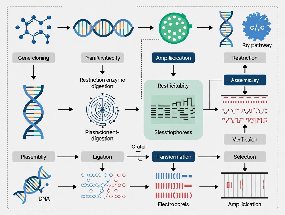
Key DNA Assembly Methods for Pathway Construction
To create pathways involving multiple genes and regulatory elements, shorter synthesized DNA fragments must be stitched together. Several highly efficient methods have been developed for this purpose.
Diagram 1: Common DNA assembly workflows for pathway engineering.
NEBuilder HiFi DNA Assembly (and related methods like Gibson Assembly) is an in vitro, sequence homology-based method. It allows for the seamless joining of multiple DNA fragments in a single-tube, isothermal reaction [21] [22]. The process involves three key enzymes acting simultaneously: an exonuclease chews back the 5' ends of DNA fragments to create single-stranded 3' overhangs; a polymerase fills in gaps within the annealed fragments; and a DNA ligase seals the nicks in the assembled DNA backbone [22]. This method is highly efficient (>95% cloning efficiency), suitable for assembling up to 12 fragments, and works with fragments from <100 bp to over 10 kb [21]. It is ideal for medium-complexity assemblies of 2-6 fragments.
Golden Gate Assembly is a restriction enzyme-based method that leverages Type IIS restriction enzymes [21] [22]. These enzymes cleave DNA outside of their recognition site, generating unique 4-base overhangs. When designed properly, multiple DNA fragments can be digested and ligated in a single-pot reaction, seamlessly assembled into a final product that lacks the original restriction sites [22]. This method is extremely efficient (>95%) and is particularly well-suited for highly complex assemblies, capable of joining up to 30-50+ fragments in a single reaction [21]. It excels with sequences containing high GC content and repetitive areas.
Polymerase Cycling Assembly (PCA) and Circular Polymerase Extension Cloning (CPEC) are methods based on overlap extension PCR [22]. In CPEC, DNA fragments with overlapping ends are mixed with a linearized vector and subjected to a PCR reaction. The polymerase extends the overlaps, splicing the fragments together and circularizing the resulting molecule in a one-step reaction. The original plasmid template is then digested, and the assembled vector is transformed into a host cell, where its endogenous repair machinery fixes any remaining nicks [22]. This method is scarless and does not require restriction enzymes or ligase.
Application Notes & Experimental Protocols
Protocol 1: Gene Assembly via NEBuilder HiFi DNA Assembly
This protocol is designed for the seamless assembly of 2-6 DNA fragments, such as when constructing a metabolic pathway for biofuel production or a gene expression cassette for a therapeutic vector.
Research Reagent Solutions
| Reagent/Material | Function/Description |
|---|---|
| NEBuilder HiFi DNA Assembly Master Mix | Proprietary blend of exonuclease, polymerase, and ligase for seamless fragment assembly [21]. |
| Linearized Vector Backbone | Plasmid digested at the intended insertion site. |
| Insert DNA Fragments | PCR-amplified or synthesized fragments with 15-30 bp overlaps with adjacent fragments/vector [21]. |
| Competent E. coli Cells | High-efficiency cells (>1 x 10^8 cfu/µg) for transformation of the assembled product. |
| Selection Agar Plates | Antibiotic-containing LB agar for selecting successful transformants. |
Procedure
- Fragment Preparation: Generate each DNA insert via PCR or synthesis. Ensure each fragment has ~15-30 bp homologous overlaps with the fragments it will connect to, and that the ends of the first and last fragments have homology to the linearized vector backbone [21]. Gel-purify all fragments to ensure purity and correct size.
- Molar Ratio Calculation: Determine the concentration (ng/µL) and length (bp) of each fragment and the vector. Use the formula:
ng of fragment = (0.02 × length of fragment) × (50 / length of vector)to calculate the amount of each fragment to use for a 1:1 molar ratio of vector to each insert. For multiple inserts, a 1:2 ratio of vector to each insert is often effective. - Assembly Reaction Setup: In a sterile PCR tube, combine the following:
- X µL Linearized Vector (calculated amount)
- X µL Insert Fragment 1 (calculated amount)
- X µL Insert Fragment 2 (calculated amount)
- 10 µL NEBuilder HiFi DNA Assembly Master Mix
- Nuclease-free water to a final volume of 20 µL. Mix the reaction by pipetting gently.
- Incubation: Incubate the reaction in a thermal cycler at 50°C for 15-60 minutes. For complex assemblies with >4 fragments, a longer incubation (up to 60 minutes) may improve results [21].
- Transformation: Transform 2-5 µL of the assembly reaction into 50 µL of high-efficiency competent E. coli cells following standard heat-shock protocols. Plate the entire transformation volume onto pre-warmed selective agar plates.
- Screening and Validation: Incubate plates overnight at 37°C. Screen resulting colonies by colony PCR and/or analytical restriction digest. Confirm the final assembly by Sanger sequencing of the entire inserted pathway.
Protocol 2: Multiplexed Promoter-RBS-Gene Assembly Using Golden Gate
This protocol is ideal for combinatorial testing of different promoters and ribosome binding sites (RBS) with a target gene in a metabolic pathway, a common task in optimizing expression levels in biofuels research.
Procedure
- Modular Part Design: Design your DNA "parts" (e.g., Promoter A, B, C; RBS X, Y, Z; Gene 1). Flank each part with the recognition site for a Type IIS restriction enzyme (e.g., BsaI). Ensure the overhangs generated are designed so that parts ligate in the correct order (e.g., Promoter overhang fuses to RBS overhang, which fuses to Gene overhang) [22].
- Source and Prepare Parts: Obtain each part as a plasmid or a PCR-amplified fragment. If using plasmids, they should not contain internal recognition sites for the chosen Type IIS enzyme; if present, these must be silently mutated.
- Golden Gate Reaction Setup: In a single PCR tube, combine:
- ~50-100 ng of destination vector (containing antibiotic resistance).
- Equimolar amounts of each insert part (Promoter, RBS, Gene).
- 1 µL of Type IIS restriction enzyme (e.g., BsaI-HFv2).
- 1 µL of T4 DNA Ligase (high concentration).
- 2 µL of 10x T4 DNA Ligase Buffer.
- Nuclease-free water to 20 µL.
- Cyclic Digestion-Ligation: Place the tube in a thermal cycler and run the following program:
- Transformation and Screening: Transform 1-5 µL of the reaction into competent E. coli. Plate on appropriate antibiotic plates. Screen colonies for the correct assemblies. Due to the high efficiency of Golden Gate, you can typically screen a small number of colonies to find all possible combinations of your modular parts [21].
Diagram 2: Engineered yeast pathway for advanced biofuel (ethanol) production from non-food biomass.
The Scientist's Toolkit
A successful pathway engineering project relies on a suite of specialized reagents and tools. The table below details essential components for DNA assembly and their functions.
Essential Research Reagent Solutions for DNA Assembly
| Tool/Reagent | Key Function in Pathway Engineering |
|---|---|
| High-Fidelity DNA Polymerase | Accurately amplifies DNA parts for assembly with minimal introduced mutations. |
| Type IIS Restriction Enzymes (e.g., BsaI, BsmBI) | Enables Golden Gate Assembly by creating unique, user-defined overhangs outside their recognition site [21] [22]. |
| DNA Ligase | Catalyzes the formation of phosphodiester bonds to seal nicks in the DNA backbone during assembly [22]. |
| Exonuclease (e.g., T5, T4) | Chews back DNA ends to create single-stranded overhangs for homologous recombination in methods like Gibson/NEBuilder HiFi [22]. |
| Competent E. coli Cells | Serve as the host for propagating assembled DNA constructs; high efficiency is crucial for complex assemblies. |
| Plasmid Vectors with Standardized Prefix/Suffix | Backbones designed for modular cloning systems (e.g., MoClo), facilitating part reuse and interoperability [22]. |
| Enzymatic DNA Synthesis Service | Provides long, accurate oligonucleotides or genes as starting points for complex pathway assembly projects [19]. |
The synergistic advancement of DNA synthesis technologies and innovative assembly protocols is directly fueling progress in two of the most critical fields of our time: advanced medicine and sustainable energy. The ability to rapidly and reliably design, write, and assemble genetic pathways is no longer a bottleneck but a powerful catalyst. For researchers and drug developers, mastering these techniques—from the simplicity of HiFi assembly to the multiplexing power of Golden Gate—is essential for translating scientific vision into real-world applications. As synthesis capabilities continue to improve, moving from reading DNA to writing it with ease, the potential for engineering biology to address global challenges in health, energy, and beyond is becoming limited less by technical constraints and more by the bounds of human imagination and understanding.
The fields of synthetic biology and metabolic engineering are fundamentally driven by two core capabilities: reading DNA (sequencing) and writing DNA (synthesis). The ability to rapidly sequence genetic material has dramatically outpaced our capacity to synthesize it, creating a significant cost gap that influences experimental design and scalability. While next-generation sequencing (NGS) technologies can generate an estimated 15 petabases of sequence data annually worldwide, the construction of synthetic biological circuits and pathways still requires a heavy dose of empirical trial and error within the design-build-test-learn cycle [23]. This application note examines the current cost structures of DNA sequencing and synthesis, details practical experimental protocols for pathway assembly, and provides researchers with a toolkit for bridging this technological divide within the context of pathway engineering research.
Cost Analysis: The Sequencing-Synthesis Landscape
Quantitative Comparison of DNA Reading vs. Writing Costs
The disparity between DNA sequencing and synthesis costs presents a fundamental challenge in synthetic biology. While the cost of sequencing a full human genome has decreased precipitously over recent decades, the expense of de novo gene synthesis has not maintained the same pace [24] [23]. The current pricing structures for both technologies reveal this persistent gap and its implications for research planning.
Table 1: DNA Sequencing Costs and Platforms (2025)
| Platform/Service | Metric | Cost | Output/Capacity | Key Applications |
|---|---|---|---|---|
| Ultima UG100 | Per human genome (30x coverage) | Not specified | >30,000 genomes/year | Large-scale whole genome sequencing |
| Element AVITI (Upgraded) | Per 1 billion reads | "Saves several hundred to one thousand dollars" compared to Illumina | 1.5B reads (300-cycle high output) | High-throughput screening, transcriptomics |
| Health Sciences Sequencing Core | Library prep (Illumina DNA Prep, ≥48 samples) | $90/sample | Varies with application | Standard WGS, targeted sequencing |
| NextSeq 2000 P3 300-cycle kit | Per run | $5,880 | 1.2T total bases | Exome, transcriptome, large genome sequencing |
Table 2: DNA Synthesis Costs and Services (2025)
| Synthesis Type | Cost Structure | Turnaround Time | Throughput/Scale | Primary Research Applications |
|---|---|---|---|---|
| Oligonucleotide synthesis | $0.05-$0.17 per base | Varies by vendor | 0.1-1.0 μmole scale | Primer assembly, site-directed mutagenesis |
| Gene synthesis (traditional) | $0.10-$0.30 per base ($100-$300 for 1kb gene) | 3-10 business days | 200-2000 bp constructs | Pathway engineering, codon optimization |
| DNA fragment synthesis | Market-specific pricing | Vendor-dependent | Multi-gene constructs | Metabolic engineering, synthetic biology |
The underlying economic factors maintaining this gap stem from fundamental technological differences. DNA sequencing is primarily a reading process that leverages enzymatic and imaging technologies that have benefited from massive scaling and automation. In contrast, DNA synthesis relies on chemical processes (typically phosphoramidite chemistry) for oligonucleotide synthesis followed by biological assembly and verification processes that remain resource-intensive [23]. This cost differential directly impacts pathway engineering research by constraining the design-build-test-learn cycle, particularly when exploring large combinatorial libraries or complex metabolic pathways requiring numerous DNA constructs.
DNA Assembly Methods for Pathway Engineering
Key DNA Assembly Technologies
Combinatorial metabolic pathway assembly requires robust, efficient DNA assembly methods that can accommodate multiple genetic parts with high fidelity. Several methods have emerged as standards for synthetic biology applications, each with distinct advantages for specific pathway engineering scenarios.
Table 3: Comparison of DNA Assembly Methods for Pathway Engineering
| Method | Mechanism | Max Parts per Reaction | Scar Characteristics | Best Applications in Pathway Engineering |
|---|---|---|---|---|
| Restriction Enzyme-based (BioBrick/BglBrick) | Type IIs restriction enzymes and ligation | 5-10 | 6-8 bp scars; may encode amino acids | Modular part assembly, educational use |
| Golden Gate Assembly | Type IIs restriction enzymes with ligation cycling | 10-20 | Scarless (properly designed) | Combinatorial library construction, multi-gene assembly |
| Gibson Assembly | Exonuclease, polymerase, and ligase in one pot | 5-15 | Scarless | Pathway construction from PCR fragments, genome assembly |
| SLIC/SLiCE | Homology-based in vitro recombination | 3-8 | Scarless | Cloning difficult fragments, multi-part assembly |
| OE-PCR/CPEC | Polymerase-based overlap extension | 3-6 | Scarless | Pathway optimization, RBS library generation |
Experimental Protocol: Combinatorial Pathway Assembly Using Golden Gate Method
This protocol describes the implementation of Golden Gate assembly for combinatorial metabolic pathway optimization, enabling researchers to efficiently test multiple enzyme variants and regulatory elements in parallel.
Materials and Reagents
- DNA Parts: Promoters, ribosome binding sites (RBS), coding sequences (CDS), and terminons in appropriate acceptor vectors
- Restriction Enzyme: BsaI-HFv2 (or similar type IIs enzyme)
- Ligase: T7 DNA Ligase
- Buffer: T4 DNA Ligase Buffer
- ATP: 10mM solution
- DpnI: For template digestion (if using PCR-amplified parts)
- Competent Cells: High-efficiency E. coli (>1×10^8 cfu/μg)
- Agar Plates: LB with appropriate selection antibiotics
- PCR Reagents: Q5 High-Fidelity DNA Polymerase, dNTPs
Step-by-Step Procedure
Part Design and Vector Preparation
- Design all DNA parts with appropriate BsaI recognition sites and 4-bp overhangs ensuring proper directional assembly
- Confirm that all internal BsaI sites have been eliminated by silent mutation if necessary
- Amplify parts using high-fidelity PCR if not already in compatible vectors
- Purify all DNA parts using silica membrane columns and quantify via fluorometry
Golden Gate Reaction Setup
- Prepare the master mix on ice:
- 2.5 μL T4 DNA Ligase Buffer (2×)
- 0.5 μL BsaI-HFv2 (5 U/μL)
- 0.5 μL T7 DNA Ligase (400 U/μL)
- 1.5 μL nuclease-free water
- Add 20-50 ng of each DNA part in equimolar ratios
- Adjust final volume to 10 μL with nuclease-free water
- Include a negative control without ligase
- Prepare the master mix on ice:
Thermocycling Conditions
- Cycle between 37°C (2-5 minutes) and 16°C (2-5 minutes) for 25-50 cycles
- Final extension at 50°C for 5 minutes
- Enzyme inactivation at 80°C for 10 minutes
- Hold at 4°C for short-term storage
Transformation and Screening
- Transform 2-5 μL of reaction into 50 μL competent E. coli cells
- Plate on selective media and incubate overnight at 37°C
- Screen 8-12 colonies by colony PCR or restriction digest
- Sequence-confirm 2-4 correct clones for each construct variant
Pathway Evaluation
- Transfer validated constructs into appropriate production chassis
- Measure pathway performance (product titer, yield, productivity)
- Analyze combinatorial library results to identify optimal configurations
Diagram 1: Design-Build-Test-Learn Cycle. This engineering cycle forms the backbone of synthetic biology and metabolic engineering efforts [23].
The Scientist's Toolkit: Research Reagent Solutions
Successful pathway engineering requires access to specialized reagents, enzymes, and genetic tools. The following table details essential components for DNA assembly and pathway optimization experiments.
Table 4: Essential Research Reagents for DNA Assembly and Pathway Engineering
| Reagent/Resource | Function | Example Applications | Key Considerations |
|---|---|---|---|
| High-Fidelity DNA Polymerase | PCR amplification with minimal errors | Part amplification, site-directed mutagenesis | Error rate, processivity, amplification length |
| Type IIs Restriction Enzymes (BsaI, BsmBI) | DNA cleavage outside recognition site | Golden Gate assembly, modular cloning | Star activity, temperature sensitivity, buffer compatibility |
| DNA Ligase (T7, T4) | Joining of DNA fragments | All assembly methods requiring ligation | Temperature optimum, fidelity, buffer compatibility |
| Phosphoramidite Reagents | Chemical synthesis of oligonucleotides | Primer synthesis, gene assembly | Coupling efficiency, depurination risk, scale |
- Assembly Kits and Toolkits: Commercial Gibson Assembly Master Mix provides optimized enzyme blends for one-step isothermal assembly. Modular cloning (MoClo) toolkits offer standardized parts for rapid pathway construction in various chassis [25].
- Specialized Competent Cells: High-efficiency cloning strains (e.g., 10^9 cfu/μg) maximize transformation success for large constructs. Protein expression strains optimize pathway performance.
- DNA Synthesis Services: Commercial providers (e.g., IDT, Twist Bioscience) offer increasingly cost-effective gene synthesis, with some specializing in long fragments or high-throughput services [18] [26].
Advanced Applications: Combinatorial Optimization Strategies
Experimental Protocol: Multi-Method Pathway Optimization
For complex metabolic engineering projects, a hierarchical approach combining multiple DNA assembly methods often yields optimal results. This protocol outlines a strategy for assembling and optimizing multi-gene pathways.
Hierarchical Assembly Workflow
Enzyme Selection and Optimization
- Identify candidate enzymes from databases (BRENDA, MetaCyc)
- Design codon-optimized sequences for target chassis
- Synthesize or amplify coding sequences with standardized prefixes/suffixes
Transcriptional Unit Assembly
- Use Golden Gate assembly to combine promoters, RBS, CDS, and terminators
- Create variants with different regulatory elements for each gene
- Assemble 2-3 transcriptional units in intermediate vectors
Pathway Assembly
- Employ Gibson Assembly to combine transcriptional units into final vector
- Alternatively, use yeast assembly for very large constructs (>50 kb)
- Transform into production chassis for functional testing
Combinatorial Library Creation
- Utilize robotic automation for high-throughput assembly
- Implement Design of Experiments (DoE) to sample design space efficiently
- Screen 100s-1000s of variants for optimal performance
Analytical and Screening Methods
- Rapid Phenotyping: Use 96-well or 384-well formats for initial screening
- Analytical Chemistry: HPLC, GC-MS, or LC-MS for product quantification
- Omics Technologies: RNA-seq to assess transcriptional profiles, proteomics for enzyme abundance
- Fermentation Optimization: Scale promising constructs to bioreactor scale
Diagram 2: Hierarchical DNA Assembly Workflow. This multi-level approach enables efficient construction of complex metabolic pathways [25].
The gap between DNA sequencing and synthesis costs continues to influence experimental design in metabolic engineering, but strategic application of modern assembly methods can maximize research efficiency. As synthesis technologies advance, emerging approaches such as enzymatic DNA synthesis and microfluidic assembly show promise for further reducing costs and increasing throughput [23]. The development of more sophisticated bioinformatics tools and automation-compatible protocols will further streamline the pathway optimization process. By implementing the protocols and strategies outlined in this application note, researchers can effectively navigate the current technological landscape while preparing for anticipated advances in DNA writing capabilities that will eventually close the read-write gap and unlock new possibilities in synthetic biology and therapeutic development.
From Oligos to Genomes: DNA Assembly Methods and Their Applications
The field of molecular biology has been revolutionized by the development of DNA assembly techniques, which serve as foundational tools for pathway engineering research. These methods enable researchers to construct complex genetic circuits, engineer metabolic pathways, and develop novel therapeutic interventions with unprecedented precision and efficiency. For researchers and drug development professionals, mastering these techniques is crucial for advancing projects in synthetic biology, gene therapy, and pharmaceutical development. Modern cloning methods have largely moved beyond traditional restriction enzyme approaches, embracing instead more flexible, efficient, and seamless assembly strategies that facilitate the construction of increasingly sophisticated genetic constructs.
Among the most powerful and widely adopted methods are Gibson Assembly and Golden Gate Cloning, each with distinct mechanisms, advantages, and optimal applications. While Gibson Assembly employs a homologous recombination-based mechanism using a multi-enzyme master mix, Golden Gate utilizes the unique properties of Type IIS restriction enzymes for a restriction-ligation approach. The selection between these methods depends on multiple project-specific factors, including the number of DNA fragments, their sizes, and the desired throughput. This application note provides a detailed comparison of these techniques, along with practical protocols and implementation guidelines to inform experimental design in pathway engineering research.
Core Principles of DNA Assembly Methods
Gibson Assembly
Gibson Assembly, developed by Daniel Gibson and colleagues, is a one-step isothermal reaction that allows for the seamless joining of multiple DNA fragments. This method employs a cocktail of three enzymes that operate simultaneously at 50°C: an exonuclease, a DNA polymerase, and a DNA ligase [27]. The mechanism begins with the exonuclease chewing back the 5' ends of DNA fragments to create single-stranded 3' overhangs. These homologous overhangs, typically 20-40 base pairs in length, then anneal to complementary sequences on adjacent fragments. The DNA polymerase fills in any remaining gaps, and finally, the DNA ligase seals the nicks in the DNA backbone, resulting in a contiguous, double-stranded molecule [27] [28].
The key advantage of this method lies in its ability to assemble up to 15 fragments simultaneously in a single reaction with high efficiency, creating seamless junctions without introducing additional nucleotide sequences ("scars") at the fusion sites [28]. Gibson Assembly is particularly valuable for constructing large DNA molecules and for applications requiring flexibility in fragment size and vector choice.
Figure 1: Gibson Assembly Workflow - A one-step isothermal reaction using three enzymes to seamlessly join DNA fragments with homologous ends.
Golden Gate Assembly
Golden Gate Assembly represents a different approach based on the unique properties of Type IIS restriction enzymes such as BsaI-HFv2, BsmBI-v2, and PaqCI [29]. Unlike traditional Type IIP restriction enzymes that cut within palindromic recognition sites, Type IIS enzymes recognize non-palindromic sequences and cut outside of their recognition sites, generating unique, user-defined 4-base overhangs that are independent of the enzyme's recognition sequence [29]. This fundamental characteristic enables the creation of custom overhangs that direct the precise, ordered assembly of multiple DNA fragments.
In a Golden Gate reaction, DNA fragments are designed with flanking Type IIS recognition sites such that digestion releases the fragment with the desired overhangs. When combined with T4 DNA ligase in the same reaction tube, the process undergoes thermal cycling between digestion and ligation temperatures. This cycling progressively digests incorrectly ligated products and amplifies correct assemblies because the desired final product no longer contains the recognition sites and is thus protected from further digestion [29]. This "one-pot" reaction can efficiently assemble up to 30 fragments or more in a single reaction, making it exceptionally powerful for combinatorial library generation and modular cloning systems [29] [28].
Figure 2: Golden Gate Assembly Workflow - A restriction-ligation method using Type IIS enzymes that cut outside recognition sites to create unique overhangs for seamless assembly.
Comparative Analysis: Gibson Assembly vs. Golden Gate
Selecting the appropriate DNA assembly method requires careful consideration of project parameters and experimental goals. The table below provides a detailed quantitative comparison to guide this decision-making process.
Table 1: Comprehensive Comparison Between Gibson Assembly and Golden Gate Cloning
| Feature | Gibson Assembly | Golden Gate Assembly |
|---|---|---|
| Enzymes Used | Exonuclease, DNA polymerase, DNA ligase [27] | Type IIS restriction enzymes, T4 DNA ligase [29] |
| Mechanism | Homologous recombination [28] | Restriction-ligation [28] |
| Reaction Conditions | Single-step, isothermal (50°C) [27] | Thermal cycling between digestion and ligation temperatures [29] |
| Seamless/Scarless | Yes [27] | Yes [29] |
| Typical Number of Fragments | Up to 15 fragments [28] | Up to 30+ fragments [28] |
| Optimal Overlap/Hang Length | 20-40 bp [27] | 4 bp overhangs [29] |
| Fragment Size Compatibility | Flexible, but fragments <200 bp can be problematic [28] | Flexible, including very short fragments [28] |
| Vector Compatibility | Any linearized vector [28] | Requires vectors with Type IIS recognition sites [29] [28] |
| Primer Design | Requires long primers with homologous overlaps [27] | Standard PCR primers with added Type IIS sites [29] |
| Multi-Fragment Efficiency | High for 2-6 fragments [28] | Very high, especially for >6 fragments [28] |
| Background Reduction | N/A | Built-in: desired product lacks recognition sites [29] |
| Cost Considerations | Generally more expensive [28] | Can be more cost-effective [28] |
Strategic Selection Guidelines
Choose Gibson Assembly when:
- Assembling a moderate number of fragments (2-6) [28]
- Working with large DNA fragments (>200 bp) [28]
- Flexibility in vector choice is required [28]
- Protocol speed is a priority (approximately one hour reaction time) [27]
Choose Golden Gate Assembly when:
- Assembling a large number of fragments (>6) in a single reaction [28]
- Performing high-throughput or combinatorial cloning [29] [28]
- Working with short DNA fragments (including <200 bp) [28]
- Building modular part systems for hierarchical assembly [29]
- Low background from empty vectors is critical [29]
Experimental Protocols
Gibson Assembly Protocol
Fragment Preparation and Primer Design
- Amplify DNA fragments via PCR using high-fidelity DNA polymerase to minimize errors [27]
- Design primers with 20-40 base pair homology overlaps at the 5' ends
- Verify fragment integrity and size through gel electrophoresis before proceeding
- Linearize your vector using restriction enzymes or PCR amplification
- Purify all DNA fragments to remove enzymes and contaminants (optional but recommended)
Assembly Reaction
- Set up reaction with recommended DNA fragment concentrations:
- For 2-3 fragments: 100 ng total DNA
- For 4-6 fragments: 200 ng total DNA
- Maintain vector:insert molar ratio between 1:2 and 1:5 [27]
- Add Gibson Assembly master mix (commercial or prepared in-house)
- Incubate at 50°C for 30-60 minutes [27]
- Transform 2-5 µL of reaction into competent E. coli cells
- Screen colonies via colony PCR, restriction digest, or sequencing
Troubleshooting Tips:
- For difficult assemblies, increase overlap length to 30-40 bp with higher GC content
- To speed up the process, use unpurified PCR products directly in the assembly [27]
- Shorten reaction time to 15 minutes for simple assemblies to save time [27]
- Use DpnI treatment when using circular plasmid DNA as PCR template to reduce background [27]
Golden Gate Assembly Protocol
Vector and Insert Design
- Select appropriate Type IIS enzyme (BsaI is recommended for beginners) [29]
- Design DNA fragments with Type IIS recognition sites flanking each fragment
- Ensure overhangs are unique and complementary only to adjacent fragments in the desired assembly
- Verify that neither vector nor inserts contain internal recognition sites for the Type IIS enzyme being used
- Remove internal sites via silent mutation or select a different Type IIS enzyme if needed
Assembly Reaction
- Set up reaction with components:
- 50-100 ng vector DNA
- Equimolar amounts of each insert fragment
- 1× T4 DNA ligase buffer
- 10 U Type IIS restriction enzyme (e.g., BsaI-HFv2)
- 400 U T4 DNA ligase [29]
- Thermal cycle using the following program:
- 25-30 cycles of:
- 37°C for 2-5 minutes (digestion)
- 16°C for 2-5 minutes (ligation)
- Final step: 50°C for 5 minutes, 80°C for 10 minutes [29]
- 25-30 cycles of:
- Transform 2-5 µL into competent cells
- Screen colonies for correct assemblies
Troubleshooting Tips:
- If efficiency is low, increase the number of thermal cycles to 30-40 cycles
- For multi-fragment assemblies, use higher enzyme concentrations
- Include a negative control (reaction without inserts) to monitor vector-only background
- Use NEBridge Ligase Fidelity Tools to design high-fidelity overhangs for multiple fragments [29]
Research Reagent Solutions
Successful implementation of DNA assembly methods requires access to high-quality reagents and tools. The following table outlines essential solutions for pathway engineering research.
Table 2: Essential Research Reagents for DNA Assembly Methods
| Reagent/Tool | Function | Examples & Notes |
|---|---|---|
| Type IIS Restriction Enzymes | Creates unique overhangs outside recognition sites for Golden Gate | BsaI-HFv2, BsmBI-v2, PaqCI [29] |
| High-Fidelity DNA Polymerase | PCR amplification of fragments with minimal errors | Platinum SuperFi II PCR Master Mix [27] |
| DNA Ligase | Seals nicks in DNA backbone | T4 DNA Ligase (Golden Gate), Taq DNA Ligase (Gibson) [29] [27] |
| Assembly Master Mixes | Pre-mixed enzymes for simplified workflow | Gibson Assembly Master Mix, NEBridge Golden Gate Assembly Kit (BsaI-HFv2) [29] [27] |
| Competent E. coli Cells | Transformation of assembled constructs | One Shot TOP10 Chemically Competent E. coli [27] |
| Golden Gate-Compatible Vectors | Destination vectors with Type IIS cloning sites | pGGAselect (compatible with BsaI, BsmBI, BbsI) [29] |
| Design Tools | In silico design of fragments and primers | NEBridge Golden Gate Assembly Tool, SnapGene [29] [27] |
Advanced Applications in Pathway Engineering
The applications of Gibson and Golden Gate assembly extend beyond basic cloning to enable sophisticated pathway engineering projects. Metabolic pathway engineering for therapeutic compound production often requires assembly of multiple genes encoding enzymatic steps in a biosynthetic pathway. Golden Gate assembly excels in this domain due to its capacity for high-fidelity, multi-fragment assembly and compatibility with modular part systems [29]. Similarly, CRISPR vector construction for gene editing applications frequently employs Gibson Assembly for its flexibility in inserting multiple components, including guide RNA expression cassettes and reporter genes, into delivery vectors [27].
Recent advances in DNA synthesis technologies have further expanded possibilities for pathway engineering. The global DNA synthesis market, valued at USD 4.97 billion in 2024 and projected to reach USD 29.98 billion by 2034, reflects the growing accessibility of synthetic DNA fragments for assembly projects [30]. Commercial gene synthesis services now provide researchers with customized, sequence-verified fragments that serve as ideal starting materials for both Gibson and Golden Gate assembly workflows, significantly accelerating the design-build-test cycle in metabolic engineering [31] [9].
Emerging technologies such as CRISPR-associated transposase (CAST) systems represent the next frontier in DNA assembly, enabling targeted integration of large DNA cargo without introducing double-strand breaks [32]. While still in early development for mammalian cells, these systems promise future capabilities for pathway engineering that complement existing assembly methods.
Gibson Assembly and Golden Gate Cloning represent two powerful, yet distinct approaches to DNA assembly for pathway engineering research. Gibson Assembly offers simplicity and flexibility for moderate numbers of fragments, while Golden Gate provides unparalleled efficiency for complex, multi-fragment assemblies. The selection between these methods should be guided by specific project requirements, including the number and size of DNA fragments, available vectors, and desired throughput.
As the field of synthetic biology continues to advance, with the DNA synthesis market experiencing rapid growth [30] [31], mastery of these DNA assembly techniques becomes increasingly essential for researchers and drug development professionals. By implementing the detailed protocols and strategic guidelines provided in this application note, scientists can effectively leverage these powerful methods to accelerate their pathway engineering projects and therapeutic development pipelines.
Combinatorial biosynthesis represents a powerful synthetic biology approach for generating structural diversity in natural products by engineering their biosynthetic pathways. This methodology enables the creation of novel "non-natural" natural products with potential enhanced therapeutic properties, addressing critical limitations in traditional drug discovery pipelines. By manipulating the genes encoding natural product biosynthesis through strategic pathway engineering, researchers can diverge synthetic routes toward previously inaccessible chemical entities. This Application Note details the fundamental principles, experimental methodologies, and practical protocols for implementing combinatorial biosynthesis, framed within the broader context of DNA synthesis and assembly techniques for pathway engineering research.
Natural products and their derivatives constitute a significant proportion of modern pharmaceuticals, particularly in anti-cancer therapies where they represent 74.8% of FDA-approved drugs from 1981 to 2010 [33]. However, traditional natural product discovery often yields rediscovery of known compounds, creating an urgent need for innovative approaches to expand chemical diversity. Combinatorial biosynthesis addresses this challenge through the manipulation of biosynthetic genes to create modified pathways that produce structural analogs [33] [34].
This approach leverages the inherent modularity of biosynthetic enzymes, particularly polyketide synthases (PKS) and non-ribosomal peptide synthetases (NRPS), which function as molecular assembly lines. The decreasing cost of DNA sequencing and synthesis has dramatically expanded the repertoire of enzymes available for pathway engineering, while bioinformatics tools like BLAST, Pfam, and CDD enable rapid prediction of enzyme function without laborious expression and isolation [33]. The integration of advanced DNA assembly techniques has further transformed combinatorial biosynthesis from a limited, painstaking process to a high-throughput methodology capable of generating extensive libraries of novel compounds [33] [25].
Key Engineering Strategies
Domain and Module Swapping
Megasynth(et)ases, such as PKS and NRPS, can be engineered through domain or module swaps to alter their catalytic functions and product output [34].
Table 1: Domain Swapping in Polyketide Synthases
| Domain Type | Function | Engineering Outcome | Example |
|---|---|---|---|
| SAT (Starter Unit Acyl Carrier Protein Transacylase) | Selects and transfers starter unit | Alters starter unit incorporation | Swapping AfoE SAT with StcA SAT produced novel polyketide with hexanoyl starter unit [34] |
| PT (Product Template) | Controls cyclization and aromatization | Changes cyclization pattern | PT swap from ApdA to PKS4 produced novel α-pyranoanthraquinone [34] |
| KS (Ketosynthase) | Catalyzes chain elongation | Controls polyketide chain length | KS domain swaps identified ten amino acids involved in chain length determination [34] |
| TE (Thioesterase) | Catalyzes product release and cyclization | Alters release mechanism and product macrocyclization | TE domain swapping converted product from flaviolin to ATHN and produced novel macrocycles [34] |
| ER (Enoylreductase) | Reduces enoyl intermediates | Modifies reduction level of polyketide | ER domain swap in DrtA produced novel drimane-type sesquiterpene esters with different saturation levels [34] |
The first successful domain swap between highly reducing (HR) PKS systems involved exchanging the KS domain from Fum1p (involved in fumonisin biosynthesis) with PKS1 (responsible for T-toxin biosynthesis). Although the chimeric PKS still produced fumonisins, the yield was significantly reduced, highlighting the importance of protein-protein interactions in maintaining pathway efficiency [34].
Pathway Reconstitution and Heterologous Expression
Complete biosynthetic pathways can be reconstituted in heterologous hosts to produce novel compounds. A prominent example includes the reconstitution of the rebeccamycin pathway in Streptomyces albus, which enabled production of the indolocarbazole core and various derivatives [35]. By expressing different combinations of genes from the rebeccamycin biosynthetic cluster alongside halogenase genes from other microorganisms, researchers generated over 30 different indolocarbazole compounds, including derivatives with chlorine atoms at novel positions [35].
de novo Pathway Assembly
Enzymes from disparate sources can be combined to create entirely novel biosynthetic pathways. For example, two flavanones (pinocembrin and naringenin) were produced in Escherichia coli by expressing a phenylalanine ammonia-lyase from the fungus Rhodotorula rubra, a 4-coumarate:CoA ligase from Streptomyces coelicolor, a chalcone synthase from Glycyrrhiza echinata, and a chalcone isomerase from Pueraria lobata [33]. This strategy was extended to produce 128 polyketide products, 42 of which were previously unreported [33].
DNA Assembly Techniques for Pathway Engineering
Traditional restriction digestion and ligation-based cloning methods are often inadequate for combinatorial biosynthesis due to their low throughput and technical limitations [33]. Recent advances in synthetic biology have introduced more efficient DNA assembly methods:
Homology-Based Assembly
The Gibson assembly method enables one-pot, isothermal assembly of multiple DNA fragments with homologous termini [33]. This process employs three enzymatic activities:
- T5 exonuclease catalyzes chew-back of 5' ends to create complementary overhangs
- Phusion polymerase fills in gaps after fragment annealing
- Taq ligase seals nicks to produce intact DNA constructs
Ligase-Based Methods
Golden Gate assembly utilizes type IIS restriction enzymes that cleave outside their recognition sequences, creating unique overhangs that facilitate seamless assembly of multiple DNA fragments in a defined order [25].
Table 2: DNA Assembly Methods for Combinatorial Biosynthesis
| Method | Principle | Key Features | Applications |
|---|---|---|---|
| Gibson Assembly | Homology-based recombination | One-pot, isothermal, no scar sequence | Pathway assembly, gene cluster construction [33] |
| Golden Gate | Type IIS restriction enzyme digestion and ligation | Standardized overhangs, modular, high efficiency | Library construction, multi-gene assemblies [25] |
| Yeast Assembly | In vivo homologous recombination | Utilizes yeast's natural recombination machinery | Large DNA construct assembly, pathway refactoring [33] |
| Mobius Assembly | Golden Gate framework with additional flexibility | Versatile, compatible with various standards | Metabolic pathway optimization [25] |
Experimental Protocol: Combinatorial Biosynthesis of Indolocarbazole Derivatives
Background
This protocol describes the combinatorial biosynthesis of indolocarbazole alkaloids, which exhibit potent antitumor and neuroprotective properties [35]. The method involves reconstituting and engineering the rebeccamycin biosynthetic pathway in a heterologous Streptomyces host to generate novel derivatives.
Materials and Reagents
- Bacterial strains: Streptomyces albus J1074 (heterologous host), L. aerocolonigenes ATCC39243 (rebeccamycin producer)
- Vectors: pEM4, pWHM3, pUWL201, pKC796 (shuttle vectors for E. coli-Streptomyces)
- Culture media: R5A medium (modified R5 medium) for Streptomyces cultivation
- Enzymes: Restriction enzymes, Phusion polymerase, T4 DNA ligase
- Analytical equipment: HPLC-MS system with C18 column, NMR spectrometer
Procedure
Gene Isolation and Vector Construction
Isolate biosynthetic genes from source organisms using PCR with primers containing appropriate restriction sites [35]:
- rebO, rebD, rebC, rebP, rebG, rebM, rebH from L. aerocolonigenes
- staC from Streptomyces sp. TP-A0274
- pyrH and thal halogenase genes from alternative sources
Clone genes into expression vectors under the control of the constitutive ermEp promoter [35]:
- Organize genes in operon-like arrangements with natural translational coupling where possible
- For long pathways, distribute genes across compatible plasmids (integrative and replicative) to reduce metabolic burden
Introduce constructs into S. albus via protoplast transformation [35]
Cultivation and Metabolite Production
Inoculate recombinant S. albus strains in R5A medium and cultivate at 30°C with appropriate antibiotics [35]
Incubate with shaking (250 rpm) for 5-7 days to allow compound production and accumulation
Metabolite Analysis and Purification
Extract metabolites from culture broth using equal volumes of ethyl acetate
Analyze extracts by HPLC-MS using the following conditions [35]:
- Column: Symmetry C18 (2.1 × 150 mm)
- Mobile phase:
- Solvent A: 1% formic acid in water
- Solvent B: acetonitrile
- Gradient: 10% B to 88% B over 30 minutes, then 100% B for 5 minutes
- Flow rate: 0.25 mL/min
- Detection: Photodiode array (200-600 nm) and mass spectrometry with electrospray ionization
Identify compounds based on:
- HPLC retention time
- UV-visible absorption spectrum
- Mass spectral data
- Comparison with authentic standards when available
Purify novel compounds for structural elucidation using preparative HPLC
Confirm structures using HRMS and NMR spectroscopy (¹H, ¹³C) [35]
Expected Results
This protocol typically yields multiple indolocarbazole derivatives with variations in:
- Halogenation pattern (e.g., 11-chlorochromopyrrolic acid, 3-chloroarcyriaflavin)
- Glycosylation pattern
- Oxidation state
The antitumor activity of novel compounds can be evaluated against tumor cell lines using assays such as the sulforhodamine B colorimetric assay [35].
The Scientist's Toolkit: Essential Research Reagents
Table 3: Key Research Reagents for Combinatorial Biosynthesis
| Reagent/Category | Specific Examples | Function/Application |
|---|---|---|
| Expression Vectors | pEM4, pWHM3, pUWL201, pKC796 | Shuttle vectors for gene expression in heterologous hosts [35] |
| Host Organisms | Streptomyces albus J1074, E. coli, S. cerevisiae | Heterologous expression chassis with different advantages [33] [35] |
| Natural Product Biosynthetic Genes | PKS, NRPS, halogenases, glycosyltransferases | Enzymes for constructing and diversifying natural product scaffolds [34] [35] |
| Culture Media | R5A medium, LB, YPD | Supports growth of microbial hosts and production of target compounds [35] |
| DNA Assembly Systems | Gibson Assembly, Golden Gate, Yeast Assembly | Methods for constructing biosynthetic pathways and gene clusters [33] [25] |
| Analytical Instruments | HPLC-MS, NMR | Detection, quantification, and structural elucidation of novel compounds [35] |
Troubleshooting and Optimization
- Low product yield: Optimize promoter strength, codon usage, and cultivation conditions; consider co-expression of chaperones for difficult-to-express enzymes [25]
- Unproductive enzyme combinations: Screen larger variant libraries; employ biosensors for rapid detection of desired products [25]
- Host toxicity: Use inducible promoters; implement dynamic pathway regulation; divide pathway between microbial consortia [33] [25]
- Incomplete pathway function: Verify gene expression and enzyme activity; supplement with cofactors; optimize metabolic flux [33]
Combinatorial biosynthesis, empowered by advanced DNA assembly techniques, provides a robust platform for generating structural diversity in natural products. The methodologies outlined in this Application Note enable researchers to engineer biosynthetic pathways for the production of novel compounds with potential therapeutic applications. As DNA synthesis and assembly technologies continue to advance, combinatorial biosynthesis approaches will play an increasingly pivotal role in drug discovery and development programs.
CRISPR-Cas Systems for Precision Genome Editing and Pathway Regulation
CRISPR-Cas systems have evolved from a prokaryotic adaptive immune mechanism into a versatile toolkit for precision genome engineering. These systems enable researchers to make targeted modifications to genomic DNA, facilitating advanced studies in functional genomics and metabolic pathway regulation. The core principle involves a guide RNA that directs a Cas nuclease to a specific DNA sequence, where it introduces a double-strand break (DSB). The cell's subsequent repair of this break—either through non-homologous end joining (NHEJ) or homology-directed repair (HDR)—allows for precise genetic alterations [36]. This technology has revolutionized pathway engineering research by providing unprecedented control over genetic elements, enabling the systematic dissection and rewiring of complex biological networks.
The classification of CRISPR-Cas systems has expanded significantly with recent discoveries. Current taxonomy now organizes these systems into 2 classes, 7 types, and 46 subtypes, reflecting substantial diversification since previous classifications that included only 6 types and 33 subtypes [37] [38]. Class 1 systems (types I, III, IV, and VII) utilize multi-protein effector complexes, while Class 2 systems (types II, V, and VI) operate through single effector proteins, with the latter being more widely adopted in biotechnology applications due to their simpler architecture [36] [39]. This expanding diversity provides researchers with an extensive molecular toolbox for addressing different genome engineering challenges.
Updated Classification and Key Characteristics
The continuous discovery of novel CRISPR-Cas variants has enriched the system diversity available for biotechnological applications. Type VII systems, recently identified mostly in archaea, employ Cas14 effector proteins with metallo-β-lactamase (β-CASP) nuclease domains that target RNA in a crRNA-dependent manner [37]. These systems lack adaptation modules and often feature CRISPR arrays with multiple substitutions, suggesting infrequent incorporation of new spacers. Analysis of the relatively few spacer hits indicates these systems primarily target transposable elements [37]. Structural studies reveal that type VII effector complexes can contain up to 12 subunits, making them among the largest Class 1 systems [37].
Additionally, newly characterized type III subtypes (III-G, III-H, and III-I) demonstrate specialized functionalities through reductive evolution. Subtypes III-G and III-H feature inactivated polymerase/cyclase domains in Cas10 and have lost the cyclic oligoadenylate (cOA) signaling pathway that induces collateral RNase activity in most type III systems [37]. The newly described subtype III-I possesses an extremely diverged Cas10 protein lacking the N-terminal polymerase/cyclase domain and a multidomain effector protein (Cas7-11i) with three fused Cas7 domains and a Cas11 domain [37]. These recently discovered variants represent the "long tail" of CRISPR-Cas diversity in prokaryotes—comparatively rare but functionally distinct systems that expand the toolkit available for specialized applications [37].
Figure 1: Updated classification of CRISPR-Cas systems showing 2 classes and 7 types. Class 1 systems utilize multi-protein effector complexes, while Class 2 systems employ single effectors.
Advanced CRISPR Systems for Large-Scale DNA Engineering
Traditional genome editing approaches that rely on double-strand breaks face limitations in efficiently integrating large DNA fragments. To address this challenge, CRISPR-associated transposase (CAST) systems have emerged as powerful tools for inserting large DNA sequences without creating DSBs. These systems combine CRISPR-guided targeting with transposase activity to enable precise integration of substantial DNA payloads [32].
The type I-F CAST system employs Cas6, Cas7, and Cas8 proteins forming the Cascade complex, which collaborates with transposase proteins TnsA, TnsB, TnsC, and TniQ to facilitate RNA-guided "cut-and-paste" transposition [32]. This system integrates DNA approximately 50 bp downstream of the target site and has demonstrated capacity for inserting donor sequences up to approximately 15.4 kb in prokaryotic hosts with nearly complete efficiency in E. coli [32]. The type V-K CAST system utilizes the single-effector protein Cas12k and follows a replicative pathway that generates cointegrate products, enabling integration of DNA payloads as large as 30 kb [32]. DNA integration occurs 60-66 bp downstream of the protospacer adjacent motif (PAM) site [32].
While CAST systems show remarkable efficiency in prokaryotes, their application in mammalian cells remains challenging. Type I-F CAST has achieved approximately 1% editing efficiency in HEK293 cells with a 1.3 kb donor DNA [32]. Recent advancements, including the metagenomically discovered V-K CAST system MG64-1, have shown improved performance—approximately 3% integration efficiency of a 3.2 kb donor at the AAVS1 locus in HEK293 cells [32]. Further engineering through directed evolution has produced the PseCAST system with enhanced potential for complex biological contexts [32].
Table 1: Performance Characteristics of CRISPR Systems for Large DNA Integration
| System | Mechanism | Max Insert Size | Efficiency (Prokaryotes) | Efficiency (Mammalian) | Key Features |
|---|---|---|---|---|---|
| HDR-based CRISPR | DSB-dependent repair | Variable | Low (~1%) | Very low (<1%) | High precision; cell cycle dependent; induces indels |
| HITI | NHEJ-mediated | Variable | Moderate | Low (1-5%) | Cell cycle independent; higher indel rates |
| Type I-F CAST | RNA-guided transposition | ~15.4 kb | Near-complete | ~1% (HEK293) | No DSBs; precise integration 50 bp downstream of target |
| Type V-K CAST | RNA-guided transposition | ~30 kb | High | ~3% (HEK293) | No DSBs; replicative pathway; integrates 60-66 bp downstream |
Quantitative Comparison of Genome Editing Platforms
The evolution of genome editing technologies has progressed from early protein-dependent systems to the current RNA-guided CRISPR platforms. Meganucleases, zinc finger nucleases (ZFNs), and transcription activator-like effector nucleases (TALENs) pioneered targeted genome modification but faced limitations in design complexity and targeting flexibility [36]. CRISPR-Cas systems dramatically simplified the targeting process by decoupling the recognition and nuclease functions—using guide RNAs for specificity and Cas proteins for cleavage activity [39].
Comparative analyses reveal significant differences in efficiency, specificity, and practical implementation across platforms. ZFNs demonstrate efficiency ranging from 0% to 12%, while TALENs show moderate efficiency of 0% to 76% [39]. CRISPR-Cas systems achieve the highest efficiency at 0% to 81% while offering substantially easier design and lower costs [39]. The CRISPR system's unique RNA-DNA recognition mechanism provides highly predictable off-target effects compared to the less predictable off-target profiles of ZFNs and TALENs [39]. Furthermore, CRISPR enables highly feasible multiplexing and large-scale library construction, capabilities that are challenging with earlier technologies [39].
Table 2: Comparative Analysis of Major Genome Editing Platforms
| Parameter | Meganuclease | ZFN | TALEN | CRISPR-Cas |
|---|---|---|---|---|
| DNA Recognition | Protein-based | Zinc finger protein | TALE protein | Guide RNA |
| Nuclease | Endonuclease | FokI | FokI | Cas9 |
| Efficiency | Low | 0-12% | 0-76% | 0-81% |
| Target Site Size | 14-40 bp | 18-36 bp/ZFN pair | 30-40 bp/TALEN pair | 22 bp |
| Design Complexity | Complex (1-6 months) | Complex (~1 month) | Complex (~1 month) | Simple (within week) |
| Cost | High | High | Medium | Low |
| Multiplexing Feasibility | Low | Less feasible | Less feasible | Highly feasible |
| Off-target Effect | Low | Less predictable | Less predictable | Highly predictable |
Experimental Protocols for Pathway Engineering
Protocol: CRISPR-Cas9 Mediated Gene Knock-in via HDR
Purpose: Precise integration of DNA sequences into specific genomic loci for pathway engineering.
Materials:
- Cas9 expression vector (e.g., pX330)
- Guide RNA template targeting genomic locus of interest
- Donor DNA template with 800-1000 bp homology arms
- Appropriate transfection reagents
- Target cells (adherent or suspension)
- Selection antibiotics (if using selective marker)
- PCR reagents for genotyping
- Surveyor or T7E1 assay for mutation detection
Procedure:
- Design and synthesis: Design gRNA with 20 bp specificity sequence followed by 5'-NGG PAM. Ensure target site is within 50 bp of desired integration site.
- Donor construction: Clone donor DNA with homology arms into appropriate vector. For large insertions (>1 kb), include 1000 bp homology arms; for smaller changes, 800 bp arms suffice.
- Transfection: Co-transfect Cas9-gRNA complex (4:1 ratio) and donor DNA into target cells using appropriate method (lipofection, electroporation).
- Selection and expansion: Apply selection 48 hours post-transfection. Culture for 7-10 days to allow integration.
- Screening: Isolate clones and screen via PCR with junction primers. Confirm integration by sequencing.
- Functional validation: Verify expression and function of integrated sequence through mRNA and protein analysis.
Troubleshooting:
- Low HDR efficiency: Optimize donor design; synchronize cell cycle; use NHEJ inhibitors.
- Off-target effects: Validate with mismatch-sensitive nucleases; use paired nickases.
Protocol: Large DNA Integration Using CAST Systems
Purpose: Insert large DNA fragments (10-30 kb) without double-strand breaks for pathway engineering.
Materials:
- CAST system plasmids (Cas genes, transposase, donor with TnsB recognition sites)
- Donor DNA (up to 30 kb) flanked by appropriate recognition sequences
- E. coli or mammalian cells (HEK293T for initial testing)
- Antibiotics for selection
- PCR reagents for verification
- Southern blot materials for large integration confirmation
Procedure:
- System assembly: Clone CAST components (Cas genes, TnsA, TnsB, TnsC, TniQ) into expression vectors.
- Donor construction: Flank donor DNA with TnsB recognition sequences (type I-F) or appropriate sites for type V-K.
- Delivery: Co-deliver CAST components and donor DNA to target cells.
- Integration: Allow 72-96 hours for integration process.
- Selection: Apply appropriate selection to identify successful integration events.
- Verification: Confirm integration via junction PCR and Southern blot.
- Stability assessment: Passage cells for 2 weeks to ensure stable maintenance.
Applications: Installation of entire metabolic pathways, large regulatory elements, or multiple gene circuits.
Figure 2: Experimental workflows for CRISPR-mediated genome editing. HDR-based editing creates precise changes using cellular repair mechanisms, while CAST systems enable large DNA integration without double-strand breaks.
Research Reagent Solutions
Table 3: Essential Research Reagents for CRISPR Pathway Engineering
| Reagent Category | Specific Examples | Function | Application Notes |
|---|---|---|---|
| CRISPR Nucleases | SpCas9, SaCas9, Cas12a, Cas12k | Target DNA recognition and cleavage | SpCas9 (NGG PAM) most common; SaCas9 smaller size for viral delivery; Cas12k for CAST systems |
| Delivery Vectors | AAV, Lentivirus, Lipid Nanoparticles | Intracellular delivery of editing components | AAV: limited capacity; Lentivirus: larger payload; LNPs: high efficiency for in vivo |
| Donor Templates | ssODN, dsDNA with homology arms | Template for HDR-mediated editing | ssODN for small changes (<100 bp); dsDNA with 800-1000 bp homology arms for larger insertions |
| Selection Markers | Puromycin, Neomycin, Fluorescent proteins | Enrichment of successfully edited cells | Antibiotic resistance for stable lines; fluorescent markers for FACS sorting |
| Validation Tools | T7E1 assay, Surveyor assay, Sanger sequencing, NGS | Detection of editing events and off-target effects | T7E1/Surveyor for initial screening; NGS for comprehensive off-target assessment |
| CAST Components | TnsA, TnsB, TnsC, TniQ | Transposase functions for large DNA integration | Required for CRISPR-associated transposase systems; species-specific variations exist |
Applications in Pathway Engineering and Therapeutic Development
CRISPR-Cas systems have demonstrated remarkable success in both basic research and clinical applications. The first approved CRISPR-based medicine, Casgevy (exagamglogene autotemcel), provides a cure for sickle cell disease and transfusion-dependent beta thalassemia through ex vivo editing of hematopoietic stem cells to restore fetal hemoglobin production [40] [41]. This landmark approval validates the therapeutic potential of precision genome editing and establishes a regulatory pathway for future CRISPR-based therapies.
Recent clinical advances include the first personalized in vivo CRISPR treatment developed for an infant with CPS1 deficiency. This bespoke therapy was created and delivered in just six months, demonstrating the accelerating pace of CRISPR therapeutic development [40]. The treatment utilized lipid nanoparticle (LNP) delivery, which enabled multiple doses to increase the percentage of edited cells—an approach not feasible with viral vectors due to immune reactions [40]. Positive outcomes from this case included symptom improvement and decreased medication dependence without serious side effects, establishing a proof-of-concept for on-demand gene editing therapies for rare genetic diseases [40].
Ongoing clinical trials continue to expand the applications of CRISPR therapeutics. Intellia Therapeutics has reported promising results from trials targeting hereditary transthyretin amyloidosis (hATTR) and hereditary angioedema (HAE), both utilizing LNP-delivered CRISPR systems that accumulate in the liver to reduce production of disease-related proteins [40]. Participants receiving higher doses showed sustained protein reduction of approximately 90% for TTR and 86% for kallikrein, with corresponding clinical improvements [40]. The ability to safely administer multiple doses of LNP-delivered CRISPR treatments represents a significant advancement in therapeutic strategy, particularly for achieving sufficient editing levels in target tissues [40].
CRISPR-Cas systems have established themselves as indispensable tools for precision genome editing and pathway regulation. The expanding diversity of naturally occurring systems, coupled with ongoing protein engineering efforts, continues to address initial limitations and broaden applications. The recent classification update to 7 types and 46 subtypes reflects the remarkable natural diversity of these systems, providing researchers with an extensive molecular toolbox [37] [38]. For pathway engineering research, the development of DSB-free editing platforms—particularly CAST systems capable of inserting large DNA fragments—represents a significant advancement for installing complex genetic circuits and entire metabolic pathways.
Future directions will likely focus on enhancing editing precision, expanding targeting scope, and improving delivery efficiency. The clinical success of ex vivo CRISPR therapies and the emergence of personalized in vivo treatments highlight the transformative potential of these technologies [40]. As the field addresses challenges related to off-target effects, delivery limitations, and immune responses, CRISPR-Cas systems are poised to become increasingly central to both basic research and therapeutic development. The integration of synthetic biology approaches with advanced CRISPR tools will further empower researchers to design and implement complex genetic pathways, accelerating progress in biotechnology and medicine.
The assembly of multi-enzyme pathways represents a cornerstone of modern synthetic biology, enabling the engineered biosynthesis of high-value compounds ranging from advanced biofuels to pharmaceutical intermediates. This field has evolved through three significant waves of innovation: the first involved rational pathway design; the second incorporated systems biology and genome-scale modeling; and the current, third wave leverages sophisticated DNA assembly techniques and synthetic biology for constructing complete non-natural metabolic pathways [42]. The core challenge in multi-enzyme pathway engineering lies in overcoming the inherent inefficiencies of traditional methods, which often lead to flux imbalances, intermediate metabolite accumulation, and suboptimal product titers [43]. DNA-assembled architectures have emerged as a transformative solution, providing precisely programmable nanoscale spatial structures that serve as ideal biological carriers for the co-immobilization and precise positioning of multiple enzyme molecules [44]. By mimicking the spatially ordered assembly found in intracellular metabolic pathways, these systems substantially enhance substrate transfer efficiency and local reaction concentrations, thereby achieving exponential signal amplification in biosensing and significant yield improvements in production systems [44]. The convergence of DNA nanotechnology with enzyme cascade engineering has heralded a new generation of high-performance biological systems, with applications spanning clinical diagnostics, environmental monitoring, sustainable chemical production, and pharmaceutical development [44] [45].
DNA Assembly Platforms for Pathway Engineering
DNA Nanostructure Architectures
DNA nanotechnology provides unprecedented spatial resolution and assembly control for organizing enzyme cascades, evolving from proof-of-concept demonstrations to a powerful paradigm for constructing next-generation biosensors and production systems [44]. The programmability of DNA self-assembly allows for meticulous spatial control over enzyme arrangement through several distinct architectural approaches:
One-Dimensional Linear Assemblies: These represent the most accessible topological configuration for organizing enzyme cascades, where enzymes are positioned along single-stranded or double-stranded DNA scaffolds with controlled spacing. This configuration facilitates substrate channeling between sequentially acting enzymes, significantly enhancing cascade efficiency compared to free enzyme systems [44].
Two-Dimensional Planar Structures: DNA origami technology exemplifies this approach, utilizing hundreds of short staple strands assembled onto a single long scaffold strand to create precisely defined two-dimensional platforms [44]. These structures offer exceptional addressability, allowing for the precise regulation of enzyme placement, inter-enzyme spacing, and orientation to optimize catalytic interactions [44].
Three-Dimensional Frameworks: Complex 3D DNA architectures, including tetrahedra, cubes, and origami-based structures, provide biomimetic compartmentalization that closely mimics natural cellular organization [44]. These frameworks enable high enzyme loading capacities and create confined microenvironments that further enhance reaction efficiency and protect enzyme functionality [44].
Advanced DNA Engineering Technologies
Recent advances in DNA engineering technologies have dramatically improved researchers' ability to efficiently build multi-gene pathway libraries where expression levels, enzyme homologs, and other attributes can be varied in a combinatorial fashion [43]. Key technologies include:
CRISPR-Based Systems: Clustered regularly interspaced short palindromic repeats (CRISPR) technology has revolutionized large-scale DNA engineering by enabling target-specific DNA insertion through the combination of CRISPR-Cas modules with recombinase enzymes [32]. This approach allows accurate and efficient one-step insertion of foreign DNA into target genes in vivo, streamlining the engineering process that previously required pre-engineering recognition sequences or genetic crossing [32]. CRISPR-based gene insertion technologies are particularly valuable for applications requiring multigene circuit engineering, reconstruction of regulatory domains, and rewiring of complex genetic networks underlying human diseases [32].
Recombinase-Assisted Assembly: Traditional site-specific recombination systems, such as Cre-lox and Flp-FRT, continue to play important roles in DNA assembly [32]. These systems enable precise DNA rearrangements including insertion, excision, and exchange of target genes across diverse cellular and tissue contexts [32]. Advanced methodologies such as Recombinase-Mediated Cassette Exchange (RMCE), Dual Integrase Cassette Exchange (DICE), and Serine recombinase-Assisted Genome Engineering (SAGE) provide robust platforms for complex pathway construction [32].
Commercial Gene Synthesis: The commercial gene synthesis industry has matured significantly, offering standardized processes for de novo gene construction [46]. Early commercial synthesis relied on step-by-step assembly using PCR, while modern approaches leverage chip-based high-throughput synthesis capable of producing thousands of gene sequences simultaneously [46]. More recently, AI-powered gene synthesis platforms have emerged, using artificial intelligence algorithms to deeply analyze and optimize gene sequences, significantly improving synthesis efficiency and accuracy for complex sequences with high GC content, repetitive sequences, or secondary structures [46].
Table 1: DNA Assembly Technologies for Pathway Engineering
| Technology | Key Features | Advantages | Typical Applications |
|---|---|---|---|
| DNA Nanostructures | Programmable spatial control; Precise enzyme positioning | Enhanced substrate channeling; Improved catalytic efficiency | Biosensing; In vitro metabolic pathways |
| CRISPR-Based Systems | RNA-guided DNA targeting; Combinatorial with recombinases | One-step insertion in vivo; High specificity | Genome integration; Pathway optimization in living cells |
| Recombinase Systems | Site-specific recombination; Wide host range | Well-characterized; Reliable efficiency | Cassette exchange; Library construction |
| Commercial Gene Synthesis | De novo gene construction; High-throughput capability | Rapid turnaround; Codon optimization | Pathway component synthesis; Library generation |
Pathway Optimization Strategies
Expression-Level Optimization
Optimizing the expression levels of individual enzymes within a pathway is crucial for achieving balanced metabolic flux and maximizing product titers. Engineered metabolic pathways often suffer from flux imbalances that can overburden the host cell and accumulate intermediate metabolites, resulting in reduced product yields [43]. Combinatorial expression libraries provide a powerful approach to address this challenge by systematically varying the expression levels of pathway enzymes. A notable methodology involves applying regression modeling to enable expression optimization using only a small number of measurements [43]. In this approach, a set of constitutive promoters spanning a wide range of expression strengths is characterized to ensure they maintain their relative strengths irrespective of the coding sequence [43]. A combinatorial library is then constructed using standardized assembly strategies, and a regression model is trained on a random sample comprising just 3% of the total library [43]. This model can subsequently predict genotypes that preferentially produce target compounds, even in highly branched pathways like the five-enzyme violacein biosynthetic pathway expressed in Saccharomyces cerevisiae [43]. This method effectively bypasses the need for high-throughput assays, which are unavailable for the vast majority of desirable target compounds.
Computational and Modeling Approaches
Computational methods play an increasingly important role in pathway optimization and metabolic engineering:
Global Optimization Techniques: Nonlinear models of metabolic pathways based on the Generalized Mass Action (GMA) representation can be globally optimized using nonconvex nonlinear programming (NLP) problems solved by outer-approximation algorithms [47]. This method relies on solving iteratively reduced NLP slave subproblems and mixed-integer linear programming (MILP) master problems that provide valid upper and lower bounds on the global solution to the original NLP [47]. This approach has been successfully applied to optimize the anaerobic fermentation pathway in Saccharomyces cerevisiae [47].
Feasibility Analysis: Identifying feasibility parametric regions that allow a system to meet physiological constraints represented through algebraic equations provides a powerful approach for metabolic engineering [47]. This technique is based on applying the outer-approximation algorithm iteratively over a reduced search space to identify regions containing feasible solutions to the problem [47]. This method can characterize feasible enzyme activity changes compatible with adaptive responses, such as the response of yeast Saccharomyces cerevisiae to heat shock [47].
Pathway Comparison Algorithms: Low-cost algorithms for metabolic pathway pairwise comparison enable researchers to identify similarities and differences between pathways across organisms [48]. These algorithms transform two-dimensional pathway graphs into one-dimensional linear structures using traversal algorithms (breadth-first or depth-first), then apply traditional sequence alignment techniques including global, local, and semi-global alignment to generate numerical comparison values [48]. Such comparisons provide insights for phylogenetic evolution studies and discovering novel metabolic capabilities [48].
Table 2: Pathway Optimization Methods and Applications
| Optimization Method | Key Principle | Technical Approach | Representative Application |
|---|---|---|---|
| Combinatorial Expression Tuning | Balancing enzyme expression to minimize metabolic burden | Regression modeling of promoter libraries | Violacein pathway in S. cerevisiae [43] |
| Global Optimization | Identifying theoretical optimum enzyme activities | Nonconvex NLP with outer-approximation algorithm | Anaerobic fermentation in S. cerevisiae [47] |
| Feasibility Analysis | Identifying parameter regions meeting physiological constraints | Iterative application of optimization over reduced search space | Heat shock response in S. cerevisiae [47] |
| Modular Pathway Engineering | Dividing pathways into discrete functional units | Independent optimization of pathway modules | ncAA production from glycerol [45] |
Application Notes and Protocols
Protocol 1: Assembly of DNA Nanostructures for Enzyme Co-immobilization
Principle: This protocol describes the design and assembly of DNA origami structures for the precise spatial organization of enzyme cascades, enhancing substrate channeling and overall pathway efficiency [44].
Materials:
- Scaffold DNA (e.g., M13mp18 genome, 7249 nucleotides)
- Staple strands (approximately 200 unique sequences)
- Enzyme-DNA conjugates with complementary modifications
- Folding buffer: 5-40 mM Tris, 1-50 mM EDTA, 5-20 mM MgCl₂, pH 7.5-8.5
- Thermal cycler or water bath
Procedure:
- Design Phase:
- Select appropriate DNA origami architecture (2D sheet, 3D tetrahedron, etc.) based on the number of enzymes and required spatial arrangement.
- Design staple strands with appropriate extensions for enzyme attachment at predetermined positions.
- Modify enzymes with DNA handles complementary to the staple extensions using chemical conjugation or enzymatic labeling.
Assembly Phase:
- Mix scaffold DNA (10-50 nM) with a 5-10× molar excess of staple strands in folding buffer.
- Perform thermal annealing ramp: Heat to 80-95°C for 5-15 minutes, then cool gradually to 4-25°C over 1-24 hours.
- Purify assembled structures using agarose gel electrophoresis or PEG precipitation.
Enzyme Loading:
- Incubate purified DNA nanostructures with enzyme-DNA conjugates at stoichiometric ratios.
- Use slow annealing from 37°C to 4°C over 2-8 hours to facilitate hybridization.
- Remove unbound enzymes using size exclusion chromatography or centrifugal filters.
Validation:
- Confirm structural integrity using atomic force microscopy or transmission electron microscopy.
- Verify enzyme loading efficiency through fluorescence quantification or activity assays.
- Assess cascade activity by monitoring substrate-to-product conversion compared to free enzyme systems.
Troubleshooting:
- Incomplete folding: Optimize Mg²⁺ concentration (typically 10-20 mM) and annealing rate.
- Low enzyme loading: Verify conjugation efficiency and increase incubation time.
- Reduced enzyme activity: Ensure conjugation does not occlude active sites; consider alternative attachment sites.
Protocol 2: Multi-enzyme Cascade Assembly for Non-Canonical Amino Acid Production
Principle: This protocol outlines the construction of a modular multi-enzyme cascade for synthesizing non-canonical amino acids (ncAAs) from glycerol, demonstrating principles applicable to biofuel and pharmaceutical production [45].
Materials:
- Plasmid system with modular cloning sites (e.g., Golden Gate, Gibson Assembly)
- Enzyme modules: Alditol oxidase (AldO), catalase, d-glycerate-3-kinase (G3K), d-3-phosphoglycerate dehydrogenase (PGDH), phosphoserine aminotransferase (PSAT), polyphosphate kinase (PPK), glutamate dehydrogenase (gluGDH), O-phospho-L-serine sulfhydrylase (OPSS)
- Nucleophilic substrates (thiols, azoles, selenols)
- Cofactors: PLP, NAD+, ATP
- Glycerol substrate
- Analytical equipment: HPLC, LC-MS
Procedure:
- Pathway Design and Modularization:
- Divide the pathway into three functional modules:
- Module I (Oxidation): Glycerol → Glycerate (AldO + catalase)
- Module II (Phosphorylation and Amination): Glycerate → O-phospho-L-serine (G3K + PGDH + PSAT + PPK + gluGDH)
- Module III (Nucleophilic Addition): OPS + nucleophile → ncAA (OPSS)
- Clone each module into separate expression vectors or a single polycistronic vector.
- Divide the pathway into three functional modules:
Enzyme Engineering:
- Perform directed evolution on key enzymes (e.g., OPSS) for enhanced catalytic efficiency toward non-natural substrates.
- Use error-prone PCR or site-saturation mutagenesis followed by high-throughput screening.
- For OPSS evolution, focus on expanding active site accessibility for diverse nucleophiles.
Cascade Assembly and Optimization:
- Express enzyme modules in appropriate host (E. coli or S. cerevisiae).
- Lyse cells and combine crude extracts in stoichiometric ratios based on enzyme activities.
- Alternatively, co-express all modules in a single host for in vivo production.
- Fine-tune enzyme ratios using promoter engineering or ribosomal binding site modification.
Process Scale-Up:
- Establish reaction conditions: 50-200 mM glycerol, 1.5-2.5 equiv nucleophile, 2-10 mM MgCl₂, pH 7.5-8.5, 25-37°C.
- Implement ATP regeneration system using polyphosphate and PPK.
- Scale reaction from milliliter to liter scale with continuous substrate feeding.
- Monitor reaction progress by HPLC and isolate products using ion-exchange chromatography.
Applications: The produced ncAAs serve as building blocks for pharmaceuticals, including kynureninease inhibitors synthesized from S-phenyl-L-cysteine [45].
The Scientist's Toolkit: Research Reagent Solutions
Table 3: Essential Research Reagents for Multi-Enzyme Pathway Assembly
| Reagent/Category | Function | Examples/Specifications | Key Suppliers |
|---|---|---|---|
| DNA Assembly Systems | Modular construction of genetic pathways | Gibson Assembly, Golden Gate, BioBricks | New England Biolabs, Thermo Fisher |
| CRISPR-Cas Systems | Targeted genome integration | Cas9, Cas12, base editors | Integrated DNA Technologies, Addgene |
| Promoter Libraries | Tunable expression control | Constitutive and inducible promoters with varying strengths | Twist Bioscience, ATCC |
| Enzyme Expression Hosts | Heterologous protein production | E. coli BL21, S. cerevisiae, P. pastoris | Academic stock centers, commercial vendors |
| Specialized Nucleotides | DNA nanostructure assembly | Modified staples, fluorescent probes | Sigma-Aldrich, Eurofins Genomics |
| Cofactor Regeneration | Sustaining catalytic cycles | ATP, NAD(P)H regeneration systems | Roche, Sigma-Aldrich |
| Analytical Standards | Pathway validation and quantification | Reference compounds for metabolites | USP, Cerilliant, Sigma-Aldrich |
Pathway Visualization and Workflows
DNA-Assembled Multi-Enzyme Pathway Workflow
Modular Multi-Enzyme Cascade for ncAA Production
The field of multi-enzyme pathway assembly continues to evolve rapidly, with DNA-assembled architectures leading the transformation from simple enzyme mixtures to sophisticated spatially organized systems [44]. Future developments will likely focus on increasing the complexity of engineerable pathways, enhancing the stability of DNA-enzyme complexes, and improving scalability for industrial applications [44] [45]. The integration of machine learning approaches for pathway design and optimization represents a particularly promising direction, potentially enabling the predictive design of efficient multi-enzyme systems without extensive trial and error [46]. Additionally, the emergence of in vivo synthesis approaches, which use living cells as "factories" to synthesize target genes directly within the organism by regulating gene expression and metabolic pathways, points toward a future where pathway assembly and optimization become increasingly integrated with cellular function [46]. As these technologies mature, they will undoubtedly expand the range of accessible compounds and improve the economic viability of biologically produced biofuels, pharmaceuticals, and specialty chemicals, ultimately contributing to more sustainable manufacturing paradigms.
Optimizing for Success: Enhancing Fidelity, Efficiency, and Specificity
High-fidelity oligonucleotide synthesis is a foundational technology for advanced research in synthetic biology, metabolic engineering, and therapeutic development. The accuracy of synthesized DNA and RNA fragments directly impacts the success of downstream applications, including gene assembly, pathway engineering, and diagnostic probe development. Error reduction is particularly critical in large-scale DNA construction projects where synthetic pathways are optimized through combinatorial assembly of genetic parts [22]. This application note outlines established and emerging strategies to minimize errors during oligonucleotide synthesis, purification, and verification, providing researchers with practical methodologies to enhance the reliability of synthetic genetic constructs for pathway engineering research.
Key Strategic Approaches for Error Minimization
The pursuit of high-fidelity oligonucleotides involves a multi-faceted approach addressing chemical processes, purification methodologies, and verification techniques. Successful implementation requires understanding both the sources of errors and the technologies available to mitigate them.
Table 1: Strategic Approaches for Error Reduction in Oligonucleotide Synthesis
| Strategy | Methodology | Key Advantage | Implementation Consideration |
|---|---|---|---|
| Advanced Synthesis Chemistry | Enzymatic synthesis vs. traditional phosphoramidite | Reduces error rates for long oligos (>100 bases); more sustainable process [49] | Higher cost for novel chemistries; requires process optimization |
| AI-Enhanced Sequence Design | Machine learning algorithms for oligo design | Predicts secondary structures; optimizes for thermal stability; reduces synthesis failures by 30% [49] | Dependent on quality training data; requires specialized software platforms |
| High-Fidelity Purification | HPLC purification with quality control | Removes truncated sequences; improves purity for sensitive applications | Adds 30-35% to production costs; requires specialized equipment [49] |
| Post-Synthesis Error Correction | Array-based synthesis with error removal | Enables construction of long DNA fragments with >99.95% accuracy [49] | Not widely accessible; primarily used by specialized synthesis facilities |
| Rigorous Verification | Mass spectrometry (MALDI-TOF) sequencing | Confirms sequence identity and detects modifications [50] | Requires specialized instrumentation and expertise |
Chemical Process Optimization
The foundation of high-fidelity oligonucleotide synthesis lies in optimizing the chemical process itself. Traditional phosphoramidite chemistry remains the industry standard but faces challenges with long oligonucleotides, where error rates can exceed 15% for sequences above 100 bases [49]. Key optimization parameters include:
- Coupling efficiency: Each coupling step must exceed 99.5% efficiency to ensure acceptable yields for long fragments, monitored through trityl cation release measurement during synthesis [50].
- Deprotection conditions: Standard cleavage from controlled-pore glass (CPG) supports using ammonia/methylamine (AMA) mixture, followed by removal of protecting groups [50].
- Modified phosphoramidites: Specialty reagents with improved coupling kinetics can enhance step-wise yields, particularly for difficult sequences prone to secondary structure formation.
Emerging enzymatic synthesis technologies present a promising alternative, offering a cleaner, more sustainable process with reduced error rates for long oligonucleotides [49]. Although not yet widely adopted, these systems demonstrate potential for overcoming inherent limitations of traditional chemical synthesis.
Purification and Verification Techniques
Rigorous purification and verification are essential components of a high-fidelity synthesis pipeline, particularly for therapeutic applications or complex pathway assembly.
Purification methodologies include:
- Polyacrylamide gel electrophoresis (PAGE): Effectively separates full-length products from truncated failure sequences, suitable for research-grade oligonucleotides [50].
- High-performance liquid chromatography (HPLC): Provides superior resolution for therapeutic-grade applications, though it adds significantly to production costs [49].
- Desalting and concentration: Final cleanup using reversed-phase chromatography (e.g., C18 columns) prepares oligonucleotides for downstream applications [50].
Verification technologies encompass:
- Mass spectrometric analysis: Matrix-assisted laser desorption/ionization time-of-flight (MALDI-TOF) mass spectrometry confirms oligonucleotide identity and modification incorporation [50].
- Next-generation sequencing: For complex libraries or pooled oligonucleotides, NGS provides comprehensive analysis of sequence populations.
- UV-visible spectroscopy: Quantifies yield and assesses purity through absorbance ratios [50].
Experimental Protocols
Solid-Phase Oligonucleotide Synthesis Using Phosphoramidite Chemistry
This protocol describes the synthesis, purification, and characterization of RNA oligonucleotides, adaptable for DNA synthesis with appropriate reagent modifications [50].
Materials and Equipment
Research Reagent Solutions
| Item | Function | Specification |
|---|---|---|
| Phosphoramidites | Nucleotide building blocks | Canonical (A, U, C, G) and modified versions; 0.1M in anhydrous acetonitrile [50] |
| Controlled-Pore Glass (CPG) | Solid support | Functionalized with initial nucleoside (40 µmol scale) [50] |
| Activator | Coupling agent | 0.25M Benzothiazole-2-sulfonic acid (BTT) in acetonitrile [50] |
| Oxidizer | Stabilizes phosphate linkage | 0.02M Iodine in THF/Pyridine/Water [50] |
| Deprotection Reagents | Cleavage and deprotection | AMA (ammonia/methylamine); HF/triethylamine/N-methylpyrrolidinone for silyl group removal [50] |
| Capping Reagents | Block uncoupled chains | Phenoxyacetic anhydride (Pac2O) and 1-methylimidazole (NMI) in THF [50] |
Equipment
- DNA/RNA synthesizer with phosphoramidite chemistry capability
- Oven-dried amber glass bottles (10 mL) with septum tops
- Heating block or oven (65°C) for deprotection
- Polyacrylamide gel electrophoresis apparatus
- HPLC system with C18 column (optional)
- MALDI-TOF mass spectrometer
- UV-Vis spectrophotometer
Step-by-Step Procedure
Solid-Phase Synthesis
- Phosphoramidite Preparation: Weigh each phosphoramidite (canonical and modified) into oven-dried amber bottles under argon atmosphere. Dilute to 0.1M concentration in anhydrous acetonitrile using gas-tight syringes [50].
- Instrument Setup: Load synthesis sequence into instrument software (e.g., OligoNet). Program synthesis cycle with appropriate step parameters for RNA or DNA chemistry.
- Synthesis Cycle Execution: Initiate automated synthesis with the following key steps repeated for each nucleotide addition [50]:
- Detritylation: Remove 5'-protecting group with trichloroacetic acid in dichloroethane (3% w/v).
- Coupling: Activate phosphoramidite with BTT reagent (0.25M), delivering for 2× longer than standard DNA coupling (typically 2×150 seconds).
- Capping: Block unreacted chains with phenoxyacetic anhydride and 1-methylimidazole.
- Oxidation: Stabilize phosphite triester linkage with iodine solution.
- Trityl Monitoring: Monitor detritylation steps to calculate coupling efficiencies (>99% recommended) [50].
Deprotection and Cleavage
- Initial Deprotection: Incubate CPG-bound oligonucleotide in AMA solution (1:1 v/v) at 65°C for 30 minutes to cleave from support and remove nucleobase protecting groups.
- Evaporation: Transfer supernatant to new tube and evaporate to dryness under vacuum.
- Silyl Group Removal: For RNA oligonucleotides, treat with triethylamine trihydrofluoride in DMSO (1:4 v/v) at 60°C for 2.5 hours to remove 2'-O-TBDMS protecting groups [50].
- Precipitation: Add 1/10 volume 3M sodium acetate and 3 volumes n-butanol, incubate at -20°C for 1 hour, centrifuge, and wash pellet with 70% ethanol.
Purification and Characterization
- Gel Electrophoresis: Purify oligonucleotides using 20% denaturing polyacrylamide gel electrophoresis. Visualize bands by UV shadowing, excise, and elute into 0.5M ammonium acetate overnight [50].
- Desalting: Desalt eluted oligonucleotides using C18 reversed-phase chromatography cartridges.
- Quantification: Measure concentration by UV-Vis spectroscopy using appropriate extinction coefficients.
- Mass Verification: Confirm identity by MALDI-TOF mass spectrometry [50].
Figure 1: Workflow for solid-phase oligonucleotide synthesis using phosphoramidite chemistry, highlighting the cyclic nature of nucleotide addition and quality monitoring steps [50].
Error Assessment and Quality Control Protocol
Materials and Equipment
- Purified oligonucleotides from synthesis protocol
- MALDI-TOF mass spectrometer
- UV-Vis spectrophotometer
- Denaturing polyacrylamide gel equipment
- NGS platform (for library quality control)
Procedure
Mass Spectrometric Analysis
- Sample Preparation: Mix 0.5-1μL of purified oligonucleotide (0.1-1 nmol/μL) with matrix solution (e.g., 3-hydroxypicolinic acid).
- Instrument Analysis: Acquire mass spectra using MALDI-TOF instrument in negative ion mode.
- Data Interpretation: Compare observed mass with theoretical calculation. Mass deviations >0.05% indicate potential sequence errors or incomplete deprotection [50].
Next-Generation Sequencing for Library Validation
- Library Preparation: Amplify oligonucleotide pools using adapter-specific primers.
- Sequencing: Run on appropriate NGS platform (Illumina, PacBio, or Oxford Nanopore).
- Error Analysis: Map sequences to expected designs to calculate error rates and identify common error types (deletions, insertions, substitutions).
Functional Validation in Pathway Engineering Context
- Assembly Test: Incorporate synthesized oligonucleotides into standard assembly systems (Golden Gate, Gibson Assembly).
- Transformation Efficiency Assessment: Clone assembled constructs into appropriate host chassis (E. coli, yeast).
- Sequence Verification: Sanger sequence multiple clones to determine functional error rate after assembly.
Integration with DNA Assembly Technologies for Pathway Engineering
High-fidelity oligonucleotides serve as essential building blocks for complex DNA assembly projects in pathway engineering. The accuracy of initial oligonucleotides directly impacts the success of subsequent assembly steps and the functionality of engineered metabolic pathways.
Assembly Methods for Pathway Construction
Table 2: DNA Assembly Methods Compatible with Synthetic Oligonucleotides
| Method | Mechanism | Fragment Capacity | Advantages for Pathway Engineering |
|---|---|---|---|
| NEBuilder HiFi DNA Assembly | In vitro homologous recombination | Up to 12 fragments [51] | >95% cloning efficiency; suitable for 2-6 fragment pathway assemblies [51] |
| Golden Gate Assembly | Type IIS restriction enzyme digestion and ligation | Up to 50+ fragments (with optimization) [51] | >95% efficiency; ideal for modular pathway swapping and high-complexity assemblies [51] |
| Gibson Assembly | One-step isothermal assembly | 2-6 fragments (typical) | Seamless cloning; minimal sequence requirements |
| Yeast Assembly | In vivo homologous recombination | 10+ fragments (typical) | Suitable for very large constructs (>100 kb); utilizes cellular repair machinery |
Figure 2: Integration of high-fidelity oligonucleotides into DNA assembly workflows for metabolic pathway engineering, showing multiple compatible assembly methods leading to functional pathway validation.
CRISPR-Assisted Integration for Large-Scale DNA Engineering
Emerging CRISPR-associated transposon (CAST) systems enable precise integration of large DNA fragments without introducing double-strand breaks, leveraging RNA-guided targeting for pathway installation [32]. These systems offer advantages for chromosomal integration of engineered pathways:
- Type I-F CAST systems: Enable integration of donor sequences up to ~15.4 kb in prokaryotic hosts with nearly 100% insertion efficiency in E. coli [32].
- Type V-K CAST systems: Capable of integrating DNA fragments up to 30 kb, though efficiency in mammalian cells remains low (approximately 3% in HEK293 cells) [32].
- Advanced systems: Engineered PseCAST systems developed through directed evolution show promise for complex biological contexts [32].
Minimizing errors in oligonucleotide synthesis requires integrated approach spanning chemical optimization, purification refinement, and rigorous validation. Implementation of the strategies outlined in this application note enables researchers to achieve the sequence fidelity necessary for demanding applications in pathway engineering and therapeutic development. As DNA synthesis technologies continue to advance, with enzymatic methods and AI-assisted design platforms maturing, further improvements in fidelity and efficiency are anticipated. These advancements will in turn support more ambitious synthetic biology projects, including genome-scale engineering and complex metabolic pathway optimization for bioindustrial applications.
The precision of CRISPR-Cas systems has revolutionized genome engineering, yet off-target effects and cytotoxicity remain significant challenges for therapeutic applications and functional genomics research. Off-target editing occurs when the CRISPR machinery induces unintended genetic modifications at sites other than the intended target, primarily due to tolerance for mismatches between the guide RNA (gRNA) and genomic DNA [52]. Concurrently, cytotoxicity can manifest through multiple mechanisms, including prolonged nuclease expression, excessive DNA damage, and cellular stress responses triggered by editing components [53]. These challenges are particularly pronounced in clinical settings where off-target mutations in oncogenes or tumor suppressor genes could have serious consequences, and cytotoxicity can limit editing efficiency and therapeutic efficacy [52].
The growing emphasis on pathway engineering research necessitates highly precise editing tools that minimize collateral damage to cellular systems. Within the framework of DNA synthesis and assembly techniques, advancements in bioinformatics, protein engineering, and experimental design are converging to address these hurdles systematically [9]. This application note provides a structured overview of current strategies, quantitative comparisons, detailed protocols, and practical tools to help researchers overcome these critical limitations in CRISPR-based experiments.
Strategic Approaches for Minimizing Off-Target Effects
Selection and Engineering of High-Fidelity CRISPR Systems
Choosing appropriate CRISPR systems forms the foundation for reducing off-target activity. While wild-type Streptococcus pyogenes Cas9 (SpCas9) can tolerate 3-5 base pair mismatches, leading to substantial off-target potential, several engineered alternatives now offer improved specificity [52]. High-fidelity Cas9 variants, such as SpCas9-HF1 and eSpCas9(1.1), incorporate mutations that reduce non-specific interactions with the DNA backbone, thereby strengthening dependency on precise guide RNA:DNA complementarity [52].
Emerging technologies beyond standard Cas9 nucleases further expand the toolbox. CRISPR-Cas12a systems exhibit different off-target profiles and PAM requirements, providing alternative targeting options [52]. Base editing and prime editing systems, which utilize catalytically impaired or nickase Cas variants, offer particularly promising avenues for reducing off-target effects since they avoid double-strand breaks (DSBs) – a significant source of genotoxicity and chromosomal abnormalities [32]. For epigenetic modifications using dCas9-effector fusions, off-target binding remains a concern despite the absence of cleavage, emphasizing the continued importance of careful gRNA design [52].
Artificial intelligence is now accelerating the development of novel editors with naturally improved specificity. Recently, AI-generated Cas proteins, such as OpenCRISPR-1, have demonstrated comparable or improved activity and specificity relative to SpCas9 while being highly divergent in sequence (approximately 400 mutations away) from natural variants [54]. These systems represent a new frontier in nuclease engineering, bypassing evolutionary constraints to optimize functional properties.
Table 1: Comparison of CRISPR Systems and Their Off-Target Profiles
| CRISPR System | Type | Key Features | Reported Off-Target Reduction | Primary Applications |
|---|---|---|---|---|
| SpCas9 (WT) | Nuclease | Standard editor, broad PAM (NGG) | Baseline | General knockout, gene editing |
| SpCas9-HF1 | High-fidelity nuclease | Engineered for reduced non-specific DNA binding | >85% reduction vs. WT [52] | Therapeutic development |
| eSpCas9(1.1) | High-fidelity nuclease | Reduced DNA binding affinity | >80% reduction vs. WT [52] | Therapeutic development |
| Cas12a (Cpf1) | Nuclease | Different PAM (TTTN), staggered cuts | Different profile, potentially fewer off-targets in AT-rich regions [52] | Gene editing, multiplexing |
| OpenCRISPR-1 | AI-designed nuclease | ~40-60% sequence identity to natural Cas9s [54] | Comparable or improved vs. SpCas9 [54] | Broad research and commercial |
| dCas9-Base Editor | Base editor | No DSBs; converts C→T or A→G | Significant reduction vs. nuclease [32] | Point mutation correction |
| Prime Editor | Prime editor | No DSBs; reverse transcriptase template | Very high specificity [32] | Precision genome editing |
| CAST (I-F, V-K) | Transposase-integrated | RNA-guided transposition without DSBs | Minimal off-target integration reported [32] | Large DNA insertion (up to 30 kb) |
Computational gRNA Design and Optimization
Guide RNA design represents the most controllable factor in minimizing off-target effects. Computational tools have become indispensable for predicting and ranking gRNAs based on their potential for off-target activity. These tools leverage algorithms that consider multiple parameters, including sequence homology, genomic context, and predicted binding energetics [52].
Effective gRNA design incorporates several key principles. First, guides with higher GC content (40-60%) generally exhibit improved specificity due to stabilized DNA:RNA duplex formation at the intended target. Second, avoiding guides with significant homology to other genomic regions, particularly in the seed sequence near the PAM site, is crucial. Tools like CRISPOR provide off-target scores that rank guides based on their predicted on-target to off-target activity ratio, enabling researchers to select optimal candidates before experimental validation [52].
Chemical modifications of synthetic gRNAs offer an additional strategy to enhance specificity. Incorporating 2'-O-methyl analogs (2'-O-Me) and 3' phosphorothioate bonds (PS) at specific positions in the guide RNA can reduce off-target editing while maintaining or even improving on-target efficiency [52]. These modifications increase nuclease resistance and can alter binding kinetics to favor on-target sites. For in vivo applications, shorter gRNAs (17-19 nucleotides instead of 20) have demonstrated reduced off-target activity while often retaining sufficient on-target efficiency, providing a simple yet effective optimization strategy [52].
Experimental Detection and Analysis Methods
Comprehensive assessment of off-target effects requires robust experimental methods that can identify both predicted and unpredicted editing events. The selection of appropriate detection strategies depends on research goals, required sensitivity, and available resources. These methods generally fall into three categories: candidate site approaches, genome-wide screening methods, and targeted enrichment techniques.
Table 2: Methods for Detecting CRISPR Off-Target Effects
| Method | Principle | Sensitivity | Advantages | Limitations | Suitable for |
|---|---|---|---|---|---|
| Candidate Site Sequencing | PCR amplification & sequencing of predicted off-target sites | Moderate | Simple, cost-effective, quantitative | Limited to predicted sites; may miss true off-targets | Initial screening, low-risk applications |
| GUIDE-seq | Captures DSB sites via integration of a double-stranded oligodeoxynucleotide tag | High (detects rare events) | Unbiased; genome-wide; identifies DSBs | Requires transfection of double-stranded tag; not for all cell types | Comprehensive off-target profiling |
| CIRCLE-seq | In vitro circularization and sequencing of genomic DNA to detect Cas9 cleavage sites | Very high (in vitro) | Ultra-sensitive; works with any DNA source | In vitro method; may not reflect cellular context | Preclinical safety assessment |
| DISCOVER-Seq | Relies on MRE11 recruitment to DSBs detected by ChIP-seq | High | In vivo relevance; identifies active DSB repair | Complex protocol; requires specific antibodies | In vivo and primary cell editing |
| CAST-Seq | Detection of chromosomal rearrangements and large deletions | High for structural variants | Specifically identifies genomic rearrangements | May not detect small indels | Safety assessment for therapeutics |
| Whole Genome Sequencing (WGS) | Comprehensive sequencing of entire genome | Ultimate comprehensive-ness | Most complete picture; detects all variants | Expensive; computationally intensive; may require deep sequencing | Final therapeutic validation, rigorous safety studies |
Detailed Protocol: Off-Target Assessment Using GUIDE-Seq
Principle: GUIDE-seq (Genome-wide Unbiased Identification of DSBs Enabled by Sequencing) captures double-strand break sites through the incorporation of a double-stranded oligodeoxynucleotide (dsODN) tag, providing an unbiased method for detecting CRISPR-Cas9 off-target activity in living cells [55].
Materials:
- GUIDE-seq dsODN (double-stranded oligodeoxynucleotide tag)
- Lipofectamine CRISPRMAX or similar transfection reagent
- Cas9 nuclease and designed sgRNA
- Target cells (adherent or suspension)
- PCR reagents and NGS library preparation kit
- Next-generation sequencing platform
Procedure:
- Cell Preparation: Plate 2×10⁵ HEK293T cells (or other relevant cell type) in a 24-well plate 24 hours before transfection to achieve 70-80% confluency at time of transfection.
Transfection Complex Formation:
- Prepare Solution A: Dilute 1.5 µL of GUIDE-seq dsODN (100 µM stock), 100 ng Cas9 expression plasmid (or 50 ng if using Cas9 ribonucleoprotein), and 50 ng sgRNA expression plasmid in 25 µL Opti-MEM.
- Prepare Solution B: Dilute 1.5 µL Lipofectamine CRISPRMAX in 25 µL Opti-MEM.
- Combine Solutions A and B, mix gently, and incubate for 10-20 minutes at room temperature.
Transfection: Add the transfection complex dropwise to cells. Gently swirl the plate to distribute evenly.
Harvest and DNA Extraction: Incubate cells for 72 hours at 37°C, 5% CO₂. Harvest cells using trypsinization and extract genomic DNA using the DNeasy Blood & Tissue Kit or similar.
Library Preparation and Sequencing:
- Perform PCR amplification of tagged integration sites using GUIDE-seq primary and nested PCR primers.
- Purify PCR products and quantify using a fluorometric method.
- Prepare sequencing libraries using the Illumina TruSeq Nano DNA LT Library Preparation Kit or equivalent.
- Sequence on an Illumina MiSeq or HiSeq platform (minimum 2 million reads per sample).
Bioinformatic Analysis:
- Process raw sequencing data using the established GUIDE-seq bioinformatics pipeline [55].
- Align reads to the reference genome and identify dsODN integration sites.
- Filter and annotate significant off-target sites based on read counts and genomic location.
- Compare identified sites with in silico predictions from tools like CRISPOR.
Troubleshooting Notes:
- Low tag integration: Optimize dsODN concentration and transfection efficiency.
- High background: Include proper negative controls (transfected without Cas9/sgRNA).
- Limited off-target detection: Ensure adequate sequencing depth and coverage.
GUIDE-seq Experimental Workflow
Advanced Engineering Strategies for Enhanced Specificity
CRISPR-Associated Transposase Systems for Large DNA Integration
CRISPR-associated transposase (CAST) systems represent a revolutionary approach for large-scale DNA engineering that circumvents the primary sources of CRISPR genotoxicity. These systems combine RNA-guided targeting with transposase-mediated integration, enabling precise insertion of large DNA fragments (up to 30 kb) without creating double-strand breaks [32].
The type I-F CAST system, derived from Vibrio cholerae, utilizes a Cascade complex (Cas6, Cas7, Cas8) for target recognition and a heteromeric transposase complex (TnsA, TnsB, TnsC) for DNA integration approximately 50 bp downstream of the target site [32]. Similarly, type V-K CAST systems employ a single-effector Cas12k protein with TniQ, facilitating integration 60-66 bp downstream of the PAM site through a replicative pathway [32]. While editing efficiencies in mammalian cells currently range from 0.06% to 3% depending on the system and donor size, ongoing engineering efforts are rapidly improving these metrics [32].
CAST systems are particularly valuable for pathway engineering applications requiring the insertion of entire biosynthetic pathways or large regulatory elements. Their avoidance of DSBs significantly reduces cellular stress and potential cytotoxicity associated with DNA damage response activation. Furthermore, the unidirectional nature of transposase-mediated integration minimizes the genomic rearrangements commonly observed with conventional CRISPR-Cas nuclease approaches [32].
AI-Designed CRISPR Systems and Computational Optimization
Artificial intelligence is transforming CRISPR system design through the generation of novel editors with optimized properties. Recent advances involve training large language models on massive datasets of CRISPR operons – over 1 million sequences mined from 26 terabases of genomic and metagenomic data – to generate functional Cas proteins with minimal sequence similarity to natural variants [54].
The AI-generated editor OpenCRISPR-1 exemplifies this approach, demonstrating high activity and specificity despite being approximately 400 mutations away from SpCas9 in sequence space [54]. These synthetic editors expand the functional diversity of CRISPR systems beyond natural evolutionary constraints, offering customized solutions for specific applications. The design process involves fine-tuning protein language models on the CRISPR-Cas Atlas, followed by generation of novel sequences that adhere to functional constraints while exploring new regions of sequence space [54].
AI-Driven Editor Design Pipeline
Table 3: Research Reagent Solutions for CRISPR Specificity Research
| Reagent/Resource | Function | Key Features | Example Providers/Sources |
|---|---|---|---|
| High-Fidelity Cas9 Variants | Engineered nucleases with reduced off-target activity | Point mutations (e.g., SpCas9-HF1, eSpCas9(1.1)) that weaken non-specific DNA binding | Addgene, Integrated DNA Technologies |
| Chemically Modified sgRNAs | Synthetic guides with enhanced stability and specificity | 2'-O-methyl and phosphorothioate modifications at specific positions reduce off-target effects | Synthego, Dharmacon |
| CAST System Components | CRISPR-associated transposases for DSB-free integration | Type I-F (Cas6/7/8 + TnsA/B/C) or V-K (Cas12k + TniQ) for large DNA insertions | Academic labs (e.g., [32]) |
| AI-Designed Editors | Novel CRISPR systems generated computationally | High functionality with minimal sequence similarity to natural Cas proteins (e.g., OpenCRISPR-1) | Proprietary platforms [54] |
| GUIDE-seq Kit | Genome-wide identification of DSBs | Includes dsODN tag and optimized protocols for off-target profiling | Commercial kits or lab-developed protocols [55] |
| CRISPOR Web Tool | gRNA design and off-target prediction | User-friendly interface incorporating multiple scoring algorithms | crispor.tefor.net |
| Inference of CRISPR Edits (ICE) | Analysis tool for editing efficiency and specificity | Free web-based tool for Sanger sequencing analysis; provides off-target assessment | Synthego (ice.synthego.com) |
| CRISPR-Cas Atlas | Database for CRISPR system diversity and design | 1.24 million CRISPR operons mined from genomic and metagenomic data [54] | Research resource [54] |
The landscape of CRISPR precision engineering is evolving rapidly, with multiple synergistic strategies now available to address the persistent challenges of off-target effects and cytotoxicity. The integration of computational gRNA design, high-fidelity editors, advanced detection methodologies, and novel systems like CAST transposases provides researchers with a comprehensive toolkit for achieving specific genomic modifications. Particularly promising are the emerging capabilities in AI-driven editor design, which leverage natural diversity while transcending its limitations to create optimized systems for therapeutic and research applications [54].
For pathway engineering research, these advancements enable more precise genetic manipulations with reduced collateral damage to cellular systems. As detection methods become more sensitive and accessible, and as designer editors like OpenCRISPR-1 become widely available, researchers can anticipate continued improvements in both safety and efficacy of CRISPR applications. The ongoing convergence of DNA synthesis technologies, computational biology, and genome engineering promises to further accelerate this progress, ultimately enabling more reliable pathway engineering and therapeutic development.
A fundamental challenge in metabolic engineering and pathway optimization is managing the metabolic burden imposed on host cells. This burden manifests as stress symptoms, including decreased growth rate, impaired protein synthesis, and genetic instability, which ultimately reduce production titers and process viability [56]. The choice of how to host recombinant genes—via chromosomal integration or plasmid-based expression—profoundly impacts this burden, pathway stability, and overall success.
This application note details the core differences between these strategies, providing a structured comparison, detailed protocols for implementation, and a practical toolkit for researchers engaged in pathway engineering.
Comparative Analysis: Key Considerations
The decision between chromosomal and plasmid-based systems involves trade-offs between stability, control, and burden. The table below summarizes the core quantitative and qualitative differences.
Table 1: Strategic Comparison between Chromosomal Integration and Plasmid-Based Expression
| Parameter | Chromosomal Integration | Plasmid-Based Expression |
|---|---|---|
| Genetic Stability | High (stable inheritance) [57] | Lower (segregational & structural instability) [57] [58] |
| Metabolic Burden | Generally lower; more balanced resource allocation [57] [56] | Generally higher due to high copy number and replication demands [56] |
| Gene Copy Number | Typically one (or low, defined copies) [57] | Variable, often high (10s-100s) [59] |
| Expression Level | Lower, tunable via genomic position [57] | Higher, but can lead to over-transcription and burden [57] |
| Selective Pressure | Not required for maintenance [57] [58] | Required (e.g., antibiotics), raising cost and safety concerns [57] [58] |
| Operational Complexity | More complex initial strain construction [60] | Simplified, rapid prototyping [59] |
| Ideal Application | Stable, long-term production; industrial bioprocesses [57] | Rapid pathway prototyping; high-yield protein production [59] |
Quantitative Performance Data
The theoretical differences outlined in Table 1 translate into measurable performance outcomes. The following table compiles key metrics from cited studies, highlighting the potential of chromosomal integration for achieving efficient production.
Table 2: Comparative Production Metrics from Engineering Case Studies
| Host & Product | Expression System | Key Performance Outcome | Reference |
|---|---|---|---|
| E. coli (Isobutanol) | Chromosomal (Random Tn5 Integration) | Titer: 10.0 ± 0.9 g/LYield: 69% of theoretical max | [57] |
| E. coli (Isobutanol) | Plasmid-Based (pUC-derived) | Titer: ~50 g/L (fed-batch)Note: High titer but requires antibiotics and suffers from heterogeneity | [57] |
| E. coli (L-Tryptophan) | Chromosomal (CIGMC, multi-copy) | Yield: Improved from 0.159 to 0.298 g/L/OD600 with 2 copies of aroK | [58] |
| E. coli (Isobutanol) | Chromosomal (CRISPR-based) | Titer: 2.2 g/L from glucoseNote: Single-step integration, but lower titer than optimized Tn5 method | [57] |
Understanding Metabolic Burden
Metabolic burden is not a single phenomenon but a cascade of stress responses triggered by over-engineering.
- Resource Depletion: (Over)expressing proteins drains cellular pools of amino acids, nucleotides, and energy (ATP) [56].
- Ribosome Competition: High transcription of recombinant genes competes for the host's transcription/translation machinery, impairing native protein synthesis [56].
- Stringent Response: Depletion of charged tRNAs leads to uncharged tRNAs in the ribosomal A-site, triggering the synthesis of alarmone (p)ppGpp. This globally shifts metabolism away from growth and halts stable RNA production [56].
- Toxicity and Misfolding: Accumulation of intermediate metabolites or misfolded proteins (due to rapid translation or codon mismatch) further activates stress responses like the heat shock pathway [56].
The following diagram illustrates the interconnected triggers and symptoms of metabolic burden.
Application Notes & Protocols
Protocol 1: Optimizing Pathway Expression via Random Chromosomal Integration
This protocol uses Tn5 transposase to create a library of integration sites, allowing for the identification of genomic positions that yield optimal expression levels with minimal burden, as demonstrated for isobutanol production in E. coli [57].
Workflow Diagram
Step-by-Step Methodology
Step 1: Construct Integration Vector
- Procedure: Clone your pathway gene(s) of interest into a Tn5 delivery vector. The construct should be under the control of a selected promoter (e.g., PLlacO1) and include a selectable marker (e.g., kanamycin resistance) [57].
- Critical Note: Using a narrow-host-range replicon (e.g., R6K) in the integrative plasmid that cannot replicate in your production host can increase integration efficiency and reduce false positives [58].
Step 2: Perform Tn5 Transposition
- Procedure: Transform the constructed vector, along with a source of Tn5 transposase, into your production host strain (e.g., E. coli JCL260 ΔlysA). Plate the transformation on selective media to select for clones with successful chromosomal integration [57].
- Expected Outcome: A library of thousands of clones, each with the pathway gene integrated at a random genomic location, resulting in a range of expression levels.
Step 3: Screen Library with High-Throughput Method
- Procedure: Screen the library using a method that links production to a selectable phenotype. The SnoCAP method is highly effective: co-encapsulate library cells (auxotrophic for lysine) with a fluorescent sensor strain (auxotrophic for your product) in water-in-oil microdroplets. The sensor strain only grows and fluoresces when the library cell produces the target molecule, enabling fluorescence-activated cell sorting (FACS) of high producers [57].
Step 4: Isolate & Sequence Top Producers
- Procedure: Isolate genomic DNA from the top-performing clones identified in Step 3. Use arbitrary PCR or similar techniques to amplify the genomic regions flanking the integrated construct. Sequence the amplified products to identify the precise chromosomal integration site for each high-performing strain [57].
Step 5: Characterize Production Strains
- Procedure: Ferment the lead isolates in shake flasks or bioreactors to validate production titers, yields, and growth characteristics under the desired conditions. Quantify metrics like final titer (g/L), yield (% theoretical maximum), and productivity (g/L/h) [57].
Protocol 2: Multi-Copy Chromosomal Integration using FLP/FRT Recombination
For pathways requiring higher expression levels than single-copy integration typically allows, this protocol uses FLP recombinase to integrate multiple copies of a gene cassette into pre-defined FRT sites on the chromosome [58].
Workflow Diagram
Step-by-Step Methodology
Step 1: Engineer FRT Sites into Host Chromosome
- Procedure: Introduce multiple FRT (FLP Recombinase Target) sites into the chromosome of your production host. This can be achieved using Tn5 transposon delivery or other methods. For example, start with a base strain like GPT101, which contains four FRT sites, and delete the recA gene to prevent homologous recombination, potentially adding another FRT site [58].
Step 2: Prepare High-Concentration Integrative Plasmid
- Procedure: Clone the target gene(s) into an integrative plasmid like pG-2, which contains an FRT site and a narrow-host-range R6K replicon. Amplify this plasmid in a pir+ E. coli strain (e.g., BW25141) and isolate a high concentration (>30 ng/μL) of the plasmid for electroporation [58].
Step 3: Electroporate Plasmid into Host
- Procedure: Electroporate the high-concentration integrative plasmid into the FRT-containing host strain from Step 1. The FLP recombinase (which can be provided in trans or be native to the host) catalyzes the recombination between the FRT site on the plasmid and the FRT sites on the chromosome, leading to integration [58].
Step 4: Screen for Multi-Copy Integrants
- Procedure: Screen integrants for the level of gene expression. If using a reporter like GFP, screen via fluorescence (RFU/OD600). Alternatively, use quantitative PCR (qPCR) to directly determine the integrated copy number, which has been shown to correlate positively with the concentration of the integrative plasmid used [58].
Step 5: Fermentation and Stability Testing
- Procedure: Cultivate the multi-copy integrants in a production medium over multiple generations (e.g., 50+ generations) without selective pressure. Monitor production titer and yield over time to confirm the genetic and functional stability of the multi-copy integrated pathway, a key advantage over plasmids [58].
The Scientist's Toolkit: Essential Research Reagents
The following table lists key reagents and their applications for implementing the strategies discussed in this note.
Table 3: Key Research Reagents for Pathway Integration and Optimization
| Reagent / Tool | Function / Application | Specific Example |
|---|---|---|
| Tn5 Transposase | Facilitates random integration of gene constructs into the host chromosome for creating expression-level libraries. | Used to generate an E. coli library for isobutanol production optimization [57]. |
| FLP Recombinase & FRT Sites | Enables site-specific, multi-copy chromosomal integration of gene cassettes. | Core component of the CIGMC system for multi-copy integration in E. coli [58]. |
| λ-Red Recombinase System | Promotes highly efficient homologous recombination using short homology arms for precise genetic modifications. | Used in recombineering for landing pad integration or direct gene knock-ins [60]. |
| I-SceI Endonuclease | Creates controlled double-strand breaks in the chromosome to stimulate DNA repair and enhance recombination efficiency. | Used in conjunction with λ-Red for the integration of large DNA fragments (>9 kbp) [60]. |
| SnoCAP Screening System | A high-throughput screening method that converts a production phenotype into a growth-based, screenable phenotype. | Used to identify high-isobutanol producers from a random integration library [57]. |
| Narrow-Host-Range Replicon (R6K) | A plasmid origin of replication that functions only in specific host strains (e.g., pir+), preventing plasmid replication after delivery and favoring integration. | Used in integrative plasmid pG-2 to improve the efficiency of multi-copy integration [58]. |
The Design-Build-Test-Learn (DBTL) cycle is a foundational framework in synthetic biology, enabling the systematic engineering of biological systems for applications such as pathway engineering and therapeutic development [61]. This iterative process involves designing genetic constructs, building them in the laboratory, testing their performance in functional assays, and learning from the data to inform the next design iteration. The traditional DBTL cycle, while effective, can be time-consuming and resource-intensive, often requiring multiple rounds of iteration to achieve a desired biological function [62].
The integration of Artificial Intelligence (AI) and Machine Learning (ML) is fundamentally reshaping this workflow [63]. A significant paradigm shift is emerging where the traditional cycle is being reordered. The "Learn" phase, supercharged by ML models capable of making zero-shot predictions from vast biological datasets, can now precede the "Design" phase. This new LDBT (Learn-Design-Build-Test) model leverages pre-trained models to generate more accurate initial designs, potentially reducing the number of experimental iterations required [62]. For pathway engineering research, this translates to an accelerated path from conceptual DNA design to a functional assembled pathway, optimizing the entire process from DNA synthesis to final system performance.
The Evolving DBTL Workflow: From DBTL to LDBT
The following diagram illustrates the fundamental shift from the traditional DBTL cycle to the new, AI-driven LDBT paradigm.
In the context of DNA synthesis and assembly, this shift is transformative. The "Learn" phase now utilizes large-scale biological datasets—including protein sequences, structures, and pathway performance data—to train foundational models [62]. These models, such as protein language models (ESM, ProGen) and structure-based tools (ProteinMPNN, MutCompute), can then directly inform the "Design" of DNA sequences, genetic parts, and entire metabolic pathways with a higher probability of success before any physical DNA is synthesized [62]. The subsequent "Build" and "Test" phases are increasingly automated using high-throughput platforms like cell-free expression systems and biofoundries, which rapidly generate experimental data to further refine the models, creating a virtuous cycle of improvement [62] [63].
AI and ML Tools for Design and Learning
The "Learn" and "Design" phases are where modern AI/ML tools exert their most significant impact. These tools leverage vast datasets to predict the behavior of biological systems, enabling more rational and effective design of DNA-encoded pathways.
Table 1: Key AI/ML Tools for Biological Design and Analysis
| Tool Name | Type/Model | Primary Application in Pathway Engineering | Key Input | Key Output |
|---|---|---|---|---|
| Protein Language Models (e.g., ESM, ProGen) [62] | Language Model | Predicting beneficial mutations, inferring protein function, generating novel protein sequences. | Amino acid sequences | Fitness predictions, novel sequences, functional annotations |
| Structure-Based Tools (e.g., ProteinMPNN, MutCompute) [62] | Deep Neural Network | Designing protein variants that fold into a specific structure (ProteinMPNN) or optimizing residues for stability/activity (MutCompute). | Protein backbone structure (ProteinMPNN), Local chemical environment (MutCompute) | New protein sequences, Specific point mutations |
| Function-Specific Predictors (e.g., Prethermut, DeepSol) [62] | Machine Learning | Optimizing protein properties critical for pathway function, such as thermostability (Prethermut) and solubility (DeepSol). | Protein sequence / structure | ΔΔG of stability (Prethermut), Solubility score (DeepSol) |
| iPROBE [62] | Neural Network | Optimizing biosynthetic pathways by predicting optimal combinations of enzymes and their expression levels. | Pathway combinations, Expression levels | Prediction of optimal pathway performance (e.g., metabolite yield) |
| AlphaFold [64] [65] | Deep Learning | Predicting 3D protein structures from amino acid sequences to understand enzyme function and guide design. | Amino acid sequence | Predicted protein structure |
The application of these tools creates a powerful workflow for the design of genetic constructs. For instance, a researcher can start with a target protein structure predicted by AlphaFold [65]. This structure is then fed into ProteinMPNN to design a sequence that will fold correctly [62]. Subsequently, Prethermut or Stability Oracle can be used to screen for and introduce mutations that enhance the protein's thermostability for industrial processes, while DeepSol checks for adequate solubility [62]. Finally, the iPROBE platform can integrate this engineered enzyme into a full biosynthetic pathway model, predicting the optimal expression levels and combination with other enzymes to maximize the yield of a desired compound [62]. This integrated, in silico design process significantly de-risks the subsequent wet-lab experiments.
High-Throughput Build and Test Methodologies
To experimentally validate AI-driven designs, high-throughput "Build" and "Test" methodologies are essential. Cell-free expression systems have emerged as a particularly powerful platform for this purpose, as they bypass the need for time-consuming cell transformation and cultivation [62].
Table 2: Quantitative Performance of High-Throughput Build-Test Platforms
| Methodology | Throughput Capability | Typical Turnaround Time | Key Application in DBTL | Notable Achievement/Example |
|---|---|---|---|---|
| Cell-Free Expression Systems [62] | Scalable from pL to kL; >100,000 reactions using microfluidics | Protein production (>1 g/L) in <4 hours | Rapid prototyping of enzymes and pathways without cloning | Coupled with cDNA display, enabled stability mapping of 776,000 protein variants [62] |
| Droplet Microfluidics (e.g., DropAI) [62] | Screening of >100,000 picoliter-scale reactions | Rapid parallel screening via multi-channel imaging | Ultra-high-throughput screening of protein libraries | Enabled large-scale data generation for training ML models [62] |
| Biofoundries [62] [63] | Automated, high-throughput cloning and assembly | Varies; significantly reduced via automation and robotics | Integrated, automated execution of Build and Test phases | ExFAB and other foundries leverage cell-free platforms for megascale data generation [62] |
Protocol: Cell-Free Prototyping of an AI-Designed Biosynthetic Pathway
This protocol outlines the use of a cell-free system to rapidly test a short biosynthetic pathway designed by AI models, such as those generated by iPROBE [62].
I. Research Reagent Solutions
Table 3: Essential Reagents for Cell-Free Pathway Prototyping
| Reagent / Material | Function / Explanation |
|---|---|
| Cell-Free Protein Synthesis (CFPS) Kit | Provides the core biochemical machinery (ribosomes, tRNA, enzymes, energy sources) for transcription and translation outside of a living cell. Crucial for rapid testing. |
| DNA Templates | Linear PCR products or plasmid DNA encoding the genes of the pathway under test. AI-designed sequences are used directly. |
| Substrates / Precursors | The starting molecules for the biosynthetic pathway. Must be included in the reaction mix for the pathway to function. |
| Liquid Handling Robot / Microfluidic Device | Enables high-throughput, reproducible assembly of hundreds to thousands of cell-free reactions with varying conditions. |
| Analytical Platform (e.g., LC-MS, Plate Reader) | Used to quantify the output of the Test phase (e.g., concentration of a final product, fluorescence of a reporter). |
II. Experimental Workflow
The following diagram details the sequential steps for executing the cell-free prototyping protocol.
III. Step-by-Step Procedure
- DNA Template Preparation: Obtain the DNA sequences for the pathway enzymes as designed by the AI models. Prepare linear DNA templates via PCR or use purified plasmids. In a high-throughput workflow, this is automated using liquid handling robots [62].
- Reaction Assembly: On a multi-well plate, assemble the cell-free reactions. Each reaction should contain:
- The core CFPS mixture.
- The DNA templates for all pathway enzymes.
- The necessary substrates for the biosynthetic pathway.
- (Optional) A reporter system if applicable. Multiple reaction conditions (e.g., varying DNA concentrations, as predicted by iPROBE) should be set up in parallel [62].
- Incubation: Seal the plate to prevent evaporation and incubate at a constant temperature (typically 30-37°C) for 24-48 hours to allow for protein synthesis and subsequent catalytic activity of the pathway.
- Product Quantification: After incubation, terminate the reactions. Use an appropriate analytical method to quantify the final product of the biosynthetic pathway. Liquid Chromatography-Mass Spectrometry (LC-MS) is preferred for absolute quantification of small molecules. For higher throughput, coupled colorimetric or fluorescent assays can be developed.
- Data Analysis and Model Feedback: The quantitative data on pathway performance (e.g., yield, rate) is collected and structured. This dataset is the crucial output of the "Test" phase and is fed back to the ML models (e.g., iPROBE) to improve their predictive accuracy for the next design cycle, closing the LDBT loop [62].
Advanced Applications in DNA Engineering and Synthesis
For the physical "Build" phase of DNA assembly, advanced genome editing technologies are crucial. CRISPR-based systems have moved beyond simple gene knockout to enable sophisticated large-scale DNA engineering, which is vital for integrating complex pathways into host organisms [32].
Protocol: CRISPR-Assisted Large DNA Integration
This protocol describes a method for integrating a large, multi-gene biosynthetic pathway (e.g., 10-30 kb) into a specific genomic locus of a bacterial host using a CRISPR-Assisted Transposase (CAST) system [32].
I. Research Reagent Solutions
- Type I-F or V-K CAST System Plasmids: Plasmids encoding the Cas proteins (Cas6/7/8 for I-F; Cas12k for V-K), transposase proteins (TnsA, TnsB, TnsC), and TniQ [32].
- Donor DNA Vector: A plasmid containing the biosynthetic pathway to be integrated, flanked by the necessary transposon ends (e.g., left-end and right-end sequences recognized by TnsB).
- Guide RNA (gRNA) Expression Vector: A plasmid for expressing the gRNA that targets the desired genomic integration site.
- Electrocompetent Host Cells: The microbial chassis (e.g., E. coli) prepared for transformation via electroporation.
- Selection Agar Plates: Antibiotic-containing plates for selecting successful transformants after the editing procedure.
II. Experimental Workflow
III. Step-by-Step Procedure
- Complex Formation: The Type I-F CAST system is used as an example. Co-transform the host cells with three plasmid sets:
- Plasmids expressing the Cas proteins (Cas6/7/8) and the specific gRNA targeting the genomic locus.
- Plasmids expressing the transposase proteins (TnsA, TnsB, TnsC) and TniQ.
- The donor DNA vector containing the pathway of interest, flanked by the appropriate transposon ends [32].
- Host Transformation: Introduce the plasmid mixture into electrocompetent E. coli cells via electroporation. After transformation, add a recovery medium and incubate the cells to allow for the expression of the CAST system components and the integration event to occur.
- Selection and Screening: Plate the cells onto agar plates containing the relevant antibiotic(s) to select for clones that have successfully integrated the donor DNA, which typically carries an antibiotic resistance gene. Incubate the plates to allow for colony formation. Screen individual colonies using colony PCR with primers that flank the target integration site to verify the correct insertion of the pathway.
- Pathway Validation: Inoculate positive clones into liquid culture and conduct functional assays to test the performance of the integrated biosynthetic pathway (e.g., by measuring the production of a target metabolite). The successfully engineered strain can then proceed to the "Test" phase of the LDBT cycle.
The integration of AI and ML into the DBTL cycle represents a transformative leap for synthetic biology and pathway engineering. The shift towards an LDBT paradigm, where learning precedes design, empowers researchers to create more effective DNA constructs and biosynthetic pathways from the outset. The synergy between predictive AI models and high-throughput experimental platforms like cell-free systems and CRISPR-based editing creates a powerful, accelerated feedback loop. This integrated approach, from in silico design to automated physical DNA assembly and testing, significantly shortens development timelines. It promises to enhance the efficiency and success rate of engineering complex biological systems for therapeutics, biofuels, and novel biomaterials.
Choosing Your Tools: A Comparative Analysis of DNA Synthesis and Assembly Technologies
Within the fields of synthetic biology and metabolic engineering, the construction of genetic pathways is a foundational activity. The choice of DNA assembly method is critical, influencing the success, efficiency, and scalability of research and development projects. For decades, restriction enzyme-based methods were the gold standard for molecular cloning. However, the past 15 years have seen the rise of powerful homology-based assembly techniques that offer new levels of flexibility and efficiency [22] [66]. This application note provides an in-depth comparison of these two strategic approaches, framing them within the context of pathway engineering to guide researchers and drug development professionals in selecting the optimal method for their work.
Core Mechanisms and Classifications
Restriction Enzyme-Based Assembly
This family of methods relies on the use of restriction endonucleases, which are bacterial enzymes that recognize and cut DNA at specific nucleotide sequences [67]. The most significant advancements have come from refined applications of these enzymes.
- Traditional Cloning (Type IIP Enzymes): This classic method uses restriction enzymes that cut within their palindromic recognition sequence to generate compatible ends on the vector and insert, which are then joined by DNA ligase. It can be performed with a single enzyme (non-directional) or two different enzymes (directional) [68].
- Golden Gate Assembly: This advanced method utilizes Type IIS restriction enzymes, which cut DNA outside of their recognition sequence. This allows for the seamless assembly of multiple DNA fragments in a single reaction, as the original restriction sites are eliminated from the final assembled construct [22] [69]. The BsaI enzyme is commonly used in this process.
- BioBrick / BglBrick Standards: These are standardized frameworks for sequential assembly. DNA parts are flanked by specific restriction sites, allowing them to be iteratively assembled into larger constructs. While foundational for synthetic biology, they can leave behind "scar" sequences between parts [22].
Homology-Based Assembly
Also known as seamless or isothermal assembly methods, these techniques rely on homologous overlapping sequences, typically 15-40 base pairs long, at the ends of DNA fragments to facilitate precise assembly without scar sequences [70].
- Gibson Assembly: A one-pot, isothermal (50°C) reaction that uses three enzymes simultaneously: a 5' exonuclease to create single-stranded overhangs, a DNA polymerase to fill in gaps, and a DNA ligase to seal nicks. It allows for the scarless assembly of multiple fragments in a single step [22] [70].
- SLIC (Sequence and Ligation-Independent Cloning): This method uses the exonuclease activity of T4 DNA polymerase in the absence of dNTPs to generate single-stranded homologous overhangs in vitro. The recombination intermediates are then transformed into E. coli, where the gaps are repaired by the host's machinery [22].
- CPEC (Circular Polymerase Extension Cloning): This is a PCR-based method that uses a polymerase to extend overlapping DNA fragments, splicing them together and circularizing the final product in a single reaction without the need for additional enzymes [22].
The following diagram illustrates the fundamental workflows for these two assembly strategies.
DNA Assembly Method Workflows
Comparative Analysis: Key Metrics for Pathway Engineering
Selecting an assembly method requires balancing factors such as efficiency, fidelity, modularity, and cost. The table below summarizes a quantitative comparison of these methods, drawing from published experimental data and reviews.
Table 1: Quantitative Comparison of DNA Assembly Methods
| Method | Typical Efficiency (Success Rate) | Multi-Fragment Assembly Capacity | Assembly Time | Cost Considerations |
|---|---|---|---|---|
| Traditional Restriction Cloning | High for 1-2 fragments [68] | Low (typically 1-2 fragments) [71] | Multi-day process [68] | Low enzyme cost, but may require sequencing and re-cloning |
| Golden Gate Assembly | >90% accuracy reported [69] | High (5-10+ fragments in one pot) [22] | Single reaction (a few hours) [22] | Moderate (cost of Type IIS enzymes and ligase) |
| Gibson Assembly | 81-100% success in experimental tests [71] [72] | High (6+ fragments in one pot) [70] | ~1 hour incubation [70] | High (commercial mix) to Moderate (home-made mix) |
| SLIC / In Vivo HR | 56-75% success in experimental tests [71] | Moderate | ~2-3 hours (excluding yeast transformation) [71] | Low (uses common lab enzymes) |
Table 2: Qualitative Comparison for Pathway Engineering Applications
| Method | Key Advantages | Key Limitations | Best Suited For |
|---|---|---|---|
| Traditional Restriction Cloning | Widely known, vast vector resources, low technical barrier [68] | Requires unique, non-internal sites; leaves scars; low modularity [22] [66] | Simple insert-vector cloning; labs with established protocols |
| Golden Gate Assembly | High fidelity, seamless, standardized, excellent for modular part reuse [22] [69] | Requires removal of internal enzyme sites from parts; design can be complex [22] | Modular pathway construction; synthetic biology standards; library generation |
| Gibson Assembly | Sequence-independent, seamless, fast one-pot reaction, highly flexible [70] [71] | Works poorly with short fragments (<200 bp); secondary structure in overhangs can hinder assembly [70] | Complex pathway assembly; large construct generation; CRISPR cassette cloning [70] |
| SLIC / In Vivo HR | Low cost, uses common reagents, no specialized kits required [22] | Lower efficiency than Gibson; requires more optimization [22] [71] | Budget-conscious projects; assembly in yeast and other fungal systems [71] |
Application Notes for Pathway Engineering
Constructing and Optimizing Metabolic Pathways
The ability to rapidly assemble and test multiple pathway variants is crucial for optimizing the production of chemicals, fuels, and therapeutic compounds. Golden Gate Assembly is exceptionally well-suited for this application due to its modularity. Researchers can pre-clone a library of promoters, genes, and terminators into standard vector positions and then use a single Golden Gate reaction to mix-and-match these parts, rapidly generating a diverse pathway library for screening [22]. For very long pathways or those with high GC content or repetitive sequences, Gibson Assembly is often the preferred choice because it is not constrained by internal restriction sites [22] [70].
Advanced Workflow: Gibson Assembly Combined with CRISPR/Cas9
For cloning into large, complex vectors that are difficult to modify via PCR or that lack convenient restriction sites, a hybrid approach can be highly effective. A published protocol demonstrates using the CRISPR/Cas9 system to linearize a large 22 kb vector in vitro at a specific target site, followed by Gibson Assembly to insert the fragment of interest. This method circumvents challenges associated with PCR-amplifying large or complex vector backbones [70].
Detailed Experimental Protocols
Protocol 1: Golden Gate Assembly for Modular Pathway Construction
This protocol is adapted for assembling multiple transcriptional units (e.g., 3 genes) into a single destination vector in a one-pot reaction [22] [69].
Research Reagent Solutions:
- Type IIS Restriction Enzyme (e.g., BsaI-HFv2): Cuts DNA outside its recognition site to generate unique overhangs.
- T4 DNA Ligase: Joins the compatible sticky ends of DNA fragments.
- Thermostable Ligase Buffer: Provides optimal conditions for both restriction and ligation activities.
- DNA Parts (Modules): Promoters, genes, and terminators flanked by appropriate BsaI sites in a standardized vector.
- Destination Vector: Contains the antibiotic resistance marker and origin of replication, with BsaI sites for accepting the assembly.
Procedure:
- Reaction Setup: In a 0.2 mL PCR tube, combine the following on ice:
- 50-100 ng of destination vector.
- Equimolar amounts of each DNA module (e.g., 20-50 fmols each).
- 1 μL of BsaI-HFv2 restriction enzyme.
- 1 μL of T4 DNA Ligase.
- 2 μL of 10X T4 DNA Ligase Buffer.
- Nuclease-free water to 20 μL.
- Cycling Reaction: Place the tube in a thermal cycler and run the following program:
- 25-50 cycles of: (37°C for 2-5 minutes → 16°C for 2-5 minutes)
- Final step: 50°C for 5 minutes (to ensure all enzymes are inactivated).
- Hold at 4°C.
- Transformation: Transform 2-5 μL of the reaction directly into competent E. coli cells, plate on selective media, and screen colonies by colony PCR or restriction digest.
Protocol 2: Gibson Assembly for Seamless Multi-Fragment Assembly
This protocol describes assembling multiple PCR-amplified fragments with overlapping ends into a linearized vector [70].
Research Reagent Solutions:
- Gibson Assembly Master Mix: Contains T5 exonuclease, DNA polymerase, and DNA ligase. Available commercially or prepared in-house.
- DNA Fragments with Homology: Vector and insert fragments PCR-amplified with 15-40 bp overlapping ends. Fragments must be >200 bp for optimal efficiency.
- DpnI Enzyme: Used to digest methylated template DNA if fragments were amplified from a plasmid template.
Procedure:
- Fragment Preparation: Generate the vector and insert fragments via PCR. Purify the PCR products using a gel extraction or PCR cleanup kit. If the vector was amplified from a methylated template (e.g., from E. coli), treat the product with DpnI to digest the template.
- Assembly Reaction: In a 0.2 mL tube, combine:
- 0.02-0.5 pmols of the linearized vector.
- An equimolar amount of each insert fragment. A 2:1 or 3:1 insert-to-vector molar ratio is often optimal.
- 10-15 μL of Gibson Assembly Master Mix.
- Total reaction volume: 20 μL.
- Mix by pipetting.
- Incubation: Incubate the reaction at 50°C for 30-60 minutes.
- Transformation: Transform 2-5 μL of the assembly reaction directly into chemically or electrocompetent E. coli. Screen resulting colonies via colony PCR and sequencing.
The following diagram visualizes the key steps and reagent solutions involved in the Gibson Assembly protocol.
Gibson Assembly Protocol Workflow
Both restriction enzyme and homology-based assembly methods are powerful tools for pathway engineering. Restriction enzyme methods, particularly Golden Gate assembly, offer unparalleled standardization and modularity for combinatorial library construction. In contrast, homology-based methods like Gibson assembly provide maximum flexibility for assembling complex, large, or unique genetic constructs without sequence constraints. The optimal choice depends on the project's specific requirements: the need for modularity versus flexibility, the number of fragments, and available resources. Modern research often benefits from having both techniques available, and increasingly, from combining them with other technologies like CRISPR/Cas9 to overcome specific cloning challenges.
In the field of pathway engineering research, the ability to precisely assemble genetic constructs is paramount. The choice of DNA assembly method directly impacts the efficiency, functionality, and success of engineered biological systems. While traditional cloning techniques have served as the foundation for recombinant DNA technology for decades, a new generation of scarless cloning methods has emerged to address the limitations of these earlier approaches [73] [74]. Scarless techniques enable the seamless joining of DNA fragments without incorporating extraneous nucleotide sequences, known as "scars," at the junctions [75] [74].
These scars, inherent to traditional restriction enzyme-based cloning, can disrupt coding sequences, alter gene expression levels, or interfere with protein structure and function [75]. For sophisticated pathway engineering applications that require the precise assembly of multiple genetic parts, the absence of such artifacts is crucial for maintaining predictable system behavior. This application note provides a comprehensive comparison of scarless and traditional cloning methodologies, offering detailed protocols, quantitative comparisons, and practical guidance for researchers selecting the most appropriate technique for their specific experimental needs in DNA synthesis and assembly.
Methodological Comparison and Workflow Analysis
Fundamental Principles and Historical Context
Traditional Cloning, primarily restriction enzyme-based cloning, represents the classical approach to recombinant DNA technology. This method relies on the use of restriction endonucleases that recognize specific palindromic sequences to cleave DNA, creating compatible ends on both the insert and vector [73] [76]. These fragments are then joined using DNA ligase, which catalyzes the formation of phosphodiester bonds between the 3'-hydroxyl and 5'-phosphate groups of adjacent nucleotides [77]. The resulting recombinant DNA molecules typically retain the restriction enzyme recognition sites at the junction points, creating permanent "scar" sequences that are not part of the native genetic code to be assembled [74].
In contrast, Scarless Cloning methods employ alternative strategies to join DNA fragments without leaving exogenous sequences. Key technologies in this category include:
- Gibson Assembly/NEBuilder HiFi DNA Assembly: Utilizes a combination of 5' exonuclease, DNA polymerase, and DNA ligase in a single isothermal reaction to join DNA fragments with homologous overlaps [77] [74].
- Golden Gate Assembly: Employs Type IIS restriction enzymes that cleave DNA outside of their recognition sequence, enabling the removal of these sites during the assembly process and resulting in seamless junctions [77] [74] [76].
- Gateway Cloning: Uses site-specific recombination mediated by bacteriophage lambda attachment (att) sites to transfer DNA fragments between vectors without incorporating restriction sites [77].
Table 1: Core Characteristics of Cloning Methodologies
| Feature | Traditional Cloning | Scarless Cloning (Gibson/Golden Gate) |
|---|---|---|
| Junction Sequences | Leaves restriction site "scars" | No exogenous sequences; seamless |
| Multi-Fragment Assembly | Challenging; typically sequential | Efficient simultaneous assembly (5-10+ fragments) |
| Directional Cloning | Requires two different restriction enzymes | Inherently directional with proper design |
| Dependence on Restriction Sites | Absolute dependence | No dependence (Gibson) or programmable (Golden Gate) |
| Typical Efficiency | Moderate | High (especially for complex assemblies) |
| Primary Applications | Simple insert-vector constructs; basic subcloning | Complex pathway assembly; synthetic biology; protein expression |
Quantitative Performance Metrics
When selecting a cloning method for pathway engineering, quantitative performance metrics provide critical decision-making parameters. The following table compares key operational characteristics across multiple techniques:
Table 2: Quantitative Comparison of Cloning Techniques for Pathway Engineering
| Technique | Max Fragment Number (Single Reaction) | Typical Efficiency (%) | Assembly Time | Cost Considerations |
|---|---|---|---|---|
| Traditional Cloning | 1-2 (typically) | Varies with restriction efficiency | 1-2 days (digestion + ligation) | Low reagent cost; may require sequencing to verify scars |
| TA Cloning | 1 | >95% with optimized systems [78] | 1 day | Moderate; specialized T-vectors required |
| Gibson Assembly | 5-10+ [74] | High with 15-80 bp overlaps [74] | 1-2 hours (isothermal) | Higher reagent cost; cost-effective for complex assemblies |
| Golden Gate Assembly | 10+ [76] | High with unique overhangs [77] | 1-2 hours (digestion/ligation) | Moderate; requires Type IIS enzymes |
| Gateway Cloning | 1 (per reaction) | High due to selection against empty vectors [77] | 1 day (BP + LR reactions) | Highest; specialized vectors and enzymes required |
Experimental Protocols for Pathway Engineering
Golden Gate Assembly for Multi-Gene Pathway Construction
Golden Gate Assembly is particularly valuable for pathway engineering applications requiring the precise, one-pot assembly of multiple DNA fragments, such as metabolic pathways or complex genetic circuits [76].
Protocol Steps:
Fragment Preparation: Amplify or synthesize all DNA fragments (promoters, genes, terminators) with flanking BsaI or other Type IIS restriction sites. Design overhangs to determine assembly order and orientation.
- Critical Step: Verify that no internal Type IIS sites exist within functional genetic elements using sequence analysis software. Mutate any internal sites silently if necessary.
Vector Preparation: Linearize the destination vector using the same Type IIS enzyme or design it as another assembly fragment.
Assembly Reaction:
- Combine approximately 50-100 ng of each fragment and vector in equimolar ratios.
- Add 1× T4 DNA Ligase Buffer, 10 U of BsaI-HFv2 (or similar Type IIS enzyme), and 400 U of T4 DNA Ligase.
- Incubate in a thermal cycler: 25-37°C for 2-5 minutes (digestion/ligation), 50°C for 5 minutes (enzyme inactivation), then hold at 4°C. Cycle this 30-50 times for enhanced efficiency with difficult assemblies.
Transformation and Screening: Transform 2-5 μL of reaction into competent E. coli. Screen colonies by colony PCR or diagnostic digest, as the assembly is scarless and leaves no restriction sites for verification.
Traditional Restriction Enzyme Cloning for Simple Constructs
Despite the advent of scarless methods, traditional cloning remains useful for straightforward, single-insert cloning tasks where restriction sites are conveniently positioned and scar sequences are not functionally consequential [73] [76].
Protocol Steps:
Insert Preparation:
- Isolate the gene of interest from source DNA (genomic, cDNA, or existing plasmid).
- Digest with selected restriction enzymes (e.g., EcoRI and HindIII) for 1-2 hours at 37°C.
- Purify the digested fragment using agarose gel electrophoresis and DNA extraction.
Vector Preparation:
- Digest the plasmid vector with the same restriction enzymes.
- Treat with alkaline phosphatase (e.g., CIP) to prevent self-ligation.
- Purify the linearized vector.
Ligation:
- Set up a 10-20 μL reaction with 50-100 ng vector, 3:1 molar ratio of insert:vector, 1× DNA Ligase Buffer, and 400 U of T4 DNA Ligase.
- Incubate at 16°C for 4-16 hours or at room temperature for 1-2 hours.
Transformation and Selection:
Workflow Visualization
The following diagrams illustrate the core mechanistic differences between traditional and scarless cloning workflows, highlighting the key steps and enzymatic components involved in each process.
Diagram 1: Traditional cloning creates scarred constructs with residual restriction sites [73] [76].
Diagram 2: Gibson Assembly uses exonuclease, polymerase, and ligase for scarless joining [74].
The Scientist's Toolkit: Essential Research Reagents
Successful implementation of cloning workflows requires carefully selected molecular reagents and biological materials. The following table outlines essential components for establishing both traditional and scarless cloning capabilities in a research setting.
Table 3: Essential Research Reagents for Cloning Workflows
| Reagent Category | Specific Examples | Function in Cloning Workflow |
|---|---|---|
| Restriction Enzymes | EcoRI, HindIII, BamHI (Traditional); BsaI, BsmBI (Golden Gate) | Site-specific DNA cleavage; Type IIS enzymes cut outside recognition site for scarless assembly [77] [74] |
| DNA Ligases | T4 DNA Ligase | Joins DNA fragments by catalyzing phosphodiester bond formation [77] |
| DNA Polymerases | Taq Polymerase (TA cloning); Q5/Phusion (Gibson) | Amplifies DNA fragments; high-fidelity polymerases reduce errors in scarless assembly [78] |
| Assembly Master Mixes | NEBuilder HiFi DNA Assembly Mix, Gibson Assembly Mix | All-in-one reagents containing exonuclease, polymerase, and ligase for seamless assembly [74] |
| Competent Cells | DH5α, TOP10 (cloning); BL21 (expression) | High-efficiency bacterial strains for plasmid propagation with selectable markers [73] [77] |
| Cloning Vectors | pUC19 (traditional); Entry/Destination vectors (Gateway) | Plasmid backbones with origin of replication, selection marker, and cloning sites [77] |
| Selection Systems | Antibiotic resistance, Blue/White screening (lacZα) | Identifies successful transformants and recombinant clones [73] [77] |
The strategic selection between scarless and traditional cloning methods represents a critical decision point in pathway engineering research. Traditional restriction enzyme-based cloning offers a straightforward, cost-effective solution for simple, single-insert constructs where junctional scars do not impact functionality. In contrast, scarless methodologies like Gibson Assembly and Golden Gate Assembly provide powerful alternatives for complex, multi-fragment assemblies requiring precise junction control without exogenous sequences.
For researchers engaged in sophisticated pathway engineering, where the accurate reconstruction of genetic networks is essential for predictable system behavior, scarless methods offer significant advantages. The initial investment in mastering these techniques and acquiring specialized reagents yields substantial returns in assembly efficiency, construct precision, and ultimately, experimental success. As synthetic biology continues to advance toward more complex biological system engineering, scarless cloning methodologies will undoubtedly remain indispensable tools in the molecular biologist's toolkit.
The selection of an optimal DNA synthesis strategy is a critical foundational decision in pathway engineering research. This application note provides a detailed cost-benefit analysis, contrasting commercial gene synthesis services with established in-house workflows. The objective is to equip researchers and drug development professionals with quantitative data and validated protocols to inform platform selection for genetic construct development. The analysis is contextualized within a broader thesis on DNA synthesis and assembly techniques, addressing the escalating demands of synthetic biology and therapeutic development [79]. The global gene synthesis market, valued at $720 million in 2025 and projected to reach $1,865 million by 2032, reflects the strategic importance of these technologies [80].
Market Context and Quantitative Data Analysis
The DNA synthesis landscape is characterized by rapid technological evolution and expanding applications. Market data reveals distinct growth patterns across service types and applications, with the therapeutics segment exhibiting the most aggressive expansion.
Table 1: DNA Synthesis Market Segmentation and Growth Projections
| Segment | Market Size/Share (2024-2025) | Projected CAGR | Key Drivers |
|---|---|---|---|
| Overall Gene Synthesis Market [80] | $720 million (2025) | 17.7% (2025-2032) | R&D investment in synthetic biology, demand for personalized medicine |
| Oligonucleotide Synthesis [79] | ~65% market share (2024) | - | Diagnostic testing, PCR applications, molecular biology research |
| Gene Synthesis [79] | - | 17% (2025-2030) | Synthetic biology, protein engineering, therapeutic development |
| Therapeutics Application [79] | - | ~18% (2025-2030) | Gene therapy, preventive medicine, personalized medicine |
| Enzymatic DNA Synthesis [81] | $371 million (2025) | 26.7% (2025-2035) | Demand for specialized DNA synthesis in biopharmaceutical development |
Cost and Performance Comparison
A direct comparison of financial and operational metrics reveals the fundamental trade-offs between outsourcing and internal execution.
Table 2: Cost-Benefit Comparison: Commercial Services vs. In-House Workflows
| Parameter | Commercial Synthesis Services | In-House Workflows |
|---|---|---|
| Typical Timeline | Varies by provider and complexity | ~3 weeks (automated framework) [82] |
| Primary Cost Components | Per-base/per-gene pricing, service fees | Capital equipment, reagents, labor, facility overhead |
| Cost Reduction Mechanism | Competitive pricing, bulk discounts | Fragment recycling (50% initial saving, 10-30% iterative) [82] |
| Setup Complexity | Low (utilize existing service) | High (requires platform integration and validation) |
| Expertise Requirement | Low (minimal technical knowledge needed) | High (requires specialized technical staff) |
| Customization & Control | Limited to provider offerings | High (full control over design and process parameters) |
| Best-Supped Applications | One-off projects, standard constructs, limited internal capacity | High-throughput needs, proprietary methods, iterative design-build-test cycles |
Experimental Protocols and Workflows
Protocol: Implementing an Automated In-House DNA Assembly Framework
The following protocol is adapted from AstraZeneca's FRAGLER system, integrated with Benchling's platform, which reduced construct generation time from 4-8 weeks to approximately 3 weeks [82].
3.1.1 Reagents and Equipment
- DNA Assembly Mix (e.g., Gibson Assembly Master Mix or similar)
- Oligonucleotide Pools (synthesized in-house or sourced)
- Benchling Platform (for design and data management) [82]
- Liquid Handling Robotics (e.g., HighRes Biosolutions' workcells) [83]
- Transformation-Competent Cells (appropriate to the assembly size)
- PCR Thermocycler
- Agarose Gel Electrophoresis System
- Sequence Verification Platform (Sanger or NGS)
3.1.2 Procedure
- Construct Design: Design the final DNA sequence using the Benchling platform. Perform codon optimization and remove toxic sequences as required.
- In Silico Fragmentation and Search: Use the integrated FRAGLER algorithm to fragment the sequence and automatically search the Benchling database for pre-existing, reusable fragments [82].
- De Novo Fragment Design: For sequences not available in the fragment library, design oligonucleotides for de novo synthesis.
- Oligo Pool Synthesis: Synthesize the required oligonucleotides using the in-house platform (e.g., enzymatic synthesis via the SYNTAX system) [84].
- PCR Amplification & Assembly: Amplify fragments via PCR and assemble them using the selected DNA assembly method (e.g., Gibson Assembly) according to the manufacturer's protocol.
- Transformation & QC: Transform the assembled construct into competent cells. Screen clones by colony PCR and analyze by agarose gel electrophoresis.
- Sequence Verification: Isolate plasmid DNA from positive clones and perform sequence verification.
- Data Management: Log all data, including QC results and sequence files, directly into the Benchling platform to maintain a "single source of truth" [82].
Protocol: Utilizing Commercial Gene Synthesis Services
3.2.1 Procedure
- Service Provider Selection: Select a commercial provider (e.g., GenScript, Twist Bioscience, IDT) based on project needs for turnaround time, cost, and sequence length capability [80] [79].
- Sequence Submission & Design: Submit the FASTA file of the desired sequence through the provider's portal. Use the provider's tools for any required codon optimization.
- Quote and Ordering: Review the provided quote, which is typically based on sequence length and complexity, and place the order.
- Cloning and Vector Delivery (Optional): Select the desired delivery format (e.g., clonal plasmid in a standard vector).
- QC and Validation: The provider typically supplies sequence verification data. Upon receipt, conduct independent functional validation of the construct.
Decision Framework and Integration Strategies
The choice between commercial and in-house strategies is not binary. The following decision pathway visualizes the key considerations, incorporating technological and economic variables.
Pathway Engineering Decision Workflow illustrates that high-volume, iterative projects requiring rapid turnaround and deep customization justify the initial investment in an in-house platform. In contrast, low-volume, standard projects are more economically served by commercial providers. A hybrid model is often optimal, leveraging in-house capabilities for core, repetitive constructs and commercial services for specialized, one-off needs.
The Scientist's Toolkit: Research Reagent Solutions
Successful implementation of DNA synthesis workflows, particularly in-house, relies on a suite of key reagents and platforms.
Table 3: Essential Research Reagents and Platforms for DNA Synthesis and Assembly
| Item | Function/Application | Example/Note |
|---|---|---|
| Enzymatic DNA Synthesis System | In-house production of high-quality ssDNA oligos, enabling rapid iteration. | SYNTAX System [84] |
| Unified Informatics Platform | Centralizes DNA design, data management, and analysis; enables workflow automation and AI integration. | Benchling [83] [82] |
| DNA Assembly Master Mix | Seamless assembly of multiple DNA fragments into a single construct. | Gibson Assembly Master Mix |
| Automated Liquid Handling Robot | Enables high-throughput, reproducible pipetting for synthesis and assembly protocols; core to "zero-click" labs. | HighRes Biosolutions workcells [83] |
| Computer-Aided Synthesis Planning (CASP) Tool | Discovers novel, efficient synthesis pathways, including hybrid chemocatalytic-enzymatic routes. | DORAnet [85] |
| Specialized Competent Cells | High-efficiency transformation of large, assembled DNA constructs. | |
| NGS Validation Platform | Comprehensive sequence verification of synthesized genes and pathways. | Ultima UG100 [86] |
The decision to invest in an in-house DNA synthesis workflow or to utilize commercial services is multifaceted, hinging on project volume, timeline, required control, and strategic research goals. Quantitative data indicates that for organizations generating a high volume of constructs (e.g., >100 annually), an automated in-house workflow can reduce operational timelines to 3 weeks and achieve significant, iterative cost savings through fragment recycling [82]. For lower-throughput needs, commercial services offer immediate access to high-quality synthesis without capital investment. The emerging integration of AI and automation into informatics platforms is a powerful force multiplier, making sophisticated in-house workflows more accessible and efficient [83] [87]. Researchers are advised to use the provided decision framework and protocols to perform a project-specific analysis, selecting the strategy that optimally aligns with their technical and economic constraints.
In modern pathway engineering, the transition from digital DNA design to a functional biological system is a critical juncture. Validation frameworks provide the essential methodologies and tools to ensure that synthesized genetic constructs are faithful to their design and that the engineered pathways perform as intended. As DNA synthesis becomes increasingly automated and accessible, robust validation is what transforms a synthesized sequence into a reliable research tool or therapeutic agent. This document outlines application notes and protocols for verifying construct fidelity and pathway functionality, framed within the broader context of DNA synthesis and assembly for engineering research.
Foundational Concepts and Workflow
The engineering of biological systems follows an iterative Design-Build-Test-Learn (DBTL) cycle, which serves as the core framework for validation and refinement [88]. In this context, "Test" constitutes the validation phase.
- Design: A genetic construct or pathway is designed in silico based on a specific hypothesis. This includes selecting genetic parts (promoters, RBS, coding sequences) and defining the experimental protocols and success metrics [88].
- Build: The theoretical design is translated into physical DNA via synthesis and assembly, and then inserted into a host organism [88].
- Test (Validate): This phase involves rigorous data collection to characterize the engineered system's behavior, measuring against the pre-defined success metrics [88].
- Learn: Data from the validation phase is analyzed to confirm or refute the initial hypothesis. The insights gained directly inform the next "Design" phase, leading to refined constructs and more targeted experiments [88].
The power of this framework lies in its iterative nature, allowing researchers to systematically narrow down variables and optimize systems, from initial proof-of-concept to final application-ready characterization [88].
Table: Core Components of a Validation Framework
| Validation Tier | Primary Objective | Key Methodologies |
|---|---|---|
| Construct Fidelity | Verify the physical DNA sequence matches the intended design. | Sequencing (Sanger, NGS), Restriction Digest, PCR verification. |
| Pathway Function | Assess the biological activity and output of the engineered system. | Fluorescence Assays, Biomolecular Assays, OMICs analyses. |
| System Performance | Evaluate the engineered pathway within a broader cellular context. | Growth Assays, Metabolomics, Phenotypic Screening. |
The following diagram illustrates the core DBTL cycle, which structures the validation process.
Application Notes: Validation in Practice
Validating DNA Assembly and Synthesis Fidelity
The first critical validation step occurs after the "Build" phase, ensuring the physical DNA construct is correct before moving to functional assays [8].
- Sanger Sequencing: The benchmark method for confirming the sequence of cloned inserts and verifying specific regions, especially in smaller constructs (< 2 kb). It is cost-effective for targeted verification.
- Next-Generation Sequencing (NGS): Essential for large constructs and entire pathways. NGS provides deep coverage, allowing for the detection of low-frequency errors that can occur during synthesis, such as single-nucleotide polymorphisms (SNPs) or small insertions/deletions (indels).
- Long-Read Sequencing (PacBio, Nanopore): Invaluable for resolving complex, repetitive regions or for validating large DNA inserts (10+kb) synthesized for pathway engineering, providing contiguous sequence data that short-read NGS cannot.
Case Study: Validating a Metabolic Pathway for Therapeutic Production
A practical application is the engineering of a host organism to produce a novel therapeutic protein. The validation framework would be applied across multiple DBTL cycles.
DBTL Cycle 1: Proof of Concept
- Design: A genetic circuit encoding the therapeutic protein is designed, codon-optimized for the host, and flanked by assembly sequences.
- Build: The circuit is synthesized de novo and assembled into a plasmid vector using a high-fidelity method such as Gibson Assembly [8].
- Test (Validate): Construct fidelity is confirmed by analytical restriction digest and Sanger sequencing. Pathway functionality is initially tested by transforming the plasmid into the host and using a simple fluorescence or absorbance assay to detect protein expression.
- Learn: Confirmation of detectable expression validates the initial design and build process, paving the way for optimization.
DBTL Cycle 2: Optimization & Scaling
- Design: Based on initial results, the promoter and RBS are redesigned to enhance expression. A purification tag is added to the construct.
- Build: The new construct is synthesized. For large-scale production, the pathway may be integrated into the host genome using CRISPR-based tools to ensure stable inheritance [32].
- Test (Validate): Fidelity of the genomic integration is confirmed by junction PCR and NGS of the integration site. Functional validation escalates to quantifying yield via HPLC, assessing protein function via a biochemical assay, and confirming purity via SDS-PAGE.
- Learn: Data on yield and purity informs further cycles of strain and process optimization.
Table: Key Analytical Methods for Pathway Validation
| Method | Application in Validation | Key Output Metrics |
|---|---|---|
| qPCR/ddPCR | Quantifies gene copy number and transcript levels. | Copy number variation, mRNA expression levels. |
| Western Blot | Confirms protein expression, size, and relative abundance. | Protein presence, molecular weight, expression level. |
| Mass Spectrometry | Definitive identification and quantification of proteins and metabolites. | Protein identity, post-translational modifications, metabolite concentration. |
| Flow Cytometry | Measures phenotypic distribution and protein expression at the single-cell level. | Population heterogeneity, expression distribution. |
Detailed Experimental Protocols
Protocol A: Validation of Large DNA Fragment Integration via CRISPR-Assisted Methods
Principle: This protocol describes the validation of a large DNA cassette (e.g., a metabolic pathway) integrated into a specific genomic locus using CRISPR-associated Transposase (CAST) systems [32]. CAST systems enable integration without introducing double-strand breaks, reducing error-prone repair [32].
I. Reagents and Equipment
- Cells: HEK293T or other relevant cell line.
- Plasmids: Donor plasmid containing the pathway cassette; CAST system plasmids (e.g., type V-K system with Cas12k, TnsB, TnsC, TniQ) [32].
- Reagents: Transfection reagent, cell culture media and supplements, lysis buffer for genotyping.
- Consumables: Sterile culture plates, PCR tubes.
- Equipment: Thermocycler, gel electrophoresis system, sequencer (Sanger or NGS).
II. Procedure
- Design & Build: Design the donor plasmid with the pathway cassette flanked by the necessary CAST recognition sequences. Assemble the CAST and donor plasmids.
- Delivery: Co-transfect the CAST system plasmids and the donor plasmid into the target cells using the standard transfection protocol.
- Harvest Genomic DNA: Harvest cells 72 hours post-transfection. Extract high-molecular-weight genomic DNA.
- Validate Integration (Two-Tiered Approach):
- Tier 1: Junction PCR: Design one primer pair where the forward primer binds upstream of the genomic integration site and the reverse primer binds within the inserted donor cassette. A second primer pair should have the forward primer within the donor cassette and the reverse primer binding downstream of the genomic integration site. Successful amplification from both reactions indicates correct 5' and 3' integration.
- Tier 2: Sequencing Verification: Purify the PCR products from Tier 1 and perform Sanger sequencing across the integration junctions. For large inserts or to check for off-target integration, use NGS with primers targeting the integration locus.
III. Data Analysis and Interpretation
- A successful validation is confirmed by a clean PCR product of the expected size for each junction and a sequencing chromatogram that perfectly matches the intended sequence across the genomic-donor DNA boundary.
- Note that editing efficiency for CAST systems in mammalian cells can be low (e.g., ~3% for a 3.2 kb donor) [32], so analysis may need to be performed on a pool of cells or followed by clonal selection.
Protocol B: Functional Validation of an Engineered Metabolic Pathway
Principle: This protocol validates the function of an engineered pathway by quantitatively measuring its output, such as the production of a specific metabolite or protein.
I. Reagents and Equipment
- Cells: Engineered and control (wild-type or empty vector) cell cultures.
- Reagents: Substrates for the engineered pathway, extraction solvent (e.g., methanol:water for metabolites), assay kits (e.g., ELISA for protein therapeutics), internal standards for MS.
- Equipment: HPLC system with UV/Vis or MS detector, microplate reader, sonicator or bead beater for cell lysis.
II. Procedure
- Culture and Induction: Culture engineered and control cells under identical conditions. Induce pathway expression if an inducible promoter is used.
- Sample Harvest: At a predetermined time point, harvest a known volume of culture. Separate cell pellet and supernatant if the product is secreted.
- Metabolite Extraction:
- For intracellular metabolites, resuspend the cell pellet in a cold extraction solvent (e.g., 80% methanol).
- Lyse cells by sonication or bead beating.
- Centrifuge to pellet cell debris and transfer the supernatant containing metabolites to a new vial for analysis.
- Product Quantification:
- For Small Molecules (HPLC-MS/MS): Separate metabolites by reverse-phase HPLC and detect/quantify the target compound using tandem mass spectrometry. Compare against a standard curve of the purified compound.
- For Proteins (ELISA): Coat an ELISA plate with a capture antibody specific to the target protein. Add cell lysate or supernatant, followed by a detection antibody. Develop the assay and measure absorbance. Quantify concentration against a standard curve.
III. Data Analysis and Interpretation
- Compare the quantified product levels between engineered and control cells. A statistically significant increase in the engineered cells confirms pathway functionality.
- Calculate the titer (e.g., mg/L), yield (product per unit substrate), and productivity (production rate) to fully characterize pathway performance.
The workflow for this functional validation is outlined below.
The Scientist's Toolkit: Research Reagent Solutions
Table: Essential Reagents and Kits for Validation
| Item | Function in Validation |
|---|---|
| High-Fidelity DNA Polymerase | For accurate amplification of constructs for sequencing and cloning verification. |
| CRISPR-Cas Systems (e.g., Cas9, CAST) | For targeted genome editing to integrate pathways, creating knock-ins for functional testing [32]. |
| Site-Specific Recombinases (Cre, Bxb1) | For precise, pre-programmed DNA rearrangements (excision, inversion, integration) in model organisms [32]. |
| Next-Generation Sequencing Kit | For deep, high-throughput sequencing of entire synthesized constructs or genomes to confirm fidelity. |
| qRT-PCR Master Mix | For quantitative assessment of transcript levels from engineered genes within a pathway. |
| Antibody Pair (Capture/Detection) | For developing specific immunoassays (ELISA, Western Blot) to detect and quantify a recombinant protein product. |
| LC-MS Grade Solvents & Standards | For precise and sensitive quantification of small molecule metabolites produced by an engineered pathway. |
Conclusion
DNA synthesis and assembly have matured from specialized techniques into foundational technologies that are accelerating innovation across biomedical research and industrial biotechnology. The integration of high-throughput oligonucleotide synthesis, robust assembly methods like Gibson assembly, and precision editing tools such as CRISPR-Cas systems has created a powerful toolkit for engineering complex metabolic pathways. As the field advances, the convergence of enzymatic synthesis, automated platforms, and AI-driven design promises to further reduce costs, improve fidelity, and shorten development timelines. These advancements are paving the way for more ambitious projects, including the synthesis of entire microbial genomes and the development of sophisticated cell factories for producing novel therapeutics, biofuels, and sustainable materials. For researchers and drug development professionals, mastering this evolving landscape is no longer optional but essential for driving the next wave of biotechnological breakthroughs.
References
- 1. Phosphoramidite Chemistry for DNA Synthesis [https://www.twistbioscience.com/blog/science/simple-guide-phosphoramidite-chemistry-and-how-it-fits-twist-biosciences-commercial]
- 2. Writing DNA to create synthetic life - Emerging biotech ... [https://cfg.eu/emerging-biotech-atlas-part-two-writing-dna-to-create-synthetic-life/]
- 3. How New Innovations Are Making Oligonucleotide ... [https://northpennnow.com/news/2025/sep/03/how-new-innovations-are-making-oligonucleotide-synthesis-faster-cleaner-and-more-scalable/]
- 4. Phosphoramidites for Oligonucleotide Synthesis [https://www.thermofisher.com/us/en/home/life-science/oligonucleotides-primers-probes-genes/phosphoramidites.html]
- 5. Phosphoramidite Platform for the Automated Synthesis of ... [https://pubmed.ncbi.nlm.nih.gov/40557808/]
- 6. How Oligonucleotide Synthesis Works — In One Simple ... [https://www.linkedin.com/pulse/how-oligonucleotide-synthesis-works-one-simple-flow-bvvpe]
- 7. From liquid-phase synthesis to chemical ligation [https://pubmed.ncbi.nlm.nih.gov/41189059/]
- 8. Engineering the Future: How Gene Synthesis Companies ... [https://www.sciencetimes.com/articles/60823/20251119/engineering-future-how-gene-synthesis-companies-are-accelerating-synthetic-biology-revolution.htm]
- 9. DNA synthesis and assembly: From oligonucleotides to ... [https://www.sciencedirect.com/science/article/abs/pii/S1385894725096135]
- 10. Research progress in high-throughput DNA synthesis and ... [https://pubs.rsc.org/en/content/articlelanding/2025/tb/d5tb00869g]
- 11. Automated high-throughput DNA synthesis and assembly [https://www.sciencedirect.com/science/article/pii/S2405844024029980]
- 12. Massively parallel homogeneous amplification of chip ... [https://www.nature.com/articles/s41467-025-55986-9]
- 13. Synthetic DNA applications in information technology - PMC [https://pmc.ncbi.nlm.nih.gov/articles/PMC8763860/]
- 14. Trends Shaping the Future of Cell and Gene Therapy ... [https://www.agcbio.com/biopharma-blog/trends-shaping-the-future-of-cell-and-gene-therapy-manufacturing]
- 15. Cell and Gene Therapies on the Rise: New Report Reveals ... [https://www.mckesson.com/about-us/newsroom/press-releases/2025/cell-and-gene-therapies-on-the-rise/]
- 16. Advanced Biofuels Market Size | Companies Analysis 2025 [https://www.towardschemandmaterials.com/insights/advanced-biofuels-market]
- 17. Biofuels: OECD-FAO Agricultural Outlook 2025-2034 [https://www.oecd.org/en/publications/2025/07/oecd-fao-agricultural-outlook-2025-2034_3eb15914/full-report/biofuels_6d29857a.html]
- 18. DNA Synthesis Simplified: A Beginner's Guide | IDT [https://sg.idtdna.com/page/support-and-education/decoded-plus/dna-synthesis-the-basics/]
- 19. DNA Synthesis Explained: Steps, Enzymes, and Importance [https://ansabio.com/dna101/how-does-dna-synthesis-work/]
- 20. What are the Common Techniques for DNA Synthesis? [https://synbio-tech.com/the-common-techniques-for-dna-synthesis]
- 21. DNA Assembly and Cloning [https://www.neb.com/en-us/applications/cloning-and-synthetic-biology/dna-assembly-and-cloning?srsltid=AfmBOopuTCa10Dt70lqhpQOSJmZuYmcuNnQhwb6mdN51F_dfl-fjZGgc]
- 22. Recent advances in DNA assembly technologies - PMC [https://pmc.ncbi.nlm.nih.gov/articles/PMC4257898/]
- 23. Synthetic DNA Synthesis and Assembly - PubMed Central - NIH [https://pmc.ncbi.nlm.nih.gov/articles/PMC5204324/]
- 24. of Cost a full human genome - Our World in Data sequencing [https://ourworldindata.org/grapher/cost-of-sequencing-a-full-human-genome]
- 25. Combinatorial metabolic pathway assembly approaches ... [https://www.sciencedirect.com/science/article/abs/pii/S1096717620301853]
- 26. Top DNA Fragment Synthesis Companies & How to Compare Them... [https://www.linkedin.com/pulse/top-dna-fragment-synthesis-companies-how-compare-9llsc/]
- 27. Gibson Assembly 101: Expert Cloning Tips You Need to ... [https://www.thermofisher.com/blog/life-in-the-lab/gibson-assembly-101-expert-cloning-tips-you-need-to-know/]
- 28. Gibson Assembly vs Golden Gate: A Complete Guide to ... [https://www.clyte.tech/post/gibson-assembly-vs-golden-gate-a-complete-guide-to-choosing-the-right-cloning-method?srsltid=AfmBOoqcH_ucC5tR9UhwWtZzH9y1UZMJKirnpq_g2YieryrjzHQr9lE0]
- 29. Getting Started with Golden Gate Assembly [https://www.neb.com/en/nebinspired-blog/getting-started-with-golden-gate?srsltid=AfmBOorYnq5hY7Xk1OaACrSaBOk__BSSE6hMBXnvQIXXl2TmNW4DGCuy]
- 30. DNA Synthesis Market Size, Share, and Trends 2025 to 2034 [https://www.precedenceresearch.com/dna-synthesis-market]
- 31. DNA Synthesis Market Size, Share | Growth Report [2025- ... [https://www.fortunebusinessinsights.com/dna-synthesis-market-109799]
- 32. Advances in large-scale DNA engineering with the ... [https://www.nature.com/articles/s12276-025-01530-0]
- 33. DNA Assembly Techniques for Next Generation Combinatorial ... [https://pmc.ncbi.nlm.nih.gov/articles/PMC3947933/]
- 34. Combinatorial biosynthesis for the engineering of novel ... [https://www.nature.com/articles/s42004-024-01172-9]
- 35. Combinatorial biosynthesis of antitumor indolocarbazole ... [https://pmc.ncbi.nlm.nih.gov/articles/PMC544307/]
- 36. Precision gene editing: The power of CRISPR-Cas in ... [https://pmc.ncbi.nlm.nih.gov/articles/PMC12590234/]
- 37. An updated evolutionary classification of CRISPR–Cas ... [https://www.nature.com/articles/s41564-025-02180-8]
- 38. An updated evolutionary classification of CRISPR-Cas ... [https://pubmed.ncbi.nlm.nih.gov/41198952/]
- 39. Exploring CRISPR-Cas: The transformative impact of gene ... [https://pmc.ncbi.nlm.nih.gov/articles/PMC12506487/]
- 40. CRISPR Clinical Trials: A 2025 Update [https://innovativegenomics.org/news/crispr-clinical-trials-2025/]
- 41. Human gene editing law risk and clinical promise in 2025 [https://pharma-journal.com/human-gene-editing-law-risk-clinical-2025/]
- 42. Hierarchical metabolic engineering for rewiring cellular ... [https://pmc.ncbi.nlm.nih.gov/articles/PMC12514951/]
- 43. Expression-level optimization of a multi-enzyme pathway in ... [https://pmc.ncbi.nlm.nih.gov/articles/PMC3905865/]
- 44. DNA-assembled architectures for multi-enzyme cascades ... [https://www.sciencedirect.com/science/article/abs/pii/S138589472510805X]
- 45. Modular multi-enzyme cascades enable green and ... [https://www.nature.com/articles/s41467-025-63341-1]
- 46. The Rise of Commercial Gene Synthesis [https://synbio-tech.com/the-evolution-of-commercial-gene-synthesis-history-of-trends-ai-innovations]
- 47. a systematic method for searching feasible enzyme activity ... [https://bmcbioinformatics.biomedcentral.com/articles/10.1186/1471-2105-10-386]
- 48. Low-Cost Algorithms for Metabolic Pathway Pairwise ... [https://pmc.ncbi.nlm.nih.gov/articles/PMC8883897/]
- 49. Oligo Synthesis Market Outlook 2025-2032 [https://www.intelmarketresearch.com/global-oligo-synthesis-forecast-2025-2032-263-2719]
- 50. Protocol for the Solid-phase Synthesis of Oligomers of RNA ... [https://pmc.ncbi.nlm.nih.gov/articles/PMC5613786/]
- 51. DNA Assembly and Cloning [https://www.neb.com/en-us/applications/cloning-and-synthetic-biology/dna-assembly-and-cloning?srsltid=AfmBOoqGofn-iol9KyRwTH0HTyxfNgiSQj6R9QrPlYZUs-ykC3j1FOlL]
- 52. CRISPR Off-Target Editing: Prediction, Analysis, and More [https://www.synthego.com/blog/crispr-off-target-editing/]
- 53. Off-target effects in CRISPR-Cas genome editing for ... [https://www.sciencedirect.com/science/article/pii/S2162253125001908]
- 54. Design of highly functional genome editors by modelling ... [https://www.nature.com/articles/s41586-025-09298-z]
- 55. CMN Weekly (3 October 2025) - Your ... [https://crisprmedicinenews.com/news/cmn-weekly-3-october-2025-your-weekly-crispr-medicine-news/]
- 56. “Metabolic burden” explained: stress symptoms and its related ... [https://pmc.ncbi.nlm.nih.gov/articles/PMC10981312/]
- 57. Optimized gene expression from bacterial chromosome by ... [https://pmc.ncbi.nlm.nih.gov/articles/PMC7880599/]
- 58. A rapid and reliable strategy for chromosomal integration of ... [https://www.nature.com/articles/srep09684]
- 59. Expression Vectors Explained: Key to genetic studies | IDT [https://www.idtdna.com/page/support-and-education/decoded-plus/expression-vectors-the-what-why-and-how/]
- 60. A place for everything: Chromosomal integration of large ... [https://pmc.ncbi.nlm.nih.gov/articles/PMC3026472/]
- 61. DBTL Approach in Synthetic Biology [https://www.moleculardevices.com/applications/synthetic-biology/dbtl-approach]
- 62. combining machine learning and rapid cell-free testing [https://www.nature.com/articles/s41467-025-65281-2]
- 63. Technologies for design-build-test-learn automation and computational modelling across the synthetic biology workflow: a review | Network Modeling Analysis in Health Informatics and Bioinformatics [https://link.springer.com/article/10.1007/s13721-024-00455-4]
- 64. Transformative Role of Artificial Intelligence in Drug ... [https://pmc.ncbi.nlm.nih.gov/articles/PMC12406033/]
- 65. AI in Pharma and Biotech: Market Trends 2025 and Beyond [https://www.coherentsolutions.com/insights/artificial-intelligence-in-pharmaceuticals-and-biotechnology-current-trends-and-innovations]
- 66. A comparative review of DNA assembly strategies [https://www.sciencedirect.com/science/article/pii/S2452014425002493]
- 67. Restriction Endonuclease Basics [https://www.thermofisher.com/us/en/home/life-science/cloning/cloning-learning-center/invitrogen-school-of-molecular-biology/molecular-cloning/restriction-enzymes/restriction-enzyme-basics.html]
- 68. Restriction Enzyme Cloning [https://www.snapgene.com/guides/restriction-enzyme-cloning]
- 69. The Vital Role of Restriction Enzymes in Molecular Cloning ... [https://www.neb.com/en-us/nebinspired-blog/the-vital-role-of-restriction-enzymes-in-molecular-cloning-and-dna-assembly?srsltid=AfmBOooqlFjKpBfop_tVIAj5xyt9Ockk7RMm2WXS1UeJvlHjJUPj_voq]
- 70. Gibson Assembly and Other Long-Homology Based ... [https://blog.addgene.org/plasmids-101-gibson-assembly]
- 71. experimental tests support homology-based DNA assembly as ... [https://jbioleng.biomedcentral.com/articles/10.1186/s13036-015-0006-z]
- 72. experimental tests support homology-based DNA assembly as ... [https://pmc.ncbi.nlm.nih.gov/articles/PMC4464999/]
- 73. Molecular Cloning Technology - Past, Present and Future [https://www.neb.com/en-us/tools-and-resources/feature-articles/foundations-of-molecular-cloning-past-present-and-future?srsltid=AfmBOorqnF44Xo7XqZCvGy_olsNqhb9Cs-iEijLD9TzEopG6syeqebbq]
- 74. Seamless Cloning [https://www.neb.com/en-us/applications/cloning-and-synthetic-biology/seamless-cloning?srsltid=AfmBOoqe0R-CkKNsHf6t_ypSDNNUHZ6BI1xX4lWQyUiw9wefz43yKFqU]
- 75. Development and comprehensive evaluation of scarless ... [https://www.sciencedirect.com/science/article/pii/S2162253125001416]
- 76. Advanced Approaches: PCR, Traditional Cloning, and ... [https://blog.edvotek.com/2025/08/06/advanced-approaches-pcr-traditional-cloning-and-golden-gate-assembly-compared/]
- 77. Brief guide to gene cloning - PMC [https://pmc.ncbi.nlm.nih.gov/articles/PMC12302143/]
- 78. Common Cloning Applications and Strategies [https://www.thermofisher.com/us/en/home/life-science/cloning/cloning-learning-center/invitrogen-school-of-molecular-biology/molecular-cloning/cloning/common-applications-strategies.html]
- 79. DNA Synthesis Market Size & Share Analysis [https://www.mordorintelligence.com/industry-reports/dna-synthesis-market]
- 80. Gene Synthesis Market Outlook 2025-2032 [https://www.intelmarketresearch.com/gene-synthesis-market-market-14699]
- 81. Enzymatic DNA Synthesis Market Analysis [https://www.futuremarketinsights.com/reports/enzymatic-dna-synthesis-market]
- 82. Benchling and AstraZeneca - Chapter 3 [https://www.benchling.com/integration-with-benchling]
- 83. Benchling Reveals the Next Chapter of R&D at Benchtalk ... [https://www.benchling.com/news/benchling-reveals-the-next-chapter-of-rd-at-benchtalk-2025]
- 84. Own Your Gene Synthesis Workflow with In-House Oligo ... [https://www.labroots.com/webinar/gain-competitive-edge-own-gene-synthesis-workflow-in-house-oligo-synthesis-enabled-syntax?srsltid=AfmBOorSPzdU8OR3cqgy1yc0ypEvOTTaL9MYML0-olFF1Hu8RQj3REkY]
- 85. Discovery of hybrid chemical synthesis pathways with DORAnet [https://pubs.rsc.org/en/content/articlehtml/2025/dd/d5dd00229j]
- 86. DNA Synthesis and Sequencing Costs and Productivity for 2025 [http://www.synthesis.cc/synthesis/2025/5/dna-synthesis-and-sequencing-costs-and-productivity-for-2025]
- 87. Benchling Reveals the Next Chapter of R&D at Benchtalk ... [https://www.prnewswire.com/news-releases/benchling-reveals-the-next-chapter-of-rd-at-benchtalk-2025-302583467.html]
- 88. Engineering | ESSENTIAL KOREA - iGEM 2025 [https://2025.igem.wiki/essential-korea/engineering]