CRISPR-Cas9 Protocols for CAR T-Cell Engineering: A Step-by-Step Guide for Researchers
This comprehensive guide details optimized CRISPR-Cas9 protocols for the precise genetic engineering of chimeric antigen receptor (CAR) T cells.
CRISPR-Cas9 Protocols for CAR T-Cell Engineering: A Step-by-Step Guide for Researchers
Abstract
This comprehensive guide details optimized CRISPR-Cas9 protocols for the precise genetic engineering of chimeric antigen receptor (CAR) T cells. Tailored for researchers and drug development professionals, it covers the foundational principles of selecting CRISPR tools and target genes for immune cell editing. We provide a detailed, application-focused workflow from gRNA design and delivery to the expansion of edited T cells. The article addresses common experimental pitfalls, offering troubleshooting strategies and methods to enhance editing efficiency and cell viability. Finally, it outlines critical validation techniques—including on- and off-target analysis and functional assays—and compares CRISPR-Cas9 to alternative gene-editing platforms like base and prime editing. This resource synthesizes current best practices to enable robust, reproducible generation of next-generation CAR T-cell therapies.
CRISPR-Cas9 Basics for CAR T Cells: Understanding Tools, Targets, and Strategic Design
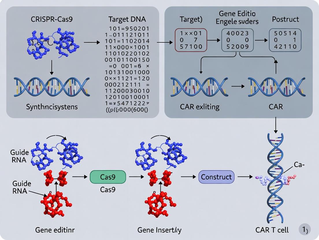
Within CAR T-cell engineering, CRISPR-Cas9 has emerged as a transformative tool for precise genomic modifications, enabling the disruption of endogenous genes (e.g., PD-1, TCR) and targeted integration of CAR transgenes. The core system comprises two essential molecular components: the Cas9 nuclease and the guide RNA (gRNA). Efficient delivery of these components into primary human T cells, which are notoriously difficult to transfect, remains a critical challenge defining protocol success. This application note details the components, delivery systems, and standardized protocols optimized for CAR T-cell research.
Core Components: Function and Specifications
Guide RNA (gRNA)
The gRNA is a chimeric RNA molecule that confers DNA target specificity. It consists of a CRISPR RNA (crRNA) derived sequence that provides ~20-nucleotide complementarity to the target DNA, and a trans-activating crRNA (tracrRNA) scaffold that binds Cas9. For convenience, these are often combined into a single guide RNA (sgRNA).
Key Design Parameters:
- Target Sequence: Must be adjacent to a Protospacer Adjacent Motif (PAM: 5'-NGG-3' for Streptococcus pyogenes Cas9).
- Specificity: Off-target effects are a major concern. Tools like CHOPCHOP or CRISPOR are used for selection.
- Format: Can be delivered as an in vitro transcribed RNA, a synthetic RNA, or encoded in a DNA plasmid/viral vector.
Cas9 Nuclease
The Cas9 endonuclease induces double-strand breaks (DSBs) 3-4 base pairs upstream of the PAM site. DNA repair via error-prone Non-Homologous End Joining (NHEJ) leads to indel mutations and gene knockout. In the presence of a donor DNA template, Homology-Directed Repair (HDR) can facilitate precise gene insertion (e.g., CAR cassette).
Cas9 Variants:
- Wild-Type (WT): Creates DSBs.
- Nickase (Cas9n): D10A mutation creates single-strand nicks; used in pairs to reduce off-targets.
- Dead Cas9 (dCas9): Catalytically inactive; fused to effector domains (activators, repressors, base editors) for transcriptional control.
Quantitative Comparison of Components
Table 1: Common CRISPR-Cas9 Component Formats for T-Cell Engineering
| Component Format | Delivery Method | Editing Efficiency (Typical Range in T Cells) | Key Advantages | Key Limitations |
|---|---|---|---|---|
| Plasmid DNA (sgRNA + Cas9) | Electroporation, Viral | 20-50% | Cost-effective; stable expression. | High immunogenicity; prolonged expression increases off-targets. |
| In vitro Transcribed (IVT) mRNA (Cas9) + Synthetic sgRNA | Electroporation | 60-90% | High efficiency; transient expression reduces off-targets. | Sensitive to degradation; requires cold chain. |
| Ribonucleoprotein (RNP) (Recombinant Cas9 + sgRNA) | Electroporation | 70-95% | Highest efficiency/specificity; very rapid action; minimal off-targets. | Most expensive; recombinant protein required. |
| All-in-One Lentiviral Vector | Lentiviral Transduction | 30-70% in transduced cells | Stable, long-term expression; good for hard-to-transfect cells. | Random integration; size limitations; persistent expression concerns. |
Delivery Systems for Primary Human T Cells
Efficient delivery is the paramount bottleneck. Non-viral methods are preferred for knockout strategies due to transient presence.
Table 2: Delivery System Comparison for CAR T-Cell Applications
| System | Mechanism | Max Payload | Throughput | Cytotoxicity/Activation Impact |
|---|---|---|---|---|
| Electroporation (Nucleofection) | Electrical pulses create pores. | >10 kb (plasmid), RNP, RNA | High | Moderate-High; can activate T cells. |
| Lentiviral Transduction | Viral integration. | ~8 kb | High | Low; but integrates into genome. |
| AAV Transduction | Viral delivery, mostly episomal. | ~4.7 kb | Medium | Very Low; limited cargo size. |
| Lipofection | Lipid nanoparticle fusion. | Varies | Medium | Low-Medium; inefficient in primary T cells. |
Detailed Protocol: RNP Electroporation forTRACLocus Knockout in CAR T Cells
Objective: Disrupt the T-cell receptor alpha constant (TRAC) locus to prevent graft-versus-host disease in allogeneic CAR T cells via NHEJ, using CRISPR-Cas9 RNP electroporation.
Materials & Reagents (The Scientist's Toolkit)
Table 3: Essential Research Reagent Solutions
| Item | Function/Description |
|---|---|
| Primary Human T Cells | Isolated from PBMCs using CD3+ selection. |
| Recombinant S. pyogenes Cas9 Nuclease | High-purity, endotoxin-free protein. |
| Synthetic sgRNA (targeting TRAC) | Chemically modified for stability; resuspended in nuclease-free buffer. |
| Nucleofector Device & Kit V | Optimized electroporation system for human T cells. |
| IL-2, IL-7, IL-15 Cytokines | For T-cell activation and post-editing culture. |
| Anti-CD3/CD28 Activator Beads | For T-cell activation pre-electroporation. |
| Genomic DNA Extraction Kit | For harvesting DNA for editing analysis. |
| T7 Endonuclease I or Tracking Indel Assay | For initial assessment of editing efficiency. |
| Flow Cytometry Antibodies (anti-CD3ε) | For phenotyping TRAC knockout efficiency (loss of surface TCR). |
Protocol Steps
Day -2: T Cell Activation
- Isolate CD3+ T cells from healthy donor PBMCs.
- Culture cells in X-VIVO 15 medium + 5% human AB serum, supplemented with IL-2 (100 IU/mL).
- Activate cells with anti-CD3/CD28 activator beads (bead-to-cell ratio 1:1).
Day 0: RNP Complex Formation & Electroporation
- Prepare RNP Complex: For 1x10^6 cells, combine 5 µg (≈37 pmol) recombinant Cas9 with 2.5 µg (≈75 pmol) synthetic TRAC sgRNA in 20 µL nucleofection buffer. Incubate at room temperature for 10 minutes.
- Harvest Cells: Collect activated T cells (≈48 hours post-activation), wash with PBS, and resuspend at 1x10^7 cells/mL in room temperature nucleofection buffer.
- Electroporation: Mix 10 µL cell suspension (1x10^5 cells) with 20 µL RNP complex. Transfer to a nucleofection cuvette. Run the designated program (e.g., EO-115 on a 4D-Nucleofector). Immediately add 80 µL pre-warmed complete medium.
- Recovery: Transfer cells to a 24-well plate with 2 mL pre-warmed complete medium containing IL-2 (100 IU/mL), IL-7 (5 ng/mL), and IL-15 (5 ng/mL). Culture at 37°C, 5% CO2.
Days 2-14: Analysis
- Day 3-4: Assess viability via trypan blue exclusion. Expect 50-70% recovery.
- Day 5-7: Harvest a sample for genomic DNA. Assess editing efficiency at the TRAC locus using T7EI assay or next-generation sequencing.
- Day 7-14: Monitor surface CD3/TCR expression via flow cytometry to confirm functional knockout.
Visualization: Workflows and Pathways
Diagram 1: RNP Workflow for TRAC Knockout
Diagram 2: CRISPR Strategy Selection Logic
Key Genomic Targets in T Cells for CAR Integration and Host Cell Engineering
Application Notes
Engineered T cells expressing Chimeric Antigen Receptors (CARs) represent a transformative immunotherapy. Precise CRISPR-Cas9-mediated genome editing is critical for both the targeted integration of the CAR transgene and the knockout of host genes to enhance therapeutic efficacy. This protocol, framed within a thesis on CRISPR-Cas9 for CAR T cells, details key genomic loci and methodologies.
1. Key Genomic Loci for CAR Integration
Safe-harbor and endogenous loci enable stable, potentially regulated CAR expression, avoiding random insertion risks.
- TRAC Locus: Disrupting the endogenous T Cell Receptor α Constant (TRAC) gene places the CAR under its physiological regulatory elements. This promotes uniform CAR expression, enhances T cell fitness, and prevents TCR-mediated graft-versus-host disease in allogeneic settings.
- AAVS1 Locus (PPP1R12C): A well-characterized mammalian safe-harbor locus on chromosome 19. It supports stable transgene expression with a low risk of insertional oncogenesis and disruption of essential genes.
- IL2RG Locus: Integration into the interleukin-2 receptor subunit gamma locus can complement gene defects and utilize its strong promoter. It is particularly relevant for combined gene correction and CAR expression strategies.
- PDCD1 Locus: Targeting the programmed cell death-1 (PD-1) locus for CAR integration simultaneously disrupts this immune checkpoint, potentially enhancing CAR T cell persistence in immunosuppressive tumor microenvironments.
2. Host Gene Knockout Targets for Cell Engineering
Knockout of specific host genes can enhance CAR T cell function, safety, and applicability for off-the-shelf therapies.
- TRAC/TRBC (T Cell Receptor): Knocking out the endogenous αβ T Cell Receptor prevents alloreactivity, a prerequisite for universal allogeneic CAR T cells.
- B2M (Beta-2-Microglobulin): Disruption of B2M ablates surface MHC Class I expression, reducing host CD8+ T cell-mediated rejection of allogeneic cells. This is often combined with CIITA knockout to remove MHC Class II.
- PDCD1 (PD-1): Knocking out the PD-1 receptor can prevent T cell exhaustion and improve anti-tumor cytotoxicity in checkpoint-rich environments.
- TGFBR2 (TGF-β Receptor II): Disruption confers resistance to immunosuppressive TGF-β signaling in the tumor microenvironment.
- UCART Target Genes (e.g., *CD52): Knocking out CD52 allows CAR T cells to resist depletion by Alemtuzumab, enabling its use as a conditioning or lymphodepleting agent.
Data Summary Tables
Table 1: Comparison of Key Genomic Loci for CAR Integration
| Locus | Chromosome | Primary Rationale | Expression Profile | Key Considerations |
|---|---|---|---|---|
| TRAC | 14q11.2 | Endogenous TCR regulation; enhanced fitness | Physiological, uniform | Disrupts endogenous TCR; ideal for allogeneic. |
| AAVS1 | 19q13.42 | Genomic safe-harbor; stable expression | Strong, constitutive | Well-studied safety profile; requires strong promoter. |
| IL2RG | Xq13.1 | Strong endogenous promoter; gene correction | Strong, constitutive | Potential for insertional mutagenesis if not precise. |
| PDCD1 | 2q37.3 | Dual knock-in/knockout; checkpoint evasion | Regulated by endogenous PD-1 elements | Complex editing; expression may be environment-dependent. |
Table 2: Key Host Gene Knockouts for CAR T Cell Engineering
| Target Gene | Pathway/Function | Engineering Goal | Therapeutic Impact | Common Delivery Format |
|---|---|---|---|---|
| TRAC/TRBC | T Cell Receptor | Prevent GvHD (allogeneic) | Enables "off-the-shelf" products | RNP with sgRNA. |
| B2M | MHC Class I Presentation | Evade Host CD8+ T Cell Rejection | Enhances allogeneic cell persistence | RNP or viral vector. |
| PDCD1 | Immune Checkpoint | Prevent Exhaustion | Potentiates activity in immunosuppressive tumors | RNP with sgRNA. |
| TGFBR2 | Immunosuppressive Cytokine Signaling | Confer TGF-β Resistance | Improves efficacy in TGF-β-rich TME | RNP or viral vector. |
Experimental Protocols
Protocol 1: Dual TRAC CAR Integration and TRAC Knockout via CRISPR-Cas9 RNP and AAV6 HDR Template
Objective: Generate allogeneic CAR T cells with targeted CAR integration at the TRAC locus and concomitant knockout of the endogenous TCR.
Materials:
- Primary human T cells.
- TRAC-targeting sgRNA (sequence: 5'-GAGTCTCTCAGCTGGTACA-3').
- High-fidelity Cas9 protein.
- Recombinant AAV6 donor template containing the CAR expression cassette flanked by ~800 bp TRAC homology arms.
- Electroporation system (e.g., Lonza 4D-Nucleofector).
- Cell culture media (e.g., TexMACS + IL-7/IL-15).
Methodology:
- RNP Complex Formation: Complex 60 pmol of sgRNA with 40 pmol of Cas9 protein to form ribonucleoprotein (RNP). Incubate at room temperature for 10 minutes.
- T Cell Activation: Activate isolated CD3+ T cells with anti-CD3/CD28 beads for 24-48 hours.
- Electroporation: Combine 1x10^6 activated T cells with RNP complex in 20µL of P3 primary cell solution. Electroporate using the EH-115 program on a 4D-Nucleofector.
- AAV6 Transduction: Immediately post-electroporation, transduce cells with AAV6 donor template at an MOI of 1x10^5 vg/cell.
- Culture and Expansion: Plate cells in pre-warmed media with IL-7 (5ng/mL) and IL-15 (10ng/mL). Expand for 7-14 days.
- Analysis: Assess editing efficiency via flow cytometry (CAR+, TCRαβ- population) and targeted deep sequencing of the TRAC locus.
Protocol 2: Multiplex Knockout of B2M and CIITA via Sequential Electroporation of CRISPR-Cas9 RNPs
Objective: Generate allogeneic CAR T cells with reduced immunogenicity by ablating MHC Class I (B2M) and Class II (CIITA) expression.
Materials:
- Primary human T cells or a pre-engineered CAR T cell line.
- B2M-targeting sgRNA (sequence: 5'-GCTTACACTGAATTCACCCC-3').
- CIITA-targeting sgRNA (sequence: 5'-GGACAGAACCAGAGACTCCC-3').
- High-fidelity Cas9 protein.
- Two separate electroporation cuvettes/solutions.
Methodology:
- First Electroporation (B2M Knockout): Form RNP with B2M sgRNA as in Protocol 1. Electroporate 1x10^6 T cells. Culture cells for 72 hours to allow for protein turnover.
- Recovery and Second Electroporation: Harvest cells, count, and assess viability. Form a second RNP complex with the CIITA sgRNA.
- Electroporate the same cell batch with the second RNP using identical conditions.
- Culture and Expand: Allow cells to recover and expand for 10-14 days post-final edit.
- Validation: Assess double-knockout efficiency via flow cytometry for surface MHC-I (HLA-A,B,C) and MHC-II (HLA-DR). Confirm indels at genomic level by T7E1 assay or NGS for each locus.
The Scientist's Toolkit: Research Reagent Solutions
| Reagent/Material | Function/Application |
|---|---|
| High-Fidelity Cas9 Protein | Minimizes off-target editing during knock-in/knockout procedures. |
| Synthetic sgRNA (chemically modified) | Enhances stability and editing efficiency in primary T cells. |
| AAV6 Serotype Donor Vectors | High-efficiency delivery of HDR templates to primary human T cells. |
| Lonza P3 Primary Cell 4D-Nucleofector Kit | Optimized system for high-viability RNP delivery into T cells. |
| Recombinant Human IL-7 & IL-15 | Maintains T cell stemness and promotes persistence post-editing. |
| Anti-CD3/CD28 Dynabeads | Provides robust, scalable T cell activation prior to editing. |
| T7 Endonuclease I (T7E1) Assay Kit | Rapid, semi-quantitative validation of indel formation. |
| CAR Detection Antibody/Protein L | Flow cytometry-based detection of surface CAR expression. |
Diagrams
Title: CAR T Cell Engineering Workflow via TRAC Targeting
Title: Knockout Targets to Enhance CAR T Cell Function
Within the framework of CRISPR-Cas9 protocols for next-generation CAR T cell engineering, a core strategic goal is to enhance therapeutic efficacy and safety by disrupting endogenous genes. Key targets include the endogenous T-cell receptor (TCR) to prevent graft-versus-host disease (GvHD) in allogeneic "off-the-shelf" CAR T products, and immune checkpoints like PD-1 to counteract the immunosuppressive tumor microenvironment. This Application Note details protocols and data for multiplexed editing of TRAC (TCRα constant), PDCD1 (PD-1), and other checkpoint genes (e.g., CTLA-4, LAG-3) using CRISPR-Cas9 ribonucleoprotein (RNP) electroporation in primary human T cells.
Table 1: Editing Efficiency and Functional Outcomes of Multiplexed Gene Disruption in Primary Human T Cells
| Target Gene (Locus) | Primary Function | Average Indel Efficiency (% via NGS)* | Phenotypic Knockout (% via Flow Cytometry)* | Key Functional Outcome |
|---|---|---|---|---|
| TRAC | TCR α-chain expression | ˃95% | ˃98% TCRαβ- | Prevents TCR-mediated alloreactivity, reduces GvHD risk. |
| PDCD1 | PD-1 checkpoint expression | 85-92% | 80-88% PD-1- | Enhances cytokine secretion & tumor killing in PD-L1+ co-culture. |
| CTLA4 | CTLA-4 checkpoint expression | 78-85% | 75-82% CTLA-4- | Synergistic improvement in T cell proliferation. |
| Dual (TRAC + PDCD1) | TCR & PD-1 disruption | TRAC: ˃90%, PDCD1: ˃85% | TCRαβ-: ˃95%, PD-1-: ˃80% | Combined resistance to exhaustion & alloreactivity. |
| Triple (TRAC + PDCD1 + CTLA4) | Multiplex checkpoint disruption | TRAC: ˃88%, PDCD1: ˃82%, CTLA4: ˃75% | TCRαβ-: ˃92%, PD-1-: ˃78%, CTLA-4-: ˃70% | Maximal in vitro expansion and sustained anti-tumor activity. |
*Data aggregated from recent literature (2023-2024) using Cas9 RNP electroporation. NGS: Next-Generation Sequencing.
Table 2: Comparative T Cell Functional Assay Data Post-Editing
| T Cell Group | IFN-γ Secretion (pg/ml)* in PD-L1+ Tumor Co-culture | Tumor Cell Lysis (% at 48h)* | Exhaustion Marker (TIM-3+) % at Day 7 Post-Activation* |
|---|---|---|---|
| Unedited (Control) | 1,200 ± 150 | 45% ± 5% | 35% ± 4% |
| TRAC KO only | 1,100 ± 200 | 48% ± 6% | 38% ± 5% |
| PDCD1 KO only | 2,950 ± 300 | 62% ± 7% | 18% ± 3% |
| Dual (TRAC/PDCD1) KO | 2,800 ± 250 | 65% ± 6% | 20% ± 4% |
| Triple (TRAC/PDCD1/CTLA4) KO | 3,500 ± 400 | 72% ± 8% | 15% ± 3% |
*Representative data from intracellular cytokine staining, real-time cytotoxicity assays, and flow cytometry. KO: Knockout.
Detailed Experimental Protocols
Protocol 1: CRISPR-Cas9 RNP Complex Formation for Multiplexed Editing Objective: To prepare specific and transient CRISPR-Cas9 ribonucleoprotein complexes targeting TRAC, PDCD1, and CTLA4. Materials: Alt-R S.p. Cas9 Nuclease V3, Alt-R CRISPR-Cas9 crRNA (target-specific), Alt-R CRISPR-Cas9 tracrRNA, Nuclease-Free Duplex Buffer. Steps:
- Resuspend and anneal crRNA and tracrRNA: For each target, combine 6 µL of 100 µM crRNA with 6 µL of 100 µM tracrRNA in a tube. Add 8 µL Nuclease-Free Duplex Buffer. Total volume: 20 µL.
- Anneal: Heat mixture to 95°C for 5 min in a thermal cycler, then cool to room temperature (25°C) at a ramp rate of 0.1°C/sec. The resulting 30 µM gRNA is stable at -20°C for up to 6 months.
- Form RNP Complex: For each target, dilute annealed gRNA to 6 µM in Opti-MEM. Dilute Cas9 nuclease to 12 µM in Opti-MEM. Combine equal volumes (e.g., 10 µL each) of diluted gRNA and Cas9 nuclease. Incubate at room temperature for 10-20 minutes to form the RNP complex. For multiplexing, combine the individual RNPs just prior to electroporation.
Protocol 2: T Cell Activation and Electroporation Objective: To efficiently deliver multiplexed RNPs into activated human primary T cells. Materials: Human PBMCs or CD3+ T cells, Anti-CD3/CD28 Dynabeads, IL-2 (200 IU/mL), P3 Primary Cell 4D-Nucleofector X Kit, 4D-Nucleofector System. Steps:
- T Cell Activation: Isolate CD3+ T cells from healthy donor PBMCs using a negative selection kit. Activate cells with anti-CD3/CD28 Dynabeads at a 1:1 bead-to-cell ratio in TexMACS medium supplemented with 5% human AB serum and IL-2 (200 IU/mL). Culture for 48 hours.
- Cell Preparation: On day 2 post-activation, harvest T cells, remove beads, and count. Wash cells once with PBS. Resuspend cells in P3 Primary Cell Nucleofector Solution at a density of 20 million cells per 100 µL.
- Electroporation Setup: For each nucleofection, mix 20 µL of the combined RNP complexes (for multiplexed editing) with 100 µL of cell suspension in a nucleofection cuvette. Include a cells-only control and a non-targeting RNP control.
- Nucleofection: Use the 4D-Nucleofector X Unit with program code EO-115. Immediately after pulsing, add 500 µL of pre-warmed, serum-free culture medium to the cuvette.
- Recovery and Culture: Transfer cells to a pre-warmed plate containing complete TexMACS medium with IL-2. Place in a 37°C, 5% CO2 incubator. Assess editing efficiency and phenotype from day 3 onwards.
Protocol 3: Assessment of Editing Efficiency and Phenotype Objective: To quantify indel formation and confirm protein-level knockout. Steps:
- Genomic DNA Extraction & NGS: At day 3-5 post-electroporation, extract genomic DNA from ~1e6 cells per sample using a commercial kit. Amplify target loci via PCR with barcoded primers. Purify amplicons and analyze by next-generation sequencing. Use tools like CRISPResso2 to calculate indel percentages.
- Flow Cytometric Analysis: At day 5-7, stain cells for surface expression of TCRαβ (anti-TCRαβ antibody), PD-1, and CTLA-4. Include a viability dye. Use an unedited sample to set negative gates. Calculate the percentage of protein-negative cells within the live cell population.
Signaling Pathways and Workflow Diagrams
Diagram 1: Checkpoint Pathways in CAR T Cell Therapy
Diagram 2: CRISPR Editing Protocol Workflow
The Scientist's Toolkit: Research Reagent Solutions
Table 3: Essential Materials for Checkpoint Disruption in CAR T Cells
| Item & Example Product | Function in Protocol | Critical Parameters |
|---|---|---|
| Alt-R S.p. Cas9 Nuclease V3 (IDT) | High-fidelity Cas9 enzyme for RNP formation. Ensures specific DNA cleavage with low off-target effects. | Concentration (µM), storage at -20°C, nuclease-free handling. |
| Alt-R CRISPR-Cas9 crRNA & tracrRNA (IDT) | Target-specific CRISPR RNA components. Guide Cas9 to genomic loci (TRAC, PDCD1, CTLA4). | CrRNA design (on-target score), chemical modification for stability, resuspension concentration (100 µM). |
| P3 Primary Cell 4D-Nucleofector X Kit (Lonza) | Optimized nucleofection solution and cuvettes for primary human T cells. Enables high-efficiency RNP delivery. | Cell density (e.g., 20e6/100µL), compatibility with 4D-Nucleofector unit. |
| Anti-CD3/CD28 Dynabeads (Gibco) | Magnetic beads for robust, consistent T cell activation prior to editing. Critical for high viability and editing rates. | Bead-to-cell ratio (typically 1:1), removal post-activation. |
| Recombinant Human IL-2 (PeproTech) | Cytokine for T cell survival and expansion post-activation and post-electroporation. | Concentration (e.g., 200 IU/mL), aliquoting to avoid freeze-thaw cycles. |
| Anti-TCRαβ/PD-1/CTLA-4 Antibodies (BioLegend) | Flow cytometry antibodies for validation of protein-level knockout post-editing. | Conjugate (e.g., FITC, PE), titration for optimal signal-to-noise. |
Within CAR T-cell therapy research, precise genetic engineering is paramount. CRISPR-Cas9 technology enables targeted genome editing, but the choice of delivery format—plasmid DNA, in vitro transcribed (IVT) mRNA, or preassembled ribonucleoprotein (RNP)—significantly impacts editing efficiency, specificity, cellular toxicity, and regulatory considerations. This application note delineates the critical parameters for selecting the optimal CRISPR format for CAR T-cell development.
Quantitative Comparison of CRISPR Delivery Formats
Table 1: Comparative Analysis of CRISPR-Cas9 Delivery Formats for Primary T Cells
| Parameter | Plasmid DNA | IVT mRNA | Ribonucleoprotein (RNP) |
|---|---|---|---|
| Onset of Activity | Slow (24-48h) | Moderate (2-8h) | Fast (<2h) |
| Duration of Activity | Prolonged (days) | Short (~24h) | Very Short (6-24h) |
| Editing Efficiency | Moderate | High | Very High |
| Off-target Effects | Higher | Moderate | Lower |
| Cellular Toxicity | Higher (TLR9, p53) | Moderate (IFN response) | Low |
| Immunogenicity Risk | High (bacterial sequences) | Moderate (modified nucleosides reduce) | Low |
| Ease of Use | Simple (standard transfection) | Requires handling RNA | Requires complex assembly |
| Regulatory Path | More complex (integration risk) | Simpler (ephemeral) | Simpler (ephemeral) |
Recent data (2023-2024) indicates that for primary human T cells, RNP electroporation consistently achieves 70-90% knockout efficiency of target genes like PD-1 or TRAC, with cell viability >60%. mRNA delivery yields 50-80% efficiency, while plasmid-based methods often result in lower efficiency (30-60%) and higher rates of apoptosis.
Detailed Protocols for CAR T-Cell Engineering
Protocol 1: RNP Complex Preparation and Electroporation for TRAC Locus Knockout
Objective: To knockout the T-cell receptor alpha constant (TRAC) gene to produce universal CAR T cells. Materials: See "The Scientist's Toolkit" below. Method:
- sgRNA Preparation: Resusynthesized sgRNA (chemical or IVT) in nuclease-free buffer to 160 µM.
- RNP Complex Assembly: Mix Cas9 protein (40 µM) with sgRNA (60 µM) at a 1:1.5 molar ratio in a sterile tube. Incubate at room temperature for 10-20 minutes.
- T Cell Activation: Isolate PBMCs from leukapheresis product. Activate CD3+ T cells using anti-CD3/CD28 beads in TexMACS medium with IL-7 and IL-15 (50 U/mL each) for 48 hours.
- Electroporation: Wash activated T cells twice in PBS. Resuspend 1-2e6 cells in 20 µL P3 primary cell electroporation buffer. Add pre-assembled RNP complex (final Cas9 concentration ~3 µM). Transfer to a 16-well electroporation cuvette. Run the 4D-Nucleofector X unit using program EO-115.
- Recovery: Immediately add 80 µL pre-warmed medium to cuvette. Transfer cells to a plate with pre-warmed complete medium containing cytokines. Culture for 48-72 hours before analysis or CAR transduction.
- Analysis: Assess editing efficiency via T7E1 assay or next-generation sequencing of the target locus. Confirm surface TCR loss via flow cytometry using anti-CD3ε antibody.
Protocol 2: IVT mRNA Electroporation for Multiple Gene Editing
Objective: To knockout both PDCD1 (PD-1) and B2M simultaneously in CAR T cells. Method:
- mRNA Synthesis: Generate Cas9 mRNA via in vitro transcription from a linearized plasmid template, incorporating 5' cap1 (CleanCap) and pseudouridine (Ψ) modifications. Synthesize sgRNAs separately.
- Co-Electroporation: For each reaction, mix 5 µg of modified Cas9 mRNA with 3 µg of each sgRNA. Combine with 2e6 activated T cells in 20 µL P3 buffer.
- Electroporation: Use program EO-115 on the 4D-Nucleofector.
- Post-Processing: Recover as in Protocol 1. Monitor protein knockout via flow cytometry at 48 hours (PD-1) and 5 days (B2M).
Visualizing Workflows and Pathways
Title: CRISPR-CAR T Cell Engineering Workflow
Title: RNP Mechanism Leading to Knockout or Knock-in
The Scientist's Toolkit
Table 2: Essential Reagents for CRISPR-CAR T Cell Protocols
| Reagent / Solution | Function & Importance | Example Product / Note |
|---|---|---|
| Recombinant Cas9 Protein | High-purity, endotoxin-free protein for RNP assembly. Critical for rapid activity. | Alt-R S.p. HiFi Cas9 Nuclease V3 |
| Chemically Modified sgRNA | Enhances stability and reduces immunogenicity versus in vitro transcribed sgRNA. | Alt-R CRISPR-Cas9 sgRNA, Trilink CleanCap sgRNA |
| Nucleofector System & Kits | Electroporation device and optimized buffers for primary T cells. High efficiency, lower toxicity. | Lonza 4D-Nucleofector X Unit, P3 Primary Cell 4D-Nucleofector Kit |
| T Cell Activation Beads | Mimic antigen presentation to activate T cells, essential for post-editing viability and expansion. | Gibco Dynabeads CD3/CD28 CTS |
| Serum-free T Cell Media | Chemically defined, supports robust expansion and editing of clinical-grade T cells. | TexMACS GMP Medium, X-VIVO 15 |
| Cytokines (IL-7 & IL-15) | Promote memory-like phenotype and survival during ex vivo manipulation. | PeproTech GMP-grade Recombinant Human IL-7/IL-15 |
| Genome Editing Detection Kit | Validates on-target editing efficiency and screens for off-targets. | IDT Alt-R Genome Editing Detection Kit (T7E1), NGS amplicon sequencing. |
| Clinical-grade AAVS1 Donor | Safe-harbor locus donor template for CAR gene knock-in via HDR. | GMP-grade AAV6 serotype vector containing CAR construct. |
Within the framework of developing robust CRISPR-Cas9 protocols for chimeric antigen receptor (CAR) T-cell engineering, the design of the donor template and adherence to ethical and regulatory guidelines are foundational. These pre-protocol steps determine not only the efficiency and precision of CAR integration but also the translational viability and safety of the resultant cellular therapeutic product.
Donor Template Design: Key Parameters and Quantitative Data
The donor DNA template directs homology-directed repair (HDR) to integrate the CAR transgene into a specified genomic safe harbor (GSH) or specific locus. Key design variables are summarized below.
Table 1: Critical Parameters for Donor Template Design in CAR T-Cell Engineering
| Parameter | Typical Specification | Rationale & Impact on CAR Expression |
|---|---|---|
| Homology Arm Length | 400-800 bp per arm | Longer arms increase HDR efficiency but may reduce viral packaging capacity for AAV delivery. Shorter arms (<300 bp) significantly reduce efficiency. |
| Target Locus | GSH: AAVS1 (PPP1R12C), TRAC, CCR5, ROSA26 | Disruption of TRAC ensures endogenous TCR knockout and targeted CAR insertion. AAVS1 is a permissive site for stable expression. |
| CAR Cassette Size | ~2.0 - 2.5 kb (scFv + hinges + TM + CD3ζ + co-stim) | Larger sizes can challenge delivery vectors (e.g., AAV cargo limit ~4.7kb) and may reduce viral titers. |
| Promoter Selection | EF-1α, CMV, PGK, MNDU3 | Constitutive promoters vary in strength and longevity. EF-1α often provides sustained expression in T cells. |
| Vector Backbone | Linear dsDNA fragment, AAV6, or ssODN (for short edits) | AAV6 is gold standard for HDR delivery in T cells due to high transduction efficiency and single-stranded DNA nature. |
| HDR Enhancer Addition | Nocodazole (M-phase sync.), SCR7 (NHEJ inhibitor), RS-1 (RAD51 stimulator) | Can boost HDR rates 1.5 to 3-fold, but requires titration to minimize cytotoxicity. |
Detailed Protocol: Donor Template Assembly & Validation forTRAC-Targeted CAR Integration
Objective: To generate and validate a dsDNA donor template for CRISPR-Cas9-mediated, homology-directed insertion of a CAR cassette into the human TRAC locus.
Materials:
- Source DNA: CAR expression cassette (e.g., EF1α-CAR-P2A-marker).
- Cloning Vector: pUC19-based plasmid with multiple cloning site.
- Homology Arms: Genomic DNA from healthy donor T cells or synthesized gBlocks.
- Enzymes: High-fidelity DNA polymerase (e.g., Q5), restriction enzymes, T4 DNA Ligase.
- Cells: DH5α competent E. coli.
- Validation Primers: Sequencing primers, junction PCR primers (see Table 2).
Methodology:
- Homology Arm Amplification: Amplify 800 bp left and right homology arms (LHAs and RHAs) flanking the TRAC Cas9 cut site (targeting exon 1) from genomic DNA using Q5 polymerase. Introduce 20-30 bp overlaps with the CAR cassette ends.
- Gibson Assembly: Perform a one-step Gibson Assembly reaction mixing linearized pUC19 backbone, LHA, CAR cassette, and RHA. Incubate at 50°C for 60 minutes.
- Transformation & Screening: Transform assembled product into DH5α cells. Select colonies on ampicillin plates. Screen via colony PCR using primers spanning the LHA-CAR and CAR-RHA junctions.
- Plasmid Validation: Isolate plasmid DNA from positive clones. Validate by:
- Restriction Digest: Confirm size via diagnostic digest.
- Sanger Sequencing: Full-length sequence the entire insert and homology arms using a primer-walking strategy.
- Donor Fragment Preparation: For electroporation, release the donor fragment (LHA-CAR-RHA) from the plasmid backbone using appropriate restriction enzymes. Purify the linear fragment via gel extraction and elute in nuclease-free water or TE buffer. Quantify via fluorometry.
Table 2: Example Primer Sequences for Donor Validation
| Primer Name | Sequence (5' -> 3') | Purpose |
|---|---|---|
| TRACLHAF | GGTGTGAACTGGCACTGACA | Amplify 5' genomic junction |
| CARIntR | CTTCAGCAGGACCATGTGCT | Verify LHA-CAR junction |
| CARIntF | GATGCCCTGGAGACAATGAC | Verify CAR-RHA junction |
| TRACRHAR | CACAGAGACAGCCAGGACTG | Amplify 3' genomic junction |
Ethical and Guideline Compliance Framework
Gene-edited cellular therapeutics operate within a stringent global regulatory landscape. Key considerations include:
- Off-Target Analysis: Comprehensive assessment using orthogonal methods (e.g., GUIDE-seq, CIRCLE-seq, or targeted deep sequencing of predicted off-target sites) is mandatory prior to clinical translation.
- On-Target Genotoxicity: Evaluation of large deletions, chromosomal translocations, or aneuploidy at the target locus (e.g., via karyotyping, FISH, or long-range PCR).
- Vector and Edit Safety: Documentation of donor template source, sequence, and purity. Clearance of residual CRISPR components post-editing.
- Informed Consent: For donor-derived T cells, rigorous informed consent protocols detailing the nature, risks, and potential long-term implications of genetic modification.
- Regulatory Pathways: Compliance with regional guidelines (FDA CBER, EMA ATMP, NMPA) for Investigational New Drug (IND) applications, focusing on Chemistry, Manufacturing, and Controls (CMC) and preclinical safety pharmacology.
Visualizing the Workflow and Key Pathways
Title: CAR T-Cell Gene Editing Pre-Protocol Workflow
Title: DNA Repair Pathways After CRISPR Cleavage
The Scientist's Toolkit: Key Research Reagent Solutions
| Reagent Category | Specific Example/Product | Function in Pre-Protocol Phase |
|---|---|---|
| Homology Arm Source | Human Genomic DNA (from T cells) or IDT gBlocks Gene Fragments | Provides sequence for designing and synthesizing locus-specific homology arms. |
| Assembly Master Mix | NEBuilder HiFi DNA Assembly Master Mix | Enables seamless, high-efficiency Gibson Assembly of donor plasmid. |
| Delivery Vector | AAV6 Serotype Production System | Produces recombinant AAV6 particles for high-efficiency donor template delivery to primary T cells. |
| HDR Enhancers | RS-1 (RAD51 agonist), L755507 (β-3 adrenergic receptor agonist) | Small molecules to temporarily bias DNA repair toward HDR, increasing knock-in efficiency. |
| Validation Kits | Illumina Amplicon-EZ or ONT Cas9 Sequencing Kits | For high-throughput sequencing of on- and off-target editing outcomes. |
| Guide RNA Design Tool | IDT Alt-R CRISPR-Cas9 guide RNA design tool | In silico design and specificity scoring for gRNAs targeting safe harbor loci. |
Step-by-Step Protocol: From gRNA Design to Engineered CAR T-Cell Expansion
Application Notes
Within the broader thesis on CRISPR-Cas9 protocols for CAR T cell research, the precise knockout of endogenous genes (e.g., PD-1, TCRα, HLA-I) or targeted integration of CAR transgenes is paramount. The initial and most critical step is the in silico design and rigorous in vitro validation of single-guide RNAs (gRNAs). This protocol details a bioinformatics-to-bench pipeline for selecting high-efficiency, specific gRNAs against human genomic targets, ensuring minimal off-target effects—a non-negotiable prerequisite for clinical-grade CAR T cell manufacturing.
1. Target Identification and gRNA Design
- Procedure: Identify the genomic locus of interest (e.g., exon 2 of the PDCD1 gene). Using the reference human genome (GRCh38/hg38), extract a 500 bp sequence flanking the target site. Input this sequence into multiple gRNA design tools. The parameters must include: a 20-nt guide sequence directly upstream of a 5'-NGG-3' Protospacer Adjacent Motif (PAM), calculation of on-target efficiency scores, and a comprehensive genome-wide search for potential off-target sites (allowing up to 3 mismatches, with special attention to mismatches in the "seed" region proximal to the PAM).
- Key Tools & Databases: UCSC Genome Browser, NCBI Nucleotide, CRISPR design tools (e.g., IDT, Broad Institute's GPP Portal, CHOPCHOP).
2. Off-Target Analysis and Prioritization
- Procedure: For each candidate gRNA, compile a list of all potential off-target genomic loci from the design tools. Cross-reference these loci with known gene exons, regulatory elements, and cancer-associated genomic regions (using databases like ClinVar, COSMIC). Prioritize gRNAs with zero high-confidence off-targets in coding regions. If unavoidable, select gRNAs where off-targets reside in intergenic or deep intronic regions.
3. In Vitro Validation of Cleavage Efficiency
- Procedure: Prior to T cell electroporation, gRNA efficiency is validated using an in vitro cleavage assay. Synthesize and purify a PCR-amplified DNA template (~500 bp) encompassing the target genomic region. Incubate the template with recombinant SpCas9 protein and the in vitro transcribed candidate gRNA. Analyze the products via agarose gel electrophoresis to quantify the fraction of cleaved DNA.
Table 1: Example In Vitro Cleavage Efficiency Data for Candidate PDCD1 gRNAs
| gRNA ID | Target Sequence (5'→3') | On-Target Score | Predicted Off-Target Sites (≤3 mismatches) | In Vitro Cleavage Efficiency (%) |
|---|---|---|---|---|
| PD1-g01 | GAGTATTCAGAGTGGTCCTT | 95 | 1 (intergenic) | 92 ± 3 |
| PD1-g02 | AGTGGTCCTTGATGTGACCG | 88 | 0 | 85 ± 5 |
| PD1-g03 | CAGACCTGAGTATTCAGAGT | 78 | 3 (1 in intron of RP11-34P13.7) | 45 ± 8 |
Experimental Protocol: In Vitro Cleavage Assay
Materials:
- Recombinant SpCas9 Nuclease (commercial source)
- Target DNA Template: PCR-amplified genomic region (200 ng/µL)
- Candidate gRNAs (chemically synthesized or in vitro transcribed)
- Nuclease-Free Duplex Buffer (IDT) or equivalent
- 10X Cas9 Reaction Buffer
- Proteinase K
- Agarose gel electrophoresis system
Method:
- Annealing: For each gRNA, prepare a 1 µM working solution in nuclease-free duplex buffer.
- Reaction Setup: In a 0.2 mL PCR tube, assemble:
- Target DNA template: 200 ng
- Recombinant SpCas9: 100 ng
- gRNA (1 µM): 2 µL
- 10X Cas9 Reaction Buffer: 2 µL
- Nuclease-free water to 20 µL.
- Incubation: Mix gently and incubate at 37°C for 60 minutes.
- Digestion Termination: Add 1 µL of Proteinase K, mix, and incubate at 56°C for 15 minutes to degrade Cas9 protein.
- Analysis: Load the entire reaction on a 2% agarose gel stained with ethidium bromide. Include uncut template DNA as a control.
- Quantification: Image the gel under UV. Calculate cleavage efficiency using the formula: (Intensity of cleaved bands) / (Intensity of total DNA) × 100%.
Visualizations
gRNA Design and Validation Workflow for CAR T Cell Engineering
Steps in the In Vitro gRNA Cleavage Efficiency Assay
The Scientist's Toolkit: Research Reagent Solutions
Table 2: Essential Reagents for gRNA Design and Validation
| Reagent / Solution | Function / Purpose | Example Vendor / Tool |
|---|---|---|
| GRCh38/hg38 Reference Genome | Accurate genomic coordinate mapping for target identification and off-target prediction. | UCSC Genome Browser, ENSEMBL |
| CRISPR gRNA Design Suite | Computes on-target activity scores and identifies potential off-target genomic loci. | IDT CRISPR Design, Broad GPP, CHOPCHOP |
| Chemically Modified sgRNA | Enhanced nuclease stability and reduced immunogenicity for primary T cell editing. | Synthego, Trilink Biotech |
| Recombinant SpCas9 Nuclease (HiFi) | High-fidelity variant for in vitro assay; reduces off-target cleavage. | Integrated DNA Technologies (IDT) |
| Nuclease-Free Duplex Buffer | Ensures proper gRNA secondary structure formation without RNase contamination. | Integrated DNA Technologies (IDT) |
| Agarose Gel DNA Recovery Kit | Purification of PCR-amplified target template for in vitro cleavage assays. | Zymo Research, Qiagen |
Within the development of CRISPR-Cas9-edited Chimeric Antigen Receptor (CAR) T cells, the delivery of Cas9 as a pre-assembled ribonucleoprotein (RNP) complex offers distinct advantages. This method transiently exposes cells to the editing machinery, reducing off-target effects and DNA integration risks associated with plasmid DNA. In CAR T cell engineering, RNP delivery is primarily used for the knockout of endogenous genes (e.g., TRAC, PDCD1) to enhance CAR T cell function and persistence. This protocol details the preparation of CRISPR-Cas9 RNPs using purified Cas9 protein and synthetic single-guide RNA (sgRNA).
Reagent and Material Preparation
Research Reagent Solutions
| Item | Function and Notes |
|---|---|
| Recombinant S. pyogenes Cas9 Nuclease | High-purity, endotoxin-free protein. The active enzyme forms the core of the RNP complex. |
| Chemically Modified sgRNA (synthethic) | Includes 2'-O-methyl and phosphorothioate modifications at terminal nucleotides to enhance stability and reduce immunogenicity. |
| Nuclease-Free Duplex Buffer | A standardized, low-salt buffer (e.g., IDT) for efficient sgRNA-Cas9 complex formation. |
| Nuclease-Free Water | Essential for diluting components and ensuring no RNase/DNase contamination. |
| Opti-MEM Reduced Serum Medium | Used for subsequent complex dilution prior to cellular delivery (e.g., electroporation). |
Table 1: Typical RNP Assembly Parameters and Outcomes
| Parameter | Typical Range | Notes |
|---|---|---|
| Molar Ratio (Cas9:sgRNA) | 1:1.2 to 1:2 | Ensures complete saturation of Cas9 with sgRNA. |
| Final Cas9 Concentration | 4 - 10 µM (in assembly mix) | Higher concentrations improve complex stability. |
| Incubation Temperature | 20-25°C (Room Temp) | |
| Incubation Time | 10 - 20 minutes | Sufficient for complete complex formation. |
| Complex Stability | < 6 hours (RT) | For best results, use immediately after assembly. |
| Recommended RNP Dose (for 1e6 T cells) | 2 - 6 pmol | Optimize for each target and cell type. |
Step-by-Step Protocol
A. Pre-assembly Calculations
- Determine the amount of RNP needed for your experiment. A typical electroporation reaction for 1-2 × 10⁶ primary human T cells uses 5 µL of RNP complex at 4 µM Cas9 concentration.
- Calculate required volumes:
- Cas9 stock (e.g., 10 µM):
Volume (µL) = (Final Cas9 conc. × Final Volume) / Cas9 stock conc. - sgRNA stock (e.g., 100 µM): Based on a 1:1.2 molar ratio.
sgRNA moles = Cas9 moles × 1.2.
- Cas9 stock (e.g., 10 µM):
B. RNP Assembly
- In a sterile, nuclease-free microcentrifuge tube, combine the calculated volumes of Nuclease-Free Duplex Buffer and Nuclease-Free Water.
- Add the calculated volume of chemically modified sgRNA to the tube.
- Add the calculated volume of recombinant Cas9 protein to the same tube. Gently pipette mix. Do not vortex.
- Incubate the mixture at room temperature (20-25°C) for 10-20 minutes to allow complete RNP complex formation.
- The assembled RNP complex can be used immediately for electroporation. If a short delay is necessary, keep on ice for up to 1 hour. For longer storage, snap-freeze in liquid nitrogen and store at -80°C (may reduce activity).
C. Preparation for Cellular Delivery (Electroporation)
- Immediately prior to electroporation, dilute the assembled RNP complex in Opti-MEM Reduced Serum Medium to the desired final volume and concentration for the electroporation system in use (e.g., Neon, Lonza 4D-Nucleofector).
- Combine the diluted RNP complex with the prepared T cell suspension in the appropriate electroporation cuvette or tip.
- Proceed with the optimized electroporation pulse code for primary T cells.
Diagram 1: RNP Assembly and Delivery Workflow (79 chars)
Diagram 2: Step by Step RNP Preparation Protocol (61 chars)
This protocol is a critical component of a thesis focused on establishing robust CRISPR-Cas9 workflows for next-generation Chimeric Antigen Receptor (CAR) T-cell engineering. Successful genetic modification, particularly for multiplexed editing (e.g., disrupting endogenous genes while inserting a CAR transgene), is fundamentally dependent on high-viability, highly activated T cells prior to nucleofection. This section details the optimized steps for isolating, activating, and electroporating primary human T cells with CRISPR-Cas9 ribonucleoprotein (RNP) complexes.
Key Research Reagent Solutions
| Reagent/Material | Function in Protocol | Example Vendor/Catalog |
|---|---|---|
| Human T-Cell Isolation Kit | Negative selection to obtain untouched, highly pure CD3+ T cells from PBMCs. | Miltenyi Biotec (Pan T Cell Isolation Kit) |
| CD3/CD28 T-Cell Activator | Provides necessary Signal 1 (CD3) and Signal 2 (CD28) for robust, uniform T-cell activation and proliferation. | Thermo Fisher (Dynabeads Human T-Activator CD3/CD28) |
| IL-2 (Recombinant Human) | Critical cytokine for T-cell survival, expansion, and maintenance of an editing-competent state. | PeproTech |
| X-Vivo 15 or TexMACS Medium | Serum-free, specialized media optimized for human T-cell culture and activation. | Lonza |
| Cas9 Nuclease (HiFi) | High-fidelity nuclease for RNP formation, reducing off-target effects. | Integrated DNA Technologies (Alt-R S.p. HiFi Cas9) |
| Alt-R CRISPR-CrRNA & tracrRNA | Synthetic, chemically modified RNAs for high-efficiency RNP complex formation. | Integrated DNA Technologies (Alt-R CRISPR system) |
| P3 Primary Cell 4D-Nucleofector X Kit | Optimized buffer and cuvettes for high-viability nucleofection of human T cells. | Lonza (V4XP-3032) |
| Nucleofector 4D Device | Device for applying optimized electrical pulses for intracellular RNP delivery. | Lonza |
Detailed Protocol: T-Cell Activation & Preparation for Nucleofection
Primary Human T-Cell Isolation & Activation
Day -2 or -3: Isolation
- Isolate Peripheral Blood Mononuclear Cells (PBMCs) from leukapheresis or buffy coat using Ficoll-Paque density gradient centrifugation.
- Resuspend PBMCs in degassed buffer. Use a human Pan-T Cell Isolation Kit (negative selection) per manufacturer's instructions to isolate untouched CD3+ T cells.
- Count cells using trypan blue exclusion. Target viability >99%.
Day 0: Activation
- Prepare complete T-cell media: X-Vivo 15 + 5% human AB serum + 100 IU/mL IL-2.
- Resuspend isolated T cells at a density of 1 x 10^6 cells/mL in complete media.
- Add CD3/CD28 activator beads at a 1:1 bead-to-cell ratio.
- Incubate cells at 37°C, 5% CO2 for 48-72 hours.
CRISPR-Cas9 RNP Complex Assembly
Day of Nucleofection (Typically Day 2 or 3 Post-Activation)
- Design & Resuspend RNAs: Resuspend Alt-R CRISPR-CrRNA and Alt-R tracrRNA to 100 µM in nuclease-free duplex buffer.
- Form gRNA Complex: Mix equal volumes of crRNA and tracrRNA (e.g., 5 µL each). Heat at 95°C for 5 min, then cool to room temp.
- Form RNP Complex: For a single reaction, combine:
- 5 µL of 40 µM Alt-R S.p. HiFi Cas9 protein (final 20 pmol).
- 5 µL of 40 µM gRNA complex (final 20 pmol). Incubate at room temperature for 15-20 minutes prior to nucleofection.
4D-Nucleofection of Activated T Cells
- Pre-warm complete media (without IL-2) and P3 Nucleofector Solution.
- Harvest Cells: Collect activated T cells. Remove activator beads using a magnet.
- Count & Aliquot: Count viable cells. Pellet 1-2 x 10^6 cells per nucleofection condition. Wash once with PBS.
- Resuspend in P3 Buffer: Completely resuspend cell pellet in 20 µL of room temperature P3 Primary Cell Solution per reaction.
- Add RNP: Add the pre-complexed 10 µL RNP to the cell suspension. Mix gently by pipetting. Do not vortex.
- Transfer & Nucleofect: Transfer the entire mixture (30 µL) to a Nucleofector cuvette. Cap tightly.
- Place cuvette in the 4D-Nucleofector X Unit.
- Run the optimized program for activated T cells: EH-115 or FF-113.
- Immediate Recovery: Immediately after pulsing, add 80 µL of pre-warmed media directly to the cuvette. Gently transfer the cells (~110 µL) to a pre-warmed 24-well plate containing 1 mL of complete media + IL-2.
- Culture: Place plate in incubator (37°C, 5% CO2). Do not disturb for 4-6 hours. Assess viability and expand culture the next day.
Critical Data & Optimization Parameters
Table 1: Impact of Activation Timing on Nucleofection Efficiency & Viability
| Activation Duration (Hours) | Cell Diameter (Avg. µm) | Nucleofection Viability (24h post) | Editing Efficiency (% INDEL) | Recommended Use |
|---|---|---|---|---|
| 24 | ~12-14 | 50-65% | Low (<30%) | Suboptimal |
| 48 | ~14-16 | 70-85% | High (60-80%) | Optimal for RNP |
| 72 | ~16-18 | 65-75% | High (60-80%) | Acceptable |
| >96 | Highly Blasted | <50% | Variable | Not Recommended |
Table 2: Comparison of 4D-Nucleofector Programs for Activated T Cells
| Program Code | Pulse Characteristics | Avg. Viability (Day 1) | Avg. Editing Efficiency | Notes |
|---|---|---|---|---|
| EH-115 | Moderate voltage/length | 75-90% | High | Gold standard for activated T cells. |
| FF-113 | Higher voltage | 65-80% | Very High | Can be harsher; test for your cell lot. |
| DN-100 | Lower voltage | 80-95% | Low to Moderate | Best for plasmid DNA, not RNP. |
Essential Workflow & Pathway Diagrams
Diagram 1: Primary T-Cell Gene Editing Workflow (65 chars)
Diagram 2: Three-Signal Model for T-Cell Activation (55 chars)
Diagram 3: Mechanism of RNP Delivery via Nucleofection (57 chars)
Application Notes
This protocol details a method for the site-specific integration of a Chimeric Antigen Receptor (CAR) transgene into a defined genomic locus of primary human T cells using CRISPR-Cas9-mediated Homology-Directed Repair (HDR). This approach, central to generating next-generation CAR T-cell products, aims to improve the consistency, potency, and safety of clinical-grade cell therapies by ensuring uniform, locus-controlled CAR expression, as opposed to random viral integration.
The strategy involves the simultaneous co-delivery of two key components: 1) a pre-assembled, synthetic CRISPR-Cas9 ribonucleoprotein (RNP) complex to create a targeted double-strand break (DSB) in a safe-harbor or therapeutically relevant locus (e.g., TRAC), and 2) a donor DNA template containing the CAR cassette flanked by homology arms specific to the target site. Efficient delivery is achieved via electroporation using clinical-grade nucleofection systems. The protocol is designed for high viability, editing efficiency, and HDR rates, optimized for primary human T cells, which are notoriously resistant to HDR.
Key Advantages:
- Controlled Transgene Expression: Integration into a defined locus (e.g., TRAC) can lead to more physiological, endogenous promoter-driven expression.
- Enhanced Genomic Safety: Avoids the risks of insertional oncogenesis associated with random viral integration.
- All-in-One Process: Synthetic RNP and DNA donor are delivered in a single manipulation, streamlining the manufacturing workflow.
- Rapid Action & Reduced Off-Targets: RNP acts quickly and degrades, minimizing prolonged nuclease activity and potential immunogenicity associated with viral vectors encoding Cas9.
Experimental Protocols
Design and Preparation of Reagents
A. CRISPR RNP Complex Assembly:
- sgRNA Design: Design a synthetic, chemically modified sgRNA targeting the desired locus (e.g., exon 1 of the human TRAC gene). Use CRISPick or similar tools for on-target scoring and off-target prediction. Resuscribe in nuclease-free duplex buffer to 160 µM.
- Complex Formation: Assemble the RNP complex immediately before electroporation. For a single reaction, mix:
- 3.2 µL of 160 µM sgRNA (final 20 pmol)
- 2.5 µL of 40 µM high-fidelity Cas9 protein (final 5 pmol)
- 4.3 µL of nuclease-free PBS or Opti-MEM
- Total Volume: 10 µL
- Incubate at room temperature for 10-20 minutes to allow RNP complex formation.
B. HDR Donor Template Preparation:
- Donor Design: Design a linear double-stranded DNA donor template (e.g., PCR-amplified or gBlock). The CAR expression cassette (often with a constitutive or endogenous promoter) must be flanked by left and right homology arms (typically 800-1000 bp each) specific to the target locus. The PAM site on the donor should be mutated to prevent re-cleavage.
- Purification: Purify the donor DNA using a PCR clean-up or gel extraction kit. Elute in nuclease-free water or low-EDTA TE buffer. Quantify via spectrophotometry (Nanodrop). A final amount of 1-4 µg per reaction is typical.
Primary Human T Cell Activation and Culture
- Isolate CD3+ or CD4+/CD8+ T cells from leukapheresis product using immunomagnetic beads.
- Activate cells using clinical-grade CD3/CD28 T Cell Activator beads or antibodies at a bead-to-cell ratio of 1:1 or as per manufacturer's instructions.
- Culture cells in X-VIVO 15 or TexMACS medium, supplemented with 5-10% human AB serum or serum-free supplements, and recombinant human IL-7 (5 ng/mL) and IL-15 (10 ng/mL).
- Perform gene editing 24-48 hours post-activation, when cells are highly viable and proliferative.
Electroporation for Co-delivery (Nucleofection)
This protocol is adapted for the Lonza 4D-Nucleofector X Unit.
- Day of Nucleofection: Count activated T cells. Centrifuge required number of cells (e.g., 1-2e6 per condition).
- Prepare Electroporation Mixture: To the pre-formed 10 µL RNP complex, add the purified HDR donor DNA (e.g., 2 µg in ≤10 µL volume). Mix gently.
- Resuspend Cells: Completely aspirate the culture medium. Resuspend the cell pellet in 100 µL of pre-warmed, supplemented P3 Primary Cell Nucleofector Solution per 1e6 cells.
- Combine and Transfer: Add 20 µL of the cell suspension to the DNA/RNP mixture. Gently mix and immediately transfer the entire 30 µL volume to a certified nucleofection cuvette. Avoid air bubbles.
- Nucleofect: Place the cuvette in the 4D-Nucleofector X Unit and run the appropriate program (e.g., EO-115 for primary human T cells).
- Recovery: Immediately after nucleofection, add 500 µL of pre-warmed, cytokine-supplemented culture medium to the cuvette. Using the supplied pipette, gently transfer the cells to a 24-well plate pre-filled with 1 mL of warm medium.
- Culture: Place cells in a 37°C, 5% CO2 incubator. After 4-6 hours, carefully remove and replace 50% of the medium with fresh, cytokine-supplemented medium to remove debris. Continue culture with regular feeding.
Post-Editing Analysis (Key Metrics)
- Viability: Assess 24 hours post-electroporation using flow cytometry with a live/dead stain (e.g., Fixable Viability Dye) or an automated cell counter with trypan blue exclusion.
- Editing Efficiency (INDELs): Assess 48-72 hours post-editing.
- Genomic DNA is extracted from an aliquot of cells.
- The target locus is PCR-amplified and analyzed by Tracking of Indels by Decomposition (TIDE) or Next-Generation Sequencing (NGS).
- HDR Integration Efficiency: Assess at day 5-7 post-editing.
- Flow Cytometry: For a CAR with a detectable surface marker (e.g., truncated EGFR, Myc tag), stain cells and analyze by flow cytometry.
- Droplet Digital PCR (ddPCR): Design a primer/probe set spanning the junction between the genomic sequence and the integrated CAR cassette for absolute quantification of HDR alleles.
Data Presentation
Table 1: Representative Quantitative Outcomes for TRAC-Targeted CAR Integration (n=3 Healthy Donors)
| Metric | Timepoint | Mean ± SD | Measurement Method |
|---|---|---|---|
| Cell Viability | 24h Post-Nucleofection | 65% ± 8% | Flow Cytometry (Live/Dead Stain) |
| INDEL Efficiency | 72h Post-Nucleofection | 92% ± 4% | NGS of Target Locus |
| HDR (CAR+) Rate | Day 7 Post-Editing | 45% ± 12% | Flow Cytometry (CAR Detection) |
| Fold Expansion | Day 10 Post-Activation | 15x ± 3x | Automated Cell Counter |
Table 2: Critical Parameters for Optimization
| Parameter | Tested Range | Recommended Optimal Value | Impact on Outcome |
|---|---|---|---|
| Cell State | Resting, 24h act., 48h act. | 48 hours post-CD3/CD28 activation | Maximizes HDR pathway activity. |
| Cas9:sgRNA Ratio | 1:1 to 1:4 (molar) | 1:4 (e.g., 5 pmol Cas9: 20 pmol sgRNA) | Ensures full RNP complexation; excess sgRNA can boost cutting. |
| Donor DNA Amount | 0.5 - 4 µg per 1e6 cells | 2 µg per 1e6 cells | Balances HDR rate with cellular toxicity from DNA. |
| Donor Form | ssDNA, dsDNA, AAV6 | Linear dsDNA (PCR fragment) | Cost-effective, high-yield production, suitable for clinical manufacturing. |
| Electroporation Program | EN-150, EO-115, FF-120 | EO-115 (for T cells in P3 buffer) | Optimized for high efficiency and viability in primary T cells. |
Visualizations
Diagram Title: Workflow for Co-delivery and HDR-Mediated CAR Integration
Diagram Title: HDR vs NHEJ Pathway at CRISPR-Induced DSB
The Scientist's Toolkit
Table 3: Essential Research Reagent Solutions for RNP + HDR Co-delivery
| Reagent / Material | Function & Role in Protocol | Example Product / Note |
|---|---|---|
| High-Fidelity Cas9 Nuclease | Catalyzes the site-specific DNA double-strand break. Synthetic protein reduces off-targets and immunogenicity. | TruCut HiFi Cas9, Alt-R S.p. HiFi Cas9 |
| Chemically Modified sgRNA | Guides Cas9 to the specific genomic target locus (e.g., TRAC). Chemical modifications enhance stability. | Synthego sgRNA, Alt-R CRISPR-Cas9 sgRNA |
| Linear dsDNA HDR Donor | Serves as the repair template for precise CAR integration. Homology arms direct insertion to the cut site. | PCR-amplified fragment or gBlock; clinical-grade production required for therapies. |
| Nucleofector System & Kit | Enables efficient co-delivery of large RNP and DNA complexes into hard-to-transfect primary T cells. | Lonza 4D-Nucleofector X Unit with P3 Primary Cell Kit, solution & cuvettes. |
| T Cell Activation Beads | Stimulates T cell proliferation and metabolic activity, priming cells for efficient HDR post-nucleofection. | Gibco Dynabeads CD3/CD28, Miltenyi MACS GMP TransAct. |
| Cytokines (IL-7, IL-15) | Supports survival, persistence, and maintenance of a stem-cell memory-like phenotype in edited T cells. | Recombinant human IL-7/IL-15; GMP-grade for manufacturing. |
| Genomic DNA Extraction Kit | For isolating high-quality gDNA from a cell aliquot to assess editing efficiency (INDELs) via NGS/TIDE. | Qiagen DNeasy Blood & Tissue Kit. |
| ddPCR or qPCR Reagents | For absolute quantification of HDR integration events using a junction-specific assay. | Bio-Rad ddPCR Supermix, TaqMan assays. |
Application Notes Following successful CRISPR-Cas9-mediated editing (e.g., knockout of endogenous TCRα/β or insertion of a CAR transgene), edited T cells require careful post-editing culture and expansion to recover viability, achieve target cell numbers, and allow for early functional assessment. This phase is critical to determine the success of the editing protocol and the fitness of the resulting CAR T cell product. Key considerations include the use of cytokine support (IL-2, IL-7/IL-15) to promote expansion and influence memory phenotypes, monitoring of editing efficiency via downstream assays, and early evaluation of target cell functionality and potential for tonic signaling. Recent studies emphasize the impact of post-editing culture duration and cytokine milieu on the final product's differentiation state and in vivo persistence.
Post-Editing Culture & Expansion Protocol
Objective: To recover, expand, and perform initial quality assessment of CRISPR-Cas9-edited human T cells.
Materials:
- CRISPR-edited T cell pellet.
- Complete T cell media: TexMACS or X-VIVO media, supplemented with 2-5% human AB serum or FBS, and 1% Penicillin/Streptomycin.
- Recombinant human cytokines: IL-2 (e.g., 100-300 IU/mL), or IL-7 (5-10 ng/mL) and IL-15 (5-10 ng/mL).
- Anti-CD3/CD28 Dynabeads or soluble anti-CD3 antibody (for re-stimulation, if required for expansion).
- Cell culture plates or flasks.
- Incubator at 37°C, 5% CO2.
- Hemocytometer or automated cell counter.
- Flow cytometer.
Method:
- Post-Electroporation Rest: Immediately after electroporation/CRISPR delivery, resuspend the cell pellet in 1-2 mL of pre-warmed complete media without cytokines. Transfer to a low-attachment plate or well. Place in incubator for 4-6 hours.
- Initial Seeding and Activation: After the rest period, gently transfer cells to a culture vessel (plate or flask) and dilute to a density of 0.5-1.0 x 10^6 cells/mL in complete media supplemented with the chosen cytokine cocktail (e.g., IL-2 or IL-7/IL-15). If cell numbers are very low or expansion is a priority, add anti-CD3/CD28 beads at a 1:1 bead-to-cell ratio. Incubate at 37°C.
- Feeding and Expansion: Feed cells every 2-3 days by replacing or adding fresh cytokine-supplemented media to maintain a cell density between 0.5-2.0 x 10^6 cells/mL. Monitor cell concentration and viability.
- Bead Removal: If using Dynabeads, remove them magnetically when expansion slows (typically day 5-7 post-activation) or if bead-to-cell ratio exceeds 3:1.
- Harvest: Harvest cells for analysis or further use when total cell numbers meet target and viability is >80% (typically days 7-14 post-editing).
Early Quality Assessment Protocol
Objective: To assess editing efficiency, phenotype, and early functional markers of expanded, edited T cells.
Part A: Genomic Editing Assessment (INDEL or Integration Efficiency)
- Method: Genomic DNA is extracted from an aliquot of cells (e.g., day 5-7 post-editing). The target locus is PCR-amplified. Editing efficiency is quantified via next-generation sequencing (NGS) or T7 Endonuclease I (T7EI) assay. For HDR-mediated CAR insertion, droplet digital PCR (ddPCR) or flow cytometry for the new surface protein is standard.
- Typical Timeline: Analysis performed on day 5-7 and at harvest.
Part B: Phenotypic Characterization by Flow Cytometry
- Method: Stain cells with antibodies for surface markers. Analyze via flow cytometry.
- Viability: Fixable viability dye.
- Editing Markers: For TCR knockout, anti-TCRα/β or anti-CD3. For CAR insertion, stain with protein-L or target antigen to detect CAR expression.
- Differentiation/Memory Phenotype: CD45RA, CD62L, CCR7, CD95.
- Exhaustion Markers: PD-1, LAG-3, TIM-3.
- Typical Timeline: Analysis at harvest (day 7-14).
Part C: Early Functional Assessment
- Cytokine Release: Co-culture edited T cells with target-positive and target-negative cell lines for 18-24 hours. Measure IFN-γ, IL-2 in supernatant by ELISA.
- Proliferation: Label cells with CellTrace Violet prior to co-culture with antigen-positive cells. Measure dye dilution via flow cytometry after 3-5 days.
- Basal Signaling (Tonic Signaling): Analyze phosphorylation of signaling molecules (e.g., p-ERK, p-AKT) in CAR T cells cultured without antigen via phospho-flow cytometry.
Data Presentation
Table 1: Impact of Cytokine Supplementation on Post-Editing Expansion and Phenotype
| Cytokine Condition | Fold Expansion (Day 10) | % Stem Cell Memory (CD45RA+ CD62L+) | % Exhaustion Markers (PD-1hi) | Key References (2022-2024) |
|---|---|---|---|---|
| IL-2 (300 IU/mL) | 25 ± 8 | 15% ± 5% | 22% ± 7% | Srivastava et al., 2023 |
| IL-7/IL-15 (5ng/mL each) | 40 ± 12 | 35% ± 10% | 12% ± 4% | Zhang et al., 2022 |
| IL-2 + IL-21 | 18 ± 6 | 10% ± 4% | 28% ± 9% | Johnson et al., 2024 |
Table 2: Expected Early Quality Assessment Metrics for Edited CAR T Cells
| Assessment | Method | Target Metric (Benchmark) | Typical Post-Editing Timeline |
|---|---|---|---|
| Viability | Trypan Blue/Flow | >80% | Daily monitoring |
| TCRα/β Knockout Efficiency | Flow Cytometry | >95% | Day 7 & Harvest |
| CAR Integration/Expression Efficiency | ddPCR or Flow Cytometry | >30% (HDR) | Day 7 & Harvest |
| Antigen-Specific IFN-γ Release | ELISA | >500 pg/mL upon stimulation | At Harvest |
| Tonic Signaling (pERK) | Phospho-flow | <2-fold increase vs. Untreated | At Harvest |
Mandatory Visualizations
Post-Editing Culture Expansion Workflow
Early Quality Assessment Pathways
The Scientist's Toolkit: Key Reagent Solutions
| Reagent/Category | Example Product/Kit | Primary Function in Post-Editing |
|---|---|---|
| Specialized T Cell Media | TexMACS Medium, X-VIVO-15 | Serum-free, defined media optimized for human T cell growth and function, reducing batch variability. |
| Recombinant Human Cytokines | PeproTech IL-2, IL-7, IL-15 | Directs T cell expansion, survival, and influences memory differentiation (IL-7/IL-15 promote stem-like memory). |
| Magnetic Activation Beads | Dynabeads CD3/CD28 | Provides strong, uniform TCR stimulation for robust expansion post-editing; removable magnetically. |
| Genomic Editing Analysis | Surveyor Nuclease S Kit (T7EI), IDT xGen NGS | Quantifies indel efficiency at the target genomic locus post-expansion. |
| Droplet Digital PCR (ddPCR) | Bio-Rad ddPCR CAR Transgene Copy Number Assay | Absolute quantification of CAR transgene integration efficiency following HDR editing. |
| Phospho-Specific Antibodies | CST p44/42 MAPK (Erk1/2) (Thr202/Tyr204) mAb | Detects basal phosphorylation (tonic signaling) in CAR T cells via flow cytometry. |
| Cell Proliferation Dye | Thermo Fisher CellTrace Violet | Tracks antigen-specific T cell division over multiple generations in co-culture assays. |
Solving Common Problems: How to Boost Editing Efficiency and T-Cell Fitness
Within the broader thesis on optimizing CRISPR-Cas9 protocols for chimeric antigen receptor (CAR) T-cell engineering, a primary obstacle is achieving high-efficiency homology-directed repair (HDR) for precise gene knock-ins. Low overall editing efficiency can stem from failures in gRNA design, delivery, or the HDR process itself. This Application Note provides a structured diagnostic framework and protocols to identify and resolve these critical bottlenecks.
Quantitative Analysis of Common Bottlenecks
Recent studies and product literature highlight key quantitative benchmarks and failure points in CRISPR-Cas9 editing of primary T cells.
Table 1: Common Causes and Impact on Editing Efficiency
| Bottleneck Category | Specific Issue | Typical Impact on HDR Efficiency | Diagnostic Readout |
|---|---|---|---|
| gRNA Design & Activity | Low on-target cleavage efficiency | Reduction by 50-80% | NGS of indels at target site (<20% indel = poor) |
| Off-target genomic cleavage | Increased toxicity, reduced viable cell yield | Off-target sequencing (e.g., GUIDE-seq) | |
| Delivery | Low RNP electroporation efficiency in primary T cells | HDR efficiency scales linearly with delivery; <70% knockout often limits HDR | Flow cytometry for nuclear Cas9 protein 24h post-editing |
| ssODN/HDR template degradation | >90% reduction in HDR rate | Gel electrophoresis of recovered template post-electroporation | |
| HDR Competition | Dominant NHEJ repair pathway | NHEJ:HDR ratio often 5:1 to 10:1 in activated T cells | High indel % coupled with low HDR % |
| Cell cycle status (T cells in G0/G1) | HDR rates in G0/G1 are <10% of those in S/G2 phase | Cell cycle analysis via Dye staining (e.g., DAPI) | |
| Template Design | Short homology arm length (<30 bp per arm) | HDR reduction of ~60% vs. 90 bp arms | PCR & sequencing of target locus |
| Lack of chemical modifications to block exonuclease degradation | HDR reduction of ~70% | qPCR for template persistence |
Experimental Diagnostic Protocols
Protocol 2.1: gRNA On-Target and Off-Target Activity Validation
Objective: Quantify cleavage efficiency and specificity of gRNA in vitro prior to T-cell editing. Materials: Synthetic gRNA, SpCas9 nuclease, genomic DNA from target cell line, T7 Endonuclease I or NGS reagents. Procedure:
- In Vitro Cleavage Assay: Form RNP complex by incubating 100 nM SpCas9 with 120 nM gRNA for 10 min at 25°C. Add 100 ng of PCR-amplified genomic target region. Incubate 1h at 37°C.
- Analyze Cleavage: Run products on 2% agarose gel. Calculate cleavage efficiency from band intensities.
- Deep Sequencing Validation: Transfer the RNP reaction to a cell line (e.g., HEK293) with high transfection efficiency. Harvest genomic DNA 72h post-delivery. Amplify target region and submit for NGS. Analyze indel frequency with tools like CRISPResso2.
- Off-Target Screening: Use predictive algorithms (e.g., CRISPOR) to identify top 5-10 potential off-target sites. Amplify these loci from edited cell genomic DNA and perform NGS or T7E1 assay.
Protocol 2.2: Intracellular Delivery Efficiency Assessment in Primary T Cells
Objective: Measure successful cytoplasmic/nuclear delivery of CRISPR components. Materials: Activated human primary T cells, Cas9-GFP protein or fluorescently tagged (e.g., FAM) gRNA, electroporator/transfection system, flow cytometer. Procedure:
- Fluorescent RNP Formation: Complex fluorescent Cas9-GFP (or Atto550-labeled Cas9) with FAM-labeled gRNA.
- Electroporation: Use manufacturer-optimized protocol (e.g., Lonza 4D-Nucleofector, P3 kit, program EH-115). Include a non-fluorescent control.
- Flow Cytometry Analysis: At 6h and 24h post-electroporation, wash cells and analyze via flow cytometry. Gate on live cells. The percentage of GFP+/FAM+ cells indicates successful RNP delivery. Target >70% for robust editing.
Protocol 2.3: HDR Template Persistence and Integration Analysis
Objective: Determine if the HDR template (e.g., ssODN) is degraded or integrates correctly. Materials: 5'-biotinylated or chemically modified ssODN, streptavidin beads, PCR reagents, primers flanking the integration site. Procedure:
- Template Recovery: At 0h, 2h, and 6h post-electroporation, lyse a sample of cells (1e5). Use streptavidin beads to pull down biotinylated ssODN. Elute and run on a 10% TBE-Urea gel to visualize degradation.
- HDR Integration Detection: At 72h post-editing, perform genomic DNA extraction. Use a dual-primer PCR strategy: one primer outside the homology arm and one primer specific to the inserted CAR sequence. Confirm precise integration by Sanger sequencing of the PCR product.
- Quantitative HDR Rate: Use droplet digital PCR (ddPCR) with two probe sets: one for the wild-type allele and one for the knocked-in CAR sequence to calculate absolute HDR efficiency.
Visualization of Diagnostic Workflows
Diagram 1: Diagnostic decision tree for low HDR.
Diagram 2: NHEJ vs HDR pathway competition in T cells.
The Scientist's Toolkit: Research Reagent Solutions
Table 2: Essential Reagents for Diagnosing Editing Efficiency
| Reagent/Material | Supplier Examples | Function in Diagnosis/Optimization |
|---|---|---|
| High-Purity, Chemically Modified gRNA | Synthego, IDT, TriLink BioTechnologies | Ensures high RNP stability and on-target activity; chemical modifications (e.g., 2'-O-methyl) reduce immune response. |
| Recombinant S.p. Cas9 Nuclease (WT & HiFi) | Thermo Fisher, IDT, Aldevron, CRISPRBio | High-specificity cleavage. HiFi variants reduce off-targets, crucial for sensitive T cells. |
| Cas9-GFP Fusion Protein | Aldevron, Cellscript | Enables direct visualization and quantification of RNP delivery efficiency via flow cytometry. |
| FAM-labeled or ATTO550-labeled gRNA | IDT, Synthego | Fluorescent tracer for co-monitoring gRNA delivery alongside Cas9 protein. |
| 4D-Nucleofector X Unit & P3 Kit | Lonza | Gold-standard electroporation system for primary human T cells; enables high-efficiency RNP delivery. |
| Chemically Modified ssODN HDR Templates | IDT (Ultramer), Sigma | Long single-stranded DNA with phosphorothioate bonds to resist exonuclease degradation, boosting HDR. |
| HDR Enhancers (Small Molecules) | MilliporeSigma (Alt-R HDR Enhancer), Selleckchem (NU7026, SCR7) | NHEJ pathway inhibitors that temporarily bias repair toward HDR, increasing knock-in rates. |
| ddPCR Supermix for Probes | Bio-Rad | Enables absolute quantification of HDR and wild-type alleles without standard curves, providing precise efficiency metrics. |
| T7 Endonuclease I | NEB | Rapid, cost-effective tool for initial assessment of indel formation at on- and off-target sites. |
| Cell Cycle Dye (e.g., DyeCycle Violet) | Thermo Fisher | Allows cell cycle analysis via flow cytometry to confirm T cells are in S/G2 phase for optimal HDR. |
Mitigating Cell Toxicity and Apoptosis Post-Electroporation
Within the broader thesis on CRISPR-Cas9 gene editing protocols for CAR T-cell manufacturing, a critical bottleneck is the significant reduction in viable cell yield and function following electroporation. This non-viral delivery method, while advantageous for introducing CRISPR ribonucleoproteins (RNPs), induces acute cellular stress, plasma membrane damage, and activation of apoptotic pathways. This application note details current, evidence-based strategies to mitigate post-electroporation toxicity and apoptosis, thereby enhancing the recovery, expansion, and ultimate potency of gene-edited CAR T cells.
Key Mechanisms of Electroporation-Induced Cell Stress
Electroporation creates transient pores in the plasma membrane via high-voltage pulses. While allowing entry of macromolecules like Cas9 RNPs, this process triggers immediate and delayed stress responses:
- Loss of Homeostasis: Rapid ion (Ca²⁺, K⁺) flux and ATP depletion.
- Membrane Damage: Persistent pore formation leading to necrosis.
- Oxidative Stress: Generation of reactive oxygen species (ROS).
- Apoptosis Activation: Mitochondrial outer membrane permeabilization (MOMP) and caspase cascade initiation, primarily via the intrinsic pathway.
Apoptotic Signaling Pathway Post-Electroporation
Quantitative Impact of Toxicity on CAR T-Cell Yields
The table below summarizes typical cell viability and recovery metrics post-electroporation for primary human T cells, based on recent literature.
Table 1: Post-Electroporation Cell Viability and Recovery (Primary Human T Cells)
| Parameter | Standard Electroporation (No Mitigation) | With Optimized Mitigation Strategies | Measurement Timepoint (Post-EP) |
|---|---|---|---|
| Immediate Viability (Annexin V-/PI-) | 40-60% | 70-85% | 2-4 hours |
| 24-hour Recovery | 20-40% | 50-75% | 24 hours |
| Apoptotic Cells (Annexin V+) | 50-70% | 20-35% | 6 hours |
| Caspase 3/7 Activity (Fold Change) | 4.0-6.0x | 1.5-2.5x | 8 hours |
| Successful Edit Rate (% INDEL) | 50-80% | 60-85% | 72 hours (by sequencing) |
| Final Expansion Fold | 10-50x | 100-300x | Day 7-10 post-stimulation |
Research Reagent Solutions Toolkit
Table 2: Essential Reagents for Mitigating Post-Electroporation Toxicity
| Reagent / Material | Function / Mechanism of Action | Example Product/Catalog |
|---|---|---|
| Electroporation Buffer with Antioxidants | Provides ionic balance and scavenges ROS generated during pulse. Reduces oxidative stress. | Custom formulation with N-acetylcysteine, Glutathione. |
| Caspase Inhibitors (e.g., Z-VAD-FMK) | Broad-spectrum, cell-permeable caspase inhibitor. Added post-EP to temporarily block executioner caspase activity. | Selleckchem S7023 |
| Rho-associated Kinase (ROCK) Inhibitor (Y-27632) | Inhibits ROCK, promotes cell survival by reducing membrane blebbing and anoikis. Critical for suspension cells post-EP. | StemCell Technologies 72304 |
| Polymer-based Membrane Sealants (e.g., Poloxamer 188) | Integrates into damaged lipid bilayers to facilitate pore resealing, preventing necrosis and ion imbalance. | Sigma-Aldrich 9003-11-6 |
| IL-2 / IL-7 / IL-15 Cytokine Cocktail | Promotes T-cell survival, proliferation, and metabolic fitness when added immediately after recovery. Prevents activation-induced cell death (AICD). | PeproTech |
| Small Molecule BH3 Mimetics (e.g., ABT-737) | Controversial. Can inhibit pro-survival BCL-2 proteins; use requires precise titration to avoid inducing apoptosis. | Selleckchem S1002 |
| Trehalose or other Osmoprotectants | Stabilizes cell membranes and proteins during osmotic stress induced by electroporation buffers. | Sigma-Aldrich T0167 |
| Annexin V Binding / PI Staining Kit | Essential kit for quantifying early apoptotic (Annexin V+/PI-) and dead (Annexin V+/PI+) cells by flow cytometry. | BioLegend 640945 |
Detailed Protocols for Toxicity Mitigation
Protocol 1: Optimized Electroporation and Immediate Post-Pulse Recovery
Aim: To deliver CRISPR-Cas9 RNP into primary human T cells while minimizing acute pore-related damage. Reagents: Primary human T cells, CRISPR-Cas9 RNP complex, P3 Primary Cell 96-well Nucleofector Kit (Lonza) or equivalent, Recovery Medium (see below).
- Preparation: Pre-warm recovery medium (RPMI-1640 + 10% FBS + 1% Pen/Strep + 10mM HEPES + 5mM N-acetylcysteine). Supplement with 20µM ROCK inhibitor (Y-27632) and 50µM Z-VAD-FMK just before use.
- Cell/RNP Mix: For 1e6 cells, mix 20pmol Cas9 protein with 60pmol sgRNA. Incubate 10 min at RT to form RNP. Combine RNP with cells in 20µL of manufacturer's provided P3 buffer.
- Electroporation: Transfer mix to a certified cuvette or well. Use the manufacturer's recommended program for human T cells (e.g., EO-115 on 4D-Nucleofector). Immediately after pulse, add 500µL of pre-warmed recovery medium to the cuvette.
- Immediate Incubation: Transfer cell suspension to a pre-warmed 24-well plate. Incubate at 37°C, 5% CO₂ for 2 hours without disturbance. This critical resting period allows for membrane resealing.
- Post-Recovery: After 2 hours, carefully transfer cells to a larger volume of complete T-cell expansion medium (with IL-2/IL-7/IL-15) without ROCK inhibitor or Z-VAD. Continue culture as per standard protocol.
Protocol 2: Assessment of Apoptosis and Viability by Flow Cytometry
Aim: To quantitatively assess the efficacy of mitigation strategies. Reagents: Cells from Protocol 1, Annexin V Binding Buffer (10mM HEPES, 140mM NaCl, 2.5mM CaCl₂, pH 7.4), FITC Annexin V, Propidium Iodide (PI), Flow Cytometry Staining Buffer.
- Sample Collection: At 4-6 hours and 24 hours post-electroporation, collect ~1e5 cells per condition in a FACS tube. Include un-electroporated and electroporated-only controls.
- Washing: Wash cells once gently with cold PBS.
- Staining: Resuspend cell pellet in 100µL of Annexin V Binding Buffer. Add 5µL of FITC Annexin V and 1µL of PI (100µg/mL stock). Incubate for 15 minutes at RT in the dark.
- Analysis: Add 400µL of Annexin V Binding Buffer and analyze on a flow cytometer within 1 hour.
- Viable: Annexin V- / PI-
- Early Apoptotic: Annexin V+ / PI-
- Late Apoptotic/Dead: Annexin V+ / PI+
- Gating Strategy: Gate on live cell population (FSC-A/SSC-A), then single cells (FSC-H/FSC-A). Plot FITC-Annexin V vs. PI.
Experimental Workflow for Toxicity Mitigation Testing
Discussion and Strategic Considerations
Successful implementation of these protocols within a CAR T-cell CRISPR editing pipeline can double the yield of viable, edited cells. Key considerations include:
- Titration is Crucial: The concentration of additives like ROCK inhibitor and caspase inhibitors must be optimized for each cell type and electroporator.
- Timing Windows: The pro-survival signaling from cytokines is most effective if added within a few hours post-electroporation, after the initial membrane recovery period.
- Functional Validation: Enhanced viability must be correlated with preserved T-cell function (e.g., proliferation, cytokine secretion, target cell killing) and specific editing outcomes.
- Clinical Translation: For clinical manufacturing, any chemical additives must be either GMP-grade or clinically approved, or have a well-defined washout strategy.
Optimizing HDR vs. NHEJ Outcomes for Precise CAR Gene Knock-in
1. Introduction Within the broader thesis on CRISPR-Cas9 protocols for CAR-T cell engineering, achieving high-efficiency, precise knock-in of a chimeric antigen receptor (CAR) transgene is paramount. The outcome is dictated by the competition between two endogenous DNA repair pathways: Homology-Directed Repair (HDR) and Non-Homologous End Joining (NHEJ). This application note details strategies and protocols to tilt this balance towards HDR for precise, targeted integration.
2. Quantitative Comparison of HDR vs. NHEJ
Table 1: Key Characteristics of DNA Repair Pathways in T Cells
| Parameter | Homology-Directed Repair (HDR) | Non-Homologous End Joining (NHEJ) |
|---|---|---|
| Primary Role | Precise, templated repair. | Error-prone, direct ligation. |
| Cell Cycle Phase | S/G2 phases. | Active throughout, dominant in G0/G1. |
| Template Requirement | Donor DNA with homology arms. | Not required. |
| Outcome for Knock-in | Precise, targeted CAR gene integration. | Random indels at cut site; potential for targeted integration via NHEJ-mediated ligation. |
| Typical Efficiency in Primary T Cells | 10-40% (with optimization). | >80% (indel formation). |
| Key Pharmacological Modulators | Small molecules inhibiting NHEJ (e.g., SCR7) or synchronizing cell cycle (e.g., nocodazole). | Inhibitors of DNA-PK (e.g., NU7441), KU-0060648. |
Table 2: Impact of Common Optimization Strategies on HDR:NHEJ Ratio
| Strategy | Mechanism | Typical Effect on HDR % | Effect on NHEJ % |
|---|---|---|---|
| Electroporation of ssODN donor | Direct delivery of HDR template. | Increase (2-5x) | Minimal change |
| Cell cycle synchronization (S/G2) | Exploits endogenous HDR activity. | Increase (2-3x) | Decrease |
| NHEJ inhibition (e.g., SCR7) | Suppresses competing pathway. | Increase (1.5-2.5x) | Decrease |
| AAV6 donor delivery | High-efficiency, homologous template delivery. | Significant increase (5-20x) | Minimal change |
| Cas9 RNP optimization | Fast, precise cutting. | Increases baseline | Increases baseline |
3. Detailed Experimental Protocols
Protocol 3.1: Primary Human T Cell Activation and Culture for HDR Optimization Objective: Prepare T cells in a proliferative state conducive to HDR.
- Isolate PBMCs from leukapheresis product using Ficoll density gradient centrifugation.
- Isolate CD3+ T cells using a negative selection magnetic bead kit.
- Activate T cells using CD3/CD28 activation beads at a 1:1 bead-to-cell ratio.
- Culture in X-VIVO 15 media, supplemented with 5% human AB serum, 100 IU/mL IL-2, and 10 ng/mL IL-7/IL-15.
- Culture for 48-72 hours prior to gene editing.
Protocol 3.2: CRISPR-Cas9 RNP Electroporation with ssODN Donor Template Objective: Deliver Cas9 ribonucleoprotein (RNP) and single-stranded oligodeoxynucleotide (ssODN) donor for precise knock-in.
- Design & Components:
- gRNA: Design to target safe harbor (e.g., TRAC locus). Synthesize as chemically modified crRNA and tracrRNA.
- Cas9: Use high-fidelity Cas9 (e.g., HiFi Cas9) protein.
- ssODN donor: Design with 100-120 nt homology arms flanking the CAR transgene. Phosphorothioate modifications on ends recommended.
- RNP Complex Formation: Combine crRNA and tracrRNA (1:1 molar ratio), heat at 95°C for 5 min, cool. Mix with Cas9 protein (1:1.2 gRNA:Cas9 molar ratio). Incubate 10-20 min at RT.
- Electroporation Assembly: Mix 1e6 activated T cells with RNP complex (e.g., 6 pmol) and ssODN donor (e.g., 2-4 nmol) in electroporation buffer.
- Electroporation: Use a 4D-Nucleofector (Lonza) with program EO-115 or equivalent. Immediately add pre-warmed media post-pulse.
- Post-Editing Culture: Culture cells with IL-2/IL-7/IL-15. Consider adding NHEJ inhibitor (e.g., 1 µM SCR7) for 48-72h to enhance HDR.
Protocol 3.3: Assessment of Knock-in Efficiency and Specificity Objective: Quantify HDR-mediated precise integration and NHEJ-induced indels.
- Flow Cytometry (72-96h post-edit): Stain for surface CAR expression using protein L or target antigen-Fc fusion protein.
- Genomic DNA PCR & NGS (Day 5-7):
- Extract gDNA.
- Perform PCR amplifying the junction regions of integration.
- Subject amplicons to next-generation sequencing (NGS).
- Analysis: Calculate % reads with precise HDR junctions vs. reads containing indels (NHEJ) at the cut site.
4. Visualizations
Title: HDR vs NHEJ Pathway Competition for CAR Integration
Title: Workflow to Optimize HDR for CAR Knock-in
5. The Scientist's Toolkit: Research Reagent Solutions
Table 3: Essential Reagents for Optimizing CAR Gene Knock-in
| Reagent / Solution | Function & Role in HDR Optimization |
|---|---|
| HiFi Cas9 Protein | High-fidelity nuclease reduces off-target cleavage, improving safety and specificity of the initial DSB. |
| Chemically Modified crRNA/traccrRNA | Enhances stability and RNP formation efficiency, leading to higher on-target cutting. |
| ssODN HDR Donor Template | Provides homology-directed repair template for precise CAR insertion. Phosphorothioate ends improve stability. |
| CD3/CD28 T Cell Activator | Induces T cell proliferation, transitioning cells into S/G2 phases where HDR is active. |
| Recombinant IL-2, IL-7, IL-15 | Cytokines promote survival and sustained proliferation post-editing, favoring HDR-competent cells. |
| NHEJ Inhibitor (e.g., SCR7, NU7441) | Temporarily suppresses the dominant NHEJ pathway, tilting repair balance towards HDR. |
| AAV6 Serotype Vectors | High-efficiency delivery vehicle for long single-stranded or double-stranded DNA donor templates. |
| 4D-Nucleofector System & P3 Kit | Gold-standard for high-efficiency, low-toxicity delivery of RNP and donors into primary T cells. |
Strategies to Minimize Exhaustion and Preserve T-Cell Proliferative Capacity
Introduction Within CAR T-cell therapy development, T-cell exhaustion remains a primary barrier to durable clinical responses. Exhausted T cells exhibit reduced proliferative capacity, diminished effector function, and sustained expression of inhibitory receptors. This document, framed within a thesis on CRISPR-Cas9 protocols for CAR T-cell engineering, details application notes and experimental protocols for genetic and pharmacological strategies to mitigate exhaustion and enhance proliferative persistence.
Key Exhaustion Markers and Associated Quantitative Data Table 1: Key Exhaustion-Associated Molecules and Functional Correlates
| Molecule | Expression on Exhausted T Cells | Primary Function/Effect | Common Assay |
|---|---|---|---|
| PD-1 (PDCD1) | High | Inhibitory checkpoint receptor; limits proliferation & cytokine production | Flow cytometry |
| TIM-3 (HAVCR2) | High | Inhibitory receptor; correlates with severe exhaustion | Flow cytometry |
| LAG-3 | High | Inhibitory receptor; dampens activation | Flow cytometry |
| TOX | High | Transcription factor; drives exhaustion program | qPCR/Western Blot |
| TCF1 (TCF7) | Low | Transcription factor; required for memory & self-renewal | qPCR/Western Blot |
| Ki-67 | Low | Marker of cellular proliferation | Flow cytometry |
| CD39 (ENTPD1) | High | Ectoenzyme; generates immunosuppressive adenosine | Flow cytometry |
| CD101 | High | Inhibitory receptor; marker of terminal exhaustion | Flow cytometry |
Research Reagent Solutions Toolkit Table 2: Essential Reagents for Exhaustion Studies
| Reagent | Function/Application | Example |
|---|---|---|
| CRISPR-Cas9 Ribonucleoprotein (RNP) | Enables knockout of exhaustion-associated genes (e.g., PD-1, DGK). | Synthego or IDT Cas9 Nuclease + sgRNA |
| Inhibitory Receptor Antibodies | Blockade for in vitro exhaustion assays. | anti-PD-1 (nivolumab), anti-TIM-3 |
| Cytokines for Culture | Modulate differentiation. IL-7/IL-15 favor memory phenotype. | Recombinant Human IL-7, IL-15 |
| Metabolic Modulators | Shift metabolism from glycolysis to oxidative phosphorylation. | 2-DG (glycolysis inhibitor), Metformin |
| Pharmacologic Inhibitors | Target intracellular exhaustion pathways (e.g., AKT, DGK). | AKTi (AKT inhibitor VIII), DGKζ inhibitor |
| Lentiviral Vectors | For overexpression of transcription factors (e.g., c-JUN, AP-1). | pLVX-EF1α-c-JUN-IRES-GFP |
| Antigen-Presenting Cells | For chronic antigen stimulation models. | K562 cells expressing target antigen |
| Dye-Based Proliferation Kits | Track division history and capacity. | CellTrace Violet, CFSE |
Detailed Experimental Protocols
Protocol 1: CRISPR-Cas9-Mediated Knockout of Exhaustion-Associated Genes in Human CAR T Cells Objective: Generate PD-1 (PDCD1) knockout CAR T cells to enhance proliferative capacity upon repeated antigen challenge. Materials: Activated human T cells, CAR transduction reagent, Cas9 nuclease, synthetic sgRNA targeting PDCD1, Nucleofector Kit, flow cytometry antibodies. Procedure:
- CAR Transduction: Day 0: Activate CD3/CD28 beads. Day 1: Transduce with CAR lentivirus.
- RNP Complex Formation: Day 4: Complex 10 µg of high-fidelity Cas9 with 6 µg of sgRNA (target sequence: consult latest databases) in 20 µL Nucleofector solution. Incubate 10 min at RT.
- Electroporation: Wash 2x10^6 CAR T cells. Resuspend in RNP complex. Electroporate using program "EO-115" on 4D-Nucleofector. Immediately add pre-warmed medium.
- Recovery & Expansion: Culture in IL-7/IL-15 (10 ng/mL each) for 7 days. Expand cells.
- Validation: Day 12: Assess knockout efficiency via flow cytometry (anti-PD-1 antibody) and indel frequency via T7E1 assay or NGS.
Protocol 2: In Vitro Chronic Stimulation Exhaustion Assay with Pharmacologic Intervention Objective: Model exhaustion and test the rescue effect of DGKζ inhibition. Materials: Control and gene-edited CAR T cells, target tumor cells (1:1 E:T ratio), DGKζ inhibitor (e.g., 5 µM), IL-2 (100 U/mL). Procedure:
- Stimulation Cycles: Co-culture CAR T cells with irradiated tumor cells. Re-stimulate every 3-4 days with fresh tumor cells.
- Pharmacologic Treatment: Add DGKζ inhibitor or vehicle control (DMSO) at each re-stimulation.
- Monitoring: Sample cells every 7 days for:
- Proliferation: Count total live cells and calculate fold expansion.
- Phenotype: Stain for PD-1, TIM-3, LAG-3.
- Function: Re-stimulate and measure IFN-γ/Granzyme B via intracellular staining.
- Analysis: Compare fold expansion and exhaustion marker MFI between treated/untreated and edited/unedited groups at each time point.
Visualization of Strategies and Pathways
Title: Strategic Interventions to Counter T Cell Exhaustion
Title: DGKζ vs. AKT in T Cell Exhaustion Signaling
Application Notes on CRISPR-Cas9 in CAR T-Cell Generation
The integration of CRISPR-Cas9 gene editing into CAR T-cell manufacturing has revolutionized adoptive cell therapy, yet the process is fraught with potential failure points. This guide addresses common failures within the context of optimizing CAR T-cell protocols for clinical-grade production. Failures often cascade from initial cell health through to final functional validation, impacting transduction efficiency, editing specificity, and ultimate anti-tumor potency.
Key Research Reagent Solutions
| Reagent / Material | Function in CRISPR-Cas9 CAR T-Cell Workflow |
|---|---|
| CD3/CD28 Activator Beads | Mimics antigen presentation, providing Signal 1 (TCR engagement) and Signal 2 (co-stimulation) necessary for T-cell activation and subsequent gene editing susceptibility. |
| High-Quality CRISPR Ribonucleoprotein (RNP) | Pre-complexed Cas9 protein and sgRNA. Direct delivery of RNP increases editing speed, reduces off-target effects, and minimizes DNA vector presence critical for clinical translation. |
| Lentiviral Vector (LV) for CAR | Delivers the CAR transgene. Pseudo-typing with VSV-G enhances tropism for human T cells. Must be produced under strict safety protocols (biosafety level 2). |
| Recombinant Human IL-7 & IL-15 | Culture cytokines promoting a less differentiated, memory-like T-cell phenotype during expansion, which is associated with superior in vivo persistence and efficacy. |
| AAV6 Donor Template | For homology-directed repair (HDR)-based CAR integration. AAV6 shows high efficiency in primary human T cells for targeted, knock-in gene delivery. |
| Magnetic Cell Separation (MACS) Beads | For purification of specific T-cell subsets (e.g., CD4+, CD8+) or removal of un-transduced cells post-editing (e.g., via biotinylated CAR detection). |
Common Failure Points & Troubleshooting Table
| Stage | Common Failure | Potential Causes | Quantitative Benchmarks & Solutions |
|---|---|---|---|
| T-Cell Activation | Low viability/ poor expansion post-activation. | Donor variability, suboptimal bead:cell ratio, low cytokine concentration. | Target: >90% viability, >5-fold expansion by Day 3. Solution: Titrate activator beads (1:1 to 3:1 bead:cell ratio). Use fresh, high-titer IL-2 (100-300 IU/mL) or IL-7/IL-15 (10-20 ng/mL each). |
| CRISPR Editing | Low knockout efficiency of endogenous gene (e.g., PD-1, TCR). | Inefficient RNP delivery, poor sgRNA design, insufficient editing time. | Target: >70% indel frequency via T7E1 or NGS. Solution: Optimize electroporation (e.g., Lonza Nucleofector, P3 kit, program EH-115). Use validated sgRNAs and 2-4µM RNP complex. Check cell health pre-nucleofection. |
| CAR Transduction | Low CAR+ percentage (% transduction). | Low LV titer, suboptimal MOI, target cell not permissive. | Target: >40% CAR+ by flow cytometry. Solution: Use LV with functional titer >1e8 IU/mL. Titrate MOI (MOI 5-20). Add transduction enhancers (e.g., Poloxamer 407, RetroNectin). Transduce 24h post-activation. |
| HDR/Knock-in | Low targeted CAR integration efficiency. | Competing NHEJ, low donor template concentration, cell cycle status. | Target: 20-40% HDR for clinically relevant loci. Solution: Deliver AAV6 donor (MOI 1e4-1e5 vg/cell) simultaneously with RNP. Consider NHEJ inhibitors (e.g., SCR7) cautiously, monitoring toxicity. |
| Cell Expansion | Excessive differentiation or senescence. | Over-stimulation, prolonged culture, high inflammatory cytokines. | Target: Maintain >30% central/ stem memory phenotype (CCR7+, CD45RO+). Solution: Limit culture to 10-14 days. Use IL-7/IL-15 instead of IL-2 for expansion. Reduce activator bead exposure time (<48h). |
| Functional Assay | Poor tumor killing in vitro. | Low CAR density, inhibitory receptor upregulation, exhaustion. | Target: Specific lysis >50% at effector:target ratios of 1:1 to 5:1. Solution: Verify CAR surface density (MFI). Check for co-expression of PD-1, LAG-3. Re-assay after "rest" with cytokines. |
Detailed Protocol: CRISPR-Cas9-Mediated TCR Knockout & CAR Knock-in
Objective: Generate allogeneic, TCR-deficient CAR T-cells via simultaneous TCRα constant chain (TRAC) knockout and CAR gene knock-in.
Materials: Healthy donor PBMCs, CD3/CD28 Activator Beads, TRAC-targeting sgRNA, Cas9 protein, AAV6 donor vector containing CAR flanked by TRAC homology arms, Nucleofector Kit P3, X-VIVO 15 media + 5% human AB serum, IL-7/IL-15.
Methodology:
- T-Cell Isolation & Activation: Isolate PBMCs via Ficoll. Isolate untouched T cells using a negative selection kit. Activate 1e6 cells/mL with CD3/CD28 beads at a 1:1 bead:cell ratio in cytokine-supplemented media.
- RNP Complex Formation (Day 1): Complex 6µg of Cas9 protein with 2µg of TRAC sgRNA (for a 20µL reaction) at room temperature for 10 minutes to form RNP.
- Cell Preparation & Nucleofection: At 24 hours post-activation, harvest cells, count, and assess viability (>95% required). Resuspend 1e6 cells in 100µL of Nucleofector P3 solution. Combine cells with pre-formed RNP and 5µL of AAV6 donor vector (MOI ~1e5 vg/cell). Transfer to a nucleocuvette and electroporate using program EH-115.
- Recovery & Expansion: Immediately add pre-warmed media to the cuvette. Transfer cells to a plate. After 24 hours, remove activator beads magnetically. Culture cells in IL-7/IL-15 (10ng/mL each). Expand for 10-14 days, feeding and splitting as needed.
- Quality Control (Day 7-14):
- Editing Efficiency: Assess TCR knockout via flow cytometry staining for TCRα/β.
- Knock-in Efficiency: Assess CAR expression via flow cytometry using protein L or target antigen.
- Phenotype: Stain for memory markers (CCR7, CD45RO, CD62L).
- Function: Perform co-culture cytotoxicity assay with target tumor cells.
Visualizations
Title: CRISPR-Cas9 CAR T-Cell Manufacturing Workflow
Title: Dual RNP & AAV6 Mechanism for Knockout & Knock-in
Ensuring Fidelity and Efficacy: Validation Assays and Platform Comparisons
Within the context of developing robust CRISPR-Cas9 gene editing protocols for CAR T-cell manufacturing, validating on-target editing efficiency is a non-negotiable critical quality control step. Unintended outcomes, such as low editing efficiency or large genomic deletions, can compromise CAR T-cell function, persistence, and safety. This application note details the integrated use of Sanger sequencing and Next-Generation Sequencing (NGS) to quantitatively assess the precision and efficacy of CRISPR-Cas9-mediated edits at the intended genomic locus in primary human T cells.
Key Quantitative Metrics & Data Presentation
The following table summarizes the core quantitative outputs and their significance from Sanger and NGS analyses for on-target assessment in CAR T-cell engineering.
Table 1: Key Quantitative Metrics for On-Target Editing Assessment
| Metric | Typical Method | Range in Optimized CAR T-Cell Edits | Interpretation & Impact on CAR T-Cell Product |
|---|---|---|---|
| Indel Frequency (%) | NGS (primary), Sanger decomposition | 50-90% (depends on target) | Primary measure of editing efficiency. High efficiency is required for knockout of endogenous genes (e.g., TRAC, PDCD1). |
| HDR Frequency (%) | NGS (via precise alignment) | 10-40% (with donor template) | Measures precise knock-in efficiency (e.g., CAR transgene). Critical for site-specific integration. |
| Large Deletion (>50 bp) Frequency (%) | NGS (split-read analysis) | <5% (desirable) | Unwanted genomic structural variants. Can lead to loss of heterozygosity or oncogenic risk. |
| Allele Complexity (Number of Unique Indels) | NGS | Variable; lower complexity may indicate clonality. | High complexity is typical in polyclonal primary T-cell edits. Dominant clones warrant investigation. |
| Read Depth for NGS | NGS QC | >10,000x (minimum) | Ensures statistical confidence in detecting low-frequency alleles (<0.1%). |
| Editing Evenness (Across Cells) | NGS & Sanger consistency | Sanger decomposition ~5-10% lower than NGS | Significant discrepancy may indicate PCR bias or subclonal editing. |
Detailed Experimental Protocols
Genomic DNA Extraction from Edited CAR T Cells
- Reagent: QIAamp DNA Micro Kit (Qiagen) or equivalent for cell pellets.
- Protocol: 1. Harvest ≥1e5 edited and control T-cells (72h post-electroporation). 2. Lyse cells with proteinase K. 3. Bind DNA to silica membrane. 4. Wash with AW1 and AW2 buffers. 5. Elute DNA in 30-50 µL nuclease-free water. 6. Quantify via Nanodrop or Qubit dsDNA HS Assay. Store at -20°C.
PCR Amplification of Target Locus
- Primer Design: Design primers ~200-300 bp upstream/downstream of the Cas9 cut site using tools like Primer-BLAST. Ensure amplicon size is compatible with both Sanger (400-800 bp) and NGS (allows for 150 bp paired-end reads).
- PCR Reaction:
- Template: 50-100 ng gDNA.
- Polymerase: High-fidelity polymerase (e.g., Q5 Hot Start, KAPA HiFi).
- Cycling: 98°C 30s; [98°C 10s, 65°C 30s, 72°C 30s] x 35 cycles; 72°C 2 min.
- Purification: Clean amplicons with AMPure XP beads (0.8x ratio) or spin column.
Sanger Sequencing & Analysis Protocol
- Sequencing Submission: Submit purified PCR product for Sanger sequencing with the forward and reverse PCR primers.
- Data Analysis (Using TIDE or ICE):
- Upload the control (unedited) and edited sample chromatogram (.ab1) files.
- Set the reference sequence and define the window of analysis around the expected cut site.
- The tool deconvolutes the mixed chromatogram, quantifying the spectrum of indels present. Provides an overall indel efficiency and a summary of major indel sequences.
- Output: Percentage of edited alleles and a list of predominant indels.
NGS Library Preparation & Analysis Protocol
- Library Prep: Use a two-step PCR approach.
- PCR1: Amplify target locus from gDNA (as in 3.2) with primers containing partial Illumina adapter overhangs.
- PCR2 (Indexing): Add full Illumina flow cell binding sequences and unique dual indices (i7/i5) to the PCR1 product.
- Pooling & Sequencing: Quantify libraries by qPCR, pool equimolarly, and sequence on an Illumina MiSeq or iSeq platform (2x150 bp or 2x250 bp). Target depth >10,000x per sample.
- Bioinformatics Analysis (CRISPResso2 Workflow):
- Demultiplex: Assign reads to samples based on indices.
- Align: Use CRISPResso2 to align reads to the reference amplicon sequence.
- Quantify: The tool quantifies HDR, NHEJ outcomes (indels), and large deletions around the specified cut site(s).
- Output: Detailed report with indel percentages, allele frequency tables, and visualizations of editing patterns.
Diagrams
Title: Workflow for On-Target Analysis of Edited CAR T Cells
Title: NGS Data Analysis Pathway for Editing Outcomes
The Scientist's Toolkit: Research Reagent Solutions
Table 2: Essential Reagents for On-Target Editing Assessment
| Reagent / Kit | Vendor Examples | Critical Function in Protocol |
|---|---|---|
| High-Fidelity PCR Master Mix | NEB Q5, KAPA HiFi, Takara PrimeSTAR | Ensures accurate amplification of the target locus from genomic DNA with minimal PCR errors. |
| SPRIselect / AMPure XP Beads | Beckman Coulter | For size-selective purification and cleanup of PCR amplicons and NGS libraries. |
| Dual Indexing Kit for Illumina | Illumina, IDT for Illumina | Adds unique barcodes to samples during NGS library prep, enabling multiplexed sequencing. |
| Sanger Sequencing Service | Eurofins, Genewiz, Azenta | Provides capillary electrophoresis-based sequencing for initial, rapid assessment of editing. |
| CRISPResso2 Software | Pinello Lab (Public) | Core bioinformatics tool for quantifying CRISPR editing outcomes from NGS data. |
| TIDE Web Tool | Brinkman Lab (Public) | Rapid decomposition of Sanger sequencing traces to estimate indel efficiency. |
| Qubit dsDNA HS Assay Kit | Thermo Fisher | Fluorometric, specific quantification of double-stranded DNA (gDNA, amplicons, libraries). |
| QIAamp DNA Micro Kit | Qiagen | Reliable silica-column-based extraction of high-quality genomic DNA from cell pellets. |
Within the CRISPR-Cas9 gene editing pipeline for generating allogeneic CAR T cells, validating on-target specificity and identifying potential off-target sites is a critical safety step. Off-target editing can lead to genotoxicity, including chromosomal rearrangements, which is a significant concern for clinical applications. GUIDE-seq (Genome-wide, Unbiased Identification of DSBs Enabled by Sequencing) and CIRCLE-seq (Circularization for In Vitro Reporting of Cleavage Effects by Sequencing) are two prominent, genome-wide methods for the unbiased profiling of Cas9 nuclease off-target activity. This application note details their protocols and integration into a CAR T cell development workflow.
Table 1: Comparison of Key Features and Quantitative Performance
| Feature | GUIDE-seq | CIRCLE-seq |
|---|---|---|
| Core Principle | Captures double-strand breaks (DSBs) in situ via integration of a dsDNA oligo tag. | Captures Cas9 cleavage events on purified, circularized genomic DNA in vitro. |
| Cellular Context | Performed in living cells (e.g., activated T cells, cell lines). | Cell-free, using genomic DNA as substrate. |
| Sensitivity | High sensitivity in cells; can detect sites with <0.1% indel frequency. | Extremely high sensitivity in vitro; can detect sites with single-molecule sensitivity. |
| Required Input | ~1-2 million edited cells. | 1-5 µg of genomic DNA. |
| Primary Output | List of in cellulo off-target sites with read counts. | Comprehensive list of in vitro cleavage-prone sites, including low-activity sites. |
| Key Advantage | Reports biologically relevant off-targets in the intended cellular context. | Unbiased, high-sensitivity detection without cellular constraints or bias. |
| Key Limitation | Dependent on oligo uptake and integration efficiency. | May identify sites not accessible in chromatin in vivo. |
| Typical Timeline | 10-14 days (from transfection to sequencing data). | 7-10 days (from gDNA to sequencing data). |
Detailed Protocols
GUIDE-seq Protocol for Primary Human T Cells
Application: To identify genomic off-target sites of a Cas9 RNP targeting the TRAC locus in activated CAR T cells.
Research Reagent Solutions & Essential Materials:
Table 2: Key Reagents for GUIDE-seq in T Cells
| Reagent/Material | Function/Description |
|---|---|
| Activated Human T Cells | Primary cells, activated with CD3/CD28 beads. |
| Cas9 Nuclease & sgRNA (TRAC-targeting) | Formulated as Ribonucleoprotein (RNP) for electroporation. |
| GUIDE-seq dsODN (e.g., from IDT) | Double-stranded oligodeoxynucleotide tag for DSB capture. Co-electroporated with RNP. |
| Electroporation System (e.g., Lonza 4D-Nucleofector) | For efficient RNP and dsODN delivery. |
| Cell Lysis & DNA Extraction Kit (e.g., QIAGEN) | For high molecular weight genomic DNA isolation. |
| Sonicator or Shearing System | For controlled DNA fragmentation to ~500 bp. |
| Adapter Ligation & PCR Reagents | For NGS library preparation specific to GUIDE-seq tag. |
| Paired-End NGS Sequencing Platform | For high-throughput sequencing of libraries. |
GUIDE-seq Analysis Software (e.g., GUIDE-seq R package, CRISPRseek) |
For alignment, tag identification, and off-target site calling. |
Workflow:
- Cell Preparation & Transfection: Electroporate 1-2 million activated T cells with pre-complexed Cas9 RNP (e.g., 30 pmol Cas9, 36 pmol sgRNA) and 100 pmol GUIDE-seq dsODN using manufacturer's protocol (e.g., Lonza P3 kit, program EO-115).
- Culture & Genomic DNA Extraction: Culture transfected cells for 3-7 days. Harvest cells and extract genomic DNA using a column-based or magnetic bead-based kit. Quantify DNA by fluorometry.
- DNA Shearing & Size Selection: Shear 1-3 µg gDNA to an average fragment size of 500 bp via sonication or enzymatic fragmentation. Perform size selection (e.g., using SPRI beads) to enrich fragments ~300-700 bp.
- GUIDE-seq Library Preparation:
- End Repair & A-Tailing: Use a standard end-repair/dA-tailing module.
- Adapter Ligation: Ligation of annealed, partially double-stranded adapters containing Illumina-compatible overhangs.
- Primary PCR (GUIDE-seq Tag Enrichment): Perform PCR (12-15 cycles) using an adapter-specific primer and a primer specific to the integrated GUIDE-seq dsODN tag.
- Secondary PCR (Add Indexes): Perform a limited-cycle PCR (4-8 cycles) to add full Illumina sequencing indexes and adapters.
- Library Purification & QC: Clean up PCR product with SPRI beads and quantify by qPCR and bioanalyzer.
- Sequencing & Data Analysis: Sequence on an Illumina MiSeq or HiSeq platform (2x150 bp or 2x250 bp recommended). Use the
GUIDE-seqcomputational pipeline (PMID: 26524662) to align reads, detect dsODN integration sites, and call off-target loci.
CIRCLE-seq Protocol for Pre-Validation
Application: To perform an ultra-sensitive, unbiased in vitro screen for potential off-target sites of a CD19-specific CAR or B2M-targeting sgRNA prior to cellular experiments.
Research Reagent Solutions & Essential Materials:
Table 3: Key Reagents for CIRCLE-seq
| Reagent/Material | Function/Description |
|---|---|
| Purified Genomic DNA (from relevant cell type, e.g., T cells) | Substrate for in vitro cleavage. |
| Circligase ssDNA Ligase | Enzyme for circularizing sheared and adapter-ligated gDNA fragments. |
| Cas9 Nuclease (high purity) | For in vitro cleavage reaction. |
| In vitro-transcribed sgRNA | Target-specific guide RNA for Cas9. |
| Phi29 Polymerase | For rolling circle amplification (RCA) of cleaved circles. |
| Nextera XT DNA Library Prep Kit (or similar) | For tagmentation-based library prep from RCA product. |
| Size Selection Beads (e.g., SPRIselect) | For precise size selection post-tagmentation and PCR. |
Workflow:
- Genomic DNA Fragmentation & Repair: Fragment 1 µg of gDNA by sonication to ~300 bp. Repair ends and phosphorylate 5' ends using a DNA repair enzyme mix.
- Adapter Ligation & Circularization: Ligate a Y-shaped adapter to repaired ends. Purify and then circularize adapter-ligated DNA using Circligase ssDNA Ligase. Digest any remaining linear DNA with a combination of exonucleases (e.g., Plasmid-Safe ATP-Dependent DNase).
- In Vitro Cas9 Cleavage: Incubate purified circular DNA with Cas9 protein complexed with the sgRNA of interest in NEBuffer r3.1 at 37°C for 2-16 hours.
- Rolling Circle Amplification (RCA): Purify the cleavage reaction and use the linearized, cleaved DNA molecules as templates for RCA with Phi29 polymerase. This exponentially amplifies sequences that were cleaved by Cas9.
- Library Preparation & Sequencing: Fragment the RCA product via tagmentation (e.g., using Nextera XT), add indexes via PCR, and purify. Sequence on an Illumina platform (2x150 bp).
- Data Analysis: Process reads through the CIRCLE-seq analysis pipeline (PMID: 28931092) to map cleavage sites, identify off-target sequences with bulges or mismatches, and rank them by read abundance.
Visualized Workflows & Conceptual Diagrams
GUIDE-seq Workflow for T Cells
CIRCLE-seq In Vitro Screening Workflow
Off-Target Screening in CAR T Cell Development
Within the comprehensive validation framework for CRISPR-Cas9-edited CAR T cells, assessing functional potency is non-negotiable. Following successful gene editing, transduction, and expansion, engineered T cells must be rigorously tested for their primary effector functions: specific target cell killing (cytotoxicity) and targeted immune activation (cytokine release). These assays directly correlate with potential clinical efficacy and safety.
Key Applications:
- Pre-clinical Potency Assessment: Determining the lot-to-listicle biological activity of edited CAR T cell products.
- Optimization of Editing Protocols: Comparing functional outcomes of different gRNA designs or delivery methods (e.g., electroporation vs. viral).
- Safety Profiling: Quantifying cytokine release to model potential risks of cytokine release syndrome (CRS).
- Mechanistic Studies: Investigating the impact of knock-out (e.g., PD-1) or knock-in edits on CAR T cell functionality and exhaustion.
Core Protocols & Methodologies
Protocol 2.1: Real-Time Cytotoxicity Assay (xCELLigence)
This label-free, impedance-based method allows for dynamic, real-time monitoring of target cell lysis.
Detailed Workflow:
- Target Cell Seeding: Seed adherent target cells (e.g., Nalm-6 for CD19, tumor cell lines expressing target antigen) at 5-10x10³ cells/well into an E-Plate in complete media. Incubate for 30 minutes at room temperature, then place in the xCELLigence RTCA analyzer for 24 hours to establish a baseline cell index.
- Effector Cell Addition: Harvest and count CRISPR-Cas9-edited CAR T cells (effectors). In a separate round-bottom plate, prepare serial effector-to-target (E:T) ratios (e.g., 10:1, 3:1, 1:1). Add the effector cell suspensions to the E-Plate containing target cells.
- Real-Time Monitoring: Immediately return the plate to the analyzer. Monitor cell impedance (expressed as Cell Index) every 15 minutes for 48-96 hours.
- Data Analysis: Normalize Cell Index at the time of effector addition. Calculate percent cytotoxicity using the formula:
% Cytotoxicity = (1 - (Cell IndexSample / Cell IndexTarget Only)) * 100. Generate time-course and dose-response curves.
Protocol 2.2: Flow Cytometry-Based Cytotoxicity (CD107a Degranulation)
This assay measures surface exposure of CD107a (LAMP-1), a marker of lytic granule exocytosis, as a proxy for cytotoxic activity.
Detailed Workflow:
- Co-culture Setup: Co-culture edited CAR T cells (effectors) with target cells at a defined E:T ratio (e.g., 1:1) in a U-bottom 96-well plate. Include appropriate controls (effectors alone, targets alone). Add monensin and anti-CD107a antibody (e.g., FITC-conjugated) at the start.
- Stimulation: Incubate for 4-6 hours at 37°C, 5% CO₂.
- Cell Staining: After incubation, wash cells and stain with surface antibodies for T cells (e.g., anti-CD3, anti-CD8) and a viability dye.
- Acquisition & Analysis: Acquire data on a flow cytometer. Gate on live, CD3⁺CD8⁺ effector cells and analyze the frequency of CD107a⁺ cells. The increase in CD107a⁺ percentage in the co-culture versus effector-alone control indicates antigen-specific degranulation.
Protocol 2.3: Multiplex Cytokine Release Assay (Luminex/MSD)
Quantifies a panel of key cytokines (e.g., IFN-γ, IL-2, TNF-α, IL-6, IL-10, GM-CSF) secreted upon antigen engagement.
Detailed Workflow:
- Supernatant Collection: Following a co-culture period (typically 18-24 hours) for cytotoxicity assays, centrifuge the assay plate at 300 x g for 5 minutes.
- Supernatant Harvest: Carefully aspirate 100-150 µL of supernatant without disturbing the cell pellet. Store at -80°C if not testing immediately.
- Immunoassay: Thaw supernatants on ice. Following manufacturer instructions for the multiplex platform (e.g., Luminex xMAP or MSD U-PLEX), incubate samples with cytokine-capture bead/microplate arrays.
- Detection & Quantification: After washing, add detection antibodies and streptavidin-conjugated reporter. Read on the appropriate analyzer. Quantify cytokine concentrations (pg/mL) by interpolation from a standard curve run in parallel.
Data Presentation
Table 1: Representative Functional Potency Data for CD19-CAR T Cells (Edited vs. Unedited)
| CAR T Cell Batch (Edit) | E:T Ratio | % Cytotoxicity (72h, RTCA) | IFN-γ (pg/mL) | IL-2 (pg/mL) | TNF-α (pg/mL) |
|---|---|---|---|---|---|
| Unedited CAR | 10:1 | 92.5 ± 3.1 | 4500 ± 320 | 1800 ± 210 | 1250 ± 95 |
| Unedited CAR | 3:1 | 78.2 ± 4.5 | 2200 ± 180 | 950 ± 110 | 650 ± 72 |
| CAR + PD-1 KO | 10:1 | 98.1 ± 1.2 | 6200 ± 405 | 2500 ± 190 | 1800 ± 130 |
| CAR + PD-1 KO | 3:1 | 89.7 ± 2.8 | 3500 ± 290 | 1500 ± 135 | 1100 ± 88 |
| Non-Transduced T Cells | 10:1 | 5.2 ± 1.8 | 45 ± 12 | 22 ± 8 | 30 ± 10 |
Data presented as mean ± SD from triplicate wells. Cytotoxicity against CD19⁺ Nalm-6 cells. Cytokine release measured after 24h co-culture at 1:1 E:T ratio. KO: Knockout via CRISPR-Cas9.
Visualizations
Title: CAR T Cell Functional Potency Pathways
Title: Functional Potency Assay Integrated Workflow
The Scientist's Toolkit: Research Reagent Solutions
| Item | Function in Assay | Example/Notes |
|---|---|---|
| xCELLigence RTCA System | Label-free, real-time monitoring of cell adhesion/viability for dynamic cytotoxicity. | Enables continuous data collection without endpoint labeling. |
| Luminex xMAP or MSD U-PLEX | Multiplex immunoassay platforms for simultaneous quantification of multiple cytokines from a single sample. | Critical for comprehensive cytokine release profiling (e.g., IL-6 for CRS risk). |
| Fluorochrome-conjugated Anti-CD107a | Flow cytometry antibody to detect degranulation of cytotoxic lymphocytes. | Must be added at culture start with protein transport inhibitor (monensin). |
| Recombinant Human Cytokine Standards | Calibrators for generating standard curves in cytokine quantification assays. | Essential for converting assay signals (MFI/RLU) to absolute concentrations (pg/mL). |
| Validated Target Cell Lines | Antigen-positive and isogenic antigen-negative control cells for specificity testing. | Engineered to stably express the target tumor antigen (e.g., CD19, BCMA). |
| Cell Viability Dyes (e.g., PI, 7-AAD) | To exclude dead cells from flow cytometry analysis of CD107a or intracellular cytokines. | Ensures accuracy by gating on live effector populations. |
Within the broader thesis on CRISPR-Cas9 gene editing protocols for CAR T-cell research, a comparative analysis of delivery and integration methodologies is critical. The primary focus is on achieving stable, safe, and efficient genomic modification of primary human T cells for adoptive immunotherapy. This document provides detailed application notes and protocols for the three dominant technologies: CRISPR-Cas9 (via non-viral delivery for knock-in), Transposon Systems (e.g., Sleeping Beauty, PiggyBac), and Viral Vectors (primarily gamma-retroviral and lentiviral vectors).
Table 1: Core Technology Characteristics
| Feature | CRISPR-Cas9 (HDR-mediated knock-in) | Transposon Systems (Sleeping Beauty) | Viral Vectors (Lentivirus) |
|---|---|---|---|
| Primary Mechanism | Site-specific double-strand break followed by Homology-Directed Repair (HDR) | Cut-and-paste transposition via transposase enzyme | Viral genome integration via viral integrase enzyme |
| Integration Pattern | Precise, targeted to specific genomic locus (e.g., TRAC locus). | Semi-random, favoring TA-dinucleotide sites. | Semi-random, with preference for active transcriptional units. |
| Theoretical Cargo Capacity | High (can be >5 kb with optimized methods). | Very High (can be >10 kb). | Moderate (~8-10 kb max, including viral elements). |
| Typical Primary T-cell Editing Efficiency (CAR knock-in) | 20-50% (highly donor and protocol dependent). | 30-60% (stable expression after selection). | 40-80% (transduction efficiency). |
| Risk of Oncogenic Insertional Mutagenesis | Low (targeted). Low risk of off-target indels. | Low-Moderate (semi-random, but no enhancer/promoter activity in inverted repeats). | Moderate (semi-random, known LTR-driven enhancer activity risk). |
| Immunogenicity Concerns | Potential anti-Cas9 immune responses in vivo. | Potential anti-transposase immune responses (mitigated by mRNA delivery). | Potential anti-viral capsid immune responses. |
| Manufacturing & Cost | Complex reagent manufacturing (gRNA, Cas9, HDR template). Scalable for clinical use. | Simple plasmid DNA/mRNA components. Lowest cost. | Complex and costly viral GMP manufacturing. |
| Key Regulatory Advantage | Enables multiplexing (e.g., TRAC knock-in with B2M knockout). | Simple, non-viral, large cargo. Avoids viral vector licensing. | Well-established clinical history and regulatory paths. |
Table 2: Performance Metrics in CAR T-cell Production (Representative Data)
| Metric | CRISPR-Cas9 (TRAC knock-in) | Sleeping Beauty Transposon | Lentiviral Transduction |
|---|---|---|---|
| Stable CAR+ % (Day 7 post-edit) | 35 ± 12% | 45 ± 15% | 65 ± 10% |
| Vector Copy Number (VCN) | 1.0 (theoretical) | 1-5 (typically) | 1-3 (typically) |
| Cell Viability (Day 2 post-electroporation/transduction) | 50-70% | 70-85% | 80-95% |
| Time to Stable Expression | Immediate if HDR successful. | Requires transposase activity and genomic integration (days). | Requires integration (days). |
| T-cell Phenotype (% Stem Cell Memory T, TSCM) | Higher (associated with TRAC integration). | Moderate. | Lower (associated with tonic signaling). |
Detailed Protocols
Protocol 3.1: CRISPR-Cas9-mediated CAR Knock-in at theTRACLocus
Objective: Disrupt the endogenous T-cell receptor α constant (TRAC) gene while simultaneously inserting a CAR cassette via HDR.
Key Research Reagent Solutions:
- Human T Cell Activation Media: X-VIVO 15 or TexMACS, supplemented with 5% human AB serum, 100 IU/mL IL-2, and anti-CD3/CD28 activation beads.
- Cas9 Protein: High-purity, chemical-grade S. pyogenes Cas9 nuclease.
- TRAC-targeting gRNA: Chemically modified, HPLC-purified synthetic gRNA.
- HDR Template: Single-stranded DNA (ssDNA) or AAV6 vector containing the CAR cassette flanked by ~800 bp homology arms to the TRAC locus.
- Electroporation System: 4D-Nucleofector System (Lonza) with P3 Primary Cell Kit.
Procedure:
- T Cell Isolation & Activation: Isolate CD3+ T cells from leukapheresis product using Ficoll and negative selection beads. Activate with anti-CD3/CD28 beads at a 1:1 bead:cell ratio in activation media for 24-48 hours.
- Ribonucleoprotein (RNP) Complex Formation: Incubate 60 µg of Cas9 protein with 200 pmol of TRAC gRNA at room temperature for 10-20 minutes to form the RNP complex.
- Electroporation Setup: For 1x10^6 activated T cells, combine the RNP complex with 2-4 µg of ssDNA HDR template. Resuspend the cell pellet in 100 µL of P3 Primary Cell Solution.
- Nucleofection: Transfer the cell/DNA/RNP mixture to a certified cuvette. Run the appropriate program (e.g., EH-115 for human T cells) on the 4D-Nucleofector.
- Recovery & Culture: Immediately add 500 µL of pre-warmed activation media to the cuvette. Transfer cells to a 24-well plate pre-filled with 1.5 mL media. Remove activation beads at 24 hours post-nucleofection. Culture cells with IL-2 (100 IU/mL) for 7-14 days, expanding as necessary.
- Analysis: Assess editing efficiency on Day 3-5 via flow cytometry for CAR expression and loss of endogenous TCR. Confirm site-specific integration by PCR and sequencing.
Protocol 3.2:Sleeping BeautyTransposon-mediated CAR T-cell Generation
Objective: Generate CAR T cells via co-delivery of a transposon plasmid containing the CAR expression cassette and a plasmid or mRNA encoding the Sleeping Beauty transposase (SB100X).
Key Research Reagent Solutions:
- Transposon Plasmid (pTrans): Contains the CAR expression cassette flanked by Sleeping Beauty inverted repeat/direct repeat (IR/DR) sequences.
- Transposase Source: pCMV-SB100X plasmid or in vitro transcribed (IVT) SB100X mRNA.
- Electroporation Reagents: Neon Transfection System (Thermo Fisher) or 4D-Nucleofector.
Procedure:
- T Cell Activation: Activate CD3+ T cells as described in Protocol 3.1, Step 1.
- Electroporation Mix Preparation: For 1x10^6 cells, mix 2 µg of pTrans plasmid with 1 µg of SB100X plasmid (or 500 ng of SB100X mRNA).
- Electroporation: Resuspend the cell pellet in the appropriate electroporation buffer (e.g., Neon Resuspension Buffer R). Use the Neon Pipette to electroporate (e.g., 1600V, 10ms, 3 pulses). Alternatively, use the 4D-Nucleofector with program EO-115.
- Recovery & Selection: Recover cells in antibiotic-free media for 24 hours, then transfer to media containing selective agents (e.g., puromycin) if the transposon contains a selection marker, or simply continue expansion with IL-2.
- Expansion: Culture cells for 2-3 weeks, with regular media changes and IL-2 supplementation, to allow for transposition, integration, and outgrowth of stable CAR-expressing cells.
- Analysis: Monitor CAR expression by flow cytometry weekly. Determine vector copy number by digital PCR.
Protocol 3.3: Lentiviral Transduction for CAR T-cell Generation
Objective: Generate CAR T cells using recombinant, replication-incompetent lentiviral vectors pseudotyped with VSV-G envelope.
Key Research Reagent Solutions:
- Lentiviral Vector: High-titer (>1x10^8 TU/mL), third-generation, self-inactivating (SIN) lentiviral vector carrying the CAR expression cassette.
- Transduction Enhancers: RetroNectin (Recombinant Human Fibronectin Fragment) or Polybrene (Hexadimethrine bromide).
- Spinfection Equipment: Centrifuge with plate carriers.
Procedure:
- T Cell Activation: Activate T cells as in Protocol 3.1, Step 1, for 48-72 hours.
- Coating Plates: Coat non-tissue culture treated 24-well plates with 5 µg/cm² RetroNectin in PBS for 2 hours at room temperature. Block with 2% BSA for 30 minutes.
- Transduction Setup: Aspirate BSA and plate 1x10^6 activated T cells in 1 mL of media containing 100 IU/mL IL-2 per well. Add the appropriate volume of lentiviral supernatant to achieve the desired multiplicity of infection (MOI, typically 3-5). For spinfection, centrifuge the plate at 800-1200 x g for 60-90 minutes at 32°C.
- Post-Transduction Culture: After spinfection, incubate cells at 37°C overnight. Replace with fresh media containing IL-2 the next day.
- Expansion: Continue culture for 10-14 days, expanding as needed. CAR expression is typically stable by 72-96 hours post-transduction.
- Analysis: Assess CAR expression by flow cytometry. Determine transduction efficiency and vector copy number.
Visualizations
Title: CAR T-cell Engineering Workflow Comparison
Title: Genomic Integration Site Profiles
Research Reagent Solutions Table
Table 3: Essential Reagents for CAR T-cell Engineering
| Category | Specific Reagent | Function & Rationale |
|---|---|---|
| Cell Culture | Anti-CD3/CD28 Dynabeads | Provides a strong, consistent, and removable signal for robust T-cell activation, critical for editing/transduction efficiency. |
| Cell Culture | Recombinant Human IL-2 | Supports T-cell survival, proliferation, and maintains a favorable phenotype post-genetic manipulation. |
| CRISPR-Cas9 | Alt-R S.p. Cas9 Nuclease V3 | High-activity, chemical-grade Cas9 protein with low endotoxin, optimal for RNP formation and clinical translation. |
| CRISPR-Cas9 | Alt-R CRISPR-Cas9 sgRNA (Modified) | Chemically modified gRNA with improved stability and reduced immunogenicity in primary cells. |
| HDR Template | Custom ssDNA Ultramer | Long (200-nt), single-stranded DNA donor with homology arms for HDR; cost-effective for small cargo. |
| Transposon System | pSBbi-RP PiggyBac Transposon Vector | A ready-to-use bidirectional vector for co-expressing a CAR and a selection/purification marker. |
| Transposase | SB100X mRNA (IVT, capped/tailed) | In vitro transcribed mRNA encoding the hyperactive SB100X transposase, minimizing genomic integration of transposase gene. |
| Viral Transduction | RetroNectin | Recombinant fibronectin fragment that co-localizes virus and cells, dramatically enhancing lentiviral transduction of T cells. |
| Delivery | P3 Primary Cell 4D-Nucleofector Kit | Optimized buffer and cuvettes for high-efficiency, low-toxicity electroporation of primary human T cells. |
| Analysis | Anti-human F(ab')₂ Antibody (Fab-specific) | Critical flow cytometry reagent for detecting surface expression of CARs with scFv derived from murine antibodies. |
The foundational thesis on CRISPR-Cas9 protocols for CAR T-cell engineering establishes a paradigm of double-strand break (DSB)-dependent gene editing for knock-out of endogenous genes (e.g., PD-1, TCR) and knock-in of the CAR transgene. While powerful, DSBs can lead to significant genotoxic risks, including p53 activation, chromosomal translocations, and indels at the target site. Base editing and prime editing represent transformative, DSB-free alternatives that enable precise point mutations and small insertions/deletions without relying on DSBs. This application note evaluates these emerging technologies within the established CAR T-cell engineering workflow, providing comparative data, detailed protocols, and reagent solutions for researchers transitioning from standard CRISPR-Cas9 methods.
Quantitative Comparison of Editing Platforms
Table 1: Comparison of Key Gene Editing Platforms for CAR T-Cell Engineering
| Parameter | CRISPR-Cas9 (DSB-Dependent) | Base Editing | Prime Editing |
|---|---|---|---|
| Primary Mechanism | DSB → HDR/NHEJ | Direct chemical conversion of base pairs | Reverse transcription of edited template |
| Editable Changes | Knock-outs, large knock-ins | C•G to T•A, A•T to G•C, C•G to G•C, A•T to T•A | All 12 possible point mutations, small insertions (<44bp), deletions (<80bp) |
| Theoretical Precision | Low (indel-prone) | High (no DSBs, but bystander edits possible) | Very High (minimal off-target, no DSBs) |
| Typical Efficiency in T Cells | 20-60% (HDR), >80% (KO) | 30-70% (depending on base change) | 5-30% (currently lower efficiency) |
| Genotoxic Risk | High (p53 activation, translocations) | Very Low | Very Low |
| Primary Delivery Method | Electroporation of RNP | Electroporation of RNP (BE protein + sgRNA) | Electroporation of RNP (PE protein + pegRNA) |
| Key Applications in CAR-T | TCR/PD-1 KO, CAR knock-in | Disrupting endogenous TCRα constant region (C→T), creating universal CAR-T | Point correction of pathogenic SNPs, in-situ gene modulation |
Table 2: Published Performance Metrics in Primary Human T Cells (2022-2024)
| Study Focus | Editing Tool | Target Gene | Editing Efficiency | Product Purity/Viability | Key Citation |
|---|---|---|---|---|---|
| TCRα Constant Region Disruption | Adenine Base Editor (ABE8e) | TRAC | 85-95% | >90% viability, >99% TCR knockout | Web search result: Roth et al., Nature Biotechnology, 2024 |
| PD-1 Disruption | Cytosine Base Editor (BE4max) | PDCD1 | ~70% | No impact on expansion vs control | Web search result: Stadtmauer et al., Science, 2023 |
| B2M Knockout for Allogeneic CAR-T | Prime Editor (PE2) | B2M | 45% (with optimized pegRNA) | Reduced indel rate (<0.5%) vs Cas9 | Web search result: Zhang et al., Cell, 2023 |
| In-situ IL12 Knock-in | Prime Editor (PE5max) | TRAC locus | 15-20% HDR-free knock-in | Functional cytokine secretion | Web search result: Anzalone et al., Nature, 2024 |
Detailed Application Notes & Protocols
Protocol A: Base Editing forTRACDisruption to Generate Universal CAR T Cells
Objective: Disrupt the TRAC constant region via an A•T to G•C conversion (using ABE) to prevent surface TCR expression without DSBs.
Key Reagent Solutions:
- Nucleofector System (Lonza): For high-efficiency RNP delivery.
- ABE8e-NG Protein: High-activity adenine base editor with NG PAM specificity.
- Chemically Modified sgRNA: Targeting the TRAC constant region exon 1. Sequence: 5'-GUUACCAGUUCAAGUCCCGU-3' (NG PAM underlined).
- T Cell Activation Kit (Immunocult): For pre-stimulation.
- High-Viability Electroporation Cuvettes.
Workflow:
- Isolate and Activate: Isolate CD3+ T cells from leukapheresis product. Activate using CD3/CD28 beads for 48 hours.
- Prepare RNP Complex: Assemble ABE8e-NG protein (100 pmol) with synthetic sgRNA (120 pmol) in PBS. Incubate at 25°C for 10 minutes.
- Electroporate: Wash activated T cells, resuspend in P3 buffer. Mix 2e6 cells with RNP complex. Electroporate using Lonza 4D-Nucleofector (program EO-115). Immediately add pre-warmed medium.
- Culture and Expand: Culture cells in IL-7/IL-15 containing medium. Remove beads after 5-7 days. Expand cells for 10-14 days.
- Validate Editing: On day 3-5, harvest a sample. Use targeted deep sequencing (amplicon-seq) of the TRAC target site to quantify A•T to G•C conversion and assess bystander editing. Confirm TCR surface loss via flow cytometry (anti-TCRαβ antibody).
Protocol B: Prime Editing forB2MKnockout & CAR Knock-in
Objective: Perform a precise deletion in B2M exon 1 to abolish β2-microglobulin expression for allogeneic application, followed by standard CAR transduction.
Key Reagent Solutions:
- PE2max Protein: Engineered reverse transcriptase-Cas9 nickase fusion protein.
- pegRNA: Contains (a) sgRNA scaffold for targeting, (b) primer binding site (PBS, 13nt), and (c) reverse transcription template (RTT) encoding the desired deletion. B2M-targeting sequence: 5'-GCGCAGATAGTTAAGTGCA-3'.
- nicking sgRNA (optional): To nick the non-edited strand and improve efficiency.
- T Cell Transfection Supplement.
Workflow:
- T Cell Preparation: As in Protocol A.
- pegRNA Design & Synthesis: Design pegRNA to create a 15bp deletion in B2M exon 1. Include 5' and 3' chemical modifications for stability.
- RNP Assembly: Complex PE2max protein (150 pmol) with pegRNA (200 pmol) and optional nicking sgRNA (100 pmol) in a specialized electroporation buffer.
- Electroporation: Use a high-fidelity electroporation system (e.g., Neon). Electroporate 1e6 cells per condition with the RNP complex.
- Post-Editing Culture: Plate cells at low density in cytokine-supplemented medium with added "PE Boost" reagent (small molecule enhancer) for 48 hours.
- CAR Introduction: On day 5 post-editing, transduce cells with a lentiviral CAR vector at an MOI of 5.
- Analysis: Use droplet digital PCR (ddPCR) with allele-specific probes to quantify precise deletion rates. Confirm B2M loss via flow cytometry and validate functional MHC-I deficiency in mixed lymphocyte reactions.
Diagrams & Visualizations
Diagram Title: Base vs Prime Editing Workflow for CAR-T Cells
Diagram Title: DSB-Free Editing Mechanisms: Base vs Prime
The Scientist's Toolkit: Research Reagent Solutions
Table 3: Essential Reagents for Base & Prime Editing in CAR T-Cells
| Reagent / Solution | Supplier Examples | Function in Protocol |
|---|---|---|
| ABE8e or BE4max Protein (GMP-grade) | Beam Therapeutics, Editas | Catalytic core for base conversion; high-purity, clinical-grade material is critical. |
| PE2max/PE5max Protein | Prime Medicine, Synthego | Engineered prime editor protein with enhanced efficiency and processivity. |
| Chemically Modified sgRNA/pegRNA | Trilink, IDT | Enhanced stability and reduced immunogenicity in primary T cells. |
| Nucleofector P3 Kit & 4D Unit | Lonza | Gold-standard electroporation system for high-efficiency RNP delivery into T cells. |
| Immunocult CD3/CD28 T Cell Activator | Stemcell Technologies | Consistent, bead-based activation for uniform editing substrate. |
| Recombinant Human IL-7 & IL-15 | PeproTech | Critical cytokines for maintaining naive/memory phenotypes post-editing. |
| PE-Boost Small Molecule Enhancer | Code available in literature (e.g., PE-dMP) | Improves prime editing efficiency by modulating DNA repair (e.g., DNA ligase inhibition). |
| Edit-Rate ddPCR Assay | Bio-Rad, Thermo Fisher | Absolute quantification of precise edit frequencies without NGS. |
| All-in-One Off-Target Analysis Kit | IDT (xGen), NEB (SITE-Seq) | Comprehensive profiling of potential off-target sites for novel pegRNA designs. |
Conclusion
CRISPR-Cas9 has revolutionized the engineering of CAR T cells, enabling precise genomic modifications that enhance therapeutic potential and safety. This guide has walked through the essential journey from foundational design and robust, application-ready protocols to systematic troubleshooting and rigorous validation. Mastering these steps allows researchers to reliably generate CAR T cells with optimized attributes, such as enhanced persistence and reduced exhaustion. Looking forward, the integration of CRISPR with emerging technologies like base editing and epigenetic modulation promises to create even more sophisticated 'off-the-shelf' allogeneic products and armored CAR T cells. As protocols standardize and safety profiles improve, these refined gene-editing strategies will be crucial in translating next-generation cellular immunotherapies from the bench to the clinic, expanding treatment options for cancer and beyond.