CRISPR-Cas9 HDR vs. Transposase Systems: A Strategic Guide for Large DNA Insertion in Therapeutic Development
The precise integration of large DNA cargos is a pivotal challenge in advanced therapeutic development.
CRISPR-Cas9 HDR vs. Transposase Systems: A Strategic Guide for Large DNA Insertion in Therapeutic Development
Abstract
The precise integration of large DNA cargos is a pivotal challenge in advanced therapeutic development. This article provides a comprehensive comparison for researchers and drug development professionals between two leading technologies: CRISPR-Cas9 Homology-Directed Repair (HDR) and emerging transposase-based systems, including CRISPR-associated transposases (CASTs). We explore their foundational mechanisms, methodological applications across various cell types, strategies for optimizing efficiency and specificity, and a direct performance comparison. By synthesizing recent advances, such as evolved CAST systems achieving >10% integration efficiency in human cells and optimized HDR protocols using 5'-modified donors, this guide aims to inform strategic decision-making for preclinical research and clinical translation.
Core Mechanisms: Understanding HDR and Transposase Pathways for DNA Integration
The CRISPR-Cas9 system has revolutionized genome editing by providing researchers with an unprecedented ability to make targeted modifications to DNA. For applications requiring precision—such as inserting specific mutations, adding epitope tags, or correcting disease-causing alleles—the homology-directed repair (HDR) pathway is harnessed following the creation of a CRISPR-induced double-strand break (DSB). This guide details the complete HDR workflow, from break induction to templated repair, and objectively compares its performance against alternative methods, such as transposase systems, for integrating large DNA fragments. Understanding the capabilities and limitations of each technology is crucial for selecting the optimal strategy for specific research goals in synthetic biology, disease modeling, and therapeutic development [1] [2].
The Fundamental Workflow of CRISPR-Cas9 HDR
Programmable Target Recognition and Double-Strand Break Induction
The CRISPR-Cas9 system consists of two core components: the Cas9 endonuclease and a guide RNA (gRNA). The gRNA is engineered with a ~20 nucleotide spacer sequence that confers specificity by binding to a complementary genomic DNA site, provided it is located immediately adjacent to a protospacer adjacent motif (PAM), which for the common S. pyogenes Cas9 is 5'-NGG-3' [3]. Upon binding, the Cas9 enzyme undergoes a conformational change, activating its two nuclease domains: the HNH domain cleaves the DNA strand complementary to the gRNA, while the RuvC domain cleaves the non-complementary strand. This results in a blunt-ended double-strand break (DSB) approximately 3-4 nucleotides upstream of the PAM sequence [4] [3].
Cellular Repair Pathway Activation
The introduction of a DSB triggers the cell's innate DNA repair machinery, initiating a competition between two primary pathways [4] [5]:
- Non-Homologous End Joining (NHEJ): This is the dominant and error-prone pathway that directly ligates the broken DNA ends, often resulting in small insertions or deletions (indels) that disrupt the gene and create knockouts [4].
- Homology-Directed Repair (HDR): This is a precise, template-dependent pathway. It requires a donor DNA template containing homologous sequences flanking the desired edit. HDR is inherently less efficient than NHEJ and is primarily active in the S and G2 phases of the cell cycle [1] [4].
The following diagram illustrates the critical decision point after a DSB is generated and the subsequent steps of the HDR pathway.
Donor Template Design and Delivery
The donor template is the blueprint for the desired edit. Its design and form are critical for HDR efficiency [5].
- Homology Arm Length: For small edits (1-50 bp) using single-stranded oligodeoxynucleotides (ssODNs), homology arms of 30-50 bases are typically used. For larger insertions (e.g., fluorescent proteins) using double-stranded DNA (dsDNA) plasmids, arms of 500-1000 bp are recommended [5].
- Template Type: ssODNs are highly efficient for small changes, while dsDNA plasmids or long single-stranded DNA (e.g., from Easi-CRISPR) are used for larger insertions [5].
- Preventing Re-cleavage: The donor template should be designed to disrupt the PAM site or the gRNA binding sequence after HDR to prevent continuous Cas9 cleavage of successfully edited alleles [5].
Methodologies for Enhancing HDR Efficiency
A significant challenge in CRISPR-HDR is its low innate efficiency compared to NHEJ. The table below summarizes key strategic and reagent-based approaches to enhance HDR outcomes.
Table 1: Experimental Strategies to Enhance HDR Efficiency
| Strategy | Methodological Approach | Key Experimental Findings |
|---|---|---|
| Tethering Donor DNA | Fuse HUH endonuclease (e.g., PCV) to Cas9 to covalently bind ssODN [6]. | Up to 30-fold HDR enhancement; most pronounced at low RNP concentrations [6]. |
| Cell Cycle Synchronization | Treat cells with nocodazole or thymidine to arrest them in S/G2 phase, where HDR is active [4]. | Controllable but can impact cell health; efficiency gains vary by cell type [4]. |
| Modulating Repair Pathways | Use small molecules (e.g., Scr7) to inhibit key NHEJ proteins or RS-1 to activate HDR factors [4] [6]. | Can produce additive effects; e.g., combining NHEJ inhibition with donor tethering [6]. |
| Cas9 Engineered Variants | Use high-fidelity Cas9 (e.g., SpCas9-HF1) or "nickase" Cas9 (Cas9n) to minimize off-targets or create single-strand breaks [1] [3]. | Nickase systems (paired sgRNAs) can reduce off-target indels by requiring two proximal binding events for a DSB [3]. |
Detailed Protocol: Covalent Tethering of Donor DNA
The covalent tethering method represents a significant advance in HDR protocol design [6].
- Reagent Preparation:
- Express and purify a fusion protein of Cas9 linked to the HUH endonuclease PCV (e.g., Cas9-PCV).
- Synthesize an ssODN donor template containing the desired edit and the PCV recognition sequence.
- RNP Complex Assembly:
- In vitro, incubate the Cas9-PCV fusion protein with the sgRNA and the PCV+ ssODN donor for 15 minutes at room temperature. This allows the PCV domain to form a covalent phosphotyrosine bond with the ssODN.
- A control reaction should use a catalytically inactive PCV mutant (Y96F) to confirm the effect is due to specific tethering.
- Delivery and Analysis:
- Transfert the pre-assembled RNP-ssODN complex into cells (e.g., HEK-293T) using a method suitable for your cell type.
- After 48-72 hours, assay for HDR efficiency using methods such as the HiBiT luminescence assay, qPCR with insertion-specific primers, or next-generation sequencing.
CRISPR-Cas9 HDR vs. Transposase Systems for Large DNA Insertion
When the research goal involves integrating large DNA cargos (>>1 kb), it is critical to compare HDR with transposase systems. The following table provides a direct, data-driven comparison.
Table 2: HDR vs. Transposase Systems for Large DNA Insertion
| Feature | CRISPR-Cas9 HDR | Transposase Systems (e.g., Tol2) |
|---|---|---|
| Primary Mechanism | DSB-dependent; relies on endogenous cellular HDR machinery [1] [4]. | DSB-independent; uses transposase enzyme to insert cargo flanked by inverted terminal repeats (ITRs) [7]. |
| Insertion Cargo Size | Challenging for large inserts; efficiency decreases as size increases [7]. | High capacity; can efficiently integrate fragments up to 10 kb [7]. |
| Insertion Specificity | High precision; insertion occurs at a predefined genomic locus specified by the gRNA [1]. | Random genome-wide insertion; lacks inherent targeting, posing risk of insertional mutagenesis [7]. |
| Editing Efficiency | Generally low for HDR, especially for large cargos; highly variable based on cell type and locus [4] [7]. | High integration efficiency and superior germline transmission rates in model organisms like zebrafish [7]. |
| Key Limitations | Low HDR efficiency in non-dividing cells; competition with error-prone NHEJ; requires custom donor for each locus [1] [4]. | Random integration can lead to variable transgene expression and potential silencing over generations [7]. |
Emerging Hybrid Systems: CRISPR-Assisted Transposases
New technologies are bridging the gap between the programmability of CRISPR and the efficient integration of transposons. CRISPR-associated transposase (CAST) systems from bacterial Tn7-like transposons have been identified and engineered [1] [8]. These systems use a catalytically inactive Cas nuclease (dCas9) or a CRISPR RNA-guided complex to home the transposase machinery to a specific genomic site, where the associated transposase then catalyzes the integration of a large DNA cargo [1] [8]. This mechanism bypasses the need for DSBs and HDR, offering a promising future alternative for programmable, large DNA integration with potentially higher efficiency and fewer indel byproducts.
The Scientist's Toolkit: Essential Reagents for CRISPR-Cas9 HDR
Table 3: Key Research Reagent Solutions for the HDR Workflow
| Reagent / Solution | Function in the HDR Workflow |
|---|---|
| Cas9 Nuclease (WT & engineered) | Creates the initial double-strand break at the target genomic locus. Engineered variants (e.g., high-fidelity Cas9) improve specificity [3]. |
| Guide RNA (gRNA) Expression Vector | Delivers the programmable targeting component; multiplex vectors can express several gRNAs from a single plasmid [3]. |
| HDR Donor Template (ssODN, dsDNA) | Serves as the homologous repair template containing the desired sequence modification flanked by homology arms [5]. |
| HUH-Cas9 Fusion Proteins | Experimental reagents that covalently tether the ssODN donor to the Cas9 RNP complex, co-localizing the break and repair template to boost HDR [6]. |
| NHEJ Inhibitors (e.g., Scr7) | Small molecule additives that suppress the competing error-prone repair pathway, indirectly enriching for HDR outcomes [4] [6]. |
| Lipid Nanoparticles (LNPs) | A delivery vehicle enabling in vivo systemic administration of CRISPR components, as demonstrated in clinical trials for liver-targeted therapies [9]. |
The CRISPR-Cas9 HDR workflow provides a powerful and precise method for site-specific genome engineering, enabling a wide range of applications from functional genomics to gene therapy. However, its efficiency for inserting large DNA fragments is limited. Transposase systems like Tol2 offer a robust alternative for large cargo integration but lack targeting specificity. The choice between these systems is not mutually exclusive and should be guided by the experimental need for precision versus cargo size. The ongoing development of hybrid technologies, such as CRISPR-associated transposases, promises to overcome current limitations, potentially offering a unified solution that combines the best features of both systems for future genetic engineering applications.
The precise integration of large DNA sequences into a host genome is a cornerstone of advanced genetic engineering, with critical applications in gene therapy, the creation of transgenic models, and synthetic biology. For years, the dominant approach has relied on CRISPR-Cas9 to create double-strand breaks (DSBs) followed by Homology-Directed Repair (HDR). While powerful, HDR is inherently limited by its dependence on the host cell's repair machinery, which is inefficient in non-dividing cells and often competes with error-prone repair pathways, leading to a high frequency of undesired indels (insertions/deletions) [10] [11] [12]. The quest for more precise and efficient methods has catalyzed the development of transposase systems, particularly CRISPR-associated transposases (CASTs), which offer a distinct "cut-and-paste" mechanism that operates independently of host DSB repair pathways. This guide provides a objective comparison between these two paradigms, focusing on their mechanisms, performance metrics, and suitability for different research and therapeutic applications.
Mechanism of Action: A Tale of Two Pathways
CRISPR-Cas9 HDR: A Host-Dependent Process
The CRISPR-Cas9 HDR pathway initiates when the Cas9 nuclease, guided by a synthetic RNA, creates a precise DSB at a target genomic locus. The cell then detects this break and activates its internal DNA repair machinery. For successful knock-in, an exogenously supplied donor DNA template containing the desired insertion, flanked by homology arms, must be used by the cell's HDR machinery. This process is highly dependent on cellular factors expressed primarily during the S and G2 phases of the cell cycle, making it inefficient in non-dividing cells [5] [12]. Critically, the initial DSB can also be repaired by alternative pathways like Non-Homologous End Joining (NHEJ) and Microhomology-Mediated End Joining (MMEJ), which often result in indels and other faulty repair patterns, thereby reducing the purity of the desired product [10] [13].
Transposase Systems: A Self-Contained "Cut-and-Paste" Mechanism
CRISPR-associated transposases (CASTs) represent a paradigm shift by bundling the recognition, excision, and integration functions into a single, coordinated system. Systems like the evolved Pseudoalteromonas sp. S983 CAST (evoCAST) use a RNA-guided CRISPR-Cas complex (e.g., Cascade) for specific target site localization [14] [8]. The core integration activity, however, is carried out by the transposase module (TnsA, TnsB, TnsC), which facilitates the direct "cut-and-paste" of a donor transposon from a delivery vector into the target genome [14]. This process does not create a DSB at the target site and operates independently of the host's HDR or NHEJ machinery. This independence from cellular repair pathways allows for integration in non-dividing cells and significantly reduces the formation of indels at the target site [14] [1].
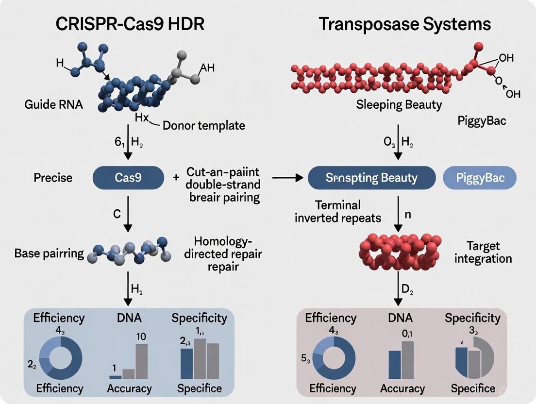
The diagrams below illustrate the key differences between these two mechanisms.
Performance Comparison: Quantitative Data
The following tables summarize key performance metrics for HDR-based systems and the novel evoCAST system, based on recent experimental findings.
Table 1: Overall System Performance Comparison
| Feature | CRISPR-Cas9 HDR | Evolved CAST (evoCAST) |
|---|---|---|
| Integration Mechanism | Host-dependent HDR | Direct, enzyme-mediated "cut-and-paste" |
| DSB Generation | Yes, required [12] | No [14] |
| Cell Cycle Dependence | Limited to S/G2 phase [12] | No [14] |
| Typical Efficiency | Highly variable; often low (<10%) [11] | 10-25% (evoCAST, kilobase cargo) [14] |
| Genotoxic Risk | DSB-associated (indels, translocations) [12] | Minimal; undetected indels in studies [14] |
| Product Purity | Low due to competing NHEJ/MMEJ [10] | High; predominantly unidirectional products [14] |
| Cargo Size Capacity | Practical limit of a few kilobases for HDR [12] | Wide range (≥1 kb to >100 kb) [14] [1] |
Table 2: Experimental Data from evoCAST Validation (2025) Data sourced from continuous evolution of PseCAST system in human cells [14].
| Parameter | Wild-Type PseCAST | Evolved CAST (evoCAST) | Fold Improvement |
|---|---|---|---|
| Average Integration Efficiency | <~0.1% | ~10-25% | ~200-fold (average) |
| Genomic Loci Tested | N/A | 14 loci | N/A |
| Indel Formation | Not reported | Not detected | N/A |
| Key Application Demonstrated | N/A | Installation of factor IX cDNA, CAR into TRAC, and cDNAs for genetic diseases | N/A |
Experimental Protocols
Protocol: Enhancing HDR Efficiency with DNA Repair Pathway Modulation
This protocol is widely used to improve HDR outcomes by suppressing competing repair pathways, as demonstrated in recent 2025 research [10].
- Cell Preparation: Use a human non-transformed diploid cell line (e.g., hTERT-RPE1) or other therapeutically relevant cells. Culture cells under standard conditions.
- CRISPR Component Delivery: Form ribonucleoprotein (RNP) complexes by mixing recombinant Cas nuclease (Cas9 or Cpf1) with guide RNA transcribed in vitro. Electroporate the RNP complexes along with the donor DNA template (e.g., a PCR-amplified fragment with 90-base homology arms) into the cells.
- Inhibitor Treatment: Immediately after electroporation, treat cells for 24 hours with small molecule inhibitors to shift the repair balance toward HDR.
- Analysis: After 4 days, analyze knock-in efficiency via flow cytometry (for fluorescent tags) and perform long-read amplicon sequencing (e.g., PacBio) followed by genotyping with a computational framework like "knock-knock" to comprehensively map all repair outcomes [10].
Protocol: evoCAST-Mediated Integration in Human Cells
This protocol outlines the key steps for using the evolved PseCAST system for DSB-free DNA integration, as detailed in a 2025 study [14].
- Component Delivery: Co-transfect human cells (e.g., HEK293T) with plasmids encoding:
- The evoCAST transposase module (evolved TnsA, TnsB, and TnsC).
- The DNA-targeting module (e.g., QCascade complex).
- A crRNA specific to the desired genomic target.
- A donor plasmid containing the kilobase-scale DNA cargo flanked by the necessary transposon ends.
- Incubation and Expression: Allow 48-72 hours for the expression of CAST components and the integration event to occur. Note that, unlike HDR, this process does not require the addition of external repair pathway inhibitors.
- Validation: Harvest genomic DNA and assess integration efficiency using droplet digital PCR (ddPCR) or next-generation sequencing. Analyze potential off-target integration sites via methods like LAM-PCR or unbiased whole-genome sequencing. The key advantage is the ability to confirm precise, unidirectional integration without indels at the on-target site [14].
The Scientist's Toolkit: Key Research Reagents
Table 3: Essential Reagents for HDR and Transposase Systems
| Reagent/Solution | Function | Example Products/Components |
|---|---|---|
| Programmable Nuclease | Creates a DSB at the target genomic locus. | Cas9 protein, Cpf1 (Cas12a) protein [10] |
| Guide RNA | Directs the nuclease to the specific DNA sequence. | crRNA, tracrRNA, or single guide RNA (sgRNA) [10] [14] |
| HDR Donor Template | Provides the DNA blueprint for precise repair/insertion. | ssODN (for small edits), dsDNA with long homology arms (for large insertions) [5] |
| DNA Repair Modulators | Shifts cellular repair toward HDR and away from NHEJ/SSA. | Alt-R HDR Enhancer V2 (NHEJi), D-I03 (Rad52/SSA inhibitor), ART558 (POLQ/MMEJ inhibitor) [10] |
| Transposase System Plasmids | Deliver the core components for "cut-and-paste" integration. | Plasmids for TnsA, TnsB, TnsC, CRISPR-targeting module (e.g., Cascade), and crRNA [14] |
| Donor Transposon Plasmid | Carries the cargo to be integrated, flanked by transposon ends. | Plasmid containing the gene-sized cargo (e.g., cDNA, CAR) and required attL and attR sites [14] |
The experimental data clearly delineates the applications for these two technologies. CRISPR-Cas9 HDR remains a powerful and familiar tool for making precise edits and smaller insertions, especially in easily transfected, dividing cells. However, the evoCAST system represents a significant leap forward for applications requiring the precise integration of large DNA sequences with high product purity.
The independence of CAST systems from host DSB repair pathways is their most transformative advantage, minimizing genotoxicity and making them uniquely suited for therapeutic applications in non-dividing cells and for engineering sensitive cell types. While the field is rapidly advancing, current challenges for CASTs include optimizing delivery of the multi-component system and further validating its specificity across a wider range of human cell types [14] [1].
In conclusion, the choice between HDR and transposase systems is not a matter of one being universally superior, but rather of strategic selection based on the experimental goal. For small-scale precision edits, HDR is a robust choice. For the one-time, mutation-agnostic integration of large genetic cargo—such as a healthy gene copy to treat a loss-of-function disease—the "cut-and-paste" mechanism of advanced transposase systems like evoCAST offers a compelling and powerful alternative [14] [12].
The ability to insert large DNA sequences efficiently and precisely into specified genomic sites represents a cornerstone capability for advanced genetic engineering, with profound implications for synthetic biology, disease modeling, and therapeutic development. For years, the genome editing field has relied primarily on two competing approaches for DNA integration: CRISPR-Cas9-mediated homology-directed repair (HDR) and transposase-based systems. While CRISPR-Cas9 HDR enables targeted integration through programmable guide RNAs, it suffers from critical limitations including low efficiency in primary cells, dependence on cellular repair machinery active primarily in dividing cells, and unintended mutagenic byproducts at both target and off-target sites [15] [16]. Transposase systems, conversely, can efficiently insert large DNA payloads but traditionally lack precise targeting capabilities, resulting in semi-random integration patterns [17].
The emergence of CRISPR-associated transposase (CAST) systems represents a transformative convergence of these technologies, combining the programmability of CRISPR with the efficient large-DNA integration capabilities of transposases. These natural bacterial systems utilize RNA-guided CRISPR complexes to direct the insertion of kilobase-scale DNA cargos into specific genomic locations without creating double-strand breaks (DSBs) [1] [14]. This review provides a comprehensive comparison of these evolving genome engineering platforms, with particular emphasis on the mechanisms, performance metrics, and experimental applications of CAST systems as emerging alternatives to established methods.
Technology Platforms: Mechanisms and Comparative Performance
CRISPR-Cas9 Homology-Directed Repair (HDR)
The CRISPR-Cas9 HDR system utilizes a Cas9 nuclease complexed with a programmable guide RNA (gRNA) to create site-specific double-strand breaks in genomic DNA. These breaks are then repaired using exogenous donor DNA templates through the cell's endogenous homology-directed repair pathways [15]. The REC lobe facilitates gRNA binding and target recognition, while the NUC lobe contains RuvC and HNH nuclease domains that cleave opposite DNA strands [15]. Successful integration requires coordination of multiple cellular processes: DSB recognition, resection to create single-stranded overhangs, and strand invasion with homologous recombination.
Despite its widespread adoption, CRISPR-Cas9 HDR faces fundamental limitations. HDR efficiency is cell cycle-dependent, being primarily active in the S and G2 phases, which restricts its application in non-dividing cells [14]. The requirement for donor DNA templates with extensive homology arms further complicates experimental design. Most critically, the induction of DSBs activates competing repair pathways, particularly error-prone non-homologous end joining (NHEJ), which generates indel mutations at target sites with frequencies often exceeding those of precise HDR [1] [16]. These limitations have motivated the development of alternative integration technologies.
CAST Systems: RNA-Guided Transposition
CAST systems represent a distinct mechanistic approach to DNA integration by combining RNA-guided targeting with transposase-mediated insertion. Natural CAST systems are derived from bacterial Tn7-like transposons that have co-opted CRISPR-Cas machinery for targeted transposition [14]. These systems typically comprise two core modules: a DNA targeting complex (often Cascade-Cas with TniQ) that identifies genomic integration sites through programmable RNA guides, and a transposase module (TnsA, TnsB, TnsC) that catalyzes the excision and insertion of DNA cargo [18] [14].
The integration mechanism occurs without DSB formation at the target site. Instead, the RNA-guided targeting complex identifies and binds to the specific genomic locus, recruiting the transposase machinery which then catalyzes the "cut-and-paste" transposition of the donor DNA into the target site [14]. This DSB-free mechanism fundamentally differs from CRISPR-Cas9 HDR and avoids activating error-prone DNA repair pathways, resulting in higher product purity and reduced indel formation [14].
Table 1: Comparison of Large DNA Insertion Technologies
| Technology | Mechanism | Insertion Size | Efficiency | Product Purity | Key Advantages | Key Limitations |
|---|---|---|---|---|---|---|
| CRISPR-Cas9 HDR | DSB-dependent with donor template | <100 bp to several kb | 0.1-10% in human cells [16] | Low (high indel frequency) [14] | Well-established protocols | Cell cycle dependence, DSB-associated risks |
| HITI | DSB-dependent via NHEJ | Several kb | 1-30% [16] | Moderate (orientation heterogeneity) [14] | Works in non-dividing cells | Indel formation, bidirectional insertion |
| Prime Editing | Reverse transcription without DSBs | <100 bp [18] | 1-50% [16] | High (low indels) [18] | Precise small edits | Limited cargo capacity |
| CAST Systems | RNA-guided transposition | Multi-kb (up to 10 kb+) [14] | 10-53% in human cells [14] [17] | High (unidirectional, low indels) [14] | DSB-free, large cargo capacity | Ongoing optimization of specificity |
Table 2: Performance Metrics of Engineered CAST Systems
| CAST Variant | Parent System | Engineering Approach | Integration Efficiency | Key Features | Experimental Validation |
|---|---|---|---|---|---|
| evoCAST [14] | PseCAST | PACE evolution + rational engineering | ~10-25% across 14 genomic sites | ~200-fold improvement over wild-type, minimal indels | Human factor IX cDNA integration, CAR insertion into TRAC |
| SuperDn29-dCas9 [17] | Dn29 LSR | Directed evolution + dCas9 fusion | Up to 53% at endogenous loci | 97% genome-wide specificity | Stable expression in stem cells and primary T cells |
| PseCAST [18] | Wild-type CAST | Structure-guided engineering | ~1% (improved with ClpX) [14] | First CAST active in human cells | kilobase-scale insertion in human cells |
| Type V-K CASTs [18] | Various natural systems | Minimal engineering | Low efficiency in human cells [18] | Compact system size | Testing in heterologous contexts |
Experimental Protocols and Workflows
evoCAST Implementation for Gene Integration
The development of evoCAST through phage-assisted continuous evolution (PACE) represents a significant advancement in CAST engineering for human cell applications [14]. The following protocol outlines the key steps for implementing evoCAST for targeted DNA integration:
Vector Design and Preparation: Clone the evoCAST system components (evolved TnsA, TnsB, TnsC, and PseCAST QCascade complex) into mammalian expression vectors with appropriate promoters. The donor plasmid containing the DNA cargo (1-12 kb) must be flanked by the appropriate transposon ends recognized by the TnsA/B transposase.
Cell Transfection: Co-transfect HEK293T cells (or other relevant cell types) with the evoCAST expression vectors and donor plasmid using standard transfection methods (e.g., PEI, lipofectamine). Optimal ratios of targeting:transposase:donor components typically range from 1:1:2 to 1:2:1.
Target Site Selection: Design crRNAs with 30-32 nt spacers complementary to the target genomic locus. The target site must contain a 5'-CC-3' protospacer adjacent motif (PAM) for PseCAST recognition [14]. For human therapeutic applications, evoCAST has been successfully targeted to safe harbor loci including ALB intron 1 and TRAC.
Analysis of Integration Events: Harvest cells 7-14 days post-transfection and assess integration efficiency using genomic PCR, sequencing, and functional assays. evoCAST typically generates unidirectional insertions with minimal indels at the integration junctions [14].
The PACE evolution process that produced evoCAST involved hundreds of generations of mutation and selection in E. coli, with selection pressure specifically designed to link transposition activity to phage propagation [14]. This resulted in transposase variants with dramatically improved integration activity in human cells while maintaining the favorable product purity characteristics of wild-type CAST systems.
Engineered Recombinase Platform Development
The engineering of large serine recombinases (LSRs) like superDn29 provides an instructive comparison to CAST development [17]. The optimization workflow involves:
Deep Scanning Mutagenesis: Create comprehensive variant libraries through single-site saturation mutagenesis of the parent recombinase (Dn29).
High-Throughput Screening: Use intra-plasmid recombination reporters to select variants with improved efficiency and specificity. Successful recombinants are selected by removal of an intervening restriction site.
Mutation Stacking: Combine beneficial mutations through DNA shuffling and rational design, with computational models to predict additive effects.
dCas9 Fusion: Enhance targeting specificity by fusing engineered recombinases to dCas9, enabling simultaneous target and donor recruitment.
Validation in Therapeutic Contexts: Test top-performing variants in stem cells and primary human T cells with cargo sizes up to 12 kb [17].
This engineering framework produced LSR variants with integration efficiencies up to 53% and 97% genome-wide specificity at endogenous human loci, demonstrating how traditional recombinase systems can be optimized to compete with emerging CAST technologies [17].
CAST System Mechanism: RNA-guided DNA integration without double-strand breaks.
The Scientist's Toolkit: Essential Research Reagents
Successful implementation of CAST systems requires specific molecular tools and reagents. The following table outlines key components for establishing RNA-guided transposition in laboratory settings:
Table 3: Essential Research Reagents for CAST System Implementation
| Reagent Category | Specific Components | Function | Example Sources/Systems |
|---|---|---|---|
| CAST Expression Plasmids | TnsA, TnsB, TnsC, Cascade subunits (Cas8, Cas7, Cas6), TniQ | Provides necessary protein components for assembly of functional CAST complexes | PseCAST [14], VchCAST [18], evoCAST [14] |
| Targeting RNAs | crRNA expression constructs or synthetic crRNAs | Guides CAST complex to specific genomic loci | Custom designs with 30-32 nt spacers complementary to target sites [18] |
| Donor Templates | Transposon donor vectors with cargo DNA | Provides DNA payload for integration | Plasmids with cargo flanked by appropriate transposon ends recognized by TnsA/B [14] |
| Host Factors | ClpX unfoldase | Enhances integration efficiency in some systems | E. coli ClpX for PseCAST [14] |
| Engineering Tools | PACE systems, structural biology resources | CAST optimization and characterization | Cryo-EM structures for rational design [18] |
| Delivery Systems | Viral vectors, electroporation, lipid nanoparticles | Introduction of CAST components into cells | AAV, lentivirus, non-viral delivery methods [19] |
CAST Engineering Workflow: From screening to therapeutic application.
CRISPR-transposon hybrid systems represent a paradigm shift in large DNA insertion technology, addressing fundamental limitations of both traditional CRISPR-HDR and conventional transposase systems. The development of engineered CAST platforms like evoCAST and optimized LSRs with dCas9 fusions demonstrates rapid progress toward efficient, precise integration of multi-kilobase DNA cargoes at therapeutic targets [14] [17].
While CAST systems currently achieve robust integration efficiencies of 10-25% across multiple genomic loci with minimal byproducts [14], ongoing challenges include further optimization of specificity, reduction of vector size for in vivo delivery, and expansion of targeting scope. The convergence of continuous evolution methods with structural insights promises to accelerate the development of next-generation CAST variants with enhanced properties for both basic research and clinical applications [18] [14].
For research teams considering implementation of these systems, CAST technologies offer distinct advantages for applications requiring precise integration of large DNA payloads, particularly in non-dividing cells where HDR efficiency is limiting. However, traditional CRISPR-HDR remains suitable for smaller edits in easily transfectable cell lines, while emerging recombinase systems provide alternative pathways for specific integration scenarios. The expanding toolkit of RNA-guided integration technologies ultimately provides researchers with multiple pathways to address the persistent challenge of targeted large DNA insertion in the human genome.
In the realm of CRISPR-based genome engineering, selecting the appropriate tool for DNA insertion is paramount to the success of a research project. While the classic CRISPR-Cas9 system leverages cellular repair pathways like Non-Homologous End Joining (NHEJ) and Homology-Directed Repair (HDR), emerging technologies are increasingly harnessing targeted transposons and recombinases. These distinct systems offer different advantages in terms of insertion size, precision, and mechanism of action. This guide provides an objective comparison of these technologies, focusing on their performance characteristics and integration specificity to inform researchers and drug development professionals in their experimental design.
NHEJ vs. HDR: Cellular Repair Pathways for CRISPR Editing
Core Definitions and Mechanisms
When the CRISPR-Cas9 system creates a double-strand break (DSB) in the DNA, the cell employs one of two primary pathways to repair the damage: Non-Homologous End Joining (NHEJ) or Homology-Directed Repair (HDR) [11] [13].
- NHEJ (Non-Homologous End Joining): This is an error-prone repair pathway that ligates the two broken ends of DNA back together without the need for a homologous template [11] [20]. Its speed and efficiency often come at the cost of precision, frequently resulting in small insertions or deletions (INDELs) at the repair site. These INDELs can disrupt the open reading frame of a gene, leading to knockouts [11].
- HDR (Homology-Directed Repair): This is a precise repair mechanism that utilizes a donor DNA template with homologous sequences flanking the DSB to accurately repair the break [11] [13]. This allows for the incorporation of specific genetic changes, such as point mutations or the insertion of a gene cassette (knock-in). However, HDR is naturally less efficient than NHEJ and is restricted to specific cell cycle phases (S and G2) [11].
Comparative Performance and Experimental Data
The choice between NHEJ and HDR is dictated by the experimental goal. The table below summarizes their key characteristics and performance data.
Table 1: Performance Comparison of NHEJ and HDR in CRISPR Editing
| Feature | NHEJ | HDR |
|---|---|---|
| Primary Application | Gene knockouts (disruption) [11] [13] | Precise knock-ins, point mutations, gene corrections [11] [13] |
| Template Requirement | Not required [11] | Requires homologous donor template (e.g., plasmid, ssODN) [11] |
| Efficiency | High (faster, more efficient pathway) [11] | Low (less efficient than NHEJ) [11] [20] |
| Precision | Imprecise; often generates INDELs [11] [13] | Highly precise; uses homology for error-free repair [11] [13] |
| Cell Cycle Dependence | Active throughout the cell cycle [20] | Primarily active in S and G2 phases [11] [20] |
| Common Outcome | Small insertions/deletions (INDELs) leading to frameshifts [11] | Precise insertion of desired sequence from donor [11] |
Experimental Protocol: HDR-based Knock-in
A standard protocol for achieving precise gene insertion via HDR involves several key steps [11]:
- DSB Induction: Co-deliver a plasmid or ribonucleoprotein (RNP) complex expressing the Cas9 nuclease and a guide RNA (gRNA) targeting the specific genomic locus of interest.
- Donor Template Design: Provide a donor DNA template containing the desired insert sequence (e.g., a GFP tag or a promoterless reporter gene). This insert must be flanked by homology arms (typically >50 bp) that match the sequences upstream and downstream of the Cas9 cut site.
- Screening and Validation: After transfection, cells are screened for the integration event. This is typically done by PCR amplification across the integration junctions, followed by sequencing to confirm precise editing.
Transposons vs. Recombinases: Engineered Systems for DNA Integration
Core Definitions and Mechanisms
Beyond harnessing endogenous cellular repair, researchers can use engineered enzyme systems for DNA insertion.
- Transposons (DNA "Cut-and-Paste"): DNA transposons are mobile genetic elements that naturally "cut and paste" themselves from a donor location to a new site in the genome [21]. CRISPR-associated transposases (CASTs), such as the ShCAST and INTEGRATE systems, fuse this natural transposition ability with the programmability of CRISPR. The Cas protein (often a nickase or deactivated version) guides the complex to a target site, where the associated transposase (e.g., TnsB) then catalyzes the integration of the donor cargo [22] [21].
- Recombinases (Site-Specific Recombination): Recombinases, such as Cre and Bxb1, catalyze the recombination between two specific DNA recognition sites [1] [23]. Traditional systems like Cre-loxP are highly specific but require pre-engineering of the genome with "landing pad" sequences. Newer technologies are fusing recombinases with CRISPR systems (e.g., dCas9) to achieve RNA-guided, programmable integration without the need for pre-engineered sites [1].
Comparative Performance and Experimental Data
The table below compares the performance of modern CRISPR-guided transposase and recombinase systems.
Table 2: Performance Comparison of CRISPR-Guided Transposase and Recombinase Systems
| Feature | CRISPR-Associated Transposases (CASTs) | CRISPR-Guided Recombinases |
|---|---|---|
| Mechanism | RNA-guided "cut-and-paste" transposition [22] [21] | RNA-guided, site-specific recombination [1] |
| DSB Induction | Often DSB-independent; relies on transposase activity [24] | Typically DSB-independent; relies on recombinase activity [1] |
| Insertion Size | Large fragments: 10 kb to >30 kb reported in prokaryotes [22] [23] | Varies; systems like PASTE report insertions up to 36 kb in human cells [22] |
| Efficiency | Up to ~100% in bacteria; ~1-3% in human cells (e.g., MG64-1 CAST) [22] [23] | 50-60% reported for PASTE in human cell lines [22] |
| Specificity | High on-target specificity possible (e.g., 88-95% for engineered CASTs) [24] | High specificity directed by gRNA and recombinase [1] |
| Key Limitation | Lower efficiency in eukaryotic cells; potential for co-integrate products [22] [24] | Often requires complex multi-component delivery [22] |
Experimental Protocol: Transposase-Assisted Targeted Integration
A protocol for plant genome engineering using the TATSI (Transposase-Assisted Target-Site Integration) system illustrates the fusion of transposase and CRISPR technologies [21]:
- System Assembly: Fuse a hyperactive transposase (e.g., rice Pong ORF2) to a programmable nuclease like Cas9 or Cas12a.
- Donor Construction: The DNA cargo to be inserted is flanked by the requisite transposon terminal inverted repeats (TIRs), which are recognized by the transposase.
- Delivery and Excision: The Pong-Cas fusion protein and a gRNA are co-delivered into a plant line containing the donor cargo. The transposase component excises the cargo from the donor site.
- Targeted Integration: The Cas component creates a DSB at the predefined genomic target site. The excised transposon cargo, bound and protected by the transposase, is then inserted into this programmed break with high precision and full-length cargo retention.
Integration Specificity and a Path to Improved Precision
Integration specificity refers to the system's ability to insert DNA solely into the intended target site without off-target integration. This is a critical safety and efficacy metric, especially for therapeutic applications.
- NHEJ and HDR: Traditional CRISPR-Cas9 HDR is susceptible to off-target effects at genomic loci with sequences similar to the gRNA target [25]. Furthermore, the DSB itself can trigger unintended on-target mutations via the NHEJ pathway, competing with precise HDR [11].
- CAST Systems: Early V-K CAST systems showed substantial off-target integration activity independent of the Cas12k effector [24]. However, high-throughput screening and protein engineering (e.g., creating the HELIX system) have demonstrated that specificity can be drastically improved to over 95% on-target activity while maintaining robust integration efficiency [22] [24].
- Recombinase Systems: Technologies like PASTE, which combine a prime editor, a recombinase, and a CRISPR-Cas system, achieve high specificity by first installing a "landing pad" sequence at the target site, which is then recognized by the recombinase for highly specific cargo integration [22].
The relationship between activity and specificity is often a trade-off. Engineering efforts reveal that mutations in transposase components can tune this balance, highlighting the need for directed evolution to create optimal tools [24].
The Scientist's Toolkit: Key Research Reagents
Successful implementation of these gene-editing strategies requires a suite of specific reagents.
Table 3: Essential Reagents for DNA Insertion Technologies
| Reagent | Function | Example Systems/Tools |
|---|---|---|
| Cas9 Nuclease | Induces a double-strand break at the target genomic locus. | SpCas9, LbCas12a [21] |
| Guide RNA (gRNA) | Directs the Cas protein or CAST complex to the specific DNA target sequence. | sgRNA, crRNA [11] [22] |
| Homologous Donor Template | Serves as the repair template for HDR. Contains the insert flanked by homology arms. | HDR donor plasmid, single-stranded oligodeoxynucleotide (ssODN) [11] [13] |
| Transposase/Recombinase | Catalyzes the excision and integration of DNA cargo. | Pong transposase, Bxb1 integrase, Cre recombinase [1] [21] |
| Transposon Donor Plasmid | Carries the genetic cargo flanked by the necessary terminal repeats for transposase recognition. | Donor plasmid with LE-cargo-RE for INTEGRATE system [22] |
| Bridge RNA | A novel RNA component that programs both target and donor recognition in certain systems. | IS110 family recombinases (e.g., IS621) [1] |
Visualizing Key Concepts and Workflows
NHEJ vs. HDR Repair Pathways
Diagram 1: NHEJ vs. HDR repair pathways initiated by a CRISPR-Cas9 double-strand break.
CRISPR-Associated Transposase (CAST) Workflow
Diagram 2: Workflow for targeted DNA insertion using a CRISPR-associated transposase (CAST) system.
Practical Deployment: Application Landscapes for HDR and Transposase Technologies
In the evolving landscape of genetic engineering, two powerful strategies have emerged for modifying genomes: CRISPR-Cas9-mediated homology-directed repair (HDR) and transposase-based systems. While HDR excels at making precise, small-scale edits such as correcting point mutations to create isogenic cell lines, transposase systems, particularly CRISPR-associated transposases (CASTs), offer a superior approach for inserting large DNA sequences without relying on the cell's repair machinery. This guide provides an objective comparison of these technologies, highlighting their performance characteristics, optimal applications, and practical implementation protocols to help researchers select the appropriate tool for their specific genetic engineering goals.
Technology Comparison: HDR vs. Transposase Systems
The table below summarizes the key characteristics of CRISPR-Cas9 HDR and transposase systems:
Table 1: Technology Comparison at a Glance
| Feature | CRISPR-Cas9 HDR | Transposase Systems (e.g., CASTs) |
|---|---|---|
| Primary Use | Precise gene correction, point mutation fixes, short sequence insertions [4] | Large DNA cargo insertion (≥1 kb), gene-sized sequence integration [14] [23] |
| Editing Mechanism | Relies on endogenous cellular repair pathways after a double-strand break (DSB) [4] | "Cut-and-paste" transposition; does not require DSBs or endogenous repair pathways [8] [14] |
| Typical Cargo Size | Most efficient for small edits; efficiency decreases with larger cargos [7] | Kilobase-scale cargos (e.g., 1-10 kb for Tol2; up to 30 kb for some CASTs) [7] [23] |
| Editing Efficiency | Generally low (often <10%), highly dependent on cell type and division state [4] | Can achieve 10-25% in human cells with evolved systems (evoCAST) [14] |
| Product Purity | Low; competes with error-prone NHEJ, leading to mixed editing outcomes and indels [4] | High; generates predominantly unidirectional products without detectable indels [14] |
| Key Advantage | High precision for small-scale edits; well-established protocols [26] | Single-step, DSB-free insertion of large sequences, suitable for non-dividing cells [14] |
| Major Limitation | Low efficiency in non-dividing cells; requires donor template [4] | Early stages of development for mammalian cells; requires specialized system optimization [1] [23] |
Quantitative Performance Data
The following table compiles experimental data from key studies to facilitate direct comparison of the systems' performance under various conditions.
Table 2: Summary of Quantitative Performance Data
| System / Study | Cargo Size | Target / Cell Type | Efficiency | Key Outcome / Note |
|---|---|---|---|---|
| HDR (CRISPR-Cas9) | N/A (Point Mutation) | Various disease models (e.g., HBB, CFTR) [26] | Variable, often low | Precision correction of mutations causing sickle cell disease, β-thalassemia, cystic fibrosis [26] |
| Tol2 Transposon | Up to 10 kb [7] | Zebrafish | High integration efficiency | Random integration; higher germline transmission rates than CRISPR/Cas9 [7] |
| Type I-F CAST (PseCAST) | ~1.3 kb | HEK293 cells [23] | ~1% | Early demonstration of CAST activity in human cells [23] |
| Evolved CAST (evoCAST) | Kilobase-sized cargos | 14 genomic sites in HEK293T cells [14] | 10-25% | Major efficiency improvement; no detected indels; low off-target integration [14] |
Experimental Protocols
Protocol 1: Generating Isogenic Cell Lines via CRISPR-Cas9 HDR
This protocol is designed for precise correction of a point mutation in a human cell line, a common step in creating isogenic controls for functional studies.
1. Design and Synthesis: - gRNA Design: Design a sgRNA with a spacer sequence that binds 10-20 bases upstream of the target mutation. Verify the uniqueness of the target sequence within the genome and ensure it is adjacent to a PAM sequence (NGG for SpCas9) [3]. - Donor Template Design: Synthesize a single-stranded oligodeoxynucleotide (ssODN) donor template (90-200 nt). The template should contain the desired corrective sequence flanked by homology arms (at least 30-40 nt on each side) that are identical to the genomic regions surrounding the DSB [26].
2. Delivery: - Co-deliver the SpCas9 protein (or expression plasmid/mRNA), the sgRNA, and the ssODN donor template into your target cells (e.g., iPSCs) using a high-efficiency method such as electroporation [4].
3. Enhance HDR Efficiency: - Synchronize Cells: Treat cells with cell cycle-arresting agents like nocodazole to enrich for cells in the S and G2 phases, where HDR is more active [4]. - Inhibit NHEJ: Add small molecule inhibitors of the NHEJ pathway, such as NU7026 or Scr7, to the culture medium shortly after editing to reduce error-prone repair and favor HDR [4].
4. Validation: - After 48-72 hours, extract genomic DNA from a subset of cells. - Use a restriction fragment length polymorphism (RFLP) assay or digital droplet PCR (ddPCR) to detect the presence of the corrected sequence. - For clonal analysis, single-cell sort the edited population and expand individual clones. Sequence the target locus in each clone to identify isogenic lines with the precise correction and no off-target indels [26].
Protocol 2: Targeted Gene Insertion via an Evolved CAST System
This protocol utilizes the recently developed evoCAST system for the targeted, DSB-free integration of a large DNA cargo, such as a therapeutic cDNA.
1. System Assembly: - Plasmid Construction: Clone your DNA cargo (e.g., a ~2 kb cDNA) into a donor plasmid between the transposon ends. Co-deliver this donor plasmid with a second plasmid expressing the evolved PseCAST machinery: the evolved TnsA-TnsB-TnsC (TnsABC*) transposase, the Cascade complex (for DNA targeting), and a guide RNA specific to your genomic target [14].
2. Delivery and Integration: - Transfect the plasmid mix into human cells (e.g., HEK293T). The Cascade complex and gRNA will guide the entire system to the target genomic locus. The evolved TnsABC* complex will then catalyze the excision of the cargo from the donor plasmid and its integration into the genome without creating a DSB [14].
3. Analysis: - After 5-7 days, assess integration efficiency using genomic PCR across the 5' and 3' junctions of the integrated cargo. - Quantify the percentage of edited cells using next-generation sequencing (NGS) of the target locus. - To verify the absence of DSBs and their associated byproducts, perform Sanger sequencing on the integrated locus to check for clean junctions and no indels [14].
Visualizing the Core Mechanisms
The diagrams below illustrate the fundamental workflows and logical relationships for both editing systems.
HDR Workflow for Point Mutation Correction
CAST System Workflow for Large DNA Insertion
The Scientist's Toolkit: Essential Research Reagents
The table below lists key reagents and materials required for executing the experiments described in this guide.
Table 3: Essential Research Reagents and Materials
| Reagent / Material | Function / Description | Example Use Case |
|---|---|---|
| High-Fidelity Cas9 | Engineered Cas9 variant (e.g., eSpCas9, SpCas9-HF1) with reduced off-target effects [3] | Increasing specificity in HDR experiments to minimize unintended edits. |
| Single-Stranded Oligodeoxynucleotide (ssODN) | A single-stranded DNA template (90-200 nt) containing the corrective sequence flanked by homology arms [26] | Serving as the donor template for precise point mutation correction via HDR. |
| NHEJ Inhibitors (e.g., Scr7) | Small molecules that temporarily inhibit the non-homologous end joining pathway [4] | Boosting HDR efficiency by suppressing the competing, error-prone NHEJ repair pathway. |
| Evolved CAST System (evoCAST) | A suite of optimized plasmids for expressing the evolved TnsABC* transposase, Cascade complex, and gRNA [14] | Enabling highly efficient, targeted integration of large DNA cargos in human cells. |
| Electroporation System | A device for delivering genetic material (proteins, RNAs, plasmids) into cells via electrical pulses. | The preferred method for co-delivering multiple CRISPR or CAST components with high efficiency. |
| Digital Droplet PCR (ddPCR) | A highly sensitive and absolute nucleic acid quantification technique. | Detecting and quantifying the efficiency of HDR correction in a mixed cell population. |
Transposon Systems for Stable Cell Line Development and Recombinant Protein Production
In the pursuit of effective recombinant protein production for biopharmaceuticals, stable cell line development represents a critical bottleneck. Traditional methods, particularly those relying on random transgene integration (RTI), often result in unpredictable and highly variable expression levels due to positional effects—where transgene expression depends on the integration site's chromosomal context [27]. While the emergence of CRISPR-Cas9-mediated homology-directed repair (HDR) has offered a pathway to precise gene editing, its efficiency for inserting large DNA cargos remains limited, especially in non-dividing cells [4]. Within this technological landscape, transposon vector systems have emerged as a powerful alternative, enabling semi-targeted integration of large genetic payloads into transcriptionally active genomic regions, thereby overcoming many limitations of both conventional random integration and HDR-based approaches [27] [28].
The broader thesis contrasting CRISPR-Cas9 HDR with transposase systems reveals a fundamental trade-off: while CRISPR-Cas9 HDR excels at precise nucleotide-level edits, its efficiency drops significantly for large DNA insertions (>1 kb) and it depends on the cell's repair machinery, which varies across cell types and states [8] [4]. Transposon systems, in contrast, utilize a DNA break-independent mechanism that reliably accommodates very large genetic payloads, making them particularly suitable for introducing entire gene circuits or multiple expression cassettes needed for complex therapeutic proteins [27].
Transposon System Mechanisms and Comparative Advantages
The Transposon "Cut-and-Paste" Mechanism
DNA transposons are mobile genetic elements that move within genomes via a "cut-and-paste" mechanism [27]. The system consists of two primary components: a donor vector containing the gene of interest (GOI) flanked by inverted terminal repeats (ITRs), and a helper vector expressing the transposase enzyme [29]. The transposase recognizes, binds to, and excises the sequence between the ITRs from the donor vector, then integrates it into the host cell genome [27]. This process bypasses the cell's native DNA repair pathways, enabling efficient integration without inducing double-strand breaks-associated damage responses that can trigger apoptosis or introduce unintended mutations [27] [28].
Comparative Analysis: Transposon Systems vs. Alternative Technologies
Table 1: Technology Comparison for Stable Cell Line Development
| Feature | Random Transgene Integration (RTI) | CRISPR-Cas9 HDR | Transposon Systems |
|---|---|---|---|
| Integration Mechanism | Non-homologous end joining or random integration [28] | Homology-directed repair with donor template [4] | Cut-and-paste transposition independent of host repair pathways [27] |
| Integration Pattern | Random throughout genome [27] | Precise, site-specific [4] | Semi-targeted to transcriptionally active regions [28] |
| Cargo Capacity | Unlimited in theory | Limited (<1 kb for high efficiency) [23] | Very large (up to 100+ kb demonstrated) [29] |
| Typical Efficiency | Low (1-5% of transfected cells) [27] | Low, especially in non-dividing cells [4] | High (up to 9-fold higher yields than RTI) [28] |
| Screening Requirement | Extensive cloning screening required [27] | Moderate, need to verify precise editing [4] | Minimal due to targeted expression [29] |
| Key Advantage | Simple to perform | Nucleotide-level precision | Reliable large payload delivery with consistent expression [27] |
| Primary Limitation | Position effects cause variable expression [27] | Low efficiency for large inserts [4] | Non-specific integration site (semi-random) [28] |
Table 2: Performance Comparison of Major Transposon Systems
| Transposon System | Origin | Integration Efficiency | Cargo Capacity | Key Applications |
|---|---|---|---|---|
| PiggyBac (PB) | Insect [27] | High: 9-fold increase in TNFR:Fc production vs. RTI [28] | Large | Clinical manufacturing, CAR-T cell therapy [28] |
| Sleeping Beauty (SB) | Fish [27] | Moderate: Reliable but lower than PB [28] | Standard | Gene therapy, protein production [28] |
| Tol2 | Medaka fish [29] | High: Efficient stable cell generation [29] | Very large (>100 kb) [29] | Recombinant protein production, large gene inserts [29] |
Experimental Protocols and Workflows
Standardized Protocol for Transposon-Mediated Cell Line Development
The following workflow outlines the optimized procedure for generating stable cell pools using the Tol2 transposon system, as validated in suspension CHO cells [29]:
Detailed Methodology [29]:
Vector Design and Preparation:
- Clone your gene of interest into a Tol2 donor vector containing inverted terminal repeats (ITRs)
- Include a strong promoter (e.g., CMV) driving GOI expression
- Incorporate a selectable marker (e.g., cycloheximide-resistant L36a gene for Tol2 system)
Cell Transfection and Transposition:
- Use suspension-adapted CHO cells in logarithmic growth phase
- Co-transfect with donor vector (10 μg) and transposase vector (25 μg) per 10^7 cells
- Use electroporation or chemical transfection methods optimized for your cell line
- Culture cells without selective pressure for 4 days to allow transposition to occur
Selection and Screening:
- Apply appropriate selective pressure (e.g., 3-30 μM cycloheximide for Tol2/CHX system)
- Maintain selection for 3 weeks, monitoring cell viability and expansion
- Harvest resistant pools for initial productivity assessment
Characterization of Stable Pools:
- Measure recombinant protein titer by ELISA or functional assay
- Determine transgene copy number by qPCR
- Assess clonal stability through extended culture (60+ generations)
CRISPR-Cas9 HDR Control Protocol
For comparative studies, the following HDR protocol highlights key differences from the transposon approach [4]:
DSB Induction:
- Design sgRNAs flanking the target integration site
- Transfect with Cas9-sgRNA ribonucleoprotein complex plus single-stranded or double-stranded donor template
- Optimal HDR occurs during S/G2 cell cycle phases
HDR Enhancement:
- Add NHEJ inhibitors (e.g., SCR7) to favor HDR over error-prone repair
- Synchronize cells to enrich for S-phase populations
- Use Cas9 nickase variants to reduce indel formation
Limitations Observed:
- Efficiency declines dramatically with donor sizes >1 kb
- Extensive screening required to identify precise integrants without indels
- Significant cell-type dependency in success rates
Experimental Data and Performance Metrics
Quantitative Performance Assessment
Table 3: Experimental Performance Data from Transposon Applications
| Application | Transposon System | Protein Yield | Copy Number | Stability | Reference |
|---|---|---|---|---|---|
| TNFR:Fc production | PiggyBac | 9-fold increase vs. RTI | 1-15 copies | Maintained over 60 generations [28] | Balasubramanian et al. |
| Monoclonal antibody production | Tol2 with CHX selection | Up to 107.3 mg/L in clonal lines | 1-15 copies | Constant over long-term culture [29] | Scientific Reports (2023) |
| CAR-T cell generation | Sleeping Beauty | Efficient transduction of human T cells | N/A | Persistent expression in clinical trials [28] | Hackett et al. |
| Therapeutic protein (benchmark) | Random Integration | ~10-20 mg/L (typical baseline) | Variable, often high | Frequently unstable [27] | Industry standard |
Integration Pattern Analysis
Different transposon systems exhibit distinct integration profiles that significantly impact transgene expression stability [30]:
- PiggyBac tends to integrate into transcriptionally active regions, favoring genic areas and regions near transcriptional start sites
- Sleeping Beauty shows a preference for integrating into palindromic AT repeats with a bias toward actively transcribed regions
- Tol2 demonstrates a relatively unbiased integration pattern with reliable expression across integration sites
This semi-targeted integration behavior contrasts sharply with random integration, which frequently results in heterochromatic positioning and subsequent transgene silencing [27].
The Scientist's Toolkit: Essential Research Reagents
Table 4: Key Reagents for Transposon-Mediated Cell Line Development
| Reagent Category | Specific Examples | Function | Implementation Considerations |
|---|---|---|---|
| Transposon Systems | PiggyBac, Sleeping Beauty, Tol2 | Mobile genetic elements for genomic integration | Choice depends on cargo size and integration efficiency requirements [27] [29] |
| Selection Markers | Cycloheximide resistance (L36a mutant), Puromycin N-acetyltransferase, Neomycin phosphotransferase | Enrichment of successfully transfected cells | CHX resistance particularly effective in suspension CHO cells [29] |
| Promoter Systems | CMV, EF-1α, CAGGS, SV40 | Drive high-level expression of transgenes and selection markers | Strong constitutive promoters ensure detectable expression [27] |
| Host Cell Lines | CHO-DG44, CHO-K1, HEK293 | Industrial protein production platforms | CHO cells dominate biopharmaceutical production [27] [28] |
| Expression Enhancers | Matrix attachment regions (MARs), Ubiquitous chromatin opening elements (UCOEs) | Mitigate positional effects and enhance expression stability | Particularly valuable for random integration approaches [27] |
Transposon systems offer a compromise between the randomness of conventional integration and the precision of CRISPR-Cas9 HDR, delivering semi-targeted integration with high efficiency for large DNA payloads. While CRISPR-Cas9 HDR remains unparalleled for single-nucleotide changes or small insertions at defined loci, transposons excel specifically in applications requiring reliable insertion of large genetic elements, such as entire expression cassettes for complex therapeutic proteins [23] [4].
The experimental data consistently demonstrates that transposon systems significantly reduce screening burden while enhancing productivity and stability of recombinant protein producers. For researchers developing cell lines for biopharmaceutical production, transposon systems—particularly PiggyBac and Tol2—represent a mature, validated technology that bridges the gap between the limitations of random integration and the cargo size restrictions of precise editing tools [28] [29].
Future directions in the field include the development of chimeric systems that combine the programmability of CRISPR with the integration capacity of transposases, such as CRISPR-associated transposase (CAST) systems [8] [23]. Although currently demonstrating modest efficiency in mammalian cells (approximately 1-3% in HEK293 cells), these emerging technologies hint at a future where RNA-guided transposition may offer both precision and high cargo capacity [23]. Until these next-generation platforms mature, established transposon systems remain the technology of choice for efficient stable cell line development with large genetic payloads.
CAST Systems for One-Step, DSB-Free Knock-in of Therapeutic Genes
The ability to insert large DNA sequences precisely into the genome represents a cornerstone of modern genetic engineering, with far-reaching implications for therapeutic development, disease modeling, and functional genomics. Traditional approaches relying on CRISPR-Cas9-induced homology-directed repair (HDR) have enabled precise edits but face significant limitations for inserting therapeutic gene-sized constructs. The field has now reached a critical juncture, divided between mature HDR-based technologies and emerging nuclease-free platforms. Among these, CRISPR-associated transposase (CAST) systems have recently emerged as promising alternatives, potentially overcoming the fundamental constraints of double-strand break (DSB)-dependent mechanisms. This review provides a comprehensive comparison of these competing approaches, focusing on their applicability for one-step, DSB-free knock-in of therapeutic genes—a capability that could revolutionize the treatment of loss-of-function genetic disorders.
Technological Foundations: DSB-Dependent vs. DSB-Free Editing Platforms
CRISPR-Cas9 Homology-Directed Repair (HDR)
The CRISPR-Cas9 HDR platform utilizes the cell's endogenous repair machinery to incorporate exogenous donor DNA at specific genomic locations following Cas9-induced double-strand breaks. This system requires three core components: the Cas9 nuclease, a target-specific guide RNA (sgRNA), and a donor template containing homology arms flanking the desired insertion sequence [4]. When successful, this process enables precise integration of foreign DNA, but efficiency remains constrained by cellular competition between HDR and error-prone non-homologous end joining (NHEJ) pathways [4] [31]. The HDR pathway is primarily active during the S and G2 phases of the cell cycle, creating particular challenges for post-mitotic cells and limiting therapeutic applications in non-dividing cell populations [12] [4].
CRISPR-Associated Transposase (CAST) Systems
CAST systems represent a paradigm shift in genome engineering by combining RNA-guided targeting with transposase-mediated integration in a single-step, DSB-free mechanism. These natural bacterial systems utilize nuclease-deficient CRISPR-Cas complexes for target recognition while employing Tn7-like transposases (TnsA, TnsB, TnsC, and TniQ) for subsequent DNA integration [1] [14] [23]. Type I-F CAST systems, among the most well-characterized, employ the Cascade complex for target site recognition, with DNA integration occurring approximately 50-60 bp downstream of the target site without introducing double-strand breaks [14] [23]. This unique mechanism theoretically eliminates the genotoxic risks associated with DSB formation while maintaining programmability through easily designed guide RNAs.
Performance Comparison: Quantitative Analysis of Editing Platforms
Table 1: Comprehensive comparison of genome insertion technologies
| Technology | Insertion Size | Efficiency in Mammalian Cells | DSB Formation | Key Advantages | Major Limitations |
|---|---|---|---|---|---|
| HDR-mediated Knock-in | 1-10 kb | High efficiency (varies by cell type) | Yes | Well-established protocol, precise integration | Cell-cycle dependent, generates indels, low efficiency in non-dividing cells |
| Prime Editing | ≤50 bp (standard), ~1 kb (paired) | Modest efficiency | No | High precision, versatile | Limited cargo size, modest efficiency |
| HITI | >1 kb | High efficiency | Yes | Works in non-dividing cells | High indel rates, uncontrolled integration orientation |
| CAST Systems | 1->100 kb | 0.06%-25% (recent evoCAST: 10-25%) | No | DSB-free, large cargo capacity, low indels | Early development, variable efficiency |
| Integrase Systems | 1->100 kb | Low efficiency | No | Large cargo capacity | Limited target sites, requires pre-installed landing pads |
Table 2: Performance metrics of CAST systems in recent studies
| CAST Variant | Insertion Size | Target Locus | Cell Type | Efficiency | Year |
|---|---|---|---|---|---|
| PseCAST (WT) | ~1.3 kb | Various | HEK293 | ~1% | 2024 |
| V-K CAST (MG64-1) | 3.2 kb | AAVS1 | HEK293 | ~3% | 2024 |
| V-K CAST (nAnil-TnsB) | 2.6 kb | Plasmid DNA | HEK293T | 0.06% | 2024 |
| evoCAST (Evolved) | Kilobase-scale | 14 genomic sites | HEK293T | 10-25% | 2025 |
| Type I-F CAST | Up to ~15.4 kb | Various | E. coli | Nearly 100% | 2024 |
Experimental Protocols: Methodologies for Enhanced Gene Insertion
Enhanced HDR Workflow: Double-Cut Donors with Cell Cycle Synchronization
Recent advances in HDR efficiency have focused on donor design and cell cycle manipulation. The double-cut HDR donor approach utilizes a donor vector flanked by sgRNA-PAM sequences that undergoes in vivo linearization by Cas9,同步izing genomic cleavage with donor availability [32]. Implementation involves:
Donor Construction: Clone insertion cassette between two sgRNA target sites identical to those targeting the genomic locus, flanked by homology arms (300-600 bp optimal) [32].
Cell Synchronization: Treat cells with nocodazole (G2/M phase synchronizer) combined with CCND1 (cyclin D1, functions in G1/S transition) to double HDR efficiency in iPSCs [32] [33].
RNase HII Supplementation: Include RNase HII in electroporation media to process RNA-DNA hybrids that may impede HDR, shown to increase editing rates [33].
NHEJ Inhibition: Co-deliver small molecule inhibitors of key NHEJ proteins (Ku70/Ku80, DNA-PK) to reduce competing repair pathways [12] [4].
This optimized protocol has achieved 20-30% HDR-mediated knock-in in human iPSCs using donors with 300-600 bp homology arms [32].
Evolved CAST System (evoCAST) Implementation
The recently developed evoCAST system demonstrates the potential of directed evolution to overcome bottlenecks in mammalian cell activity [14]. The workflow includes:
Component Delivery: Co-deliver four core plasmids encoding (1) evolved TnsABC transposase, (2) QCascade targeting complex, (3) transposon donor with cargo, and (4) Cas9/sgRNA for genomic target exposure.
Target Site Selection: Choose genomic sites with appropriate PAM sequences recognized by the Cascade complex, typically 30-60 bp upstream of desired integration site.
Transposition Reaction: The evolved TnsABC transposase complex catalyzes "cut-and-paste" transposition without requiring bacterial unfoldase ClpX, reducing cytotoxicity [14].
Validation: Screen for precise, unidirectional integration 50-60 bp downstream of target site using junction PCR and sequencing.
This system achieves 10-25% integration efficiency of kilobase-sized cargoes across multiple genomic loci in HEK293T cells with predominantly unidirectional transposition products and undetectable indels [14].
Figure 1: Comparative mechanisms of HDR-based knock-in and CAST systems for gene integration
The Scientist's Toolkit: Essential Reagents for Advanced Genome Editing
Table 3: Key research reagents for implementing advanced knock-in technologies
| Reagent Category | Specific Examples | Function | Application Notes |
|---|---|---|---|
| CRISPR Components | SpCas9, sgRNA, RNP complexes | Target recognition and cleavage | RNP delivery reduces off-target effects |
| HDR Donor Templates | Double-cut donors, ssODNs, IDLVs | Provide repair template | Double-cut design improves efficiency 2-5x |
| Cell Cycle Modulators | Nocodazole, CCND1, AB-521 | Synchronize cells for HDR | Nocodazole + CCND1 doubles HDR in iPSCs |
| DNA Repair Modulators | RNase HII, AZD7648 (DNA-PKi) | Favor HDR over NHEJ | RNase HII enhances HDR in electroporation |
| CAST System Components | TnsA, TnsB, TnsC, TniQ, Cascade | RNA-guided transposition | Evolved variants show 200x improved activity |
| Delivery Vehicles | IDLVs, LVNPs, electroporation | Component delivery | All-in-one vectors improve synchronization |
Discussion: Therapeutic Applications and Future Directions
The development of CAST systems represents significant progress toward addressing the mutational heterogeneity of genetic diseases through allele-agnostic strategies. The recent evolution of PseCAST (evoCAST) demonstrating 10-25% integration efficiency of kilobase-scale DNA cargoes across multiple genomic loci in human cells marks a critical milestone [14]. These advances enable novel therapeutic approaches, including the installation of full-length cDNA copies of diseased genes (e.g., factor IX for hemophilia B) or chimeric antigen receptors (CARs) for cancer immunotherapy at safe-harbor loci [14].
Despite these promising developments, HDR-based approaches currently maintain advantages in standardization and predictability, with well-established protocols for specific applications, particularly in dividing cells. The double-cut HDR donor strategy combined with cell cycle synchronization achieves 20-30% knock-in efficiency in iPSCs, sufficient for many research applications [32] [33]. Furthermore, the single-vector IDLV approach utilizing a Cas9 off switch demonstrates >80% knock-in efficiency for full-length EGFP, highlighting the continued innovation in HDR technology [34].
Figure 2: Decision framework for selecting knock-in technologies based on therapeutic application requirements
The evolving landscape of large DNA insertion technologies presents researchers with multiple pathways for therapeutic gene integration, each with distinct advantages and limitations. While HDR-based methods benefit from established protocols and continued refinement, CAST systems offer a promising DSB-free alternative with potentially superior safety profiles for therapeutic applications. The recent development of evolved CAST systems with significantly improved efficiency in human cells suggests a forthcoming paradigm shift in gene integration strategies. Research and drug development professionals should consider cargo size, target cell type, and precision requirements when selecting between these platforms, with CAST systems particularly advantageous for applications requiring large insertions in non-dividing cells with minimal genotoxic risk. As both approaches continue to mature, the future of therapeutic gene integration appears increasingly capable of addressing the diverse mutational spectra of human genetic diseases through tailored integration strategies.
The ability to insert large DNA sequences—ranging from therapeutic gene cassettes, such as those encoding chimeric antigen receptors (CARs), to full-length cDNAs for correcting loss-of-function genetic diseases—into specific genomic locations is a central goal in modern genetic medicine. For years, two primary strategies have been employed: CRISPR-Cas9-mediated homology-directed repair (HDR) and viral- or transposon-based random integration. However, both approaches have significant limitations. HDR is inefficient, especially for large cargos, and is active primarily in dividing cells, while random integration leads to heterogeneous expression and potential insertional mutagenesis [35] [1] [36]. The recent development of evolved CRISPR-associated transposases (CASTs) presents a new, precise, and efficient alternative for site-specific gene-sized DNA integration. This case study objectively compares the performance of these systems, providing experimental data and protocols to guide researchers in selecting the appropriate tool for their genome engineering applications.
The following table summarizes the core characteristics, performance metrics, and key differentiators of HDR, transposase systems, and the newly evolved CASTs.
Table 1: Comparative Analysis of Large DNA Insertion Technologies
| Feature | CRISPR-Cas9 HDR | Traditional CASTs | Evolved CASTs (evoCAST) |
|---|---|---|---|
| Core Mechanism | DSB induction followed by repair with donor DNA template [1] | RNA-guided, DSB-free cut-and-paste transposition [14] | Evolved transposase for enhanced activity in human cells [14] |
| Typical Efficiency | Low (often <10% for large inserts) [35] | Very low in human cells (<~0.1% to ~1%) [14] | High (10-25%) across multiple genomic loci [14] |
| Primary Editing Outcome | Precise integration via HDR | Predominantly unidirectional transposition [14] | Predominantly unidirectional transposition [14] |
| Indel Formation | High (due to DSBs and competing NHEJ) [1] | Undetectable levels in studies [14] | Undetectable levels in studies [14] |
| Cargo Size Capacity | Limited by HDR efficiency | Kilobase-scale transposons [14] | Kilobase-scale DNA cargoes [14] |
| Key Advantage | High precision with long homology arms | DSB-free, high product purity | Combines DSB-free editing with high efficiency in human cells |
| Key Limitation | Low efficiency, cell-cycle dependent, induces DSBs | Minimal activity in mammalian cells [14] | New technology, long-term cellular effects still under study |
Experimental Data and Protocols
Installing CAR Cassettes with CRISPR-Cas9 HDR
- Experimental Protocol: To generate HIV-resistant CAR T cells, researchers used a CCR5 megaTAL nuclease to create a double-strand break in the CCR5 locus. A recombinant adeno-associated virus (rAAV) donor template was co-delivered, containing an anti-CD19 CAR expression cassette flanked by homology arms matching the sequences around the CCR5 cut site. Primary human T cells were transfected with the nuclease and infected with the rAAV donor to facilitate HDR [35].
- Performance Data: This HDR-based approach achieved CAR integration rates of 20-35% in primary human T cells, as measured by surface CAR expression 14 days post-editing. In an in vivo xenograft model, these edited T cells mediated significant tumor clearance and extended mouse survival, with functionality equivalent to CAR T cells generated by lentiviral transduction [35].
Installing CAR Cassettes with Evolved CASTs
- Experimental Protocol: The evoCAST system was deployed to insert a CD19-targeted CAR directly into the TRAC locus. The system components—including the evolved transposase and the donor DNA containing the CAR cassette within a transposon—were delivered to the T cells. The process is RNA-guided, targeting the TRAC locus without requiring a double-strand break [14].
- Performance Data: The evoCAST system demonstrated the ability to integrate kilobase-sized CAR cargos into the human genome with efficiencies ranging from 10% to 25% across 14 tested genomic sites. This integration occurred with low levels of off-target activity and without generating detectable indel mutations at the target site, a significant advantage over DSB-dependent methods [14].
Correcting Loss-of-Function Genes
- Experimental Protocol: To address diverse pathogenic alleles in loss-of-function diseases, evoCAST was used to integrate wild-type cDNA sequences into intron 1 of their respective endogenous loci. For example, researchers installed the human factor IX cDNA into intron 1 of the ALB locus, leveraging the host's endogenous regulatory elements for physiologically appropriate expression [14].
- Performance Data: This strategy of targeted cDNA integration at endogenous loci provides a mutation-agnostic therapeutic approach. By placing the healthy gene copy within its native genomic context, evoCAST enables preservation of the natural regulatory environment, which is a key limitation of traditional viral gene addition therapies [14].
Visualizing the Core Mechanisms
The diagram below illustrates the fundamental operational differences between the CRISPR-Cas9 HDR and evolved CAST mechanisms for installing a CAR cassette.
The Scientist's Toolkit: Key Research Reagents
Table 2: Essential Reagents for Advanced Genome Engineering
| Reagent / Solution | Function in Experiment | Specific Example / Note |
|---|---|---|
| Evolved Transposase (evoCAST) | Catalyzes RNA-guided, DSB-free integration of large DNA cargo. | Core enzyme evolved from Pseudoalteromonas sp. S983 system (PseCAST) for ~200-fold higher activity in human cells [14]. |
| rAAV Donor Template | Provides homology-directed repair template with long homology arms for HDR. | Critical for high-efficiency HDR in primary cells; can carry therapeutic cassettes up to ~4-5 kb [35]. |
| CSSDNA Donor Template | Circular single-stranded DNA used for non-viral HDR; reduces cell toxicity and concatemerization. | Used with systems like enGager/TESOGENASE; suitable for cargos up to 2 kb in size [37]. |
| enGager/TESOGENASE System | Fusion of Cas9 to ssDNA-binding peptides to tether donor cssDNA, boosting HDR efficiency. | Reported to enable ~33% CAR integration efficiency in primary T cells [37]. |
| PASSIGE with eeBxb1 | Combines prime editing with an evolved recombinase for landing-pad-independent integration. | Achieves >30% integration efficiency in primary human fibroblasts [38]. |
| Engineered LSRs (e.g., superDn29) | Engineered large serine recombinases for single-step, site-specific DNA insertion. | Can achieve up to 53% integration efficiency at an endogenous human locus with high specificity [17]. |
Discussion and Research Outlook
The data presented in this case study reveals a shifting paradigm in genome engineering. While CRISPR-Cas9 HDR remains a valuable and precise tool, its dependency on the cellular repair machinery and the inherent risks associated with double-strand breaks constrain its efficiency and safety profile [35] [1]. The emergence of evolved CASTs and other next-generation integrases like eeBxb1 and superDn29-dCas9 offers a compelling alternative [14] [38] [17]. These systems achieve therapeutically relevant integration efficiencies (often exceeding 20-30%) while avoiding the pitfalls of DSB generation, thereby minimizing the formation of indels and other chromosomal abnormalities.
The choice of system for a specific application depends on the experimental goals. For the highest possible efficiency where pre-installed landing pads are feasible, PASSIGE with eeBxb1 is a top contender [38]. For completely landing-pad-free, single-step integration of large cargos, evolved CASTs and engineered LSRs currently lead the field in combining efficiency with precision [14] [17]. As these non-viral, site-specific integration technologies continue to mature, they are poised to unlock new possibilities in cell and gene therapy, from the streamlined production of allogeneic CAR-T cells to one-time cures for a wide spectrum of inherited genetic disorders.
Overcoming Limitations: Strategies to Enhance Efficiency and Precision
In the rapidly evolving field of genome engineering, two primary technologies dominate the landscape of large DNA insertion: CRISPR-Cas9-mediated homology-directed repair (HDR) and transposase-based systems. While transposase systems like CAST (CRISPR-associated transposase) and INTEGRATE offer promising avenues for large fragment insertion, CRISPR-Cas9 HDR remains the workhorse for precise genome editing in most research applications. A significant bottleneck, however, lies in the typically low efficiency of HDR, which is often outpaced by error-prone repair pathways like non-homologous end joining (NHEJ). This guide provides a comparative analysis of experimentally validated strategies to overcome this limitation, focusing on donor DNA design, 5'-end modifications, and protein supplementation to boost HDR efficiency for research applications.
The optimization of HDR efficiency involves a multi-faceted approach centered on preparing the donor DNA template to be a more effective substrate for the cell's repair machinery. The table below summarizes the key strategies, their mechanisms of action, and their performance outcomes.
Table 1: Comparison of Key HDR Optimization Strategies
| Strategy | Mechanism of Action | Reported Efficiency Gain | Key Advantages | Key Limitations |
|---|---|---|---|---|
| Donor DNA Denaturation | Using single-stranded DNA (ssDNA) reduces concatemer formation and may mimic a more native repair intermediate [39]. | ~4-fold increase in precise editing vs. dsDNA [39]. | Reduces template multiplication; simpler protocol. | Can be accompanied by increased aberrant template integration [39]. |
| RAD52 Supplementation | RAD52 protein binds ssDNA and promotes strand annealing, directly facilitating the HDR pathway [39]. | ~4-fold increase in ssDNA integration; ~13-fold vs. original dsDNA [39]. | Strong boost in correctly targeted loci. | Increases template multiplication; requires purification/delivery of recombinant protein [39]. |
| 5'-Biotin Modification | "Bulky" moiety shields DNA ends, preventing multimerization and NHEJ-mediated random integration [39] [40]. | Up to 8-fold increase in single-copy integration [39]. | Reduces concatemerization; favors single-copy integration. | Chemical synthesis required. |
| 5'-C3 Spacer Modification | Carbon-based spacer blocks DNA ends similarly to biotin, favoring HDR over NHEJ [39] [40]. | Up to 20-fold rise in correctly edited mice [39]. | Highly effective; can be incorporated during PCR primer synthesis. | Chemical synthesis required. |
| 5'-Triethylene Glycol (TEG) | Modifies ends to reduce availability for NHEJ ligation reactions, including self-ligation into concatemers [41]. | 2- to 5-fold increase in precision editing across multiple species [41]. | Consistent efficacy in human cells, C. elegans, zebrafish, and mice. | Chemical synthesis required. |
| HDR-Boosting ssDNA Modules | Incorporates RAD51-preferred binding sequences to recruit endogenous repair proteins to the donor [42]. | Up to 90.03% HDR efficiency when combined with NHEJ inhibition [42]. | Chemical modification-free; leverages endogenous machinery. | Sequence module must be incorporated into donor design. |
Detailed Experimental Protocols & Data
To ensure the successful implementation of these strategies, the following section details specific experimental protocols and the quantitative data supporting their efficacy.
Optimizing Donor DNA Strandedness and RAD52 Supplementation
Experimental Protocol: A one-step strategy for generating conditional knockout mouse models was employed. CRISPR-Cas9 complexes and long donor DNA templates (~600 bp) containing LoxP sites were microinjected into zygotes. The donor templates were tested in double-stranded (dsDNA) and denatured single-stranded (ssDNA) forms. For the RAD52 condition, the protein was added directly to the injection mix containing the denatured DNA template [39].
Results and Data: The quantitative outcomes from this large-scale experiment, involving over 2,000 zygotes, clearly demonstrate the impact of each approach on precision and unwanted recombination events.
Table 2: Performance of Donor DNA and RAD52 Strategies in Mouse Zygotes
| Experimental Condition | % Correct HDR (F0 HDR%) | % Template Multiplication (F0 HtT%) | % Locus Modification |
|---|---|---|---|
| dsDNA (5'-monophosphorylated) | 2% | 34% | 40% |
| Denatured ssDNA (5'-monophosphorylated) | 8% | 17% | 50% |
| Denatured ssDNA + RAD52 | 26% | 30% | 83% |
The data shows that while RAD52 supplementation provides the most dramatic boost in precise HDR, it comes with a trade-off of increased template multiplication [39].
Implementing 5'-End Modifications for Enhanced HDR
Experimental Protocol: Long dsDNA donors were generated by PCR using 5'-modified primers. Modifications included Biotin, Amino-dT (A-dT), and carbon spacers (SpC3). These modified donors were co-injected with Cas9 mRNA and locus-specific sgRNAs into medaka fish one-cell stage embryos. HDR efficiency was assessed by genotyping GFP-expressing embryos to confirm precise, single-copy integration [40]. Similar protocols were applied in mouse zygotes [39] and human cells [41].
Results and Data: The 5'-modifications, particularly SpC3 and Biotin, proved highly effective in preventing donor multimerization and promoting correct integration.
Table 3: Efficacy of 5'-End Modifications Across Different Experimental Systems
| Modification Type | Experimental System | Key Finding | Reported Efficiency |
|---|---|---|---|
| 5'-C3 Spacer | Mouse zygotes (Nup93 locus) | Massive increase in correctly edited founders [39]. | Up to 20-fold rise |
| 5'-C3 Spacer | Medaka fish (multiple loci) | Enabled efficient single-copy HDR in F0 generation [40]. | 9.5% of surviving zygotes (rx2 locus) |
| 5'-Biotin | Mouse zygotes (Nup93 locus) | Significant boost in single-copy integration [39]. | Up to 8-fold increase |
| 5'-TEG | Human cells (TLR assay) | Consistently increased precision editing potency [41]. | ~4-fold more potent |
| 5'-TEG | Zebrafish, C. elegans, Mouse | Increased germline editing efficiency [41]. | 2- to 5-fold increase |
The Scientist's Toolkit: Essential Research Reagents
Successful implementation of these HDR-boosting strategies requires key reagents. The following table lists essential materials and their functions.
Table 4: Key Reagents for HDR Optimization Experiments
| Reagent / Material | Function / Description | Example Application |
|---|---|---|
| Long ssDNA Donor | Denatured dsDNA or synthetically produced single-stranded template; reduces concatemerization [39]. | Template for precise knock-in with reduced multimerization. |
| Recombinant RAD52 Protein | DNA annealing protein added to editing mix to promote HDR [39]. | Supplementation to enhance ssDNA donor integration. |
| 5'-Modified PCR Primers | Primers with Biotin, SpC3, or TEG for producing modified dsDNA donors [39] [40] [41]. | Generating blocked donors resistant to multimerization. |
| HDR-Boosting Modules | Short sequences (e.g., "TCCCC" motif) incorporated into ssDNA donors to recruit RAD51 [42]. | Creating chemically-free, high-efficiency modular ssDNA donors. |
| NHEJ Inhibitors (e.g., AZD-7648) | Small molecule inhibitors of DNA-PK; suppress the competing NHEJ pathway [43]. | Shifting repair balance toward HDR for greater precision. |
Molecular Mechanisms and Experimental Workflows
The following diagrams illustrate the logical relationships and key pathways involved in the discussed HDR optimization strategies.
Diagram 1: Mechanisms of HDR-Enhancing Strategies
Diagram 2: Experimental Workflow for HDR Optimization
The choice of HDR optimization strategy depends heavily on specific research goals and constraints. For maximal precision and minimal concatemerization, 5'-C3 spacer or Biotin modification of dsDNA donors is highly effective. When working with ssDNA donors, supplementing with RAD52 provides a powerful boost, though researchers must be mindful of the increased risk of template multiplication. The emerging strategy of incorporating RAD51-recruiting modules offers a chemical-free alternative with exceptional reported efficiencies, especially when combined with NHEJ inhibitors.
Within the broader thesis of CRISPR-Cas9 HDR versus transposase systems, these optimization strategies significantly enhance the competitiveness of HDR for applications requiring precise, scarless integration of large DNA fragments. While transposase systems can efficiently insert very large payloads, they often leave short "scar" sequences and their in vivo safety profiles are less characterized [1] [22]. The refined HDR approaches detailed here push the boundaries of precision editing, providing researchers and drug development professionals with a powerful, versatile toolkit for advanced genome engineering.
The pursuit of precise genomic integration of large DNA cargos is a central focus in modern genetic engineering, driving advancements in both basic research and therapeutic development. Within this field, two primary technological strategies have emerged: CRISPR-Cas9-mediated homology-directed repair (HDR) and transposase-based integration systems. Each approach presents a distinct profile of advantages and challenges, particularly concerning editing efficiency in non-dividing cells and the propensity for generating unwanted insertion-deletion mutations (indels). While CRISPR-Cas9 HDR enables targeted integration using the cell's endogenous repair machinery, its efficiency is severely limited in postmitotic cells such as neurons and cardiomyocytes, where the competing non-homologous end joining (NHEJ) pathway dominates and often introduces indels at the target site [4] [44]. Transposase systems, including CRISPR-associated transposons (CASTs), offer a potentially promising alternative for large cargo integration without strict cell cycle dependence, though they face their own limitations regarding efficiency and targeting specificity [1] [12]. This guide provides an objective comparison of these platforms, summarizing key experimental data and methodologies to inform researchers and drug development professionals in selecting appropriate tools for their specific applications.
Understanding the Fundamental Mechanisms and Challenges
DNA Repair Pathways in Genome Editing
Programmable nucleases, including CRISPR-Cas9, function by creating double-strand breaks (DSBs) at targeted genomic loci. The cellular response to these breaks determines the editing outcome, primarily through three competing repair pathways [12]:
- Homology-Directed Repair (HDR): An accurate repair pathway that requires a homologous DNA template and is active primarily during the S and G2 phases of the cell cycle. In genome editing, researchers provide an exogenous donor template containing the desired modification flanked by homology arms, enabling precise gene correction or insertion [4] [12].
- Non-Homologous End Joining (NHEJ): The dominant and more rapid DSB repair pathway in mammalian cells, operating throughout all cell cycle phases. NHEJ directly ligates broken DNA ends without a template, often resulting in small insertions or deletions (indels) that can disrupt gene function [4] [12].
- Microhomology-Mediated End Joining (MMEJ): An alternative error-prone repair pathway that utilizes microhomology sequences (5-25 bp) flanking the break site, typically active during G1/early S phase. Like NHEJ, MMEJ frequently generates deletions at the repair junction [12].
The core challenge in achieving precise editing via HDR lies in its natural inefficiency compared to NHEJ, particularly in non-dividing cells where HDR is largely inactive due to cell cycle restrictions [44].
The Critical Challenge of Non-Dividing Cells
Recent investigations reveal that DNA repair in postmitotic cells differs fundamentally from that in dividing cells, with significant implications for genome editing outcomes. A 2025 study comparing induced pluripotent stem cells (iPSCs) and iPSC-derived neurons found that neurons exhibit a much narrower distribution of editing outcomes, predominantly small indels characteristic of NHEJ, unlike dividing cells which show broader outcome distributions including MMEJ-associated larger deletions [44]. Furthermore, the timeline for indel accumulation differs dramatically—while editing outcomes in dividing cells plateau within days, indels in neurons continue to increase for up to two weeks post-Cas9 delivery, a pattern also observed in iPSC-derived cardiomyocytes and primary T cells [44]. This prolonged editing window in non-dividing cells presents both challenges and opportunities for therapeutic genome editing in clinically relevant tissues.
Figure 1: DNA Repair Pathway Utilization in Different Cell Types. HDR is restricted to specific cell cycle phases, making it inefficient in non-dividing cells where NHEJ dominates.
Comparative Performance Analysis: CRISPR-Cas9 HDR vs. Alternative Systems
The following tables summarize the key characteristics and performance metrics of CRISPR-Cas9 HDR compared to other genome insertion technologies, based on current experimental evidence.
Table 1: Overall Comparison of Genome Insertion Technologies
| Technology | Insertion Mechanism | Cell Cycle Dependence | DSB Generation | Theoretical Cargo Capacity | Key Limitations |
|---|---|---|---|---|---|
| HDR-Mediated Knock-in | DSB repair with homologous template | Limited to S/G2 phase | Yes | 1-10 kb [12] | Low efficiency in non-dividing cells; significant indel formation [4] [44] |
| NHEJ-Mediated Knock-in | Direct ligation of linearized donor | No | Yes | >1 kb [12] | High indel rates; inversion of donor DNA; duplication events [12] |
| Prime Editing | Reverse transcription of edited sequence | No | No | ~50 bp (PE); ~1 kb (paired PE) [12] | Modest efficiency; limited cargo size [1] [12] |
| CAST Systems | RNA-guided transposition | No | No | >100 kb [1] [12] | Extremely low efficiency in mammalian cells; co-integrate products [12] |
| Recombinase/Integrase | Site-specific recombination between attachment sites | No | No | 1->100 kb [1] [12] | Requires pre-engineered "landing pads"; limited target site availability [1] |
Table 2: Quantitative Performance Metrics in Mammalian Cells
| Performance Metric | HDR-Mediated Knock-in | NHEJ-Mediated Knock-in | Prime Editing | CAST Systems |
|---|---|---|---|---|
| Editing Efficiency | High efficiency [12] | High efficiency [12] | Modest efficiency [12] | Extremely low efficiency [12] |
| Product Purity | Modest indels [12] | High indels [12] | Low indels [12] | Inversion of donor DNA [12] |
| Non-Dividing Cell Efficiency | Very low [44] | Moderate | Moderate [12] | Not fully characterized |
| Theoretical Cargo Size | 1-10 kb [12] | >1 kb [12] | ~50 bp-1 kb [12] | >100 kb [1] [12] |
| Genotoxicity Risk | DSB-associated genotoxicity [12] [45] | DSB-associated genotoxicity [12] [45] | Minimal genotoxicity [12] | Non-specific DNA filamentation-associated genotoxicity [12] |
Table 3: Experimental HDR Efficiency Optimization Data
| Optimization Strategy | Experimental System | Effect on HDR Efficiency | Impact on Indel Formation |
|---|---|---|---|
| DNA-PKcs Inhibition (AZD7648) | Human cell lines | Up to 50-fold increase [12] | Increased large deletions and chromosomal rearrangements [45] |
| Dual Inhibition (DNA-PKcs + POLQ) | Human cell lines | Improved integration efficiency and precision [12] | Reduced kilobase-scale deletions (but not megabase-scale) [45] |
| RAD51 Overexpression | Cell culture | Up to 6-fold enhancement [12] | Not specified |
| RAD51-Stimulatory Compound 1 | Cell culture media | Up to 6-fold enhancement [12] | Not specified |
| ssODN Design Optimization | Jurkat and HAP1 cells | Significant improvement with proper strand selection and blocking mutations [46] | Reduced re-cleavage and indels through blocking mutations [46] |
| Cell Cycle Synchronization | Various mammalian cells | Substantial improvement in HDR rates [4] [45] | Reduced NHEJ outcomes when synchronized to S/G2 phase [4] |
Methodological Approaches: Detailed Experimental Protocols
Optimized HDR Protocol with ssODN Donor Templates
Comprehensive design parameters for highly efficient HDR using single-stranded oligodeoxynucleotide (ssODN) donor templates have been systematically investigated for multiple CRISPR-Cas systems [46]. The optimized protocol includes:
Guide RNA Selection Criteria:
- For S.p. Cas9: Select gRNAs with minimal off-target potential and maximal on-target efficiency using validated prediction tools.
- For A.s. Cas12a: Utilize the natural 41-44 nt gRNA, prioritizing targets in AT-rich regions with TTTV PAM sequences.
ssODN Design Parameters:
- Homology arm length: 30-40 nucleotides on each side of the DSB provides optimal efficiency [46].
- Strand preference: For S.p. Cas9 RNP delivery, the non-targeting strand (containing the PAM sequence) shows significant preference in certain cell types, while the targeting strand works better in others [46].
- Blocking mutations: Incorporate silent mutations in the PAM sequence or seed region of the protospacer to prevent repeated cleavage of successfully edited alleles [46].
- Chemical modifications: Phosphorothioate (PS) linkages at the ends of ssODNs can improve stability and HDR efficiency [46].
Experimental Workflow:
- RNP complex formation: Pre-complex S.p. Cas9 protein with sgRNA at a 1:2 molar ratio in a suitable buffer, incubate at room temperature for 10-15 minutes.
- Delivery method: Utilize nucleofection for hard-to-transfect cells like Jurkat and HAP1 cells.
- Co-delivery: Deliver RNP complexes and ssODN donor templates simultaneously.
- Small molecule enhancement: Include DNA-PKcs inhibitors like AZD7648 or RAD51-stimulatory compounds during the first 24-48 hours post-editing [12].
- Analysis timeframe: Allow 48-72 hours for dividing cells, but extend to 2 weeks for non-dividing cells to account for slower editing kinetics [44].
Figure 2: Optimized HDR Experimental Workflow. The protocol incorporates small molecule enhancers and extended incubation for non-dividing cells, with comprehensive analysis to detect structural variations.
Advanced Delivery Methods for Non-Dividing Cells
Efficient delivery of editing components to non-dividing cells presents unique challenges. Recent advances in delivery methodologies include:
Virus-Like Particles (VLPs):
- Pseudotyping optimization: VLPs pseudotyped with VSVG and BaEVRless (BRL) envelope proteins show up to 97% transduction efficiency in human iPSC-derived neurons [44].
- Cargo packaging: VLPs can deliver Cas9 ribonucleoprotein (RNP) complexes, bypassing the need for nuclear import of nucleic acids.
- Cell type specificity: Different pseudotyping strategies can be optimized for specific postmitotic cell types (neurons, cardiomyocytes, etc.).
Electroporation Optimization for Primary Cells:
- Resting T cell editing: Direct electroporation of Cas9 RNP into resting primary human T cells enables editing of non-dividing immune cells [44].
- Parameter optimization: Cell-type specific electrical parameters minimize toxicity while maintaining editing efficiency.
Table 4: Key Research Reagent Solutions for HDR Research
| Reagent Category | Specific Examples | Function/Application | Considerations |
|---|---|---|---|
| CRISPR Nucleases | S.p. Cas9 WT, S.p. Cas9 D10A nickase, A.s. Cas12a | DSB induction for HDR; nickase variants reduce off-target effects [46] | Cas9 nickases reduce off-target editing by 50-1500 fold compared to WT Cas9 [46] |
| HDR Donor Templates | ssODNs with 30-40 nt homology arms, dsDNA donors with 50-1000 bp homology arms | Template for precise repair; ssODNs optimal for small edits (<200 nt) [46] | Asymmetric donor oligos can improve HDR; blocking mutations prevent re-cleavage [46] |
| HDR Enhancers | AZD7648 (DNA-PKcs inhibitor), RAD51-stimulatory compound 1, RS-1 | Shift repair balance toward HDR; enhance RAD51 activity [12] | DNA-PKcs inhibitors can increase large structural variations; use requires careful safety assessment [45] |
| Delivery Tools | VSVG/BRL-pseudotyped VLPs, Nucleofection systems, AAV vectors | Efficient delivery to hard-to-transfect cells including non-dividing cells [44] | VLP delivery to neurons achieves up to 97% efficiency; optimal nuclear localization sequences critical [44] |
| Analysis Tools | Long-read sequencers, CAST-Seq, LAM-HTGTS, IDT HDR Design Tool | Detect complex structural variations; design optimized HDR templates | Standard short-read sequencing misses large deletions; specialized SV detection essential [45] |
Emerging Solutions and Future Directions
Novel Approaches to Enhance HDR and Reduce Indels
Recent investigative approaches have revealed several promising strategies for overcoming the fundamental limitations of HDR in non-dividing cells:
Chemical Modulation of DNA Repair Pathways:
- The 2iHDR approach combining DNA-PKcs and POLQ inhibition shows improved integration efficiency and precision while reducing kilobase-scale deletions, though megabase-scale deletions remain a concern [12] [45].
- Transient p53 suppression with pifithrin-α has been reported to reduce the frequency of large chromosomal aberrations, though this raises oncogenic concerns due to p53's tumor suppressor role [45].
Mechanistic Insights from Non-Dividing Cell Studies:
- Research in iPSC-derived neurons reveals that manipulating the non-canonical DNA repair factors upregulated in postmitotic cells can direct repair toward desired outcomes [44].
- The extended time course of editing in neurons (up to 2 weeks) suggests alternative strategies for enhancing precise editing through prolonged manipulation of the repair environment [44].
Safety Considerations and Structural Variation Risks
As CRISPR-based therapies advance toward clinical application, comprehensive safety assessment becomes paramount:
Detection of Structural Variations:
- Traditional short-read sequencing significantly overestimates HDR rates and underestimates indels because large deletions that remove primer-binding sites remain undetected [45].
- Specialized methods like CAST-Seq and LAM-HTGTS are essential for comprehensive evaluation of kilobase- to megabase-scale deletions, chromosomal translocations, and other complex rearrangements [45].
Risk Mitigation Strategies:
- For therapeutic applications, post-editing selection methods can enrich for successfully edited cells, potentially reducing the need for extremely high editing efficiency [45].
- Alternative editor platforms including base editing and prime editing offer reduced structural variation risks, though they currently have limitations in cargo size and efficiency [12] [45].
The continuing evolution of genome insertion technologies suggests a future where combined approaches—perhaps leveraging the cargo capacity of transposase systems with the targeting precision of CRISPR—may ultimately overcome the current challenges of HDR efficiency in non-dividing cells and unwanted indel formation.
The precise insertion of genetic cargo into a host genome is a fundamental requirement in genetic engineering, serving applications ranging from gene therapy to the generation of recombinant cell lines for biotherapeutic production. Two primary technologies dominate this landscape: CRISPR-Cas9-mediated Homology-Directed Repair (HDR) and transposase-based systems. While CRISPR-Cas9 HDR enables targeted integration, its efficiency for inserting large DNA payloads remains challenging due to the inherent competition from error-prone non-homologous end joining (NHEJ) repair pathways and low HDR rates, particularly in non-dividing cells [12]. Transposase systems, derived from natural "jumping genes," offer a potent alternative for stable integration of large DNA sequences, often achieving higher efficiency but traditionally facing challenges with concatemer formation—where multiple copies of the donor DNA integrate randomly in a head-to-tail fashion—and variable transgene copy number [40] [47].
This guide objectively compares the performance of modern, optimized transposase systems against alternatives, with a specific focus on strategies to achieve high-efficiency, single-copy integration—a critical determinant for consistent transgene expression and reduced genotoxic risk. The data and protocols presented herein are framed within the broader thesis of selecting the appropriate gene delivery system for large DNA insertions, providing researchers with evidence-based criteria for platform selection.
Comparative Analysis of Transposase Systems
Performance Metrics of Major Transposase Systems
The expanding transposon toolkit includes systems such as Sleeping Beauty (SB), piggyBac (PB), and Tol2, which differ in their phylogenetic origin, biochemical properties, and optimal application conditions [48]. A side-by-side characterization of these systems reveals critical differences in their overall activity, sensitivity to overproduction inhibition (OPI), and optimal transposition conditions.
Table 1: Comparative Performance of Transposase Systems in Human Cells
| Transposase System | Phylogenetic Origin | Peak Activity Helper Plasmid (Low Transposon) | Peak Activity Helper Plasmid (High Transposon) | Transposition Efficiency (Low Transposon) | Transposition Efficiency (High Transposon) | Overproduction Inhibition |
|---|---|---|---|---|---|---|
| Sleeping Beauty (SB100X) | Tc1/mariner (fish) | 5 ng | 50 ng | ~10% | ~31% | Yes, sensitive |
| piggyBac (mPB, codon-optimized) | PB superfamily (moth) | 50 ng | 250 ng | ~3.5% | ~27% | Yes |
| Tol2 | hAT superfamily (medaka fish) | 125 ng | 250 ng | ~1% | ~12% | Yes |
Data derived from a comparative study in HeLa cells using identical vector backbones and promoter systems [48].
Key findings from this comparative analysis indicate that Sleeping Beauty (SB100X) is the most efficient system under conditions where transposon DNA availability is limiting, such as in hard-to-transfect primary cells [48]. All three major systems exhibit overproduction inhibition (OPI), a phenomenon where excess transposase protein inhibits the transposition reaction. This underscores the necessity for careful titration of transposase components in experimental design [48].
Advanced Engineered Transposases
Recent molecular engineering efforts have significantly enhanced the performance and safety of transposase systems. For instance, the Baize (BZ) transposon system, a member of the Tc1/mariner superfamily, has been engineered to create hyperactive variants.
Table 2: Performance of Engineered Baize (BZ) Transposase Variants
| BZ Transposase Variant | Performance vs. BZwt (at 500 ng donor) | Performance vs. BZwt (at 10 ng donor) | Key Application Findings |
|---|---|---|---|
| BZ325 | ~1.2-fold higher | ~2.3-fold higher | Higher CAR-T engineering rates and CAR expression levels |
| BZ326 | Similar to BZ325 | Similar to BZ325 | Higher CAR-T engineering rates and CAR expression levels |
| BZ327 | Superior to BZ325/BZ326 | Superior to BZ325/BZ326 | Highest CAR-T engineering rates and CAR expression levels |
Engineering strategies, including combinatorial mutagenesis and donor vector backbone minimization ("Miniplasmid" of ~800 bp), have yielded these hyperactive BZ transposase variants with improved integration efficiency and safety profiles [49].
Transposase System Optimization: Protocols and Workflows
Core Challenges: Concatemers and Copy Number Control
A significant challenge with non-viral gene delivery is the tendency of linear DNA donors to multimerize (form concatemers) before integration. This occurs both for transposon systems and for linear dsDNA donors used in CRISPR-HDR [40]. Consequently, this leads to integration of multiple, randomly arranged transgene copies, which can cause variable expression and increase the risk of insertional mutagenesis [40] [47]. In fact, studies of Sleeping Beauty transposition in human T cells have revealed "unexpectedly high copy numbers" of randomly integrated transposase-encoding sequences in bulk cell populations, highlighting the importance of controlling this variable [47].
Key Strategy: Blocking Concatemerization with 5' Modifications
A powerful method to favor single-copy integration involves the chemical modification of the 5' ends of linear double-stranded DNA (dsDNA) donors. This strategy physically blocks the ends of the DNA, preventing them from being ligated together by the cell's NHEJ machinery, thereby retaining the donor in a monomeric conformation that is preferentially used for precise, single-copy integration [40].
Experimental Protocol: Using 5' Modified dsDNA Donors
This protocol is adapted from a study that successfully achieved precise gene tagging in medaka fish [40].
- Donor Cassette Design: A dsDNA donor cassette is generated, typically by PCR, containing the transgene (e.g., GFP) flanked by homology arms (400-500 bp) for the target locus. For transposon systems, the transgene is flanked by the cognate Inverted Terminal Repeats (ITRs).
- 5' Modification: The PCR primers used to generate the donor cassette are synthesized with specific 5' modifications. The study found that Biotin and Spacer C3 (SpC3) were highly effective, while Amino-dT (A-dT) was not.
- Co-delivery and Transposition/Integration:
- The modified donor is co-delivered with the transposase mRNA (or protein) into the target cells (e.g., via nucleofection for T cells or injection for zygotes).
- The 5' modifications shield the DNA ends, preventing multimerization. This ensures the transposase encounters a monomeric donor, facilitating a clean, single "cut-and-paste" event.
- Validation: Genotyping of successfully transposed cells using locus-spanning PCR with a limited number of cycles (<30) allows for the distinction between single-copy HDR-mediated integration and the integration of multimers.
The workflow above illustrates how 5' end modification prevents multimerization to favor single-copy integration.
Optimizing Transposase Component Ratios
A universal finding in transposon system optimization is the critical need to titrate the relative amounts of the transposon donor and transposase helper. All major systems are sensitive to OPI, where an overabundance of transposase inhibits the reaction [48].
Experimental Protocol: Titrating for Optimal Transposition
- Keep Transposon DNA Constant: Start with a fixed amount of transposon donor plasmid (e.g., 500 ng).
- Vary Transposase: Co-transfect with a gradient of transposase plasmid or mRNA (e.g., from 5 ng to 500 ng).
- Quantify Efficiency: Use a colony-forming assay (if using a drug-resistance transposon) or flow cytometry (if using a fluorescent reporter) to quantify stable transposition efficiency.
- Identify the Peak: The amount of transposase that yields the highest number of stable colonies or fluorescent-positive cells is the optimal ratio. Exceeding this amount will result in decreased efficiency due to OPI [48].
Transposase vs. CRISPR-Cas9 HDR for Large DNA Insertion
The choice between transposase systems and CRISPR-Cas9 HDR is not a matter of which is universally superior, but which is optimal for a specific research goal. The following table summarizes the core distinctions.
Table 3: Transposase Systems vs. CRISPR-Cas9 HDR for Large DNA Insertion
| Feature | Transposase Systems | CRISPR-Cas9 HDR |
|---|---|---|
| Integration Mechanism | Cut-and-paste transposition [48] | DSB repair via homology-directed repair [12] |
| Typical Cargo Capacity | Large (>>10 kb) [49] [50] | More limited, efficiency decreases with size [12] |
| Targeting Specificity | Semi-random (prefers transcriptional units) [48] [50] | High (user-defined by gRNA) [12] |
| Single-Copy Control | Achievable with optimized donors (e.g., 5' modified) [40] | Challenging; requires specialized donors to prevent concatemers [40] |
| Cell Cycle Dependence | No [12] | Yes, HDR is active in S/G2 phase [12] |
| Primary Risk | Random integration genotoxicity [47] | Off-target editing; DSB-associated genotoxicity [12] |
| Ideal Application | High-efficiency delivery of large genes where specific locus is not critical (e.g., CAR-T cells, recombinant protein cell lines) [49] [50] | Precision editing where base-pair accuracy or specific locus targeting is mandatory [12] [51] |
In industrial settings, transposase systems have demonstrated significant productivity advantages. For example, switching from random integration to a transposase system for generating CHO cell lines led to a 3 to 7-fold increase in cell-line productivity and a 33% reduction in the standard development timeline [50]. In another case, transposase-derived CHO cell pools achieved up to a ninefold higher productivity than those generated through random integration [50].
The Scientist's Toolkit: Essential Reagents for Transposon Research
Table 4: Key Research Reagent Solutions for Transposase-Based Integration
| Reagent / Solution | Function | Example Use-Case |
|---|---|---|
| Hyperactive Transposase Variants | High-efficiency catalytic engine for genomic integration. | SB100X, hyPB, and engineered BZ325-327 variants for superior stable cell line generation [48] [49]. |
| Miniaturized Donor Vectors | Reduced plasmid backbones (e.g., Miniplasmid, Minicircle DNA) to enhance transfection efficiency and cargo capacity [49]. | pBZ/MDP-CAR with a ~700 bp backbone for improved CAR-T cell engineering [49]. |
| 5' Modified Primers | Synthesis of dsDNA donors with blocked ends (Biotin, SpC3) to prevent concatemerization and enforce single-copy integration [40]. | Production of monomeric long dsDNA donors for precise gene tagging in medaka [40]. |
| Transposase mRNA | RNA source of transposase to limit persistent expression and genotoxicity from potential DNA integration [47]. | Non-viral reprogramming of iPSCs; clinical applications to enhance safety profile. |
| Stable Cell Line Platforms | Integrated systems combining transposase technology with optimized host cells. | Revvity's CHOSOURCE TnT platform for fast, reliable recombinant protein production [50]. |
The decision tree above provides a strategic workflow for choosing between CRISPR-HDR and transposase systems.
The optimization of transposase systems has reached a sophisticated stage, with clear strategies to overcome historical limitations like concatemerization and unpredictable copy number. The implementation of 5' modified dsDNA donors emerges as a powerful and relatively simple method to enforce single-copy integration, a benefit that also extends to improving CRISPR-Cas9 HDR protocols. When the research objective involves the highly efficient delivery of large genetic payloads without a strict requirement for a specific genomic locus, modern, engineered transposase systems offer a compelling advantage. The availability of hyperactive transposases, minimized vector backbones, and refined protocols positions transposon technology as a robust, scalable, and efficient platform for next-generation genetic engineering in both basic research and clinical applications.
The ability to insert large DNA sequences site-specifically into the human genome is a cornerstone of advanced genetic engineering, with far-reaching implications for gene therapy, synthetic biology, and functional genomics. Conventional CRISPR-Cas9 systems rely on inducing DNA double-strand breaks (DSBs) and harnessing endogenous cellular repair pathways, primarily homology-directed repair (HDR), to integrate foreign genetic material [18] [22]. While effective for small modifications, HDR efficiency decreases drastically with increasing insertion size, is largely restricted to dividing cells, and generates heterogeneous editing outcomes including indel mutations and chromosomal rearrangements [18] [23]. CRISPR-associated transposase (CAST) systems represent a paradigm shift by enabling RNA-guided, DSB-free integration of multi-kilobase DNA cargo, operating independently of host repair machinery and thereby avoiding the fundamental limitations of HDR-based approaches [18] [52] [23].
CAST Systems: A Primer on Mechanisms and Advantages
CAST systems are natural bacterial fusion proteins that couple programmable CRISPR-based DNA targeting with Tn7-like transposase activity. The most well-characterized subtypes for genome engineering are type I-F and type V-K CASTs, which utilize distinct but analogous molecular architectures [23]. Type I-F systems (e.g., PseCAST, VchCAST) employ a multi-protein Cascade complex (Cas8, Cas7, Cas6) for DNA recognition, along with TniQ and the transposase proteins TnsA, TnsB, and TnsC [18] [53]. Type V-K systems (e.g., ShCAST) utilize a single Cas12k effector for DNA targeting, along with TniQ, TnsB, and TnsC, but lack the TnsA endonuclease [22] [23].
The fundamental advantage of CAST systems lies in their cut-and-paste transposition mechanism. The transposase complex excises the donor DNA cargo and integrates it at a genomic site specified by the guide RNA, typically 50-70 bp downstream of the protospacer adjacent motif (PAM) [23]. This process occurs without generating double-strand breaks in the genomic target, resulting in highly specific, unidirectional integrations with minimal indel byproducts [14] [52]. In bacterial systems, CASTs have demonstrated remarkable efficiency, achieving near-complete integration of cargoes exceeding 10 kb [23]. However, translating this efficiency to mammalian cells has presented significant challenges, with early systems achieving only ~1% integration efficiency in human cells, necessitating advanced engineering approaches [14] [23].
Table 1: Key Characteristics of Major CAST Systems
| CAST System | Type | Key Components | Integration Site | Notable Features |
|---|---|---|---|---|
| PseCAST (Tn7016) | I-F | Cascade, TnsA, TnsB, TnsC, TniQ | ~50 bp downstream of PAM | Lead candidate for mammalian cells; subject of recent engineering breakthroughs |
| VchCAST (Tn6677) | I-F | Cascade, TnsA, TnsB, TnsC, TniQ | ~50 bp downstream of PAM | Well-characterized; robust DNA binding but lower activity in human cells |
| ShCAST | V-K | Cas12k, TnsB, TnsC, TniQ | 60-66 bp downstream of PAM | Compact system; tendency for cointegrate formation |
| INTEGRATE | I-F | Cascade, TnsA, TnsB, TnsC, TniQ | 47-51 bp downstream of PAM | Demonstrated cargo up to 10 kb in bacteria |
Engineering Barriers: The Mammalian Cell Efficiency Challenge
The primary obstacle to therapeutic application of CAST systems has been their strikingly low efficiency in mammalian environments compared to bacterial hosts. Wild-type PseCAST, identified as the most active natural type I-F CAST in human cells, initially demonstrated less than 0.1% genomic DNA insertion efficiency in HEK293T cells [14]. This efficiency could be improved to approximately 1% with supplementation of the bacterial unfoldase ClpX, though with associated cytotoxicity [14]. Multiple hypotheses have been proposed for this efficiency barrier, including suboptimal DNA binding by the QCascade complex, inefficient transposition catalysis in the mammalian nuclear environment, and potential mismatches in host factor requirements [18] [14].
Structural studies have been instrumental in identifying bottlenecks. Cryo-EM analysis of the PseCAST QCascade complex revealed significant flexibility in the TniQ dimer and Cas8 α-helical domain, suggesting dynamic interactions that might limit efficient recruitment of the transposase machinery [18] [53]. This structural insight guided rational engineering approaches focused on stabilizing these protein interfaces to improve DNA targeting [18].
Protein Engineering Strategies: Structure-Guided Optimization
Structure-guided engineering has emerged as a powerful approach for optimizing CAST function. The determination of the PseCAST QCascade structure using single-particle cryogenic electron microscopy (cryo-EM) revealed novel subtype-specific interactions and RNA-DNA heteroduplex features at near-atomic resolution (2.6-3.0 Å) [18] [53]. This structural blueprint enabled several targeted engineering strategies:
DNA Binding Domain Optimization
Analysis of the PAM-interacting and crRNA-binding regions identified specific residues that could be mutated to enhance DNA binding affinity and modify PAM stringency, thereby expanding the targeting scope of CAST systems [18]. Mutations in Cas8 and Cas7 subunits were shown to increase integration efficiency in human cells while maintaining high specificity [18].
Hybrid CAST Design
AlphaFold-Multimer predictions of key interfaces within the transpososome enabled the rational design of chimeric CAST systems that combine DNA binding modules from one homolog with integration modules from another [18] [53]. This approach created hybrid CASTs with optimized function by leveraging beneficial properties from distinct natural systems.
Table 2: Comparison of CAST Engineering Approaches
| Engineering Approach | Key Methodology | Targeted Bottleneck | Outcome |
|---|---|---|---|
| Structure-Guided Engineering | Cryo-EM structure analysis followed by rational mutagenesis | DNA binding affinity and PAM recognition | Variants with increased integration efficiency and modified PAM stringency |
| Hybrid CAST Design | AlphaFold-Multimer predictions to create chimeric systems | Suboptimal protein-protein interactions | Functional hybrids combining orthogonal DNA binding and integration modules |
| Phage-Assisted Continuous Evolution (PACE) | Continuous directed evolution of transposase module | Transposition catalysis in mammalian environment | ~200-fold improvement in integration activity |
The following diagram illustrates the core mechanism of a Type I-F CAST system and the key components targeted by engineering efforts:
CAST Mechanism and Engineering Targets
PACE: A Breakthrough in CAST Evolution
Phage-assisted continuous evolution (PACE) represents a transformative approach for overcoming the catalytic limitations of CAST systems in mammalian cells. PACE maps the key steps of Darwinian evolution onto the bacteriophage life cycle, enabling hundreds of generations of protein evolution with minimal researcher intervention [54]. In this system, propagation of a selection phage (SP) encoding the evolving CAST transposase genes (TnsA, TnsB, TnsC) is coupled to successful integration activity through a clever genetic circuit [14].
PACE Methodology for CAST Evolution
The PACE selection linked CAST transposition activity to phage propagation through the following mechanism [14]:
- Selection Phage (SP): M13 bacteriophage engineered to replace gene III (gIII, essential for infection) with the PseCAST TnsABC transposase module.
- Accessory Plasmid (AP): Contains a promoter-less gIII gene, which requires successful promoter transposition for expression.
- Complementary Plasmid 1 (CP1): Expresses the PseCAST QCascade DNA-targeting components.
- Complementary Plasmid 2 (CP2): Provides a transposon-encoded promoter sequence as the donor for integration.
- Mutagenesis Plasmid (MP): Expresses mutagenic genes to continuously diversify the SP population.
Only SP encoding active transposase variants successfully integrate the promoter upstream of gIII on the AP, enabling phage propagation and survival. This creates a powerful selection-pressure link between transposition efficiency and evolutionary fitness [14] [54].
The following diagram illustrates the PACE workflow for evolving CAST systems:
PACE Workflow for CAST Evolution
EvoCAST: A Therapeutically Relevant Outcome
After hundreds of generations of PACE, researchers identified evolved transposase variants with approximately 200-fold improved integration activity in human cells compared to wild-type PseCAST [14]. Combining these evolved transposases with structure-guided engineering of the QCascade DNA-binding module yielded an optimized evolved CAST (evoCAST) system achieving 10-25% integration efficiencies of kilobase-sized DNA cargoes across 14 tested genomic loci in HEK293T cells [14] [52]. Notably, evoCAST maintained favorable properties including predominantly unidirectional transposition products, undetectable indel formation at the target site, and minimal off-target integration [14].
Comparative Performance: CAST vs. CRISPR-Cas9 HDR
The engineering advances in CAST systems, particularly the development of evoCAST, position this technology as a compelling alternative to CRISPR-Cas9 HDR for large DNA insertions. The table below summarizes key performance comparisons:
Table 3: Performance Comparison: CAST vs. CRISPR-Cas9 HDR for Large DNA Insertion
| Parameter | CRISPR-Cas9 HDR | Early CAST Systems | Evolved/Engineered CAST |
|---|---|---|---|
| Integration Efficiency | Highly variable (0.1-20%); decreases with cargo size | ~1% in human cells | 10-25% (evoCAST) |
| Cargo Size Capacity | Limited by HDR efficiency; typically <1 kb | Up to 10+ kb | Up to 10+ kb |
| Byproducts | High indel rates; heterogeneous outcomes | Low indels; primarily simple insertions | Undetectable indels; high product purity |
| Cell Cycle Dependence | Requires S/G2 phase; inefficient in non-dividing cells | Cell cycle independent | Cell cycle independent |
| Mechanism | DSB-dependent; requires host repair | DSB-free; self-contained | DSB-free; self-contained |
| Directionality | Bidirectional (uncontrolled) | Unidirectional | Primarily unidirectional |
| Therapeutic Applicability | Limited by efficiency and byproducts | Limited by efficiency | Promising for mutation-agnostic therapies |
The Scientist's Toolkit: Essential Research Reagents
Implementing CAST systems in research requires several key biological reagents and their functions:
Table 4: Essential Research Reagents for CAST Systems
| Reagent | Type | Function | Example/Notes |
|---|---|---|---|
| CAST Expression Constructs | Plasmid DNA | Encodes CAST protein components | Multiple plasmids typically required for TnsABC, QCascade |
| Guide RNA Expression System | Plasmid DNA | Expresses crRNA for target specificity | Type-specific designs for I-F vs V-K systems |
| Donor Template | Plasmid DNA | Contains cargo flanked by transposon ends | LE-cargo-RE configuration; size affects efficiency |
| Host Factor Supplements | Protein/mRNA | Enhances activity in heterologous systems | ClpX unfoldase for some systems (with cytotoxicity) |
| Delivery Vehicles | Viral/Non-viral | Introduces components into cells | Lentiviral, AAV, or lipid nanoparticle systems |
| Evolved CAST Variants | Protein/DNA | Engineered for mammalian cell activity | evoCAST transposase variants for enhanced efficiency |
The engineering of CAST systems through structure-guided design and PACE represents a milestone in large-DNA genome editing. The development of CAST variants with robust activity in human cells addresses the fundamental limitations of DSB-dependent editing methods while retaining programmability, precision, and large cargo capacity. The ~200-fold improvement in integration efficiency achieved through PACE, culminating in therapeutically relevant 10-25% efficiency rates, positions CAST systems as viable tools for both basic research and therapeutic development [14].
Future directions will likely focus on expanding targeting scope through PAM relaxation, optimizing delivery methods for diverse cell types, and further enhancing efficiency in therapeutically relevant primary cells. The ability to perform mutation-agnostic gene integration offers particular promise for treating diverse loss-of-function genetic diseases with single therapeutic agents [14]. As CAST systems continue to evolve, they are poised to complement and potentially surpass existing genome editing technologies for applications requiring precise, efficient integration of large DNA sequences.
Head-to-Head Analysis: Validating Performance Across Key Metrics
The precise insertion of large DNA sequences into the genome is a cornerstone of advanced genetic research and therapeutic development [12]. While CRISPR-Cas9-mediated Homology-Directed Repair (HDR) has been a widely adopted method, its limitations for large-scale integration have spurred the development of novel transposase and recombinase systems [14] [17]. This guide provides an objective, data-driven comparison of these technologies, focusing on critical performance metrics for researchers and drug development professionals.
Performance Metrics at a Glance
The following table summarizes the key characteristics of contemporary genome insertion systems, based on recent experimental data.
| Technology | Integration Efficiency (in human cells) | Typical Cargo Size Capacity | Key Byproducts & Genotoxicity | DSB Dependence |
|---|---|---|---|---|
| CRISPR-Cas9 HDR [12] [4] | Variable (often low, especially in non-dividing cells) | 1 - 10 kb | Indel mutations at the target site; DSB-associated genotoxicity (e.g., large deletions, translocations) [14]. | Yes |
| NHEJ/HITI-based methods [1] [23] | Can be high | >1 kb | High frequency of indels; inversion of donor DNA; duplications [12]. | Yes |
| Prime Editing [12] [2] | Modest | ~50 bp - 1 kb | Low indels and off-target editing; minimal genotoxicity [12]. | No |
| CAST Systems (Evolved) [14] [23] | ~10 - 25% (evoCAST, kilobase-size cargo) | Wide range (1 to >100 kb) | Predominantly unidirectional products; undetected indels in studies; low off-target integration [14]. | No |
| Engineered LSRs (e.g., superDn29) [17] | Up to 53% | Up to 12 kb demonstrated | High genome-wide specificity (up to 97%); effective in non-dividing cells, stem cells, and primary T cells [17]. | No |
Experimental Protocols for Key Systems
CRISPR-Cas9 HDR-Mediated Knock-In
Objective: To insert a custom DNA sequence into a specific genomic locus using a donor template.
- Step 1 - DSB Induction: A CRISPR-Cas9 ribonucleoprotein complex (e.g., SpCas9 and a target-specific sgRNA) is delivered into the cell. The complex binds to the target genomic DNA via sgRNA complementarity and induces a double-strand break (DSB) adjacent to the PAM sequence [4] [3].
- Step 2 - Donor Template Delivery: A donor DNA template (double-stranded or single-stranded) containing the desired insertion sequence flanked by homology arms (typically 50-1000 bp) is co-delivered [12].
- Step 3 - HDR Pathway Activation: During the S/G2 phase of the cell cycle, the cell's endogenous HDR machinery uses the donor template to repair the DSB, resulting in the precise integration of the new sequence [12] [4].
- Step 4 - Efficiency Enhancement (Common Modifications): Researchers often modulate the cellular repair pathways to favor HDR over NHEJ. This can be achieved by:
Evolved CAST (evoCAST) for Programmable Transposition
Objective: To integrate large DNA cargoes into specific genomic sites without creating double-strand breaks.
- Step 1 - Complex Assembly: The evolved CAST system components are delivered. This includes:
- Targeting Module: The Cascade complex (Cas6, Cas7, Cas8) and a crRNA that guides the complex to the target DNA [14] [23].
- Transposition Module: The evolved transposase variants (TnsA, TnsB, TnsC) and the TniQ accessory factor [14].
- Donor DNA: A plasmid containing the large cargo DNA (up to kilobases) flanked by the necessary transposon ends [14].
- Step 2 - Target Site Recruitment: The CRISPR targeting module binds to the genomic target. The TniQ protein, associated with the targeting complex, recruits the transposase component TnsC [14] [23].
- Step 3 - "Cut-and-Paste" Transposition: The heteromeric transposase (TnsA-TnsB) excises the cargo DNA from the donor plasmid and integrates it at the target site, which is typically ~50 bp downstream of the target sequence in Type I-F systems [14] [23]. This process does not generate a DSB.
Engineered Large Serine Recombinases (LSRs)
Objective: To achieve high-efficiency, site-specific integration without pre-installed landing pads.
- Step 1 - Recombinase Engineering: The LSR (e.g., Dn29) is engineered for improved efficiency and specificity through iterative cycles of directed evolution and machine-learning-guided mutation stacking. Key mutations (e.g., N341K for specificity) are identified and combined [17].
- Step 2 - Target and Donor Recruitment: The engineered LSR (often fused to dCas9 for enhanced targeting) is delivered along with the donor DNA. The donor contains the cargo flanked by optimized attachment site (attP) sequences [17].
- Step 3 - Synapsis and Strand Exchange: The recombinase binds simultaneously to the target genomic pseudosite (attH) and the donor attP sites. It catalyzes a concerted cleavage, strand exchange, and ligation of all four DNA strands via subunit rotation, inserting the cargo without a DSB [17].
Mechanistic Pathways and Workflows
CRISPR-Cas9 HDR Workflow
DSB-Free CAST Transposition Mechanism
The Scientist's Toolkit: Essential Research Reagents
The following table lists key reagents and their functions for implementing these genome insertion technologies.
| Reagent / Solution | Function in Experiment | Example Systems / Notes |
|---|---|---|
| Programmable Nuclease | Induces a site-specific DNA break to initiate repair. | SpCas9, SaCas9; High-fidelity variants (e.g., eSpCas9, SpCas9-HF1) reduce off-target effects [3]. |
| Guide RNA (sgRNA/crRNA) | Directs the nuclease or targeting complex to the specific genomic locus. | Designed with ~20 nt spacer sequence; specificity is critical [3]. |
| HDR Donor Template | Serves as a repair template containing the desired insertion. | Can be dsDNA (with long homology arms) or ssDNA; design affects HDR efficiency [12] [4]. |
| NHEJ Inhibitors | Small molecules that suppress the NHEJ pathway to favor HDR. | Compounds like AZD7648 (DNA-PK inhibitor) can boost HDR efficiency by up to 50-fold [12]. |
| Evolved Transposase | Catalyzes the excision and integration of donor DNA without DSBs. | evoCAST TnsA, TnsB, TnsC variants with ~200-fold improved activity in human cells [14]. |
| CAST Targeting Complex | Provides RNA-guided DNA targeting for the transposase. | PseCAST Cascade complex (Cas6, Cas7, Cas8) and TniQ [14] [23]. |
| Engineered Recombinase | Catalyzes site-specific recombination between donor and genomic DNA. | superDn29-dCas9, a large serine recombinase engineered for high efficiency and specificity [17]. |
| Optimized Attachment Site | The DNA sequence on the donor plasmid recognized by the recombinase. | Engineered attP sequences that enhance recombination efficiency with genomic pseudosites (attH) [17]. |
The landscape of large DNA insertion is rapidly evolving. While CRISPR-Cas9 HDR remains a versatile tool, its dependency on the cell cycle and propensity for genotoxic byproducts are significant limitations for therapeutic applications [4]. The emergence of DSB-free systems like evolved CASTs and engineered LSRs marks a pivotal advance, offering higher efficiency, larger cargo capacity, and superior product purity [14] [17]. The choice of system depends on the experimental requirements: HDR may suffice for smaller edits in dividing cells, whereas the newer transposase and recombinase systems are increasingly compelling for therapeutically relevant, one-time integration of large genetic payloads.
The pursuit of precision in genome engineering has brought to the forefront two powerful strategies for DNA insertion: CRISPR-Cas9-mediated Homology-Directed Repair (HDR) and transposase systems, particularly CRISPR-associated transposases (CASTs). While both enable researchers to modify genomes, their mechanisms and consequent safety profiles differ substantially. CRISPR-Cas9 HDR operates by creating deliberate double-strand breaks (DSBs) in DNA, leveraging the cell's natural repair mechanisms to incorporate new genetic material [11] [4]. In contrast, emerging transposase systems like CASTs employ a 'cut-and-paste' mechanism that integrates DNA without generating DSBs, potentially bypassing some of the genomic instability concerns associated with break-dependent methods [1] [14]. For researchers and drug development professionals, understanding the specificity and safety implications of these approaches is paramount for experimental design and therapeutic development. This guide provides a comparative assessment of off-target effects and genomic instability associated with these systems, supported by experimental data and methodologies for comprehensive evaluation.
Fundamental Mechanisms and Associated Safety Concerns
CRISPR-Cas9 HDR: A Double-Strand Break Dependent Pathway
The CRISPR-Cas9 system utilizes a guide RNA (gRNA) to direct the Cas9 nuclease to a specific genomic locus, where it induces a DSB [4]. In the presence of a donor DNA template with homologous arms, the cell may utilize the HDR pathway for precise repair, resulting in the targeted insertion of the desired sequence [11] [12]. This process is highly specific in its ideal form but is inherently tied to the creation of DSBs, which are potent instigators of genomic instability.
The primary safety concerns for HDR stem from this DSB intermediate. The cellular response to DSBs can lead to:
- Uncontrolled Indel Formation: The competing Non-Homologous End Joining (NHEJ) pathway often repairs the break error-pronely, generating small insertions and deletions (indels) at the target site with high frequency [12] [4].
- Large Structural Variations: Beyond small indels, DSBs can trigger large, complex on-target rearrangements, including deletions ≥50 base pairs and chromosomal translocations [55].
- Off-Target Cleavage: Cas9 can cleave DNA at sites other than the intended target, particularly at genomic loci with sequence similarity to the gRNA, leading to mutations in unintended locations [56].
Transposase Systems: A DSB-Independent Alternative
Transposase systems, particularly the advanced CASTs, function through a fundamentally different mechanism. Systems like the evolved CAST (evoCAST) from Pseudoalteromonas sp. S983 utilize a CRISPR-guided complex to identify the target site but employ associated transposase proteins (TnsA, TnsB, TnsC) to catalyze the integration of the donor DNA without generating a DSB [14] [23]. The Type I-F CAST system, for example, integrates DNA approximately 50-66 base pairs downstream of the target site recognized by the guide RNA [23].
The safety profile of CASTs is shaped by this DSB-free mechanism:
- Minimal On-Target Indels: Since no DSB is created or requires repair, these systems generate few to no small indels at the integration site [14].
- Reduced Large Structural Variants: The avoidance of DSBs theoretically circumvents the pathway that leads to large deletions and complex rearrangements, a significant advantage over Cas9-based methods.
- Distinct Off-Target Profile: The risk shifts from off-target cleavage to off-target integration, where the donor DNA may be inserted at an incorrect genomic location, though initial reports for evolved systems indicate low off-target activity [14].
The table below summarizes the core mechanistic differences and their direct implications for genomic safety.
Table 1: Fundamental Mechanism and Safety Implications Comparison
| Feature | CRISPR-Cas9 HDR | Transposase Systems (CASTs) |
|---|---|---|
| Core Mechanism | DSB-dependent, donor-templated repair [11] [4] | DSB-independent, 'cut-and-paste' transposition [14] |
| Primary Safety Concern | Genotoxicity from DSBs: indels, large deletions, translocations [55] | Off-target integration of donor cargo [14] |
| On-Target Indel Rate | High (NHEJ is a dominant, competing pathway) [4] | Undetectable to very low [14] |
| Potential for Large Structural Variants | Documented in vivo (e.g., zebrafish models) [55] | Not reported, theoretically low risk |
| Cell Cycle Dependence | Active primarily in S/G2 phases [12] | Not cell cycle dependent [12] |
Visualizing the Key Mechanisms and Safety Concerns
The following diagram illustrates the core mechanisms of CRISPR-Cas9 HDR and CASTs, highlighting the critical points where safety issues can arise.
Quantitative Comparison of Editing Outcomes
Direct comparison of experimental data reveals distinct safety profiles for HDR and transposase systems. The following table summarizes key quantitative findings from recent studies.
Table 2: Quantitative Comparison of Editing and Safety Profiles
| Parameter | CRISPR-Cas9 HDR | Transposase Systems (evoCAST) |
|---|---|---|
| On-Target Integration Efficiency | Varies widely; can be high but is often limited by low HDR rates [12] | ~10-25% for kilobase-size cargo across multiple loci [14] |
| Unwanted On-Target Indels | High frequency; comparable to or exceeding HDR rate [14] [4] | Undetectable levels reported [14] |
| Large Structural Variants (On-Target) | ~6% of editing outcomes in zebrafish founders [55] | Not reported in current studies |
| Off-Target Mutation Rate | Can be significant; depends on gRNA specificity. One study found 26% of F1 zebrafish carried off-target mutations [55] | Reported as "low levels" of off-target integration [14] |
| Product Purity / Byproducts | Low; mixture of precise integration, indels, and other rearrangements [14] | High; predominantly unidirectional, single-copy insertions [14] |
Methodologies for Assessing Specificity and Safety
Robust validation of genome editing outcomes requires specialized experimental protocols designed to detect a wide range of unintended effects.
Detecting Off-Target Effects
For CRISPR-Cas9 HDR:
- In silico Prediction Tools: Software like Cas-OFFinder and CCTop nominate potential off-target sites based on sequence similarity to the gRNA, though they may miss sites affected by chromatin structure [56].
- Cell-Based Assays:
- GUIDE-seq: This highly sensitive method integrates double-stranded oligodeoxynucleotides (dsODNs) into DSBs, allowing for genome-wide amplification and sequencing of off-target sites. It is limited by transfection efficiency [56].
- Circle-Seq: An in vitro, biochemical method that circularizes sheared genomic DNA, incubates it with Cas9-gRNA ribonucleoprotein (RNP), and sequences the linearized DNA. It is highly sensitive but performed outside the cellular context [56].
For Transposase Systems:
- Off-target integration is assessed using methods like LAM-HTGTS, which detects chromosomal translocations caused by aberrant integration events, or whole-genome sequencing (WGS) of edited clones to identify spurious insertion sites [56] [14].
Assessing Genomic Instability and On-Target Integrity
- Long-Range PCR and Long-Read Sequencing: This is a critical protocol for uncovering large, on-target structural variants missed by standard techniques. As demonstrated in zebrafish studies [55], the workflow involves:
- Designing primers that flank the target site to generate an amplicon of several kilobases.
- Performing long-range PCR on genomic DNA from edited samples and controls.
- Sequencing the amplicons with a long-read platform (e.g., PacBio Sequel).
- Analyzing the data with specialized tools (e.g., SIQ software) to identify large deletions, insertions, and complex rearrangements.
- Karyotyping and FISH: These cytogenetic methods can reveal large-scale chromosomal abnormalities, such as translocations and aneuploidy, that may result from DSB repair [55].
The workflow for a comprehensive safety assessment using long-read sequencing is illustrated below.
The Scientist's Toolkit: Essential Reagents and Methods
Table 3: Key Research Reagent Solutions for Safety Assessment
| Reagent / Method | Function | Application in HDR/Transposase Studies |
|---|---|---|
| Cas9 Nuclease (Wild-type) | Induces targeted DSBs for HDR [4] | Essential for CRISPR-Cas9 HDR editing; source of DSB-related genotoxicity. |
| evoCAST System | Evolved CAST for efficient DSB-free insertion in human cells [14] | Key reagent for evaluating the safety profile of advanced transposase systems. |
| GUIDE-seq dsODN Tag | Double-stranded oligo tag for marking DSBs genome-wide [56] | Gold-standard for empirical, cell-based identification of CRISPR-Cas9 off-target sites. |
| Long-Read Sequencer (PacBio/ONT) | Platforms for generating long DNA reads (>10 kb) [55] | Critical for detecting large structural variants and complex rearrangements at on- and off-target sites. |
| NHEJ Inhibitors (e.g., AZD7648) | Small molecule inhibitors of DNA-PK to suppress NHEJ [12] | Used to boost HDR efficiency in CRISPR-Cas9 experiments by suppressing a key competing repair pathway. |
| HDR Enhancers (e.g., RAD51) | Proteins or compounds that stimulate the HDR pathway [12] | Used to improve the rate of precise integration in CRISPR-Cas9 HDR experiments. |
The choice between CRISPR-Cas9 HDR and transposase systems involves a direct trade-off between established efficiency and superior safety. CRISPR-Cas9 HDR is a powerful and versatile tool but carries a significant and well-documented risk of genomic instability due to its reliance on DSBs. Transposase systems like evoCAST represent a promising next-generation alternative, demonstrating that efficient, therapeutically relevant integration of large DNA cargoes is achievable without the genotoxic cost of DSBs. For clinical applications, particularly those intending to treat genetic diseases, a rigorous safety assessment using the described methodologies is non-negotiable. As the field progresses, the ideal genome editing tool will combine the high efficiency of optimized HDR protocols with the clean safety profile of DSB-free transposase systems.
The choice between CRISPR-Cas9 Homology-Directed Repair (HDR) and transposase-based systems for large DNA insertion is fundamentally dictated by the cell division status of the target cell. This distinction presents a significant challenge in therapeutic applications, particularly for diseases affecting non-dividing cells, such as neurons and cardiomyocytes. While CRISPR-Cas9 HDR has become a powerful tool for precise genome editing, its efficiency is tightly coupled to the cell cycle, limiting its application in dividing cells [31]. In contrast, emerging CRISPR-associated transposase (CAST) systems, which bypass endogenous repair pathways, offer a promising alternative for both dividing and non-dividing cells [14] [57]. This guide provides a detailed comparison of these technologies, supported by experimental data and methodologies, to inform strategic decisions in research and therapy development.
Core Mechanisms and Dependencies
The fundamental difference between these systems lies in their mechanism of action and their reliance on cellular machinery.
- CRISPR-Cas9 HDR: This two-step process first involves creating a double-strand break (DSB) in the DNA using the Cas9 nuclease, which is guided to a specific genomic location by a guide RNA (gRNA) [58]. The cell then attempts to repair this break using an provided donor DNA template via the HDR pathway. A critical limitation is that the HDR pathway is highly active only in the S and G2 phases of the cell cycle, making it inefficient in post-mitotic cells [1] [31].
- Transposase Systems (e.g., CAST): Systems like the evolved CAST (evoCAST) function through a "cut-and-paste" mechanism [14]. A transposase complex, guided by a CRISPR-derived RNA, directly excises a DNA cargo and inserts it into the target site. This process is independent of the cell's DSB repair pathways and does not require a specific phase of the cell cycle, enabling DNA integration in non-dividing cells [14] [57].
The diagram below illustrates the core operational difference between the two systems.
Quantitative Performance Comparison
The dependency on cell division status leads to stark differences in experimental outcomes, as summarized in the table below.
Table 1: Performance Comparison in Dividing vs. Non-Dividing Cells
| Performance Metric | CRISPR-Cas9 HDR | Transposase Systems (evoCAST) |
|---|---|---|
| Editing Efficiency in iPSCs (dividing) | Variable; highly dependent on HDR efficiency [31] | Not Primary Application |
| Editing Efficiency in Neurons (non-dividing) | Very Low [59] [31] | ~10-25% (evoCAST, kilobase-size cargo) [14] |
| Typical Cargo Size | Limited by delivery vector (e.g., <4.7 kb for AAV) [58] | Up to 10 kb (INTEGRATE), ~30 kb (Type V-K CAST) [23] |
| DSB Formation | Yes (induces DNA damage response) [58] | No (avoids p53 activation, chromosomal translocations) [14] |
| Byproduct Formation | High (indels via NHEJ common) [59] [31] | Low (primarily unidirectional, precise products) [14] |
| Time to Maximal Editing | Days (in dividing cells) [59] | Weeks (in neurons) [59] |
Experimental Evidence and Protocols
Key Findings in Non-Dividing Cells
Recent studies have directly quantified the challenges of genome editing in non-dividing cells:
- Prolonged Editing Timeline in Neurons: When identical doses of Cas9 ribonucleoprotein (RNP) were delivered to human induced pluripotent stem cells (iPSCs) and iPSC-derived neurons, indels in the dividing iPSCs plateaued within days. In contrast, indels in neurons continued to increase for up to two weeks, indicating a much slower and less efficient DNA repair process in post-mitotic cells [59].
- Divergent Repair Pathways: The same study found that edited iPSCs predominantly used microhomology-mediated end joining (MMEJ), an end resection-dependent pathway active in the S/G2/M phases. Neurons, however, showed a repair outcome profile dominated by the classical non-homologous end joining (cNHEJ) pathway, which is active throughout the cell cycle but is error-prone and inefficient for large DNA insertion [59].
- Breakthrough with Evolved CAST (evoCAST): To overcome the inactivity of natural CAST systems in human cells, researchers used phage-assisted continuous evolution (PACE) over hundreds of generations to evolve a transposase with ~200-fold improved integration activity. This evoCAST system achieved 10-25% integration efficiency of kilobase-sized DNA cargos across 14 tested genomic loci in HEK293T cells without inducing detectable indels, demonstrating a viable path for efficient, precise integration in human cells [14].
Detailed Experimental Protocol: Editing Human iPSC-Derived Neurons
The protocol below, adapted from key studies, highlights the methodological considerations for working with non-dividing cells [59] [14].
Table 2: Key Research Reagent Solutions for Neuronal Editing
| Reagent / Tool | Function / Explanation |
|---|---|
| Virus-Like Particles (VLPs) | Delivery vehicle for Cas9 RNP or transposase components; pseudotyped with VSVG/BRL for high neuronal transduction efficiency. |
| iPSC-Derived Neurons | A clinically relevant, post-mitotic human cell model; >95% purity confirmed by NeuN staining. |
| evoCAST System | An evolved CAST system comprising evolved TnsA/B/C transposase and targeting complex (QCascade) for DSB-free integration. |
| ClpX Unfoldase | Bacterial co-factor that can be supplemented to enhance the activity of some natural CAST systems (e.g., PseCAST) in human cells. |
Discussion and Strategic Recommendations
The experimental data leads to clear strategic recommendations for different research contexts:
- For Editing Dividing Cells (e.g., iPSCs, cell lines): CRISPR-Cas9 HDR remains a viable and widely adopted option. Its efficiency can be optimized by synchronizing cells to the S/G2 phase and using reagents that suppress the NHEJ pathway [31].
- For Editing Non-Dividing or Primary Cells (e.g., neurons, T-cells, cardiomyocytes): Transposase systems like evoCAST are strongly preferred. Their independence from DSB repair and the cell cycle provides a decisive advantage in achieving meaningful integration efficiencies without the genotoxic risks associated with double-strand breaks [14].
- For Large Cargo Insertion (>5 kb): The cargo capacity of transposase systems surpasses that of common viral vectors like AAV, making them the superior choice for inserting large genetic elements such as full-length cDNA or complex cargos like chimeric antigen receptors (CARs) [23] [14].
In conclusion, the applicability of genome editing tools across cell types is no longer an insurmountable barrier. While CRISPR-Cas9 HDR is constrained by cell division, the rapid advancement of CRISPR-associated transposases provides a powerful and precise alternative for integrating large DNA sequences into therapeutically relevant non-dividing and primary cells.
In the evolving field of large DNA insertion research, the strategic competition between CRISPR-Cas9 homology-directed repair (HDR) and transposase-based systems hinges on a critical phase: downstream validation. This process transcends mere confirmation of integration success to encompass comprehensive assessment of transgene stability, consistent expression profiles, and functional persistence across cellular generations. For researchers and drug development professionals, the validation paradigm must address multifaceted challenges including positional effects from semi-random integration, transcriptional silencing, and structural rearrangements that can compromise experimental reproducibility and therapeutic outcomes.
The emergence of sophisticated genome engineering tools has intensified the need for rigorous validation frameworks. While CRISPR-Cas9 HDR facilitates targeted integration through cellular repair mechanisms, it faces limitations in efficiency and cargo size capacity, especially in primary cells and non-dividing cells. Transposase systems offer alternative pathways for large DNA insertion but introduce distinct considerations for genomic integrity and expression stability. This comparison guide examines the experimental approaches and quantitative metrics essential for evaluating the performance of these systems across relevant biological contexts, providing researchers with structured methodologies for critical technology selection.
Comparative Performance Analysis: CRISPR-Cas9 HDR vs. Transposase Systems
Efficiency and Specificity Metrics
Table 1: Performance Comparison of Genome Insertion Technologies
| Technology | Max Insert Size | Integration Efficiency | Key Advantages | Primary Limitations |
|---|---|---|---|---|
| CRISPR-Cas9 HDR | Limited by HDR efficiency [1] | Variable (cell-type dependent) [60] | High precision; targeted integration [1] | Requires DSBs; low efficiency in non-dividing cells [1] |
| Prime Editing | ~34 bp [21] | Lower than HDR for large inserts [21] | No DSBs; precise small edits [1] | Limited cargo capacity [21] |
| CAST Systems | Several kb [1] | Moderate in prokaryotes [1] | Programmable without DSBs [1] | Early development for eukaryotic cells [1] |
| Engineered LSRs | Up to 12 kb [17] | Up to 53% in human cells [17] | Single-step integration; works in non-dividing cells [17] | Requires optimization for high specificity [17] |
| TATSI (Plant) | Entire expression cassettes [21] | Higher than HDR methods [21] | High-fidelity insertion; minimal deletions [21] | Primarily demonstrated in plants [21] |
Molecular Characterization and Stability Assessment
Table 2: Validation Metrics for Transgene Performance Assessment
| Validation Parameter | CRISPR-Cas9 HDR | Transposase Systems | Optimal Measurement Approach |
|---|---|---|---|
| Integration Accuracy | Frequent target site deletions [21] | Precise junctions; full-length insertion (>66%) [21] | Amplicon sequencing; junction PCR [17] [21] |
| Copy Number Uniformity | Variable [1] | Consistent (single-copy) [21] | Digital PCR; Southern blot [60] |
| Expression Stability (Short-term) | Position-dependent effects [60] | Endogenous promoter-driven consistency [17] | RT-qPCR; flow cytometry [60] |
| Expression Stability (Long-term) | Epigenetic silencing concerns [1] | Maintained in stem cells/T cells [17] | Longitudinal expression tracking [60] |
| Genomic Integrity | Indel formation at DSBs [1] | Minimal disruption at insertion site [21] | WGS; OFF-target analysis [17] |
Experimental Protocols for Comprehensive Validation
Multi-Scale Validation Framework
The validation of transgene stability and expression requires a hierarchical approach spanning molecular, cellular, and organismal levels. A cross-scale validation methodology examining cellular, embryonic, and individual levels has been successfully implemented in caprine models using CRISPR-Cas9, establishing a template for systematic evaluation [60].
Cellular-Level Validation Protocols:
- Cell Culture and Transfection: Culture goat fetal fibroblasts in DMEM/F12 medium supplemented with 10% FBS and 1% penicillin-streptomycin at 37°C in 5% CO2. Transfect using CRISPR-Cas9 nucleofection with donor templates containing homologous arms specific to safe harbor loci (H11 or Rosa26) [60].
- Flow Cytometry Analysis: Harvest cells 72-hours post-transfection and analyze EGFP expression using flow cytometry. Compare expression levels between edited and wild-type populations to quantify functional integration [60].
- RNA Extraction and RT-qPCR: Extract total RNA using RNAiso Plus, eliminate genomic DNA contamination with gDNA Eraser, and synthesize cDNA using PrimeScript RT Reagent Kit. Perform RT-qPCR with TB Green Premix Ex Taq II using primers spanning exon-exon junctions with three biological and technical replicates each [60].
- Cell Cycle and Apoptosis Analysis: Analyze cell cycle progression via propidium iodide staining and apoptosis levels via Annexin V staining to ensure integration does not impair cellular viability [60].
Embryonic and Organismal Validation:
- Somatic Cell Nuclear Transfer: Use verified donor cells to produce transgenic cloned embryos via somatic cell nuclear transfer [60].
- Longitudinal Expression Analysis: Track transgene expression (e.g., EGFP) across pre-implantation embryonic stages and through postnatal development in multiple tissue types (eight tissues minimum) to assess expression stability across developmental contexts [60].
- Phenotypic Assessment: Compare growth metrics, physiological parameters, and tissue histology between edited and wild-type animals to identify potential unintended consequences [60].
Molecular Validation Techniques
Integration Site Characterization:
- Junction Sequencing: Design PCR primers flanking the predicted integration site and sequence amplicons to verify precise insertion and absence of vector backbone integration [17] [21].
- Long-Read Sequencing: Employ PacBio or Nanopore sequencing to resolve complex integration architectures and detect structural variations affecting transgene stability [17].
- Digital PCR: Quantify transgene copy number using target-specific and reference assays to confirm single-copy integration events and exclude concatemers [60].
Epigenetic Status Assessment:
- Chromatin Accessibility Assays: Perform ATAC-seq on edited cells to confirm that integrated transgenes reside in accessible chromatin regions, particularly important when targeting endogenous pseudosites [17].
- DNA Methylation Analysis: Utilize bisulfite sequencing of transgene promoters to monitor epigenetic silencing over extended culture periods or developmental stages [1].
Visualization of Validation Workflows and System Architectures
Downstream Validation Workflow
Transposase-Assisted Integration Mechanism
The Scientist's Toolkit: Essential Research Reagents
Table 3: Key Research Reagents for Transgene Validation
| Reagent/Category | Specific Examples | Function in Validation |
|---|---|---|
| Programmable Nucleases | Cas9, Cas12a [21] | Induction of DSBs for HDR; target site cleavage for transposase targeting |
| Recombinase Systems | Engineered LSRs (superDn29, goldDn29, hifiDn29) [17] | Single-step large DNA insertion without pre-installed landing pads |
| Transposase Systems | Pong transposase, ORF2-Cas9 fusions [21] | Targeted insertion of DNA cargo with protected ends minimizing deletions |
| Validation Enzymes | PrimeSTAR GXL DNA Polymerase [60] | High-fidelity amplification of integration junctions |
| Selection Markers | Puromycin, Neomycin, EGFP [60] [17] | Enrichment of successfully modified cells and tracking of expression |
| Safe Harbor Targeting Vectors | H11-targeting, Rosa26-targeting constructs [60] | Precise integration into genomic contexts supporting stable expression |
| Analytical Reagents | RNAiso Plus, TB Green Premix Ex Taq II [60] | RNA extraction and quantitative PCR for expression assessment |
The comprehensive comparison of CRISPR-Cas9 HDR and transposase systems reveals a critical paradigm: technology selection must align with validation capacity. CRISPR-Cas9 HDR offers precision but struggles with efficiency and cargo size limitations, particularly in therapeutically relevant primary cells. Transposase systems, particularly engineered LSRs and TATSI platforms, demonstrate superior performance for large DNA insertions with maintained stability across cell divisions.
For research and therapeutic applications requiring consistent long-term transgene expression, the emerging generation of recombinase and transposase technologies presents compelling advantages. Their ability to mediate single-step integration of multi-kilobase sequences into specific genomic contexts, coupled with reduced reliance on error-prone DNA repair pathways, positions these systems as transformative tools for complex genome engineering challenges. However, regardless of the chosen technology, implementation of the rigorous, multi-scale validation frameworks outlined in this guide remains essential for ensuring experimental reproducibility and therapeutic efficacy.
Conclusion
The choice between CRISPR-Cas9 HDR and transposase systems is not a simple verdict but a strategic decision guided by project-specific needs. HDR remains a powerful tool for precise, small-scale edits in permissive cell types, while transposase systems excel in the highly efficient, stable integration of large DNA cargos for applications like stable cell line generation. The most promising future lies in hybrid and next-generation technologies, particularly evolved CAST systems, which combine the programmability of CRISPR with the efficient, DSB-free integration of transposons. As these systems mature through protein engineering and continuous evolution, they are poised to enable a new class of one-time, mutation-agnostic gene therapies and accelerate the development of complex cellular therapeutics. Researchers are encouraged to adopt a nuanced view, leveraging the strengths of each platform to overcome the longstanding challenge of targeted large DNA insertion in biomedical research.
References
- 1. Advances in large-scale DNA engineering with the ... [https://pmc.ncbi.nlm.nih.gov/articles/PMC12508223/]
- 2. Review Recent advances in CRISPR-Cas9-based genome ... [https://www.sciencedirect.com/science/article/pii/S2162253124000258]
- 3. CRISPR Guide [https://www.addgene.org/guides/crispr/]
- 4. CRISPR-Cas9-mediated homology-directed repair for ... [https://pmc.ncbi.nlm.nih.gov/articles/PMC11531618/]
- 5. CRISPR 101: Homology Directed Repair [https://blog.addgene.org/crispr-101-homology-directed-repair]
- 6. Increasing Cas9-mediated homology-directed repair ... [https://www.nature.com/articles/s42003-018-0054-2]
- 7. CRISPR/Cas9 vs. Tol2: Zebrafish Genetic Editing [https://www.zeclinics.com/blog/comparison-of-crispr-cas9-and-tol2-transposase-systems]
- 8. Transposons and CRISPR: Rewiring Gene Editing - PMC [https://pmc.ncbi.nlm.nih.gov/articles/PMC10734217/]
- 9. CRISPR Clinical Trials: A 2025 Update [https://innovativegenomics.org/news/crispr-clinical-trials-2025/]
- 10. Comparative analysis of multiple DNA double-strand break ... [https://pmc.ncbi.nlm.nih.gov/articles/PMC12075812/]
- 11. HDR vs NHEJ: DNA Repair Pathway Comparison [https://invivobiosystems.com/crispr/hdr-vs-nhej/]
- 12. Recent advances in CRISPR-Cas9-based genome insertion ... [https://pmc.ncbi.nlm.nih.gov/articles/PMC10878794/]
- 13. HDR vs. NHEJ: CRISPR Gene Editing Techniques [https://www.zeclinics.com/blog/hdr-vs-nhej-in-crispr-gene]
- 14. Continuous evolution of CRISPR-associated transposases ... [https://pmc.ncbi.nlm.nih.gov/articles/PMC12326709/]
- 15. Recent applications, future perspectives, and limitations of the ... [https://pmc.ncbi.nlm.nih.gov/articles/PMC12329533/]
- 16. Precise genome-editing in human diseases [https://www.nature.com/articles/s41392-024-01750-2]
- 17. Site-specific DNA insertion into the human genome with ... [https://www.nature.com/articles/s41587-025-02895-3]
- 18. Structure-guided engineering of type I-F CASTs for ... [https://www.nature.com/articles/s41467-025-63164-0]
- 19. CMN Weekly (26 September 2025) [https://crisprmedicinenews.com/news/cmn-weekly-26-september-2025-your-weekly-crispr-medicine-news/]
- 20. NHEJ vs HDR: Difference when Repairing CRISPR DSB [https://www.idtdna.com/pages/support/faqs/which-repair-pathway-is-most-commonly-used-to-repair-crispr-mediated-double-stranded-breaks-nhej-or-hdr]
- 21. Transposase-assisted target-site integration for efficient ... [https://www.nature.com/articles/s41586-024-07613-8]
- 22. Long sequence insertion via CRISPR/Cas gene-editing with ... [https://pmc.ncbi.nlm.nih.gov/articles/PMC10976843/]
- 23. Advances in large-scale DNA engineering with the ... [https://www.nature.com/articles/s12276-025-01530-0]
- 24. Comprehensive profiling of activity and specificity of RNA ... [https://pmc.ncbi.nlm.nih.gov/articles/PMC12455611/]
- 25. CRISPR/Cas9 therapeutics: progress and prospects [https://www.nature.com/articles/s41392-023-01309-7]
- 26. Current trends in gene recovery mediated by the CRISPR- ... [https://www.nature.com/articles/s12276-020-0466-1]
- 27. Progress of Transposon Vector System for Production ... [https://pmc.ncbi.nlm.nih.gov/articles/PMC9114503/]
- 28. Application of transposon expression system in ... [https://www.sciencedirect.com/science/article/abs/pii/S0300908425002172]
- 29. Efficient production of recombinant proteins in suspension ... [https://www.nature.com/articles/s41598-023-34636-4]
- 30. Transposable elements employ distinct integration ... [https://pmc.ncbi.nlm.nih.gov/articles/PMC7337890/]
- 31. CRISPR-Cas9-mediated homology-directed repair for ... [https://www.sciencedirect.com/science/article/pii/S2162253124002312]
- 32. Efficient precise knockin with a double cut HDR donor after ... [https://genomebiology.biomedcentral.com/articles/10.1186/s13059-017-1164-8]
- 33. CRISPR/Cas9-mediated targeted knock-in of large ... [https://www.nature.com/articles/s41598-023-29789-1]
- 34. CRISPR-Cas9-directed gene tagging using a single ... [https://pmc.ncbi.nlm.nih.gov/articles/PMC9403905/]
- 35. Homology-Directed Recombination for Enhanced ... [https://pmc.ncbi.nlm.nih.gov/articles/PMC5363294/]
- 36. The Past, Present, and Future of Non-Viral CAR T Cells [https://www.frontiersin.org/journals/immunology/articles/10.3389/fimmu.2022.867013/full]
- 37. Engineering tripartite gene editing machinery for highly ... [https://www.nature.com/articles/s41467-025-59790-3]
- 38. Efficient site-specific integration of large genes in ... [https://www.nature.com/articles/s41551-024-01227-1]
- 39. CRISPR-Cas9 HDR optimization: RAD52, denatured, and 5 [https://pmc.ncbi.nlm.nih.gov/articles/PMC12616026/]
- 40. Efficient single-copy HDR by 5' modified long dsDNA donors [https://elifesciences.org/articles/39468]
- 41. Modifications improve potency and efficacy of DNA donors for ... [https://pmc.ncbi.nlm.nih.gov/articles/PMC8568340/]
- 42. Enhancing homology-directed repair efficiency with HDR ... [https://www.nature.com/articles/s41467-024-50788-x]
- 43. Prime assembly with linear DNA donors enables large ... [https://pmc.ncbi.nlm.nih.gov/articles/PMC12262537/]
- 44. Characterizing and controlling CRISPR repair outcomes in ... [https://www.nature.com/articles/s41467-025-66058-3]
- 45. The hidden risks of CRISPR/Cas: structural variations and ... [https://www.nature.com/articles/s41467-025-62606-z]
- 46. Optimized design parameters for CRISPR Cas9 and ... [https://www.nature.com/articles/s41598-021-98965-y]
- 47. Unexpectedly High Copy Number of Random Integration ... [https://pmc.ncbi.nlm.nih.gov/articles/PMC3000639/]
- 48. Comparative Analysis of Transposable Element Vector ... [https://pmc.ncbi.nlm.nih.gov/articles/PMC2889740/]
- 49. Engineering of BZ transposase and transposon donor vector ... [https://pmc.ncbi.nlm.nih.gov/articles/PMC12455594/]
- 50. Three productivity advantages of switching to transposon- ... [https://www.revvity.com/blog/three-productivity-advantages-switching-transposon-based-integration]
- 51. Successful CRISPR / Cas mediated homologous... | F1000Research 9 [https://f1000research.com/articles/7-238]
- 52. Navigating the CAST Landscape: A Review of Mechanisms ... [https://crisprmedicinenews.com/news/navigating-the-cast-landscape-a-review-of-mechanisms-and-therapeutic-potential/]
- 53. Structure-guided engineering of type I-F CASTs for ... [https://pmc.ncbi.nlm.nih.gov/articles/PMC11429998/]
- 54. Phage-assisted continuous and non-continuous evolution [https://pmc.ncbi.nlm.nih.gov/articles/PMC7865204/]
- 55. CRISPR-Cas9 induces large structural variants at on-target ... [https://www.nature.com/articles/s41467-022-28244-5]
- 56. Off-target effects in CRISPR/Cas9 gene editing - PMC [https://pmc.ncbi.nlm.nih.gov/articles/PMC10034092/]
- 57. Expanding the frontiers of genome engineering [https://www.sciencedirect.com/science/article/abs/pii/S0734975024001757]
- 58. Challenges and Opportunities in the Application of CRISPR ... [https://pmc.ncbi.nlm.nih.gov/articles/PMC12503735/]
- 59. Characterizing and controlling CRISPR repair outcomes in ... [https://pmc.ncbi.nlm.nih.gov/articles/PMC12623481/]
- 60. Validation of caprine H11 and the Rosa26 platform for ... [https://pmc.ncbi.nlm.nih.gov/articles/PMC12397189/]