CRISPR/Cas9 for Therapeutic Chassis Engineering: A Comprehensive Guide for Researchers and Drug Developers
This article provides a detailed roadmap for leveraging CRISPR/Cas9 genome editing in the design and construction of optimized therapeutic chassis.
CRISPR/Cas9 for Therapeutic Chassis Engineering: A Comprehensive Guide for Researchers and Drug Developers
Abstract
This article provides a detailed roadmap for leveraging CRISPR/Cas9 genome editing in the design and construction of optimized therapeutic chassis. Targeting researchers and drug development professionals, the guide explores foundational principles of chassis organisms, methodological workflows for precise engineering, strategies for troubleshooting and enhancing editing efficiency, and rigorous validation frameworks. We synthesize current advances to empower the creation of next-generation cellular factories for advanced therapies, including cell-based treatments and in vivo delivery systems.
CRISPR Chassis 101: Defining the Ideal Platform Organism for Therapeutic Development
What is a Therapeutic Chassis? From Bacteria to Human Cells as Foundational Platforms
In the context of CRISPR/Cas9 genome editing for therapeutic development, a "Therapeutic Chassis" refers to a standardized, genetically engineered biological platform—derived from bacteria, yeast, mammalian, or human cells—that serves as a foundational system for the predictable and efficient production of therapeutic agents or for direct therapeutic intervention. These chassis cells are modified to possess core functionalities such as safety features, standardized genetic landing pads, optimized metabolic pathways, and controlled gene expression systems. They act as "plug-and-play" platforms where therapeutic transgenes (e.g., for antibody production, cytokine delivery, or cell-killing) can be reliably integrated and expressed.
Key Chassis Platforms: Applications & Quantitative Comparison
Table 1: Comparative Analysis of Major Therapeutic Chassis Platforms
| Chassis Type | Primary Therapeutic Application | Key Engineering Features (via CRISPR/Cas9) | Typical Yield/Titer | Development Timeline | Key Advantage |
|---|---|---|---|---|---|
| Bacteria (E. coli) | Recombinant protein/peptide, DNA vaccine, microbiome therapy. | Knockout of endotoxin genes (e.g., msbB), insertion of protein fusion tags, protease knockouts. | 1-5 g/L for soluble proteins. | 6-12 months to clinical candidate. | Rapid growth, high yield, well-characterized genetics. |
| Yeast (P. pastoris) | Recombinant proteins, viral-like particles, subunit vaccines. | Humanization of glycosylation pathways, knockout of proteases, AOX1 promoter engineering. | 1-10 g/L for secreted proteins. | 12-18 months to clinical candidate. | Eukaryotic secretion & folding, scalable fermentation. |
| Insect Cells (Sf9) | Baculovirus-expressed proteins, complex vaccines, gene therapy vectors. | CRISPR-mediated engineering of glycosylation pathways, BEVS optimization. | 10-100 mg/L for complex glycoproteins. | 12-24 months to clinical candidate. | Post-translational modification, high protein complexity. |
| CHO Cells | Monoclonal antibodies, complex biotherapeutics. | Site-specific integration (SSI) into hotspots (e.g., CCR5 safe harbor), knockout of host cell proteins (e.g., FUT8 for afucosylation). | 5-10 g/L for mAbs in fed-batch. | 18-36 months to clinical candidate. | Industry standard, human-like glycosylation, scalability. |
| Human Cell Lines (HEK293, HT-1080) | Viral vectors (AAV, Lentivirus), cell therapies, exosomes. | Safe harbor locus editing (e.g., AAVS1, ROSA26), knockout of immunogenic genes (e.g., B2M), insertion of inducible suicide switches. | 1e5 - 1e14 vector genomes/L depending on system. | 12-24 months to clinical candidate. | Human-native processing, ideal for viral vector production. |
| Primary Human Cells (T-cells, iPSCs) | CAR-T, TCR-T, regenerative medicine, engineered tissue. | Knock-in of CAR/TCR genes at TRAC locus, knockout of endogenous receptors (e.g., PD1), insertion of safety switches. | N/A (cell-based product). | 24-48 months to clinical candidate. | Direct therapeutic use, in vivo persistence, autologous potential. |
Core Experimental Protocols
Protocol 3.1: Engineering a CHO Cell Chassis with Targeted Transgene Integration
Aim: To create a stable, high-producing CHO cell line by integrating a therapeutic transgene (e.g., mAb light chain) into a predefined genomic safe harbor locus using CRISPR/Cas9.
Materials: CHO-S cells, pCas9-Guide plasmid (targeting CCR5 safe harbor), pDonor-HR plasmid (containing homology arms, promoter, transgene, and selection marker), Lipofectamine 3000, Puromycin, genomic DNA extraction kit, PCR reagents, ELISA kit for product quantification.
Method:
- Design & Preparation: Design sgRNA targeting a permissive site in the CHO CCR5 locus. Clone into pCas9-Guide. Assemble donor plasmid with 800 bp homology arms flanking the sgRNA cut site, a strong promoter (EF1α), the transgene, and a puromycin resistance gene.
- Transfection: Seed CHO-S cells at 5e5 cells/well in a 6-well plate. At 90% confluency, co-transfect with 1 µg pCas9-Guide and 2 µg pDonor-HR using Lipofectamine 3000.
- Selection & Cloning: 48h post-transfection, add puromycin (5 µg/mL). Maintain selection for 10-14 days. Isolate single-cell clones by limiting dilution.
- Screening: Extract genomic DNA from clones. Perform junction PCR using one primer in the genomic region outside the homology arm and one primer within the integrated transgene to confirm precise integration.
- Validation: Expand positive clones and assess productivity in a 14-day fed-batch culture. Quantify product titer via ELISA and assess genetic stability by PCR over 20 generations.
Protocol 3.2: Generating an "Off-the-Shelf" CAR-T Cell Chassis from Human iPSCs
Aim: To create a universal, immunologically cloaked CAR-T cell chassis by multiplex CRISPR editing of human induced pluripotent stem cells (iPSCs).
Materials: Human iPSCs, nucleofector, Cas9 RNP complexes (for TRAC, B2M, CIITA targeting), ssODN donor template for CAR knock-in at TRAC, mTeSR1 medium, STEMdiff Hematopoietic Kit, flow cytometry antibodies (for CD3, CAR detection).
Method:
- Multiplex Editing: Electroporate iPSCs with a pre-complexed mix of: i) Cas9 protein + sgRNA targeting the TRAC start codon, ii) Cas9 protein + sgRNA targeting B2M, iii) Cas9 protein + sgRNA targeting CIITA, and iv) ssODN donor containing the CAR construct flanked by TRAC homology.
- Clone Isolation: Culture edited iPSCs in mTeSR1. After 7 days, harvest and single-cell sort into 96-well plates. Expand clonal lines.
- Genotypic Screening: Perform PCR and Sanger sequencing on clones to confirm biallelic TRAC replacement with CAR, and frameshift indels in B2M and CIITA.
- Differentiation: Differentiate validated iPSC clones into hematopoietic progenitor cells using a defined cytokine cocktail, then further differentiate into T-cell lineage using OP9-DL1 co-culture or a directed differentiation kit.
- Functional Assay: Harvest engineered T cells. Validate CAR surface expression by flow cytometry. Co-culture with target antigen-positive tumor cells and measure cytokine release (IFN-γ ELISA) and specific cytotoxicity (incucyte-based killing assay).
Diagrams & Visualizations
Therapeutic Chassis Engineering Workflow
CRISPR-HDR Editing Pathway for Chassis Engineering
The Scientist's Toolkit: Key Reagent Solutions
Table 2: Essential Research Reagents for Therapeutic Chassis Engineering
| Reagent/Material | Supplier Examples | Function in Chassis Engineering |
|---|---|---|
| High-Efficiency Cas9 Nuclease | Integrated DNA Technologies (IDT), Thermo Fisher, Synthego | Provides the core endonuclease activity for creating targeted DNA double-strand breaks. Modified HiFi Cas9 variants reduce off-target effects. |
| Synthetic sgRNA (chemically modified) | Synthego, Dharmacon, IDT | Guides Cas9 to the specific genomic target site. Chemical modifications (e.g., 2'-O-methyl) enhance stability and editing efficiency, especially in primary cells. |
| HDR Donor Template (ssODN / dsDNA) | IDT, Genewiz, Twist Bioscience | Serves as the repair template for precise knock-in. Single-stranded oligodeoxynucleotides (ssODNs) are ideal for short inserts; long double-stranded donors (with homology arms) are used for large transgenes. |
| Electroporation/Nucleofection Kits | Lonza (Nucleofector), Bio-Rad (Gene Pulser), MaxCyte | Enables efficient, non-viral delivery of CRISPR RNP complexes and donor DNA into difficult-to-transfect chassis cells (e.g., T-cells, iPSCs, primary cells). |
| Clonal Selection & Isolation Tools | Molecular Devices (CloneSelect), Cytena (single-cell printer), FACS Aria | Facilitates the isolation and expansion of single-cell-derived clones following editing, essential for creating a homogeneous chassis population. |
| Safe Harbor Targeting Kits | Systems Biosciences, VectorBuilder | Pre-validated CRISPR components and donor vectors for targeting human (AAVS1, ROSA26) or mouse (H11) safe harbor loci, accelerating chassis development. |
| Genomic Integrity Assay Kits | Promega (CellTiter-Glo), Agilent (Seahorse), NGS off-target analysis services | Assesses the viability, metabolic health, and genetic fidelity of engineered chassis cells to ensure no deleterious off-target effects or genomic instability. |
Application Notes
CRISPR/Cas9 genome editing is a foundational technology for therapeutic chassis engineering, enabling precise genetic modifications in cell lines, organoids, and in vivo models. Its core function is to create targeted double-strand breaks (DSBs) in DNA, which are then repaired by endogenous cellular mechanisms, leading to gene knockouts, corrections, or insertions. For therapeutic research, this facilitates the engineering of immune cells (e.g., CAR-T), the creation of disease models, and the direct correction of pathogenic mutations.
Current advancements highlight increased precision through high-fidelity Cas9 variants (e.g., SpCas9-HF1, eSpCas9) and base editors, which reduce off-target effects—a critical consideration for therapeutic safety. Delivery remains a key challenge; physical methods (electroporation) are standard for ex vivo engineering (e.g., T-cells), while viral vectors (AAV, lentivirus) and lipid nanoparticles (LNPs) are optimized for in vivo delivery. The integration of CRISPR screens with single-cell RNA sequencing is accelerating the identification of novel therapeutic targets.
Table 1: Quantitative Comparison of Common CRISPR/Cas9 Systems
| Component/Parameter | SpCas9 (Standard) | SpCas9-HF1 (High-Fidelity) | StCas9 (Smaller Size) | AaCas12b (Thermophilic) |
|---|---|---|---|---|
| PAM Sequence | 5'-NGG-3' | 5'-NGG-3' | 5'-NGG-3' | 5'-TTN-3' |
| Protein Size (aa) | 1,368 | ~1,368 | 1,053 | 1,129 |
| Editing Efficiency Range | 20-80% | 10-60% | 15-70% | 30-70%* |
| Relative Off-Target Rate | High | Very Low | Medium | Low |
| Primary Application | Standard KO/KI | Therapeutic-grade editing | AAV delivery | High-temperature assays |
| Note: Efficiency is cell-type and locus dependent. *AaCas12b requires elevated temps (~48°C).* |
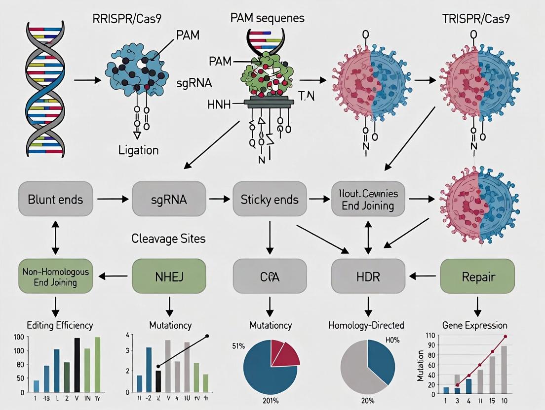
Table 2: Key Double-Strand Break Repair Pathways
| Pathway | Key Mediators | Template Required? | Outcome | Fidelity |
|---|---|---|---|---|
| Non-Homologous End Joining (NHEJ) | DNA-PKcs, Ku70/80, XLF | No | Small insertions/deletions (Indels), gene knockout | Error-prone |
| Homology-Directed Repair (HDR) | BRCA1, Rad51, RPA | Yes (donor template) | Precise insertion or correction | High-fidelity |
| Microhomology-Mediated End Joining (MMEJ) | PARP1, Polθ, CtIP | No (uses microhomology) | Deletions with microhomology flanking | Error-prone |
Experimental Protocols
Protocol 1: Design and Cloning of sgRNA Expression Constructs
Objective: To clone a target-specific single guide RNA (sgRNA) sequence into a CRISPR plasmid vector for mammalian expression.
Materials (Research Reagent Solutions):
- pSpCas9(BB)-2A-Puro (PX459) V2.0 Plasmid (Addgene #62988): A commonly used all-in-one vector expressing SpCas9, a sgRNA scaffold, and a puromycin resistance marker.
- Paired Oligonucleotides: Designed 20-nt target sequences with appropriate overhangs for ligation into the BbsI site.
- FastDigest BbsI (Thermo Fisher): Restriction enzyme for linearizing the vector.
- T4 DNA Ligase (NEB): For ligating annealed oligos into the digested vector.
- Stbl3 Competent E. coli (Thermo Fisher): High-efficiency cells for transforming repetitive/DNA structures.
- PCR & Sequencing Primers (U6 Forward, sgRNA scaffold Reverse): For verifying correct insertion.
Methodology:
- sgRNA Design: Using tools like CRISPOR or ChopChop, select a 20-nt target sequence immediately 5' of a PAM (NGG for SpCas9). Check for potential off-targets.
- Oligo Annealing: Synthesize and resuspend oligonucleotides. Mix forward and reverse oligos (1 µL each of 100 µM) with 1 µL of 10X T4 Ligation Buffer and 7 µL H₂O. Anneal in a thermal cycler: 95°C for 5 min, ramp down to 25°C at 5°C/min.
- Vector Digestion: Digest 2 µg of pX459 plasmid with BbsI (1 µL) in 1X FastDigest Buffer (20 µL total) at 37°C for 15 min. Purify using a spin column.
- Ligation: Mix 50 ng digested vector, 1 µL annealed oligo duplex (1:200 dilution), 1 µL T4 DNA Ligase, 1X Ligase Buffer. Incubate at room temperature for 10 min.
- Transformation: Transform 2 µL ligation mix into 50 µL Stbl3 cells via heat shock. Plate on ampicillin LB agar. Incubate overnight at 37°C.
- Screening: Pick 3-5 colonies for colony PCR or plasmid miniprep. Verify insertion by Sanger sequencing using the U6 forward primer.
Protocol 2:In VitroCleavage Assay (Cas9 RNP Validation)
Objective: To validate the activity of purified Cas9 protein complexed with in vitro-transcribed sgRNA before cellular experiments.
Materials (Research Reagent Solutions):
- Recombinant SpCas9 Nuclease (NEB #M0386): Purified, ready-to-use Cas9 protein.
- Target DNA Template: A PCR-amplified genomic region (300-500 bp) containing the target site.
- T7 RiboMAX Express Kit (Promega): For high-yield in vitro transcription of sgRNA from a DNA template with a T7 promoter.
- Nuclease-Free Duplex Buffer (IDT): For annealing sgRNA with tracrRNA if using a two-part system.
- Agarose Gel Electrophoresis System: For visualizing cleavage products.
Methodology:
- Prepare sgRNA: Generate a DNA template via PCR with a T7 promoter sequence. Perform in vitro transcription per kit instructions. Purify sgRNA using spin columns.
- Form RNP Complex: Mix 30 pmol Cas9 protein with 36 pmol sgRNA in 1X Cas9 Nuclease Reaction Buffer (20 µL total). Incubate at 25°C for 10 min.
- Cleavage Reaction: Add 200 ng of target DNA template to the RNP complex. Bring final volume to 30 µL with nuclease-free water and buffer. Incubate at 37°C for 1 hour.
- Analysis: Stop reaction with Proteinase K (0.5 µg/µL) at 56°C for 10 min. Run products on a 2% agarose gel. Successful cleavage yields two smaller bands (e.g., 200 bp and 100 bp from a 300 bp template).
Protocol 3: HDR-Mediated Knock-in in HEK293T Cells
Objective: To integrate a donor DNA template (e.g., a fluorescent protein gene) via homology-directed repair.
Materials (Research Reagent Solutions):
- HEK293T Cells (ATCC CRL-3216): Readily transfected, high HDR efficiency model cell line.
- Lipofectamine CRISPRMAX (Thermo Fisher): A lipid-based transfection reagent optimized for Cas9/sgRNA RNP delivery.
- Cas9 RNP Complex: Formed from purified Cas9 protein and synthetic sgRNA (Alt-R CRISPR-Cas9 system, IDT).
- Single-Stranded Oligodeoxynucleotide (ssODN) Donor Template: 100-nt ultramer with homology arms (40-50 nt each) flanking the desired insertion sequence.
- Flow Cytometry Antibodies & Buffers: For analyzing fluorescent protein expression.
Methodology:
- Seed Cells: Plate 2 x 10⁵ HEK293T cells per well in a 24-well plate 24 hours before transfection.
- Prepare RNP/Donor Mix: Complex 10 pmol Alt-R SpCas9 nuclease with 12 pmol Alt-R crRNA:tracrRNA duplex in Opti-MEM. Incubate 10 min. Add 2 µL of 100 µM ssODN donor.
- Transfection: Dilute 1.5 µL CRISPRMAX in Opti-MEM. Combine with RNP/donor mix. Incubate 10-20 min, then add dropwise to cells.
- Analysis: Harvest cells 72-96 hours post-transfection. For fluorescent reporters, analyze by flow cytometry. For precise edits, extract genomic DNA and analyze by PCR followed by Sanger sequencing or TIDE analysis.
Visualizations
Title: CRISPR-Induced DNA Break Repair Pathways
Title: HDR-Mediated Knock-in Experimental Workflow
The Scientist's Toolkit: Key Research Reagent Solutions
Table 3: Essential CRISPR/Cas9 Reagents for Therapeutic Chassis Engineering
| Reagent | Example Product/Supplier | Primary Function in Experiments |
|---|---|---|
| High-Fidelity Cas9 Nuclease | Alt-R S.p. HiFi Cas9 Nuclease V3 (IDT) | Reduces off-target edits; critical for therapeutic safety assessments. |
| Synthetic sgRNA (crRNA + tracrRNA) | Alt-R CRISPR-Cas9 crRNA & tracrRNA (IDT) | Defines target specificity; synthetic RNA improves consistency and reduces immune response. |
| Cas9 Expression Plasmid | pSpCas9(BB)-2A-GFP (PX458, Addgene) | All-in-one vector for co-expressing Cas9, sgRNA, and a fluorescent reporter for cell sorting. |
| HDR Donor Template | Ultramer ssODN (IDT) or dsDNA donor with homology arms | Serves as repair template for precise insertions or point corrections via HDR. |
| Transfection Reagent for RNP | Lipofectamine CRISPRMAX (Thermo Fisher) | Lipid-based formulation optimized for delivering Cas9 ribonucleoprotein (RNP) complexes. |
| Nucleofection Kit | Cell Line Nucleofector Kit V (Lonza) | Electroporation-based method for high-efficiency RNP delivery into hard-to-transfect primary cells (e.g., T-cells). |
| Off-Target Analysis Kit | GUIDE-seq Kit (NEB) | Identifies genome-wide off-target cleavage sites via integration of a double-stranded oligodeoxynucleotide tag. |
| Genome Editing Detection | T7 Endonuclease I (NEB) or ICE Analysis Tool (Synthego) | Enables quick assessment of editing efficiency by detecting mismatches in heteroduplex PCR products. |
| Clone Isolation Substrate | CloneDetect (STEMCELL Technologies) | Facilitates the isolation and expansion of single-cell-derived clones after editing. |
| AAV Serotype for In Vivo Delivery | AAV-DJ Kit (Takara Bio) | Provides a suite of AAV capsids with high tropism for different tissues for in vivo CRISPR delivery. |
Table 1: Quantitative Metrics for Engineered Cell Chassis Traits
| Trait Category | Key Metric | Target Range | Measurement Technique | Typical Benchmark (Primary Cells) | Engineered Chassis Target |
|---|---|---|---|---|---|
| Safety | Off-Target Editing Frequency | < 0.1% | GUIDE-seq / CIRCLE-seq | Varies by guide (0.1-10%) | < 0.01% |
| Safety | Translocation Frequency | < 0.001% | FISH / NGS | Up to 5% in high-edit scenarios | < 0.0001% |
| Scalability | Fold Expansion (Ex Vivo) | > 10^9 | Cell Counting / Metabolite Analysis | Limited (10-20 doublings) | > 50 doublings |
| Scalability | Viral Transduction Efficiency | > 80% | Flow Cytometry (GFP) | 30-70% (primary T-cells) | > 90% |
| Immunocompatibility | Surface HLA Expression | Downregulated | Flow Cytometry (Anti-HLA I/II) | High (constitutive) | > 90% Reduction |
| Immunocompatibility | NK Cell Lysis (In Vitro) | < 15% | Calcein-AM Cytotoxicity Assay | 40-80% | < 10% |
| Metabolic Fitness | Basal OCR (pmol/min) | > 100 | Seahorse Mito Stress Test | 50-150 | > 120 |
| Metabolic Fitness | Lactate Production Rate | Low | Biochemical Assay | High (Warburg effect) | < 50% of primary cell baseline |
Table 2: CRISPR/Cas9 Reagent Formats for Chassis Engineering
| Reagent Format | Delivery Efficiency | Cost per 10^6 Cells | Scalability | Key Safety Feature |
|---|---|---|---|---|
| Plasmid DNA (pDNA) | 20-40% | $0.50 | Low-Moderate | Risk of genomic integration |
| In Vitro Transcribed (IVT) mRNA | 70-90% | $3.00 | High | Transient expression, low risk |
| Ribonucleoprotein (RNP) | 80-95% | $5.00 | High | Ultra-transient, highest specificity |
| All-in-One AAV Vector | >95% (permissive cells) | $10.00 | Low | Persistent expression, immunogenic risk |
Detailed Experimental Protocols
Protocol 2.1: Multi-Gene Knockout for Immunocompatibility Using Cas9 RNP
Objective: Simultaneously disrupt B2M, CIITA, and TRAC genes to generate universal donor chassis cells (e.g., iPSCs or T-cells) with reduced immunogenicity.
Materials:
- Target cells (e.g., human iPSCs, activated T-cells).
- Chemically modified sgRNAs (Synthego or IDT) targeting B2M, CIITA, TRAC.
- Alt-R S.p. HiFi Cas9 Nuclease V3 (IDT).
- Electroporation buffer (P3 Primary Cell Solution, Lonza) or equivalent.
- 4D-Nucleofector X Unit (Lonza) or similar electroporator.
- Flow antibodies: Anti-HLA-ABC-APC, Anti-CD3-FITC.
Procedure:
- RNP Complex Formation: For each sgRNA, complex 60 pmol of Cas9 protein with 240 pmol of sgRNA in a separate tube. Incubate at 25°C for 10 minutes.
- Cell Preparation: Harvest and count target cells. Wash once in PBS. Resuspend at 1x10^6 cells per 20 µL of electroporation buffer.
- Electroporation: Combine cell suspension with the three RNP complexes. Transfer to a 16-well nucleofection strip. Electroporate using the appropriate preset program (e.g., EO-115 for T-cells, CB-150 for iPSCs).
- Recovery: Immediately add 80 µL of pre-warmed complete media. Transfer cells to a 24-well plate with 1 mL pre-warmed media. Culture at 37°C, 5% CO2.
- Analysis (Day 3-5): Harvest cells. Stain for surface HLA-ABC and CD3. Analyze knockout efficiency via flow cytometry (loss of marker expression).
Protocol 2.2: In Vitro Metabolic Fitness Assessment via Seahorse Analyzer
Objective: Quantitatively measure the mitochondrial respiration and glycolytic rate of engineered chassis cells post-editing.
Materials:
- Engineered and control cell populations.
- Seahorse XFp/XFe96 Analyzer (Agilent).
- Seahorse XF RPMI Medium, pH 7.4 (Agilent).
- XF Cell Mito Stress Test Kit: Oligomycin, FCCP, Rotenone/Antimycin A.
- XF Glycolysis Stress Test Kit: Glucose, Oligomycin, 2-DG.
- Cell-Tak (Corning) for adherent cells.
Procedure:
- Cell Seeding: 18-24 hours pre-assay, seed 20,000-80,000 cells per well in a Seahorse microplate. For non-adherent cells, use Cell-Tak coating.
- Sensor Cartridge Hydration: Hydrate the Seahorse sensor cartridge in XF Calibrant at 37°C, non-CO2 overnight.
- Medium Replacement: Prior to assay, replace growth medium with 180 µL of assay-specific, unbuffered XF RPMI Medium (supplemented with 10 mM glucose, 1 mM pyruvate, 2 mM L-glutamine for Mito Stress Test). Incubate at 37°C, non-CO2 for 1 hour.
- Mito Stress Test Execution:
- Load port A with Oligomycin (1.5 µM final).
- Load port B with FCCP (1.0 µM final, titrate for cell type).
- Load port C with Rotenone/Antimycin A (0.5 µM final each).
- Run the standard 3-measurement cycle protocol (Mix-Wait-Measure).
- Data Analysis: Calculate key parameters: Basal OCR, ATP-linked OCR (pre-oligo), Maximal OCR (post-FCCP), and Spare Respiratory Capacity (Max-Basal).
Diagrams and Workflows
Title: Four Pillars of Therapeutic Chassis Engineering
Title: Scalable CRISPR Workflow for Chassis Engineering
The Scientist's Toolkit: Research Reagent Solutions
| Item (Vendor Example) | Function in Chassis Engineering | Key Trait Addressed |
|---|---|---|
| Alt-R S.p. HiFi Cas9 Nuclease V3 (IDT) | High-fidelity Cas9 variant; reduces off-target editing while maintaining on-target activity. | Safety |
| Synthego Engineered sgRNA EZ Kit | Chemically modified, pooled sgRNAs for enhanced stability and editing efficiency. | Scalability, Safety |
| Lonza P3 Primary Cell 4D-Nucleofector Kit | Optimized buffer/electroporation programs for efficient RNP delivery into sensitive primary cells. | Scalability |
| Takara Bio CellAvidin HLA-ABC Antibody | High-sensitivity antibody for flow cytometric validation of HLA knockout efficiency. | Immunocompatibility |
| Agilent Seahorse XFp Analyzer Kits | Real-time, label-free measurement of cellular metabolic function (OCR, ECAR). | Metabolic Fitness |
| Nucleic Acids-Based Off-Target Assay (GUIDE-seq) | Comprehensive, unbiased genome-wide method for identifying CRISPR off-target sites. | Safety |
| Gibco CTS Immune Cell Serum-Free Media | Chemically defined, xeno-free media supporting robust expansion of edited immune cells. | Scalability, Metabolic Fitness |
Within the paradigm of CRISPR/Cas9-driven therapeutic chassis engineering, selecting and optimizing the appropriate biological system is paramount. This article provides Application Notes and Protocols for four leading chassis candidates: engineered T cells, stem cells, yeast (Saccharomyces cerevisiae), and bacteria (e.g., E. coli, probiotics). Each offers unique advantages for therapeutic development, from personalized cellular therapies to scalable biologic production.
Engineered T Cells
Application Notes
Primary use: Adoptive Cell Therapies (ACT), notably Chimeric Antigen Receptor (CAR) T cells and T Cell Receptor (TCR) T cells for oncology and autoimmune diseases. Key Quantitative Metrics:
| Metric | CAR-T (CD19-targeting) | TCR-T (NY-ESO-1) | Notes |
|---|---|---|---|
| Clinical Response Rate | 70-90% (B-ALL) | 40-60% (Synovial Sarcoma) | Complete remission rates in relapsed/refractory cases. |
| Manufacturing Time | 7-14 days | 10-21 days | From leukapheresis to infusion. |
| Persistence in Vivo | Up to 10+ years | Months to 2+ years | Varies with product design. |
| Common Target Antigens | CD19, BCMA, CD22 | NY-ESO-1, MART-1 | Tumor-associated antigens. |
Protocol: CRISPR/Cas9-mediated TRAC Disruption for CAR Insertion
Objective: Generate universal, off-the-shelf CAR-T cells by knocking out the endogenous T Cell Receptor Alpha Constant (TRAC) locus and inserting a CAR construct via HDR. Key Research Reagent Solutions:
| Reagent/Kit | Function |
|---|---|
| Human T Cell Nucleofector Kit | High-efficiency electroporation reagent for primary T cells. |
| Cas9 RNP complex | Pre-complexed S.pyogenes Cas9 protein and TRAC-targeting gRNA for high-activity, transient editing. |
| AAV6 HDR donor template | Recombinant Adeno-Associated Virus serotype 6 delivering homology-directed repair template with CAR cassette. |
| IL-2 & IL-7/IL-15 cytokines | Promote T cell expansion and persistence during ex vivo culture. |
| Anti-CD3/CD28 Dynabeads | Artificial Antigen-Presenting Cells for T cell activation pre-editing. |
Methodology:
- Isolation & Activation: Isolate PBMCs from leukapheresis product, activate T cells using anti-CD3/CD28 beads in X-VIVO 15 media with 5% human AB serum and 100 IU/mL IL-2 for 24-48h.
- CRISPR Delivery: Electroporate 1x10^6 activated T cells with 10µg Cas9 RNP complex targeting TRAC locus using a Lonza 4D-Nucleofector.
- HDR Donor Delivery: Immediately post-electroporation, transduce cells with AAV6 HDR donor (MOI=10^5 vg/cell). Centrifuge at 2000 x g for 90 min at 32°C to enhance infection.
- Culture & Expansion: Culture cells in IL-2 (100 IU/mL) and IL-7/IL-15 (10ng/mL each). Remove beads after 3-5 days. Expand cells for 10-14 days, maintaining density at 0.5-2x10^6 cells/mL.
- QC & Validation: Assess editing efficiency via flow cytometry (loss of TCRαβ, gain of CAR expression) and indel frequency at TRAC via NGS.
Title: Workflow for CRISPR-Engineered Universal CAR-T Cell Manufacturing
Stem Cells (Human Induced Pluripotent Stem Cells - hiPSCs)
Application Notes
Primary use: Source for differentiated therapeutic cells (neurons, cardiomyocytes, beta-cells) for regenerative medicine, disease modeling, and allogeneic "off-the-shelf" therapies. Key Quantitative Metrics:
| Metric | Typical Value/Range | Notes |
|---|---|---|
| CRISPR Editing Efficiency (hiPSCs) | 10-80% (HDR) | Varies by delivery method (RNP vs. plasmid) and locus. |
| Clonal Selection Timeline | 4-8 weeks | From editing to expansion of validated clonal line. |
| In Vivo Differentiation Efficiency | 50-95% for major lineages | e.g., >90% TNNT2+ cardiomyocytes. |
| Tumorigenicity Risk (Residual Undifferentiated) | Target: <1 in 10^6 cells | Critical release criterion for transplants. |
Protocol: CRISPR/Cas9 Knock-in at a Safe Harbor Locus in hiPSCs
Objective: Precisely insert a therapeutic transgene (e.g., GDNF) into the AAVS1 safe harbor locus in hiPSCs via HDR. Key Research Reagent Solutions:
| Reagent/Kit | Function |
|---|---|
| hiPSC-Culture Qualified Matrigel | Defined extracellular matrix for feeder-free hiPSC culture. |
| mTeSR Plus Medium | Chemically defined, xeno-free maintenance medium for hiPSCs. |
| CloneR Supplement | Enhances survival of single hiPSCs during clonal expansion. |
| Lipofectamine Stem Transfection Reagent | Low-toxicity polymer for plasmid or RNP delivery to hiPSCs. |
| AAVS1-specific gRNA & Cas9 plasmid | CRISPR components targeting the human AAVS1 (PPP1R12C) locus. |
Methodology:
- Culture: Maintain hiPSCs on Matrigel-coated plates in mTeSR Plus. Passage as single cells using Accutase.
- Transfection: At 60% confluence, co-transfect 1µg AAVS1-targeting Cas9/gRNA plasmid and 1µg AAVS1-HDR donor plasmid (containing GOI and puromycin resistance flanked by homology arms) using Lipofectamine Stem.
- Selection & Cloning: 48h post-transfection, apply puromycin (0.5 µg/mL) for 5-7 days. Disperse surviving cells to single cells with CloneR into 96-well plates for clonal expansion.
- Genotyping & Validation: Screen clones by PCR for 5'/3' junction integration. Validate via Sanger sequencing, off-target analysis (e.g., GUIDE-seq), and pluripotency marker staining (OCT4, SOX2).
- Differentiation: Direct validated clonal line to desired lineage (e.g., using cardiomyocyte differentiation kit) and assess transgene expression/function.
Title: Safe Harbor Gene Knock-in Protocol for hiPSCs
Yeast (Saccharomyces cerevisiae)
Application Notes
Primary use: Eukaryotic model for pathway engineering, production of complex natural products, vaccines, and therapeutic proteins (e.g., insulin, hepatitis B vaccine). Key Quantitative Metrics:
| Metric | Typical Value/Range | Notes |
|---|---|---|
| CRISPR Editing Efficiency (S. cerevisiae) | >90% (with HR) | High endogenous homologous recombination facilitates editing. |
| Titer for Heterologous Protein | mg/L to g/L scale | Depends on product and strain optimization. |
| Fermentation Timeline | 3-10 days (lab scale) | From inoculation to harvest. |
| Glycosylation Capability | High-mannose type | Distinct from mammalian cells; may require humanization. |
Protocol: CRISPR/Cas9-mediated Multiplex Gene Integration in Yeast
Objective: Simultaneously integrate multiple genes of a biosynthetic pathway into predefined genomic loci in S. cerevisiae. Key Research Reagent Solutions:
| Reagent/Kit | Function |
|---|---|
| Yeast Extract Peptone Dextrose (YPD) Media | Rich medium for routine yeast cultivation. |
| PEG/LiAc Transformation Mix | Chemical transformation reagents for yeast. |
| Cas9 Plasmid (with yeast promoter) | Expresses S. pyogenes Cas9 in yeast (e.g., pCAS plasmid). |
| gRNA Expression Plasmid(s) | Contains tRNA-gRNA polycistrons for multiplex targeting. |
| Double-stranded DNA Donor Fragments | PCR-amplified cassettes with 40-50bp homology arms for each integration site. |
Methodology:
- Strain & Plasmid Prep: Grow parental yeast strain (e.g., CEN.PK2) in YPD to mid-log phase. Maintain Cas9 and gRNA expression plasmids in E. coli.
- Donor & CRISPR Component Assembly: Amplify linear donor DNA fragments (containing gene+marker) by PCR. Co-transform yeast with: 1µg Cas9 plasmid, 1µg multiplex gRNA plasmid, and ~500ng of each donor fragment using high-efficiency LiAc/SS carrier DNA/PEG method.
- Selection & Screening: Plate on appropriate synthetic dropout media to select for integrated markers. Incubate at 30°C for 2-3 days.
- Validation: Pick colonies, patch/re-streak. Validate integrations via colony PCR across all junctions. Screen for functional product production in microtiter plate assays.
- Curing of CRISPR Plasmids: Culture validated strains in non-selective media for ~10 generations to lose Cas9/gRNA plasmids.
Title: CRISPR Multiplex Pathway Integration in Yeast Workflow
Bacteria (Therapeutic Engineered Bacteria)
Application Notes
Primary use: Live biotherapeutics (e.g., engineered probiotics for IBD, cancer), in situ drug production, microbiome modulation, and delivery of therapeutic proteins/antigens. Key Quantitative Metrics:
| Metric | Typical Value (E. coli Nissle) | Notes |
|---|---|---|
| CRISPR Editing Efficiency | 80-100% (λ-Red recombineering + Cas9) | In strains with efficient recombinase systems. |
| Colonization Duration | Days to weeks | Strain and host dependent. |
| Therapeutic Protein Secretion | ng to µg/mL/g biomass | In gut or tumor microenvironment. |
| Biosafety Containment | Engineered auxotrophies | Required for clinical translation. |
Protocol: CRISPR/Cas9 Counter-selection for Chromosomal Integration inE. coli Nissle 1917
Objective: Knock-in a therapeutic gene cassette into the chromosome of probiotic E. coli Nissle 1917 (EcN), replacing a non-essential gene without leaving antibiotic resistance markers. Key Research Reagent Solutions:
| Reagent/Kit | Function |
|---|---|
| LB Lennox Media | Standard medium for E. coli growth. |
| pKD46 or pSIM Plasmid | Temperature-sensitive plasmid expressing λ-Red recombinase proteins. |
| pCas9cr4 Plasmid | Expresses Cas9 and a counter-selectable gRNA targeting the locus to be replaced. |
| Electrocompetent Cell Preparation Kit | For making high-efficiency electrocompetent EcN cells. |
| Sucrose-containing Media | For counter-selection against sacB gene (if used in donor). |
Methodology:
- Recombineering Prep: Transform EcN with pKD46 (AmpR). Grow at 30°C in LB+Arabinose (to induce λ-Red) to mid-log. Make cells electrocompetent.
- Donor Electroporation: Electroporate with a linear dsDNA donor fragment containing the therapeutic gene, flanked by 500bp homology arms to the target locus, and a sacB marker for counter-selection. Recover at 30°C for 2h, plate on selective media (e.g., chloramphenicol for sacB-cat).
- Cas9 Counter-selection: Transform candidate colonies (now with integrated sacB-cat) with pCas9cr4 expressing a gRNA targeting the original chromosomal locus (now absent in integrants). Plate on LB+Kanamycin (Cas9 plasmid) at 30°C. Cas9 will kill any cells that retained the wild-type locus.
- Curing & Validation: Screen surviving colonies for sucrose sensitivity (loss of sacB) and antibiotic sensitivity (loss of cat). Culture at 37°C to lose temperature-sensitive pKD46 and pCas9cr4. Validate integration by PCR and sequencing.
- Functional Assay: Perform in vitro assay (e.g., cytokine or enzyme production) to confirm therapeutic gene function.
Title: CRISPR-Counter-selection for Marker-Free Bacterial Engineering
Ethical and Safety Considerations in Genetically Engineered Living Therapeutics
The development of genetically engineered living therapeutics (GELTs), such as CAR-T cells, oncolytic viruses, and engineered bacterial strains, represents a frontier in precision medicine. Within the thesis framework of CRISPR/Cas9 genome editing for therapeutic chassis engineering, this document outlines critical ethical and safety considerations, supported by current data and standardized protocols. The inherent ability of GELTs to persist, replicate, and evolve in vivo necessitates a robust and proactive risk assessment framework that extends beyond conventional biologics.
Quantitative Risk Assessment Data
Table 1: Reported Adverse Events in Clinical Trials for Select GELTs (2019-2024)
| Therapeutic Class | Number of Trials Reviewed | Incidence of CRS* (%) | Incidence of Neurotoxicity (%) | Incidence of Off-Target Tumorigenesis (%) | Cases of Vector-Mediated Insertional Mutagenesis |
|---|---|---|---|---|---|
| CAR-T Cells (Allogenic) | 127 | 45-85 | 15-50 | 0.05 | 2 reported cases |
| Oncolytic Viruses | 89 | 1-5 | <1 | 0.01 (viral shedding) | Not Applicable |
| Engineered Bacteria | 23 | 3-10 (sepsis-like) | <1 | 0.1 (bacterial dissemination) | Not Applicable |
| CRISPR-Edited In Vivo Therapies | 18 | Variable by target | Variable by target | 1.5 (Theoretical risk; detected via NGS in pre-clinical models) | 0 (Clinical) |
CRS: Cytokine Release Syndrome. Data compiled from recent publications in *Nature Biotechnology, The Lancet Oncology, and clinicaltrials.gov.
Table 2: Key Safety Thresholds for Release Criteria of CRISPR-Edited Cell Products
| Quality Attribute | Test Method | Required Threshold | Rationale |
|---|---|---|---|
| Vector Copy Number (VCN) | ddPCR | < 5 copies per cell | Limit risk of insertional mutagenesis |
| Off-Target Editing Frequency | GUIDE-seq or CIRCLE-seq | < 0.1% of total reads at any predicted site | Minimize unintended genomic alterations |
| Tumorigenicity (in vitro) | Soft Agar Colony Formation | 0% colony formation | Ensure no malignant transformation potential |
| Residual Plasmid DNA | qPCR | < 10 ng per 10^6 cells | Reduce immunogenic and transduction risks |
| Microbial Sterility | USP <71> | No growth | Prevent adventitious agent contamination |
Application Notes & Protocols
AN-1: Protocol for Comprehensive Off-Target Analysis of CRISPR/Cas9-Edited Therapeutic Chassis
Objective: To identify and quantify off-target editing events in a CRISPR/Cas9-engineered human T-cell line intended for adoptive therapy.
Materials:
- Edited T-cell genomic DNA (gDNA, 1 µg).
- Control (un-edited) T-cell gDNA (1 µg).
- GUIDE-seq Kit (Integrated DNA Technologies) or reagents for CIRCLE-seq.
- NGS Library Prep Kit (e.g., Illumina Nextera XT).
- High-fidelity DNA polymerase.
- Bioinformatic Pipeline: CRISPResso2 or Cas-OFFinder.
Procedure:
- In Silico Prediction: Use algorithms (e.g., Cas-OFFinder) with the specific sgRNA sequence to generate a list of top 50 potential off-target sites genome-wide.
- Empirical Identification (GUIDE-seq): a. Transfect target cells with Cas9/sgRNA RNP complex + GUIDE-seq oligonucleotide tag. b. Harvest genomic DNA 72 hours post-transfection. c. Perform tag-specific PCR amplification and prepare NGS libraries. d. Sequence and analyze using the GUIDE-seq analysis software to identify double-strand break locations.
- Quantification: a. Design amplicons for the top 10 predicted and top 5 empirical off-target sites. b. Perform deep amplicon sequencing (≥50,000x coverage) on both edited and control gDNA. c. Analyze sequencing data with CRISPResso2 to calculate indel percentages at each locus.
- Reporting: Document all sites with indel frequency >0.01%. Compare to the in silico prediction list.
Safety Note: Any off-target site within an oncogene or tumor suppressor gene with frequency >0.1% must be evaluated for lead candidate disqualification.
AN-2: Protocol for Environmental Containment and Kill-Switch Validation
Objective: To validate the functional efficacy of an inducible safety switch (e.g., caspase-9 or thymidine kinase) in an engineered bacterial therapeutic.
Materials:
- Engineered E. coli Nissle 1917 strain with genomically integrated inducible kill-switch.
- Inducer molecule (e.g., small molecule drug, specific sugar).
- LB broth and agar plates.
- Mammalian cell co-culture system (e.g., Caco-2 cells).
- Viability stains (propidium iodide, SYTOX Green).
Procedure:
- In Vitro Kinetics: a. Culture the engineered bacteria to mid-log phase. b. Split culture and add inducer to test condition. c. Take OD600 measurements and perform CFU counts at 0, 1, 2, 4, 8, and 24 hours post-induction. d. Plot log(CFU/mL) vs. time. A functional switch should reduce CFU by >99.9% within 4 hours.
- Co-culture Validation: a. Establish a monolayer of mammalian cells in a transwell system. b. Introduce bacteria to the apical compartment. c. After 24 hours of colonization, add inducer to the basolateral media (mimicking systemic delivery). d. Monitor mammalian cell viability (e.g., MTT assay) and bacterial presence (CFU, microscopy) for 72 hours.
- Data Analysis: The kill-switch is considered effective if it eliminates bacteria and prevents mammalian cell death upon induction, while the non-induced control shows stable colonization.
Visualizations
Diagram 1: Safety by Design Workflow for GELT Development
Diagram 2: Key Immune-Related Adverse Event Pathways for Cell Therapies
The Scientist's Toolkit: Research Reagent Solutions
Table 3: Essential Reagents for GELT Safety Assessment
| Reagent / Kit | Vendor Examples | Primary Function in Safety Assessment |
|---|---|---|
| CRISPR/Cas9 Off-Target Discovery Kit (GUIDE-seq) | Integrated DNA Technologies | Unbiased genome-wide identification of double-strand breaks caused by CRISPR nucleases. |
| CIRCLE-seq Kit | Custom or from published protocols | High-sensitivity, in vitro method to profile Cas9 nuclease off-target activity using circularized genomic DNA. |
| ddPCR Assay for Vector Copy Number | Bio-Rad | Absolute quantification of vector integration events per genome, critical for release criteria. |
| LAL Endotoxin Assay Kit | Lonza, Thermo Fisher | Detection of bacterial endotoxins in final cell therapy product, a key sterility and safety test. |
| Inducible Safety Switch Systems (e.g., iCasp9, HSV-TK) | Takara Bio, academic constructs | Provides a genetic "kill-switch" to ablate engineered cells in case of adverse events. |
| Tumorigenicity Assay Kit (Soft Agar) | Cell Biolabs, Inc. | Assesses anchorage-independent growth, a hallmark of cellular transformation, pre-release. |
| Cytokine Multiplex Assay (Luminex/ELISA) | R&D Systems, Thermo Fisher | Quantifies cytokine levels in patient serum or culture supernatant to monitor for CRS. |
| Next-Generation Sequencing Service (WGS) | Illumina, Novogene | Comprehensive genomic analysis for identity, off-target, and stability assessment. |
From Design to Delivery: A Step-by-Step CRISPR Workflow for Chassis Engineering
Target Selection and gRNA Design for Knock-Ins, Knock-Outs, and Gene Regulation
Within the paradigm of CRISPR/Cas9-based therapeutic chassis engineering, precise genomic manipulation is foundational. The success of knock-out (KO), knock-in (KI), and gene regulation strategies is critically dependent on the initial steps of target selection and guide RNA (gRNA) design. This application note provides updated protocols and frameworks for these processes, integrating current best practices and quantitative data to inform research and drug development.
Table 1: Key Design Parameters for CRISPR/Cas9 Applications
| Application | Primary Cas Protein | gRNA Length (nt) | PAM Sequence (Example) | Optimal Edit Distance from PAM | Key Design Priority |
|---|---|---|---|---|---|
| Knock-Out (KO) | SpCas9 | 20 | NGG | Within exons, near 5' of coding sequence | On-target efficiency, predicted off-target score |
| Knock-In (HDR) | SpCas9 or HiFi Cas9 | 20 | NGG | <10-15 bp from PAM; close to desired edit site | On-target efficiency, HDR donor design |
| Gene Repression (CRISPRi) | dCas9 (SpCas9) | 20 | NGG | Within -50 to +300 bp relative to TSS | Proximity to Transcription Start Site (TSS) |
| Gene Activation (CRISPRa) | dCas9-VPR (SpCas9) | 20 | NGG | Within -400 to -50 bp upstream of TSS | Proximity to TSS, avoid nucleosome occupancy |
| Base Editing (C->T) | BE4max (nCas9) | 20 | NGG (NG for SpCas9-NG) | Within editing window (positions 4-8, C protospacer) | Target base must be in window, off-target RNA editing |
| Prime Editing | PE2 (nCas9-RT) | 30 (including PBS & RT template) | NGG | Flexible; PE guide spans target & template | Primer Binding Site (PBS) & RT template design |
Table 2: Current Off-Target Prediction Tools (2024)
| Tool Name | Type | Access | Key Output Metric | Best For |
|---|---|---|---|---|
| CHOPCHOP v3 | Web Server / Standalone | Open Source | On-target efficiency, off-target scores | Quick, integrated design for KO/KI |
| CRISPick (Broad) | Web Server | Open Source | On-/Off-target scores, specificity | Therapeutic-grade design |
| CRISPRseek | R/Bioconductor | Open Source | Genome-wide off-target count | Batch analysis, custom genomes |
| Cas-OFFinder | Web/Standalone | Open Source | List of potential off-target sites | Mismatch & bulge identification |
| GuideScan2 | Web Server | Open Access | Off-targets with activity prediction | Design for Cas9, Cas12, epigenetic editors |
Protocols
Protocol 1: Comprehensive gRNA Design for Knock-Outs
Objective: Design high-efficiency, specific gRNAs to generate frameshift mutations via NHEJ.
- Target Identification: Identify all exons of the target gene using resources like NCBI RefSeq or Ensembl. Prioritize early exons, especially those encoding critical functional domains.
- gRNA Candidate Generation: Input a 300-500 bp genomic sequence surrounding the target region into a design tool (e.g., CRISPick). Set parameters: SpCas9 (NGG PAM), gRNA length 20bp.
- On-Target Scoring: Select candidates with high on-target efficiency scores (e.g., >60 using the Doench et al. 2016 rule set).
- Off-Target Analysis: Run selected candidate sequences through an off-target predictor (e.g., Cas-OFFinder). Allowable parameters: ≤3 mismatches, no DNA bulges. Reject gRNAs with perfect or 1-mismatch hits elsewhere in the genome.
- Final Selection: Choose 3-4 top-ranked gRNAs per target locus for experimental validation. Ensure they are not within predicted genomic repeats or high-density SNP regions.
Protocol 2: gRNA and Donor Design for Precise Knock-Ins
Objective: Design components for homology-directed repair (HDR)-mediated insertion.
- gRNA Design: Follow Protocol 1, but with a critical constraint: the Cas9 cut site must be within 15 bp of the intended insertion site. The double-strand break (DSB) must be close to minimize resection and ensure donor template homology arms are effective.
- Single-Stranded Donor Oligonucleotide (ssODN) Design:
- Homology Arm Length: For ssODNs, use 60-90 bp homology arms on each side of the insertion. For viral or plasmid donors, extend to 400-800 bp.
- Silent Mutations: Incorporate 1-2 silent mutations within the gRNA seed region (bases 1-12 of the protospacer) in the donor to prevent re-cutting of the edited allele.
- Purity: HPLC-purify ssODNs.
- HDR Enhancer Considerations: Plan to co-deliver an HDR-enhancing agent (e.g., Rad51 inhibitor RS-1 or NHEJ inhibitor SCR7) during transfection to boost KI rates.
Protocol 3: gRNA Design for dCas9-Mediated Gene Regulation (CRISPRi/a)
Objective: Design gRNAs to recruit effector domains to modulate transcription.
- Identify TSS: Use a dedicated database (e.g., EPDnew) to locate the primary Transcription Start Site(s) for the target gene.
- CRISPRi Design (for repression):
- Target a window from -50 to +300 bp relative to the TSS.
- Design 5-10 gRNAs across this region, focusing on the non-template strand to recruit dCas9-KRAB most effectively.
- CRISPRa Design (for activation):
- Target a window from -400 to -50 bp upstream of the TSS.
- Design 5-10 gRNAs, prioritizing regions of open chromatin (use DNase-seq/ATAC-seq data if available). Avoid nucleosome-dense regions.
- Specificity Check: Perform off-target analysis as in Protocol 1. For CRISPRa, pay special attention to avoiding gRNAs that may activate non-target genes' promoters or enhancers.
Protocol 4: Validation of gRNA Activity (Essential Follow-Up)
Objective: Experimentally validate cutting efficiency and specificity of designed gRNAs.
- Transfection: Co-transfect your target cell line (e.g., HEK293T) with a plasmid expressing SpCas9 and the individual gRNA (or deliver as RNP complexes).
- Harvest Genomic DNA: 72 hours post-transfection, extract genomic DNA.
- T7 Endonuclease I (T7EI) or Surveyor Assay:
- PCR-amplify a ~500 bp region surrounding the target site.
- Hybridize and re-anneal PCR products to form heteroduplexes.
- Digest with T7EI (NEB) for 1 hour at 37°C.
- Analyze fragments on an agarose gel. Indel percentage is calculated from band intensities.
- Next-Gen Sequencing Validation: For definitive quantification and off-target profiling, perform targeted amplicon sequencing of the on-target locus and top predicted off-target sites. Analyze with tools like CRISPResso2.
Title: gRNA Design & Validation Workflow
Title: CRISPR/Cas9 Action Pathways for KO, KI & Regulation
The Scientist's Toolkit
Table 3: Essential Research Reagent Solutions
| Item | Function & Application | Example Product/Source |
|---|---|---|
| High-Fidelity Cas9 Nuclease | Reduces off-target cutting; critical for therapeutic design. | Alt-R S.p. HiFi Cas9 (IDT), TrueCut HiFi Cas9 (Thermo). |
| Synthetic gRNA (2-part crRNA:tracrRNA) | Allows rapid RNP complex formation; often higher efficiency and lower toxicity than plasmid delivery. | Alt-R CRISPR-Cas9 crRNA & tracrRNA (IDT). |
| Electroporation Enhancer | Improves delivery efficiency of RNPs or nucleic acids into hard-to-transfect primary cells. | Alt-R Cas9 Electroporation Enhancer (IDT). |
| HDR Enhancer System | Small molecules that shift repair balance from NHEJ to HDR, boosting knock-in rates. | Alt-R HDR Enhancer (IDT), or RS-1 (Tocris). |
| T7 Endonuclease I | Enzyme for mismatch cleavage assay to rapidly quantify indel efficiency post-editing. | T7 Endonuclease I (NEB, #M0302). |
| NGS-based Off-Target Kit | Comprehensive solution for unbiased, genome-wide off-target profiling. | GUIDE-seq or CIRCLE-seq kits (e.g., from IDT or in-house protocols). |
| dCas9-VPR/ KRAB Expression Plasmids | Stable expression systems for robust, persistent gene activation or repression. | dCas9-VPR (Addgene #63798), dCas9-KRAB (Addgene #89567). |
| High-Purity ssODN Donors | Single-stranded DNA donors for precise HDR-mediated edits with short homology arms. | Ultramer DNA Oligos (IDT) or GeneBlocks (IDT). |
Application Notes
This application note provides a comparative analysis of three primary non-viral delivery methods—lipid nanoparticles (LNPs), viral vectors, and electroporation—within the context of CRISPR/Cas9 genome editing for engineering therapeutic cell chassis. Optimal chassis selection is contingent on delivery efficiency, cargo capacity, cytotoxicity, and scalability.
Table 1: Quantitative Comparison of CRISPR/Cas9 Delivery Methods for Cell Chassis Engineering
| Parameter | Viral Vectors (AAV/LV) | Electroporation | Lipid Nanoparticles (LNPs) |
|---|---|---|---|
| Primary Chassis | In vivo targets, Primary T/NK cells, Neurons | Immune cells (T, NK), HSPCs, Cell lines | In vivo targets, Hepatocytes, Immune cells, Cell lines |
| Max Cargo Size | AAV: ~4.7 kb; LV: ~8-10 kb | Virtually unlimited (plasmid, RNP) | Moderate (~10 kb plasmid, RNP, mRNA) |
| Delivery Efficiency (Typical Range) | High (70-95% in vitro) | Very High (80-99% for RNP) | Variable (40-90%, chassis-dependent) |
| Cytotoxicity/Immunogenicity | High (immune clearance, insertional mutagenesis risk) | Moderate-High (cell stress, mortality) | Low-Moderate (dose-dependent) |
| Transient vs. Stable | Stable (integrating LV) or Prolonged (AAV) | Typically Transient (esp. RNP) | Transient (days to weeks) |
| Clinical Stage | Multiple approved therapies & late-phase trials | Common for ex vivo therapies (e.g., CAR-T) | Approved for siRNA & mRNA vaccines |
| Key Advantage | High tropism, durable expression | High efficiency, protocol simplicity | Modular, low immunogenicity, scalable |
| Key Limitation | Cargo limit, pre-existing immunity, cost | Low throughput in vivo, high cell death | Endosomal escape hurdle, batch variability |
Protocols
Protocol 1: Electroporation of Primary Human T Cells with CRISPR/Cas9 RNP. Objective: Generate knock-out T cell chassis for therapeutic engineering (e.g., TRAC disruption for CAR-T). Materials: Human primary T cells, Cas9 protein, synthetic sgRNA, Electroporation buffer (P3, Lonza), Nucleofector/Electroporator, pre-warmed culture medium.
- Isolate and activate T cells using CD3/CD28 beads for 48-72 hours.
- Pre-complex CRISPR RNP: Incubate 60 µg Cas9 protein with 200 pmol sgRNA (3:1 molar ratio) in duplex buffer for 10 min at room temperature.
- Wash activated T cells, count, and resuspend in P3 buffer at 20e6 cells/100 µL.
- Mix 100 µL cell suspension with pre-complexed RNP. Transfer to a certified cuvette.
- Electroporate using a 4D-Nucleofector (program EO-115 or equivalent).
- Immediately add pre-warmed medium and transfer cells to a pre-coated culture plate. Assess editing efficiency at 48-72h via flow cytometry or T7E1 assay.
Protocol 2: Formulation & In Vitro Transfection of mRNA-LNPs for Hepatocyte Editing. Objective: Deliver Cas9 mRNA and sgRNA to HepG2 cells for in vitro modeling of gene correction. Materials: Ionizable lipid (e.g., DLin-MC3-DMA), DSPC, Cholesterol, PEG-lipid, Cas9 mRNA, sgRNA, Microfluidic mixer, HepG2 cells.
- Lipid Stock Prep: Dissolve lipids in ethanol at: Ionizable lipid (50 mM), DSPC (10 mM), Cholesterol (30 mM), PEG-lipid (10 mM).
- Aqueous Phase: Prepare 10 µg Cas9 mRNA + 3 µg sgRNA in 50 mM citrate buffer (pH 4.0).
- Formulation: Using a microfluidic device, mix aqueous and ethanol phases at a 3:1 ratio (aqueous:ethanol) with a total flow rate of 12 mL/min.
- Dialyze: Dialyze the formed LNPs against PBS (pH 7.4) for 2-3 hours to remove ethanol and adjust pH.
- Transfection: Seed HepG2 cells in a 24-well plate. At 80% confluency, add mRNA-LNPs at an mRNA dose of 0.5 µg/well. Analyze editing after 72h.
Visualizations
Title: Delivery Method Selection Workflow for CRISPR Chassis
Title: LNP Intracellular Delivery & Endosomal Escape Pathway
The Scientist's Toolkit: Key Reagent Solutions
| Item | Function in CRISPR Delivery |
|---|---|
| Ionizable Cationic Lipid (e.g., DLin-MC3-DMA) | Core component of LNPs; protonates in acidic endosome, enabling membrane disruption and cargo escape. |
| Cas9 Nuclease (WT or HiFi), recombinant | For RNP assembly with sgRNA; direct delivery via electroporation or encapsulation, reduces off-targets. |
| CD3/CD28 T Cell Activator | Magnetic beads or antibodies used to activate primary T cells pre-electroporation, enhancing viability and editing. |
| Chemically Modified sgRNA | 2'-O-methyl, phosphorothioate modifications increase stability and reduce immunogenicity of synthetic guides. |
| Nucleofector Electroporation System | Specialized electroporator and buffers (e.g., P3, SF) optimized for high-efficiency delivery to hard-to-transfect chassis. |
| AAV Serotype Library (e.g., AAV6, AAV9) | Different capsids provide tropism for specific chassis (e.g., AAV6 for HSPCs, AAV9 for CNS). |
| T7 Endonuclease I (T7E1) or ICE Analysis Software | Tools for rapid quantification of indel efficiency post-editing, prior to deep sequencing. |
This application note details a CRISPR/Cas9-based genome engineering strategy to generate "off-the-shelf" universal CAR-T cells. The primary goal is to disrupt endogenous T-cell receptor (TCR) genes to prevent graft-versus-host disease (GvHD) and beta-2 microglobulin (B2M) to eliminate surface expression of HLA class I molecules, thereby reducing host immune rejection. This work is presented within the broader thesis of employing CRISPR/Cas9 as a foundational tool for engineering therapeutic cellular chassis, enhancing safety, efficacy, and scalability for adoptive immunotherapies.
Key Genetic Modifications and Rationale
Table 1: Target Genes for Disruption in Universal CAR-T Engineering
| Target Gene | Locus | Purpose of Disruption | Expected Outcome |
|---|---|---|---|
| TCR Alpha Constant (TRAC) | 14q11.2 | Prevents assembly of the endogenous αβTCR. | Abolishes TCR-mediated recognition of host alloantigens, mitigating GvHD risk. |
| TCR Beta Constant (TRBC) | 7q34 | Prevents assembly of the endogenous αβTCR. | Works synergistically with TRAC disruption to ensure complete TCR knockout. |
| Beta-2 Microglobulin (B2M) | 15q21.1 | Prevents assembly and surface expression of HLA Class I molecules. | Renders T-cells "invisible" to host CD8+ T-cells, reducing immune rejection. |
Table 2: Representative Experimental Outcomes from Recent Studies
| Parameter | Method | Typical Efficiency (Range) | Functional Outcome |
|---|---|---|---|
| Combined TRAC & B2M KO | Electroporation of Cas9 RNP | 70-90% dual KO in primary T-cells | >95% reduction in alloreactive TCR signaling in mixed lymphocyte reactions. |
| CAR Integration + Gene KO | Lentiviral CAR + Cas9 RNP | 40-60% triple-positive (CAR+ TCR- HLA-I-) cells | CAR-specific cytotoxicity maintained; no GvHD in immunodeficient mouse models. |
| Alloreactivity Reduction | MLR / IFN-γ ELISA | 85-99% reduction vs. unedited CAR-T | Confirms functional ablation of TCR signaling. |
| Evasion of Host Immunity | CD8+ T-cell killing assay | 60-80% protection from allo-CD8+ killing | Demonstrates functional benefit of HLA-I knockout. |
Experimental Protocols
Protocol 1: Simultaneous Knockout ofTRACandB2Min Primary Human T-Cells
Objective: Generate TCR- and HLA-I-deficient T-cells suitable for universal CAR-T engineering. Materials: Human PBMCs, anti-CD3/CD28 activation beads, Cas9 nuclease, synthetic sgRNAs targeting TRAC and B2M, electroporation system, IL-2, culture medium. Procedure:
- Isolate PBMCs and activate T-cells with anti-CD3/CD28 beads for 48 hours.
- Form Ribonucleoproteins (RNPs) by complexing 60µg Cas9 with 60pmol each of TRAC and B2M sgRNAs (per 10^6 cells) for 10 minutes at room temperature.
- Wash cells, resuspend in electroporation buffer. Electroporate using a validated program (e.g., 1600V, 3 pulses, 10ms interval).
- Immediately transfer cells to pre-warmed medium with IL-2 (100 IU/mL). Remove activation beads after 24 hours.
- Culture cells, expanding with IL-2. Assess knockout efficiency at day 5-7 via flow cytometry using antibodies against TCRαβ and HLA-ABC.
Protocol 2: CAR Integration into TCR/HLA-I Knockout T-Cells
Objective: Generate universal CAR-T cells with specific antitumor function. Materials: TCR/HLA-I KO T-cells (from Protocol 1), lentiviral vector encoding the CAR of interest (e.g., anti-CD19), polybrene, retronectin-coated plates. Procedure:
- At 48-72 hours post-electroporation, harvest and resuspend KO T-cells at 1x10^6 cells/mL.
- Add lentiviral supernatant at a pre-titered MOI (typically 3-5) to retronectin-coated plates. Add polybrene (final 8µg/mL). Centrifuge (2000g, 32°C, 90 min).
- Remove supernatant, seed T-cells onto the viral-coated plate. Centrifuge (800g, 32°C, 30 min).
- Incubate overnight (37°C, 5% CO2). Replace with fresh medium + IL-2 the next day.
- Monitor CAR expression by flow cytometry from day 3 onward. Expand cells for functional assays.
The Scientist's Toolkit: Research Reagent Solutions
Table 3: Essential Materials for Universal CAR-T Engineering
| Reagent / Material | Function / Purpose | Example / Notes |
|---|---|---|
| CRISPR/Cas9 System | Precise genome editing. | Alt-R S.p. Cas9 Nuclease V3; high-fidelity Cas9 variants for reduced off-targets. |
| Synthetic sgRNAs | Targets Cas9 to specific genomic loci. | Alt-R CRISPR-Cas9 sgRNAs, chemically modified for stability. |
| Electroporation System | Efficient delivery of Cas9 RNP into primary T-cells. | Lonza 4D-Nucleofector (SF or X unit), P3 primary cell kit. |
| Activation Beads | T-cell stimulation and proliferation. | Gibco Dynabeads CD3/CD28. |
| Lentiviral Vectors | Stable integration of CAR transgene. | Second/third-generation packaging systems, VSV-G pseudotyped. |
| Cytokines | Supports T-cell growth and viability. | Recombinant human IL-2, IL-7, and IL-15. |
| Flow Cytometry Antibodies | Validation of knockout and CAR expression. | Anti-TCRαβ, anti-HLA-ABC, anti-CAR detection tag (e.g., F(ab')2). |
| Alloreactivity Assay Kits | Functional validation of TCR knockout. | One-way Mixed Lymphocyte Reaction (MLR) kits with CFSE/IFN-γ detection. |
Visualizations
Workflow for Engineering Universal CAR-T Cells
Dual Signaling Pathways in Engineered CAR-T Cells
Within the broader thesis on CRISPR/Cas9 genome editing for therapeutic chassis engineering, this case study examines the systematic humanization of the yeast Saccharomyces cerevisiae for the production of complex human biologics. The primary objective is to engineer yeast by integrating human glycosylation and protein folding pathways, transforming it from a simple eukaryotic host into a viable platform for manufacturing therapeutics like monoclonal antibodies, hormones, and enzymes. This research demonstrates the pivotal role of CRISPR/Cas9 in enabling precise, multiplexed genomic integrations and knockouts essential for such extensive pathway engineering.
Application Notes: Key Engineering Targets & Outcomes
Humanized N-Glycosylation Pathway
The native yeast glycosylation pathway produces high-mannose glycans, which are immunogenic in humans. Engineering involves knocking out yeast-specific activities and introducing human enzymes to produce complex, sialylated glycans like GnGn (G0) and bi-antennary structures.
Key Modifications:
- Knockouts (Δ): OCH1 (initiates hypermannosylation), MNN4 (adds mannose-phosphate), BUL1 (affects Golgi pH).
- Human Gene Integrations: ManI (mannosidase I), ManII (mannosidase II), GnTI (N-acetylglucosaminyltransferase I), GnTII, GalT (Galactosyltransferase), SiaT (Sialyltransferase).
Table 1: Glyco-Engineering Outcomes in Engineered Yeast Strain (GlycoYeast-7B)
| Glycan Parameter | Wild-Type Yeast | Engineered Strain | Target Human Cell Line (CHO) |
|---|---|---|---|
| Predominant N-Glycan | Man8-12GlcNAc2 | GnGn (G0) & GnGnF[6]A2 (G0F) | GnGnF[6]A2 (G0F) |
| Sialylation (% of glycans) | 0% | ~45% | ~55-65% |
| Terminal Galactose | Absent | Present | Present |
| Immunogenic Mannose Residues | High (>50 Mannose) | Low (<3 Mannose) | Low |
| Product Titer (mAb) | Not Applicable | 1.8 g/L | 2.5-3.5 g/L |
Enhanced Protein Folding and Secretion
Human proteins often misfold or are degraded in yeast. Engineering focuses on co-expressing human chaperones and modulating the Unfolded Protein Response (UPR).
Key Modifications:
- Human Gene Integrations: PDI (Protein Disulfide Isomerase), ERO1-Lα, BiP (HSPA5).
- Yeast Gene Overexpression: KAR2 (yeast BiP), HAC1 (spliced, constitutive UPR activation).
Table 2: Impact of Folding Machinery Engineering on Secretion Yield
| Engineered Strain | Integrated Human Chaperone | Model Biologic (Human Transferrin) | Secreted Yield (mg/L) | Fold Increase vs. Control |
|---|---|---|---|---|
| FY-Control | None | Human Transferrin | 12 | 1.0x |
| FY-PDI | PDI | Human Transferrin | 38 | 3.2x |
| FY-BiP/PDI | BiP + PDI | Human Transferrin | 87 | 7.3x |
| FY-Full Suite | BiP + PDI + ERO1 | Human Transferrin | 102 | 8.5x |
Experimental Protocols
Protocol: Multiplexed CRISPR/Cas9 Knockout & Integration for Glycosylation
Objective: Simultaneously delete OCH1 and MNN4 and integrate the ManI and GnTI expression cassettes.
Materials: S. cerevisiae BY4741, pCAS9-2A-GFP plasmid (Cas9, gRNA scaffold), donor DNA fragments, Lithium Acetate transformation reagents, Synthetic Defined (SD) dropout media.
Procedure:
- gRNA Design & Cloning: Design four gRNAs targeting genomic loci near the OCH1 and MNN4 stop codons and two safe-harbor intergenic loci for integration. Clone tandem gRNA expression units into pCAS9-2A-GFP via Golden Gate assembly.
- Donor DNA Preparation: Amplify ManI and GnTI expression cassettes (each with a TEF1 promoter and CYC1 terminator, flanked by 80bp homology arms matching the safe-harbor loci) by PCR.
- Yeast Transformation: Perform high-efficiency LiAc/SS Carrier DNA/PEG transformation. Mix 100ng pCAS9-gRNA plasmid, 500ng of each donor DNA fragment, and 50µl competent yeast cells. Heat shock at 42°C for 40 minutes.
- Selection & Screening: Plate on SD -Ura to select for plasmid. Screen colonies by colony PCR across integration junctions and knockout sites. Verify loss of OCH1 function by sensitivity to hygromycin B (50 µg/mL).
- Plasmid Curing: Grow positive clones in YPD non-selective medium for >8 generations. Plate for single colonies and screen for loss of GFP fluorescence and uracil prototrophy.
Protocol: Assessing N-Glycan Profiles via HILIC-UPLC
Objective: Analyze released N-glycans from purified yeast-produced antibody. Materials: PNGase F, GlycoWorks RapiFluor-MS N-Glycan Kit, ACQUITY UPLC BEH Glycan Column, UPLC with FLR detector. Procedure:
- Glycan Release: Denature 50µg of purified mAb at 90°C for 3 min. Incubate with PNGase F in PBS at 50°C for 30 min.
- Glycan Labeling: Following the RapiFluor-MS kit, label released glycans with the fluorescent tag.
- UPLC Analysis: Inject labeled glycans onto the BEH Glycan column (1.7µm, 2.1 x 150mm) at 40°C. Use a gradient of 50mM Ammonium Formate pH 4.4 (mobile phase A) and Acetonitrile (mobile phase B). Flow rate: 0.4 mL/min.
- Data Processing: Identify peaks by comparison with external glucose unit ladder and known standards. Quantify peak areas to determine glycan distribution.
The Scientist's Toolkit: Research Reagent Solutions
| Item/Catalog (Example) | Function in Humanization Workflow |
|---|---|
| CRISPR/Cas9 Yeast Toolkit (e.g., pCAS Series Plasmids) | All-in-one plasmids expressing Cas9 and cloning sites for gRNAs; essential for targeted genome editing. |
| Yeast Homology Cloning Kit | High-efficiency assembly of donor DNA with long homology arms for HDR. |
| Glycan Release & Labeling Kit (e.g., GlycoWorks RapiFluor-MS) | Standardizes the process of enzymatic N-glycan release and fluorescent labeling for sensitive detection. |
| Human ORF Clone Collection (e.g., from cDNA libraries) | Source of codon-optimized human genes (PDI, MAN2A1, etc.) for integration into yeast genome. |
| Yeast Synthetic Drop-out Media Mixes | Selective media for maintaining plasmids and selecting for auxotrophic markers during strain engineering. |
| UPLC Glycan Reference Standard (e.g., A2G2S2) | Essential standard for calibrating chromatography and identifying sialylated complex N-glycans. |
Visualization Diagrams
Diagram 1: N-Glycosylation Pathway Humanization Workflow
Diagram 2: Protein Folding Pathway Engineering Logic
Application Notes: CRISPR-Engineered Attenuated Bacterial Vectors in Cancer Therapy
The integration of CRISPR/Cas9 genome editing into therapeutic chassis engineering has revolutionized the development of attenuated bacterial vectors (ABVs) for oncology. This case study examines the design, application, and protocol for ABVs engineered to selectively colonize tumor microenvironments (TMEs) and deliver therapeutic payloads.
1.1 Rationale & Therapeutic Mechanism: Solid tumors provide a unique niche conducive to bacterial colonization due to immune privilege, necrosis, and hypoxia. Attenuated strains of Salmonella typhimurium, Escherichia coli, and Listeria monocytogenes are engineered using CRISPR/Cas9 to reduce virulence while maintaining tumor-targeting efficacy. These vectors can be programmed to express or deliver:
- Cytotoxic agents (e.g., cytokines, prodrug-converting enzymes).
- Tumor-associated antigens for immune stimulation.
- CRISPR-Cas9 systems for in-situ tumor suppressor gene reactivation or oncogene knockout.
1.2 Key Engineering Targets via CRISPR/Cas9: CRISPR/Cas9 is utilized to create precise, stable genomic modifications in the bacterial chassis, moving beyond traditional random mutagenesis.
Table 1: Common CRISPR/Cas9-Mediated Attenuation Targets in Bacterial Vectors
| Bacterial Species | Targeted Gene(s) | Modification Purpose | Therapeutic Outcome |
|---|---|---|---|
| Salmonella typhimurium | aroA, purA, msbB | Auxotrophic attenuation; Reduced endotoxicity | Safe systemic administration; Tumor-specific replication |
| Escherichia coli Nissle 1917 | thyA, syna | Conditional auxotrophy; Lysis circuit integration | Controlled bacterial persistence; Timed drug release |
| Listeria monocytogenes | actA, plcB | Attenuation of cell-to-cell spread | Containment within tumor; Enhanced safety profile |
1.3 Quantitative Efficacy Data from Recent Pre-Clinical Studies:
Table 2: Summary of Pre-Clinical Efficacy Data for Engineered ABVs (2022-2024)
| Vector (Strain) | Cancer Model | Payload | Tumor Growth Inhibition | Median Survival Increase |
|---|---|---|---|---|
| SL7207 ΔaroA (S. typhimurium) | Murine CT26 colon carcinoma | Anti-CD47 nanobody | 78% vs. control | >150% |
| EcN ΔthyA (E. coli) | Murine 4T1 breast cancer | IL-15/IL-15Rα complex | 85% vs. control | 125% |
| Lm ΔactA/ΔinlB (L. monocytogenes) | Murine B16-F10 melanoma | PD-1 shRNA | 70% vs. control | 110% |
Protocols
Protocol 1: CRISPR/Cas9-MediatedaroAGene Deletion inSalmonella typhimuriumfor Auxotrophic Attenuation
Objective: Generate a stable, attenuated S. typhimurium SL7207 strain with a deletion in the aroA gene, rendering it dependent on exogenous aromatic amino acids absent in mammalian tissues.
Materials: See "The Scientist's Toolkit" below.
Method:
- sgRNA Design & Plasmid Construction: Design two sgRNAs targeting sequences ~500bp upstream and downstream of the aroA coding region. Clone them into the pTargetF plasmid (addgene #62226). Clone a ~1kb homology-directed repair (HDR) template, containing a chloramphenicol resistance marker (CatR) flanked by 500bp homology arms, into pCas9 (addgene #62225).
- Electroporation: Transform pCas9 into S. typhimurium SL7207 by electroporation (1.8 kV, 200Ω, 25µF). Grow at 30°C.
- Curing & Selection: Transform pTargetF into the pCas9-containing strain. Plate on LB + Kan + Cm at 30°C. Pick colonies and streak at 37°C to cure pTargetF. Screen for Kan-sensitive, Cm-resistant clones.
- Verification: Validate the aroA::CatR deletion via colony PCR using primers external to the homology arms and Sanger sequencing.
- In Vitro Attenuation Assay: Grow the ΔaroA strain in minimal M9 media with and without supplementary aromatic amino acids (phe, tyr, trp). Monitor OD600 over 24h. The attenuated strain should only grow in supplemented media.
Protocol 2: In Vivo Tumor Colonization & Therapeutic Efficacy Assessment
Objective: Evaluate the tumor-targeting capability and anti-tumor effect of an engineered ABV delivering a cytokine payload.
Method:
- Tumor Implantation: Inject 1x10^6 murine CT26 cells subcutaneously into the right flank of BALB/c mice (n=8/group).
- Bacterial Administration: When tumors reach ~100 mm³, inject 1x10^7 CFU of attenuated S. typhimurium ΔaroA (expressing murine IL-2) or PBS (control) intravenously via the tail vein.
- Colonization Analysis (Day 3 post-injection): Sacrifice half the cohort (n=4). Harvest tumor, liver, and spleen. Homogenize tissues, plate serial dilutions on selective LB agar, and incubate at 37°C. Count CFUs to determine bacterial load.
- Therapeutic Monitoring: Measure tumor dimensions every 2-3 days with calipers. Volume = (length x width²)/2. Monitor mouse weight and survival.
- Endpoint Immune Profiling: At study endpoint, digest tumors to create single-cell suspensions. Analyze immune cell infiltration (CD8+ T cells, Tregs, Macrophages) by flow cytometry.
Diagrams
CRISPR/Cas9 Workflow for Bacterial Vector Attenuation
Mechanism of Tumor-Selective Targeting & Therapy by ABVs
The Scientist's Toolkit
Table 3: Essential Research Reagents & Materials
| Item | Function/Description | Example (Supplier) |
|---|---|---|
| CRISPR/Cas9 Plasmids | All-in-one vectors for bacterial genome editing. | pCas9 & pTargetF (Addgene #62225, #62226) |
| Electrocompetent Cells | Bacteria prepared for efficient plasmid uptake via electroporation. | Salmonella typhimurium SL7207 electrocompetent cells (in-house prep) |
| Homology-Directed Repair (HDR) Template | DNA fragment with desired mutation flanked by homology arms for precise editing. | Synthesized dsDNA fragment (IDT, Twist Bioscience) |
| Selective Growth Media | For auxotrophic strain validation and post-editing selection. | M9 Minimal Media, LB Agar + Antibiotics (Thermo Fisher, Sigma) |
| Animal Tumor Model | In vivo system for colonization and efficacy studies. | BALB/c mice with syngeneic CT26 tumors (Charles River) |
| CFU Counting Assay Kit | Quantify bacterial load in tissues. | Tissue Homogenizer & LB Agar Plates (Omni International, BD Biosciences) |
| Flow Cytometry Antibody Panel | Analyze tumor immune cell infiltration post-therapy. | Anti-mouse CD8a, CD4, FoxP3, F4/80 (BioLegend) |
| In Vivo Imaging System (IVIS) | Non-invasive tracking of bioluminescent bacteria in live animals. | PerkinElmer IVIS Spectrum |
1. Introduction Within CRISPR/Cas9-based therapeutic chassis engineering, the generation of correctly edited clones is a stochastic process. High-throughput screening (HTS) and efficient selection are critical bottlenecks. This document details contemporary strategies to isolate desired genotypes from polyclonal populations, emphasizing scalability and precision for translational research.
2. Key Screening Modalities & Quantitative Comparison The choice of strategy depends on the edit type, throughput needs, and available infrastructure.
Table 1: Comparison of High-Throughput Screening & Selection Modalities
| Strategy | Primary Readout | Approx. Throughput (Clones) | Key Advantage | Key Limitation |
|---|---|---|---|---|
| Fluorescence-Activated Cell Sorting (FACS) | Fluorescent Protein Expression | 10,000 - 100,000 cells/sec | Ultra-high-speed, viable cell recovery | Requires reporter integration; indirect genotype link. |
| Digital PCR (dPCR) | Target DNA Sequence (Absolute Quantification) | 1 - 1,000s (multiplexed) | Absolute copy number; detects low-frequency edits (<1%) | Lower throughput than NGS; limited multiplexing. |
| Next-Gen Sequencing (NGS) Amplicon | Deep Sequencing of Target Loci | 10,000 - 1,000,000 clones (pooled) | Comprehensive variant detection; indel spectrum analysis | Higher cost; complex data analysis. |
| Surrogate Reporter Enrichment | Fluorescence/Bioluminescence | Entire transfected population | Enriches for cells with nuclease activity prior to cloning. | False positives from transient reporter; not sequence-specific. |
| Antibiotic/Metabolic Selection | Cell Survival | Entire population | Simple; strong positive selection. | Limited to knock-ins or specific resistance edits. |
3. Detailed Protocols
Protocol 3.1: High-Throughput Clone Screening via NGS Amplicon Sequencing
Objective: To identify exact indel sequences and zygosity in a 96-well plate of single-cell-derived clones. Materials: Lysis buffer (QuickExtract, Lucigen), PCR primers with Illumina adapters, high-fidelity PCR mix, AMPure XP beads, Qubit fluorometer. Workflow:
- Clone Lysis: Add 20µL QuickExtract to confluent wells of a 96-well plate. Cycle: 65°C for 10 min, 98°C for 2 min.
- Primary PCR: Amplify target locus from 2µL lysate with locus-specific primers containing partial adapter sequences. (35 cycles).
- Indexing PCR: Add unique dual indices (Nextera XT) to each sample via a second, limited-cycle (8-10) PCR.
- Pool & Clean: Combine 2µL from each well. Purify pool with 0.8x AMPure XP beads. Quantify by Qubit.
- Sequencing: Run on Illumina MiSeq (2x300bp) for deep coverage (>10,000x per clone).
- Analysis: Use CRISPResso2 or similar tool to quantify indels relative to unedited sequence.
Protocol 3.2: FACS Enrichment Using a Co-Reporter System
Objective: To enrich for HDR-mediated knock-ins via a fluorescent reporter. Materials: CRISPR RNP; HDR donor template; "Traffic Light" reporter plasmid (e.g., GFP+ for HDR, RFP+ for NHEJ); electroporation system; FACS sorter. Workflow:
- Co-Delivery: Electroporate target cells with Cas9-gRNA RNP, HDR donor, and reporter plasmid at a 1:1 mass ratio.
- Recovery: Culture for 48-72 hours.
- Sorting: Use FACS to isolate the GFP+/RFP- population, indicative of precise HDR without indels.
- Clone Expansion: Deposit single GFP+ cells into 96-well plates.
- Genotype Validation: Screen clones via PCR/sequencing (as in Protocol 3.1) to confirm precise integration.
4. Visual Workflows
Title: HTS Clone Screening Pipeline
5. The Scientist's Toolkit: Essential Reagent Solutions
Table 2: Key Research Reagents for Clone Screening
| Reagent/Material | Function & Rationale |
|---|---|
| RNP Complex (Cas9 + sgRNA) | Direct delivery of editing machinery; reduces off-targets and DNA vector integration risk. |
| Electroporation Enhancer (e.g., Alt-R Cas9 Electroporation Enhancer) | ssDNA oligonucleotide that improves HDR rates in electroporation by competing with NHEJ. |
| "Traffic Light" Reporter Plasmid | Dual-fluorescent reporter to simultaneously quantify HDR (GFP) and NHEJ (RFP) events in live cells. |
| QuickExtract DNA Solution | Rapid, single-tube lysis and DNA extraction compatible with 96-well plates and direct PCR. |
| Droplet Digital PCR (ddPCR) Assays | For absolute quantification of copy number variation (CNV) or specific knock-in events without standards. |
| CRISPResso2 Analysis Software | Open-source tool for quantitative analysis of NGS data from CRISPR-edited pools; defines indel sizes and frequencies. |
| CloneSelect Imager / Fiji | Automated imaging to confirm single-cell deposition and monitor clonal outgrowth. |
| Puromycin/Methotrexate | Selectable agents for stable integration of resistance genes (e.g., puromycin N-acetyltransferase, DHFR). |
Overcoming Hurdles: Troubleshooting CRISPR Editing Efficiency and Chassis Viability
Application Notes
The therapeutic application of CRISPR/Cas9 for chassis engineering (e.g., in immune cells, stem cells) is hindered by three major pitfalls. Recent data (2023-2024) quantifies these challenges and outlines evolving mitigation strategies.
Table 1: Quantification of Common CRISPR/Cas9 Pitfalls in Therapeutic Cell Engineering
| Pitfall | Typical Frequency/Rate | Key Influencing Factors | Common Measurement Assays |
|---|---|---|---|
| Off-Target Effects | 0.1% to >50% of total edits (Varies widely by guide, delivery method, cell type) | sgRNA specificity, Cas9 variant, delivery method (RNP vs. plasmid), cellular context | GUIDE-seq, CIRCLE-seq, Digenome-seq, targeted deep sequencing |
| Low HDR Efficiency | Often <10-30% of total edits in primary cells; NHEJ dominates | Cell cycle stage (S/G2), donor design & delivery, HDR enhancers/inhibitors, Cas9 nuclease activity | Flow cytometry (reporter), PCR/RFLP, next-generation sequencing |
| Cytotoxicity | Varies; can reduce viability by 20-80% | Delivery method (electroporation toxicity), sustained Cas9 expression, p53 activation, genomic damage | Cell viability assays (MTT, Annexin V/PI), cell growth curves, p53 pathway activation assays |
Key Insights:
- Off-Targets: High-fidelity Cas9 variants (e.g., HiFi Cas9, eSpCas9) reduce off-target editing by 10- to 100-fold with minimal on-target impact. Structure-guided sgRNA redesign remains critical.
- HDR Efficiency: Synchronizing cells in S/G2 phase and using small-molecule inhibitors of NHEJ (e.g., SCR7, NU7026) or HDR enhancers (e.g., RS-1) can improve HDR rates 2-5 fold in permissive cells. Single-stranded oligonucleotide (ssODN) donors outperform dsDNA in many chassis.
- Cytotoxicity: Ribonucleoprotein (RNP) electroporation minimizes sustained DNA damage response compared to plasmid delivery. Engineering Cas9 with reduced non-specific DNA binding lowers p53 activation and improves cell fitness post-editing.
Detailed Protocols
Protocol 1: Assessing Off-Target Effects via CIRCLE-seq
Purpose: Genome-wide, unbiased identification of potential Cas9 off-target sites. Reagents: Genomic DNA, Cas9 nuclease, sgRNA, CIRCLE-seq kit (commercial or components: Circligase, Phi29 polymerase, Nextera XT library prep kit), NGS reagents. Steps:
- Genomic DNA Isolation & Shearing: Extract high-molecular-weight gDNA from target cells. Shear to ~300 bp fragments.
- In vitro Cleavage: Incubate sheared gDNA (500 ng) with pre-complexed Cas9 RNP (100 nM) for 16h at 37°C in CutSmart buffer.
- Circularization: Treat cleaved DNA with Circligase ssDNA Ligase. This preferentially circularizes off-target fragments possessing Cas9-induced nicks or small overhangs.
- Exonuclease Digestion: Digest remaining linear DNA with exonuclease mix (Exo I, Exo III, RecJf).
- Rolling Circle Amplification: Amplify circularized DNA using Phi29 polymerase.
- Library Prep & Sequencing: Fragment amplified product, prepare sequencing libraries using Nextera XT, and perform paired-end sequencing on an Illumina platform.
- Bioinformatic Analysis: Map sequences to reference genome, identify sites with junctional sequences corresponding to Cas9 cleavage motifs.
Protocol 2: Enhancing HDR in Primary T-cells for CAR Integration
Purpose: Improve knock-in efficiency of a CAR cassette at a defined locus (e.g., TRAC). Reagents: Primary human T-cells, Cas9 RNP (HiFi Cas9 + sgRNA targeting TRAC), AAV6 donor vector (with homology arms), Electroporation buffer, NHEJ inhibitor (e.g., NU7026, 10 µM), IL-2 cytokine. Steps:
- Cell Preparation: Isolate and activate primary T-cells for 48-72 hours using CD3/CD28 beads.
- Pre-treatment: Add NHEJ inhibitor NU7026 (10 µM) to culture 1 hour prior to editing.
- RNP Complex Formation: Complex HiFi Cas9 protein (60 pmol) with chemically modified sgRNA (60 pmol) at room temp for 10 min.
- Electroporation: Mix 1e6 cells with RNP complex and AAV6 donor (MOI 10,000 vg/cell) in electroporation buffer. Electroporate using a 4D-Nucleofector (program EO-115).
- Post-Editing Culture: Immediately transfer cells to pre-warmed medium containing IL-2 (100 U/mL) and NU7026 (10 µM). Maintain inhibitor for 24-48h post-editing.
- Analysis: At day 5-7, assess CAR knock-in via flow cytometry (surface staining) and targeted amplicon sequencing for precise junction analysis.
Protocol 3: Evaluating CRISPR-Induced Cytotoxicity
Purpose: Quantify cell viability, apoptosis, and DNA damage response post-editing. Reagents: Edited cell samples, Annexin V binding buffer, FITC Annexin V, Propidium Iodide (PI), anti-p53 (phospho S15) antibody, lysis buffer, CellTiter-Glo kit. Steps:
- Cell Viability (Metabolic Activity): At 24, 48, and 72h post-editing, lyse cells with CellTiter-Glo reagent. Measure luminescence to determine ATP content as a proxy for viable cells.
- Apoptosis Assay (Annexin V/PI): Harvest cells. Resuspend in Annexin V Binding Buffer. Add FITC Annexin V and PI. Incubate 15 min in dark. Analyze by flow cytometry (Annexin V+/PI- for early apoptosis; Annexin V+/PI+ for late apoptosis/necrosis).
- DNA Damage Response (Western Blot for p53): At 6h and 24h post-editing, lyse cells in RIPA buffer. Run protein lysates on SDS-PAGE, transfer to PVDF membrane, and probe for phospho-p53 (Ser15) and total p53. GAPDH serves as loading control.
Visualizations
Title: CRISPR Pitfalls: From DNA Repair to Adverse Outcomes
Title: High-Efficiency HDR Protocol for T-Cell Engineering
The Scientist's Toolkit
Table 2: Key Reagent Solutions for Mitigating CRISPR Pitfalls
| Reagent/Material | Function & Rationale | Example Product/Catalog |
|---|---|---|
| High-Fidelity Cas9 Variant | Reduces off-target cleavage while maintaining on-target activity. Essential for therapeutic safety. | HiFi Cas9 (IDT), eSpCas9(1.1) (Thermo), SpCas9-HF1 (Addgene) |
| Chemically Modified sgRNA | Enhances stability and reduces immune activation in primary cells, improving editing efficiency. | Alt-R CRISPR-Cas9 sgRNA (IDT, with 2'-O-methyl 3' phosphorothioate) |
| NHEJ Pathway Inhibitor | Temporarily suppresses the dominant NHEJ pathway to favor HDR-mediated repair for precise knock-in. | SCR7, NU7026 (Selleckchem) |
| HDR Enhancer (Small Molecule) | Stabilizes Rad51 filaments or otherwise promotes the HDR pathway to increase precise editing rates. | RS-1 (Rad51 stimulator), L755507 (Sigma) |
| Recombinant AAV6 Serotype | Highly efficient delivery vehicle for donor DNA templates in hard-to-transfect primary cells (e.g., T-cells, HSCs). | AAV6 (VectorBuilder, Vigene) |
| Electroporation Enhancer | Adds to electroporation buffer to improve cell viability and macromolecule delivery post-pulse. | Alt-R Cas9 Electroporation Enhancer (IDT) |
| p53 Pathway Inhibitor | Briefly inhibits p53 to reduce CRISPR-induced toxicity in p53-sensitive cell types (use with caution). | AZD5153, Pifithrin-α (Selleckchem) |
| GUIDE-seq/CIRCLE-seq Kit | For unbiased, genome-wide off-target profiling. Critical for preclinical safety assessment of sgRNAs. | GUIDE-seq Kit (Integrated DNA Technologies) |
Optimizing Donor DNA Design and Delivery to Enhance Homology-Directed Repair (HDR).
1. Introduction within the Thesis Context Within the broader thesis on CRISPR/Cas9-mediated therapeutic chassis engineering, achieving precise, scarless genome integration is paramount. While Cas9-induced double-strand breaks (DSBs) are efficient, they are predominantly repaired via error-prone non-homologous end joining (NHEJ). HDR offers the high-fidelity pathway for precise edits but is inherently less efficient in many clinically relevant cell types, especially non-cycling cells. This application note details strategies to shift this repair balance by optimizing the two critical factors under experimental control: the design of the donor DNA template and its method of delivery.
2. Quantitative Data Summary: Impact of Donor Design & Delivery on HDR Efficiency
Table 1: Comparison of Donor DNA Formats for HDR Enhancement
| Donor Format | Key Features | Typical Relative HDR Efficiency* | Primary Applications |
|---|---|---|---|
| Single-Stranded Oligodeoxynucleotides (ssODNs) | Short (50-200 nt), synthetic, sense or antisense strand. | 1X (Baseline) | Short insertions, point mutations, epitope tagging. |
| Double-Stranded Donors (Plasmid, PCR fragment) | Long homology arms (500-1000 bp), can carry large payloads. | 0.5-2X (cell-type dependent) | Large insertions (e.g., reporter genes, therapeutic transgenes). |
| Asymmetric Donors (e.g., ssODN with long 5' arm) | Combines ssODN efficiency with longer homology on one side. | 2-5X | Small to medium insertions with improved efficiency. |
| Adeno-Associated Virus (AAV) Donor | Single-stranded DNA vector with ~1.5 kb homology per arm. | 10-50X | High-efficiency, large-sequence integration in dividing & non-dividing cells. |
*Efficiencies are normalized to a standard ssODN design and vary significantly by cell type and locus.
Table 2: Delivery Methods and Their Influence on HDR Outcomes
| Delivery Method | Max Donor Size | Typical HDR Efficiency (in cells) | Key Advantages | Key Limitations |
|---|---|---|---|---|
| Electroporation (Nucleofection) | Unlimited (ssODN to plasmid) | Moderate-High (10-40% in iPSCs) | High versatility, works for many cell types, good for ssODNs. | Cytotoxicity, requires optimized protocols per cell line. |
| Lipid Nanoparticles (LNPs) | ~10 kb (plasmid) | Low-Moderate (1-20%) | Low immunogenicity, suitable for in vivo delivery. | Variable efficiency across cell types, potential for lysosomal trapping. |
| Viral Delivery (AAV) | ~4.7 kb total | Very High (can exceed 60%) | Exceptional nuclear delivery, high efficiency in non-dividing cells. | Limited cargo capacity, potential immunogenicity, complex production. |
| Microinjection | Unlimited | Very High (in embryos) | Direct delivery to nucleus, high precision. | Low throughput, technically demanding, not scalable. |
3. Detailed Experimental Protocols
Protocol 3.1: Designing and Using Asymmetric ssODN Donors for Point Mutations Objective: Introduce a specific point mutation with enhanced HDR efficiency. Materials: See "The Scientist's Toolkit" (Section 5). Procedure:
- Design: Using genomic reference, design a 100-200 nt ssODN. Center the desired mutation(s). Make the "PAM-distal" homology arm longer (90-120 nt) than the "PAM-proximal" arm (30-60 nt) relative to the Cas9 cut site (3-5 bp upstream of PAM).
- Synthesis: Order the ssODN as an ultramer, phosphorothioate-modified at terminal 2-3 nucleotides to resist exonuclease degradation.
- Delivery: Co-deliver the ssODN with RNP complexes.
- For HEK293T cells in a 24-well plate: Complex 1 µg of purified Cas9 protein, 60 pmol of sgRNA (or 300 ng of crRNA:tracrRNA duplex), and 1 µL of 100 µM ssODN in a total of 20 µL Opti-MEM. Incubate 10 min at RT. Add 3 µL of a transfection reagent (e.g., Lipofectamine CRISPRMAX). Incubate 15 min. Add mixture dropwise to cells in 500 µL fresh medium.
- Analysis: Harvest cells 72 hours post-transfection. Isolate genomic DNA and perform targeted PCR (amplicon spanning edit site). Analyze HDR efficiency via next-generation sequencing (NGS) or T7 Endonuclease I assay with restriction fragment length polymorphism (RFLP) if a new restriction site was introduced.
Protocol 3.2: AAV Donor Production and Delivery for Large Knock-ins Objective: Integrate a large transgene (e.g., GFP-P2A-therapeutic protein) into a safe-harbor locus. Materials: AAV donor plasmid (ITR-flanked homology arms and payload), packaging plasmids (pAAV-DJ/8/9, pHelper), HEK293T cells, PEI transfection reagent, iodixanol gradient solution. Procedure:
- Donor Design: Clone 800 bp homology arms flanking the Cas9 target site into an AAV plasmid backbone. Place the desired expression cassette (promoter-GFP-P2A-gene-polyA) between the arms. The total ITR-to-ITR size must be ≤ 4.7 kb.
- Virus Production: Co-transfect HEK293T cells at 80% confluency in 15 cm dishes with the AAV donor plasmid, pAAV-DJ, and pHelper plasmid using PEI. Harvest cells and supernatant 72 hours post-transfection.
- Purification: Lyse cells via freeze-thaw, treat with Benzonase, and purify AAV particles via iodixanol density gradient ultracentrifugation. Concentrate and buffer exchange using Amicon columns. Titrate via qPCR.
- Genome Editing: First, deliver Cas9-sgRNA as ribonucleoprotein (RNP) via nucleofection to the target cells (e.g., primary T cells or iPSCs). 4-6 hours later, transduce cells with the purified AAV donor at an MOI of 10^5 vg/cell.
- Analysis: Allow 7-10 days for expression. Analyze via flow cytometry for GFP+ cells and perform genomic PCR/Southern blot to confirm precise integration at the junction sites.
4. Signaling Pathways and Workflow Visualizations
HDR Optimization Decision and Workflow
CRISPR DSB Repair Pathway Competition
5. The Scientist's Toolkit: Key Research Reagent Solutions
| Reagent / Material | Function & Role in HDR Optimization |
|---|---|
| Chemically Modified ssODNs (Ultramers) | Protect against exonuclease degradation, increasing donor stability and half-life in the cell. Crucial for Protocol 3.1. |
| High-Fidelity Cas9 Protein (WT) | Minimizes off-target DSBs, ensuring cellular repair machinery is focused on the target locus. Preferred over plasmid DNA for RNP formation. |
| AAV Serotype DJ/8/9 | Recombinant AAV capsids with high transduction efficiency in a broad range of dividing and non-dividing cells (e.g., stem cells, neurons, T cells). Essential for Protocol 3.2. |
| Homology-Directed Repair Enhancers (e.g., RS-1, SCR7) | Small molecules that transiently inhibit NHEJ (SCR7) or promote RAD51 activity (RS-1), shifting repair balance towards HDR. Used during/after RNP+donor delivery. |
| CRISPRMAX or Similar Lipid Transfection Reagent | Specialized formulations for high-efficiency co-delivery of RNP complexes and donor DNA (especially ssODNs) into hard-to-transfect cells. |
| Nucleofector System & Kits | Electroporation-based technology for delivering RNP and donors (any format) directly to the nucleus of primary and stem cells, often yielding the highest HDR rates ex vivo. |
| Next-Generation Sequencing (NGS) Analysis Service/Kits | For unbiased, quantitative measurement of precise HDR and indel frequencies. The gold standard for assessing editing outcome efficiency and purity. |
Within the context of CRISPR/Cas9 genome editing for therapeutic chassis engineering research, achieving precise genetic modifications is paramount. The clinical translation of CRISPR-based therapies is heavily contingent upon minimizing off-target editing events, which could lead to deleterious consequences such as oncogenesis or unintended functional disruptions. This application note details two synergistic approaches for off-target minimization: the use of engineered high-fidelity Cas9 variants and computational prediction tools for guide RNA design and validation.
High-Fidelity Cas9 Variants
Wild-type Streptococcus pyogenes Cas9 (SpCas9) can tolerate mismatches between the guide RNA (gRNA) and genomic DNA, leading to off-target cleavage. Protein engineering has yielded several high-fidelity variants with reduced off-target activity while retaining robust on-target editing.
Quantitative Comparison of High-Fidelity SpCas9 Variants
Table 1: Key Characteristics of Engineered High-Fidelity SpCas9 Variants
| Variant | Key Mutations | Reported Reduction in Off-Target Activity (vs. wtSpCas9) | Relative On-Target Efficiency (Approx. %) | Primary Engineering Strategy | Key Reference |
|---|---|---|---|---|---|
| SpCas9-HF1 | N497A, R661A, Q695A, Q926A | >85% reduction | 60-80% | Weakening non-specific interactions with DNA phosphate backbone | Kleinstiver et al., 2016 |
| eSpCas9(1.1) | K848A, K1003A, R1060A | >90% reduction | 70-90% | Reducing non-specific interactions with the non-target DNA strand | Slaymaker et al., 2016 |
| HypaCas9 | N692A, M694A, Q695A, H698A | >90% reduction | 70-100% | Stabilizing the REC3 domain to prevent promiscuous activation | Chen et al., 2017 |
| evoCas9 | M495V, Y515N, K526E, R661Q | Undetectable by GUIDE-seq | 60-70% | Phage-assisted continuous evolution (PACE) | Casini et al., 2018 |
| Sniper-Cas9 | F539S, M763I, K890N | ~90% reduction | 80-100% | Laboratory evolution based on in vivo positive selection | Lee et al., 2018 |
Protocol: Evaluating High-Fidelity Variants with GUIDE-seq
Objective: To empirically determine the off-target profile of a gRNA using a high-fidelity Cas9 variant compared to wild-type SpCas9.
Materials:
- HEK293T or other relevant cell line
- Plasmid expressing high-fidelity Cas9 variant (e.g., HypaCas9) or wtSpCas9
- Plasmid expressing the gRNA of interest
- GUIDE-seq oligonucleotide (dsODN)
- Transfection reagent
- Primers for on- and off-target amplification
- Next-generation sequencing (NGS) platform
Procedure:
- Cell Seeding: Seed 2e5 HEK293T cells per well in a 24-well plate.
- Co-transfection: At 24h, co-transfect cells with:
- 500 ng Cas9 expression plasmid.
- 100 ng gRNA expression plasmid.
- 100 pmol of annealed GUIDE-seq dsODN.
- Genomic DNA Extraction: Harvest cells 72h post-transfection. Extract genomic DNA using a silica-column based kit.
- GUIDE-seq Library Preparation:
- Shear 1.5 µg gDNA to ~500 bp.
- End-repair, A-tail, and ligate with Illumina adapters.
- Perform two nested PCRs using primers specific to the GUIDE-seq oligo and adapters to enrich for integration sites.
- Sequencing & Analysis:
- Purify the final library and sequence on an Illumina MiSeq (2x150 bp).
- Align reads to the reference genome (e.g., hg38) using tools like
Bowtie2. - Identify off-target sites using the GUIDE-seq analysis software (available on GitHub), requiring ≥2 unique reads per site.
- Validation: Cleavage at identified off-target sites should be validated by targeted amplicon sequencing.
GUIDE-seq Experimental Workflow for Off-Target Detection
Computational Prediction Tools
In silico tools predict potential off-target sites by scanning the genome for sequences with homology to the gRNA spacer, allowing for proactive gRNA selection and risk assessment.
Table 2: Comparison of Computational Off-Target Prediction Tools
| Tool Name | Algorithm Basis | Input Requirements | Output | Key Feature | Accessibility |
|---|---|---|---|---|---|
| CRISPOR | MIT & CFD scoring | Target sequence & genome assembly | Ranked list of gRNAs with on/off-target scores | Integrates multiple scoring algorithms (Doench '16, Moreno-Mateos), user-friendly web interface | Web server, Command line |
| Cas-OFFinder | Seed & full-sequence mismatch search | PAM sequence, mismatch numbers, genome | List of all possible off-target sites | Allows search with non-canonical PAMs, very fast, batch processing | Web server, Command line |
| CHOPCHOP | MIT specificity score, efficiency scores | Gene ID/sequence & genome | gRNA designs with off-target predictions | Integrated design for knockouts, GFP fusions, sequencing primers | Web server, API |
| CCTop | Empirical rules from GUIDE-seq data | Target sequence & genome | Predicted off-targets with severity scores | Includes GUIDE-seq-like off-target prediction, estimates cutting frequency | Web server |
Protocol: Integrated gRNA Selection Using CRISPOR and Experimental Validation
Objective: To design and select a high-specificity gRNA for a target gene using computational prediction, followed by empirical off-target assessment.
Materials:
- CRISPOR web server (http://crispor.tefor.net)
- Target genomic sequence (e.g., from UCSC Genome Browser)
- Genomic DNA from edited cells
- PCR reagents and primers for off-target loci
- T7 Endonuclease I (T7EI) or NGS for validation
Procedure: Part A: In Silico Design & Selection
- Input: Navigate to CRISPOR. Paste the genomic sequence (~500 bp) surrounding your target site or input the gene identifier and genome assembly (e.g., hg38).
- Parameter Setting: Select the appropriate Cas9 variant (e.g., "SpCas9-HF1"). Set the off-target search parameters (e.g., "search for off-targets with up to 4 mismatches").
- Analysis: Run the analysis. CRISPOR will output a list of candidate gRNAs.
- Selection: Prioritize gRNAs based on:
- High "Specificity" score (a composite of MIT and CFD scores).
- Few or zero predicted off-targets with ≤3 mismatches.
- High "Efficiency" score (e.g., Doench '16 score >50).
Part B: Experimental Validation of Predicted Off-Targets
- List Generation: From the chosen gRNA's CRISPOR output, compile the top 10-15 predicted off-target sites (prioritizing those with high CFD scores or in coding regions).
- PCR Amplification: Design primers to amplify ~300-500 bp regions surrounding each predicted off-target locus from edited cell gDNA.
- Detection of Indels:
- Option 1 (T7EI Assay): Hybridize PCR products, digest with T7EI, and analyze fragments by agarose gel electrophoresis. Calculate indel % by band intensity.
- Option 2 (Targeted NGS): Add sequencing adapters via a second PCR, pool amplicons, and sequence. Analyze reads with tools like CRISPResso2 to quantify insertion/deletion mutations.
- Interpretation: Compare indel frequencies at off-target loci to the on-target site. A high-fidelity variant should show minimal to no indels at off-targets while maintaining on-target activity.
Integrated gRNA Selection and Validation Workflow
The Scientist's Toolkit: Research Reagent Solutions
Table 3: Essential Materials for Off-Target Assessment Studies
| Item | Function & Application | Example Product/Catalog |
|---|---|---|
| High-Fidelity Cas9 Expression Plasmid | Source of engineered nuclease with reduced off-target propensity. | Addgene #72247 (SpCas9-HF1), #71814 (eSpCas9(1.1)) |
| GUIDE-seq dsODN | Double-stranded oligodeoxynucleotide that integrates at double-strand breaks for genome-wide off-target detection. | Custom synthesized, 5' phosphorylated, desalted. Sequence as per Tsai et al., Nat. Biotechnol., 2015. |
| Next-Generation Sequencing Kit | For preparing libraries from GUIDE-seq or amplicon-based validation. | Illumina DNA Prep Kit, NEBNext Ultra II DNA Library Prep Kit |
| T7 Endonuclease I (T7EI) | Detects mismatches in heteroduplex DNA, enabling quick validation of cleavage at predicted off-target sites. | New England Biolabs (NEB) #M0302 |
| CRISPResso2 Software | Bioinformatics tool for precise quantification of indel mutations from NGS data of targeted amplicons. | Available on GitHub (https://github.com/pinellolab/CRISPResso2) |
| Genomic DNA Extraction Kit | High-quality, PCR-grade gDNA is essential for all downstream detection methods. | QIAamp DNA Mini Kit (Qiagen), DNeasy Blood & Tissue Kit |
| Transfection Reagent | For efficient delivery of CRISPR components into mammalian cells. | Lipofectamine CRISPRMAX, FuGENE HD |
For therapeutic chassis engineering, a dual-pronged strategy employing both high-fidelity Cas9 variants (e.g., HypaCas9, evoCas9) and rigorous computational gRNA selection with tools like CRISPOR is recommended. This approach maximizes on-target efficacy while systematically minimizing off-target risks, a critical step toward safe and effective CRISPR-based therapeutics. Empirical validation via methods like GUIDE-seq or targeted sequencing remains the gold standard for definitive off-target profiling.
Balancing Genetic Modifications with Chassis Fitness and Proliferation
Application Notes
Therapeutic chassis engineering, utilizing platforms like human induced pluripotent stem cells (iPSCs), primary T-cells, or mesenchymal stem cells (MSCs), aims to create living therapeutics. A central challenge is that extensive CRISPR/Cas9-mediated genetic modifications—whether for introducing therapeutic transgenes (e.g., Chimeric Antigen Receptors), knockouts of immune checkpoints (e.g., PD-1), or corrective edits—can impose significant fitness costs. These costs manifest as reduced proliferation, increased apoptosis, or metabolic dysfunction, ultimately compromising therapeutic efficacy in vivo. The core thesis is that successful chassis engineering requires an integrated design principle where editing strategies are optimized a priori to maintain cellular robustness.
Key Quantitative Insights: Recent studies highlight the delicate balance. For instance, multiplexed editing in T-cells targeting >3 loci can reduce expansion capacity by 40-60% compared to unedited controls. Furthermore, the method of DNA repair template delivery (AAV6 vs. electroporation of dsDNA) impacts both editing efficiency and post-editing recovery. Data also shows that the selection of guide RNA sequences to minimize off-target effects is non-negotiable, as even low-frequency off-target indels in proliferative or tumor suppressor genes can confer long-term selective disadvantages.
Table 1: Impact of Multiplexed CRISPR Edits on T-cell Chassis Fitness
| Number of Loci Edited | Avg. Editing Efficiency (%) | Fold Expansion (Day 7 post-activation) | Apoptosis Rate Increase (vs. Ctrl) | Key Metabolic Shift |
|---|---|---|---|---|
| 1 (CAR insertion) | 45-65 | 12-15x | 5-10% | Mild Glycolytic Increase |
| 2 (CAR + PD1 KO) | 35-50 (dual) | 8-11x | 15-25% | Increased Glycolysis |
| 3 (CAR + PD1 + TCR KO) | 25-40 (triple) | 4-7x | 30-50% | Elevated Oxidative Stress |
| Unedited Control | N/A | 16-20x | Baseline | Normal |
Table 2: Comparison of DNA Repair Template Delivery Methods
| Delivery Method | HDR Efficiency (%) | Cell Viability at 24h (%) | Post-Editing Doubling Time (Hours) | Risk of Random Integration |
|---|---|---|---|---|
| AAV6 | 25-50 | 70-85 | 28-32 | Low |
| dsDNA Electroporation | 10-30 | 50-70 | 36-48 | Moderate-High |
| ssDNA Electroporation | 5-20 | 60-75 | 34-40 | Low-Moderate |
Experimental Protocols
Protocol 1: Assessing T-cell Chassis Fitness Post-Multiplex CRISPR Editing
Objective: Quantify the impact of triple knockout (PD-1, TCRα, TCRβ) on human primary T-cell proliferation, metabolism, and apoptosis. Materials: Human PBMCs, CRISPR/Cas9 RNP complexes (3x), Nucleofector, IL-2, TexMACS medium, Flow cytometer, Seahorse XF Analyzer reagents. Procedure:
- Isolation & Activation: Isolate CD3+ T-cells from PBMCs using magnetic separation. Activate with CD3/CD28 beads for 48 hours.
- CRISPR Electroporation: Form RNP complexes for each target by incubating 60pmol sgRNA with 40pmol Cas9 protein for 10min. Combine all three RNPs. Electroporate 1e6 activated T-cells using program EO-115 in a 100µL nucleofection cuvette.
- Recovery & Expansion: Immediately transfer cells to pre-warmed medium with 300IU/mL IL-2. Count and seed at 0.5e6 cells/mL. Remove activation beads at 48h post-nucleofection.
- Fitness Metrics:
- Proliferation: Perform live cell counts every 2 days using trypan blue. Calculate population doublings.
- Apoptosis: At day 5, stain with Annexin V/PI and analyze via flow cytometry.
- Metabolism: At day 6, assay on Seahorse XF Analyzer to measure extracellular acidification rate (ECAR) and oxygen consumption rate (OCR).
- Validation: Confirm knockout efficiency at genomic (T7E1 assay) and protein (flow cytometry) levels on day 5.
Protocol 2: HDR-Mediated CAR Knock-in with Concurrent Fitness Monitoring
Objective: Insert a CAR transgene into the TRAC locus while preserving high chassis fitness via AAV6 HDR template delivery. Materials: CRISPR/Cas9 RNP (targeting TRAC locus), AAV6-HDR template (containing CAR flanked by ~800bp homology arms), Anti-CD19 CAR-T cell detection reagent, CellTrace Violet dye. Procedure:
- T-cell Preparation: Isolate and activate CD3+ T-cells as in Protocol 1. One day post-activation, label cells with CellTrace Violet to track proliferation.
- Co-Delivery: Electroporate cells with TRAC-targeting RNP. Immediately post-electroporation, transduce with AAV6-HDR template at an MOI of 1e5 vg/cell.
- Expansion & Sampling: Expand cells in IL-2 medium. Sample aliquots daily from days 3-7.
- Analysis:
- Knock-in Efficiency: Use flow cytometry to detect surface CAR expression.
- Proliferation Dilution: Monitor dilution of CellTrace Violet dye. Compare division index of CAR+ vs. CAR- populations within the same culture.
- Phenotype: At day 10, stain for memory (CD62L, CD45RO) and exhaustion (PD-1, LAG-3) markers on CAR+ cells.
Visualizations
The Scientist's Toolkit
Table 3: Key Research Reagent Solutions for Fitness-Balanced Editing
| Reagent/Material | Supplier Examples | Function in Experimental Context |
|---|---|---|
| Cas9 Nuclease (HiFi) | IDT, Thermo Fisher | High-fidelity variant reduces off-target effects, preserving genomic integrity and long-term fitness. |
| Chemically Modified sgRNA | Synthego, Dharmacon | Enhances stability and editing efficiency, allowing lower RNP doses that reduce cellular stress. |
| AAV6 HDR Template | Vigene, VectorBuilder | Enables high-efficiency, precise knock-in with lower toxicity compared to dsDNA electroporation. |
| TexMACS or X-VIVO Medium | Miltenyi, Lonza | Serum-free, optimized formulation supports robust expansion of edited primary immune cells. |
| Recombinant Human IL-2/IL-7/IL-15 | PeproTech, BioLegend | Cytokine cocktails promote persistence and favorable memory phenotypes post-editing. |
| CellTrace Proliferation Dyes | Thermo Fisher | Allows longitudinal tracking of division kinetics in edited vs. unedited cell populations. |
| Seahorse XFp Kits | Agilent Technologies | Measures real-time metabolic function (glycolysis, OXPHOS) as a sensitive fitness readout. |
| Annexin V / PI Apoptosis Kit | BD Biosciences | Quantifies early and late apoptosis induced by editing-associated DNA damage stress. |
Mitigating Unwanted Immune Responses in Engineered Therapeutic Cells
Within the broader thesis on CRISPR/Cas9 genome editing for therapeutic chassis engineering, a critical hurdle is host immune recognition and destruction of engineered cells. Unwanted immune responses, primarily mediated by T-cells and Natural Killer (NK) cells, can eliminate therapeutic cells (e.g., CAR-T, stem cell-derived replacements), reducing efficacy and potentially causing toxicity. This application note outlines strategies and protocols to mitigate these responses using genome editing.
Table 1: Primary Genome Editing Targets for Immune Evasion
| Target Gene/Locus | Immune Function | Editing Strategy | Key Quantitative Outcome (Reported Range) |
|---|---|---|---|
| β2-microglobulin (B2M) | Required for MHC Class I surface expression. | CRISPR knock-out. | >90% reduction in MHC-I. Increases resistance to CD8+ T-cells. Can increase NK cell susceptibility. |
| CIITA | Master regulator of MHC Class II expression. | CRISPR knock-out. | >95% reduction in MHC-II expression. Critical for allogeneic applications. |
| PD-L1 | Immune checkpoint; inhibits T-cell activation. | CRISPRa or knock-in to constitutive promoter. | 2-5 fold increased surface PD-L1. Enhances resistance to T-cell killing in inflammatory environments. |
| HLA-E | Inhibitory ligand for NKG2A receptor on NK & T-cells. | Knock-in of HLA-G or other stabilizing sequences into B2M locus. | Surface HLA-E stabilization. Confers protection from NK cell cytotoxicity (up to 70% increased survival vs B2M KO alone). |
| CD47 | "Don't eat me" signal to macrophages. | CRISPRa or knock-in to strong promoter. | 10-50 fold increase in surface CD47. Significantly reduces phagocytosis by macrophages. |
Table 2: Multiplexed Editing Outcomes for Immune Evasion
| Edited Combinations | Primary Goal | Key Challenge Addressed | Reported Efficacy (In Vitro/In Vivo) |
|---|---|---|---|
| B2M KO + CIITA KO | Ablate adaptive immune recognition (MHC I & II). | NK cell-mediated rejection. | High T-cell resistance; limited persistence in immunocompetent hosts due to NK cells. |
| B2M KO + HLA-E KI | Evade both T-cells and NK cells. | Balancing MHC-I loss with NK inhibition. | Significantly improved persistence in humanized mouse models (e.g., 3-5x longer survival vs single B2M KO). |
| Triple Edit (B2M/HLA-E + PD-L1 KI) | Evade innate/adaptive and suppress local T-cells. | Complex multiplex editing efficiency. | Synergistic effect observed; up to 10x more engineered cells survive in co-culture with peripheral blood mononuclear cells. |
Experimental Protocols
Protocol 3.1: Multiplex CRISPR/Cas9 Editing for B2M and CIITA Knockout in Human T-cells
Objective: Generate MHC-I/II deficient therapeutic T-cells to reduce alloreactivity. Materials: Human primary T-cells, Nucleofector, P3 Primary Cell Kit (Lonza), recombinant IL-2, Cas9 protein, synthetic sgRNAs targeting B2M and CIITA, flow cytometry antibodies (anti-HLA-ABC, anti-HLA-DR).
- T-cell Activation: Isolate CD3+ T-cells. Activate with CD3/CD28 beads for 48 hours.
- RNP Complex Formation: For each target (B2M, CIITA), complex 60 pmol of Cas9 protein with 120 pmol of sgRNA. Incubate 10 min at RT.
- Electroporation: Combine RNP complexes (for multiplexing). Mix with 1e6 activated T-cells in 20µL P3 buffer. Electroporate using pulse code EH-115. Immediately add pre-warmed medium with IL-2 (200 U/mL).
- Culture & Expansion: Culture cells, replenishing IL-2 every 2-3 days. Remove activation beads on day 5.
- Validation (Day 7): Analyze MHC-I/II surface expression via flow cytometry. Expect >90% double-negative population.
Protocol 3.2: Knock-in of HLA-E Gene into the B2M Locus
Objective: Disrupt B2M while knock-in HLA-E to suppress NK cell activity. Materials: As in 3.1, plus ssODN or AAV6 donor template containing HLA-E (preceded by a P2A self-cleaving peptide) and homologous arms for B2M locus.
- Donor Design: Design donor template with 5’ and 3’ homology arms (~800 bp) flanking the B2M stop codon. Insert HLA-E cDNA linked via P2A to the B2M ORF.
- Editing: Co-electroporate T-cells (as in 3.1) with RNP targeting the B2M stop codon region and donor template (100 pmol ssODN or 1e4 vg/cell AAV6).
- Culture: Expand cells for 10-14 days to allow expression.
- Validation:
- Flow Cytometry: Stain for B2M (reduced) and HLA-E (increased).
- Functional Assay: Co-culture edited cells with NK92 cells expressing NKG2A (Effector:Target = 2:1). Measure target cell lysis via LDH release. Expect ~50-70% reduction in lysis compared to B2M KO only cells.
The Scientist's Toolkit: Key Reagent Solutions
Table 3: Essential Research Reagents for Immune Evasion Engineering
| Reagent Category | Specific Example | Function in Context |
|---|---|---|
| CRISPR Nuclease | HiFi Cas9 or Cas9 protein | High-fidelity cutting to minimize off-targets while disrupting immune genes. |
| Delivery Tool | Neon Transfection System or Lonza Nucleofector | High-efficiency delivery of RNPs into primary immune cells. |
| Donor Template | AAV6 serotype or long ssODN | Enables high-efficiency knock-in of protective transgenes (e.g., HLA-E, CD47). |
| Cytokines | Recombinant human IL-2, IL-7, IL-15 | Critical for survival and expansion of edited primary T-cells post-electroporation. |
| Validation Antibodies | Anti-HLA-ABC, Anti-HLA-DR, Anti-HLA-E, Anti-CD47 | Flow cytometry-based quantification of target protein surface expression. |
| Functional Assay Kits | LDH Cytotoxicity Assay Kit, Real-Time Cell Analyzer (xCELLigence) | Quantify engineered cell survival under immune attack (T-cell, NK cell co-culture). |
Visualization Diagrams
Title: Immune Attack Pathways and CRISPR Blockades
Title: Workflow for Immune-Evasive Cell Engineering
Within the therapeutic chassis engineering research paradigm, the principal limitation of conventional CRISPR-Cas9 nuclease systems is the reliance on double-strand breaks (DSBs), which predominantly trigger error-prone non-homologous end joining (NHEJ). This can lead to undesirable indels and genomic instability. Base editing and prime editing represent transformative, nuclease-free strategies that enable precise, targeted nucleotide conversions without creating DSBs, thereby expanding the toolkit for precise genetic correction in therapeutic applications.
Part 1: Base Editing
Base editors (BEs) are fusion proteins comprising a catalytically impaired Cas9 (Cas9 nickase or dead Cas9) tethered to a nucleotide deaminase enzyme. They facilitate the direct, irreversible conversion of one target DNA base pair into another without DSBs.
Mechanism and Classes
- Cytosine Base Editors (CBEs): Convert C•G to T•A. A cytidine deaminase (e.g., rAPOBEC1) catalyzes the deamination of cytidine (C) to uridine (U) on the single-stranded DNA loop created by Cas9. Cellular DNA repair machinery then treats U as T, resulting in a C•G to T•A transition.
- Adenine Base Editors (ABEs): Convert A•T to G•C. An engineered adenine deaminase (e.g., TadA) converts adenine (A) to inosine (I), which is read as guanine (G) by polymerases, resulting in an A•T to G•C transition.
Quantitative Performance Metrics
Table 1: Representative Performance Metrics of Advanced Base Editors
| Base Editor Class | Example System | Primary Conversion | Typical Efficiency Range (in vivo/vitro) | Product Purity (Desired Product % of total edits) | Common Indel Rate |
|---|---|---|---|---|---|
| Cytosine Base Editor (CBE) | BE4max | C•G → T•A | 10-50% | 50-99% | < 1% |
| Adenine Base Editor (ABE) | ABE8e | A•T → G•C | 20-60% | >99% | < 0.1% |
| Dual Base Editor | ACBE | C•G → T•A & A•T → G•C | 5-30% each | Varies | < 1% |
Data compiled from recent literature (2023-2024). Efficiency is highly dependent on cell type, delivery method, and genomic context.
Protocol: Base Editing in Mammalian Cells
Objective: Introduce a specific A•T to G•C correction in a HEK293T cell line.
Materials:
- Cells: HEK293T harboring the target mutation.
- Plasmids: pCMV_ABE8e (expresses ABE8e with UGI and nCas9) and pU6-sgRNA expression vector.
- Reagents: Lipofectamine 3000, Opti-MEM, Puromycin, PBS, Genomic DNA extraction kit, PCR reagents, NGS library prep kit.
Procedure:
- Design & Cloning: Design a 20-nt sgRNA spacer sequence adjacent (protospacer position 4-8, counting PAM as 21-23) to the target adenine. Clone oligonucleotides into the pU6-sgRNA vector.
- Cell Transfection: Seed HEK293T cells in a 24-well plate. At 70% confluency, co-transfect 500 ng pCMV_ABE8e and 250 ng pU6-sgRNA using Lipofectamine 3000 per manufacturer's protocol.
- Selection & Expansion: 48h post-transfection, apply puromycin (1-2 µg/mL) for 72h to select transfected cells. Allow recovered cells to expand for 7 days.
- Genomic Analysis: Extract genomic DNA. PCR-amplify the target locus (~300bp amplicon). Quantify editing efficiency via Sanger sequencing (tracked via decomposition tools like EditR or BEAT) or, for high accuracy, deep sequencing (NGS).
- Validation: Clone PCR products and sequence individual colonies to confirm precise base conversion and assess bystander editing.
Part 2: Prime Editing
Prime editors (PEs) are versatile fusion proteins consisting of a Cas9 nickase (H840A) reverse transcriptase (RT) enzyme. They are programmed with a prime editing guide RNA (pegRNA) that specifies the target site and encodes the desired edit.
Mechanism
The pegRNA contains a spacer for target binding, a scaffold, and a 3' extension encoding the primer binding site (PBS) and the reverse transcriptase template (RTT) with the desired edit. The nCas9 nicks the non-edited strand, exposing a 3' hydroxyl group that primes reverse transcription from the pegRNA extension. The newly synthesized edited DNA flap then replaces the original strand via cellular DNA repair pathways.
Quantitative Performance Metrics
Table 2: Prime Editing System Performance Overview
| Prime Editor System | Key Components | Typical Efficiency Range* | Max Edit Length (bp) | Indel Rate |
|---|---|---|---|---|
| PE2 | nCas9-RT + pegRNA | 1-10% | ~40 | Low (<1%) |
| PE3/PE3b | PE2 + nicking sgRNA | 5-30% | ~40 | Moderate (1-5%) |
| PEmax | Optimized PE2 (codon, NLS, linker) | 10-50% | ~100 | Low (<1%) |
Efficiency varies dramatically by edit type (transitions, transversions, insertions, deletions), locus, and cell type. PEmax represents state-of-the-art (2024).
Protocol: Prime Editing for a Small Insertion
Objective: Insert a 12-bp sequence into a defined genomic locus in induced pluripotent stem cells (iPSCs).
Materials:
- Cells: Human iPSCs.
- Plasmids: pCMV-PEmax and pU6-pegRNA-expression vector.
- Reagents: Stem cell-certified transfection reagent (e.g., Lipofectamine Stem), mTeSR Plus medium, RevitaCell supplement, ROCK inhibitor Y-27632, QuickExtract DNA Solution, PCR reagents.
Procedure:
- pegRNA Design: Design the pegRNA using in silico tools (e.g., PrimeDesign). The 3' extension must include a PBS (13 nt) and an RTT containing the 12-bp insertion. An optional nicking sgRNA (for PE3 strategy) is designed 40-90 bp away from the pegRNA cut site on the non-edited strand.
- Cloning: Synthesize and clone the pegRNA sequence into the expression vector.
- iPSC Transfection: Pre-treat iPSCs with ROCK inhibitor. In a 96-well plate, co-transfect 300 ng PEmax and 150 ng pegRNA plasmid per well using stem cell-certified transfection reagent.
- Recovery: 24h post-transfection, replace medium with fresh mTeSR Plus containing RevitaCell. Allow colonies to form over 5-7 days.
- Screening: Pick individual clones, extract genomic DNA using QuickExtract, and screen by PCR. Analyze amplicon size by gel electrophoresis (successful insertion increases amplicon size by 12 bp) and confirm sequence by Sanger or NGS.
The Scientist's Toolkit
Table 3: Essential Research Reagents for Base and Prime Editing
| Reagent / Solution | Function in Experiment | Key Consideration |
|---|---|---|
| High-Fidelity Polymerase (Q5, KAPA) | Accurate amplification of target loci for sequencing validation. | Essential for minimizing PCR errors during analysis. |
| Next-Generation Sequencing (NGS) Kit | Deep, quantitative assessment of editing efficiency and byproduct analysis. | Required for unbiased detection of indels, bystander edits, and precise edit rates. |
| Lipofectamine 3000 / Stem | Lipid-based delivery of editor RNP or plasmid DNA into mammalian cells. | Cell-type specificity is critical; stem cells require specialized formulations. |
| Puromycin / Geneticin (G418) | Selection antibiotic for cells transfected with plasmids containing resistance markers. | Determines optimal kill curve concentration for each cell line. |
| EditR / BEAT Analysis Software | Decomposition of Sanger sequencing traces to quantify base editing efficiency. | Rapid, cost-effective initial screen but less accurate than NGS. |
| PrimeDesign Web Tool | Algorithm for designing optimal pegRNA and nicking sgRNA sequences. | Significantly improves the probability of successful prime editing. |
| Recombinant Cas9 Protein (HiFi) | For RNP delivery of base editor or prime editor complexes (more rapid, less immunogenic). | Reduces off-target effects and editor persistence compared to plasmid delivery. |
Visualizations
Base Editing Mechanism Diagram
Prime Editing Workflow Steps
Editor Selection Logic for Therapies
Rigorous Validation and Platform Comparison: Ensuring Clinical-Grade Chassis
Within the framework of a thesis on CRISPR/Cas9 genome editing for therapeutic chassis engineering, rigorous analytical validation is paramount. Engineering robust microbial or cellular chassis for therapeutic production requires precise, on-target editing with minimal off-target effects. This application note details contemporary protocols for off-target analysis using Whole Genome Sequencing (WGS) and GUIDE-seq, and on-target confirmation via Sanger sequencing and Next-Generation Sequencing (NGS). These methods are critical for de-risking therapeutic development by ensuring genomic integrity.
Off-Target Analysis
Whole Genome Sequencing (WGS)
WGS provides an unbiased, genome-wide survey for off-target sites and large-scale structural variations.
Protocol: WGS for Off-Target Assessment in Engineered Chassis Cells
- Genomic DNA Isolation: Post-editing, harvest >1x10^6 chassis cells (e.g., HEK293, CHO, or engineered yeast). Use a high-molecular-weight DNA extraction kit (e.g., Qiagen Gentra Puregene). Assess integrity via pulse-field gel electrophoresis; target DNA integrity number (DIN) >8.5.
- Library Preparation: Fragment 100ng-1μg gDNA via acoustic shearing to ~350bp. Prepare sequencing libraries using a PCR-free kit (e.g., Illumina TruSeq DNA PCR-Free) to avoid GC bias. Perform optional size selection (SPRI beads).
- Sequencing: Utilize an Illumina NovaSeq 6000 platform. Target >30x average coverage for human-sized genomes. For bacterial/yeast chassis, target >100x coverage. Use paired-end sequencing (2x150bp).
- Bioinformatic Analysis:
- Alignment: Map reads to the reference genome (e.g., GRCh38, or custom chassis genome) using BWA-MEM or Bowtie2.
- Variant Calling: Use GATK (HaplotypeCaller) for broad-spectrum variant discovery. Call structural variants (SVs) using Manta or Delly.
- Off-Target Filtering: Filter variants against parental cell line background. Intersect indel locations with in silico predicted off-target sites (using tools like Cas-OFFinder). Manually inspect reads at loci with sequence similarity to the sgRNA.
Quantitative Data Summary: WGS Off-Target Analysis
| Parameter | Typical Specification | Thesis Application Notes |
|---|---|---|
| Coverage Depth | >30x (Mammalian); >100x (Microbial) | Ensures statistical power to detect low-frequency variants. |
| Read Length | 2x150 bp (Paired-End) | Balances cost, accuracy, and ability to map repetitive regions. |
| Variant Detection Limit | ~5% Allele Frequency | Limits for confident indel calling from heterogeneous edits. |
| Key Output | Genome-wide indel & SV list, filtered for on-target. | Baseline genomic stability data for therapeutic chassis. |
GUIDE-seq (Genome-wide, Unbiased Identification of DSBs Enabled by Sequencing)
GUIDE-seq is a sensitive, amplification-based method to detect double-strand breaks (DSBs) in situ.
Protocol: GUIDE-seq for Unbiased Off-Target Screening
- Transfection & Tag Integration: Co-transfect 2x10^5 mammalian chassis cells with 100 pmol of Cas9:sgRNA RNP and 100 pmol of phosphorylated, blunt-ended double-stranded GUIDE-seq oligonucleotide tag using a high-efficiency transfection reagent (e.g., Lipofectamine CRISPRMAX). Incubate 48-72h.
- Genomic DNA Extraction & Shearing: Isolate gDNA. Shear 1-5μg gDNA to ~500 bp via sonication (Covaris S220).
- Enrichment of Tag-Integrated Fragments:
- Blunt-End Repair & A-Tailing: Perform end-repair and dA-tailing of sheared DNA.
- Linker Ligation: Ligate truncated Illumina adapters (P5/P7) with T-overhangs.
- PCR Enrichment: Perform nested PCR using an outer primer specific to the GUIDE-seq tag and an inner primer targeting the Illumina adapter. Use Phusion U Green Hot Start DNA Polymerase for high fidelity.
- Sequencing & Analysis: Sequence on an Illumina MiSeq (2x300bp). Analyze using the standard GUIDE-seq computational pipeline (e.g.,
guideseqpackage) to identify tag integration sites, which correspond to DSB locations.
Quantitative Data Summary: GUIDE-seq Analysis
| Parameter | Typical Specification | Thesis Application Notes |
|---|---|---|
| Tag Oligo Amount | 100 pmol per transfection | Optimal for detection sensitivity without excessive background. |
| Sequencing Depth | 5-10 million reads per sample | Sufficient for identifying rare off-target sites. |
| Detection Sensitivity | Can identify sites with <0.1% indel frequency. | Superior to WGS for identifying rare, nuclease-dependent off-targets. |
| Key Output | Ranked list of off-target sites with read counts. | Informs sgRNA redesign and risk assessment for chassis engineering. |
On-Target Confirmation
Sanger Sequencing with Deconvolution
The gold standard for validating intended edits in clonal isolates.
Protocol: On-Target Analysis via Sanger Sequencing of Clonal Populations
- Clonal Expansion: Following editing, single-cell sort or dilute clone chassis cells. Expand for 10-14 days.
- PCR Amplification: Design primers ~200-300bp flanking the target site. Perform colony PCR or gDNA PCR using a high-fidelity polymerase.
- Purification & Sequencing: Purify PCR amplicons (e.g., ExoSAP-IT). Perform Sanger sequencing with the forward or reverse PCR primer.
- Sequence Analysis: Use chromatogram decomposition tools: For indel analysis: Use ICE (Inference of CRISPR Edits) from Synthego or TIDE (Tracking of Indels by Decomposition). For precise HDR edits: Align sequences to reference using tools like SnapGene or perform manual inspection.
Quantitative Data Summary: Sanger-Based On-Target Analysis
| Parameter | Typical Specification | Thesis Application Notes |
|---|---|---|
| Amplicon Length | 200-500 bp flanking target | Ensures clean sequencing read across the edit site. |
| Analysis Tool Accuracy | >90% concordance with NGS for indels | Reliable for clonal screening and preliminary efficiency checks. |
| Throughput | Low to Medium (10s-100s of clones) | Ideal for validating final engineered chassis clones. |
Next-Generation Sequencing (Targeted Amplicon Sequencing)
Provides quantitative, high-throughput assessment of editing efficiency and precision in mixed populations.
Protocol: Targeted Amplicon Sequencing for On-Target Efficiency
- Amplicon Design & PCR: Design primers with Illumina adapter overhangs to amplify a ~300-400bp region surrounding the target. Perform limited-cycle PCR (≤25 cycles) on gDNA from a heterogeneous edited population.
- Indexing & Library Pooling: Perform a second, limited-cycle PCR to add dual indices (i7 and i5). Purify amplicons and quantify via qPCR (KAPA Library Quant Kit). Pool libraries equimolarly.
- Sequencing: Sequence on an Illumina MiSeq using a v2 (2x250bp) kit to allow paired-end overlap for high accuracy at the target site.
- Bioinformatic Analysis: Merge paired-end reads (FLASH, PEAR). Align to reference (BWA). Quantify indel percentages or precise HDR frequencies using CRISPResso2 or ampliCan.
Quantitative Data Summary: Targeted NGS On-Target Analysis
| Parameter | Typical Specification | Thesis Application Notes |
|---|---|---|
| Read Depth per Amplicon | >10,000x | Enables detection of edits at <0.1% frequency. |
| Amplicon Length | <400 bp | Optimizes for sequencing quality and PCR efficiency. |
| Quantitative Precision | ±0.5% for allele frequencies >1% | Essential for measuring editing efficiency in population assays. |
| Key Output | Precise % of indels, HDR, or base edits at target locus. | Critical data for optimizing editing conditions in chassis. |
The Scientist's Toolkit: Research Reagent Solutions
| Item | Function & Application |
|---|---|
| High-Fidelity DNA Polymerase (e.g., Q5, Phusion U) | Reduces PCR errors during amplicon generation for sequencing libraries. |
| PCR-Free WGS Library Prep Kit (e.g., Illumina TruSeq DNA PCR-Free) | Eliminates amplification bias, providing uniform genome coverage. |
| Phosphorylated GUIDE-seq Oligoduplex | Blunt-ended dsDNA tag that integrates into Cas9-induced DSBs for genome-wide off-target identification. |
| CRISPR/Cas9 RNP Complex | Pre-complexed sgRNA and purified Cas9 protein; increases editing efficiency and reduces off-targets compared to plasmid delivery. |
| CRISPResso2 Software | Standard tool for quantifying editing outcomes from targeted NGS amplicon data. |
| High-Sensitivity DNA Assay (e.g., Qubit, Bioanalyzer) | Accurate quantification and quality control of gDNA and sequencing libraries. |
| SPRI Beads (e.g., AMPure XP) | For size selection and purification of DNA fragments during library prep. |
Experimental Workflow & Relationship Diagrams
Title: CRISPR Analytical Workflow for Chassis Engineering
Title: Analytical Method Classification & Output
Title: GUIDE-seq Experimental Protocol Steps
CRISPR/Cas9-mediated genome engineering of microbial or mammalian chassis cells is a cornerstone of next-generation biotherapeutics. The central thesis posits that precise genomic modifications—such as knock-ins of transgenes, promoter swaps, or enhancer insertions—can optimize the cellular factory for high-titer, consistent production of therapeutic proteins (e.g., monoclonal antibodies, enzymes, cytokines). However, the genomic edit is merely the starting point. Rigorous functional validation is required to confirm that the engineered chassis not only expresses the protein but also processes, secretes, and delivers it with the intended biological activity. This document details the application notes and protocols for these essential post-editing functional assays.
The validation pipeline progresses from expression analysis to functional potency.
Table 1: Summary of Key Functional Assays and Typical Output Metrics
| Assay Tier | Assay Name | Key Quantitative Readout | Typical Target for Engineered Chassis | Technology/Platform |
|---|---|---|---|---|
| Tier 1: Expression & Secretion | Intracellular Protein Titer | Concentration (µg/mL/10^6 cells) | >2-5 fold increase vs. parental line | ELISA, Flow Cytometry |
| Secreted Protein Titer (Harvest) | Concentration (mg/L) | >1 g/L for mAbs; project-specific for other proteins | ELISA, HPLC | |
| Specific Productivity (qP) | picograms/cell/day (pcd) | >20-50 pcd for CHO cells | Calculated from titer & viable cell density | |
| Tier 2: Structural & Purity | Size Variant Analysis | % Main Peak, % High-Molecular-Weight (HMW), % Low-Molecular-Weight (LMW) | Main peak >90%, HMW <5% | Size-Exclusion Chromatography (SEC-HPLC) |
| Charge Variant Analysis | % Acidic, Main, Basic species | Profile matches reference standard | Cation-Exchange Chromatography (CEX-HPLC) | |
| Glycosylation Profile | % Afucosylation, % Galactosylation, % Sialylation | Tailored to mechanism (e.g., high afucosylation for enhanced ADCC) | HILIC/UPLC or LC-MS | |
| Tier 3: Functional Potency | Target Binding Affinity | Equilibrium Dissociation Constant (KD), nM or pM | KD equal or superior to comparator | Surface Plasmon Resonance (Biocore/Octet) |
| Cell-Based Bioactivity | Half-maximal Effective Concentration (EC50), ng/mL | EC50 equal or superior to comparator; no loss from editing | Reporter Gene Assay, Primary Cell Proliferation/Apoptosis | |
| Effector Function (e.g., ADCC) | Half-maximal Effective Concentration (EC50) or % Lysis at set concentration | Enhanced for modalities designed to boost ADCC | ADCC Reporter Bioassay, PBMC-based cytotoxicity |
Detailed Experimental Protocols
Protocol 3.1: Rapid Titer Analysis via ELISA for Secreted Therapeutic Protein
Objective: Quantify the concentration of secreted protein in cell culture supernatant 72-120 hours post-transfection/seeding.
Materials (Research Reagent Solutions):
- Coating Antibody (Capture): Anti-human Fc (for mAbs) or target-specific antibody.
- Detection Antibody: Biotinylated antibody against a non-competing epitope.
- Standard: Purified reference protein of known concentration.
- Streptavidin-Poly-Horseradish Peroxidase (HRP).
- TMB Substrate Solution and Stop Solution (1M H2SO4).
- Coating Buffer (0.1 M Carbonate-Bicarbonate, pH 9.6).
- Wash Buffer (PBS + 0.05% Tween-20).
- Blocking Buffer (PBS + 1% BSA or 5% non-fat dry milk).
Procedure:
- Coat Plate: Dilute capture antibody in coating buffer. Add 100 µL/well to a 96-well microplate. Seal & incubate overnight at 4°C.
- Block: Aspirate, wash 3x with wash buffer. Add 200 µL/well blocking buffer. Incubate 1-2 hours at room temperature (RT). Wash 3x.
- Add Samples & Standard: Prepare a standard curve (e.g., 2-fold dilutions from 100 ng/mL to 0.78 ng/mL) in culture medium or assay buffer. Load 100 µL of standard, sample supernatant (neat and diluted), and blank per well. Incubate 2 hours at RT. Wash 5x.
- Detect: Add 100 µL/well of detection antibody (diluted in blocking buffer). Incubate 1-2 hours at RT. Wash 5x.
- Add Enzyme Conjugate: Add 100 µL/well Streptavidin-Poly-HRP (1:10,000 in blocking buffer). Incubate 30 minutes at RT in the dark. Wash 7x.
- Develop & Measure: Add 100 µL/well TMB substrate. Incubate 5-15 minutes for color development. Stop reaction with 50 µL/well 1M H2SO4. Immediately read absorbance at 450 nm with a reference at 570/650 nm.
- Analyze: Fit standard curve using a 4-parameter logistic (4PL) model. Interpolate sample concentrations, applying dilution factors.
Protocol 3.2: Cell-Based Potency Assay Using a Reporter Gene System
Objective: Determine the EC50 of the CRISPR-engineered therapeutic protein (e.g., a cytokine or engineered antibody) in a physiologically relevant, quantitative cell system.
Materials (Research Reagent Solutions):
- Reporter Cell Line: Engineered cell (e.g., HEK293, Jurkat) with a luciferase gene under control of a responsive element (e.g., STAT response element for cytokines, NFAT response element for TCR engagement).
- Assay Medium: Phenol-red free, low-autofluorescence medium.
- Reference Standard: Fully characterized therapeutic protein lot.
- Luciferase Assay Substrate: One-Glo, Bright-Glo, or equivalent.
- White, clear-bottom 96-well or 384-well assay plates.
Procedure:
- Cell Preparation: Harvest reporter cells in log growth phase. Wash and resuspend in assay medium at a density of 0.5-1 x 10^6 cells/mL.
- Plate Cells & Dose: Add 80-90 µL of cell suspension per well. Prepare a 5- or 10-point, 3-fold serial dilution series of the test article (from engineered chassis) and reference standard. Add 10-20 µL of each dilution to triplicate wells. Include medium-only (background) and maximum stimulus controls.
- Incubate: Incubate plate for 6-24 hours (optimize per pathway) at 37°C, 5% CO2.
- Develop: Equilibrate plate and luciferase substrate to RT. Add an equal volume of substrate to each well (e.g., 100 µL to 100 µL of culture). Mix on an orbital shaker for 2 minutes.
- Measure: Allow signal to stabilize for 10 minutes. Measure luminescence on a plate reader.
- Analyze: Subtract average background luminescence. Normalize response as a percentage of the maximum reference standard response. Plot log(concentration) vs. normalized response and fit a 4PL curve to calculate the EC50 for both standard and test article. Report relative potency (Test EC50 / Standard EC50).
Signaling Pathway & Experimental Workflow Diagrams
Diagram Title: Cell-Based Potency Assay Signaling Pathway
Diagram Title: Post-CRISPR Functional Validation Workflow
The Scientist's Toolkit: Research Reagent Solutions
Table 2: Essential Reagents and Materials for Functional Validation
| Item Category | Specific Example(s) | Function in Validation Pipeline |
|---|---|---|
| Critical Assay Reagents | ELISA Pair (Capture/Detection) | Quantification of specific protein in complex mixtures like supernatant. |
| Reference Standard (Ph. Grade) | Acts as the gold-standard comparator for potency, binding, and quality assays. | |
| Luciferase Reporter Cell Line | Provides a sensitive, quantitative readout of pathway-specific biological activity. | |
| SPR/Bio-Layer Interferometry Chips | Enable label-free, real-time measurement of binding kinetics (KD, Kon, Koff). | |
| Chromatography Columns | SEC Column (e.g., Acquity UPLC Protein BEH) | Separates monomers from aggregates and fragments to assess product purity. |
| HILIC Column (e.g., Acquity UPLC Glycan BEH) | Resolves released glycan species to profile critical quality attributes. | |
| Cell Culture & Analysis | Chemically Defined Cell Culture Medium | Supports consistent growth and protein production of engineered chassis. |
| Automated Cell Counter (Viability Analyzer) | Provides accurate cell density and viability for qP calculations. | |
| Microplate Reader (Absorbance/Luminescence) | The core instrument for reading ELISA, glow-type, and flash-type assays. |
Application Notes
Within the broader thesis on CRISPR/Cas9-based therapeutic chassis engineering, the assessment of long-term genomic stability and phenotypic drift is a critical determinant of clinical translatability. Engineered cell lines, such as induced pluripotent stem cells (iPSCs) or immune effector cells (e.g., CAR-T), must maintain their edited genotype and desired phenotype through extensive in vitro expansion and in vivo engraftment. Genomic instability can manifest as karyotypic abnormalities, off-target mutations, or vector integration events, while phenotypic drift refers to the unintended alteration of cellular function, differentiation state, or transgene expression over time. This document outlines standardized protocols for longitudinal monitoring.
Key Quantitative Data Summary
Table 1: Common Genomic Stability Assessment Metrics and Benchmarks
| Metric | Assessment Method | Acceptability Threshold (Therapeutic Grade) | Frequency of Testing |
|---|---|---|---|
| Karyotypic Integrity | G-banding karyotyping | Normal diploid karyotype with no recurrent abnormalities | Pre-master cell bank (MCB) and post-thaw |
| Copy Number Variations (CNVs) | SNP microarray or shallow WGS | No large (>1 Mb), clinically significant CNVs | MCB and every 10 population doublings |
| Off-Target Indel Frequency | Targeted deep sequencing (e.g., GUIDE-seq, CIRCLE-seq sites) | <0.1% allele frequency at top predicted sites | Pre-clonal selection and final cell product |
| Vector Integration Site Analysis | LAM-PCR or NexGen sequencing | No integration in oncogenic loci (e.g., LM02, CCND2) | For randomly integrating vectors, in final master clone |
| Pluripotency Marker Expression | Flow Cytometry (OCT4, SOX2, TRA-1-60) | >90% positive for key markers (iPSCs) | Every 5 passages |
Table 2: Phenotypic Drift Monitoring Parameters
| Cell Type | Critical Phenotype | Monitoring Assay | Drift Alarm Signal |
|---|---|---|---|
| Gene-Edited iPSCs | Differentiation Capacity | In vitro trilineage differentiation assay | <70% efficiency in any germ layer |
| CAR-T Cells | Cytotoxic Function | Serial co-culture killing assay (target cells) | >30% reduction in specific lysis |
| Engineered MSC | Immunomodulatory Secretome | Luminex cytokine array (PGE2, IDO) | >50% reduction in key secreted factors |
| All | Transgene Expression Stability | Flow Cytometry or qPCR | >20% decrease in median fluorescence intensity or expression |
Experimental Protocols
Protocol 1: Longitudinal Karyotyping and CNV Analysis for Clonal Lines Objective: To monitor gross chromosomal abnormalities over extended culture. Materials: Colcemid, hypotonic solution (0.075 M KCl), fixative (3:1 methanol:acetic acid), Giemsa stain, SNP array kit. Procedure:
- At predetermined passages (e.g., every 10 population doublings), treat sub-confluent cells with Colcemid (0.1 µg/mL) for 45-60 min.
- Harvest cells by trypsinization, incubate in pre-warmed hypotonic solution for 15 min at 37°C, and fix with cold fixative (3 changes).
- Drop cell suspension onto clean slides, age, and stain with Giemsa for G-banding. Analyze 20-50 metaphase spreads per sample.
- In parallel, extract genomic DNA (Qiagen DNeasy) from the same culture for SNP microarray analysis per manufacturer's instructions to detect CNVs.
- Compare results to the baseline karyotype and CNV profile of the original validated clone.
Protocol 2: Targeted Deep Sequencing for Off-Target Surveillance Objective: To quantify indel frequencies at predicted and validated off-target sites over time. Materials: Predesigned primers for on-target and off-target loci, high-fidelity PCR mix, NGS library prep kit, bioinformatics pipeline (CRISPResso2). Procedure:
- Design and validate PCR amplicons (∼300 bp) covering each on-target and top 10-20 predicted off-target sites (from GUIDE-seq or in silico prediction).
- At critical timepoints (pre-banking, post-expansion), extract gDNA. Perform PCR amplification in triplicate for each locus.
- Pool amplicons equimolarly, prepare NGS library, and sequence on an Illumina MiSeq (≥10,000x depth per amplicon).
- Analyze sequencing data using CRISPResso2 with appropriate parameters (e.g., -q 30) to quantify indel percentages relative to the unedited control.
- Track any increase in indel frequency >0.1% at any site as a potential stability concern.
Protocol 3: Functional Phenotype Stability Assay (CAR-T Cytotoxicity) Objective: To assess consistency of effector function. Materials: Target cells expressing antigen, flow cytometry kit for viability (Annexin V/7-AAD), Incucyte or similar real-time cell analyzer (optional). Procedure:
- At manufacture (Day 0) and after 7, 14, and 21 days of restimulation in culture, harvest CAR-T cells.
- Co-culture CAR-T cells with target cells at multiple effector:target (E:T) ratios (e.g., 1:1, 5:1) in a 96-well plate. Include target-only controls.
- After 24-48 hours, collect supernatant for cytokine analysis (e.g., IFN-γ ELISA) and harvest cells for flow cytometry.
- Stain cells with Annexin V and 7-AAD. Calculate specific lysis:
[1 - (% viable targets in co-culture / % viable targets alone)] * 100. - A consistent decline (>30%) in specific lysis over time indicates functional phenotypic drift.
Visualizations
Title: Long-Term Stability Assessment Workflow for Clones
Title: CRISPR Repair Pathways Impact on Genomic Stability
The Scientist's Toolkit
Table 3: Key Research Reagent Solutions for Stability Assessment
| Reagent / Material | Function / Application | Example Vendor |
|---|---|---|
| G-band Giemsa Stain | Chromosome banding for karyotypic analysis | Sigma-Aldrich |
| CytoScan HD Array | High-resolution SNP microarray for CNV detection | Thermo Fisher Scientific |
| ILLUMINA MISEQ REAGENT KIT | Targeted deep sequencing of on/off-target loci | Illumina |
| CRISPResso2 Software | Bioinformatics tool for quantifying editing outcomes | Public GitHub Repository |
| Annexin V Apoptosis Detection Kit | Flow cytometry-based measurement of specific cell lysis | BioLegend |
| LIVE CELL ANALYSIS SYSTEM (INCUCYTE) | Real-time, label-free monitoring of cell growth and death | Sartorius |
| MYCOALERT ASSAY | Detection of mycoplasma contamination during long-term culture | Lonza |
| HUMAN STEM CELL MULTIPLEXED QPCR ARRAY | Quantitative profiling of pluripotency and differentiation markers | Qiagen |
Within the broader thesis on CRISPR/Cas9 genome editing for therapeutic chassis engineering, the selection of an appropriate host organism—or chassis—is a foundational decision. This analysis directly compares mammalian (e.g., CHO, HEK293, patient-derived cells) and microbial (e.g., E. coli, S. cerevisiae, B. subtilis) chassis systems. The goal is to evaluate their suitability for engineered cell therapies, biologics production, and synthetic biology applications, with a focus on the unique opportunities and constraints presented by CRISPR/Cas9 toolkits in each system.
Table 1: Core Characteristics & Capabilities
| Parameter | Mammalian Chassis | Microbial Chassis |
|---|---|---|
| Genetic Toolbox (CRISPR) | Highly developed; Cas9, base/prime editing, knock-in/out efficient. | Extremely advanced; vast array of Cas variants, high-throughput screening. |
| Genomic Complexity | Large, intron-containing, epigenetic regulation. | Small, compact, minimal non-coding DNA. |
| Post-Translational Modifications | Native human-like PTMs (glycosylation, folding). | Limited; requires engineering (e.g., yeast glycoengineering). |
| Growth Rate & Scalability | Slow (24-48h doubling); expensive media; complex bioreactors. | Very fast (20-30 min for E. coli); inexpensive media; simple fermentation. |
| Therapeutic Relevance | Direct for cell therapies (CAR-T, stem cells); essential for complex proteins. | Indirect for vaccines, simple peptides, metabolic precursors. |
| Titer/Yield for Biologics | Typically 1-10 g/L for mAbs; lower volumetric productivity. | Can exceed 10 g/L for simpler proteins; high volumetric productivity. |
| Cost of Goods (COGs) | Very High. | Low to Very Low. |
| Regulatory Path | Complex, cell-line specific master files. | Generally more established for microbial fermentation. |
Table 2: CRISPR/Cas9 Engineering Considerations
| Aspect | Mammalian Chassis | Microbial Chassis |
|---|---|---|
| Delivery Efficiency | Variable; requires viral or electroporation methods. | Near 100% via simple transformation. |
| Multiplexing Capacity | Moderate (limited by delivery & survival). | Very High (via arrayed sgRNAs). |
| Off-target Concerns | Significant; requires careful design and validation. | Minimal due to smaller genomes and easier clonal isolation. |
| Genome Size Impact | Large size can hinder homology-directed repair (HDR). | Small size facilitates rapid and precise genome edits. |
| Primary Application | Functional genomics, therapeutic cell line engineering, disease modeling. | Metabolic engineering, pathway optimization, synthetic biology constructs. |
Application Notes
Note 1: CHO Cell Line Engineering for Bispecific Antibodies
- Context: Engineering Chinese Hamster Ovary (CHO) cells to stably produce complex bispecific antibodies (bsAbs) with correct assembly and glycosylation.
- CRISPR Application: Use of Cas9 nickase (D10A) paired with dual sgRNAs and donor templates to simultaneously knock-out endogenous protease genes (e.g., Furin) and knock-in multiple antibody chain genes at safe-harbor loci (e.g., AAVS1 analog).
- Advantage (Mammalian): Ensures proper folding, assembly, and human-compatible glycosylation critical for drug efficacy and pharmacokinetics.
- Challenge: Low HDR efficiency necessitates FACS sorting with co-expressed fluorescent markers and lengthy clonal expansion (4-6 weeks).
Note 2: S. cerevisiae for Terpene-Based Therapeutic Synthesis
- Context: Reprogramming Saccharomyces cerevisiae to produce high-value plant-derived terpenes (e.g., artemisinin precursor) via engineered metabolic pathways.
- CRISPR Application: CRISPR/Cas9-mediated in vivo assembly of large DNA constructs (up to 50 kb) integrating multiple plant-derived enzyme genes into yeast chromosomal loci, while simultaneously knocking out competing pathways.
- Advantage (Microbial): Rapid design-build-test-learn cycles (weeks). Enables high-level production in scalable, contained fermenters.
- Challenge: Requires optimization of codon usage, promoter strength, and intracellular trafficking to avoid toxicity and maximize flux.
Experimental Protocols
Protocol 1: Multiplexed Gene Knock-Out in HEK293T Cells Using CRISPR/Cas9 RNP Electroporation
- Objective: Generate a clonal HEK293T cell line with multiple gene knock-outs (e.g., B2M, CIITA, TRAC) for universal CAR-T cell development.
- Materials: See "Research Reagent Solutions" below.
- Method:
- sgRNA Design & Synthesis: Design 20-nt guide sequences for each target gene using an online validator (e.g., Benchling). Synthesize as chemically modified sgRNAs.
- RNP Complex Formation: For each target, combine 60 pmol of purified S. pyogenes Cas9 protein with 120 pmol of sgRNA in nucleofector solution. Incubate 10 min at RT.
- Cell Preparation: Harvest 1x10^6 log-phase HEK293T cells, wash with PBS.
- Electroporation: Resuspend cell pellet in the RNP mix. Transfer to a nucleofection cuvette. Electroporate using program DS-150 on a 4D-Nucleofector System.
- Recovery & Expansion: Immediately add pre-warmed medium, transfer to a plate. Expand for 48-72 hours.
- Validation: Analyze editing efficiency via T7E1 assay or NGS on bulk population. Perform single-cell cloning by FACS into 96-well plates. Screen clones by PCR and Sanger sequencing.
Protocol 2: CRISPR-Mediated Metabolic Pathway Integration in E. coli
- Objective: Integrate a heterologous 5-gene pathway for a novel therapeutic small molecule into a defined chromosomal locus in E. coli BL21(DE3).
- Materials: See "Research Reagent Solutions" below.
- Method:
- Donor Construct Assembly: Assemble the pathway operon with flanking 1-kb homology arms targeting the lacZ locus via Gibson Assembly or Golden Gate cloning.
- Plasmid Co-Transformation: Co-transform competent cells with 1) a temperature-sensitive plasmid expressing Cas9 and a lacZ-targeting sgRNA, and 2) the linear donor DNA fragment.
- Selection & Curing: Plate on selective media (e.g., kanamycin for donor marker) at 30°C. Screen for successful integrants (white colonies on X-Gal plates). Restreak selected colonies at 37°C without selection to cure the Cas9/sgRNA plasmid.
- Genotype Verification: Perform colony PCR across both junctions of the integrated pathway. Confirm sequence by Sanger sequencing.
- Phenotype Validation: Induce pathway expression and quantify product formation via LC-MS.
The Scientist's Toolkit: Research Reagent Solutions
| Item | Function | Example/Supplier |
|---|---|---|
| Alt-R S.p. Cas9 Nuclease V3 | High-purity, recombinant Cas9 for reliable RNP formation in mammalian systems. | Integrated DNA Technologies (IDT) |
| Neon Transfection System | Electroporation device for efficient delivery of RNPs into hard-to-transfect mammalian cells. | Thermo Fisher Scientific |
| CloneA HP CHO Cells | High-productivity, suspension-adapted CHO cell line pre-engineered for high growth and titer. | Horizon Discovery |
| Gibson Assembly Master Mix | Enzymatic mix for seamless, one-step assembly of multiple DNA fragments for donor construct creation. | New England Biolabs (NEB) |
| pCas9/pTargetF System | Plasmid system for CRISPR editing in E. coli; allows for easy curing and multiplexing. | Addgene #62225/62226 |
| Zymo YeastStar Kit | Enables high-efficiency transformation of CRISPR components into S. cerevisiae. | Zymo Research |
| Beacon Optofluidic System | Enables rapid, automated single-cell cloning and screening of edited mammalian clones. | Berkeley Lights |
| ddSEQ Single-Cell Isolator | For low-cost, droplet-based single-cell isolation post-editing to ensure clonality. | Bio-Rad Laboratories |
Visualization Diagrams
This application note details the preclinical validation benchmarks required for the submission of an Investigational New Drug (IND) or Clinical Trial Application (CTA) for CRISPR/Cas9-based therapeutic chassis engineering products. The focus is on generating the necessary safety, efficacy, and biodistribution data to support first-in-human trials, framed within a thesis on developing engineered cellular therapies.
The following tables summarize the core quantitative benchmarks and data expectations for key preclinical study areas.
Table 1: Core Safety & Toxicology Benchmarks
| Study Type | Key Parameters | Typical Duration | Regulatory Guideline Reference |
|---|---|---|---|
| General Toxicology | Clinical observations, hematology, clinical chemistry, histopathology. | Minimum 2-4 weeks post-final dose. | ICH S4, S6(R1) |
| Genotoxicity | Assessment of off-target editing, chromosomal aberrations (e.g., via NGS). | In vitro endpoints. | ICH S2(R1) |
| Tumorigenicity | Soft agar colony formation, in vivo tumor formation in immunodeficient mice. | Up to 6 months for in vivo studies. | ICH S1B, ICH S6(R1) |
| Immunogenicity | Anti-Cas9 antibody titers, cellular immune responses (ELISpot, flow cytometry). | Multiple timepoints post-administration. | ICH S6(R1), S8 |
Table 2: Efficacy & Proof-of-Concept Benchmarks
| Parameter | Measurement Method | Target Benchmark | Data Presentation |
|---|---|---|---|
| Editing Efficiency | NGS (amplicon sequencing), Sanger TIDE/TIDER analysis. | >70% allelic modification in target cell population. | Mean ± SD across N≥3 replicates. |
| Functional Protein Expression/ Knockout | Flow cytometry, Western blot, functional assay (e.g., cytokine release). | >80% correlation of edit with functional outcome. | Dose-response curves, statistical significance. |
| Persistence & Durability | qPCR/ddPCR for vector persistence, longitudinal functional assays. | Stability of effect for duration of preclinical study. | Time-series graphs. |
Table 3: Biodistribution & Pharmacokinetics Benchmarks
| Tissue/Compartment | Primary Analytical Method | Key Data Output | Safety Implication |
|---|---|---|---|
| Target Tissue (e.g., Hematopoietic) | qPCR/ddPCR for vector genomes or edited sequences. | Copies per genome, percentage of edited cells. | Demonstrates on-target delivery. |
| Germline Tissue | Highly sensitive NGS or ddPCR. | Must be absent or below justified threshold. | Risk of heritable alterations. |
| Off-Target Organs (Liver, Spleen, Brain) | NGS-based off-target screening (GUIDE-seq, CIRCLE-seq), qPCR. | List of potential off-target sites with indel frequencies. | Assess potential for toxicity or oncogenesis. |
Detailed Experimental Protocols
Protocol 3.1: Comprehensive Off-Target Analysis via CIRCLE-seq
Objective: To identify genome-wide, unbiased off-target cleavage sites of a CRISPR/Cas9 ribonucleoprotein (RNP) complex.
Materials:
- Purified Cas9 protein and synthetic sgRNA.
- Genomic DNA (gDNA) from target cell type.
- CIRCLE-seq kit components or individual reagents: Circligase, Phi29 polymerase, T7 Endonuclease I, NGS library prep kit.
- Bioinformatic analysis pipeline (e.g., recommended pipeline from Tsai et al., Nat Methods, 2017).
Procedure:
- Genomic DNA Isolation & Shearing: Extract high-molecular-weight gDNA. Fragment to ~300bp via sonication.
- End-Repair & A-tailing: Perform standard end-repair and dA-tailing reactions to prepare fragments for adapter ligation.
- Adapter Ligation & Circularization: Ligate sequencing adapters. Purify and circularize DNA using Circligase. This step self-ligates the DNA, creating circles that preclude amplification of un-cleaved genomic sites.
- Cas9 RNP Cleavage In Vitro: Incubate circularized DNA with pre-complexed Cas9:sgRNA RNP under optimal reaction conditions.
- Linearization of Cleaved Fragments: Treat with T7 Endonuclease I to specifically linearize circles that were nicked by Cas9 cleavage.
- Amplification & Library Prep: Amplify linearized DNA using Phi29 polymerase. Prepare sequencing library from amplified product.
- Next-Generation Sequencing & Analysis: Perform high-depth NGS (Illumina). Map reads to reference genome and identify sites of enrichment (cleavage sites) using the established CIRCLE-seq bioinformatic tools.
Protocol 3.2: In Vivo Tumorigenicity Assay in Immunodeficient Mice
Objective: To assess the potential of CRISPR-edited therapeutic cells to form tumors in vivo.
Materials:
- Test article: CRISPR-edited human cells (e.g., engineered T-cells or stem cells).
- Control articles: Unedited cells, positive control tumorigenic cell line.
- NOD-scid-IL2Rγnull (NSG) mice, 6-8 weeks old.
- Bioluminescence imaging (BLI) system if cells are luciferase-transduced.
Procedure:
- Cell Preparation: Prepare doses of test and control cells in appropriate vehicle (e.g., PBS with low serum).
- Animal Dosing: Administer cells to NSG mice via the intended clinical route (e.g., intravenous) and at a dose significantly exceeding the planned human dose (e.g., 10x). Include vehicle control group.
- Monitoring: Monitor animals twice weekly for clinical signs, body weight, and palpable mass formation.
- Longitudinal Imaging (if applicable): For BLI-enabled cells, perform weekly imaging to track cell proliferation and location.
- Termination & Necropsy: The study typically runs for 4-6 months. Perform full necropsy on all animals. Weigh major organs.
- Histopathology: Preserve tissues (injection site, spleen, liver, lungs, lymph nodes, any gross lesions) in formalin. Process, section, stain with H&E, and evaluate microscopically for abnormal cellular growth.
- Analysis: Report incidence of tumor formation, time to tumor, and histopathological characterization.
Visualizations
Title: Preclinical Package Flow to IND Submission
Title: CIRCLE-seq Off-Target Workflow
The Scientist's Toolkit: Research Reagent Solutions
Table 4: Essential Reagents for Preclinical CRISPR Validation
| Reagent / Material | Function / Application | Example Vendor(s) |
|---|---|---|
| Recombinant Cas9 Nuclease | Core editing enzyme for RNP complex formation in vitro and ex vivo. | Thermo Fisher, Synthego, IDT. |
| Chemically Modified sgRNA | Increases stability and reduces immunogenicity; guides Cas9 to target locus. | Synthego, Trilink, IDT. |
| NGS Off-Target Kit (e.g., GUIDE-seq, CIRCLE-seq) | All-in-one or modular kits for unbiased genome-wide off-target identification. | IDT (GUIDE-seq), custom protocol. |
| ddPCR Assay Kits | Absolute quantification of vector copy number and biodistribution in tissues. | Bio-Rad. |
| Cell Sorting Reagents (Antibodies, Beads) | Isolation and purification of target cell populations pre- and post-editing. | Miltenyi Biotec, STEMCELL Tech. |
| In Vivo Imaging System (IVIS) | Non-invasive longitudinal tracking of edited cell persistence and location in animal models. | PerkinElmer. |
| Cytokine Release Assay Kits | Functional assessment of engineered immune cells (e.g., CAR-T). | Promega, Thermo Fisher. |
| Genomic DNA Isolation Kits (from tissues) | High-quality, high-molecular-weight gDNA for off-target and biodistribution studies. | Qiagen, Macherey-Nagel. |
Within a thesis focused on CRISPR/Cas9 for therapeutic chassis engineering, benchmarking against legacy editing technologies like Zinc Finger Nucleases (ZFNs) and Transcription Activator-Like Effector Nucleases (TALENs) is critical. This application note provides a comparative analysis and detailed protocols to guide researchers in selecting and implementing the appropriate genome-editing platform for their specific therapeutic development goals.
Quantitative Comparison of Technologies
Table 1: Core Characteristics of Programmable Nucleases
| Feature | Zinc Finger Nucleases (ZFNs) | TALENs | CRISPR/Cas9 |
|---|---|---|---|
| DNA Recognition | Protein-based (Zinc finger domains) | Protein-based (TAL effector repeats) | RNA-guided (crRNA) |
| Targeting Specificity | 9-18 bp (per monomer) | 30-40 bp (pair) | 20 bp + NGG PAM |
| Ease of Engineering | Complex (context-dependent assembly) | Modular but repetitive | Simple (cloning of sgRNA) |
| Multiplexing Potential | Low | Low | High (multiple gRNAs) |
| Typical Efficiency (%) | 1-50% | 1-50% | 50-90%* |
| Off-Target Effects | Moderate | Low | Can be higher |
| Primary Cost Driver | Protein design & validation | DNA cloning & validation | Guide RNA synthesis |
| Therapeutic Delivery | Viral vectors (e.g., AAV) | Viral vectors | RNPs, viral vectors |
Highly dependent on cell type and delivery. *Mitigated by high-fidelity Cas variants.
Table 2: Suitability for Therapeutic Chassis Engineering
| Application | Recommended Technology | Rationale |
|---|---|---|
| Single Gene Knockout | CRISPR/Cas9 or TALENs | Highest efficiency (CRISPR) or high specificity (TALENs) |
| Large Gene Insertion | CRISPR/Cas9 with HDR donors | Superior multiplexing for large edits |
| Gene Correction (SNV) | Base Editors (CRISPR-derived) | Direct chemical conversion; avoids DSBs |
| High-Specificity Editing (Sensitive loci) | TALENs or High-Fidelity Cas9 | Lower off-target profile |
| Multiplexed Pathway Engineering | CRISPR/Cas9 | Simultaneous delivery of multiple gRNAs |
Detailed Experimental Protocols
Protocol 1: Side-by-Side Editing Efficiency Assay
Objective: To compare the knockout efficiency of ZFNs, TALENs, and CRISPR/Cas9 at the same genomic locus in a mammalian cell line relevant to chassis engineering (e.g., HEK293T, iPSCs).
Materials:
- Target cells
- Pre-validated ZFN pair, TALEN pair, and CRISPR sgRNA for the AAVS1 safe harbor locus.
- Delivery reagents (e.g., Lipofectamine 3000 for plasmids, or nucleofection reagents for RNPs).
- Surveyor or T7 Endonuclease I assay kit.
- Next-Generation Sequencing (NGS) library prep kit.
Procedure:
- Design & Cloning: For CRISPR, clone the AAVS1-targeting sgRNA into a Cas9 expression plasmid. For TALENs and ZFNs, obtain validated expression plasmids.
- Cell Transfection: Seed cells in 24-well plates. Transfect in triplicate with:
- a. ZFN pair plasmids (500 ng total).
- b. TALEN pair plasmids (500 ng each).
- c. CRISPR/Cas9 + sgRNA plasmid (500 ng).
- d. (Optional) CRISPR RNP complex (100 pmol Cas9 protein + 120 pmol sgRNA). Include a GFP-expressing plasmid control to assess transfection efficiency (>70% required).
- Harvest Genomic DNA: 72 hours post-transfection, harvest cells and extract gDNA.
- Efficiency Analysis:
- PCR Amplification: Amplify the target region from 100 ng gDNA.
- T7EI Assay: Hybridize PCR products, digest with T7EI, and analyze fragments on a bioanalyzer. Calculate indel percentage.
- NGS Validation: For the most accurate comparison, prepare NGS libraries from PCR amplicons and sequence. Analyze indel frequencies using tools like CRISPResso2.
- Data Analysis: Compare mean indel frequencies (%) ± SD across the three technologies. Perform statistical analysis (e.g., one-way ANOVA).
Protocol 2: Off-Target Analysis for Lead Constructs
Objective: To assess the specificity of the most efficient ZFN, TALEN, and CRISPR constructs from Protocol 1.
Materials:
- Genomic DNA from edited cells.
- Predicted off-target site primers (from tools like COSMID for ZFNs/TALENs, or Cas-OFFinder for CRISPR).
- NGS platform.
Procedure:
- In Silico Prediction: Use design tools to predict top 10-20 potential off-target sites for each nuclease.
- PCR Amplification: Amplify all predicted off-target loci from treated and untreated control gDNA.
- Deep Sequencing: Pool and barcode amplicons for NGS.
- Bioinformatics Analysis: Align sequences to the reference genome. Calculate the frequency of indels at each off-target site. Compare the ratio of on-target to off-target activity for each platform.
Diagrams
Title: Benchmarking Workflow for Nuclease Selection
Title: Mechanism and Pros/Cons of Nuclease Platforms
The Scientist's Toolkit: Research Reagent Solutions
Table 3: Essential Reagents for Nuclease Benchmarking
| Reagent / Kit | Function in Benchmarking | Example Vendor(s) |
|---|---|---|
| T7 Endonuclease I | Detects mismatches in heteroduplex DNA; measures indel % from nuclease activity. | NEB, Integrated DNA Technologies |
| Lipofectamine 3000 | Lipid-based transfection reagent for plasmid delivery into adherent mammalian cell lines. | Thermo Fisher Scientific |
| Nucleofector Kit | Electroporation-based system for high-efficiency delivery of RNPs or plasmids into hard-to-transfect cells (e.g., iPSCs). | Lonza |
| Alt-R S.p. HiFi Cas9 | High-fidelity Cas9 protein for RNP formation; reduces off-target effects in CRISPR arm. | Integrated DNA Technologies |
| KAPA HiFi HotStart ReadyMix | High-fidelity PCR polymerase for accurate amplification of target genomic loci for sequencing or assay. | Roche |
| Illumina DNA Prep Kit | Library preparation for next-generation sequencing of on- and off-target sites. | Illumina |
| AAVS1 Safe Harbor TALEN Kit | Pre-validated TALEN pair targeting the AAVS1 locus; positive control for TALEN experiments. | Addgene / SIDANSAI |
| Surveyor Mutation Detection Kit | Alternative to T7EI for detecting small insertions/deletions. | Transgenomic |
| Qubit dsDNA HS Assay Kit | Accurate quantitation of low-concentration DNA samples (e.g., PCR amplicons) prior to NGS. | Thermo Fisher Scientific |
Conclusion
CRISPR/Cas9 has revolutionized the systematic engineering of therapeutic chassis, offering unparalleled precision in creating optimized cellular platforms for a new generation of medicines. By mastering foundational principles, implementing robust methodological workflows, proactively troubleshooting efficiency and safety hurdles, and adhering to rigorous validation standards, researchers can translate chassis engineering from concept to clinical reality. The future of the field hinges on developing more sophisticated delivery systems, enhancing editing precision with next-generation editors like prime editors, and integrating synthetic biology for autonomous therapeutic control. As these platforms mature, we anticipate a paradigm shift towards off-the-shelf, engineered living therapeutics capable of addressing complex diseases with unprecedented specificity and efficacy.