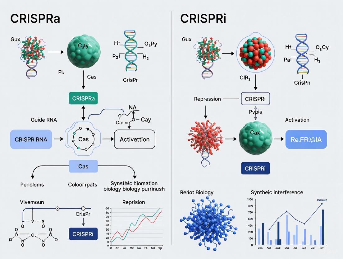
CRISPRa vs CRISPRi: Mechanisms, Applications, and Optimization for Precision Genetic Control
This article provides a comprehensive comparison of CRISPR activation (CRISPRa) and CRISPR interference (CRISPRi) technologies for researchers and drug development professionals.
CRISPRa vs CRISPRi: Mechanisms, Applications, and Optimization for Precision Genetic Control
Abstract
This article provides a comprehensive comparison of CRISPR activation (CRISPRa) and CRISPR interference (CRISPRi) technologies for researchers and drug development professionals. It covers the foundational mechanisms of these programmable transcriptional tools, detailing how catalytically dead Cas9 (dCas9) is fused to effector domains like VP64 or KRAB to upregulate or repress gene expression. The scope extends to methodological considerations for experimental design, troubleshooting common challenges, and a comparative analysis of their performance against alternative technologies like RNAi and ORF overexpression. By synthesizing recent advances, including novel drug-inducible and dual-mode systems, this review serves as a practical guide for leveraging CRISPRa/i in functional genomics screens and therapeutic development.
The Core Machinery: Deconstructing CRISPRa and CRISPRi Mechanisms
The Clustered Regularly Interspaced Short Palindromic Repeats (CRISPR) system, derived from a bacterial adaptive immune system, has revolutionized genetic engineering. While the native system functions as programmable "DNA scissors" capable of cutting DNA, a transformative advancement emerged with the creation of catalytically dead Cas9 (dCas9). This engineered variant contains point mutations (D10A and H840A in Streptococcus pyogenes Cas9) that abolish its nuclease activity while preserving its programmable DNA-binding capability [1] [2]. This fundamental shift converted CRISPR from a destructive tool into a precise targeting system, enabling transcriptional modulation without altering the underlying DNA sequence [3].
This technical guide explores the core mechanisms and applications of dCas9-based systems, specifically CRISPR interference (CRISPRi) for gene repression and CRISPR activation (CRISPRa) for gene upregulation. Framed within a broader thesis on CRISPRi versus CRISPRa mechanisms, this review provides an in-depth analysis for researchers and drug development professionals, detailing the experimental paradigms, key reagent solutions, and therapeutic potential of these powerful technologies.
Core dCas9 Mechanisms: CRISPRi vs. CRISPRa
The dCas9 protein serves as a programmable DNA-binding scaffold. When guided by a single-guide RNA (sgRNA) to a specific genomic locus, it can sterically hinder transcription or recruit effector domains to repress or activate gene expression [3] [4]. The table below summarizes the fundamental characteristics of these two systems.
Table 1: Core Characteristics of CRISPRi and CRISPRa Systems
| Feature | CRISPR Interference (CRISPRi) | CRISPR Activation (CRISPRa) |
|---|---|---|
| Primary Goal | Gene knockdown/silencing | Gene overexpression/activation |
| dCas9 Fusion | Transcriptional repressors (e.g., KRAB domain) | Transcriptional activators (e.g., VP64, VPR, SunTag, SAM) |
| Mechanism of Action | Blocks RNA polymerase binding or elongation; induces heterochromatin formation [4] [5] | Recruits transcriptional machinery to promoter/enhancer regions [4] [5] |
| Level of Regulation | Transcriptional (DNA level) | Transcriptional (DNA level) |
| Typical Repression/Activation | Up to 60-80% with dCas9 alone; >90% with dCas9-KRAB [4] | Varies by system; advanced systems (SAM, VPR) enable robust, synergistic activation [5] |
| Analogous Technology | RNA interference (RNAi) | ORF (Open Reading Frame) overexpression [5] |
The following diagram illustrates the fundamental mechanistic differences and key components of the CRISPRi and CRISPRa systems.
Advanced Architectures & Experimental Workflows
Enhanced System Architectures
While a simple dCas9-effector fusion can modulate transcription, more sophisticated architectures have been developed for enhanced efficacy, particularly for CRISPRa.
- Direct Fusion Systems: The simplest format, where the effector domain (e.g., KRAB for repression, VP64 for activation) is directly fused to dCas9. The VPR system is a potent direct fusion activator, combining VP64, p65, and Rta domains [5].
- Protein Scaffold Systems: These systems separate the dCas9 binder from the effector proteins. The SunTag system uses dCas9 fused to a repeating peptide array (GCN4), which is then recognized by single-chain variable fragments (scFvs) fused to effector domains like VP64. This allows for the recruitment of multiple activator molecules per dCas9, significantly amplifying the transcriptional signal [4] [5].
- RNA Scaffold Systems: This approach leverages modifications to the sgRNA itself. The Synergistic Activation Mediator (SAM) system uses an sgRNA engineered with RNA aptamers (e.g., MS2). These aptamers recruit fusion proteins (e.g., MCP-p65-HSF1), which work synergistically with a dCas9-VP64 fusion to drive strong gene activation [5].
A Standard Workflow for Pooled CRISPRi/a Screens
Pooled genetic screens are a primary application for CRISPRi/a, enabling the systematic identification of genes involved in biological processes or drug mechanisms. The general workflow is summarized below.
Table 2: Key Stages of a Pooled CRISPRi/a Screen
| Stage | Key Actions | Output/Quality Control |
|---|---|---|
| 1. Library Design & Cloning | Select genome-wide or sub-library of sgRNAs; clone into lentiviral vector [5]. | A plasmid library representing the entire sgRNA pool. |
| 2. Viral Production & Cell Transduction | Produce lentivirus from plasmid library; transduce target cells at low MOI. | A population of cells where each cell receives, in theory, a single sgRNA. |
| 3. Phenotypic Selection | Apply selective pressure (e.g., drug treatment, FACS sorting based on reporter, or long-term growth) [5]. | Enrichment or depletion of specific sgRNA-containing cells based on phenotype. |
| 4. Sequencing & Hit Identification | Extract genomic DNA from pre- and post-selection cells; PCR-amplify sgRNA sequences; NGS to quantify abundance [5]. | List of sgRNAs significantly enriched/depleted, revealing candidate hit genes. |
The corresponding experimental workflow is visualized in the following diagram.
The Scientist's Toolkit: Essential Research Reagents
Successful implementation of CRISPRi/a experiments requires a suite of well-characterized reagents. The table below details key solutions and their functions.
Table 3: Essential Research Reagent Solutions for dCas9 Experiments
| Reagent Category | Specific Examples / Systems | Function & Utility |
|---|---|---|
| dCas9 Effector Plasmids | dCas9-KRAB (for CRISPRi), dCas9-VP64, dCas9-VPR, SunTag, SAM [4] [5] [6] | Constitutive or inducible expression of the dCas9-effector fusion protein. The core of the system. |
| sgRNA Cloning Vectors | Lentiviral sgRNA vectors (single or multiplex), with Pol III promoters (e.g., U6) [1] [6] | For expression of one or more sgRNAs. Multiplex vectors enable targeting several genomic sites simultaneously. |
| sgRNA Libraries | Genome-wide human/mouse knockout (GeCKO) libraries, focused sub-libraries (e.g., kinase-family) [4] [5] | Pre-designed pools of sgRNAs for large-scale genetic screens. Available from multiple non-profit and commercial sources. |
| Delivery Tools | Lentiviral, adenoviral (AVV) vectors; lipid nanoparticles (LNPs); synthetic sgRNA [4] [7] [1] | Enable efficient introduction of CRISPR components into target cells, both in vitro and in vivo. |
| Validation Assays | RT-qPCR, RNA-seq; Western blot; flow cytometry; targeted bisulfite sequencing (for epigenome editing) [6] | Essential for confirming changes in gene expression, protein levels, and epigenetic state at on-target and potential off-target sites. |
| Cell Lines | HEK293T (for virus production), K562, HeLa, iPSCs, and other disease-relevant models [5] [6] | Well-characterized models used for tool development and functional screens. |
Quantitative Data & Applications in Functional Genomics
Performance in Genetic Screens
CRISPRi and CRISPRa screens have become indispensable for functional genomics. The table below summarizes quantitative findings and applications from key studies, demonstrating their power and versatility.
Table 4: Applications and Findings from CRISPRi/a Screens
| Application Area | System Used | Key Finding / Outcome | Reference |
|---|---|---|---|
| Identifying Essential Genes | Genome-wide CRISPRi in K562 cells | Successfully identified "gold-standard" essential genes (e.g., ribosomal subunits) and cell-type specific essential genes. | [5] |
| Drug Target & Mechanism | CRISPRa screen in A375 melanoma cells | Identified genes whose overexpression conferred resistance to a BRAF inhibitor, revealing drug resistance mechanisms. | [5] |
| Non-Coding RNA Functional Screening | CRISPRi screen targeting lncRNAs in multiple cell lines | Discovered cell-type specific essential long non-coding RNAs (lncRNAs), a class difficult to study with other methods. | [5] |
| Epigenetic Therapy Exploration | dCas9-TET1 targeting miR-200c promoter | Targeted demethylation reactivated the tumor-suppressor miR-200c, reducing cell viability and increasing apoptosis in breast cancer cells. | [6] |
| Neuroscience & Disease Modeling | CRISPRi in iPSC-derived neurons | Uncovered genes essential for neuronal growth and survival that were not essential in pluripotent stem cells or cancer cells. | [4] |
Advantages Over Alternative Technologies
Compared to previous technologies for gene modulation, CRISPRi/a offers distinct advantages:
- vs. RNA Interference (RNAi): CRISPRi suppresses gene expression at the transcriptional level, leading to more complete and specific knockdown with fewer off-target effects than RNAi, which operates at the post-transcriptional level [3] [5].
- vs. CRISPR Nuclease (CRISPRn): Unlike CRISPRn, which causes permanent DNA double-strand breaks and gene knockouts, CRISPRi/a is reversible and does not damage the DNA. This is crucial for studying essential genes, as CRISPRn knockout can be lethal, while CRISPRi knockdown allows for viable phenotypic analysis [4].
- vs. cDNA Overexpression: CRISPRa drives expression from the native endogenous gene locus, preserving natural splice variants, regulatory feedback, and gene dosage, which is often not the case with cDNA ORF overexpression from viral vectors [4].
Clinical & Commercial Translation
The dCas9 platform is paving the way for next-generation therapeutics. The global gene editing market is projected to surpass $13 billion USD by 2025, with a significant portion driven by CRISPR technologies [8]. As of Q2 2025, the American Society of Gene & Cell Therapy (ASGCT) reported 4,469 advanced therapies in development, with 49% being gene therapies [7].
A compelling clinical example is the development of a bespoke CRISPR treatment for a child with a rare defect in protein metabolism; the therapy was developed in under six months through rapid collaboration across academia, industry, and regulators [7]. This highlights the potential for patient-specific gene editing in clinical care.
In drug discovery, CRISPRi/a screens are used to identify novel drug targets and biomarkers. For instance, screens have successfully identified 19S proteasomal subunit levels as a biomarker predictive of patient response to the proteasome inhibitor carfilzomib [5]. Furthermore, the adaptability of CRISPRi has been demonstrated in challenging organisms like the malaria parasite Plasmodium yoelii, opening new avenues for combating infectious diseases [4].
The creation of catalytically dead Cas9 (dCas9) has fundamentally expanded the CRISPR toolkit beyond simple genome editing. By serving as a programmable DNA-binding scaffold, dCas9 powers both CRISPRi and CRISPRa technologies, allowing for precise, reversible, and efficient modulation of gene expression. As detailed in this guide, the mechanistic understanding, experimental protocols, and reagent toolkits for these systems are now mature, enabling their widespread application in functional genomics, drug discovery, and therapeutic development.
The distinct yet complementary nature of CRISPRi and CRISPRa mechanisms provides researchers with an unparalleled ability to probe gene function, model disease, and identify novel therapeutic targets. While challenges remain—particularly in optimizing delivery and minimizing off-target effects—the continued refinement of dCas9-based systems solidifies their role as indispensable "gene dimmers" in the modern molecular biology arsenal, poised to drive significant innovation in both basic research and clinical medicine.
CRISPR activation (CRISPRa) represents a powerful gain-of-function technology within the broader landscape of CRISPR-based transcriptional regulation. This approach utilizes a catalytically inactive Cas9 (dCas9) that binds to DNA without introducing double-strand breaks, serving as a programmable platform for recruiting transcriptional activators to specific genomic loci [9] [10]. When contrasted with CRISPR interference (CRISPRi), which employs dCas9 fused to repressors like KRAB to silence gene expression, CRISPRa enables precise upregulation of endogenous genes [11] [12]. This capability is particularly valuable for investigating gene function, modeling genetic diseases, and developing therapeutic strategies aimed at compensating for deficient gene expression.
The evolution of CRISPRa systems has progressed significantly from first-generation constructs to sophisticated multi-component architectures. While the initial dCas9-VP64 fusion demonstrated proof-of-concept, its modest activation levels prompted the development of enhanced systems including VPR, SAM (Synergistic Activation Mediator), and SunTag, which employ distinct strategies to amplify transcriptional output [9] [13]. This technical guide examines the core mechanistic principles, comparative performance, and experimental implementation of three principal CRISPRa effector systems—VP64, VPR, and SAM—providing researchers with a framework for selecting and applying these tools in diverse biological contexts.
Molecular Architecture and Mechanism
Core Components and Activation Strategies
CRISPRa systems share fundamental components but diverge in their strategies for recruiting transcriptional machinery. Each system utilizes dCas9 for programmable DNA binding and VP64 domains as foundational activation units, but differs in how these units are organized and amplified.
dCas9-VP64: As the first-generation CRISPR activator, this system employs a direct fusion of dCas9 to a single VP64 activation domain (a tetramer of the Herpes Simplex Viral Protein 16) [9]. The simplicity of this architecture is both its strength and limitation: while facilitating easy delivery, the solitary VP64 domain provides limited recruitment capacity, resulting in modest transcriptional activation typically around 2-fold for many target genes [9].
VPR (VP64-p65-Rta): This system enhances activation potency through a tripartite activator fusion, combining VP64 with two additional strong activation domains: p65 (a subunit of NF-κB) and Rta (a transcriptional activator from Epstein-Barr virus) [9] [13]. Unlike multi-component systems, VPR functions as a single fusion protein with dCas9, streamlining delivery while significantly increasing activation levels—generally exceeding dCas9-VP64 though typically lower than SAM for single-gene targeting [9].
SAM (Synergistic Activation Mediator): SAM employs a cooperative, multi-component approach that leverages both protein and RNA engineering. The system consists of: (1) dCas9-VP64; (2) specially engineered sgRNAs containing two MS2 RNA aptamers; and (3) MS2 coat proteins fused to the heterologous activation domains p65 and HSF1 (Heat Shock Factor 1) [9] [13]. This design enables the recruitment of multiple activator complexes per dCas9 molecule, creating a highly potent transcriptional activation platform that consistently demonstrates the highest activation levels for single-gene targeting [9] [13].
Table 1: Core Components of Major CRISPRa Systems
| System | dCas9 Fusion | RNA Components | Additional Protein Components | Recruitment Strategy |
|---|---|---|---|---|
| dCas9-VP64 | dCas9-VP64 | Standard sgRNA | None | Direct fusion |
| VPR | dCas9-VP64-p65-Rta | Standard sgRNA | None | Tripartite direct fusion |
| SAM | dCas9-VP64 | sgRNA with MS2 aptamers | MS2-p65-HSF1 fusion | Aptamer-mediated recruitment |
Mechanism Visualization
The following diagram illustrates the structural organization and transcriptional activation mechanisms of the three CRISPRa systems:
Comparative Performance Analysis
Activation Efficiency Across Systems
The quantitative performance of CRISPRa systems varies significantly depending on target genes, cell types, and experimental conditions. Direct comparisons reveal distinct activation profiles and efficiency patterns across platforms.
Table 2: Performance Comparison of CRISPRa Systems
| System | Activation Level | Multiplexing Efficiency | Specificity | Delivery Complexity | Optimal Use Cases |
|---|---|---|---|---|---|
| dCas9-VP64 | ~2-10 fold (modest) [9] | Moderate reduction [13] | High [13] | Low (single component) [9] | Modest activation requirements, minimal delivery constraints |
| VPR | Significantly higher than VP64, lower than SAM for single genes [9] | Comparable to SAM and SunTag [9] [13] | High [13] | Medium (single fusion protein) [9] | Balanced applications requiring good activation with simplified delivery |
| SAM | Highest for single-gene activation [9] [13] | Comparable to VPR and SunTag [9] | High [13] | High (three components) [9] | Maximal single-gene activation, when delivery complexity is manageable |
Performance characteristics exhibit notable context-dependence. In HEK293T cells, SAM consistently delivers the highest activation levels, though typically within five-fold of VPR or SunTag systems [13]. However, in other cell lines including U-2 OS and MCF7, VPR and SunTag can outperform SAM, indicating that cell-type-specific factors influence optimal system selection [13]. This pattern extends across species, with VPR, SAM, and SunTag showing similar activation levels (within five-fold) in mouse and Drosophila cells [13].
All three systems maintain high specificity according to RNA-seq analyses, with global gene expression correlations between activator-targeted samples and controls nearly identical to biological replicate correlations (R ≈ 0.98) [13]. This suggests that CRISPRa-mediated effects are highly specific to intended targets without widespread transcriptional disruption.
Advanced System Characterization
Recent single-cell analyses provide deeper insights into how these systems modulate transcriptional dynamics. The SunTag3xVPR system—a hybrid approach combining SunTag scaffolding with VPR activators—demonstrates exceptional potency by prolonging transcriptional burst duration (approximately 95 minutes) and increasing burst amplitude [14]. This system achieved a 48.6% activation ratio in single-cell analyses, surpassing SAM (35.8%), VPR (18.8%), and dCas9-VP64 (13.2%) [14].
Unexpectedly, increasing activator recruitment beyond optimal points can diminish effectiveness. Systems with 10 or more SunTag scaffolds can form solid-like condensates that sequester co-activators like p300 and MED1, reducing dynamicity and impairing activation [14]. This demonstrates that maximal activator multimerization does not necessarily correlate with optimal transcriptional output.
Experimental Implementation
Protocol for CRISPRa System Evaluation
Implementing CRISPRa experiments requires careful planning and optimization. The following protocol outlines key steps for evaluating and comparing CRISPRa systems:
1. System Selection and Vector Assembly:
- Select appropriate CRISPRa plasmids (available from Addgene and other repositories) [9]
- For SAM: Obtain dCas9-VP64, MS2-p65-HSF1, and modified sgRNA with MS2 aptamers [9] [13]
- For VPR: Utilize single vector encoding dCas9-VP64-p65-Rta fusion [9]
- For VP64: Use dCas9-VP64 construct [9]
- Clone sgRNAs targeting promoters of interest into appropriate expression vectors
2. Cell Line Development and Transfection:
- Culture relevant cell lines (HEK293T, HeLa, U-2 OS, or specialized models) [13]
- For stable expression: Use lentiviral transduction with puromycin selection to generate dCas9-expressing cells [12]
- Transfect CRISPRa components using appropriate methods (lipofection, electroporation)
- Include controls: non-targeting sgRNA, transfection controls, and uninduced controls for inducible systems
3. Induction and Timing:
- For inducible systems (e.g., Tet-On), administer doxycycline (1 μg/mL) to initiate dCas9 expression [12]
- Allow 48-72 hours for robust activation before analysis [14]
- For time-course studies, collect samples at 24, 48, and 72 hours post-induction
4. Activation Assessment:
- Quantify mRNA expression using RT-qPCR for target genes
- Normalize to housekeeping genes and calculate fold-change versus controls
- For single-cell resolution: Use flow cytometry with reporter systems (e.g., BFP, GFP) [14]
- Assess protein expression by Western blot or immunofluorescence when antibodies available
- For genome-wide specificity: Perform RNA-seq on activated samples versus controls [13]
5. Data Analysis and Validation:
- Calculate fold-activation for each system across multiple target genes
- Determine statistical significance using appropriate tests (t-tests, ANOVA)
- Confirm specificity by examining off-target gene expression
- Validate phenotypes through functional assays relevant to target genes
Specialized Applications and Model Systems
Inducible CRISPRa Systems: Recent advances enable precise temporal control through drug-responsive designs. The iCRISPRa/i systems fuse mutated human estrogen receptor (ERT2) domains to CRISPRa components, causing cytoplasmic sequestration until 4-hydroxytamoxifen (4OHT) administration induces nuclear translocation [11]. These systems show rapid response, reversibility, and lower baseline leakage compared to constitutive expression systems [11].
Complex Model Systems: CRISPRa implementation has expanded to physiologically relevant models including primary human 3D organoids. In gastric organoid models, doxycycline-inducible dCas9-VPR (iCRISPRa) systems successfully upregulated endogenous genes like CXCR4, with target-positive populations increasing from 13.1% to 57.6% following activation [12]. This demonstrates the efficacy of CRISPRa in challenging, translationally relevant systems.
Bacterial CRISPRa Considerations: Bacterial systems present unique challenges with strict target site requirements. Effective activation in E. coli requires precise positioning (2-4 base windows with 10-11 base periodicity) approximately 80 bases upstream of the transcriptional start site [15]. Successful bacterial CRISPRa also depends on promoter strength, sigma factor compatibility, and intervening sequence composition [15].
The Scientist's Toolkit: Essential Research Reagents
Table 3: Key Reagents for CRISPRa Research
| Reagent Category | Specific Examples | Function and Application | Source/Reference |
|---|---|---|---|
| dCas9 Activators | dCas9-VP64, dCas9-VPR, dCas9-VP64-p65-Rta | Programmable DNA binding and transcriptional activation | Addgene [9] |
| Engineered sgRNAs | MS2-modified sgRNAs (for SAM), standard sgRNAs | Target specification and additional recruiter binding (SAM) | [9] [13] |
| Recruitment Modules | MS2-p65-HSF1 (for SAM), scFv-VP64 (for SunTag) | Enhanced activator recruitment for amplified response | [9] [13] |
| Inducible Systems | ERT2-fused constructs, Tet-On dCas9 | Temporal control of CRISPRa activity | [11] [12] |
| Delivery Vectors | Lentiviral, piggyBac transposon, plasmid systems | Efficient delivery to diverse cell types | [12] |
| Reporter Systems | BFP, GFP, mCherry, luciferase | Quantitative assessment of activation efficiency | [14] |
| Validation Tools | RT-qPCR primers, RNA-seq, Western blot antibodies | Confirmation of transcriptional and translational activation | [12] [13] |
CRISPRa technologies have evolved substantially from the initial dCas9-VP64 design to sophisticated multi-activator systems. The VP64, VPR, and SAM architectures represent distinct approaches to transcriptional activation, each with characteristic strengths and optimal applications. VP64 offers simplicity and reliable delivery, VPR provides balanced potency with reduced complexity, and SAM delivers maximal activation at the cost of increased component complexity.
Future developments will likely focus on enhancing precision, reducing off-target effects, and improving in vivo delivery efficiency. The integration of CRISPRa with single-cell technologies and advanced model systems like human organoids will expand its applications in functional genomics and therapeutic development [12] [14]. As these tools mature, they will increasingly enable researchers to precisely manipulate transcriptional programs in both basic research and clinical applications, providing powerful capabilities to complement the CRISPRi toolkit for comprehensive gene regulation.
CRISPR interference (CRISPRi) has emerged as a powerful genetic tool that enables precise, reversible silencing of gene expression without altering the underlying DNA sequence. This technology centers on a catalytically dead Cas9 (dCas9) that serves as a programmable DNA-binding platform. When fused to transcriptional repressor domains and guided to specific genomic loci by a single-guide RNA (sgRNA), dCas9 can effectively suppress transcription [4]. The earliest and most widely adopted repressor domain is the Krüppel-associated box (KRAB) from the human protein KOX1, which recapitulates natural heterochromatin formation to silence target genes [16] [17]. However, inconsistent performance across cell lines, gene targets, and guide RNAs has highlighted the need for improved repressors [16]. Recent advances in protein engineering have yielded a new generation of CRISPRi systems with enhanced repression efficacy and reliability. This technical guide explores the mechanistic basis, performance characteristics, and experimental implementation of both established and novel CRISPRi repressor domains, providing researchers with the insights needed to select optimal tools for their specific applications.
Core Mechanisms of CRISPRi Repression
Foundational KRAB Domain Function
The KRAB domain is a potent transcriptional repressor derived from human zinc finger proteins. When dCas9-KRAB is targeted to a promoter or enhancer region, it initiates a native chromatin remodeling process. The KRAB domain recruits the co-repressor KAP1, which in turn assembles a heterochromatin-forming complex containing histone methyltransferases (e.g., SETDB1) and histone deacetylases (HDACs) [17]. This complex catalyzes the deposition of repressive histone marks, primarily H3K9me3, leading to chromatin condensation and transcriptional silencing. Genome-wide studies have confirmed that dCas9-KRAB targeted to distal regulatory elements like the HS2 enhancer in the globin locus specifically induces H3K9me3 at the intended site with minimal off-target effects, demonstrating its precision for epigenetic editing [17].
Novel Repressor Domains and Combinatorial Approaches
Research has revealed that KRAB domains from different human proteins exhibit varying repression potencies. For instance, the ZIM3(KRAB) domain demonstrates significantly stronger silencing activity than the historically used KOX1(KRAB) [16] [18]. Beyond KRAB domains, other potent repressor modules include:
- MeCP2(t): A truncated 80-amino acid version of methyl-CpG binding protein 2 that interacts with Sin3A and histone deacetylases [16]
- SCMH1, CTCF, and RCOR1: Non-KRAB domains identified through systematic screening with repressive activity exceeding canonical MeCP2 [16]
- MAX: A transcriptional regulator that functions effectively in combinatorial repressors [16]
Combinatorial fusion of multiple repressor domains to dCas9 creates synergistic effects that enhance gene knockdown. Engineering efforts have produced bipartite and tripartite repressors such as dCas9-ZIM3(KRAB)-MeCP2(t) and dCas9-ZIM3-NID-MXD1-NLS, which show substantially improved repression across diverse cell lines and target genes [16] [19].
Table 1: Key CRISPRi Repressor Domains and Their Characteristics
| Repressor Domain | Type | Size (aa) | Key Mechanisms | Performance Notes |
|---|---|---|---|---|
| KOX1(KRAB) | KRAB | ~45 | Recruits KAP1, SETDB1, HDACs, H3K9me3 | Foundational domain, moderate silencing |
| ZIM3(KRAB) | KRAB | ~45 | Enhanced KRAB activity | Superior to KOX1(KRAB) in silencing [16] [18] |
| MeCP2 (full) | Non-KRAB | 283 | Binds Sin3A/HDAC complex | Improves silencing in fusion proteins [16] |
| MeCP2(t) | Non-KRAB | 80 | Compact repressor core | Performance similar to full MeCP2 [16] |
| SCMH1 | Non-KRAB | Varies | Chromatin association | Outperforms MeCP2 in initial screens [16] |
| RCOR1 | Non-KRAB | Varies | Corepressor for REST complex | Novel repressor with strong activity [16] |
Quantitative Performance Comparison of CRISPRi Systems
Systematic Screening and Validation
Recent large-scale screening approaches have enabled the direct comparison of CRISPRi repressor efficacy. One comprehensive study screened >100 bipartite and tripartite repressor fusion proteins in HEK293T cells using an eGFP reporter system to quantify knockdown efficiency [16]. This systematic approach identified several novel repressor combinations that significantly outperformed gold standard systems like dCas9-ZIM3(KRAB). The top-performing variants included dCas9-KRBOX1(KRAB)-MAX, dCas9-ZIM3(KRAB)-MAX, and dCas9-KOX1(KRAB)-MeCP2(t), which improved gene knockdown by approximately 20-30% compared to dCas9-ZIM3(KRAB) alone [16]. Notably, the dCas9-ZIM3(KRAB)-MeCP2 combination – while highly effective – was identified as a previously characterized system, highlighting the importance of proper attribution in technology development [20].
Further engineering optimization has included nuclear localization signal (NLS) configuration, with the addition of a carboxy-terminal NLS enhancing gene knockdown efficiency by an average of ~50% [19]. The current state-of-the-art system, dCas9-ZIM3-NID-MXD1-NLS, incorporates an optimized MeCP2 NID truncation domain with additional repressor elements and proper localization signals to achieve superior silencing capabilities across multiple cell lines and in genome-wide dropout screens [19].
Table 2: Performance Comparison of Selected CRISPRi Repressor Systems
| CRISPRi System | Repressor Architecture | Relative Knockdown Efficiency | Key Advantages | Validation Context |
|---|---|---|---|---|
| dCas9-KOX1(KRAB) | Single domain | Baseline | Established, well-characterized | Multiple cell lines [16] [17] |
| dCas9-ZIM3(KRAB) | Single domain | ++ vs. KOX1 | Enhanced KRAB activity | HEK293T, stem cells [16] |
| dCas9-KOX1(KRAB)-MeCP2 | Bipartite | +++ vs. single domain | Synergistic repression | Gold standard [16] |
| dCas9-ZIM3-MeCP2 | Bipartite | ++++ vs. single domain | Potent long-term silencing | Previously characterized [20] |
| dCas9-ZIM3(KRAB)-MAX | Bipartite | +20-30% vs. ZIM3 | Novel effective combination | HEK293T reporter [16] |
| dCas9-KOX1(KRAB)-MeCP2(t) | Bipartite | +20-30% vs. ZIM3 | Compact, highly efficient | HEK293T reporter [16] |
| dCas9-ZIM3-NID-MXD1-NLS | Optimized bipartite | +50% with NLS | Superior silencing, optimized | Multiple cell lines, genome-wide screens [19] |
Cell Type-Specific Performance Considerations
CRISPRi performance varies across cellular contexts, influencing repressor selection for specific applications. Studies comparing induced pluripotent stem cells (iPSCs), neural progenitors, and differentiated cells have revealed distinct dependencies on translation machinery genes when targeted with the same dCas9-KRAB system [21]. For instance, iPSCs demonstrated higher sensitivity to mRNA translation perturbations compared to differentiated cell types, potentially reflecting their elevated global protein synthesis rates [21]. These findings underscore that optimal CRISPRi repressor choice may depend on the specific biological context, with certain cell types potentially requiring enhanced repressor systems for effective gene knockdown.
Experimental Design and Implementation
Delivery Methods for CRISPRi Systems
Effective delivery of CRISPRi components is crucial for successful gene silencing. Multiple delivery strategies have been developed, each with distinct advantages:
- Lentiviral Transduction: Enables stable integration and long-term expression, ideal for pooled screens and chronic knockdown studies. Used in the Virtual Cell Challenge with dual-guide designs to ensure strong and consistent knockdown [22].
- Virus-Like Particles (VLPs): The RENDER platform (Robust ENveloped Delivery of Epigenome-editor Ribonucleoproteins) facilitates transient delivery of CRISPRi ribonucleoprotein complexes, minimizing off-target exposure and enabling editing in hard-to-transfect cells, including primary T cells and neurons [18].
- Plasmid Transfection: Suitable for transient expression in easily transfectable cell lines like HEK293T, commonly used for initial system validation [16].
Recent advances in VLP delivery have demonstrated efficient packaging and transfer of various CRISPRi effectors, with the ZIM3 KRAB domain showing higher epigenetic silencing compared to KOX1 within CRISPRi-eVLPs [18]. This delivery method is particularly valuable for therapeutic applications where transient editor expression is desirable.
Guide RNA Design and Target Selection
Effective CRISPRi requires careful sgRNA design with particular attention to:
- Target Location: sgRNAs should bind to promoter regions or transcriptional start sites (TSS), though these are not always well-annotated in the genome [4]. For enhancer targeting, coverage across the core regulatory region (e.g., 400bp for HS2 enhancer) with multiple sgRNAs is recommended [17].
- Dual-guide Designs: Empirical data shows that expressing two guides per target gene from the same vector significantly improves knockdown consistency and efficacy compared to single-guide approaches [22].
- Nucleosome Positioning: Histone-DNA binding can impede dCas9 access, necessitating consideration of chromatin accessibility in guide design [23].
- Specificity Controls: Include non-targeting sgRNAs and target-negative cell lines to distinguish specific effects from background [17].
Validation and Optimization Methodologies
Rigorous validation of CRISPRi systems involves multiple complementary approaches:
- Reporter Assays: Fluorescent reporters (e.g., eGFP) under control of targeted promoters enable rapid quantification of knockdown efficiency via flow cytometry [16].
- Endogenous Gene Analysis: qRT-PCR and RNA-seq measure transcript-level changes, while western blotting and flow cytometry assess protein knockdown [16] [17].
- Epigenetic Modification Tracking: Chromatin immunoprecipitation (ChIP) for H3K9me3 and other repressive marks confirms expected mechanism of action [17].
- Phenotypic Characterization: Growth assays following essential gene knockdown and genome-wide dropout screens validate functional impact [16] [21].
The Virtual Cell Challenge established rigorous evaluation metrics including Differential Expression Score (DES), Perturbation Discrimination Score (PDS), and Mean Absolute Error (MAE) that provide standardized frameworks for assessing CRISPRi performance [22].
The Scientist's Toolkit: Essential Research Reagents
Table 3: Key Reagents for CRISPRi Experiments
| Reagent Category | Specific Examples | Function & Application Notes |
|---|---|---|
| dCas9-Repressor Fusions | dCas9-KOX1(KRAB), dCas9-ZIM3(KRAB), dCas9-ZIM3-MeCP2, dCas9-ZIM3-NID-MXD1-NLS | Core effector proteins; selection depends on required knockdown strength |
| Delivery Vectors | Lentiviral constructs with dual-guide expression, Plasmid vectors for transient expression, VLP systems for RNP delivery | Determine expression stability and duration |
| Cell Lines | HEK293T (validation), K562 (erythroid models), H1 ESCs (pluripotent models), iPSC-derived lineages | Impact repressor performance; choose biologically relevant models |
| Validation Tools | qPCR primers for target genes, RNA-seq libraries, Flow cytometry antibodies, Western blot antibodies | Essential for quantifying knockdown at transcript and protein levels |
| Control Reagents | Non-targeting sgRNAs, dCas9-only constructs, Untargeted cell lines, Expression markers (mCherry) | Critical for establishing specificity and normalization |
The CRISPRi landscape continues to evolve with several promising research directions. First, the engineering of compact, highly efficient repressor domains like MeCP2(t) (80aa) addresses the packaging constraints of viral delivery systems, particularly AAVs with limited cargo capacity [16] [18]. Second, transient delivery methods like the RENDER platform enable precise temporal control over gene silencing while minimizing potential off-target effects associated with prolonged editor expression [18]. Third, the application of CRISPRi to increasingly complex biological systems, including patient-derived primary cells and organoid models, will further elucidate cell-type-specific genetic dependencies [21] [18].
The development of novel repressor domains beyond KRAB represents a significant advancement in CRISPRi technology. While KRAB-based systems remain valuable for many applications, new combinatorial repressors like dCas9-ZIM3(KRAB)-MeCP2(t) and dCas9-ZIM3-NID-MXD1-NLS offer substantially enhanced performance with reduced variability across guide RNAs and cellular contexts [16] [19]. These improved tools provide researchers with more reliable and potent options for probing gene function, conducting genetic screens, and developing potential therapeutic applications. As the CRISPRi toolbox continues to expand, careful consideration of repressor selection, delivery method, and experimental design will ensure optimal outcomes for specific research objectives.
In the realm of CRISPR-based transcriptional regulation, CRISPR activation (CRISPRa) and interference (CRISPRi) have emerged as powerful tools for precise gene expression control. While both systems utilize a guide RNA (gRNA) and a catalytically dead Cas9 (dCas9) to target specific genomic loci, their functional outcomes are diametrically opposed. The strategic design of the gRNA is the paramount factor determining the success of either approach. gRNA targeting is not a one-size-fits-all process; optimal sequences for activation differ significantly from those for repression. This guide details the key differences in gRNA design principles for CRISPRa and CRISPRi, providing researchers with a framework for effective experimental implementation within the broader context of programmable transcription factors.
Fundamental Mechanisms: How gRNA Targeting Directs Activation and Repression
The core difference between CRISPRa and CRISPRi lies in the effector domain fused to the dCas9 protein. Despite this difference, both systems rely entirely on the gRNA for DNA binding and specificity.
CRISPR Interference (CRISPRi) Mechanism
In CRISPRi, the dCas9 protein is typically fused to a repressor domain such as the Krüppel-associated box (KRAB) [11] [4]. The gRNA-dCas9 complex binds to the target DNA, and the KRAB domain recruits additional proteins that establish a transcriptionally silent heterochromatin state, effectively shutting down gene expression [4]. The binding location of the gRNA is critical for effective repression. The most effective CRISPRi occurs when the gRNA targets a region that physically blocks the binding or progression of RNA polymerase (RNAP). This is typically achieved by designing gRNAs to bind within a window of -50 to +300 nucleotides relative to the transcription start site (TSS), with the most potent repression often occurring when targeting the region immediately downstream of the TSS [4].
CRISPR Activation (CRISPRa) Mechanism
Conversely, CRISPRa employs a dCas9 protein fused to transcriptional activator domains, such as VP64, VP64-p65-Rta (VPR), or components of the Synergistic Activation Mediator (SAM) system [24] [25] [4]. The gRNA directs this complex to the promoter region of a target gene, where the activation domain recruits the cellular transcription machinery to initiate gene expression. For CRISPRa, gRNA binding location is even more constrained. Effective activation requires targeting upstream regulatory elements, typically in the region between -200 and -500 base pairs upstream of the TSS [26]. This positioning is believed to mimic natural enhancer elements, allowing the activator domains to interact effectively with the basal transcription apparatus at the core promoter.
The following diagram illustrates the core mechanistic differences and optimal gRNA binding sites for CRISPRa and CRISPRi.
Comparative gRNA Design Principles: A Detailed Analysis
The divergent mechanisms of CRISPRa and CRISPRi necessitate distinct gRNA design strategies. The table below summarizes the critical differences in gRNA targeting parameters for the two systems.
Table 1: Key gRNA Design Parameters for CRISPRa vs. CRISPRi
| Design Parameter | CRISPRa (Activation) | CRISPRi (Interference) |
|---|---|---|
| Optimal Target Region | Upstream of TSS (-200 to -500 bp) [26] | Overlapping or downstream of TSS (-50 to +300 bp) [4] |
| Primary Goal | Recruit transcriptional machinery | Sterically hinder RNA polymerase |
| gRNA Efficiency | Highly variable; depends on local chromatin state [4] | Generally high and more consistent |
| Specificity Concerns | Potential for inadvertent enhancer effects | Blocking expression of overlapping genes |
| Multiplexing Strategy | qgRNAs (4 guides/gene) significantly boost efficacy [27] | Often effective with a single guide, but multiples enhance robustness |
A leading cause of failed CRISPRa experiments is the use of gRNAs designed with CRISPRi principles. The most critical distinction is the target location relative to the TSS. A gRNA designed for CRISPRi that binds downstream of the TSS will be ineffective for CRISPRa, as this location is not conducive to recruiting activators to the promoter. Furthermore, the inherent openness of the chromatin at the target site is a more significant factor for CRISPRa than for CRISPRi. A gRNA targeting a region in tightly packed heterochromatin may fail to activate transcription, even if its sequence is perfectly designed, because the dCas9-activator complex cannot access the DNA [4].
Advanced Strategies and Quantitative Performance
Enhancing Efficiency with Multiplexed gRNAs
For CRISPRa, using multiple gRNAs (multiplexing) per gene is highly advantageous. Single sgRNAs often induce variable and low-level gene activation [27]. A powerful solution is the use of quadruple-guide RNA (qgRNA) vectors, where a single plasmid expresses four distinct sgRNAs targeting the same gene. Research has demonstrated that this approach massively increases target gene activation compared to individual sgRNAs, delivering more robust and consistent results [27]. This synergistic effect is less critical for CRISPRi, which can be highly effective with a single, well-chosen gRNA, though multiplexing can still ensure complete repression.
Orthogonal Systems for Simultaneous Application
A cutting-edge application involves using CRISPRa and CRISPRi simultaneously within the same cell. This requires orthogonal CRISPR systems to prevent cross-talk. For example, a study used dSpCas9-VPR for activation and dSaCas9-KOX1 for repression, exploiting the fact that the gRNAs for these different Cas9 orthologs are not interchangeable [24]. This allows for concurrent activation of one gene and repression of another in a single experiment, enabling complex genetic engineering workflows.
Table 2: Quantitative Performance of Optimized CRISPRa/i Systems
| Application | System Used | Reported Efficiency | Key Experimental Validation |
|---|---|---|---|
| CRISPRa | dCas9-VPR with qgRNAs | Up to 98% positive cells for surface markers (e.g., CD123) [24] | Flow cytometry on primary human T cells |
| CRISPRi | dSaCas9-KOX1 | ~5% of CD5-positive cells remaining [24] | Flow cytometry on Jurkat cells |
| Genome-wide Screening | dxCas9-CRP dual-mode system | Coordinated activation/repression increased violacein production [26] | Fluorescent reporters and metabolite production in E. coli |
Experimental Protocol: A Workflow for gRNA Design and Validation
Implementing a successful CRISPRa or CRISPRi experiment requires a structured workflow. The following diagram and detailed protocol outline the key steps from initial design to final validation.
Step 1: Annotate the Transcription Start Site (TSS). Precise TSS annotation is the most critical step. Use curated databases like RefSeq to identify the canonical TSS for your gene of interest. An error of even 100 base pairs can determine the success or failure of the experiment.
Step 2: Define the gRNA Target Window. Based on the chosen modality (CRISPRa or CRISPRi), define the genomic search window. For CRISPRa, scan the region from -200 to -500 bp upstream of the TSS. For CRISPRi, scan the region from -50 to +300 bp around the TSS.
Step 3: Design and Select gRNA Sequences. Within the target window, identify all possible gRNA sequences with appropriate PAM sites (e.g., NGG for SpCas9). Use modern design algorithms (e.g., from platforms like Synthego) that incorporate rules for specificity and on-target efficiency [4]. For CRISPRa, design 3-4 non-overlapping gRNAs per gene to be used in a qgRNA vector for maximal effect [27].
Step 4: Select and Synthesize Guides. For the highest editing efficiency and lowest off-target effects, synthetic, chemically modified sgRNAs in a ribonucleoprotein (RNP) format are the preferred choice [28]. This avoids the variability and potential toxicity associated with plasmid-based expression.
Step 5: Deliver System and Validate Targeting. Co-deliver the dCas9-effector (as mRNA or protein) and the sgRNA(s) into your target cells. Validate successful on-target binding and transcriptional modulation 48-72 hours post-delivery by measuring changes in mRNA levels using quantitative RT-PCR.
Step 6: Conduct Functional Phenotypic Assay. The final step is to link the transcriptional change to a functional phenotype. This can be assessed by flow cytometry (for surface proteins), immunoblotting (for protein levels), or a specific functional assay (e.g., metabolite production or drug resistance) [26] [24].
The Scientist's Toolkit: Essential Reagents and Solutions
Table 3: Essential Research Reagents for CRISPRa/i Experiments
| Reagent / Solution | Function | Example / Note |
|---|---|---|
| dCas9-Effector Fusions | Targetable transcription core; defines a/i function | dCas9-VPR (for CRISPRa), dCas9-KRAB (for CRISPRi) [24] [4] |
| gRNA Expression Format | Determines specificity and delivery efficiency | Synthetic sgRNA (RNP format recommended for high efficiency) [28] |
| qgRNA Vectors | Plasmid expressing 4 guides for synergistic activation | Significantly boosts CRISPRa efficacy versus single guides [27] |
| Orthogonal Cas Proteins | Enable simultaneous a/i in one cell; prevent cross-talk | dSpCas9 for activation + dSaCas9 for repression [24] |
| Delivery Method | Critical for primary and hard-to-transfect cells | Electroporation of RNP complexes is highly effective [24] |
| Inducible Systems | Provide temporal control over CRISPRa/i activity | ERT2-based iCRISPRa/i systems activated by 4OHT [11] |
The precision of gRNA targeting is the cornerstone of effective CRISPRa and CRISPRi experiments. The fundamental distinction—targeting upstream promoter regions for activation versus the core promoter/TSS region for repression—must guide all experimental design. By adhering to these specialized principles, employing multiplexed gRNAs for activation, and leveraging orthogonal systems for complex manipulations, researchers can fully harness the power of programmable transcription to dissect gene function and engineer novel cellular phenotypes. As the field advances, continued optimization of gRNA design and delivery will further solidify CRISPRa and CRISPRi as indispensable tools in functional genomics and therapeutic development.
CRISPR activation (CRISPRa) and CRISPR interference (CRISPRi) are powerful technologies derived from the CRISPR-Cas9 system that enable precise transcriptional control without altering the underlying DNA sequence. These systems utilize a catalytically inactive or "dead" Cas9 (dCas9) that retains its ability to bind DNA target sites specified by a guide RNA (sgRNA) but does not cleave the DNA. The fundamental distinction between CRISPRa and CRISPRi lies in the effector domains fused to dCas9: CRISPRa employs transcriptional activators to increase gene expression, whereas CRISPRi uses repressors to decrease it. While in vitro applications of these tools are well-established, their function in vivo—particularly the critical processes of nuclear translocation and complex assembly—presents unique challenges and considerations for therapeutic development. Understanding these mechanisms is essential for applying CRISPRa/i to gene therapy, functional genomics, and the treatment of complex diseases in living organisms.
Core Mechanisms of Nuclear Translocation
For CRISPRa/i systems to function, the dCas9-effector fusion proteins must efficiently localize to the nucleus where they can access genomic DNA. This process of nuclear translocation is a critical regulatory point, especially for in vivo applications.
The Challenge of Cytoplasmic Sequestration
In the context of in vivo models, a significant barrier to nuclear import is the sequestration of CRISPR components in the cytoplasm. Research has shown that the use of nuclear localization signals (NLS) is a standard method to facilitate nuclear import. However, a more sophisticated, controllable method involves fusing the CRISPRa/i components to a mutated human estrogen receptor (ERT2) domain. This ERT2 domain causes the fusion protein to be sequestered in the cytoplasm through interaction with heat shock protein 90 (HSP90), effectively preventing its uncontrolled entry into the nucleus [11].
Inducible Nuclear Translocation
The ERT2-based system provides a drug-inducible mechanism for nuclear translocation. Upon administration of an estrogen analogue such as 4-hydroxy-tamoxifen (4OHT), the conformational change of the ERT2 domain disrupts its interaction with HSP90. This disruption releases the CRISPRa/i fusion protein, allowing its subsequent translocation into the nucleus [11]. A specific system named iCRISPRa/i, which features two ERT2 domains at the N-terminus and one at the C-terminus of the CRISPRa/i construct, demonstrates rapid and efficient nuclear translocation upon 4OHT induction. This process is reversible; upon withdrawal of 4OHT, the system can be restored to its original state, highlighting its potential for precise temporal control of gene expression in vivo [11].
Table: Key Systems for Controlling Nuclear Translocation In Vivo
| System Name | Inducing Molecule | Core Mechanism | Effect on Translocation | Reversibility |
|---|---|---|---|---|
| iCRISPRa/i [11] | 4-hydroxy-tamoxifen (4OHT) | ERT2 domain fusion; dissociation from HSP90 complex | Drug-induced nuclear import | Yes |
| CRISPR-StAR [29] | Tamoxifen (for Cre-ERT2) | Cre recombinase-mediated sgRNA activation | Controls functional sgRNA assembly in the nucleus | No (irreversible recombination) |
Nuclear Entry in Therapeutic Delivery
The mode of delivery for CRISPR components significantly impacts nuclear translocation in vivo. For instance, lipid nanoparticles (LNPs) used for systemic delivery tend to naturally accumulate in liver cells. Once inside hepatocytes, the Cas9 mRNA and sgRNA are released into the cytoplasm, where the mRNA is translated into protein. The resulting dCas9-effector fusion proteins, equipped with NLSs, are then actively imported into the nucleus [30]. This principle is leveraged in several ongoing clinical trials for liver-specific diseases [30] [31].
Assembly of Functional Complexes In Vivo
Once inside the nucleus, the dCas9-sgRNA ribonucleoprotein complex must locate and bind its target DNA sequence. Subsequently, the transcriptional effector domains must recruit the cellular machinery to either activate or repress gene expression.
CRISPRi Complex Assembly and Repression Mechanism
CRISPRi complexes are typically formed by fusing dCas9 to a transcriptional repressor domain, most commonly the Krüppel-associated box (KRAB) domain. The assembled dCas9-KRAB/sgRNA complex binds to the promoter region of a target gene, typically within a window from -50 to +300 base pairs relative to the transcriptional start site (TSS), with the most effective binding sites often located just downstream of the TSS [32]. The KRAB domain then recruits co-repressors and histone-modifying enzymes that promote the formation of heterochromatin—a compact, transcriptionally silent form of DNA—thereby effectively interfering with the initiation and elongation of transcription by RNA polymerase [4] [5].
CRISPRa Complex Assembly and Activation Mechanism
CRISPRa systems are designed to recruit multiple transcriptional activator domains to a gene's promoter. Simple CRISPRa systems involve direct fusion of activator domains like VP64 to dCas9. However, more powerful and sophisticated systems have been developed to achieve robust gene activation in vivo. The primary strategies include:
- Direct Fusions: Such as dCas9-VPR, a tripartite fusion of VP64, p65, and Rta transactivator domains [11] [5].
- Protein Scaffolds: Such as the SunTag system, where dCas9 is fused to a peptide array that recruits multiple copies of antibody-activator fusions [4] [5].
- RNA Scaffolds: Such as the Synergistic Activation Mediator (SAM), where engineered sgRNAs contain RNA aptamers that recruit additional activator proteins like p65 and HSF1 [5] [32].
For effective activation, the CRISPRa complex must bind upstream of the TSS, typically within the -400 to -50 bp window [32]. Once bound, the activator domains recruit co-activators and the general transcription machinery to initiate gene expression.
Advanced Systems for Controlled Assembly In Vivo
Novel screening systems like CRISPR-StAR (Stochastic Activation by Recombination) demonstrate sophisticated control over functional complex assembly in complex in vivo models, such as tumors in mice [29]. This system uses a Cre-lox mechanism to stochastically and irreversibly generate either an active or an inactive sgRNA configuration within each single-cell-derived clone. This design creates an internal control by ensuring that within each clone, a population of cells with active sgRNAs is intermingled with a corresponding wild-type population carrying the identical sgRNA in an inactive state. This internal control accounts for both intrinsic cellular heterogeneity and extrinsic microenvironmental factors, allowing for high-resolution genetic screening in vivo by comparing cells with active versus inactive sgRNAs within the same clonal population and microenvironment [29].
The following diagram illustrates the fundamental workflows and logical relationships of inducible CRISPRa/i systems for in vivo application.
Quantitative Data and Experimental Comparisons
The functionality of CRISPRa/i systems in vivo is quantifiable through various metrics, including efficiency, specificity, and dynamic range.
Table: Quantitative Comparison of CRISPRa/i Performance In Vivo
| Parameter | CRISPRi (dCas9-KRAB) | CRISPRa (e.g., dCas9-VPR/SAM) | Inducible System (iCRISPRa/i) | Measurement Context |
|---|---|---|---|---|
| Repression/Efficiency | Up to 80-90% repression [4] [5] | Up to several hundred-fold activation [5] [32] | Comparable to non-inducible counterparts [11] | Endogenous gene regulation in cell lines & mouse models |
| Targeting Window | -50 to +300 bp from TSS [32] | -400 to -50 bp from TSS [32] | N/A (same as fused effector) | Optimal sgRNA binding region for efficacy |
| Leakiness | Variable (constitutive systems) | Variable (constitutive systems) | Lower leakage [11] | Basal activity in the absence of induction |
| Response Time | Hours to days (constitutive) | Hours to days (constitutive) | Fast drug response [11] | Time from induction to detectable transcriptional change |
| Reversibility | Limited (requires degradation of components) | Limited (requires degradation of components) | Reversible upon 4OHT withdrawal [11] | Ability to return to basal expression state |
Detailed Experimental Protocols for In Vivo Study
To investigate nuclear translocation and complex assembly in vivo, robust experimental methodologies are required. The following protocol details the use of the iCRISPRa/i system, incorporating key steps for controlled nuclear import.
Protocol: Evaluating Inducible CRISPRa/i in a Mouse Model
This protocol outlines the steps to test the functionality of an ERT2-based inducible CRISPRa/i system, such as iCRISPRa/i, in a murine model, with a focus on tracking nuclear translocation and transcriptional outcomes [11].
Materials:
- Plasmids:
pHR-CMV-iCRISPRa/i-2A-mCherry(for expressing the inducible dCas9-effector) andpU6-sgRNA-EF1Alpha-puro-T2A-BFP(for expressing the sgRNA). - Cell line: e.g., B16 murine melanoma cells or NIH/3T3 cells.
- Inducer: 4-hydroxy-tamoxifen (4OHT) prepared in an appropriate vehicle (e.g., corn oil).
- Animal Model: Immunocompromised mice (e.g., NSG) for xenograft studies.
- Analytical Tools: Immunofluorescence microscopes, flow cytometer, RNA extraction kit, qPCR system.
Procedure:
- Stable Cell Line Generation:
- Co-transduce target cells (e.g., B16) with lentivirus produced from the
pHR-CMV-iCRISPRa/i-2A-mCherryplasmid and thepU6-sgRNAplasmid. - Select successfully transduced cells using antibiotics (e.g., puromycin) and/or fluorescence-activated cell sorting (FACS) for mCherry and BFP markers.
- Validate the cytoplasmic localization of the iCRISPRa/i fusion protein in the absence of 4OHT via immunofluorescence staining against the myc-His tag or the mCherry reporter.
- Co-transduce target cells (e.g., B16) with lentivirus produced from the
In Vivo Tumor Engraftment and Induction:
- Subcutaneously inject ~1 million stable cells into the flanks of mice.
- Allow tumors to establish to a palpable size (~100-150 mm³).
- Randomize mice into two groups: experimental and control.
- Administer 4OHT (e.g., 2 mg/dose via intraperitoneal injection) to the experimental group. The control group receives the vehicle only.
Monitoring Nuclear Translocation:
- At various time points post-induction (e.g., 6, 12, 24 hours), harvest tumors from a subset of animals from each group.
- Fix tumor tissue sections and perform immunofluorescence staining for the dCas9-effector fusion protein and a nuclear marker (e.g., DAPI).
- Image using confocal microscopy to qualitatively and quantitatively assess the shift in protein localization from the cytoplasm to the nucleus upon 4OHT administration.
Assessing Transcriptional and Phenotypic Outcomes:
- Harvest tumor tissues at a later time point (e.g., 48-72 hours post-induction) to assess downstream effects.
- Gene Expression Analysis: Extract total RNA from tumor tissue, reverse transcribe to cDNA, and perform qPCR to measure the mRNA levels of the target gene(s). Compare the 4OHT-induced group to the vehicle control group.
- Phenotypic Analysis: Measure tumor growth characteristics or other relevant phenotypic changes (e.g., melanin synthesis in B16 models) over time.
Protocol: Internally Controlled Screening with CRISPR-StAR
The CRISPR-StAR protocol is designed for high-resolution genetic screening in complex in vivo environments like tumors, providing a robust method to control for heterogeneity [29].
Procedure:
- Library Cloning and Cell Preparation:
- Clone an sgRNA library (e.g., targeting 1,245 genes) into the CRISPR-StAR backbone, which contains a Cre-inducible, barcoded sgRNA expression cassette.
- Transduce a Cas9- and Cre-ERT2-expressing cell line (e.g., mouse melanoma cells) with the lentiviral library at high coverage (>500 cells per sgRNA).
In Vivo Engraftment and Clonal Expansion:
- Inject transduced cells into mouse models and allow tumors to develop. The engraftment process creates a natural bottleneck, resulting in tumors derived from a few thousand clones, each marked by a unique barcode.
Induction of Stochastic sgRNA Activation:
- Once tumors are established, administer tamoxifen to activate the nuclear Cre-ERT2. This stochastically and irreversibly generates two populations within each single-cell-derived clone: one with active sgRNAs and one with inactive sgRNAs, serving as an internal control.
Analysis and Hit Calling:
- Harvest tumors and extract genomic DNA.
- Amplify and sequence the sgRNA and barcode regions.
- For each unique barcode (clone), compare the abundance of the active sgRNA conformation to the inactive one. This internal comparison controls for clonal variability and microenvironmental effects.
- Genes are identified as hits (e.g., essential for in vivo growth) if their active sgRNAs are significantly depleted compared to the internal control across many independent clones.
The Scientist's Toolkit: Essential Reagents for In Vivo Studies
Table: Key Research Reagents for Investigating CRISPRa/i Function In Vivo
| Reagent / Solution | Function / Purpose | Example Use Case |
|---|---|---|
| dCas9-Effector Plasmids | Core protein component for transcription modulation. | pcDNA3.1-CMV-CRISPRa/i-myc-His for constitutive expression; pHR-CMV-iCRISPRa/i-2A-mCherry for inducible expression [11]. |
| Inducible Systems (ERT2) | Enables drug-controlled nuclear translocation. | Fusing ERT2 domains to CRISPRa/i components for 4OHT-inducible nuclear import [11]. |
| sgRNA Expression Vectors | Delivers targeting specificity to the genomic locus. | pU6-sgRNA EF1Alpha-puro-T2A-BFP for co-expression of a selection marker [11]. |
| Cre-lox Systems | Allows irreversible, stochastic activation of genetic elements. | CRISPR-StAR vector for creating internal control populations during in vivo screening [29]. |
| Lipid Nanoparticles (LNPs) | In vivo delivery vehicle for CRISPR components. | Systemic delivery of Cas9 mRNA and sgRNA to liver cells for gene regulation [30]. |
| 4-Hydroxy-Tamoxifen (4OHT) | Small-molecule inducer for ERT2-based and Cre-ERT2 systems. | Activating nuclear translocation of iCRISPRa/i or inducing recombination in CRISPR-StAR [11] [29]. |
| Unique Molecular Identifiers | DNA barcodes for tracking clonal populations. | Tracing the origin and expansion of single cells in complex in vivo models like tumors [29]. |
From Bench to Bedside: Experimental Design and Translational Applications
Within the expanding CRISPR toolkit, CRISPR activation (CRISPRa) and CRISPR interference (CRISPRi) have emerged as powerful technologies for the precise, reversible modulation of gene expression without altering the underlying DNA sequence. These techniques are foundational for functional genomics screens, target validation, and elucidating complex gene networks in drug discovery. Their efficacy, however, is profoundly dependent on one critical factor: the precise genomic positioning of the guide RNA (gRNA). This technical guide delineates the optimal gRNA design windows—targeting -400 to -50 bp for CRISPRa and -50 to +300 bp for CRISPRi—framing them within the context of their distinct mechanistic operations. A thorough grasp of these principles is essential for researchers and drug development professionals aiming to harness these technologies for robust and reproducible scientific and therapeutic outcomes [32].
Core Principles: Mechanisms of CRISPRa and CRISPRi
Both CRISPRa and CRISPRi utilize a catalytically dead Cas9 (dCas9) variant, which retains its programmable DNA-binding capability but is incapable of cleaving the DNA backbone [4] [32]. The fundamental difference lies in the effector domains fused to dCas9, which dictate transcriptional outcome.
- CRISPRi (Interference): This approach represses gene transcription. The most common system fuses dCas9 to a transcriptional repressor domain, most frequently the Krüppel-associated box (KRAB) [5] [32]. When guided to a target site, the dCas9-KRAB complex recruits additional proteins that promote the formation of heterochromatin—a tightly packed, transcriptionally silent form of DNA—thereby sterically hindering the binding and progression of RNA polymerase [5] [4].
- CRISPRa (Activation): This approach enhances gene transcription. Simple fusions of dCas9 to a single activator domain (e.g., VP64) often yield modest activation [5]. More advanced, highly effective systems, such as the Synergistic Activation Mediator (SAM), employ sophisticated protein or RNA scaffolds to recruit multiple, distinct transcriptional activator domains (e.g., VP64, p65, HSF1) to the promoter region, leading to robust gene upregulation [5] [32].
Optimal gRNA Design Windows: A Quantitative Guide
The mechanistic differences between CRISPRa and CRISPRi necessitate distinct gRNA positioning strategies relative to the Transcription Start Site (TSS). The TSS is the reference point (0 bp) from which all targeting positions are measured, and accurate TSS annotation, using databases like FANTOM, is critical for success [33].
Table 1: Optimal gRNA Design Windows for CRISPRa and CRISPRi
| Parameter | CRISPRa (Activation) | CRISPRi (Interference) |
|---|---|---|
| Optimal Targeting Window | -400 to -50 bp upstream of the TSS [34] [32] | -50 to +300 bp relative to the TSS [34] [32] |
| Mechanistic Rationale | Positions activators within the core promoter region to efficiently recruit the transcriptional machinery [32]. | For maximal efficiency, best-performing gRNAs target the first 100 bp downstream of the TSS [32]. This blocks the binding or elongation of RNA polymerase [4]. |
| Key Consideration | Alternative or poorly annotated TSSs can complicate design [34]. | Alternative TSSs can complicate design [34]. Off-targets near other genes' TSSs are a concern [34]. |
Experimental Protocol for gRNA Design and Screening
The following workflow provides a detailed methodology for designing and executing a CRISPRa or CRISPRi experiment, from initial gRNA selection to final validation.
gRNA Design and Library Preparation
- TSS Identification: For the gene of interest, determine the precise TSS using a reliable database such as FANTOM, which uses CAGE-seq data for accurate mapping [33].
- gRNA Selection: Using a specialized design tool (see Section 5), generate a list of candidate gRNAs that fall within the optimal windows specified in Table 1. For CRISPRa, focus on the region from -400 to -50 bp upstream. For CRISPRi, prioritize the region from -50 to +300 bp, with the highest efficacy often found within the first +100 bp downstream of the TSS [32].
- Sequence Optimization: Filter candidate gRNAs using on-target efficacy prediction algorithms (e.g., Doench rules) [35]. Avoid gRNAs with long homopolymers (e.g., AAAA) and those with high sequence similarity to off-target genomic sites [32].
- Library Cloning: Clone the selected gRNA sequences into an appropriate sgRNA expression vector. Ensure the gRNA sequence itself does not contain unintended restriction sites used for cloning or promoter terminator sequences [34].
Cell Line Engineering and Screening
- Generate Helper Cell Line: Create a stable cell line expressing the dCas9-effector fusion (e.g., dCas9-KRAB for CRISPRi or the SAM complex for CRISPRa). Lentiviral transduction is the most consistent method for achieving robust and uniform expression [32].
- Deliver gRNA Library: Transduce the helper cell line with the pooled sgRNA lentiviral library at a low Multiplicity of Infection (MOI) to ensure most cells receive only one sgRNA [5].
- Apply Selective Pressure: Culture the transduced cells and subject them to the relevant phenotypic screen, such as cell growth/proliferation, sensitivity to a drug or toxin, or a fluorescence-activated cell sorting (FACS)-based assay [5].
- Sequence and Analyze:
- At the beginning (t0) and end of the experiment, harvest cells and isolate genomic DNA.
- PCR-amplify the integrated sgRNA locus and subject the products to next-generation sequencing.
- Quantify the enrichment or depletion of each sgRNA in the final population compared to t0. sgRNAs that confer a selective advantage or disadvantage will be significantly enriched or depleted, respectively, pointing to genes involved in the screened phenotype [5].
Validation
- Essential Step: Never base conclusions on a single gRNA. The gold standard for validation is to demonstrate that multiple, independent gRNAs targeting the same gene produce the same phenotypic effect, confirming an on-target result [33].
The Scientist's Toolkit: Essential Research Reagents
Successful implementation of CRISPRa/i screens relies on a suite of key reagents and bioinformatic tools.
Table 2: Key Research Reagent Solutions for CRISPRa/i Experiments
| Reagent / Tool Category | Specific Examples | Function & Importance |
|---|---|---|
| dCas9 Effector Systems | dCas9-KRAB (CRISPRi), SAM, VPR, SunTag (CRISPRa) | The core protein complex that determines transcriptional outcome (repression or activation) [5] [32]. |
| gRNA Design Tools | CHOP-CHOP [34] [36], CRISPR-ERA [34], Benchling [35] [36], E-CRISP [34] | Bioinformatics platforms to design gRNAs within optimal windows, predict on-target efficiency, and minimize off-target effects. |
| Delivery Vectors | Lentiviral sgRNA Vectors | Ensure consistent and efficient delivery of sgRNA constructs into a wide variety of cell types, including hard-to-transfect cells [32]. |
| Analysis Software | MAGeCK, CRISPResso2 [36] | Bioinformatics tools for analyzing next-generation sequencing data from pooled screens to identify hit genes. |
Comparative Analysis with Alternative Technologies
Understanding how CRISPRa/i compare to other perturbation technologies highlights their unique advantages and guides technology selection.
- CRISPRi vs. RNAi: Both achieve gene knockdown, but CRISPRi is generally more specific with fewer sequence-based off-target effects. Furthermore, RNAi is primarily effective in the cytoplasm, whereas CRISPRi can efficiently target non-coding RNAs in the nucleus [4] [32].
- CRISPRi vs. CRISPR Nuclease (KO): CRISPR knockout (KO) permanently disrupts a gene, which is ideal for complete LOF studies but can be cytotoxic or lethal for essential genes. CRISPRi offers a reversible, titratable knockdown, making it superior for studying essential genes and providing a better mimic of pharmacological inhibition [4] [32].
- CRISPRa vs. ORF Overexpression: Traditional ORF overexpression uses strong viral promoters to drive supraphysiological expression of a transgenic cDNA. CRISPRa, in contrast, upregulates genes from their native genomic context, preserving physiological splice variants and avoiding positional effects associated with random transgene integration [32].
The precise delineation of gRNA design windows is not a mere suggestion but a foundational requirement for harnessing the full potential of CRISPRa and CRISPRi technologies. Adherence to the -400 to -50 bp window for CRISPRa and the -50 to +300 bp window for CRISPRi, as outlined in this guide, ensures maximal on-target efficacy by aligning the molecular tools with their underlying biological mechanisms. For the drug development community, these precision controls enable the systematic identification of novel therapeutic targets, validation of mechanism of action, and the discovery of resistance pathways through highly robust genetic screens. As these technologies continue to evolve, their strategic application, grounded in sound design principles, will undoubtedly accelerate the pace of discovery and therapeutic innovation.
CRISPR activation (CRISPRa) and CRISPR interference (CRISPRi) represent powerful technologies for precise transcriptional modulation without altering the underlying DNA sequence. These systems are derived from the CRISPR-Cas9 genome editing platform but utilize a catalytically inactive or "dead" Cas9 (dCas9) that retains its DNA-binding capability but lacks endonuclease activity [32]. The dCas9 serves as a programmable scaffold that can be fused to various effector domains to regulate gene expression [23]. When targeted to promoter or enhancer regions by guide RNAs (gRNAs), CRISPRa recruits transcriptional activators to increase gene expression, while CRISPRi recruits repressors to decrease expression [37] [32].
The core distinction between these mechanisms lies in their fused effector domains. For CRISPRi, the most common repressor is the Krüppel-associated box (KRAB) domain, which silences gene expression by recruiting chromatin-remodeling factors that promote heterochromatin formation [38] [32]. Additional repressor domains include SID4X [37]. For CRISPRa, common activators include VP64 (a tetramer of the VP16 domain), VP160, p65, and Rta (VP64-p65-Rta or VPR) [37] [23]. More advanced systems like the Synergistic Activation Mediator (SAM) combine multiple activators (e.g., dCas9-VP64 with MS2-p65-HSF1) for enhanced potency [39].
The effectiveness of CRISPRa and CRISPRi is highly dependent on the delivery system used to introduce these components into target cells. Optimal delivery vehicles must efficiently transport often bulky genetic constructs while maintaining safety and providing appropriate persistence of expression for the intended application [40] [41].
Lentiviral Vectors for CRISPR Delivery
Lentiviral vectors (LVs), particularly those derived from HIV-1, represent one of the primary delivery vehicles for CRISPR/Cas systems due to their ability to carry large and complex transgenes and sustain robust, long-term expression in both dividing and non-dividing cells [40].
Basic Biology and Engineering
Lentiviral vectors are enveloped viruses approximately 100 nm in diameter with a single-stranded RNA genome of approximately 10.7 kb [40]. The engineering of LVs for gene therapy has progressed through several generations focused on improving safety:
- Second-generation systems retained only the essential tat and rev genes from HIV, removing accessory genes to enhance safety and create space for larger inserts [40].
- Third-generation systems further improved safety by eliminating the tat gene and the endogenous promoter harboring the TAR element, instead utilizing heterologous promoters like CMV or RSV [40].
- Fourth-generation systems represent the safest option by splitting gag/pol and rev sequences into separate cassettes [40].
Additional enhancements include the incorporation of the woodchuck hepatitis virus posttranscriptional regulatory element (WPRE) and central polypurine tract (cPPT) to improve RNA stability, transcription efficiency, and overall viral titer [40].
Life Cycle and Integration
The LV life cycle begins with attachment and entry mediated by envelope-receptor interactions, with VSV-G being the most common envelope used for pseudotyping due to its broad tropism [40]. Following entry and uncoating, reverse transcriptase converts viral RNA to double-stranded DNA, which is then imported into the nucleus via host importin complexes [40]. A key distinction exists between integrase-competent lentiviral vectors (ICLVs), which permanently integrate into the host genome, and integrase-deficient lentiviral vectors (IDLVs), which remain primarily as episomal DNA [40]. IDLVs significantly reduce the risk of insertional mutagenesis while still supporting substantial transgene expression, particularly in non-dividing cells [40] [42].
Stable Cell Line Generation
Lentiviral vectors are particularly valuable for generating stable cell lines expressing CRISPRa/i components. The process typically involves:
- Vector Design: Packaging CRISPRa/i components (dCas9-effector and selection marker) into a lentiviral transfer plasmid [38] [39].
- Virus Production: Transfecting HEK293T packaging cells with the transfer plasmid along with packaging plasmids (psPAX2) and envelope plasmid (pCMV-VSV-G) using transfection reagents like polyethylenimine (PEI) [38].
- Transduction: Incubating target cells with viral supernatant in the presence of polybrene to enhance infection efficiency [38].
- Selection: Treating transduced cells with appropriate antibiotics (e.g., G418, puromycin) for 2-3 weeks to select successfully transduced cells [38].
- Validation: Confirming dCas9-effector expression and functionality through Western blot, qPCR, and reporter assays [38] [39].
Advanced systems incorporate inducible elements, such as tetracycline-responsive (TRE) promoters, allowing controlled expression of dCas9-effectors using doxycycline [38]. The piggyBac transposon system represents an alternative non-viral method for generating stable cell lines, offering higher cargo capacity and the ability to incorporate multiple transgene cassettes within a single vector [39].
Adeno-Associated Viral Vectors for CRISPR Delivery
Adeno-associated viruses (AAVs) have emerged as a leading delivery platform for therapeutic CRISPR applications due to their favorable safety profile, including low immunogenicity and minimal integration into the host genome [37] [41] [43].
Biology and Packaging Constraints
AAVs are small (∼20 nm), non-pathogenic viruses with a single-stranded DNA genome [41]. A major limitation of AAV is its constrained packaging capacity of approximately 4.7 kb, which presents challenges for delivering large CRISPR constructs [37] [41] [43]. For comparison, the commonly used Streptococcus pyogenes Cas9 (SpCas9) is approximately 4.2 kb, leaving minimal space for additional components [43].
Strategies to overcome this limitation include:
- Using smaller Cas9 orthologs such as Staphylococcus aureus Cas9 (SaCas9, ∼3.2 kb) [37] [43].
- Employing engineered miniature systems like CasMINI derived from Cas12f [43].
- Split-intein systems where Cas9 is divided into separate fragments packaged into dual AAVs that reconstitute post-transduction [41] [42].
- Minimizing regulatory elements using compact promoters (e.g., Mecp2, 235 bp) and synthetic polyA signals (e.g., spA, 48 bp) [37].
Capsid Engineering and Tropism
Natural AAV serotypes exhibit distinct tissue tropisms, but genetic engineering has further enhanced their targeting capabilities. For central nervous system delivery, AAV9-PHP.B demonstrates significantly improved blood-brain barrier crossing compared to wild-type AAV9 [37]. This approach can be extended to other serotypes; for example, AAV1-PHP.B also shows enhanced CNS transduction [37]. Capsid engineering enables researchers to select or design vectors optimized for specific target tissues and administration routes.
All-in-One AAV Systems for Epigenome Editing
Recent advances have enabled the development of all-in-one AAV systems for targeted transcriptional regulation. These compact platforms typically include:
- A small dCas9 variant (e.g., dSaCas9)
- A fused effector domain (e.g., KRAB, MeCP2's TRD for repression; VP64, VP160 for activation)
- The gRNA expression cassette
- Optimized regulatory elements [43]
Vector backbone optimization, such as incorporating concatemers of Sp1 and NF-κB recognition sites, can further enhance expression levels and overall system performance [43].
Comparative Analysis of Delivery Systems
The selection of an appropriate delivery system depends on multiple factors, including experimental goals, target cells, required persistence of expression, and safety considerations.
Table 1: Comparison of Viral Delivery Systems for CRISPRa/i
| Feature | Lentiviral Vectors | Adeno-Associated Viruses |
|---|---|---|
| Packaging Capacity | Large (>10 kb) [40] | Limited (~4.7 kb) [41] [43] |
| Integration Profile | Integrating (ICLV) or non-integrating (IDLV) [40] | Primarily episomal [41] |
| Expression Duration | Long-term (stable integration) [40] [42] | Typically months (transient) [41] [42] |
| Titer | High [40] | Moderate to high [42] |
| Transduction Efficiency | High in dividing and non-dividing cells [40] [42] | Variable; serotype-dependent [42] |
| Immunogenicity | Moderate [42] | Low [41] [42] |
| Primary Applications | Stable cell line generation, in vitro/ex vivo studies, genome-wide screens [40] [42] | In vivo delivery, clinical applications, targeting post-mitotic tissues [37] [42] |
| Key Safety Concerns | Insertional mutagenesis (ICLV) [40] | Limited cargo capacity, pre-existing immunity [41] |
Table 2: gRNA Design Guidelines for CRISPRa/i
| Parameter | CRISPRi | CRISPRa |
|---|---|---|
| Optimal Targeting Window | -50 to +300 bp from TSS (best: +1 to +100 bp) [32] | -400 to -50 bp from TSS [32] |
| gRNA Length | ≤21 bp (avoids homopolymers) [32] | ≤21 bp (avoids homopolymers) [32] |
| Chromatin Considerations | Accessible regions preferred [23] | Accessible regions preferred [23] |
| Multiplexing Strategy | Multiple gRNAs to same promoter enhance repression [37] | Multiple gRNAs to promoter/enhancer enhance activation [37] |
Experimental Protocols for Delivery System Evaluation
Protocol: Generating a Stable Inducible CRISPRi/a Cell Line Using Lentivirus
This protocol outlines the creation of a stable cell line with inducible dCas9-effector expression for controlled transcriptional modulation [38].
Materials:
- Plasmids: Tet-regulable dCas9-VP64 (Addgene #50916) or dCas9-KRAB (Addgene #50917), psPAX2, pCMV-VSV-G
- Cell lines: HEK293T packaging cells, target cells (e.g., hMSCs)
- Reagents: Polyethylenimine (PEI), polybrene, doxycycline, appropriate selection antibiotics (e.g., G418, puromycin)
Procedure:
- Virus Production:
- Culture HEK293T cells to 50-60% confluence in growth medium.
- Replace medium with fresh prewarmed medium 2 hours before transfection.
- Prepare DNA mixture containing dCas9-effector vector, psPAX2, and pCMV-VSV-G at 4:3:1 ratio in OptiMEM.
- Add PEI at 4:1 ratio (PEI:DNA), incubate 5-10 minutes at room temperature.
- Add mixture gradually to cells, incubate 6-8 hours in 3.5% CO₂ at 37°C.
- Replace medium with fresh growth medium containing 25 mM HEPES and 3% FBS.
- Add sodium butyrate (10 mM) 10 hours post-transfection.
- Collect supernatant 48 and 72 hours post-transfection, pool, and treat with DNase I.
- Concentrate viral particles by ultracentrifugation (80,000 × g, 2 hours, 4°C) over 20% sucrose cushion.
- Resuspend pellet in HBSS buffer and aliquot for storage at -80°C.
Cell Transduction:
- Seed target cells at 70% confluence.
- Infect with lentivirus at MOI 5-10 in medium containing 8 μg/mL polybrene.
- Incubate for 48 hours, then discard supernatant to remove excess virus.
Selection and Validation:
- Apply appropriate selection antibiotic (e.g., 400 μg/mL G418) for 2-3 weeks.
- Maintain selection until all non-transduced control cells die.
- Validate expression by inducing with doxycycline and performing Western blot for dCas9.
- Test functionality using validated gRNAs and measuring target gene expression via qRT-PCR.
Protocol: Evaluating AAV-Mediated CRISPRa/i in Vivo
This protocol describes the use of AAV for in vivo delivery of CRISPRa/i components to the mouse central nervous system [37].
Materials:
- AAV1-PHP.B particles expressing dSaCas9-effector and gRNA
- Adult mice (6-8 weeks old)
- Appropriate anesthesia and surgical equipment
Procedure:
- Vector Preparation:
- Package minimal CRISPRa/i transgenes (dSaCas9-VP64/VP160 or dSaCas9-KRAB/SID4X) into AAV1-PHP.B.
- Use compact regulatory elements: minimal Mecp2 promoter (235 bp) and synthetic polyA signal (48 bp).
- Purify and concentrate AAV particles, determine titer.
Systemic Administration:
- Anesthetize mice according to approved protocols.
- Administer AAV1-PHP.B intravenously via lateral tail vein injection (typical dose: 1×10¹¹ - 1×10¹² vg/mouse).
- Allow 2-4 weeks for transduction and transgene expression.
Analysis:
- Sacrifice animals and perfuse with PBS.
- Isolate brain tissue, process for molecular analysis.
- Assess transduction efficiency via immunohistochemistry for reporter genes (e.g., GFP).
- Measure target gene activation/repression using qRT-PCR and Western blot.
- Evaluate phenotypic consequences using behavior tests or histological staining.
The Scientist's Toolkit: Essential Research Reagents
Table 3: Key Research Reagent Solutions for CRISPRa/i Delivery
| Reagent/Category | Specific Examples | Function/Application |
|---|---|---|
| dCas9-Effector Plasmids | pHAGE TRE dCas9-VP64 (Addgene #50916), dCas9-KRAB (Addgene #50917) [38] | Core components for inducible CRISPRa/i systems |
| Packaging Plasmids | psPAX2 (Addgene #12259), pCMV-VSV-G (Addgene #8454) [38] | Lentiviral packaging; provides structural and envelope proteins |
| Compact Expression Elements | Minimal Mecp2 promoter (235 bp), synthetic polyA (48 bp) [37] | Enables AAV packaging of large CRISPRa/i constructs |
| Selection Antibiotics | G418 (Geneticin), Puromycin [38] | Selection of stably transduced cells |
| Transfection Reagents | Polyethylenimine (PEI) [38] | Chemical transfection of packaging cells |
| Transduction Enhancers | Polybrene [38] | Increases viral transduction efficiency |
| Induction Agents | Doxycycline [38] | Induces expression in Tet-On/Tet-Off systems |
| Vector Backbone Enhancers | Concatemers of Sp1/NF-κB sites [43] | Boosts expression from viral vectors |
| Advanced gRNA Scaffolds | Optimized MS2, PP7, com aptamers [39] | Enhances recruitment of effector domains in systems like SAM |
Decision Framework and Future Perspectives
The choice between lentiviral and AAV delivery systems for CRISPRa/i applications should be guided by specific experimental needs:
- Choose lentiviral vectors when: generating stable cell lines, conducting genome-wide screens, working with difficult-to-transfect cells, or needing long-term persistent expression in dividing cells [40] [42].
- Choose AAV vectors when: conducting in vivo studies, targeting post-mitotic cells (e.g., neurons), minimizing genomic integration risks, or working with immunocompetent models [37] [41] [43].
Future directions in delivery system development include:
- Next-generation viral vectors with enhanced tissue specificity through capsid engineering and improved safety profiles [37] [43].
- Non-viral delivery methods such as lipid nanoparticles (LNPs) that show promise for clinical applications, particularly for RNP delivery [41] [42].
- Advanced genome engineering using prime editing and base editing systems that require sophisticated delivery solutions for their larger payloads [40].
- Hybrid systems that combine favorable attributes of different platforms, such as the high cargo capacity of piggyBac with the transduction efficiency of viral systems [39].
Diagram 1: Decision framework for selecting CRISPRa/i delivery systems
Diagram 2: Experimental workflows for stable cell line generation
The completion of the human genome project revealed a vast inventory of transcribed loci, but the functional significance of most of these elements remains poorly defined [44]. This knowledge gap is particularly pronounced in the context of drug development, where understanding gene function is paramount for identifying and validating therapeutic targets. Functional genomics aims to bridge this gap by systematically investigating gene function on a genome-wide scale. Among the most powerful tools to emerge for this purpose are CRISPR activation (CRISPRa) and CRISPR interference (CRISPRi), which enable precise, programmable control over gene expression without permanently altering DNA sequences [5] [45].
These technologies represent a significant advancement over previous methods. While RNA interference (RNAi) has been widely used for loss-of-function studies, it suffers from pervasive off-target effects and incomplete knockdown [44]. Similarly, traditional gain-of-function approaches relying on cDNA overexpression often lead to non-physiological expression levels and fail to capture native isoform regulation [5] [46]. CRISPRa and CRISPRi overcome these limitations by targeting endogenous genes in their native genomic context, allowing for more physiologically relevant studies of gene function [47].
The core of both systems centers on a catalytically dead Cas9 (dCas9) protein, which retains its DNA-binding capability but lacks nuclease activity [45]. When fused with transcriptional effector domains and guided by a single guide RNA (sgRNA) to specific genomic loci, dCas9 becomes a versatile platform for precision transcriptional control [4]. This review provides a comprehensive technical guide to implementing genome-wide CRISPRa and CRISPRi screens for target discovery, emphasizing their complementary applications in functional genomics and drug development.
Molecular Mechanisms of CRISPRa and CRISPRi
Core Architecture and Key Innovations
The fundamental distinction between CRISPRa and CRISPRi lies in the effector domains fused to dCas9. CRISPRi typically employs repressive domains such as the Krüppel-associated box (KRAB), which recruits heterochromatin-forming complexes to silence target genes [5] [45]. Early implementations used dCas9 alone, which achieved modest repression (60-80%) by sterically hindering transcription machinery, but the addition of KRAB significantly potentiated repression in mammalian cells [5] [4].
CRISPRa systems have evolved through several generations to enhance their activation potency:
- First-generation systems used simple fusions of dCas9 to a single activator domain like VP64 [45].
- Second-generation systems dramatically improved activation through three principal strategies:
- Direct fusion of multiple activator domains, exemplified by VPR (a tripartite fusion of VP64, p65, and Rta) [5] [45].
- Protein scaffolding systems like SunTag, which uses a peptide array to recruit multiple activator molecules [5] [45].
- RNA scaffolding approaches such as the Synergistic Activation Mediator (SAM) system, which incorporates RNA aptamers into the sgRNA to recruit additional activator domains [5] [45].
Table 1: Comparison of Major CRISPRa/i Systems
| System | Core Components | Mechanism of Action | Key Advantages | Typical Fold-Change |
|---|---|---|---|---|
| CRISPRi | dCas9-KRAB | Recruits repressive complexes, blocks RNA polymerase | High specificity (>90% knockdown), minimal off-target effects | 90-99% knockdown [44] |
| VPR | dCas9-VP64-p65-Rta | Direct fusion of three activator domains | Single-component system, strong activation | Varies by gene; stronger than VP64 alone [45] |
| SAM | dCas9-VP64 + MS2-p65-HSF1 | RNA aptamer-recruited secondary activators | Very strong activation, highly versatile | Up to 100x activation for some genes [45] |
| SunTag | dCas9-GCN4 + scFv-VP64 | Protein scaffold-recruited multiple activators | Modular design, reduced steric constraints | Comparable to SAM [5] |
Optimized Targeting Rules for Effective Gene Modulation
Systematic tiling screens have identified precise genomic targeting rules that maximize CRISPRa and CRISPRi efficiency [44]. For CRISPRi, the optimal window for sgRNA targeting is from -50 to +300 bp relative to the transcription start site (TSS), with peak activity observed +50 to +100 bp downstream of the TSS, where dCas9-KRAB effectively blocks progressing RNA polymerase [44]. For CRISPRa, the optimal targeting window is typically -400 to -50 bp upstream of the TSS, where activators can effectively recruit the transcriptional machinery to the promoter region [45] [46].
sgRNA design considerations include:
- Protospacer length of 18-21 base pairs for optimal activity [44]
- Avoidance of nucleotide homopolymers that impair sgRNA efficacy [44]
- Selection of target sites with accessible chromatin for improved binding
- Implementation of Folding Barrier analysis to predict functional sgRNAs, where sgRNAs with folding barriers ≤10 kcal/mol show more consistent performance [48]
Experimental Framework for Genome-wide Screens
Pooled Screening Workflow and Design
Genome-wide CRISPRa/i screens typically employ a pooled format where complex sgRNA libraries are introduced into cells via lentiviral transduction at a low multiplicity of infection (MOI ~0.3-0.4) to ensure most cells receive a single sgRNA [5] [25]. The fundamental workflow consists of several key stages:
Phenotypic Readouts and Selection Strategies
Different phenotypic readouts can be employed depending on the biological question:
- Fitness/Survival Screens: Compare sgRNA abundance between initial (t₀) and final timepoints to identify genes essential for proliferation [5]
- Sensitization/Resistance Screens: Expose parallel cultures to compounds or toxins versus vehicle control to identify genetic modifiers of drug sensitivity [5] [49]
- FACS-Based Screens: Use fluorescent reporters or cell surface markers to isolate populations with desired phenotypes [5]
- Single-Cell RNA Sequencing: Combine CRISPR screening with transcriptomic profiling at single-cell resolution [5]
Table 2: Common Screen Types and Applications
| Screen Type | Selection Method | Primary Readout | Example Application |
|---|---|---|---|
| Fitness-Based | Cell proliferation in standard conditions | Depletion of essential gene sgRNAs | Identification of essential genes and pathways [5] |
| Drug Sensitivity | Viability in drug vs vehicle control | Enrichment/depletion of modifier sgRNAs | Mechanism of action studies [49] |
| Reporter-Based | FACS sorting | Fluorescence intensity | Pathway-specific gene discovery [5] |
| Combinatorial | Multiple parallel selections | Genetic interaction scores | Mapping genetic interactions [5] |
Applications in Target Discovery and Validation
Chemical Genetic Approaches for Target Deconvolution
CRISPRa/i screens are particularly powerful for identifying molecular targets of small molecules with unknown mechanisms of action—a long-standing challenge in drug development [49]. The central premise is that sensitivity to a compound is influenced by the expression level of its molecular target(s) [49]. CRISPRi knockdown of a drug target typically sensitizes cells to the compound, while CRISPRa overexpression of the target often confers resistance [49]. This approach mirrors earlier chemical genetic strategies developed in yeast but now enables direct application in human disease models [49].
Notable applications include:
- Identification of 19S proteasomal subunits as biomarkers predictive of patient response to the proteasome inhibitor carfilzomib [5]
- Discovery of PI3Kδ inhibitors and dexamethasone as a synergistic combination therapy in B-cell precursor malignancies [5]
- Uncovering mechanisms of toxin entry and trafficking through sensitivity screens with bacterial toxins [5] [44]
Beyond Protein-Coding Genes: Expanding the Target Universe
CRISPRa/i enables systematic investigation of non-coding genomic elements that are difficult to study with traditional methods:
- Long non-coding RNAs (lncRNAs): CRISPRi screens identified cell-type specific essential lncRNAs that modulate growth and survival [5]
- Enhancer elements: CRISPRa can selectively activate specific enhancers to dissect their regulatory functions
- Gene deserts: Previously unannotated regions can be screened for functional elements
In one notable example, a growth-based CRISPRi screen targeting lncRNAs identified cell-type specific essential lncRNAs in K562 leukemia, HeLa cervical cancer, U87 glioblastoma, and other cancer cell lines [5].
Advanced Systems and Technical Considerations
Inducible and Combinatorial Systems
Recent advancements have addressed key limitations of constitutive CRISPRa/i systems:
Inducible CRISPRa/i systems enable temporal control over gene modulation, which is particularly valuable for studying essential genes or achieving precise phenotypic effects [11]. The iCRISPRa/i system utilizes mutated human estrogen receptor (ERT2) domains that translocate from cytoplasm to nucleus upon 4-hydroxy-tamoxifen (4OHT) treatment, achieving rapid, reversible transcriptional control with lower leakage than previous inducible systems [11].
Combinatorial screening approaches enable systematic mapping of genetic interactions by targeting large numbers of gene pairs simultaneously [5]. These screens can reveal synthetic lethal relationships, epistatic interactions, and functional pathway relationships, providing insights into protein complexes and backup pathways [5].
The Scientist's Toolkit: Essential Research Reagents
Table 3: Key Research Reagent Solutions for CRISPRa/i Screens
| Reagent Category | Specific Examples | Function/Purpose | Technical Considerations |
|---|---|---|---|
| dCas9 Effectors | dCas9-KRAB (CRISPRi), dCas9-VPR (CRISPRa), dCas9-SunTag | Core transcriptional modulator | SunTag and SAM typically require multiple components [45] [46] |
| sgRNA Libraries | Calabrese (CRISPRa), Dolcetto (CRISPRi) | Genome-wide gene targeting | 3-10 sgRNAs/gene recommended; include non-targeting controls [11] |
| Delivery Systems | Lentiviral vectors, All-in-one constructs | Stable integration of components | Large dCas9-effector fusions may reduce viral titer [25] |
| Cell Line Models | K562, A375, HEK293T, iPSC-derived cells | Screening platforms | Essential gene sets vary by cell type [5] |
| Inducible Systems | iCRISPRa/i (ERT2-based), TRE-CRISPRa/i (doxycycline) | Temporal control of perturbation | 4OHT-responsive systems show faster kinetics [11] |
Addressing Technical Challenges and Limitations
Despite their power, CRISPRa/i screens present several technical challenges:
Cytotoxicity of strong activator domains has been observed in some systems, particularly those expressing p65 and HSF1 components of the SAM system [25]. This toxicity can lead to low lentiviral titers and selective pressure against cells with high activator expression, potentially confounding screen results [25]. Mitigation strategies include:
- Using inducible systems to limit prolonged activator expression [11]
- Screening activator domains for optimal efficiency-toxicity profiles
- Employing weaker promoters to reduce expression levels
Delivery efficiency remains challenging, particularly for large multi-component systems. All-in-one vectors that combine dCas9 effectors with sgRNA expression can simplify delivery but may exceed packaging capacity of lentiviral systems [25].
sgRNA efficacy prediction continues to evolve, with recent approaches using kinetic parameters like Folding Barrier to identify functional sgRNAs more reliably [48]. For bacterial CRISPRa systems, scRNAs with Folding Barriers ≤10 kcal/mol showed significantly more consistent activation [48].
CRISPRa and CRISPRi have emerged as transformative technologies for functional genomics and target discovery, providing complementary tools to map gene function across diverse biological contexts. Their ability to precisely modulate endogenous gene expression without permanent DNA damage enables sophisticated studies of gene dosage effects, essential gene function, and complex genetic interactions.
As these technologies continue to evolve, several exciting directions are emerging. The development of more compact and efficient activator domains with reduced cytotoxicity will expand applications in sensitive primary cells [25]. Multi-modal screening approaches that combine CRISPRa/i with single-cell transcriptomics or epigenomics will provide deeper insights into gene regulatory networks [5]. Finally, improved inducible systems with faster kinetics and lower background will enable more precise temporal control over gene perturbation [11].
For researchers embarking on CRISPRa/i screens, success depends on careful experimental design: selecting the appropriate system (CRISPRa vs. CRISPRi) for the biological question, implementing proper controls, following established targeting rules for sgRNA design, and validating screening hits through orthogonal approaches. When properly executed, genome-wide CRISPRa/i screens provide unparalleled insights into gene function and therapeutic target discovery, accelerating both basic biological understanding and drug development pipelines.
The Clustered Regularly Interspaced Short Palindromic Repeats (CRISPR) system has evolved beyond the revolutionary CRISPR-Cas9 nuclease to include sophisticated tools for precise transcriptional control. Among these, CRISPR activation (CRISPRa) and CRISPR interference (CRISPRi) represent powerful technologies that modulate gene expression without altering the underlying DNA sequence. These systems utilize a catalytically dead Cas9 (dCas9) that retains its ability to bind DNA based on guide RNA (gRNA) programming but does not cleave the target. When fused to transcriptional effector domains, dCas9 can be directed to specific genomic loci to either activate or repress gene expression [4] [10] [5].
This technical guide examines the preclinical application of CRISPRa and CRISPRi in two major therapeutic areas: neurological disorders and cancer. The capacity to reversibly tune endogenous gene expression levels makes these tools particularly suited for functional genomics screens, disease modeling, and identification of therapeutic targets, offering a more nuanced approach than complete gene knockout. This review is framed within a broader thesis comparing the distinct mechanisms and applications of CRISPRa versus CRISPRi, detailing specific preclinical successes, experimental protocols, and the emerging toolkit for researchers.
Core Mechanisms: Contrasting CRISPRa and CRISPRi
The fundamental difference between CRISPRa and CRISPRi lies in the effector domains fused to dCas9, which dictate whether a target gene is up-regulated or down-regulated.
CRISPR Interference (CRISPRi) for Transcriptional Repression
CRISPRi functions as a programmable gene "off" switch. In mammalian cells, effective repression is achieved by fusing dCas9 to a Krüppel-associated box (KRAB) domain, a potent repressor that silences transcription by promoting heterochromatin formation [4] [5]. The dCas9-KRAB complex is guided by a single guide RNA (sgRNA) to bind the promoter or transcriptional start site of a target gene, where it sterically hinders RNA polymerase and recruits chromatin-modifying enzymes to suppress transcription [5]. CRISPRi enables reversible, sequence-specific gene knockdown, often achieving 60-80% repression with dCas9 alone and even higher levels with KRAB fusion [4].
CRISPR Activation (CRISPRa) for Transcriptional Enhancement
Conversely, CRISPRa functions as a programmable gene "on" switch. The basic system involves fusing dCas9 to a transcriptional activator domain like VP64. However, to achieve robust gene activation with a single sgRNA—a necessity for pooled genetic screens—more complex multi-domain systems have been engineered. These advanced systems employ one of three primary strategies [4] [5]:
- Direct fusions: dCas9 is directly fused to a tripartite activator like VPR (a combination of VP64, p65, and Rta) [5].
- Protein scaffolds: Systems like the SunTag use a peptide array fused to dCas9 to recruit multiple copies of an activator antibody fragment, amplifying the transcriptional signal [4] [5].
- RNA scaffolds: The Synergistic Activation Mediator (SAM) system incorporates RNA aptamers into the sgRNA that recruit activator proteins (e.g., p65 and HSF1 fused to MS2 coat protein) to the target locus [5].
Together, CRISPRi and CRISPRa can control transcript levels over several orders of magnitude, enabling precise dissection of gene function and dose-response relationships in disease states [5].
Table 1: Core Components and Mechanisms of CRISPRa and CRISPRi Systems
| Feature | CRISPR Interference (CRISPRi) | CRISPR Activation (CRISPRa) |
|---|---|---|
| Core Mechanism | Transcriptional repression | Transcriptional activation |
| dCas9 Fusion | Repressor domain (e.g., KRAB) | Activator domain(s) (e.g., VP64, VPR, SunTag, SAM) |
| Gene Expression Outcome | Knock-down (reduced expression) | Overexpression (increased expression) |
| Typical sgRNA Target | Promoter or Transcriptional Start Site (TSS) | Promoter or enhancer regions upstream of TSS |
| Key Advantage | Reversible, tunable repression; fewer off-targets than RNAi | Endogenous overexpression in native genomic context |
Diagram 1: CRISPRa vs. CRISPRi core mechanism.
Preclinical Applications in Neurological Disorders
Neurodegenerative and neuropsychiatric disorders present a significant challenge due to the limited understanding of their molecular mechanisms and the paucity of disease-modifying treatments. CRISPRa and CRISPRi are being deployed in preclinical research to unravel disease mechanisms and identify novel therapeutic strategies, particularly when combined with human induced pluripotent stem cell (iPSC) technology.
Functional Genomics and Disease Modeling
A primary application is the use of CRISPRi/a screens in patient-derived iPSCs and differentiated neuronal cells to elucidate the functional impact of disease-associated genetic variants. This approach is crucial for pinpointing the causal genes affected by non-coding risk variants identified in genome-wide association studies (GWAS). For instance, an Alzheimer's disease risk locus initially attributed to the CELF1 gene was later shown via functional genomics to affect expression of the myeloid lineage transcription factor SPI1/PU.1 [50]. Furthermore, CRISPRi and CRISPRa enable researchers to model the dose-sensitive effects of gene expression changes, helping to determine whether a variant acts through a loss-of-function or gain-of-function mechanism—a key question for disorders like ALS/FTD caused by C9orf72 hexanucleotide repeats [50].
Elucidating Cell-Type-Specific Vulnerability
The selective vulnerability of specific neuronal subtypes is a hallmark of many neurological diseases. CRISPRi and CRISPRa screens in mixed cell populations, including neurons and glia derived from iPSCs, can identify genetic modifiers that confer resilience or susceptibility to disease-causing mutations. This helps deconvolute the contributions of different cell types (e.g., neurons, astrocytes, microglia) to disease pathology and can reveal why some neurons are affected while others are spared, even when expressing the same mutant protein [50].
Target Discovery and Validation
Screens using CRISPRa and CRISPRi have successfully identified genes and pathways that modify disease-related phenotypes. For example:
- A CRISPR activation (CRISPRa) screen in human iPSC-derived neurons identified essential genes for neuronal growth and survival that were not identified in screens using cancer cell lines or undifferentiated iPSCs [4] [50].
- In a study of Focal Cortical Dysplasia (FCD), a neurological disorder causing intractable epilepsy, a CRISPRa screen identified a cilia disassembly pathway involving F2R receptor, SARM1, and RhoA. The study further demonstrated that patient-derived mutations in SARM1 and RhoA increased cilia loss and impaired cortical development, while SARM1 inhibition restored cilia in FCD patient cells, highlighting a potential therapeutic avenue [51].
Table 2: Preclinical Applications of CRISPRa/i in Neurological Disorders
| Application | Technology | Disease Model / Context | Key Finding / Outcome |
|---|---|---|---|
| Functional Genomics | CRISPRi/a screens | iPSC-derived neurons and glia [50] | Pinpointed causal genes from non-coding GWAS loci (e.g., SPI1/PU.1 in Alzheimer's) |
| Disease Mechanism | CRISPRa screen | Focal Cortical Dysplasia (FCD) [51] | Identified cilia disassembly pathway (F2R, SARM1, RhoA) as somatically mutated |
| Neuronal Vulnerability | CRISPRi screen | iPSC-derived neurons [4] | Uncovered genes essential for neurons, but not iPSCs or cancer cells |
| Therapeutic Target Validation | CRISPRa/i modulation | Neuropathic pain pathways [52] | Identified key targets (e.g., Nav1.7, P2X3) for gene silencing therapies |
Preclinical Applications in Cancer
In oncology, CRISPRa and CRISPRi have become indispensable tools for uncovering cancer vulnerabilities, modeling tumorigenesis, and elucidating mechanisms of drug resistance. Their ability to precisely alter gene expression levels is ideal for mimicking the subtle dysregulation found in cancer and for performing systematic genetic interaction maps.
Identification of Oncogenes and Tumor Suppressors
CRISPRa and CRISPRi are extensively used in pooled genetic modifier screens to identify genes that affect cancer cell fitness. These screens have successfully identified both growth-driving genes (often tumor suppressors whose overexpression is detrimental) and essential genes (often oncogenes or housekeeping genes whose knockdown inhibits growth) [5] [53]. For instance, a growth-based CRISPRi screen in leukemia cells confirmed essentiality of known housekeeping genes and also identified cell-type-specific essential long non-coding RNAs (lncRNAs), revealing new potential therapeutic targets [5].
Unraveling Drug Resistance Mechanisms
Sensitization or resistance screens are a powerful application of CRISPRa/i. In these assays, cells are treated with a chemotherapeutic agent or targeted therapy, and the screens identify genes whose overexpression (via CRISPRa) or knockdown (via CRISPRi) confers resistance or hypersensitivity to the drug. This approach has been used to:
- Identify genetic units mediating resistance to cytarabine, a cornerstone treatment for acute myeloid leukemia (AML), by performing a CRISPRa screen targeting 14,701 long non-coding RNA genes [4].
- Uncover genes that mediate resistance to a BRAF inhibitor in melanoma cells using a CRISPRa screen, providing insights into bypass signaling pathways that could be co-targeted [5].
Modeling Non-Coding Variants and In Vivo Screening
CRISPRa has been adapted for use in vivo to validate findings from cell-based screens and identify genes that drive tumorigenesis in a more physiologically relevant context. For example, researchers used CRISPRa screening in a live mouse model of liver injury and repopulation to identify protein-coding genes that drive hepatocyte proliferation and tumorigenesis. This in vivo screen significantly enriched for proto-oncogenes and led to the development of hepatocellular carcinoma, demonstrating the power of this approach for discovering cancer-initiating genes [4]. Furthermore, CRISPRi/a facilitates the study of non-coding elements, such as enhancers and promoters, by tethering epigenetic modifiers to these regions to probe their function in cancer development [53].
Table 3: Preclinical Applications of CRISPRa/i in Cancer Research
| Application | Technology | Cancer Type / Context | Key Finding / Outcome |
|---|---|---|---|
| Fitness Screen | CRISPRi & CRISPRa | K562 Leukemia cells [5] | Identified essential genes (CRISPRi) and tumor suppressors (CRISPRa) affecting growth |
| Drug Resistance Screen | CRISPRa screen | Acute Myeloid Leukemia (AML) [4] | Discovered lncRNAs conferring resistance to cytarabine chemotherapy |
| Drug Resistance Screen | CRISPRa screen | A375 Melanoma [5] | Uncovered genes mediating resistance to a BRAF inhibitor |
| In Vivo Oncogene Discovery | CRISPRa screen | In vivo mouse liver model [4] | Identified proto-oncogenes driving hepatocyte proliferation and liver cancer |
Experimental Protocols for Key Applications
This section details the methodology for a standard pooled CRISPRi/a screen, a cornerstone technique in the preclinical applications discussed above.
Pooled CRISPRi/a Screening Workflow
Step 1: Library Design and Cloning
- Design sgRNAs: Using established algorithms, design a library of sgRNAs targeting the genes of interest (e.g., a genome-wide library or a focused subset). For CRISPRi, sgRNAs are typically designed to bind within ~50-500 bp upstream of the transcriptional start site. For CRISPRa, optimal binding is often ~200-500 bp upstream of the TSS [4] [5].
- Library Synthesis: Synthesize the pooled sgRNA oligonucleotide library and clone them into a lentiviral vector containing the appropriate selection marker (e.g., puromycin resistance).
Step 2: Cell Line Engineering and Viral Transduction
- Stable Cell Line Generation: Generate a stable cell line (e.g., HEK293T, K562, or iPSC-derived neurons) that constitutively expresses the dCas9-KRAB (for CRISPRi) or dCas9-activator (for CRISPRa) protein.
- Lentiviral Production: Produce lentivirus containing the pooled sgRNA library.
- Transduction and Selection: Transduce the engineered cell line with the sgRNA library lentivirus at a low Multiplicity of Infection (MOI ~0.3) to ensure most cells receive only one sgRNA. Select transduced cells with antibiotics (e.g., puromycin) to create a representative library of mutant cells [5].
Step 3: Screening and Phenotypic Selection
- Split Cells: Split the transduced cell population into experimental and control arms.
- Apply Selective Pressure: For a positive selection screen (e.g., drug resistance), treat the experimental arm with the compound of interest. For a negative selection fitness screen (e.g., essential gene identification), culture both arms for multiple population doublings without treatment [5].
- Harvest Genomic DNA: At the end of the screening period (e.g., after 14-21 doublings for a fitness screen), harvest genomic DNA from both the initial (t0) and final (t1) cell populations.
Step 4: Sequencing and Data Analysis
- Amplify and Sequence sgRNAs: Amplify the integrated sgRNA sequences from the genomic DNA by PCR and subject them to next-generation sequencing.
- Quantify sgRNA Enrichment/Depletion: For each sgRNA, calculate the fold-change in abundance between the t1 and t0 populations. sgRNAs that are significantly enriched indicate genes whose perturbation confers a growth advantage (e.g., tumor suppressor knockdown), while depleted sgRNAs indicate genes essential for survival (e.g., oncogene knockdown) [5].
- Hit Validation: Top candidate genes from the primary screen require validation using individual sgRNAs and orthogonal assays (e.g., RT-qPCR, Western blot, functional assays).
Diagram 2: Pooled CRISPRi/a screen workflow.
The Scientist's Toolkit: Essential Research Reagents
Successful implementation of CRISPRa and CRISPRi technologies relies on a core set of reagents and tools. The table below details essential components for setting up and executing these experiments.
Table 4: Essential Research Reagents for CRISPRa and CRISPRi Applications
| Reagent / Tool | Function | Key Considerations & Examples |
|---|---|---|
| dCas9 Effector Plasmids | Provides the programmable DNA-binding protein fused to activator/repressor domains. | Choose based on application: dCas9-KRAB (CRISPRi), dCas9-VPR or SAM (CRISPRa). Ensure compatibility with delivery method (lentiviral, LNP) [4] [5]. |
| sgRNA Library | Guides the dCas9-effector complex to specific DNA sequences. | Design using validated algorithms. Can be cloned in-house or purchased as synthetic, pre-cloned libraries for specific organisms (human, mouse) and applications (genome-wide, focused) [4] [5]. |
| Lentiviral Delivery System | Enables efficient, stable integration of sgRNAs and/or dCas9 effectors into target cells. | Essential for pooled screens. Requires packaging plasmids (psPAX2, pMD2.G). Biosafety Level 2 (BSL-2) protocols must be followed [5]. |
| Cell Culture Models | Provides the biological context for the experiment. | Immortalized cell lines (e.g., K562, HEK293T). iPSC-derived neurons/glia are critical for neurological disease research [50]. Primary T cells are used for immunology [51]. |
| Next-Generation Sequencing (NGS) | Quantifies sgRNA abundance before and after screening to identify hits. | Required for deconvolution of pooled screens. Standard Illumina platforms are commonly used [5]. |
| Bioinformatics Pipelines | Analyzes NGS data to determine significantly enriched or depleted sgRNAs. | Software like MAGeCK or PinAPL-Py is used for statistical analysis and hit calling [5]. |
CRISPRa and CRISPRi have firmly established themselves as transformative technologies in preclinical research for neurological disorders and cancer. Their unique ability to precisely and reversibly tune endogenous gene expression levels provides a critical advantage over all-or-nothing gene knockout, enabling the modeling of subtle disease-associated expression changes and the identification of dose-sensitive therapeutic targets. As illustrated by the growing number of successful screens and functional studies, these tools are accelerating our understanding of complex disease mechanisms, from neuronal vulnerability in the brain to drug resistance in oncology. The ongoing refinement of delivery systems, such as lipid nanoparticles for in vivo applications, and the development of more specific effector domains promise to further expand the therapeutic potential of programmable transcriptional modulation, paving the way for a new class of genetic medicines.
The field of microbial metabolic engineering is increasingly shifting from traditional gene knockout strategies toward sophisticated, multi-layered approaches for pathway optimization. While conventional CRISPR-Cas9 systems introduced revolutionary precision for creating gene knockouts via double-strand breaks, this "cutting" approach presents limitations for fine-tuning metabolic networks [54]. Dual-mode CRISPR activation and interference (CRISPRa/i) represents a transformative advancement, enabling researchers to simultaneously upregulate and downregulate target genes without permanent genomic alterations [55]. This technical guide explores the mechanisms, implementation, and applications of dual-mode CRISPRa/i systems, with a specific focus on their groundbreaking potential for rewiring microbial metabolism for enhanced bioproduction.
CRISPRa/i systems utilize catalytically dead Cas proteins (dCas9, dCas12) that retain DNA-binding capability but lack nuclease activity [5] [4]. When fused to transcriptional effector domains, these programmable DNA-binding platforms can either repress (CRISPRi) or activate (CRISPRa) gene expression with remarkable specificity [10]. The true power for metabolic engineering emerges when both modalities are deployed orthogonally within the same cell, creating a powerful system for dynamically balancing pathway expression [55] [56]. This dual-mode capability is particularly valuable for overcoming the fundamental challenge of metabolic engineering: optimizing flux through multi-gene pathways where both insufficient and excessive expression of individual enzymes can limit overall yield [48].
Core Mechanisms: CRISPRa vs. CRISPRi Technologies
Fundamental Principles and Components
Dual-mode CRISPRa/i systems rely on engineered Cas proteins programmed with single guide RNAs (sgRNAs) to target specific genomic loci. The core distinction from nuclease-active CRISPR systems lies in the use of catalytically deactivated Cas proteins (dCas9/dCas12), which contain point mutations (e.g., D10A and H840A for SpdCas9) that abolish nuclease activity while preserving DNA-binding function [54] [4]. These dCas proteins serve as programmable scaffolds that can be fused to transcriptional regulatory domains.
For CRISPRi (interference), the dCas protein is typically fused to repressor domains such as the Krüppel-associated box (KRAB), which silences gene expression by recruiting chromatin-modifying complexes that promote heterochromatin formation [5] [4]. The dCas-repressor fusion is targeted to promoter regions or transcription start sites, where it sterically hinders RNA polymerase binding or progression [5].
For CRISPRa (activation), the dCas scaffold is fused to transcriptional activator domains such as VP64, p65, or Rta [5]. More advanced CRISPRa systems employ multi-domain recruitment strategies to enhance activation potency, including:
- SunTag System: dCas9 fused to a peptide array that recruits multiple copies of activator domains [5]
- SAM (Synergistic Activation Mediator): RNA scaffolds (e.g., MS2 hairpins) incorporated into sgRNAs that recruit additional activation domains [5]
- VPR Fusion: Direct fusion of dCas9 to a tripartite activator (VP64-p65-Rta) [5]
Table 1: Core Components of Dual-Mode CRISPRa/i Systems
| Component | Function | Examples & Variants |
|---|---|---|
| deactivated Cas Protein | Programmable DNA-binding scaffold | dCas9, dCas12a, dxCas9 (PAM-flexible) |
| Guide RNA (gRNA) | Target specificity determinant | sgRNA, scRNA (scaffold RNA) |
| Activation Domains | Enhance transcription initiation | VP64, p65, Rta, VPR combination |
| Repression Domains | Suppress transcription | KRAB, Mxi1, SID4x |
| Recruitment Systems | Amplify activator/repressor recruitment | SunTag, SAM, CRP fusion |
Orthogonal Control Systems
A critical advancement for dual-mode applications is the development of orthogonal CRISPR systems that can operate independently within the same cell [57]. This is typically achieved by utilizing dCas proteins from different bacterial species with distinct PAM (protospacer adjacent motif) requirements and guide RNA structures. For example, a 2025 study demonstrated orthogonal transcriptional modulation using dCas9 from Staphylococcus aureus (SaCas9) and Streptococcus pyogenes (SpCas9) to simultaneously upregulate one gene set while downregulating another [57]. This orthogonal capability enables complex metabolic engineering strategies where activating biosynthetic genes must be coordinated with repressing competing pathways.
The following diagram illustrates the core mechanism of a dual-mode CRISPRa/i system for orthogonal gene regulation:
Implementation: System Design and Optimization
Advanced System Architectures
Recent advancements in dual-mode CRISPRa/i have focused on creating increasingly sophisticated regulatory systems. A 2025 study detailed the development of a dxCas9-CRP system in E. coli that integrates an evolved PAM-flexible dxCas9 with an engineered E. coli cAMP receptor protein (CRP) [55]. This system demonstrated robust activation when targeted upstream of regulatory regions and effective repression when targeted within coding sequences, enabling coordinated genome-scale metabolic rewiring.
For multiplexed pathway engineering, combinatorial CRISPRa programs have been developed using specially designed scaffold RNAs (scRNAs). Research has shown that the kinetic folding properties of these scRNAs—specifically the energy barrier for transitioning to active structures—strongly predicts CRISPRa efficacy (rS = 0.8) [48]. By designing scRNAs with low folding barriers (≤10 kcal/mol), researchers can create highly effective, orthogonal promoter systems for simultaneously tuning multiple genes in metabolic pathways.
The Scientist's Toolkit: Essential Research Reagents
Table 2: Key Research Reagents for Dual-Mode CRISPRa/i Experiments
| Reagent/Category | Function & Importance | Specific Examples & Notes |
|---|---|---|
| dCas9 Variants | Programmable DNA-binding scaffold | dSpCas9, dSaCas9, dxCas9 (PAM-flexible) |
| Transcriptional Effectors | Gene activation/repression | VP64, p65, Rta (activation); KRAB (repression) |
| Engineered CRP | Bacterial-specific activation | Optimized cAMP receptor protein for E. coli [55] |
| scRNA Libraries | Multiplexed guide RNA systems | Designed with low folding barriers (<10 kcal/mol) [48] |
| Delivery Vectors | Intracellular component transport | Lentiviral, plasmid, or RNP formats for different applications |
| Reporter Systems | Efficiency quantification | Fluorescent proteins (GFP, RFP), luciferase |
Experimental Protocols: Methodology for Dual-Mode Applications
Genome-Scale Metabolic Rewiring for Bioproduction
A 2025 study established a comprehensive protocol for implementing dual-mode CRISPRa/i to enhance violacein production in E. coli [55]. The methodology can be adapted for optimizing various microbial metabolic pathways:
Stage 1: System Design and Library Construction
- Select orthogonal dCas effectors: Choose dCas proteins with distinct PAM requirements (e.g., SpCas9 and SaCas9) for simultaneous activation and repression [57].
- Clone effector constructs: Fuse dCas9 to activator domains (VP64-p65-Rta or engineered CRP) for CRISPRa and to repressor domains (KRAB) for CRISPRi using appropriate linkers [55].
- Design and synthesize sgRNA library: Create a pooled library targeting genes in both biosynthetic (for activation) and competing (for repression) pathways. For CRISPRa, position sgRNAs to target upstream regulatory regions; for CRISPRi, target promoter regions or early coding sequences [55] [48].
- Implement scRNA design principles: Calculate Folding Barrier parameters to ensure proper scRNA function, selecting designs with low energy barriers (≤10 kcal/mol) for reliable activation [48].
Stage 2: Delivery and Screening
- Transform microbial host: Co-deliver dCas effector constructs and sgRNA library to the production host (e.g., E. coli) using appropriate methods (electroporation, conjugation) [55] [54].
- Execute multiplexed screening: Culture transformed populations under production conditions, tracking both target compound production and cell growth [55].
- Apply selective pressure: Use fluorescence-activated cell sorting (FACS) or selective media to enrich high-producing variants, particularly when using fluorescent reporters or antibiotic resistance markers linked to production [5] [48].
- Monitor population dynamics: Perform next-generation sequencing of sgRNA loci at multiple timepoints to quantify enrichment/depletion of specific guides [5].
Stage 3: Hit Validation and Optimization
- Isolate individual clones: Screen individual isolates from enriched populations for production titers.
- Characterize gene expression: Validate expected expression changes for both activated and repressed targets using RT-qPCR or RNA-seq.
- Iterate system optimization: Fine-tune expression levels by modifying sgRNA target positions or incorporating inducible promoters for dynamic control [56].
The experimental workflow for a genome-scale dual-mode screen is visualized below:
Quantitative Performance Data
Table 3: Performance Metrics from Dual-Mode CRISPRa/i Applications
| Application/System | Key Performance Metrics | Experimental Outcomes |
|---|---|---|
| dxCas9-CRP in E. coli [55] | Violacein production increase | Significant titer improvement via coordinated activation/repression |
| Combinatorial CRISPRa [48] | Gene activation fold-change | >35-fold activation with optimized scRNAs |
| Orthogonal SpCas9/SaCas9 [57] | Modulation efficiency in T cells | Simultaneous up/downregulation with preserved cell health |
| Multi-layer Circuits [56] | Dynamic control range | Tunable expression across multiple gene targets |
Applications in Metabolic Pathway Optimization
Dual-mode CRISPRa/i systems excel in metabolic engineering applications that require balancing expression across multiple genes in a pathway. A prominent example is the optimization of violacein production in E. coli, where researchers applied genome-scale activation and repression libraries to identify gene targets whose coordinated expression enhanced yields [55]. This approach enabled simultaneous upregulation of biosynthetic genes while downregulating competing pathways and regulatory bottlenecks.
The technology is particularly valuable for biosynthetic profiling of complex pathways. Researchers have implemented combinatorial CRISPRa programs to systematically explore expression space for multi-gene pathways producing biopterins and human milk oligosaccharides in E. coli [48]. By testing numerous expression level combinations, they identified optimal expression programs that increased production up to 2.3-fold compared to maximal expression alone, demonstrating the critical importance of enzyme balance rather than simply maximizing expression of all pathway components [48].
For industrial applications, dual-mode systems enable dynamic pathway control strategies. Multi-layer CRISPRa/i circuits can be designed to automatically respond to metabolic cues, activating biosynthetic genes when precursors are abundant while repressing competitive pathways [56]. This dynamic control minimizes metabolic burden and prevents intermediate toxicity, leading to more stable and productive microbial cell factories.
Dual-mode CRISPRa/i represents a paradigm shift in microbial metabolic engineering, moving beyond static gene knockouts to enable dynamic, tunable control of metabolic pathways. The integration of orthogonal CRISPR systems with advanced effector domains and computationally-designed guide RNAs creates a powerful platform for systems-level metabolic optimization [55] [57] [48]. As these tools continue to evolve, we anticipate increased adoption of combinatorial multiplexing approaches that simultaneously coordinate expression across dozens of pathway genes.
Future developments will likely focus on dynamic control systems that automatically adjust pathway expression in response to metabolic status [56], machine learning-guided design of optimal expression programs [48], and expansion to non-model industrial microbes with sophisticated synthetic biology toolkits [54]. The continued refinement of dual-mode CRISPRa/i systems promises to accelerate the development of microbial cell factories for sustainable bioproduction, ultimately contributing to a more circular bioeconomy.
Overcoming Technical Hurdles: A Guide to Troubleshooting and Enhancing Performance
The advent of CRISPR-mediated transcriptional modulation, specifically CRISPR activation (CRISPRa) and CRISPR interference (CRISPRi), has revolutionized functional genomics by enabling precise gain-of-function and loss-of-function studies without permanently altering DNA sequences [4]. These technologies utilize a catalytically dead Cas9 (dCas9) fused to transcriptional effector domains to respectively enhance or repress gene expression [4]. Unlike CRISPR nuclease (CRISPRn) approaches that create double-stranded breaks, CRISPRa/i offer reversible, tunable control over gene expression [5]. However, the therapeutic and research application of these powerful tools is challenged by potential off-target effects—erroneous editing or modulation of non-target genomic sites—which can confound experimental results and raise safety concerns in therapeutic contexts [58]. The specificity of CRISPR systems is therefore paramount, particularly for the nuanced transcriptional control required in CRISPRa/i applications where partial off-target effects could significantly alter phenotypic outcomes [44]. This technical guide examines the mechanisms underlying off-target effects and presents comprehensive, evidence-based strategies to minimize them, framed within the broader thesis of optimizing CRISPRa/i mechanisms for research and therapeutic development.
Understanding Off-Target Mechanisms in CRISPR Systems
Off-target effects in CRISPR systems originate from the fundamental biochemistry of Cas9-guide RNA interactions with genomic DNA. The Cas9-sgRNA complex exhibits tolerance for mismatches between the guide RNA and DNA target sequence, particularly in the distal region from the protospacer adjacent motif (PAM) [58]. This tolerance enables the complex to bind and cleave or bind and modulate transcription at sites with imperfect complementarity. Structural studies reveal that the Cas9 protein undergoes conformational changes upon binding to PAM sequences, triggering local DNA melting and facilitating RNA-DNA hybridization [58]. Even with several mismatches, this hybrid formation can remain sufficiently stable to trigger Cas9 activity, leading to off-target effects.
Several key factors influence the rate and severity of off-target effects:
- Mismatch position and type: Mismatches in the seed region proximal to the PAM are generally more disruptive to binding than those in the distal region [44]. The position of mismatches significantly influences off-target activity, with specific locations and types of base pair mismatches being more readily tolerated than others [58].
- DNA context and chromatin environment: Nucleosome occupancy directly blocks Cas9 access to DNA, with closed chromatin regions naturally protected from off-target effects [59]. The chromatin landscape significantly influences Cas9 activity, including off-target binding.
- sgRNA secondary structure: Guide RNA formations such as hairpins can affect binding efficiency and specificity by altering the availability of the spacer sequence for DNA binding [58].
- Cellular delivery method and expression levels: High concentrations of Cas9 and sgRNA increase the likelihood of off-target interactions [58]. The method of delivery (plasmid vs. mRNA/sgRNA ribonucleoprotein complexes) affects the duration and concentration of components in cells.
Table 1: Factors Influencing CRISPR Off-Target Effects
| Factor Category | Specific Parameter | Impact on Off-Target Effects |
|---|---|---|
| Sequence Characteristics | Mismatch position relative to PAM | Distal mismatches are better tolerated than proximal ones [44] |
| Mismatch type | Some base substitutions are more readily tolerated than others [58] | |
| Nucleotide homopolymers | Presence can negatively impact sgRNA activity and specificity [44] | |
| Cellular Environment | Chromatin accessibility | Open chromatin increases both on-target and off-target activity [59] |
| Nucleosome positioning | Nucleosome-bound regions are protected from Cas9 binding [59] | |
| Molecular Components | sgRNA secondary structure | Complex structures can reduce specificity [58] |
| Cas9 concentration | Higher concentrations increase off-target effects [58] | |
| Effector domain selection | KRAB domain in CRISPRi can enhance specificity through localized repression [4] |
Computational and sgRNA Design Strategies
Strategic sgRNA design represents the most effective frontline defense against off-target effects. Research has identified specific rules for optimizing sgRNA sequences to maximize on-target activity while minimizing off-target potential:
Position-Specific Targeting Rules
For CRISPRi, the optimal window for sgRNA targeting falls between -50 and +300 bp relative to the transcription start site (TSS), with maximal activity observed approximately 50-100 bp downstream of the TSS [44]. CRISPRa requires targeting upstream of the TSS for optimal activation [59]. This precise positioning takes advantage of naturally nucleosome-depleted regions and enhances specificity by focusing on accessible genomic regions where minimal sgRNA binding energy is required for effective targeting.
sgRNA Sequence Optimization
Truncated sgRNAs with shorter protospacer lengths (17-19 nucleotides instead of the standard 20) demonstrate reduced tolerance for mismatches while often maintaining sufficient on-target activity [44]. The reduction in complementarity length increases the relative impact of each base pair match, making the binding more specific. Additionally, avoiding guanine nucleotides directly downstream of the PAM disfavors and disfavoring nucleotide homopolymers within the sgRNA sequence enhances specificity [44] [59].
Chromatin-Aware Design
Machine learning approaches that incorporate nucleosome positioning data, chromatin accessibility, and sequence features can accurately predict highly effective sgRNAs with minimal off-target potential [59]. These algorithms assign quantitative scores to potential target sites based on multiple parameters, enabling selection of optimal sgRNAs. The incorporation of FANTOM consortium TSS annotations instead of standard Ensembl/GENCODE annotations has improved prediction accuracy by providing more precise transcription start site mappings [59].
Diagram 1: Comprehensive strategies for minimizing CRISPR off-target effects, covering computational design, molecular engineering, experimental validation, and delivery optimization.
Molecular Engineering Approaches
Protein engineering has yielded advanced Cas9 variants with dramatically improved specificity profiles. These "high-fidelity" variants incorporate mutations that reduce non-specific interactions with DNA while maintaining on-target activity:
Enhanced Specificity Cas9 Variants
The evolution of DNA targeting technologies has prompted the development of engineered Cas9 variants with reduced off-target effects [58]. These variants typically contain mutations that weaken Cas9's affinity for non-specific DNA sequences while preserving binding to perfectly matched target sites. The enhanced specificity variants achieve this by altering the energy landscape of DNA binding, requiring more perfect complementarity for stable association and activation.
Effector Domain Optimization
For CRISPRa applications, the choice of transcriptional activation system significantly impacts specificity. The Synergistic Activation Mediator (SAM) system utilizes an optimized scaffold that enhances activation efficiency with single sgRNAs, reducing the need for multiple guides that could increase off-target potential [39]. Similarly, for CRISPRi, the fusion of dCas9 to the KRAB repressor domain produces efficient transcriptional silencing with high specificity [4] [60]. Recent advances include relocating functional domains within the fusion proteins; for example, repositioning the uracil DNA glycosylase inhibitor (UGI) in base editors has been shown to maintain on-target efficiency while significantly reducing Cas9-dependent off-target DNA effects [61].
Synthetic Circuitry and Control Systems
Inducible CRISPR systems provide temporal control over Cas9 activity, enabling researchers to limit the window of potential off-target effects. The development of self-selecting CRISPRa systems (CRISPRa-sel) that enrich for cells with high CRISPRa functionality creates more uniform populations with reduced cell-to-cell variability, which can mask off-target effects in bulk analyses [39]. These systems use a CRISPRa-dependent puromycin resistance gene to selectively maintain cells with proper transgene expression, creating more consistent experimental conditions.
Experimental Validation and Detection Methods
Rigorous experimental validation is essential for quantifying and verifying specificity in CRISPRa/i applications. Multiple methods have been developed to detect and characterize off-target effects:
Cell-Based Fitness Screens
Pooled screens targeting essential genes provide an indirect but powerful method to assess specificity. Guides with high on-target activity and minimal off-target effects will produce strong fitness defects when targeting essential genes, while promiscuous guides might show toxicity even with non-targeting controls [59]. CRISPRi has been shown to lack detectable non-specific toxicity associated with genomic DNA breaks and repair, enabling more sensitive detection of genuine gene-specific phenotypes [59].
Next-Generation Sequencing Profiling
Comprehensive off-target assessment requires specialized sequencing methods. Genome-wide, unbiased identification of DSBs enabled by sequencing (GUIDE-seq) and related methods physically capture off-target sites by integrating oligonucleotide tags at double-strand break locations [58]. For CRISPRa/i applications that don't create breaks, RNA-seq can transcriptome-wide off-target transcriptional changes. The detection of ultra-low levels of off-target activity remains challenging due to sensitivity limitations of current technologies [58].
Table 2: Experimental Methods for Off-Target Assessment
| Method Category | Specific Technique | Application in CRISPRa/i | Key Metrics |
|---|---|---|---|
| Cell-Based Screening | Essential gene targeting | Assessment of specificity through clean phenotype separation [59] | Signal-to-noise ratio, phenotype strength |
| Growth-based screens | Identification of non-specific toxicity [5] | Z-scores, false positive rates | |
| Sensitization/resistance screens | Evaluation of pathway-specific effects [5] | Phenotype effect size, validation rate | |
| Molecular Profiling | RNA-seq | Genome-wide transcriptome analysis for off-target activation/repression [44] | Differential expression of non-target genes |
| Chromatin immunoprecipitation (ChIP-seq) | Direct mapping of dCas9 binding sites [44] | Peak distribution, off-target binding sites | |
| GUIDE-seq | Identification of double-strand break locations [58] | Off-target site enumeration | |
| Computational Prediction | Machine learning models | sgRNA specificity scoring [59] | Prediction accuracy, validation rate |
The Scientist's Toolkit: Research Reagent Solutions
Table 3: Essential Reagents for CRISPRa/i Specificity Research
| Reagent Category | Specific Tool | Function in Specificity Research | Key Features |
|---|---|---|---|
| Cas9 Variants | dCas9-KRAB (CRISPRi) | Transcriptional repression with enhanced specificity [60] | Krüppel-associated box domain fusion |
| dCas9-VPR (CRISPRa) | Strong transcriptional activation [5] | VP64-p65-Rta tripartite activator | |
| High-fidelity dCas9 variants | Reduced off-target binding [58] | Engineered protein with mutated DNA binding interfaces | |
| sgRNA Systems | SAM-compatible sgRNAs | Enhanced activation with single sgRNAs [39] | MS2 aptamer loops for recruiter binding |
| Truncated sgRNAs (tru-sgRNAs) | Reduced off-target potential [44] | 17-19 nucleotide spacers | |
| Secondary structure-optimized guides | Improved binding specificity [58] | Algorithm-designed minimal internal structure | |
| Delivery Tools | PiggyBac transposon system | Stable integration of large CRISPRa/i constructs [39] | High cargo capacity, single-vector delivery |
| Lentiviral sgRNA libraries | Pooled screening at genome scale [59] | Low MOI delivery, stable integration | |
| Ribonucleoprotein (RNP) complexes | Transient activity with reduced off-target risk [58] | Precomplexed dCas9-sgRNA, rapid degradation | |
| Selection Systems | CRISPRa-sel self-selection | Enrichment of high-activity populations [39] | CRISPRa-dependent puromycin resistance |
| Fluorescent reporters | Monitoring sgRNA activity and specificity [39] | GFP under control of synthetic promoter |
Minimizing off-target effects in CRISPRa/i applications requires a multifaceted approach combining computational design, protein engineering, and rigorous experimental validation. The strategic integration of chromatin-aware sgRNA selection, high-fidelity Cas9 variants, optimized delivery methods, and comprehensive off-target assessment represents the current state-of-the-art for ensuring specificity in CRISPR-based transcriptional modulation. As these technologies continue to evolve toward therapeutic applications, particularly in clinical trials for genetic diseases and cancer [62], the development of even more precise systems remains paramount. Emerging approaches such as base editing and prime editing offer alternative pathways for genetic manipulation without double-strand breaks [62], while advances in machine learning prediction models continue to enhance our ability to design highly specific CRISPR tools [59]. Through the systematic implementation of these strategies, researchers can harness the full potential of CRISPRa/i technologies while minimizing confounding off-target effects, enabling more accurate functional genomics studies and safer therapeutic applications.
Addressing Chromatin Inaccessibility and gRNA Efficacy Challenges
The advent of CRISPR activation (CRISPRa) and interference (CRISPRi) has transformed functional genomics by enabling precise transcriptional control without altering DNA sequences. These technologies, centered on catalytically dead Cas9 (dCas9) fused to effector domains, offer reversible gene regulation essential for studying gene function, signaling pathways, and therapeutic target validation [4]. However, their efficacy is fundamentally constrained by two interconnected biological challenges: chromatin inaccessibility and guide RNA (gRNA) efficacy. The compacted nature of chromatin can sterically hinder dCas9-effector complexes from binding target sites, particularly in heterochromatic regions rich in repressive histone marks [4]. Simultaneously, gRNA efficacy varies dramatically based on sequence-specific properties and local genomic context. Within the broader thesis of comparing CRISPRa and CRISPRi mechanisms, this guide addresses these technical bottlenecks with current methodological solutions, providing a framework for reliable experimental design in preclinical research.
Core Challenges: Mechanisms and Impact on CRISPRa/i
The Chromatin Accessibility Problem
Chromatin inaccessibility presents a primary barrier to dCas9 binding. In mammalian cells, DNA is tightly wrapped around nucleosomes, forming chromatin structures that regulate transcriptional activity. Heterochromatin, characterized by methylation of histone H3 at lysine 9 (H3K9me) and lysine 27 (H3K27me), creates a condensed environment that physically blocks the recruitment of transcriptional machinery [4]. When dCas9-effector complexes cannot access their target DNA sequences, both CRISPRa and CRISPRi efficiency plummets. This challenge is particularly pronounced for CRISPRa applications, which often require binding at promoter or enhancer regions that may reside in inaccessible chromatin. Furthermore, the inherent stochasticity of chromatin states across cell populations contributes to significant cell-to-cell variability in editing outcomes, complicating data interpretation and phenotype analysis.
gRNA Efficacy Determinants
gRNA efficacy is governed by multiple factors beyond simple sequence complementarity. The local chromatin environment at the target site significantly influences gRNA binding kinetics. Even perfectly designed gRNAs can fail if their target sequence is nucleosome-occupied [4]. Additional sequence-specific factors include:
- Epigenetic modifications: DNA methylation and histone marks that create physical barriers.
- Target site saturation: Pre-existing transcription factors competing for binding sites.
- gRNA secondary structure: Self-complementary regions that reduce binding availability.
- Genomic position effects: Variable accessibility across different chromosomal regions.
The table below summarizes key challenges and their differential impact on CRISPRa versus CRISPRi systems:
Table 1: Core Challenges in CRISPRa/i Implementation
| Challenge | Impact on CRISPRa | Impact on CRISPRi | Underlying Mechanism |
|---|---|---|---|
| Heterochromatin Environment | Severe reduction in activation efficiency | Moderate reduction in repression efficiency | Physical blockade of dCas9 binding and activator recruitment |
| Promoter Accessibility | Critical for success; often targets specific promoters | Less critical; can target transcriptional start sites | CRISPRa requires recruitment to specific regulatory elements |
| gRNA Binding Kinetics | High-affinity binding essential for effector recruitment | Tolerates moderate-affinity binding for steric hindrance | CRISPRa necessitates stable complex formation for transcription activation |
| Cell-to-Cell Variability | High variability in activation levels | Moderate variability in repression levels | Heterogeneous chromatin states across cell populations |
Technical Solutions: Epigenetic Engineering and gRNA Design
Advanced CRISPRai Systems for Bidirectional Control
Recent developments in bidirectional epigenetic editing systems provide powerful tools to dissect gene regulatory networks while overcoming accessibility constraints. The CRISPRai system enables simultaneous activation and repression of two distinct genomic loci within single cells using orthogonal dCas9 proteins from different bacterial species [63]. This platform combines:
- VPR-dSaCas9: An activator-fused Staphylococcus aureus dCas9 for gene activation
- dSpCas9-KRAB: A repressor-fused Streptococcus pyogenes dCas9 for gene interference
This orthogonal system demonstrates robust bidirectional control with gene expression changes ranging from -3 to +13 log₂ fold change, enabling researchers to study genetic interactions and hierarchical relationships in gene regulatory networks while mitigating position effects [63]. The system's compatibility with single-cell RNA sequencing (CRISPRai Perturb-seq) allows high-resolution mapping of perturbation outcomes across heterogeneous cell populations, directly revealing how chromatin context influences editing efficacy.
Inducible Systems for Temporal Control
Drug-inducible CRISPRa/i systems provide temporal resolution that can help circumvent chromatin barriers by allowing controlled timing of intervention. The iCRISPRa/i system employs mutated human estrogen receptor (ERT2) domains fused to CRISPRa/i components that rapidly translocate to the nucleus upon 4-hydroxytamoxifen (4OHT) treatment [11]. This system offers:
- Rapid nuclear translocation within hours of inducer application
- Reversible perturbation upon inducer withdrawal
- Lower baseline leakage compared to tetracycline-inducible systems
- Dose-dependent response enabling tunable expression control
The inducible nature of these systems allows researchers to time interventions to coincide with more permissive chromatin states during cell cycle progression or differentiation, effectively bypassing static accessibility constraints.
Computational gRNA Design and Screening
Optimizing gRNA design requires integrated computational approaches that account for both sequence features and epigenetic parameters. The following strategies significantly improve gRNA success rates:
- Epigenetic-aware gRNA selection: Prioritize target sites with open chromatin signatures (DNase I hypersensitivity, H3K27ac marks, ATAC-seq peaks) [4]
- Machine learning optimization: Implement algorithms trained on genome-wide CRISPRa/i screening data to predict gRNA efficacy
- Multi-gRNA screening approaches: Utilize pooled libraries with 3-5 gRNAs per target to ensure at least one effective guide
- Off-target prediction: Employ tools like CRISPOR to minimize non-specific binding [11]
The table below quantifies performance metrics for different gRNA design strategies based on empirical data from recent studies:
Table 2: Quantitative Performance of gRNA Design Strategies
| Design Strategy | Success Rate (%) | Fold-Improvement Over Conventional Design | Key Limitations |
|---|---|---|---|
| Conventional (Sequence-only) | 25-40% | 1.0x | High failure rate in heterochromatin |
| Epigenetic-aware | 55-70% | 1.8-2.2x | Requires cell-type-specific chromatin data |
| Machine Learning-Optimized | 70-85% | 2.5-3.0x | Training data limited to specific cell types |
| Multi-gRNA Array | >90% | 3.5-4.0x | Increased payload size, potential toxicity |
Experimental Protocols: Methodologies for Enhanced Efficiency
CRISPRai Perturb-seq for Single-Cell Resolution
The CRISPRai Perturb-seq protocol enables high-resolution mapping of bidirectional perturbations at single-cell resolution, directly addressing cell-to-cell variability stemming from chromatin heterogeneity [63].
Workflow:
- Stable Cell Line Generation: Create K562 or Jurkat T cell lines expressing both Tet-On inducible VPR-dSaCas9 and dSpCas9-KRAB
- gRNA Library Design: Design 82 single (42 CRISPRa, 40 CRISPRi) and 22 double perturbation gRNAs against target genes
- Viral Transduction: Deliver gRNAs via lentiviral vectors at low MOI (0.3-0.4) to ensure single integration
- Doxycycline Induction: Add 1μg/mL doxycycline for 48-72 hours to induce dCas9 expression
- Single-Cell Capture: Use 10x Genomics platform to capture ~25,000 cells
- gRNA Detection: Spike in scaffold-complementary oligos during reverse transcription to directly capture gRNA sequences
- Bioinformatic Analysis: Map genetic interactions and assess perturbation efficacy across single-cell transcriptomes
Validation Metrics: This approach typically achieves 94.4% detection efficiency for single perturbations and 78.7% for double perturbations, enabling robust assessment of how chromatin context influences editing outcomes [63].
Inducible System Implementation for Temporal Control
The iCRISPRa/i protocol provides precise temporal control over epigenetic perturbations, allowing researchers to intervene during specific chromatin states [11].
Workflow:
- Plasmid Construction: Clone ERT2-ERT2-CRISPRa/i-ERT2 fusion constructs into pcDNA3.1 vector under CMV promoter
- Cell Line Transfection: Transfert HEK293T, NIH/3T3, or B16 cells using lipid-based methods
- System Validation: Confirm cytoplasmic localization of iCRISPRa/i without 4OHT and nuclear translocation post-induction
- Dose Optimization: Titrate 4OHT (100-500nM) to establish dose-response relationship
- Time-Course Analysis: Assess transcriptional responses at 6, 12, 24, and 48 hours post-induction
- Reversibility Testing: Withdraw 4OHT and monitor return to baseline expression over 72 hours
Key Parameters: The optimal iCRISPRa/i system demonstrates minimal baseline leakage, rapid nuclear translocation within 2-4 hours of induction, and full reversibility within 24-48 hours of inducer withdrawal [11].
Visualization: Experimental Workflows and Regulatory Networks
Diagram 1: CRISPRa/i experimental workflow.
Diagram 2: Chromatin accessibility impacts dCas9 binding.
Research Reagent Solutions: Essential Tools and Materials
Table 3: Essential Research Reagents for CRISPRa/i Studies
| Reagent Category | Specific Examples | Function | Considerations |
|---|---|---|---|
| dCas9 Effector Systems | dCas9-KRAB (CRISPRi), dCas9-VPR (CRISPRa), SAM, CRISPRai | Transcriptional repression/activation | Orthogonal systems enable dual perturbations [63] |
| Inducible Systems | iCRISPRa/i (ERT2-4OHT), Tet-On (Doxycycline) | Temporal control over perturbations | iCRISPRa/i offers faster kinetics and lower leakage [11] |
| Delivery Vectors | Lentiviral vectors, AAV, Lipid Nanoparticles (LNP) | Component delivery to target cells | AAV has limited capacity; LNPs suitable for in vivo delivery [64] |
| gRNA Design Tools | CRISPOR, ATAC-seq data, DNase-seq data | Target site selection | Epigenetic data significantly improves success rates [4] |
| Validation Assays | RNA-seq, Perturb-seq, qPCR, Flow cytometry | Outcome assessment | Single-cell methods essential for heterogeneous populations [63] |
| Cell Lines | K562, Jurkat, HEK293T, iPSCs | Experimental models | Cell-type specific chromatin states significantly impact results |
Addressing chromatin inaccessibility and gRNA efficacy challenges requires an integrated approach that combines epigenetic-aware gRNA design, advanced CRISPR systems with bidirectional capability, and temporal control mechanisms. The solutions presented here—particularly the CRISPRai platform for simultaneous activation/repression [63] and the iCRISPRa/i system for inducible control [11]—represent significant advances in overcoming these fundamental limitations. As the field progresses, the integration of cell-type-specific epigenetic data with machine learning approaches will further enhance prediction of gRNA efficacy across diverse genomic contexts. These developments are essential for realizing the full potential of CRISPRa/i technologies in both basic research and therapeutic applications, particularly for drug development professionals seeking to validate targets in physiologically relevant models.
CRISPR activation (CRISPRa) and interference (CRISPRi) have revolutionized functional genomics by enabling precise transcriptional control without altering DNA sequences. These systems utilize a catalytically dead Cas9 (dCas9) fused to transcriptional effector domains to activate or repress gene expression [5] [4]. While powerful, constitutive operation of these systems lacks temporal precision, making it difficult to study dynamic biological processes or essential genes where timing is critical. The integration of drug-inducible control mechanisms, particularly those utilizing engineered human estrogen receptor (ERT2) domains and 4-hydroxytamoxifen (4OHT), represents a significant advancement, offering researchers reversible, rapid, and tight temporal control over gene perturbation [65] [66]. This technical guide examines the mechanisms, optimization, and implementation of these advanced regulatory systems within the broader context of CRISPRa/i research.
Core Mechanisms of CRISPRa and CRISPRi
Fundamental Principles
CRISPRa and CRISPRi are derived from the CRISPR-Cas9 system but employ a catalytically inactive "dead" Cas9 (dCas9) that lacks endonuclease activity due to point mutations (D10A and H840A) in its RuvC and HNH nuclease domains [32]. This dCas9 retains its ability to bind DNA in a guide RNA (gRNA)-directed manner, serving as a programmable platform for recruiting transcriptional regulators [5] [4].
- CRISPRi (Interference): dCas9 is typically fused to transcriptional repressor domains, most commonly the Krüppel-associated box (KRAB) domain. When targeted to a gene's promoter region, dCas9-KRAB sterically hinders RNA polymerase and recruits chromatin-modifying complexes that establish heterochromatin, leading to gene silencing [5] [67].
- CRISPRa (Activation): dCas9 is fused to transcriptional activator domains such as VP64, p65, or Rta. More advanced systems like the SunTag or SAM (Synergistic Activation Mediator) recruit multiple activator domains to a single dCas9, significantly enhancing activation potency [5] [67]. The SAM system, for instance, combines dCas9-VP64 with engineered gRNAs containing RNA aptamers that recruit additional activator proteins, creating a highly synergistic effect [32].
Guide RNA Design Considerations
The design rules for gRNAs in CRISPRa/i differ significantly from those for nuclease-based CRISPR knockout:
- CRISPRi gRNAs: The optimal targeting window spans from -50 to +300 base pairs relative to the transcriptional start site (TSS), with the most effective gRNAs targeting the first 100 bp downstream of the TSS [32].
- CRISPRa gRNAs: The optimal window is typically -400 to -50 bp upstream of the TSS [32].
gRNA design must account for chromatin accessibility, avoid nucleotide homopolymers, and use protospacer lengths of ~21 bp for optimal performance [32].
Engineering Drug-Inducible Control with ERT2
The ERT2-4OHT Molecular Switch
The inducible CRISPRa/i systems center on a mutated human estrogen receptor (ERT2) domain containing three key mutations (G400V, M543A, L544A) that render it insensitive to endogenous β-estradiol while maintaining high affinity for the synthetic ligand 4-hydroxytamoxifen (4OHT) [66] [68].
Mechanism of Action: In the absence of 4OHT, the ERT2 domain maintains the fused protein complex (e.g., dCas9-effector) in the cytoplasm, often through interactions with heat shock proteins. Upon 4OHT binding, a conformational change occurs, exposing nuclear localization signals and causing rapid translocation of the entire complex into the nucleus, where it can access genomic DNA and modulate transcription [65] [66].
System Architecture and Optimization
Recent research has optimized the fusion architecture to minimize background activity (leakiness) while maintaining high induction efficiency:
- Terminal Optimized Design: The most effective architecture, termed iCRISPRa/i, follows an ERT2-ERT2-CRISPRa/i-ERT2 configuration, incorporating three ERT2 domains for enhanced control [65].
- Nuclear Export Signal (NES) Engineering: To further reduce background activity, NES peptides are inserted between Cas9 and ERT2 domains. The C2N2E construct (Cas9-2xNES-2xERT2) demonstrated significantly lower background activity while retaining robust drug-inducible action [66].
- Comparison to Alternative Systems: The optimized HIT-Cas9 (Hybrid drug Inducible CRISPR/Cas9 Technology) system delivered superior performance over several existing designs, including earlier iCas variants, with tight control across multiple human cell types, including embryonic stem cells and mesenchymal stem cells [66].
The table below summarizes quantitative performance data for ERT2-based inducible CRISPR systems:
| System Name | Architecture | Inducer | Key Performance Metrics | Applications Demonstrated |
|---|---|---|---|---|
| iCRISPRa/i [65] | ERT2-ERT2-dCas9-Effector-ERT2 | 4OHT | Rapid nuclear translocation; Lower leakage; Faster drug response; Reversible | Phenotypic changes in various cell lines |
| HIT-Cas9 [66] | dCas9-2xNES-2xERT2 (C2N2E) | 4OHT | Tight editing control; Low background; Tunable; Selective for 4OHT | Genome editing in ESCs, MSCs, HepG2 |
Experimental Protocols and Methodologies
Implementing the iCRISPRa/i System
The following protocol outlines the key steps for establishing and validating a drug-inducible CRISPRa/i system:
Step 1: System Selection and Vector Construction
- Select the appropriate effector domain (VP64-p65-Rta for CRISPRa; KRAB for CRISPRi) and clone into the iCRISPRa/i backbone containing the ERT2-ERT2-dCas9-effector-ERT2 architecture [65].
- Design and clone gRNAs targeting the gene of interest, ensuring they fall within the optimal window relative to the TSS (-400 to -50 bp for CRISPRa; -50 to +300 bp for CRISPRi) [32].
Step 2: Generating Stable Helper Cell Lines
- For consistent results, especially in large-scale screens, create a stable "helper" cell line expressing the iCRISPRa/i construct via lentiviral transduction [32].
- Determine viral titer and use multiplicity of infection (MOI) <1 to ensure most cells receive a single integration site.
- Select stable pools using appropriate antibiotics (e.g., puromycin) for 7-14 days.
Step 3: Induction and Validation
- Treat cells with 100-500 nM 4OHT (depending on cell type and desired induction level) for 24-72 hours [65] [66].
- Include vehicle control (ethanol or DMSO) for background activity assessment.
- For reversible systems, remove 4OHT by washing and monitor recovery of basal expression over 24-96 hours.
Step 4: Phenotypic Readouts
- Transcriptional Analysis: Quantify mRNA levels via qRT-PCR 48 hours post-induction.
- Protein Analysis: Assess protein levels via Western blot or flow cytometry 72-96 hours post-induction.
- Functional Assays: Perform proliferation, differentiation, or drug sensitivity assays as appropriate for the biological question.
Critical Optimization Parameters
- 4OHT Concentration Titration: Test concentrations from 10 nM to 1 μM to establish the dose-response relationship [68].
- Time-Course Analysis: Monitor induction at 6, 12, 24, 48, and 72 hours to determine peak activation/repression timing.
- gRNA Validation: Test 3-5 gRNAs per target to identify the most effective sequence [32].
Comparative Analysis with Alternative Technologies
Advantages Over Other Inducible Systems
ERT2-4OHT systems offer distinct benefits compared to other inducible platforms:
- Versus Doxycycline-Inducible Systems: iCRISPRa/i demonstrates lower baseline leakage and faster induction kinetics [65].
- Versus Constitutive CRISPRa/i: Enables study of dynamic processes, essential genes, and developmental transitions where timing is critical [65].
- Versus CRISPR Nuclease (CRISPRn): Avoids permanent DNA damage, cytotoxicity, and genomic instability associated with double-strand breaks [5] [32].
Applications in Functional Genomics and Drug Discovery
The temporal control offered by these systems enables unique applications:
- Genetic Interaction Mapping: Sequential gene perturbation to unravel pathway relationships and synthetic lethality [5].
- Therapeutic Target Validation: Mimicking drug action through titratable gene knockdown rather than complete knockout [4].
- Studying Essential Genes: Transient rather than permanent perturbation of genes required for viability [32].
- Developmental Biology: Precise temporal control to model the effects of gene expression changes at specific developmental windows [65].
The table below outlines key research reagents for implementing ERT2-based inducible CRISPR systems:
| Research Reagent | Function/Description | Example Application |
|---|---|---|
| 4-Hydroxytamoxifen (4OHT) | Synthetic estrogen receptor ligand; induces nuclear translocation of ERT2-fusion proteins [68] | Primary inducer for iCRISPRa/i and HIT-Cas9 systems; typically used at 100-500 nM [65] [66] |
| dCas9-KRAB Effector | Transcriptional repressor for CRISPRi; fused to ERT2 domains for inducible control [5] [32] | Loss-of-function studies; target validation; essential gene analysis |
| dCas9-VPR Effector | Strong transcriptional activator (VP64-p65-Rta) for CRISPRa [5] [67] | Gain-of-function studies; gene dosage studies; differentiation modeling |
| Lentiviral Vectors | Delivery of inducible CRISPR components for stable cell line generation [32] | Creating helper cell lines; genome-wide screens; hard-to-transfect cells |
| Validated gRNA Libraries | Pre-designed gRNAs targeting promoter regions for CRISPRa/i [32] | Genome-scale screens; optimized for minimal off-target effects |
Signaling Pathways and System Workflows
The core mechanism and experimental workflow of drug-inducible CRISPRa/i systems can be visualized through the following diagrams:
Diagram 1: Mechanism of 4OHT-Inducible Nuclear Translocation. In the uninduced state (1), ERT2 domains sequester the dCas9-effector complex in the cytoplasm. 4OHT binding (2) induces a conformational change, exposing nuclear localization signals (NLS) and triggering active nuclear import, enabling gene regulation [65] [66].
Diagram 2: Experimental Workflow for Inducible CRISPRa/i Studies. The protocol involves creating stable cell lines, designing target-specific gRNAs, inducing with 4OHT, analyzing molecular and phenotypic outcomes, and testing system reversibility [65] [32].
The development of drug-inducible CRISPRa/i systems using ERT2 domains and 4OHT represents a significant advancement in precision genetic control. These systems provide researchers with unprecedented temporal resolution for probing gene function, enabling the study of dynamic biological processes, essential genes, and therapeutic targets with minimal background activity and enhanced reversibility. As these platforms continue to evolve, they will undoubtedly yield deeper insights into complex biological networks and accelerate the development of targeted therapeutic interventions.
The adeno-associated virus (AAV) has emerged as a premier delivery vehicle for gene therapy applications, including the rapidly advancing field of CRISPR-based transcriptional modulation. However, its utility is fundamentally constrained by a finite packaging capacity of approximately 4.7 kilobases (kb) for the genetic payload [69] [70]. This physical limitation presents a significant hurdle for researchers aiming to deliver complex CRISPR activation (CRISPRa) or interference (CRISPRi) systems, which often require multiple large components. The compact design of AAV capsids, while beneficial for stability and tropism, imposes a strict size ceiling on the transgene and its necessary regulatory elements.
Understanding and navigating this size constraint is critical for the successful design of AAV-delivered CRISPR tools. While the CRISPR-Cas9 system itself has been successfully packaged into AAV vectors, the addition of transcriptional activators or repressors, multiple guide RNAs, and regulatory sequences for precise control can easily exceed the natural packaging limit [71] [72]. This technical guide examines the fundamental aspects of AAV packaging limitations, provides experimental evidence of size impacts on genome integrity, and outlines practical strategies for designing effective CRISPRa and CRISPRi delivery systems that respect these biological boundaries.
The Fundamental AAV Packaging Limit
Defining the Capacity and Its Origins
The AAV packaging capacity is not an arbitrary design choice but a biological reality dictated by the virus's physical structure. Wild-type AAV has a genome of approximately 4.7 kb, and this size corresponds to the internal volume available within the preformed capsid [69]. When engineering recombinant AAV (rAAV) vectors for gene therapy, the viral rep and cap genes are removed, creating space for a therapeutic transgene. However, the inverted terminal repeat (ITR) sequences – essential for genome replication, packaging, and integration – must be retained, consuming approximately 300 base pairs of the available capacity [69].
Research consistently shows that the optimal packaging efficiency occurs when the total vector genome remains within the 4.7 kb to 5.0 kb range [73] [74]. This size range represents the sweet spot where the balance between the need for larger genetic constructs and effective payload delivery is maintained. While it is physically possible to attempt packaging of slightly larger genomes, the efficiency of producing functional, full-length vectors drops significantly as this upper boundary is approached. This fundamental constraint applies across most commonly used AAV serotypes, including AAV1, AAV2, AAV5, AAV6, AAV8, AAV9, AAVDJ, AAVrh10, and AAnc80, all of which share the approximate 4.7 kb limit [69].
Quantitative Analysis of Packaging Efficiency
Recent studies utilizing nanopore long-read sequencing have provided precise quantitative data on how packaging efficiency declines with increasing genome size. This technology allows for direct assessment of genomic integrity within AAV vectors, revealing critical thresholds where packaging efficiency precipitously drops.
Table 1: Impact of Genome Size on AAV Packaging Efficiency
| Vector Genome Size | Proportion of Full-Length Genomes | Packaging Efficiency |
|---|---|---|
| 4.7 kb | Baseline | High efficiency, considered optimal |
| 4.9 kb | Reduced proportion | Marked decline in full-length genomes |
| 4.9-5.0 kb | Rapidly declining | Sharp transition zone |
| 5.0 kb | 86.3% reduction vs. 4.7 kb | Severely compromised |
Data derived from long-read sequencing reveals that AAV shows a reduced proportion of full-length genomes at a vector length of 4.9 kb, which declines rapidly as the size approaches 5.0 kb [73] [74]. This phenomenon is primarily attributable to defects in genome packaging rather than issues with genome synthesis during production. Importantly, the pattern of packaged DNA appears to be unique to each oversized vector design, suggesting that the specific arrangement of genetic elements influences how genomes are truncated during packaging [73] [74].
Experimental Assessment of Genome Integrity
Protocol for Evaluating Packaging Efficiency
Determining whether a candidate AAV vector has been successfully packaged requires a method that can distinguish between intact full-length genomes and fragmented or partial sequences. While traditional quality control methods like qPCR or droplet digital PCR (ddPCR) can quantify vector genomes, they cannot assess sequence integrity across the entire construct. The following protocol, adapted from contemporary research, utilizes nanopore long-read sequencing to directly evaluate the integrity of packaged AAV genomes [73] [74].
Table 2: Key Research Reagents for AAV Genome Integrity Analysis
| Reagent/Equipment | Function | Application Note |
|---|---|---|
| Nanopore Sequencer | Long-read sequencing platform | Enables full-length AAV genome sequencing |
| AAV DNA Extraction Kit | Isolate viral genomes | Gentle lysis to preserve genome integrity |
| PCR Reagents | Amplify AAV genomes | Optional step if direct sequencing yield is low |
| Bioinformatics Pipeline | Analyze sequencing reads | Map reads to reference genome, identify truncations |
| Reference Genome | AAV vector plasmid map | Essential for read alignment and integrity assessment |
Step-by-Step Methodology:
- AAV Sample Preparation: Purify AAV vectors using standard laboratory methods (e.g., iodixanol gradient centrifugation, affinity chromatography). Determine the physical titer of the preparation.
- Genome Extraction: Extract the viral genome from the purified AAV vectors. This can be achieved through proteinase K digestion followed by ethanol precipitation or using commercial viral nucleic acid extraction kits.
- Library Preparation for Sequencing: Prepare the extracted DNA for nanopore sequencing according to the manufacturer's instructions. For AAV genomes, a ligation sequencing kit is typically used without fragmentation to preserve read length.
- Sequencing Run: Load the library onto a nanopore flow cell and perform sequencing. The goal is to generate reads that are longer than the expected AAV genome size (typically >5 kb) to ensure complete coverage of the vector in single reads.
- Data Analysis:
- Basecalling and Quality Filtering: Convert raw electrical signals into nucleotide sequences and filter reads based on quality scores.
- Read Alignment: Map the sequencing reads to the reference AAV vector sequence using a long-read aligner.
- Integrity Assessment: Calculate the proportion of reads that span the entire reference sequence from ITR to ITR. Identify common truncation sites and patterns.
- Size Distribution Analysis: Plot the size distribution of all aligned reads to visualize the population of full-length versus truncated genomes.
This method provides a direct quantitative measure of packaging success and can reveal whether a designed vector exceeds the practical packaging limits of AAV. The resulting data is far more informative than titer alone, as it directly correlates with the functional potential of the vector preparation [73] [74].
Workflow Visualization
The following diagram illustrates the experimental workflow for assessing AAV genome integrity using long-read sequencing:
Strategies for Overcoming Size Constraints in CRISPR Research
Vector Genome Optimization Techniques
When delivering CRISPRa or CRISPRi systems via AAV, every nucleotide counts. Efficient use of the limited capacity is paramount. The following optimization strategies can help reduce the overall size of the genetic payload without compromising function:
Utilize Minimal Regulatory Elements: Replace full-length promoters with compact, efficient alternatives. For example, the miniCMV promoter provides strong expression in a significantly smaller footprint. Similarly, employ minimal polyadenylation signals and avoid unnecessarily long UTRs [70].
Select Compact CRISPR Effectors: The most commonly used Cas9 protein from S. pyogenes is relatively large (~4.2 kb coding sequence). Consider smaller orthologs such as S. aureus Cas9 (SaCas9, ~3.2 kb) when possible, as these can free up substantial space for additional components like transcriptional activation domains [71].
Implement Single-Guide RNA (sgRNA) Optimization: For multiplexed systems targeting several genomic loci, use a single promoter to drive a tandem array of gRNAs separated by self-cleaving ribozymes or tRNAs. This is more compact than having individual promoters for each gRNA.
Employ Codon Optimization: While not always reducing size, sophisticated codon optimization can sometimes allow for a shorter coding sequence while maintaining high expression levels, particularly when combined with the removal of non-essential regulatory elements.
The goal of these optimizations is to create a lean vector genome that stays within the optimal sub-4.7 kb range, thereby ensuring high packaging efficiency and maximal titer of functional vectors.
Dual and Split Vector Systems
For CRISPRa systems that exceed the packaging capacity of a single AAV vector, dual-vector approaches provide a viable solution. These strategies involve splitting the large genetic cargo between two separate AAV vectors that, when combined in the same cell, reconstruct a functional unit.
Table 3: Comparison of Dual AAV Vector Strategies
| Strategy | Mechanism | Advantages | Challenges |
|---|---|---|---|
| Trans-Splicing | Two vectors carry parts of a gene; cellular machinery splices mRNAs together. | Conceptual simplicity. | Low splicing efficiency; reduced overall expression. |
| Cre-lox Recombination | One vector has a "split" transgene with loxP sites; a second delivers Cre recombinase. | High recombination efficiency; predictable reconstruction. | Requires delivery of a second component (Cre). |
| Overlapping Dual Vectors | Vectors have homologous overlapping sequences to facilitate recombination. | No need for external recombinase; relies on natural homology-directed repair. | Efficiency depends on the overlapping sequence length and design. |
| Hybrid Dual Vectors | Combines overlapping and trans-splicing approaches. | Enhanced precision and reliability. | Increased design complexity. |
The dual AAV vector approach represents the most common and well-validated method for delivering oversized transgenes. While these systems inevitably lead to a reduction in overall transduction efficiency (as a cell must be co-infected with both vectors for function), they enable the delivery of CRISPRa systems that would otherwise be impossible to package [69] [70]. The choice between strategies depends on the specific application, with the trans-splicing and overlapping dual vectors being the most frequently employed for delivering large transcriptional activation complexes.
Strategy Selection Workflow
The following diagram outlines a logical decision process for selecting the appropriate AAV packaging strategy based on transgene size:
Implications for CRISPRa and CRISPRi Research
The AAV packaging constraint has profound implications for the design of experiments involving CRISPR-mediated transcriptional regulation. CRISPRa systems, which typically require a dCas9 domain fused to one or more transcriptional activation modules (e.g., VP64, p65, Rta), are particularly susceptible to exceeding the packaging limit. A single dCas9-VP64 fusion already occupies a significant portion of the AAV capacity, leaving limited space for multiple gRNAs and regulatory elements needed for robust and specific gene activation [72] [75].
Research utilizing CRISPRa in human pluripotent stem cells (hPSCs) has demonstrated the effectiveness of systems like the Synergistic Activation Mediator (SAM) for activating silent genes [75]. However, delivering the full SAM system (which includes MS2-p65-HSF1 activation components) via a single AAV vector is challenging. This often necessitates the use of optimized, compact activators or dual-vector approaches. Furthermore, the integrity of the packaged genome is not just about size; the arrangement of components within the oversized genome influences which parts are preferentially retained, potentially leading to unpredictable expression outcomes in vivo [73] [74].
For CRISPRi applications, the cargo is generally smaller (dCas9 fused to a repressor domain like KRAB), making single-vector delivery more feasible. Nevertheless, the push for multiplexed gRNA delivery for simultaneous regulation of multiple loci continues to test the limits of AAV capacity. The field is increasingly moving toward the development of miniaturized CRISPR effectors and more compact regulatory elements specifically tailored for AAV delivery, ensuring that the powerful techniques of CRISPRa and CRISPRi can be applied effectively to basic research and therapeutic development.
The 4.7 kb packaging limit of AAV is a fundamental parameter that must be central to the experimental design of gene delivery strategies, especially for complex systems like CRISPRa and CRISPRi. The use of long-read sequencing technologies has quantitatively demonstrated the severe penalty in packaging efficiency for vectors that exceed this limit, with a rapid decline in full-length genomes occurring between 4.9 and 5.0 kb [73] [74]. Success in this field requires a toolkit of strategies, including rigorous vector genome optimization, informed selection of compact CRISPR components, and when necessary, the implementation of dual-vector systems.
As CRISPR technologies continue to evolve toward more sophisticated multi-component systems for transcriptional programming, the pressure on delivery capacity will only intensify. Future advancements will likely come from the continued discovery and engineering of smaller Cas effectors, the development of more efficient split-intein systems, and innovative vector designs that maximize the functional output from every base pair within the constrained but valuable AAV payload.
The advent of CRISPR-based transcriptional regulation, specifically CRISPR activation (CRISPRa) and interference (CRISPRi), has transformed functional genomics and therapeutic development. These technologies enable precise upregulation or downregulation of gene expression without altering the underlying DNA sequence. However, biological systems operate through complex networks of interacting genes rather than through single genes functioning in isolation. This reality has driven the development of multiplexed approaches that allow for coordinated regulation of multiple genetic targets simultaneously. The ability to perform such coordinated perturbation is crucial for understanding complex biological processes, including cellular differentiation, disease pathogenesis, and metabolic pathway engineering.
Multiplexed CRISPR systems address fundamental limitations of single-locus targeting by enabling the study of genetic interactions, epistatic relationships, and combinatorial gene regulation. Unlike earlier genome editing technologies such as ZFNs and TALENs, which required extensive protein engineering for each new target, CRISPR systems achieve multiplexing simply by expressing multiple guide RNAs (gRNAs) alongside Cas proteins [76]. This technical simplicity has enabled scalable approaches for coordinated gene regulation across diverse biological systems, from prokaryotes to human cells. The scalability of these systems now allows researchers to probe gene function and regulatory networks at an unprecedented scale and complexity, facilitating both basic research and therapeutic applications.
Molecular Architecture of Multiplexed CRISPR Systems
CRISPRa/i System Components and Design Principles
Multiplexed CRISPRa/i systems share core components that enable their programmable nature and scalability. The foundation consists of a nuclease-dead Cas protein (dCas9) that serves as a programmable DNA-binding platform, fused to effector domains that determine the transcriptional outcome [11] [76]. For CRISPRa, common activation domains include VP64, p65, and Rta, which can be used individually or in combination systems like VPR (VP64-p65-Rta) [77]. For CRISPRi, the Krüppel-associated box (KRAB) domain is most frequently employed to recruit repressive complexes [76].
The scalability of these systems depends on the expression and processing of multiple gRNAs. Two primary strategies have emerged for delivering multiple gRNAs: (1) crRNA arrays where multiple guide sequences are expressed from a single transcript and subsequently processed into individual gRNAs, and (2) multi-vector approaches where individual gRNAs are expressed from separate transcriptional units [76] [78]. Cas12a systems offer particular advantages for multiplexing because they naturally process crRNA arrays from a single transcript, simplifying the delivery of multiple guides [78]. In contrast, Cas9-based systems often require additional processing elements such as tRNA sequences or ribozymes to generate individual functional gRNAs from a single transcript.
Table 1: Core Components of Multiplexed CRISPRa/i Systems
| Component | Function | Examples/Variants |
|---|---|---|
| dCas Protein | Programmable DNA-binding scaffold | dCas9, dCas12a, dxCas9 |
| Activation Domains | Recruit transcriptional machinery | VP64, VPR, SAM, CRP (bacterial) |
| Repression Domains | Recruit chromatin-modifying complexes | KRAB, ZNF10, KOX1 |
| gRNA Processing | Enable multiplex guide expression | tRNA, ribozymes, Cas12a direct processing |
| Delivery System | Introduce components into cells | Lentiviral, piggyBac transposon, LNPs |
Advanced System Architectures for Specialized Applications
Recent innovations have expanded the capabilities of multiplexed CRISPR systems beyond simple activation or repression. Bidirectional epigenetic editing systems, such as CRISPRai, enable simultaneous activation and repression of two distinct loci in the same cell [63]. This system leverages orthogonal dCas9 proteins from different bacterial species (e.g., S. aureus and S. pyogenes) fused to activating and repressing effectors, respectively, allowing for independent targeting of different genomic loci [63].
For applications requiring precise temporal control, inducible CRISPRa/i systems have been developed. The iCRISPRa/i system incorporates mutated human estrogen receptor (ERT2) domains that respond to 4-hydroxy-tamoxifen (4OHT), enabling rapid nuclear translocation of the CRISPR machinery upon drug administration [11]. This system shows minimal leakage and fast drug response kinetics, facilitating the study of dynamic biological processes where timing of gene expression is critical.
In bacterial systems, challenges with conventional activation domains have led to innovative approaches like the dxCas9-CRP system, which integrates an evolved PAM-flexible dCas9 with an engineered E. coli cAMP receptor protein [26]. This system enables dual-mode activation and repression in prokaryotes, addressing the historical limitation of CRISPRa in bacterial systems and opening new possibilities for metabolic engineering in bacteria.
Experimental Approaches for Multiplexed Screening
Single-Cell Multiplexed Perturbation Screening
The combination of multiplexed CRISPR perturbations with single-cell RNA sequencing (scRNA-seq) represents a powerful approach for large-scale functional genomics. Researchers have developed experimental frameworks that introduce random combinations of multiple gRNAs to individual cells, followed by scRNA-seq to capture both the perturbation identity and the resulting transcriptomic changes [77]. In a proof-of-concept study, this approach was used to target 493 candidate cis-regulatory elements in K562 cells and iPSC-derived excitatory neurons, successfully identifying gRNAs that specifically upregulated intended target genes without affecting neighboring genes within 1 Mb [77].
The general workflow for such experiments involves: (1) designing a gRNA library targeting genes or regulatory elements of interest; (2) introducing this library into cells stably expressing CRISPRa/i machinery at a high multiplicity of infection to ensure each cell receives multiple gRNAs; (3) culturing cells for sufficient time to allow transcriptional changes to occur (typically 7-14 days); (4) preparing single-cell libraries that capture both transcriptomes and gRNA identities; and (5) computational analysis to associate specific perturbations with transcriptomic outcomes [77].
A key innovation in these approaches is the computational partitioning of cells into test and control groups based on detected gRNAs, which enables greater statistical power than single-plex screens since each single-cell transcriptome provides information about multiple perturbations [77]. The differential expression testing typically focuses on genes within 1 megabase of the perturbation site, corresponding to the approximate size of topologically associated domains in mammalian genomes [77].
Figure 1: Workflow for Multiplexed Single-Cell CRISPR Screening
Bidirectional Perturbation Screening with CRISPRai
For studying genetic interactions, CRISPRai Perturb-seq enables bidirectional epigenetic editing of two loci simultaneously in single cells [63]. This method combines CRISPRa and CRISPRi machinery using orthogonal dCas9 proteins from S. aureus and S. pyogenes to avoid cross-talk between systems. The experimental protocol involves:
Cell Line Development: Generate stable cell lines expressing both CRISPRa and CRISPRi components. For the CRISPRai system, this typically involves a Tet-On inducible system for dCas9 expression to allow controlled timing of perturbation [63].
Dual gRNA Library Design: Design gRNA pairs targeting gene or enhancer combinations of interest. Include single perturbations and non-targeting controls for comparison.
gRNA Delivery and Detection: Implement direct gRNA capture in scRNA-seq by spiking in oligos complementary to each gRNA scaffold during reverse transcription. This enables detection of both gRNAs in double-perturbed cells [63].
Quality Control: Assess gRNA detection efficacy by calculating the percentage of cells with expected gRNA assignments. In published work, 94.4% of cells expected to have single perturbations had one gRNA assigned, while 78.7% of cells expected to have double perturbations had two gRNAs assigned [63].
This approach has been successfully applied to study interactions between lineage-determining transcription factors and to elucidate enhancer-mediated gene regulation, revealing different modes of co-regulation based on transcription factor occupancy at downstream targets [63].
Quantitative Performance of Multiplexed Systems
Efficiency and Specificity Across Platforms
The performance of multiplexed CRISPR systems varies significantly depending on the specific architecture, cell type, and target loci. In a comprehensive study comparing different CRISPRa systems, VP64-mediated activation in K562 cells showed consistently stronger and more significant effects compared to VPR systems, though this may reflect differences in monoclonal versus polyclonal cell lines or expression levels rather than intrinsic effector potency [77]. Using an empirical false discovery rate cutoff of 0.1, researchers identified 59 activating gRNA hits from a library of 493 gRNAs, with successful gRNAs strongly enriched for targeting regions proximal to the genes they upregulated [77].
Table 2: Performance Metrics of Multiplexed CRISPR Systems
| System | Cell Type | Efficiency | Specificity | Key Applications |
|---|---|---|---|---|
| Multiplex CRISPRa [77] | K562 cells | 59/493 gRNAs significant (FDR<0.1) | 45/47 promoter-targeting gRNAs exclusively upregulated predicted target | Functional validation of regulatory elements |
| CRISPRai [63] | K562, Jurkat, primary T cells | log2FC range: -3 to +13 | Cell-type specific enhancer responses | Genetic interaction studies |
| iCRISPRa/i [11] | HEK293T, NIH/3T3 | Comparable to non-inducible systems with lower leakage | Rapid nuclear translocation upon 4OHT induction | Temporal control of gene expression |
| dxCas9-CRP [26] | E. coli, P. putida | Significant violacein production increase | Programmable activation/repression in bacteria | Metabolic engineering |
For bidirectional CRISPRai systems, perturbation strength shows interesting relationships with baseline gene expression. CRISPRi efficiency is inversely correlated with baseline expression (R² = 0.47), while CRISPRa efficiency shows no clear relationship with baseline expression (R² = 0.003) [63]. This suggests that highly expressed genes are more susceptible to repression, while activation potential is largely independent of starting expression levels.
Cell-Type Specificity and Context Dependencies
A consistent finding across multiplexed CRISPR studies is that the responsiveness of individual enhancers to CRISPRa is often restricted by cell type, implying dependencies on either chromatin landscape or additional trans-acting factors [77]. This cell-type specificity actually represents an advantage for certain applications, particularly for developing therapeutic approaches that require precise cellular targeting.
In neuronal cells, CRISPRa screens have successfully identified gRNAs capable of specifically upregulating six autism spectrum disorder and neurodevelopmental disorder risk genes, demonstrating the potential for therapeutic application in neurological disorders [77]. The specificity of these effects is remarkable, with most successful gRNAs exclusively upregulating their predicted target without affecting other genes within 1 Mb [77].
Practical Implementation Considerations
Research Reagent Solutions
Successful implementation of multiplexed CRISPR approaches requires careful selection of reagents and experimental components. The table below outlines essential materials and their functions based on recently published studies.
Table 3: Research Reagent Solutions for Multiplexed CRISPR Studies
| Reagent Category | Specific Examples | Function | Considerations |
|---|---|---|---|
| CRISPR Effectors | dCas9-VP64, dCas9-VPR, dSaCas9-VPR | DNA-binding scaffold fused to effector domains | Monoclonal cell lines show more consistent effects than polyclonal [77] |
| gRNA Expression | piggyBac transposon, lentiviral vectors, crRNA arrays | Deliver and express multiple gRNAs | Transposon systems avoid recombination during viral packaging [77] |
| Delivery Systems | Lentiviral particles, lipid nanoparticles, electroporation | Introduce CRISPR components into cells | Inducible systems reduce cytotoxicity [11] [25] |
| Detection Tools | Direct gRNA capture oligos, scRNA-seq barcodes | Identify perturbations in single cells | Spike-in oligos enable dual gRNA detection [63] |
Addressing Technical Challenges
A significant consideration in implementing CRISPRa systems is the potential for cytotoxicity from potent activation domains. Studies have shown that commonly used SAM system components (MS2-P65-HSF1) can exhibit pronounced toxicity, leading to low lentiviral titers and transduced cell death [25]. This toxicity appears to be dependent on expression levels, as cell pools that survive selection often have reduced activator expression [25]. Strategies to mitigate this toxicity include using inducible systems, titrating expression levels, and exploring less toxic activator domains.
For bacterial systems, the dxCas9-CRP system addresses the historical challenge of inefficient CRISPRa in prokaryotes by engineering a compact, single-effector framework capable of both activation and repression [26]. This system has been successfully applied for genome-scale activation and repression screening in E. coli, identifying key regulatory targets that significantly increase violacein production [26].
Figure 2: Challenges and Solutions in Multiplexed Gene Regulation
Applications and Future Directions
Multiplexed CRISPRa/i approaches have enabled diverse applications across biological research and therapeutic development. In functional genomics, these systems allow for large-scale validation of candidate regulatory elements and identification of gene regulatory networks [77] [63]. For therapeutic development, the cell-type specificity of CRISPRa responses suggests potential for "cis regulatory therapy" approaches for haploinsufficient disorders [77]. In metabolic engineering, multiplexed CRISPRa/i enables coordinated rewiring of metabolic pathways in both eukaryotic and prokaryotic systems [26].
Future directions for multiplexed gene regulation include the development of more compact effector systems with reduced immunogenicity and cytotoxicity, improved inducible systems with faster on/off kinetics, and enhanced computational methods for predicting effective gRNA combinations. As these technologies mature, they will increasingly enable the programmed manipulation of complex genetic networks for both basic research and clinical applications.
The integration of multiplexed CRISPR screening with single-cell multi-omics approaches will further enhance our ability to understand and manipulate gene regulatory networks, potentially enabling the development of sophisticated therapeutic interventions for complex diseases that involve dysregulation of multiple genetic elements.
CRISPRa/i in the Technological Landscape: Validation and Comparative Advantages
In the context of functional genomics and drug discovery, precise control over gene expression is fundamental to dissecting biological pathways and validating therapeutic targets. For decades, RNA interference (RNAi) served as the primary tool for loss-of-function studies, operating at the mRNA level to achieve gene knockdown [28]. However, the emergence of CRISPR interference (CRISPRi) has redefined the landscape of gene silencing by offering unparalleled specificity and the unique capability to target nuclear RNAs, including non-coding species [79]. This technical analysis, framed within a broader thesis on CRISPRa (activation) versus CRISPRi (interference) mechanisms, delineates the mechanistic and practical superiority of CRISPRi for modern genetic research and drug development.
The core distinction lies in their operational loci: RNAi mediates its effects post-transcriptionally in the cytoplasm, while CRISPRi functions co-transcriptionally within the nucleus [28] [79]. This fundamental difference underpins the divergent specificities, off-target profiles, and experimental applications of the two technologies. For researchers and drug development professionals, understanding these distinctions is critical for selecting the optimal tool for target identification, validation, and the development of genetically-informed therapies.
Molecular Mechanisms: A Tale of Two Specificities
RNA Interference (RNAi): Cytoplasmic mRNA Knockdown
RNAi is an endogenous biological process that silences gene expression at the translational level. Its experimental application typically involves delivering small interfering RNAs (siRNAs) or short hairpin RNAs (shRNAs) into the cell [28]. These molecules are loaded into the RNA-induced silencing complex (RISC). The antisense strand of the siRNA then guides RISC to complementary mRNA sequences, leading to mRNA cleavage or translational blockade [28] [80]. A significant limitation is that even imperfect sequence complementarity can lead to off-target silencing, as the RISC complex may physically block the mRNA or cleave near-matching sequences [28].
CRISPR Interference (CRISPRi): Nuclear Gene Silencing
CRISPRi, in contrast, is a DNA-targeting technology derived from the prokaryotic adaptive immune system. It employs a catalytically dead Cas9 (dCas9) protein, which retains DNA-binding ability but lacks nuclease activity [81]. This dCas9 is directed to specific genomic loci by a guide RNA (gRNA). When targeted to gene promoter regions or transcription start sites, the dCas9 complex physically obstructs the binding of RNA polymerase or other transcription machinery, thereby repressing transcription initiation [81]. This mechanism occurs directly on the DNA template within the nucleus, preventing the synthesis of the target RNA in the first place.
Table 1: Fundamental Mechanistic Differences Between RNAi and CRISPRi
| Feature | RNAi | CRISPRi |
|---|---|---|
| Mechanism of Action | Cytoplasmic mRNA degradation/translational inhibition [28] | Steric blockade of transcription in the nucleus [81] |
| Level of Intervention | Post-transcriptional (mRNA) | Co-transcriptional (DNA) |
| Key Effectors | siRNA/shRNA, RISC complex, Dicer [28] | dCas9, guide RNA (gRNA) [81] |
| Primary Outcome | Knockdown (reduction of mRNA/protein levels) [28] | Transcriptional repression (prevention of RNA synthesis) [81] |
| Persistence | Transient / Reversible | Stable / Reversible |
Figure 1: Core Mechanisms of RNAi and CRISPRi. RNAi acts in the cytoplasm to degrade existing mRNA, while CRISPRi acts in the nucleus to prevent transcription.
Quantitative Comparison: Specificity and Efficacy
The most compelling advantage of CRISPRi over RNAi is its dramatically superior specificity, which translates into higher reliability in genetic screens and functional studies.
Off-Target Effects
Off-target effects represent the most significant limitation of RNAi technology. These occur through two primary mechanisms: sequence-independent triggering of interferon pathways and sequence-dependent binding to mRNAs with partial complementarity [28]. One comparative study concluded that CRISPR has far fewer off-target effects than RNAi [28]. This enhanced specificity is attributed to CRISPRi's dual requirement for target recognition: a sufficiently long sequence complementarity between the gRNA and the target DNA, and the presence of a specific Protospacer Adjacent Motif (PAM) sequence adjacent to the target site [79]. This two-factor authentication system minimizes spurious binding.
Screening Performance
In head-to-head comparisons for high-throughput genetic screening, CRISPR/Cas9-based screening outperforms shRNA libraries in both efficiency and reproducibility [79]. CRISPRi screens demonstrate a higher true-positive rate in identifying essential genes, leading to more robust and interpretable results [82] [79]. This performance makes CRISPRi the preferred tool for large-scale target identification and validation in drug discovery pipelines.
Table 2: Performance Comparison in Gene Silencing
| Performance Metric | RNAi | CRISPRi |
|---|---|---|
| Specificity (Off-Target Rate) | High (Major Limitation) [28] [79] | Low (High Specificity) [28] [79] |
| Gene Silencing Efficiency | Moderate to Low (Knockdown) [79] | High (Repression) [79] |
| Screening Reproducibility | Lower false positives, higher true-positive rate [79] | Higher false positives, higher true-positive rate [79] |
| Phenotype Penetrance | Incomplete, variable | Robust, consistent |
The Nuclear Advantage: Targeting the Regulome
A defining capability of CRISPRi that is beyond the reach of conventional RNAi is its ability to target genomic elements beyond protein-coding genes, directly within the nucleus.
Targeting Non-Coding RNAs and Regulatory DNA
CRISPRi can be deployed to silence not only mRNA transcripts but also non-coding RNAs (ncRNAs), including long non-coding RNAs (lncRNAs) and circular RNAs (circRNAs) that reside in the nucleus [79]. Furthermore, by targeting dCas9 to promoters, enhancers, and other cis-regulatory elements (CREs), CRISPRi can dissect the function of these DNA sequences in gene regulation [81]. For instance, a recent study used truncated guide RNAs with CRISPRi to simultaneously target thousands of CTCF transcription factor binding sites, demonstrating an unprecedented ability to screen regulatory elements at scale [81].
Epigenetic Modulation
When dCas9 is fused to repressive chromatin-modifying domains like KRAB, CRISPRi can initiate the deposition of repressive histone marks (e.g., H3K9me3) [81]. This leads to a more stable and heritable form of silencing, providing a tool to study epigenetic memory and its role in disease [83]. This application is directly relevant to the study of CRISPRa/i mechanisms, as it represents a more profound and potentially persistent form of gene regulation.
Experimental Protocols and Workflows
CRISPRi Workflow for Enhancer Screening
The following protocol, adapted from a study screening over 13,000 CTCF binding sites, details a robust method for multi-locus CRISPRi screening [81].
- Guide RNA (gRNA) Design and Library Cloning: Design gRNAs targeting the transcriptional start sites (TSS) of genes of interest or specific regulatory elements like transcription factor binding sites. For multi-locus targeting, truncated guides (as short as 9-11nt) can be used to target hundreds to thousands of genomic sites with a single gRNA [81]. Clone the gRNA sequences into a lentiviral vector containing the KRAB-dCas9 effector.
- Delivery and Transduction: Package the gRNA library into lentiviral particles. Transduce the target cells (e.g., Jurkat, K562, A375) at a low Multiplicity of Infection (MOI ~0.5) to ensure most cells receive only one gRNA. Select transduced cells with antibiotics.
- Phenotypic Selection and Analysis: Culture cells for a sufficient duration (e.g., 21 days) to allow for phenotypic manifestation [81]. Analyze the outcome based on the screening goal:
- For fitness-based screens, sequence the gRNA pool from the population over time to identify enriched or depleted guides.
- For specific molecular phenotypes, use flow cytometry (for surface markers) or RNA-seq to quantify gene expression changes (e.g., RT-qPCR for target genes like EPB41) [81].
- Validation: Validate hits using individual gRNAs and orthogonal assays, such as ChIP-seq to confirm loss of transcription factor binding or deposition of H3K9me3 marks [81].
Figure 2: CRISPRi Screening Workflow. A standard protocol for conducting a high-throughput CRISPRi screen, from gRNA design to hit validation.
The Scientist's Toolkit: Essential Reagents for CRISPRi
Table 3: Key Research Reagent Solutions for CRISPRi Experiments
| Reagent / Tool | Function | Key Considerations |
|---|---|---|
| dCas9-KRAB Expression System | Effector module for transcriptional repression; KRAB domain recruits repressive complexes [81]. | Choose a system with proven, strong repression. Can be delivered as a plasmid, mRNA, or protein. |
| Guide RNA (gRNA) Library | Determines targeting specificity; can be full-length (20nt) for single genes or truncated (~10nt) for multi-locus screens [81]. | Design is critical for specificity. Use state-of-the-art design tools to minimize off-target effects. |
| Lentiviral Delivery System | Efficient method for stable integration of CRISPRi components into target cells [81]. | Monitor MOI to ensure single guide integration per cell. Safety considerations for biocontainment are required. |
| Lipid Nanoparticles (LNPs) | A non-viral delivery method for in vivo applications; safer than viral vectors as they do not trigger strong immune responses [30] [84]. | Newer systems like LNP-SNAs show 3x higher editing efficiency and reduced toxicity [84]. |
| Synthetic sgRNA | Chemically modified, high-quality guide RNA for complexing with Cas protein in a Ribonucleoprotein (RNP) format [28]. | The RNP format enables the highest editing efficiencies and most reproducible results [28]. |
The evidence from mechanistic studies and practical applications firmly establishes CRISPRi as the superior technology for targeted gene silencing where high specificity, minimal off-target effects, and the ability to interrogate the nuclear regulome are paramount. Its operational mode at the transcriptional level provides a more definitive and consistent repression phenotype compared to the cytoplasmic, post-transcriptional knockdown achieved by RNAi.
For the research and drug development community, the choice between CRISPRi and RNAi should be guided by the biological question. RNAi remains useful for transient knockdowns and in situations where complete gene knockout is lethal [28]. However, for high-throughput functional genomics, target validation, and dissecting the role of non-coding genomic elements, CRISPRi is the unequivocal tool of choice. Its integration into the broader CRISPR toolbox, alongside CRISPRa and epigenome editors, provides a versatile platform for comprehensively mapping and manipulating gene regulatory networks, thereby accelerating the pace of discovery and therapeutic innovation.
Within the broader research on CRISPRa and CRISPRi mechanisms, a critical comparison exists between novel transcriptional activation tools and established overexpression techniques. CRISPR activation (CRISPRa) and open reading frame (ORF) overexpression represent two fundamentally different approaches for achieving gain-of-function (GOF) outcomes in genetic research. While ORF overexpression has been a longstanding method, CRISPRa offers a paradigm shift by activating genes from their native genomic context. This technical guide examines their key differences, with a particular emphasis on how CRISPRa preserves native gene regulation and endogenous splice variants—advantages that are crucial for both basic research and therapeutic development.
CRISPRa employs a catalytically deactivated Cas9 (dCas9) fused to transcriptional activator domains, which is guided by a single-guide RNA (sgRNA) to specific genomic loci to upregulate endogenous gene expression without altering the DNA sequence itself [4] [32]. In contrast, ORF Overexpression typically involves introducing exogenous cDNA sequences into cells via plasmids or viral vectors, driving expression under strong synthetic promoters that frequently result in non-physiological expression levels [32].
Fundamental Mechanisms and Key Technological Differences
The mechanistic distinction between these technologies lies primarily in their site of action and the nature of the expressed gene product. CRISPRa targets endogenous genes in their natural chromosomal environment, while ORF overexpression introduces foreign genetic constructs that bypass native regulatory mechanisms.
CRISPRa Systems and Architectures
Several advanced CRISPRa systems have been developed to enhance activation potency:
- VP64 Fusion: The foundational approach where dCas9 is fused to the VP64 transcriptional activator, providing modest activation [32].
- SAM (Synergistic Activation Mediator): An optimized system where dCas9-VP64 is combined with engineered sgRNAs containing MS2 RNA aptamers that recruit additional activator domains (p65 and HSF1), creating a highly potent transcriptional activation complex [39] [32].
- VPR System: A tripartite activator fusion comprising VP64, p65, and Rta domains directly linked to dCas9, offering strong activation without requiring modified sgRNAs [4].
- SunTag System: A scaffold-based approach where dCas9 recruits multiple copies of VP64 via a peptide array, enabling potent activation through avidity effects [4].
ORF Overexpression Limitations
Traditional ORF overexpression faces several inherent challenges that CRISPRa effectively addresses. ORF expression typically relies on strong viral promoters such as CMV that drive supraphysiological expression levels unrepresentative of natural gene regulation [32]. Additionally, because ORFs represent cDNA versions of genes, they cannot replicate the diverse splice variant patterns that occur in endogenous transcription, potentially leading to non-functional or imbalanced protein expression [32]. The random integration of ORF constructs into the genome can also cause positional effects and disrupt native genetic elements, while the exogenous nature of the transcripts themselves prevents the study of non-coding RNAs and regulatory elements that depend on native genomic context [32].
Figure 1: Comparative Mechanisms of CRISPRa and ORF Overexpression. CRISPRa activates endogenous genes in their native context, preserving natural splice variants, while ORF overexpression introduces exogenous cDNA that expresses only a single isoform.
Quantitative Comparison: Performance Metrics
The table below summarizes key performance characteristics and advantages of each technology, particularly regarding their ability to preserve native gene context and expression profiles.
Table 1: Quantitative Comparison of CRISPRa vs. ORF Overexpression
| Parameter | CRISPRa | ORF Overexpression |
|---|---|---|
| Expression Level Control | Physiological to moderately elevated (typically 2-10 fold increase) [32] | Often supraphysiological (can exceed 100x endogenous levels) [32] |
| Splice Variant Preservation | Maintains all endogenous splice variants [32] | Limited to single cDNA isoform [32] |
| Native Regulatory Context | Preserves endogenous promoter, enhancers, and epigenetic regulation [72] | Bypasses native regulation via synthetic promoters [32] |
| Non-Coding RNA Targeting | Capable of activating long non-coding RNAs and regulatory elements [5] | Limited to protein-coding sequences [5] |
| Genomic Integration | No integration required for target gene; activator system may be integrated | Random integration of vector with potential for positional effects [32] |
| Screening Application | Genome-scale libraries available (3-10 sgRNAs/gene) [32] | More complex library generation for genome-scale approaches [32] |
Experimental Evidence and Case Studies
Splice Variant Preservation in Disease Modeling
Research has demonstrated CRISPRa's unique ability to characterize splice-altering variants while maintaining endogenous splicing patterns. A seminal study utilized dCas9-VPR mRNA-based delivery to upregulate the myelin protein zero (MPZ) and spastin (SPAST) genes in skin fibroblasts—cells where these genes are not normally expressed—enabling functional characterization of splicing patterns in their native genomic context [85]. This approach allowed researchers to profile splice variants associated with germline variants using reverse transcription PCR, next-generation sequencing, and long-read sequencing, confirming that CRISPRa maintains the complete endogenous splicing profile rather than expressing a single cDNA isoform [85].
Functional Genomics Applications
CRISPRa has proven particularly valuable in gain-of-function genetic screens where preserving native gene regulation is essential for identifying biologically relevant hits. In a notable application, CRISPRa screening in acute myeloid leukemia identified SPI1 as a regulator of cell growth, while parallel CRISPRi screening identified GATA1—both manipulations increased growth in K562 cells, validating known regulatory relationships between these factors [5]. This complementary approach demonstrates how CRISPRa can reveal GOF phenotypes that might be missed by traditional overexpression methods that produce non-physiological expression levels.
Therapeutic Applications
The preservation of native splice variants makes CRISPRa particularly attractive for therapeutic development. Recent advances have demonstrated CRISPRa's potential for treating neurodevelopmental disorders caused by haploinsufficiency, where precise upregulation of the remaining functional allele is required [86]. In mouse models of SCN2A-related disorders and fragile X syndrome, CRISPRa successfully ameliorated disease phenotypes by enhancing expression of endogenous genes while preserving their natural regulation and splicing patterns [86]—an outcome difficult to achieve with ORF overexpression approaches that would introduce non-physiological expression levels and potentially incorrect isoforms.
Experimental Design and Implementation
CRISPRa Workflow and Optimization
Implementing CRISPRa effectively requires careful experimental design, particularly in sgRNA selection and delivery system optimization. The workflow below outlines key steps for establishing a robust CRISPRa system.
Figure 2: CRISPRa Experimental Workflow. Key steps for implementing CRISPRa, from target identification to functional validation.
Critical Protocol: sgRNA Design for Endogenous Activation
Optimal sgRNA targeting is crucial for effective CRISPRa-mediated activation. Unlike CRISPR knockout that targets exonic regions, CRISPRa requires sgRNAs targeting specific promoter regions:
- Targeting Window: Design sgRNAs to bind between -400 to -50 base pairs upstream of the transcriptional start site (TSS) for optimal activation [32].
- TSS Annotation Verification: Confirm TSS annotation using databases like ENSEMBL or RefSeq, as incorrect TSS annotation is a common cause of sgRNA failure.
- Multiplexing Approach: For difficult-to-activate genes, use multiple sgRNAs (3-5) targeting the same promoter region to enhance activation through synergistic effects [32].
- Specificity Controls: Include non-targeting sgRNAs as negative controls and target positive control genes with known expression in your cell type.
Advanced Protocol: Inducible CRISPRa Systems
For applications requiring temporal control, inducible CRISPRa systems provide precise regulation of gene activation. Recent developments include the iCRISPRa/i system, which incorporates mutated human estrogen receptor (ERT2) domains that respond to 4-hydroxy-tamoxifen (4OHT) [11]. This system demonstrates rapid nuclear translocation upon induction, efficient transcriptional activation comparable to constitutive systems, and reversibility upon 4OHT withdrawal—making it ideal for studying dynamic biological processes and essential genes [11].
Essential Research Reagents and Tools
Successful implementation of CRISPRa requires specific reagents and systems optimized for transcriptional activation. The table below outlines key components for establishing CRISPRa experiments.
Table 2: Essential Research Reagents for CRISPRa Experiments
| Reagent Category | Specific Examples | Function & Application |
|---|---|---|
| CRISPRa Activators | dCas9-VPR, SAM system, SunTag | Core activation systems with varying potency profiles [4] [39] |
| Delivery Systems | piggyBac transposon, Lentiviral vectors, mRNA delivery | Enable stable or transient delivery of CRISPRa components [11] [39] |
| sgRNA Design Tools | CRISPOR, Genome-wide library designs (e.g., Calabrese) | Computational tools for predicting high-efficiency sgRNAs [11] [32] |
| Optimized sgRNA Scaffolds | MS2-modified sgRNAs, Modified stem-loop structures | Enhanced scaffolds for improved activator recruitment [39] |
| Selection Systems | CRISPRa-sel (self-selecting), Puromycin resistance | Enrichment strategies for CRISPRa-competent cells [39] |
| Inducible Systems | iCRISPRa/i (ERT2-4OHT), Doxycycline-inducible | Temporal control of CRISPRa activity [11] |
CRISPRa represents a significant advancement over traditional ORF overexpression by preserving the native genomic context and endogenous splice variant profiles of target genes. This capability is particularly valuable for studying complex genetic diseases, performing functional genomic screens, and developing therapeutic interventions where physiological expression levels and correct isoform ratios are critical. As CRISPRa systems continue to evolve—with improvements in inducibility, delivery efficiency, and activator potency—their advantage over cDNA-based overexpression approaches will likely expand, further establishing CRISPRa as the preferred method for gain-of-function studies requiring biological fidelity.
The integration of CRISPRa with other functional genomics approaches, such as single-cell sequencing and multi-omics technologies, will enable unprecedented resolution in mapping gene regulatory networks in their native context. Furthermore, the ongoing development of plant-specific programmable transcriptional activators demonstrates the expanding applicability of these principles across biological systems [72]. For the research community, adopting CRISPRa methodologies represents not just a technical choice, but a strategic commitment to biological relevance in gain-of-function experimentation.
CRISPR-Cas9-based genome editing has revolutionized biological research by enabling precise DNA sequence modifications. While CRISPR knockout (CRISPRn) permanently disrupts gene function, recent advancements have created powerful alternative tools for transient gene expression modulation. CRISPR activation (CRISPRa) and CRISPR interference (CRISPRi) represent these significant developments, offering reversible control over transcription without altering DNA sequences [4]. These technologies employ a catalytically dead Cas9 (dCas9) that maintains DNA-binding capability but lacks nuclease activity, functioning as a programmable platform for recruiting transcriptional regulators [32] [87]. This technical guide examines the mechanistic foundations, experimental applications, and practical considerations of CRISPRa/i in comparison to traditional CRISPR-KO, with particular emphasis on their indispensable role in studying essential genes and modeling complex biological processes like drug action.
Fundamental Mechanisms: How CRISPRa, CRISPRi, and CRISPR-KO Work
Core Molecular Components
All three technologies utilize the programmable DNA-targeting function of the CRISPR system but differ fundamentally in their execution and outcomes:
CRISPR-KO (Knockout): Employs nuclease-active Cas9 to create double-stranded breaks (DSBs) in the DNA target. Cellular repair via error-prone non-homologous end joining (NHEJ) introduces insertion/deletion mutations (indels) that often disrupt the reading frame, leading to permanent gene disruption and loss-of-function [5] [45].
CRISPRa/i (Activation/Interference): Utilizes dCas9, generated by introducing point mutations (D10A and H840A) that inactivate the RuvC and HNH nuclease domains [32] [45]. dCas9 binds target DNA without cutting it, serving as a scaffold for transcriptional effector domains that either activate or repress gene expression [4].
Mechanism of Action Comparison
Table 1: Fundamental Characteristics of CRISPR Technologies
| Feature | CRISPR-KO | CRISPRi | CRISPRa |
|---|---|---|---|
| Cas9 Form | Nuclease-active (wild-type) | Catalytically dead (dCas9) | Catalytically dead (dCas9) |
| DNA Cleavage | Yes, creates DSBs | No | No |
| Genetic Alteration | Permanent (indels) | Reversible, no sequence change | Reversible, no sequence change |
| Primary Mechanism | Error-prone NHEJ repair | Steric hindrance & chromatin silencing [4] | Recruitment of transcriptional activators [4] |
| Key Effector Domains | None required | KRAB repressor domain [4] [32] | VP64, p65, Rta (VPR) [88] [45] |
| Expression Dynamics | All-or-nothing | Tunable repression (up to 1000-fold) [32] | Tunable activation (up to 1000-fold) [32] |
Key Advantages of CRISPRa/i Over CRISPR-KO
Reversible and Titratable Control
Unlike the permanent effects of CRISPR-KO, CRISPRa/i enable reversible gene modulation. This permits researchers to study temporal aspects of gene function, including the consequences of gene re-expression after initial repression [87]. The level of modulation can be precisely controlled, allowing for dose-response studies that establish relationships between gene expression levels and phenotypic outcomes [5].
Essential Gene Investigation
CRISPR-KO of essential genes causes cellular toxicity or lethality, making their functional study challenging [4] [32]. CRISPRi enables partial knockdown of essential genes, allowing cells to remain viable while revealing the phenotypic consequences of reduced expression. This facilitates the study of genes critical for fundamental cellular processes like cell cycle progression and DNA replication [5].
Superior Disease and Therapeutic Modeling
CRISPRa/i better mimics the action of many therapeutic compounds, which typically modulate gene activity rather than completely ablate gene function [4]. CRISPRi's partial knockdown more closely resembles the pharmacological inhibition achieved by many drugs, making it particularly valuable for preclinical research [4]. Additionally, CRISPRa enables endogenous gene activation at physiological levels and appropriate splice variants, unlike supraphysiological overexpression from viral vectors [32].
Expanded Genomic Target Space
CRISPRa/i can effectively target non-coding genes and regulatory elements that are difficult to manipulate with traditional KO approaches [32]. This includes long non-coding RNAs (lncRNAs), promoters, and enhancers, significantly expanding the investigational space for functional genomics [5].
Table 2: Application-Based Technology Selection Guide
| Research Goal | Recommended Technology | Rationale |
|---|---|---|
| Complete gene function ablation | CRISPR-KO | Creates permanent loss-of-function mutations |
| Essential gene study | CRISPRi | Enables partial knockdown without cell death [4] [32] |
| Drug target validation | CRISPRi | Mimics pharmacological inhibition [4] |
| Gene overexpression studies | CRISPRa | Activates endogenous expression at physiological levels [32] |
| Non-coding RNA functional screens | CRISPRi/CRISPRa | Effective for lncRNAs, unlike RNAi [5] |
| Dynamic biological processes | Inducible CRISPRa/i [11] | Enables temporal control of gene modulation |
Advanced CRISPRa/i Systems and Experimental Implementation
Enhanced CRISPRa/i Systems
First-generation CRISPRa/i systems have evolved into more potent architectures:
CRISPRa Systems: Second-generation CRISPRa systems include dCas9-VPR (VP64-p65-Rta tripartite fusion) [88] [45], dCas9-SAM (Synergistic Activation Mediator) which uses modified sgRNAs with aptamers to recruit MS2-p65-HSF1 activators [88] [45], and dCas9-SunTag which employs a peptide array for recruiting multiple activator copies [4] [45].
CRISPRi Systems: The most common implementation fuses dCas9 to the KRAB repressor domain, which recruits chromatin-modifying complexes to establish heterochromatin and silence gene expression [4] [5].
Guide RNA Design Considerations
gRNA design principles differ significantly between CRISPR-KO and CRISPRa/i:
CRISPR-KO gRNAs: Target early exons to maximize frameshift probability and prevent functional truncated protein formation.
CRISPRi gRNAs: Most effective when targeting regions from -50 to +300 base pairs relative to the transcription start site (TSS), with optimal activity in the first 100 bp downstream of the TSS [32].
CRISPRa gRNAs: Most effective when targeting regions from -400 to -50 bp upstream of the TSS [32].
Accurate TSS annotation is critical for CRISPRa/i success, as positioning outside these windows dramatically reduces efficiency [4].
Inducible and Reversible Systems
Recent advances include drug-responsive CRISPRa/i systems that enable precise temporal control. The iCRISPRa/i system fuses mutated human estrogen receptor (ERT2) domains to CRISPRa/i components, causing cytoplasmic sequestration until administration of 4-hydroxy-tamoxifen (4OHT) induces nuclear translocation and system activation [11]. This allows for:
- Rapid induction (within hours of drug treatment)
- Reversible activation/repression (returns to baseline after inducer withdrawal)
- Reduced baseline activity (lower leakage compared to constitutive systems)
- Temporal studies of gene function at specific developmental or disease stages [11]
Experimental Protocols and Workflows
Typical CRISPRa/i Experimental Workflow
The Scientist's Toolkit: Essential Research Reagents
Table 3: Key Reagents for CRISPRa/i Experiments
| Reagent Category | Specific Examples | Function & Importance |
|---|---|---|
| dCas9 Effector Plasmids | dCas9-KRAB (for CRISPRi), dCas9-VPR, dCas9-SAM components (for CRISPRa) [88] [45] | Core transcriptional modulator; determines system efficiency and specificity |
| Guide RNA Backbones | Modified sgRNA with MS2 aptamers (for SAM), traditional sgRNA expression vectors [88] | Provides targeting specificity; engineered versions recruit additional effectors |
| Delivery Vehicles | Lentiviral vectors (for stable integration), AAV vectors (for in vivo work) [89] [88] | Enables efficient transduction of target cells; AAV favored for therapeutic applications |
| Validation Tools | qRT-PCR primers, Western blot antibodies, RNA-seq reagents | Confirms successful gene modulation at transcript and protein levels |
| Selection Markers | Puromycin, hygromycin resistance genes | Enriches for successfully transduced cells in stable line generation |
| Inducible System Components | ERT2 domains, 4-hydroxy-tamoxifen (for iCRISPRa/i) [11] | Enables temporal control over gene modulation |
Applications in Functional Genomics and Therapeutic Development
Genetic Screening Applications
CRISPRa/i have enabled novel screening paradigms that complement traditional CRISPR-KO approaches:
CRISPRi Fitness Screens: Identify essential genes in specific cell types or conditions, revealing cancer-specific vulnerabilities that represent potential therapeutic targets [5].
CRISPRa Gain-of-Function Screens: Discover genes whose overexpression confers selective advantages, such as drug resistance or enhanced proliferation [5]. For example, CRISPRa screens have identified genes that mediate resistance to BRAF inhibitors in melanoma [4] [5].
Dual CRISPRa/i Screens: Parallel screens using both technologies can reveal complementary insights. A notable example identified SPI1 (by CRISPRa) and GATA1 (by CRISPRi) as opposing regulators of cell growth in K562 leukemia cells, confirming known regulatory relationships [32].
Therapeutic Applications and In Vivo Implementation
CRISPRa shows significant promise for therapeutic applications, with successful in vivo demonstrations in disease models including:
Dravet Syndrome: CRISPRa-mediated activation of the intact Scn1a allele rescued disease phenotypes in haplodeficient mice [88].
Retinitis Pigmentosa: dCas9-VPR activation of a middle-wavelength opsin gene in rod photoreceptors showed therapeutic potential in a mouse model [88].
Muscular Dystrophy (MDC1A): Activation of the compensatory Lama1 gene via SadCas9-VP64 improved outcomes in a mouse model [88].
A key advantage for therapeutic applications is that CRISPRa generates minimal off-target effects in vivo and is independent of target gene size and specific mutations, making it applicable to broader patient populations [89].
Current Challenges and Limitations
Despite their considerable advantages, CRISPRa/i technologies face several important challenges:
Delivery Limitations: Adeno-associated virus vectors remain the primary delivery method for in vivo applications but have limited DNA cargo capacity, necessitating sophisticated split-vector systems for larger CRISPRa/i constructs [89] [88].
Cytotoxicity Concerns: Recent studies indicate that potent activator domains used in CRISPRa systems (particularly p65 and HSF1 components of the SAM system) can exhibit pronounced cytotoxicity, leading to low lentiviral titers and target cell death [25]. This necessitates careful optimization of expression levels and potentially the development of less toxic activator domains.
Pre-existing Immunity: Clinical applications may face challenges from pre-existing immunity to bacterial Cas9 proteins or viral delivery vectors in human populations [89].
gRNA Design Complexity: Accurate gRNA design for CRISPRa/i requires precise transcriptional start site annotation, which remains challenging for poorly annotated genomes or genes with alternative promoters [4].
CRISPRa and CRISPRi technologies represent sophisticated additions to the molecular biology toolkit, offering reversible, titratable control over gene expression that complements the permanent modifications achieved by CRISPR-KO. Their ability to study essential genes, model pharmacological effects, and target non-coding genomic elements makes them particularly valuable for both basic research and therapeutic development. While challenges remain in delivery optimization and cytotoxicity mitigation, ongoing advancements in system engineering and protocol refinement continue to expand their capabilities. For researchers investigating dynamic biological processes, essential gene functions, or developing novel therapeutic strategies, CRISPRa/i provide powerful and often indispensable alternatives to traditional gene knockout approaches.
CRISPR activation (CRISPRa) and CRISPR interference (CRISPRi) represent a significant evolution in genetic perturbation technologies, moving beyond traditional gene editing to achieve precise transcriptional control. These systems utilize a catalytically dead Cas9 (dCas9) that retains DNA-binding capability but lacks nuclease activity, thereby eliminating permanent genetic alterations [4]. When fused with transcriptional regulatory domains and guided to specific genomic loci, dCas9 enables reversible gene upregulation (CRISPRa) or downregulation (CRISPRi) without changing the underlying DNA sequence [87]. This technological paradigm offers unprecedented opportunities for functional genomics, disease modeling, and therapeutic development by allowing researchers to mimic the subtle, dose-dependent effects of pharmaceutical interventions more accurately than all-or-nothing knockout approaches [4].
The fundamental distinction between CRISPRa/i and established methods lies in their mechanistic approach to gene modulation. While CRISPR knockout (CRISPRko) permanently disrupts gene function by creating double-strand breaks and exploiting error-prone DNA repair pathways, CRISPRa/i operates at the transcriptional level, producing reversible effects that more closely resemble the transient modulation achieved by many therapeutic compounds [4]. This temporary, tunable control is particularly valuable for studying essential genes whose complete disruption would cause cell death, as well as for investigating complex biological processes where precise gene dosage effects determine phenotypic outcomes [21] [4]. As the field advances, understanding the relative performance characteristics of CRISPRa/i compared to both traditional genetic manipulation methods and CRISPRko has become essential for selecting the optimal approach for specific research applications.
Performance Benchmarking: Quantitative Comparisons Across Methods
Efficiency Metrics for CRISPRa/i and Alternative Technologies
Direct comparisons of efficiency between CRISPRa/i and established methods reveal distinct performance profiles across multiple parameters. CRISPRa systems demonstrate particularly robust activation capabilities, with quadruple-guide RNA (qgRNA) designs achieving substantial fold changes in activation experiments and significantly outperforming single-guide approaches [27]. In systematic evaluations, qgRNA vectors massively increased target gene activation compared to individual sgRNAs, with one study reporting high perturbation efficacies of 75-99% for deletion and 76-92% for silencing experiments [27]. The enhanced performance of multi-guide RNA designs highlights the importance of optimized experimental configurations in maximizing CRISPRa efficacy.
Table 1: Performance Comparison of Genetic Perturbation Technologies
| Method | Mechanism of Action | Efficiency Range | Persistence | Key Advantages | Principal Limitations |
|---|---|---|---|---|---|
| CRISPRa | dCas9 fused to transcriptional activators (e.g., VP64, VPR) targets promoter regions | Varies by system; qgRNA designs show substantial improvement over single guides [27] | Transient; reversible upon dCas9 clearance [87] | Enables endogenous gene overexpression in native context; suitable for essential genes; ideal for gain-of-function studies [4] | Guide RNA must bind promoter regions, which may be inaccessible; potential for incomplete activation [4] |
| CRISPRi | dCas9 fused to repressors (e.g., KRAB) causes steric hindrance and epigenetic silencing | 60-80% repression with dCas9 alone; significantly enhanced with KRAB fusion [4] | Transient; reversible upon dCas9 clearance [87] | Fewer sequence-specific off-target effects than RNAi; can target non-coding genes; highly specific [4] | Repression may be incomplete; potential for variable efficiency across cell types [4] |
| CRISPRko | Wild-type Cas9 creates double-strand breaks, leading to frameshift mutations | Typically high (varies by target); qgRNA designs achieve 75-99% deletion efficiency [27] | Permanent; heritable genetic alteration | Complete loss-of-function; suitable for strong phenotype screening [4] | Not suitable for essential genes (causes cellular toxicity); poorly mimics drug action [4] |
| RNAi | Degrades mRNA or inhibits translation via complementary RNA molecules | Variable; often incomplete with significant off-target effects [4] | Transient; depends on turnover of existing proteins | Well-established methodology; transient effect | High off-target effects; cannot target non-coding regions effectively [4] |
| ORF Overexpression | Introduces exogenous gene copies via plasmids or viral vectors | Typically high but expresses non-native gene products | Persistent while vector is maintained | Predictable strong expression | Does not preserve native genomic context; unsuitable for large transcripts [4] |
When compared directly with RNA interference (RNAi), CRISPRi demonstrates superior specificity with fewer sequence-specific off-target effects [4]. This advantage stems from CRISPRi's DNA-targeting mechanism, which avoids the common pitfalls of RNAi's competition with endogenous RNAi machinery and unintended regulation of transcripts with partial sequence similarity. Additionally, CRISPRi can target both coding and non-coding genes, expanding its utility beyond RNAi's capabilities [4]. For gene activation, CRISPRa enables endogenous gene overexpression in the native genomic context, contrasting with open reading frame (ORF)-based methods that primarily drive exogenous gene expression and may not properly regulate large transcripts [4].
Applications-Based Performance Considerations
The relative performance of CRISPRa/i varies significantly across biological contexts and application requirements. In functional genomics screens, CRISPRi has proven exceptionally valuable in challenging model systems, such as human induced pluripotent stem cells (hiPSCs) and differentiated lineages, where it avoids the p53-mediated toxicity triggered by double-strand breaks from CRISPRko approaches [21]. One comparative CRISPRi screen examining mRNA translation machinery essentiality revealed that hiPSCs exhibited higher sensitivity to translation perturbations than other cell types, with 76% of targeted genes scoring as essential compared to 67% in HEK293 cells and neural progenitor cells [21]. This context-dependent performance highlights how cellular state influences CRISPRi efficacy.
CRISPRa performance is strongly influenced by basal gene expression levels, with genes exhibiting low baseline expression typically showing the highest fold activation [27]. This activation pattern makes CRISPRa particularly valuable for studying normally silent genes or those with tightly regulated expression. The integration of CRISPRa with epigenetic modifiers, such as TET1, further enhances activation of methylated genes, overcoming epigenetic barriers that limit conventional activation methods [90]. For rapid verification of genome editing in hiPSCs—particularly at silent loci that would otherwise require complex differentiation—CRISPRa enables target gene activation within 48 hours, dramatically accelerating experimental workflows [90].
Experimental Design: Protocols for CRISPRa/i Implementation
Vector Design and Delivery Optimization
The performance of CRISPRa/i systems depends critically on vector design and delivery optimization. The development of quadruple-guide RNA (qgRNA) vectors, incorporating four distinct sgRNAs targeting the same gene, has demonstrated marked improvements in perturbation efficacy compared to single-guide approaches [27]. These multi-guide configurations, when driven by different RNA polymerase III promoters (human U6, mouse U6, human H1, and human 7SK), enhance robustness and reduce cell-to-cell heterogeneity in gene activation [27]. The Automated Liquid-Phase Assembly (ALPA) cloning method enables high-throughput construction of these complex qgRNA libraries, incorporating dual antibiotic selection to enrich for correctly assembled plasmids without requiring single-colony picking [27].
Table 2: Essential Research Reagents for CRISPRa/i Experiments
| Reagent Category | Specific Examples | Function and Application Notes |
|---|---|---|
| dCas9 Effector Fusions | dCas9-KRAB (CRISPRi), dCas9-VPR (CRISPRa), SAM system, Synergistic Activation Mediator [4] | Core transcriptional regulators; KRAB provides repression, while VPR (VP64-p65-Rta) enables strong activation |
| Guide RNA Configurations | Single sgRNA, quadruple-guide RNA (qgRNA) arrays [27] | Targeting components; multi-guide designs significantly enhance efficacy and reduce heterogeneity |
| Delivery Vectors | Lentiviral vectors (for stable integration), PiggyBac transposon system, lipid nanoparticles [27] [30] | Enable efficient introduction of CRISPR components; choice depends on application (transient vs. stable expression) |
| Promoter Systems | Constitutive (CMV, EF1α), Inducible (doxycycline, tamoxifen/4OHT) [11] [21] | Control timing and magnitude of dCas9 expression; inducible systems enable temporal precision |
| Reporters and Selection Markers | Puromycin resistance, Fluorescent proteins (BFP, mCherry, GFP) [27] [21] | Enable tracking, selection, and enrichment of successfully transduced cells |
| Algorithmic Design Tools | CRISPOR, CRISPRiaDesign [11] [21] | Computational tools for sgRNA design with optimized on-target efficiency and minimized off-target effects |
Inducible CRISPRa/i systems provide temporal control over gene perturbation, addressing limitations of constitutive systems where prolonged expression can increase off-target potential or cause embryonic lethality in developmental models [11]. Recent advances include drug-responsive systems such as iCRISPRa/i, which fuse mutated human estrogen receptor (ERT2) domains to CRISPRa/i components, creating systems that rapidly translocate from cytoplasm to nucleus upon 4-hydroxy-tamoxifen (4OHT) treatment [11]. These systems show lower leakage and faster drug response compared to earlier inducible designs like doxycycline-inducible systems, with the added advantage of reversible regulation upon inducer withdrawal [11].
Guide RNA Design and Validation Strategies
Effective guide RNA design is paramount for successful CRISPRa/i experiments. Unlike CRISPRko, which targets coding sequences, CRISPRa/i requires sgRNAs complementary to promoter regions or transcriptional start sites, presenting unique challenges due to frequently incomplete annotation of these regulatory elements in genome databases [4]. Systematic sgRNA screening at the genome scale has enabled the development of design algorithms that identify optimal sgRNA sequences with high specificity and efficacy for human and mouse genomes [4]. When designing sgRNAs, researchers should prioritize regions with known chromatin accessibility and consider potential cryptic or alternative promoters that might influence targeting efficiency.
Validation of successful CRISPRa/i perturbation requires careful experimental design. For CRISPRi, measurement of mRNA reduction via RT-qPCR provides direct evidence of knockdown efficiency, while for CRISPRa, both transcript upregulation and consequent protein level increases should be assessed [21]. When targeting cell surface proteins, flow cytometry enables quantitative assessment of activation efficiency at single-cell resolution, revealing the heterogeneity that often characterizes CRISPRa outcomes [27]. For genome-wide screens, incorporating barcoded sequencing approaches allows parallel assessment of multiple perturbations, while high-content imaging can capture complex phenotypic outcomes beyond simple survival readouts [27].
Technical Applications and Validation Case Studies
Advanced Screening Applications Across Biological Systems
CRISPRa/i technologies have enabled sophisticated functional genomics screens across diverse biological systems. In one notable application, researchers conducted an arrayed CRISPRa screen of 1,634 human transcription factors, identifying 11 novel regulators of the cellular prion protein PrPC [27]. This approach leveraged the qgRNA library design to achieve comprehensive coverage with high activation efficacy, demonstrating how CRISPRa can reveal novel biological relationships that might remain undetected with loss-of-function approaches alone. Similarly, CRISPRi screens in hiPSC-derived neural and cardiac cells have uncovered cell-type-specific dependencies on mRNA translation-coupled quality control pathways, highlighting how these tools can elucidate specialized biological mechanisms operating in differentiated lineages [21].
The application of CRISPRa/i extends beyond conventional cell lines to challenging model systems. CRISPRi has been successfully adapted to probe gene function in the malaria parasite Plasmodium yoelii, where traditional genetic manipulation is particularly challenging [4]. In stem cell research, CRISPRa has proven invaluable for rapidly verifying genome editing at silent loci in human pluripotent stem cells, bypassing the need for complex and time-intensive differentiation protocols that would otherwise be necessary to induce target gene expression [90]. These applications demonstrate the versatility of CRISPRa/i across diverse biological contexts and experimental requirements.
Pathway Analysis and Mechanistic Investigations
CRISPRa/i enables sophisticated pathway analysis by allowing precise manipulation of individual components within signaling networks. The reversible, dose-dependent nature of these perturbations makes them particularly suitable for investigating feedback mechanisms and network dynamics. For example, inducible CRISPRi systems have been used to dissect the temporal requirements for specific genes in developmental transitions, revealing how the same factor may play distinct roles at different stages of differentiation [21]. Similarly, CRISPRa has been employed to identify genetic units mediating chemotherapy resistance in acute myeloid leukemia through activation of 14,701 long non-coding RNA genes, uncovering novel cell-cycle, survival/apoptosis, and cancer signaling genes of therapeutic relevance [4].
Figure 1: CRISPRa/i Experimental Workflow. This diagram outlines the key stages in designing and executing CRISPRa/i experiments, from initial guide RNA design through mechanistic analysis of results.
The modularity of CRISPRa/i systems facilitates their integration with other technologies for enhanced mechanistic insights. For instance, combining CRISPRa with single-cell RNA sequencing enables high-resolution mapping of transcriptional consequences following targeted perturbation. Similarly, coupling CRISPRi with proteomic approaches can reveal post-transcriptional compensatory mechanisms that might buffer against gene repression. These integrated approaches leverage the precision of CRISPRa/i to establish causal relationships between specific gene perturbations and multidimensional phenotypic outcomes, advancing our understanding of complex biological systems.
Emerging Innovations and Technical Challenges
Advanced System Engineering and Delivery Solutions
Recent innovations in CRISPRa/i technology focus on enhancing precision, efficiency, and applicability across diverse research contexts. Inducible systems with improved pharmacological properties represent a significant advancement, with novel tamoxifen-responsive CRISPRa/i systems (iCRISPRa/i) demonstrating rapid nuclear translocation within hours of inducer addition and quick reversal upon withdrawal [11]. These systems address the critical need for temporal precision in manipulating dynamic biological processes, particularly during differentiation or cellular reprogramming where the timing of gene expression changes determines functional outcomes.
Delivery remains a fundamental challenge for CRISPRa/i applications, especially for therapeutic development. While viral vectors, particularly lentivirus, remain common for in vitro studies, lipid nanoparticles (LNPs) have emerged as promising vehicles for in vivo delivery, as demonstrated by their successful use in clinical trials of CRISPR-based therapies [30]. LNPs offer advantages including reduced immunogenicity compared to viral vectors and the potential for redosing, which is typically problematic with viral delivery methods due to immune responses [30]. Ongoing engineering efforts focus on developing LNPs with tropism for organs beyond the liver, which would dramatically expand the therapeutic applications of CRISPRa/i technologies.
Addressing Technical Limitations and Off-Target Effects
Despite their considerable advantages, CRISPRa/i systems face technical limitations that require careful consideration in experimental design. Off-target effects, though generally less severe than with RNAi, remain a concern, particularly with prolonged dCas9 expression [11]. Strategies to mitigate these effects include the use of high-fidelity Cas9 variants, optimized sgRNA designs with computational off-target prediction, and inducible systems that limit the duration of dCas9 expression [11]. Additionally, the variable chromatin accessibility of target regions can significantly impact CRISPRa/i efficiency, with heterochromatic regions presenting particular challenges for sgRNA binding [4].
Figure 2: CRISPRa/i Mechanism Comparison. This diagram illustrates the distinct molecular components of CRISPR interference and CRISPR activation systems, highlighting their different effector domains and functional outcomes.
Incomplete perturbation represents another limitation, with CRISPRi typically achieving 60-80% repression with dCas9 alone and CRISPRa showing variable activation depending on baseline expression levels [27] [4]. These efficiency limitations necessitate careful validation at both transcript and protein levels, as residual expression may preserve biological function despite statistically significant perturbation. For essential genes where even partial repression causes strong phenotypes, CRISPRi provides sufficient knockdown to reveal function, but for other targets, the incomplete efficacy may mask true biological effects. Understanding these constraints enables researchers to appropriately match CRISPRa/i approaches to their specific biological questions and interpretation requirements.
CRISPRa and CRISPRi technologies represent powerful additions to the genetic perturbation toolkit, offering distinct advantages and limitations compared to established methods. Their ability to produce reversible, dose-dependent effects makes them particularly valuable for modeling pharmaceutical interventions, studying essential genes, and investigating complex biological processes where precise gene dosage determines phenotypic outcomes. The performance benchmarks and experimental protocols outlined in this technical guide provide a foundation for selecting and implementing the most appropriate approach for specific research applications.
As the field advances, ongoing refinements in guide RNA design, delivery methods, and effector domain engineering will continue to enhance the efficiency and specificity of CRISPRa/i systems. The integration of these technologies with single-cell omics approaches, high-content imaging, and computational modeling promises to further expand their applications in basic research and therapeutic development. By understanding the comparative performance characteristics and optimal implementation strategies for CRISPRa/i, researchers can leverage these powerful tools to uncover novel biological insights and advance the development of precision genetic medicines.
The integration of multiple CRISPR screening modalities—knockout (KO), interference (i), and activation (a)—provides a powerful, multi-dimensional approach to deciphering complex genetic networks. Moving beyond the limitations of single-modality screens, this combined strategy enables the systematic perturbation of gene expression across a continuum, from complete ablation to subtle knockdown and overexpression. This technical guide details the experimental and computational frameworks for implementing synergistic CRISPR screens, highlighting their unique capacity to reveal context-specific genetic interactions, buffering relationships, and therapeutic targets within biologically relevant model systems, including primary human organoids.
CRISPR technology has evolved far beyond simple gene knockout. While CRISPR knockout (CRISPRko) utilizes the Cas9 nuclease to create double-strand breaks, resulting in indel mutations and permanent gene disruption, CRISPR interference (CRISPRi) and CRISPR activation (CRISPRa) offer reversible and tunable control over gene expression [4]. CRISPRi employs a catalytically dead Cas9 (dCas9) fused to a transcriptional repressor domain like KRAB to sterically hinder and silence gene transcription. Conversely, CRISPRa uses dCas9 fused to transcriptional activators (e.g., VP64, VPR) to enhance gene expression [12] [4].
Individually, each tool has distinct advantages and limitations for functional genomics. CRISPRko is ideal for studying essential genes and complete loss-of-function phenotypes but can be confounded by cellular compensation or toxicity when knocking out essential genes. CRISPRi provides a reversible knockdown, useful for studying essential genes and mimicking pharmacological inhibition, while CRISPRa enables gain-of-function studies and exploration of gene dosage effects [4]. However, it is the strategic combination of these approaches in parallel screens that unlocks a more comprehensive view of gene function and interaction, revealing insights that no single method can provide.
Experimental Design and Workflows
Core Screening Methodologies
A successful integrated CRISPR screen requires meticulous planning and execution across several key stages.
Library Design and Selection: Libraries must be tailored to the screening modality. For CRISPRko, guides are typically designed to target early exons to induce frameshift mutations. For CRISPRi and CRISPRa, guides are designed to bind promoter or enhancer regions proximal to the transcription start site (TSS) to effectively repress or activate transcription [4]. The use of validated, genome-wide libraries (e.g., for membrane proteins, kinases, or whole genomes) is critical, with each gene targeted by multiple (e.g., 5-10) guides to ensure robustness and control for guide-specific effects [12].
Delivery Systems and Cell Engineering: The choice of delivery system depends on the cell model. Lentiviral transduction is the most common method for introducing sgRNA libraries into cells due to its high efficiency and stable integration. For CRISPRi and CRISPRa screens, recipient cells must first be engineered to stably express the requisite dCas9-effector fusion protein (e.g., dCas9-KRAB for CRISPRi, dCas9-VPR for CRISPRa) [12]. The use of inducible systems (e.g., doxycycline-inducible) allows for temporal control over dCas9 expression, which is vital for studying dynamic processes or essential genes [12].
Phenotypic Induction and Selection: Following library delivery and selection, cells are subjected to a phenotypic pressure, such as exposure to a chemotherapeutic drug (e.g., cisplatin) or a specific growth factor. The screen then identifies sgRNAs that become enriched or depleted under this selective condition, indicating which genetic perturbations confer resistance or sensitivity [12]. The complexity of the model, such as 3D organoids, may require optimized protocols for maintaining cell coverage and library representation throughout the screening duration [12].
Analysis and Hit Calling: Next-generation sequencing of sgRNAs across experimental time points (e.g., pre- and post-selection) is performed. Analytical tools like Waterbear, a Bayesian random effects model, are specifically designed for robust analysis of CRISPR screen data, even with limited replicates. Waterbear infers gene-level effects by modeling guide-level data, sharing information across guides and replicates, and accounting for experimental noise, providing posterior probabilities for a gene's phenotypic impact [91].
Workflow Visualization
The following diagram illustrates the integrated workflow for conducting parallel CRISPRa/i/KO screens in a physiologically relevant model system, from cell engineering to data analysis.
Quantitative Data Comparison Across Screening Modalities
The power of combined screens is best demonstrated by comparing the quantitative outcomes of each modality when applied to the same biological question. The tables below synthesize data from key studies to illustrate their complementary nature.
Table 1: Comparison of Core CRISPR Screening Modalities
| Feature | CRISPRko | CRISPRi | CRISPRa |
|---|---|---|---|
| Mechanism | Cas9-induced DSBs; indels | dCas9-KRAB blocks transcription | dCas9-VPR activates transcription |
| Effect on Gene | Permanent loss-of-function | Reversible knockdown | Gain-of-function |
| Phenotype Mimic | Gene deletion | Drug action/Partial inhibition | Oncogene overexpression |
| Key Advantage | Studies complete gene function | Studies essential genes; reversible | Identifies suppressors & drivers |
| Typical Efficiency | >95% protein loss [12] | 60-80% repression (dCas9 alone); stronger with KRAB [4] | Variable; enhanced by systems like SunTag [4] |
Table 2: Hypothetical Screen Outcomes for Cisplatin Response Genes (based on [12])
| Gene Function | CRISPRko Phenotype | CRISPRi Phenotype | CRISPRa Phenotype | Synergistic Insight |
|---|---|---|---|---|
| DNA Repair Gene | Sensitive (Strong) | Sensitive (Moderate) | Resistant | Gene is essential; dosage-sensitive buffering |
| Tumor Suppressor | Resistant | Resistant | Sensitive | Confirms oncogenic role; activation harmful |
| Pro-Survival Pathway | Sensitive (Lethal) | Mildly Sensitive | Resistant | Essential gene; partial inhibition tolerable |
| Detoxification Enzyme | No Change | No Change | Resistant | Overexpression is protective; not a core target |
The Scientist's Toolkit: Essential Reagents and Solutions
Successful execution of combined CRISPR screens relies on a suite of specialized reagents and tools. The following table details the key components and their functions.
Table 3: Essential Research Reagent Solutions for Combined CRISPR Screens
| Reagent / Solution | Function | Key Considerations |
|---|---|---|
| dCas9-Effector Cell Lines | Stable lines expressing dCas9-KRAB (i) or dCas9-VPR (a). | Use inducible systems (e.g., doxycycline) for temporal control and to minimize toxicity [12]. |
| Validated sgRNA Libraries | Pooled libraries targeting the genome, specific pathways (e.g., kinases), or non-coding regions. | Ensure high coverage (>1000 cells/sgRNA); use libraries with 5-10 sgRNAs/gene and non-targeting controls [12] [92]. |
| Lentiviral Packaging System | Produces viral particles for efficient sgRNA library delivery. | Optimize transduction for high efficiency and low MOI to minimize multiple integrations [91]. |
| Primary Human Organoid Cultures | Physiologically relevant 3D cell models. | Preserve patient-specific genetics and tissue architecture; requires optimized culture and editing protocols [12]. |
| Next-Generation Sequencing (NGS) | Quantifies sgRNA abundance pre- and post-selection. | Critical for determining enrichment/depletion of guides; requires high-depth sequencing. |
| Analytical Software (e.g., Waterbear) | Computationally identifies significant gene hits from raw NGS data. | Bayesian models are robust for FACS-based screens with limited replicates and complex distributions [91]. |
Advanced Applications and Integrated Analysis
Pathway and Genetic Interaction Mapping
The convergence of data from KO, i, and a screens allows for the construction of detailed genetic interaction maps. For instance, a recent study in primary human gastric organoids screened for genes affecting sensitivity to cisplatin. The combined approach revealed an unexpected link between fucosylation and cisplatin sensitivity and identified TAF6L as a critical regulator of cell recovery post-treatment [12]. These findings were only possible by observing phenotypes across different levels of gene perturbation.
Furthermore, combining CRISPR perturbations with single-cell RNA sequencing (scRNA-seq) resolves how genetic alterations interact with treatments at the level of individual cells. This can uncover transcriptomic convergence, where different perturbations lead to similar expression profiles, revealing core regulatory networks and cellular states induced by selective pressures [12].
Visualizing Genetic Interactions
The following diagram models the type of synergistic insight generated by a multi-modal screen, illustrating how different perturbations on a single gene can map to distinct phenotypic outcomes and inform network relationships.
The synergistic application of CRISPRa, CRISPRi, and CRISPRko screening represents a paradigm shift in functional genomics. By moving beyond binary on/off states to probe a continuous spectrum of gene activity, this multi-modal approach provides an unparalleled, systems-level view of genetic regulation and interaction. The insights gleaned—into gene dosage sensitivity, buffering relationships, and context-specific pathway activity—are rapidly accelerating the identification and validation of novel therapeutic targets, particularly in complex human models like primary organoids. As computational tools and delivery methods continue to advance, the integrated use of these CRISPR technologies will undoubtedly remain a cornerstone of mechanistic discovery and translational research.
Conclusion
CRISPRa and CRISPRi have revolutionized genetic research by providing reversible, tunable, and highly specific control over gene expression without altering the DNA sequence. Their distinct yet complementary mechanisms offer powerful tools for functional genomics, drug target validation, and therapeutic development. The advent of drug-inducible systems, enhanced effector domains, and optimized delivery methods continues to expand their applicability. Future directions will likely focus on improving in vivo delivery efficiency, further reducing immunogenicity, and advancing these technologies into clinical trials. For researchers and drug developers, mastering CRISPRa and CRISPRi is no longer optional but essential for probing complex biological networks and creating the next generation of genetic medicines.
References
- 1. CRISPR Guide [https://www.addgene.org/guides/crispr/]
- 2. CRISPR/Cas9-based epigenome editing: An overview of ... [https://www.sciencedirect.com/science/article/abs/pii/S1046202318303219]
- 3. CRISPR/dCas9-Based Systems: Mechanisms and ... [https://pmc.ncbi.nlm.nih.gov/articles/PMC8540305/]
- 4. CRISPRa and CRISPRi [https://www.synthego.com/guide/crispr-methods/crispri-crispra/]
- 5. CRISPRi and CRISPRa screens in mammalian cells for ... [https://pmc.ncbi.nlm.nih.gov/articles/PMC5886776/]
- 6. CRISPR/dCas9-TET1–mediated epigenetic editing ... [https://www.nature.com/articles/s41598-025-16911-8]
- 7. Top five scientific trends shaping precision medicine in 2025 [https://sanogenetics.com/resources/blog/top-five-scientific-trends-shaping-precision-medicine-in-2025]
- 8. Global Gene Editing Market 2025 Growth Top Leaders [https://www.intellectualmarketinsights.com/blogs/global-gene-editing-market-2025-growth-top-leaders]
- 9. A Comparison Between dCas9-VP64, SAM, SunTag, VPR, ... [https://blog.addgene.org/crispr-activators-dcas9-vp64-sam-suntag-vpr]
- 10. The therapeutic promises of CRISPRa and CRISPRi [https://www.sciencedirect.com/science/article/pii/S1525001623001454]
- 11. Novel drug-inducible CRISPRa/i systems for rapid and ... [https://pmc.ncbi.nlm.nih.gov/articles/PMC12185803/]
- 12. Large-scale CRISPR screening in primary human 3D ... [https://www.nature.com/articles/s41467-025-62818-3]
- 13. Comparative Analysis of Cas9 Activators Across Multiple ... [https://pmc.ncbi.nlm.nih.gov/articles/PMC4927356/]
- 14. Dynamic properties of transcriptional condensates ... [https://www.nature.com/articles/s41467-025-56735-8]
- 15. Effective CRISPRa-mediated control of gene expression in ... [https://www.nature.com/articles/s41467-020-15454-y]
- 16. Engineering novel CRISPRi repressors for highly efficient ... [https://genomebiology.biomedcentral.com/articles/10.1186/s13059-025-03640-4]
- 17. Highly Specific Epigenome Editing by CRISPR/Cas9 ... [https://pmc.ncbi.nlm.nih.gov/articles/PMC4666778/]
- 18. Programmable epigenome editing by transient delivery of ... [https://www.nature.com/articles/s41467-025-63167-x]
- 19. A Next-Generation Platform for Highly Optimized CRISPR- ... [https://www.sciencedirect.com/science/article/abs/pii/S0168165625002391]
- 20. Author Correction: Engineering novel CRISPRi repressors ... [https://pmc.ncbi.nlm.nih.gov/articles/PMC12427113/]
- 21. Comparative CRISPRi screens reveal a human stem cell ... [https://www.nature.com/articles/s41594-025-01616-3]
- 22. Behind the Data of the Virtual Cell Challenge [https://arcinstitute.org/news/behind-the-data-virtual-cell-challenge]
- 23. CRISPRa and CRISPRi: tracing the origins and exploring ... [https://www.revvity.com/blog/crispra-and-crispri-tracing-origins-and-exploring-future]
- 24. New Triple-Action CRISPR Technology Enables ... [https://crisprmedicinenews.com/news/new-triple-action-crispr-technology-enables-simultaneous-gene-activation-repression-and-deletion-i/]
- 25. Cytotoxicity of activator expression in CRISPR-based ... [https://www.nature.com/articles/s41467-025-63570-4]
- 26. Dual-mode CRISPRa/i for genome-scale metabolic ... [https://pmc.ncbi.nlm.nih.gov/articles/PMC12370623/]
- 27. Arrayed CRISPR libraries for the genome-wide activation ... [https://www.nature.com/articles/s41551-024-01278-4]
- 28. RNAi vs. CRISPR: Guide to Selecting the Best Gene ... [https://www.synthego.com/blog/rnai-vs-crispr-guide/]
- 29. CRISPR-StAR enables high-resolution genetic screening ... [https://www.nature.com/articles/s41587-024-02512-9]
- 30. CRISPR Clinical Trials: A 2025 Update [https://innovativegenomics.org/news/crispr-clinical-trials-2025/]
- 31. Pipeline [https://crisprtx.com/pipeline]
- 32. CRISPRa and CRISPRi: Why You Need These Powerful ... [https://bitesizebio.com/47575/crispra-and-crispri/]
- 33. How to Design Your gRNA for CRISPR Genome Editing [https://blog.addgene.org/how-to-design-your-grna-for-crispr-genome-editing]
- 34. CRISPR guide RNA design for research applications - PMC [https://pmc.ncbi.nlm.nih.gov/articles/PMC5014588/]
- 35. How to Design CRISPR Guide RNAs For Highest Editing Efficiencies [https://www.synthego.com/guide/how-to-use-crispr/design-grna-crispr]
- 36. CRISPR Design Tools [https://www.synthego.com/crispr-design-tools/]
- 37. Targeted Transgene Activation in the Brain Tissue by ... [https://pmc.ncbi.nlm.nih.gov/articles/PMC6526230/]
- 38. Generation of Inducible CRISPRi and CRISPRa Human ... [https://pmc.ncbi.nlm.nih.gov/articles/PMC7453244/]
- 39. A versatile, high-efficiency platform for CRISPR-based ... [https://www.nature.com/articles/s41467-023-36452-w]
- 40. Lentiviral Vectors for Delivery of Gene-Editing Systems Based ... [https://pmc.ncbi.nlm.nih.gov/articles/PMC8310029/]
- 41. CRISPR Delivery Methods: Cargo, Vehicles, and Challenges [https://www.synthego.com/blog/delivery-crispr-cas9/]
- 42. CRISPR This Way: Choosing Between Delivery System [https://en.vectorbuilder.com/resources/vector-academy/lessons/crispr-delivery-systems.html]
- 43. The therapeutic implications of all-in-one AAV-delivered ... [https://www.nature.com/articles/s41467-024-50515-6]
- 44. Genome-Scale CRISPR-Mediated Control of Gene ... [https://pmc.ncbi.nlm.nih.gov/articles/PMC4253859/]
- 45. CRISPR activation and interference as investigative tools ... [https://pmc.ncbi.nlm.nih.gov/articles/PMC10265131/]
- 46. CRISPRa: A Simpler Way to CRISPR Gene Activation [https://bitesizebio.com/44248/crispr-based-activation-crispra-of-genes-a-how-to-guide/]
- 47. CRISPR Activation: Identifying and Using Novel Genes for ... [https://www.frontiersin.org/journals/genome-editing/articles/10.3389/fgeed.2025.1596600/abstract]
- 48. Guide RNA structure design enables combinatorial ... [https://www.nature.com/articles/s41467-024-50528-1]
- 49. CRISPR approaches to small molecule target identification [https://pmc.ncbi.nlm.nih.gov/articles/PMC5834945/]
- 50. CRISPR-based functional genomics for neurological disease [https://pmc.ncbi.nlm.nih.gov/articles/PMC7484261/]
- 51. CMN Weekly (7 November 2025) [https://crisprmedicinenews.com/news/cmn-weekly-7-november-2025-your-weekly-crispr-medicine-news/]
- 52. Advances in Therapeutic Applications of CRISPR Genome ... [https://pmc.ncbi.nlm.nih.gov/articles/PMC12242737/]
- 53. CRISPR's impact on cancer: From fundamental models to ... [https://www.sciencedirect.com/science/article/abs/pii/S0024320525007234]
- 54. Beyond Cutting: CRISPR-Driven Synthetic Biology Toolkit for ... [https://pmc.ncbi.nlm.nih.gov/articles/PMC12347677/]
- 55. Dual-mode CRISPRa/i for genome-scale metabolic ... [https://pubmed.ncbi.nlm.nih.gov/40842235/]
- 56. Multi-layer CRISPRa/i circuits for dynamic genetic ... [https://www.sciencedirect.com/science/article/pii/S2405471221004191]
- 57. Orthogonal transcriptional modulation and gene editing ... [https://www.sciencedirect.com/science/article/pii/S1525001624007512]
- 58. Off-target interactions in the CRISPR-Cas9 Machinery [https://www.sciencedirect.com/science/article/pii/S2405580825002213]
- 59. Compact and highly active next-generation libraries for ... [https://elifesciences.org/articles/19760]
- 60. CRISPR Interference Efficiently Induces Specific and ... [https://pmc.ncbi.nlm.nih.gov/articles/PMC4830697/]
- 61. CMN Weekly (17 October 2025) [https://crisprmedicinenews.com/news/cmn-weekly-17-october-2025-your-weekly-crispr-medicine-news/]
- 62. Advancing CRISPR genome editing into gene therapy clinical ... [https://pmc.ncbi.nlm.nih.gov/articles/PMC12094669/]
- 63. Bidirectional epigenetic editing reveals hierarchies in gene ... [https://www.nature.com/articles/s41587-024-02213-3]
- 64. Challenges and Opportunities in the Application of CRISPR ... [https://pmc.ncbi.nlm.nih.gov/articles/PMC12503735/]
- 65. Novel drug-inducible CRISPRa/i systems for rapid and ... [https://pubmed.ncbi.nlm.nih.gov/40549156/]
- 66. A CRISPR/Cas9 Genome-Editing Device under Tight and ... [https://pmc.ncbi.nlm.nih.gov/articles/PMC6178243/]
- 67. Spatiotemporal control of CRISPR/Cas9 gene editing [https://www.nature.com/articles/s41392-021-00645-w]
- 68. 4-Hydroxytamoxifen (Z-4-OHT) | Genome editing compound [https://hellobio.com/z-4-hydroxytamoxifen.html]
- 69. AAV Packaging Capacity Resources [https://bioinnovatise.com/articles/aav-packaging-capacity/]
- 70. AAV Genome Optimization: Size Limitation [https://aavnergene.com/packaging-capacity/]
- 71. Overcoming the Delivery Challenges in CRISPR/Cas9 Gene ... [https://pmc.ncbi.nlm.nih.gov/articles/PMC12434829/]
- 72. CRISPR activation: identifying and using novel genes for ... [https://www.frontiersin.org/journals/genome-editing/articles/10.3389/fgeed.2025.1596600/full]
- 73. Evaluation of the loading capacity and patterns ... [https://www.sciencedirect.com/science/article/pii/S2329050125000695]
- 74. Evaluation of the loading capacity and patterns ... [https://pubmed.ncbi.nlm.nih.gov/40454420/]
- 75. Leveraging CRISPR activation for rapid assessment of gene ... [https://pubmed.ncbi.nlm.nih.gov/40345204/]
- 76. Applications of multiplexed CRISPR–Cas for genome ... [https://www.nature.com/articles/s12276-025-01500-6]
- 77. Multiplex, single-cell CRISPRa screening for cell type ... [https://www.nature.com/articles/s41467-024-52490-4]
- 78. Optimization of Cas12a for multiplexed genome-scale ... [https://pubmed.ncbi.nlm.nih.gov/37719144/]
- 79. CRISPR vs RNAi: Why CRISPR Excels in Genetic Research [https://www.ubigene.us/application/crispr-gene-editing.html]
- 80. CRISPR and RNA interference: revolutionary tools for ... [https://www.frontiersin.org/journals/food-science-and-technology/articles/10.3389/frfst.2025.1609948/full]
- 81. Multi-locus CRISPRi targeting with a single truncated guide ... [https://www.nature.com/articles/s41467-025-56144-x]
- 82. Will CRISPR Cut Out the Competition or Can RNAi Help ... [https://www.sciencedirect.com/science/article/pii/S2472555222072720]
- 83. CMN Weekly (31 October 2025) [https://crisprmedicinenews.com/news/cmn-weekly-31-october-2025-your-weekly-crispr-medicine-news/]
- 84. CRISPR's Efficiency Triples with Spherical Nucleic Acid ... [https://www.mccormick.northwestern.edu/news/articles/2025/09/crisprs-efficiency-triples-with-spherical-nucleic-acid-delivery-system/]
- 85. CRISPR activation to characterize splice-altering variants in ... [https://pubmed.ncbi.nlm.nih.gov/38272032/]
- 86. CRISPR activation tackles neurodevelopmental disorders [https://www.nature.com/articles/d41573-025-00179-8]
- 87. CRISPRa and CRISPRi for Genome Editing | IDT [https://sg.idtdna.com/page/support-and-education/decoded-plus/crispra-and-crispri-similarities-and-differences-you-should-know-about/]
- 88. Maybe you can turn me on: CRISPRa-based strategies for ... [https://pmc.ncbi.nlm.nih.gov/articles/PMC8840918/]
- 89. The Potential and Pitfalls of CRISPRa [https://frontlinegenomics.com/crispra-therapeutic-applications/]
- 90. Leveraging CRISPR activation for rapid assessment of ... [https://www.sciencedirect.com/science/article/pii/S2213671125001031]
- 91. A model for accurate quantification of CRISPR effects in ... [https://pmc.ncbi.nlm.nih.gov/articles/PMC11213010/]
- 92. CRISPR libraries: A powerful screening tool for function ... [https://www.sciencedirect.com/science/article/pii/S3050538025000389]