CRISPR Synthetic Biology Tools: A 2025 Review of Mechanisms, Applications, and Clinical Translation
This review comprehensively examines the rapidly evolving landscape of CRISPR-based synthetic biology tools, tailored for researchers and drug development professionals.
CRISPR Synthetic Biology Tools: A 2025 Review of Mechanisms, Applications, and Clinical Translation
Abstract
This review comprehensively examines the rapidly evolving landscape of CRISPR-based synthetic biology tools, tailored for researchers and drug development professionals. It explores the foundational mechanisms of CRISPR-Cas systems, from the classic Cas9 to novel editors like Cas12f and Cas13. The article details cutting-edge methodological applications in therapy, diagnostics, and functional genomics, while critically addressing troubleshooting for off-target effects and delivery challenges. It further provides a validation and comparative analysis of editing platforms, integrating the latest advances in AI-driven tool discovery and safety assessments. By synthesizing insights from recent clinical trials and emerging research, this review serves as a strategic resource for navigating the current capabilities and future directions of CRISPR technology in biomedicine.
The CRISPR Toolbox: From Bacterial Immunity to Programmable Genome Engineering
The Clustered Regularly Interspaced Short Palindromic Repeats (CRISPR) and CRISPR-associated (Cas) systems function as adaptive immune mechanisms in prokaryotes, but have been repurposed as revolutionary gene-editing tools in molecular biology [1]. These systems enable precise, programmable modifications to DNA and RNA sequences, opening new frontiers in genetic research, therapeutic development, and synthetic biology [2]. The core functionality of CRISPR-Cas systems depends on three fundamental components working in concert: the single-guide RNA (sgRNA) for target recognition, the Cas nuclease for DNA or RNA cleavage, and the protospacer adjacent motif (PAM) for self versus non-self discrimination [3].
The simplicity and programmability of these systems have catalyzed their rapid adoption across biological research and clinical applications. CRISPR-based technologies have evolved from simple "molecular scissors" for creating double-strand breaks into a versatile "synthetic biology Swiss Army knife" capable of transcriptional modulation, base editing, epigenetic modification, and diagnostic applications [2]. This technical guide examines the core mechanisms of sgRNA guidance, Cas nuclease action, and PAM requirements, providing researchers with a comprehensive framework for understanding and utilizing CRISPR technologies in their experimental designs.
sgRNA Guidance Mechanism
Structure and Function of sgRNA
The single-guide RNA (sgRNA) is a synthetic fusion of two natural RNA components: the CRISPR RNA (crRNA), which contains the target-specific spacer sequence, and the trans-activating crRNA (tracrRNA), which serves as a scaffold for Cas protein binding [1]. The sgRNA functions as a programmable homing device that directs the Cas nuclease to specific genomic loci through complementary base pairing. The 5' end of the sgRNA contains a 18-22 nucleotide spacer sequence that is complementary to the target DNA, while the 3' end forms a hairpin structure that interacts with the Cas protein [4].
The targeting specificity of sgRNAs is determined by complementarity between the guide sequence and the corresponding genomic DNA sequence [4]. For successful DNA recognition and cleavage, a strong interaction must occur between the guide sequence and the complementary DNA sequence. The specificity is influenced by multiple factors, including the GC content, the position of mismatches, and the thermodynamic properties of the RNA-DNA heteroduplex. Bioinformatics analyses have revealed that even single-nucleotide mismatches can significantly reduce interaction strength between the guide sequence and complementary genomic sequence, resulting in reduced cutting/editing efficiency [4].
sgRNA Design Considerations
Effective sgRNA design is critical for successful CRISPR experiments and involves balancing multiple parameters to maximize on-target efficiency while minimizing off-target effects [4]. Computational algorithms have been developed to score and rank potential guide sequences based on these parameters:
- On-target score: Predicts the efficiency of Cas protein binding and cleavage at the intended target site (score range: 0-1, with higher scores indicating stronger on-target activity) [4]
- Off-target score: Indicates the inverse probability of off-target cutting (score range: 0-1, with higher scores denoting lower off-target potential) [4]
- Relative target position: Targets closer to the N-terminus of a gene (lower scores) have greater probability of resulting in functional knockout by disrupting a larger portion of the protein [4]
- SNP probability: Likelihood of single-nucleotide polymorphisms in the target sequence that could reduce editing efficiency [4]
- Fraction of transcripts covered: For genes with multiple isoforms, guides that target all or most isoforms are preferred [4]
Advanced tools like Azimuth and Crisflash incorporate these parameters to generate optimized sgRNA designs, with weighting typically prioritizing relative target position and transcript coverage over raw on-target scores [4].
Cas Nuclease Action
Diversity of Cas Proteins
The Cas protein serves as the effector enzyme in CRISPR systems, with different classes and variants offering distinct properties and applications. The most widely characterized Cas protein is Cas9 from Streptococcus pyogenes (SpCas9), which contains two nuclease domains: HNH, which cleaves the target strand, and RuvC, which cleaves the non-target strand [3]. However, numerous other Cas proteins have been discovered and engineered for specialized applications:
Table 1: Comparison of Major Cas Nuclease Types
| Cas Type | Source Organism | PAM Requirement | Cleavage Type | Primary Applications | Key Features |
|---|---|---|---|---|---|
| Cas9 | Streptococcus pyogenes | 5'-NGG-3' [3] | Blunt ends [2] | Gene knockout, knock-in [2] | Most widely characterized; two nuclease domains |
| Cas12a (Cpf1) | Francisella novicida | 5'-TTTV-3' [2] | Staggered ends [2] | Multiplexed editing [2] | Simpler crRNAs; efficient multiplexing |
| Cas13 | Various | Non-specific for RNA [1] | ssRNA cleavage [1] | RNA targeting, diagnostics [1] | RNA-guided RNA targeting; collateral activity |
| CasMINI | Engineered from Prevotella P5C062 | Compact variant [2] | DNA cleavage [2] | Delivery-constrained applications [2] | Ultra-compact (~1.5 kb); ideal for viral delivery |
Molecular Mechanism of DNA Cleavage
Upon sgRNA-mediated localization to the target DNA sequence, Cas nucleases undergo conformational changes that activate their catalytic domains. For Cas9, the HNH domain cleaves the DNA strand complementary to the sgRNA, while the RuvC-like domain cleaves the non-complementary strand, resulting in a blunt-ended double-strand break (DSB) [3]. Cas12 proteins employ a single RuvC domain to cleave both DNA strands, generating staggered ends with short overhangs [2].
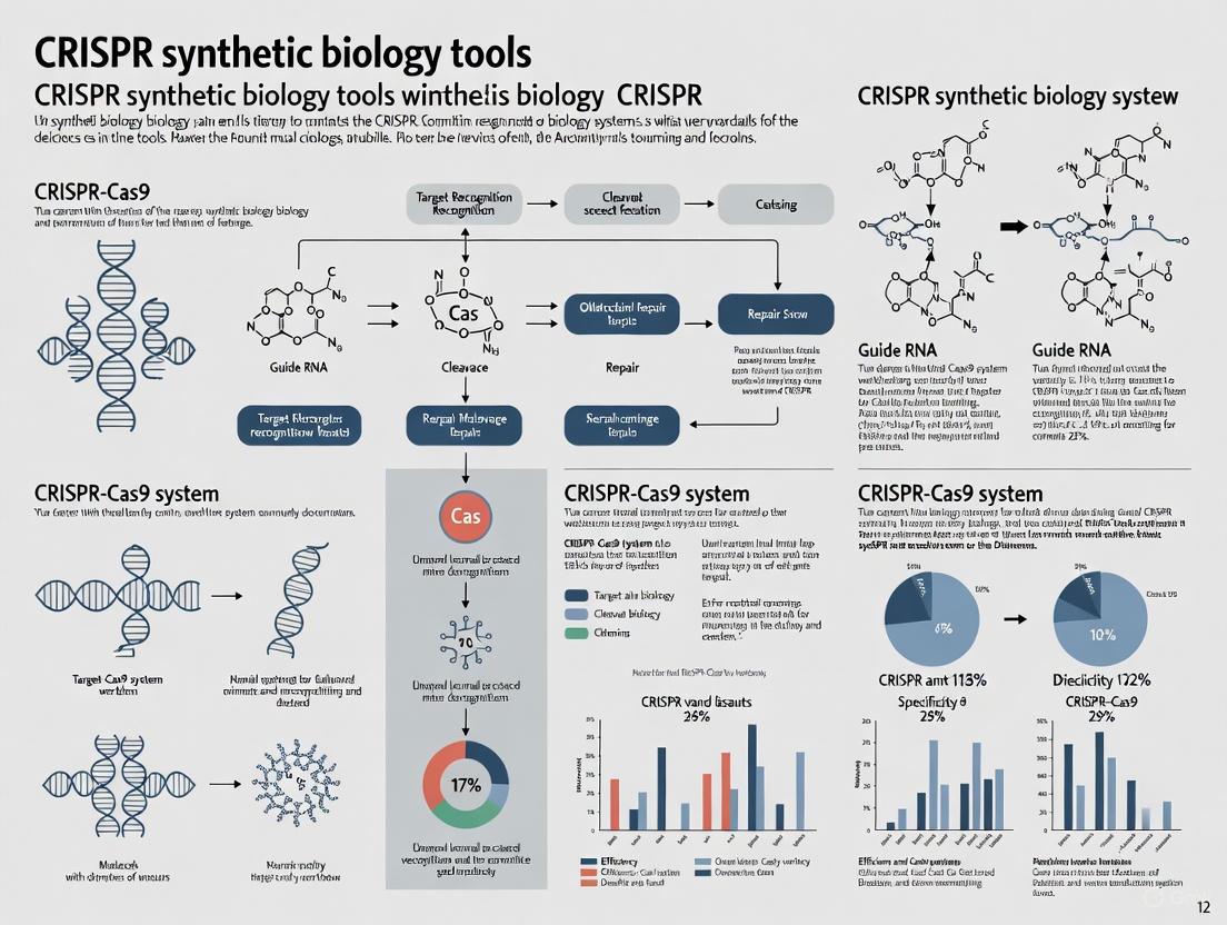
The resulting DSB activates the cell's endogenous DNA repair mechanisms, primarily non-homologous end joining (NHEJ) or homology-directed repair (HDR) [3]. NHEJ is an error-prone pathway that often results in small insertions or deletions (indels) that can disrupt gene function, making it ideal for gene knockout applications. HDR uses a donor DNA template to facilitate precise genetic modifications, enabling specific nucleotide changes or gene insertions [3]. The fundamental mechanism of CRISPR-Cas action, from sgRNA guidance to DNA repair, is illustrated in Figure 1.
Figure 1: Core CRISPR-Cas Mechanism. The diagram illustrates the sequence of events from RNP complex formation to DNA repair outcomes. sgRNA guides the Cas nuclease to target DNA, with PAM recognition enabling DNA cleavage and subsequent repair via NHEJ or HDR pathways.
Advanced Cas Engineering
Protein engineering approaches have generated numerous Cas variants with enhanced properties. High-fidelity variants (e.g., SpCas9-HF1, eSpCas9, HypaCas9) contain mutations that reduce non-specific DNA contacts, minimizing off-target effects [2]. Engineered Cas variants with altered PAM specificities (e.g., SpG and SpRY) have broadened the range of editable genomic locations by recognizing non-canonical PAM sequences [3]. Catalytically dead Cas proteins (dCas9/dCas12) lack nuclease activity but retain DNA-binding capability, serving as programmable scaffolds for transcriptional modulators, epigenetic editors, and imaging reagents [2].
PAM Requirements
PAM Recognition and Specificity
The protospacer adjacent motif (PAM) is a short DNA sequence (typically 2-5 nucleotides) adjacent to the target site that is essential for Cas protein recognition and activation [3]. The PAM requirement serves as a self versus non-self discrimination mechanism in native CRISPR systems, preventing autoimmunity by ensuring that the Cas nuclease only targets foreign DNA sequences that flank the appropriate PAM [1]. Different Cas proteins have distinct PAM requirements that fundamentally constrain their targeting range:
Table 2: PAM Requirements and Targeting Range of Cas Variants
| Cas Variant | Canonical PAM | Targeting Frequency | Sequence Constraints | Notable Applications |
|---|---|---|---|---|
| SpCas9 | 5'-NGG-3' [3] | ~1 in 8 bp in human genome [2] | Strict NGG requirement | Most widely used; broad applications |
| Cas12a | 5'-TTTV-3' [2] | Prefers T-rich regions | T-rich PAM useful for specific genomes | Efficient multiplexing; staggered cuts |
| SpG | 5'-NGN-3' [3] | ~1 in 4 bp | Relaxed PAM requirement | Increased targeting range |
| SpRY | 5'-NRN > NYN-3' [3] | ~1 in 2 bp | Near PAM-less | Maximum targeting flexibility |
| CasMINI | Compact variant [2] | Varies by engineering | Optimized for size | Viral delivery applications |
PAM-Dependent Activation Mechanism
PAM recognition triggers conformational changes in the Cas protein that facilitate DNA melting and subsequent R-loop formation. Upon PAM identification, the Cas protein undergoes structural rearrangements that displace the non-target DNA strand, enabling the sgRNA to form a heteroduplex with the target strand [1]. This process, known as R-loop formation, positions the DNA scissile bonds within the Cas protein's catalytic centers, activating cleavage activity only when full complementarity exists between the sgRNA spacer and target DNA [1].
The stringency of PAM recognition varies among Cas orthologs, with some exhibiting strict requirements while others tolerate degenerate sequences. This fundamental mechanism ensures that DNA cleavage occurs exclusively at sites flanked by the appropriate PAM sequence, providing a crucial safety mechanism that restricts Cas activity to defined genomic contexts [3].
Experimental Protocols for CRISPR Validation
sgRNA Design and Validation Workflow
Robust CRISPR experiments require careful sgRNA design and thorough validation. The following protocol outlines a standardized workflow for sgRNA design and experimental validation:
- Target Identification: Select target genomic region based on experimental goals (e.g., coding exons for knockouts, specific nucleotides for base editing)
- PAM Identification: Scan target region for available PAM sequences compatible with selected Cas nuclease
- sgRNA Design: Design 3-5 candidate sgRNAs using computational tools (e.g., CRISPR Guide Design Tool [4]) with the following criteria:
- Synthesis and Cloning: Synthesize sgRNA sequences and clone into appropriate expression vectors
- Delivery: Transfect target cells with Cas and sgRNA expression constructs using appropriate methods (electroporation, lipofection, viral delivery)
- Validation: Assess editing efficiency 48-72 hours post-transfection using tracking of indels by decomposition (TIDE) or next-generation sequencing
Editing Efficiency Analysis with ICE
The Inference of CRISPR Edits (ICE) tool provides a standardized method for analyzing CRISPR editing efficiency from Sanger sequencing data [5]. The ICE protocol involves:
Sample Preparation:
- Extract genomic DNA from edited and control cells
- PCR-amplify target region using flanking primers
- Purify amplicons and perform Sanger sequencing
ICE Analysis:
- Upload Sanger sequencing traces (.ab1 files) to ICE tool
- Input gRNA sequence and select appropriate nuclease
- For knock-in experiments, include donor sequence (up to 300 bp)
- Run analysis in sample-by-sample or batch mode
Data Interpretation:
- Editing Efficiency: Percentage of edited sample with non-wild type sequence
- Knockout Score: Proportion of cells with frameshift or 21+ bp indel (predicts functional knockout)
- Knock-in Score: Proportion of sequences with desired knock-in edit
- Model Fit (R²): Quality metric for sequencing data and indel distribution
ICE generates NGS-quality analysis from Sanger sequencing data at significantly reduced cost, enabling rapid assessment of editing outcomes without specialized equipment [5]. The complete experimental workflow from sgRNA design to validation is depicted in Figure 2.
Figure 2: CRISPR Experimental Workflow. The diagram outlines key stages in CRISPR experiment design and validation, from computational sgRNA design through delivery to comprehensive analysis of editing outcomes.
The Scientist's Toolkit: Essential Research Reagents
Table 3: Essential Reagents for CRISPR-Cas Research
| Reagent Category | Specific Examples | Function | Considerations |
|---|---|---|---|
| Cas Expression Systems | SpCas9, FnCas12a, dCas9 variants [2] | Effector nuclease for DNA/RNA manipulation | Choose based on PAM requirements, size constraints, and editing precision needs |
| sgRNA Design Tools | CRISPR Guide Design Tool [4], CHOPCHOP, CRISPResso [6] | Computational design of optimal guide RNAs | Prioritize guides with high on-target and low off-target scores; validate multiple guides |
| Delivery Methods | Electroporation, lipid nanoparticles, AAV vectors [2] [3] | Introduction of CRISPR components into cells | Balance efficiency with cytotoxicity; consider viral vs. non-viral approaches |
| Validation Tools | ICE [5], NGS, T7E1 assay | Assessment of editing efficiency and specificity | ICE provides cost-effective Sanger-based quantification; NGS offers comprehensive profiling |
| Control Reagents | Non-targeting sgRNAs, wild-type Cas9 | Experimental controls for specificity assessment | Essential for distinguishing specific effects from background mutations |
The core mechanisms of CRISPR-Cas systems—sgRNA guidance, Cas nuclease action, and PAM recognition—represent a powerful framework for programmable genome manipulation. The precise complementarity between sgRNA and target DNA enables unprecedented targeting specificity, while the diversity of natural and engineered Cas proteins provides researchers with a versatile toolkit for diverse applications. The PAM requirement, while historically a constraint, has driven innovation in Cas protein engineering to expand the targeting range of CRISPR systems.
Understanding these core mechanisms is essential for designing effective CRISPR experiments and interpreting results accurately. As the field advances, integration of artificial intelligence with structural biology and protein engineering continues to generate novel CRISPR systems with enhanced capabilities [7]. These advancements, coupled with improved delivery methods and validation approaches, are accelerating the translation of CRISPR technologies from basic research to therapeutic applications, ultimately fulfilling their potential to transform biology and medicine.
The clustered regularly interspaced short palindromic repeats (CRISPR) and CRISPR-associated (Cas) system functions as an adaptive immune system in bacteria and archaea, protecting them from mobile genetic elements such as viruses and plasmids [8]. This system incorporates fragments of foreign DNA (spacers) into CRISPR cassettes within the host genome, which are then transcribed and processed to create guide RNA (gRNA). These gRNAs direct Cas proteins to recognize and cleave complementary sequences in invading genetic elements, providing sequence-specific immunity [8]. Due to this programmable specificity, CRISPR-Cas systems have been repurposed as revolutionary tools for genome engineering, with applications spanning therapeutic development, agricultural biotechnology, molecular diagnostics, and synthetic biology [8] [9].
CRISPR-Cas systems are broadly classified into two main classes based on their effector module architecture [8] [10]. Class 1 systems (encompassing types I, III, IV, and VII) utilize multi-protein complexes for interference, whereas Class 2 systems (encompassing types II, V, and VI) employ single, multi-domain effector proteins for target recognition and cleavage [8] [10]. This review focuses on the Class 2 systems, which include the widely used Cas9 (type II), Cas12 (type V), and Cas13 (type VI) proteins. The modular nature of these systems, where target specificity is determined by an easily programmable RNA component, makes them exceptionally suitable for synthetic biology applications [9]. Their simplicity, cost-effectiveness, and programmability have positioned CRISPR-Cas as the foundation for next-generation genetic engineering tools that are rapidly transforming biological research and therapeutic development [8] [9].
Classification and Comparative Analysis of Cas Proteins
The expanding universe of Cas proteins now includes 2 classes, 7 types, and 46 subtypes, reflecting substantial diversity since the initial discovery of these systems [10]. This classification is based on evolutionary relationships, cas gene composition, and the architecture of effector modules [10]. For synthetic biology and therapeutic applications, the Class 2 systems are particularly valuable due to their simplicity and single-protein effectors.
Table 1: Fundamental Characteristics of Major Class 2 Cas Proteins
| Feature | Cas9 (Type II) | Cas12 (Type V) | Cas13 (Type VI) |
|---|---|---|---|
| Target Molecule | Double-stranded DNA (dsDNA) | dsDNA or single-stranded DNA (ssDNA) | Single-stranded RNA (ssRNA) |
| Key Domains | RuvC, HNH | Single RuvC-like | Two HEPN (Higher Eukaryotes and Prokaryotes Nucleotide-binding) |
| PAM/PFS Requirement | 3'-NGG (for SpCas9) | 5'-TTTV (for LbCas12a) | Protospacer Flanking Site (PFS) with low specificity |
| Guide RNA Component | crRNA + tracrRNA | crRNA only | crRNA only |
| Cleavage Mechanism | Blunt-ended double-strand breaks | Staggered double-strand breaks | RNA cleavage; non-specific collateral trans-cleavage of ssRNA upon activation |
| Collateral Activity | No | Yes (trans-cleavage of ssDNA) | Yes (trans-cleavage of ssRNA) |
Beyond the fundamental differences outlined in Table 1, these proteins exhibit distinct structural adaptations that define their functional niches. Cas9 possesses two independent nuclease domains, RuvC and HNH, which cleave the non-target and target DNA strands, respectively, resulting in a blunt-ended double-strand break [11]. In contrast, Cas12 proteins feature a single RuvC-like nuclease domain that cleaves both DNA strands, producing staggered ends with short overhangs [12]. Furthermore, upon recognition and cleavage of its target DNA, Cas12 exhibits nonspecific collateral trans-cleavage activity against single-stranded DNA (ssDNA) [12] [13]. Similarly, Cas13 recognizes and cleaves single-stranded RNA targets via its two HEPN domains and subsequently unleashes collateral trans-cleavage of non-target ssRNA molecules [8]. This collateral activity is absent in Cas9 but has been ingeniously harnessed for sensitive diagnostic tools like SHERLOCK (for Cas13) and DETECTR (for Cas12) [8] [13].
Detailed Profiles of Major Cas Proteins
Cas9: The Versatile Genome Editor
Cas9, derived from Streptococcus pyogenes (SpCas9), is the most extensively characterized and utilized CRISPR effector. Its mechanism requires two RNA components: a CRISPR RNA (crRNA) that specifies the target sequence, and a trans-activating crRNA (tracrRNA) that facilitates complex formation [11]. In practice, these are often combined into a single-guide RNA (sgRNA). Cas9 identifies its target genomic site by locating a short protospacer adjacent motif (PAM), typically 5'-NGG-3' for SpCas9, adjacent to the sequence complementary to the gRNA [11]. Upon target binding, the Cas9-gRNA complex undergoes a conformational change, positioning its HNH and RuvC nuclease domains to create a blunt-ended double-strand break (DSB) approximately 3-4 nucleotides upstream of the PAM site [11].
The cellular repair of this DSB is leveraged for genome editing. The dominant non-homologous end joining (NHEJ) pathway often results in small insertions or deletions (indels) that can disrupt gene function, enabling gene knockouts [11]. The less frequent homology-directed repair (HDR) pathway can be co-opted with an exogenous donor DNA template to introduce precise genetic modifications, such as point mutations or gene insertions [11]. The primary limitations of wild-type SpCas9 are its relatively large size (~1.4 kDa), which challenges delivery, and its potential for off-target activity [14] [15]. Consequently, extensive engineering has produced high-fidelity variants (e.g., eSpCas9, SpCas9-HF1), Cas9 nickases (Cas9n), and catalytically dead Cas9 (dCas9) [11]. The dCas9 variant, in particular, serves as a programmable DNA-binding platform for recruiting functional effectors to specific genomic loci, enabling transcriptional regulation, epigenetic modification, and live-cell imaging without altering the DNA sequence [11].
Cas12: The Compact and Multiplexable Alternative
The Cas12 family (including Cas12a/Cpf1, Cas12f, and others) offers distinct advantages and functionalities that complement and extend those of Cas9. Cas12a (Cpf1) is a representative type V effector that differs from Cas9 in several key aspects. It requires only a single crRNA for activity, lacking the need for a tracrRNA [12]. It recognizes a T-rich PAM (5'-TTTV-3') located upstream of the target sequence, and its single RuvC domain generates staggered DNA breaks with 5-8 nt overhangs [12]. A hallmark of many Cas12 proteins is their robust collateral trans-cleavage activity against ssDNA following target recognition, a feature absent in Cas9 [12] [13].
Recent research highlights the significant potential of ultra-compact Cas12 proteins, such as Cas12f (also known as Cas14), which is only ~400-700 amino acids in size (compared to ~1,360 aa for SpCas9) [14]. This small size (~552 Da for Cas12f) facilitates more efficient cellular delivery, a critical bottleneck for therapeutic applications [14]. Studies demonstrate that Cas12f ribonucleoproteins (RNPs) form smaller complexes with delivery peptides (~250 nm hydrodynamic diameter) compared to Cas9 RNPs (~1100 nm), leading to improved cellular uptake in human cells [14]. Furthermore, Cas12 systems are generally reported to exhibit lower off-target editing compared to Cas9, enhancing their safety profile for clinical applications [12]. Ongoing engineering efforts, such as the development of Flex-Cas12a through directed evolution, are successfully expanding the PAM recognition range of these effectors, thereby increasing the targetable genomic space [16].
Cas13: The RNA-Targeting Specialist
Cas13 is a Type VI effector that uniquely targets single-stranded RNA (ssRNA) rather than DNA, opening the field of RNA manipulation and diagnostics. Upon binding to its target RNA sequence via its crRNA guide, Cas13's two HEPN domains cleave the target transcript, enabling knockdown of gene expression without permanent genomic alteration [8]. Similar to Cas12, activated Cas13 exhibits potent collateral trans-cleavage activity, but against non-target ssRNA molecules [8].
This collateral RNAse activity has been ingeniously repurposed for ultrasensitive diagnostic platforms. In tools like SHERLOCK, the presence of a specific RNA target (e.g., from a pathogen) activates Cas13, which then promiscuously cleaves a reporter RNA molecule, generating a detectable fluorescent signal [8]. This principle enables attomolar-level sensitivity for detecting viral RNAs and other biomarkers. Recent advancements continue to build on this mechanism. For instance, the Target-amplification-free Collateral-cleavage-enhancing CRISPR-CasΦ (TCC) method, which utilizes the compact Cas12j (CasΦ) protein, employs a sophisticated DNA amplifier to enhance the collateral cleavage signal, achieving a record-low detection limit of 0.11 copies/μL for clinical pathogens without target pre-amplification [13]. This demonstrates the rapid evolution of CRISPR-based diagnostics toward greater sensitivity, speed, and simplicity.
Experimental Protocols and Methodologies
Protocol: Assessing Cellular Uptake of Compact vs. Large Cas Proteins
Objective: To compare the cellular delivery efficiency of large Cas9 ribonucleoproteins (RNPs) versus compact Cas12f RNPs using amphipathic peptide vectors in human cells [14].
- 1. Cell Culture: Maintain HEK293T cells in Dulbecco's Modified Eagle Medium (DMEM) supplemented with 10% fetal bovine serum (FBS) and 1% penicillin-streptomycin at 37°C in a 5% CO₂ atmosphere [14].
- 2. RNP Complex Formation:
- Express and purify recombinant Cas12f protein (e.g., from Addgene plasmid #171613) using Ni-NTA chromatography. Use commercial recombinant Cas9 as a control [14].
- Synthesize target-specific guide RNAs (e.g., from IDT).
- Form RNP complexes by incubating Cas9 or Cas12f proteins with sgRNA at a 1:1.1 molar ratio in nuclease-free duplex buffer. Incubate Cas9 at 25°C and Cas12f at 45°C for 10 minutes [14].
- 3. Complex Formation with Delivery Vector:
- Use an amphipathic peptide such as PepFect14 (PF14) or a lipid-based vector like Lipofectamine CRISPRMAX.
- Add PF14 to the RNP complexes at varying molar ratios (e.g., 1:50, 1:100 RNP:PF14) in HEPES-buffered glucose solution. Vortex immediately and incubate for 40 minutes at room temperature to form RNP/PF14 complexes [14].
- 4. Transfection:
- Seed HEK293T cells in a 96-well plate at 15,000 cells per well 24 hours before transfection.
- Add RNP/PF14 complexes (containing 1.2 pmol RNP) directly to the cells [14].
- 5. Evaluation of Cellular Uptake:
- Use a guide RNA fluorescently labeled with ATTO550.
- At 6 and 24 hours post-transfection, analyze cells using fluorescence microscopy and quantify uptake via flow cytometry (e.g., using a BD FACSAria flow cytometer with a PE-CF594 filter) [14].
- Express results as the percentage of cells with uptake and the mean fluorescence intensity ratio (MFIR) compared to untreated controls [14].
The following workflow diagram illustrates this experimental process:
Protocol: Qualitative and Quantitative PCR Detection of Cas12a (Cpf1)
Objective: To establish specific and sensitive qualitative PCR and quantitative PCR (qPCR) assays for detecting the presence of the Cas12a (Cpf1) transgene in gene-edited products [12].
- 1. Sample Preparation:
- Prepare samples from gene-edited materials (e.g., cotton, rice) and non-edited controls.
- Create calibrated mixtures with known mass fractions of gene-edited material (e.g., 100%, 10%, 1%, 0.1%, 0.05%) for sensitivity testing [12].
- 2. DNA Extraction:
- Weigh 100 mg of plant powder and extract genomic DNA using a commercial plant DNA extraction kit according to the manufacturer's instructions [12].
- 3. Qualitative PCR:
- Reaction System (25 µL): 2.5 µL 10× PCR buffer (Mg²⁺ Plus), 2 µL dNTP mixture, 0.5 µL each of forward and reverse primers (10 µmol/L) specific to the Cpf1 gene, 0.2 µL Taq DNA polymerase, and 2 µL template DNA [12].
- Amplification Program: Initial denaturation at 95°C for 5 min; 35 cycles of 95°C for 30 s, optimal annealing temperature (e.g., 60°C) for 30 s, 72°C for 30 s; final extension at 72°C for 5-10 min [12].
- Analysis: Analyze PCR products by agarose gel electrophoresis and gel imaging [12].
- 4. Quantitative PCR (qPCR):
- Reaction System (20 µL): 10 µL Fast Start Essential DNA Probes Master, 0.4 µL each of forward and reverse primers, 0.2 µL probe, 2 µL template DNA, and nuclease-free water to 20 µL [12].
- Amplification Program: Pre-incubation at 95°C for 10 min; 45 cycles of 95°C for 10 s and 60°C for 30 s [12].
- Data Analysis: Determine the cycle threshold (Ct) values and analyze using the standard curve method to determine copy numbers [12].
Table 2: Key Reagents for CRISPR-Cas Experimental Work
| Reagent / Material | Function / Description | Example Sources / Notes |
|---|---|---|
| Recombinant Cas Protein | The core effector nuclease (e.g., Cas9, Cas12f) for genome editing. | New England Biolabs (NEB); expressed and purified from plasmids (e.g., Addgene) [14]. |
| Guide RNA (gRNA/crRNA) | Synthetic RNA that confers target specificity to the Cas protein. | Integrated DNA Technologies (IDT); can be chemically modified to enhance stability and reduce off-targets [14] [15]. |
| Amphipathic Peptides (e.g., PF14) | Non-viral delivery vector that forms nanosized complexes with RNPs for cellular internalization. | Pepscan; binds negatively charged RNPs via cationic hydrophilic portion, facilitates endocytosis via hydrophobic section [14]. |
| Lipid-Based Vectors (e.g., Lipofectamine CRISPRMAX) | Commercial lipid nanoparticles for delivering CRISPR components into cells. | Thermo Fisher Scientific; standard for in vitro transfection [14]. |
| Plant DNA Extraction Kit | For isolating high-quality genomic DNA from plant tissues for genotyping and transgene detection. | Tiangen Biochemical Technology Co., Ltd. [12]. |
| Fast Start Essential DNA Probes Master | Optimized master mix for quantitative real-time PCR (qPCR) using hydrolysis probes. | Roche; used for sensitive and specific detection of transgene copy numbers [12]. |
Technical Considerations and Challenges
Off-Target Effects: Prediction and Mitigation
A primary technical challenge in applying CRISPR technologies, particularly for therapeutic purposes, is off-target editing—the non-specific activity of the Cas nuclease at sites other than the intended target [15]. This can confound experimental results and poses significant safety risks in clinical settings, potentially leading to oncogenic mutations if key genes are disrupted [15].
Multiple strategies have been developed to minimize off-target effects:
- gRNA Design Optimization: The simplest strategy involves careful selection of gRNAs using design software (e.g., CRISPOR) that ranks guides based on predicted on-target efficiency and off-target potential. Guides with high GC content and minimal similarity to other genomic sites are preferred [15].
- High-Fidelity Cas Variants: Engineered nucleases like eSpCas9(1.1) and SpCas9-HF1 have mutations that reduce off-target editing by weakening non-specific interactions with DNA [15] [11].
- RNP Delivery: Delivering pre-assembled Cas protein-gRNA complexes (ribonucleoproteins, RNPs) instead of plasmid DNA results in transient activity, reducing the time window for off-target cleavage [14] [15].
- Modified gRNAs: Chemical modifications, such as 2'-O-methyl analogs (2'-O-Me) and 3' phosphorothioate bonds (PS), can increase gRNA stability and reduce off-target interactions [15].
- Alternative Cas Proteins: Cas12 proteins often demonstrate different off-target profiles compared to Cas9, and their compact size may offer advantages [14] [12].
Delivery Challenges and the Promise of Compact Cas Proteins
Efficient delivery of CRISPR machinery into target cells remains a major obstacle for clinical translation [14]. The large size of commonly used effectors like SpCas9 (~1.4 kDa) challenges packaging into delivery vehicles with limited cargo capacity, such as adeno-associated viruses (AAVs) [14].
The discovery and engineering of ultra-compact Cas proteins (e.g., Cas12f at ~552 Da, Cas12k at ~639 Da) provide a promising solution to this delivery bottleneck [14]. Studies directly comparing cellular uptake demonstrate that Cas12f RNPs form smaller, more uniform complexes with peptide-based delivery vectors and exhibit significantly enhanced cellular penetration compared to bulkier Cas9 RNPs [14]. This improved uptake efficiency, combined with ongoing efforts to optimize the editing efficiency of these miniature systems, positions them as highly attractive candidates for the next generation of CRISPR-based therapies where delivery is a critical limiting factor [14].
The expanding universe of Cas proteins, encompassing DNA-targeting Cas9 and Cas12, and RNA-targeting Cas13, has provided researchers and clinicians with an unprecedentedly versatile and powerful toolkit for genetic manipulation. Each protein family offers a unique combination of characteristics—size, cleavage mechanism, PAM requirement, and collateral activities—that makes it suited for specific applications, from therapeutic genome editing and multiplexed gene regulation to ultrasensitive molecular diagnostics [14] [8] [12].
The future of this field lies in continued exploration and engineering. The discovery of rare, uncharacterized CRISPR-Cas variants from the "long tail" of prokaryotic diversity promises to yield new tools with novel functionalities [10]. Simultaneously, directed evolution and rational protein design are rapidly overcoming current limitations, such as PAM restrictions and off-target effects, as exemplified by the development of PAM-flexible Cas12a variants [16]. The integration of these refined CRISPR tools with synthetic biology principles and advanced delivery platforms like programmable nanomedicines will undoubtedly accelerate the development of sophisticated genetic circuits and transformative cell and gene therapies, solidifying the central role of CRISPR-Cas technologies in shaping the future of biotechnology and medicine [9] [17].
Programmable nucleases represent a cornerstone of modern genetic engineering, enabling precise, site-specific modifications to the genomes of a wide variety of organisms. These molecular tools have revolutionized life science research and therapeutic development by facilitating targeted DNA double-strand breaks (DSBs) that harness cellular repair mechanisms to achieve desired genetic outcomes [18]. The evolution of these technologies—from zinc finger nucleases (ZFNs) and transcription activator-like effector nucleases (TALENs) to the clustered regularly interspaced short palindromic repeats (CRISPR)-CRISPR-associated protein 9 (Cas9) systems and their derivatives—has progressively enhanced the precision, efficiency, and accessibility of genome editing [18]. This progression is marked by key technological milestones that have expanded the capabilities and applications of programmable nucleases, ultimately paving the way for their use in clinical settings, with the first CRISPR-based medicine, Casgevy, receiving approval in late 2023 [19] [20]. This review details the discovery and development of these foundational technologies, framing them within the broader context of CRISPR synthetic biology tools.
The Fundamental Classes of Programmable Nucleases
Programmable nucleases function by creating targeted DNA double-strand breaks, which are subsequently repaired by the cell's endogenous mechanisms, primarily non-homologous end joining (NHEJ) or homology-directed repair (HDR). The NHEJ pathway often results in insertion/deletion (indel) mutations that can disrupt gene function, while HDR allows for the precise integration of a donor DNA sequence [18]. Four major classes of programmable nucleases have been developed, each with distinct architectures and mechanisms.
Zinc Finger Nucleases (ZFNs)
ZFNs were among the first programmable nucleases developed. A ZFN is a chimeric protein composed of a DNA-binding domain—a zinc finger protein—fused to the non-specific DNA cleavage domain of the FokI restriction enzyme [18]. Each zinc finger typically recognizes three DNA bases, and an array of 3-6 fingers is used to recognize a sequence of 9-18 bases. Because the FokI domain must dimerize to become active, a pair of ZFNs is required to bind to opposite DNA strands with appropriate spacing and orientation to generate a DSB at the intended target locus [18]. This requirement enhances specificity, as cleavage only occurs when both ZFNs bind correctly.
Transcription Activator-Like Effector Nucleases (TALENs)
TALENs followed ZFNs and share a similar modular structure, comprising a DNA-binding domain derived from transcription activator-like effectors (TALEs) fused to the FokI nuclease domain [18]. The DNA-binding domain consists of a series of highly conserved 33-35 amino acid repeats. The specificity of each repeat is determined by two variable amino acids at positions 12 and 13, known as the repeat variable diresidue (RVD). Each RVD recognizes a single DNA base [18]. Like ZFNs, TALENs function as pairs, with their binding sites flanking the target sequence to allow FokI dimerization and DSB formation. The simpler code for DNA recognition (one RVD to one base pair) made TALENs easier to engineer for novel targets compared to ZFNs.
CRISPR-Cas9 Systems
The discovery and adaptation of the CRISPR-Cas9 system marked a paradigm shift in genome editing. Unlike ZFNs and TALENs, which rely on protein-DNA interactions for targeting, CRISPR-Cas9 uses a RNA-DNA recognition system. The system consists of two key components: the Cas9 nuclease and a single guide RNA (gRNA) [18]. The gRNA is a short RNA molecule comprising a 5' 17-20 nucleotide sequence that is complementary to the target DNA and a 3' end that forms a structure recognized by the Cas9 protein. Cas9 is directed by the gRNA to the target site, where it creates a DSB. A critical requirement for Cas9 binding is the presence of a short protospacer adjacent motif (PAM) sequence immediately downstream of the target site on the non-complementary DNA strand [18]. The simplicity of programming the system by designing a new gRNA, without the need for complex protein engineering, made CRISPR-Cas9 vastly more accessible and scalable.
Base Editors
A more recent advancement is the development of base editors, which enable precise single-nucleotide changes without creating a DSB, thereby reducing unwanted indel mutations [18]. Base editors are fusion proteins that typically consist of a catalytically impaired Cas9 (a nickase, nCas9, that cuts only one DNA strand) linked to a deaminase enzyme. Cytosine base editors (CBEs) convert a cytosine to a thymine, while adenine base editors (ABEs) convert an adenine to a guanine [18]. By avoiding DSBs, base editors offer a safer profile for therapeutic applications where minimizing genomic instability is crucial. Prime editors, which use a Cas9 nickase fused to an engineered reverse transcriptase and are programmed with a prime editing guide RNA (pegRNA), represent a further evolution capable of making all 12 possible base-to-base conversions, as well as small insertions and deletions, but are beyond the scope of this foundational review [18].
Table 1: Comparison of Major Programmable Nuclease Systems
| Feature | ZFNs | TALENs | CRISPR-Cas9 | Base Editors |
|---|---|---|---|---|
| Targeting Moiety | Protein (Zinc Fingers) | Protein (TALE Repeats) | RNA (gRNA) | RNA (gRNA) |
| Cleavage Domain | FokI | FokI | Cas9 | Cas9 nickase + Deaminase |
| Recognition Code | ~3 bp per zinc finger | 1 bp per RVD | 17-20 nt gRNA sequence | 17-20 nt gRNA sequence |
| Nuclease Pairs Required | Yes | Yes | No | No |
| PAM Requirement | No | No | Yes (e.g., NGG for SpCas9) | Yes |
| Primary Editing Outcome | Indels (NHEJ) / HDR | Indels (NHEJ) / HDR | Indels (NHEJ) / HDR | Point mutations (C>T or A>G) |
| Key Advantage | First programmable nucleases | Simpler design than ZFNs | High simplicity & multiplexability | DSB-free, precise point mutation |
Experimental Workflows and Protocols
The application of programmable nucleases involves a standardized workflow, from design to validation. The following protocol outlines the key steps for a typical CRISPR-Cas9 gene knockout experiment, which can be adapted for other nuclease platforms.
Protocol: CRISPR-Cas9 Mediated Gene Knockout
1. Target Selection and gRNA Design:
- Identify Target Gene: Select a coding exon near the 5' end of the gene to maximize the likelihood of generating a null allele via frameshift mutations.
- Design gRNA Sequences: Use in silico tools (e.g., CRISPRscan, ChopChop) to identify 20-nucleotide target sequences adjacent to a PAM (e.g., 5'-NGG-3' for Streptococcus pyogenes Cas9).
- Predict Off-Targets: Utilize bioinformatics tools (e.g., Cas-OFFinder) to identify and rank potential off-target sites across the genome. Select gRNAs with minimal predicted off-target activity [18].
- Order gRNAs: Synthesize the selected gRNA sequence as a single-guide RNA (sgRNA) or as a crRNA:tracrRNA duplex.
2. Delivery of CRISPR Components:
- Choose Delivery Method: Transfert cells with a plasmid encoding Cas9 and the gRNA, mRNA encoding Cas9 plus the gRNA, or pre-assembled Cas9-gRNA ribonucleoprotein (RNP) complexes.
- Optimize Transfection: Use appropriate methods (electroporation, lipofection, nucleofection) optimized for the specific cell type. RNP delivery is often favored for its high efficiency and reduced off-target effects.
3. Analysis of Editing Efficiency:
- Harvest Genomic DNA: Collect genomic DNA from transfected cells 48-72 hours post-transfection.
- Assess Indel Formation:
- T7 Endonuclease I or Surveyor Assay: PCR-amplify the target region, denature and reanneal the amplicons. Mismatches from heteroduplex DNA formed by indels are cleaved by the nuclease. Analyze fragments by gel electrophoresis.
- Tracking of Indels by Decomposition (TIDE): A commonly used method that decomposes Sanger sequencing data of the PCR-amplified target region from a mixed population to quantify the spectrum and frequency of indels [18].
- Next-Generation Sequencing (NGS): For the most accurate and comprehensive assessment, perform targeted amplicon sequencing of the genomic region. This is considered the gold standard for quantifying editing efficiency and characterizing the spectrum of mutations [18].
4. Off-Target Analysis:
- In Silico Prediction: Use the gRNA sequence to run predictions of potential off-target sites.
- Experimental Validation: For candidate off-target sites identified in silico, perform targeted amplicon-based NGS to definitively quantify off-target editing frequencies [18]. More comprehensive, unbiased methods like CIRCLE-seq or GUIDE-seq can be used for genome-wide off-target profiling.
Diagram 1: CRISPR Gene Editing Workflow
The Research Toolkit: Essential Reagents and Solutions
Successful genome editing experiments rely on a core set of reagents and tools. The table below details the essential components of a researcher's toolkit for CRISPR-based work.
Table 2: Key Research Reagent Solutions for CRISPR Experimentation
| Reagent / Solution | Function / Description | Key Considerations |
|---|---|---|
| Cas9 Nuclease | The enzyme that creates the double-strand break at the target DNA site. | Available as protein, mRNA, or expression plasmid. High-fidelity variants (e.g., SpCas9-HF1) reduce off-target effects [21]. |
| Guide RNA (gRNA) | The RNA molecule that directs Cas9 to the specific genomic locus. | Can be supplied as a single-guide RNA (sgRNA) or a two-part system (crRNA + tracrRNA). Chemical modifications can enhance stability [18]. |
| Delivery Vectors | Plasmids or viral vectors (e.g., lentivirus, AAV) for delivering Cas9 and gRNA coding sequences into cells. | Choice depends on cell type, efficiency, and application (e.g., AAV has a small cargo capacity). |
| Lipid Nanoparticles (LNPs) | Synthetic nanoparticles for delivering Cas9-gRNA RNPs or mRNA in vivo. | Particularly effective for liver-targeted delivery, as used in several clinical trials [19] [20] [22]. |
| Cell Culture Media | Formulated media to support the growth and viability of the cells being edited. | Must be optimized for the specific cell type (e.g., primary T-cells, stem cells). |
| Transfection Reagent | Chemical agents (e.g., lipofectamine, PEI) to facilitate the uptake of nucleic acids or proteins into cells. | Efficiency and toxicity vary greatly by cell type; requires optimization. |
| Electroporation System | Device that uses electrical pulses to create transient pores in cell membranes for reagent delivery. | Often the most efficient method for hard-to-transfect cells like primary cells. |
| PCR Reagents | Enzymes and mixes for amplifying the target genomic region for analysis. | High-fidelity polymerases are recommended to avoid introducing errors during amplification. |
| NGS Library Prep Kit | Commercial kits for preparing sequencing libraries from amplified target sites. | Essential for high-sensitivity, quantitative analysis of on-target and off-target editing [18]. |
Key Milestones and the Current Clinical Landscape
The trajectory of programmable nucleases from basic research tools to clinical therapeutics is marked by pivotal achievements. The first approval of a CRISPR-based medicine, CASGEVY (exagamglogene autotemcel [exa-cel]) for sickle cell disease (SCD) and transfusion-dependent beta thalassemia (TDT) in late 2023, stands as a landmark validation of the technology [19] [20]. This ex vivo therapy involves editing a patient's own hematopoietic stem cells to produce high levels of fetal hemoglobin [19].
The clinical landscape in 2025 is dynamic, with key milestones anticipated across several companies and therapeutic areas [19]. CRISPR Therapeutics highlights ongoing progress with CASGEVY, including the activation of over 50 authorized treatment centers globally and successful reimbursement agreements with payers [19]. The pipeline is expanding into new areas:
- Oncology & Autoimmune Diseases: CTX112, a next-generation allogeneic CAR-T cell therapy targeting CD19, has shown strong efficacy in B-cell malignancies and is being explored for autoimmune diseases like lupus. Updates are expected in mid-2025 [19].
- In Vivo Cardiovascular Therapies: Programs like CTX310 (targeting ANGPTL3 for hypercholesterolemia) and CTX320 (targeting Lp(a)) are advancing, with dose escalation updates expected in the first half of 2025 [19].
- Rare Genetic Diseases: Intellia Therapeutics is progressing in vivo therapies for hereditary transthyretin amyloidosis (hATTR) and hereditary angioedema (HAE), both delivered via lipid nanoparticles (LNPs) to the liver [20]. Recent results for their HAE treatment showed an 86% reduction in the disease-driving kallikrein protein and a significant reduction in attacks [20].
A major recent breakthrough was the first personalized, in vivo CRISPR base editing therapy for an infant with the rare genetic disorder CPS1 deficiency. The treatment was developed, approved, and delivered in just six months, demonstrating the potential for rapid, bespoke genetic medicine [20] [22]. This case also underscored the utility of LNP delivery, which allowed for multiple, safe therapeutic doses [20].
Diagram 2: Progression to Clinical Application
Critical Considerations and Future Directions
Despite rapid progress, significant challenges remain. Off-target effects—editing at unintended genomic sites—continue to be a primary safety concern [18]. A combination of in silico prediction tools and sensitive experimental assays, particularly amplicon-based NGS, is recommended for comprehensive off-target profiling [18]. Delivery of editing components to the correct tissues in the body ("in vivo delivery") is another major hurdle, though LNPs have emerged as a leading solution for liver-directed therapies [20] [22].
The field is now moving "beyond cutting" into a new era of CRISPR-driven synthetic biology [21]. Catalytically dead Cas9 (dCas9) serves as a programmable scaffold for transcriptional activators (CRISPRa) or repressors (CRISPRi), allowing precise control of gene expression without altering the DNA sequence [21]. Furthermore, the integration of artificial intelligence (AI) is set to accelerate the field. Tools like CRISPR-GPT, a large language model developed at Stanford Medicine, can assist scientists in designing experiments, predicting outcomes, and troubleshooting, thereby flattening the learning curve and speeding up the development of new therapies [23].
In conclusion, the journey of programmable nucleases from ZFNs to the current CRISPR-based toolkit demonstrates a relentless pursuit of precision and utility in genome engineering. These technologies have not only transformed basic research but have also begun to deliver on the promise of curative genetic therapies, setting the stage for a future where genetic diseases can be addressed with unprecedented precision and flexibility.
The Clustered Regularly Interspaced Short Palindromic Repeats (CRISPR) and CRISPR-associated (Cas) systems have revolutionized genetic engineering, yet the field has predominantly relied on a handful of well-characterized effectors like Cas9 and Cas12a. The true diversity of CRISPR systems, however, extends far beyond these common tools, encompassing rare variants from transposon families like IS200/IS605 and ultra-compact editors such as Cas12f. These novel systems offer unique advantages—including dramatically reduced size for viral delivery and distinct mechanistic properties—that address critical limitations in therapeutic and biotechnological applications.
Recent advances in computational biology and protein engineering have enabled the discovery and optimization of these rare systems. This review synthesizes current knowledge on mining rare CRISPR systems and engineering hypercompact editors, providing researchers with a technical framework for leveraging these emerging tools. We present updated classification schemas, detailed experimental protocols for characterizing new systems, and performance data for engineered Cas12f variants, contextualizing these developments within the broader landscape of CRISPR synthetic biology.
Updated Classification and Diversity of CRISPR-Cas Systems
Expanded Taxonomy of CRISPR Systems
The evolutionary classification of CRISPR-Cas systems has recently undergone significant expansion, now encompassing 2 classes, 7 types, and 46 subtypes—a substantial increase from the 6 types and 33 subtypes recognized just five years ago [10]. This updated taxonomy reflects the accelerating discovery of rare and previously uncharacterized systems through advanced bioinformatics approaches applied to massive genomic and metagenomic datasets.
Class 1 systems (utilizing multi-protein effector complexes) now include the newly characterized type VII, which features a unique metallo-β-lactamase (β-CASP) effector nuclease designated Cas14 [10]. Type VII systems are found predominantly in diverse archaeal genomes and operate as RNA-targeting systems despite their evolutionary relationship to DNA-targeting type III systems. Additionally, three new type III subtypes (III-G, III-H, and III-I) have been formally described, each exhibiting features suggestive of reductive evolution [10].
Class 2 systems (utilizing single-protein effectors) continue to expand with newly characterized variants of established types and the exploration of their evolutionary ancestors, particularly the transposon-associated TnpB systems of the IS200/IS605 family [24]. These TnpB proteins, now collectively termed OMEGA (Obligate Mobile Element-Guided Activity) systems, represent the compact progenitors from which Cas12 effectors evolved and are emerging as valuable genome-editing tools in their own right [24].
Table 1: Recently Characterized CRISPR-Cas Systems and Their Features
| System | Class | Effector | Size | Target | Key Features |
|---|---|---|---|---|---|
| Type VII | 1 | Cas14 complex | ~12 subunits | RNA | Metallo-β-lactamase nuclease; likely evolved from type III [10] |
| Type III-G | 1 | Cas10-Cas7 complex | Multi-subunit | DNA (predicted) | Lost cOA signaling; lacks adaptation module [10] |
| Type III-H | 1 | Cas10-Cas7 complex | Multi-subunit | DNA (predicted) | Highly diverged Cas11; distantly related to III-F [10] |
| Type III-I | 1 | Cas7-11i fusion | Multi-subunit | RNA | Cas7-Cas11 fusion protein; independent origin from III-E [10] |
| Cas12f (V-F) | 2 | AsCas12f, Un1Cas12f | 400-700 aa | dsDNA | Naturally hypercompact; dimeric architecture [25] |
| TnpB/OMEGA | - | ISDra2 TnpB | ~400 aa | dsDNA | Cas12 ancestor; ωRNA-guided; ultra-compact [24] |
Functional Diversity and Novel Mechanisms
Beyond taxonomic expansion, recent studies have revealed remarkable functional diversity among rare CRISPR systems, including non-canonical interference mechanisms that expand their biotechnological potential:
- DNA cleavage-deficient variants: Certain type IV and type V systems have been identified that inhibit target replication without cleaving DNA, suggesting novel regulatory functions [10].
- HNH nuclease integrations: Variants of I-E, I-F, and IV systems have been discovered that incorporate HNH nucleases fused to Cas5, Cas8f, and CasDinG proteins, respectively, enabling robust crRNA-guided double-stranded DNA cleavage [10].
- Type IV systems with specified interference: Recent work has identified the first type IV system with a defined interference mechanism, incorporating an HNH nuclease [26].
- Candidate RNA-targeting systems: A candidate type VII system has been experimentally confirmed to act on RNA targets, expanding the RNA-targeting capabilities of CRISPR systems beyond the established Cas13 family [26].
Mining Rare CRISPR Systems: IS200/IS605 and Beyond
Computational Discovery Pipelines
The identification of rare CRISPR systems from genomic and metagenomic data requires specialized computational approaches that overcome the limitations of traditional homology-based methods. The FLSHclust (Fast Locality-Sensitive Hashing-based clustering) algorithm represents a breakthrough in this domain, enabling deep clustering on terabyte-scale datasets with linearithmic time complexity [O(N logN)] rather than the quadratic scaling [O(N²)] that renders all-against-all comparisons impractical for billions of protein sequences [26].
The FLSHclust-based CRISPR discovery pipeline involves several key stages:
- Dataset Curation: Compilation of 8.8 Tbp of prokaryotic genomic and metagenomic contigs from NCBI, WGS, and JGI sources, excluding contigs <2 kbp to minimize fragmentation artifacts [26].
- Coding Sequence Prediction: Application of Genemark for gene calling across the curated dataset, generating approximately 8 billion candidate proteins [26].
- CRISPR Array Identification: Implementation of multiple CRISPR finders (including PILER-CR, CRT, MinCED) complemented by CRONUS, a specialized tool developed to detect smaller CRISPR arrays with imperfect repeats and hypervariable spacers that conventional tools often miss [26].
- Deep Protein Clustering: Iterative clustering of all proteins using FLSHclust at 30% sequence identity, generating 499.9 million deep clusters that capture evolutionary relationships beyond immediate homology [26].
- CRISPR Association Scoring: Calculation of both naive and enhanced CRISPR association scores for each cluster, quantifying the weighted fraction of non-redundant proteins encoded within 3 kbp of a CRISPR array while adjusting for contig truncations in metagenomic data [26].
Figure 1: FLSHclust Computational Workflow for Protein Clustering
This pipeline has enabled the discovery of 188 previously unreported CRISPR-linked gene modules, revealing extensive biochemical functionality coupled to adaptive immunity beyond the canonical CRISPR mechanisms [26]. The application of FLSHclust to CRISPR discovery demonstrates how algorithmic innovations can unlock the functional diversity hidden within massive sequence databases.
Experimental Characterization of Novel Systems
Computational identification represents only the first step; functional validation is essential to confirm the activity and mechanism of newly discovered systems. The following experimental protocol provides a framework for characterizing candidate CRISPR systems:
Protocol: Functional Validation of Novel CRISPR Systems
Locus Reconstruction and Synthesis
- Clone candidate CRISPR arrays and associated cas genes into expression vectors under inducible promoters.
- For systems lacking identifiable adaptation modules, provide CRISPR arrays in trans or utilize pre-crRNA expression constructs.
- Include appropriate control constructs with inactivated nuclease domains (e.g., D-to-A mutations in catalytic residues).
Interference Assay Establishment
- Co-transform interference constructs with target plasmids containing protospacer sequences flanked by appropriate PAM sequences.
- Include mismatched controls to assess targeting specificity.
- Measure interference efficiency through antibiotic resistance markers or fluorescence reporters.
Biochemical Characterization
- Heterologously express and purify candidate effector complexes.
- Perform in vitro cleavage assays with synthetic crRNAs and target substrates (dsDNA, ssDNA, RNA).
- Determine cleavage specificity and kinetics via gel electrophoresis and real-time fluorescence assays.
Structural Analysis
- Determine cryo-EM structures of effector complexes bound to crRNA and target substrates.
- Identify key catalytic residues and conformational changes through structural alignment with characterized systems.
- Utilize structure-guided mutagenesis to confirm mechanistic hypotheses.
This approach has successfully characterized multiple novel systems, including three HNH nuclease-containing CRISPR systems (one type IV system with a defined interference mechanism) and a candidate type VII system with RNA-targeting activity [26].
Engineering Compact CRISPR Editors: The Cas12f Revolution
Natural Diversity and Properties of Cas12f Systems
Cas12f systems (formerly known as Cas14) represent the most compact RNA-guided DNA endonucleases currently known, with sizes ranging from 400-700 amino acids—approximately one-third the size of SpCas9 [25]. These naturally hypercompact systems include:
- Un1Cas12f1 (529 aa): Originally identified from an uncultured archaeon; the foundation for early engineering efforts.
- AsCas12f1 (422 aa): Derived from Acidibacillus sulfuroxidans; exhibits robust activity in eukaryotic cells.
- OsCas12f1 (433 aa): From Oscillibacter sp.; offers balanced size and activity profile.
- RhCas12f1 (415 aa): From Ruminiclostridium herbifermentans; among the most compact functional variants.
Despite their small size, Cas12f systems operate through a unique dimeric architecture where two protomers adopt distinct conformations to facilitate DNA recognition and cleavage [27]. This quaternary structure enables dsDNA targeting despite the minimal size of individual subunits. Naturally occurring Cas12f systems typically recognize T-rich PAM sequences (TTN or CCN) and generate staggered DNA cuts with 5-nt overhangs [25].
Engineering Strategies for Enhanced Cas12f Activity
While naturally compact, wild-type Cas12f systems typically exhibit modest editing efficiency compared to larger Cas effectors. Multiple protein engineering approaches have successfully enhanced their activity while maintaining their small size:
1. Electrostatic Optimization
- Rationale: Introduction of basic residues (lysine, arginine) at positions occupied by neutral or negatively charged residues in wild-type proteins increases affinity for negatively charged nucleic acid backbones [25].
- Implementation: Comparative analysis with natural homologs identified 32 candidate positions for mutation in AsCas12f, with eight single-point mutations (D196K, N199K, G276R, D281K, T327K, N328G, D364K, D364R) demonstrating increased activity [25].
- Optimal Combination: The quintuple mutant AsCas12f-v5.2 (D196K/N199K/G276R/N328G/D364R), designated enAsCas12f, shows 2.5- to 3.5-fold higher editing efficiency than wild-type while maintaining specificity [25].
2. Structural Domain Augmentation
- Rationale: Comparison with more active but larger Cas12a effectors revealed missing structural elements in Cas12f, particularly an N-terminal α-helix in the WED domain that stabilizes the crRNA-target DNA complex [27].
- Implementation: Fusion of engineered α-helical peptides (Gp41S2, GCN4-2.5H) to the N-terminus of CasMINI (an engineered Un1Cas12f1) via optimized linkers [27].
- Optimal Construct: The hpCasMINI system (Gp41S2 with EAAAK linker) demonstrates 1.4-3.0-fold enhanced gene activation and 1.1-19.5-fold improved DNA cleavage compared to CasMINI [27].
3. Guide RNA Engineering
- Rationale: Truncation of non-essential regions of the sgRNA reduces size and potentially improves stability without compromising activity.
- Implementation: Structure-guided design based on cryo-EM structures of the AsCas12f-sgRNA-DNA complex enabled truncation of 72 nt from the original 194 nt sgRNA [25].
- Outcome: The engineered sgRNA-v2 maintains on-par activity with the full-length guide while offering a 33% reduction in size, facilitating viral packaging [25].
Table 2: Performance Comparison of Engineered Cas12f Systems
| System | Parent | Size (aa) | Key Modifications | Editing Efficiency | Specificity | Viral Delivery |
|---|---|---|---|---|---|---|
| enAsCas12f | AsCas12f | 422 | D196K/N199K/G276R/N328G/D364R | 2.5-3.5× improvement over WT [25] | Minimal off-targets (GUIDE-seq) [25] | AAV compatible |
| hpCasMINI | Un1Cas12f1 (CasMINI) | 554 | N-terminal Gp41S2 α-helix | 1.1-19.5× DNA cleavage improvement [27] | High specificity maintained [27] | AAV compatible |
| hpOsCas12f1 | OsCas12f1 | 458 | N-terminal α-helix fusion | Increased DNA cleavage [27] | Data not specified | AAV compatible |
| hpAsCas12f1 | AsCas12f1-HKRA | 447 | N-terminal α-helix fusion | Increased DNA cleavage & gene activation [27] | Data not specified | AAV compatible |
| AsCas12f-HKRA | AsCas12f | 422 | H289K/R301K/R319A (DNA-binding interface) | Improved eukaryotic activity [28] | High fidelity | AAV compatible |
Figure 2: Cas12f Engineering Strategies and Workflow
Experimental Protocol: Cas12f Engineering and Validation
The following protocol outlines a comprehensive approach for engineering and characterizing enhanced Cas12f variants:
Protocol: Engineering and Validation of Enhanced Cas12f Systems
Rational Design Phase
- Perform multiple sequence alignment with natural Cas12f homologs to identify conserved vs. variable regions.
- Analyze cryo-EM structures (when available) to identify potential nucleic acid interaction sites.
- Select candidate positions for mutagenesis based on electrostatic properties and conservation.
Library Construction and Screening
- Generate site-saturation mutagenesis libraries at selected positions.
- Use deep mutational scanning (DMS) to assess functional impacts across variants.
- Employ mammalian cell-based reporters (e.g., mNeonGreen activation) for high-throughput activity screening.
Combinatorial Optimization
- Combine beneficial mutations from initial screening.
- Assess epistatic interactions through targeted mutagenesis.
- Optimize linkers for structural augmentations (e.g., flexible GS vs. rigid EAAAK linkers).
Comprehensive Functional Validation
- Measure editing efficiency at multiple genomic loci in human cell lines (e.g., HEK293T).
- Assess specificity through genome-wide methods (GUIDE-seq, CIRCLE-seq).
- Evaluate protein expression and stability via Western blotting.
- Determine in vitro cleavage kinetics using purified proteins.
Therapeutic Potential Assessment
- Package optimized systems into AAV vectors to assess delivery efficiency.
- Perform in vivo editing experiments in mouse models.
- Evaluate immunogenicity and potential toxicities.
This systematic approach has yielded multiple therapeutically promising Cas12f variants, including enAsCas12f, hpCasMINI, and others currently advancing toward clinical applications [27] [25] [28].
The Scientist's Toolkit: Essential Research Reagents
Table 3: Key Research Reagents for Novel CRISPR System Investigation
| Reagent/Category | Specific Examples | Function/Application | Notes |
|---|---|---|---|
| Clustering Algorithms | FLSHclust [26], LinClust, MMSeqs2 | Deep clustering of protein sequences | FLSHclust offers O(N logN) scaling for billion-sequence datasets |
| CRISPR Array Detectors | PILER-CR, CRT, MinCED, CRONUS [26] | Identification of CRISPR repeats | CRONUS specialized for imperfect repeats and hypervariable spacers |
| Expression Systems | Bacterial: T7, L-rhamnose; Mammalian: CAG, U6 | Heterologous expression of novel systems | Codon optimization essential for eukaryotic expression |
| Activity Reporters | Fluorescent proteins (mNeonGreen, BFP), luciferase | Quantification of editing efficiency | Enable high-throughput screening of variants |
| Specificity Assays | GUIDE-seq [25], CIRCLE-seq, SITE-seq | Genome-wide off-target profiling | Essential for therapeutic development |
| Delivery Vehicles | AAV (serotypes 2, 8, 9), LNPs [29] | In vivo delivery of editing components | Cas12f size enables packaging with sgRNA in single AAV |
| Structural Tools | Cryo-EM [25] [28], X-ray crystallography | Mechanism determination | Guide engineering through structure-function insights |
The systematic mining of rare CRISPR systems from microbial diversity and the engineering of hypercompact editors represent complementary frontiers in genome engineering. The discovery of 188 previously unreported CRISPR-linked gene modules through advanced computational methods like FLSHclust demonstrates that considerable functional diversity remains unexplored [26]. Simultaneously, engineering efforts have transformed Cas12f from a curiosity into a therapeutically viable platform, with multiple variants now exhibiting robust editing efficiency in mammalian systems [27] [25].
These advances come at a critical juncture for therapeutic genome editing, as the first CRISPR-based medicines receive regulatory approval [29]. The compact dimensions of engineered Cas12f systems (∼400-550 aa) enable single-AAV delivery of both effector and guide components—addressing a major limitation of larger Cas enzymes [27] [25]. Combined with delivery innovations such as lipid nanoparticles (LNPs) that permit repeat dosing [29], these systems expand the therapeutic horizon for genetic disorders requiring in vivo editing.
Looking forward, several trajectories appear particularly promising: First, the continued integration of AI and protein language models will accelerate the mining and design process, potentially enabling de novo creation of CRISPR effectors with customized properties. Second, the exploration of CRISPR system diversity in extreme environments may yield novel mechanisms with unique applications. Finally, the clinical translation of compact editors will benefit from improved delivery systems with enhanced tissue tropism and reduced immunogenicity.
As these technologies mature, they will undoubtedly expand the CRISPR toolkit beyond its current boundaries, enabling new therapeutic modalities and fundamental biological insights. The convergence of natural diversity and engineering innovation promises to unlock the full potential of CRISPR-based genome engineering in research and medicine.
Precision Applications: From Therapeutic Breakthroughs to Next-Generation Diagnostics
The advent of CRISPR-Cas technology has catalyzed a paradigm shift in therapeutic development, moving from managing symptoms to addressing the fundamental genetic causes of disease. This in-depth technical guide examines the clinical translation of therapeutic genome editing through three landmark successes: sickle cell disease (SCD), hereditary transthyretin amyloidosis (hATTR), and hereditary angioedema (HAE). Each condition exemplifies a distinct therapeutic strategy—from ex vivo stem cell engineering to systemic in vivo editing—showcasing the versatility of CRISPR synthetic biology tools. Framed within a broader review of CRISPR tools, this analysis provides researchers and drug development professionals with detailed methodologies, quantitative outcomes, and the essential reagent toolkit driving this revolutionary field.
Clinical Trial Data and Outcomes
The following tables summarize key quantitative data from pivotal clinical trials for each of the three diseases, providing a consolidated overview of efficacy, patient demographics, and treatment parameters.
Table 1: Clinical Trial Efficacy and Patient Data
| Therapeutic & Disease | Patient Cohort | Primary Efficacy Endpoint | Key Efficacy Results | Follow-up Duration |
|---|---|---|---|---|
| Exa-cel (Casgevy) for SCD [30] | 12 years and older with severe SCD [30] | Proportion of patients free from severe vaso-occlusive crises (VOCs) [30] | 96.6% achieved "functional cure" (freedom from severe VOCs) [30] | Remained durable at ~3.5 years [30] |
| NTLA-2001 for hATTR [20] | Adults with hATTR with polyneuropathy or cardiomyopathy [20] | Reduction in serum transthyretin (TTR) protein [20] | ~90% mean reduction in TTR levels [20] | Sustained response at 2 years [20] |
| NTLA-2002 for HAE [31] [32] | Adults with Type I or II HAE [31] | Mean change in monthly HAE attack rate [31] | 77% reduction (vs. placebo) at 50 mg dose; 86% mean reduction in kallikrein [31] | 16 weeks (8 attack-free patients sustained at median 8 months) [31] |
Table 2: Treatment Characteristics and Administration
| Therapeutic & Disease | Editing Target | Delivery Mechanism | Dosing Regimen | ClinicalTrials.gov Phase |
|---|---|---|---|---|
| Exa-cel (Casgevy) for SCD [30] [33] | BCL11A gene in hematopoietic stem cells (HSCs) [30] | Ex vivo editing of CD34+ cells; myeloablative conditioning followed by autologous transplant [30] | One-time infusion of edited cells [30] | Approved (Phase 3 data) [30] |
| NTLA-2001 for hATTR [34] [20] | TTR gene in hepatocytes [34] | In vivo systemic IV infusion via Lipid Nanoparticles (LNPs) [34] [20] | Single dose (0.1 - 1.0 mg/kg in Phase 1) [34] [20] | Phase 3 (NCT06128629) [20] |
| NTLA-2002 for HAE [31] [20] | KLKB1 gene (encodes Prekallikrein) in hepatocytes [31] [20] | In vivo systemic IV infusion via Lipid Nanoparticles (LNPs) [31] [20] | Single dose (25 mg or 50 mg in Phase 2) [31] | Phase 3 (NCT06634420) [31] |
Detailed Experimental Protocols and Workflows
Exa-cel for Sickle Cell Disease: Ex Vivo Workflow
Exa-cel exemplifies a sophisticated ex vivo editing protocol requiring specialized facilities for cell processing and transplantation [30].
- HSC Mobilization and Apheresis: Patients undergo mobilization of CD34+ hematopoietic stem and progenitor cells (HSPCs) from bone marrow into peripheral blood using granulocyte colony-stimulating factor (G-CSF) and plerixafor. Cells are then collected via apheresis [30].
- Cell Processing and CRISPR Editing:
- The apheresis product is transferred to a manufacturing facility where CD34+ HSPCs are isolated and purified.
- Cells are electroporated with CRISPR-Cas9 ribonucleoprotein (RNP) complexes targeting the erythroid-specific enhancer region of the BCL11A gene [30].
- The double-strand break generated by Cas9 is repaired via non-homologous end joining (NHEJ), disrupting the BCL11A enhancer and reducing expression of BCL11A, a transcriptional repressor of fetal hemoglobin (HbF) [30].
- Myeloablative Conditioning: Patients receive busulfan conditioning to ablate the native bone marrow, creating a niche for the engraftment of the edited cells [30].
- Reinfusion and Engraftment: The edited CD34+ cells are infused back into the patient. Patients are monitored in a clinical setting until successful engraftment is confirmed, typically evidenced by neutrophil and platelet count recovery [30].
NTLA-2001 for hATTR & NTLA-2002 for HAE: In Vivo Workflow
The Intellia Therapeutics programs for hATTR and HAE share a common in vivo delivery strategy, streamlining the treatment to a single intravenous infusion [34] [31] [20].
- Formulation: The therapeutic agent is formulated into biodegradable lipid nanoparticles (LNPs). For NTLA-2001, the LNP contains two key components: a single guide RNA (sgRNA) targeting the TTR gene and an mRNA sequence encoding the Streptococcus pyogenes Cas9 protein. The same principle applies to NTLA-2002, which uses a sgRNA targeting the KLKB1 gene [34] [31].
- Systemic Administration: The LNP formulation is administered to patients via a single intravenous (IV) infusion on an outpatient or short-stay basis, without the need for conditioning chemotherapy [20].
- Hepatocyte-Specific Delivery and Editing:
- Following IV infusion, the LNPs naturally accumulate in the liver due to their physicochemical properties and interaction with plasma proteins like apolipoprotein E [34].
- Hepatocytes endocytose the LNPs via the LDL receptor [34].
- Inside the hepatocyte cytoplasm, the LNPs disassemble, releasing the Cas9 mRNA and sgRNA. The Cas9 mRNA is translated into the Cas9 protein.
- The Cas9 protein and sgRNA form a complex, which is imported into the nucleus and induces a double-strand break in the target gene (TTR or KLKB1). This leads to gene knockout via NHEJ, resulting in a durable reduction of the pathogenic protein (TTR or kallikrein) [34] [31] [20].
The following diagram visualizes the contrasting experimental workflows for ex vivo and in vivo therapeutic genome editing.
Signaling Pathways and Molecular Mechanisms
Hereditary Angioedema (HAE) Pathway and Intervention
Hereditary Angioedema is driven by dysregulation of the kallikrein-kinin pathway, leading to excessive bradykinin production and episodic swelling attacks [35]. NTLA-2002 intervenes upstream in this pathway by targeting prekallikrein.
The Scientist's Toolkit: Research Reagent Solutions
Table 3: Essential Reagents and Materials for Therapeutic Genome Editing
| Research Reagent / Material | Function in Experimental Protocol | Specific Examples & Notes |
|---|---|---|
| CRISPR Nuclease | The enzyme that creates a double-strand break in the target DNA sequence. | Streptococcus pyogenes Cas9 (SpCas9) is widely used. High-fidelity variants (e.g., SpCas9-HF1) minimize off-target effects [21]. |
| Guide RNA (gRNA) | A synthetic RNA molecule that complexes with the Cas nuclease and directs it to a specific genomic locus via Watson-Crick base pairing. | Chemically modified sgRNAs can enhance stability and reduce immunogenicity in vivo [21]. |
| Delivery Vector | A vehicle for transporting CRISPR machinery into target cells. | Ex Vivo: Electroporation for RNP delivery to HSCs [30]. In Vivo: Lipid Nanoparticles (LNPs) for liver-targeted delivery of mRNA and sgRNA [34] [20]; Viral vectors (e.g., AAV) for other tissues. |
| Stem Cell Media & Cytokines | Provides the necessary environment for the survival, expansion, and maintenance of hematopoietic stem cells during ex vivo manipulation. | Includes basal media (e.g., StemSpan) supplemented with cytokines (e.g., SCF, TPO, FLT3-L) to support cell viability during and after editing [30]. |
| Cell Separation Reagents | Isolates specific cell populations from a heterogeneous mixture. | Clinical-grade immunomagnetic beads (e.g., anti-CD34) are critical for purifying HSCs from apheresis products for ex vivo editing [30]. |
| Analytical Tools | Used to assess editing efficiency, specificity, and product quality. | NGS: For precise quantification of on-target editing and detection of off-target events. ddPCR: For sensitive quantification of vector copy number and editing frequency. HPLC/MS: To measure functional outcomes like fetal hemoglobin (HbF) levels [30]. |
The clinical successes of Exa-cel, NTLA-2001, and NTLA-2002 represent foundational milestones in therapeutic genome editing, collectively demonstrating the technology's potential to provide functional cures for genetic disorders. These case studies highlight distinct and complementary strategic paradigms: sophisticated ex vivo cell engineering for hematologic diseases and streamlined systemic in vivo administration for disorders involving secreted proteins. The detailed methodologies, quantitative outcomes, and essential research tools outlined in this guide provide a framework for researchers and drug development professionals to advance the next generation of CRISPR-based therapies. As the field evolves, addressing challenges in delivery beyond the liver, optimizing editing efficiency, and ensuring long-term safety will be paramount. Nevertheless, the proven clinical efficacy across these three diseases firmly establishes therapeutic genome editing as a powerful and transformative modality within the modern synthetic biology arsenal.
The advent of CRISPR-Cas systems has revolutionized genetic engineering, moving beyond simple gene knockout strategies to enable precise DNA modifications without inducing double-strand breaks (DSBs). This evolution has given rise to three transformative technologies: base editing, prime editing, and epigenome modulation. These advanced platforms address critical limitations of early CRISPR tools, particularly unwanted indels (insertions/deletions) and chromosomal rearrangements associated with DSBs [36] [37]. For researchers and drug development professionals, understanding these technologies is crucial for developing next-generation therapeutic interventions and research tools.
Base editing enables direct, irreversible conversion of one DNA base pair to another without requiring DSBs or donor DNA templates [38]. Prime editing offers even greater versatility, functioning as a "search-and-replace" system that can install all 12 possible base-to-base conversions, small insertions, and deletions [36] [37]. Meanwhile, epigenome modulation allows transient or durable alteration of gene expression states through targeted epigenetic modifications without changing the underlying DNA sequence [39] [40]. These technologies collectively represent a paradigm shift toward precision genetic medicine, with applications ranging from therapeutic development to agricultural biotechnology.
Base Editing Systems and Mechanisms
Technical Foundations and Editor Classes
Base editing technology utilizes fusion proteins consisting of a catalytically impaired CRISPR-Cas protein (typically nCas9 with single-strand nicking activity) linked to a nucleobase deaminase enzyme [38]. This complex operates within a defined editing window of approximately 4-5 nucleotides in the spacer region and is guided to specific genomic loci by a sgRNA. The mechanism involves enzymatic deamination of nucleobases within single-stranded DNA, followed by cellular DNA repair mechanisms that convert the deaminated base to its desired counterpart [38].
Currently, base editing systems are categorized into three primary classes based on their deaminase activity and output conversions:
Cytosine Base Editors (CBEs): Convert C•G base pairs to T•A through deamination of cytosine to uracil, which is subsequently recognized as thymine during DNA replication or repair [38]. The first-generation CBE (CBE1) demonstrated modest editing efficiency of 0.8-7.7% in HEK293T cells, while optimized versions like CBE4max achieve efficiencies up to 89% through the addition of uracil DNA glycosylase inhibitor (UGI) domains and nuclear localization signal optimization [38].
Adenine Base Editors (ABEs): Mediate A•T to G•C conversions using engineered tRNA adenosine deaminase (TadA) derived from E. coli [38]. Unlike CBEs, ABEs do not require UGI co-expression since the deamination product (inosine) is not efficiently recognized by DNA repair pathways.
Glycosylase Base Editors (GBEs): Enable C•G to G•C transversions by combining cytidine deaminases with uracil DNA glycosylase (UNG), initiating a base excision repair process that results in transversion mutations [38].
Experimental Protocol for Base Editing
Implementing base editing requires careful experimental design and optimization. The following protocol outlines key steps for conducting base editing experiments in mammalian cells:
Target Site Selection: Identify genomic targets with the desired editable base within the editing window (typically positions 4-8 within the protospacer, counting the PAM as positions 21-23). Consider sequence context, as certain deaminases exhibit sequence preferences (e.g., TC motifs for some CBEs) [38].
Editor Selection: Choose appropriate base editor based on the desired conversion:
- For C-to-T conversions: Use optimized CBE (e.g., BE4max, evoFERNY-BE4max)
- For A-to-G conversions: Use ABE (e.g., ABE8e, ABE-NW1 for reduced bystander editing)
- For C-to-G conversions: Use CGBE [41]
Guide RNA Design: Design sgRNAs with optimal length (typically 20 nucleotides) and verify minimal off-target potential using tools like Cas-OFFinder. For editors with narrow editing windows (e.g., ABE-NW1), ensure the target base is positioned within the refined activity window [41].
Delivery Method Selection: Choose delivery method based on experimental system:
- Plasmid transfection: Suitable for easily transfectable cell lines (HEK293T, HeLa)
- Viral delivery: Use lentivirus for stable integration or AAV for in vivo applications (consider packaging size constraints)
- Ribonucleoprotein (RNP) delivery: Use for reduced off-target effects and transient editor exposure [39]
Editing Validation: Assess editing efficiency 48-72 hours post-delivery using targeted amplicon sequencing. Analyze bystander editing at adjacent editable bases and screen for potential off-target effects through whole-genome sequencing or targeted approaches [41].
Advanced Optimization: Reducing Bystander Editing
A significant challenge in therapeutic base editing is bystander editing—unintended modifications of adjacent editable bases within the activity window. Recent engineering approaches have substantially improved editing precision. For example, integrating oligonucleotide binding modules into the deaminase active center has yielded editors with narrowed activity windows [41].
The engineered TadA-NW1 variant, when conjugated with Cas9 nickase, achieves robust A-to-G editing within a refined 4-nucleotide window (protospacer positions 4-7), substantially narrower than the 10-bp editing window of ABE8e [41]. In a cystic fibrosis cell model, ABE-NW1 outperformed existing ABEs in accurately correcting the CFTR W1282X variant while minimizing bystander editing, demonstrating its therapeutic potential [41].
Table 1: Evolution of Cytosine Base Editors (CBEs)
| Editor Version | Components | Key Improvements | Editing Efficiency |
|---|---|---|---|
| CBE1 | dCas9 + rAPOBEC1 | First-generation proof-of-concept | 0.8-7.7% in HEK293T |
| CBE2 | dCas9 + rAPOBEC1 + UGI | UGI blocks uracil excision repair | ~20% (3x improvement) |
| CBE3 | nCas9 + rAPOBEC1 + UGI | Single-strand nicking enhances efficiency | Up to 37% (2-6x improvement) |
| CBE4 | nCas9 + rAPOBEC1 + 2xUGI + extended linkers | Additional UGI and optimized linkers | 15-90% (50% improvement over CBE3) |
| CBE4max | nCas9 + optimized rAPOBEC1 + 2xUGI + bpNLS | Codon optimization + bipartite NLS | Up to 89%, enhanced performance at difficult sites |
Table 2: Base Editing Systems and Their Applications
| Editor Type | Key Components | Conversion | Therapeutic Example |
|---|---|---|---|
| Cytosine Base Editor (CBE) | nCas9 + cytidine deaminase + UGI | C•G to T•A | Correcting progeria-associated mutation in LMNA gene |
| Adenine Base Editor (ABE) | nCas9 + engineered TadA | A•T to G•C | Correcting sickle cell disease in HBB gene |
| Glycosylase Base Editor (GBE) | nCas9 + cytidine deaminase + UNG | C•G to G•C | Targeting transversion mutations associated with metabolic disorders |
| Narrow-Window ABE (ABE-NW1) | nCas9 + TadA-NW1 | A•T to G•C (restricted window) | Precise correction of CFTR W1282X with reduced bystander editing |
Prime Editing Technology
Architecture and Development
Prime editing represents a monumental advancement in precision genome editing by enabling targeted small insertions, deletions, and all 12 possible base-to-base conversions without requiring double-strand breaks or donor DNA templates [36] [37]. The system consists of two primary components: (1) a prime editor protein, which is a fusion of a Cas9 nickase (H840A) and an engineered reverse transcriptase (RT), and (2) a prime editing guide RNA (pegRNA) that specifies the target site and encodes the desired edit [36].
The editing process initiates when the prime editor complex binds to the target DNA sequence guided by the pegRNA. The nCas9 (H840A) nickase cleaves the non-target DNA strand, exposing a 3'-hydroxyl group that primes reverse transcription using the RT template (RTT) encoded within the pegRNA [37]. This creates a branched DNA intermediate containing both the original unedited strand and the newly synthesized edited strand. Cellular repair mechanisms then resolve this intermediate by preferentially removing the unedited 5' flap and ligating the edited 3' flap, thereby incorporating the desired edit into the genome [36] [37].
Evolution of Prime Editor Systems
The development of prime editing has progressed through several generations, each offering improved efficiency and precision:
PE1: The original proof-of-concept system demonstrated approximately 10-20% editing efficiency in HEK293T cells but exhibited limitations in broader applications [36].
PE2: Incorporated an engineered reverse transcriptase with enhanced processivity and thermostability, improving editing efficiency to 20-40% in HEK293T cells [36] [37].
PE3: Added a second sgRNA to nick the non-edited DNA strand, encouraging cellular repair machinery to use the edited strand as a template. This increased editing efficiency to 30-50% but slightly elevated indel formation [36].
PE4 & PE5: Integrated dominant-negative MLH1 (MLH1dn) to suppress DNA mismatch repair, increasing editing efficiency to 50-80% while reducing indel formation [36].
PE6: Featured compact RT variants and enhanced Cas9 variants combined with engineered pegRNAs (epegRNAs) for improved stability, achieving 70-90% editing efficiency [36].
PE7: Fused La protein to the prime editor complex to enhance pegRNA stability, achieving 80-95% editing efficiency in challenging cell types [36].
Recent research from MIT has further refined prime editing precision. By identifying Cas9 mutations that destabilize the old DNA strand, researchers developed a "vPE" system that reduced the error rate to just 1/60th of the original, ranging from one error in 101 edits to one in 543 edits across different editing modes [42].
Experimental Protocol for Prime Editing
Implementing prime editing requires meticulous experimental design. The following protocol outlines critical steps for successful prime editing experiments:
pegRNA Design:
- Spacer sequence: 20-nt complementarity to target site
- Primer binding site (PBS): 10-15 nucleotides, optimized for melting temperature
- RT template (RTT): Encodes desired edit with 10-30 nt homology arms
- Incorporate 3' RNA stability motifs (e.g., evopreQ, mpknot) to create epegRNAs [37]
Editor Selection:
- For high efficiency: Use PE5 or PE6 systems
- For reduced indels: Use PE4 with MLH1dn
- For challenging targets: Use PE7 with La fusion
Delivery Optimization:
- Plasmid transfection: Suitable for testing multiple pegRNAs
- mRNA delivery: Reduces editor persistence and potential off-target effects
- Viral delivery: AAV for in vivo applications (consider size constraints)
- Split systems: sPE for improved delivery efficiency [37]
Experimental Controls:
- Include non-targeting pegRNA controls
- Validate editing with Sanger sequencing or next-generation sequencing
- Assess indel formation with T7E1 assay or targeted sequencing
Efficiency Validation:
- Harvest cells 72-96 hours post-transfection
- Extract genomic DNA and amplify target region
- Analyze editing efficiency via NGS (recommended) or restriction fragment length polymorphism
Table 3: Evolution of Prime Editing Systems
| Editor Version | Key Components | Editing Efficiency | Major Improvements |
|---|---|---|---|
| PE1 | nCas9 (H840A) + M-MLV RT | ~10-20% | Proof-of-concept for search-and-replace editing |
| PE2 | nCas9 (H840A) + engineered RT | ~20-40% | Optimized RT for higher processivity and stability |
| PE3 | PE2 + additional sgRNA for non-edited strand nicking | ~30-50% | Dual nicking enhances editing efficiency |
| PE4 | PE2 + dominant-negative MLH1 | ~50-70% | MMR suppression increases editing efficiency |
| PE5 | PE3 + dominant-negative MLH1 | ~60-80% | Combines dual nicking with MMR inhibition |
| PE6 | Modified RT variants + epegRNAs | ~70-90% | Compact RT for better delivery, stabilized pegRNAs |
| PE7 | PE6 + La protein fusion | ~80-95% | Enhanced pegRNA stability in challenging cells |
Epigenome Modulation
CRISPR-Based Epigenetic Engineering
Epigenome modulation represents a powerful approach for regulating gene expression without altering the underlying DNA sequence. CRISPR-based epigenetic engineering typically utilizes nuclease-deactivated Cas proteins (dCas9, dCas12a) as programmable DNA-binding scaffolds fused to epigenetic effector domains [39] [40]. This enables precise manipulation of DNA methylation, histone modifications, and chromatin architecture at specific genomic loci.
Key epigenetic editing platforms include:
CRISPRoff/on: Uses dCas9 fused to DNMT3A-DNMT3L (DNA methyltransferases) and KRAB repressor domains to establish durable gene silencing through DNA methylation and H3K9me3 deposition. The system can maintain silencing through multiple cell divisions, even after the editor is no longer present [39].
CRISPRa/i: Employs dCas9 fused to transcriptional activation domains (e.g., VP64, p65) or repressive domains (e.g., KRAB, MeCP2) for transient gene activation or repression without permanent epigenetic marks [40].
TET-dCas9: Utilizes dCas9 fused to TET1 demethylase catalytic domains to remove repressive DNA methylation marks and activate gene expression [39].
The RENDER (Robust ENveloped Delivery of Epigenome-editor Ribonucleoproteins) platform enables transient delivery of CRISPR epigenome editors as ribonucleoprotein complexes using engineered virus-like particles (eVLPs) [39]. This approach minimizes off-target effects and eliminates the risk of viral genome integration while maintaining high editing efficiency across diverse human cell types, including primary T cells and stem cell-derived neurons [39].
Experimental Protocol for Epigenome Editing
Implementing epigenome editing requires careful consideration of editor design and delivery:
Effector Selection:
- For durable silencing: Use CRISPRoff (dCas9-DNMT3A-3L-KRAB)
- For transient modulation: Use CRISPRi (dCas9-KRAB) or CRISPRa (dCas9-VP64)
- For DNA demethylation: Use TET1-dCas9
Guide RNA Design:
- Target promoter regions for transcriptional regulation
- Consider chromatin accessibility using ATAC-seq or DNase-seq data
- Design multiple sgRNAs per target (typically 3-5) for optimal efficiency
Delivery Methods:
- eVLP-RNP delivery: Use RENDER platform for transient delivery with minimal off-target effects [39]
- Lentiviral transduction: For stable expression in hard-to-transfect cells
- AAV delivery: For in vivo applications (consider size constraints)
Validation:
- Assess gene expression changes: RNA-seq or RT-qPCR 72-96 hours post-delivery
- Evaluate epigenetic marks: Bisulfite sequencing (DNA methylation), ChIP-seq (histone modifications)
- Monitor durability: Track expression and epigenetic marks over multiple cell passages
Functional Assays:
- Conduct phenotype-specific assays (e.g., proliferation, differentiation, migration)
- Assess pathway activation through Western blotting or immunofluorescence
CRISPR-Epigenetics Regulatory Circuit
Recent research has revealed a bidirectional relationship between CRISPR systems and cellular epigenetics, forming what has been termed the "CRISPR-Epigenetics Regulatory Circuit" [40]. This model highlights how epigenetic landscapes influence CRISPR activity while CRISPR itself can reshape epigenetic states:
Epigenetic Impact on CRISPR: DNA methylation and histone modifications significantly modulate Cas protein binding and editing efficiency. Heterochromatin regions with high DNA methylation or repressive marks (H3K9me3, H3K27me3) impede Cas9 access, while euchromatin with activating marks (H3K27ac) facilitates efficient editing [40]. Computational tools like EPIGuide demonstrate that incorporating epigenetic features improves sgRNA efficacy prediction by 32-48% compared to sequence-based models alone [40].
CRISPR Impact on Epigenetics: CRISPR-based epigenetic editors can deliberately rewrite epigenetic states, enabling both fundamental research and therapeutic applications. For example, targeted DNA demethylation of enhancer elements can direct cell differentiation, while promoter methylation can durably silence disease-associated genes [40].
This reciprocal relationship presents both challenges and opportunities. While epigenetic barriers can reduce editing efficiency in therapeutically relevant loci, understanding this circuit enables strategic approaches such as epigenetic preconditioning—temporarily modulating chromatin state to enhance subsequent editing efficiency [40].
Table 4: CRISPR-Based Epigenome Editing Platforms
| Platform | Effector Domains | Epigenetic Modification | Output | Durability |
|---|---|---|---|---|
| CRISPRoff | dCas9 + DNMT3A-3L + KRAB | DNA methylation + H3K9me3 | Gene silencing | Long-term (weeks-months) |
| CRISPRon | dCas9 + TET1 + transcriptional activators | DNA demethylation + histone acetylation | Gene activation | Transient to long-term |
| CRISPRi | dCas9 + KRAB | Histone methylation (H3K9me3) | Gene repression | Transient |
| CRISPRa | dCas9 + VP64/p65 | Histone acetylation (H3K27ac) | Gene activation | Transient |
| TET1-dCas9 | dCas9 + TET1 catalytic domain | DNA demethylation | Gene activation | Context-dependent |
Research Reagent Solutions
Successful implementation of advanced genome editing technologies requires access to specialized reagents and tools. The following table outlines essential research reagents for base editing, prime editing, and epigenome modulation experiments:
Table 5: Essential Research Reagents for Precision Genome Editing
| Reagent Category | Specific Examples | Function | Considerations |
|---|---|---|---|
| Base Editors | BE4max (CBE), ABE8e, ABE-NW1, evoFERNY-BE4max | Mediate precise base conversions | Select based on target base, sequence context, and desired editing window |
| Prime Editors | PE2, PE5, PE6, PE7, vPE | Enable search-and-replace editing without DSBs | Consider efficiency vs. size trade-offs for delivery |
| Epigenome Editors | CRISPRoff, CRISPRon, TET1-dCas9, dCas9-KRAB | Modulate gene expression via epigenetic marks | Choose based on desired durability and direction of regulation |
| Guide RNA Systems | pegRNAs, epegRNAs, sgRNAs with modified scaffolds | Target editors to specific genomic loci | Optimize PBS and RTT for prime editing; include stability motifs |
| Delivery Systems | eVLPs (RENDER platform), AAV, lentivirus, lipid nanoparticles | Introduce editing components into cells | Match delivery method to editor size and target cell type |
| Validation Tools | Targeted amplicon sequencing kits, anti-deaminase antibodies, epigenetic profiling reagents | Assess editing efficiency and specificity | Use orthogonal validation methods for therapeutic applications |
| Cell Lines | HEK293T (testing), target disease models, primary cells | Provide experimental context | Consider genetic background and epigenetic landscape |
| Engineering Tools | MLH1dn (MMR suppression), La fusion proteins, bipartite NLS | Enhance editing efficiency and specificity | Implement based on specific editing challenges |
The rapid evolution of base editing, prime editing, and epigenome modulation technologies has fundamentally transformed our approach to genetic engineering. These precision tools offer unprecedented capabilities to correct pathogenic mutations, regulate gene expression, and dissect gene function with minimal off-target effects. As these technologies continue to mature, they hold tremendous promise for therapeutic development across a broad spectrum of genetic disorders.
Future directions in the field include further refinement of editing precision through protein engineering, development of more efficient delivery systems capable of targeting diverse tissues in vivo, and enhanced computational prediction tools that account for the interplay between CRISPR systems and cellular epigenetics. The integration of artificial intelligence through systems like CRISPR-GPT further promises to democratize access to these complex technologies, enabling researchers to design and optimize editing strategies through interactive AI collaboration [43].
For the research community, these advanced editing platforms represent powerful tools for both basic science and translational applications. By understanding the unique capabilities, limitations, and experimental requirements of each system, scientists can select the most appropriate technology for their specific research questions, accelerating the development of novel therapeutic interventions and deepening our understanding of genome biology.
The Clustered Regularly Interspaced Short Palindromic Repeats (CRISPR) system, an adaptive immune mechanism in bacteria and archaea, has transcended its revolutionary role in genome editing to emerge as a powerful framework for molecular diagnostics and biosensing [1] [44]. This transition was catalyzed by the discovery that certain Cas effector proteins, such as Cas12 and Cas13, exhibit promiscuous trans-cleavage activity upon recognition of their target nucleic acids [1]. This activity enables the degradation of surrounding reporter molecules, providing a highly sensitive and specific mechanism for signal amplification in detection assays. The integration of these CRISPR-Cas systems with nanomaterials is pushing the boundaries of diagnostic capabilities, particularly for intracellular sensing and point-of-care applications [45] [46]. This synergy enhances the stability, delivery, and sensitivity of CRISPR-based biosensors, creating a new generation of tools for researchers and clinicians working in synthetic biology and drug development. This technical guide reviews the core mechanisms, nanomaterials integration, experimental protocols, and key reagents that form the foundation of this rapidly advancing field.
Core Mechanisms of CRISPR-Based Diagnostics
The diagnostic functionality of CRISPR-Cas systems hinges on the programmable, RNA-guided target recognition and the subsequent activation of specific enzymatic activities. The systems most relevant for diagnostics belong to Class 2, which utilize single effector proteins like Cas9, Cas12, Cas13, and Cas14 [47] [48].
Target Recognition and Trans-Cleavage Activity
The process is initiated by a designed guide RNA (gRNA or crRNA) that is complementary to a specific target DNA or RNA sequence. Upon forming a complex with the target, the Cas protein undergoes a conformational change that activates its enzymatic function [1].
- Cas12 (Types V-A, V-B): Targets double-stranded DNA (dsDNA) and requires a T-rich protospacer adjacent motif (PAM) for target recognition. Upon activation, it exhibits trans-cleavage activity against single-stranded DNA (ssDNA) [1] [47]. This makes it ideal for DNA virus detection and genotyping.
- Cas13 (Types VI-A, VI-B): Targets single-stranded RNA (ssRNA). Its collateral cleavage of surrounding RNA reporters is harnessed in platforms like SHERLOCK for detecting RNA viruses and gene expression biomarkers [1] [47].
- Cas9 (Type II): Primarily known for its precise dsDNA cleavage for gene editing, it can also be repurposed for diagnostics, though it lacks trans-cleavage activity. Its high specificity is used for target binding and isolation [1] [44].
Table 1: Key CRISPR-Cas Effector Proteins Used in Diagnostics and Biosensing
| Cas Protein | Class/Type | Target Nucleic Acid | Collateral Activity | Primary Diagnostic Platforms |
|---|---|---|---|---|
| Cas12a | Class 2, Type V-A | dsDNA (requires T-rich PAM) | ssDNA trans-cleavage | DETECTR, HOLMES |
| Cas13a | Class 2, Type VI-A | ssRNA | ssRNA trans-cleavage | SHERLOCK |
| Cas9 | Class 2, Type II | dsDNA (requires NGG PAM) | None | FELUDA, E-CRISPR |
| Cas14 | Class 2, Type V-F | ssDNA | ssDNA trans-cleavage | Cas14-DETECTR |
The following diagram illustrates the fundamental mechanism of target recognition and signal generation via trans-cleavage for Cas12 and Cas13 effectors.
Signal Readout Modalities
The trans-cleavage of reporter molecules can be coupled with various detection modalities, making CRISPR diagnostics highly adaptable [47] [48].
- Fluorescence: The most common readout, where cleavage of a quenched fluorescent ssDNA or RNA probe releases a measurable fluorescent signal. It offers high sensitivity and quantitative capability.
- Colorimetry: Reporter cleavage can produce a visible color change detectable by the naked eye, often using gold nanoparticles or catalytic reactions, ideal for point-of-care use.
- Electrochemical (E-CRISPR): Trans-cleavage alters the electrochemical properties at an electrode-solution interface, allowing for ultrasensitive, portable, and low-cost detection [48].
- Lateral Flow Assay (LFA): Cleaved products can be captured on a test strip, producing visible lines, similar to home pregnancy tests, for equipment-free result interpretation.
Integration with Nanomaterials for Enhanced Performance
The convergence of CRISPR-Cas systems with nanotechnology has created synergistic platforms that address key challenges in stability, delivery, and sensitivity, particularly for intracellular sensing and complex matrix analysis.
Roles and Types of Nanomaterials
Nanomaterials serve multiple critical functions in CRISPR biosensing [45] [46]:
- Protective Carriers: Shield Cas effector proteins and gRNAs from degradation in suboptimal conditions (e.g., high temperature, proteolytic enzymes).
- Delivery Vehicles: Facilitate the efficient intracellular delivery of the large CRISPR-Cas ribonucleoprotein (RNP) complex for intracellular sensing and gene editing.
- Signal Amplifiers: Enhance the output signal through their unique optical, electrical, or catalytic properties.
- Immobilization Scaffolds: Provide a high-surface-area platform for anchoring CRISPR components on biosensor surfaces.
Table 2: Nanomaterials Used in CRISPR Biosensing and Their Functional Roles
| Nanomaterial Class | Examples | Key Functions in CRISPR Biosensing | Performance Enhancement |
|---|---|---|---|
| Metal-Organic Frameworks (MOFs) | ZIF-8, ZIF-90, UiO-66, Eu-MOF | Protective exoskeleton, signal probe, delivery vehicle | Stabilizes Cas proteins for weeks at room temperature; enables ratiometric fluorescence sensing [49]. |
| Gold Nanoparticles (AuNPs) | Spherical AuNPs, Au nanorods | Colorimetric probe, electrochemical tag, immobilization matrix | Enables visual detection; enhances electrochemical signal [48] [45]. |
| MXenes | Ti₃C₂Tₓ | Electrode modifier, signal amplifier | Increases electrode surface area and conductivity for ultrasensitive E-CRISPR [48]. |
| Lipid Nanoparticles (LNPs) | Cationic/ionizable LNPs | Intracellular delivery vehicle | Encapsulates and delivers CRISPR RNP to cells with high efficiency [44] [46]. |
| Magnetic Nanoparticles | Fe₃O₄ NPs | Sample preparation, target isolation | Concentrates target analytes from complex samples, improving sensitivity and reducing interference [48]. |
Exemplary Platform: MOF-Integrated CRISPR Biosensors
Metal-Organic Frameworks (MOFs) have shown exceptional utility. Their porous crystalline structure, formed by metal ions and organic linkers, is ideal for encapsulating and protecting Cas proteins [49]. For instance, a Eu-MOF-assisted ratiometric CRISPR-Cas12a biosensor has been developed for detecting Staphylococcus aureus.
- Mechanism: The Eu-MOF provides a stable fluorescence background signal. Upon Cas12a activation by target DNA, it cleaves a ssDNA reporter quencher, causing a change in the system's fluorescence signal relative to the MOF's stable signal.
- Advantage: This "ratiometric" approach corrects for environmental fluctuations, yielding exceptional sensitivity down to 3 CFU/mL and robust performance in complex samples [49].
Another innovative platform uses Pt@MOF with Cas12a for norovirus detection. The MOF stabilizes the Cas protein at room temperature, while the embedded platinum nanoparticles (Pt NPs) catalyze a colorimetric reaction, allowing for dual-mode (colorimetric and fluorescent) detection with 100% accuracy in food matrices compared to RT-qPCR [49].
The workflow for developing and utilizing such a nanomaterial-protected CRISPR biosensor is depicted below.
Experimental Protocols for CRISPR-Nanomaterial Biosensing
This section provides a detailed methodology for a representative experiment: constructing a Mn-MOF-based electrochemical CRISPR-Cas12a biosensor for the amplification-free detection of circulating tumor DNA (ctDNA) [49] [48].
Protocol 1: Fabrication of the Mn-MOF/Cas12a Electrochemical Biosensor
Objective: To immobilize the CRISPR-Cas12a system on an electrode using a Mn-MOF for sensitive electrochemical detection.
Materials:
- Cas12a protein and crRNA: Designed to be complementary to the target ctDNA sequence.
- Mn-MOF synthesis reagents: Manganese chloride (MnCl₂) and organic linkers (e.g., H₃BTC).
- Electrode: Glassy carbon electrode (GCE) or screen-printed gold electrode (SPGE).
- Methylene Blue (MB)-labeled ssDNA reporter probe.
- Buffer components: NEBuffer 2.1 or a custom reaction buffer.
Procedure:
- Mn-MOF Synthesis: Hydrothermally synthesize Mn-MOF crystals by reacting MnCl₂ with the organic linker in a Teflon-lined autoclave at 120°C for 24 hours. Wash and dry the resulting crystals.
- Electrode Modification: Disperse the synthesized Mn-MOF in ethanol (1 mg/mL) and drop-cast 5 µL onto the polished surface of the GCE. Allow it to dry at room temperature.
- Cas12a RNP Immobilization: Pre-complex the Cas12a protein and target-specific crRNA (molar ratio 1:2) in NEBuffer 2.1 at 25°C for 10 minutes to form the RNP complex. Drop-cast 5 µL of this RNP complex onto the Mn-MOF/GCE surface and incubate in a humidified chamber at 37°C for 1 hour.
- Reporter Probe Adsorption: Incubate the modified electrode with a solution containing the MB-ssDNA reporter probe (1 µM) for 30 minutes. The Mn-MOF's high surface area adsorbs the probe, and the proximity of Mn²+ ions is known to facilitate Cas12a activity [49].
- Washing: Rinse the electrode thoroughly with the reaction buffer to remove any unbound reporter probes, leaving only the MOF/RNP/reporter assembly on the electrode surface.
Protocol 2: Detection of Target Nucleic Acid via E-CRISPR
Objective: To perform the electrochemical detection of target ctDNA using the fabricated biosensor.
Materials:
- Fabricated Mn-MOF/Cas12a/Reporter biosensor from Protocol 1.
- Target ctDNA sample.
- Electrochemical workstation.
Procedure:
- Target Introduction: Pipette 50 µL of the sample solution (containing or lacking the target ctDNA) onto the surface of the biosensor.
- Incubation for Trans-Cleavage: Incubate the biosensor at 37°C for 30-60 minutes. If the target ctDNA is present, the Cas12a RNP will recognize it, activating its trans-cleavage activity and cleaving the MB-ssDNA reporters immobilized on the MOF.
- Electrochemical Measurement: Wash the electrode gently and place it in a solution containing only the reaction buffer. Using an electrochemical workstation, perform Differential Pulse Voltammetry (DPV). The measured reduction current of MB will be inversely proportional to the target concentration because cleavage releases the redox tag from the electrode surface.
- Data Analysis: Plot the DPV peak current against the target concentration to generate a calibration curve. This Mn-MOF-mediated E-CRISPR biosensor has demonstrated an ultra-low limit of detection (LOD) of 0.28 fM for ctDNA without any pre-amplification step [49].
The Scientist's Toolkit: Key Research Reagent Solutions
Successful implementation of CRISPR-nanomaterial biosensing platforms relies on a suite of essential reagents and materials. The following table catalogs the key components and their functions for researchers in this field.
Table 3: Essential Research Reagents and Materials for CRISPR-Nanomaterial Biosensing
| Category | Reagent/Material | Function/Description | Key Considerations |
|---|---|---|---|
| CRISPR Components | Recombinant Cas Proteins (Cas12a, Cas13a) | Core effector enzymes for target recognition and trans-cleavage. | Purity, activity (U/µg), storage buffer. Source (bacterial, eukaryotic). |
| Synthetic crRNA / gRNA | Programmable RNA guide for specific target binding. | Sequence specificity, chemical modifications (e.g., 2'-O-methyl) for stability, HPLC purification. | |
| Nanomaterials | Metal-Organic Frameworks (MOFs) | Protective exoskeleton and signal-enhancing scaffold. | Pore size (must accommodate RNP), water stability, biodegradability. |
| Gold Nanoparticles (AuNPs) | Colorimetric signal generation and electrode surface modification. | Size (10-50 nm), surface functionalization (e.g., thiolated DNA). | |
| Lipid Nanoparticles (LNPs) | Intracellular delivery of CRISPR RNP for live-cell sensing. | Composition (cationic/ionizable lipids), encapsulation efficiency, cytotoxicity. | |
| Signal Detection | Fluorescent Reporter Probes (FQ-reporters) | ssDNA/RNA probes with fluorophore/quencher pairs for fluorescence readout. | Quenching efficiency, spectral properties, compatibility with Cas type. |
| Electrochemical Reporters (e.g., Methylene Blue) | Redox-active labels for E-CRISPR readout. | Redox potential, stability, and linkage chemistry to ssDNA. | |
| Amplification & Cloning | Isothermal Amplification Kits (RPA/LAMP) | Pre-amplification of target nucleic acids to enhance sensitivity. | Speed, temperature, compatibility with subsequent CRISPR step. |
| Lentiviral Vectors (e.g., lentiGuide-puro) | For stable gRNA expression in cells, used in Perturb-FISH screens [50]. | Titer, packaging system, selection marker. | |
| Assembly & Delivery | T7 RNA Polymerase | For in situ transcription and amplification of gRNA in fixed cells (Perturb-FISH) [50]. | High yield, fidelity. |
| VSV-G Pseudotyped Lentivirus | Broadens tropism for efficient delivery of CRISPR components into hard-to-transfect cells. | Biosafety level (BSL-2). |
The integration of CRISPR's programmable nucleic acid recognition with the versatile properties of nanomaterials has given rise to a powerful and adaptable platform for diagnostics and intracellular biosensing. These systems achieve unparalleled levels of sensitivity, specificity, and robustness, meeting the ASSURED criteria for point-of-care testing. As research progresses, the focus will be on refining delivery vehicles for in vivo applications, developing multiplexed arrays for complex biomarker panels, and integrating artificial intelligence for data analysis and guide RNA design [1] [51]. Overcoming challenges related to the environmental impact of nanomaterials, cost-effective scaling, and navigating regulatory pathways will be crucial for the widespread clinical and commercial adoption of these transformative technologies [45]. The ongoing synergy between synthetic biology, nanotechnology, and diagnostics promises to usher in a new era of precision medicine.
High-throughput CRISPR screening has emerged as a powerful methodology in functional genomics, enabling the systematic interrogation of gene function at scale. By leveraging the programmability of CRISPR-Cas systems, researchers can simultaneously perturb thousands of genomic loci and assess the resulting phenotypic consequences in a single experiment. This approach has revolutionized target identification in drug discovery by providing an unbiased means to identify genes essential for specific biological processes, disease states, or drug responses [52] [53]. The versatility of CRISPR technology allows for diverse perturbation types—including gene knockouts, transcriptional modulation, and epigenetic editing—making it possible to model various disease mechanisms and identify potential therapeutic targets with unprecedented efficiency [54] [53].
The fundamental principle underlying CRISPR screening involves introducing a library of guide RNAs (gRNAs) into cells expressing Cas proteins, creating a population of cells with distinct genetic perturbations. These cells are then subjected to selective pressures relevant to disease biology or drug treatment, and the relative abundance of each gRNA is quantified to identify genetic modifications that confer sensitivity or resistance [52] [54]. This approach has been successfully applied to identify drug targets across diverse therapeutic areas, including oncology, infectious diseases, and immunology [54]. As CRISPR technologies continue to evolve, incorporating advanced readouts such as single-cell RNA sequencing and spatial imaging, their utility in deconvoluting complex biological networks and identifying novel therapeutic targets continues to expand [52] [55].
Core Principles of CRISPR Screening Technology
CRISPR Systems and Perturbation Mechanisms
CRISPR screening platforms utilize diverse Cas proteins from various bacterial systems, broadly categorized into Class 1 (multi-protein effector complexes) and Class 2 (single-protein effectors) [53]. The most widely adopted system for functional genomics is the type II effector Streptococcus pyogenes Cas9 (SpCas9), which creates double-strand breaks (DSBs) at DNA targets specified by guide RNAs and flanked by a protospacer adjacent motif (PAM) sequence [21] [53]. Cellular repair of these DSBs through error-prone non-homologous end joining (NHEJ) often results in frameshift mutations and gene knockouts, making this approach ideal for loss-of-function studies [53].
Beyond simple gene disruption, CRISPR technology has evolved to encompass a diverse toolkit for precision genetic manipulation. Catalytically deactivated Cas9 (dCas9) serves as a programmable DNA-binding scaffold that can be fused to effector domains to modulate gene expression without altering DNA sequence [21] [53]. CRISPR interference (CRISPRi) utilizes dCas9 fused to transcriptional repressors like the KRAB domain to suppress gene expression, while CRISPR activation (CRISPRa) employs activators such as VP64-p65 to enhance transcription [56] [53]. More recently developed base editors enable direct conversion of single nucleotides without creating DSBs, and prime editors offer even greater precision for targeted insertions, deletions, and all possible base-to-base conversions [21] [57]. These advanced tools expand the scope of phenotypic perturbations possible in screening contexts, allowing researchers to model diverse disease-associated genetic alterations [54].
Screening Methodologies: Pooled vs. Arrayed Approaches
High-throughput CRISPR screens primarily follow two experimental formats: pooled and arrayed, each with distinct advantages and applications.
Pooled screens introduce a complex library of gRNAs into a population of cells simultaneously, with each cell typically incorporating a single guide. The edited cells are then subjected to a selective pressure—such as drug treatment, pathogen infection, or simply cell growth competition—and gRNA abundance in the resulting population is quantified by next-generation sequencing [52] [54]. Depletion or enrichment of specific gRNAs under selection identifies genes whose perturbation affects cellular fitness. Pooled screens are particularly efficient for assessing simple, scalable phenotypes like viability and proliferation, and they enable the functional assessment of thousands to tens of thousands of genes in a single experiment [54].
Arrayed screens involve introducing individual gRNAs or small sets of gRNAs into separate wells of multi-well plates, maintaining physical separation of perturbations throughout the experiment. Although more resource-intensive, arrayed screens enable the assessment of complex, multi-parametric phenotypes using high-content readouts such as high-resolution imaging, proteomics, and transcriptomics [54]. This approach is particularly valuable when precise control over perturbation identity is required or when the phenotypic readout is not compatible with bulk population analysis.
Table 1: Comparison of Pooled vs. Arrayed CRISPR Screening Approaches
| Feature | Pooled Screening | Arrayed Screening |
|---|---|---|
| Throughput | High (entire genome in one experiment) | Moderate (typically 96-384 well plates) |
| Perturbation Identity | Inferred by sequencing | Known by design |
| Readout Compatibility | Bulk phenotypes (viability, FACS sorting) | High-content (imaging, omics, kinetics) |
| Resource Requirements | Lower cost per perturbation | Higher cost, more labor-intensive |
| Primary Applications | Discovery, essentiality mapping | Validation, mechanistic follow-up |
Experimental Workflow for CRISPR Screening
The implementation of a high-throughput CRISPR screen involves a coordinated series of steps from library design to data analysis, each requiring careful optimization to ensure robust results.
Library Design and Delivery
The foundation of any CRISPR screen is a well-designed gRNA library. For loss-of-function screens, multiple gRNAs (typically 3-10) are designed per gene to mitigate off-target effects and account for variable cutting efficiency [52] [56]. Key considerations in library design include gRNA specificity (minimizing off-target matches), on-target efficiency (predicted by established algorithms), and genomic context (accessibility of target sites considering chromatin state) [52]. Libraries are typically delivered to cells via lentiviral transduction at low multiplicity of infection to ensure most cells receive a single gRNA [54].
Effective delivery of CRISPR components remains a critical challenge, particularly in difficult-to-transfect cells like primary human cells. Physical methods (electroporation), chemical approaches (lipid nanoparticles), and viral vectors (lentivirus, adenovirus) have all been employed with varying success depending on the cell type [21] [54]. The choice of delivery method must balance efficiency, cytotoxicity, and scalability for the specific biological system under investigation.
Selection Strategies and Phenotypic Readouts
Appropriate selection pressure is essential for generating meaningful signals in a CRISPR screen. The choice of selection strategy depends entirely on the biological question being addressed. Common approaches include:
- Viability/proliferation-based selection: Identifies genes essential for cell growth or survival under specific conditions [56] [53]
- Drug treatment: Reveals genes whose perturbation confers sensitivity or resistance to therapeutic compounds [54] [58]
- Fluorescence-activated cell sorting (FACS): Enables selection based on surface markers, intracellular proteins, or reporter gene expression [56]
- Migration/invasion assays: Identifies regulators of cellular motility and metastatic potential [54]
Recent technological advances have significantly expanded the phenotypic readouts available for CRISPR screening. Single-cell RNA sequencing (scRNA-seq) can now be coupled with CRISPR perturbations (Perturb-seq, CRISP-seq, CROP-seq) to obtain comprehensive transcriptomic profiles for each genetic perturbation [52] [56]. Similarly, spatial imaging approaches like Perturb-map enable the assessment of how genetic perturbations influence cellular organization and microenvironmental interactions in tissue context [55].
Diagram 1: CRISPR Screen Workflow. This diagram illustrates the key steps in a pooled CRISPR screening workflow, from library design to data analysis.
Bioinformatics Analysis of Screening Data
Robust bioinformatic analysis is crucial for deriving meaningful biological insights from CRISPR screening data. The analysis workflow typically begins with processing raw sequencing reads to quantify gRNA abundance across experimental conditions. Following quality control, statistical methods are applied to identify significantly enriched or depleted gRNAs, which are then aggregated to rank genes based on their functional importance [56].
Several specialized computational tools have been developed specifically for CRISPR screen analysis. The Model-based Analysis of Genome-wide CRISPR/Cas9 Knockout (MAGeCK) tool suite utilizes a negative binomial distribution to model gRNA counts and implements robust rank aggregation (RRA) to identify significantly enriched or depleted genes [56]. Alternative approaches include BAGEL, which uses a Bayesian framework to compare gRNA abundances to a reference set of essential and non-essential genes, and CRISPRCloud2, which employs a beta-binomial model for statistical testing [56].
For more complex screening paradigms, specialized analysis tools have emerged. DrugZ analyzes chemogenetic interaction screens to identify genes that modulate response to therapeutic compounds [56]. Similarly, single-cell CRISPR screening data (e.g., from Perturb-seq) requires specialized analytical approaches like MIMOSCA, which uses linear modeling to associate perturbations with transcriptomic changes, or MUSIC, which employs topic modeling to identify broader patterns in the data [56].
Table 2: Bioinformatics Tools for CRISPR Screen Analysis
| Tool | Primary Application | Statistical Method | Key Features |
|---|---|---|---|
| MAGeCK | Knockout screens | Negative binomial + Robust Rank Aggregation | Comprehensive workflow, QC metrics, visualization |
| BAGEL | Essentiality screens | Bayesian classifier with reference sets | Gene-level Bayes factor output, benchmarking against known essentials |
| CRISPhieRmix | High-complexity screens | Hierarchical mixture model | Handles multiple gRNAs per gene, probabilistic inference |
| DrugZ | Drug-gene interactions | Normalized z-score analysis | Identifies sensitizers and suppressors in chemical screens |
| MIMOSCA | Single-cell Perturb-seq | Linear modeling | Associates perturbations with transcriptomic changes |
Advanced Applications in Drug Discovery
Target Identification and Validation
CRISPR screening has become an indispensable tool for systematic identification of novel drug targets across diverse disease areas. In oncology, genome-wide knockout screens have identified synthetic lethal interactions with oncogenic drivers, revealing tumor-specific vulnerabilities that can be exploited therapeutically [54]. For example, screens conducted in cancer cell lines treated with chemotherapeutic agents have revealed genes whose loss confers resistance or sensitivity, providing insights into drug mechanisms and potential combination therapies [54].
In infectious disease, CRISPR screens offer powerful approaches to elucidate host-pathogen interactions. Genome-wide knockout screens in host cells can identify essential factors for pathogen entry, replication, and dissemination [54]. A recent study in Leishmania infantum demonstrated the feasibility of whole-genome CRISPR screening to identify mechanisms of drug resistance to antileishmanial agents miltefosine and amphotericin B [58]. The screen successfully identified both known resistance genes (e.g., the miltefosine transporter) and novel candidates, validating this approach for target discovery in parasitic diseases [58].
Mechanism of Action Studies
Beyond target identification, CRISPR screening provides powerful approaches to deconvolute the mechanisms of action of small molecules and biological therapeutics. Chemogenetic screens—in which cells expressing CRISPR libraries are treated with compounds—can identify both the direct targets and resistance mechanisms of drugs [56] [54]. Genes whose perturbation confers resistance may encode the drug target itself or modulators of the target pathway, providing critical insights into compound mechanism and potential biomarkers of response [54].
Pooled CRISPR screening is particularly valuable for characterizing complex biological processes that influence therapeutic response. In immuno-oncology, screens have identified regulators of T-cell activation, exhaustion, and cytotoxicity that can be targeted to enhance cancer immunotherapy [54]. Similarly, screens in patient-derived organoids and co-culture systems can model tumor-immune interactions and identify mechanisms of immune evasion in a more physiologically relevant context [54].
Diagram 2: Drug Discovery Pipeline. This diagram illustrates how CRISPR screening integrates into the drug discovery pipeline, from initial screening to therapeutic development.
Research Reagent Solutions
Successful implementation of CRISPR screening requires carefully selected reagents and tools. The following table outlines essential components and their functions in a typical screening workflow.
Table 3: Essential Research Reagents for CRISPR Screening
| Reagent Category | Specific Examples | Function in Screening |
|---|---|---|
| Cas9 Variants | SpCas9, HiFi Cas9, Cas12a | Catalyzes targeted DNA cleavage; high-fidelity variants reduce off-target effects |
| gRNA Library | Genome-wide, sublibrary, custom-designed | Specifies genomic targets; library quality critical for screen performance |
| Delivery Systems | Lentiviral vectors, electroporation reagents | Introduces CRISPR components into target cells |
| Selection Markers | Puromycin, blasticidin, fluorescence reporters | Enriches for successfully transfected cells |
| Sequencing Prep | PCR primers, NGS library prep kits | Amplifies and prepares gRNA representation for sequencing |
| Analysis Tools | MAGeCK, BAGEL, custom pipelines | Processes sequencing data to identify hit genes |
Future Directions and Concluding Remarks
The field of high-throughput CRISPR screening continues to evolve rapidly, with several emerging technologies poised to expand its applications in drug discovery. The integration of single-cell multi-omics readouts with CRISPR perturbations enables comprehensive molecular profiling of genetic effects, moving beyond simple fitness readouts to reveal mechanistic insights [52] [56]. Spatial functional genomics approaches like Perturb-map allow for the investigation of how genetic perturbations influence cellular organization and microenvironmental interactions in tissue context, particularly valuable for studying tumor-immune interactions and stromal effects [55].
Advancements in CRISPR tool development are also expanding screening capabilities. Base editors and prime editors enable precise single-nucleotide modifications, allowing for the functional assessment of specific disease-associated variants rather than complete gene knockouts [21] [57]. CRISPR-associated transposase systems (CAST) offer potential for efficient, targeted insertion of large DNA sequences without double-strand breaks, though their application in mammalian cells remains challenging [59]. Additionally, the combination of multiple perturbation modalities—such as simultaneous knockout and activation—enables more sophisticated modeling of complex genetic interactions [21] [54].
As these technologies mature, their implementation in more physiologically relevant models—including patient-derived organoids, complex co-culture systems, and in vivo contexts—will enhance the translational potential of CRISPR screening for drug discovery [54]. The integration of artificial intelligence and machine learning with screening data will further accelerate target prioritization and validation. Despite the impressive progress, challenges remain in areas such as delivery efficiency, especially in primary human cells; improving the specificity of CRISPR tools to minimize off-target effects; and developing more sophisticated computational methods for analyzing complex screening datasets [21] [54]. As these limitations are addressed, high-throughput CRISPR screening will continue to transform functional genomics and drug discovery, enabling the systematic identification and validation of novel therapeutic targets across human diseases.
The transformative potential of CRISPR-Cas gene editing in therapeutic applications is fundamentally constrained by a critical biological barrier: the efficient delivery of editing components to target cells in living organisms. [60] [61] While CRISPR systems offer unprecedented precision for correcting genetic defects, their clinical translation depends on overcoming significant delivery challenges, including poor cellular uptake of nucleic acids, degradation in the bloodstream, and unwanted immune responses. [62] [63] Viral vectors, particularly adeno-associated viruses (AAVs), initially emerged as a primary delivery method but present inherent limitations such as modest payload capacity, potential immunogenicity, and the inability to support redosing due to neutralizing antibody responses. [63] [61]
Lipid nanoparticles (LNPs) have emerged as a transformative non-viral platform that effectively addresses these delivery constraints while enabling repeated administration. These sophisticated nanocarriers have demonstrated considerable success in clinical applications, most notably in mRNA-based COVID-19 vaccines, proving their viability for nucleic acid delivery. [63] For CRISPR therapeutics, LNPs offer a flexible, scalable, and transient delivery system that can accommodate various editing payloads—including plasmid DNA, mRNA, and ribonucleoprotein (RNP) complexes—while supporting the multi-dose regimens often necessary for achieving therapeutic efficacy. [62] [63] This technical review examines the composition, mechanisms, and experimental applications of LNP-mediated CRISPR delivery, with particular emphasis on their emerging role in facilitating therapeutic redosing and advancing in vivo gene editing.
LNP Fundamentals: Composition and Mechanism
Structural Components and Formulation
LNPs are sophisticated spherical vesicles typically ranging from 50-120nm in diameter, specifically engineered to encapsulate and protect nucleic acid payloads. [63] Their architecture comprises four principal lipidic components, each fulfilling distinct structural and functional roles as detailed in Table 1.
Table 1: Core Components of CRISPR-Loaded Lipid Nanoparticles
| Component | Category | Representative Examples | Primary Function |
|---|---|---|---|
| Ionizable Lipids | Cationic/ionizable lipids | ALC-0315, ALC-0307 [63] | pH-dependent charge; nucleic acid encapsulation & endosomal release |
| Phospholipids | Structural lipids | DSPC, DOPE [61] | Bilayer formation & stability enhancement |
| Cholesterol | Stability lipid | Natural cholesterol [61] | Membrane integrity & fusion promotion |
| PEGylated Lipids | Steric-stabilizing lipids | DMG-PEG2000, ALC-0159 [63] [61] | Particle size control & circulation time extension |
The ionizable lipids constitute the most critically functional component, exhibiting pH-dependent behavior that facilitates both efficient nucleic acid encapsulation during formulation and subsequent endosomal release within target cells. [63] Phospholipids, particularly those with cylindrical geometry such as DSPC (1,2-distearoyl-sn-glycero-3-phosphocholine), provide structural foundation for the LNP bilayer, while cholesterol integrates within the membrane to enhance stability and promote cellular fusion. [61] PEGylated lipids control particle size and improve stability during storage and circulation, though their proportion must be carefully optimized as excessive PEG content can hinder cellular uptake and endosomal escape. [61]
Cargo Options and Loading Strategies
CRISPR components can be delivered via LNPs in three primary formats, each with distinct advantages and limitations for therapeutic applications as detailed in Table 2.
Table 2: CRISPR Cargo Formats for LNP Delivery
| Cargo Format | Components | Advantages | Limitations |
|---|---|---|---|
| Plasmid DNA (pDNA) | DNA encoding Cas9 + gRNA [62] [61] | Sustained expression | Cytotoxicity, variable efficiency, prolonged Cas9 expression increasing off-target risks [62] |
| mRNA + gRNA | mRNA encoding Cas9 + separate gRNA [62] [63] | Transient expression, reduced off-target risks, high efficiency | Requires nuclear entry for activity, moderate durability |
| Ribonucleoprotein (RNP) | Precomplexed Cas9 protein + gRNA [62] [63] | Immediate activity, highest precision, minimal off-target effects | Rapid degradation, formulation complexity |
The RNP format has gained significant traction for therapeutic applications due to its transient activity profile and enhanced editing precision. [62] By delivering functional Cas9-gRNA complexes directly to cells, RNPs initiate editing immediately upon delivery and undergo rapid degradation, thereby minimizing the window for off-target activity. [62] This transient nature is particularly advantageous for therapeutic applications where prolonged Cas9 expression is undesirable.
LNP Advantages Over Viral Delivery Systems
LNPs offer several distinct advantages that position them as superior delivery vehicles for CRISPR therapeutics, particularly in the context of in vivo editing and redosing strategies as detailed in Table 3.
Table 3: Comparative Analysis: LNP vs. Viral Vector Delivery
| Characteristic | LNP Platform | Viral Vectors (AAV) | Therapeutic Implications |
|---|---|---|---|
| Payload Capacity | High (~6kb mRNA) [63] | Limited (~4.7kb) [62] | LNPs accommodate larger Cas proteins & complex editing systems |
| Immunogenicity | Low, enabling redosing [63] | High, neutralizes repeat doses [63] | LNPs support multi-dose regimens; AAV limited to single administration |
| Editing Duration | Transient (days) [63] | Persistent (months-years) [62] | LNPs minimize off-target risks; AAVs increase long-term safety concerns |
| Manufacturing | Rapid (days), scalable [63] | Complex, lengthy (weeks) [63] | LNPs offer greater production flexibility & cost efficiency |
| Preexisting Immunity | None [63] | Common [63] | LNPs avoid neutralization in previously exposed patients |
A critical therapeutic advantage of LNPs is their capacity for redosing, enabling clinicians to administer multiple treatments until the desired therapeutic effect is achieved. [63] This "dosing to effect" capability is particularly valuable for genetic disorders requiring substantial editing thresholds for clinical efficacy. Furthermore, the transient nature of LNP-mediated expression—typically lasting several days rather than months—significantly reduces the risk of off-target effects associated with prolonged Cas9 activity. [63]
Experimental Protocols and Workflows
LNP Formulation and Characterization
The standardized methodology for LNP preparation involves microfluidic mixing of lipids dissolved in ethanol with nucleic acids in aqueous buffer at controlled flow rates and ratios. [63] Following formulation, LNPs undergo characterization to ensure optimal properties for in vivo delivery:
- Size and Zeta Potential: Dynamic light scattering to confirm diameter (50-120nm) and surface charge. [63]
- Encapsulation Efficiency: Quantified using Ribogreen assays to determine nucleic acid loading. [63]
- Sterility and Endotoxin: Testing to ensure compliance with injectable grade requirements. [63]
LNP Formulation Workflow: Diagram illustrating the stepwise process of lipid nanoparticle formulation, from initial component preparation through to final characterization for in vivo use.
In Vivo Delivery and Editing Assessment
A landmark study demonstrating LNP-mediated base editing in mice employed the following experimental protocol targeting Angptl3, a gene regulator of blood lipid levels: [64]
- LNP Payload: ABE mRNA + synthetic sgRNA targeting Angptl3 splice site. [64]
- Formulation: LNPs containing ionizable lipid ALC-0307 and PEG-lipid ALC-0159. [63]
- Administration: Single intravenous injection via tail vein. [64]
- Dosing: Three escalating doses administered to assess safety and efficacy. [63]
Post-administration analysis included:
- Editing Efficiency: DNA sequencing of liver tissue biopsies at 7, 30, and 100 days post-injection. [64]
- Protein Analysis: Serum ANGPTL3 quantification via ELISA. [64]
- Phenotypic Assessment: Longitudinal monitoring of LDL cholesterol and triglycerides. [64]
- Challenge Testing: High-fat diet administration at day 100 to assess durability of effect. [64]
This protocol achieved remarkable outcomes: >60% base editing in hepatocytes, durable reduction of serum ANGPTL3 and blood lipids for at least 100 days, and maintained efficacy through 191 days despite dietary challenge. [64]
In Vivo Editing Mechanism: Sequential process of LNP-mediated base editing from administration to durable phenotypic change in a mouse model.
Redosing Capabilities and Clinical Translation
The reduced immunogenicity of LNPs compared to viral vectors enables repeated administration, a critical feature for achieving therapeutic editing thresholds in clinical applications. [63] Research in non-human primates has demonstrated consistent pharmacokinetics, pharmacodynamics, and safety metrics following repeated CRISPR-LNP dosing, supporting the feasibility of multi-dose regimens. [63]
A groundbreaking clinical application of this approach emerged from a 2025 single-patient trial at the Children's Hospital of Philadelphia and University of Pennsylvania, where clinicians treated an infant with severe carbamoyl-phosphate synthetase 1 deficiency using a personalised CRISPR therapy delivered via LNPs. [63] This case established several critical precedents:
- Rapid Development: Therapy developed and administered within six months. [63]
- Escalating Dosing: Three safely tolerated doses with no serious adverse events. [63]
- Platform Approach: Used clinically-approved LNP delivery system. [63]
This case presents a new clinical blueprint for rapid-response, personalised gene-editing therapeutics that leverages the redosing capability of LNP platforms. [63]
The Scientist's Toolkit: Essential Research Reagents
Table 4: Essential Research Reagents for LNP-CRISPR Experiments
| Reagent/Category | Specification | Research Function | Commercial Examples |
|---|---|---|---|
| Ionizable Lipids | ALC-0315, ALC-0307, proprietary formulations [63] | Core LNP structure, nucleic acid binding, endosomal escape | Acuitas Therapeutics LNP systems [63] |
| Cas9 mRNA | Modified nucleotides, HPLC purified [61] | Template for Cas9 protein translation in target cells | GenScript GMP-grade mRNA [61] |
| Guide RNA | Synthetic, chemically modified [61] | Target specificity for genomic editing | GenScript sgRNA services [61] |
| HDR Templates | ssDNA, dsDNA with homology arms [61] | Precise gene insertion/correction via homology-directed repair | GenScript RUO to GMP templates [61] |
| Formulation Systems | Microfluidic mixers, buffer exchange systems [63] | LNP assembly, purification, sterilization | Precilaboratory equipment |
| Analytical Tools | DLS, HPLC, Ribogreen assays [63] | LNP characterization, quality control | Malvern Zetasizer, Agilent HPLC |
Future Perspectives and Emerging Applications
While LNPs have demonstrated exceptional efficacy in liver-targeted applications, ongoing research focuses on expanding their utility to other tissues and disease contexts. Several promising directions are emerging:
- Selective Organ Targeting (SORT): Engineered LNPs incorporating specialized lipid compositions (SORT molecules) enable tissue-specific delivery to lung, spleen, and T cells. [62] [63] This technology has achieved up to 98% binding and approximately 90% expression in human CD8⁺ T cells ex vivo. [63]
- All-in-One Formulations: Advanced LNP systems capable of co-delivering multiple payload types, including base editor mRNA, gRNA, and HDR templates, to enable complex editing strategies. [61] Early development has achieved knockout efficiencies >80% and knock-in efficiencies of ~40% in vitro. [61]
- Expanded Therapeutic Applications: Moving beyond monogenic liver disorders to oncology, cardiovascular disease, and central nervous system disorders through improved targeting technologies. [63]
The integration of LNP delivery with next-generation CRISPR systems—including base editors, prime editors, and CRISPRa/i platforms—promises to further expand the therapeutic landscape by enabling precise genetic modifications without double-strand breaks and with reduced off-target effects. [65] [57] As these technologies mature, LNP-based CRISPR delivery is poised to become a foundational platform for in vivo gene editing, potentially enabling safe, effective, and redosable treatments for a broad spectrum of genetic disorders.
Navigating Challenges: Safety, Specificity, and Delivery Optimization
The emergence of CRISPR-Cas systems has revolutionized genome editing by enabling precise modification of target genes and transcripts, facilitating numerous pre-clinical and clinical studies aimed at developing treatments for human diseases [66]. However, the clinical translation of these powerful technologies is significantly hampered by substantial concerns regarding off-target genotoxicity [66]. Off-target editing occurs when the Cas nuclease tolerates mismatches between the guide RNA (gRNA) and genomic DNA, leading to unintended double-stranded breaks (DSBs) at sites with partial sequence complementarity [67]. In therapeutic contexts, these errors can disrupt essential genes, trigger genomic instability, or activate oncogenic pathways, thereby undermining both the safety and efficacy of editing strategies [67]. The recent FDA approval of the first CRISPR-based therapy, Casgevy (exa-cel) for sickle cell disease, has further intensified scrutiny of off-target effects, with regulatory guidance now emphasizing the need for comprehensive characterization during preclinical and clinical development [15] [68].
Addressing this challenge requires a multi-faceted approach spanning improved gRNA design, advanced detection methodologies, and the development of engineered Cas variants with enhanced specificity. This technical guide synthesizes current strategies for profiling and quantifying off-target effects while exploring the engineering of high-fidelity Cas variants, providing researchers with a comprehensive framework for ensuring precision in genome editing applications.
Mechanisms and Consequences of Off-Target Activity
Fundamental Mechanisms Leading to Off-Target Effects
Wild-type CRISPR systems inherently possess a degree of tolerance for mismatches between their target sequence and gRNA. For instance, the wild-type Cas9 from Streptococcus pyogenes (SpCas9) can tolerate between three and five base pair mismatches, enabling potential cleavage at genomic sites bearing similarity to the intended target, provided they contain the correct protospacer adjacent motif (PAM) sequence [15]. This promiscuity stems from the structural flexibility of Cas proteins in recognizing DNA duplexes, where certain base pair mismatches, particularly those distal to the PAM sequence, minimally disrupt the protein-DNA interaction stability [67].
The off-target activity manifests through two primary mechanisms: DNA binding and cleavage. Importantly, engineered high-fidelity nucleases may exhibit reduced off-target cleavage without equivalently reducing off-target DNA binding. This distinction is particularly relevant when using catalytically dead Cas9 (dCas9) for epigenetic editing or transcriptional regulation, where binding alone can produce functional outcomes [15].
Functional Consequences in Research and Therapeutics
The implications of off-target effects vary significantly based on the application:
- Research Settings: In functional genomics applications where CRISPR knockout is used to determine gene function, off-target activity can confound experimental results, making it difficult to ascertain whether observed phenotypes stem from the intended edit or off-target effects, thereby reducing experimental reproducibility [15].
- Therapeutic Applications: The risk profile escalates considerably in clinical applications. For cell therapies, where editing occurs ex vivo, individual cells can be selected to minimize off-target mutations. However, for in vivo gene therapies, off-target edits cannot be selected against or reversed after treatment administration, creating potential safety risks [15]. The FDA has specifically highlighted concerns that patients carrying rare genetic variants may be at higher risk for off-target effects, emphasizing the need for population-representative validation [68].
Methodologies for Profiling Off-Target Effects
Comprehensive off-target assessment employs a hierarchical approach, progressing from in silico prediction to experimental validation with increasingly complex biological systems. The table below summarizes the primary methodological categories for off-target detection.
Table 1: Methodologies for Off-Target Effect Detection and Analysis
| Approach | Example Assays | Input Material | Key Strengths | Key Limitations |
|---|---|---|---|---|
| In Silico Prediction | Cas-OFFinder, CRISPOR, CCTop | Genome sequence + computational models | Fast, inexpensive; useful for guide design | Predictions only; lacks biological context |
| Biochemical | CIRCLE-seq, CHANGE-seq, SITE-seq, DIGENOME-seq | Purified genomic DNA | Ultra-sensitive; comprehensive; standardized | May overestimate cleavage; lacks cellular context |
| Cellular | GUIDE-seq, DISCOVER-seq, UDiTaS, HTGTS | Living cells (edited) | Reflects true cellular activity; identifies biologically relevant edits | Requires efficient delivery; may miss rare sites |
| In Situ | BLISS, BLESS, END-seq | Fixed/permeabilized cells or nuclei | Preserves genome architecture; captures breaks in situ | Technically complex; lower throughput |
In Silico Prediction Tools
Initial off-target assessment typically begins with computational prediction using tools such as Cas-OFFinder, CRISPOR, and CCTop [68]. These algorithms identify potential off-target sites based on sequence similarity to the gRNA, allowing for PAM rules and predictive models of nuclease activity. While essential for guide RNA design, these methods solely rely on sequence analysis and cannot account for cellular factors like chromatin accessibility or DNA repair mechanisms [68].
Biochemical Detection Methods
Biochemical methods employ purified genomic DNA exposed to Cas nucleases under controlled conditions to map cleavage sites without cellular influences:
- CIRCLE-seq (Circularization for In vitro Reporting of Cleavage Effects by Sequencing): Utilizes circularized genomic DNA and exonuclease digestion to enrich nuclease-induced breaks, offering high sensitivity with lower sequencing depth requirements compared to earlier methods [68].
- CHANGE-seq (Circularization for High-throughput Analysis of Nuclease Genome-wide Effects by Sequencing): An advanced version of CIRCLE-seq incorporating tagmentation-based library preparation for enhanced sensitivity and reduced bias [68].
- DIGENOME-seq (DIGested GENOME Sequencing): Involves treating purified genomic DNA with nuclease followed by direct whole-genome sequencing of cleavage sites without specific enrichment [68].
- SITE-seq (Selective enrichment and Identification of Tagged genomic DNA Ends by Sequencing): Uses biotinylated Cas9 ribonucleoprotein (RNP) to capture cleavage sites on genomic DNA, followed by sequencing [68].
While these methods provide comprehensive, sensitive detection of potential cleavage sites, they often overestimate biologically relevant off-target activity due to the absence of cellular context like chromatin structure [68].
Cellular Detection Methods
Cellular methods assess nuclease activity within living or fixed cells, capturing the influence of native chromatin structure and DNA repair pathways:
- GUIDE-seq (Genome-wide, Unbiased Identification of DSBs Enabled by Sequencing): Incorporates a double-stranded oligonucleotide into DSBs, followed by sequencing to identify off-target integration sites [68].
- DISCOVER-seq (Discovery of In Situ Cas Off-Targets and Verification by Sequencing): Leverages the recruitment of DNA repair protein MRE11 to cleavage sites, identified through ChIP-seq, to capture real-time nuclease activity in cells [68].
- UDiTaS (Uni-Directional Targeted Sequencing): An amplicon-based NGS assay that quantifies indels, translocations, and vector integration at targeted loci with high sensitivity [68].
- HTGTS (High Throughput Genome-wide Translocation Sequencing): Identifies translocations originating from programmed DSBs to map nuclease activity genome-wide [68].
These approaches provide biologically relevant insights by identifying which off-target sites are actually edited under physiological conditions, making them valuable for validating the clinical relevance of off-target effects [68].
Diagram 1: Hierarchical off-target assessment workflow progressing from computational prediction to experimental validation.
Experimental Protocol: CHANGE-seq for Genome-wide Off-Target Profiling
For comprehensive off-target identification, CHANGE-seq offers a sensitive, scalable approach:
- DNA Preparation: Extract and purify genomic DNA from appropriate cell types (nanogram quantities sufficient).
- DNA Circularization: Fragment genomic DNA and circularize using ssDNA circligase.
- Cas9 RNP Complex Formation: Incubate purified Cas9 protein with synthesized sgRNA to form ribonucleoprotein complexes.
- In Vitro Cleavage: Treat circularized DNA with RNP complexes in reaction buffer.
- Tagmentation Library Preparation: Use Tn5 transposase for efficient adapter incorporation at cleavage sites.
- PCR Amplification and Sequencing: Amplify libraries with indexed primers for multiplexed high-throughput sequencing.
- Bioinformatic Analysis: Map sequencing reads to reference genome, identify cleavage sites, and filter background signals.
This protocol enables sensitive detection of rare off-target sites with reduced false negatives compared to earlier methods [68].
Engineering High-Fidelity Cas Variants
Protein engineering approaches have yielded numerous Cas variants with enhanced specificity through rational design and directed evolution strategies.
Engineering Strategies
Rational Design
Rational design approaches leverage structural knowledge of Cas protein-DNA interactions to introduce mutations that destabilize binding to mismatched DNA sequences. Key strategies include:
- Weakening non-specific DNA contacts: Engineering mutations that reduce interactions with the DNA phosphate backbone, increasing dependency on precise guide RNA:DNA hybridization [67].
- Enhancing conformational proofreading: Modifying residues to reinforce the requirement for proper target strand separation and R-loop formation [67].
Directed Evolution
Directed evolution employs selective pressure to isolate enhanced specificity variants:
- Bacterial Selection Systems: These systems couple cell survival with high-fidelity editing. One approach utilizes a toxic gene (ccdB) under control of an inducible promoter, where expression is lethal unless an on-target sequence is accurately cleaved. Concurrently, a co-maintained plasmid encodes antibiotic resistance only if an off-target site remains intact, creating dual selection pressure for both on-target efficiency and off-target discrimination [67].
High-Fidelity Cas Variants
Engineering efforts have produced several high-fidelity variants with significantly reduced off-target activity:
Table 2: Engineered High-Fidelity Cas Variants and Their Performance Characteristics
| Variant | Parent Nuclease | Key Mutations | Off-Target Reduction | On-Target Efficiency | PAM Requirement |
|---|---|---|---|---|---|
| MAD7_HF | MAD7 (Cas12a) | R187C, S350T, K1019N | >20-fold across multiple mismatch contexts | Comparable to wild-type | YTTV |
| SpCas9-HF1 | SpCas9 | K848A, K1003A, R1060A | Significant reduction | Varies by cell type | NGG |
| eSpCas9(1.1) | SpCas9 | K848A, K1003A, R1060A | Enhanced specificity | Moderate reduction | NGG |
| HypaCas9 | SpCas9 | N692A, M694A, Q695A, H698A | Improved proofreading | Maintained in many contexts | NGG |
| hfCas12Max | Cas12 | Engineered compact variant | High fidelity | Enhanced efficiency | Various |
The MAD7_HF variant exemplifies recent advancements, demonstrating >20-fold reduction in off-target cleavage across multiple mismatch contexts while maintaining on-target efficiency comparable to wild-type MAD7 [67]. Structural modeling indicates these mutations stabilize the guide RNA-DNA hybrid at on-target sites while weakening interactions with mismatched sequences [67].
Diagram 2: Engineering workflow for high-fidelity Cas variants using bacterial screening systems.
Experimental Protocol: Bacterial Screening for High-Fidelity Variants
The dual-plasmid bacterial screening system provides a powerful method for identifying high-fidelity variants:
Construct Dual-Plasmid System:
- Expression Plasmid: Clone MAD7 (or other Cas nuclease) under inducible promoter into a pSC101 plasmid. Introduce designed off-target site via site-directed mutagenesis.
- Toxic Plasmid: Insert toxin gene (ccdB) under inducible promoter into pBBR1MCS-2 plasmid. Introduce on-target site via mutagenesis.
Design On-target and Off-target Sites:
- Use Cas-Designer to identify YTTV PAM-targetable sites within a target gene (e.g., human TTR).
- Perform off-target analysis using Cas-OFFinder to ensure specificity.
- Design artificial off-target site differing by a single nucleotide.
Generate Mutant Library:
- Perform error-prone PCR on nuclease gene with targeted mutation frequency of 5–10 mutations per kilobase.
- Assemble mutated fragments into expression backbone using NEBuilder HiFi DNA Assembly.
- Transform into electrocompetent E. coli cells via electroporation.
Screen for High-Fidelity Variants:
- Co-transform expression library with toxic plasmid.
- Apply dual selection: survival requires efficient on-target cleavage (prevents toxin expression) AND minimal off-target activity (preserves antibiotic resistance).
- Isplicate surviving clones and sequence candidate variants.
Validate Specificity:
- Test lead variants using deep sequencing of on-target loci to assess cleavage efficiency.
- Perform targeted analysis of off-target sites with varying mismatch patterns.
- Conduct structural modeling to elucidate mechanism of enhanced specificity [67].
Table 3: Essential Research Reagents for Off-Target Assessment and High-Fidelity Editing
| Reagent/Category | Specific Examples | Function/Application |
|---|---|---|
| Prediction Tools | Cas-OFFinder, CRISPOR, CCTop | In silico gRNA design and off-target prediction |
| Detection Assays | GUIDE-seq, CIRCLE-seq, CHANGE-seq, DISCOVER-seq | Experimental profiling of off-target activity |
| High-Fidelity Nucleases | MAD7_HF, SpCas9-HF1, eSpCas9(1.1), HypaCas9 | Engineered variants with enhanced specificity |
| Editing Platforms | Base editors, Prime editors, CAST systems | Alternatives to standard CRISPR-Cas9 with different off-target profiles |
| Delivery Vehicles | AAVs, Lipid Nanoparticles (LNPs), Electroporation | Methods for introducing CRISPR components into cells |
| Analysis Software | ICE (Inference of CRISPR Edits), NGS analysis pipelines | Quantifying editing efficiency and specificity |
The strategic integration of sophisticated off-target profiling methods with engineered high-fidelity Cas variants represents a critical pathway toward safer, more reliable genome editing. As CRISPR technologies advance toward broader clinical application, comprehensive off-target assessment using hierarchical approaches—combining computational prediction, biochemical screening, and cellular validation—becomes increasingly essential. Concurrently, protein engineering efforts continue to yield enhanced specificity variants like MAD7_HF that maintain robust on-target activity while minimizing off-target effects. By adopting these advanced tools and methodologies, researchers can address one of the most significant challenges in genome editing, paving the way for more precise research applications and safer therapeutic implementations of CRISPR technology.
The advent of CRISPR-Cas technology has revolutionized genome engineering, unlocking unprecedented therapeutic potential for genetic disorders. However, beneath the promise of precise editing lies a layer of underappreciated risk: the generation of large, complex structural variations (SVs). While early focus centered on simple small insertions or deletions (indels) and off-target effects at predicted sites, emerging evidence reveals a more pressing challenge—large structural variations including chromosomal translocations, megabase-scale deletions, and complex rearrangements [69]. These unintended genomic alterations raise substantial safety concerns for clinical translation, particularly as more CRISPR-based therapies progress toward regulatory approval [69] [70].
The genotoxic potential of double-strand breaks (DSBs) has long been recognized in cancer biology, yet early genome editing efforts largely prioritized editing efficiency over thorough assessment of downstream genomic consequences [69]. Recent work has uncovered a more intricate picture of unintended outcomes extending beyond simple indels, including kilobase- to megabase-scale deletions at on-target sites, chromosomal losses or truncations, chromothripsis, and translocations between heterologous chromosomes [69]. As any genomic aberration can ultimately lead to hazardous cellular consequences, understanding these risks is paramount for researchers and drug development professionals working with CRISPR synthetic biology tools.
Quantitative Landscape of Structural Variations
Frequency and Spectrum of Structural Variations
Extensive research across diverse experimental models has quantified the prevalence and types of structural variations associated with CRISPR-Cas9 editing. The table below summarizes key findings from recent studies:
Table 1: Documented Structural Variations from CRISPR-Cas9 Editing
| Experimental System | Structural Variation Type | Frequency Range | Key Observations | Citation |
|---|---|---|---|---|
| Human cancer cell lines (HEK293T, K562, etc.) | Kilobase-scale deletions (0.1-5 kb) | ~3% | Detected following single DSB induction | [70] |
| Human cancer cell lines | Megabase-scale deletions & chromosomal arm truncations | 2-25.5% | Independent of target loci; higher in aneuploid cells | [69] [70] |
| Human cancer cell lines | Intra-chromosomal translocations | 6.2-14% of editing outcomes | Occur even without predicted off-target DSBs | [70] |
| Zebrafish (in vivo) | Large SVs (≥50 bp) in F0 larvae | 6% of editing outcomes | Occur at both on-target and off-target sites | [71] |
| Multiple human cell types | Large deletions (>100 bp) with Cas9 nuclease | 4.4-6.4% (average) | Varies by cell type and target site | [72] |
| Multiple human cell types | Large deletions with base/prime editors | ~20-fold lower than Cas9 nuclease | Still detectable despite single-strand breaks | [72] |
| Human hematopoietic stem cells | Kilobase-scale deletions at BCL11A locus | Documented but frequency not specified | Relevant to approved therapy Casgevy | [69] |
Comparative Analysis of Editing Platforms
The frequency of structural variations differs significantly across genome editing platforms. A comprehensive 2025 study systematically quantified large deletions (>100 bp) across various editors, revealing that Cas9 nucleases induce large deletions at approximately 20-fold higher frequency than base editors or prime editors [72]. This difference is attributed to the distinct DNA lesion types: DSBs with Cas9 nucleases versus single-strand breaks or nicks with base and prime editors.
However, it is crucial to note that while high-fidelity Cas9 variants or paired nickase strategies can reduce off-target activity, they still introduce substantial on-target structural variations [69]. Even standalone base editor or prime editor systems, which employ partially inactivated Cas9 (nCas9) that generates single-strand nicks, do not completely eliminate genetic alterations including structural variations [69] [72].
Methodologies for Detecting Structural Variations
Advanced Sequencing Approaches
Conventional short-read sequencing methods frequently fail to detect large structural variations due to limitations in read length and alignment challenges, particularly for complex rearrangements. The scientific community has developed specialized methodologies to address these limitations:
Table 2: Methodologies for Detecting Structural Variations
| Method | Principle | Advantages | Limitations | Applications |
|---|---|---|---|---|
| Long-read sequencing (PacBio, ONT) | Sequence long DNA fragments (≥10 kb) | Detects complex SVs, resolves repetitive regions | Higher error rate, cost considerations | Genome-wide SV discovery [73] [71] |
| Optimized long-range amplicon sequencing | Long-range PCR (10-15 kb) + Illumina sequencing | High accuracy for both small indels and large deletions | Targeted approach, not genome-wide | Quantifying on-target large deletions [72] |
| CAST-Seq | Chromosomal translocation capture + sequencing | Sensitive detection of translocations | Specialized protocol | Identifying translocations between on/off-target sites [69] |
| LAM-HTGTS | Translocation capture method | Genome-wide translocation profiling | Complex data analysis | Comprehensive translocation mapping [69] |
Optimized Long-Range Amplicon Sequencing Protocol
For precise quantification of both large deletions and small indels, an optimized long-range amplicon sequencing method has been developed, leveraging the high accuracy of Illumina sequencing while overcoming length limitations [72]:
- gDNA Extraction: Extract genomic DNA from CRISPR-treated and control cells.
- Long-Range PCR: Amplify ~10-15 kb regions encompassing CRISPR target sites using bias-minimized polymerases (KOD Multi & Epi showed least length bias).
- Fragmentation and Library Prep: Fragment amplified products to ~300 bp and prepare NGS libraries through end repair, dA tailing, adaptor ligation, and PCR enrichment.
- Sequencing and Analysis: Sequence on Illumina platforms and analyze using specialized tools (ExCas-Analyzer) with k-mer alignment algorithms.
This protocol successfully detected both 10-bp and 1,075-bp deletion events with high accuracy in validation experiments, providing a robust method for comprehensive editing outcome analysis [72].
Diagram 1: Long-range amplicon sequencing workflow. This optimized method enables simultaneous detection of small indels and large deletions with high accuracy [72].
DNA Repair Mechanisms Underlying Structural Variations
Key Pathways and Their Roles
The formation of structural variations following CRISPR editing is dictated by the cellular DNA repair pathways engaged after DNA cleavage. The diagram below illustrates the primary repair pathways and their contributions to different types of structural variations:
Diagram 2: DNA repair pathways and associated structural variations. TMEJ is the dominant pathway generating large deletions, while NHEJ contributes to small indels and translocations [69] [72].
Impact of DNA Repair Modulation
Research has revealed that strategies to enhance precise editing outcomes can inadvertently increase the risk of structural variations. Specifically, the use of DNA-PKcs inhibitors (e.g., AZD7648) to promote homology-directed repair by suppressing non-homologous end joining has been shown to exacerbate genomic aberrations [69]. These compounds significantly increased frequencies of kilobase- and megabase-scale deletions as well as chromosomal arm losses across multiple human cell types and loci [69]. Furthermore, off-target profiles were markedly aggravated, with surveys of off-target-mediated chromosomal translocations revealing not only a qualitative rise in the number of translocation sites but also an alarming thousand-fold increase in the frequency of such structural variations [69].
The effect of p53 suppression presents a particularly complex trade-off. Editing in the presence of pifithrin-α (a p53 inhibitor) was reported to reduce the frequency of large chromosomal aberrations, while TP53-knockout increased genome instability [69]. This paradoxical effect highlights the delicate balance in DNA damage response management, as p53 pathway activation following DSBs can trigger apoptosis or cell cycle arrest, potentially promoting selective expansion of p53-deficient cell clones with inherent oncogenic risk [69].
The Scientist's Toolkit: Essential Research Reagents
Table 3: Key Reagents for Studying Structural Variations
| Reagent/Category | Specific Examples | Function/Application | Considerations |
|---|---|---|---|
| CRISPR Nucleases | Wild-type Cas9, HiFi Cas9 | Induce DSBs at target sites | HiFi variants reduce but don't eliminate SVs [69] |
| Alternative Editors | Base editors, Prime editors | Generate single-strand breaks or nicks | 20-fold lower large deletion rates than Cas9 [72] |
| DNA Repair Modulators | AZD7648 (DNA-PKcs inhibitor), pifithrin-α (p53 inhibitor) | Manipulate repair pathway balance | May increase SV risk despite HDR enhancement [69] |
| Specialized Polymerases | KOD (Multi & Epi) DNA polymerase | Long-range PCR with minimal length bias | Critical for accurate SV detection [72] |
| SV Detection Assays | CAST-Seq, LAM-HTGTS, Nano-OTS | Detect translocations and complex SVs | Specialized protocols beyond standard sequencing [69] [71] |
| Analysis Tools | ExCas-Analyzer, Sniffles, DELLY | Identify SVs from sequencing data | k-mer alignment excels for targeted analyses [73] [72] |
The hidden risk of structural variations represents a significant challenge for therapeutic genome editing applications. While complete elimination of these unintended outcomes may not be feasible, several strategies can mitigate their impact:
- Editor Selection: Choose base or prime editors over standard Cas9 nucleases when possible, given their substantially lower rates of large deletions [72].
- Comprehensive Assessment: Employ specialized detection methods (long-read sequencing, CAST-Seq) that can identify complex structural variations missed by conventional short-read approaches [69] [73].
- Careful Repair Modulation: Exercise caution when using DNA repair enhancers like DNA-PKcs inhibitors, which may exacerbate structural variations despite improving HDR rates [69].
- Rigorous Safety Profiling: Implement thorough genomic integrity assessment for clinically bound therapies, including evaluation of both on-target and off-target structural variations [69] [70].
As CRISPR-based therapies advance, acknowledging and addressing the full spectrum of genomic outcomes—not just simple indels—will be essential for realizing the technology's therapeutic potential while minimizing risks. The scientific toolkit continues to evolve, offering increasingly sophisticated methods to understand and manage these complex genomic alterations.
Mitigating Immune Responses and Improving Biocompatibility
The clinical success of CRISPR-based gene editing is fundamentally constrained by host immune responses to its bacterial-derived components. These responses can compromise both the safety and efficacy of treatments by triggering reactions against the delivery vectors and the CRISPR nucleases themselves. Immune recognition can lead to rapid clearance of the editing machinery, reduced therapeutic efficacy, and potential adverse events, presenting a significant barrier to clinical translation [74]. This technical guide synthesizes the most recent advances in understanding and mitigating these challenges, providing a framework for researchers to enhance the biocompatibility of CRISPR synthetic biology tools.
A critical consideration is the high prevalence of pre-existing immunity in the human population. Approximately 80% of people have pre-existing antibodies and T cells that recognize commonly used Cas proteins like Streptococcus pyogenes Cas9 (SpCas9), acquired through natural exposure to these commensal bacteria [74]. This pre-immunity can potentially neutralize CRISPR therapies before they achieve their therapeutic effect, particularly for systemically administered treatments.
Mechanisms of Immune Recognition
The immune system recognizes CRISPR components through multiple pathways. Viral vectors, particularly adeno-associated viruses (AAVs), can trigger both innate and adaptive immune responses, while the bacterial origins of Cas proteins make them inherently immunogenic. The recent identification of specific immunogenic epitopes on Cas9 and Cas12 proteins has enabled more targeted engineering approaches [74]. Mass spectrometry analyses have revealed that immune recognition focuses on short, discrete amino acid sequences approximately eight residues in length, providing a roadmap for de-immunization efforts.
In the context of in vivo delivery, lipid nanoparticles (LNPs) have emerged as a promising alternative to viral vectors due to their reduced immunogenicity profile. Unlike viral vectors, LNP-mediated delivery enables redosing potential, as demonstrated in clinical trials for hereditary transthyretin amyloidosis (hATTR) where multiple administrations were safely achieved [20]. This represents a significant advantage for treatments requiring repeated administration.
Table 1: Primary Immune Challenges in CRISPR Therapeutics
| Challenge | Impact | Prevalence |
|---|---|---|
| Pre-existing immunity to Cas9 | Reduced efficacy, potential adverse events | ~80% of population [74] |
| Immune recognition of viral vectors | Rapid clearance, reduced transduction | Varies by serotype |
| Immune interference in cancer models | Distorted research outcomes | Significant in immunocompetent models [75] |
| LNP-related infusion reactions | Mild to moderate adverse events | Common but manageable [20] |
Engineering Immune-Evasive CRISPR Systems
Protein Engineering Approaches
Rational engineering of CRISPR nucleases to eliminate immunogenic epitopes represents a promising strategy for reducing immune recognition. Researchers have successfully applied structure-based computational design to create Cas9 and Cas12 variants with reduced immunogenicity while maintaining editing efficiency [74]. This process involves:
Epitope Mapping: Using mass spectrometry to identify specific amino acid sequences (typically ~8 residues) recognized by immune cells.
Computational Redesign: Partnering with protein design platforms to engineer variants that exclude immune-triggering sequences while preserving catalytic function.
Validation: Testing engineered nucleases in humanized mouse models and primary human cells to confirm reduced immune activation.
These de-immunized nucleases have demonstrated significantly reduced immune responses in mice genetically modified with components of the human immune system, while maintaining DNA cleavage efficiency comparable to their wild-type counterparts [74].
Stealth Delivery and Editing Modalities
Alternative delivery strategies can further minimize immune detection. The development of virus-like particles (VLPs) pseudotyped with various envelope proteins (e.g., VSVG, BaEVRless) enables efficient delivery of Cas9 ribonucleoprotein complexes to human neurons with up to 97% efficiency while potentially reducing immune recognition compared to conventional viral vectors [76]. VLPs can be engineered to optimize tropism for specific target cells through pseudotype selection.
For cancer research applications, a "stealth" CRISPR method has been developed that eliminates bacterial components after editing is complete. This approach involves briefly exposing tumor cells to Cas9, then selecting only successfully edited cells that no longer contain Cas9 or other immune-triggering elements [75]. This method enables more accurate study of tumor-immune interactions in immunocompetent models by avoiding immune rejection of edited cells.
Figure 1: Workflow for Engineering Immune-Evasive CRISPR Nucleases
Experimental Protocols for Immune Evasion
Protocol: Stealth CRISPR for Immunocompetent Cancer Models
This protocol adapts the method developed by ETH Zurich researchers for conducting CRISPR screens in mice with intact immune systems [75]:
Day 1: Cell Preparation and Transfection
- Isolate tumor cells of interest and culture in appropriate medium.
- Transfect cells with CRISPR-Cas9 components (RNP or plasmid format) using preferred method.
- Include a selection marker (e.g., puromycin resistance) for subsequent enrichment.
Day 2-4: Transient Expression and Editing
- Maintain cells under standard culture conditions (37°C, 5% CO₂).
- Allow 48-72 hours for CRISPR editing to occur.
Day 5: Selection and Clearance
- Apply selection pressure to eliminate non-transfected cells.
- Passage cells repeatedly (minimum 3 passages) to dilute out CRISPR components.
- Verify clearance of bacterial components via PCR or immunostaining.
Day 7-10: Validation and Transplantation
- Confirm gene editing efficiency via sequencing or functional assays.
- Isolate successfully edited cells using FACS if reporter is available.
- Transplant edited cells into immunocompetent mouse models.
Key Modification: Replace standard fluorescent reporter genes with versions encoding proteins similar to naturally occurring mouse proteins to avoid immune recognition of foreign reporters [75].
Protocol: Evaluating Pre-existing Immunity to CRISPR Components
Sample Collection:
- Collect human serum samples from donors (minimum n=10 recommended).
- Include diverse age groups to account for differential bacterial exposure.
T Cell Assay:
- Isolate PBMCs from fresh blood samples by density gradient centrifugation.
- Stimulate with Cas9 or Cas12 peptide pools (15-mer peptides overlapping by 11 amino acids).
- Use ELISpot to measure IFN-γ production as indicator of T cell response.
- Include positive controls (anti-CD3 antibody) and negative controls (DMSO).
Antibody Detection:
- Coat ELISA plates with recombinant Cas9 or Cas12 protein (1 μg/mL in carbonate buffer).
- Incubate with serial dilutions of human serum (1:50 to 1:5000).
- Detect using anti-human IgG-HRP and appropriate substrate.
- Quantitate relative to standard curve.
Data Interpretation:
- Establish cutoff values based on healthy donor controls.
- Consider titer levels >1:1000 as indicative of significant pre-existing immunity.
- Correlate with editing efficiency in primary cells from same donors.
Table 2: Research Reagent Solutions for Immune Evasion Studies
| Reagent | Function | Example Application |
|---|---|---|
| Engineered low-immunogenicity Cas9 | Reduced immune recognition | In vivo therapeutic editing [74] |
| VSVG/BRL-pseudotyped VLPs | Efficient RNP delivery to neurons | Editing postmitotic cells [76] |
| Mouse protein-based reporters | Avoid immune detection in models | Cancer research in immunocompetent mice [75] |
| LNP formulations | Non-viral delivery with redosing capability | Liver-targeted therapies (hATTR, HAE) [20] |
| Epitope-mapped Cas variants | Identify immunogenic regions | Protein engineering for reduced immunogenicity [74] |
Analytical Methods for Assessing Immune Compatibility
Immune Profiling Assays
Comprehensive immune profiling is essential for characterizing the immunogenicity of CRISPR therapeutics. The following assays provide complementary data on immune responses:
Cytokine Profiling: Multiplex cytokine analysis (IL-6, IFN-γ, TNF-α, IL-1β) in serum or supernatant following exposure to CRISPR components. Elevated pro-inflammatory cytokines indicate immune activation.
T Cell Proliferation Assays: CFSE-based dilution assays to measure T cell proliferation in response to Cas protein stimulation. Use peptide pools covering the entire protein sequence to identify immunodominant regions.
Dendritic Cell Activation: Assess surface markers (CD80, CD86, HLA-DR) on human dendritic cells after exposure to CRISPR delivery vehicles (LNPs, VLPs, AAVs).
Complement Activation: Measure C3a and C5a levels following incubation of CRISPR formulations with human serum to assess complement activation.
Functional Editing Assessment
It is critical to confirm that immune-evasive engineering does not compromise editing efficiency:
Primary Human Cell Editing: Compare editing efficiency between standard and engineered CRISPR systems in primary T cells and hepatocytes from multiple donors.
In Vivo Potency: Assess editing efficiency in humanized mouse models with reconstituted human immune systems to simultaneously evaluate immunogenicity and functionality.
Long-term Persistence: Monitor edited cell populations over time (≥4 weeks) in immunocompetent models to detect immune-mediated clearance.
Figure 2: Immune Recognition by CRISPR Delivery Modality
Clinical Translation and Case Studies
Clinical Evidence of Immune Challenges
Recent clinical trials provide compelling evidence of both the challenges and potential solutions for immune responses to CRISPR therapies:
The severe liver toxicity (Grade 4) observed in Intellia Therapeutics' Phase 3 trial of nexiguran ziclumeran for transthyretin amyloidosis highlights the potential clinical consequences of immune-related adverse events, though interestingly, delivery vectors were not initially suspected as the cause [77]. This case underscores the complexity of attributing toxicities specifically to anti-CRISPR immune responses versus other mechanisms.
Conversely, the redosing capability of LNP-delivered CRISPR systems represents a significant advance in managing immune limitations. In both the hATTR trial and the personalized treatment for CPS1 deficiency, patients successfully received multiple doses without apparent loss of efficacy or severe immune reactions [20]. The CPS1 deficiency case is particularly noteworthy as the infant patient received three LNP-delivered doses with additional editing observed after each administration and no serious side effects.
Preclinical Validation Studies
Robust preclinical models are essential for predicting clinical immune responses:
Humanized Mouse Models: Mice engrafted with human immune system components provide a more physiologically relevant platform for assessing anti-CRISPR immunity. Testing of de-immunized Cas variants in these models has demonstrated significantly reduced immune responses compared to standard nucleases [74].
Immunocompetent Tumor Models: The stealth CRISPR approach has enabled more accurate study of gene function in cancer immunology by preventing immune-mediated rejection of edited tumor cells [75]. This method identified the AMH/AMHR2 signaling pathway as a regulator of metastasis, a finding that was validated in human patient data where high AMH levels correlated with poor outcomes.
Table 3: Quantitative Comparison of Immune-Evasion Strategies
| Strategy | Reduction in Immune Response | Editing Efficiency Retention | Clinical Stage |
|---|---|---|---|
| Epitope-engineered Cas9 | Significant reduction in mice with human immune systems [74] | Comparable to wild-type [74] | Preclinical |
| LNP delivery | Lower immunogenicity than viral vectors, enables redosing [20] | ~90% protein reduction in hATTR trial [20] | Phase 3/Approved |
| Stealth CRISPR method | Prevents immune rejection in mouse models [75] | Enables identification of novel metastasis genes [75] | Preclinical |
| VLP delivery to neurons | Efficient transduction without viral genome integration [76] | Varies by sgRNA, different outcome profile than dividing cells [76] | Preclinical |
The growing toolkit for mitigating immune responses to CRISPR therapeutics has advanced significantly beyond simple immunosuppression. The strategic engineering of CRISPR components themselves, combined with sophisticated delivery systems, now enables researchers to develop genuinely immune-evasive therapies. The successful clinical application of LNP-delivered CRISPR therapies and the emerging preclinical data on de-immunized Cas proteins suggest that comprehensive immune compatibility is an achievable goal.
Future directions will likely focus on personalized immunogenicity profiling to match patients with appropriate CRISPR modalities, the development of switchable systems that can be inactivated if unwanted immune responses occur, and combination approaches that pair immune-evasive engineering with transient immunomodulation. As these technologies mature, the field will need to establish standardized assays for predicting clinical immunogenicity and define acceptable thresholds for immune activation in different therapeutic contexts.
The integration of these immune mitigation strategies will ultimately expand the applicability of CRISPR technologies to broader patient populations, enable repeat dosing for chronic conditions, and improve the safety profile of gene editing therapies across diverse clinical indications.
The therapeutic promise of CRISPR-based genome editing hinges on the cellular response to DNA double-strand breaks (DSBs). When the CRISPR-Cas9 system introduces a targeted DSB, the cell orchestrates repair primarily through two competing pathways: error-prone non-homologous end joining (NHEJ) and high-fidelity homology-directed repair (HDR) [78] [79]. NHEJ directly ligates broken DNA ends throughout the cell cycle, often resulting in small insertions or deletions (indels) that disrupt gene function. In contrast, HDR uses a homologous DNA template to precisely repair the break, but is restricted primarily to the S and G2 phases of the cell cycle and occurs at significantly lower frequencies in mammalian cells [79]. This efficiency imbalance presents a major challenge for therapeutic applications requiring precise gene correction.
The DNA-dependent protein kinase catalytic subunit (DNA-PKcs) plays a pivotal role in directing repair toward NHEJ. As a core component of the DNA-PK complex, DNA-PKcs is rapidly recruited to DSBs where it phosphorylates downstream targets and facilitates the assembly of the repair machinery [80]. Recognizing its central role, researchers have explored DNA-PKcs inhibition as a strategy to suppress NHEJ and redirect repair toward HDR. However, recent evidence reveals that this approach carries significant risks, including previously unappreciated large-scale genomic alterations that evade conventional detection methods [81] [82]. This technical guide examines the complex interplay between DNA repair pathways and discusses the implications of therapeutic intervention in these fundamental cellular processes.
DNA Repair Pathway Mechanics and Interactions
Eukaryotic cells employ multiple mechanisms to repair CRISPR-Cas9-induced DSBs, each with distinct fidelity and genetic outcomes [78]. The classical non-homologous end joining (cNHEJ) pathway functions throughout the cell cycle by directly ligating broken DNA ends. This rapid process requires no template and is error-prone, frequently generating small insertions or deletions (indels) [79]. Homology-directed repair (HDR) provides high-fidelity repair using sister chromatids or exogenous donor templates, but is restricted to late S and G2 phases when homologous templates are available [79]. Two alternative pathways, microhomology-mediated end joining (MMEJ) and single-strand annealing (SSA), utilize short homologous sequences (MMEJ) or longer homologous repeats (SSA) for repair, both resulting in deletional mutations [78] [83].
The choice between these pathways is influenced by multiple factors, including cell cycle phase, chromatin accessibility, and the expression levels of key repair proteins [78]. Postmitotic cells like neurons exhibit distinct repair preferences, favoring NHEJ-like small indels over the MMEJ-like larger deletions predominant in dividing cells [76]. Additionally, the kinetics of repair differ significantly between cell types, with indels in neurons accumulating over weeks compared to days in dividing cells [76].
DNA-PKcs in NHEJ and Pathway Regulation
DNA-PKcs serves as the master regulator of NHEJ initiation and progression. Upon DSB formation, the Ku70/Ku80 heterodimer recognizes and binds to broken DNA ends, recruiting DNA-PKcs to form the active DNA-PK complex [80]. This complex phosphorylates numerous substrates, including itself at multiple sites, to facilitate end processing, synapsis, and ultimately ligation by DNA ligase IV [80]. Beyond its canonical role in NHEJ, DNA-PKcs also influences the balance between repair pathways by competitively phosphorylapping shared substrates with other PIKK family members like ATM and ATR, thereby modulating homologous recombination proteins including BRCA1 and EXO1 [80].
The critical role of DNA-PKcs in pathway choice makes it an attractive target for improving HDR efficiency. However, inhibiting this key regulator disrupts the delicate balance of DNA repair, potentially leading to unintended consequences. As recent studies demonstrate, shifting repair toward HDR through DNA-PKcs inhibition comes at the cost of increased genomic instability, including kilobase-scale deletions and chromosomal rearrangements [81].
Quantitative Analysis of DNA-PKcs Inhibition Outcomes
AZD7648-Induced Genomic Alterations Across Cell Types
Recent investigations reveal that the DNA-PKcs inhibitor AZD7648, while effective at enhancing HDR rates, frequently causes large-scale genomic damage across diverse cell types. The table below summarizes the increased frequencies of kilobase-scale deletions observed at multiple genomic loci when editing is performed with AZD7648 treatment.
Table 1: Frequency of kilobase-scale deletions induced by AZD7648 during CRISPR editing
| Cell Type | Target Locus | Large Deletion Frequency (Control) | Large Deletion Frequency (+AZD7648) | Fold Increase |
|---|---|---|---|---|
| RPE-1 p53-/- | GAPDH | 1.2% | 43.3% | 35.7 |
| RPE-1 p53-/- | CDK2 | 3.5% | 17.8% | 5.1 |
| RPE-1 p53-/- | LMNA | 5.8% | 21.3% | 3.7 |
| Primary CD34+ HSPCs (Donor 1) | GAPDH | 6.9% | 29.8% | 4.3 |
| Primary CD34+ HSPCs (Donor 2) | CDK2 | 11.3% | 13.6% | 1.2 |
Beyond these targeted large deletions, AZD7648 treatment also promotes megabase-scale chromosomal aberrations. When editing at a site 1.3 Mb centromeric to an integrated eGFP reporter, AZD7648 treatment resulted in complete eGFP loss in a subset of cells, suggesting chromosome arm loss [81]. Single-cell RNA sequencing of edited upper airway organoids and hematopoietic stem and progenitor cells (HSPCs) confirmed that AZD7648 markedly increased gene expression loss spanning a 6.5-Mb telomeric segment, with up to 47.8% of organoid cells and 22.5% of HSPCs exhibiting patterns consistent with chromosome arm loss [81].
Comparative Analysis of DNA Repair Modulation Strategies
Researchers have employed various chemical inhibitors to manipulate DNA repair pathway choice. The table below compares key reagents used to shift the balance from error-prone repair toward HDR.
Table 2: Research reagents for manipulating DNA repair pathways
| Reagent | Target | Primary Effect | Key Findings | Reported Risks |
|---|---|---|---|---|
| AZD7648 | DNA-PKcs | NHEJ inhibition, HDR enhancement | Increases HDR rates but causes kilobase-scale deletions, chromosome arm loss, and translocations [81] | Large-scale genomic alterations (deletions, translocations), chromosomal instability [81] [82] |
| ART558 | POLQ (MMEJ inhibitor) | MMEJ suppression | Reduces large deletions (≥50 nt) and complex indels; increases perfect HDR frequency [83] | Limited efficacy against megabase-scale deletions when used alone [82] |
| D-I03 | Rad52 (SSA inhibitor) | SSA suppression | Reduces asymmetric HDR and imprecise donor integration [83] | Effects are DNA end configuration-dependent [83] |
| Alt-R HDR Enhancer V2 | NHEJ pathway | NHEJ inhibition | Increases knock-in efficiency by approximately 3-fold in endogenous tagging [83] | Does not fully suppress non-HDR repairs; imprecise integration remains substantial [83] |
These findings collectively demonstrate that while pathway-specific inhibitors can successfully modulate repair outcomes, each approach carries distinct limitations and risks. DNA-PKcs inhibition appears particularly problematic due to its association with catastrophic chromosomal damage, suggesting that alternative strategies for enhancing precise editing may offer superior safety profiles.
Experimental Evidence and Methodologies
Detecting Large-Scale Genomic Alterations
Conventional short-read sequencing approaches frequently fail to detect large structural variations induced by CRISPR editing, as these alterations often delete primer binding sites required for amplification [81] [82]. This limitation has led to systematic overestimation of HDR efficiency and corresponding underestimation of indels in studies employing DNA-PKcs inhibitors. To comprehensively characterize editing outcomes, researchers have developed multifaceted approaches:
Long-read sequencing using Oxford Nanopore Technologies (ONT) or PacBio platforms enables amplification and sequencing of larger DNA fragments (3.5-5.9 kb) spanning the target site, allowing detection of kilobase-scale deletions that evade short-read sequencing [81]. In one study, this approach revealed that AZD7648 treatment doubled the frequency of kilobase-scale deletions at a FIRE reporter locus from 7.5% to 14.7% [81].
Droplet digital PCR (ddPCR) provides absolute quantification of DNA copy number variations, enabling identification of megabase-scale deletions and chromosomal losses. Following editing at a site 1.3 Mb from an eGFP reporter, ddPCR confirmed complete eGFP copy number loss in sorted eGFP- cells when editing was performed with AZD7648 [81].
Single-cell RNA sequencing (scRNA-seq) can infer copy number variations by identifying coherent blocks of lost gene expression across chromosomal regions. Editing primary upper airway organoids and HSPCs at the GAPDH locus with AZD7648 resulted in gene expression loss patterns consistent with 6.5-Mb telomeric segment deletion in nearly half of organoid cells [81].
Unbiased translocation detection methods like CAST-Seq and LAM-HTGTS specifically identify chromosomal rearrangements and translocations resulting from misrepaired DSBs. These approaches have revealed that DNA-PKcs inhibition can increase translocation frequencies by up to a thousand-fold [82].
The Scientist's Toolkit: Essential Research Reagents
Table 3: Key reagents and their applications in DNA repair studies
| Reagent / Method | Function | Application Notes |
|---|---|---|
| AZD7648 | Potent and selective DNA-PKcs inhibitor | Used at varying concentrations (typically 0.5-1 µM) for 24-48 hours during editing; enhances HDR but increases large deletions [81] |
| ART558 | POLQ inhibitor targeting MMEJ pathway | Reduces large deletions and complex indels; often used in combination with NHEJ inhibitors [83] |
| D-I03 | Rad52 inhibitor suppressing SSA pathway | Particularly effective at reducing asymmetric HDR and imprecise donor integration [83] |
| Alt-R HDR Enhancer V2 | NHEJ pathway inhibitor | Commercial formulation; increases knock-in efficiency 3-fold in RPE1 cells [83] |
| Virus-like particles (VLPs) | Protein delivery vehicle | Efficiently delivers Cas9 RNP to hard-to-transfect cells like neurons (up to 97% efficiency) [76] |
| Long-read sequencing (ONT/PacBio) | Detects large structural variations | Essential for comprehensive genotyping; identifies kilobase-scale deletions missed by short-read sequencing [81] [83] |
Visualization of DNA Repair Pathways and Experimental Outcomes
DNA Repair Pathway Dynamics Following CRISPR Cleavage
DNA Repair Pathway Dynamics: This diagram illustrates the major DNA repair pathways activated following CRISPR-Cas9-induced double-strand breaks (DSBs), highlighting how DNA-PKcs inhibition alters pathway balance.
Experimental Detection of Large-Scale Genomic Alterations
Detecting Genomic Alterations: This workflow compares conventional short-read sequencing with comprehensive analysis methods for detecting DNA-PKcs inhibitor-induced genomic alterations.
Discussion and Research Implications
Balancing Efficiency and Safety in Therapeutic Editing
The findings regarding DNA-PKcs inhibition necessitate a reevaluation of strategies for enhancing precise genome editing. While AZD7648 and similar compounds effectively increase HDR rates, their association with large-scale genomic alterations presents substantial safety concerns for therapeutic applications [81] [82]. These catastrophic events—including megabase-scale deletions, chromosome arm losses, and translocations—have potentially oncogenic consequences if they affect tumor suppressor genes or proto-oncogenes [82].
Several factors compound these risks. First, the inflation of apparent HDR efficiency caused by undetected large deletions creates a false impression of editing precision [81]. Second, the cell-type specific responses to DNA-PKcs inhibition vary, with p53-deficient cells showing particularly high frequencies of chromosomal aberrations [81]. Third, the extended timeline of DSB repair in non-dividing cells like neurons suggests that prolonged exposure to editing complexes may increase the opportunity for aberrant repair [76].
Alternative Strategies for Precise Genome Editing
Given the risks associated with DNA-PKcs inhibition, researchers are exploring safer approaches to achieve precise genetic modifications:
Pathway-specific inhibition combinations that simultaneously target multiple backup repair pathways may offer more controlled outcomes. Co-inhibition of DNA-PKcs and POLQ has shown protective effects against kilobase-scale deletions, though not megabase-scale events [82] [83]. Similarly, SSA pathway suppression reduces asymmetric HDR and imprecise donor integration without exacerbating large deletions [83].
Novel editor platforms including base editors and prime editors enable precise nucleotide changes without inducing DSBs, thereby bypassing the competing endogenous repair pathways entirely [84]. These systems have demonstrated high precision with significantly reduced structural variations compared to nuclease-based approaches.
Cell cycle synchronization techniques that enrich for S/G2 phase populations provide a physiological method to enhance HDR without chemical perturbation of repair pathways [79]. Although challenging for in vivo applications, this approach offers particular utility for ex vivo therapeutic editing.
Advanced delivery systems such as virus-like particles (VLPs) enable transient, dose-controlled delivery of editing components, potentially limiting off-target activity and reducing the duration of DSB exposure [76].
The intricate balance between DNA repair pathways presents both challenges and opportunities for CRISPR-based genome editing. While DNA-PKcs inhibition represents a potent strategy for enhancing HDR, recent evidence reveals that this approach carries significant risks of large-scale genomic alterations that evade conventional detection methods. The research community must therefore adopt more comprehensive genotyping approaches, including long-read sequencing and structural variation assays, to fully characterize editing outcomes.
Future directions should prioritize the development of editing strategies that achieve precise genetic modifications without compromising genomic integrity. This may include combining targeted inhibition of backup repair pathways, utilizing DSB-free editing platforms, or developing novel interventions that temporarily shift the natural repair balance without inducing catastrophic chromosomal damage. As CRISPR-based therapies advance toward clinical application, maintaining the delicate balance between editing efficiency and safety remains paramount for realizing the full therapeutic potential of genome engineering.
In CRISPR-Cas9-mediated genome editing, the creation of a double-strand break (DSB) triggers the cell's innate DNA repair machinery. The two primary pathways for repair are the error-prone non-homologous end joining (NHEJ) and the precise homology-directed repair (HDR) [85]. While NHEJ is highly efficient and active throughout the cell cycle, it often results in insertions or deletions (indels). HDR, in contrast, uses a donor DNA template to facilitate precise gene knock-in or specific nucleotide corrections, but it is a low-frequency event and is restricted to the S and G2 phases of the cell cycle [86]. This inherent biological preference for NHEJ presents a major bottleneck for applications requiring precise genetic modifications, from creating animal models to developing therapeutic interventions. This guide synthesizes current strategies to shift this balance, enhancing HDR efficiency and overcoming persistent cellular barriers.
Core Challenges in HDR
The dominance of the NHEJ pathway over HDR is the central challenge in precise genome editing. Several factors contribute to this imbalance and the overall low efficiency of HDR:
- Competing Repair Pathways: NHEJ is active throughout the cell cycle and is typically faster and more accessible than HDR, which requires a homologous template and specific cellular conditions [86] [85].
- Cellular Delivery Barriers: The efficient delivery of CRISPR components (Cas protein and gRNA) and the donor DNA template into the cell nucleus is a significant hurdle. The large size and charged nature of these molecules complicate their transit through cellular membranes [21] [85].
- Template Design and Integrity: The design and stability of the donor DNA template are critical. Conventional double-stranded DNA (dsDNA) templates are prone to concatemerization and random integration, leading to imprecise editing outcomes [86].
Strategic Approaches to Enhance HDR
Overcoming the low efficiency of HDR requires a multi-pronged approach. The following strategies, used in combination, can significantly improve the frequency of precise edits.
Small Molecule and Protein Enhancers
Modulating the cellular environment with small molecules or recombinant proteins can directly influence the activity of DNA repair pathways.
- Histone Deacetylase (HDAC) Inhibitors: Compounds such as tacedinaline and entinostat have been identified through high-throughput screening to significantly enhance HDR efficiency. They work by altering chromatin accessibility, which may facilitate the HDR machinery's access to the DSB site. Studies have shown that entinostat treatment can increase HDR-associated gene editing both in vitro and in vivo [87].
- Recombinant HDR Enhancer Proteins: The commercial development of specific recombinant proteins marks a significant advance. For instance, the Alt-R HDR Enhancer Protein (Integrated DNA Technologies) is designed to shift the DNA repair pathway balance toward HDR. In challenging primary cells like iPSCs and HSPCs, it can facilitate an up to two-fold increase in HDR efficiency without compromising cell viability or increasing off-target effects [88].
- DNA Repair Protein Supplementation: Directly supplementing the CRISPR-Cas9 complex with key repair proteins can boost HDR. Research shows that adding the RAD52 protein to the injection mix increases single-stranded DNA (ssDNA) integration by nearly four-fold. However, this can be accompanied by a higher rate of template multiplication, which requires careful consideration [86].
Table 1: Small Molecule and Protein HDR Enhancers
| Strategy | Key Agent(s) | Reported HDR Enhancement | Mechanism of Action | Considerations |
|---|---|---|---|---|
| HDAC Inhibition | Entinostat, Tacedinaline | Significant increase in vivo and in vitro [87] | Modifies chromatin structure to improve access for HDR machinery [87] | Requires cytotoxicity screening to identify optimal concentrations [87]. |
| Recombinant Protein | Alt-R HDR Enhancer Protein | Up to 2-fold in iPSCs and HSPCs [88] | Pathway-specific; shifts repair balance towards HDR [88] | Maintains cell viability and genomic integrity; no increase in off-target edits [88]. |
| Protein Supplementation | RAD52 | ~4-fold for ssDNA integration [86] | Directly facilitates the HDR repair process | Can increase template multiplication (up to 30% rate observed) [86]. |
Donor DNA Template Engineering
The design and chemical modification of the donor DNA template are critical levers for improving HDR precision and efficiency.
- Template Denaturation: Using heat-denatured double-stranded DNA (creating a primarily single-stranded population) has been shown to enhance precise editing and reduce the formation of unwanted template concatemers. One study demonstrated that denaturation of long 5′-monophosphorylated dsDNA templates led to a nearly 4-fold increase in correctly targeted animals compared to double-stranded templates [86].
- 5′ End Modifications: Chemically modifying the ends of the donor DNA can dramatically improve HDR efficiency by protecting the template and potentially enhancing its recruitment to the DSB site.
- 5′-Biotin Modification: Biotinylation of the donor DNA 5' end can increase single-copy integration by up to 8-fold. This is thought to work through enhanced recruitment when used with Cas9-streptavidin fusion proteins [86].
- 5′-C3 Spacer Modification: Incorporating a 5′-C3 spacer (5′-propyl) modification has produced the most dramatic results, yielding up to a 20-fold rise in correctly edited models, regardless of whether the donor is single or double-stranded [86].
- Strand Targeting: The choice of which DNA strand to target with the CRISPR RNA (crRNA) can influence efficiency. Targeting the antisense strand has been shown to improve HDR precision, particularly in transcriptionally active genes [86].
Table 2: Donor DNA Template Engineering Strategies
| Strategy | Method | Key Outcome | Considerations |
|---|---|---|---|
| Template Format | Use of heat-denatured dsDNA (ssDNA) | ~4-fold increase in precise HDR; reduced concatemer formation [86] | Can increase rates of aberrant template integration [86]. |
| 5' End Modification | 5'-Biotin | Up to 8-fold increase in single-copy HDR [86] | Functions by enhancing donor recruitment to the Cas9 complex [86]. |
| 5' End Modification | 5'-C3 Spacer | Up to 20-fold increase in correctly edited models [86] | Highly effective regardless of donor strandness [86]. |
| crRNA Design | Targeting the antisense strand | Improved HDR precision [86] | Particularly effective in transcriptionally active genes [86]. |
Advanced Delivery Systems
Efficient intracellular delivery of CRISPR components and donor templates is a prerequisite for successful editing. The choice of cargo and vehicle profoundly impacts outcomes.
- CRISPR Cargo Options: The CRISPR-Cas9 system can be delivered in several forms, each with distinct advantages.
- Plasmid DNA (pDNA): Simple and low-cost, but large size and need for nuclear entry limit efficiency [85].
- mRNA and gRNA: Offers fast editing with low toxicity and reduced off-target effects due to transient Cas9 expression [85].
- Ribonucleoprotein (RNP) Complexes: Pre-assembled Cas9 protein and gRNA complexes offer the highest gene editing efficiency and specificity, minimize off-target effects, and reduce toxicity. They are the preferred cargo for many therapeutic applications [85].
- Delivery Vehicles: The vehicle must protect the cargo and facilitate its entry into the target cell.
- Physical Methods: Electroporation is widely used, especially ex vivo, as demonstrated by the FDA-approved therapy CASGEVY, which achieved up to 90% indels in hematopoietic stem cells [85]. However, it can cause high cell mortality.
- Lipid Nanoparticles (LNPs): LNPs are a leading non-viral platform for in vivo delivery. Their efficiency depends on a four-component system: ionizable lipids, phospholipids, cholesterol, and PEG-lipids [89]. The ionizable lipid is crucial for encapsulating nucleic acids and facilitating endosomal escape. Newer biodegradable ionizable lipids (e.g., containing ester or disulfide bonds) enhance safety by allowing metabolic clearance after payload delivery [89]. LNPs have a natural tropism for the liver, making them ideal for hepatic targets, and they offer a key advantage: the ability for repeat dosing, as they do not trigger the same immune responses as viral vectors [29].
Integrated Experimental Workflow
The following diagram and protocol outline a comprehensive workflow for implementing the key strategies discussed, based on a successful in vivo study [86].
Detailed Protocol for High-Efficiency HDR in Zygotes (adapted from [86]):
Design and Preparation:
- Design two crRNAs to flank the target region, preferably targeting the antisense strand for active genes.
- Synthesize a ~600 bp donor DNA template with homologous arms (60 bp and 58 bp, respectively) and incorporate the desired modification (e.g., LoxP sites).
- Chemically modify the 5' ends of the donor DNA using a 5'-C3 spacer or 5'-biotin modification.
Template Denaturation:
- Heat the 5′-monophosphorylated dsDNA template to denature it and create a primarily single-stranded population before introduction into cells.
Injection Mix Formulation:
- Combine the following components:
- Pre-assembled Cas9 RNP complex (Cas9 protein + crRNAs).
- The modified and denatured donor DNA template.
- HDR enhancer (e.g., RAD52 protein at an optimized concentration or a commercial Alt-R HDR Enhancer Protein).
- Combine the following components:
Delivery and Analysis:
- Inject the formulated mix directly into the pronucleus of mouse zygotes. For ex vivo cell editing, use electroporation.
- Analyze resulting embryos or cells for precise HDR events using Southern blot or next-generation sequencing. Monitor for potential template multiplication and off-target effects.
The Scientist's Toolkit: Essential Reagents for HDR Enhancement
Table 3: Key Research Reagents for HDR Optimization
| Reagent / Tool | Function | Example Use Case |
|---|---|---|
| Alt-R HDR Enhancer Protein (IDT) | Recombinant protein that shifts DNA repair balance towards HDR, boosting precise knock-in [88]. | Achieving up to 2-fold HDR increase in difficult-to-edit cells like iPSCs and HSPCs for therapeutic development [88]. |
| 5'-C3 Spacer / 5'-Biotin Modified Donor DNA | Chemically modified donor templates that reduce multimerization and improve single-copy HDR integration [86]. | Enabling up to 20-fold (C3) or 8-fold (Biotin) increases in correctly edited animal models in zygote microinjection [86]. |
| HDAC Inhibitors (e.g., Entinostat) | Small molecule that alters chromatin accessibility to enhance the efficiency of HDR-mediated editing [87]. | Used in in vitro and in vivo screens to significantly increase HDR efficiency for precise genetic modifications [87]. |
| Ionizable Lipids for LNP Delivery | Key component of LNPs that enables efficient packaging and intracellular delivery of CRISPR cargo (mRNA, RNP) [89]. | Formulating LNPs for in vivo delivery of base editors or Cas9 RNP to the liver, allowing for repeat dosing [89] [29]. |
| High-Fidelity Cas Variants (e.g., SpCas9-HF1) | Engineered Cas9 proteins with reduced non-specific DNA contacts to minimize off-target editing [21]. | Essential for maintaining editing specificity when using HDR enhancers, ensuring precise modifications in target cells [21] [88]. |
Enhancing HDR efficiency in CRISPR-based genome editing is not a single-factor problem but requires an integrated solution. As outlined in this guide, the synergistic application of chemical and protein enhancers to modulate cellular pathways, strategic donor template engineering to improve stability and recruitment, and advanced delivery systems to ensure efficient nuclear delivery of all components, collectively can overcome the innate dominance of the NHEJ pathway. The experimental workflow and toolkit provided offer a actionable roadmap for researchers to implement these strategies, paving the way for more reliable creation of animal models, more effective ex vivo cell engineering, and the continued advancement of precise in vivo gene therapies.
Benchmarking Success: Clinical Validation and Platform Comparisons
The advent of CRISPR-Cas systems has revolutionized functional genomics by enabling precise genetic manipulations across model organisms and human patients [57]. What began as a bacterial adaptive immune system has been harnessed as a programmable genome engineering tool, culminating in the first approved CRISPR-based therapies [20]. This whitepaper analyzes the translational pathway of CRISPR technology from fundamental research to clinical application, examining two landmark trials that exemplify different technological approaches: Casgevy (exagamglogene autotemcel) for sickle cell disease and beta thalassemia, and Intellia Therapeutics' NTLA-2001 for hereditary transthyretin amyloidosis (hATTR). These case studies represent the vanguard of CRISPR-based medicine, demonstrating both ex vivo and in vivo therapeutic paradigms with profound implications for treating genetic disorders.
CRISPR Tool Evolution: From Molecular Scissors to Synthetic Biology Toolkit
The initial CRISPR-Cas9 system functioned primarily as programmable "molecular scissors" creating double-strand breaks (DSBs) in DNA [57]. While effective for gene knockouts, this approach relied on endogenous DNA repair mechanisms and posed limitations for precise editing. The field has since evolved into a versatile synthetic biology platform with enhanced precision and functionality [21].
Advanced CRISPR Systems
Base editors enable direct, irreversible chemical conversion of one DNA base to another without DSBs, while prime editors use a reverse transcriptase to copy edited information from a prime editing guide RNA (pegRNA) into the target DNA [57]. CRISPR interference (CRISPRi) and CRISPR activation (CRISPRa) systems utilize catalytically dead Cas9 (dCas9) fused to repressors or activators for tunable gene regulation without altering DNA sequence [21]. These tools collectively form a "Swiss Army Knife" for genetic manipulation, enabling everything from single-nucleotide corrections to multiplexed metabolic pathway engineering [21].
Landmark Clinical Trial #1: CASGEVY (exagamglogene autotemcel)
Therapeutic Mechanism and Protocol
CASGEVY represents a breakthrough ex vivo cell therapy for sickle cell disease (SCD) and transfusion-dependent beta thalassemia (TDT). This autologous therapy uses CRISPR-Cas9 to edit the BCL11A gene in patient-derived hematopoietic stem and progenitor cells (HSPCs) [90]. The BCL11A gene encodes a transcriptional repressor of fetal hemoglobin (HbF); its disruption leads to sustained HbF production, which compensates for the defective adult hemoglobin in SCD and TDT patients [90].
The manufacturing process involves four critical stages [90]:
- Mobilization and Apheresis: HSPCs are mobilized from bone marrow into peripheral blood and collected via leukapheresis.
- Manufacturing: CD34+ HSPCs are isolated and edited ex vivo using CRISPR-Cas9. This includes a "rescue cell" collection as contingency.
- Conditioning: Patients receive myeloablative busulfan conditioning to clear bone marrow niches.
- Infusion: The edited cells are reinfused, followed by extended hospitalization for monitoring engraftment and blood count recovery.
Table 1: CASGEVY Clinical Trial Efficacy Outcomes
| Parameter | Sickle Cell Disease (SCD) | Transfusion-Dependent Beta Thalassemia (TDT) |
|---|---|---|
| Primary Endpoint | Freedom from severe vaso-occlusive crises (VOCs) for ≥12 consecutive months [90] | Transfusion independence for ≥12 consecutive months [91] |
| Efficacy Rate | 95.6% (43/45 evaluable patients) [91] | 98.2% (54/55 evaluable patients) [91] |
| Duration of Response | Mean VOC-free duration: 35.0 months (range: 14.4-66.2) [91] | Mean transfusion-free duration: 40.5 months (range: 13.6-70.8) [91] |
| Longest Follow-up | >5.5 years [91] | >6 years [91] |
| Key Biomarker | Stable fetal hemoglobin (HbF) and allelic editing [91] | Stable fetal hemoglobin (HbF) and allelic editing [91] |
Figure 1: CASGEVY Therapeutic Workflow. The process involves collecting a patient's own stem cells, editing them outside the body to target the BCL11A gene, and reinfusing them after conditioning to enable production of fetal hemoglobin.
Safety and Global Implementation
The safety profile of CASGEVY is generally consistent with myeloablative conditioning with busulfan and autologous hematopoietic stem cell transplant [91]. The most common side effects relate to low blood cell levels including platelets and white blood cells, requiring extended hospitalization (typically 4-6 weeks) until engraftment is established [90].
Commercial progress for CASGEVY demonstrates accelerating clinical adoption. As of September 2025, nearly 300 patients had been referred to Authorized Treatment Centers, approximately 165 had completed cell collection, and 39 had received infusions across all regions [92]. Vertex has secured reimbursement agreements in multiple countries including the US, UK, Italy, and several Middle Eastern nations, expanding access for eligible patients [91] [92].
Landmark Clinical Trial #2: Intellia's NTLA-2001 for hATTR
Therapeutic Mechanism and Protocol
NTLA-2001 represents the first systemically administered in vivo CRISPR therapy to demonstrate clinical efficacy. This landmark approach treats hereditary transthyretin amyloidosis (hATTR) by reducing the production of misfolded transthyretin (TTR) protein through targeted knockout of the TTR gene in hepatocytes [20].
The therapeutic strategy employs a non-viral delivery system [20]:
- Editing Component: CRISPR-Cas9 system designed to introduce knockout mutations in the TTR gene
- Delivery Vehicle: Lipid nanoparticles (LNPs) that preferentially accumulate in liver cells following intravenous infusion
- Mechanism: Permanent reduction of TTR protein production in hepatocytes, preventing amyloid fibril formation
Table 2: NTLA-2001 Clinical Trial Outcomes
| Parameter | Phase I Results | Phase III Status |
|---|---|---|
| Primary Endpoint | Reduction in serum TTR protein levels [20] | Change in Norfolk Quality of Life Questionnaire [93] |
| Efficacy | ~90% reduction in TTR levels sustained through trial duration [20] | Ongoing (MAGNITUDE trial: NCT06128629) [93] |
| Durability | Sustained response with no weakening at 2-year follow-up (27 patients) [20] | N/A |
| Dosing | Single dose administration [20] | Single dose compared to placebo in >700 patients [93] |
| Clinical Outcomes | Functional and quality-of-life assessments showed stability or improvement [20] | Results pending |
Figure 2: NTLA-2001 In Vivo Mechanism. The therapy is administered systemically via lipid nanoparticles that deliver CRISPR components to liver cells, where they knock out the TTR gene, reducing production of the disease-causing protein.
Safety and Clinical Advancement
The Phase I trial demonstrated that NTLA-2001 was generally well-tolerated, with mild or moderate infusion-related events being the most commonly observed side effects [20]. A significant advantage of the LNP delivery platform is the potential for redosing, as LNPs don't trigger the same immune responses as viral vectors [20]. This contrasts with earlier viral vector approaches where redosing was typically not feasible.
Based on the strong Phase I results, Intellia has advanced NTLA-2001 into global Phase III trials for both cardiomyopathy and polyneuropathy forms of hATTR [20]. The MAGNITUDE trial (NCT06128629) is comparing a single dose of NTLA-2001 against placebo in over 700 patients, with results expected to support commercialization applications in the coming years [93].
The Scientist's Toolkit: Essential Research Reagents
Table 3: Essential Research Reagents for CRISPR Therapeutic Development
| Reagent/Tool | Function | Application in Featured Trials |
|---|---|---|
| CRISPR-Cas9 Nuclease | RNA-programmed DNA endonuclease creating double-strand breaks | Gene editing in both CASGEVY (BCL11A) and NTLA-2001 (TTR) [90] [20] |
| Guide RNA (gRNA) | Sequence-specific targeting component complexed with Cas9 | BCL11A erythroid enhancer targeting (CASGEVY); TTR gene targeting (NTLA-2001) [90] [20] |
| Lipid Nanoparticles (LNPs) | Non-viral delivery vehicle for in vivo administration | Hepatocyte-targeted delivery of NTLA-2001 [20] |
| Hematopoietic Stem Cells (CD34+) | Primary cells for ex vivo manipulation and transplantation | Autologous cell source for CASGEVY manufacturing [90] |
| Busulfan | Myeloablative conditioning agent | Bone marrow clearance prior to CASGEVY infusion [90] |
| CRISPOR/CHOPCHOP | Bioinformatics tools for gRNA design and off-target prediction | Guide RNA design and optimization [94] |
The successful translation of CASGEVY and NTLA-2001 from fundamental research to clinical application marks a transformative period for genetic medicine. These trials demonstrate two complementary therapeutic paradigms: ex vivo cell engineering for hematologic disorders and in vivo systemic administration for monogenic diseases affecting solid organs. Both approaches show durable clinical benefits with acceptable safety profiles, supporting their potential as one-time treatments for lifelong conditions.
Current research focuses on expanding this toolkit through base editing, prime editing, and epigenetic modulation to address a broader range of genetic variations [21] [57]. Delivery technologies continue to evolve beyond LNPs to enable targeting of additional tissues, while computational tools like machine learning-enhanced gRNA design are improving precision and efficiency [94]. As the field addresses challenges in scalability, accessibility, and expanding therapeutic indications, CRISPR-based therapies are poised to redefine treatment paradigms across a spectrum of genetic disorders, infectious diseases, and cancers, fulfilling the promise of precision genetic medicine.
The ability to precisely modify the genome represents one of the most transformative advances in modern biology. Programmable gene-editing technologies have revolutionized functional genomics, drug discovery, and therapeutic development by enabling targeted manipulation of DNA sequences. The evolution of these technologies began with first-generation tools like zinc-finger nucleases (ZFNs) and transcription activator-like effector nucleases (TALENs), which demonstrated the feasibility of targeted genome modification [95]. However, the advent of the clustered regularly interspaced short palindromic repeats (CRISPR)-Cas system marked a paradigm shift, offering an unprecedented combination of precision, efficiency, and ease of use [96]. This whitepaper provides a comparative analysis of ZFNs, TALENs, and CRISPR-Cas systems, focusing on their molecular mechanisms, experimental applications, and relative advantages within the context of synthetic biology and drug development. Understanding the capabilities and limitations of each platform is crucial for researchers selecting the optimal tool for specific experimental or therapeutic goals.
Molecular Mechanisms and Core Architectures
The fundamental operation of ZFNs, TALENs, and CRISPR-Cas systems revolves around creating a double-strand break (DSB) in DNA at a predetermined location. The cellular repair of this break then facilitates the desired genetic alteration. However, the mechanisms by which each platform achieves target recognition and cleavage differ significantly.
- ZFNs (Zinc-Finger Nucleases): ZFNs are chimeric proteins composed of a DNA-binding domain and a cleavage domain. The DNA-binding domain is built from multiple zinc-finger motifs, each recognizing a specific 3-base pair (bp) sequence [95]. By assembling an array of these motifs, researchers can target a longer, unique DNA sequence (typically 9-18 bp). The cleavage domain is derived from the FokI restriction endonuclease, which must dimerize to become active. Consequently, a pair of ZFN monomers is designed to bind opposite DNA strands at the target site, with their FokI domains dimerizing across a short spacer sequence to introduce a DSB [95].
- TALENs (Transcription Activator-Like Effector Nucleases): Similar to ZFNs, TALENs also fuse a customizable DNA-binding domain to a FokI nuclease domain. The DNA-binding domain, however, is derived from transcription activator-like effector (TALE) proteins from the plant pathogen Xanthomonas [95]. TALE domains are built from repeats of 33-35 amino acids, where the 12th and 13th residues (known as repeat-variable diresidues or RVDs) determine nucleotide specificity (e.g., NI for adenine, NG for thymine, HD for cytosine, and NN for guanine) [95]. This one-to-one recognition code makes TALEN design more straightforward than ZFN design. Like ZFNs, TALENs function as pairs, with FokI dimerization required for DNA cleavage [95].
- CRISPR-Cas Systems: The CRISPR-Cas system functions as an RNA-guided DNA endonuclease. The core components are a Cas nuclease (e.g., Cas9) and a guide RNA (gRNA) [96] [97]. The ~20-nucleotide sequence at the 5' end of the gRNA is programmable and directs the Cas nuclease to a complementary DNA target site adjacent to a short DNA sequence known as a protospacer adjacent motif (PAM) [21] [97]. Upon binding, the Cas protein induces a DSB. This mechanism separates the recognition and cleavage functions: the gRNA handles target specificity through base-pairing, while the Cas protein provides the catalytic activity [97]. This is a fundamental departure from the protein-based recognition of ZFNs and TALENs.
The following diagram illustrates the core architectural differences and the DSB repair pathways engaged by these technologies.
Quantitative Comparative Analysis
A direct comparison of key parameters highlights the distinct profiles of each gene-editing platform. The following table synthesizes quantitative and qualitative data on their design, efficiency, and operational characteristics.
Table 1: Comparative Analysis of Major Gene-Editing Technologies
| Feature | ZFNs | TALENs | CRISPR-Cas9 |
|---|---|---|---|
| DNA Recognition Mechanism | Protein-based (Zinc-finger domains) | Protein-based (TALE domains) | RNA-guided (gRNA) [98] [95] |
| Nuclease | FokI (requires dimerization) | FokI (requires dimerization) | Cas9 (single nuclease) [98] |
| Targeting Specificity | 9-18 bp (via 3-6 zinc fingers) | 14-20 bp (via TALE repeats) | 20-23 bp (via gRNA sequence) + PAM [95] |
| Design & Cloning | Complex, context-dependent effects; can take ~1 month [98] [95] | Modular but repetitive assembly; can take ~1 month [98] [95] | Simple, straightforward; within a week [98] [95] |
| Efficiency | Variable, can be high | Variable, can be high | Consistently high [98] |
| Off-Target Effects | Lower than CRISPR-Cas9 [95] | Lower than CRISPR-Cas9 [95] | Historically higher, but improved with high-fidelity variants [82] [95] |
| Multiplexing Potential | Low | Low | High (with multiple gRNAs) [21] [57] |
| Cost | High [95] | Medium [95] | Low [95] |
| Key Advantages | High specificity; smaller size for delivery | Simple protein-DNA recognition code; high specificity | Ease of design, high efficiency, versatility, multiplexing [98] |
| Key Limitations | Difficult design, labor-intensive, potential cytotoxicity | Large size hinders viral delivery, labor-intensive | PAM dependency, off-target concerns, immunogenicity [96] [82] |
Experimental Workflows and Protocol Considerations
The choice of editing platform dictates the experimental workflow, from design to validation. Below are detailed protocols for implementing each technology.
ZFN Workflow
- Target Site Selection: Identify a target sequence with the form 5'-(NNN)₃–6–(NNN)₃-3', where (NNN)₃ represents a zinc finger triplet binding site and the central region is the spacer for FokI dimerization.
- ZFN Assembly: Clone genes encoding the designed zinc-finger arrays, fused to the FokI nuclease domain, into expression plasmids. This process is complex and often requires specialized expertise [95].
- Delivery: Co-transfect the pair of ZFN-encoding plasmids (or their mRNA) into the target cells. Physical methods like electroporation are commonly used.
- Validation: Screen for edits using mismatch detection assays (e.g., T7E1) followed by Sanger sequencing of the target locus to confirm mutation spectra.
TALEN Workflow
- Target Site Selection: Identify a target sequence with the form 5'-T-(N)₁₃–₂₀-(N)₁₃–₂₀-3', where each "N" is recognized by a single TALE repeat and the central region is the spacer.
- TALEN Assembly: Clone the TALE repeat arrays, which are highly repetitive, using specialized golden gate or modular assembly kits. This is more straightforward than ZFN design but still laborious [95].
- Delivery: Co-transfect the pair of TALEN-encoding plasmids (or mRNA) into target cells. Their large size can be a limitation for viral delivery [95].
- Validation: Similar to ZFNs, use mismatch detection assays and sequencing to confirm editing efficiency and specificity.
CRISPR-Cas9 Workflow
- Target Site and gRNA Design: Select a 20-nucleotide target sequence adjacent to a PAM (5'-NGG-3' for standard SpCas9). Design a gRNA expression construct (plasmid, PCR cassette, or synthetic gRNA). Numerous online tools simplify this step.
- Delivery of CRISPR Components: Deliver the CRISPR machinery as DNA (Cas9+gRNA plasmid), mRNA (Cas9 mRNA + gRNA), or preassembled Ribonucleoprotein (RNP) complexes. RNP delivery offers high efficiency, rapid action, and reduced off-target effects [62]. Methods include:
- Viral Delivery: Adeno-associated viruses (AAVs) are common but have limited cargo capacity, necessitating the use of smaller Cas orthologs like SaCas9 [62].
- Non-Viral Delivery: Lipid nanoparticles (LNPs) and electroporation are highly effective for delivering RNP complexes in vivo and ex vivo, respectively [62].
- Validation: Analyze editing outcomes. Short-read amplicon sequencing is standard but may miss large structural variations. Techniques like CAST-Seq or LAM-HTGTS are recommended for comprehensive off-target and structural variation analysis [82].
The following diagram outlines the key decision points in a typical CRISPR experiment, highlighting the central choice of cargo format.
The Scientist's Toolkit: Essential Reagents and Solutions
Successful implementation of gene-editing experiments requires a suite of core reagents. The table below details essential tools and materials for CRISPR-based workflows, which are now the most widely adopted.
Table 2: Key Research Reagent Solutions for CRISPR-Cas Experiments
| Item | Function | Key Considerations |
|---|---|---|
| Cas Nuclease Variants | Catalyzes DNA cleavage. | Choose based on PAM requirement (SpCas9: NGG), size (SaCas9 for AAV), or fidelity (HiFi Cas9 for reduced off-targets) [21] [82] [62]. |
| gRNA Expression Constructs | Guides Cas to target DNA. | Can be expressed from plasmids (U6 promoter) or used as synthetic, chemically-modified gRNAs for enhanced stability [62]. |
| Delivery Vehicles | Introduces CRISPR cargo into cells. | AAV: Good for in vivo but small cargo size [62]. LNPs: Excellent for mRNA/RNP delivery in vivo [62]. Electroporation: Preferred for ex vivo RNP delivery. |
| Donor DNA Template | Provides sequence for HDR. | Single-stranded oligodeoxynucleotide (ssODN) for small edits; double-stranded DNA donors for larger insertions. |
| Editing Enhancers | Modulates DNA repair outcomes. | Small molecule inhibitors (e.g., for DNA-PKcs to enhance HDR) but must be used with caution due to risk of genomic aberrations [82]. |
| Validation Tools | Confirms editing efficiency and specificity. | Mismatch assays (T7E1): Initial low-cost screening. NGS: For comprehensive on-target and off-target analysis. CAST-Seq: For detecting large structural variations [82]. |
Limitations, Safety Considerations, and Future Perspectives
Despite their power, all gene-editing platforms present limitations that must be carefully managed, especially in therapeutic contexts.
- Off-Target Effects: While CRISPR-Cas9 is highly efficient, its off-target activity has been a primary concern [82] [95]. This risk is mitigated by using high-fidelity Cas variants [82], optimized gRNA design, and transient RNP delivery [62]. It is noteworthy that ZFNs and TALENs generally exhibit lower off-target effects due to their longer, protein-based recognition sites and the requirement for nuclease dimerization [95].
- On-Target Genomic Aberrations: Beyond small insertions and deletions, CRISPR-Cas9 can induce large, on-target structural variations (SVs), including kilobase- to megabase-scale deletions and chromosomal rearrangements [82]. These SVs are often underestimated by standard PCR-based assays and pose significant safety concerns. Strategies to enhance HDR, such as using DNA-PKcs inhibitors, can paradoxically exacerbate these large deletions and increase chromosomal translocations [82].
- Delivery Challenges: Efficient and safe in vivo delivery remains a major bottleneck. The large size of TALENs complicates viral packaging [95], while the immunogenicity of viral vectors and the limited packaging capacity of AAVs for Cas9 orthologs are hurdles for CRISPR therapies [62].
- Future Directions: The field is rapidly advancing beyond the "cutting" paradigm of first-generation CRISPR. Base editing and prime editing enable precise nucleotide changes without introducing DSBs, thereby minimizing unwanted indels and SVs [21] [57] [99]. Furthermore, CRISPR toolkits have expanded to include transcriptional regulators (CRISPRa/i), epigenome editors, and systems for multiplexed genome engineering, solidifying CRISPR's role as a versatile synthetic biology "Swiss Army Knife" [21].
The comparative analysis of ZFNs, TALENs, and CRISPR-Cas systems reveals a clear trade-off between simplicity and specificity. ZFNs and TALENs offer high precision through protein-DNA recognition but are hampered by complex design processes. CRISPR-Cas systems, with their simple RNA-guided mechanism, have democratized gene editing due to their ease of use, high efficiency, and superb versatility for multiplexing and advanced applications [98]. However, users must be acutely aware of its limitations, particularly off-target effects and the potential for on-target structural variations [82]. The choice of platform must be guided by the specific application: TALENs or high-fidelity CRISPR systems may be preferable for targets requiring utmost specificity, while standard CRISPR is ideal for rapid prototyping, functional screens, and applications where its multiplexing capability is key. As the field evolves, the integration of next-generation editors like base and prime editors, coupled with improved delivery and safety profiling, will further empower researchers and clinicians to precisely rewrite the code of life.
The Role of AI and Machine Learning in Validating and Predicting Editing Outcomes
CRISPR-based genome editing technologies, including nucleases, base editors, and prime editors, have revolutionized biological research and therapeutic development by enabling precise, programmable modification of the genome [100] [84]. Despite their transformative potential, these technologies face significant challenges in predicting editing outcomes, minimizing off-target effects, and ensuring consistent efficiency across diverse cell types and genomic contexts [101] [102]. The inherent variability in editing efficiency and the persistent risk of unintended genetic alterations have necessitated advanced computational approaches to guide experimental design and outcome prediction.
Artificial intelligence (AI) and machine learning (ML) have emerged as powerful solutions to these challenges, leveraging large-scale experimental data to build predictive models that enhance the precision and reliability of CRISPR systems [100] [84]. By analyzing patterns across diverse datasets, AI-driven tools can now optimize guide RNA (gRNA) design, predict both on-target and off-target activities, and accelerate the development of novel editing tools, thereby transforming the landscape of CRISPR genome editing validation and application [100].
AI/ML Approaches for CRISPR Outcome Prediction
Fundamental Machine Learning Methodologies
AI and ML encompass a range of computational techniques that enable computers to perform tasks that typically require human intelligence. In the context of CRISPR validation, several specialized ML approaches have proven particularly valuable:
- Supervised Learning: Models are trained on labeled datasets where each training example is paired with an output label, enabling the model to learn a function that generates correct outputs based on input data. This approach is widely used for predicting gRNA efficiency and specificity [84].
- Deep Learning (DL): A specialized area within ML that leverages artificial neural networks with multiple layers to process complex data. DL excels at identifying intricate patterns in large-scale genomic datasets that may be imperceptible to human researchers or traditional algorithms [101] [84].
- Reinforcement Learning: A method where models interact with an environment, take specific actions, and receive feedback based on outcomes, gradually learning to maximize rewards through repeated interactions. This approach shows promise for optimizing experimental parameters [84].
- Generative AI: Including large language models (LLMs) adapted for biological sequences, these models can generate novel DNA, RNA, and amino acid sequences, bringing innovation to genome editing fields that were previously difficult to access [84].
Key Computational Frameworks
The application of these ML methodologies has led to the development of specialized computational frameworks tailored to CRISPR validation challenges. CRISPR-GPT, developed at Stanford Medicine, represents a significant advancement as a gene-editing "copilot" that assists researchers in generating experimental designs, analyzing data, and troubleshooting flaws [23]. This system uses years of published data to hone experimental designs and predict off-target edits and their likelihood of causing damage, allowing experts to choose optimal paths forward [23].
For base editing applications, novel deep learning models employ dataset-aware training that simultaneously processes multiple experimental datasets while tracking their origins [103]. This approach addresses the critical challenge of data incompatibility caused by factors such as varying expression levels of base editors, different versions of base editors, and cell-type differences [103]. The model architecture employs deep convolutional neural networks with multiple filter sizes to process 30-nucleotide target sequences, alongside molecular features including gRNA-DNA binding energy and predicted Cas9 efficiency [103].
Table 1: Key AI/ML Models for CRISPR Outcome Prediction
| Model Name | AI Approach | Primary Application | Key Features |
|---|---|---|---|
| CRISPR-GPT | Large Language Model (LLM) | Experimental design & troubleshooting | Uses 11 years of expert discussions and scientific papers; functions as AI "copilot" [23] |
| CRISPRon-ABE/CRISPRon-CBE | Deep Convolutional Neural Networks | Base editing prediction | Dataset-aware training; predicts efficiency and full spectrum of editing outcomes [103] |
| DeepCRISPR | Deep Learning | On/off-target prediction | Simultaneously predicts on-target efficiencies and genome-wide off-target effects [84] |
| Rule Set 3 | Light Gradient Boosting Machine (LightGBM) | gRNA activity prediction | Incorporates variations among tracrRNA variants that influence gRNA activity [84] |
| DeepSpCas9 | Convolutional Neural Network | gRNA efficiency | Shows better generalization across different datasets compared to existing models [84] |
Experimental Design and Workflow Integration
AI-Guided gRNA Design and Optimization
The design of guide RNAs represents one of the most critical factors determining CRISPR experiment success, and AI has dramatically transformed this process. Traditional gRNA design relied on heuristic rules and manual optimization, but AI-driven approaches now leverage comprehensive experimental data to build predictive models with substantially improved accuracy [84].
The experimental workflow for developing these predictive models typically begins with large-scale screening using lentiviral integration of targets and conducting multiple rounds of testing to evaluate thousands of gRNAs across numerous genomic loci [84]. For instance, researchers have performed high-throughput screening of 12,832 target sequences in human cells using libraries that include target DNA and corresponding gRNA [84]. The resulting data trains convolutional neural networks that can generalize across different datasets more effectively than previous approaches.
For base editing applications, researchers have addressed data heterogeneity through innovative training strategies. By generating substantial new experimental data using SURRO-seq technology—which creates libraries pairing gRNAs with their target sequences integrated into the genome—teams have measured base-editing efficiency for approximately 11,500 gRNAs each for ABE7.10 and BE4-Gam base editors in HEK293T cells [103]. After quality filtering, they obtained robust measurements for over 11,000 gRNAs per editor, providing the foundation for highly accurate prediction models.
Experimental Protocol: Multi-Dataset Training for Base Editing Prediction
Objective: Develop a deep learning model that accurately predicts base editing outcomes by integrating multiple heterogeneous datasets.
Materials and Methods:
Data Generation:
- Utilize SURRO-seq technology to create libraries pairing gRNAs with target sequences integrated into the genome
- Measure base-editing efficiency for approximately 11,500 gRNAs each for ABE7.10 and BE4-Gam base editors in HEK293T cells
- Apply quality filtering to obtain robust measurements for over 11,000 gRNAs per editor [103]
Data Integration:
- For adenine base editors, incorporate five datasets: SURRO-seq data and published data from Song, Arbab, and two Kissling datasets
- For cytosine base editors, use three datasets from SURRO-seq, Song, and Arbab studies
- Implement dataset-aware training architecture that explicitly labels each data point's origin [103]
Model Architecture:
- Employ deep convolutional neural networks with multiple filter sizes to process 30-nucleotide target sequences
- Include molecular features: gRNA-DNA binding energy and predicted Cas9 efficiency
- Encode dataset origin as a feature vector to learn systematic differences during training [103]
Model Validation:
- Test on independent datasets using two-dimensional correlation coefficients that extend standard Pearson and Spearman measures
- Compare performance against existing methods: DeepABE/CBE, BE-HIVE, BE-DICT, BE_Endo, and BEDICT2.0 [103]
Table 2: Research Reagent Solutions for AI-Guided CRISPR Validation
| Reagent/Tool | Function | Application in AI/CRISPR Workflow |
|---|---|---|
| SURRO-seq Technology | Creates libraries pairing gRNAs with target sequences | Generates high-quality training data for base editing prediction models [103] |
| CRISPR-GPT | Large language model for experimental design | Assists researchers in planning gene-editing experiments and troubleshooting designs [23] |
| Lipid Nanoparticles (LNPs) | Delivery vehicle for CRISPR components | Enables in vivo editing; facilitates redosing in clinical applications [20] |
| CRISPRon-ABE/CRISPRon-CBE | Deep learning models for base editing | Predicts base editing efficiency and outcomes; available as web server and standalone software [103] |
| Agent4Genomics Platform | Host for AI tools for scientists | Provides access to multiple AI agents for genomic discovery and experimental design [23] |
AI Applications Across CRISPR Editing Modalities
Enhancing CRISPR-Cas Nucleases
CRISPR-Cas nuclease systems represent the foundational technology that revolutionized genome editing, but their application is constrained by variable efficiencies and off-target effects. AI-driven approaches have substantially improved both the precision and predictability of these systems through comprehensive gRNA optimization and off-target effect prediction [84].
Multiple research groups have developed specialized AI models to address these challenges. Doench et al. established a series of progressively sophisticated models (Rule Set 1, Rule Set 2, and Rule Set 3) that incorporate an expanding understanding of sequence features that influence gRNA activity, including variations among trans-activating CRISPR RNA (tracrRNA) variants [84]. Concurrently, Chuai et al. formulated DeepCRISPR, a deep learning model that simultaneously predicts on-target efficiencies and genome-wide off-target effects of Cas9 by addressing data imbalances through augmentation and bootstrapping to enhance model performance [84].
The integration of large-scale experimental data has been crucial for developing robust predictive models. Kim et al. performed high-throughput screening of 12,832 target sequences in human cells, using the resulting data to train DeepSpCas9, a convolutional neural network-based activity prediction model that demonstrates superior generalization across different datasets compared to existing models [84]. These AI-enhanced approaches have significantly flattened the learning curve for CRISPR experimentation, enabling even novice researchers to achieve successful outcomes on their first attempt by leveraging AI guidance [23].
Optimizing Base Editing and Prime Editing
Base editors and prime editors represent more precise CRISPR technologies that enable targeted nucleotide changes without double-strand breaks, but their editing outcomes are influenced by complex contextual factors that make prediction challenging [103]. AI approaches have proven particularly valuable for these advanced editing platforms by capturing the nuanced relationships between sequence context and editing efficiency.
Recent research has revealed that base editors exhibit substantial editing windows that can introduce unintended "bystander" edits within an approximately eight-nucleotide window, creating multiple possible outcomes for any given gRNA [103]. To address this challenge, deep learning models trained on multiple datasets have demonstrated remarkable improvements in predicting both efficiency and the full spectrum of editing outcomes. The dataset-aware training approach allows researchers to tailor predictions to specific base editors and experimental conditions, addressing a longstanding challenge in the precise design of genome editing experiments [103].
For prime editing, AI models contribute to optimizing the complex experimental parameters that influence editing success, including pegRNA design and cellular determinant manipulation. While prime editing supports a wider range of genetic modifications—including insertions, deletions, and all types of substitution—its efficiency varies substantially based on multiple factors that AI models can help optimize [84]. As these datasets continue to expand, AI-driven prediction for prime editing is expected to follow a similar trajectory of improvement as witnessed with base editing platforms.
Emerging Applications and Future Directions
Clinical Translation and Therapeutic Development
The integration of AI and CRISPR technologies is accelerating the development of novel therapeutics and their translation into clinical applications. Remarkable clinical milestones have already been achieved, including the first personalized in vivo CRISPR treatment developed and delivered to an infant with CPS1 deficiency in just six months—a process that leveraged AI-guided design principles [20]. This case serves as a proof of concept for the industry and regulators, paving the way for on-demand gene-editing therapies for individuals with rare, previously untreatable genetic diseases [20].
The clinical landscape for CRISPR-based therapies has expanded significantly, with multiple approaches entering clinical trials:
- Cas Nucleases: Representing the foundational technology, these continue to show promise in clinical applications, with the first FDA-approved CRISPR therapy (Casgevy for sickle cell disease and transfusion-dependent beta thalassemia) establishing an important precedent [20] [84].
- Base Editors: Offering more precise genetic modifications without double-strand breaks, these have entered clinical trials and benefit substantially from AI-guided optimization [84].
- Prime Editors: Supporting the broadest range of genetic modifications, these represent the cutting edge of precision editing and are increasingly relying on AI for experimental design [84].
Notably, the use of lipid nanoparticles (LNPs) for delivery has enabled unprecedented flexibility in clinical dosing strategies. For the first time, researchers have reported multiple dosing of in vivo CRISPR therapies, as LNPs don't trigger the immune system like viral vectors do, opening the possibility for redosing to increase editing efficiency [20]. This advancement, coupled with AI-guided design, creates new paradigms for therapeutic optimization throughout clinical development.
Novel CRISPR System Discovery and Optimization
Beyond optimizing existing CRISPR tools, AI is accelerating the discovery and development of novel CRISPR systems with unique properties. Deep learning approaches are being applied to mine microbial genomes for previously undiscovered CRISPR systems, expanding the available toolkit for genome editing [100]. These novel systems often exhibit distinct properties, including different PAM requirements, altered editing windows, and reduced off-target effects, addressing limitations of current platforms.
The application of generative AI and protein structure prediction tools like AlphaFold2 and RoseTTAFold has revolutionized the engineering of CRISPR systems [84]. By predicting how structural modifications will impact function, researchers can now design novel editors with enhanced properties without exhaustive experimental screening. This structure-guided approach has yielded compact editors with improved specificity and novel functionalities, expanding the possible applications of CRISPR technology [100].
As these AI-driven discovery and optimization pipelines mature, they promise to deliver next-generation editing tools with enhanced precision, efficiency, and versatility. The integration of multi-omic data, including transcriptomic and epigenomic information, will further refine prediction accuracy, enabling truly context-aware editing outcome prediction across diverse cell types and physiological conditions.
The integration of artificial intelligence and machine learning with CRISPR genome editing has transformed the paradigm for validating and predicting editing outcomes. From guiding initial experimental design to optimizing complex editing parameters, AI-driven approaches have substantially enhanced the precision, efficiency, and reliability of CRISPR technologies across diverse applications. The development of specialized tools like CRISPR-GPT and dataset-aware deep learning models has democratized access to sophisticated experimental design while simultaneously improving success rates for both novice and experienced researchers [23] [103].
As AI methodologies continue to evolve and incorporate increasingly diverse datasets, their predictive capabilities will further enhance the safety and efficacy of CRISPR-based therapies moving through clinical development. The remarkable progress in this interdisciplinary field highlights the synergistic potential of combining biological insight with computational power, paving the way for next-generation genome editing applications that can address previously intractable genetic diseases. For researchers and drug development professionals, mastering these integrated AI-CRISPR workflows has become essential for advancing the frontier of precision genetic medicine.
The advent of CRISPR-based synthetic biology tools has revolutionized therapeutic development, creating an urgent need for robust yet adaptable regulatory frameworks. The U.S. Food and Drug Administration (FDA) and European Medicines Agency (EMA) have established comprehensive pathways to evaluate the safety and efficacy of these innovative treatments. For researchers and drug development professionals, navigating these requirements is paramount for successful clinical translation. The regulatory landscape is evolving rapidly, with recent updates addressing the unique challenges posed by CRISPR technologies, including their precision, delivery mechanisms, and potential for both intended and unintended genomic alterations [104]. The FDA's Center for Biologics Evaluation and Research (CBER) oversees these products through its Office of Therapeutic Products (OTP), which has recently been expanded with additional staff and expertise to better handle the influx of complex biologics [104].
The regulatory approach balances rigorous safety assessment with flexibility to accommodate the innovative nature of CRISPR therapies. This is particularly important for bespoke treatments for ultra-rare diseases, where traditional large-scale clinical trials are not feasible. In November 2025, the FDA unveiled a new "plausible mechanism" pathway designed to accelerate treatments for serious conditions that affect individuals or very small patient populations [105]. This pathway, articulated by FDA Commissioner Martin Makary and Deputy Vinay Prasad, addresses concerns that "current regulations are onerous and unnecessarily demanding, provide unclear patient protection, and stifle innovation" [105]. Simultaneously, regulatory bodies maintain focus on comprehensive safety assessments, requiring sponsors to address not only traditional off-target effects but also newly recognized risks such as large structural variations that pose substantial safety concerns for clinical translation [82].
Current Regulatory Frameworks and Guidelines
FDA Regulatory Framework
The FDA has established specialized guidance documents specifically addressing gene therapy products incorporating human genome editing. The January 2024 final guidance, "Human Gene Therapy Products Incorporating Human Genome Editing," provides detailed recommendations for sponsors developing these products [106]. This document outlines requirements for Investigational New Drug (IND) applications, covering critical areas such as product design, manufacturing and testing, nonclinical safety assessment, and clinical trial design [106]. The guidance emphasizes the need for comprehensive information to assess the safety and quality of investigational genome editing products, as required under 21 CFR 312.23 [106].
The FDA has also demonstrated flexibility in clinical trial design to accommodate the unique challenges of CRISPR therapy development. The 2022 guidance "Studying Multiple Versions of a Cellular or Gene Therapy Product in an Early-Phase Clinical Trial" endorses umbrella trial designs with master protocols that allow simultaneous evaluation of different versions of a therapy [104]. This approach enables sponsors to test multiple product variants – such as AAV vectors with different capsid proteins – under a single master protocol, even if they constitute separate drug products requiring separate INDs [104]. This regulatory flexibility accelerates the drug development process by allowing direct comparison of different versions with the same control group, reducing participant recruitment challenges, particularly valuable in rare disease research [104].
For highly individualized treatments, the FDA's new "plausible mechanism" pathway represents a significant regulatory innovation. To qualify, treatments must meet specific criteria: they must target the known biological cause of a disease, developers must have well-characterized historical data on natural disease progression, and they must confirm through biopsy or preclinical tests that the treatment successfully engages its target and improves outcomes [105]. The FDA will initiate an approval process for developers that meet these objectives in "several consecutive patients with different bespoke therapies" [105]. This pathway is particularly relevant for CRISPR therapies, as illustrated by the case of baby KJ, who received a personalized CRISPR treatment for CPS1 deficiency that was developed, approved, and delivered within seven months [20] [105].
EMA Regulatory Framework
The EMA provides scientific guidelines on gene therapy to help medicine developers prepare marketing authorisation applications for human medicines [107]. While the search results provide less detailed information about specific EMA guidelines compared to the FDA, it is known that the EMA requires comprehensive assessment of both on-target and off-target effects as well as evaluation of structural genomic integrity to ensure the safety of therapeutic gene editing applications [82]. Both agencies recognize that beyond well-documented concerns of off-target mutagenesis, more pressing challenges include large structural variations such as chromosomal translocations and megabase-scale deletions [82].
The regulatory frameworks of both the FDA and EMA continue to evolve in response to emerging safety data and technological advancements. Sponsors should consult the most recent guidance from both agencies and consider engaging with regulators early in the development process to address potential concerns before submitting formal applications.
Table 1: Key FDA Guidance Documents for CRISPR-Based Therapies
| Guidance Document | Issue Date | Key Focus Areas | Relevance to CRISPR Therapies |
|---|---|---|---|
| Human Gene Therapy Products Incorporating Human Genome Editing [106] | January 2024 | IND requirements for product design, manufacturing, nonclinical safety, clinical trial design | Comprehensive framework for all genome editing products |
| Studying Multiple Versions of a Cellular or Gene Therapy Product in an Early-Phase Clinical Trial [104] | November 2022 | Umbrella trial designs with master protocols | Accelerates development by testing multiple product variants simultaneously |
| Considerations for the Development of CAR T Cell Products [104] | 2022 (Draft) | Safety, manufacturing, clinical study design for gene-edited cell therapies | Broadly applicable to gene-edited cell therapies beyond CAR-T |
Safety Assessment Requirements
Comprehensive Genotoxicity Assessment
Regulatory agencies require thorough assessment of genotoxicity risks associated with CRISPR therapies, extending beyond simple verification of intended edits. While early concerns focused primarily on off-target mutagenesis at sites with sequence similarity to the target, recent research reveals more complex genomic consequences that demand careful evaluation [82]. These include large structural variations (SVs) such as chromosomal translocations, megabase-scale deletions, chromosomal losses or truncations, and chromothripsis [82]. The genotoxic potential of double-strand breaks (DSBs) has long been recognized in cancer biology, but early genome editing efforts prioritized editing efficiency over comprehensive assessment of these downstream genomic consequences [82].
The FDA and EMA now require evaluation of structural genomic integrity to ensure the safety of therapeutic gene editing applications [82]. This includes assessment of both on-target and off-target effects, as large-scale aberrations at either location can have profound consequences. For example, the deletion of critical cis-regulatory elements through on-target editing can have unpredictable consequences, while translocations between different chromosomes can occur when simultaneous cleavage happens at the target site and an off-target site [82]. Traditional short-read sequencing techniques often fail to detect these large-scale deletions or genomic rearrangements when they delete primer-binding sites, rendering them "invisible" to conventional analysis and leading to overestimation of precise editing outcomes [82].
Assessment of Editing Efficiency and Specificity
Regulatory guidelines require comprehensive evaluation of both editing efficiency (on-target activity) and specificity (minimizing off-target activity). This includes cell-based genome-wide analyses of off-target activity at sites with sequence similarity to the intended target [82]. Advances in sensitive detection methods have deepened understanding of parameters prompting off-target activity, driving the engineering of Cas variants with enhanced specificity and refined gRNA design [82]. Techniques such as CAST-Seq and LAM-HTGTS have been developed specifically to detect structural variations in genome-wide analyses [82].
The biological relevance of unintended edits remains challenging to interpret, but alterations in tumor suppressor genes or proto-oncogenes represent worst-case scenarios, as even rare events at these sites could drive malignant transformation [82]. The field still lacks adequate tools to fully assess the biological relevance of unintended edits and chromosomal aberrations, so genetic evaluations must rely on existing knowledge of the function of affected gene loci [82].
Table 2: Key Genotoxicity Concerns in CRISPR Safety Assessment
| Risk Category | Specific Concerns | Detection Methods | Regulatory Significance |
|---|---|---|---|
| Off-Target Effects | Single-nucleotide mutations at sites with sequence similarity to target [82] | Whole-genome sequencing, CIRCLE-Seq, GUIDE-Seq | Traditional focus of safety assessment; well-established in guidelines |
| Structural Variations | Kilobase- to megabase-scale deletions, chromosomal translocations, chromothripsis [82] | CAST-Seq, LAM-HTGTS, long-read sequencing | Emerging as potentially more significant risk; requires specialized detection methods |
| On-Target Aberrations | Large deletions at intended target site, unintended integration of DNA templates [82] | Long-range PCR, optical mapping, SMRT sequencing | Can affect interpretation of editing efficiency; may delete regulatory elements |
Experimental Protocols for Safety Assessment
Comprehensive On-Target and Off-Target Analysis
Protocol Title: Genome-Wide Assessment of CRISPR Editing Outcomes Using Orthogonal Methods
Background: Regulatory agencies require comprehensive evaluation of both intended and unintended genomic alterations resulting from CRISPR-mediated editing. This protocol employs orthogonal methods to detect the full spectrum of editing outcomes, from small indels to large structural variations.
Materials and Reagents:
- CRISPR reagents (Cas nuclease, gRNA)
- Target cells (relevant to therapeutic application)
- DNA extraction kit (high molecular weight)
- PCR reagents and primers flanking target site
- Next-generation sequencing library preparation kits
- CAST-Seq or LAM-HTGTS reagents for structural variation detection
- Long-read sequencing platform (Oxford Nanopore or PacBio)
Procedure:
Cell Processing and Editing:
- Deliver CRISPR reagents to target cells using appropriate method (electroporation for ex vivo, LNP or AAV for in vivo)
- Include appropriate controls (untreated cells, delivery vehicle-only control)
- Culture cells for sufficient time to allow editing and repair (typically 72-96 hours)
DNA Extraction:
- Harvest cells at multiple time points (e.g., 72 hours, 7 days, 14 days) to assess kinetics of edit formation
- Extract genomic DNA using methods that preserve high molecular weight DNA
- Quantify DNA concentration and assess quality
Short-Range Amplicon Sequencing:
- Design primers flanking the target site (amplicon size 300-500 bp)
- Amplify target region from sample and control DNA
- Prepare sequencing libraries and sequence with sufficient coverage (>10,000x)
- Analyze for small indels and precise edits using tools like CRISPResso2
Long-Range PCR for Large Deletion Detection:
- Design primers 1-10 kb apart flanking the target site
- Perform long-range PCR with polymerase optimized for large fragments
- Analyze products by agarose gel electrophoresis for size variations
- Sequence any aberrantly sized products
Structural Variation Analysis:
- Perform CAST-Seq (Circularization for In Vitro Reporting of Cleavage Effects by Sequencing) according to established protocols
- This method circularizes fragmented DNA, followed by inverse PCR and NGS to identify translocations and large rearrangements
- Alternatively, use LAM-HTGTS (Linear Amplification-Mediated High-Throughput Genome-Wide Translocation Sequencing)
- Analyze sequencing data for chromosomal translocations, megabase-scale deletions, and other structural variations
Whole Genome Sequencing:
- Perform whole genome sequencing with both short-read (Illumina) and long-read (Nanopore, PacBio) technologies
- Use 30-50x coverage for comprehensive variant detection
- Analyze data using multiple algorithms specialized for different variant types
Data Analysis and Interpretation:
- Compare editing outcomes in treated versus control samples
- Quantify frequency of different edit types (small indels, precise edits, large deletions, translocations)
- Map all identified off-target sites and structural variations to genomic features
- Assess potential functional consequences of unintended edits, particularly in coding regions, regulatory elements, and cancer-associated genes
In Vivo Safety and Biodistribution Studies
Protocol Title: Comprehensive In Vivo Safety and Biodistribution Assessment for CRISPR Therapies
Background: For in vivo delivered CRISPR therapies, regulatory agencies require thorough assessment of biodistribution, persistence, and potential toxicities in relevant animal models. This protocol outlines key studies to address these requirements.
Materials and Reagents:
- CRISPR therapeutic formulation (LNP, AAV, etc.)
- Relevant animal model (rodent and non-rodent where appropriate)
- Tissue collection and preservation supplies
- qPCR reagents for vector biodistribution
- Histopathology reagents and equipment
- Clinical pathology analyzers
- Antibodies for immunogenicity assessment
Procedure:
Study Design:
- Include multiple dose levels (low, mid, high) and appropriate controls
- Use sufficient animals per group for statistical power (typically n=10-15/group for rodents)
- Include multiple time points for assessment (acute, subacute, chronic)
Biodistribution Assessment:
- Administer CRISPR therapeutic to animals via intended clinical route
- At predetermined time points, euthanize animals and collect tissues (liver, spleen, heart, kidney, lung, brain, gonads, etc.)
- Extract DNA from tissues and quantify vector persistence using qPCR with primers specific to the vector or edit
- Express results as vector copies per microgram of genomic DNA
Toxicology Assessment:
- Monitor animals daily for clinical signs of toxicity
- Perform detailed clinical observations twice weekly
- Measure body weight and food consumption weekly
- Conduct ophthalmologic examinations pre-dose and at study termination
- Perform clinical pathology (hematology, clinical chemistry, coagulation) at interim and terminal time points
Histopathology:
- Preserve tissues in 10% neutral buffered formalin
- Process tissues, embed in paraffin, section, and stain with H&E
- Examine all tissues from control and high-dose groups
- Perform detailed analysis of target tissues and any tissues with test article-related findings
Immunogenicity Assessment:
- Collect serum samples at multiple time points
- Assess anti-Cas antibody formation using ELISA or similar methods
- Evaluate potential immune cell activation by flow cytometry or cytokine analysis
Germline Transmission Assessment:
- Specifically analyze gonads for presence of vector/edits
- Assess potential for germline integration through sensitive PCR methods
Data Analysis and Reporting:
- Correlate biodistribution data with toxicological findings
- Establish no-observed-adverse-effect-level (NOAEL) or maximum tolerated dose
- Identify target organs of toxicity and determine reversibility
- Assess relationship between exposure (dose) and response (efficacy and toxicity)
Visualization of Key Safety Assessment Pathways
CRISPR Safety Assessment Workflow
The following diagram illustrates the comprehensive safety assessment pathway for CRISPR-based therapies required by regulatory agencies:
Diagram Title: CRISPR Therapy Safety Assessment Pathway
DNA Repair Pathways and Genetic Outcomes
The following diagram illustrates how different DNA repair mechanisms contribute to varied genetic outcomes following CRISPR-mediated cleavage, highlighting both intended edits and safety concerns:
Diagram Title: DNA Repair Pathways and Genetic Outcomes
The Scientist's Toolkit: Essential Research Reagents and Methods
Table 3: Essential Research Reagents for CRISPR Safety Assessment
| Reagent/Method | Function | Application in Safety Assessment | Regulatory Relevance |
|---|---|---|---|
| High-Fidelity Cas Variants (e.g., HiFi Cas9) [82] | Engineered nucleases with reduced off-target activity | Minimize off-target effects while maintaining on-target efficiency | Required mitigation strategy for off-target risk |
| CAST-Seq [82] | Detection of structural variations and translocations | Genome-wide identification of chromosomal rearrangements | Recommended for comprehensive genotoxicity assessment |
| LAM-HTGTS [82] | Linear amplification-mediated translocation sequencing | Sensitive detection of translocation events | Complementary method for SV detection |
| DNA-PKcs Inhibitors (e.g., AZD7648) [82] | Enhance HDR efficiency by suppressing NHEJ | Improve precise editing but may increase structural variations | Requires careful risk-benefit assessment due to safety concerns |
| Long-Read Sequencing (Nanopore, PacBio) | Detection of large deletions and complex rearrangements | Identifies variations missed by short-read sequencing | Increasingly important for comprehensive safety data |
| p53 Inhibitors (e.g., pifithrin-α) [82] | Transient suppression of p53 pathway | Reduce apoptosis in edited cells but raise oncogenic concerns | Use requires justification and careful safety monitoring |
| Lipid Nanoparticles (LNPs) [20] | In vivo delivery of CRISPR components | Enable redosing potential without viral vector immunogenicity | Important for therapeutic delivery with safety implications |
The regulatory landscape for CRISPR-based therapies requires rigorous safety assessments that address both traditional concerns like off-target effects and emerging challenges such as large structural variations. The FDA and EMA have established evolving frameworks that balance comprehensive safety evaluation with flexibility for innovative approaches, including umbrella trial designs and the new "plausible mechanism" pathway for bespoke therapies [106] [104] [105]. As the field advances, researchers must employ orthogonal methods to fully characterize editing outcomes, from small indels to chromosomal-scale rearrangements, using the sophisticated tools and methodologies outlined in this technical guide. By integrating these comprehensive safety assessment approaches early in development, researchers can better navigate regulatory requirements and advance safe, effective CRISPR therapies to the clinic.
The advent of CRISPR-based gene editing has ushered in a new era of therapeutic intervention, moving from theoretical promise to clinical reality. As these innovative treatments transition from initial trials to broader application, the critical importance of robust, multi-year clinical follow-up has come sharply into focus. Understanding the long-term durability of therapeutic effects and the potential for delayed adverse events is paramount for ensuring patient safety and validating the therapeutic paradigm [108]. This assessment is embedded within a broader thesis on CRISPR synthetic biology tools, where the ultimate measure of success extends beyond initial proof-of-concept to encompass sustained and safe clinical application. Framed by regulatory mandates for 15-year safety monitoring [109], the data emerging from long-term follow-up studies are beginning to shape the future of precision medicine. This review synthesizes insights from ongoing clinical trials, detailing the efficacy and safety profiles of leading CRISPR therapies, the experimental protocols governing their evaluation, and the essential toolkit driving this revolutionary field forward.
Established Long-Term Data from Landmark Trials
The most substantial long-term data for in vivo CRISPR therapies come from trials targeting hereditary transthyretin amyloidosis (hATTR). Intellia Therapeutics' phase I trial of nexiguran ziclumeran (nex-z), a CRISPR-Cas9 therapy delivered via lipid nanoparticle (LNP) to knock out the TTR gene, has demonstrated remarkable durability.
Table 1: Long-Term Efficacy Data from Selected CRISPR Clinical Trials
| Therapy (Indication) | Target | Editing Approach | Follow-up Duration | Key Efficacy Metric | Result |
|---|---|---|---|---|---|
| NTLA-2001 (hATTR) [20] | TTR gene | In vivo CRISPR-Cas9 knockout via LNP | 2 years | Reduction in disease-causing TTR protein | ~90% average reduction sustained in all 27 participants at 2-year follow-up. |
| CTX310 (Dyslipidemias) [109] | ANGPTL3 gene | In vivo CRISPR-Cas9 knockout via LNP | 60 days | Reduction in LDL Cholesterol & Triglycerides | Up to 60% reduction in LDL-C and TGs at highest dose; effects sustained through 60-day follow-up. |
| CASGEVY (SCD/TDT) [110] | BCL11A gene | Ex vivo CRISPR-Cas9 edit of CD34+ HSPCs | Commercial stage (Post-approval) | Freedom from vaso-occlusive crises (SCD) or transfusions (TDT) | Clinical benefits maintained in treated patients; ~115 patients with cells collected, 29 infused as of mid-2025. |
The sustained response observed in the hATTR trial, with no evidence of the effect weakening over time, provides compelling evidence for the potential of a one-time, durable treatment. Furthermore, functional and quality-of-life assessments in these participants have largely shown disease stability or improvement, linking molecular efficacy to clinical benefit [20].
For ex vivo therapies, Casgevy (exagamglogene autotemcel) for sickle cell disease (SCD) and transfusion-dependent beta thalassemia (TBT) represents the most advanced milestone. As a commercially approved product, its long-term follow-up is ongoing. The foundational trials demonstrated that the therapy successfully eliminates vaso-occlusive crises and transfusion requirements [110]. The continued collection of real-world data from over 75 activated authorized treatment centers globally will be critical for confirming the permanence of these clinical outcomes and identifying any very long-term safety signals [110].
Methodologies for Long-Term Safety and Efficacy Monitoring
The assessment of long-term outcomes in CRISPR clinical trials relies on a multi-faceted approach, combining molecular, biochemical, and clinical techniques. The following workflow outlines the core components of a long-term follow-up protocol.
Diagram 1: Long-term follow-up workflow for CRISPR therapies.
Molecular and Cellular Monitoring Protocols
- Targeted Deep Sequencing: This is the gold-standard method for quantifying on-target editing efficiency and identifying on-target modifications over time. Genomic DNA is periodically isolated from patient cells (e.g., peripheral blood mononuclear cells for ex vivo therapies or liver biopsies for in vivo therapies). The target locus is amplified via PCR and subjected to high-throughput sequencing to determine the percentage of alleles with intended insertions or deletions (indels). This confirms the stability of the edit in relevant cell populations [111].
- Off-Target Analysis: Genome-wide identification of potential off-target sites is critical for safety. Methods like GUIDE-seq (in cellulo) or CIRCLE-seq (in vitro) are employed. GUIDE-seq involves transfecting cells with a short, double-stranded oligonucleotide tag that incorporates into double-strand break sites during the editing process. These tagged sites are then enriched and sequenced to identify unintended editing events across the genome, providing a map for subsequent monitoring via targeted deep sequencing [111].
- Protein Biomarker Assays: For many in vivo therapies, the primary efficacy endpoint is the reduction of a disease-related protein. For example, in hATTR and HAE trials, serum levels of TTR and kallikrein, respectively, are measured routinely using standardized immunoassays (e.g., ELISA). A sustained reduction in these biomarkers serves as a direct proxy for continued therapeutic activity [20].
Clinical and Safety Monitoring Protocols
- Immune Response Monitoring: A key safety concern for in vivo therapies, especially those using bacterial-derived Cas proteins, is the potential for immunogenicity. Serum samples are regularly screened for the development of anti-Cas9 antibodies (both binding and neutralizing) and anti-PEG antibodies (if PEGylated LNPs are used). The clinical consequences of such immune responses, including infusion reactions or reduced efficacy upon re-dosing, are closely tracked [20].
- Organ Function and Comprehensive Hematology: Standard clinical blood tests are performed to monitor for signs of organ toxicity, with a particular focus on the liver, given the hepatotropic nature of current LNP systems. This includes tracking alanine aminotransferase (ALT), aspartate aminotransferase (AST), and bilirubin levels. For ex vivo therapies involving hematopoietic stem cells, complete blood counts are monitored long-term to ensure stable engraftment and no evidence of clonal dominance [109].
- Disease-Specific Clinical Outcomes: Long-term efficacy is ultimately defined by durable clinical benefit. This is measured through disease-specific endpoints, such as the frequency of vaso-occlusive crises in SCD, transfusion independence in TBT, or neuropathy scores and quality-of-life metrics in hATTR [20] [110].
Key Safety Considerations and Emerging Insights
Long-term follow-up has begun to illuminate the unique safety profile of CRISPR therapies. A predominant observation in in vivo LNP-delivered therapies has been mild-to-moderate infusion-related reactions, which are generally manageable with pre-medication [20] [109]. The ability to re-dose LNP-based therapies, as demonstrated in the hATTR trial and the personalized CPS1 deficiency case, marks a significant advantage over viral vector-based approaches, where re-dosing is often precluded by immune responses [20].
The risk of off-target editing remains a central theoretical concern. While no major off-target events have been reported in clinical trials to date, the field employs increasingly sophisticated methods for their detection and continues to develop high-fidelity Cas variants to minimize this risk [111]. The long-term biological consequences of permanent genome editing, while intended to be therapeutic, necessitate the recommended 15-year monitoring period to vigilantly assess for any delayed adverse events, including potential oncogenic transformation [109].
The Scientist's Toolkit: Essential Reagents and Platforms
The advancement of CRISPR therapies relies on a suite of specialized research reagents and platforms.
Table 2: Key Research Reagent Solutions for CRISPR Therapy Development
| Reagent / Platform | Function | Application in Therapy Development |
|---|---|---|
| Lipid Nanoparticles (LNPs) [20] | In vivo delivery vehicle for CRISPR machinery. | Encapsulates and protects CRISPR RNA and Cas9 mRNA, facilitating targeted delivery to organs like the liver. |
| Alt-R HDR Enhancer Protein [112] | Boosts homology-directed repair (HDR) efficiency. | Enhances precise gene correction in hard-to-edit primary cells like iPSCs and HSPCs for ex vivo therapies. |
| CRISPR Screening Platforms (e.g., CELLFIE) [112] | High-throughput functional genomics. | Identifies genetic modifications to enhance cell therapies (e.g., boosting CAR-T cell performance). |
| Anti-CD117 (c-Kit) ADC [110] | Targeted conditioning agent. | A next-generation alternative to chemotherapy for selectively clearing bone marrow stem cells prior to ex vivo therapy infusion. |
| GUIDE-seq & CIRCLE-seq [111] | Genome-wide off-target identification. | Profiles the potential for unintended genomic edits during the preclinical development phase to inform gRNA selection and risk assessment. |
The multi-year clinical follow-up of the first wave of CRISPR therapies provides compelling early evidence for the durable efficacy and manageable safety profile of this transformative technology. Sustained knockout of disease-causing genes and lasting clinical benefits over several years underscore the potential for single-treatment cures for certain genetic diseases. The established methodological framework for long-term monitoring, encompassing sophisticated molecular tools and rigorous clinical surveillance, is essential for validating this promise and ensuring patient safety. As the field matures, the continued refinement of delivery systems, editing precision, and conditioning regimens—supported by the advanced research toolkit—will be crucial for expanding the reach of CRISPR medicine to a broader spectrum of diseases while vigilously managing long-term risks. The ongoing collection of long-term data remains the critical foundation for this endeavor.
Conclusion
CRISPR synthetic biology has unequivocally transitioned from a powerful research tool to a clinical reality, marked by approved therapies and an expanding pipeline of precision treatments. This review underscores that the core challenges of delivery, specificity, and safety are being actively addressed through innovations in nanoparticle delivery, engineered editors, and a deeper understanding of DNA repair. The convergence of CRISPR with AI is poised to dramatically accelerate the discovery of novel tools and the prediction of their functional outcomes. Looking forward, the field is set to move beyond monogenic diseases to tackle complex disorders, with the recent success of personalized, in vivo therapies heralding a new era of bespoke medicine. For researchers and drug developers, the priority remains the responsible translation of these advanced tools, ensuring that growing capabilities in genome engineering are matched by rigorous safety validation and equitable access.
References
- 1. CRISPR‐driven diagnostics: Molecular mechanisms, clinical ... [https://pmc.ncbi.nlm.nih.gov/articles/PMC12464570/]
- 2. Beyond Cutting: CRISPR-Driven Synthetic Biology Toolkit ... [https://www.mdpi.com/1422-0067/26/15/7470]
- 3. CRISPR challenges in clinical developments [https://www.sciencedirect.com/science/article/abs/pii/S1877117324001704]
- 4. CRISPR Guide Design Tools and Algorithm [https://www.stemcell.com/crispr-guide-design-algorithm]
- 5. How To Use ICE: A Detailed Guide for Analyzing CRISPR ... [https://www.synthego.com/guide/how-to-use-crispr/ice-analysis-guide/]
- 6. CRISPR-Cas9 and Its Bioinformatics Tools: A Systematic ... [https://pmc.ncbi.nlm.nih.gov/articles/PMC12109910/]
- 7. Design of highly functional genome editors by modelling ... [https://www.nature.com/articles/s41586-025-09298-z]
- 8. A Review on the Mechanism and Applications of CRISPR/ ... [https://pmc.ncbi.nlm.nih.gov/articles/PMC9512960/]
- 9. Recent Advances in CRISPR-Cas Technologies for ... [https://pmc.ncbi.nlm.nih.gov/articles/PMC9890466/]
- 10. An updated evolutionary classification of CRISPR–Cas ... [https://www.nature.com/articles/s41564-025-02180-8]
- 11. CRISPR Guide [https://www.addgene.org/guides/crispr/]
- 12. Qualitative and Quantitative Detection of CRISPR ... [https://pmc.ncbi.nlm.nih.gov/articles/PMC10572612/]
- 13. Ultrasensitive detection of clinical pathogens through a ... [https://www.nature.com/articles/s41467-025-59219-x]
- 14. A Comparative Study of Cas12f and Cas9 in Human Cells [https://pmc.ncbi.nlm.nih.gov/articles/PMC12464724/]
- 15. CRISPR Off-Target Editing: Prediction, Analysis, and More [https://www.synthego.com/blog/crispr-off-target-editing/]
- 16. Directed evolution expands CRISPR-Cas12a genome-editing ... [https://pubmed.ncbi.nlm.nih.gov/40671523/]
- 17. CRISPR/Cas9 in the era of nanomedicine and synthetic ... [https://www.sciencedirect.com/science/article/pii/S1359644622003683]
- 18. Tools for experimental and computational analyses of off ... [https://pmc.ncbi.nlm.nih.gov/articles/PMC8049448/]
- 19. CRISPR Therapeutics Highlights Strategic Priorities and ... [https://ir.crisprtx.com/news-releases/news-release-details/crispr-therapeutics-highlights-strategic-priorities-and]
- 20. CRISPR Clinical Trials: A 2025 Update [https://innovativegenomics.org/news/crispr-clinical-trials-2025/]
- 21. Beyond Cutting: CRISPR-Driven Synthetic Biology Toolkit for ... [https://pmc.ncbi.nlm.nih.gov/articles/PMC12347677/]
- 22. The CRISPR therapeutics landscape in 2025 [https://www.kilburnstrode.com/knowledge/european-ip/the-crispr-therapeutics-landscape-in-2025]
- 23. AI-powered CRISPR could lead to faster gene therapies ... [https://med.stanford.edu/news/all-news/2025/09/ai-crispr-gene-therapy.html]
- 24. Application of the transposon-associated TnpB system ... [https://www.frontiersin.org/journals/microbiology/articles/10.3389/fmicb.2025.1604032/full]
- 25. An Engineered Hypercompact CRISPR-Cas12f System ... [https://pmc.ncbi.nlm.nih.gov/articles/PMC10625714/]
- 26. Uncovering the functional diversity of rare CRISPR-Cas ... [https://pmc.ncbi.nlm.nih.gov/articles/PMC10910872/]
- 27. hpCasMINI: An engineered hypercompact CRISPR-Cas12f ... [https://www.nature.com/articles/s41467-025-60124-6]
- 28. An AsCas12f-based compact genome-editing tool derived ... [https://www.sciencedirect.com/science/article/pii/S0092867423009637]
- 29. CRISPR 临床试验:2025 年更新- 创新基因组学研究所(IGI) [https://innovativegenomics.org/zh-CN/%E6%96%B0%E9%97%BB/Crispr-%E4%B8%B4%E5%BA%8A%E8%AF%95%E9%AA%8C-2025/]
- 30. NHS England » Revolutionary gene- editing for therapy ... sickle cell [https://www.england.nhs.uk/2025/01/revolutionary-gene-editing-therapy-for-sickle-cell/]
- 31. Intellia Presents Positive Results from the Phase 2 Study of ... [https://ir.intelliatx.com/news-releases/news-release-details/intellia-presents-positive-results-phase-2-study-ntla-2002]
- 32. Phase Two results with CRISPR-Cas9 gene editing support ... [https://www.amsterdamumc.org/en/spotlight/phase-two-results-with-crispr-cas9-gene-editing-support-further-development-as-treatment-for-hereditary-angioedema.htm]
- 33. Advancing CRISPR genome into gene editing ... therapy clinical trials [https://pubmed.ncbi.nlm.nih.gov/40160040/]
- 34. CRISPR/Cas9 gene editing: A new hope for transthyretin ... [https://pmc.ncbi.nlm.nih.gov/articles/PMC9574705/]
- 35. Therapeutic Advances in Hereditary Angioedema: A Focus on ... [https://pmc.ncbi.nlm.nih.gov/articles/PMC12618362/]
- 36. Emerging trends in prime editing for precision genome ... [https://pmc.ncbi.nlm.nih.gov/articles/PMC12322275/]
- 37. Emerging trends in prime editing for precision genome ... [https://www.nature.com/articles/s12276-025-01463-8]
- 38. Advances and Applications of Plant Base Editing Technologies [https://pmc.ncbi.nlm.nih.gov/articles/PMC12524351/]
- 39. Programmable epigenome editing by transient delivery of ... [https://www.nature.com/articles/s41467-025-63167-x]
- 40. CRISPR-epigenetic crosstalk: From bidirectional regulation ... [https://pmc.ncbi.nlm.nih.gov/articles/PMC12595283/]
- 41. A streamlined base editor engineering strategy to reduce ... [https://www.nature.com/articles/s41467-025-63609-6]
- 42. A more precise way to edit the genome [https://news.mit.edu/2025/more-precise-way-edit-genome-0917]
- 43. CRISPR-GPT for agentic automation of gene-editing ... [https://www.nature.com/articles/s41551-025-01463-z]
- 44. CRISPR/Cas9 technology in tumor research and drug ... [https://www.frontiersin.org/journals/pharmacology/articles/10.3389/fphar.2025.1552741/full]
- 45. Nanomaterials-Integrated CRISPR/Cas Systems [https://pubmed.ncbi.nlm.nih.gov/40202271/]
- 46. Nanotechnological Approaches for the Targeted Delivery ... [https://pubs.rsc.org/en/content/articlelanding/2025/bm/d5bm00711a]
- 47. Advancements in CRISPR-Based Biosensing for Next-Gen ... [https://pmc.ncbi.nlm.nih.gov/articles/PMC9953454/]
- 48. Recent advances in electrochemical-based CRISPR/Cas ... [https://www.sciencedirect.com/science/article/abs/pii/S0963996925015510]
- 49. MOFs and CRISPR: A powerful duo for biosensing nucleic ... [https://www.sciencedirect.com/science/article/abs/pii/S0026265X25034927]
- 50. Simultaneous CRISPR screening and spatial transcriptomics ... [https://pmc.ncbi.nlm.nih.gov/articles/PMC12135205/]
- 51. CRISPR-Based Diagnostics Market Size to Hit USD 15.14 ... [https://www.precedenceresearch.com/crispr-based-diagnostics-market]
- 52. High-content CRISPR screening [https://www.nature.com/articles/s43586-021-00093-4]
- 53. Functional Genomics via CRISPR-Cas - PMC [https://pmc.ncbi.nlm.nih.gov/articles/PMC6309720/]
- 54. High-throughput CRISPR approaches: New solutions for ... [https://www.sciencedirect.com/science/article/abs/pii/S016599362500113X]
- 55. Spatial CRISPR genomics identifies regulators of the tumor ... [https://pmc.ncbi.nlm.nih.gov/articles/PMC8992964/]
- 56. Bioinformatics approaches to analyzing CRISPR screen data [https://pmc.ncbi.nlm.nih.gov/articles/PMC10019185/]
- 57. CRISPR-based functional genomics tools in vertebrate ... [https://www.nature.com/articles/s12276-025-01514-0]
- 58. CRISPR-Cas9 high-throughput screening to study drug ... [https://pubmed.ncbi.nlm.nih.gov/38864609/]
- 59. Advances in large-scale DNA engineering with the ... [https://www.nature.com/articles/s12276-025-01530-0]
- 60. Recent applications, future perspectives, and limitations of the ... [https://pmc.ncbi.nlm.nih.gov/articles/PMC12329533/]
- 61. Lipid Nanoparticles: A Breakthrough in CRISPR Delivery ... [https://www.genscript.com/lipid-nanoparticles-the-vanguard-of-crispr-delivery-systems.html]
- 62. CRISPR Delivery Methods: Cargo, Vehicles, and Challenges [https://www.synthego.com/blog/delivery-crispr-cas9/]
- 63. Advancing gene editing: the role of lipid nanoparticles in ... [https://www.drugtargetreview.com/article/188056/advancing-gene-editing-the-role-of-lipid-nanoparticles-in-crispr-delivery/]
- 64. In vivo base editing of Angptl3 via lipid nanoparticles to ... [https://www.sciencedirect.com/science/article/pii/S216225312500040X]
- 65. CRISPR-based therapeutics: current challenges and future ... [https://pmc.ncbi.nlm.nih.gov/articles/PMC9726229/]
- 66. Off-target effects in CRISPR-Cas genome editing for ... [https://www.sciencedirect.com/science/article/pii/S2162253125001908]
- 67. Engineering a High-Fidelity MAD7 Variant with Enhanced ... [https://pmc.ncbi.nlm.nih.gov/articles/PMC12563919/]
- 68. BLOG: Selecting the Right Gene Editing Off-Target Assay [https://seqwell.com/guide-to-selecting-right-gene-editing-off-target-assay/]
- 69. The hidden risks of CRISPR/Cas: structural variations and ... [https://pmc.ncbi.nlm.nih.gov/articles/PMC12325979/]
- 70. Unintended CRISPR-Cas9 editing outcomes [https://pmc.ncbi.nlm.nih.gov/articles/PMC10182114/]
- 71. CRISPR-Cas9 induces large structural variants at on-target ... [https://pmc.ncbi.nlm.nih.gov/articles/PMC8810904/]
- 72. Large DNA deletions occur during DNA repair at 20-fold ... [https://www.nature.com/articles/s41551-024-01277-5]
- 73. Structural variation in 1019 diverse humans based on long- ... [https://www.nature.com/articles/s41586-025-09290-7]
- 74. Scientists engineer CRISPR enzymes that evade ... [https://www.broadinstitute.org/news/scientists-engineer-crispr-enzymes-evade-immune-system]
- 75. New stealth CRISPR method reduces immune interference ... [https://www.drugtargetreview.com/news/190813/new-stealth-crispr-method-reduces-immune-interference-in-tumours/]
- 76. Characterizing and controlling CRISPR repair outcomes in ... [https://www.nature.com/articles/s41467-025-66058-3]
- 77. CMN Weekly (31 October 2025) [https://crisprmedicinenews.com/news/cmn-weekly-31-october-2025-your-weekly-crispr-medicine-news/]
- 78. DNA repair pathway choices in CRISPR-Cas9 mediated ... [https://pmc.ncbi.nlm.nih.gov/articles/PMC8187289/]
- 79. Methods for Enhancing Clustered Regularly Interspaced ... [https://www.frontiersin.org/journals/genetics/articles/10.3389/fgene.2019.00551/full]
- 80. Unlocking the Therapeutic Potential of DNA-PKcs in Cancer [https://www.mdpi.com/2072-6694/17/17/2787]
- 81. Genome editing with the HDR-enhancing DNA-PKcs ... [https://www.nature.com/articles/s41587-024-02488-6]
- 82. The hidden risks of CRISPR/Cas: structural variations and ... [https://www.nature.com/articles/s41467-025-62606-z]
- 83. Comparative analysis of multiple DNA double-strand break ... [https://pmc.ncbi.nlm.nih.gov/articles/PMC12075812/]
- 84. Revolutionizing CRISPR technology with artificial intelligence [https://www.nature.com/articles/s12276-025-01462-9]
- 85. CRISPR/Cas9 Delivery Systems to Enhance Gene Editing ... [https://www.mdpi.com/1422-0067/26/9/4420]
- 86. CRISPR-Cas9 HDR optimization: RAD52, denatured, and 5 [https://pmc.ncbi.nlm.nih.gov/articles/PMC12616026/]
- 87. Optimizing HDAC inhibitors to enhance HDR-associated ... [https://www.sciencedirect.com/science/article/pii/S2405844025013520]
- 88. Boost HDR Efficiency with new HDR Enhancer Protein | IDT [https://www.idtdna.com/pages/about/news/2025/09/25/integrated-dna-technologies-launches-new-enhancer-protein-to-accelerate-crispr-based-therapies-from-translational-research-to-clinical-breakthrough]
- 89. 中科院韩雪祥团队总结用于递送CRISPR基因编辑组件的脂质 ... [https://bydrug.pharmcube.com/news/detail/66383feb3c018a232fb92f1c662d3ba1]
- 90. CASGEVY® Clinical Trial and Results [https://www.casgevy.com/sickle-cell-disease/study-information]
- 91. Vertex Presents Longer-Term Data at the 2025 European ... [https://investors.vrtx.com/news-releases/news-release-details/vertex-presents-longer-term-data-2025-european-hematology]
- 92. Press Release [https://ir.crisprtx.com/news-releases/news-release-details/crispr-therapeutics-provides-business-update-and-reports-third-6]
- 93. CRISPR Clinical Trials to Follow [https://www.synthego.com/blog/crispr-clinical-trials/]
- 94. Unlocking the potential of CRISPR tools and databases for ... [https://www.frontiersin.org/journals/plant-science/articles/10.3389/fpls.2025.1563711/full]
- 95. Precision gene editing: The power of CRISPR-Cas in ... [https://pmc.ncbi.nlm.nih.gov/articles/PMC12590234/]
- 96. Recent applications, future perspectives, and limitations of ... [https://www.sciencedirect.com/science/article/pii/S216225312500188X]
- 97. CRISPR Technology and Its Emerging Applications [https://pubmed.ncbi.nlm.nih.gov/40268745/]
- 98. CRISPR-Cas9 vs TALEN vs ZFN: Precision Gene Editing ... [https://synapse.patsnap.com/article/crispr-cas9-vs-talen-vs-zfn-precision-gene-editing-compared]
- 99. Review Third-generation novel technologies for gene editing [https://www.sciencedirect.com/science/article/abs/pii/S0167779925002732]
- 100. Harnessing artificial intelligence to advance CRISPR-based genome editing technologies | Nature Reviews Genetics [https://www.nature.com/articles/s41576-025-00907-1]
- 101. Review Advancing CRISPR with deep learning [https://www.sciencedirect.com/science/article/pii/S2162253125002458]
- 102. Next-generation CRISPR gene editing tools in the ... [https://www.sciencedirect.com/science/article/abs/pii/S1568163725001977]
- 103. Breaking: Deep learning multi-dataset training improves ... [https://crisprmedicinenews.com/news/breaking-deep-learning-multi-dataset-training-improves-crispr-base-editing-predictions/]
- 104. Understanding FDA Cell and Gene Therapy Guidance [https://www.synthego.com/blog/fda-cell-and-gene-therapy-guidance/]
- 105. FDA unveils new regulatory roadmap for bespoke drug therapies | BioPharma Dive [https://www.biopharmadive.com/news/fda-plausible-mechanism-pathway-n-of-1-crispr/805235/]
- 106. Human Gene Therapy Products Incorporating Human Genome Editing | FDA [https://www.fda.gov/regulatory-information/search-fda-guidance-documents/human-gene-therapy-products-incorporating-human-genome-editing]
- 107. Multidisciplinary: gene therapy [https://www.ema.europa.eu/en/human-regulatory-overview/research-development/scientific-guidelines/multidisciplinary-guidelines/multidisciplinary-gene-therapy]
- 108. Comprehensive review of CRISPR-based gene editing [https://molecular-cancer.biomedcentral.com/articles/10.1186/s12943-023-01925-5]
- 109. First-in-human trial of CRISPR gene-editing therapy safely ... [https://newsroom.heart.org/news/first-in-human-trial-of-crispr-gene-editing-therapy-safely-lowered-cholesterol-triglycerides]
- 110. Press Release [https://ir.crisprtx.com/news-releases/news-release-details/crispr-therapeutics-provides-business-update-and-reports-13]
- 111. Assessing and advancing the safety of CRISPR-Cas tools [https://pmc.ncbi.nlm.nih.gov/articles/PMC9838544/]
- 112. CMN Weekly (26 September 2025) [https://crisprmedicinenews.com/news/cmn-weekly-26-september-2025-your-weekly-crispr-medicine-news/]