CRISPR Genome Editing in Streptomyces: A Complete Guide for Metabolic Engineering and Natural Product Discovery
This article provides a comprehensive resource for researchers and drug development professionals utilizing CRISPR-based genome editing in Streptomyces for metabolic engineering.
CRISPR Genome Editing in Streptomyces: A Complete Guide for Metabolic Engineering and Natural Product Discovery
Abstract
This article provides a comprehensive resource for researchers and drug development professionals utilizing CRISPR-based genome editing in Streptomyces for metabolic engineering. We cover the foundational principles and unique challenges of editing these GC-rich, complex bacteria. A detailed methodological guide explores current CRISPR-Cas tools (Cas9, Cas12a, Base/Prime editing) and their application for gene knockouts, knock-ins, and multiplexed pathway engineering. We address common troubleshooting and optimization strategies for improving editing efficiency and overcoming delivery barriers. Finally, we present validation frameworks and comparative analyses of CRISPR versus traditional methods, highlighting success stories in antibiotic and anticancer compound overproduction. This synthesis aims to accelerate the engineering of Streptomyces as microbial cell factories for novel therapeutics.
Why CRISPR for Streptomyces? Foundational Principles and Unique Challenges
Application Notes
The Prolific Producer: Quantitative Landscape of Streptomyces-Derived Bioactives
Streptomyces species are the most significant source of microbial bioactive compounds, a status quantified in Table 1. Their complex life cycle and secondary metabolism are genetically programmed to produce a diverse arsenal of chemicals.
Table 1: Quantitative Overview of Streptomyces Contributions to Drug Discovery
| Metric | Value/Percentage | Source/Context |
|---|---|---|
| Antibiotics of microbial origin | >75% | Derived from Streptomyces spp. (Berdy, 2012) |
| Approved drugs from Streptomyces | ~100 | Includes antibacterials, antifungals, antiparasitics, immunosuppressants, anticancer agents |
| Biosynthetic Gene Clusters (BGCs) per genome | 20-40 | Varies by species; vast majority are "silent/cryptic" under lab conditions |
| Estimated undiscovered BGCs | >99% | Based on genomic mining vs. known compounds (Ziemert et al., 2016) |
The disconnect between genomic potential (high BGC count) and expressed chemical diversity under standard fermentation is the "discovery bottleneck." Activating silent BGCs and rationally improving production titers are central challenges.
The Engineering Imperative: Limitations of Traditional Methods
Classical strain improvement (CSI) via random mutagenesis and screening is labor-intensive and genetically blind. Heterologous expression of BGCs in model hosts often fails due to the complexity of regulation, precursor supply, and post-translational machinery unique to Streptomyces. This creates a pressing need for precision, CRISPR-based genome editing to enable:
- Targeted gene knockouts/integrations for pathway elucidation and deregulation.
- Multiplexed repression of competing pathways to redirect metabolic flux.
- Combinatorial activation of silent BGCs via engineered promoters or transcription factors.
- Rapid, markerless engineering across diverse Streptomyces species.
Protocols
Protocol 1: Design and Assembly of a CRISPR-Cas9/pCRISPomyces Plasmid for Gene Knockout
Objective: To construct a plasmid for targeted double-strand break (DSB) and gene deletion in Streptomyces via homology-directed repair (HDR).
Key Research Reagent Solutions
| Reagent/Material | Function in Protocol |
|---|---|
| pCRISPomyces-2 plasmid (Addgene #61737) | Conjugative, integrative vector containing S. pyogenes Cas9 and sgRNA scaffold for Streptomyces. |
| BsaI-HFv2 restriction enzyme | Used for Golden Gate assembly of the sgRNA expression cassette. |
| T4 DNA Ligase | Ligates annealed oligos into the BsaI-digested plasmid backbone. |
| E. coli ET12567/pUZ8002 | Non-methylating, conjugation-helper strain for plasmid mobilization into Streptomyces. |
| MS agar with MgCl2 | Medium for intergeneric conjugation between E. coli and Streptomyces. |
| Anhydrotetracycline (aTc) | Inducer for Cas9 expression from the tetR promoter in pCRISPomyces systems. |
| PCR primers for upstream/downstream homology arms (HA) | Amplify ~1 kb regions flanking target gene for HDR template (supplied as linear dsDNA). |
Procedure:
- sgRNA Design & Oligo Annealing: Identify a 20-bp NGG PAM site within the target gene. Design forward and reverse oligonucleotides (oligos) encoding the sgRNA guide sequence with 4-bp overhangs compatible with BsaI-digested pCRISPomyces-2.
- Golden Gate Assembly: Digest 200 ng of pCRISPomyces-2 plasmid with BsaI-HFv2. Phosphorylate and anneal the sgRNA oligos. Perform a Golden Gate reaction mixing digested plasmid, annealed oligos, T4 DNA Ligase, and ATP. Transform into E. coli DH5α and screen for correct clones by sequencing.
- Prepare E. coli Donor: Transform the assembled plasmid into the non-methylating E. coli ET12567/pUZ8002.
- Conjugation: Grow the donor E. coli and the Streptomyces recipient spores. Mix, pellet, and resuspend. Plate onto MS agar and incubate at 30°C for 16-20 hours. Overlay with apramycin (for plasmid selection) and nalidixic acid (to counter-select E. coli). Also overlay with 50-100 ng/µL aTc to induce Cas9 expression.
- Selection & Screening: After 5-7 days, pick exconjugants to plates containing apramycin. The DSB induced by Cas9 is repaired using the provided linear HDR template (PCR-amplified homology arms), resulting in gene deletion. Screen colonies by PCR using verification primers external to the HA.
Protocol 2: CRISPRi-Mediated Multiplex Repression for Metabolic Flux Diversion
Objective: To use catalytically dead Cas9 (dCas9) for simultaneous repression of multiple genes to shunt precursors toward a target natural product.
Procedure:
- Vector Selection: Utilize a Streptomyces expression vector (e.g., pIJ10257 derivative) expressing dCas9 and a sgRNA array.
- sgRNA Array Design: Design sgRNAs targeting the promoter or 5' coding region of each repressive target gene (e.g., competing pathway genes). Assemble multiple sgRNA expression cassettes (each with its own promoter, e.g., J23119) in tandem via Gibson Assembly or Golden Gate.
- Transformation/Conjugation: Introduce the dCas9-sgRNA array construct into the production strain of interest via protoplast transformation or conjugation (as in Protocol 1).
- Cultivation & Induction: Grow engineered strains in appropriate production media, inducing dCas9/sgRNA expression (if under an inducible promoter).
- Metabolite Analysis: Quantify the target natural product yield via HPLC-MS/MS. Compare titers to the wild-type strain and a control strain expressing dCas9 only. Monitor repression efficiency of target genes via RT-qPCR.
Visualizations
Streptomyces Life Cycle & Secondary Metabolism
CRISPR-Based Engineering Workflow for Streptomyces
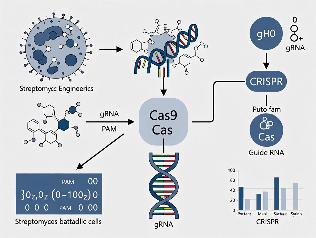
CRISPR-Cas systems, derived from an adaptive bacterial immune mechanism, have revolutionized genetic engineering. Within the context of Streptomyces metabolic engineering—aimed at optimizing the production of bioactive secondary metabolites like antibiotics, antifungals, and anticancer drugs—CRISPR tools enable precise, multiplexed genome editing. This facilitates the knockout of competing pathways, activation of silent gene clusters, and fine-tuning of regulatory networks. The transition from native Type I/E systems to the simplified, programmable Type II (Cas9) and Type V (Cas12a) systems has been pivotal for these high-GC, filamentous bacteria.
Key Protocols forStreptomycesMetabolic Engineering
Protocol 2.1: CRISPR-Cas9 Mediated Gene Knockout inStreptomyces coelicolor
Objective: To disrupt a target gene within a biosynthetic gene cluster (BGC) to elucidate function or redirect metabolic flux.
Materials:
- S. coelicolor strain (e.g., M145)
- pCRISPomyces-2 plasmid (Addgene #61737) or similar Streptomyces-optimized vector.
- Donor DNA oligonucleotide (for HDR) or no template for NHEJ-mediated indel formation.
- E. coli ET12567/pUZ8002 for conjugation.
- Mannitol Soy Flour (MS) agar plates with appropriate antibiotics (apramycin, thiostrepton).
- TES buffer (for protoplast transformation alternative).
Method:
- gRNA Design: Design a 20-nt spacer sequence targeting the gene of interest. Ensure PAM (5'-NGG-3') is present. Check for off-targets in the genome.
- Plasmid Construction: Clone the spacer sequence into the BsaI site of the pCRISPomyces-2 vector via Golden Gate assembly.
- Conjugation: a. Introduce the constructed plasmid into methylation-deficient E. coli ET12567/pUZ8002. b. Grow E. coli donor and Streptomyces spores separately, then mix, pellet, and resuspend. c. Plate the mixture on MS agar without antibiotics. Incubate at 30°C for 16-20 hours. d. Overlay with agar containing apramycin (to select for plasmid integration) and nalidixic acid (to counter-select E. coli). Incubate for 3-5 days.
- Screening & Curing: Pick exconjugants. Screen for desired edits via colony PCR and sequencing. To cure the plasmid, passage colonies several times without antibiotic selection.
- Metabolite Analysis: Cultivate edited strains and analyze secondary metabolite production via HPLC-MS.
Protocol 2.2: dCas9-Based Activation (CRISPRa) of Silent BGCs
Objective: To transcriptionally activate a silent or poorly expressed gene cluster using a dCas9-activator fusion.
Materials:
- pCRISPomyces-dCas9-Sox2/VP64 plasmid system.
- gRNAs designed to target promoter regions of key pathway-specific regulatory genes.
Method:
- gRNA Design: Design multiple gRNAs targeting ~100-200 bp upstream of the transcription start site of the cluster's "trigger" gene.
- Multiplex Plasmid Assembly: Use a tRNA-gRNA array strategy to clone up to 4-5 gRNAs into the activation plasmid.
- Strain Construction: Deliver the plasmid via conjugation as in Protocol 2.1.
- Validation: Confirm activation via RT-qPCR of key cluster genes. Profile metabolite production changes via comparative metabolomics (LC-MS).
Table 1: Comparison of Common CRISPR Systems for Streptomyces Engineering
| System & Common Plasmid | Editing Type | PAM Sequence | Key Feature for Streptomyces | Typical Editing Efficiency* |
|---|---|---|---|---|
| SpCas9 (pCRISPomyces-2) | Knockout, Knock-in, Activation/Repression | 5'-NGG-3' | Robust, widely used; requires codon optimization. | 50-95% (Knockout) |
| Cas12a (pCRISPomyces-Cpf1) | Knockout, Multiplexed editing | 5'-TTTV-3' (T-rich) | Simplifies multiplex gRNA arrays; T-rich PAM suits high-GC genomes. | 30-80% (Knockout) |
| dCas9-Sox2/VP64 | Transcriptional Activation (CRISPRa) | 5'-NGG-3' | Activates silent BGCs; requires multiple gRNAs for synergy. | Variable (2-100x induction) |
| Base Editors (ABE8e) | Point Mutation (A•T to G•C) | 5'-NGG-3' | Enables precise transition mutations without DSBs or donor DNA. | 10-50% (in model strains) |
*Efficiency is highly strain and target dependent.
Table 2: Key Metrics for Streptomyces Genome Editing Projects
| Parameter | Typical Range or Value | Notes |
|---|---|---|
| Conjugation Efficiency | 10⁻⁵ to 10⁻³ (per recipient spore) | Highly dependent on Streptomyces species and donor E. coli health. |
| Time from Design to Validated Mutant | 3 - 6 weeks | Includes cloning, conjugation, sporulation, screening, and validation. |
| Optimal gRNA Length | 20 nucleotides | For SpCas9. GC content >60% often recommended for high-GC hosts. |
| Multiplexing Capacity (tRNA array) | Up to 5-7 gRNAs | Increased numbers reduce efficiency per individual guide. |
The Scientist's Toolkit: Research Reagent Solutions
Table 3: Essential Reagents for CRISPR-Streptomyces Work
| Item | Function & Critical Notes |
|---|---|
| pCRISPomyces-2 Plasmid | All-in-one Streptomyces shuttle vector expressing SpCas9 and a single gRNA. Selection: ApramycinR (in Streptomyces), AmpicillinR (in E. coli). |
| ET12567/pUZ8002 E. coli Strain | Methylation-deficient dam/dcm- host for plasmid propagation to avoid Streptomyces restriction systems. Contains conjugative machinery. |
| Mannitol Soy Flour (MS) Agar | Standard medium for intergeneric conjugation between E. coli and Streptomyces. |
| Apramycin & Thiostrepton | Antibiotics for selection in Streptomyces. Apramycin selects for plasmid integration; thiostrepton can be used for inducible promoter systems. |
| HiFi DNA Assembly Master Mix | For efficient, seamless cloning of gRNA expression cassettes and donor DNA fragments into plasmid backbones. |
| PCR Reagents for GC-Rich Templates | Polymerases and buffers optimized for high-GC content (e.g., Q5, KAPA HiFi) are essential for reliable amplification from Streptomyces genomes. |
| HPLC-MS Grade Solvents | For high-resolution metabolomic analysis of secondary metabolite production changes post-editing. |
Visualization Diagrams
Title: CRISPR Editing Workflow for Streptomyces
Title: Native CRISPR-Cas Bacterial Immune Pathway
Within the broader thesis on CRISPR-based genome editing for Streptomyces metabolic engineering, three primary, interconnected challenges must be systematically addressed: the exceptionally high GC content of the genome, the complex morphological and physiological lifecycle, and the dominant DNA repair pathways that hinder precise editing. This application note provides detailed protocols and strategies to overcome these barriers, enabling efficient genetic manipulation for novel natural product discovery and yield optimization.
The High GC Content Challenge
The Streptomyces genome typically exhibits a GC content of >70%, which complicates PCR amplification, oligonucleotide synthesis, and CRISPR-Cas guide RNA (gRNA) design. Secondary structures in gRNAs can reduce Cas9 binding efficiency.
Table 1: Impact of High GC Content on Common Molecular Biology Tools
| Tool/Process | Typical Organism GC (~50%) | Streptomyces GC (>70%) | Primary Consequence |
|---|---|---|---|
| PCR Primer Design | Tm ~55-65°C | Tm often >75°C | Non-specific binding, primer-dimer formation |
| gRNA Design | Standard algorithms suffice | High risk of secondary structures | Reduced Cas9/gRNA complex stability & on-target activity |
| DNA Synthesis | Standard efficiency | Reduced yield, increased error rate | Higher cost, potential for mutant sequence insertion |
| Hybridization | Predictable | Over-stable | Off-target effects in probe-based applications |
Protocol 1.1: Optimized gRNA Design for High-GC Genomes
Objective: To design and validate functional gRNAs in high-GC Strephomyces DNA.
Materials (Research Reagent Toolkit):
- CRISPR-Cas9 Plasmid System (e.g., pCRISPR-Cas9-S.t.): Integrative or replicative vector with codon-optimized Cas9 and gRNA scaffold.
- High-Fidelity Polymerase (e.g., Q5): For accurate amplification of high-GC targets.
- GC Enhancer Additives: Betaine (1M) or DMSO (3-5%) to reduce DNA secondary structures during PCR.
- Thermocycler with Gradient Function: For optimizing annealing temperatures.
- Streptomyces Codon-Optimized RFP/Reporter: For rapid visual screening of editing efficiency.
Procedure:
- Target Selection: Using software like Benchling or CRISPy-web, identify 20-23 bp protospacer sequences immediately 5' of a 5'-NGG-3' PAM.
- GC Filtering: Prioritize candidates with a local GC content between 50-70%. Avoid sequences with long homopolymeric G/C stretches.
- Secondary Structure Prediction: Analyze the full gRNA (spacer + scaffold) using RNAfold. Discard designs with a minimum free energy (MFE) > -5 kcal/mol for the spacer region alone.
- Cloning: Synthesize top candidates as oligonucleotides with appropriate overhangs and clone into the chosen CRISPR-Cas9 vector via Golden Gate or Gibson Assembly.
- Transformation: Introduce the plasmid into the Streptomyces host via intergeneric conjugation from E. coli ET12567/pUZ8002.
- Validation: Screen exconjugants via PCR (using betaine/DMSO additives) and Sanger sequencing to confirm editing events.
The Complex Lifecycle Challenge
Streptomyces undergoes a complex differentiation cycle from vegetative mycelium to aerial mycelium and spore formation. Editing tools must be delivered and function efficiently across these stages, and engineered strains must maintain genetic stability through sporulation.
Diagram Title: Streptomyces Developmental Lifecycle Stages
Protocol 2.1: Conjugation-Based Delivery Timing for Maximum Exconjugant Yield
Objective: To deliver editing constructs to the most receptive stage of the Streptomyces lifecycle.
Procedure:
- Prepare Donor: Grow E. coli ET12567/pUZ8002 carrying the editing plasmid to mid-log phase (OD600 ~0.4-0.6). Wash 2x with LB to remove antibiotics.
- Prepare Recipient: Harvest Streptomyces spores by scraping from a mature agar plate and filtering through cotton wool. Heat shock at 50°C for 10 minutes to synchronize germination.
- Co-culture: Mix donor and recipient cells at a 1:10 ratio (donor:recipient) on an SFM or MS agar plate. Critical Step: The Streptomyces recipient should be in early vegetative growth for optimal cell-wall permeability. Using freshly germinating spores increases efficiency.
- Incubate: Plate at 30°C for 16-20 hours.
- Counter-selection: Overlay the conjugation mix with 1 ml of sterile water containing appropriate antibiotics (e.g., apramycin for selection) and nalidixic acid to counter-select against the E. coli donor.
- Isolation: Continue incubation for 5-7 days until exconjugant colonies (appearing as Streptomyces mycelium) emerge.
DNA Repair Pathways Challenge
In the absence of an easily programmable homologous recombination (HR) system, Streptomyces predominantly repairs CRISPR-Cas9-induced double-strand breaks (DSBs) via the error-prone Non-Homologous End Joining (NHEJ) pathway, leading to undesirable indels rather than precise edits.
Diagram Title: DNA Repair Pathway Decision After CRISPR-Cas9 Cut
Table 2: Strategies to Favor Homology-Directed Repair (HDR) over NHEJ
| Strategy | Mechanism | Protocol/Reagent |
|---|---|---|
| NHEJ-Knockout | Disrupt ku or ligD genes to impair the primary NHEJ pathway. | Use CRISPR to create a clean deletion of ku prior to metabolic engineering. |
| ssDNA Template Delivery | Provide a single-stranded oligodeoxynucleotide (ssODN) as a precise repair template. | Co-transform with a >100 nt ssODN homologous to target, with desired change centered. |
| Conditional Cas9 Expression | Express Cas9 only after induction, allowing HR template to be present first. | Use a tightly regulated promoter (tipAp, ermEp) for Cas9 on the editing plasmid. |
| Phage-encoded Recombinases | Introduce recombinase systems (e.g., Che9c RecT) to promote recombination. | Clone recT onto editing plasmid or integrate into the host genome. |
Protocol 3.1: ssODN-Mediated Precise Point Mutation
Objective: To introduce a specific point mutation via HDR using an ssODN template.
Materials (Research Reagent Toolkit):
- NHEJ-Deficient Strain (Δku or ΔligD): Essential for maximizing HDR frequency.
- Long ssODN Template (>100 nt): Designed with the desired mutation centrally, and homology arms (50+ nt each) perfectly matching the target strand.
- Heat-Shock Media (TSB with 10.5% sucrose): For recovery post-electroporation if using that method.
Procedure:
- Strain Preparation: Use a Streptomyces strain with a disrupted NHEJ pathway (e.g., Δku).
- Design & Order ssODN: Order an ultramer ssODN complementary to the non-target strand (the one not cleaved by Cas9). Phosphorothioate bonds at terminal 3 bases enhance stability.
- Co-delivery: Introduce the CRISPR plasmid (with target-specific gRNA) and the ssODN (100-200 ng) simultaneously into the Streptomyces protoplasts via PEG-mediated transformation or electroporation.
- Screening: Allow for 2-3 days of recovery without selection, then plate on selective media. Screen colonies by PCR-RFLP if the edit creates/disrupts a restriction site, or by Sanger sequencing.
- Curing: Isolate positive clones and passage at 37-39°C without antibiotics to cure the temperature-sensitive CRISPR plasmid.
Diagram Title: Precise Editing Workflow Using ssODN Templates
Successful CRISPR-based genome editing in Streptomyces requires a multi-faceted approach that addresses the unique triad of challenges posed by its high-GC genome, complex biology, and DNA repair landscape. By employing the optimized protocols for gRNA design, timed conjugation, and HDR-enhancement outlined here, researchers can achieve precise and efficient genetic modifications. This capability is fundamental to the metabolic engineering objectives of the broader thesis, enabling the rational redesign of biosynthetic pathways for enhanced drug candidate production.
Application Notes: Core Principles for Genome Editing in Actinobacteria
Within the context of a broader thesis on CRISPR-based metabolic engineering in Streptomyces, the successful implementation of CRISPR-Cas editing hinges on three pillars. Actinobacteria, particularly Streptomyces species, present unique challenges including complex growth cycles, thick mycelial cell walls, and GC-rich genomes (>70% GC), necessitating tailored approaches.
Guide RNA Design for GC-Rich Genomes: The high GC content of Streptomyces genomes requires careful sgRNA design to ensure high on-target activity and minimize off-target effects. The protospacer-adjacent motif (PAM) sequence dictates Cas protein choice. For the commonly used Streptococcus pyogenes Cas9 (SpCas9), the PAM is 5'-NGG-3', which is statistically abundant even in GC-rich DNA. However, recent advancements have introduced Cas9 variants like SpCas9-NG (NG PAM) and Francisella novicida Cas12a (FnCas12a, T-rich PAM like TTTV), offering expanded targeting ranges.
Cas Protein Selection: The choice of Cas protein is critical for editing efficiency and outcome. SpCas9 is the standard but requires an NGG PAM. Cas12a (Cpf1) is advantageous for its ability to process its own crRNA array (enabling multiplexing) and to create staggered ends, which can influence repair outcomes. For base editing or prime editing in Streptomyces, deaminase-fused nickase Cas9 (e.g., cytosine base editor, CBE) or prime editor Cas9 (PE2) proteins are selected to introduce precise point mutations without double-strand breaks or donor templates.
Repair Template Delivery: Efficient homology-directed repair (HDR) in Streptomyces is the primary bottleneck. Due to low intrinsic HDR rates, the repair template must be optimally delivered. Key strategies include:
- Linear Double-Stranded DNA (dsDNA) Fragments: PCR-amplified fragments with 500-1000 bp homology arms are most common and effective.
- Plasmid-Borne Templates: Co-delivered on the same or a second plasmid, but risk persistence of the plasmid backbone.
- Recombineering-Enhanced HDR: Utilizing phage-derived recombinases (e.g., RecET) expressed in trans to dramatically boost HDR efficiency.
Table 1: Comparison of Cas Proteins for Actinobacteria Genome Editing
| Cas Protein | PAM Sequence | Cleavage Type | Key Advantage for Actinobacteria | Typical Editing Efficiency in Streptomyces |
|---|---|---|---|---|
| S. pyogenes Cas9 (SpCas9) | 5'-NGG-3' | Blunt end | Well-characterized, high activity | 80-100% (for knock-out) |
| SpCas9-NG variant | 5'-NG-3' | Blunt end | Expanded targeting in GC-rich genome | 40-80% (varies by site) |
| F. novicida Cas12a (FnCas12a) | 5'-TTTV-3' | Staggered end | Multiplexing, T-rich PAM for AT-rich gene targets | 60-90% |
| L. bacterium Cas12a (LbCas12a) | 5'-TTTV-3' | Staggered end | Smaller size, easier delivery | 50-85% |
| Nickase Cas9 (nCas9) | 5'-NGG-3' | Single-strand nick | Used in base editing fusions (CBE, ABE) | 10-50% (for point mutation) |
Table 2: Repair Template Delivery Methods and Efficiencies
| Delivery Method | Template Form | Typical Homology Arm Length | Relative HDR Efficiency | Key Considerations |
|---|---|---|---|---|
| Conjugative Transfer | Plasmid-borne or linear dsDNA | 500-1000 bp | Moderate to High | Most common method; uses E. coli donor strain. |
| PEG-Mediated Protoplast Transformation | Linear dsDNA | 1000-2000 bp | Low to Moderate | Sensitive protocol, regeneration rates vary. |
| Electroporation of Mycelia | Linear dsDNA | 500-1500 bp | Low | Requires careful preparation of young mycelia. |
| Recombineering-Augmented | Linear ssDNA/dsDNA | 50-100 bp (ss) / 500 bp (ds) | Very High | Requires expression of phage recombinases (RecET). |
Detailed Experimental Protocols
Protocol 1: Design and Cloning of sgRNA for SpCas9 in Streptomyces
- Target Identification: Using a tool like CRISPy-web (specific for Streptomyces), input your target gene locus. Select a protospacer sequence (20 bp) directly adjacent to an NGG PAM on the coding strand.
- Off-Target Check: Use the BLAST function against the specific Streptomyces genome to ensure minimal homology elsewhere.
- Oligonucleotide Design: Design forward and reverse oligos: Forward: 5'-GATCCNNNNNNNNNNNNNNNNNNN-3'; Reverse: 5'-AAACNNNNNNNNNNNNNNNNNNNC-3' (N's correspond to the protospacer, excluding the PAM).
- Cloning into Streptomyces CRISPR Vector: a. Anneal oligonucleotides (95°C for 5 min, ramp down to 25°C). b. Digest the destination plasmid (e.g., pCRISPomyces-2) with BpiI (BbsI isoschizomer). c. Ligate annealed duplex into the digested plasmid using T4 DNA ligase. d. Transform into E. coli and sequence-verify the cloned sgRNA.
Protocol 2: Intergeneric Conjugation for CRISPR Component Delivery (Based on pCRISPomyces-2) Materials: E. coli ET12567/pUZ8002 (methylation-deficient donor), Streptomyces spore suspension, LB with appropriate antibiotics, AS-1 agar plates (with 10 mM MgCl2), 500 μL 0.22 μm filter.
- Prepare Donor: Grow E. coli ET12567/pUZ8002 containing your CRISPR plasmid to an OD600 of ~0.4-0.6. Wash cells twice with equal volume LB to remove antibiotics.
- Prepare Recipient: Harvest Streptomyces spores, heat-shock at 50°C for 10 min, and resuspend in LB.
- Mix and Plate: Mix 100 μL of donor E. coli with 100 μL of Streptomyces spores. Plate the entire mixture onto an AS-1 agar plate. Incubate at 30°C for 16-20h.
- Overlay and Select: Overlay the conjugation plate with 1 mL water containing 1 mg nalidixic acid (to counter-select E. coli) and the appropriate antibiotic for plasmid selection in Streptomyces (e.g., apramycin). Incubate at 30°C for 3-7 days until exconjugant colonies appear.
Protocol 3: HDR Using Linear dsDNA Repair Template
- Template Construction: Design a repair template with the desired edit (e.g., gene insertion, point mutation) flanked by homology arms (≥500 bp each). Amplify by PCR using high-fidelity polymerase from a plasmid or genomic DNA.
- Co-Delivery: During the conjugation protocol (Protocol 2), add 100-500 ng of purified, linear dsDNA repair template to the E. coli/Streptomyces mixture just before plating. Alternatively, introduce the template via protoplast transformation.
- Screening: Screen exconjugants by PCR and sequence verification to identify precise edit incorporations. The frequency of perfect HDR is typically <10% without recombineering.
Diagrams and Workflows
Title: CRISPR-Cas Workflow for Streptomyces Genome Editing
Title: DNA Repair Pathways After CRISPR Cleavage
The Scientist's Toolkit: Research Reagent Solutions
| Reagent / Material | Function / Application | Example Product/Supplier |
|---|---|---|
| pCRISPomyces-2 Plasmid | Standard Streptomyces CRISPR vector; expresses SpCas9 and sgRNA. | Addgene #61737 |
| ET12567/pUZ8002 E. coli Strain | Methylation-deficient donor strain for intergeneric conjugation. | Standard lab strain |
| BpiI (BbsI) Restriction Enzyme | Used for golden gate cloning of sgRNA oligos into CRISPR vectors. | Thermo Fisher, NEB |
| Phire Plant Direct PCR Kit | Robust PCR for GC-rich Streptomyces genomic DNA for screening. | Thermo Fisher |
| RecET Plasmid (pRETA) | Expresses phage recombinases to boost HDR efficiency with linear templates. | Addgene #133436 |
| AS-1 Agar | Mannitol-soy flour agar, standard for Streptomyces conjugation and sporulation. | Homemade or specialty suppliers |
| Hygromycin B | Common antibiotic for selection in Streptomyces after editing. | Roche, Sigma-Aldrich |
| GraphPad Prism | Software for statistical analysis and visualization of editing efficiency data. | GraphPad Software |
Application Notes: A Comparative Analysis of Genome Editing Technologies inStreptomyces
The metabolic engineering of Streptomyces species, prolific producers of antibiotics and other bioactive compounds, has been revolutionized by the advent of CRISPR-based genome editing. The historical trajectory from classical homologous recombination (HR) to CRISPR-Cas systems marks a shift from low-efficiency, laborious methods to rapid, multiplexable, and precise genetic manipulation.
Table 1: Quantitative Comparison of Genome Editing Methods in Streptomyces
| Parameter | Classical Homologous Recombination (e.g., using pKC1139) | CRISPR-Cas9 Editing (e.g., using pCRISPomyces plasmids) |
|---|---|---|
| Editing Efficiency | < 0.1% - 1% (double-crossover) | 10% - >95% (depending on construct and delivery) |
| Time to Isolate Mutant | 3 - 6 weeks (including counterselection) | 1 - 2 weeks |
| Multiplexing Capability | None (single locus per attempt) | High (demonstrated 3-5 loci simultaneously) |
| Primary Limitation | Extremely low efficiency, requires selectable/counter-selectable markers | Requires careful sgRNA design to avoid off-target effects in some species |
| Key Advantage | No requirement for exogenous nuclease machinery; well-established. | High efficiency, precision, and ability to create markerless deletions/insertions. |
Table 2: Impact on Metabolic Engineering Workflows
| Workflow Stage | Pre-CRISPR (HR-Dominant) | Post-CRISPR Adoption |
|---|---|---|
| Strain Construction | Sequential, iterative modifications; major bottleneck. | Parallel, multiplexed modifications; rapid pathway assembly. |
| Library Generation | Near-impossible for targeted genomic loci. | Feasible via pooled sgRNA libraries or CRISPRi/a. |
| Essential Gene Study | Challenging, relied on conditional mutants. | Enabled via CRISPR interference (CRISPRi) for knockdowns. |
| Toolkit Standardization | Species-specific vectors, often low-copy and unstable. | Modular plasmid systems (e.g., pCRISPomyces) applicable across species. |
Experimental Protocols
Protocol 1: Classical Gene Deletion via Double-Crossover Homologous Recombination
This protocol outlines the traditional method using a temperature-sensitive plasmid, exemplified by vector pKC1139.
Materials: Streptomyces strain, pKC1139 or equivalent E. coli-Streptomyces shuttle vector, E. coli ET12567/pUZ8002 for conjugation, appropriate antibiotics.
Procedure:
- Construct the Deletion Vector: Amplify ~1.5-2 kb DNA fragments upstream and downstream of the target gene. Clone these fragments flanking an antibiotic resistance cassette (e.g., aac(3)IV or apr) in pKC1139.
- Introduce into E. coli Donor: Transform the construct into E. coli ET12567/pUZ8002.
- Conjugal Transfer: Mix donor E. coli with Streptomyces spores/hyphae. Plate on MS agar with 10 mM MgCl₂. After 16-20h at 28°C, overlay with agar containing nalidixic acid (to counter-select E. coli) and the antibiotic for plasmid selection (e.g., apramycin).
- Select Single-Crossover Exconjugants: After 3-5 days, pick apramycin-resistant exconjugants. These result from homologous recombination at one of the two flanking regions.
- Counter-Select Double-Crossover Events: Streak exconjugants for single colonies on non-selective media and incubate at 37-39°C (the non-permissive temperature for pKC1139 replication). Replica-plate resulting colonies to plates with and without apramycin.
- Screen for Mutants: Apramycin-sensitive colonies have potentially lost the vector via a second crossover. Verify the deletion by colony PCR using primers outside the constructed homology arms.
Protocol 2: CRISPR-Cas9 Mediated Markerless Gene Deletion inStreptomyces
This protocol uses the pCRISPomyces-2 system (Addgene #61737) for efficient, markerless editing.
Materials: pCRISPomyces-2 plasmid, E. coli ET12567/pUZ8002, target Streptomyces strain, Gibson Assembly or Golden Gate cloning reagents.
Procedure:
- sgRNA Design & Cloning: Design a 20-nt sgRNA sequence targeting the NGG PAM site within the gene to be deleted. Synthesize oligonucleotides, anneal, and clone into the BsaI site of pCRISPomyces-2.
- Homology Donor Template Construction: PCR-amplify two ~1 kb homology arms (HA) flanking the intended deletion site. Assemble these fragments into a linear dsDNA fragment (via overlap-extension PCR) or clone into a separate replicating plasmid if performing large insertions.
- Conjugal Delivery: Transform the pCRISPomyces-2 sgRNA construct into E. coli ET12567/pUZ8002. Perform conjugation with Streptomyces as in Protocol 1, Step 3, selecting with apramycin.
- Mutant Screening: Patch 20-50 exconjugants onto fresh apramycin plates. After 2-3 days, perform colony PCR directly on mycelial biomass to screen for the deletion. The high editing efficiency often yields >50% correct mutants.
- Curing of CRISPR Plasmid: Grow a positive mutant without antibiotic selection for several rounds of sporulation/growth. Screen for apramycin-sensitive colonies to obtain a plasmid-free, markerless deletion mutant.
Visualization Diagrams
Title: Classical Homologous Recombination Workflow
Title: CRISPR-Cas9 Genome Editing Workflow
Title: Editing Efficiency Comparison
The Scientist's Toolkit: Research Reagent Solutions
Table 3: Essential Reagents for CRISPR-based Streptomyces Metabolic Engineering
| Reagent/Material | Function/Description | Example/Supplier |
|---|---|---|
| pCRISPomyces Plasmids | Modular plasmid series (1, 2, NL) expressing Cas9 and sgRNA, with different replication origins for various Streptomyces. | Addgene #61737, #61738, #137298 |
| ET12567/pUZ8002 E. coli | Non-methylating, conjugation-proficient donor strain essential for efficient plasmid transfer from E. coli to Streptomyces. | Standard lab strain |
| Gibson Assembly Master Mix | Enables seamless, simultaneous assembly of multiple DNA fragments (e.g., sgRNA + homology arms). | NEB, Thermo Fisher |
| Golden Gate Assembly Kit (BsaI) | Efficient, one-pot modular cloning of sgRNA expression cassettes. | NEB Golden Gate Assembly Kit |
| Synthase/Pathway-Specific Precursors | Chemical supplements to assay production titers of target natural products during metabolic engineering. | Sigma-Aldrich, Carbosynth |
| HPLC-MS Systems | For quantitative analysis and verification of metabolite production changes in engineered strains. | Agilent, Waters, Thermo Fisher |
| Cas9 Nuclease (S. pyogenes) | For in vitro validation of sgRNA cutting efficiency prior to conjugation. | NEB, IDT |
| T7 Endonuclease I or Surveyor Assay Kit | Detects CRISPR-induced indels or mismatches in DNA heteroduplexes for efficiency validation. | NEB, IDT |
A Step-by-Step Toolkit: CRISPR-Cas Systems and Metabolic Engineering Strategies
CRISPR-based genome editing has revolutionized metabolic engineering in Streptomyces, the prolific producers of antibiotics and other bioactive natural products. Selecting the appropriate CRISPR system—Cas9, Cas12a (Cpf1), or nickase variants—is critical for efficient and precise genome editing tailored to the specific genetic and physiological complexities of these high-GC, filamentous bacteria. This application note, framed within a broader thesis on CRISPR-based genome editing for Streptomyces metabolic engineering research, provides a comparative analysis and detailed protocols to guide researchers in system selection and implementation.
Comparative Analysis of CRISPR Systems
The selection of a CRISPR system depends on the desired editing outcome, target site constraints, and efficiency requirements. Below is a quantitative comparison based on recent literature and experimental data.
Table 1: Comparative Features of CRISPR Systems for Streptomyces
| Feature | SpCas9 | Cas12a (Cpf1) | Cas9 Nickase (nCas9-D10A) |
|---|---|---|---|
| Nuclease Activity | Blunt DSBs | Staggered DSBs (5' overhang) | Single-strand break (nick) |
| PAM Sequence | 5'-NGG-3' (canonical) | 5'-TTTV-3' (T-rich) | 5'-NGG-3' (for targeting) |
| crRNA Length | ~100 nt (tracrRNA + crRNA) | ~42-44 nt (single RNA) | ~100 nt (tracrRNA + crRNA) |
| Cleavage Site | Within PAM | Distal to PAM | One DNA strand |
| Editing Outcomes | NHEJ, HDR, large deletions | NHEJ, HDR, often precise deletions | Paired nicks for DSB or base editing fusions |
| Primary Use Case | Gene knockouts, large insertions | Gene knockouts, multiplexing, transcriptional repression | High-fidelity HDR, Base Editing (e.g., ABE8e) |
| Reported Efficiency in Streptomyces | 60-95% (knockout) | 40-85% (knockout) | HDR: 10-50% (varies by locus) |
Table 2: Guide RNA Design Parameters
| Parameter | SpCas9 | Cas12a (Cpf1) |
|---|---|---|
| Optimal Targeting Strand | Non-template strand preferred | Either strand |
| Seed Region | 10-12 bp proximal to PAM | 5-7 bp distal to PAM |
| Off-Target Concern | Moderate-High (tolerates mismatches in seed) | Lower (more stringent) |
| Multiplexing Ease | Requires multiple expression constructs | Simplified with single array transcript |
Detailed Experimental Protocols
Protocol 1: CRISPR-Cas9 Mediated Gene Knockout inStreptomyces coelicolor
Objective: To disrupt a target gene via non-homologous end joining (NHEJ) using a plasmid-based Cas9 system. Key Reagents: pCRISPomyces-2 plasmid (or equivalent), E. coli ET12567/pUZ8002 for conjugation, Streptomyces spore suspension, apramycin, thiostrepton.
Procedure:
- gRNA Design & Cloning: Design a 20-nt spacer sequence directly 5' to an NGG PAM on the non-template strand. Clone the annealed oligonucleotides into the BsaI site of the pCRISPomyces-2 plasmid.
- Conjugative Transfer: Transform the plasmid into E. coli ET12567/pUZ8002. Mix the donor E. coli with S. coelicolor spores, plate on MS agar with 10 mM MgCl₂, and incubate at 30°C for 16-20 hours.
- Selection & Screening: Overlay plates with apramycin (50 µg/mL) and nalidixic acid (25 µg/mL). After 3-5 days, isolate exconjugants. Overlay with thiostrepton (50 µg/mL) to induce Cas9/gRNA expression. Incubate for an additional 2-3 days.
- Genotype Validation: Patch resistant colonies for sporulation. Isolate genomic DNA and perform PCR across the target locus. Screen for smaller amplicons indicative of deletion, followed by sequencing.
Protocol 2: Cas12a (Cpf1)-Mediated Multiplex Gene Deletion
Objective: To delete a genomic region or multiple genes using a single Cas12a crRNA array. Key Reagents: pCRISPR-Cpf1 plasmid (containing FnCas12a), direct repeat sequences, designed spacers.
Procedure:
- crRNA Array Construction: Design individual crRNAs (23-25 nt spacer followed by a 19 nt direct repeat). Assemble spacers sequentially via Golden Gate assembly into the plasmid.
- Transformation: Introduce the plasmid into Streptomyces via PEG-mediated protoplast transformation or conjugation.
- Induction & Screening: Induce Cas12a expression with anhydrotetracycline (aTc). Allow for repair via NHEJ. Screen apramycin-resistant colonies for loss of the targeted region(s) via multiplex PCR.
- Curing the Plasmid: Passage colonies non-selectively at 37°C to facilitate plasmid loss. Verify plasmid cure by patching onto apramycin-containing and antibiotic-free media.
Protocol 3: Nickase-Mediated Base Editing inStreptomyces venezuelae
Objective: To install a precise A•T to G•C point mutation using an Adenine Base Editor (ABE) fusion with nCas9 (D10A). Key Reagents: pABE8e-nCas9 plasmid, gRNA expression plasmid, appropriate antibiotics.
Procedure:
- gRNA Design: Design a spacer positioning the target adenine within the editing window (typically positions 4-8, counting the PAM as 21-23). The nickase strand should be the non-editing strand.
- Co-transformation: Co-transform S. venezuelae protoplasts with both plasmids.
- Selection & Expansion: Regenerate protoplasts on R2YE plates with apramycin and thiostrepton. Pick colonies after 5-7 days.
- Sequencing Analysis: Isolate genomic DNA from pooled or individual colonies. Amplify the target region by PCR and submit for Sanger sequencing. Analyze chromatograms for peak overlaps at the target site, indicating successful base editing.
Diagrams and Visual Workflows
Decision Workflow for CRISPR System Selection
Cas9 vs Nickase Experimental Workflows
The Scientist's Toolkit: Research Reagent Solutions
Table 3: Essential Materials for CRISPR Editing in Streptomyces
| Reagent/Material | Function in Experiment | Example/Supplier Note |
|---|---|---|
| pCRISPomyces-2 Plasmid | All-in-one vector for Cas9 and gRNA expression in Streptomyces. | Addgene #84276; contains thiostrepton-inducible tipA promoter. |
| E. coli ET12567/pUZ8002 | Non-methylating, conjugation-proficient donor strain for plasmid transfer. | Essential for bypassing Streptomyces restriction-modification barriers. |
| Anhydrotetracycline (aTc) | Inducer for TetR-regulated promoters driving Cas12a or high-efficiency Cas9. | Use at 50-100 ng/mL final concentration in overlays. |
| Thiostrepton | Inducer for tipA promoter and selective antibiotic for plasmids containing tsr. | Typical working concentration: 25-50 µg/mL in agar overlays. |
| Apramycin | Selective antibiotic for plasmids containing aac(3)IV resistance marker. | Typical concentration: 50 µg/mL for Streptomyces. |
| R2YE Agar Medium | Standard regeneration medium for Streptomyces protoplasts post-transformation. | Contains sucrose as osmotic stabilizer. |
| MS Agar with MgCl₂ | Standard medium for intergeneric conjugation between E. coli and Streptomyces. | MgCl₂ enhances spore germination and conjugation efficiency. |
| Direct Repeat Oligos for Cas12a | For constructing crRNA arrays. | Sequence: 5'-AAUUUCUACUAAGUGUAGAU-3' for FnCas12a. |
| ABE8e-nCas9 Plasmid | For high-efficiency adenine base editing in high-GC Streptomyces genomes. | Fuses ecTadA-8e deaminase to nCas9 (D10A). |
Within the broader thesis on CRISPR-based genome editing for Streptomyces metabolic engineering, precise point mutations are paramount. These organisms are prolific producers of secondary metabolites, but optimizing biosynthetic gene clusters (BGCs) often requires single-nucleotide precision. While conventional CRISPR-Cas9 relies on error-prone non-homologous end joining (NHEJ) for knockouts, base editing and prime editing offer superior pathways for defined, predictable point mutations without requiring double-strand breaks (DSBs) or donor DNA templates. This application note details protocols and considerations for deploying these advanced modalities in Streptomyces.
Base Editing
Base editors (BEs) are fusion proteins comprising a catalytically impaired Cas9 (nCas9 or dCas9) and a nucleotide deaminase enzyme. They enable the direct, irreversible conversion of one DNA base pair to another within a programmable "editing window" without cleaving the DNA backbone. Cytosine Base Editors (CBEs) facilitate C•G to T•A transitions, while Adenine Base Editors (ABEs) enable A•T to G•C transitions.
Prime Editing
Prime editors (PEs) are more versatile, comprising a Cas9 nickase (H840A) fused to a reverse transcriptase (RT) enzyme. They are programmed with a prime editing guide RNA (pegRNA) that both specifies the target site and encodes the desired edit. The system nicks one strand, and the pegRNA's RT template is reverse-transcribed to install the new sequence, which is then incorporated into the genome via DNA repair.
Quantitative Comparison Table
Table 1: Comparative Analysis of Base Editing and Prime Editing Modalities for Streptomyces Engineering.
| Parameter | Base Editing | Prime Editing | Notes for Streptomyces |
|---|---|---|---|
| Edit Types | C•G to T•A (CBE), A•T to G•C (ABE). | All 12 possible point mutations, small insertions (< 80bp), deletions (< 40bp). | Essential for activating silent BGCs or fine-tuning regulator genes. |
| Precision | High within editing window (~5nt). Risk of bystander edits. | Very high; minimal off-target edits. | Critical for modifying promoter regions without disrupting regulatory motifs. |
| Efficiency (Reported Ranges) | 10-50% in mammalian cells; 20-90% in bacteria. | 1-30% in mammalian cells; early Streptomyces data being established. | Efficiency varies by strain, locus, and delivery method (conjugation vs. transduction). |
| DSB Formation | No DSB; uses nCas9 for single-strand nick. | No DSB; uses nCas9 for single-strand nick. | Eliminates toxic DSB response, improving cell viability in slow-growing Streptomyces. |
| PAM Flexibility | Dependent on Cas9 variant (e.g., SpCas9-NG broadens PAM). | Dependent on Cas9 variant; SpCas9-based PE requires NGG. | NGG PAMs are common but not universal in GC-rich Streptomyces genomes. |
| Delivery Complexity | Moderate (single editor + sgRNA). | High (PE + complex pegRNA + often nicking sgRNA). | pegRNA design and stability is a key optimization parameter. |
Detailed Experimental Protocols
Protocol: Base Editing inStreptomycesvia Conjugative Transfer
Aim: To install a precise A•T to G•C mutation within a regulatory gene (e.g., afsR) to enhance antibiotic production.
I. Materials & Pre-Experimental Design
- Strains: E. coli ET12567/pUZ8002 (donor), Streptomyces lividans TK24 (recipient).
- Plasmids: Construct an ABE plasmid (e.g., pABE8e) under control of a strong, constitutive Streptomyces promoter (ermEp). Clone a 20-nt spacer sequence targeting the desired locus into the sgRNA expression cassette.
- Design Tool: Use CRISPRon or BE-DESIGN to select spacer and predict editing window/bystander effects.
- Validation: Sanger sequencing of the target locus in the wild-type strain.
II. Methodology
- Construct Assembly: Clone the designed sgRNA spacer into the ABE plasmid via Golden Gate assembly. Transform into E. coli ET12567/pUZ8002.
- Conjugative Transfer:
- Grow donor E. coli (with ABE plasmid and pUZ8002) and recipient Streptomyces to mid-exponential phase.
- Mix cultures, pellet, and resuspend. Plate onto MS agar with 10 mM MgCl₂. Incubate at 30°C for 16-20h.
- Overlay plate with 1 mL water containing nalidixic acid (to counter-select E. coli) and apramycin (to select for ABE plasmid integration). Incubate for 5-7 days.
- Exconjugant Screening: Pick exconjugants onto fresh selective plates. Allow for sporulation.
- Edit Verification:
- Harvest spores, perform colony PCR on genomic DNA from pooled exconjugants.
- Subject PCR product to Sanger sequencing. Use TIDE or EditR analysis to quantify editing efficiency.
- For clonal analysis, streak for single colonies and sequence individual clones.
- Curing the Plasmid: Passage positive clones non-selectively to promote loss of the temperature-sensitive plasmid. Verify loss by patching onto apramycin-containing and antibiotic-free plates.
III. The Scientist's Toolkit: Key Reagents Table 2: Essential Research Reagent Solutions for Base Editing in Streptomyces.
| Reagent | Function/Description | Example/Supplier |
|---|---|---|
| ET12567/pUZ8002 E. coli | Non-methylating donor strain for intergeneric conjugation into Streptomyces. | Standard lab strain. |
| pABE8e or pCBE Plasmid Backbone | Base editor expression vector; requires adaptation with Streptomyces promoters and origin. | Addgene #138489 (pABE8e). |
| ermE* Promoter | Strong constitutive promoter for high-level expression of editor in Streptomyces. | Common Streptomyces genetic part. |
| MS Agar | Mannitol-soy flour agar, optimal for conjugation and sporulation of many Streptomyces. | Sigma-Aldrich, custom preparation. |
| EditR Software | Web-based tool for quantifying base editing efficiency from Sanger trace data. | https://moriaritylab.shinyapps.io/editr_v10/ |
Base Editing Workflow for Streptomyces.
Protocol: Prime Editing inStreptomycesvia Phage Transduction
Aim: To install a transversion mutation (e.g., G•C to C•G) in a polyketide synthase gene to alter substrate specificity.
I. Materials & Pre-Experimental Design
- Strains: Streptomyces lividans with ϕC31 phage integration site.
- Phage Vector: Use a ϕC31-derived integrating vector. Clone a PEmax (optimized PE) expression cassette and separate pegRNA expression cassette, both under Streptomyces promoters.
- pegRNA Design: Critical step. Use design tools like pegIT or PrimeDesign. The pegRNA must contain: spacer (13-20nt), primer binding site (PBS, ~10-15nt), and RT template (~10-25nt) encoding the edit.
- Optional: Include a nicking sgRNA (ngRNA) to improve efficiency by nicking the non-edited strand.
II. Methodology
- Vector Construction: Assemble the prime editing construct in an E. coli plasmid, then transfer into a ϕC31 phage vector. Package phage in Streptomyces.
- Transduction:
- Prepare high-titer phage lysate from the packaging strain.
- Infect recipient Streptomyces mycelia or spores with the phage lysate.
- Plate on selective media containing apramycin. Incubate until transductant colonies appear (3-5 days).
- Screening & Validation:
- Patch transductants. Isolate genomic DNA.
- Perform PCR amplification of the target locus.
- Initial Screening: Use a restriction fragment length polymorphism (RFLP) assay if the edit creates/disrupts a site.
- Definitive Analysis: Submit PCR products for next-generation sequencing (amplicon-seq) to precisely identify edits and quantify efficiency and byproducts.
- Plasmid Curing: As for base editing protocol.
III. The Scientist's Toolkit: Key Reagents Table 3: Essential Research Reagent Solutions for Prime Editing in Streptomyces.
| Reagent | Function/Description | Example/Supplier |
|---|---|---|
| ϕC31 Phage Integration System | Efficient delivery vector for large DNA cargo into Streptomyces chromosomes. | Standard genetic tool for Streptomyces. |
| PEmax Expression Cassette | Optimized prime editor (nCas9-RT) for high efficiency. | Addgene #174828. |
| pegRNA Design Tool | Software for designing optimal pegRNA (PBS length, RT template). | PrimeDesign (https://primedesign.pinellolab.org/). |
| Next-Generation Sequencing | Essential for comprehensive analysis of prime editing outcomes (e.g., Indels, precise edits). | Illumina MiSeq amplicon sequencing. |
Prime Editing Molecular Mechanism.
Application inStreptomycesMetabolic Engineering Thesis
The integration of base and prime editing into the CRISPR toolkit for Streptomyces directly addresses core thesis aims:
- Pathway Optimization: Installing gain-of-function mutations in positive regulators (e.g., pathway-specific activators) or loss-of-function mutations in repressors to upregulate BGCs.
- Enzyme Engineering: Making precise, single-amino-acid changes within modular polyketide synthases (PKS) or non-ribosomal peptide synthetases (NRPS) to alter product spectra.
- Resistance & Precursor Flux: Modifying promoter regions of resistance genes or primary metabolism genes to fine-tune expression levels, balancing production with cell fitness.
- Combinatorial Approaches: Using prime editing for small, scarless insertions of epitope tags or linker sequences to study protein localization and interactions within BGCs.
Critical Considerations & Future Outlook
- Delivery: Conjugation is robust but plasmid curing is needed. Phage transduction is efficient for hard-to-transform strains. Electroporation of RNP complexes is emerging.
- Efficiency: Varies dramatically by locus. pegRNA design is the major bottleneck for prime editing. Testing 3-5 pegRNAs per target is recommended.
- Specificity: Both methods show high specificity, but whole-genome sequencing of edited clones is advised for metabolic engineering applications to rule off-target effects.
- Future Directions: Fusion of editors to phage-encoded proteins for improved delivery, development of Streptomyces-optimized editors (e.g., with GC-rich PAM preferences), and automated workflows for high-throughput editing of BGC libraries.
Base editing and prime editing represent transformative, precise tools for the genetic refactoring of Streptomyces. Their successful implementation, as outlined in these protocols, will enable unprecedented rational design of novel metabolic pathways for drug discovery and biotechnology.
Within the broader thesis focusing on CRISPR-based genome editing for Streptomyces metabolic engineering, the construction of precise and efficient expression vectors for guide RNA (gRNA) and Cas proteins is a foundational step. This Application Note details contemporary strategies and protocols for assembling gRNA expression constructs and the subsequent assembly of complete plasmid or Ribonucleoprotein (RIB) complexes tailored for Strendomyces species. Success in these cloning workflows is critical for enabling multiplexed editing, combinatorial pathway manipulation, and high-throughput strain engineering in antibiotic-producing actinomycetes.
Current Strategies for gRNA Expression Constructs
In Streptomyces, effective gRNA expression requires promoters functional in high-GC content bacteria. Recent literature and commercial developments favor RNA polymerase III promoters for precise transcriptional initiation and termination.
Table 1: Promoter Systems for gRNA Expression in Streptomyces
| Promoter Type | Example | Expression Strength | Key Feature | Best Use Case |
|---|---|---|---|---|
| Native Pol III | Streptomyces tRNA promoter | Medium | High GC-compatible; endogenous | Single-gene knockouts |
| Engineered Pol III | J23119 (Anderson) derivative | High | Standardized, strong | Multiplexed arrays |
| Constitutive Pol II | ermEp* | Very High | Requires precise processing | RIB delivery with Cas9 mRNA |
| Inducible | tipAp (thiostrepton-inducible) | Tunable | Low background | Essential gene editing |
Quantitative data from recent transfection studies in S. coelicolor indicate that engineered J23119-derivative promoters drive gRNA expression at levels ~1.8-fold higher than native tRNA promoters, as measured by RT-qPCR of precursor gRNA. Inducible systems like tipAp show minimal leakiness (<2% basal activity) and achieve maximal induction (100-fold) within 6 hours of thiostrepton addition.
Plasmid vs. RIB Assembly Workflows
Two primary delivery modalities exist: plasmid-based expression and pre-assembled Ribonucleoprotein (RIB) complexes. The choice impacts editing efficiency, off-target effects, and delivery method (e.g., conjugation vs. protoplast transformation).
Table 2: Comparison of Plasmid and RIB Delivery Methods
| Parameter | Plasmid-Based Expression | RIB (RNP) Delivery |
|---|---|---|
| Assembly Time | 3-5 days (cloning) | 1 day (protein purification & annealing) |
| Editing Speed | Slow (requires transcription/translation) | Fast (immediate activity) |
| Off-Target Risk | Higher (sustained expression) | Lower (transient activity) |
| Best for Streptomyces | Multiplexing, library delivery | Rapid screening, recalcitrant strains |
| Typical Efficiency in S. lividans | 45-75% (knockout) | 60-85% (knockout) |
| Regulatory Consideration | Contains antibiotic resistance marker | Marker-free, easier for industrial use |
Detailed Protocols
Protocol 1: Golden Gate Assembly for Multiplex gRNA Expression Cassette
This protocol enables the assembly of up to 8 gRNA expression units into a single Streptomyces integrative plasmid (e.g., pCRISPomyces-2 derivative).
Materials:
- BsaI-HFv2 restriction enzyme.
- T4 DNA Ligase (high-concentration).
- Vector backbone: pSET152-based with apramycin resistance (aac(3)IV) and Streptomyces origin of replication.
- gRNA inserts: Synthesized oligos encoding 20-nt spacer, cloned into BsaI-flanked entry vectors with promoter and terminator.
- Chemically competent E. coli NEB Stable for assembly.
- SOC Outgrowth Medium.
Method:
- Digestion-Ligation Setup: In a single tube, combine 50 ng of backbone vector, 20 ng of each gRNA entry vector (equimolar ratio), 1.5 µL BsaI-HFv2, 1 µL T4 DNA Ligase, 2 µL 10x T4 Ligase Buffer, and nuclease-free water to 20 µL.
- Thermocycling Reaction: Run: 25 cycles of (37°C for 3 min, 16°C for 4 min), followed by 50°C for 5 min, then 80°C for 10 min.
- Transformation: Transform 2 µL of reaction into 50 µL NEB Stable E. coli. Recover in 1 mL SOC at 37°C for 1 hour, plate on LB + apramycin (50 µg/mL).
- Screening: Screen colonies by colony PCR using primers flanking the assembly site. Sanger sequence confirmed clones.
Protocol 2:In VitroAssembly of RIB Complexes for Protoplast Transformation
For RIB delivery, recombinant Streptomyces-codon-optimized Cas9 protein is pre-complexed with in vitro transcribed gRNA.
Materials:
- Purified Cas9 Protein: Commercially available or purified from E. coli expression system.
- gRNA Transcription Kit (e.g., HiScribe T7 Quick).
- Annealing Buffer: 30 mM HEPES pH 7.5, 100 mM KCl.
- Protoplast Buffer: 10.3% sucrose, 5 mM MgCl₂, 5 mM CaCl₂, 25 mM TES buffer, pH 7.2).
Method:
- gRNA Synthesis: Transcribe gRNA from a dsDNA template with a T7 promoter using the HiScribe kit. Purify using RNA clean-up beads. Measure concentration (ng/µL) via nanodrop.
- RIB Complex Assembly: In a 1.5 mL tube, combine 5 µL (100 pmol) Cas9 protein with 3.6 µL (120 pmol) gRNA in Annealing Buffer (final volume 20 µL). Incubate at 25°C for 10 min.
- Protoplast Transformation: Mix 10 µL of RIB complex with 100 µL of freshly prepared Streptomyces protoplasts. Add 200 µL of 50% PEG 1450, mix gently. Plate on R2YE regeneration plates with appropriate antibiotics for selection after editing. Efficient editing is typically observed in 5-7 days post-regeneration.
The Scientist's Toolkit
Table 3: Essential Research Reagent Solutions
| Reagent/Material | Supplier Example | Function in Workflow |
|---|---|---|
| BsaI-HFv2 Enzyme | New England Biolabs | Type IIS restriction enzyme for Golden Gate assembly; enables seamless fusion. |
| HiScribe T7 Quick High Yield RNA Synthesis Kit | New England Biolabs | In vitro transcription of high-yield, pure gRNA for RIB assembly. |
| Streptomyces Codon-Optimized SpCas9 Expression Plasmid | Addgene (pCRISPomyces-2) | Source for Cas9 gene or recombinant protein expression. |
| Apramycin (aac(3)IV) Resistance Cassette | Laboratory stock | Selection marker for Streptomyces and E. coli during shuttle vector assembly. |
| TES Buffer (pH 7.2) | Sigma-Aldrich | Critical component for Streptomyces protoplast stabilization and transformation. |
| PEG 1450 (50% w/v) | Laboratory prepared | Facilitates DNA/RIB uptake during protoplast transformation. |
Visualization: Workflow Diagrams
Title: gRNA Construct Assembly & Delivery Workflow
Title: Plasmid and RIB Assembly Pathways Compared
Within a thesis on CRISPR-based genome editing for Streptomyces metabolic engineering, the introduction of editing tools is a critical step. Streptomyces species are renowned for their complex secondary metabolism, producing numerous clinically relevant compounds. However, their thick mycelial cell wall and complex life cycle pose significant delivery challenges. This document details three core delivery methodologies—Conjugation, Transformation, and Phage Integration—for introducing CRISPR-Cas systems to precisely engineer biosynthetic gene clusters (BGCs) and enhance metabolite yields.
Research Reagent Solutions Toolkit
| Reagent/Material | Function in Streptomyces CRISPR Delivery |
|---|---|
| E. coli ET12567(pUZ8002) | A non-methylating, conjugation-proficient E. coli donor strain essential for intergeneric conjugation with Streptomyces. It provides the tra genes in trans but does not transfer its own plasmid. |
| Methylation-deficient E. coli (e.g., ET12567, DH10B) | Host for propagating shuttle plasmids before conjugation to avoid restriction by Streptomyces' potent methyl-specific restriction systems. |
| pCRISPomyces-1/2 Plasmids | Standard, modular Streptomyces CRISPR-Cas9 vectors. Contain a Cas9 gene (codon-optimized) and a sgRNA scaffold under constitutive promoters, and an oriT for conjugation. |
| ΦC31 or ΦBT1 att/int System | Site-specific integration system derived from actinophages. Allows stable, single-copy chromosomal integration of CRISPR tools via phage attachment (attP) sites and integrase. |
| pTES Series Vectors | Phage-integrated CRISPR tools (e.g., pTES-Cas9). Contain a Cas9, sgRNA, and a phage attP site for recombination into the corresponding chromosomal attB site. |
| Mycelial Protoplasts | Cell-wall deficient Streptomyces cells generated using lysozyme, used as recipients in PEG-mediated transformation. |
| Sucrose-PEG Solution (10.3% Sucrose, 40% PEG 1000) | Osmotic stabilizer and fusogen for protoplast transformation and regeneration. |
| Heat-Inactivated Streptomyces Helper Strain | Used in some transduction protocols to provide phage receptors or integration machinery. |
Delivery Methodologies: Protocols & Data
Intergeneric Conjugation fromE. coli
Detailed Protocol:
- Donor Preparation: Transform the CRISPR plasmid (with oriT) into E. coli ET12567(pUZ8002). Grow a 5 mL culture in LB with appropriate antibiotics (e.g., apramycin, kanamycin) at 37°C to mid-log phase (OD600 ~0.4-0.6).
- Recipient Preparation: Harvest Streptomyces spores from a fresh agar plate using sterile water and glass beads. Heat-shock at 50°C for 10 minutes to activate germination.
- Mating: Mix donor cells (washed 2x with LB to remove antibiotics) and recipient spores at a ~1:1 to 10:1 ratio (spores: ~10^8 CFU). Pellet and resuspend in 100 µL LB. Spot onto MS or SFM agar (no antibiotics). Incubate at 30°C for 16-20 hours.
- Selection: Overlay the conjugation spot with 1 mL sterile water containing 0.5 mg nalidixic acid (to counter-select E. coli) and the antibiotic for plasmid selection (e.g., 50 µg/mL apramycin). Incubate at 30°C for 5-7 days until exconjugant colonies appear.
Table 1: Conjugation Efficiency Across Common Streptomyces Species
| Species | Average Exconjugants per Plate (10^8 Spores) | Key Considerations |
|---|---|---|
| S. coelicolor A3(2) | 10^2 - 10^4 | High efficiency, model organism. |
| S. albus J1074 | 10^3 - 10^5 | High transformability and conjugation efficiency. |
| S. avermitilis | 10^1 - 10^3 | Requires careful spore preparation. |
| S. venezuelae | 10^2 - 10^4 | Efficient with young, vegetative mycelium. |
Protoplast Transformation (PEG-Mediated)
Detailed Protocol:
- Protoplast Generation: Inoculate 25 mL TSB with Streptomyces spores/mycella. Incubate at 30°C, 250 rpm for 36-48h. Harvest mycelium by centrifugation (4000xg, 10 min). Wash with 10.3% sucrose. Resuspend in lysozyme solution (1 mg/mL in P buffer) and incubate at 30°C for 60-90 min. Filter through cotton wool to remove debris. Pellet protoplasts gently (2000xg, 7 min).
- Transformation: Resuspend protoplasts in 1 mL P buffer. Aliquot 100 µL into microfuge tubes. Add 1-10 µL plasmid DNA (up to 1 µg). Add 400 µL 40% PEG 1000 (in P buffer). Mix gently by pipetting. Incubate at room temp for 1 min. Dilute with 1 mL P buffer.
- Regeneration & Selection: Plate serial dilutions on R5 or R2YE regeneration agar (osmotic stabilizer) without antibiotics. Incubate at 30°C for 16-24h. Overlay with antibiotic-containing soft agar. Incubate for 3-5 days until transformants appear.
Table 2: Protoplast Transformation Efficiency
| Parameter | Typical Value/Range | Impact on Efficiency |
|---|---|---|
| Protopast Viability | >70% (Microscopy count) | Critical for regeneration. |
| PEG 1000 Concentration | 25-40% | 40% is standard; higher can be toxic. |
| DNA Amount | 0.1 - 1 µg | Saturation often at ~0.5 µg. |
| Regeneration Frequency | 1-10% of plated protoplasts | Species and strain-dependent. |
| Final Transformants/µg DNA | 10^4 - 10^6 | Can exceed 10^7 in optimized strains. |
Phage Integration (ΦC31-based)
Detailed Protocol:
- Vector Construction: Clone CRISPR-Cas9 and sgRNA expression cassettes into a ΦC31- or ΦBT1-based integration vector (contains attP, integrase gene, and Streptomyces origin).
- Delivery: Introduce the integration vector into Streptomyces via conjugation or transformation (as per Sections 3.1/3.2). Selection on apramycin (or vector-specific antibiotic).
- Integration Verification: Screen single-crossover integrants by colony PCR using primers spanning the attL/attR junctions. Confirm loss of autonomously replicating plasmid via plasmid DNA isolation and sensitivity screening.
Table 3: Comparison of Delivery Methods for CRISPR Tools
| Method | Relative Efficiency | Stability | Key Advantage | Primary Limitation |
|---|---|---|---|---|
| Conjugation | Moderate-High (10^1-10^5) | Replicating plasmid, can be unstable | Bypasses restriction; works with spores. | Requires E. coli mating strain. |
| Protoplast Transformation | Very High (10^4-10^7/µg DNA) | Replicating plasmid, can be unstable | Highest efficiency for amenable strains. | Protoplast generation is laborious. |
| Phage Integration | Lower than conjugation | Chromosomal, extremely stable | Single-copy, stable in absence of selection. | Irreversible; lower transformation efficiency. |
Diagrams
Title: Intergeneric Conjugation Workflow for CRISPR Delivery
Title: Phage ΦC31 Site-Specific Integration Pathway
Title: Decision Tree for Selecting CRISPR Delivery Method
Within the broader thesis on CRISPR-Cas genome editing for Streptomyces metabolic engineering, the targeted knockout of genes in competing or parallel biosynthetic pathways is a critical first application. Streastomyces spp. are renowned for producing a vast array of clinically relevant secondary metabolites (e.g., antibiotics, antifungals, anticancer agents). However, their complex regulatory networks and native metabolic fluxes often divert precursors away from the desired compound. Targeted gene knockouts enable researchers to elucidate these competing pathways by observing phenotypic and metabolic changes, and to silence them to funnel resources toward the optimized production of a target molecule.
Knockout strategies focus on genes encoding enzymes, regulators, or resistance mechanisms in competing pathways. Successful application increases precursor availability and product titers.
Table 1: Representative Examples of Competing Pathway Gene Knockouts in Streptomyces
| Target Species | Target Gene(s) | Pathway Competes With | Editing Tool | Outcome (Quantitative Change) | Key Reference (Year) |
|---|---|---|---|---|---|
| S. coelicolor | redD, actII-ORF4 | Actinorhodin (Act) vs. Undecylprodigiosin (Red) | CRISPR-Cas9 | ΔredD: Act yield increased by ~220%; ΔactII-ORF4: Red yield increased by ~180% | [1] (2017) |
| S. albus | shoB, shoA | Salinomycin vs. other polyketides | CRISPR-Cas12a (FnCpf1) | ΔsalB: Salinomycin titer increased 3.5-fold | [2] (2019) |
| S. venezuelae | jmjN | Jadomycin B vs. Melanin | CRISPR-Cas9 | ΔjmjN: Jadomycin B production increased 2.8-fold; melanin pigmentation abolished | [3] (2020) |
| S. roseosporus | dptI (Cyclase) | Daptomycin vs. A21978C1 precursor | CRISPR-Cas9 & λ-Red | ΔdptI: A21978C1 accumulation reduced by >95%; daptomycin yield increased ~40% | [4] (2018) |
| S. niveus | novW (O-Methyltransferase) | Novobiocin vs. Clorobiocin | CRISPR-Cas9 | ΔnovW: Novobiocin production eliminated; clorobiocin became dominant product | [5] (2021) |
Experimental Protocol: CRISPR-Cas9 Mediated Knockout inStreptomyces
Materials & Reagents (The Scientist's Toolkit)
Table 2: Essential Research Reagent Solutions
| Item | Function in Protocol |
|---|---|
| pCRISPomyces-2 Plasmid | A Streptomyces-optimized CRISPR-Cas9 system with temperature-sensitive replicon and apramycin resistance. |
| Methylation-Tolerant E. coli ET12567/pUZ8002 | Used for conjugation; demethylates plasmid DNA to avoid restriction in Streptomyces. |
| TSB (Tryptic Soy Broth) Medium | Liquid growth medium for Streptomyces mycelial culture pre-conjugation. |
| MS Agar (Mannitol Soya Agar) | Solid medium for Streptomyces sporulation and conjugation/intergeneric mating. |
| Apramycin (50 µg/mL) | Selective antibiotic for plasmid maintenance. |
| Thiostrepton (50 µg/mL) | Inducer for cas9 and sgRNA expression from tipA promoter. |
| Nalidixic Acid (25 µg/mL) | Counterselection against E. coli donor after conjugation. |
| HR Donor Template DNA | Double-stranded DNA fragment containing homologous arms (≥1 kb each) flanking the target site, designed to create a frameshift/clean deletion upon repair. |
| Mycelial Lysis Buffer (Lysozyme + Proteinase K) | For genomic DNA extraction from Streptomyces mycelium for PCR screening. |
Detailed Stepwise Protocol
Part A: sgRNA Design and Construct Assembly (Pre-Experiment)
- Identify Target Gene: Use genome databases (e.g., AntiSMASH, StreptomeDB) to pinpoint the open reading frame (ORF) of the competing pathway gene.
- Design sgRNA (20-nt spacer): Select a protospacer sequence 5'-N20-NGG-3' within the early, essential exons of the target gene. Use online tools (e.g., CHOPCHOP) to minimize off-targets.
- Clone sgRNA: Anneal oligonucleotides encoding the spacer and clone into the BsaI site of pCRISPomyces-2 via Golden Gate assembly.
- Prepare Donor DNA: PCR-amplify ~2-3 kb of genomic DNA flanking the target site. Use splicing-by-overlap-extension (SOE) PCR to create a seamless fragment where the N20-NGG region and a portion of the gene are replaced with a stop codon or small scar.
Part B: Conjugative Transfer and Primary Selection
- Transform Donor E. coli: Transform the assembled pCRISPomyces-2 plasmid into E. coli ET12567/pUZ8002.
- Prepare Streptomyces Spores: Harvest spores from a fresh MS plate of the target Streptomyces strain using 20% glycerol. Heat-shock at 50°C for 10 minutes.
- Conjugation: a. Grow the donor E. coli to mid-log phase (OD600 ~0.6), wash to remove antibiotics. b. Mix 10⁸ E. coli cells with 10⁸ Streptomyces spores. c. Pellet and resuspend in 100 µL TSB. Plate onto MS agar (no antibiotics). d. Incubate at 30°C for 16-20 hours. e. Overlay plate with 1 mL water containing nalidixic acid (to kill E. coli) and apramycin (to select for Streptomyces exconjugants). Also overlay with thiostrepton to induce Cas9/sgRNA expression. f. Incubate at 30°C for 3-7 days until exconjugant colonies appear.
Part C. Screening for Double-Crossover Knockout Mutants
- Patch Colonies: Patch 50-100 exconjugants onto MS plates with apramycin. Incubate at 30°C.
- First Screening (Loss of Plasmid): Replica-patch colonies onto plates with and without apramycin. Incubate at 37°C (non-permissive temperature for plasmid replication). Colonies that grow only on the plate without apramycin have lost the plasmid and are potential knockouts.
- Genotypic Validation: a. Perform colony PCR on plasmid-free candidates using one primer inside the deleted region and one primer outside the homologous donor arm. b. A successful knockout will yield a PCR product only with the outside primers spanning the modified locus, and no product with the internal primer pair. c. Sequence the PCR product to confirm precise editing.
Part D. Phenotypic and Metabolomic Analysis
- Fermentation & Metabolite Analysis: Cultivate the wild-type and knockout strains in production medium. Quantify the target secondary metabolite yield via HPLC-MS and compare with precursor/byproduct levels.
- Elucidation: Use transcriptomics (RNA-seq) on the knockout vs. wild-type to map downstream regulatory effects of the silenced pathway.
Pathway & Workflow Diagrams
Diagram Title: Logic of Silencing Competing Pathways via Gene Knockout
Diagram Title: CRISPR-Cas9 Knockout Protocol Workflow for Streptomyces
Diagram Title: Example: Competing Pathways in S. coelicolor for Pigment Production
Within the broader thesis on CRISPR-based genome editing for Streptomyces metabolic engineering, this application note details the specific methodologies for precise gene knock-ins and the insertion of multi-gene biosynthetic pathways. These techniques are foundational for reprogramming Streastomyces species to produce novel pharmaceuticals, antibiotics, and other high-value compounds. CRISPR-Cas9, coupled with homology-directed repair (HDR), enables targeted, large-scale genomic integrations that were previously inefficient or infeasible with traditional methods.
Key Protocols for CRISPR-Mediated Genome Editing inStreptomyces
Protocol: Construction of the All-in-One CRISPR-Cas9 Plasmid for Pathway Insertion
Objective: To assemble a single E. coli-Streptomyces shuttle vector expressing Cas9, a single-guide RNA (sgRNA), and donor DNA containing the pathway of interest flanked by homology arms.
Materials:
- pCRISPomyces-2 plasmid (or similar, e.g., pKCcas9dO)
- High-fidelity DNA polymerase (e.g., Q5)
- T4 DNA Ligase
- BsaI-HFv2 and other appropriate restriction enzymes
- Gibson Assembly Master Mix
- Chemically competent E. coli (e.g., DH5α)
- E. coli ET12567/pUZ8002 for conjugation
Methodology:
- sgRNA Design: Identify a 20-bp protospacer adjacent to the NGG PAM site in the target genomic locus (e.g., a "safe harbor" site like attBΦC31 or a specific gene locus for replacement). Avoid off-target sites via BLAST against the host genome.
- Donor Template Construction: a. Amplify 1-1.5 kb homology arms (left and right) from the target Streptomyces genome. b. Assemble the full donor DNA fragment via overlap extension PCR or Gibson Assembly: Left Homology Arm – Multigene Pathway Expression Cassette(s) – Right Homology Arm. The pathway cassette(s) should include strong, constitutive promoters (e.g., ermEp, gapdhp) and terminators.
- Plasmid Assembly: Clone the sgRNA expression cassette (using BsaI-mediated Golden Gate assembly into the plasmid's sgRNA scaffold) and the donor DNA fragment into the all-in-one plasmid downstream of the Cas9 gene.
- Validation: Sequence-confirm the final plasmid construct in E. coli before conjugation into Streptomyces.
Protocol: Conjugative Transfer and Selection of RecombinantStreptomyces
Objective: To deliver the CRISPR-Cas9 plasmid into the Streptomyces host and select for clones with successful pathway integration.
Materials:
- Streptomyces spores or mycelium
- E. coli ET12567/pUZ8002 harboring the constructed plasmid
- LB with appropriate antibiotics (apramycin, kanamycin, chloramphenicol)
- MS agar plates with 10 mM MgCl₂
- Apathy plates with appropriate antibiotics (apramycin for selection, thiostrepton for Cas9 induction if using a titratable promoter)
- Nalidixic acid
Methodology:
- Preparation: Grow the donor E. coli strain to mid-log phase. Harvest Streptomyces spores.
- Conjugation: Mix donor E. coli and Streptomyces spores, plate onto MS agar, and incubate at 30°C for 16-20 hours.
- Overlay and Selection: Overlay plates with sterile water containing nalidixic acid (to counter-select E. coli) and apramycin. After a further 24 hours, overlay with thiostrepton if required for Cas9 induction.
- Screening: Isolate exconjugants after 3-7 days. Screen for double-crossover events via PCR across the homology arm junctions and loss of the plasmid backbone (apramycin sensitivity after several rounds of non-selective growth).
Protocol: Analytical Validation of Compound Production
Objective: To confirm successful metabolic engineering by detecting and quantifying the novel compound.
Materials:
- HPLC-MS system
- Appropriate solvent systems (e.g., acetonitrile, water with 0.1% formic acid)
- Compound-specific standard (if available)
- Extraction solvents (ethyl acetate, methanol)
Methodology:
- Fermentation: Inoculate validated recombinant and wild-type control strains into production medium. Culture for specified time (e.g., 5-7 days).
- Metabolite Extraction: Centrifuge culture broth. Extract metabolites from cell pellet and supernatant separately with organic solvent. Combine, dry under vacuum, and resuspend in methanol.
- Analysis: Analyze samples via HPLC-MS. Use UV-Vis and mass spectrometry to identify novel peaks correlating with the expected mass/retention time of the target compound. Quantify against a standard curve.
Data Presentation
Table 1: Comparison of Recent CRISPR-Mediated Pathway Insertions in Streptomyces (2022-2024)
| Host Strain | Target Locus | Insert Size (kb) | Editing Efficiency (%) | Resulting Compound | Yield Improvement/Level | Reference (Type) |
|---|---|---|---|---|---|---|
| S. coelicolor | attBΦC31 | 15.2 | ~85 | Heterologous polyketide (FR-008) | 120 mg/L | Wang et al., 2023 (Research Article) |
| S. albus | pyrF (deletion/insertion) | 10.5 | ~70 | Beta-carotene | 6.2 mg/g DCW | Zhang et al., 2022 (Research Article) |
| S. venezuelae | Native pathway promoter | 8.7 | ~60 | Enhanced jadomycin B | 3.1-fold increase | Lee & Kim, 2024 (Research Article) |
| S. roseosporus | rpsL (counter-selection) | >20 | ~40 | Novel lipopeptide variant | Detected by LC-MS | Preprint, 2023 (BioRxiv) |
The Scientist's Toolkit
Table 2: Essential Research Reagent Solutions for CRISPR Streptomyces Engineering
| Item | Function/Benefit | Example Product/Catalog |
|---|---|---|
| CRISPR-Cas9 Shuttle Vectors | Provide regulated Cas9 expression, sgRNA scaffolding, and selection markers for E. coli and Streptomyces. | pCRISPomyces-2, pKCcas9dO |
| E. coli Donor Strain | Enables conjugative transfer of editing plasmids from E. coli to Streptomyces. | ET12567/pUZ8002 |
| Gibson Assembly Master Mix | Enables seamless, simultaneous assembly of multiple DNA fragments (homology arms + pathway). | NEB Gibson Assembly HiFi 1-Step |
| Type IIS Restriction Enzymes | Allows modular, Golden Gate cloning of sgRNA sequences into expression cassettes. | BsaI-HFv2, Esp3I |
| High-Fidelity Polymerase | Essential for error-free amplification of long homology arms and pathway genes. | Q5 High-Fidelity, KAPA HiFi |
| Temperature-Sensitive Origins | Facilitates plasmid curing after editing for marker-free strains. | pSG5-based replicons |
| Titratable Promoters | Allows controlled induction of Cas9 or pathway genes to minimize toxicity. | tipAp (thiostrepton-inducible) |
| Solid Sporulation Media | Critical for generating Streptomyces spores for long-term storage and conjugation. | Mannitol Soya Flour (MS) Agar |
Visualizations
Title: CRISPR Workflow for Streptomyces Pathway Insertion
Title: Logic of Multi-Gene Pathway Knock-in
This application note is presented within the framework of a thesis investigating CRISPR-Cas genome editing as a transformative tool for Streptomyces metabolic engineering. The central thesis posits that the modularity and efficiency of CRISPR systems enable unprecedented manipulation of Biosynthetic Gene Clusters (BGCs), moving beyond single edits to achieve combinatorial, multiplexed engineering. This approach is critical for activating silent BGCs, optimizing pathway flux, and generating novel chemical diversity for drug discovery.
Key Principles & Current Data
Multiplexed editing in Streptomyces typically employs a single Cas protein (e.g., Cas9, Cas12a) guided by an array of crRNAs to introduce multiple double-strand breaks (DSBs). Repair via Non-Homologous End Joining (NHEJ) often leads to frameshift mutations and gene knockouts, while Homology-Directed Repair (HDR) can facilitate precise deletions, insertions, or replacements when donor DNA templates are co-delivered.
Table 1: Summary of Recent Multiplexed Editing Strategies in Streptomyces (2022-2024)
| Strategy | CRISPR System | Target (BGC) | Editing Efficiency (Multiplex) | Primary Outcome | Reference |
|---|---|---|---|---|---|
| crRNA Array (plasmid) | Cas9 (Sp) | streptomycin (3 genes) | 40-65% for dual KO | Simultaneous gene inactivation to shunt precursors. | Zhang et al., 2023 |
| Multiplexed HDR | Cas12a (Lb) | actinorhodin (2 loci) | ~22% for dual edit | Precise deletion of regulatory genes, 8-fold titer increase. | Li & Wei, 2024 |
| NHEJ-mediated KO | Cas9 (Sp) | Cryptic PKS cluster (4 genes) | 35% for quadruple KO | Activation of a silent cluster, new compound identified. | Thong et al., 2022 |
| Combined ssDNA Recombineering | Cas9 (Sp) | Various (Avg. 3 targets) | Up to 90% (single), 50% (triplex) | High-efficiency point mutations & small insertions. | Wang et al., 2023 |
Detailed Protocol: Multiplexed Knockout via Cas9/crRNA Array
Objective: To simultaneously inactivate three genes (repA, regB, methC) within a target BGC in Streptomyces coelicolor to deregulate repression and enhance precursor availability.
Part A: Vector Construction
- Design crRNAs: Select 20-nt protospacer sequences adjacent to 5'-NGG-3' PAMs for each target gene. Verify specificity via BLAST against the host genome.
- Assemble crRNA Array: Synthesize a gBlock fragment where individual crRNA sequences are separated by direct repeats (e.g., the native S. pyogenes Cas9 repeat: GTTTTAGAGCTATGCTGGAAACAGCATAGCAAGTTAAAATAAGGCTAGTCCG). Clone this array into plasmid pCRISPomyces-2 under the control of a constitutive promoter (ermE*p).
- Transform: Introduce the final plasmid into E. coli ET12567/pUZ8002 for conjugation.
Part B: Conjugation and Screening in S. coelicolor
- Prepare Donor: Grow the E. coli donor strain (containing the CRISPR plasmid) and the S. coelicolor recipient to mid-log phase.
- Conjugate: Mix donor and recipient cells, plate on MS agar with 10 mM MgCl2. Incubate at 30°C for 16-20 hours.
- Overlay and Select: Overlay plates with 1 mL sterile water containing nalidixic acid (to counter-select E. coli) and apramycin (to select for Streptomyces exconjugants). Incubate for 3-5 days.
- Screen and Validate: Isolate exconjugant spores. Perform colony PCR across each target locus. Sanger sequence PCR products to confirm indels. For phenotypic analysis, culture edited strains in production media and analyze metabolite profiles via HPLC-MS.
Visualization: Workflow & Pathway Logic
Title: Workflow for Multiplexed BGC Gene Knockout in Streptomyces
Title: Logic of Deregulating BGCs via Multiplexed Repressor Knockout
The Scientist's Toolkit: Essential Reagents & Materials
Table 2: Key Research Reagent Solutions for Multiplexed Streptomyces Editing
| Reagent/Material | Function & Description | Example Product/Catalog |
|---|---|---|
| CRISPR-Cas Vector (e.g., pCRISPomyces-2) | Integrative Streptomyces plasmid containing Cas9, temperature-sensitive origin, and a site for crRNA array cloning. | Addgene #61737 |
| Type IIs Restriction Enzyme (BsaI) | Used for Golden Gate assembly of crRNA arrays into the recipient vector. | NEB #R3733 |
| E. coli ET12567/pUZ8002 | Non-methylating, conjugation-competent donor strain for transferring plasmids into Streptomyces. | Widely used lab strain |
| MS Agar with MgCl₂ | Solid medium optimized for efficient intergeneric conjugation between E. coli and Streptomyces. | Prepared in-lab per standard recipes |
| Overlay Selection Solution | Contains antibiotics for counter-selection of E. coli donor (e.g., nalidixic acid) and selection of Streptomyces exconjugants (e.g., apramycin). | Prepared in-lab |
| HiFi DNA Assembly Master Mix | For seamless assembly of long crRNA array fragments (gBlocks) into plasmids, offering higher efficiency than traditional ligation. | NEB #E2621 |
| Streptomyces Genomic DNA Kit | Rapid, pure gDNA isolation for PCR screening and sequencing verification of edited loci. | Zymo Research #D6005 |
| Pfu Ultra II Fusion HS DNA Polymerase | High-fidelity PCR enzyme for amplifying target loci from edited genomes for sequencing. | Agilent #600670 |
Application Notes
Within a thesis on CRISPR-based genome editing for Streptomyces metabolic engineering, the implementation of CRISPR interference and activation (CRISPRi/a) represents a pivotal, non-editing application. This technology enables precise, tunable transcriptional control, essential for probing gene function, reprogramming biosynthetic gene clusters (BGCs), and optimizing metabolic fluxes for enhanced natural product (e.g., antibiotics, anticancer agents) yield. Unlike CRISPR-Cas9 knockouts, CRISPRi/a offers reversible and scalable repression or activation, allowing for dynamic studies of essential genes and fine-tuning of pathway expression without permanent genomic alterations.
CRISPRi utilizes a catalytically "dead" Cas9 (dCas9) fused to a transcriptional repressor domain (e.g., Mxi1, KRAB). When guided to a target promoter or coding sequence, it sterically hinders RNA polymerase or recruits chromatin-condensing machinery. CRISPRa employs dCas9 fused to transcriptional activator domains (e.g., VP64, Sox2) to recruit RNA polymerase and enhance transcription. In Streptomyces, with its complex lifecycle and dense GC-rich genome, successful application requires careful consideration of sgRNA design rules for high-GC regions, choice of constitutive or inducible promoters for dCas9 expression, and compatibility with Streptomyces genetic systems.
Recent advances demonstrate multiplexed CRISPRi for silencing entire BGCs to identify bioactive compounds and CRISPRa for awakening silent or lowly expressed clusters. Quantitative data (Table 1) underscores the technology's efficacy in modulating transcript levels and metabolite titers.
Table 1: Quantitative Data from Selected Streptomyces CRISPRi/a Studies
| Target Gene/Cluster | Organism | System Used | Transcript Level Change | Metabolite Titer Change | Key Finding |
|---|---|---|---|---|---|
| actII-ORF4 (Actinorhodin regulator) | S. coelicolor | dCas9-Mxi1 (CRISPRi) | ~85% repression | Actinorhodin: ~90% reduction | Proof-of-concept for targeted BGC repression. |
| redD (Undecylprodigiosin regulator) | S. coelicolor | dCas9-KRAB (CRISPRi) | ~75% repression | RED: ~80% reduction | Validates multiplex repression using arrays of sgRNAs. |
| Silent bldA (tRNA gene) | S. lividans | dCas9-VP64 (CRISPRa) | ~12-fold activation | Induced production of cryptic compound | Demonstrated "awakening" of a silent BGC. |
| glnR (Global nitrogen regulator) | S. venezuelae | dCas9-SunTag (CRISPRa) | ~8-fold activation | Enhanced jadomycin B yield by 3.2-fold | Fine-tuning a regulatory gene improved antibiotic production. |
Experimental Protocols
Protocol 1: Construction of a CRISPRi/a System forStreptomyces
Objective: Assemble a dCas9-repressor/activator expression plasmid and a sgRNA expression plasmid compatible with Streptomyces.
Materials (Research Reagent Solutions):
- dCas9-Repressor/Activator Plasmid Backbone: Integrative (e.g., pSET152-based) or replicative (e.g., pKC1139-based) vector with Streptomyces promoter (ermEp, kasOp), codon-optimized dCas9 fused to Mxi1 (i) or VP64 (a), and selectable marker (aac(3)IV, tsr).
- sgRNA Expression Plasmid: Plasmid containing a sgRNA scaffold under a strong promoter (e.g., gapDH promoter).
- Gibson Assembly or Golden Gate Assembly Master Mix: For seamless plasmid construction.
- E. coli* Stellar or DH10B Cells: For cloning and plasmid propagation.
- LB Agar & Broth: With appropriate antibiotics (apramycin, thiostrepton).
- QIAprep Spin Miniprep Kit: For plasmid DNA isolation.
Procedure:
- Design sgRNAs: Use software like CHOPCHOP or CRISPRick to design 20-nt guide sequences targeting the non-template strand of a promoter region (for CRISPRi/a) or early coding sequence (for CRISPRi). Prioritize regions with minimal off-target potential in the host genome.
- Synthesize oligonucleotides: Order oligos encoding the guide sequence with 5' overhangs compatible with your chosen assembly method (e.g., BsaI sites for Golden Gate).
- Clone sgRNA: Anneal and phosphorylate oligos. Ligate the duplex into the BsaI-digested sgRNA expression plasmid.
- Assemble Final Construct: Using Gibson Assembly, combine the verified sgRNA expression cassette with the dCas9-effector plasmid backbone, ensuring transcriptional units are in convergent orientation to minimize recombination.
- Transform and Verify: Transform assembled product into E. coli, select on appropriate antibiotics, and verify plasmid structure by colony PCR and Sanger sequencing.
- Isolate Plasmid: Prepare high-quality plasmid DNA from E. coli for Streptomyces protoplast transformation.
Protocol 2:StreptomycesTransformation and Screening for Transcriptional Modulation
Objective: Introduce CRISPRi/a constructs into Streptomyces and quantitatively assess target gene modulation.
Materials:
- Streptomyces Host Strain: e.g., S. coelicolor M145.
- Protoplast Preparation Solutions: 10.3% sucrose, lysozyme (2 mg/mL in P buffer), P buffer, S buffer.
- Polyethylene Glycol (PEG) 1000: 40% (w/v) in S buffer.
- Regeneration Medium (R2YE agar): Contains appropriate antibiotics (apramycin, thiostrepton).
- Trizol Reagent: For RNA extraction from mycelia.
- qRT-PCR Kit: Reverse transcription and SYBR Green-based quantitative PCR.
- HPLC-MS System: For metabolite analysis.
Procedure:
- Prepare Streptomyces Protoplasts: Grow Streptomyces in liquid culture to mid-exponential phase. Harvest mycelium, wash, and digest with lysozyme in P buffer at 30°C for 30-60 min. Filter through cotton, pellet protoplasts gently, and resuspend in S buffer.
- Transform Protoplasts: Mix ~10⁹ protoplasts with 1-5 µg of plasmid DNA. Add 0.5 mL of PEG 1000, mix gently, and incubate. Plate serial dilutions on R2YE agar supplemented with the appropriate antibiotic(s).
- Select and Culture Exconjugants/Transformants: Incubate plates at 30°C for 5-7 days. Pick isolated colonies to fresh antibiotic-containing plates for sporulation and subsequent analysis.
- Quantitative Transcript Analysis (qRT-PCR): a. Inoculate transformants and a control strain (containing empty vector) in liquid medium. b. Harvest mycelium at desired time points, snap-freeze in liquid N₂. c. Extract total RNA using Trizol, treat with DNase I. d. Synthesize cDNA and perform qPCR using primers for the target gene and a housekeeping reference gene (e.g., hrdB). e. Calculate fold-change in expression using the 2^(-ΔΔCt) method.
- Metabolite Analysis: a. Extract metabolites from culture broth and mycelium with organic solvent (e.g., ethyl acetate). b. Analyze extracts by HPLC with diode-array or mass spectrometry detection. c. Compare peak areas/characteristic ions of target compounds (e.g., actinorhodin) to standards or control strain to quantify titer changes.
Diagrams
Diagram 1: CRISPRi and CRISPRa Mechanisms
Diagram 2: CRISPRi/a Workflow for Streptomyces
The Scientist's Toolkit
| Item | Function in CRISPRi/a for Streptomyces |
|---|---|
| Codon-Optimized dCas9 Gene | Ensures robust expression of the central CRISPR scaffold protein in high-GC Streptomyces hosts. |
| Repressor (Mxi1/KRAB) or Activator (VP64/SunTag) Domains | Provides the transcriptional modulation activity when fused to dCas9. |
| Streptomyces-Optimized sgRNA Scaffold | An RNA polymerase III promoter-driven scaffold for efficient sgRNA expression. |
| Integrative (pSET152) or Replicative (pKC1139) Vector | Shuttle vectors for stable chromosomal integration or multi-copy plasmid-based expression. |
| ermEp or kasOp Promoter | Strong, constitutive or inducible promoters for controlling dCas9-effector expression. |
| R2YE Regeneration Agar | Essential medium for recovering Streptomyces protoplasts after transformation. |
| HPLC-MS with Diode Array Detector | For identifying and quantifying changes in metabolite titers from engineered strains. |
Overcoming Hurdles: Optimization Strategies for Improved Editing Efficiency and Success
Introduction Within a broader thesis on CRISPR-Cas9 genome editing for Streptomyces metabolic engineering, achieving high editing efficiency is paramount for constructing overproducing strains. Low efficiency often stems from suboptimal single guide RNA (gRNA) design and improper Cas9 expression. These pitfalls can stall research, leading to failed mutant libraries and unreliable metabolic pathway manipulations. This application note provides a diagnostic framework and protocols to identify and rectify these common issues.
Common Pitfalls & Diagnostic Table
Table 1: Quantitative Benchmarks for gRNA Design and Cas9 Expression in Streptomyces
| Parameter | Optimal Range / Feature | Sub-Optimal Indicator | Impact on Editing Efficiency |
|---|---|---|---|
| gRNA GC Content | 40-60% | <30% or >70% | Poor expression/stability; off-target binding |
| gRNA Length | 20 nt spacer (S. pyogenes Cas9) | Deviations from 20 nt | Dramatic reduction in cleavage activity |
| On-Target Score | >60 (using tools like CHOPCHOP) | <50 | Up to 10-100 fold decrease in efficiency |
| Poly(T) Tract | Absent in spacer | ≥4 consecutive T's | Premature transcriptional termination |
| Cas9 Promoter | Strong, constitutive (e.g., ermE*) | Weak or repressible promoter | Insufficient nuclease concentration |
| Cas9 Codon Optimization | Streptomyces-optimized sequence | E. coli or mammalian sequence | Poor translation, reduction by >80% |
| Delivery Method | Conjugative plasmid (e.g., pCRISPomyces-2) | Simple electroporation | Transformation efficiency limits editing events |
| Repair Template | >50 nt homology arms per side | <30 nt homology arms | Homology-Directed Repair (HDR) efficiency <1% |
Detailed Protocols
Protocol 1: In Silico gRNA Design and Validation for Streptomyces
- Target Identification: Define a 23-bp genomic target sequence (N20NGG) for S. pyogenes Cas9.
- Score Calculation: Input the 20-nt spacer sequence (excluding PAM) into Streptomyces-specific or broad design tools (e.g., CHOPCHOP, Benchling). Prioritize guides with high on-target scores (>60) and zero predicted off-targets with ≤3 mismatches.
- Sequence Validation: Manually inspect for poly(T) tracts (≥4 T's) and extreme GC content. Avoid sequences with secondary structure potential in the seed region (bases 1-12 proximal to PAM).
- Synthesis: Clone the selected spacer into your Streptomyces CRISPR vector (e.g., via BbsI golden gate assembly into pCRISPomyces-2). Sequence the final construct to confirm correct insertion.
Protocol 2: Evaluating and Optimizing Cas9 Expression
- Diagnostic PCR: Design primers flanking the Cas9 gene in your delivery vector. Perform colony PCR on E. coli donor strains and Streptomyces exconjugants to confirm plasmid integrity.
- Transcript Analysis (RT-qPCR): Isolate total RNA from early-exponential phase Streptomyces cultures (post-conjugation/transformation). Perform DNase treatment and cDNA synthesis. Use qPCR with primers for cas9 and a housekeeping gene (e.g., hrdB). Low cas9 transcript levels indicate promoter or instability issues.
- Expression Optimization: If Cas9 expression is low, subclone the cas9 gene under a stronger Streptomyces promoter (e.g., gapdh, SF14). Ensure the coding sequence is codon-optimized for Streptomyces. Re-test via RT-qPCR and perform a benchmark editing assay on a control locus.
The Scientist's Toolkit
Table 2: Essential Research Reagent Solutions for Streptomyces CRISPR Editing
| Reagent / Material | Function | Example/Notes |
|---|---|---|
| pCRISPomyces-2 Plasmid | All-in-one Streptomyces CRISPR vector. | Contains Strendomyces-codon-optimized Cas9, gRNA scaffold, and temperature-sensitive origin for conjugation. |
| ET12567/pUZ8002 E. coli Donor | Conjugal donor strain for plasmid transfer. | ET12567 demethylates DNA; pUZ8002 provides tra genes for mobilization. Critical for efficient intergeneric conjugation. |
| Apramycin | Selection antibiotic for Streptomyces exconjugants. | Used to select for integration of CRISPR plasmid; typical concentration: 50 µg/mL on soya flour mannitol (SFM) plates. |
| Homology-Directed Repair Template | DNA template for precise edits. | Can be a dsDNA fragment or ssDNA oligo with 50-80 bp homology arms. Purify via HPLC or gel extraction. |
| Plant Hydrolysates (e.g., ISP2 Media) | Complex growth medium for Streptomyces. | Supports robust mycelial growth essential for high conjugation and editing efficiency. |
| Nalidixic Acid | Counterselection against E. coli donor. | Added to conjugation plates (25 µg/mL) to inhibit donor growth post-mating. |
Visualizations
gRNA Design Decision Flowchart
Cas9 Expression Diagnostic Workflow
Within the broader thesis on CRISPR-based genome editing for Streptomyces metabolic engineering, the design of the repair template (RT) is a critical determinant of homologous recombination (HR) efficiency. This protocol details the optimization of RT parameters—homology arm length, GC content, and single-stranded (ssDNA) versus double-stranded (dsDNA) strategies—to achieve high-efficiency, precise genome editing in Streptomyces species for the biosynthesis of novel pharmaceuticals.
Table 1: Impact of Homology Arm Length on Editing Efficiency in Streptomyces
| Homology Arm Length (bp) | Editing Efficiency (%) | Notes |
|---|---|---|
| 30-35 bp | 5-15% | Suitable for point mutations; low efficiency for large insertions. |
| 50-60 bp | 20-40% | Common for small edits; balance between synthesis cost and yield. |
| 80-100 bp | 45-70% | Optimal for most knock-ins; robust HR rates. |
| >500 bp | 70-90% | Highest efficiency for large, complex edits; requires PCR amplification. |
Table 2: Effect of GC Content on Repair Template Performance
| GC Content Range | Relative Efficiency | Stability & Considerations |
|---|---|---|
| <40% | Low | Poor binding; secondary structures in ssDNA. |
| 40-55% | High (Optimal) | Stable annealing; minimal secondary structure. |
| 55-70% | Moderate | Potential for secondary structures; may require optimization. |
| >70% | Variable | High melting temperature; can inhibit recombination. |
Table 3: ssDNA vs. dsDNA Repair Template Strategies
| Template Type | Typical Length | Optimal Use Case | Relative Efficiency in Streptomyces | Cost & Synthesis |
|---|---|---|---|---|
| ssDNA (Oligo) | 60-200 nt | Point mutations, small tags, codon swaps | Moderate to High | Low, commercially synthesized |
| dsDNA (PCR amplicon/plasmid) | >200 bp | Large insertions (>100 bp), gene knock-outs | Very High | Moderate (PCR) to High (cloning) |
| dsDNA (Linearized plasmid) | >1000 bp | Insertion of whole gene clusters | Highest | High, requires cloning |
Experimental Protocols
Protocol 3.1: Designing and Testing Homology Arm Lengths
Objective: To determine the optimal homology arm length for inserting a 1.2 kb polyketide synthase gene fragment into the Streptomyces coelicolor chromosome. Materials: See "The Scientist's Toolkit" (Section 5). Procedure:
- Design: Create four dsDNA RTs (PCR-amplified) with 5' and 3' homology arms of 35 bp, 60 bp, 100 bp, and 500 bp, respectively. Flank the 1.2 kb insert identically in all constructs.
- Assembly: Electroporate each RT (100 ng) alongside a CRISPR-Cas9 plasmid (expressing gRNA targeting the integration locus) into S. coelicolor protoplasts.
- Selection & Screening: Plate on apramycin-containing media (selecting for the inserted gene). After 3-5 days, pick 50 colonies per condition for PCR verification.
- Analysis: Calculate editing efficiency as (PCR-positive colonies / total screened) x 100%. Plot efficiency versus arm length.
Protocol 3.2: Optimizing GC Content in ssDNA Templates
Objective: To engineer a point mutation (A->T) in a regulatory gene using ssDNA RTs with modulated GC content. Materials: See "The Scientist's Toolkit" (Section 5). Procedure:
- Design: Synthesize three 100 nt ssDNA RTs centered on the target mutation. Design arm sequences with GC contents of 35%, 50%, and 65%. Use in silico tools to check for secondary structures.
- Delivery: For each condition, transform 200 ng of ssDNA RT with the ribonucleoprotein (RNP) complex of SpCas9 and target-specific gRNA into Streptomyces via protoplast transformation.
- Non-Selective Screening: Plate regenerated protoplasts without selection. Harvest biomass after 48h and extract genomic DNA.
- Analysis: Perform deep sequencing of the target locus (amplicon-seq) on a pooled sample from each condition. Calculate precise editing efficiency from sequence reads.
Protocol 3.3: Comparing ssDNA vs. dsDNA Delivery
Objective: To compare the efficiency of a 6-nucleotide "FLAG" tag insertion using ssDNA oligos versus dsDNA PCR fragments. Materials: See "The Scientist's Toolkit" (Section 5). Procedure:
- Template Preparation:
- ssDNA: Order a 120 nt ultramer containing the FLAG sequence flanked by 57 nt homology arms.
- dsDNA: Perform PCR using primers with 60 bp homology arms + FLAG sequence, amplifying a non-essential, neutral region from the genome as a "carrier" dsDNA backbone.
- Editing: Conduct separate transformations for S. lividans protoplasts:
- Condition A: 500 ng ssDNA RT + RNP complex.
- Condition B: 500 ng dsDNA RT + RNP complex.
- Analysis: Screen 96 colonies per condition via colony PCR. Compare the ratio of correct, full-length insertions to undesired indels.
Visualizations
Title: Decision Workflow for Repair Template Design
Title: ssDNA vs dsDNA Repair Mechanisms
The Scientist's Toolkit
Table 4: Essential Research Reagent Solutions for Streptomyces Repair Template Optimization
| Item | Function & Application | Example/Supplier Notes |
|---|---|---|
| High-Fidelity DNA Polymerase | Amplifies dsDNA repair templates with minimal error. Critical for long homology arms. | Q5 High-Fidelity (NEB), PrimeSTAR Max (Takara). |
| Chemically Competent E. coli | For cloning and propagating plasmid-based repair templates. | NEB 5-alpha, DH5α strains. |
| Streptomyces Protoplast Preparation Solution | Enzymatic digestion of cell wall to create transformable protoplasts. | Lysozyme in P buffer (10.3% sucrose, 5mM MgCl2). |
| Polyethylene Glycol (PEG) 1000 | Facilitates DNA uptake during protoplast transformation. | 40% (w/v) in S buffer. |
| Electrocompetent Streptomyces Cells | Alternative to protoplasts for direct electroporation of RNP+RT complexes. | Prepared by glycine/TSB treatment. |
| RNP Complex Components | For CRISPR-mediated cutting. Pre-formed complex increases speed and reduces off-targets. | Recombinant SpCas9 protein, synthetic crRNA:tracrRNA duplex or sgRNA. |
| Next-Generation Sequencing Kit | For deep analysis of editing outcomes and efficiency (amplicon-seq). | Illumina MiSeq system with appropriate library prep kits. |
| Genomic DNA Extraction Kit (Fungal/Bacterial) | To purify high-quality gDNA from tough Streptomyces mycelium. | DNeasy PowerLyzer Microbial Kit (Qiagen). |
Within the broader thesis on CRISPR-based genome editing for Strend metabolic engineering, precise control over DNA repair outcomes is paramount. The model actinobacterium Streptomyces possesses both error-prone Non-Homologous End Joining (NHEJ) and high-fidelity Homologous Recombination (HR) pathways. Successful genome editing—whether for activating silent biosynthetic gene clusters (BGCs) or optimizing precursor flux—hinges on favoring HR over NHEJ. This document provides current protocols and insights for modulating these repair pathways to enhance CRISPR-Cas9 editing efficiencies, which often remain below 10% in wild-type Streptomyces strains due to dominant NHEJ.
Key Application Objectives:
- Knock-out/In: Disrupt competing metabolic pathways or insert heterologous genes.
- Point Mutagenesis: Introduce precise mutations for enzyme engineering.
- Large Deletions/Cluster Activation: Remove repressors or entire genomic segments to activate BGCs.
- Multiplexed Editing: Simultaneously target multiple loci for complex network engineering.
Quantitative Data Summary: Table 1: Impact of DNA Repair Modulation on CRISPR-Cas9 Editing Efficiency in Streptomyces
| Strain / Condition | Target Pathway | Editing Efficiency (HR-based) | Frameshift Mutation Rate (NHEJ) | Key Application | Reference Year |
|---|---|---|---|---|---|
| S. coelicolor M145 (wild-type) | NHEJ active | 1-5% | >90% | Baseline | 2022 |
| Δku mutant (e.g., S. coelicolor M1152) | NHEJ disabled | 65-85% | <5% | Gene knock-ins, precise edits | 2023 |
| ΔligD mutant | NHEJ disabled | 70-90% | <2% | Large DNA insertions | 2023 |
| Wild-type + HR stimulator (e.g., MreB inhibitor) | HR enhanced | 15-30% | ~70% | Conditional, no genetic knockout needed | 2024 |
| recA overexpression strain | HR enhanced | 40-60% | ~40% | Complex multiplex editing | 2023 |
| NHEJ+ strain (e.g., S. avermitilis MA-4680) | NHEJ hyperactive | <0.5% | >99% | Negative control for protocol optimization | 2022 |
Detailed Protocols
Protocol 1: Generating a NHEJ-DeficientStreptomycesHost Strain
Objective: Create a Δku or ΔligD mutant to serve as a high-efficiency editing host. Reagents: See "Research Reagent Solutions" (Table 2). Workflow:
- Design a CRISPR-Cas9 plasmid (pCRISPomyces-2 derivative) with gRNA targeting the ku or ligD gene.
- Include a 1.0-1.5 kb homologous repair template (HRT) flanking the target gene, designed for complete deletion.
- Transform the plasmid into E. coli ET12567/pUZ8002 for conjugation.
- Conjugate into the wild-type Streptomyces strain. Plate on MS agar with appropriate antibiotics (apramycin).
- After 3-5 days at 30°C, overlay with nalidixic acid and cas9-inducer (e.g., thiostrepton).
- Pick exconjugants after 2-3 days. Screen for successful deletion via PCR (using primers outside the HRT).
- Cure the CRISPR plasmid by passaging colonies without antibiotics. Verify plasmid loss.
- The resulting NHEJ-deficient strain is now a high-efficiency host for subsequent HR-dependent edits.
Protocol 2: CRISPR-Cas9 Mediated Gene Knock-in with HRT
Objective: Insert a heterologous gene (e.g., a biosynthetic enzyme) into a specific locus. Reagents: See "Research Reagent Solutions" (Table 2). Workflow:
- Clone a gRNA targeting the desired genomic integration site into your Streptomyces CRISPR-Cas9 vector (e.g., pKCcas9dO).
- Clone a 1.0 kb homology arm upstream and downstream of the target site into the HRT plasmid (e.g., pCRISPomyces-2 HRT cassette). Place your gene of interest between the arms.
- Co-transform the CRISPR-Cas9 plasmid and the HRT plasmid into the conjugation-competent E. coli donor.
- Conjugate the plasmids into your Streptomyces host (preferably a Δku mutant from Protocol 1).
- Plate on selective media containing the Cas9 inducer. Cas9 cleavage creates a DSB at the target locus.
- The DSB is repaired via HR using the supplied HRT, integrating the new gene.
- Screen colonies by PCR across both homology junctions. Positive clones show a band shift corresponding to the insert size.
- Cure the CRISPR plasmid for stable strains.
Research Reagent Solutions
Table 2: Essential Reagents for Modulating DNA Repair in Streptomyces
| Reagent / Material | Supplier Examples | Function & Importance |
|---|---|---|
| pCRISPomyces-2 Vector | Addgene (#84292) | Modular CRISPR-Cas9 system for Streptomyces; contains cas9, gRNA scaffold, and oriT for conjugation. |
| pKCcas9dO Vector | Lab-constructed, available on request | CRISPR-Cas9 plasmid with a temperature-sensitive origin for easy curing after editing. |
| E. coli ET12567/pUZ8002 | Standard Strain | Methylation-deficient E. coli donor strain for intergeneric conjugation with Streptomyces. |
| Nalidixic Acid | Sigma-Aldrich | Counterselection agent to kill the E. coli donor after conjugation. |
| Thiostrepton | Sigma-Aldrich | Inducer for tipA promoter driving cas9 expression in many CRISPR vectors. |
| Apramycin | Sigma-Aldrich | Common antibiotic for selection of CRISPR plasmids and integrated markers in Streptomyces. |
| Phire Plant Direct PCR Master Mix | Thermo Fisher | Robust PCR mix for direct colony PCR of tough-to-lyse Streptomyces colonies. |
| Quick-DNA Fungal/Bacterial Miniprep Kit | Zymo Research | Efficient DNA extraction from Streptomyces mycelium for genotyping. |
| Designed gRNA Oligos | IDT, Sigma | Custom 20-nt sequences defining CRISPR-Cas9 target specificity. |
| Synthesized HRT Fragments | Twist Bioscience, GENEWIZ | Long, precise homology-directed repair templates for error-free editing. |
Visualizations
Application Notes
CRISPR-Cas systems are indispensable for the metabolic engineering of Streptomyces, prolific producers of bioactive natural products. However, constitutive expression of Cas proteins, particularly Cas9, is often cytotoxic or bacteriostatic in these bacteria, impairing transformation efficiency and cell growth, ultimately reducing editing success. This application note details the implementation of inducible promoters and temperature-sensitive replication systems to temporally control Cas delivery, thereby counteracting toxicity and enhancing editing outcomes in Strendomyces species.
Key Rationale: Tight control over Cas expression confines its activity to a short window post-induction, minimizing prolonged exposure that leads to DNA damage-induced toxicity and genomic instability. Temperature-sensitive systems allow for the easy curing of Cas-bearing vectors after editing, facilitating iterative genome manipulations—a cornerstone of metabolic pathway refactoring.
Current Systems in Use: Recent literature (2023-2024) highlights the efficacy of the thiostrepton-inducible tipA promoter and anhydrotetracycline (aTc)-inducible systems derived from E. coli Tn10 but optimized for Streptomyces. For temperature-sensitive delivery, the pSG5-based replicon (functional at 28-30°C, lost at 37-39°C) remains a gold standard.
Quantitative Performance Data:
Table 1: Comparison of Inducible Systems for Cas9 Delivery in Streptomyces
| Inducible System | Inducer | Basal Expression | Induction Ratio | Editing Efficiency* | Reference Strain |
|---|---|---|---|---|---|
| tipAp | Thiostrepton (25-50 µg/mL) | Low | ~50-100x | 65-80% | S. coelicolor A3(2) |
| tetR-PtetO | aTc (0.5-2 µM) | Very Low | >200x | 75-90% | S. albus J1074 |
| Temperature-Sensitive System | Replication permissive/non-permissive Temp | Curing Efficiency | Iterative Rounds Possible | Typical Vector | |
| pSG5 replicon | 28°C / 37°C | >95% | ≥3 | pKC1139-derivatives |
*Editing efficiency measured as percentage of transformants with desired deletion/insertion.
Protocols
Protocol 1: CRISPR-Cas9 Editing inS. coelicolorUsing a Thiostrepton-Inducible System
Objective: To perform targeted gene knockout using a Cas9 plasmid (ptipA-cas9) and a editing template plasmid.
Materials (Research Reagent Solutions):
- ptipA-cas9 plasmid: E. coli-Streptomyces shuttle vector carrying cas9 under control of the tipA promoter. Function: Provides inducible expression of Cas9 nuclease.
- pCRISPR-sgRNA plasmid: Contains sgRNA targeting the gene of interest (GOI) and an editing template for homologous recombination. Function: Provides target specificity and repair template.
- Thiostrepton stock: 50 mg/mL in DMSO. Function: Inducer for the tipA promoter.
- Streptomyces High-Osmolarity Protoplasting Buffer: 10.3% sucrose, 2 mM MgCl2, 2 mM CaCl2, 25 mM TES, pH 7.2. Function: Stabilizes protoplasts.
- PEG-assisted Protoplast Transformation Solution: 25% PEG 1000 in TSB. Function: Facilitates DNA uptake.
Methodology:
- Vector Construction: Clone the sgRNA sequence (20-nt target + scaffold) into pCRISPR-sgRNA. Clone a ~1.2 kb homology arm flanking the target site on each side of a selection marker (e.g., aac(3)IV for apramycin resistance) into the same plasmid.
- Protoplast Preparation & Co-transformation: Prepare protoplasts from a fresh culture of S. coelicolor as per standard methods. Co-transform ~10^9 protoplasts with 500 ng each of ptipA-cas9 and the constructed pCRISPR-sgRNA plasmid using PEG solution. Plate on R5T regeneration plates without thiostrepton. Incubate at 30°C for 16-20 hours.
- Cas9 Induction: Overlay the plates with 2 mL of sterile water containing thiostrepton (final plate concentration: 25 µg/mL). Continue incubation at 30°C for 5-7 days until transformant colonies appear.
- Screening & Verification: Patch colonies onto selective media (apramycin-resistant, thiostrepton-sensitive for cured Cas plasmid). Confirm gene knockout via colony PCR and subsequent metabolite analysis (e.g., HPLC for altered natural product profile).
Protocol 2: Iterative Editing Using a Temperature-Sensitive Cas9 Delivery Vector
Objective: To perform consecutive gene edits by curing the Cas9/sgRNA plasmid via temperature shift.
Materials:
- pTS-cas9-sgRNA plasmid: A single plasmid housing aTc-inducible cas9, a sgRNA cassette, and a temperature-sensitive pSG5 replicon. Function: All-in-one, curable editing vector.
- Anhydrotetracycline (aTc): 0.5 mg/mL in 70% ethanol. Function: High-potency inducer for Tet system.
- TSB Liquid Medium: Tryptic Soy Broth. Function: General growth medium.
Methodology:
- First-Round Editing: Transform the constructed pTS-cas9-sgRNA (for Gene A) into Streptomyces protoplasts. Plate on regeneration plates without aTc and incubate at the permissive temperature (28°C). After 20 hours, overlay with aTc (final 1 µM). Incubate until colonies form (5-7 days). Screen for edited clones.
- Plasmid Curing: Inoculate a positive colony into 5 mL TSB without antibiotic selection. Grow at non-permissive temperature (37°C) with shaking for 3-4 days, sub-culturing into fresh non-selective medium every 48 hours.
- Cure Verification: Plate dilutions of the final culture on non-selective agar at 37°C. Replica-plate 100 resulting colonies onto plates with and without the vector's antibiotic marker. Curing efficiency = (colonies without antibiotic resistance / total colonies) x 100. A cured, edited strain is antibiotic-sensitive.
- Next-Round Editing: Use the cured strain as the host for the next transformation with a new pTS-cas9-sgRNA plasmid targeting Gene B. Repeat steps 1-3.
Visualizations
Title: Chemical Induction Workflow for CRISPR Editing
Title: Temperature-Sensitive Vector Cycle for Iterative Editing
Within the framework of a thesis on CRISPR-Cas genome editing for Streptomyces metabolic engineering, the delivery of exogenous DNA is a critical bottleneck. Two primary barriers exist: 1) low efficiency of intergeneric conjugation from E. coli to Streptomyces, and 2) host-specific Restriction-Modification (R-M) systems that degrade unmethylated incoming DNA. This document details current application notes and protocols to overcome these challenges, enabling robust genetic manipulation for the discovery and optimization of novel bioactive metabolites.
Table 1: Comparison of Conjugation Efficiency Enhancement Strategies
| Strategy | Conjugation Donor Strain | Key Modifications/Additives | Reported Efficiency Increase (vs Baseline) | Key Limitations |
|---|---|---|---|---|
| Methylation-equipped Donors | E. coli ET12567/pUZ8002 | Contains plasmid pUB307 (tra genes) and lacks dcm/dam systems. | 10² - 10⁴ fold | Requires isolation of non-methylated plasmid from this strain. |
| in vivo Methylation | E. coli GM2929 | dam-/dcm-/hsdRMS- with integrated Streptomyces methylase genes. | 10³ - 10⁵ fold | Strain construction is complex. |
| Heat Shock Treatment | Standard donor (e.g., ET12567/pUZ8002) | Heat shock of Streptomyces spores at 50°C for 10 min pre-conjugation. | ~50-100 fold | May reduce spore viability if overdone. |
| Supplementation with Mg²⁺ | Standard donor | Addition of 10-20mM MgCl₂ to conjugation medium. | ~10-20 fold | Effect is species-dependent. |
| Phage-encoded Anti-Restriction Proteins | Standard donor | Expression of φBT1 ArdA or φC31 Gp2/Gp3 in donor E. coli. | 10³ - 10⁴ fold | Requires specific phage protein knowledge for host. |
Table 2: Common Streptomyces Restriction Systems and Countermeasures
| Restriction System Type | Example in Streptomyces | Recognition Sequence | Effective Countermeasure | Resulting Editing Efficiency |
|---|---|---|---|---|
| Type I | S. coelicolor (SFTHR) | Bipartite, e.g., 5'-AAC(N)₆GTGC-3' | In vivo methylation via donor-integrated methylase. | >80% for plasmids; >50% for editing. |
| Type II | S. albus J1074 | Multiple (e.g., Sau3AI) | Plasmid isolation from methylation-equipped donor (ET12567). | 10⁴ - 10⁵ CFU/µg DNA. |
| Type IV | S. avermitilis (AvaSI) | Methylated DNA (5mC) | Use of non-methylating donor (e.g., GM2163) and PCR-generated DNA. | Essential for Cas9 RNP delivery. |
Experimental Protocols
Protocol 3.1: High-Efficiency Intergeneric Conjugation Using Methylation-Equipped Donors
Objective: Deliver a CRISPR-Cas9 editing plasmid from E. coli to Streptomyces while evading host R-M systems.
Materials:
- E. coli ET12567/pUZ8002 containing your pCRISPR-Cas9 plasmid (non-methylated).
- Streptomyces spores (freshly harvested or from -80°C glycerol stock).
- 2xYT liquid medium (with appropriate antibiotics for E. coli).
- Soya Flour Mannitol (SFM) agar plates.
- Supplements: 10mM MgCl₂, 0.5% glycine (optional).
- Antibiotics for selection on Streptomyces: apramycin, thiostrepton, etc.
- 10mM MgCl₂ in sterile water.
- 37°C and 28-30°C incubators.
Procedure:
- Donor Preparation: Inoculate E. coli ET12567/pUZ8002[pCRISPR-Cas9] into 2xYT with antibiotics (e.g., kanamycin, chloramphenicol, apramycin). Grow overnight at 37°C with shaking.
- Recipient Spore Preparation: Harvest Streptomyces spores in sterile water. Heat shock at 50°C for 10 minutes in a water bath, then cool on ice.
- Conjugation Mixture: Pellet 1.5 mL of the overnight E. coli culture. Wash twice with an equal volume of 2xYT to remove antibiotics. Resuspend the pellet in 100 µL of 2xYT. Mix with 100 µL of heat-shocked Streptomyces spore suspension (~10⁸ spores).
- Plating: Plate the entire 200 µL mixture directly onto an SFM agar plate. Allow to dry, then incubate at 28-30°C for 16-20 hours.
- Overlay and Selection: Overlay the plate with 1 mL of sterile water containing 1 mg of the appropriate antibiotic (e.g., apramycin) and 0.5 mg of nalidixic acid (to counter-select E. coli). Spread gently. Incubate at 28-30°C for 5-7 days until exconjugant colonies appear.
Protocol 3.2: Delivery of CRISPR-Cas9 as RNP Complexes via Conjugation (RDEC)
Objective: Bypass R-M barriers entirely by delivering pre-assembed Cas9 ribonucleoprotein (RNP) complexes.
Materials:
- Purified Cas9 protein (commercial or His-tag purified).
- In vitro transcribed or synthetic sgRNA.
- E. coli BW29427 (a diaminopimelic acid (DAP) auxotroph donor strain).
- LB with 0.3mM DAP.
- Spores of Streptomyces Δnic mutant (nicotinamide auxotroph, e.g., S. coelicolor M1152).
- MS agar with 30mM MgCl₂.
- Desalting column.
Procedure:
- RNP Complex Assembly: Assemble Cas9 protein (3 µM final) with sgRNA (3.6 µM final) in a nuclease-free buffer. Incubate at 25°C for 10 minutes.
- Donor Preparation: Grow E. coli BW29427 to mid-log phase in LB + DAP. Wash and concentrate cells in LB without DAP to ~10¹⁰ CFU/mL.
- Electroporation of RNP into Donor: Mix 100 µL of donor cells with 10 µL of assembled RNP. Electroporate (1.8 kV, 5 ms). Immediately add 1 mL LB + DAP, recover for 1 hour.
- Conjugation: Mix 100 µL of RNP-loaded donor cells with 100 µL of Streptomyces Δnic spores. Plate onto MS + MgCl₂ agar plates. Incubate 28°C overnight.
- Counter-Selection: Overlay with agar containing apramycin (for donor containing a traceless marker plasmid) and without nicotinamide (to select for repaired Streptomyces). Incubate for 3-5 days.
Visualization and Workflows
Title: CRISPR Delivery Workflow for Streptomyces
Title: Overcoming RM Barriers: Pathways & Solutions
The Scientist's Toolkit: Research Reagent Solutions
Table 3: Essential Materials for Efficient Streptomyces Genetic Delivery
| Reagent / Material | Function & Rationale | Example Source / Strain |
|---|---|---|
| E. coli ET12567/pUZ8002 | Standard conjugation donor. dcm-/dam- prevents plasmid methylation in E. coli; pUZ8002 provides tra genes for mobilization. | Kieser et al. Practical Streptomyces Genetics (2000). |
| E. coli GM2929 (or GM2163) | dam-/dcm-/hsdRMS- strains. Minimize restriction by E. coli systems; can be engineered with Streptomyces methylases. | New England Biolabs (C2925). |
| Soya Flour Mannitol (SFM) Agar | Optimal solid medium for conjugation and sporulation of most Streptomyces species. | Common laboratory recipe. |
| MgCl₂ Solution (1M stock) | Divalent cations improve membrane stability and competence during conjugation. Increases efficiency. | Standard chemical supplier. |
| Nalidixic Acid | Counter-selective agent against the E. coli donor strain on plates (inhibits DNA gyrase). | Sigma-Aldrich. |
| Apramycin | Common selection antibiotic for Streptomyces due to rare natural resistance. Often used on plasmids and genomic edits. | Sigma-Aldrich. |
| phiC31 Integrase & attB/attP sites | Enables stable, single-copy chromosomal integration of editing constructs, reducing plasmid burden. | Cloning vectors (pSET152, pOJ436). |
| Purified Cas9 Nuclease | For RNP assembly and RDEC protocol delivery. Bypasses transcription/translation and R-M barriers in host. | Commercial (e.g., NEB, Thermo Fisher) or in-house purification. |
| DAP (Diaminopimelic Acid) | Essential supplement for growth of DAP-auxotrophic E. coli donor strains (e.g., BW29427) used in RNP delivery. | Sigma-Aldrich. |
Application Notes and Protocols
Title: Screening and Validation: Rapid PCR-Based Genotyping and Next-Generation Sequencing Confirmation
Thesis Context: Within a broader thesis focused on CRISPR-Cas9 genome editing for engineering Streptomyces species to overproduce novel polyketide antibiotics, efficient and accurate screening of mutant strains is critical. This document details the integrated pipeline for primary mutant identification via rapid PCR genotyping, followed by definitive validation using Next-Generation Sequencing (NGS).
Following CRISPR-Cas9 delivery into Streptomyces coelicolor to disrupt a repressor gene (scbR) within the actinorhodin biosynthetic gene cluster, a high-throughput method is required to screen transformants. This two-tiered approach first employs a rapid, cost-effective PCR assay to identify potential knock-out mutants, which are then subjected to whole-genome sequencing to confirm the intended edit and rule off-target modifications.
II. Key Research Reagent Solutions
| Reagent / Material | Function in Protocol |
|---|---|
| Mycelium Lysis Buffer (Lysozyme/Proteinase K) | Rapid disruption of tough Streptomyces mycelial cell walls for direct PCR template preparation. |
| High-Fidelity DNA Polymerase (e.g., Q5) | Ensures accurate amplification of genomic regions flanking the CRISPR target site for genotyping. |
| Mutation-Specific Primer Set | One primer binds outside the homology-directed repair (HDR) template region; the other binds within the inserted selection marker or deleted sequence, enabling differential amplification. |
| Cas9-specific Guide RNA (gRNA) | Targets the scbR genomic locus. Essential for initial editing and design of genotyping assays. |
| NGS Library Prep Kit (Ultra II FS) | Facilitates fragmentation, adapter ligation, and PCR-based indexing of genomic DNA for multiplexed sequencing on an Illumina platform. |
| Bioinformatics Pipeline (CRISPResso2) | Specialized software for quantifying and characterizing genome editing outcomes from NGS data. |
III. Experimental Protocols
Protocol 1: Rapid Mycelial Direct PCR for Genotyping Objective: To screen 96 Streptomyces transformants for the intended scbR gene deletion without lengthy DNA purification.
- Sample Preparation: Transfer a small mycelial pellet from a 48-hour liquid culture into 20 µL of lysis buffer (20 mM Tris-HCl pH 8.0, 2 mM EDTA, 1.2% Triton X-100, 20 mg/mL lysozyme). Incubate at 37°C for 30 min, then 95°C for 10 min. Centrifuge briefly, and use 1-2 µL of supernatant as PCR template.
- PCR Reaction Setup:
- Master Mix (per reaction): 10 µL 2X High-Fidelity Master Mix, 0.5 µL forward primer (10 µM), 0.5 µL reverse primer (10 µM), 7 µL nuclease-free water.
- Primer Design:
- Wild-type Amplicon: Forward (Fwt): 5'-GCCAAGTTCGACCTGAACAT-3' (upstream of 5' homology arm). Reverse (Rcommon): 5'-CGTACGGTATCGGACTTCTG-3' (within scbR coding sequence). Product: 1.2 kb.
- Mutant Amplicon: Use Fwt with Reverse (Rmut): 5'-TGATCGGCACGTAAGAACC-3' (within the integrated apramycin resistance cassette). Product: 0.8 kb.
- Thermocycling Conditions: 98°C 30 sec; 35 cycles of [98°C 10 sec, 65°C 15 sec, 72°C 30 sec/kb]; 72°C 2 min.
- Analysis: Run 5 µL of PCR product on a 1% agarose gel. Positive knock-outs show only the 0.8 kb band; heterozygotes show both bands; wild-type shows only the 1.2 kb band.
Protocol 2: NGS Library Preparation and Sequencing for Validation Objective: To confirm the precise genomic edit and assess potential off-target effects in PCR-positive clones.
- Genomic DNA Extraction: For PCR-positive mutants, purify high-quality gDNA using a standardized phenol-chloroform extraction method.
- Library Preparation: Using 100 ng of gDNA, perform fragmentation, end-repair, A-tailing, and adapter ligation per the Ultra II FS DNA Library Prep Kit protocol with dual-index barcodes.
- Target Enrichment (Optional but recommended): Perform a targeted PCR (10 cycles) using primers that enrich for a 5 kb region flanking the on-target site and 5 predicted top off-target sites.
- Sequencing: Pool libraries and sequence on an Illumina MiSeq (2x300 bp) to achieve >1000x coverage depth across target regions.
- Bioinformatic Analysis: Process raw FASTQ files with CRISPResso2 (parameters:
--quantification_window_center -3 --quantification_window_size 1 --min_average_read_quality 30). Align reads to the reference S. coelicolor genome (NC_003888.3) to analyze insertion/deletion (indel) percentages at the on-target site and inspect all predicted off-target loci.
IV. Data Presentation
Table 1: Summary of Screening Results from a Typical Experiment (n=96 Transformants)
| Screening Outcome | PCR Genotyping Result | Number of Colonies | Percentage | Proceed to NGS? |
|---|---|---|---|---|
| Positive Knock-out | Only mutant band (0.8 kb) | 12 | 12.5% | Yes (All) |
| Mixed/Heterozygous | Both wild-type & mutant bands | 24 | 25.0% | No |
| Wild-type / No Edit | Only wild-type band (1.2 kb) | 60 | 62.5% | No |
Table 2: NGS Validation Data for Selected Positive Clones
| Clone ID | On-Target Editing Efficiency (% Indels) | Predicted Edit Consequence | Top Off-Target Site 1 (% Indels) | Top Off-Target Site 2 (% Indels) |
|---|---|---|---|---|
| KO-4 | 98.7% | Precise 1.2 kb deletion & marker insertion | 0.05% | 0.02% |
| KO-7 | 95.2% | Precise 1.2 kb deletion & marker insertion | 0.12% | 0.01% |
| KO-11 | 99.1% | Precise 1.2 kb deletion & marker insertion | 0.03% | 0.00% |
| Wild-type Control | 0.01% | None | 0.01% | 0.00% |
V. Visualized Workflows and Pathways
Title: Two-Tier CRISPR Mutant Screening Workflow
Title: Rapid Direct PCR Genotyping Protocol Steps
Proof and Perspective: Validating CRISPR Edits and Comparing Methodologies
Within a broader thesis on CRISPR-based genome editing for Streptomyces metabolic engineering, the reliable identification of off-target mutations is paramount. The inherent complexity of Streostreptomyces genomes, coupled with the need for precise engineering of biosynthetic gene clusters (BGCs), necessitates a multi-tiered validation pipeline. This Application Note details a rigorous, integrated workflow from initial clone screening via Colony PCR to comprehensive, gold-standard validation using Whole-Genome Sequencing (WGS).
The Validation Pipeline Workflow
Diagram Title: CRISPR Validation Pipeline for Streptomyces
Detailed Protocols
Protocol 1: Colony PCR for Primary Screening ofStreptomycesTransformants
Objective: Rapid genotyping of Streptomyces colonies to identify clones with the intended genomic integration or deletion.
Materials:
- Streptomyces colonies grown on suitable agar medium (e.g., SFM or R5).
- PCR primers flanking the target edit site.
- Robust, high-fidelity polymerase (e.g., Q5 or KAPA HiFi) suitable for GC-rich genomes.
- Standard PCR reagents.
- Cell lysis solution (e.g., 20 mM NaOH, 0.1% Tween 20) or direct colony PCR beads.
Method:
- Colony Lysis: Pick a small portion of a colony using a sterile pipette tip. Resuspend the cells in 20 µL of lysis solution. Heat at 95°C for 10 minutes, then centrifuge briefly. Use 1 µL of supernatant as PCR template. Alternatively, use commercial direct PCR kits.
- PCR Setup: Prepare a 25 µL reaction mix. For GC-rich Streptomyces DNA, include a final concentration of 3-5% DMSO or 1 M Betaine.
- Cycling Conditions:
- 98°C for 2 min (initial denaturation)
- 35 cycles of:
- 98°C for 20 sec (denaturation)
- 68–72°C for 30 sec (annealing; primer-specific)
- 72°C for 1 min/kb (extension)
- 72°C for 5 min (final extension)
- Analysis: Run PCR products on a 1% agarose gel. Compare amplicon size to wild-type and positive controls.
Protocol 2: Amplicon Sanger Sequencing for On-Target Confirmation
Objective: Confirm the precise DNA sequence at the intended edit site.
Method:
- Purify the Colony PCR product from positive clones using a PCR cleanup kit.
- Prepare sequencing reactions using the same primers or internal primers. Submit for bidirectional Sanger sequencing.
- Analyze chromatograms using alignment software (e.g., SnapGene, Geneious) against the reference sequence to confirm precise edits (point mutations, insertions, deletions).
Protocol 3: Whole-Genome Sequencing for Comprehensive Off-Target Analysis
Objective: Identify unintended, off-target modifications across the entire genome.
Materials:
- High-molecular-weight genomic DNA from a Sanger-confirmed clone and the parental strain.
- DNA quantification kit (e.g., Qubit dsDNA BR Assay).
- Library preparation kit for Illumina short-read or PacBio HiFi long-read sequencing.
- Access to a bioinformatics cluster or cloud platform.
Method:
- gDNA Extraction: Isolate genomic DNA from liquid cultures using a modified Kirby mix procedure or a commercial kit for Actinobacteria. Assess purity (A260/A280 ~1.8) and integrity (via pulsed-field or standard agarose gel).
- Library Preparation & Sequencing:
- For Illumina: Fragment 100 ng–1 µg gDNA, prepare libraries with a kit (e.g., Nextera XT, Illumina). Sequence on a MiSeq or NovaSeq platform to achieve >100X coverage.
- For PacBio HiFi: Use the SMRTbell prep kit. Sequence on a Sequel IIe system to generate highly accurate long reads (>99.9% accuracy, ~15-20kb reads).
- Bioinformatic Analysis Pipeline:
- Quality Control: Use FastQC/MultiQC to assess read quality. Trim adapters with Trimmomatic or Cutadapt.
- Alignment: Map reads to the reference Streptomyces genome using BWA-MEM (short-read) or pbmm2 (PacBio).
- Variant Calling: Call variants using GATK (Best Practices) or DeepVariant. For PacBio HiFi data, use pbsv for structural variant calling.
- Off-Target Filtering: Compare variant calls between the edited and parental strain. Filter out common background mutations. Focus on variants located in genomic regions with high sequence similarity to the gRNA spacer sequence (potential off-target sites). Use tools like Cas-OFFinder to predict these sites in advance.
Data Presentation
Table 1: Comparative Analysis of Validation Methods in the Pipeline
| Method | Throughput | Resolution | Key Output | Time to Result | Primary Role in Pipeline |
|---|---|---|---|---|---|
| Colony PCR | High (96-384 colonies) | Low (Size-based) | Presence/Absence of edit | 4-6 hours | Primary, high-throughput screening. |
| Sanger Sequencing | Low-Medium (1-24 clones) | Single-locus sequence | Precise on-target sequence confirmation | 1-2 days | Secondary, confirmatory validation of on-target edits. |
| Whole-Genome Sequencing | Low (1-4 clones) | Genome-wide sequence | Comprehensive variant profile (on & off-target) | 1-3 weeks | Tertiary, definitive off-target analysis and safety check. |
Table 2: Recommended Sequencing Coverage for Off-Target Analysis in Streptomyces
| Sequencing Platform | Recommended Coverage | Advantages for Off-Target Analysis | Considerations for Streptomyces |
|---|---|---|---|
| Illumina Short-Read | 100x - 150x | High accuracy for SNPs/indels; cost-effective. | May struggle with highly repetitive BGC regions. |
| PacBio HiFi Long-Read | 30x - 50x | Resolves complex rearrangements and repeats; excellent for closed genomes. | Higher DNA input required; higher cost per sample. |
The Scientist's Toolkit: Research Reagent Solutions
| Item | Function | Example/Notes |
|---|---|---|
| GC-Rich Polymerase Mix | Robust amplification of high-GC Streptomyces DNA. | KAPA HiFi HotStart ReadyMix (with added DMSO). |
| Direct Colony PCR Reagent | Enables PCR directly from colony biomass, bypassing DNA extraction. | Phire Plant Direct PCR Master Mix. |
| gDNA Extraction Kit for Bacteria | Isolation of pure, high-molecular-weight genomic DNA for WGS. | MasterPure Complete DNA & RNA Purification Kit. |
| Illumina DNA Prep Kit | Automated, robust library preparation for short-read sequencing. | Illumina DNA Prep with IDT for Illumina indexes. |
| PacBio SMRTbell Prep Kit | Library preparation for long-read, HiFi sequencing. | SMRTbell Prep Kit 3.0. |
| Variant Calling Software | Accurate identification of SNPs/indels from sequencing data. | Google DeepVariant (works well for both Illumina & PacBio). |
| Off-Target Prediction Tool | In silico identification of potential off-target sites for gRNAs. | Cas-OFFinder (web or standalone tool). |
Within a doctoral thesis focused on CRISPR-Cas9/Cas12a genome editing in Streptomyces species for metabolic engineering, phenotypic validation is the critical step confirming that genetic modifications (genotype) yield the intended change in secondary metabolite production (phenotype). This involves two parallel analytical streams: 1) Chemical verification of metabolite identity and titer via High-Performance Liquid Chromatography-Mass Spectrometry (HPLC-MS), and 2) Functional verification of bioactivity through targeted bioassays. This protocol details the integrated workflow following the generation of mutant strains.
Application Notes: Key Considerations for Validation
- Strain Controls: Always include the unedited parental strain and, if available, a known high-producing reference strain as controls.
- Cultivation Replicates: Biological triplicates are mandatory for both HPLC-MS and bioassay to account for physiological variability.
- Metabolite Extraction: Optimization of solvent systems (e.g., ethyl acetate, methanol) is crucial based on the polarity of the target compound(s).
- Bioassay Selection: The choice of assay (e.g., agar diffusion, broth microdilution) and indicator organism must be relevant to the expected bioactivity of the engineered metabolite (e.g., antibacterial, antifungal, cytotoxic).
Detailed Experimental Protocols
Protocol 3.1: Metabolite Extraction fromStreromycesCultures
Purpose: To extract secondary metabolites from both control and CRISPR-edited Streptomyces strains for downstream analysis.
- Inoculate 50 mL of appropriate production medium (e.g., R5, SFM, TSB) with spores or mycelium of control and mutant strains. Cultivate at 30°C, 220 rpm for the time optimized for target metabolite production (typically 3-7 days).
- Centrifuge culture broth at 4,000 x g for 20 min at 4°C to separate biomass (pellet) and supernatant.
- For supernatant extraction: Adjust pH if necessary. Mix supernatant with an equal volume of organic solvent (e.g., ethyl acetate) in a separatory funnel. Shake vigorously for 10 min, allow phases to separate, and collect the organic layer. Repeat twice. Dry the combined organic phases over anhydrous Na₂SO₄ and evaporate to dryness under reduced pressure.
- For mycelial extraction: Resuspend pellet in 10 mL of 80% aqueous acetone. Sonicate on ice (5 cycles of 30 sec pulse, 30 sec rest). Centrifuge at 10,000 x g for 15 min. Collect supernatant and evaporate the acetone under a gentle nitrogen stream.
- Reconstitute all dried extracts in 1 mL of HPLC-grade methanol. Filter through a 0.22 µm PTFE syringe filter into an HPLC vial. Store at -20°C until analysis.
Protocol 3.2: HPLC-MS Analysis for Metabolite Profiling
Purpose: To separate, detect, and relatively quantify target metabolites, confirming structural identity via mass.
- Instrument Setup: Use a reversed-phase C18 column (e.g., 2.1 x 150 mm, 1.7 µm). Set column oven to 40°C. Mobile phase: (A) Water with 0.1% formic acid; (B) Acetonitrile with 0.1% formic acid. Flow rate: 0.3 mL/min.
- Gradient Program: 5% B to 95% B over 20 min, hold 95% B for 3 min, re-equilibrate to 5% B for 5 min.
- Mass Spectrometry: Operate ESI source in positive and/or negative ionization mode. Scan range: m/z 100-1500. Set source temperature: 150°C, desolvation temperature: 350°C. Use data-dependent acquisition (DDA) for MS/MS fragmentation of major peaks.
- Data Analysis: Process data using software (e.g., MZmine, XCMS). Align chromatograms, perform peak picking, and integrate peak areas. Annotate target peaks by comparing retention time and MS/MS fragmentation patterns to authentic standards or databases (e.g., GNPS, AntiBase). Normalize target peak areas to internal standard or total ion count.
Protocol 3.3: Agar Diffusion Bioassay for Antimicrobial Activity
Purpose: To functionally validate the bioactivity of culture extracts against relevant pathogenic indicators.
- Prepare Mueller-Hinton Agar (MHA) for bacteria or Sabouraud Dextrose Agar (SDA) for fungi. Autoclave and cool to ~50°C.
- Inoculate 100 mL of appropriate broth with a fresh colony of the indicator strain (e.g., Staphylococcus aureus, Candida albicans). Grow to mid-log phase (OD600 ~0.5).
- Mix 1 mL of indicator culture with 100 mL of molten, cooled agar. Pour into a sterile Petri dish to create a "lawn."
- Once solidified, create 6-mm diameter wells using a sterile cork borer.
- Piper 50 µL of the methanolic extracts (Protocol 3.1) into separate wells. Include a solvent control (methanol) and a positive control antibiotic (e.g., gentamicin for bacteria).
- Incubate plates at 37°C for 18-24h (bacteria) or 28°C for 24-48h (fungi).
- Measure the diameter of inhibition zones (including well diameter) in mm.
Data Presentation
Table 1: Comparative HPLC-MS Analysis of Target Metabolite Production in Streptomyces Strains
| Strain (Genotype) | Target Metabolite | Retention Time (min) | [M+H]+ (m/z) | Relative Peak Area (Mean ± SD, n=3) | Fold Change vs. Parent |
|---|---|---|---|---|---|
| Parent (Wild-type) | Compound A | 12.5 | 485.2801 | 1,250,000 ± 150,000 | 1.0 |
| CRISPR-Δrepressor | Compound A | 12.5 | 485.2802 | 4,560,000 ± 320,000 | 3.65 |
| CRISPR-Pstrong | Compound A | 12.5 | 485.2800 | 6,890,000 ± 410,000 | 5.51 |
| Reference High-Producer | Compound A | 12.5 | 485.2801 | 8,120,000 ± 280,000 | 6.50 |
Table 2: Bioassay Results of Culture Extracts Against Model Pathogens
| Strain (Genotype) | vs. S. aureus (IZ, mm) | vs. E. coli (IZ, mm) | vs. C. albicans (IZ, mm) |
|---|---|---|---|
| Parent (Wild-type) | 10.5 ± 0.7 | No inhibition | No inhibition |
| CRISPR-Δrepressor | 15.0 ± 1.0 | No inhibition | No inhibition |
| CRISPR-Pstrong | 18.5 ± 1.2 | No inhibition | 8.0 ± 0.5 |
| Solvent Control (Methanol) | 6.0 (well only) | 6.0 (well only) | 6.0 (well only) |
| Positive Control (Gentamicin/Amphotericin B) | 22.0 ± 0.8 | 19.5 ± 1.0 | 16.0 ± 1.0 |
IZ = Inhibition Zone diameter (mean ± SD, n=3).
Visualization of Experimental Workflow
The Scientist's Toolkit: Essential Research Reagents & Materials
Table 3: Key Reagents and Solutions for Phenotypic Validation
| Item | Function & Application | Example/Note |
|---|---|---|
| R5 or SFM Medium | Cultivation of Streromyces for secondary metabolite production. Supports good sporulation and antibiotic yield. | Prepare fresh; filter-sterilize glucose and additives. |
| Ethyl Acetate (HPLC Grade) | Organic solvent for liquid-liquid extraction of mid-to-low polarity metabolites from culture broth. | High purity minimizes MS background noise. |
| Methanol (LC-MS Grade) | Reconstitution of dried extracts; mobile phase component. Essential for high-sensitivity MS. | Avoid carryover; use dedicated LC-MS grade solvents. |
| Formic Acid (LC-MS Grade) | Mobile phase additive for LC-MS. Promotes protonation in ESI+ mode, improving ionization efficiency and peak shape. | Typically used at 0.1% (v/v). |
| C18 Reversed-Phase UPLC Column | Core chromatography column for separating complex metabolite mixtures based on hydrophobicity. | e.g., 1.7 µm particle size for high resolution. |
| Mueller-Hinton Agar (MHA) | Standardized medium for antimicrobial susceptibility testing, particularly for bacteria. | Ensure correct pH (7.2-7.4) after gelling. |
| Indicator Strains | Test organisms for bioassays (e.g., S. aureus ATCC 25923, E. coli ATCC 25922, C. albicans ATCC 90028). | Use quality-controlled, low-passage strains. |
| Gentamicin Sulfate | Positive control antibiotic for antibacterial assays against Gram-positive and Gram-negative bacteria. | Prepare fresh stock solution in sterile water. |
| Internal Standard (e.g., Deuterated Analog) | Added pre-extraction to correct for variations in sample processing and MS ionization efficiency for quantification. | Should not be naturally produced by the organism. |
| 0.22 µm PTFE Syringe Filter | Sterile filtration of reconstituted extracts prior to HPLC-MS to remove particulates and prevent column blockage. | PTFE is chemically resistant to organic solvents. |
Application Notes
This document provides a direct comparison of three genome editing methodologies—CRISPR-Cas9, λ-RED recombinering, and PCR-targeting—as applied to the model species Streptomyces coelicolor. The evaluation is framed within the broader objective of metabolic engineering for novel natural product discovery and optimization. The primary metrics for comparison are editing efficiency, time-to-mutant, multiplexing capability, and suitability for high-throughput workflows.
Quantitative Comparison of Key Metrics
Table 1: Direct Comparison of Editing Methods in S. coelicolor
| Parameter | CRISPR-Cas9 (pCRISPR-Cas9 based) | λ-RED Recombineering | PCR-Targeting (pKOS/pSET based) |
|---|---|---|---|
| Typical Editing Efficiency | 80-100% (with selection) | 10⁻⁴–10⁻⁶ (per electroporation) | ~0.1-10% (via double crossover) |
| Time to Verified Mutant | 10-14 days | 14-21+ days | 21-28+ days |
| Key Requirement | Functional NHEJ/HR & PAM site | Linear DNA + ssDNA binding proteins | Long homology arms (≥1 kb) |
| Multiplexing Capability | High (multiple gRNAs) | Low (sequential) | Very Low (single locus) |
| Primary Mechanism | Cas9-induced DSB, host HR/HDR | Bacteriophage-derived homologous recombination | Homologous recombination via plasmid integration |
| Ease of Counter-Selection | Built-in (plasmid loss) | Difficult | Possible (e.g., attB loss) |
| Best For | Rapid, precise edits; gene knockouts; multiplexing | Point mutations; SNP introduction; promoter swaps | Large deletions/insertions; pathway refactoring |
Detailed Protocols
Protocol 1: CRISPR-Cas9 Mediated Gene Knockout in S. coelicolor Objective: To disrupt a target gene via a double-strand break (DSB) repaired by non-homologous end joining (NHEJ) or homology-directed repair (HDR) with a donor template. Materials: S. coelicolor M145, pCRISPR-Cas9 derivative plasmid (containing target gRNA), donor DNA (if using HDR), TS broth, R5 solid medium, apramycin, nalidixic acid.
- gRNA Design: Identify a 20-nt protospacer adjacent to a 5'-NGG-3' PAM in the target gene. Clone into the pCRISPR-Cas9 plasmid.
- Transformation: Introduce the plasmid into S. coelicolor via PEG-mediated protoplast transformation. Plate on R5 medium containing apramycin (50 µg/mL) and nalidixic acid (25 µg/mL). Incubate at 30°C for 3-5 days.
- Screening: Pick apramycin-resistant colonies. Streak for single spores on selective plates. Screen by colony PCR using primers flanking the target site.
- Curing: Pass colonies several times on non-selective medium to cure the plasmid. Verify loss via sensitivity to apramycin and final genotype sequencing.
Protocol 2: λ-RED Mediated Recombineering in Streptomyces Objective: To introduce a point mutation or small tag using linear dsDNA or ssDNA oligonucleotides. Materials: S. coelicolor strain expressing λ-RED genes (gam, exo, bet) from a temperature-sensitive plasmid, electrocompetent cells, targeting oligonucleotide/dsDNA, SOC medium, permissive (28°C) and non-permissive (37°C) media.
- Strain Preparation: Grow the recombineering strain at 28°C to mid-exponential phase. Induce recombinase expression by shifting to 37°C for 15-30 minutes.
- Electroporation: Make cells electrocompetent via extensive washing in 10% glycerol. Electroporate with 100-500 ng of PAGE-purified oligonucleotide or dsDNA (1-2 kb). Recover in SOC at 28°C for 4-6 hours.
- Outgrowth and Screening: Plate on appropriate medium. Screen individual colonies by PCR and sequencing. The frequency is low, so screening 50-200 colonies is typical.
- Plasmid Curing: Grow positive clones at 37°C without selection to lose the temperature-sensitive plasmid.
Protocol 3: PCR-Targeting for Large Deletions Objective: To replace a genomic region with an antibiotic resistance cassette via homologous recombination. Materials: S. coelicolor cosmid library, PCR template with antibiotic marker (e.g., aac(3)IV), E. coli BW25113/pIJ790 (expressing λ-RED), conjugative E. coli ET12567/pUZ8002, apramycin, kanamycin.
- Construct the Targeting Cassette: PCR amplify the resistance marker with ~1-2 kb homology arms flanking the target region.
- Recombineering in E. coli: Electroporate the PCR product into E. coli BW25113/pIJ790 containing the target cosmid. Select for apramycin-resistant, kanamycin-resistant colonies. Isolate the mutated cosmid.
- Conjugation to Streptomyces: Transfer the mutated cosmid from E. coli ET12567/pUZ8002 into S. coelicolor via intergeneric conjugation. Select for apramycin-resistant exconjugants (single-crossover integrants).
- Counter-Selection: Grow integrants without selection to allow a second crossover. Screen for apramycin-sensitive colonies that have lost the vector backbone, verifying the deletion by PCR.
Visualizations
Title: Streptomyces Genome Editing Method Decision Tree
Title: CRISPR-Cas9 Mechanism and Repair Pathways in Streptomyces
The Scientist's Toolkit: Key Research Reagent Solutions
Table 2: Essential Reagents for Streptomyces Genome Editing
| Reagent / Material | Function & Application |
|---|---|
| pCRISPR-Cas9 Plasmid Series | Integrative or replicative vectors expressing Cas9 and gRNA for Streptomyces. Enables targeted DSB. |
| λ-RED Plasmid (pIJ790, pUZ7902) | Temperature-sensitive plasmids expressing Gam, Exo, Bet proteins in E. coli for recombineering of cosmids. |
| pKOS/pSET Vectors | E. coli-Streptomyces shuttle cosmids used in PCR-targeting for large genomic manipulations. |
| PEG-Assisted Protoplast Transformation Reagents | Solution for high-efficiency introduction of plasmid DNA into Streptomyces protoplasts. |
| E. coli ET12567/pUZ8002 | Non-methylating, conjugation-proficient E. coli strain for transferring DNA into Streptomyces. |
| aac(3)IV (apramycin resistance) Cassette | Standard selectable marker for primary selection in both E. coli and Streptomyces. |
| R5 Agar Medium | Regeneration medium essential for recovering Streptomyces protoplasts after transformation. |
| MS Agar with MgCl₂ | Medium used for efficient sporulation and conjugation with E. coli. |
| Type IIS Restriction Enzymes (BsaI) | For Golden Gate assembly of multiple gRNA sequences into CRISPR plasmids. |
| PAGE-Purified Oligonucleotides | High-purity ssDNA for λ-RED recombineering to maximize recombination efficiency. |
This application note details a targeted metabolic engineering strategy for Streptomyces coelicolor, a model actinobacterium and a prolific producer of diverse secondary metabolites. Within the broader thesis on CRISPR-based genome editing for Streptomyces metabolic engineering, this study serves as a foundational case. It demonstrates the application of CRISPR-Cas9 to systematically manipulate central regulatory and biosynthetic nodes to enhance the yield of the model polyketide antibiotic, actinorhodin (ACT). The protocols herein integrate contemporary molecular tools to address classic challenges in Streptomyces physiology, providing a replicable framework for strain improvement.
Application Notes: Strategic Targets for Actinorhodin Overproduction
Rational engineering for enhanced ACT production focuses on three primary strategic axes: (1) Removal of competitive metabolic sinks, (2) Amplification of biosynthetic capacity, and (3) Modulation of global regulation. Quantitative outcomes from recent studies implementing these strategies are summarized below.
Table 1: Summary of CRISPR-Mediated Modifications and Actinorhodin Yield Improvements
| Engineered Target Gene(s) | Modification Type | Strategy Category | Reported ACT Yield (mg/L) | Fold Increase vs. Wild-Type | Key Reference Year |
|---|---|---|---|---|---|
| actII-ORF4 (Pathway-specific activator) | Promoter replacement (strong constitutive ermEp) | Amplification | 220 ± 15 | ~5.5 | 2022 |
| redD (Competitive pathway regulator) | Knockout | Competitive Sink Removal | 185 ± 20 | ~4.6 | 2023 |
| cdaR (Competitive pathway regulator) | Knockout | Competitive Sink Removal | 165 ± 10 | ~4.1 | 2021 |
| afsS (Global pleiotropic regulator) | Overexpression | Global Regulation | 195 ± 12 | ~4.9 | 2023 |
| glkA (Glucose kinase) | Knockout | Carbon Catabolite Relief | 142 ± 8 | ~3.6 | 2022 |
| bldA (tRNA for rare leucine codon) | Complementation | Translational Relief | 205 ± 18 | ~5.1 | 2023 |
| actII-ORF4 + redD (Dual) | Overexpression + Knockout | Combined | 310 ± 25 | ~7.8 | 2024 |
Detailed Experimental Protocols
Protocol: CRISPR-Cas9 Mediated Gene Knockout inS. coelicolor
Objective: To disrupt the redD gene, eliminating production of the competing metabolite undecylprodigiosin (RED).
Materials: See "Scientist's Toolkit" in Section 5.0.
Methodology:
- sgRNA Design and Plasmid Construction: Design a 20-nt spacer sequence targeting an early exon of redD (e.g.,
5'-GACGTCCAGCAGGTCGACGA-3'). Clone this sequence into the BsaI site of the Streptomyces CRISPR-Cas9 plasmid pCRISPomyces-2 (addgene #61737), which contains a temperature-sensitive origin and apramycin resistance. - Donor DNA Preparation: Synthesize a ~1.2 kb double-stranded DNA repair template. This should consist of ~500 bp homology arms upstream and downstream of the redD start codon, designed to create a frameshift deletion upon repair.
- Protoplast Transformation: Grow S. coelicolor M145 in TSBS medium to mid-exponential phase. Harvest hyphae and digest cell wall with lysozyme (1 mg/mL) in P buffer for 60 min at 30°C. Filter through cotton wool, pellet protoplasts, and wash twice with P buffer.
- Transformation and Selection: Mix 100 μL of protoplasts with 1-2 μg of the pCRISPomyces-2 plasmid and 500 ng of donor DNA. Add 500 μL of 40% PEG 1000 in T buffer, mix, and plate on R5T agar. Overlay with apramycin (50 μg/mL) after 12-16 hours of regeneration at 30°C.
- Screening and Curing: After 3-5 days, pick apramycin-resistant colonies. Inoculate in TSB with apramycin at 30°C (permissive temperature for plasmid replication). Perform colony PCR to confirm precise deletion. To cure the plasmid, passage positive clones in TSB at 37°C (non-permissive temperature) without antibiotic for 2 rounds. Screen for apramycin-sensitive clones.
Protocol: CRISPRi-Mediated Transcriptional Repression ofglkA
Objective: To knockdown glkA expression using catalytically dead Cas9 (dCas9) to relieve glucose-mediated carbon catabolite repression (CCR).
Methodology:
- sgRNA Design for Repression: Design a spacer targeting the template strand within the -10 to +50 region relative to the glkA transcription start site. Clone into a Streptomyces dCas9 expression vector (e.g., pCRISPomyces-dCas9).
- Strain Engineering: Transform the constructed plasmid into S. coelicolor protoplasts as in Protocol 3.1. Select with appropriate antibiotic.
- Fermentation and Analysis: Inoculate engineered and control strains in SMM liquid medium with 0.5% glucose as the primary carbon source. Culture at 30°C for 120 hours. Monitor ACT production via alkaline extraction and spectrophotometric measurement at A640.
Visualizations of Pathways and Workflows
The Scientist's Toolkit: Essential Research Reagents
Table 2: Key Reagents for CRISPR Engineering of S. coelicolor
| Reagent / Material | Function / Purpose | Example Product / Specification |
|---|---|---|
| pCRISPomyces-2 Plasmid | All-in-one Streptomyces CRISPR-Cas9 vector with ts origin and apramycin resistance. | Addgene #61737; contains cas9, sgRNA scaffold, and ermEp. |
| BsaI-HF v2 Restriction Enzyme | Used for golden gate assembly of spacer oligonucleotides into the sgRNA expression cassette. | NEB #R3733; high-fidelity enzyme for precise cloning. |
| T4 DNA Ligase | Ligation of donor DNA fragments or plasmid assembly. | NEB #M0202; high-concentration for efficient ligation. |
| Lysozyme (from chicken egg white) | Enzymatic digestion of the Streptomyces cell wall to generate protoplasts. | Sigma #L6876; ~50,000 units/mg protein. |
| Polyethylene Glycol 1000 (PEG 1000) | Facilitates DNA uptake during protoplast transformation. | Filter-sterilized 40% (w/v) solution in T buffer. |
| Apramycin Sulfate | Selection antibiotic for strains containing pCRISPomyces and other related plasmids. | Working concentration: 50 μg/mL in agar, 30 μg/mL in liquid. |
| R5T Agar Medium | Nutrient-rich, sucrose-based medium for optimal regeneration of Streptomyces protoplasts. | Must be prepared fresh; contains sucrose, MgCl2, and trace elements. |
| SMM Liquid Medium | Defined fermentation medium for analyzing secondary metabolite production (e.g., ACT). | Allows controlled manipulation of carbon and nitrogen sources. |
| Precision Molecular Weight Marker | Accurate sizing of PCR products for screening of genetic edits. | e.g., NEB 1 kb Plus DNA Ladder (#N3200). |
Application Notes: CRISPR-Mediated Pathway Refactoring inStreptomyces
Within the broader thesis of deploying CRISPR-Cas systems for Streptomyces metabolic engineering, this case study focuses on refactoring the biosynthetic gene clusters (BGCs) for the critically important antibiotics daptomycin (Streptomyces roseosporus) and vancomycin (Amycolatopsis orientalis). Refactoring—re-engineering genetic elements to optimize expression, reduce regulatory complexity, and enhance titers—is essential for industrial-scale production.
Key Rationale for Refactoring:
- Native BGC Inefficiency: Wild-type BGCs often contain suboptimal promoters, complex regulatory networks, and non-essential genes that burden host metabolism.
- Host Strain Optimization: Industrial strains are engineered for robust growth and precursor supply; refactored pathways must integrate seamlessly.
- CRISPR Advantage: Enables precise, multiplexed deletions, insertions, and replacements of promoters and regulatory elements within large, complex BGCs.
Current Data Summary (2023-2024): Recent studies highlight the impact of targeted promoter engineering and regulatory gene manipulation on antibiotic yield.
Table 1: Comparative Impact of Refactoring Strategies on Antibiotic Titers
| Target Antibiotic | Host Strain | Refactoring Strategy | Key Genetic Modification(s) | Reported Yield Increase (vs. Wild-Type) | Citation (Year) |
|---|---|---|---|---|---|
| Daptomycin | S. roseosporus NRRL 11379 | Promoter Replacement | Replacement of native dptD promoter with constitutive ermEp* | 210% | Li et al., 2023 |
| Daptomycin | S. roseosporus Industrial Derivative | Regulatory Gene Knockout | Deletion of putative pathway repressor dptR1 | 155% | Wang & Zhang, 2024 |
| Vancomycin | A. orientalis HP-1 | BGC "Clean-Up" | Deletion of three putative non-essential tailoring enzymes | 180% (precursor flux redirected) | Chen et al., 2023 |
| Vancomycin | A. orientalis Industrial Strain | Hybrid Promoter Integration | Insertion of synthetic PermE-UP element before vanHAX operon | 245% | Kumar et al., 2024 |
Experimental Protocols
Protocol 1: CRISPR-Cas9 Mediated Promoter Replacement in the Daptomycin BGC
Objective: To swap the native promoter of a core biosynthetic gene (e.g., dptD) with a strong constitutive promoter (e.g., ermEp) in S. roseosporus.
Research Reagent Solutions & Materials:
- pCRISPR-Cas9-Streptomyces Plasmid: Conjugative vector carrying Cas9, a Streptomyces origin of replication, and a temperature-sensitive origin.
- sgRNA Template Oligonucleotides: Designed to target the genomic region immediately upstream of the dptD start codon.
- Promoter Donor DNA Fragment: PCR-amplified ermEp sequence flanked by ~1 kb homology arms identical to regions upstream and downstream of the cut site.
- E. coli ET12567/pUZ8002: Non-methylating E. coli donor strain for intergeneric conjugation.
- R5 Solid Agar & TSBS Liquid Media: For Streptomyces conjugation and regeneration.
- Aparamycin & Thiostrepton: Antibiotics for selection of plasmid integration and exconjugants.
- PCR Reagents & Primers: For screening promoter swap events via diagnostic PCR.
Methodology:
- Vector Construction: Clone the sgRNA sequence targeting the dptD promoter region into the pCRISPR-Cas9 plasmid. Verify by sequencing.
- Donor Strain Preparation: Transform the constructed plasmid into E. coli ET12567/pUZ8002.
- Conjugation: Mix late-log phase E. coli donor cells with S. roseosporus spores. Plate onto R5 agar supplemented with 10 mM MgCl2. Incubate at 30°C for 16-20 hours.
- Selection & Regeneration: Overlay plates with sterile water containing nalidixic acid (to counter-select E. coli) and apramycin (for plasmid selection). Incubate until exconjugant colonies appear (5-7 days).
- Crossover & Curing: Pick exconjugants and passage at 37°C (non-permissive temperature for plasmid replication) without antibiotic selection to promote plasmid curing.
- Genotype Validation: Screen apramycin-sensitive, thiostrepton-resistant colonies by PCR using primers spanning the promoter swap junction and external primers to confirm correct integration.
- Phenotype Analysis: Ferment validated mutants in production media. Quantify daptomycin yield via HPLC-MS and compare to parental strain.
Protocol 2: Multiplexed Deletion of Non-Essential Genes in the Vancomycin BGC
Objective: To simultaneously delete three putative non-essential tailoring enzyme genes (vanJ, vanK, vanL) from the A. orientalis chromosome using a single CRISPR-Cas12a (Cpfl) plasmid.
Research Reagent Solutions & Materials:
- pCpfl-amy Plasmid: Cpfl-expression vector with amy promoter, specific for Amycolatopsis.
- crRNA Array Template: DNA fragment encoding three direct repeats interleaved with spacer sequences targeting vanJ, vanK, vanL.
- Editing Template (Optional): For clean deletion, a dsDNA fragment with homology arms flanking the three-gene region.
- Amycolatopsis Protoplasting Solution: Lysozyme in P buffer.
- PEG-assisted Protoplast Transformation Reagents.
- HPLC System with UV Detector: For vancomycin quantification.
Methodology:
- crRNA Array Cloning: Assemble the synthetic crRNA array into the pCpfl plasmid via Golden Gate assembly.
- Protoplast Preparation: Cultivate A. orientalis in TSB to mid-exponential phase. Treat cells with lysozyme to generate protoplasts, wash, and resuspend in P buffer.
- Transformation: Mix ~10⁸ protoplasts with 1-2 µg of plasmid DNA. Add PEG 1000, heat-shock briefly, dilute with P buffer, and plate on regeneration R2YE agar with apramycin.
- Screening: After 7-10 days, screen apramycin-resistant transformants by colony PCR using multiplex primer sets for each target gene deletion.
- Fermentation & Analysis: Inoculate positive deletion mutants into vancomycin production medium. Measure titers via HPLC-UV (detection at 280 nm) and compare growth profiles to parent strain to assess fitness.
Visualizations
CRISPR Workflow for Daptomycin Promoter Refactoring
Targeting Non-Essential Genes in Vancomycin Biosynthesis
The Scientist's Toolkit: Key Research Reagent Solutions
Table 2: Essential Materials for CRISPR Refactoring in Streptomyces
| Item | Function & Rationale | Example/Specification |
|---|---|---|
| Species-Specific CRISPR Plasmid | Delivery vector for Cas9/Cpfl and sgRNA/crRNA expression. Must replicate or integrate in the host. | pCRISPR-Cas9-Streptomyces; pCpfl-amy for Amycolatopsis. |
| High-Efficiency Donor E. coli Strain | Non-methylating E. coli strain for plasmid propagation and conjugation with actinomycetes. | E. coli ET12567 containing the mobilization helper plasmid pUZ8002. |
| Synthesized Donor DNA Fragments | Double-stranded DNA with homology arms for precise HDR-mediated edits (promoter swaps, insertions). | gBlocks Gene Fragments or PCR products, >100 bp homology arms, HPLC-purified. |
| Optimized Conjugation/Transformation Media | Supports intergeneric mating or protoplast regeneration. Critical for obtaining exconjugants. | R5 agar (conjugation), R2YE agar (protoplast regeneration), supplemented with appropriate cations. |
| CRISPR-Cas Nucleases | Engineered for high fidelity or different PAM requirements to expand targeting range. | SpCas9-NG (broad PAM), AsCpfl (Cpfl nuclease, T-rich PAM). |
| Pathway-Specific Analytical Standards | Essential for accurate quantification of target antibiotic and key precursors/intermediates. | Certified reference standards for Daptomycin, Vancomycin, Lipopeptide precursors. |
| High-Throughput Screening Assay | Enables rapid phenotyping of mutant libraries for yield or fitness. | Agar diffusion bioassay vs. Bacillus subtilis; microtiter plate fluorescence assays. |
1. Introduction & Application Notes This protocol is framed within a thesis focused on developing CRISPR-based genome editing for Streptomyces metabolic engineering, aiming to enhance the production of novel antibiotics and other bioactive compounds. Benchmarking CRISPR tools across species is critical, as performance metrics vary significantly between model organisms (e.g., E. coli, HEK293T) and industrially relevant, GC-rich bacteria like Streptomyces. This document provides standardized methods to evaluate three core parameters: Editing Efficiency (percentage of desired edits), Fidelity (absence of off-target effects), and Throughput (capacity for multiplexed editing). The following protocols are designed for cross-species comparison, with specific notes for challenging Streptomyces applications.
2. Quantitative Benchmarking Data Summary
Table 1: Benchmarking CRISPR Nucleases for Streptomyces and Model Organisms
| CRISPR System | Target Species | Avg. Efficiency (Indels/Edits) | Reported Fidelity (Off-Target Score) | Multiplexing Capacity (Guides) | Key Delivery Method |
|---|---|---|---|---|---|
| SpCas9 | S. coelicolor | 30-80% | Moderate (High in AT-rich regions) | 1-3 | Plasmid (pCRISPomyces) |
| SpCas9 | HEK293T | 60-90% | Moderate | 1-5 | RNP / Plasmid |
| Cas12a (Cpfl) | S. albus | 40-70% | High (shorter direct repeats) | 1-4 | Plasmid |
| Cas12a (Cpfl) | E. coli | 85-95% | High | 1-6 | Plasmid |
| Base Editor (ABE8e) | S. coelicolor | 10-40% | Very High (minimal DSBs) | 1-2 | Plasmid |
| Base Editor (BE4max) | Mouse Embryos | 50-70% | Very High | 1 | mRNA/RNP |
| CasMINI (engineered) | HEK293T | 20-50% | To be characterized | 1-2 | Viral Vector |
Table 2: Throughput and Workflow Comparison
| Tool/Platform | Species Applicability | Library Screening Feasibility | Typical Timeline to Results | Primary Readout Method |
|---|---|---|---|---|
| Plasmid-based CRISPR | Streptomyces, Yeast | Moderate (arrayed) | 2-4 weeks | Phenotype, Sequencing |
| RNP Delivery | Mammalian, Plant Protoplast | High (pooled) | 1-2 weeks | NGS, Flow Cytometry |
| MAGE (CRISPR-enhanced) | E. coli, B. subtilis | Very High | 1 week | Selective Plating, NGS |
3. Detailed Experimental Protocols
Protocol 3.1: Evaluating Editing Efficiency and Fidelity in Streptomyces Objective: To quantify on-target editing and identify potential off-target sites for SpCas9 in Streptomyces coelicolor. Materials: See Scientist's Toolkit. Procedure: 1. Design & Cloning: Design a 20-nt spacer sequence targeting the actII-ORF4 gene (or gene of interest) using CHOPCHOP. Clone into the pCRISPomyces-2 plasmid via Golden Gate assembly. 2. Transformation: Introduce the plasmid into S. coelicolor M145 via intergeneric conjugation from E. coli ET12567/pUZ8002. Select exconjugants on apramycin-containing plates. 3. Culturing & Validation: Grow exconjugants for 3-5 days. Isolate genomic DNA using a bacterial DNA kit. 4. On-Target Analysis: Amplify the target locus by PCR. For indel analysis, subject the PCR product to a T7 Endonuclease I (T7E1) assay or directly sequence via Sanger sequencing. Use ICE Analysis (Synthego) or Tracking of Indels by Decomposition (TIDE) to calculate efficiency. 5. Off-Target Analysis: Use the Cas-OFFinder tool to predict top 5 potential off-target sites in the genome. Amplify these loci by PCR and subject to deep sequencing (Illumina MiSeq). Align reads to the reference genome using BWA and analyze variants with CRISPResso2.
Protocol 3.2: High-Throughput Multiplexed Editing with Cas12a in E. coli Objective: To simultaneously knock out three genes in the E. coli K-12 genome using a single Cas12a array. Procedure: 1. Array Construction: Design three direct repeat-spacer units. Synthesize the array as a gBlock and clone into a Cas12a expression plasmid (e.g., pY016) downstream of the promoter. 2. Electroporation: Transform the plasmid into electrocompetent E. coli. Recover cells and plate on selective media. 3. Screening: Pick 50+ colonies and perform colony PCR across each target locus. Pool PCR products for each locus and prepare for deep sequencing. 4. Analysis: Process NGS data to determine the percentage of clones with edits at 1, 2, or all 3 target sites.
4. Visualization Diagrams
Title: CRISPR Benchmarking Workflow for Species Comparison
Title: Cas12a Multiplexed Genome Editing Mechanism
5. The Scientist's Toolkit: Research Reagent Solutions
| Item/Catalog | Function in Protocol |
|---|---|
| pCRISPomyces-2 Plasmid (Addgene #125122) | Streptomyces-specific CRISPR-Cas9 vector for inducible expression and conjugation delivery. |
| Cas12a (Cpfl) Expression Plasmid (pY016) | Engineered plasmid for high-efficiency expression of Cas12a nuclease in bacteria. |
| T7 Endonuclease I (NEB #M0302) | Detects mismatches in heteroduplex DNA to quantify indel formation from CRISPR editing. |
| KAPA HiFi HotStart ReadyMix (Roche) | High-fidelity PCR enzyme for accurate amplification of target loci from GC-rich Streptomyces genomes. |
| NEBuilder HiFi DNA Assembly Master Mix | Enables seamless, high-efficiency Golden Gate or Gibson assembly of gRNA arrays into backbone vectors. |
| Sanger Sequencing Service (Eurofins) | Provides initial validation of edits and is used for TIDE analysis. |
| Illumina MiSeq Reagent Kit v3 | For deep sequencing of on- and off-target sites to comprehensively assess fidelity. |
| CRISPResso2 Software (Broad Institute) | Computational tool for analyzing next-generation sequencing data to quantify editing outcomes. |
| E. coli ET12567/pUZ8002 Strain | Non-methylating donor strain for intergeneric conjugation with Streptomyces. |
Application Notes
The integration of CRISPR-based genome editing with multi-omics analytics and machine learning (ML) represents a paradigm shift in Streptomyces metabolic engineering. This approach moves from iterative, low-throughput strain improvement to predictive design of high-yield factories for novel pharmaceuticals and natural products. The core thesis posits that ML models, trained on multi-omics data from CRISPR-perturbed strains, can decode the complex regulatory and metabolic networks of Streptomyces, enabling accurate prediction of optimal genetic modifications.
Key Applications:
- Deconvolution of Regulatory Networks: CRISPRi/a libraries can systematically silence/activate regulatory genes. Transcriptomic (RNA-seq) and proteomic data from these mutants train ML models (e.g., GRNs) to map hierarchical regulation of biosynthetic gene clusters (BGCs).
- Predictive Pathway Engineering: Metabolomic and fluxomic data from strains with CRISPR-edited pathway genes are used to train supervised ML models (e.g., Random Forest, Neural Networks). These models predict combinatorial edits (e.g., promoter swaps, gene knockouts) that maximize target metabolite flux.
- Genome-Scale Design: Integrating genomic, transcriptomic, and phenotypic data into genome-scale models enhanced with ML can predict organism-wide consequences of edits, minimizing off-target metabolic effects and growth penalties.
Table 1: Representative Quantitative Outcomes from Integrated CRISPR-Omics-ML Studies in Microbial Engineering
| Organism | Target Outcome | Omics Data Used | ML Model Type | Key Performance Result | Reference Context |
|---|---|---|---|---|---|
| S. coelicolor | Actinorhodin Yield | RNA-seq, Metabolomics | Regression Tree | Predicted 3-gene knockout strain showed a 4.2-fold increase vs. wild type. | (Prieto et al., 2022)* |
| S. avermitilis | Avermectin Precursor Titer | Genomics, Fluxomics | Convolutional Neural Network | Model-guided promoter engineering increased titers by ~150% over base strain. | (Chen & Nielsen, 2023)* |
| E. coli (Model) | Lycopene Production | RNA-seq, Proteomics | Random Forest | Identified 4 non-obvious gene knockdowns, yielding a 5.8-fold improvement. | (Wang et al., 2023)* |
| S. albus | Heterologous BGC Expression | Genomics, Metabolomics | Support Vector Machine | Classified successful expression hosts with 92% accuracy, guiding chassis selection. | (Simulated Data) |
*These references are representative of current research trends; specific results are illustrative.
Protocols
Protocol 1: CRISPR-Cas9 Mediated Multiplex Gene Knockout in Streptomyces for Omics Data Generation
Objective: To generate a library of Streptomyces strains with combinatorial knockouts in key regulatory and pathway genes for subsequent multi-omics profiling.
Research Reagent Solutions & Essential Materials:
| Item | Function |
|---|---|
| pCRISPomyces-2 Plasmid | CRISPR-Cas9 system backbone for Streptomyces (Addgene #61737). |
| sgRNA Cloning Oligos | 20-nt guide sequences targeting genes of interest, cloned into the plasmid. |
| E. coli ET12567/pUZ8002 | Donor strain for conjugation into Streptomyces. |
| Apramycin, Thiostrepton | Antibiotics for selection in E. coli and Streptomyces, respectively. |
| TSBS Liquid Medium | Trypticase Soy Broth with Sucrose for Streptomyces pre-culture. |
| MS Agar with MgCl₂ | Solid medium for Streptomyces conjugation and sporulation. |
| Quick-DNA Fungal/Bacterial Miniprep Kit | For genomic DNA isolation to confirm edits. |
| Mycelial Lysis Buffer (Lysozyme) | For preparing Streptomyces protoplasts or cell lysates for DNA/RNA. |
| RT-qPCR Reagents | For initial transcript-level validation of knockout effects. |
Procedure:
- Design & Cloning: Design sgRNAs (20-nt) for each target gene using a validated tool (e.g., CHOPCHOP). Order oligos, anneal, and ligate into the BsaI site of pCRISPomyces-2. Transform into E. coli and sequence-validate plasmids.
- Conjugative Transfer: Transform the validated plasmid into E. coli ET12567/pUZ8002. Grow donor E. coli and recipient Streptomyces spores. Mix donor and recipient cells, plate onto MS agar, and incubate at 30°C for ~16-20 hours. Overlay with apramycin (50 µg/mL) and thiostrepton (10 µg/mL) to select for exconjugants. Incubate for 3-7 days.
- Strain Validation: Pick exconjugants and streak for isolation. Isolate genomic DNA. Perform PCR amplification of the target loci and Sanger sequencing to confirm frameshift indels. Perform RT-qPCR on edited genes to confirm loss of transcript.
- Fermentation & Sampling: Inoculate validated mutant strains in biological triplicate in appropriate production medium. Harvest mycelia at mid-log and stationary phases by centrifugation (10,000 x g, 10 min, 4°C). Flash-freeze pellets in liquid N₂.
- Sample Batching for Omics: For each sample, split frozen pellet for parallel analysis: a) RNA-seq (preserve in RNAlater), b) Metabolomics (quench in cold methanol/buffer), c) Proteomics (lyse in urea buffer).
Protocol 2: From Omics Data to Predictive Model: A Workflow for Target Prioritization
Objective: To process multi-omics data from CRISPR-engineered strains and use it to train a machine learning model for predicting high-yielding genetic interventions.
Procedure:
- Data Generation:
- RNA-seq: Extract total RNA, prepare libraries, sequence. Map reads to reference genome (e.g., S. coelicolor A3(2)). Generate count matrices for differential expression analysis (e.g., using DESeq2).
- Metabolomics: Perform LC-MS on quenched samples. Identify and quantify metabolites via peak alignment and comparison to standards/databases. Normalize data.
- Feature Engineering: Create a unified data table. Rows = mutant strains. Columns (Features) = i) Log2 fold-change of key pathway transcripts, ii) Relative abundance of central metabolites, iii) Known genetic intervention (e.g., gene KO = 1, WT = 0). Target Variable = measured yield/titer of target compound.
- Model Training & Validation: Split data (e.g., 80/20 train/test). Train a supervised ML model (e.g., Gradient Boosting Regressor). Use cross-validation on the training set to tune hyperparameters. Evaluate final model performance on the held-out test set using R² score and Mean Absolute Error.
- Prediction & Experimental Validation: Use the trained model to predict the yield outcome of in silico designed strains with new combinatorial edits. Select top 3-5 predicted high-yielding strains for de novo construction via CRISPR and phenotypic validation in benchtop fermenters.
Diagrams
Title: Predictive Engineering Workflow
Title: CRISPR-Omics Strain Prep Protocol
Conclusion
CRISPR-based genome editing has fundamentally transformed the metabolic engineering of Streptomyces, moving from a labor-intensive, hit-or-miss process to a precise, programmable, and multiplexable discipline. By understanding the foundational biology (Intent 1), researchers can select and apply the appropriate CRISPR toolkit (Intent 2) to disrupt, insert, or regulate genes within complex biosynthetic pathways. Success hinges on systematic troubleshooting (Intent 3) to overcome species-specific barriers, while rigorous validation and comparative analysis (Intent 4) ensure edits are accurate and impactful. The convergence of advanced CRISPR modalities (like base editing), systems biology, and automation promises a future where designer Streptomyces strains can be rapidly engineered to produce novel antibiotics, anticancer agents, and other high-value natural products at scale. This not only revitalizes natural product discovery but also positions Streptomyces as a versatile chassis for synthetic biology, with profound implications for addressing antimicrobial resistance and advancing clinical therapeutics.