Comparative Safety Profiles of Engineered Therapeutic Cells: A Comprehensive Analysis for Researchers
This article provides a systematic comparison of the safety profiles of major engineered therapeutic cells, including CAR-T, TCR-T, CAR-NK, and TIL therapies.
Comparative Safety Profiles of Engineered Therapeutic Cells: A Comprehensive Analysis for Researchers
Abstract
This article provides a systematic comparison of the safety profiles of major engineered therapeutic cells, including CAR-T, TCR-T, CAR-NK, and TIL therapies. Tailored for researchers, scientists, and drug development professionals, it explores fundamental safety concepts, methodologies for risk assessment, strategies for troubleshooting and mitigating adverse events, and validation through real-world and comparative data. The scope encompasses established and emerging cell therapies, addressing critical safety challenges such as cytokine release syndrome (CRS), on-target/off-tumor toxicity, oncogenicity, and immunogenicity to inform preclinical development and clinical trial design.
Understanding the Safety Landscape of Engineered Cell Therapies
The advent of engineered cell therapies, particularly chimeric antigen receptor (CAR)-based treatments, has revolutionized cancer treatment and expanded into new therapeutic areas for autoimmune diseases, fibrosis, and infectious diseases [1]. While CAR-T cell therapy has demonstrated remarkable efficacy in hematologic malignancies, its broader application is constrained by significant safety challenges that require rigorous evaluation [2] [3]. All advanced therapy medicinal products (ATMPs), including genetically modified immune cells and stem cell-based therapies, must undergo comprehensive safety profiling before clinical translation [4]. This comparative guide examines the four cornerstone safety parameters—toxicity, immunogenicity, oncogenicity, and biodistribution—across different engineered cell products, providing researchers with standardized frameworks for preclinical safety assessment.
The fundamental difference between living cells as therapeutic agents versus traditional pharmaceuticals necessitates specialized safety assessment approaches. Unlike chemical drugs, cells are dynamic entities capable of proliferation, migration, differentiation, and complex interactions with host tissues [4]. These biological properties introduce unique risks including uncontrolled expansion, malignant transformation, and inappropriate engraftment in non-target tissues. Furthermore, cell therapies can mediate tissue damage through multiple mechanisms, including immunological responses, tumorigenesis, cellular senescence, and administration-related complications [4]. A practice-oriented biosafety framework must therefore address these distinctive risk profiles through targeted experimental approaches and standardized methodologies.
Comparative Safety Profiles of Engineered Cell Therapies
Quantitative Safety Comparison of CAR-T vs. CAR-NK Cell Therapies
Table 1: Comparative safety profiles of engineered immune cell therapies
| Safety Parameter | CAR-T Cells | CAR-NK Cells | Experimental Evidence |
|---|---|---|---|
| Severe CRS Incidence | 50-90% (grade ≥3: 10-20%) [5] | Markedly reduced; no CRS ≥ grade 3 in CD19-CAR-NK trial (NCT03056339) [5] | Clinical trial data showing 73% ORR with CD19-CAR-NK without severe CRS |
| Neurotoxicity Incidence | Significant risk (varies by product) [2] | Not reported as a major concern [5] | Multiple clinical trials demonstrating superior safety profile |
| On-Target/Off-Tumor Toxicity | High risk with shared antigen expression (e.g., CD19 on normal B cells) [3] | Similar antigen-specific risks but potentially mitigated by native biology | B-cell aplasia observed in both approaches, managed with immunoglobulin replacement |
| Graft-versus-Host Disease | Significant concern for allogeneic products [6] | Reduced risk due to native biology | Allogeneic NK cells successfully used without severe GvHD in multiple trials |
| Oncogenic Risk | Theoretical insertional mutagenesis risk from viral vectors [1] | Similar theoretical risk with genetic modification | No significant reports in clinical trials to date with either approach |
Safety Parameter Assessment Across Cell Therapy Types
Table 2: Core safety parameter assessment methodologies and outcomes
| Safety Parameter | Key Assessment Methods | CAR-T Cell Findings | Stem Cell Therapy Findings |
|---|---|---|---|
| Toxicity | Clinical observations, blood/urine tests, histopathology, cytokine profiling [4] | CRS, ICANS, cytopenias common; CD28-costimulated products (Axi-cel) show higher ICANS risk vs. 4-1BB products (Tisa-cel) [7] | Administration site reactions, potential for embolic events with intravascular delivery |
| Immunogenicity | HLA typing, immune cell activation assays, cytokine release assays [4] | Host versus graft rejection limits persistence of allogeneic UCAR-T cells [6] | Allogeneic cells face immune rejection; HLA matching improves engraftment |
| Oncogenicity | In vitro transformation assays, in vivo tumorigenicity studies in immunocompromised models [4] | Insertional mutagenesis theoretical risk with viral vectors; no significant clinical reports [1] | Higher concern with pluripotent stem cells; teratoma formation possible |
| Biodistribution | qPCR, PET, MRI, bioluminescent imaging [4] | Limited trafficking to solid tumors; preferential lymphoid tissue homing [2] | Varies by administration route; cells may migrate to non-target tissues |
Experimental Protocols for Safety Assessment
Comprehensive Toxicity Assessment Protocol
General toxicity assessment requires both acute and chronic evaluation through carefully designed in vivo studies. The protocol should include:
Dose Range Finding: Determine the maximum tolerated dose for single and repeated administration using escalating cell doses [4].
Clinical Monitoring: Document mortality rates, body weight changes, behavioral patterns, appetite, and general clinical condition throughout the study period [4].
Laboratory Analysis: Perform complete blood count with differential, biochemical parameters including liver enzymes (AST, ALT, ALP), kidney function markers (BUN, creatinine), electrolyte balance, and metabolic markers [4].
Histopathological Examination: Conduct macroscopic and microscopic examination of all major organ systems, with particular attention to organs showing cellular accumulation based on biodistribution studies [4].
Immunotoxicity Evaluation: Assess cytokine profiles, lymphocyte subset analysis, and functional immune tests, particularly important for products with immunomodulatory properties [4].
All analytical methods must undergo rigorous validation according to ICH guidelines, including accuracy, precision, linearity, range, specificity, and robustness [4].
Standardized Biodistribution Tracking Protocol
Biodistribution assessment monitors the movement and persistence of therapeutic cells within the recipient. The standardized protocol includes:
Cell Labeling: Use of non-invasive imaging labels (e.g., luciferase for bioluminescence, ferumoxides for MRI) or genetic labels (reporter genes) [4].
Quantitative PCR: For non-imaging approaches, design species-specific primers (for human cells in animal models) to detect and quantify cells in various tissues [4].
Longitudinal Imaging: Perform serial imaging sessions (PET, MRI, or bioluminescence) at predetermined time points (e.g., 24 hours, 1 week, 1 month, 3 months) post-administration [4].
Tissue Collection and Analysis: At study endpoint, collect major organs (lungs, liver, spleen, kidneys, heart, brain, reproductive organs) for qPCR analysis to quantify cell presence [4].
Data Interpretation: Establish criteria for significant biodistribution differences based on cell numbers per microgram of DNA or per organ weight, comparing test groups to controls [4].
Oncogenicity and Tumorigenicity Testing Protocol
The risk of malignant transformation requires specialized assessment, particularly for therapies involving pluripotent stem cells or extensive genetic modification:
In Vitro Transformation Assays:
- Soft agar colony formation assay to assess anchorage-independent growth
- Telomerase activity measurement
- Karyotype analysis and genomic stability assessment
In Vivo Tumorigenicity Studies:
- Utilize immunocompromised animal models (e.g., NSG mice)
- Administer test cells at various doses alongside positive and negative controls
- Monitor for tumor formation over extended periods (at least 16 weeks)
- Perform histopathological analysis of any masses or lesions [4]
Teratoma Assessment (for pluripotent stem cells):
- Inject cells into immunodeficient mice at sites known to support teratoma formation
- Monitor for teratoma development over 12-20 weeks
- Histologically examine tumors for evidence of all three germ layers [4]
The Scientist's Toolkit: Essential Research Reagents
Table 3: Key research reagents for safety assessment of engineered cell therapies
| Reagent Category | Specific Examples | Research Application |
|---|---|---|
| Cell Tracking Reagents | Luciferase reporters, Ferumoxides, Radiolabels (¹¹In, ⁹⁹mTc) | Non-invasive biodistribution monitoring via BLI, MRI, or PET imaging [4] |
| Immunogenicity Assays | HLA typing panels, IFN-γ ELISpot, Cytokine multiplex panels, Complement-dependent cytotoxicity assays | Detection of host immune responses against therapeutic cells [4] [6] |
| Toxicity Biomarkers | CRS markers (IL-6, IFN-γ, IL-10), Neurotoxicity markers (GFAP, S100B), Liver/kidney function panels | Monitoring therapy-induced toxicities and organ damage [4] [7] |
| Oncogenicity Assays | Soft agar, Karyotyping reagents, Telomerase activity kits, Tumor suppressor/oncogene PCR arrays | Assessment of malignant transformation potential [4] |
| Gene Editing Tools | CRISPR/Cas9 systems, TALENs, ZFNs, Base editors | Creating safety-enhanced cells (TCR knockout, HLA modification) [6] |
| Quality Control Reagents | Flow cytometry antibodies (CD3, CD56, CD19), Sterility testing kits, Endotoxin detection assays | Product characterization and release testing [8] |
Critical Safety Pathways in Engineered Cell Therapies
The comparative safety assessment of engineered therapeutic cells reveals distinct risk profiles across different platform technologies. CAR-T cells demonstrate remarkable efficacy against hematological malignancies but carry significant risks of CRS, neurotoxicity, and on-target/off-tumor effects [2] [7]. CAR-NK cells offer a superior safety profile with reduced risks of CRS and neurotoxicity, positioning them as promising "off-the-shelf" alternatives [5]. Universal CAR-T cells address manufacturing limitations but introduce unique challenges related to graft-versus-host disease and host-mediated rejection [6].
Future safety engineering strategies focus on enhancing specificity and controllability through sophisticated molecular designs. These include next-generation CAR architectures with tunable activation thresholds, improved co-stimulatory domain combinations that balance efficacy and toxicity, and precision gene editing to eliminate alloreactivity while maintaining anti-tumor function [1] [6] [3]. The integration of safety switches, such as inducible suicide genes, provides additional control mechanisms to mitigate adverse events [6]. As the field advances toward more complex indications, particularly solid tumors, comprehensive safety assessment embracing these core parameters will remain essential for successful clinical translation.
Standardization of safety assessment protocols across research institutions and industry partners will enable more meaningful comparisons between technology platforms and accelerate the development of safer, more effective engineered cell therapies. Harmonization of critical quality attributes, validated analytical methods, and consensus on acceptable risk-benefit ratios will strengthen the entire development pipeline from discovery to clinical application [8].
Chimeric antigen receptor T-cell (CAR-T) therapy represents a paradigm shift in cancer treatment, demonstrating remarkable efficacy in hematological malignancies while facing significant safety challenges that differ substantially across cancer types. The core safety profile of CAR-T therapy is predominantly characterized by cytokine release syndrome (CRS) and immune effector cell-associated neurotoxicity syndrome (ICANS), with incidence and severity varying significantly between hematologic and solid tumor applications [3] [7]. These toxicities arise from the fundamental mechanism of CAR-T action: engineered T-cells targeting tumor antigens trigger massive inflammatory responses and on-target, off-tumor effects when target antigens are expressed on healthy tissues [9] [10]. Understanding the comparative safety profiles across different applications is crucial for researchers developing next-generation constructs and clinicians managing treatment-related adverse events.
The safety challenges differ substantially between hematologic and solid tumors due to variations in target antigen specificity, tumor microenvironment characteristics, and administration routes. In hematological malignancies, the CD19 and BCMA targets have demonstrated manageable safety profiles despite significant CRS and ICANS rates, leading to multiple FDA approvals [3] [11]. Conversely, solid tumors present amplified challenges including on-target, off-tumor toxicities due to shared antigen expression on healthy tissues and physical barriers limiting CAR-T infiltration [12] [13] [10]. This analysis comprehensively compares the safety evidence across applications, providing researchers with methodological frameworks and safety mitigation strategies for advancing engineered cell therapies.
Comparative Safety Profiles: Hematologic versus Solid Malignancies
Safety in Hematologic Malignancies
CAR-T therapy has demonstrated consistent safety patterns across hematological malignancies, with toxicity profiles well-characterized through extensive clinical experience and meta-analyses. The umbrella review of 105 meta-analyses confirmed that CD19-targeted CAR-T therapies achieve superior efficacy in acute lymphoblastic leukemia (ALL) and diffuse large B-cell lymphoma (DLBCL) but with significant safety concerns [7]. The analysis revealed that combination therapies, particularly CAR-T with hematopoietic stem cell transplantation (HSCT), improved complete response rates but were associated with increased severe adverse events, including heightened risks of CRS and neurotoxicity [7].
A critical safety consideration in hematologic malignancies is the choice of costimulatory domains, which significantly influence toxicity profiles. Comparative analyses demonstrate that products incorporating CD28 costimulatory domains (e.g., axicabtagene ciloleucel) associate with higher risks of ICANS and neutropenia compared to those with 4-1BB domains (e.g., tisagenlecleucel) [7]. This difference likely stems from the enhanced T-cell activation potency and rapid expansion characteristics of CD28-based constructs, resulting in more intense inflammatory responses [11]. The safety profile also varies by disease entity, with CAR-T monotherapy demonstrating reduced efficacy and distinct toxicity patterns in central nervous system lymphoma (CNSL) compared to systemic lymphomas [7].
Table 1: Safety Profiles of CAR-T Therapies in Hematologic Malignancies
| Malignancy Type | Common Targets | CRS Incidence (Grade ≥3) | ICANS Incidence (Grade ≥3) | Unique Safety Concerns |
|---|---|---|---|---|
| B-ALL | CD19 | 40-50% [7] | 20-30% [7] | B-cell aplasia, prolonged cytopenias |
| DLBCL | CD19 | 15-25% [7] | 10-20% [7] | Tumor lysis syndrome, hemodynamic instability |
| Multiple Myeloma | BCMA | 5-15% [14] | 3-10% [14] | Delayed neurotoxicity, infection risk |
| Follicular Lymphoma | CD19 | <1% grade 3-4 [14] | <1% grade 3-4 [14] | Lower overall toxicity in indolent disease |
Real-world safety data from registry studies corroborate clinical trial findings. The French DESCAR-T registry analysis of tisagenlecleucel in 129 relapsed/refractory follicular lymphoma patients demonstrated exceptional safety with less than 1% experiencing grade 3-4 CRS and/or ICANS [14]. Similarly, a real-world comparison of BCMA CAR-T therapies in multiple myeloma found ciltacabtagene autoleucel achieved improved overall survival despite a higher CRS rate compared to bispecific T-cell engagers, while maintaining similar ICANS incidence [14]. These findings highlight the importance of patient selection and toxicity management expertise in optimizing safety outcomes.
Safety in Solid Tumors
The safety profile of CAR-T therapy in solid tumors differs substantially from hematologic malignancies, characterized by distinct toxicity patterns and enhanced challenges. The fundamental safety concern in solid tumors is on-target, off-tumor toxicity, wherein target antigens shared between tumors and healthy tissues lead to damage of vital organs [12] [10]. This phenomenon has been observed across multiple solid tumor targets, including mesothelin (MSLN) in pleural and peritoneal tissues, carcinoembryonic antigen (CEA) in gastrointestinal mucosa, and EGFR in skin tissues [12] [10].
Clinical trials in solid tumors have demonstrated generally lower rates of severe CRS and ICANS compared to hematologic applications, but with emerging unique toxicities related to target antigen distribution. For instance, a phase I trial of Claudin18.2 (CLDN18.2) CAR-T cells (CT041) in advanced gastrointestinal tumors demonstrated manageable safety with predominantly low-grade CRS despite achieving a 38.8% objective response rate among 98 treated patients [10]. Similarly, a phase I trial of GPC3-targeting C-CAR031 in hepatocellular carcinoma demonstrated a favorable safety profile alongside a 90.9% disease control rate [14].
Table 2: Safety Profiles of CAR-T Therapies in Solid Tumors
| Solid Tumor Type | Promising Targets | CRS Incidence (Grade ≥3) | Unique Toxicities | Safety Mitigation Strategies |
|---|---|---|---|---|
| Glioblastoma | EGFR/IL13Rα2 dual-target [15] | Not specified | Grade 3 neurotoxicity (56%) [15] | Intrathecal administration, dose fractionation |
| Hepatocellular Carcinoma | GPC3 [14] | Low incidence reported | Liver enzyme elevations | IL-15 armoring to enhance persistence |
| Pancreatic Cancer | MSLN [12] | <10% | Pleuritis, peritonitis | Regional delivery, dose optimization |
| Ovarian Cancer | MSLN [12] | 13% grade ≥3 [10] | Ascites, peritoneal inflammation | Lymphodepletion intensity modulation |
| Prostate Cancer | PSMA [10] | 38% grade ≥2 in one trial [10] | Urethral toxicity, cytopenia | TGFβRDN armoring to resist suppression |
A recent phase I trial of a dual-target CAR-T therapy for glioblastoma targeting both EGFR and IL13Rα2 revealed significant neurotoxicity concerns, with 10 of 18 patients (56%) experiencing grade 3 neurotoxicity [15]. Despite these challenges, the treatment was deemed feasible with appropriate management, and no unexpected side effects emerged beyond established CAR-T toxicity profiles. The administration route appears to influence safety outcomes, with locoregional delivery (e.g., intrathecal or intraventricular administration for brain tumors) potentially mitigating systemic toxicity while enhancing tumor exposure [15] [10].
Mechanisms and Methodologies: Investigating CAR-T Toxicity
Experimental Models for Safety Assessment
Preclinical safety assessment of CAR-T therapies employs specialized experimental models designed to predict human toxicity profiles. The immunodeficient mouse model with human tumor xenografts represents the gold standard for evaluating antitumor efficacy and initial safety signals [13]. However, these models have significant limitations in predicting CRS and neurotoxicity due to the absence of a fully functional human immune system [13]. To address this limitation, humanized mouse models incorporating human hematopoietic cells and cytokine environments provide enhanced predictive value for inflammatory toxicities [13].
Advanced models now incorporate organoid co-culture systems featuring human tumor cells alongside relevant healthy tissue organoids to better predict on-target, off-tumor toxicity [10]. For example, mesothelin-targeting CAR-T cells can be co-cultured with pleural mesothelial organoids to assess potential pulmonary toxicity before clinical translation [12] [10]. Similarly, microfluidic devices modeling the blood-brain barrier enable researchers to study CAR-T cell trafficking and potential neurotoxicity mechanisms [13] [10].
Analytical Methods for Toxicity Mechanism Elucidation
Comprehensive safety evaluation employs sophisticated analytical methodologies to decipher toxicity mechanisms. Multiplex cytokine profiling quantifies 30+ inflammatory mediators (e.g., IL-6, IFN-γ, IL-10) in serial patient samples to establish CRS correlates and severity predictors [7] [11]. Flow cytometric immunophenotyping of peripheral blood mononuclear cells tracks CAR-T expansion, persistence, and differentiation patterns correlated with toxicity development [11].
Single-cell RNA sequencing of patient-derived CAR-T cells and tumor microenvironment elements reveals exhaustion signatures and transcriptional programs associated with severe toxicities [13] [10]. Intravital imaging in murine models visualizes real-time CAR-T cell behavior, including endothelial activation and blood-brain barrier disruption mechanisms underlying ICANS [13]. These methodologies collectively enable researchers to establish mechanistic relationships between CAR-T design elements and adverse event profiles.
Diagram: CAR-T Therapy Toxicity Mechanisms. This pathway illustrates the sequential events from CAR-T cell activation through cytokine release syndrome (CRS) and immune effector cell-associated neurotoxicity syndrome (ICANS), highlighting key mediators including IL-6, IFN-γ, and macrophage activation that contribute to these adverse events.
Emerging Safety Mitigation Strategies
Engineering Approaches for Enhanced Safety
Next-generation CAR-T designs incorporate sophisticated safety switches and control mechanisms to enhance the therapeutic index. Suicide genes such as inducible caspase 9 (iCasp9) enable rapid elimination of CAR-T cells upon administration of a small-molecule activator, providing an emergency off-switch for severe toxicity [13] [11]. Tumor-specific antigen targeting strategies utilizing logic-gated CAR systems require recognition of multiple antigens before full T-cell activation, potentially reducing on-target, off-tumor effects [9] [10].
Novel engineering approaches include avidity-controlled CARs with fine-tuned binding domains that discriminate between high antigen density on tumors and low density on healthy tissues [10]. Transient expression systems utilizing mRNA electroporation rather than viral transduction create self-limiting CAR-T populations with reduced risk of prolonged toxicity [13]. The in vivo CAR therapy MT-302, which uses mRNA-lipid nanoparticles for transient TROP2 targeting, represents this approach currently in clinical development for advanced epithelial tumors [14].
Armored CAR designs incorporating cytokine modulation capabilities show promise in reducing toxicity while maintaining efficacy. The ssCART-19 product incorporates shRNA technology to silence IL-6 expression, demonstrating a favorable safety profile in a phase I trial of relapsed/refractory B-ALL with no grade ≥4 CRS or ICANS observed among 17 patients [14]. Similarly, fourth-generation "TRUCK" CAR-T cells engineered to express IL-18 (EU307) enhance persistence and reprogram the immunosuppressive tumor microenvironment without exacerbating inflammatory toxicities [14].
Clinical Management Protocols
Standardized toxicity management algorithms have significantly improved safety outcomes across CAR-T applications. The American Society for Transplantation and Cellular Therapy (ASTCT) consensus guidelines provide standardized CRS and ICANS grading and management protocols [7] [11]. Prophylactic strategies including earlier use of anti-IL-6R monoclonal antibodies (tocilizumab) and corticosteroids in high-risk patients demonstrate promise in mitigating severe toxicity without compromising efficacy [7].
Novel supportive care approaches include NT-I7 (efineptakin alfa), a long-acting IL-7 administered post-CAR-T infusion to enhance expansion and persistence. A phase Ib trial in diffuse large B-cell lymphoma demonstrated that NT-I7 administered on day 21 post-infusion was well tolerated with no exacerbation of CRS or ICANS, while enhancing CAR-T expansion and stemness [14]. Biomarker-directed preemptive therapy using serum C-reactive protein (CRP) and ferritin trends to identify impending severe CRS enables earlier intervention and toxicity mitigation [7] [11].
Research Reagent Solutions for CAR-T Safety Evaluation
Table 3: Essential Research Reagents for CAR-T Safety Assessment
| Reagent Category | Specific Examples | Research Application | Safety Assessment Utility |
|---|---|---|---|
| Cytokine Detection | Luminex multiplex assays, ELLA microfluidic cartridges | Quantification of 30+ inflammatory mediators | CRS prediction and monitoring, toxicity correlation |
| Immune Cell Phenotyping | Anti-human CD3, CD45, CD69 antibodies, viability dyes | Flow cytometric immunophenotyping | CAR-T expansion tracking, exhaustion marker detection |
| Endothelial Activation Markers | Anti-VCAM-1, ICAM-1, Ang2 antibodies | Immunohistochemistry, ELISA | ICANS mechanism elucidation, BBB integrity assessment |
| Toxicity Modeling | Humanized mouse models, organoid co-culture systems | Preclinical safety screening | On-target, off-tumor toxicity prediction |
| Single-Cell Analysis | 10X Genomics Chromium, BD Rhapsody | scRNA-seq of patient samples | Exhaustion signature identification, heterogeneity analysis |
| Safety Switch Components | Rapamycin-inducible caspase 9, truncated EGFR | Controllable CAR-T elimination | Emergency off-switch validation |
The comparative safety analysis of CAR-T therapy across hematologic and solid tumor applications reveals distinct toxicity profiles necessitating specialized management approaches. In hematologic malignancies, safety challenges predominantly involve CRS and ICANS mediated by robust inflammatory responses to rapidly proliferating tumor cells, with incidence and severity influenced by costimulatory domains and disease characteristics [7] [11]. Solid tumors present different safety concerns centered on on-target, off-tumor toxicities due to target antigen sharing with healthy tissues, though with generally lower rates of severe CRS and ICANS [12] [10].
Future research directions should prioritize the development of tumor-specific targeting strategies utilizing logic-gated CAR systems and affinity-tuned receptors to enhance safety margins [9] [10]. Improved preclinical models incorporating human immune components and relevant tissue contexts will enable better prediction of human toxicity [13]. Biomarker discovery efforts focusing on genomic, proteomic, and cellular signatures of toxicity will facilitate patient selection and preemptive intervention [7] [11]. As CAR-T therapy expands beyond oncology to autoimmune and infectious diseases, these safety principles and mitigation strategies will provide the foundation for next-generation engineered cell therapies with enhanced therapeutic indices.
T cell receptor-engineered T cell (TCR-T) therapy represents a pioneering frontier in cancer immunotherapy, particularly for solid tumors. While chimeric antigen receptor (CAR)-T cell therapies have revolutionized the treatment of hematological malignancies, their application to solid tumors has faced substantial barriers including the immunosuppressive tumor microenvironment (TME), antigen heterogeneity, and poor T cell infiltration [16] [17]. TCR-T cell therapy emerges as a promising alternative that leverages the natural biology of T cell recognition to overcome many limitations of CAR-T approaches in solid tumors [18] [19]. This review comprehensively examines the comparative advantages of TCR-T therapy in solid tumor treatment, the fundamental challenges associated with its MHC-restricted nature, and the innovative strategies being developed to optimize its clinical application, with particular emphasis on safety profiles within the broader context of engineered therapeutic cell development.
Structural and Functional Basis of TCR-T Cell Therapy
Fundamental Architecture of TCR-T Cells
The therapeutic potential of TCR-T cells stems from their sophisticated recognition system centered on the native T-cell receptor complex. Unlike synthetic CAR constructs, TCR-T cells utilize natural αβ or γδ TCR heterodimers that recognize processed peptide antigens presented by major histocompatibility complex (MHC) molecules on target cells [18]. The complete TCR-CD3 complex consists of an antigen-recognition module of disulfide-bonded TCRα/β heterodimers together with three CD3 dimers (CD3γε, CD3δε, and CD3ζζ) in a 1:1:1:1 stoichiometry [18]. This complex contains 10 immunoreceptor tyrosine-based activation motifs (ITAMs) with 20 tyrosine phosphorylation sites, enabling sensitive responses to diverse antigenic stimuli and robust activation signaling upon target recognition [18].
Figure 1: TCR-CD3 Complex Signaling Pathway. The TCRα/TCRβ heterodimer recognizes peptide-MHC complexes, transmitting signals through associated CD3 dimers containing ITAM motifs to initiate T cell activation.
Key Comparative Advantages Over CAR-T Therapy
TCR-T therapy possesses several distinct biological advantages that position it favorably for solid tumor treatment compared to CAR-T approaches. The most significant advantage lies in its capacity to target intracellular antigens processed and presented as peptide fragments by MHC molecules [16]. This dramatically expands the targetable antigen repertoire to approximately 90% of cellular proteins, including cancer testis antigens, neoantigens derived from tumor-specific mutations, and intracellular oncoproteins [18] [19]. Additionally, TCR-T cells demonstrate superior homing capacity to solid tumor sites and can initiate intracellular signaling cascades with higher sensitivity to low antigen densities compared to CAR-T cells [18] [19].
Table 1: Key Structural and Functional Differences Between CAR-T and TCR-T Therapies
| Feature | CAR-T Cell Therapy | TCR-T Cell Therapy |
|---|---|---|
| Target Antigens | Surface antigens only (e.g., CD19, BCMA) | Intracellular and surface peptides presented by MHC (e.g., NY-ESO-1, MAGE-A4) |
| Recognition Mechanism | Antibody-derived scFv domain | Natural T cell receptor |
| MHC Dependency | MHC-independent | MHC-dependent |
| Targetable Antigen Pool | ~10% of cellular proteins (cell surface only) | ~90% of cellular proteins (intracellular and surface) |
| Antigen Sensitivity | Requires higher antigen density for activation | Highly sensitive to low epitope density |
| Homing Capacity to Solid Tumors | Limited | Enhanced |
| Approved Products | Multiple for hematologic malignancies | Afamitresgene autoleucel (2024 FDA approval for synovial sarcoma) |
Advantages of TCR-T Therapy in Solid Tumor Applications
Expanded Target Antigen Repertoire
The MHC-dependent antigen recognition mechanism of TCR-T cells fundamentally expands the universe of targetable tumor antigens beyond the surfaceome accessible to CAR-T approaches [16]. This enables targeting of several privileged categories of tumor antigens with high therapeutic potential:
Cancer Testis Antigens (CTAs): antigens such as NY-ESO-1 and MAGE-A4 exhibit restricted expression in immunoprivileged germline tissues and various solid tumors, providing favorable therapeutic windows [18] [20]. TCR-T therapy targeting NY-ESO-1 has demonstrated promising results in multiple myeloma, metastatic melanoma, and metastatic synovial sarcoma, providing antigen-specific and multifunctional activity with durable antitumor responses [18] [19].
Neoantigens: tumor-specific mutations generate truly tumor-restricted epitopes that ideally circumvent central tolerance mechanisms. Driver mutations in genes like KRAS G12D and CTNNB1S37F represent particularly compelling targets due to their functional importance in oncogenesis and presentation across multiple patients [16] [21]. A landmark 2025 study demonstrated that TCR-T cells targeting the shared CTNNB1S37F mutation effectively eradicated established tumors in melanoma and patient-derived xenograft models of endometrial adenocarcinoma [21].
Viral Oncoproteins: virally-driven cancers express foreign viral antigens that represent ideal TCR-T targets. Clinical trials are actively investigating TCR-T therapies targeting HPV, EBV, and HBV antigens in cervical carcinoma, throat cancer, and hepatocellular carcinoma, respectively [19].
Enhanced Tumor Penetration and Microenvironment Adaptation
TCR-T cells demonstrate superior capacity to infiltrate solid tumor masses and function within suppressive TMEs compared to CAR-T counterparts [18]. Their natural biology includes expression of chemokine receptors and adhesion molecules that facilitate trafficking to tumor sites. Furthermore, the more nuanced activation thresholds of native TCR signaling may confer relative resistance to TME-mediated suppression compared to the robust, constitutively active signaling domains engineered into CAR constructs [18] [19].
Table 2: Clinical Response Rates of Selected TCR-T Therapies in Solid Tumors
| Target Antigen | Cancer Type | Clinical Trial Phase | Response Rate | Key Findings |
|---|---|---|---|---|
| MAGE-A4 | Synovial Sarcoma | SPEARHEAD-1 (Pivotal Trial) | 39% ORR (44 patients) | Led to FDA accelerated approval (afamitresgene autoleucel) in August 2024 [16] |
| NY-ESO-1 | Multiple Myeloma | Phase I/II | Specific activity and durable responses | Multifunctional activity with promising antitumor responses [18] [19] |
| NY-ESO-1 | Metastatic Melanoma | Phase I/II | Antigen-specific activity | Durable antitumor responses observed [18] [19] |
| KRAS G12D | Colorectal Cancer | Early Phase | Case reports of clinical responses | Targeting of shared driver mutation [16] |
| CTNNB1 S37F | Endometrial Cancer, Melanoma | Preclinical (2025) | Tumor eradication in PDX models | Proof-of-concept for targeting shared driver mutation [21] |
MHC-Restricted Challenges and Limitations
HLA Restriction and Patient Population Limitations
The MHC dependency of TCR-T therapy constitutes both its fundamental advantage and its most significant clinical limitation. The extreme polymorphism of the human HLA system necessitates patient-specific HLA matching, dramatically restricting the applicable patient population for any given TCR construct [16]. For example, a TCR specific for an antigen presented by HLA-A*02:01 – present in approximately 40-50% of Caucasian populations – would be inaccessible to the remaining half of patients [16]. This HLA restriction complicates clinical development and commercial viability compared to HLA-agnostic CAR-T approaches.
Tumor Immune Evasion Through MHC Downregulation
Advanced solid tumors frequently employ MHC downregulation as a primary immune evasion mechanism, rendering them invisible to TCR-T cell recognition [16]. This fundamental vulnerability represents a significant therapeutic barrier not encountered by MHC-independent CAR-T cells. The loss of MHC class I expression has been documented across numerous solid tumor types, including melanoma, breast cancer, and colorectal carcinoma, substantially limiting the applicability of TCR-T approaches in advanced disease settings [16].
Safety Considerations and Off-Target Toxicities
While TCR-T therapy benefits from a potentially superior safety profile compared to CAR-T cells regarding cytokine release syndrome and neurotoxicity, it presents unique safety challenges [18] [19]. The most significant concern involves off-target recognition of structurally similar peptides presented by the same MHC molecule on healthy tissues [16] [20]. This risk was tragically illustrated in a clinical trial where TCR-T cells recognizing MAGE-A3 cross-reacted with titin epitopes in cardiac tissue, resulting in fatal cardiac toxicity [20]. Furthermore, TCR mispairing between introduced and endogenous TCR chains can generate unpredictable specificities with potential autoimmune consequences [16].
Emerging Solutions and Technological Innovations
Advanced Antigen Discovery and Validation Platforms
Novel high-throughput technologies are accelerating the discovery of optimal TCR targets while simultaneously screening for potential off-target reactivities. TCR-MAP represents a particularly promising approach that uses synthetic cellular circuits to map TCR specificities against comprehensive peptide libraries [22]. This platform enables simultaneous discovery of both MHC class I- and II-restricted epitopes with superior sensitivity, capturing both high-affinity and low-affinity TCR-antigen interactions [22]. The methodology involves:
- Engineered Reporter System: Jurkat T cells expressing an inducible mouse CD40 ligand-sortase A (mCD40L-SrtA) fusion protein under NFAT promoter control
- Target Cell Engineering: HLA-deficient HEK-293T cells transduced with N-terminal oligoglycine-tagged mouse CD40 receptor (G5-mCD40)
- Activation-Dependent Labeling: Upon TCR recognition, surface SrtA biotinylates cognate target cells for identification and sorting
- Specificity Deconvolution: Sequencing of labeled target cells reveals recognized epitopes from pooled libraries [22]
Figure 2: TCR-MAP Antigen Discovery Workflow. This high-throughput method identifies TCR specificities using activation-dependent biotinylation of cognate antigen-presenting cells.
TCR Engineering and Specificity Enhancement
Protein engineering approaches are being deployed to enhance TCR safety profiles while maintaining therapeutic efficacy:
Affinity Optimization: structure-guided mutagenesis enhances TCR affinity for tumor antigens while minimizing cross-reactivity with unrelated epitopes [20]
Safety-Switch Incorporation: inducible caspase-9 suicide genes enable rapid elimination of engineered T cells upon manifestation of unacceptable toxicity [16]
TCR Mispairing Prevention: cysteine modifications and alternative scaffold designs minimize mispairing between therapeutic and endogenous TCR chains [16]
Neoantigen Targeting: focusing on truly tumor-restricted mutations (e.g., CTNNB1S37F) virtually eliminates on-target, off-tumor toxicity concerns [21]
Combinatorial Approaches to Overcome MHC Limitations
Innovative combination strategies are addressing the fundamental challenge of MHC dependency:
MHC Induction: histone deacetylase inhibitors and epigenetic modulators can upregulate MHC expression on tumor cells, restoring their visibility to TCR-T cells [18]
Dual-Targeting Approaches: co-expression of multiple TCRs targeting different antigens or combining TCRs with CARs can mitigate antigen escape due to MHC loss [16]
Armored TCR-T Cells: engineering TCR-T cells to secrete cytokines (e.g., IL-12) or express dominant-negative TGF-β receptors can reverse TME immunosuppression [16]
The Scientist's Toolkit: Essential Research Reagents and Methodologies
Table 3: Key Research Reagent Solutions for TCR-T Therapy Development
| Reagent/Methodology | Function | Application Examples |
|---|---|---|
| TCR-MAP Platform | High-throughput antigen discovery for class I and II MHC | Identification of neoantigen targets; off-target reactivity screening [22] |
| pMHC Tetramers | TCR specificity validation and T cell sorting | Confirmation of TCR binding to target epitope [20] |
| HLA-Matched Cell Lines | Target expression and cytotoxicity assays | Endogenous processing and presentation validation [21] |
| Patient-Derived Organoids | Preclinical efficacy and safety modeling | Human-specific tumor biology in physiologically relevant context [21] |
| Cytokine Release Assays | In vitro safety assessment | Detection of potential off-target reactivities [20] |
| Mass Spectrometry Immunopeptidomics | Direct identification of presented peptides | Validation of endogenous peptide presentation [21] |
TCR-T cell therapy represents a rapidly advancing modality with distinct advantages for solid tumor treatment, primarily through its capacity to target the vastly expanded universe of intracellular antigens. The MHC-restricted nature of TCR recognition simultaneously constitutes both the fundamental strength and the most significant challenge for this therapeutic platform. Current innovations in antigen discovery, TCR engineering, and combinatorial approaches are systematically addressing these limitations while enhancing safety profiles. The recent FDA approval of afamitresgene autoleucel for synovial sarcoma in 2024 marks a pivotal milestone in the field, validating TCR-T therapy as a viable approach for solid tumors [16]. As technologies for neoantigen discovery and specificity validation continue to mature, particularly with platforms like TCR-MAP enabling comprehensive antigen screening [22], TCR-T therapy is positioned to become an increasingly precise and potent weapon in the oncologist's arsenal against solid malignancies. The ongoing challenge remains balancing the exceptional targeting precision afforded by MHC-restricted recognition with the practical limitations imposed by HLA restriction and tumor immune evasion mechanisms.
The field of adoptive cell therapy has been revolutionized by chimeric antigen receptor (CAR) technologies, beginning with autologous CAR-T cells which utilize a patient's own T cells. While these therapies have demonstrated remarkable efficacy, particularly against hematological malignancies, they face significant challenges including complex and costly patient-specific manufacturing, lengthy production times, and potentially severe toxicities such as cytokine release syndrome (CRS) and immune effector cell-associated neurotoxicity syndrome (ICANS) [23] [24]. To overcome these limitations, the field has increasingly focused on developing allogeneic, or "off-the-shelf," cell therapies derived from healthy donors [25]. Among these, CAR-natural killer (CAR-NK) cells have emerged as a particularly promising platform due to their inherent safety advantages and potential for immediate clinical availability [23] [26].
This review provides a comparative analysis of the safety profiles of emerging allogeneic platforms, with particular emphasis on CAR-NK cells. We examine clinical safety data, explore the biological mechanisms underlying their favorable toxicity profile, detail critical experimental methodologies for safety assessment, and discuss the regulatory landscape governing their development. For researchers and drug development professionals, understanding these comparative safety aspects is crucial for guiding platform selection and clinical translation.
Comparative Safety Profiles of Allogeneic Platforms
Clinical Safety Data: CAR-NK vs. CAR-T Cells
Direct comparisons from clinical trials reveal substantial differences in the safety profiles of allogeneic CAR-NK cells and autologous CAR-T cells. The table below summarizes key safety outcomes from recent clinical studies.
Table 1: Comparative Clinical Safety Profiles of CAR-NK and CAR-T Cell Therapies
| Safety Parameter | CAR-NK Cells (CD19-Targeted) | Autologous CAR-T Cells (CD19-Targeted) |
|---|---|---|
| Cytokine Release Syndrome (CRS) | No cases observed in multiple trials [24] [26] | Frequent (37%-93%), with severe (grade ≥3) cases in 13%-31% [24] |
| Neurotoxicity (ICANS) | No cases observed [24] [26] | Occurs in 21%-67%, with severe cases in 10%-31% [24] |
| Graft-versus-Host Disease (GvHD) | No cases reported despite allogeneic nature [24] | Not applicable (autologous source) |
| Other Notable Toxicities | Well-tolerated; adverse events largely attributed to lymphodepleting chemotherapy [24] [27] | Cytopenias, infections, hypogammaglobulinemia [24] |
A phase 1 trial of cord blood-derived CD19-BBz CAR-NK cells for relapsed/refractory large B-cell lymphoma demonstrated this favorable safety profile unequivocally. The study reported no cases of CRS, neurotoxicity, or GvHD in any of the eight treated patients, despite the allogeneic origin of the cells. Furthermore, no dose-limiting toxicities occurred, and the maximum tolerated dose was not reached [24]. This safety finding is corroborated by a larger phase I/II trial of 37 patients with B-cell malignancies, which also reported no incidents of CRS, neurotoxicity, or GvHD, resulting in a 48.6% response rate at 100 days post-treatment and a one-year overall survival rate of 68% [26].
Similar favorable results were observed in a phase I study of SENTI-202, a logic-gated CAR-NK cell therapy targeting CD33 and FLT3 for acute myeloid leukemia. The therapy was reported to have a well-tolerated safety profile, with adverse events like febrile neutropenia and decreased platelet count deemed related to the lymphodepleting chemotherapy rather than the CAR-NK product itself [27].
Mechanisms Underlying the Superior Safety Profile of CAR-NK Cells
The enhanced safety of CAR-NK cells is not serendipitous but stems from their distinct biological characteristics. The following diagram illustrates the key mechanistic differences that contribute to the improved safety profile of CAR-NK cells compared to CAR-T cells.
Diagram 1: Mechanistic basis for differential safety profiles between CAR-NK and CAR-T cells. CAR-NK cells possess intrinsic biological properties that naturally mitigate severe toxicities, whereas CAR-T cells are prone to hyperactivation and sustained responses that drive their characteristic adverse events.
The differential cytokine profile is a primary factor. Upon activation, CAR-NK cells release cytokines like IFN-γ and GM-CSF, which are generally less pro-inflammatory than the robust combination of IFN-γ, IL-6, and other cytokines released by activated CAR-T cells that drive CRS and neurotoxicity [24] [28]. Furthermore, NK cells natively express a repertoire of inhibitory receptors, including killer-cell immunoglobulin-like receptors (KIRs) and NKG2A, which recognize self-MHC class I molecules on healthy cells [28]. This provides a crucial built-in safety check. Even when a CAR engages its target on a healthy cell with low antigen density, the concurrent inhibitory signals can prevent full NK cell activation, thereby reducing "on-target, off-tumor" toxicity [28]. This balance allows effective tumor control while potentially sparing healthy cells with low antigen expression, offering greater flexibility in target antigen selection [28].
Methodologies for Safety Assessment in Preclinical Development
Evaluating On-Target, Off-Tumor Toxicity
A critical step in the preclinical safety assessment of allogeneic CAR cells is the comprehensive evaluation of on-target, off-tumor effects. This involves testing cytotoxicity against a panel of target-positive healthy cells. The experimental protocol below is commonly used for this purpose.
Table 2: Key Research Reagent Solutions for Safety and Functional Assays
| Research Reagent | Function in Experimental Protocols | Example Application |
|---|---|---|
| BaEV-LV (Baboon Envelope Pseudotyped Lentiviral Vector) | High-efficiency transduction of primary immune cells, especially NK cells [24]. | Used in generating CD19-BBz CAR-NK cells with high transduction efficiency (~55%) [24]. |
| IL-15 (Interleukin-15) | Promotes NK cell survival, persistence, and metabolic fitness in vitro and in vivo [24] [26]. | Engineered as a transgene in CAR-NK constructs to enhance longevity without exogenous cytokine support [24]. |
| Anti-NKG2A & Anti-pan-HLA-ABC Antibodies | Block inhibitory receptors on NK cells to study their role in modulating CAR-mediated activation and toxicity [28]. | Used to interrogate the balance between activating (CAR) and inhibitory signals in functional assays [28]. |
| Biotinylated Anti-Linker mAbs (e.g., anti-Whitlow, anti-G4S) | Detect and quantify CAR surface expression on transduced cells via flow cytometry [28]. | Essential for evaluating transduction efficiency and correlating CAR density with functional outcomes [28]. |
| Next-Generation Sequencing (NGS) | Detect adventitious viral contaminants and sequence vector integration sites [29]. | Recommended by ICH Q5A(R2) as an alternative or complement to traditional in vitro virus testing [29]. |
Experimental Protocol:
- Effector Cell Generation: CAR-NK and CAR-T cells are generated from the same healthy donor(s) using gamma-retroviral or lentiviral vectors (e.g., pBullet vector with BaEV envelope for enhanced NK cell transduction) [24] [28]. CAR constructs often include a reporter gene like GFP for tracking.
- Target Cell Panel: A panel of target cells is established, including:
- Tumor cell lines expressing high levels of the target antigen (e.g., Raji, JeKo-1 for CD19) as positive controls.
- Healthy primary cells (e.g., lymphocytes, monocytes, hematopoietic stem cells) that express varying, often low, levels of the target antigen (e.g., BCMA on plasma cells, CD38 on immune cells) [28].
- Cytotoxicity Assay: Effector and target cells are co-cultured at varying effector-to-target (E:T) ratios. Cytotoxicity is quantified using real-time cell analysis (RTCA) systems or flow cytometry-based assays measuring specific lysis.
- Analysis and Interpretation: The cytotoxic potential of CAR-NK and CAR-T cells against both tumor and healthy cells is compared. Studies have shown that while both cell types effectively kill tumor cells, CAR-NK cells exhibit significantly reduced cytotoxicity against healthy cells with low antigen density due to the influence of their inhibitory receptors [28].
Assessing Genomic Safety in Gene-Edited Products
The use of CRISPR/Cas9 and other gene-editing technologies to enhance CAR-NK cell function (e.g., knocking out inhibitory receptors) necessitates rigorous assessment of genomic integrity [30]. Key platforms for this safety assessment include:
- In Silico Prediction Tools: Computational tools (e.g., CRISPRseek, CCTop) predict potential off-target sites based on sequence similarity to the guide RNA (gRNA). These provide an initial risk assessment but may yield false positives and negatives [30].
- Biochemical In Vitro Assays: Methods like CIRCLE-seq and SITE-seq use purified genomic DNA treated with CRISPR ribonucleoproteins (RNPs) in vitro to identify off-target sites susceptible to cleavage. These are highly sensitive and not limited by cellular context [30].
- Cell-Based Detection Systems: GUIDE-seq and HTGTS exploit the integration of oligonucleotides or the capture of translocation events to map double-strand breaks in living cells, providing a more physiologically relevant profile of off-target activity in the actual cell type being engineered [30].
Regulatory guidance, such as the FDA's "Safety Testing of Human Allogeneic Cells Expanded for Use in Cell-Based Medical Products," recommends whole genome sequencing (WGS) with at least 50X read depth on cell banks of genome-edited cells to identify off-target editing, on-target editing outcomes, and vector integration events [29]. The workflow for this comprehensive safety assessment is illustrated below.
Diagram 2: Integrated workflow for genomic safety assessment of CRISPR-engineered CAR-NK cells. The workflow combines predictive, biochemical, and cell-based methods, culminating in whole genome sequencing to build a comprehensive profile of unintended genomic modifications.
Regulatory and Manufacturing Considerations
The development of allogeneic CAR-NK products is guided by an evolving regulatory framework that addresses their unique manufacturing and safety aspects. The U.S. Food and Drug Administration (FDA) has issued several relevant guidance documents, including "Considerations for the Development of Chimeric Antigen Receptor (CAR) T Cell Products," "Human Gene Therapy Products Incorporating Human Genome Editing," and the draft "Safety Testing of Human Allogeneic Cells Expanded for Use in Cell-Based Medical Products" [29] [31].
A central regulatory consideration is the level of ex vivo manipulation and expansion. The FDA's guidance distinguishes between primary cells with "extensive expansion," "limited expansion," and those "administered to a few individuals" [29]. For highly expanded cells or clones—common in allogeneic "off-the-shelf" products—the guidance recommends rigorous testing, including:
- Adventitious Virus Testing: Using in vitro co-culture assays on three cell lines (e.g., MRC5, Vero, and a production-relevant line) and/or advanced methods like next-generation sequencing (NGS) [29].
- Tumorigenicity Assessment: Cytogenetic testing (e.g., karyotyping) is recommended to detect gross chromosomal abnormalities. For genetically modified and extensively cultured cells, whole genome sequencing (WGS) is advised to screen for mutations of concern, including cancer-associated mutations [29].
A significant advantage of the CAR-NK platform is its suitability for master cell banking. A single unit of cord blood or a donor leukapheresis product can be used to generate hundreds to thousands of clinical doses, ensuring batch-to-batch consistency and facilitating standardized safety and quality control testing prior to patient treatment [26]. This aligns well with regulatory expectations for well-characterized, off-the-shelf products.
The comparative safety data for allogeneic CAR-NK cells are compelling. Clinical evidence consistently demonstrates a markedly superior safety profile compared to CAR-T cells, with a near-absence of severe CRS, ICANS, and GvHD. This profile is rooted in the innate biology of NK cells, including their distinct cytokine secretion and a built-in balance of activating and inhibitory signals that mitigate on-target, off-tumor toxicity. While the allogeneic CAR-T platform seeks to solve the scalability issues of autologous therapies, it still grapples with the fundamental safety challenges inherent to T-cell biology.
For researchers and clinicians, these differences are pivotal. The favorable safety profile of CAR-NK cells potentially allows for outpatient administration, combination with other therapies, and treatment of patients who are too frail for aggressive CAR-T regimens. Furthermore, it expands the universe of targetable antigens to include those expressed at lower densities on healthy tissues. As the field advances, the integration of precise gene editing and robust safety assessment platforms will be crucial to fully realizing the potential of allogeneic CAR-NK cell therapies as safe, effective, and scalable treatments for cancer.
Tumor-infiltrating lymphocyte (TIL) therapy represents a distinct approach within the field of adoptive cell therapy (ACT), characterized by its use of naturally selected, polyclonal T cells harvested directly from a patient's tumor microenvironment [32]. Unlike genetically engineered chimeric antigen receptor (CAR)-T cells, which are modified to target a single tumor-associated antigen and have demonstrated significant efficacy in hematologic malignancies, TILs possess a broad repertoire of T-cell receptors capable of recognizing multiple tumor neoantigens simultaneously [33] [34]. This intrinsic polyclonality positions TIL therapy as a particularly promising modality for addressing the heterogeneity of solid tumors, though it introduces unique safety considerations within the comparative landscape of engineered therapeutic cells.
The safety profile of TIL therapy is primarily shaped by its complex treatment regimen rather than the cellular product itself. The complete protocol involves tumor resection, lymphodepleting chemotherapy, TIL infusion, and interleukin-2 (IL-2) administration, each contributing distinct toxicities that require careful management [35] [36]. This stands in contrast to CAR-T cell therapies, where safety concerns predominantly revolve around cytokine release syndrome (CRS) and neurotoxicity directly linked to the engineered cells [33]. Understanding this safety paradigm is essential for researchers and clinicians navigating the comparative risk-benefit profiles of emerging cellular therapies for solid tumors.
Comparative Safety Profiles of TIL Therapy
The safety signature of TIL therapy differs substantially from other adoptive cell therapies, particularly in the origin and management of treatment-emergent adverse events (TEAEs). Table 1 summarizes the primary safety characteristics of TIL therapy in comparison to other therapeutic approaches.
Table 1: Comparative Safety Profiles of Adoptive Cell Therapies
| Therapy Type | Primary Safety Concerns | Origin of Toxicities | Typical Onset | Management Strategies |
|---|---|---|---|---|
| TIL Therapy | Cytopenias, infections, capillary leak syndrome, IL-2-related toxicities (hypotension, pulmonary edema) [33] [36] | Predominantly from lymphodepletion and IL-2 rather than cellular product [36] | During/following lymphodepletion and IL-2 administration [35] | Prophylactic antimicrobials, transfusion support, vasopressors, pulmonary monitoring [35] |
| CAR-T Therapy | Cytokine release syndrome (CRS), immune effector cell-associated neurotoxicity syndrome (ICANS) [33] | Directly from engineered cellular product and immune activation [33] | 1-14 days post-infusion [33] | Tocilizumab, corticosteroids, supportive care [33] |
| TCR Engineered T Cells | Off-target toxicities, CRS, HLA-restricted side effects [32] | Engineered T-cell receptor cross-reactivity [32] | Variable depending on antigen targeted | Similar to CAR-T, with emphasis on target validation |
A critical differentiator in TIL therapy's safety profile is that the expanded TIL product itself demonstrates minimal direct toxicity, with severe adverse events primarily attributable to the non-myeloablative lymphodepletion (NMA-LD) chemotherapy and subsequent high-dose IL-2 administration [36]. This contrasts with engineered cell products where modifications directly contribute to adverse events. The polyclonal nature of TILs, possessing naturally selected T-cell receptors, results in lower off-target toxicity risks compared to genetically engineered alternatives [32].
Quantitative Analysis of Treatment-Emergent Adverse Events
Comprehensive safety data from clinical trials and meta-analyses provide quantitative insights into the specific adverse event profile of TIL therapy. The documented toxicity patterns are largely consistent across studies, with predictable and manageable side effects. Table 2 summarizes the incidence of key grade 3-4 adverse events based on pooled clinical trial data.
Table 2: Incidence of Grade 3-4 Adverse Events in TIL Therapy [37] [36]
| Adverse Event Category | Incidence Range (%) | Primary Causative Factor | Typical Duration | Management Approach |
|---|---|---|---|---|
| Hematologic Toxicities | ||||
| Febrile neutropenia | 70-100% | Lymphodepleting chemotherapy [36] | 7-14 days | Granulocyte colony-stimulating factor, antimicrobial prophylaxis [35] |
| Thrombocytopenia | 80-95% | Lymphodepleting chemotherapy [36] | 7-21 days | Platelet transfusion support [35] |
| Anemia | 60-90% | Lymphodepleting chemotherapy [36] | 14-28 days | Red blood cell transfusions [35] |
| IL-2 Related Toxicities | ||||
| Hypotension | 50-80% | High-dose IL-2 administration [36] | During IL-2 treatment only | Vasopressor support, fluid management [35] |
| Capillary leak syndrome | 20-40% | High-dose IL-2 administration [36] | During IL-2 treatment only | Careful fluid management, pulmonary monitoring [35] |
| Other Notable Events | ||||
| Infection | 20-40% | Combined lymphodepletion and IL-2 effects [36] | Variable | Antimicrobial prophylaxis, vigilant monitoring [35] |
A systematic review and meta-analysis of TIL therapy in advanced cutaneous melanoma encompassing 702 patients confirmed that grade 3-4 adverse events occurred in nearly all patients, but these were predominantly reversible and manageable in experienced clinical settings [36]. Importantly, no significant long-term infectious complications or late-onset toxicities have been directly attributed to the TIL product itself, with the majority of severe adverse events resolving within the initial treatment period [36].
Methodological Approaches to Safety Assessment
Standardized TIL Therapy Protocol
The safety assessment of TIL therapy requires understanding of its complex, multi-step treatment protocol. The following workflow outlines the standardized procedure implemented in clinical trials and approved therapy settings:
Figure 1: TIL Therapy Workflow and Safety Monitoring Protocol [35] [38]
Key Methodologies for Safety Evaluation in Clinical Trials
Safety assessment in TIL therapy trials employs standardized methodologies to ensure comprehensive toxicity profiling:
Adverse Event Collection and Grading: Studies consistently utilize the Common Terminology Criteria for Adverse Events (CTCAE) version 4.03 or later for standardized toxicity grading [37]. In the systematic review by Martín-Lluesma et al. (2025), all included studies employed CTCAE grading, enabling cross-trial comparisons and meta-analyses [36].
Patient Eligibility and Monitoring Protocols: Clinical trials implement strict inclusion criteria to identify appropriate candidates. Key parameters include Eastern Cooperative Oncology Group (ECOG) performance status of 0-1, adequate renal function (creatinine clearance ≥60 mL/min), sufficient cardiac function (left ventricular ejection fraction ≥50%), and satisfactory pulmonary reserve (pulse oximetry >92% on room air) [35]. These criteria ensure patients can tolerate the rigorous treatment regimen.
Toxicity Management Algorithms: Expert consensus guidelines provide standardized approaches for managing expected toxicities. These include protocols for hematologic support (transfusion thresholds), infection prophylaxis (antibacterial, antifungal, and antiviral medications), IL-2 toxicity management (vasopressor algorithms, pulmonary monitoring), and timing of growth factor support [35].
Essential Research Reagents and Materials
The experimental evaluation of TIL therapy safety and efficacy relies on specialized reagents and materials throughout the manufacturing and treatment process. Table 3 catalogues these essential research components and their functions.
Table 3: Essential Research Reagents for TIL Therapy Development
| Reagent/Material Category | Specific Examples | Primary Function | Safety Relevance |
|---|---|---|---|
| TIL Expansion Reagents | IL-2, anti-CD3 antibody, agonistic anti-4-1BB (urelumab) [37] | Stimulate ex vivo TIL proliferation and maintain functionality | Ensures production of highly active, non-exhausted TIL populations with reduced persistence issues |
| Lymphodepleting Chemotherapy | Cyclophosphamide, fludarabine [35] [36] | Create host immune space and reduce regulatory T-cell populations | Primary contributor to hematologic toxicities; dosing optimization critical for safety |
| Supportive Cytokines | High-dose recombinant IL-2 [36] | Enhance in vivo TIL persistence and activity | Major source of non-hematologic toxicities; dose limitation strategies improve safety |
| Cell Culture Systems | G-Rex flasks, WAVE bioreactor [34] | Enable large-scale TIL expansion under optimized conditions | Closed systems reduce contamination risk; gas-permeable designs improve cell viability |
| Cryopreservation Media | Dimethyl sulfoxide (DMSO)-based cryoprotectants | Maintain TIL viability during frozen storage | Potential source of infusion-related reactions if not properly washed pre-infusion |
| Tumor Dissociation Reagents | Collagenase, DNase | Liberate TILs from tumor stroma for initial culture | Optimization reduces culture failures and manufacturing delays |
The selection and quality control of these reagents directly impacts both the safety profile and efficacy of the resulting TIL product. For instance, the use of urelumab (anti-4-1BB) in manufacturing has been associated with enhanced CD8+ TIL expansion without introducing new safety signals [37]. Similarly, innovations in culture systems like gas-permeable G-Rex flasks have improved manufacturing success rates to approximately 90%, reducing the risk of treatment delays or failures [34].
Emerging Safety Innovations and Future Directions
Next-Generation TIL Products with Enhanced Safety Profiles
Research efforts are actively developing novel TIL approaches designed to improve the safety profile while maintaining efficacy:
IL-2-Free Protocols: Second-generation TIL products like OBX-115 and GT201 incorporate membrane-bound IL-15 (mbIL-15) expression, eliminating the need for toxic high-dose IL-2 administration post-infusion [39]. In Phase I trials, these products have demonstrated promising efficacy (ORR 67% for OBX-115 in melanoma) without IL-2-related toxicities, representing a significant advancement in safety optimization [39].
Gene-Edited TILs: CRISPR/Cas9-engineered TIL products such as GT300 target exhaustion pathways (PD-1, other checkpoints) to enhance persistence and functionality [39]. Early clinical data show favorable safety profiles with ORR of 60% in advanced gynecological cancers without identified long-term safety concerns [39].
Low-Intensity Regimens: Approaches like HS-IT101 utilize low-dose lymphodepletion and IL-2 support while maintaining efficacy (ORR 50% in melanoma) with reduced treatment-related toxicity and no reported serious adverse events [39].
Clinical Management Advancements
Refinements in clinical management protocols have substantially improved safety outcomes:
IL-2 Dose Optimization: Contemporary protocols have reduced the maximum number of high-dose IL-2 administrations from 15 to 6 doses, with studies confirming that 3-8 doses are sufficient for clinical response while significantly reducing toxicity [36].
Risk-Adapted Lymphodepletion: Modified cyclophosphamide dosing (30 mg/kg versus 60 mg/kg) in patients with heavy prior treatment exposure helps mitigate hematologic toxicity while maintaining efficacy [37].
Standardized Toxicity Management: Expert consensus guidelines now provide detailed algorithms for managing expected toxicities, including transfusion thresholds, infection prophylaxis protocols, and IL-2 toxicity management, enabling more consistent safety outcomes across treatment centers [35].
The continuing evolution of TIL therapy demonstrates a concerted focus on dissociating efficacy from treatment-related toxicity, particularly through technological innovations that target the most toxic components of the regimen (IL-2 administration) while enhancing the intrinsic anti-tumor activity of the cellular product.
The advent of engineered cell therapies, particularly chimeric antigen receptor T-cells (CAR-T), has revolutionized cancer treatment for hematologic malignancies. However, their application, especially in solid tumors and with emerging platforms like CAR-Natural Killer (CAR-NK) cells, is constrained by a distinct and complex profile of toxicities. The most prominent challenges include cytokine release syndrome (CRS), immune effector cell-associated neurotoxicity syndrome (ICANS), graft-versus-host disease (GvHD), and on-target/off-tumor toxicity. Understanding the comparative incidence, severity, and underlying mechanisms of these adverse events across different therapeutic platforms is crucial for guiding preclinical development and clinical management. This guide provides an objective, data-driven comparison of these safety profiles, framing them within the critical context of advancing therapeutic efficacy while mitigating patient risk.
Comparative Safety Profiles of Engineered Cell Therapies
The safety profiles of autologous CAR-T, allogeneic CAR-T, and CAR-NK therapies are distinct, shaped by their fundamental biology and mechanisms of action. The quantitative comparison of key safety challenges across these platforms is summarized in the table below.
Table 1: Comparative Safety Profiles of Engineered Cell Therapy Platforms
| Safety Challenge | Autologous CAR-T | Allogeneic CAR-T | CAR-NK Cells |
|---|---|---|---|
| Cytokine Release Syndrome (CRS) | High Incidence (~50-90%); Grade ≥3 in 10-20% of CD19-targeted therapies [40] | Similar high risk to autologous CAR-T; requires genetic editing to avoid host rejection [3] [25] | Markedly Reduced; No CRS ≥ Grade 3 reported in key trials (e.g., CD19-CAR-NK) [5] [40] |
| Neurotoxicity (ICANS) | Significant Risk (~29% all grades; ≥ Grade 3 in ~12%) [40] | Significant risk; profile similar to autologous products [3] | Negligible Risk; superior safety profile with no significant neurotoxicity reported [5] [40] |
| Graft-versus-Host Disease (GvHD) | Not applicable (autologous source) | Theoretical & Practical Risk; requires gene editing (e.g., TRAC locus disruption) to minimize [3] [25] | Very Low Risk; inherent inability to cause severe GvHD facilitates "off-the-shelf" use [5] [40] |
| On-Target/Off-Tumor Toxicity | Significant Risk; documented in multiple trials due to shared antigen expression on healthy cells [3] [40] | Similar high risk to autologous CAR-T; dictated by target antigen specificity, not cell source [3] | Similar high risk if target antigen is shared; requires strategies like logic-gated CARs to mitigate [40] |
Key Challenges in Detail
Cytokine Release Syndrome (CRS) and ICANS: CRS is a systemic inflammatory response driven by robust immune cell activation and massive cytokine release. It is a prevalent toxicity in CAR-T therapy, occurring in 50-90% of patients, with severe cases in 10-20% [40]. Neurotoxicity (ICANS) is another major concern, observed in ~29% of CAR-T patients, and can range from confusion and language deficits to life-threatening cerebral edema [40]. In glioblastoma trials, intracerebroventricular CAR-T administration led to grade 3 ICANS in 56% of patients, though no grade 4-5 events were reported [41]. In contrast, CAR-NK cell therapy demonstrates a markedly superior safety profile for these toxicities, with key clinical trials reporting no instances of severe CRS or significant neurotoxicity [5] [40].
Graft-versus-Host Disease (GvHD): GvHD is a potentially life-threatening condition where donor-derived immune cells attack host tissues. This is a primary challenge for allogeneic cell products. While allogeneic CAR-T cells are engineered to reduce this risk—for example, by disrupting the T-cell receptor (TCR) via gene editing of the
TRAClocus—the risk remains a significant development hurdle [3] [25]. CAR-NK cells, however, possess an inherently low potential to induce GvHD, making them a leading candidate for accessible "off-the-shelf" therapies [5] [40].On-Target/Off-Tumor Toxicity: This occurs when the target antigen for the CAR is expressed not only on tumor cells but also on healthy tissues, leading to damage of normal organs. This is a platform-agnostic risk dictated by antigen choice. For instance, in Acute Myeloid Leukemia (AML), the lack of a tumor-exclusive target antigen risks on-target/off-tumor toxicity against healthy hematopoietic stem and progenitor cells, causing prolonged myeloablation [3]. Mitigation strategies, such as the logic-gated CAR-T cell
A2B694which requires both the presence of a tumor antigen (mesothelin) and the absence of a "self" marker (HLA-A*02) to activate, are being clinically tested to overcome this fundamental challenge [41] [40].
Experimental Data and Management Protocols
Toxicity Incidence and Management in Clinical Trials
Data from recent clinical trials provide quantitative insights into the manifestation and management of these toxicities. The table below summarizes safety data and intervention strategies from select clinical studies.
Table 2: Clinical Toxicity Profiles and Management Strategies from Recent Trials
| Trial / Agent (Indication) | Therapy Type | Key Safety Findings | Reported Incidence | Management Strategies |
|---|---|---|---|---|
| CART-EGFR-IL13Rα2 (rGBM) [41] | Autologous CAR-T (Local delivery) | • Grade 3 ICANS• Grade 3 Lethargy | • 56% G3 ICANS• G3 CRS: Not observed | • Intracerebroventricular delivery to limit systemic exposure.• Acute management of neuroinflammation. |
| B7H3-CAR-T (rGBM) [41] | Autologous CAR-T (Local delivery) | • Inflammation-associated neurotoxicity (TIAN) | • 81% of infusions | • Prophylactic/acute use of Anakinra (IL-1 receptor antagonist) and dexamethasone. |
| LB1908 (Gastric Cancer) [41] | Autologous CAR-T (Claudin 18.2) | • Upper GI toxicity• No severe CRS/ICANS | • No G≥3 CRS or ICANS | • Management of localized gastrointestinal effects. |
| GCC19CART (Colorectal Cancer) [41] | Autologous CAR-T | • Severe diarrhea• Treatment-related death | • ORR 80% at higher dose, but with significant toxicity | • Dose-dependent toxicity, highlighting the need for careful dose-finding. |
| CD19-CAR-NK (Lymphoid Tumors) [5] | Allogeneic CAR-NK | • No severe CRS or neurotoxicity | • 73% ORR, no CRS ≥ G3 | • "Off-the-shelf" administration without matched donor. |
Detailed Experimental and Management Methodologies
The protocols for managing toxicities in clinical trials are critical for patient safety and inform standard of care.
Neurotoxicity Management in Glioblastoma Trials: In the B7H3-CAR-T trial for recurrent glioblastoma (NCT05474378), researchers observed T-cell inflammation-associated neurotoxicity (TIAN) after 81% of infusions. The established management protocol involved the acute administration of Anakinra, an interleukin-1 receptor antagonist, combined with corticosteroids (e.g., dexamethasone) to control the inflammatory response within the central nervous system [41].
Mitigating On-Target/Off-Tumor Toxicity with Logic Gates: The phase I trial for
A2B694(NCT06051695) employs a "AND-gate" CAR-T construct. This T-cell is engineered to be activated only when two conditions are met: the presence of a tumor antigen (mesothelin, MSLN) AND the absence of the "self" marker HLA-A*02 on the target cell. This design aims to spare healthy tissues that express MSLN but are HLA-A*02 positive, thereby preventing on-target/off-tumor toxicity. Early results show CAR-T expansion and tumor infiltration without dose-limiting CRS or neurotoxicity [41].CRS and ICANS Grading and Intervention: Standard management for CRS and ICANS, derived from hematologic malignancy trials, involves the use of the anti-IL-6 receptor monoclonal antibody Tocilizumab for severe CRS, and corticosteroids for ICANS that is unresponsive to supportive care [42]. The severity of these toxicities is systematically graded using established criteria like the ASTCT consensus grading system.
The development and testing of safer engineered cell therapies rely on a suite of specialized research tools and reagents.
Table 3: Key Research Reagent Solutions for Cell Therapy Safety Investigation
| Research Reagent / Tool | Primary Function in Safety Research | Experimental Context |
|---|---|---|
| Anti-IL-6R (Tocilizumab) | CRS Management: Blocks the IL-6 receptor, mitigating systemic inflammatory response. | Standard of care for managing severe CRS in clinical trials and practice [42]. |
| Anakinra (IL-1Ra) | Neuroinflammation Management: Antagonizes IL-1 signaling to reduce CNS inflammation. | Used to manage TIAN in B7H3-CAR-T glioblastoma trial [41]. |
| CRISPR/Cas9 Gene Editing | Safety Engineering: Knocks out endogenous TCR (e.g., in TRAC locus) to reduce GvHD risk in allogeneic T-cells. | Strategy for developing allogeneic "off-the-shelf" CAR-T cells [3]. |
| DN TGFβRII (Dominant-Negative Receptor) | Armoring: Confers resistance to immunosuppressive TGF-β in the tumor microenvironment. | Incorporated into DLL3-targeted LB2102 CAR-T for SCLC to enhance persistence/efficacy [41]. |
| Lipid Nanoparticles (LNPs) with mRNA | In-situ Generation: Enables in-vivo generation of CAR-T cells, potentially improving safety profile. | Preclinical study showed tumor eradication in mice without observed toxicity, even after 18 doses [43]. |
| GLDH (Glutamate Dehydrogenase) Assay | Safety Biomarker: Specific biomarker for drug-induced liver injury, superior to ALT in muscle-wasting diseases. | FDA-qualified for liver safety monitoring in clinical trials, relevant for therapies with muscle toxicity [44]. |
Visualizing Experimental Workflows and Safety Mitigation Logic
The following diagrams illustrate a key experimental workflow and an engineered safety logic system based on current research.
In Situ CAR-T Generation and Safety Assessment Workflow
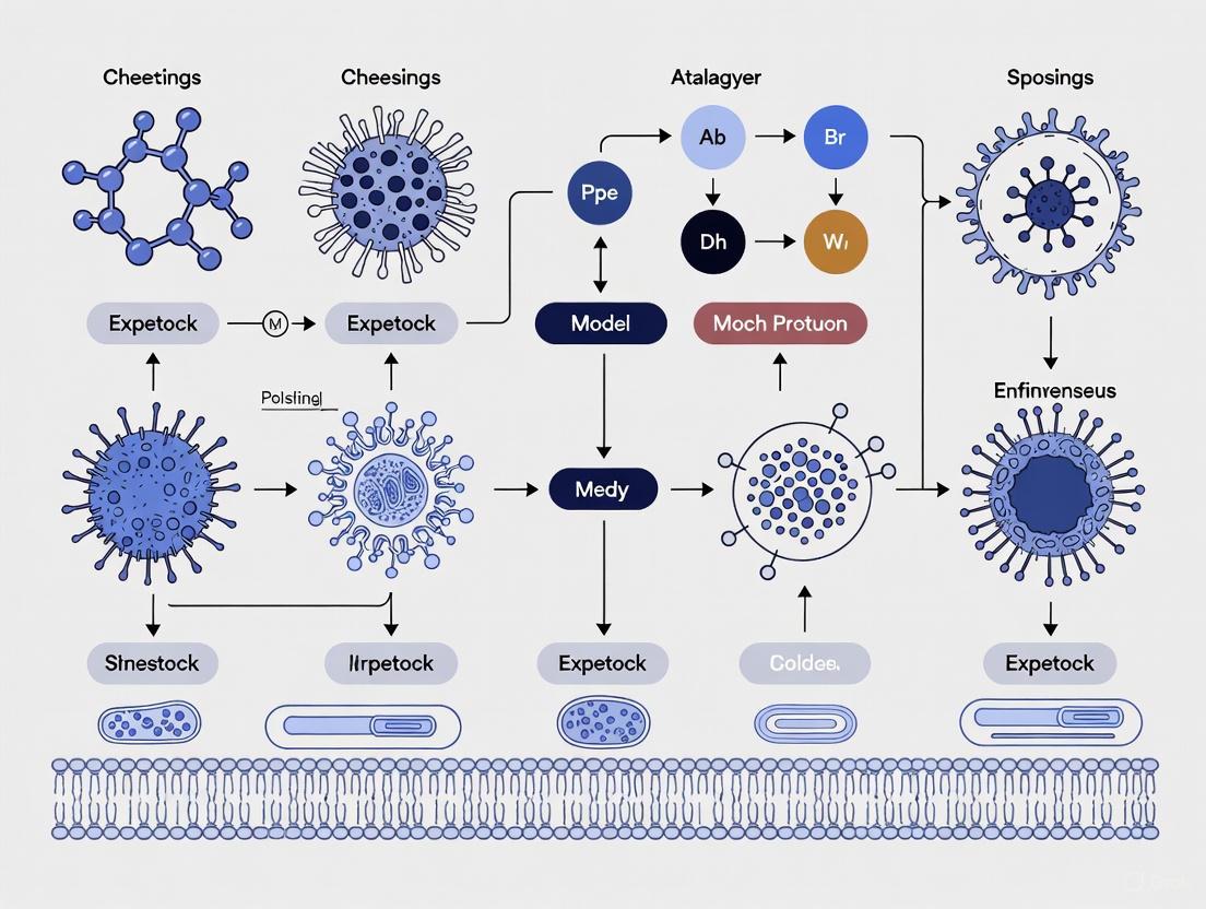
This diagram visualizes the innovative mRNA-based method for generating CAR-T cells inside the body (in situ), a strategy aimed at improving safety and reducing complexity, as demonstrated in a Stanford Medicine-led preclinical study [43].
Logic-Gated CAR-T Cell Activation for Safety
This diagram outlines the "AND-gate" logic mechanism used in next-generation CAR-T cells (e.g., A2B694) to prevent on-target/off-tumor toxicity by requiring two signals for activation [41] [40].
Methodologies for Preclinical Safety Assessment and Risk Mitigation
Preclinical toxicity evaluation is a critical gateway through which all novel therapeutic candidates must pass before entering clinical trials. For engineered therapeutic cells, such as chimeric antigen receptor T-cells (CAR-Ts), accurately defining their safety profile is paramount due to their unique mechanism of action and potential for severe adverse effects like cytokine release syndrome (CRS) and neurotoxicity [45] [46]. This guide objectively compares the performance of established and emerging models and monitoring technologies used to assess the safety of these advanced therapies. The continuous evolution of in vivo models, enhanced by artificial intelligence (AI)-powered prediction tools and sophisticated physiological monitoring devices, is providing researchers with an unprecedented ability to identify and mitigate risks earlier in the development pipeline [47] [48] [49].
Comparative Analysis of In Vivo Models for Cell Therapy Safety Assessment
The choice of in vivo model significantly influences the reliability of safety data collected for engineered therapeutic cells. The table below compares the primary animal models used in this context.
Table 1: Comparison of In Vivo Models for Safety Assessment of Engineered Therapeutic Cells
| Model Type | Common Applications | Key Safety Endpoints Measured | Advantages | Limitations |
|---|---|---|---|---|
| Mouse Models (e.g., immunodeficient, humanized) | Preliminary safety, biodistribution, tumorigenicity, cytokine release assessment [45]. | Off-target toxicity, CRS-like symptoms, cell proliferation/persistence, organ infiltration [45]. | Low cost, well-characterized genetics, high throughput availability. | Often fails to fully predict human immune responses (e.g., CRS, ICANS) due to physiological differences [45]. |
| Rabbit Elastase Aneurysm Model [50] | Safety evaluation of neuroendovascular devices; can be adapted for cell delivery to the CNS. | Device-related thrombosis, inflammation, endothelial healing, aneurysm occlusion [50]. | Pulsatile circulation suitable for evaluating localized biological responses. | Limited replication of human chronic arterial disease; anatomy differs from humans [50]. |
| Swine (Pig) Models [50] | Thrombectomy device safety, evaluation of liquid embolic agents; useful for cardiovascular toxicity of cell therapies. | Acute complications (perforation, vasospasm, distal emboli), chronic sequelae (device migration, restenosis) [50]. | Arterial caliber similar to humans; dense micro-vascular network (rete mirabile) for complex vascular targets [50]. | Intracranial clot insertion is impossible due to rete mirabile; simpler vessel geometry than humans [50]. |
A transformative shift in the field is the move from traditional adoptive cell therapy (where cells are engineered ex vivo) to in vivo cell engineering. This emerging approach involves administering gene delivery vectors directly to the patient to reprogram their own T-cells inside the body, bypassing complex ex vivo manufacturing [45] [46]. While this promises to reduce costs and improve accessibility, its safety profile is distinct. The persistence of in vivo-generated CAR-T cells is currently unknown due to insufficient data, and the ability to control their phenotype is more limited compared to cells pre-differentiated in a lab [45]. Furthermore, while in vivo approaches may avoid the risks of prolonged ex vivo culture, they still carry known risks such as CRS, neurotoxicity (ICANS), and hematotoxicity, alongside the potential for immunogenicity from repeated dosing [45] [46].
Advanced Monitoring Technologies for Real-Time Safety Assessment
Capturing subtle signs of toxicity in real-time requires advanced monitoring technologies. The following table compares several modern physiological monitoring systems.
Table 2: Performance Comparison of Physiological Monitoring Technologies in Preclinical and Clinical Translation
| Technology / Device | Monitoring Capabilities | Key Performance Data | Advantages | Limitations / Factors Affecting Accuracy |
|---|---|---|---|---|
| BioIntelliSense BioButton [51] | Heart rate, respiratory rate, skin temperature. | High agreement with manual vitals (bias ≈ 2 bpm for HR); 73% of deterioration events alerted ~14.8 hrs early [51]. | Minimal alert fatigue (<1/patient/day); centralized monitoring (1 nurse:250 patients) [51]. | Data quality dependent on participant compliance for long-term wear [48]. |
| IR-UWB Radar [52] | Non-contact heart rate (HR), respiratory rate (RR). | RR: CCC* 0.925 vs. capnometry; HR: CCC 0.749 vs. ECG [52]. | Penetrates clothes/light barriers; does not require skin contact [52]. | Sitting position and low HR (<70 bpm) or low RR (<18 breaths/min) increase measurement bias [52]. |
| Sibel ANNE One System [48] | Continuous HR, RR, SpO₂, skin temperature, movement. | Used in large global cohort studies for predictive algorithm development [48]. | Dual sensor system (chest/limb); validated; records for up to 7 days (chest) [48]. | Dependent on Bluetooth and internet for data offloading; requires adhesive stickers [48]. |
| VitalPatch & Other Wearables [51] | Typically HR, RR, activity, sometimes ECG. | Part of a growing category of FDA-cleared devices for remote monitoring [51]. | Ambulatory, continuous data; integrates with cloud analytics platforms [51]. | Performance specifics vary by device; general limitation is battery life and sensor adhesion. |
*CCC: Concordance Correlation Coefficient
These monitoring systems are crucial for detecting preclinical and clinical deterioration. A large-scale observational study demonstrated that continuous monitoring with the BioButton device on medical-surgical units reduced the average hospital length of stay from 3.07 to 2.75 days and provided actionable data that led to 114 documented changes in clinical management, such as medication adjustments and new diagnoses [51]. The "shadow-mode" study design, where data is collected but not initially used for clinical decisions, is a key methodology for validating these technologies without impacting patient care [48].
Experimental Protocols for Key Preclinical Assessments
Protocol: In Vivo CAR-T Safety and Efficacy Study
This protocol is adapted from established models for evaluating traditional and in vivo-generated CAR-T cells [45] [53].
- Animal Model Preparation: Utilize immunodeficient or humanized mouse models bearing patient-derived xenografts (PDX) of the target cancer (e.g., B-cell lymphoma) [45].
- Test Article Administration:
- For Traditional CAR-T: Administer a single intravenous bolus of ex vivo-expanded CAR-T cells at a predetermined dose (e.g., 1-10 million cells/mouse) [45].
- For In Vivo CAR-T: Administer the gene delivery vector (e.g., lentivirus, AAV, or LNP-packaged mRNA) intravenously at a specific titer or dose [45] [46].
- Toxicological Monitoring & Endpoint Assessment:
- Clinical Observations: Monitor daily for signs of toxicity (lethargy, piloerection, weight loss) and CRS (hunching, tremor) using a standardized scoring sheet.
- Physiological Monitoring: Employ wearable sensors (e.g., BioButton) or non-contact radar to continuously track core physiological parameters like heart rate, respiratory rate, and activity levels [52] [51].
- Blood Collection: Perform serial retro-orbital or submandibular bleeding to assess:
- Cytokine Levels: Quantify CRS-associated cytokines (e.g., IL-6, IFN-γ) via multiplex ELISA.
- CAR-T Cell Kinetics: Use flow cytometry to track CAR-T cell expansion, persistence, and immunophenotype in peripheral blood.
- Histopathological Analysis: Upon study termination, harvest and preserve key organs (spleen, liver, lungs, brain) in formalin. Process, section, and stain with H&E for microscopic analysis of organ damage, infiltration, or inflammation.
Protocol: Continuous Physiological Monitoring in a Large Animal Model
This protocol outlines the use of wearable sensors for safety monitoring in large animals, a critical step for translational studies [48].
- Device Selection & Calibration: Select a validated, continuous monitoring wearable device (e.g., Sibel ANNE One). Calibrate the device according to manufacturer specifications before application [48].
- Subject Instrumentation:
- Anesthetize the animal (e.g., swine) following approved institutional protocols.
- Shave the fur from the chest and a limb. Clean the skin with alcohol wipes and allow to dry.
- Apply the chest and limb sensor components using the proprietary adhesive stickers. Ensure firm contact.
- Data Acquisition:
- Initiate data recording on the device. The device should automatically collect physiological waveforms and vital signs (HR, RR, SpO₂, temperature).
- Simultaneously, collect reference vital signs using a standard patient monitor (e.g., ECG for HR, capnometry for RR) [52].
- Record data for a predetermined period (e.g., up to 7 days or until discharge). In "shadow-mode," the device data is not used for clinical decisions [48].
- Data Analysis:
- Synchronize timestamps of the wearable device data and the reference monitor data.
- Calculate agreement metrics (e.g., Concordance Correlation Coefficient, Limits of Agreement) between the wearable-derived vitals and the gold-standard reference values [52].
- Use the collected, time-stamped physiological data to design and train predictive algorithms for early detection of deterioration [48].
Visualizing Workflows and Signaling Pathways
Workflow for AI-Enhanced In Vivo Toxicity Prediction
The following diagram illustrates the sequential knowledge transfer strategy of the MT-Tox model, which integrates chemical and in vitro data to improve in vivo toxicity predictions [47].
Signal Processing in Non-Contact Radar Monitoring
This diagram outlines the signal processing workflow for extracting heart and respiratory rates from impulse-radio ultrawideband (IR-UWB) radar data [52].
The Scientist's Toolkit: Essential Reagents and Materials
Table 3: Key Research Reagents and Solutions for Preclinical Toxicity Evaluation
| Item | Function / Application |
|---|---|
| Humanized Mouse Models | In vivo systems for assessing human-specific immune cell interactions, biodistribution, and toxicity of engineered therapeutic cells [45]. |
| ChEMBL / Tox21 Datasets | Large-scale public databases of bioactive molecules and toxicity assays used for training and validating AI-based prediction models like MT-Tox [47] [49]. |
| Wearable Physiological Sensors (e.g., BioButton, VitalPatch, Sibel ANNE One) | Devices for continuous, real-time monitoring of vital signs (HR, RR, temperature) in animal models or human subjects to detect early signs of physiological deterioration [48] [51]. |
| IR-UWB Radar Sensor | A non-contact technology used to monitor vital signs like HR and RR from a distance, minimizing stress and the risk of disease transmission in preclinical settings [52]. |
| Cytokine Multiplex ELISA Kits | Reagent kits for quantifying a panel of inflammatory cytokines (e.g., IL-6, IFN-γ, IL-2) in serum or plasma to identify and grade cytokine release syndrome (CRS) [45]. |
| Flow Cytometry Antibody Panels | Antibodies specific to human and animal immune cell markers (e.g., CD3, CD4, CD8, CAR detection tags) for tracking the persistence, expansion, and phenotype of administered therapeutic cells in vivo [45]. |
| LNP or Viral Vectors (e.g., AAV, Lentivirus) | Delivery systems for in vivo genetic engineering of T-cells to express CARs, enabling the study of in vivo-generated CAR-T cell therapies and their associated toxicities [45] [46]. |
Assessing Oncogenic and Tumorigenic Potential in Immunocompromised Models
Immunocompromised animal models serve as indispensable tools in preclinical oncology and cell therapy research, providing a foundational platform for assessing the oncogenic and tumorigenic potential of novel therapeutic cells [54] [55]. These models, which range from nude mice to highly immunodeficient NOD/SCID and NSG strains, enable the in vivo study of human cell behavior by circumventing xenogeneic rejection [54] [56]. Within the context of comparative safety profiling for engineered therapeutic cells, these models allow researchers to quantify critical risk parameters including tumor initiation capacity, metastatic potential, and the self-renewal capabilities of cancer stem cells (CSCs) [54] [55]. Understanding the strengths, limitations, and appropriate applications of these models is paramount for accurately predicting clinical safety and de-risking the translation of cellular therapies from bench to bedside.
The selection of an appropriate immunocompromised model directly influences experimental outcomes and safety predictions. Research demonstrates that the specific immune deficiencies in different models create distinct selective pressures that shape tumor evolution and immunogenicity [57] [58]. For instance, tumors developing in immunodeficient environments may exhibit enhanced immunogenicity upon transfer to immunocompetent hosts, a phenomenon with significant implications for assessing the therapeutic window of engineered cells [57]. This comparative guide examines the operational characteristics, experimental applications, and methodological considerations for leveraging immunocompromised models in comprehensive oncogenic risk assessment.
Comparative Analysis of Immunocompromised Models
Model Classification and Key Characteristics
Immunocompromised models exhibit distinct evolutionary pathways reflecting progressive advancements in immunodeficiency engineering. The development began with nude mice, discovered in the 1960s with characteristic thymic dysgenesis leading to T-cell deficiency [54]. Subsequent milestones included SCID mice identified in 1983 with defects in both T- and B-lymphocytes, and NOD/SCID mice developed in 1992 which combined the SCID mutation with non-obese diabetic background, resulting in reduced NK cell activity and less mature macrophage populations [54]. Contemporary innovations include humanized mouse models such as huNOG and huNOG-EXL, which incorporate human immune systems to better mimic human-specific immune interactions [59].
Table 1: Classification and Characteristics of Immunocompromised Models
| Model Type | Genetic Background/Defect | Immune Deficiencies | Strengths | Limitations |
|---|---|---|---|---|
| Nude Mice | Foxn1 mutation | T-cell deficiency | Historical reliability, Cost-effective | Limited human cell engraftment, Retained NK cell and macrophage activity |
| SCID Mice | Prkdcscid mutation | T- and B-cell deficiency | Improved engraftment over nude mice | Functional NK cells and macrophages, Radiation-sensitive |
| NOD/SCID Mice | Prkdcscid on NOD background | T- and B-cell deficiency, Reduced NK cell activity, Less mature macrophages | Enhanced engraftment efficiency, Gold standard for CSCs assays [54] | Residual innate immunity, Limited lifespan |
| NSG Mice (NOD-scid gamma) | Prkdcscid, IL2rgnull on NOD background | T-, B-, and NK cell deficiency, Defective dendritic cell function | Superior engraftment rates, Support human immune system reconstitution | Higher cost, Increased susceptibility to opportunistic infections |
| Humanized Mice (huNOG, huNOG-EXL) | Immunodeficient base with engrafted human immune cells | Mouse immunity absent, Functional human immune system present | Clinically relevant immune context, Allows immunotherapy testing [59] | Technical complexity, Variable human immune cell reconstitution |
| Immunocompromised Pigs | RAG2/IL2RG knockout via CRISPR/Cas9 [60] | B-, T-, NK cell deficiency (B-T-NK-SCID phenotype) | Human-scale tumor growth, Anatomical relevance for device testing [60] | Specialized housing requirements, Limited commercial availability |
Quantitative Performance Metrics Across Models
The engraftment efficiency and tumor growth kinetics vary significantly across immunocompromised models, directly impacting their utility for specific research applications. NOD/SCID models demonstrate approximately 50-fold higher initiating-cell frequencies compared to immunocompetent controls in leukemia studies, with reported frequencies of <1 in 9,300 for NUP98∷NSD1-driven leukemia initiating cells versus 1 in 830,000 in control models [61]. In pancreatic cancer research, RAG2/IL2RG deficient pigs showed 100% engraftment success with Panc01 cells, achieving target tumor diameters of 1.0–1.6 cm within 36 days post-injection [60].
Table 2: Functional Performance Metrics of Immunocompromised Models
| Application Domain | Model of Choice | Key Metrics | Representative Findings |
|---|---|---|---|
| Cancer Stem Cell (CSC) Research | NOD/SCID mice [54] | Limiting dilution analysis, Self-renewal capacity | CD44+ population in HNSCC demonstrated self-renewal and differentiation [54]; ALDHhigh cells showed enhanced tumorigenicity [54] |
| Metastasis Studies | SCID mice [54] | Invasion, Angioinvasion, Distant metastasis formation | Orthotopic OSC-19 models showed cervical lymph node metastasis inhibited by cisplatin [54]; IL-6 promoted metastasis via JAK-STAT3-SNAIL pathway [54] |
| Therapeutic Response Evaluation | Humanized mice (huNOG/huNOG-EXL) [59] | Tumor growth inhibition, Immune profiling, Metastasis reduction | Enzalutamide blocked metastasis in humanized models but accelerated spread in immunocompromised settings [59]; Combination with pembrolizumab induced complete regression [59] |
| Oncogenic Signaling Investigation | Nude mice [54] | Tumor growth kinetics, Angiogenesis, Pathway inhibition | HIF-1α/HIF-2α knockdown inhibited xenograft angiogenesis; VEGF-C/VEGF-D suppression inhibited lymphatic metastasis [54] |
| Device Development & Ablation Studies | Immunocompromised pigs [60] | Tumor size, Electrical properties, Treatment response | Generated ample tumor tissue (1.0-1.6 cm) for accurate electrical property modeling of pancreatic cancer [60] |
Experimental Approaches and Methodological Frameworks
Standardized Protocols for Oncogenicity Assessment
The assessment of oncogenic potential follows established methodological frameworks that leverage the unique capabilities of immunocompromised models. The tumorigenicity testing workflow typically begins with cell preparation, followed by implantation into appropriately selected models, and concludes with comprehensive endpoint analyses. For cancer stem cell research, the NOD/SCID mouse model represents the gold standard assay, with studies demonstrating that CD44+ populations from primary human HNSCC samples possess unique self-renewal and differentiation capacities when transplanted into these models [54]. Similarly, ALDHhigh activity has been validated as a selective CSC marker in HNSCC through NOD/SCID transplantation assays [54].
For therapeutic cell safety assessment, researchers typically employ a combination of in vitro methods and in vivo models in immunocompromised animals to analyze risks of oncogenicity, tumorigenicity, and teratogenicity [55]. Biodistribution studies utilizing quantitative PCR and imaging techniques (PET, MRI) provide critical data on cell fate over time, while histological examination of transplantation sites and major organs (liver, lungs, kidneys) identifies potential pathological changes [55]. The selection of administration route—whether subcutaneous, orthotopic, or systemic—should reflect the intended clinical application to ensure relevance and reliability of results [55].
Advanced Methodologies for Tumorigenic Potential Assessment
Limiting Dilution Assays for Cancer Stem Cell Quantification
The limiting dilution assay (LDA) represents a critical quantitative approach for determining the frequency of tumor-initiating cells within a population. In practice, serial dilutions of test cells are implanted into NOD/SCID or NSG mice, with the percentage of tumor-free animals at each dilution point used to calculate stem cell frequency using statistical models like Poisson distribution [61]. Secondary transplantation further assesses self-renewal capacity, a defining characteristic of CSCs. Research on NUP98∷NSD1-driven leukemia demonstrated dramatic differences in initiating-cell frequencies between immunocompromised and immunocompetent settings, with FL NUP98∷NSD1 xenografts exhibiting >50-fold higher initiating-cell frequencies (<1 in 9,300 cells) compared to controls (1 in 830,000 cells) [61].
Orthotopic Transplantation for Metastasis Research
Orthotopic implantation techniques, where tumor cells are injected into the anatomically correct tissue of origin, significantly enhance the clinical relevance of metastasis studies. In head and neck squamous cell carcinoma research, orthotopic transplantation of OSC-19 cell lines into the tongues of nude mice produced invasive growth patterns and cervical lymph node metastases that closely mimicked human disease progression [54]. This approach demonstrated that cisplatin or peplomycin treatment markedly inhibited cervical lymph node metastasis in this model [54]. Similarly, bone invasion models created by injecting UM-SCC-1 cell lines into the mylohyoid muscle of nude mice enabled quantification of tumor invasion into bone [54].
Longitudinal Monitoring with Advanced Imaging
Modern oncogenicity assessment increasingly incorporates in vivo imaging technologies for longitudinal monitoring of tumor development and metastasis. Bioluminescent imaging, utilizing luciferase-transfected cells, enables non-invasive tracking of tumor growth and therapeutic response [54]. Studies with SAS/luc cells in NOD/SCID mice demonstrated significant inhibition of tumor growth by curcumin, highlighting the utility of this approach for therapy evaluation [54]. Additionally, ultrasound imaging in immunocompromised porcine models provided clear tumor delineation from surrounding tissues, with tumor measurements collected extradermally, subdermally, and via ultrasound showing strong correlation [60].
Key Signaling Pathways in Tumorigenesis and Immune Evasion
Immunocompromised models have been instrumental in elucidating critical signaling pathways driving tumor development and immune evasion mechanisms. Research utilizing these models has identified several pivotal pathways that influence oncogenic potential and therapeutic responses.
The PI3K/AKT/mTOR pathway emerges as a central regulator of tumor development, with studies in NOD/SCID mice demonstrating that rapamycin and its analog RAD001 diminish lymphangiogenesis in primary tumors and prevent dissemination of HNSCC cells to cervical lymph nodes [54]. Hypoxia-inducible factors (HIF-1α and HIF-2α) play complementary roles in tumor angiogenesis, with simultaneous knockdown producing superior inhibitory effects on xenograft growth compared to individual unit knockdown [54]. The VEGF-C/VEGF-D signaling axis specifically mediates lymphatic metastasis, with local hyperthermia shown to suppress these factors and consequently inhibit cancer cell spread in tongue SCC models [54].
Cytokine signaling, particularly through IL-6, promotes metastatic progression via induction of epithelial-mesenchymal transition (EMT) through the JAK-STAT3-SNAIL pathway in SCID mouse models [54]. Immune checkpoint regulation, especially the PD-1/PD-L1 axis, facilitates immune escape in humanized systems, with PI3K/AKT pathway stimulation and IFN-γ exposure promoting PD-L1 expression on tumor cells [62]. Developmental pathways, including HOX gene clusters, drive self-renewal and block differentiation in leukemia models, with NUP98∷NSD1 fusions inducing strong enrichment of HOX cluster genes in fetal-derived hematopoietic stem cells [61].
Essential Research Reagents and Materials
The effective implementation of immunocompromised models requires specialized reagents and materials tailored to the unique requirements of these systems. The following table details critical components of the research toolkit for oncogenicity assessment.
Table 3: Essential Research Reagents and Experimental Materials
| Reagent/Material Category | Specific Examples | Research Application | Function/Purpose |
|---|---|---|---|
| Immunodeficient Animal Models | Nude mice (Foxn1mut), SCID (Prkdcscid), NOD/SCID, NSG (NOD-scid gamma), RAG2/IL2RG deficient pigs [54] [60] | In vivo tumorigenicity testing, Metastasis studies, Therapy evaluation | Provide permissive environment for human cell engraftment and tumor development |
| Humanized Mouse Models | huNOG, huNOG-EXL [59] | Immunotherapy testing, Human-specific immune interactions | Reconstitute human immune system for clinically relevant immune context |
| Cell Line Panels | OSC-19 (oral SCC), CAL27 (oral SCC), CT26.WT (colon carcinoma), K1735 (melanoma), Panc01 (pancreatic adenocarcinoma) [54] [57] [58] | Tumor growth kinetics, Invasion assays, Metastasis modeling | Provide standardized, reproducible tumor cells for transplantation studies |
| Specialized Culture Media | Serum-free stem cell media (SCM), Melanoma serum-free stem cell media (MSCM), Methylcellulose colony-forming media [61] [58] | Cancer stem cell enrichment, Sphere formation assays, Clonogenicity assessment | Maintain stemness properties, Support anchorage-independent growth |
| In Vivo Imaging Reagents | Luciferase-transfected cells (SAS/luc), Ultrasound contrast agents [54] [60] | Longitudinal tumor monitoring, Metastasis tracking, Therapy response assessment | Enable non-invasive quantification of tumor burden and distribution |
| Molecular Analysis Tools | CRISPR/Cas9 genome editing system, GoT-ChA single-cell genotyping, Liquid chromatography-tandem mass spectrometry (LC-MS/MS) [61] [57] | Genetic modification, Clonal tracking, Proteomic profiling | Enable precise genome engineering and multidimensional characterization |
| Cell Isolation & Processing Reagents | Collagenase-hyaluronidase solution, DNase solution, Fluorescent-activated cell sorting (FACS) antibodies (CD44, CD133, ALDH substrates) [54] [57] [58] | Tumor digestion, Stem cell isolation, Population purification | Facilitate tissue processing and specific cell population isolation |
Comparative Insights and Research Implications
Model Selection Guidance for Specific Research Objectives
The selection of an appropriate immunocompromised model should align with specific research objectives and account for the distinctive advantages and limitations of each system. For cancer stem cell research and tumor initiation studies, NOD/SCID models remain the gold standard, with demonstrated utility in identifying and quantifying CSC populations through limiting dilution assays [54] [58]. For metastasis and invasion research, SCID models with orthotopic implantation provide valuable platforms for investigating molecular mechanisms of spread, particularly for bone invasion in HNSCC [54]. For therapeutic development and immunotherapy testing, humanized models (huNOG, huNOG-EXL) offer unparalleled clinical relevance, enabling evaluation of immune-mediated mechanisms and combination therapies [59].
The developmental stage of target cells significantly influences their oncogenic potential, as demonstrated by studies showing that fetal-derived hematopoietic stem cells readily transform into leukemia with NUP98∷NSD1 fusions, while stem cells from later developmental stages become progressively resistant to transformation [61]. This ontogeny-dependent transformation susceptibility highlights the importance of selecting biologically relevant cell sources for transplantation studies.
Limitations and Alternative Considerations
While immunocompromised models provide invaluable insights, researchers must acknowledge their inherent limitations. Tumors developing in immunodeficient hosts may exhibit altered immunogenicity, with studies demonstrating that CT26 tumors grown in NOD.SCID mice regressed upon reinoculation into immunocompetent hosts due to increased immunogenicity [57]. Similarly, K1735 melanoma cell lines with high tumorigenic potential in SCID mice spontaneously regressed in syngeneic C3H/HeN mice, suggesting enhanced immunogenicity of aggressively tumorigenic cells [58].
These findings highlight the critical importance of recognizing that immunocompromised models, while essential for assessing intrinsic tumorigenic potential, cannot fully replicate the complex immune interactions of clinical settings. Combining results from immunocompromised models with data from syngeneic or humanized systems provides a more comprehensive safety profile, particularly for therapeutic cells with immunomodulatory properties [59] [56]. This integrated approach ensures robust preclinical safety assessment while acknowledging the biological complexities of human tumor development.
Biodistribution studies are a cornerstone in the non-clinical safety assessment of Cell Therapy Products (CTPs). They provide essential data on the movement, persistence, and localization of administered cells within the body, which is critical for predicting and assessing both efficacy and toxicity profiles [63]. For regenerative medicine therapies using engineered therapeutic cells, understanding biodistribution is particularly vital for evaluating critical risks such as tumorigenicity arising from undifferentiated pluripotent stem cells, ectopic tissue formation, and unwanted immune responses [63] [55]. Regulatory agencies worldwide, including the FDA, EMA, and Japan's PMDA, emphasize the importance of these studies for determining cell fate—encompassing survival, engraftment, distribution, differentiation, and integration [63]. The fundamental question these studies address is whether the administered cells localize to the intended target tissues and for how long they persist, or if they migrate to non-target organs where they could potentially cause adverse effects.
Comparative Analysis of Biodistribution Methodologies
The two primary technical approaches for tracking cells in biodistribution studies are quantitative Polymerase Chain Reaction (qPCR) and non-invasive imaging, notably Positron Emission Tomography (PET) and Magnetic Resonance Imaging (MRI). Each method offers distinct advantages, limitations, and applications, making them suitable for different stages of the product development pipeline.
Quantitative PCR (qPCR) for Biodistribution Assessment
qPCR is a widely used, highly sensitive molecular biology technique primarily applied in non-clinical biodistribution studies. It functions by detecting and quantifying specific DNA sequences unique to the administered cells, such as a transgene or a genetic marker not present in the host organism [63].
- Experimental Protocol: The standard methodology involves administering the CTP to animal models, followed by sacrifice and collection of target and non-target organs at predetermined time points. Tissue samples are processed for genomic DNA extraction. The qPCR assay is then run using primers and probes designed to target the specific marker sequence. Quantification is achieved by comparing the cycle threshold (Ct) values of samples to a standard curve generated from known quantities of the target DNA [63]. Data is typically expressed as the number of genome equivalents or cell equivalents per microgram of total DNA or per gram of tissue.
- Key Characteristics:
- High Sensitivity: Capable of detecting a very small number of cells, which is crucial for assessing low-level engraftment or distribution [63].
- Quantitative and Specific: Provides precise numerical data on cell numbers in various tissues with high specificity for the administered cells.
- Destructive and Terminal: Requires euthanasia of animals at each time point, preventing longitudinal follow-up in the same subject. This necessitates larger group sizes to obtain time-course data [63].
- Lacks Spatial Context: While it quantifies cell presence in a tissue homogenate, it does not provide information on the precise spatial localization of cells within an organ (e.g., perivascular, parenchymal).
Imaging Technologies: PET and MRI
Non-invasive imaging allows for the longitudinal tracking of the same animal or subject over time, providing dynamic data on cell migration and persistence.
Positron Emission Tomography (PET) utilizes radiotracers (e.g., 18F-FDG, 18F-FCH, 89Zr, 111In) that emit positrons. Cells can be tracked by either direct radiolabeling or by using reporter genes [64].
- Experimental Protocol for Direct Labeling: Cells are labeled ex vivo with a radiotracer like 89Zr-oxine or 111In-oxine. After washing, the labeled cells are administered to the subject. Serial PET scans are then performed over time to detect the location of the radioactive signal. The signal intensity can be semi-quantified using standardized uptake values (SUV) [65] [64]. A key limitation is that the signal represents the radiotracer, not necessarily viable cells; signal loss can occur due to radionuclide decay, cell death, or tracer dilution through cell division [64].
- Key Characteristics:
- Very High Sensitivity: Can detect picomolar concentrations of tracer, requiring a relatively low number of labeled cells [64].
- Excellent for Whole-Body Screening: Ideal for identifying unexpected sites of cell distribution [65].
- Limited Anatomical Detail: Often combined with CT or MRI (PET/CT or PET/MRI) for anatomical correlation [65] [66]. For instance, PET/CT is superior for lung metastasis monitoring, while PET/MRI performs better for liver and bone [65].
- Signal is Not Necessarily Cell-Specific: As with all direct labels, released radiotracer can be taken up by host phagocytic cells, leading to false positives [64].
Magnetic Resonance Imaging (MRI) provides exceptional soft-tissue contrast and anatomical resolution. Cell tracking requires labeling cells with contrast agents, typically superparamagnetic iron oxide (SPIO) nanoparticles for T2/T2* weighting, or gadolinium chelates for T1 weighting [64].
- Experimental Protocol: Cells are labeled with MRI contrast agents in culture. After administration, the subject is scanned using high-resolution MRI sequences. The contrast agents create a local disturbance in the magnetic field, appearing as dark (for SPIOs) or bright (for gadolinium) spots on the image [64].
- Key Characteristics:
- High Spatial Resolution: Provides detailed anatomical context, allowing precise localization of cells within tissue structures [64].
- No Ionizing Radiation: Enables repeated scanning without radiotoxicity concerns.
- Lower Sensitivity: Requires a high local concentration of contrast agent (typically >10^5 cells) for detection, making it less suitable for tracking single cells or small clusters [64].
- Ambiguity of Signal: Hypointense signals from SPIOs can be difficult to distinguish from other hypointense features like bleeding or calcifications [64].
The following diagram illustrates the core decision-making workflow for selecting and implementing these primary biodistribution methodologies.
Head-to-Head Technical Comparison
The table below provides a consolidated, data-driven comparison of the key techniques based on performance metrics and practical considerations.
Table 1: Technical Comparison of Biodistribution Assessment Methods
| Feature | qPCR | PET Imaging | MRI Imaging |
|---|---|---|---|
| Primary Metric | Cell number per mass of tissue [63] | Spatial distribution & intensity (SUV) [65] [67] | Spatial distribution & hypointense/hyperintense regions [64] |
| Sensitivity | Very High (Can detect a small number of cells) [63] | Very High (Picomolar tracer concentration) [64] | Low (Requires > 10^5 cells per voxel) [64] |
| Spatial Resolution | None (Tissue homogenate) | Low (4-5 mm) [66] | High (50-100 µm) [64] |
| Quantification | Excellent (Absolute cell numbers) [63] | Good (Semi-quantitative, e.g., SUV) [66] | Poor (Qualitative or semi-quantitative) [64] |
| Longitudinal Tracking | No (Terminal procedure) | Yes (Ideal for dynamic studies) [64] | Yes (Ideal for dynamic studies) [64] |
| Key Advantage | Gold standard for sensitivity and quantification [63] | Excellent for whole-body screening and deep-tissue detection [65] | Superior anatomical context and soft-tissue contrast [64] |
| Key Limitation | No spatial data; requires animal sacrifice per time point [63] | Radiation exposure; poor anatomical detail without CT/MRI [65] | Low sensitivity; ambiguous signal interpretation [64] |
Advanced and Multimodal Imaging Approaches
To overcome the limitations of individual techniques, the field is increasingly adopting advanced and integrated approaches.
Hybrid Imaging: PET/CT and PET/MRI
Combining modalities leverages the strengths of each to provide more comprehensive data.
- PET/CT: Integrates the high sensitivity of PET for detecting active cells with the detailed anatomical mapping of CT. This is particularly valuable for pinpointing the location of metastatic lesions, with studies showing it can change treatment plans in one-third of patients with advanced cancer by identifying metastases missed by conventional imaging [65].
- PET/MRI: A more recent innovation that simultaneously acquires metabolic PET data and high-contrast morphological MRI. This hybrid approach has demonstrated increased diagnostic accuracy over MRI alone. For example, in prostate cancer diagnosis, a meta-analysis found PET/MRI had a significantly higher area under the curve (AUC) than multiparametric MRI (0.93 vs. 0.84) [68]. PET/MRI is also superior to PET/CT for detecting liver and bone metastases [65].
The Promise of Multispectral Optoacoustic Tomography (MSOT)
MSOT is an emerging modality that detects acoustic waves generated by light absorption, overcoming the light-scattering limitations of traditional optical imaging. It offers several advantages for regenerative medicine:
- Improved Depth Penetration: Allows for whole-body imaging in mice at depths of several centimeters [64].
- Multispectral Unmixing: By imaging at multiple wavelengths, it can differentiate signals from multiple contrast agents or endogenous molecules (e.g., oxy-hemoglobin) simultaneously [64].
- High Spatial and Temporal Resolution: Enables real-time, quantitative functional assessment in addition to cell tracking [64].
Essential Experimental Protocols and Reagents
Successful execution of biodistribution studies relies on standardized protocols and high-quality reagents. The following section outlines critical methodologies and tools.
Detailed ex vivo Biodistribution Protocol with Radiotracers
This protocol is a cornerstone for quantitative validation of imaging data, particularly in radiopharmaceutical development [67].
- Radiotracer Administration: Precisely measure the activity and mass of the formulated radiotracer solution in the syringe before and after injection to calculate the exact injected dose per animal. Using a calibrated analytical balance is recommended for accuracy [67].
- Tissue Harvesting: At designated time points post-injection, euthanize animals and systematically collect tissues of interest (e.g., target organ, liver, spleen, kidneys, lungs, blood). Tissues should be rinsed, blotted dry, and weighed [67].
- Radioactivity Counting: Place each tissue into pre-weighed tubes and measure radioactivity using a calibrated automatic gamma counter. The counter must be preset with the appropriate energy window for the isotope and apply decay correction [67].
- Data Processing and Analysis: Calculate the percentage of Injected Dose per gram of tissue (%ID/g) using the formula:
(Activity in tissue [kBq] / Mass of tissue [g]) / Injected Dose [kBq] * 100. Data can also be expressed as Standardized Uptake Value (SUV):(Activity in tissue [kBq] / Mass of tissue [g]) / (Injected Dose [kBq] / Body Weight [g])[67].
The Scientist's Toolkit: Key Research Reagent Solutions
Table 2: Essential Reagents and Materials for Biodistribution Studies
| Item | Function | Application Notes |
|---|---|---|
| Species-Specific qPCR Assay | Detects and quantifies a unique DNA sequence (e.g., transgene, Alu repeat) from administered cells in host tissue [63]. | Requires rigorous validation for specificity, sensitivity, and linearity. A standard curve from known cell numbers is essential [63]. |
| Radiotracers (e.g., 18F-FDG, 89Zr-oxine) | Serves as a beacon for PET imaging. 18F-FDG measures metabolic activity; direct labels like 89Zr-oxine are incorporated into cells for tracking [65] [64]. | Short half-life isotopes (e.g., 18F) require on-site cyclotron. Direct labels can be toxic to cells and do not proliferate with cell division [64]. |
| MRI Contrast Agents (e.g., SPIOs) | Alters local magnetic properties, creating contrast for MRI. SPIOs generate strong hypointense (dark) signals on T2/T2*-weighted images [64]. | High loading is needed for detection. Signal can be ambiguous and does not indicate cell viability [64]. |
| Calibrated Analytical Balance | Precisely measures the mass of the injected radiotracer dose, which is critical for accurate %ID/g calculation [67]. | Accuracy to at least four decimal places is preferred. Shielding should be used to adhere to ALARA radiation safety principles [67]. |
| Calibrated Gamma Counter | Quantifies radioactivity in excised tissue samples for ex vivo biodistribution analysis [67]. | Must be calibrated for the specific radioisotope used. Regular linearity checks are required to ensure accuracy across the activity range [67]. |
In the context of comparative safety profiles for engineered therapeutic cells, no single biodistribution method provides a complete picture. A strategic, integrated approach is paramount. The optimal framework combines the high sensitivity and quantitative power of qPCR with the spatial and longitudinal capabilities of non-invasive imaging. For instance, initial whole-body PET/CT screening can identify potential off-target sites, which can then be quantitatively confirmed and validated using the more sensitive qPCR assay on collected tissues. Similarly, the exceptional anatomical detail from MRI can provide context for localized engraftment, which can be further quantified by qPCR. As regulatory guidelines emphasize, understanding the "cell fate" — where cells go, how long they survive, and what they become — is indispensable for de-risking the development of cell-based therapies [63]. Employing a multimodal biodistribution strategy is therefore not just a technical choice, but a critical component in building a robust safety thesis that can support successful clinical translation.
In Silico and In Vitro Approaches for Predicting Cross-Reactivity
The advancement of engineered therapeutic cells, such as CAR-T and CAR-NK cells, represents a transformative approach in treating cancer and other diseases. However, their development is fraught with safety challenges, particularly the risk of unintended immune responses triggered by cross-reactivity. Cross-reactivity occurs when T-cell receptors (TCRs) or chimeric antigen receptors (CARs) recognize not only their intended target epitopes but also structurally similar peptides presented on major histocompatibility complexes (pMHC) on healthy cells. This molecular mimicry can lead to potentially severe off-target toxicities, including autoimmune-like reactions and damage to healthy tissues.
Within the context of comparative safety profiles of engineered therapeutic cells, predicting cross-reactivity becomes paramount for both preclinical development and clinical application. The scientific community has increasingly addressed this challenge through integrated methodologies that combine in silico predictions with in vitro validations. These approaches allow researchers to identify potential cross-reactivity risks earlier in the development pipeline, potentially preventing adverse events in clinical trials and improving the overall safety profile of cellular therapies. This guide systematically compares the leading experimental and computational approaches for cross-reactivity prediction, providing researchers with actionable protocols and analytical frameworks for safety assessment.
Comparative Analysis of Cross-Reactivity Prediction Approaches
The table below summarizes the primary methodologies used for predicting cross-reactivity, their key features, and their applications in therapeutic cell development.
| Methodology | Key Principle | Therapeutic Context | Primary Output | Key Advantages |
|---|---|---|---|---|
| MatchTope (In Silico) [69] | Calculates & compares molecular electrostatic potentials (MEP) of pMHC complexes | Vaccine development, cancer immunotherapy, TCR therapy | Hierarchical clustering of pMHCs by electrostatic similarity | Accounts for 3D structure & physicochemistry; can predict cross-reactivity between low-sequence-similarity peptides |
| In Vitro T-cell Activation Assays | Measures T-cell activation (e.g., cytokine release, proliferation) when exposed to target vs. off-target peptides | Safety assessment for engineered TCRs and CAR-T cells | Quantitative activation metrics (e.g., % cytotoxicity, cytokine concentration) | Provides direct functional readout of immune cell activity; accounts for biological complexity |
| Cytotoxicity Assays [70] | Co-cultures engineered immune cells with target and non-target cells to measure specific killing | Preclinical safety profiling of CAR-NK, CAR-T, and CAR-γδ T cells | Percentage of specific cell lysis or tumor cell killing | Directly measures the most critical functional outcome for cytotoxic therapies |
| Exhaustion Profiling [70] | Single-cell RNA sequencing to assess transcriptional signs of T-cell exhaustion | Evaluating long-term persistence and safety of CAR-T cells | Exhaustion scores and expression profiles of checkpoint genes | Predicts potential for functional failure and reduced tumor control over time |
In Silico Prediction Tools and Workflows
MatchTope: A Structural Bioinformatics Tool for Cross-Reactivity Prediction
MatchTope is a computational tool designed to predict peptide similarity that can trigger cross-reactivity events by analyzing the three-dimensional structural and electrostatic properties of pMHC complexes, rather than relying solely on linear peptide sequences [69]. This approach is significant because traditional sequence-based methods often fail to predict cross-reactivity between peptides that share low amino acid identity but maintain similar structural and electrostatic properties in the TCR-binding interface.
The underlying principle of MatchTope is that T-cell receptor (TCR) recognition depends heavily on the complementarity of electrostatic potentials at the TCR-pMHC interface. The tool uses the following technical workflow:
- Input Preparation: Users provide a set of pMHC complex files in PDB format. These can be experimentally determined structures or modeled complexes.
- Structural Alignment: All input structures are superimposed onto a reference pMHC structure to ensure consistent spatial orientation for comparison.
- Electrostatic Calculation: The standalone PIPSA (Protein Interaction Property Similarity Analysis) software calculates the molecular electrostatic potential (MEP) for each aligned pMHC complex.
- Focused Analysis: Similarity computation is focused on a cylindrical region encompassing the pMHC binding cleft, reducing noise from identical structural surroundings.
- Clustering: Finally, pMHC complexes are clustered based on their MEP similarity indices using hierarchical clustering with bootstrap validation.
MatchTope analysis workflow for predicting pMHC cross-reactivity.
Experimental Validation of Computational Predictions
MatchTope has been validated against multiple experimental datasets. In one validation study using a Hepatitis E Virus (HEV)-specific TCR and various epitopes, MatchTope successfully clustered epitopes known to trigger cross-reactivity based on their electrostatic similarity, demonstrating strong agreement with in vitro results [69]. This confirms that structural electrostatic similarity, rather than mere sequence alignment, is a robust predictor of functional cross-reactivity.
The tool is particularly valuable in vaccine development for checking efficacy across pathogen subtypes and in cancer immunotherapy for ensuring that tumor-targeting therapies do not cross-react with self-proteins. Its ability to predict cross-reactivity between peptides with less than 50% sequence identity makes it superior to many sequence-based predictors [69].
In Vitro Experimental Approaches for Validation
Cytotoxicity and Safety Assessment Protocols
While in silico tools provide valuable initial screening, in vitro functional assays remain essential for experimentally validating cross-reactivity predictions. The following protocols represent standardized methodologies for assessing the efficacy and safety of engineered therapeutic cells.
In Vitro Repetitive Tumor Challenge Assay This protocol is used to evaluate CAR-T cell functionality, persistence, and exhaustion under conditions that mimic chronic antigen exposure, which is particularly relevant for predicting long-term safety and efficacy profiles [71].
Workflow for in vitro repetitive tumor challenge assay.
Key Readouts and Safety Metrics:
- Tumor Cell Killing: Measured via flow cytometry every 48 hours; sustained killing >90% indicates potent, persistent activity [71].
- T Cell Expansion: Total T cell counts at each challenge; IL-15 fusions typically induce the highest expansion [71].
- Activation/Exhaustion Markers: 4-1BB MFI (activation) and TIM-3 expression (IL-12 signaling marker) are tracked [71].
- Cytokine Secretion: IFNγ is measured by ELISA; sustained elevation with IL-12 fusions indicates potent Th1 response [71].
Comparative Safety Profiling of Engineered Immune Cells Different engineered immune cell types demonstrate distinct safety profiles, which can be quantitatively compared using standardized in vitro and in vivo assessments [70]:
| Cell Type | GvHD Risk | CRS Risk | Exhaustion Profile | Therapeutic Advantages |
|---|---|---|---|---|
| CAR-αβ T cells [70] | High | High | Moderate | Established manufacturing, potent efficacy |
| CAR-Vδ2 γδ T cells [70] | Low | Low | Higher exhaustion | MHC-independent recognition |
| CAR-Vδ1 γδ T cells [70] | None observed | None observed | Lower exhaustion | Favorable safety profile, tissue tropism |
| CAR-NK cells [72] | Low | Significantly reduced | N/A | "Off-the-shelf" potential, multiple activation mechanisms |
Engineering Approaches to Enhance Safety
Several engineering strategies have been developed to mitigate cross-reactivity and other safety concerns:
Immune-Evasive CAR-NK Cells Researchers from MIT and Harvard have engineered CAR-NK cells with a multi-gene construct that simultaneously enables immune evasion and enhances antitumor activity [72]. The engineering strategy includes:
- siRNA against HLA Class I: Reduces surface expression of HLA proteins to evade host T-cell rejection.
- PD-L1 or single-chain HLA-E (SCE): Enhances cancer-killing ability and persistence.
- All-in-one construct: Simplifies manufacturing and ensures co-expression of all modifications.
In mouse models with humanized immune systems, these engineered NK cells persisted for at least three weeks and nearly eliminated cancer, while unmodified NK cells were rejected within two weeks [72]. This approach significantly reduces the risk of cytokine release syndrome compared to CAR-T therapies.
Bifunctional Fusion Proteins for CAR-T Cells Another advanced engineering approach involves arming CAR-T cells to secrete bifunctional fusion proteins that combine immunomodulatory factors with checkpoint inhibition [71]. For example:
- αPD-L1–IL-12 fusion: Demonstrates superior safety and efficacy compared to other fusions (αPD-L1–TGFβtrap, αPD-L1–IL-15).
- Tumor-localized activity: PD-L1 binding helps sequester IL-12 within the tumor microenvironment, reducing systemic exposure and toxicity.
- Enhanced antitumor response: Improves T-cell trafficking, tumor infiltration, and local IFNγ production while minimizing systemic inflammation.
The Scientist's Toolkit: Essential Research Reagents
The table below catalogues essential reagents and tools used in the featured studies for cross-reactivity prediction and safety assessment of engineered therapeutic cells.
| Research Reagent/Tool | Function/Application | Example Use Case |
|---|---|---|
| MatchTope [69] | Predicts peptide cross-reactivity via electrostatic potential similarity | Identifying potential off-target peptides during TCR therapy design |
| PIPSA (Protein Interaction Property Similarity Analysis) [69] | Calculates and compares molecular electrostatic potentials of proteins | Analyzing similarity between pMHC complexes in MatchTope pipeline |
| siRNA for HLA Class I [72] | Knocks down HLA surface proteins to prevent host immune rejection | Creating allogeneic "off-the-shelf" CAR-NK cells that evade T-cell attack |
| αPD-L1–IL-12 fusion protein [71] | Enables localized PD-L1 blockade and IL-12 stimulation in TME | Armoring CAR-T cells for enhanced efficacy with reduced systemic toxicity |
| PSCA-CAR [71] | Targets prostate stem cell antigen on cancer cells | Engineering T cells for prostate and pancreatic cancer models |
| Single-cell RNA sequencing | Profiles transcriptional states of individual cells | Evaluating exhaustion profiles of different CAR-T cell subsets [70] |
| Cytotoxicity Assay Kits | Measure specific cell killing by engineered immune cells | Quantifying tumor cell lysis by CAR-NK or CAR-T cells in co-culture |
The comparative analysis presented in this guide demonstrates that robust cross-reactivity prediction requires an integrated approach combining computational and experimental methodologies. In silico tools like MatchTope provide valuable early screening for potential off-target interactions by leveraging structural and electrostatic properties that often elude sequence-based analyses. Subsequently, in vitro functional assays—including repetitive challenge assays, cytotoxicity measurements, and exhaustion profiling—deliver essential experimental validation of these predictions.
The emerging paradigm in engineered therapeutic cell development emphasizes proactive safety engineering through molecular design strategies such as HLA evasion for allogeneic cells, localized cytokine delivery, and the selection of cell types with intrinsically favorable safety profiles like Vδ1 γδ T cells and NK cells. By implementing these complementary prediction and validation approaches throughout the therapeutic development pipeline, researchers can significantly enhance the safety profiles of engineered cellular therapies while maintaining their potent antitumor efficacy.
The development of engineered therapeutic cells, including CAR-T cells, CAR-NK cells, and regulatory T cell therapies, represents a paradigm shift in treating cancer, autoimmune diseases, and other complex conditions. As these living medicines transition from research to clinical application, robust quality control (QC) assays are critical for ensuring their comparative safety profiles. Unlike traditional pharmaceuticals, cell therapies are dynamic, living products that require specialized testing to guarantee their identity, purity, potency, and safety before patient administration. Regulatory frameworks mandate rigorous quality assessment throughout product development and manufacturing, with particular emphasis on sterility, potency, viability, and genetic stability testing. These QC parameters form the foundation of a comprehensive biosafety assessment required for clinical translation, directly impacting therapeutic efficacy and patient safety [55] [73].
The complexity of cell-based products introduces unique safety considerations, including risks of contamination, unpredictable biological behavior, and potential for malignant transformation. A thorough biosafety assessment must therefore include analysis of biodistribution patterns, toxicity profiles, proliferative activity, oncogenic potential, immunogenicity, and confirmation of cellular product quality. Adherence to evolving guidelines from regulatory bodies such as the FDA, EMA, and international organizations like the International Society for Stem Cell Research (ISSCR) ensures that these innovative therapies meet the stringent standards required for human application [55] [74].
Comparative Analysis of Key Quality Control Assays
Assay Specifications and Regulatory Requirements
Table 1: Comparative Specifications for Core Quality Control Assays
| Assay Category | Key Measured Parameters | Common Methodologies | Regulatory Standards | Typical Acceptance Criteria |
|---|---|---|---|---|
| Sterility | Bacterial/fungal contamination; Mycoplasma; Endotoxins | Microbial culture; NAT; LAL/rFC assay | Ph. Eur. 2.6.27; USP <71>; Ph. Eur. 2.6.7 | No growth in culture; No detection via NAT; Endotoxin <5 EU/kg/hr [73] |
| Potency | Biological activity; Cytokine release; Cytotoxic activity; Gene/protein expression | IFN-γ ELISA upon antigen stimulation; Cytotoxicity assays; Flow cytometry | FDA 21 CFR 610.10 | Evidence of mechanism of action; Dose-response relationship [75] [73] |
| Viability | Membrane integrity; Metabolic activity; Cell count | Trypan blue exclusion; Flow cytometry with viability dyes; Automated cell counters | FDA CMC guidelines | Typically >70-80% viability; Product-specific [75] [55] |
| Genetic Stability | Vector copy number; Karyotypic abnormalities; Identity | qPCR/ddPCR; Karyotyping; STR analysis | FDA Guidance for Gene Therapy | VCN <5 copies/cell; Normal karyotype [55] [73] |
Performance Metrics Across Cell Therapy Types
Table 2: Quantitative Quality Control Data from Approved Cell Therapies
| Therapy Product (Year Approved) | Viability & Count Tests | Expression Tests | Bioassays | Genetic Modification Tests | Reference |
|---|---|---|---|---|---|
| Hemacord (2011) | Total nucleated cells; Viability of CD45+ cells; Viable CD34+ count | - | Colony forming unit (CFU) | - | [75] |
| Kymriah (2017) | - | CAR expression by flow cytometry | IFN-γ release in response to CD19+ targets | - | [75] |
| Yescarta (2017) | Cell viability | Anti-CD19 CAR expression | Interferon-γ production upon CD19+ stimulation | - | [75] |
| Zynteglo (2022) | - | βA-T87Q-globin protein expression | Colony forming cells (CFC) | Vector copy number (qPCR); % LVV+ cells | [75] |
| Tecelra (2024) | - | - | Cytotoxic activity (flow cytometry) | - | [75] |
| Industry Average (31 CTPs) | 61% of products employ | 65% of products employ | 23% of products employ (up to 77% with redactions) | Product-dependent | [75] |
Analysis of 31 FDA-approved cell therapy products (CTPs) reveals that each product utilizes an average of 3.4 potency tests, with viability/count and expression assays being most prevalent. Significantly, 52% of CTPs use both viability/count and expression tests in combination, highlighting the multi-parametric approach required for comprehensive quality assessment. Bioassays, while reported in only 23% of products, may be underreported due to redactions, with potential usage as high as 77% of CTPs. Genetic modification tests are particularly crucial for genetically engineered products, with vector copy number (VCN) quantification being a standard requirement [75].
Detailed Experimental Protocols and Methodologies
Sterility Testing Implementation
Sterility testing for cell therapy products requires a multi-tiered approach to detect potential contaminants. The European Pharmacopoeia chapter 2.6.7 outlines the reference method for mycoplasma detection, which involves broth culture, solid permissive medium culture, and fluorescent antibody detection. However, the 28-day turnaround time and large sample volume requirements (approximately 15mL) make this impractical for products with short shelf lives (48-72 hours). As such, validated nucleic acid amplification techniques (NAT) are now widely accepted as alternatives [73].
For mycoplasma detection using NAT, the recommended protocol begins with sample preparation from cell culture supernatant or cell suspension. DNA extraction is performed using validated commercial kits, followed by amplification using PCR-based methods. Key validation parameters include demonstration of detection for all Pharmacopoeia-recommended mycoplasma strains with a sensitivity of at least 10 CFU/mL for each targeted strain. The method must show high specificity to prevent false positives from bacterial DNA cross-reactivity. Each user must perform local validation to confirm performance with their specific equipment and matrices, even when using commercially validated kits [73].
Endotoxin testing employs either the traditional Limulus Amebocyte Lysate (LAL) or recombinant Factor C (rFC) assays. The testing protocol involves sample preparation with appropriate dilution to overcome matrix interference, followed by the chromogenic or turbidimetric reaction. Validation must include spike-and-recovery studies to demonstrate accurate detection in the product matrix, with acceptance criteria of endotoxin levels below 5 EU/kg/hour [73].
Potency Assay Methodologies
Potency assays must provide quantitative data on the biological activity of the cell product relevant to its intended mechanism of action. For engineered cell therapies like CAR-T cells, this typically involves measuring specific functional responses upon antigen encounter [75].
The standardized protocol for CAR-T cell potency assessment involves co-culturing CAR-T cells with antigen-expressing target cells at varying effector-to-target ratios. Following incubation, supernatant is collected and analyzed for IFN-γ release using ELISA. The detailed methodology includes seeding target cells (e.g., CD19+ NALM-6 cells for anti-CD19 CAR-T cells) in a 96-well plate at a density of 1×10^4 cells per well. CAR-T cells are added at ratios ranging from 1:1 to 10:1 (effector:target) and incubated for 18-24 hours at 37°C, 5% CO2. Supernatant is then collected and IFN-γ quantification is performed using a commercial ELISA kit according to manufacturer specifications, with absorbance measured at 450nm. The potency is expressed as the amount of IFN-γ produced per cell or the specific activity relative to a reference standard [75] [73].
For non-immune cell therapies like mesenchymal stromal cells (MSCs), potency assays may measure alternative parameters such as collagen production (e.g., for Laviv, an autologous fibroblast therapy for nasolabial fold wrinkles) or histological assessments of tissue organization and cellular viability (e.g., for Gintuit, allogeneic cultured keratinocytes and fibroblasts for gingival defects) [75].
Viability and Genetic Stability Assessment
Cell viability assessment employs multiple complementary methods to ensure accurate quantification of living cells. The trypan blue exclusion method represents the most common approach, where cells are mixed with trypan blue dye (typically 0.4% solution) at a 1:1 ratio and counted using a hemocytometer or automated cell counter. Live cells with intact membranes exclude the dye, while dead cells uptake it and appear blue. Flow cytometry methods using viability dyes like 7-AAD or propidium iodide provide greater accuracy, particularly for heterogeneous cell populations. These methods are often combined with specific cell population identification using antibodies against surface markers (e.g., CD45 for leukocytes, CD34 for hematopoietic stem cells) [75] [55].
Genetic stability testing is particularly critical for genetically modified cell products. Vector copy number (VCN) quantification uses either qPCR or digital droplet PCR (ddPCR) methods. The standard protocol involves genomic DNA extraction from approximately 1×10^6 cells using validated kits. For qPCR, standards with known copy numbers are run alongside test samples, with amplification of the transgene sequence and a reference single-copy gene (e.g., RNase P). VCN is calculated using the ΔΔCt method. Digital droplet PCR provides absolute quantification without standard curves by partitioning the reaction into thousands of droplets. Acceptance criteria typically require VCN <5 copies per cell to minimize insertional mutagenesis risk [73].
Figure 1: Comprehensive QC workflow for cell therapy products. All assays must pass established criteria for product release.
Advanced Testing Strategies for Next-Generation Therapies
Emerging Technologies and Novel Modalities
The field of engineered cell therapies is rapidly evolving beyond traditional CAR-T cells to include novel platforms such as allogeneic ("off-the-shelf") natural killer (NK) cells, in vivo-generated CAR-T cells, and engineered regulatory T cells (Tregs) for autoimmune diseases. Each modality presents unique quality control challenges that require adaptation of standard assays [76] [77] [43].
For allogeneic CAR-NK cell products, enhanced potency assessment is critical. Next-generation NK cell therapies increasingly incorporate precision gene editing to enhance effector function, persistence, and resistance to the immunosuppressive tumor microenvironment. Potency testing for these products should include multiplexed cytokine analysis (beyond IFN-γ to include GM-CSF, IL-2, and CCL3), direct cytotoxicity assays against both hematological and solid tumor targets, and evaluation of memory-like phenotype persistence through extended co-culture periods. High-throughput discovery platforms, including CRISPR screening and perturbomics, are identifying new actionable gene targets for NK cell reprogramming, creating a path to design multi-engineered CAR-NK cells that overcome the challenges of solid tumors [77].
In vivo-generated CAR-T cells, such as those produced using mRNA lipid nanoparticles, represent a paradigm shift that eliminates ex vivo manufacturing. Quality control for these products transitions from traditional batch release testing to in-process controls and sophisticated biodistribution monitoring. As demonstrated in murine studies, real-time tracking of in situ-generated CAR-T cells using PET imaging allows researchers to monitor both production efficiency and tumor infiltration while identifying potential off-target effects. This approach enables dynamic adjustment of dosing regimens based on individual patient response, representing a move toward personalized quality assessment [43].
Safety-Specific Quality Assessment
Beyond traditional quality parameters, comprehensive safety assessment requires specialized testing for oncogenicity, tumorigenicity, and immunogenicity. The recommended framework includes in vitro transformation assays and in vivo studies in immunocompromised animal models to assess tumorigenic potential. For pluripotent stem cell-derived products, teratoma formation assays are essential to confirm the absence of undifferentiated cells with teratogenic potential [55].
Biodistribution assessment employs quantitative PCR and imaging techniques (PET, MRI) to monitor cell fate over time. This is particularly important for understanding both therapeutic mechanisms and potential safety concerns related to ectopic tissue formation. Immunogenicity testing should evaluate activation of both innate immunity (complement activation, T- and NK-cell responses) and adaptive immune responses, including the need for HLA typing for allogeneic products [55].
Figure 2: Advanced safety assessment framework for engineered cell therapies, incorporating multiple specialized testing domains.
The Scientist's Toolkit: Essential Research Reagent Solutions
Table 3: Key Research Reagents and Platforms for Quality Control Assays
| Reagent/Platform Category | Specific Examples | Primary Application | Critical Function in QC |
|---|---|---|---|
| Nucleic Acid Detection Kits | Commercial mycoplasma NAT kits; Endotoxin LAL/rFC reagents | Sterility testing | Detection of microbial contaminants with validated sensitivity/specificity [73] |
| Cell Characterization Reagents | Anti-CAR detection antibodies; Viability dyes (7-AAD); Cell surface marker antibodies | Identity, purity, viability | Verification of product composition and critical quality attributes [75] [73] |
| Molecular Analysis Tools | qPCR/ddPCR reagents for VCN; STR analysis kits | Genetic stability, identity | Quantification of genetic modification and confirmation of cell line identity [55] [73] |
| Functional Assay Components | ELISA kits for IFN-γ; Target cell lines; Cytotoxicity detection reagents | Potency assessment | Measurement of biological activity relevant to mechanism of action [75] [73] |
| Automated Production/QC Systems | Closed-system bioreactors; AI-driven QC platforms | Manufacturing consistency | Standardization of production and quality assessment across sites [76] [73] |
The selection of appropriate research reagents represents a critical decision point in establishing robust quality control assays. For nucleic acid testing, commercial mycoplasma detection kits must be validated against the reference methods described in the European Pharmacopoeia chapter 2.6.7, with demonstrated capability to detect at least 10 CFU/mL for all targeted strains. Similarly, LAL or rFC reagents for endotoxin testing require validation in the specific product matrix to overcome potential interference [73].
For potency assessment, critical reagents include anti-CAR detection antibodies for flow cytometry, which should demonstrate specific binding without cross-reactivity to endogenous T-cell receptors. Target cell lines for functional assays must consistently express the relevant antigen at appropriate levels, with documentation of passage history and authentication. Cytokine detection ELISA kits require validation for linearity, accuracy, and precision in the specific sample matrix used for testing [75] [73].
The emergence of automated, closed-system production platforms and AI-driven quality control systems represents a significant advancement in standardizing cell therapy manufacturing. These systems reduce variability and enable real-time monitoring of critical quality attributes, particularly important for academic production centers operating under hospital exemption frameworks [76] [73].
The comparative analysis of quality control assays for engineered therapeutic cells reveals an evolving landscape where traditional potency tests based on viability and cell counting are increasingly supplemented with sophisticated molecular and functional assessments. The data from approved products demonstrates that successful regulatory approval typically involves multiple complementary assay formats that collectively provide comprehensive product characterization. As the field advances toward more complex engineered cells for solid tumors, autoimmune diseases, and other challenging indications, quality control strategies must similarly evolve to address novel safety and efficacy concerns.
The harmonization of quality control testing across academic and commercial production centers, as championed by organizations like the UNITC Consortium, represents a critical step toward ensuring consistent product quality and patient safety. By establishing standardized methodologies for key assays while maintaining flexibility for product-specific adaptations, the field can accelerate the development of these promising therapies while upholding the rigorous safety standards required for clinical application. Implementation of quality-by-design principles throughout product development, coupled with advancing technologies in automated production and real-time monitoring, will further enhance the safety profile of engineered therapeutic cells as they become increasingly integrated into mainstream medical practice.
Regulatory Frameworks and Guidelines for Cell Therapy Biosafety
The regulatory landscape for cell therapy biosafety is a critical component in the translation of innovative treatments from laboratory research to clinical application. Regulatory frameworks ensure that cell therapies, including chimeric antigen receptor (CAR) T-cells and Natural Killer (NK) cells, meet stringent safety, quality, and efficacy standards before patient administration. These living therapies present unique biosafety challenges distinct from traditional pharmaceuticals, including potential for oncogenicity, immunogenicity, and complex toxicity profiles such as cytokine release syndrome (CRS) and immune effector cell-associated neurotoxicity syndrome (ICANS) [78] [55].
In 2025, regulatory agencies worldwide have demonstrated significant evolution in their approach to cell therapy oversight. The U.S. Food and Drug Administration (FDA) has adopted a proactive stance, releasing several draft guidance documents specifically addressing cell and gene therapies (CGT) [79]. Concurrently, regulatory science has advanced to embrace real-world evidence collection and artificial intelligence (AI) tools to manage the complexity of CGT manufacturing and patient monitoring [79]. This review comprehensively compares current regulatory frameworks and guidelines governing cell therapy biosafety, providing researchers and drug development professionals with essential insights into compliance requirements and safety assessment methodologies.
Global Regulatory Frameworks
United States Regulatory Approach
The FDA regulates cellular therapies through a tiered, risk-based approach primarily implemented by the Center for Biologics Evaluation and Research (CBER) [80]. Two principal regulatory pathways govern these products:
361 Products: Regulated solely under Section 361 of the Public Health Service (PHS) Act, these products meet all criteria in 21 CFR 1271.10(a) and are not required to be licensed, approved, or cleared. The regulatory focus for these products is primarily communicable disease prevention [80].
351 Products: Cellular therapy products that do not meet all Section 361 criteria are regulated as drugs, devices, and/or biological products under the Federal Food, Drug, and Cosmetic Act and Section 351 of the PHS Act. These products require premarket review of safety and efficacy data and must comply with biological, drug, and device regulations in Title 21 of the CFR [80].
The FDA's recent regulatory advancements include three significant Draft Guidance documents issued in September 2025:
Expedited Programs for Regenerative Medicine Therapies for Serious Conditions: Clarifies how sponsors can leverage RMAT designation, Fast Track, and Breakthrough Therapy pathways to accelerate patient access [79] [31].
Postapproval Methods to Capture Safety and Efficacy Data for Cell and Gene Therapy Products: Emphasizes real-world data collection to ensure long-term safety and effectiveness without delaying initial approvals [79] [31].
Innovative Designs for Clinical Trials of Cellular and Gene Therapy Products in Small Populations: Encourages adaptive, Bayesian, and externally controlled designs to generate robust evidence with fewer patients [79] [31].
Additionally, the FDA has launched the Gene Therapies Global Pilot Program (CoGenT), an initiative exploring concurrent, collaborative regulatory reviews of gene therapy applications with international partners like the European Medicines Agency (EMA). Modeled after Project Orbis for oncology, CoGenT aims to increase regulatory harmonization, improve review efficiency, and accelerate global patient access [79].
For safety surveillance, the FDA has mandated Risk Evaluation and Mitigation Strategies (REMS) programs for the first six CAR-T therapies approved in the United States. These programs ensure safe use through specialized education, training, and toxicity management protocols. However, recent developments indicate a shift toward streamlining these requirements, with the seventh commercial CAR-T product (Aucatzyl) approved without a REMS program as of November 2024 [81].
International Regulatory Landscape
Cellular therapies are increasingly developed and administered through international collaborations. The FDA's International Programs have expanded globally with offices in Africa, Asia, Europe, India, and Latin America [80]. CBER has established an International Program focused on regulatory harmonization, capacity building, and collaborative research for biological products [80].
The World Health Organization (WHO) serves as the directing and coordinating authority for health within the United Nations system. The WHO Guiding Principles on Human Cell, Tissue and Organ Transplantation provide an orderly, ethical framework that has influenced professional codes, practices, and legislation worldwide [80].
Regional and local competent authorities outside the U.S. determine specific regulations for cellular therapies, with varying requirements for premarket approval, safety monitoring, and quality control [80].
Table 1: Key Regulatory Guidance Documents for Cell Therapy Biosafety
| Guidance Document | Release Date | Focus Area | Key Provisions |
|---|---|---|---|
| Expedited Programs for Regenerative Medicine Therapies for Serious Conditions | 09/2025 | Clinical Development | Clarifies RMAT designation, Fast Track, and Breakthrough Therapy pathways [31] |
| Postapproval Methods to Capture Safety and Efficacy Data for Cell and Gene Therapy Products | 09/2025 | Post-Market Surveillance | Emphasizes real-world data collection for long-term safety [31] |
| Innovational Designs for Clinical Trials of Cellular and Gene Therapy Products in Small Populations | 09/2025 | Clinical Trial Design | Encourages adaptive, Bayesian, and externally controlled designs [31] |
| Human Gene Therapy Products Incorporating Human Genome Editing | 01/2024 | Genome Editing | Safety standards for genome-edited therapies [31] |
| Considerations for the Development of CAR T Cell Products | 01/2024 | CAR-T Development | Specific requirements for CAR-T product development [31] |
| Potency Assurance for Cellular and Gene Therapy Products | 12/2023 | Product Quality | Framework for ensuring product potency [31] |
| Long Term Follow-up After Administration of Human Gene Therapy Products | 01/2020 | Safety Monitoring | Requirements for long-term patient monitoring [31] |
Comparative Safety Profiles of Engineered Therapeutic Cells
CAR T-Cell Therapies
CAR T-cell therapies have demonstrated remarkable efficacy against hematological malignancies but present distinct safety challenges. The primary toxicities include cytokine release syndrome (CRS), immune effector cell-associated neurotoxicity syndrome (ICANS), and prolonged immunosuppression [82] [78].
A recent comparative study analyzed safety profiles of CD19-targeting CAR T-cell therapy in patients with B-cell non-Hodgkin lymphoma (B-NHL) versus systemic lupus erythematosus (SLE). The study revealed significant differences in toxicity patterns between these patient populations [82]. Interestingly, despite the systemic proinflammatory state in autoimmune patients, CAR T-cell therapy showed remarkably acceptable side-effect profiles in SLE patients [82].
Key findings from this comparative safety analysis include:
CRS Incidence: All SLE patients (100%) developed CRS, compared to 85.4% of B-NHL patients, though most cases were low-grade (≤ grade 2) [82].
Neurotoxicity: ICANS was exclusively observed in B-NHL patients (20.8%), with no cases reported in the SLE cohort [82].
Hematological Toxicity: All B-NHL patients (100%) experienced ICAHT (immune effector cell-associated hematotoxicity), compared to 66.7% of SLE patients [82].
Infection Risk: Non-relapse mortality in B-NHL was dominated by infections following CAR T-cell therapy, attributed to persistent T-cell and B-cell depletion and hypogammaglobulinemia [82].
These differential safety profiles highlight the importance of disease-specific risk assessment and toxicity management protocols when developing CAR T-cell therapies for different indications.
NK Cell Therapies
NK cell therapies present a contrasting safety profile to CAR T-cells, offering potential advantages including reduced risks of cytokine release syndrome and neurotoxicity [83]. However, standardized biosafety frameworks for NK cell products are still evolving.
Current FDA guidance recommends parameters for quality testing but does not formally define critical quality attributes (CQAs) specifically for NK cell therapies. Regulatory agencies currently refrain from defining generalized acceptance criteria, as these must be adapted to each manufacturing platform, therapeutic indication, and target patient group [83].
Analysis of Phase 1 and 2 clinical trials of NK cell therapies for cancer reveals consistency in certain safety parameters:
Viability: All clinical trials uniformly set acceptance criteria at ≥70% viability [83].
Purity: NK cell purity criteria ranged from ≥30% to ≥90% CD56+ cells in the final product [83].
Contaminant Limits: T and B cell contaminants were strictly limited between ≤0.2% and <3% [83].
Sterility Requirements: Uniform standards requiring sterility and strict endotoxin limits (<5 EU/kg) [83].
A recent study evaluating the safety and feasibility of prophylactic third-party NK cell administration in high-risk AML patients post-hematopoietic stem cell transplantation demonstrated excellent tolerability. NK cell infusion was well tolerated, with no grade 3 or higher infusion-related toxicities reported [84].
Table 2: Comparative Safety Profiles of Engineered Cell Therapies
| Safety Parameter | CAR T-Cell Therapy | NK Cell Therapy | Remarks |
|---|---|---|---|
| CRS Incidence | 85-100% (mostly low-grade) [82] | Significantly reduced [83] | CRS management required for CAR-T |
| Neurotoxicity (ICANS) | Up to 20.8% in B-NHL [82] | Rare [83] | ICANS monitoring critical for CAR-T |
| Viability Requirement | Varies by product | ≥70% [83] | Consistent across NK trials |
| Purity Standards | Product-specific | 30-90% CD56+ [83] | NK purity range reflects different sources |
| T-cell Contamination | Not applicable | ≤0.2-3% [83] | Critical for allogeneic NK products |
| GvHD Risk | Limited data | Low risk in HLA-mismatched setting [84] | NK cells may enhance GvL without GvHD |
| Long-term Monitoring | Required (REMS) [81] | Evolving requirements | FDA REMS for initial CAR-T products |
Biosafety Assessment Methodologies
Comprehensive Biosafety Framework
A rigorous biosafety assessment for cell therapies must evaluate multiple critical parameters to ensure patient safety. Key elements include [55]:
- Biodistribution: Tracking movement and distribution of cells within the recipient
- Toxicity Profiles: Both general systemic and local adverse effects
- Proliferative Activity: Understanding cell multiplication post-transplantation
- Oncogenic Potential: Risk of malignant transformation
- Teratogenic Effects: Particularly relevant for pluripotent cells
- Immunogenicity: Interaction with the recipient's immune system
- Cell Survival Rates: Post-implantation viability in target tissues
- Product Quality: Sterility, authenticity, and functional activity
This comprehensive framework requires sophisticated preclinical testing and analytical methods to thoroughly characterize cell products before clinical application.
Toxicity Assessment Protocols
Toxicity studies for cell therapies aim to determine the relationship between product exposure and adverse effects. A multifaceted approach includes [55]:
In Vivo Monitoring: Comprehensive physiological parameter tracking in appropriate animal models, with special attention to mortality rates, behavioral changes, and clinical observations.
Laboratory Testing: Blood and urine analyses including complete blood count with differential, biochemical parameters (liver enzymes, renal function markers, electrolytes, metabolic markers).
Histopathological Examination: Macroscopic and microscopic tissue evaluation of all major organ systems, with standardized toxicity scoring systems.
Immunotoxicity Assessment: Evaluation of intended and unintended effects on immune function, including cytokine profiles, lymphocyte subset analysis, and functional immune tests.
The choice of animal models and experimental design should reflect the intended clinical application to ensure relevance and reliability of results [55].
Diagram 1: Comprehensive Toxicity Assessment Workflow for Cell Therapies. This diagram illustrates the multifaceted approach required for thorough biosafety evaluation, encompassing preclinical studies and key safety parameters.
Critical Quality Attributes (CQAs) and Quality by Design (QbD)
Implementing a Quality by Design (QbD) framework is essential for ensuring consistent product quality and safety. For cell therapies, this involves defining Critical Quality Attributes (CQAs) - physical, chemical, biological, or microbiological properties that must be controlled within appropriate limits to ensure desired product quality [83].
Despite their importance, CQAs have not been formally defined for all cell therapy types. For NK cell therapies, current guidance only roughly defines parameters for quality testing, recommending that CQAs should minimally consist of identity, quality, purity, and potency [83]. Regulatory agencies currently refrain from defining fixed acceptance criteria, as these must be adapted to each manufacturing platform and therapeutic indication.
Analysis of completed Phase 1 and 2 clinical trials provides insights into potential CQAs for NK cell products [83]:
- Viability: Uniformly set at ≥70% across trials
- Purity: Defined by presence of other cell types (CD56+ for NK cells, CD3+ for T cells)
- Potency: Measured through tumor cell lysis assays, though standardized protocols are lacking
- Contaminants: Strict limits on endotoxins (<5 EU/kg) and feeder cell residuals
Harmonizing CQAs through a QbD-driven approach, aligned with ICH Q8-Q11 and FDA/EMA frameworks, can facilitate robust, reproducible production protocols and support smoother regulatory approvals [83].
Essential Research Reagents and Methodologies
The Scientist's Toolkit: Core Reagents for Biosafety Assessment
Table 3: Essential Research Reagents for Cell Therapy Biosafety Assessment
| Reagent/Category | Function in Biosafety Assessment | Application Examples |
|---|---|---|
| Flow Cytometry Antibodies | Cell phenotyping, purity analysis, impurity detection | CD56+ (NK cells), CD3+ (T cells), CD14+ (monocytes), CD19+/20+ (B cells) [83] |
| Cytotoxicity Assay Components | Potency assessment through tumor cell lysis quantification | K562 target cells, chromium-51 or calcein AM labeling, effector-to-target ratio setups [83] |
| Cell Viability Assays | Determination of live cell percentage in final product | Trypan blue exclusion, flow cytometry with viability dyes, automated cell counters [83] |
| Sterility Testing Kits | Detection of bacterial/fungal contamination | BacT/ALERT culture systems, PCR-based mycoplasma detection, endotoxin LAL tests [83] |
| Cytokine Detection Assays | Monitoring of cytokine release syndrome (CRS) risk | Multiplex cytokine panels (IL-6, IFN-γ, IL-2, TNF-α), ELISA kits [82] |
| Molecular Biology Tools | Biodistribution, genetic stability, insertion site analysis | qPCR/ddPCR, next-generation sequencing (NGS), integration site analysis [55] |
| Histopathology Reagents | Tissue toxicity and structural analysis | H&E staining, immunohistochemistry reagents, tissue fixation solutions [55] |
Standardized Experimental Protocols
NK Cell Potency Assay Protocol
Based on analyzed clinical trials, a standardized approach to NK cell potency assessment includes [83]:
Target Cell Preparation: K562 cells (chronic myelogenous leukemia cell line) maintained in logarithmic growth phase.
Effector Cell Preparation: NK cells serially diluted to achieve effector-to-target (E:T) ratios of 20:1, 10:1, 5:1, and 2.5:1.
Co-culture Conditions: 4-hour incubation at 37°C in 5% CO₂ atmosphere.
Cytotoxicity Measurement: Using chromium-51 release assay or flow cytometry-based alternatives (e.g., calcein AM release).
Calculation: Specific lysis % = (Experimental release - Spontaneous release) / (Maximum release - Spontaneous release) × 100.
The lack of harmonized E:T ratios and target cell types across studies currently challenges cross-trial comparability, underscoring the need for standardized protocols [83].
CAR-T Cell Toxicity Monitoring Protocol
For comprehensive safety assessment of CAR T-cell therapies, clinical trials should implement [82] [78]:
CRS Grading: According to American Society for Transplantation and Cellular Therapy (ASTCT) consensus criteria.
ICANS Assessment: Using ASTCT grading system, including immune effector cell-associated encephalopathy (ICE) assessment.
Cytokine Monitoring: Regular measurement of serum IL-6, IFN-γ, IL-2, TNF-α, and other relevant cytokines.
Hematological Parameters: Complete blood count with differential, monitoring for ICAHT (immune effector cell-associated hematotoxicity).
Management Protocols: Predefined algorithms for tocilizumab and corticosteroid administration for toxicity management.
Diagram 2: CAR-T Cell Therapy Safety Monitoring and Management Protocol. This workflow outlines comprehensive toxicity assessment and corresponding management strategies based on established consensus criteria.
Emerging Trends and Future Directions
Regulatory Evolution and Harmonization
The regulatory landscape for cell therapy biosafety continues to evolve rapidly. Recent developments indicate a shift toward streamlined requirements based on accumulated clinical experience. The approval of the seventh commercial CAR-T product without a REMS program in November 2024 signals this transition, recognizing that safety standards have become embedded in standard clinical practice [81].
Global harmonization initiatives are gaining momentum. The FDA's Gene Therapies Global Pilot Program (CoGenT) represents a significant step toward international regulatory alignment, potentially reducing duplication and accelerating global access to innovative therapies [79].
Advanced Technologies in Biosafety Assessment
Regulatory bodies are increasingly embracing artificial intelligence (AI) and data analytics to manage the complexity of CGT manufacturing and patient monitoring [79]. In January 2025, the FDA released draft guidance on 'Considerations for the Use of Artificial Intelligence To Support Regulatory Decision-Making for Drug and Biological Products', outlining a risk-based credibility assessment framework for AI models used in drug development [79].
Companies are developing augmented intelligence systems that use machine learning to scan global databases for relevant regulations, processing thousands of regulations daily with high accuracy [79]. Natural Language Processing (NLP) tools are being deployed to analyze FDA and EMA inspection reports, warning letters, and scientific literature to identify compliance trends and anticipate regulatory risks [79].
Addressing Manufacturing and Access Challenges
Despite regulatory progress, significant hurdles remain in manufacturing scalability and equitable access. Regulatory bodies are pushing for standardized protocols and centralized manufacturing hubs to ensure consistency and scalability [79]. There is growing recognition that costs and turnaround times might be improved through adoption of decentralized models, though regulatory agencies remain constrained by existing legal frameworks [79].
The high cost of CGT products continues to pose ethical and logistical challenges, with regulators increasingly involved in shaping value-based reimbursement models and encouraging public-private partnerships to expand access [79].
The regulatory frameworks for cell therapy biosafety have matured significantly, with 2025 marking a pivotal year in the evolution of CGT oversight. Regulatory agencies worldwide are embracing flexibility, global collaboration, and technology-driven approaches to balance innovation with patient safety.
Comparative analysis reveals distinct safety profiles between different cell therapy modalities, with CAR T-cells carrying higher risks of CRS and neurotoxicity compared to NK cell therapies. These differences necessitate product-specific safety assessment protocols and management strategies.
As the field advances, successful navigation of the regulatory landscape will require early adoption of QbD principles, robust characterization of CQAs, and implementation of comprehensive toxicity assessment frameworks. Collaboration among researchers, manufacturers, regulators, and clinicians remains essential to ensure that transformative cell therapies reach patients safely, swiftly, and equitably.
The future of cell therapy biosafety will likely see increased regulatory harmonization, advanced analytical approaches, and evolving frameworks that keep pace with scientific innovation while maintaining rigorous protection for patient welfare.
Addressing Safety Challenges with Advanced Engineering and Protocols
The advent of engineered therapeutic cells, particularly chimeric antigen receptor (CAR) T-cells, has revolutionized the treatment of relapsed/refractory malignancies and is expanding into autoimmune diseases [85] [86]. Despite remarkable efficacy, these therapies introduce unique safety challenges, with cytokine release syndrome (CRS), immune effector cell-associated neurotoxicity syndrome (ICANS), and infections representing the most concerning complications. These adverse events not only pose significant patient risks but also lead to intensive care unit admissions, prolonged hospitalization, and increased treatment costs [85]. Understanding the comparative safety profiles across different cell therapy products is therefore essential for optimizing patient management and guiding drug development decisions. This guide objectively compares the incidence, severity, and risk stratification of these adverse events across therapeutic modalities, providing researchers and clinicians with evidence-based frameworks for toxicity management.
Comparative Incidence and Severity Across Therapeutic Modalities
CRS and ICANS in CAR T-Cell Therapies
Table 1: Incidence of CRS and ICANS in Large B-Cell Lymphoma Patients Treated with CAR T-Cell Therapy (N=925) [85]
| Toxicity Type | Any Grade Incidence | Grade ≥3 Incidence | Key Risk Factors |
|---|---|---|---|
| CRS | 778/925 patients (84.1%) | 74/925 patients (8.0%) | Bulky disease, platelets <150 G/L, CRP >30 mg/L, no bridging therapy or SD/PD after bridging |
| ICANS | 375/925 patients (40.5%) | 112/925 patients (12.1%) | Female sex, platelets <150 G/L, use of axi-cel, no bridging therapy or SD/PD after bridging |
Real-world evidence from the French DESCAR-T registry demonstrates significant variation in toxicity profiles between CAR T-cell products. Grade ≥3 CRS occurs in approximately 5-15% of patients regardless of the CAR T product (axi-cel or tisa-cel), while grade ≥3 ICANS shows substantial product-specific variation, occurring in 15-40% of patients treated with axi-cel compared with approximately 5-15% of patients treated with tisa-cel [85].
Infectious Complications in Phagocyte Disorders
Table 2: Characteristic Infections in Phagocyte Deficiencies versus CAR T-Cell Therapy [87]
| Deficiency Type | Characteristic Pathogens | Unique Clinical Features |
|---|---|---|
| Chronic Granulomatous Disease (CGD) | Staphylococcus aureus, Pseudomonas species, Nocardia species, Aspergillus species, Candida albicans | Granuloma formation in vital organs, inflammatory complications |
| CAR T-Cell Therapy | Opportunistic infections during lymphodepletion and cytopenic phases | Overlapping presentation with CRS/ICANS, timing-related to immune reconstitution |
Chronic Granulomatous Disease (CGD), while not a therapy itself, provides important insights into infections relevant to cell therapies that affect phagocyte function. CGD results from defects in the NADPH oxidase system, rendering phagocytes unable to generate superoxide for pathogen killing [87]. The infectious profile differs substantially from that seen in CAR T-cell therapy, highlighting the importance of understanding mechanism-specific vulnerabilities.
Prognostic Scoring Systems and Risk Stratification
Validated Prognostic Scoring Systems for CRS and ICANS
Recent research has yielded validated prognostic scoring systems (PSS) that enable early identification of high-risk patients before CAR T-cell infusion [85]. These systems were derived from multivariable analyses of large patient cohorts and externally validated in international populations.
The CRS-PSS incorporates four key parameters: bulky disease, platelet count <150 G/L, C-reactive protein (CRP) level >30 mg/L, and no bridging therapy or stable/progressive disease (SD/PD) after bridging. Patients with a CRS-PSS score >2 demonstrated significantly higher risk of developing grade ≥3 CRS [85].
The ICANS-PSS includes female sex, low platelet count (<150 G/L), use of axi-cel (versus tisa-cel), and no bridging therapy or SD/PD after bridging. Similarly, patients with an ICANS-PSS score >2 had significantly higher risk of developing grade ≥3 ICANS [85].
These scoring systems offer superior predictive capability compared to previous models such as m-EASIX (modified Endothelial Activation and Stress Index) and s-EASIX (simplified EASIX), which were originally designed for graft-versus-host disease prediction [85].
Inflammatory Complications in Immunodeficiencies
CGD patients experience significant inflammatory complications that provide insights into the intersection of infection and inflammation. Gastrointestinal inflammatory manifestations affect up to 61% of individuals, while non-infectious pulmonary complications occur in up to a third of patients [88]. These inflammatory complications often coexist with infections, creating complex management challenges. The severity of the pathogenic defect in CGD, specifically absent neutrophil residual oxidase activity, associates with higher infection risk and more severe illness [88].
Experimental Models and Assessment Methodologies
Biosafety Assessment Framework for Cell Therapies
Rigorous biosafety assessment is essential for cell therapy development and clinical application. A comprehensive framework should address multiple critical parameters [4]:
Toxicity Assessment Protocols
Comprehensive toxicity assessment requires both general and organ-specific evaluation. Key components include [4]:
- General Toxicity Monitoring: Mortality rates, weight changes, behavioral patterns, appetite
- Laboratory Parameters: Complete blood count with differential, liver function tests (albumin, AST, ALT, alkaline phosphatase), renal function markers (blood urea nitrogen, creatinine), electrolyte balance, metabolic markers
- Histopathological Examination: Multi-organ assessment with particular attention to transplantation sites and organs showing cellular accumulation
- Immunotoxicity Assessment: Cytokine profiles, lymphocyte subset analysis, functional immune tests
For neurological toxicity assessment specifically, specialized models and testing protocols are required to evaluate blood-brain barrier penetration and neuroinflammation [4].
The Scientist's Toolkit: Essential Research Reagents
Table 3: Key Research Reagents for CRS, ICANS, and Infection Studies
| Reagent Category | Specific Examples | Research Application |
|---|---|---|
| Cytokine Detection | IL-6, IFN-γ, TNF-α assays | Monitoring CRS severity and response to interventions |
| Toxicity Grading | ASTCT consensus criteria | Standardized assessment of CRS/ICANS severity |
| Oxidative Burst Assays | Dihydrorhodamine 123 (DHR), Nitroblue tetrazolium (NBT) | Phagocyte function assessment in infection models [87] [88] |
| Immune Cell Characterization | Flow cytometry panels for T-cells, NK cells, macrophages | Phenotypic analysis of engineered cells and host response |
| Pathogen Detection | Multiplex PCR, fungal culture, galactomannan testing | Identification of characteristic pathogens in immunocompromised hosts |
| Biodistribution Tracking | Luciferase reporters, qPCR for human-specific sequences | In vivo monitoring of cell fate and migration patterns [4] |
Emerging Therapeutic Approaches and Safety Considerations
Next-Generation Engineering for Improved Safety
The field is evolving toward safer cell therapy designs through several strategic approaches [86]:
- Suicide Genes: Incorporation of inducible caspase systems for controlled elimination of engineered cells
- Tuning Mechanisms: Modulating CAR signaling intensity to balance efficacy and toxicity
- Target Selection: Choosing antigens with restricted expression patterns to minimize on-target, off-tumor effects
- Allogeneic Platforms: Developing off-the-shelf products with reduced risk of graft-versus-host disease through gene editing
Management Strategies for Overlapping Toxicities
The complex interplay between CRS, ICANS, and infections necessitates integrated management protocols. Key considerations include:
- Diagnostic Differentiation: Distinguishing between ICANS and CNS infections through cerebrospinal fluid analysis and neuroimaging
- Immunosuppression Timing: Balancing anti-cytokine therapies (tocilizumab) and corticosteroids against antimicrobial prophylaxis
- Biomarker Development: Identifying predictive signatures for severe toxicity to enable preemptive interventions
The comparative safety profiles of engineered therapeutic cells reveal substantial differences in CRS, ICANS, and infection patterns across product types and patient populations. Validated prognostic scoring systems now enable risk stratification before treatment initiation, while comprehensive biosafety assessment frameworks provide structured approaches for evaluating novel constructs. As the field advances toward next-generation therapies with improved safety features, the integration of robust risk prediction, careful patient selection, and preemptive management strategies will be crucial for maximizing therapeutic benefit while minimizing adverse outcomes. Continuing research into the underlying mechanisms of these toxicities will further refine their management and prevention across the expanding spectrum of engineered cell therapies.
The field of adoptive cell therapy, particularly chimeric antigen receptor (CAR)-based therapies, has revolutionized cancer treatment. However, the profound efficacy of these living drugs is accompanied by significant safety challenges, including cytokine release syndrome (CRS), immune effector cell-associated neurotoxicity syndrome (ICANS), and on-target, off-tumor toxicities [89] [90]. To mitigate these risks, the field has turned to sophisticated engineering solutions that provide spatial and temporal control over therapeutic cell activity. This guide provides a comparative analysis of three leading safety strategies: suicide switches, logic-gated CARs, and hypoxia-activated systems, examining their mechanisms, applications, and comparative safety profiles for researchers and drug development professionals.
Suicide Switches: Controlled Elimination of Therapeutic Cells
Suicide switches represent a fundamental safety approach in cellular therapeutics, enabling rapid elimination of engineered cells upon administration of a triggering agent. These systems function as "safety switches" that can be activated when severe adverse effects occur, providing ultimate control over therapy-related toxicities [89]. The cell elimination process exploits natural cell death mechanisms, including intrinsic or extrinsic apoptotic pathways, or antibody-dependent cell-mediated cytotoxicity (ADCC) [89].
Key Suicide Switch Systems
Table 1: Comparison of Major Suicide Switch Systems
| System | Mechanism of Action | Activating Agent | Efficiency | Clinical Validation | Advantages | Limitations |
|---|---|---|---|---|---|---|
| HSV-TK | Phosphorylates nucleoside analogs, inhibiting DNA synthesis | Ganciclovir/Acyclovir | Variable, often incomplete | Phase I-II trials [89] | Well-characterized | Immunogenic, multiple doses required, clinical incompatibility |
| iC9 | CID induces Caspase-9 dimerization, triggering apoptosis | Rimiducid (AP1903) | >85-90% cell elimination | Preclinical and clinical settings [89] | Rapid, minimal immunogenicity, dose-dependent | Permanent loss of therapeutic cells |
| CD20/Rituximab | Surface marker enables ADCC-mediated cell killing | Rituximab | High (via ADCC) | Preclinical development [89] | Non-immunogenic, combines detection/depletion | Requires endogenous immune effectors |
Experimental Protocols
Inducible Caspase 9 (iC9) System Validation Protocol (based on [89]):
- Genetic Construction: Fuse human Caspase-9 to FK506-binding protein (FKBP) dimerization domains via a flexible linker
- Cell Engineering: Introduce iC9 construct alongside CAR using lentiviral transduction
- Activation Testing: Expose engineered cells to dimerizing drug (AP1903/Rimiducid) at concentrations ranging from 1-100 nM
- Efficiency Assessment: Measure apoptosis via Annexin V/Propidium Iodide staining at 24-hour post-induction
- Functional Validation: Confirm loss of CAR-mediated cytokine production and cytotoxicity in surviving cells
Key Parameters: Time to elimination: 2-24 hours; Efficiency: 85-90% elimination with single dose; Dose-dependency: Linear relationship between AP1903 concentration and cell elimination [89].
Logic-Gated CARs: Precision Targeting Through Computational Principles
Logic-gated CARs apply Boolean logic principles to therapeutic cell activation, requiring multiple inputs for cytotoxic activity. These systems enhance tumor selectivity by integrating recognition of two or more antigens, thereby reducing off-tumor toxicity against healthy tissues expressing only single antigens [91]. The fundamental logic gates include AND, OR, and NOT gates, each providing distinct targeting specificity.
Logic Gate Architectures
Table 2: Comparison of Logic-Gated CAR Systems
| Gate Type | Mechanism | Activation Requirements | Tumor Selectivity | Clinical Status | Advantages | Challenges |
|---|---|---|---|---|---|---|
| AND Gate | Separate targeting and signaling domains | Antigen A + Antigen B | High | Preclinical development [91] | Requires two antigens for activation | Potential for incomplete activation |
| OR Gate | Dual targeting capability | Antigen A OR Antigen B | Moderate | Early clinical trials | Reduces antigen escape | Increased risk of on-target, off-tumor toxicity |
| NOT Gate | Inhibitory recognition | Antigen A WITHOUT Antigen B | High | Preclinical development [91] | Excludes activity in healthy tissues | Complex engineering requirements |
| Contextual AND | Environmental sensing | Antigen + TME signal (e.g., hypoxia, pH) | Very High | Preclinical development [92] [91] | Leverages tumor microenvironment | Limited to applicable tumor types |
Experimental Protocols
TME-Gated Inducible CAR (TME-iCAR) Development Protocol (based on [92]):
Split CAR Design:
- Construct split CAR part 1 (p1): Extracellular scFv fused to plant-derived ABI adaptor protein
- Construct split CAR part 2 (p2): Intracellular signaling domains (CD3ζ+costimulatory) fused to PYL adaptor protein
Hypoxia-Activated Prodrug Synthesis:
- Conjugate abscisic acid (ABA) to nitroaromatic derivatives via cleavable linkers
- Validate hypoxia sensitivity via HPLC under varying oxygen concentrations (21% to 0.5% O₂)
System Validation:
- Expose TME-iCAR T cells to target antigen-positive cells in normoxic (21% O₂) vs. hypoxic (<1% O₂) conditions
- Measure T-cell activation (CD69 expression), cytokine production (IFN-γ, IL-2), and cytotoxicity
- Confirm absence of activation in normoxic conditions with antigen-positive cells
Key Parameters: Hypoxia threshold: <1% O₂; Activation kinetics: 24-48 hours; Specificity: >100-fold selectivity for hypoxic vs. normoxic conditions [92].
Hypoxia-Activated Systems: Exploiting Tumor Microenvironment
Hypoxia-activated CAR systems exploit the characteristically low oxygen tension in solid tumors (often <1-2% O₂) to restrict CAR T-cell activity to the tumor microenvironment [93]. These systems integrate oxygen-sensing domains from natural proteins like HIF1α to create CARs whose expression or function is regulated by oxygen concentration, thereby minimizing on-target, off-tumor toxicity against normal tissues with similar antigen expression.
Hypoxia-Sensing Architectures
Table 3: Comparison of Hypoxia-Activated CAR Systems
| System | Sensing Mechanism | Oxygen Sensitivity | Activation Ratio (Hypoxia/Normoxia) | Response Time | Advantages | Limitations |
|---|---|---|---|---|---|---|
| HIF-CAR1 | HIF1α aa 380-603 fused to CAR C-terminus | <1% O₂ | ~7.6-fold (53% vs 7% CAR+ cells) [93] | 2-hour off-kinetics | Strong oxygen regulation | Potential residual activity in normoxia |
| HIF-CAR2 | HIF1α aa 344-417 fused to CAR C-terminus | <1% O₂ | ~5.3-fold (58% vs 11% CAR+ cells) [93] | 2-hour off-kinetics | Rapid degradation | Moderate baseline expression |
| TME-iCAR | ABA prodrug activated by hypoxia | <1% O₂ | >100-fold cytokine production [92] | 24-48 hours | Very high specificity | Requires prodrug administration |
Experimental Protocols
HIF-CAR Functional Validation Protocol (based on [93]):
HIF-CAR Engineering:
- Amplify HIF1α domains (e.g., 380-603, 344-417) via PCR from human cDNA
- Fuse selected domain to C-terminus of CAR α-chain via (GS) linker
- Clone into multicistronic vector with β and γ chains of FcεRI-based CAR scaffold
Oxygen Sensitivity Assessment:
- Transfect primary T-cells via mRNA electroporation
- Culture transfected cells in normoxia (21% O₂) vs. hypoxia (<1% O₂) for 16-20 hours
- Quantify surface CAR expression via flow cytometry using scFv-specific antibodies
Functional Characterization:
- Measure cytokine production (IFN-γ, IL-2) and cytotoxicity against target cells under varying oxygen tensions
- Assess activation kinetics by shifting from hypoxic to normoxic conditions
- Evaluate in vivo antitumor activity and safety in xenograft models
Key Parameters: Oxygen threshold: <2% O₂ for significant stabilization; Protein stabilization: Exponential increase below 2% O₂; Off-kinetics: >80% reduction in surface expression within 2 hours of normoxic exposure [93].
Comparative Safety Profiles
Table 4: Comprehensive Safety Profile Comparison of Engineering Solutions
| Parameter | Suicide Switches | Logic-Gated CARs | Hypoxia-Activated Systems |
|---|---|---|---|
| Toxicity Reversal | Complete | Partial prevention | Partial prevention |
| Response Time | Hours to days | Prevention-based | Prevention-based |
| Clinical Experience | Extensive (iC9) | Limited | Preclinical |
| Immunogenicity | Low (iC9) to High (HSV-TK) | Variable | Low (human domains) |
| Target Antigen Requirements | Independent | Dependent (multiple antigens) | Independent |
| Applicable Malignancies | Hematologic and solid tumors | Primarily solid tumors | Hypoxic solid tumors |
| Therapeutic Loss | Complete upon activation | None | None |
| Combination Potential | High with all systems | Moderate | Moderate |
Clinical Toxicity Incidence Comparison
While direct comparisons are challenging due to varying clinical contexts, available data illustrates the safety potential of these approaches:
- Standard CAR-T cells in hematologic malignancies show Grade ≥3 CRS incidence of 0-28% and Grade ≥3 ICANS incidence of 0-28% across different targets (CD19, BCMA, GPRC5D) [90]
- Suicide switch-equipped CAR-Ts (iC9 system) demonstrate capability to eliminate >85-90% of circulating CAR-T cells within hours of activation, rapidly reversing severe toxicities [89]
- Logic-gated and hypoxia-activated systems primarily show preclinical efficacy with significantly reduced off-target activation in appropriate models, though clinical incidence data is still emerging [92] [91]
The Scientist's Toolkit: Essential Research Reagents
Table 5: Key Research Reagents for Engineering Solution Development
| Reagent/Category | Specific Examples | Function/Application | Key Considerations |
|---|---|---|---|
| Dimerizing Agents | AP20187, AP1903 (Rimiducid) | iC9 suicide switch activation | Dose-dependent effects; pharmacokinetics |
| Nucleoside Analogs | Ganciclovir, Acyclovir | HSV-TK suicide switch activation | Potential clinical conflicts with antiviral use |
| Hypoxia-Inducing Agents | Cobalt chloride, Dimethyloxallyl glycine | HIF pathway stabilization in vitro | Non-physiological induction; confirm with low O₂ |
| Oxygen-Control Systems | Hypoxia chambers, AnaeroPacks | Physiologic hypoxia modeling | Precision control of O₂ levels (0.1-5%) |
| CAR Activation Reporters | NFAT-GFP, CD69, CD25 | Early activation detection | Correlate with functional outcomes |
| Cytotoxicity Assays | Incucyte, xCelligence, 51Cr-release | Real-time killing assessment | Multiple mechanisms possible |
| Apoptosis Detection | Annexin V, Caspase-3/7 assays | Suicide switch efficiency validation | Distinguish early vs. late apoptosis |
| Cytokine Multiplexing | Luminex, ELISA, ISB | Comprehensive immune monitoring | Correlate with toxicity severity |
Signaling Pathways and Experimental Workflows
Suicide Switch Mechanisms
Diagram Title: Suicide Switch Activation Pathways
Logic-Gated CAR Activation Logic
Diagram Title: Logic-Gated CAR Decision Making
Hypoxia-Activated CAR Mechanism
Diagram Title: Hypoxia-Regulated CAR Expression
The comparative analysis of suicide switches, logic-gated CARs, and hypoxia-activated systems reveals distinct safety profiles and application landscapes. Suicide switches, particularly the iC9 system, provide the highest level of safety assurance through irreversible elimination capabilities, making them valuable for first-generation clinical applications. Logic-gated CARs offer sophisticated targeting precision ideal for solid tumors with heterogeneous antigen expression, while hypoxia-activated systems exploit fundamental physiological differences between tumor and normal tissues. The optimal safety strategy depends on specific clinical contexts, target antigens, and malignancy types, with emerging trends favoring combination approaches that integrate multiple safety layers. As these technologies mature, they promise to expand the therapeutic window of engineered cell therapies, enabling safer application across broader disease indications.
Mitigating On-Target/Off-Tumor Toxicity through Antigen Selection and Receptor Affinity
On-target/off-tumor toxicity (OTOT) represents a fundamental barrier to the broader application of engineered cell therapies, particularly for solid tumors. This phenomenon occurs when therapeutic T cells, such as those expressing Chimeric Antigen Receptors (CARs) or T-cell receptors (TCRs), correctly identify their intended target antigen but attack non-malignant tissues that express the same antigen [94]. Unlike hematological malignancies where target antigens may be lineage-restricted, solid tumor targets are frequently tumor-associated antigens (TAAs) also expressed on healthy tissues at varying levels [94]. The clinical consequences can be severe, including lethal events reported in trials targeting HER2, ERBB2, and other widely expressed antigens [94] [95]. This comparative analysis examines two cornerstone strategies for mitigating OTOT: (1) strategic antigen selection and (2) precision engineering of receptor affinity, evaluating their mechanistic bases, experimental validation, and relative safety profiles.
Comparative Analysis of Mitigation Strategies
The field has developed multiple engineering approaches to address OTOT, each with distinct mechanisms and trade-offs between specificity and efficacy. The following table summarizes the primary strategies documented in recent literature.
Table 1: Comparative Strategies for Mitigating On-Target/Off-Tumor Toxicity
| Strategy | Mechanism of Action | Key Advantages | Limitations/Evidence |
|---|---|---|---|
| Affinity Tuning | Attenuating binding affinity of the scFv or TCR to preferentially recognize high antigen density on tumors [96] [97]. | Maintains simple, single-target approach; Can use avidity effects (2+1 formats) for better discrimination [97]. | Optimal affinity must be empirically determined for each target [98]. CD3 affinity attenuation in TCEs reduced cytokine release while maintaining cytotoxicity [96]. |
| Switchable CAR Systems | CAR-T cells target a universal tag (e.g., cotinine); Tumor recognition is controlled by a separate, dose-tunable adapter antibody [95]. | Enables precise external control over activity; Adaptor dosing can be stopped to halt toxicity [95]. | Demonstrated prevention of lethal CD40-targeting OTOT in murine models [95]. Requires a more complex pharmacological setup. |
| Logic-Gated CARs (e.g., SynNotch) | T cells require recognition of two independent tumor antigens for full activation, increasing specificity [95]. | Dramatically improves specificity for tumor tissue expressing both antigens [95]. | Complex construct design with large genetic payload; Limited to tumors with two co-expressed antigens. |
| TCR "Fingerprinting" | Identifies the crucial amino acids for TCR recognition to computationally screen for cross-reactive peptides in the human proteome [99]. | Preclinically identifies potential off-target reactivity, including from unintended HLA alleles [99]. | Computational prediction requires subsequent functional validation of natural peptide processing and presentation [99]. |
The following diagram illustrates the core mechanistic differences between a conventional CAR and three engineered safety-enhanced systems.
Antigen Selection: The First Line of Defense
Strategic antigen selection constitutes the primary foundation for minimizing OTOT risk. Ideal targets are neoantigens derived from tumor-specific mutations, which are virtually absent on healthy tissues [94]. However, such cell-surface neoantigens are rare, forcing most clinical programs to target tumor-associated antigens (TAAs) with varying expression on vital organs [94].
The critical importance of comprehensive tissue expression screening is highlighted by clinical trials targeting CEACAM5. While intestinal expression was anticipated, subsequent analysis revealed intermediate-to-strong CEACAM5 expression in non-malignant lung samples from 63% of patients, correlating with unexpected pulmonary toxicity including tachypnea and respiratory distress [94]. Similarly, targeting CD40 with conventional CAR-T cells induced lethal toxicity in murine models due to CD40 expression on macrophages, dendritic cells, and other non-hematopoietic cells, despite potent anti-tumor activity [95].
Table 2: Clinical OTOT Evidence for Selected Target Antigens
| Target Antigen | Tumor Indication | Reported OTOT Manifestations | Expression on Normal Tissues |
|---|---|---|---|
| CAIX | Metastatic Renal Cell Carcinoma | Grade 2-4 liver toxicity (cholangiocyte expression) [94]. | Bile duct epithelium (CAIX) [94]. |
| CEACAM5 | Advanced Solid Tumors | Pulmonary toxicity (tachypnea, respiratory distress) [94]. | Intestines, lung alveolar cells [94]. |
| HER2 | Metastatic Colon Cancer | Acute respiratory distress, fatal outcome [94]. | Low levels on various epithelial tissues [94]. |
| CD40 | Lymphoma (Preclinical) | Lethality, weight loss, elevated IL-6 (mouse model) [95]. | Macrophages, dendritic cells, endothelial cells [95]. |
| EGFR | Biliary Tract & Pancreatic Cancers | Manageable dermal toxicity, oral mucositis, GI hemorrhage [94]. | Widespread (skin, mucosa) [94]. |
| CLDN18.2 | Gastric Cancer | Grade 3 mucosal toxicity [94]. | Differentiated gastric mucosa [94]. |
Receptor Affinity Engineering: A Quantitative Approach to Safety
When ideal antigen targets are unavailable, affinity engineering provides a powerful method to discriminate between tumor and healthy tissue based on antigen density. The underlying principle posits that reducing the binding affinity (KD) of the scFv or TCR creates a therapeutic window where T cells activate only upon encountering high antigen density (typical of tumors) while ignoring identical antigens presented at lower densities (typical of healthy tissues) [96] [97].
Affinity Tuning in T-Cell Engagers (TCEs)
Recent research on T-cell bispecific antibodies (TCBs) demonstrates that attenuating CD3 binder affinity can decouple robust tumor killing from excessive cytokine release, a key contributor to toxicity. A 2024 study generated a series of TCBs with CD3 binders spanning affinities from high (KD = 1.56E-09 M) to very low (KD = 8.24E-08 M) [96]. The intermediate affinity binder (CD3intermed, KD = 2.91E-08 M) against FOLR1 maintained robust tumor cell killing while reducing cytokine secretion by more than 4-fold compared to the high-affinity binder [96]. This confirms that a threshold of T cell activation exists, below which cytotoxicity is initiated without triggering a full cytokine storm [96].
Avidity-Enabled Selectivity (2+1 Formats)
The "2+1" TCE format represents a sophisticated advancement in affinity engineering, leveraging avidity rather than simple monovalent affinity to enhance tumor selectivity. These constructs contain two binding domains for the tumor antigen and one for CD3 [97]. This configuration enables the use of individual tumor-binding domains with relatively low affinity. While these binders attach weakly to normal cells with low antigen density, they bind synergistically and with high avidity to tumor cells exhibiting high antigen density, creating a potent therapeutic window [97]. Glofitamab (CD20×CD3), an FDA-approved 2+1 TCE, exemplifies the clinical success of this approach [97].
Experimental Protocols for Preclinical Safety Assessment
A systematic, multi-step pipeline is essential for the preclinical prediction and validation of OTOT risk and the efficacy of mitigation strategies.
In Vitro TCR Fingerprinting and Cross-Reactivity Screening
A comprehensive safety pipeline for TCRs involves a five-step protocol to de-risk off-target reactivity [99]:
- Positional Scanning: TCR-T cells are co-cultured with target cells loaded with a library of peptide variants (e.g., the target peptide with single amino acid substitutions at each position). T-cell activation (e.g., IFN-γ secretion) is measured to identify which amino acid positions are crucial for recognition [99].
- Computational Proteome Search: The resulting "TCR fingerprint" is used to query human proteome databases (e.g., UniProt) to identify candidate cross-reactive peptides with similar motifs [99].
- Peptide Reactivity Screening: Synthetic candidate peptides are loaded onto target cells to test for TCR-T cell activation in co-culture assays [99].
- Validation of Natural Processing: The natural processing and presentation of recognized peptides is evaluated by transfecting cells with mRNA encoding the peptide flanked by its natural sequence [99].
- Full-Length Protein Recognition: Finally, recognition of cells endogenously expressing the full-length candidate off-target protein is tested [99].
In Vivo Modeling of OTOT
While xenograft models in immunodeficient mice are standard for efficacy studies, they are inadequate for predicting OTOT against human antigens expressed on normal mouse tissues. Superior models include:
- Syngeneic, HLA-Transgenic Mouse Models: These immunocompetent mice express human HLA molecules and can be used to evaluate both anti-tumor efficacy and T cell-induced autoimmunity in a physiological context [99] [95].
- Murine CAR-T Models: Using CARs that recognize the murine version of the target antigen in fully immunocompetent mice allows for a direct assessment of toxicity against normal tissues, as demonstrated in the lethal CD40 CAR-T model [95].
The following diagram outlines a standard experimental workflow integrating these key protocols.
The Scientist's Toolkit: Essential Research Reagents and Models
Table 3: Key Reagents and Models for OTOT Research
| Tool Category | Specific Examples | Primary Function in OTOT Research |
|---|---|---|
| In Vitro Assay Reagents | Peptide libraries for positional scanning [99]. | Defines TCR recognition motifs and identifies permissible amino acid substitutions. |
| HLA-positive cell lines (e.g., LCLs, K562-A2.1) [99]. | Presents peptide-MHC complexes for screening T cell recognition and activation. | |
| Cytokine detection kits (e.g., IFN-γ, IL-6) [99] [95]. | Quantifies T-cell activation and pro-inflammatory cytokine release. | |
| Computational Tools | Proteome databases (UniProt, Swiss-Prot) [99]. | Identifies candidate cross-reactive peptides from the human proteome. |
| In Vivo Models | Syngeneic, HLA-transgenic mouse models [99]. | Evaluates efficacy and autoimmunity in a physiologically relevant immune context. |
| Murine CAR-T models (targeting murine antigens) [95]. | Directly assesses on-target, off-tumor toxicity against normal tissues expressing the native antigen. | |
| Engineered Cell Lines | Target antigen-positive/-negative tumor lines. | Tests specificity and activation-induced killing in controlled co-cultures. |
| Cells expressing full-length candidate off-target proteins [99]. | Validates natural processing and presentation of off-target peptides. |
The mitigation of on-target/off-tumor toxicity demands a multi-layered strategy, integrating prudent antigen selection with sophisticated receptor engineering. The comparative data indicates that no single solution is universally superior; rather, the optimal approach depends on the biological context of the target antigen. For targets with well-defined overexpression on tumors, affinity tuning and avidity-based formats offer a direct path to enhanced specificity. For targets with widespread and critical expression in healthy tissues, more complex solutions like switchable or logic-gated systems may be necessary to achieve a viable therapeutic window. The consistent thread across all strategies is the necessity of robust, physiologically relevant preclinical models capable of predicting the complex interplay between engineered cells, tumors, and the normal tissue environment. As the field progresses, the continued refinement of these comparative safety profiles will be instrumental in expanding the safe and effective application of engineered cell therapies to a broader range of cancers.
Strategies to Prevent Graft-versus-Host Disease in Allogeneic Therapies
Graft-versus-host disease (GVHD) remains a major barrier to the success of allogeneic hematopoietic cell transplantation (allo-HCT), a curative therapy for numerous hematological malignancies [100] [101]. Despite being a cornerstone procedure, allo-HCT is frequently complicated by GVHD, which occurs when donor immunocompetent T cells recognize recipient tissues as foreign, launching an immune attack that can lead to significant morbidity and mortality [101]. Conventional GVHD prophylaxis relies heavily on broad immunosuppressive pharmacotherapy, which, while partially effective, fails to prevent clinically significant GVHD in 30-50% of patients and often compromises the beneficial graft-versus-tumor (GVT) effect and delays immune reconstitution [100] [102]. This landscape has driven the development of more sophisticated strategies, including T cell-targeted serotherapy, advanced pharmacological regimens, and innovative cellular therapies, all aiming to achieve a more precise balance between preventing GVHD and preserving anti-leukemic immunity and overall immune competence [100] [103] [102]. This guide provides a comparative analysis of the current and emerging strategies for GVHD prevention, focusing on their mechanisms, clinical efficacy, and safety profiles to inform preclinical and clinical research.
Comparative Analysis of Major GVHD Prophylaxis Strategies
The table below summarizes the key characteristics, efficacy data, and safety profiles of the primary GVHD prevention modalities used in clinical practice and clinical trials.
Table 1: Comparison of Major GVHD Prophylaxis Strategies
| Strategy | Mechanism of Action | Key Efficacy Endpoints | Advantages | Limitations & Key Risks |
|---|---|---|---|---|
| Post-Transplant Cyclophosphamide (PTCy) [103] | Selectively eliminates alloreactive T cells proliferating early after transplant; spares regulatory T-cells and hematopoietic stem cells. | GRFS at 2 years: 67.8% [103]Grade III-IV aGVHD: Comparable to ATG [103]cGVHD: Comparable to ATG [103] | Effective in haploidentical and matched transplants; promotes immune tolerance. | Delayed immune reconstitution [103]; increased infection risk; non-hematologic toxicities (e.g., cardiac, renal) [100]. |
| Anti-Thymocyte Globulin (ATG) [103] | Polyclonal antibody depletes T cells (both conventional and regulatory) in the graft and host pre-transplant. | GRFS at 2 years: 57.9% [103]Neutrophil Engraftment: Median 13 days [103]aGVHD & cGVHD: Comparable to PTCy [103] | Faster neutrophil and platelet engraftment vs. PTCy [103]. | Broad T-cell depletion increases infection risk and may impair immune reconstitution and GVT effect. |
| Regulatory T-Cell (Treg) Therapy [100] [102] [101] | Infusion of donor-derived Tregs to actively suppress alloreactive T-cell responses and re-establish immune tolerance. | 1-year GRFS: 64% (Phase II trial) [102]Grade III-IV aGVHD: 7% [102]Moderate-Severe cGVHD: 11% [102] | Targets GVHD without broad immunosuppression; preserves GVT effect in studies [100] [102]; high steroid response (91%) in breakthrough GVHD [102]. | Complex and costly manufacturing; rarity of Tregs in blood requires isolation/expansion [100]; long-term persistence in vivo can be a challenge. |
| Calcineurin Inhibitors (CNIs: Tacrolimus, Cyclosporine) + Antimetabolites [100] [104] [105] | Inhibits T-cell activation and cytokine production (CNIs); antimetabolites like methotrexate inhibit T-cell proliferation. | Standard of care in many settings, often used as a backbone for other strategies. | Extensive clinical experience; widely available. | Significant nephrotoxicity, neurotoxicity, and other side effects; cyclosporine-containing combinations may be less effective [105]. |
Detailed Experimental Protocols and Workflows
Pharmacologic Prophylaxis: PTCy vs. Low-Dose ATG
A 2025 comparative study provides a direct protocol comparison for PTCy and low-dose ATG in peripheral blood stem cell transplantation (PBSCT) [103].
- JSCT-PTCY19 Protocol (PTCy Group): Patients received cyclophosphamide at 50 mg/kg on days +3 and +4 post-transplant. This was combined with tacrolimus and mycophenolate mofetil for GVHD prophylaxis. The protocol allowed for both myeloablative conditioning (MAC) and reduced-intensity conditioning (RIC), and included both HLA-matched and 1-2 allele mismatched donors [103].
- JSCT-ATG15 Protocol (ATG Group): Patients received low-dose rabbit ATG (Thymoglobulin) at 1 mg/kg/day on days -2 and -1 pre-transplant (total 2 mg/kg). This was combined with a calcineurin inhibitor (cyclosporine or tacrolimus) and short-term methotrexate. This protocol was used for HLA-matched PBSCT with myeloablative conditioning [103].
The primary endpoint for comparison was GVHD-free, relapse-free survival (GRFS) at 2 years, defined as the absence of grade III-IV acute GVHD, chronic GVHD requiring systemic therapy, relapse, or death [103].
Cellular Therapy: Treg Product Manufacturing and Administration
The adoption of Treg therapy requires sophisticated cell manufacturing, with one prominent protocol from Stanford University achieving notable clinical results [102].
- Cell Sourcing and Isolation: Tregs are sourced from mobilized peripheral blood stem cells of the donor. The critical step is high-purity isolation using Good Manufacturing Practice (GMP) flow sorting for CD4+ T cells with a CD25highCD127low phenotype. This is often preceded by a CD25+ pre-enrichment step to achieve an extremely pure final Treg product, crucial for safety and efficacy [102] [101].
- Therapeutic Administration: The purified Treg product is infused immediately after transplantation. A defined dose of conventional T-cells (Tcon) is administered 2-3 days later. This strategy, combined with only single-agent GVHD prophylaxis, allows the Tregs to establish an immunoregulatory environment before the bulk of effector T cells are introduced [102].
Treg Therapy Workflow
Signaling Pathways and Mechanisms of Action
Mechanism of Treg-Mediated Suppression
Regulatory T cells employ multiple contact-dependent and soluble factor-mediated mechanisms to suppress immune activation and maintain tolerance, which is central to their therapeutic effect [101].
- Cytokine Deprivation: Tregs constitutively express the high-affinity IL-2 receptor (CD25). They consume environmental IL-2, depriving conventional T-cells (Tcon) of this critical growth factor, thereby terminating their expansion and differentiation [101].
- Modulation of Antigen-Presenting Cells (APCs): Through constitutive expression of CTLA-4, Tregs interact with CD80/CD86 on APCs. This not only competes with the CD28 costimulatory signal for Tcon but also actively downregulates these molecules from the APC surface via trogocytosis, rendering the APC less capable of activating T cells [101].
- Suppressive Cytokines: Tregs secrete anti-inflammatory cytokines like IL-10, Transforming Growth Factor β (TGF-β), and IL-35, which can directly suppress the activity of T cells, NK cells, and other immune effectors [101].
- Other Mechanisms: Additional suppressive mechanisms include the production of adenosine via the ectoenzymes CD39 and CD73, and cytotoxic killing of APCs via granzyme [101].
Treg Suppression Mechanisms
The Scientist's Toolkit: Key Research Reagents
For researchers designing preclinical or clinical studies on GVHD prevention, the following tools and reagents are fundamental.
Table 2: Essential Research Reagents for GVHD Prophylaxis Studies
| Reagent / Tool | Primary Function in Research | Application Context |
|---|---|---|
| Anti-CD3/CD28 Antibodies | Polyclonal T-cell activation and expansion; critical for in vitro Treg expansion protocols [100]. | Treg cellular therapy development. |
| Recombinant IL-2 | Survival and expansion signal for T-cells; essential for maintaining and expanding Tregs in vitro and in vivo [101]. | Treg cellular therapy development; cytokine support. |
| Flow Cytometry Antibodies (CD4, CD25, CD127, FOXP3) | Identification, phenotyping, and high-purity isolation of Treg populations (CD4+CD25+CD127lowFOXP3+) [100] [102] [101]. | Immune monitoring; product characterization; cell sorting. |
| Biomarker Assays (REG3α, ST2) | Measurement of serum proteins to diagnose GI GVHD, predict severity, treatment response, and non-relapse mortality [104]. | Patient stratification; endpoint assessment in clinical trials. |
| ROCK2 & JAK Inhibitors (Belumosudil, Ruxolitinib) | Small molecule inhibitors targeting key signaling pathways in immune cell activation; used for treatment but also investigated in prophylaxis [106] [101]. | Investigating targeted pharmacological prophylaxis. |
Addressing Tumor Antigen Escape and Downregulation
Tumor antigen escape represents a fundamental challenge in cancer immunotherapy, particularly for engineered cell therapies. This phenomenon occurs when tumor cells evade immune detection and destruction by downregulating or completely losing the specific antigens that targeted therapies are designed to recognize [107]. The immune system typically identifies and eliminates malignant cells through recognition of tumor-associated antigens presented by major histocompatibility complex (MHC) molecules [62]. However, cancer cells develop multiple sophisticated strategies to circumvent this surveillance, enabling them to persist and proliferate despite therapeutic intervention [108].
The clinical significance of antigen escape is profound, contributing substantially to treatment resistance and disease relapse across various immunotherapies. In B-cell acute lymphoblastic leukemia (B-ALL) treated with CD19-directed chimeric antigen receptor (CAR) T-cell therapy, studies report that antigen-negative relapses account for 41%-94% of all relapses, highlighting antigen escape as a dominant resistance mechanism [107]. Similarly, although less frequent, antigen loss or downregulation has been observed following therapies targeting B-cell maturation antigen (BCMA) in multiple myeloma, CD22 in lymphoma, and various antigens in solid tumors including EGFRvIII and IL-13Rα2 [107] [109].
Table 1: Documented Antigen Escape Rates Following CAR T-Cell Therapy
| Target Antigen | Cancer Type | Reported Antigen Escape Rate | Primary Escape Mechanisms |
|---|---|---|---|
| CD19 [107] | B-ALL | 41%-94% of relapses | Alternative splicing, mutations, trogocytosis |
| CD19 [107] | Large B-cell Lymphoma | ~63% of progressive disease | Complete loss or diminished CD19 |
| BCMA [107] | Multiple Myeloma | 5%-10% (complete loss) | Gene deletion, mutations, reduced expression |
| BCMA [2] | Multiple Myeloma | 5%-10% | Reduced antigen density |
| CD19 [2] | B-cell Malignancies | 10%-20% | Selection of pre-existing antigen-negative clones |
Mechanisms of Tumor Antigen Escape
Tumor cells employ diverse biological strategies to evade antigen-specific targeting, creating substantial barriers to durable treatment responses. Understanding these mechanisms is crucial for developing effective countermeasures.
Genetic and Epigenetic Alterations
Genetic modifications represent a primary route for antigen escape. Tumor cells can acquire mutations in antigen-encoding genes, including point mutations, deletions, or alternative splicing events that disrupt epitopes recognized by therapeutic agents [110]. In B-ALL, for instance, CD19-negative relapse following CAR T-cell therapy frequently involves selection for pre-existing clones expressing CD19 splice variants (Δexon-2 or Δexon-5,6) that lack critical extracellular or transmembrane domains [110]. Similarly, biallelic loss of target genes through chromosomal deletions has been documented in malignancies such as plasmacytoma post GPRC5D-targeted CAR-T therapy [110].
Epigenetic modifications provide another escape route through transcriptional silencing of antigen genes. Aberrant hypermethylation of promoter regions or histone modifications can suppress antigen expression without altering the underlying DNA sequence [107]. This epigenetic silencing reduces surface antigen density below the threshold required for immune recognition, enabling escape while preserving genetic flexibility for potential reversion.
Post-Translational and Cell Surface Mechanisms
Beyond genetic alterations, tumors exploit various post-translational processes to avoid immune detection. Impaired antigen processing machinery, including deficits in chaperone proteins like CD81 or nudix hydrolase 21 (NUDT21), can prevent proper surface expression of target antigens despite intact genes [110]. Additionally, tumor cells can actively redistribute antigens from the cell membrane to intracellular compartments upon therapeutic pressure, as observed with CD19 clustering and internalization following CAR T-cell engagement [110].
Trogocytosis represents a particularly sophisticated mechanism wherein immune cells and tumor cells exchange membrane fragments during immunological synapse formation [110]. This process transfers tumor antigens onto therapeutic cells, resulting in two detrimental consequences: reduced antigen density on tumor cells facilitates escape, while CAR T-cells displaying acquired antigens become targets for fratricide killing [110]. Furthermore, persistent low-level CAR signaling from acquired antigens may drive T-cell exhaustion, further diminishing anti-tumor efficacy [110].
Lineage Switching and Microenvironmental Factors
Lineage plasticity enables another escape strategy, especially in hematological malignancies with specific molecular alterations. For example, KMT2A-rearranged B-ALL can undergo lymphoid-to-myeloid lineage switch following CD19-directed therapy, resulting in emergent acute myeloid leukemia that no longer expresses the target antigen [110]. This transformation is facilitated by epigenetic reprogramming of hematopoietic differentiation pathways, particularly through dysregulation of HOX gene expression [110].
The tumor microenvironment (TME) further promotes antigen escape through multiple immunosuppressive mechanisms. Metabolic competition within the TME leads to nutrient deprivation and accumulation of inhibitory metabolites like lactate and ammonia, which impair T-cell function and viability [62]. Additionally, immunosuppressive cells including regulatory T-cells (Tregs), myeloid-derived suppressor cells (MDSCs), and M2-type tumor-associated macrophages (TAMs) secrete inhibitory cytokines such as TGF-β and IL-10, creating conditions that favor the emergence of antigen-loss variants [62] [108].
Diagram Title: Multifactorial Mechanisms of Tumor Antigen Escape
Comparative Analysis of Therapeutic Strategies
Multi-Targeting Approaches
Single-antigen targeting inevitably imposes selective pressure favoring antigen-loss variants. Consequently, multi-specific strategies have emerged to address tumor heterogeneity and prevent escape.
Table 2: Comparison of Multi-Targeting Strategies Against Antigen Escape
| Therapeutic Approach | Mechanism of Action | Evidence of Efficacy | Limitations & Safety Considerations |
|---|---|---|---|
| Dual/Tandem CARs [110] | Sequential or simultaneous targeting of multiple antigens | Extended median survival to 19.8 months in diffuse midline glioma with GD2/B7-H3 targeting [2] | Increased risk of on-target/off-tumor toxicity with additional targets |
| Bispecific T-cell Engagers (BiTEs) [110] | Redirects endogenous T-cells to multiple tumor antigens | Preclinical models show reduced outgrowth of antigen-negative variants | Cytokine release syndrome; limited persistence requiring continuous infusion |
| Adaptor CAR Systems [110] | Universal CARs activated by tumor-specific adapter molecules | Tunable specificity; potential for rapid intervention upon escape | Immunogenicity against adapter molecules; complex pharmacokinetics |
| Logic-Gated CARs (synNotch) [110] | Conditional activation requiring multiple antigen inputs | Enhanced specificity; reduced off-tumor toxicity in preclinical models | Manufacturing complexity; potential immunogenicity of synthetic circuits |
Bispecific T-cell engagers (BiTEs) represent an alternative multi-targeting approach that redirects endogenous T-cells to multiple tumor antigens simultaneously [110]. While showing promise in reducing antigen escape, BiTEs carry risks of cytokine release syndrome and have limited persistence requiring continuous infusion. Adaptor CAR systems offer a more flexible platform utilizing universal CARs activated by tumor-specific adapter molecules, enabling rapid intervention upon antigen escape, though they face challenges with immunogenicity against adapter molecules and complex pharmacokinetics [110].
Logic-gated CAR systems, particularly synthetic Notch (synNotch) receptors, represent the most sophisticated multi-targeting approach. These circuits conditionally activate therapeutic functions only when multiple antigen inputs are present, thereby enhancing specificity and reducing off-tumor toxicity in preclinical models [110]. The main limitations include manufacturing complexity and potential immunogenicity of synthetic protein circuits.
Antigen Modulation Strategies
Rather than engineering T-cells to recognize multiple targets, antigen modulation strategies aim to increase the visibility of existing tumor antigens to immune effectors.
Pharmacologic modulation represents a promising complementary approach to enhance antigen recognition. γ-Secretase inhibitors have shown particular promise in multiple myeloma by preventing cleavage of BCMA from the plasma cell surface, thereby substantially increasing BCMA surface density [107]. In a phase I clinical trial combining γ-secretase inhibitors with anti-BCMA CAR T-cells, the overall response rate reached 89% with a median duration of response of 14.4 months, significantly exceeding historical controls [107]. Similarly, bryostatin 1 has been demonstrated to upregulate CD22 surface expression in preclinical models, potentially enhancing the efficacy of CD22-targeted therapies [107].
Epigenetic modulators including DNA methyltransferase inhibitors and histone deacetylase inhibitors can reverse the epigenetic silencing of tumor antigens [107]. These agents reactivate antigen expression by remodeling chromatin structure and increasing transcription of antigen-encoding genes. While promising, epigenetic therapies face challenges related to their pleiotropic effects and potential for widespread gene activation.
Alternative Immune Effectors and Engineering Strategies
Beyond conventional αβ T-cells, alternative immune cell types offer inherent advantages against antigen escape. Invariant natural killer T (iNKT) cells, gamma delta T (γδ T) cells, and mucosal-associated invariant T (MAIT) cells recognize non-polymorphic antigens independent of MHC presentation, bypassing common escape mechanisms [110]. These unconventional lymphocytes demonstrate potent innate-like cytotoxicity while exhibiting reduced risks of graft-versus-host disease, making them promising platforms for allogeneic therapies [110].
CAR-NK cells represent another emerging alternative that may mitigate certain escape mechanisms. Natural killer cells employ multiple activating and inhibitory receptors that provide natural surveillance against transformed cells, potentially compensating for single antigen loss [2]. Additionally, CAR-NK cells demonstrate favorable safety profiles with reduced incidence of severe cytokine release syndrome and neurotoxicity compared to CAR-T cells [2].
Novel engineering approaches focus on enhancing T-cell sensitivity to low antigen densities. High-affinity CAR constructs, optimized costimulatory domains, and combined cytokine signaling (e.g., IL-7, IL-15, IL-21) can lower the activation threshold and improve recognition of tumors with diminished antigen expression [107] [109]. However, these strategies must balance increased sensitivity with potential for off-tumor recognition, requiring careful optimization to maintain therapeutic window.
Diagram Title: Strategic Approaches to Counter Antigen Escape
Experimental Models and Assessment Methodologies
Preclinical Models for Studying Antigen Escape
Robust experimental models are essential for evaluating novel strategies against antigen escape. In vitro co-culture systems provide initial screening platforms where engineered T-cells are repeatedly exposed to tumor cells under selective pressure [110]. These systems can quantify the outgrowth of antigen-negative variants and assess fratricide following trogocytosis [110]. For example, live microscopy of B-ALL cells co-cultured with CD19 CAR T-cells has visualized CD19 clustering at immune synapses and subsequent internalization, providing direct evidence of antigen redistribution [110].
Patient-derived xenograft (PDX) models in immunodeficient mice offer more physiologically relevant systems for studying antigen escape dynamics. These models maintain tumor heterogeneity and stromal interactions that influence escape mechanisms [2]. Importantly, PDX models enable tracking of antigen expression changes at relapse following CAR T-cell treatment, mirroring clinical observations of antigen loss [107]. Syngeneic immunocompetent models provide the additional advantage of intact immune ecosystems, allowing investigation of endogenous immune responses including epitope spreading [107].
Assessment Technologies and Methodologies
Comprehensive evaluation of antigen escape requires multimodal assessment of antigen expression and immune function. Flow cytometry remains the workhorse for quantifying surface antigen density using fluorescent-conjugated antibodies, with careful attention to antibody clones and epitope recognition to avoid detection blind spots [107]. However, flow cytometry alone may miss partial downregulation or intracellular retention, necessitating complementary approaches.
Immunohistochemistry (IHC) and immunofluorescence provide spatial context for antigen expression within tumor architecture, revealing heterogeneous expression patterns and zones of antigen loss [107]. Quantitative IHC platforms enable semiquantitative assessment of antigen density changes between baseline and relapse specimens, with studies demonstrating significant CD19 decreases at relapse in large B-cell lymphoma [107].
Single-cell RNA sequencing offers unprecedented resolution for characterizing tumor heterogeneity and identifying pre-existing antigen-low subpopulations that may be selected during therapy [107]. Genomic sequencing detects mutations and alternative splicing events contributing to antigen loss, while proteomic analyses assess functional consequences on protein expression and modification [110] [107].
Functional assays critical for evaluating countermeasures include cytotoxicity assays with mixed antigen-positive/negative tumor populations, cytokine secretion profiling, and long-term serial killing assays that model therapeutic pressure [107]. For clinical correlative studies, paired pre- and post-treatment tumor samples enable direct assessment of antigen modulation, while circulating tumor DNA can monitor clonal dynamics non-invasively [107].
The Scientist's Toolkit: Key Research Reagents
Table 3: Essential Research Reagents for Antigen Escape Studies
| Reagent Category | Specific Examples | Research Applications | Technical Considerations |
|---|---|---|---|
| γ-Secretase Inhibitors [107] | LY3039478, MK-0752 | BCMA surface upregulation in multiple myeloma models | Optimal dosing and timing relative to CAR-T administration |
| Epigenetic Modulators [107] | DNA methyltransferase inhibitors (azacitidine), HDAC inhibitors (vorinostat) | Reactivation of silenced tumor antigens | Pleiotropic effects require careful control experiments |
| Protein Transport Inhibitors [110] | Brefeldin A, Monensin | Assessment of antigen processing and intracellular retention | Cytotoxicity at high concentrations; limited to short-term assays |
| CD19 Splice Variant-Specific Antibodies [110] | Anti-CD19 Δexon-2, Anti-CD19 Δexon-5,6 | Detection of alternative CD19 isoforms in B-ALL relapse | Require validation against recombinant splice variant proteins |
| Trogocytosis Detection Reagents [110] | pH-sensitive fluorescent dyes, membrane labeling probes | Quantification of antigen transfer between cells | Distinguish true transfer from nonspecific antibody binding |
| Cytokine Support Formulations [62] | IL-2, IL-7, IL-15, IL-21 | Enhanced T-cell persistence and function | Concentration optimization critical to prevent exhaustion |
| Metabolic Modulators [62] | Proton pump inhibitors, bicarbonate | Neutralization of acidic tumor microenvironment | Monitoring systemic alkalosis in in vivo models |
Tumor antigen escape remains a formidable barrier to durable responses in engineered cell therapies, with documented escape rates exceeding 90% for some CD19-targeted approaches in B-ALL [107]. The biological complexity of escape mechanisms—spanning genetic, epigenetic, post-translational, and cellular processes—necessitates equally sophisticated countermeasures. Multi-targeting approaches, particularly dual-specific CARs and logic-gated systems, show promising clinical results but introduce manufacturing and safety complexities [110] [2]. Antigen modulation strategies like γ-secretase inhibition demonstrate compelling clinical synergy but may be limited to specific target classes [107].
Future progress will likely require integrated approaches that combine multiple strategies tailored to specific tumor contexts and escape mechanisms. The ideal intervention might simultaneously target multiple antigens while enhancing native antigen visibility and employing effector cells capable of recognizing broader tumor patterns. As these technologies mature, careful attention to safety profiles remains paramount, particularly as increased targeting complexity may introduce novel toxicities.
The field is rapidly evolving toward biomarker-driven strategies that anticipate escape mechanisms based on tumor genetics and microenvironmental features. Advances in single-cell technologies and liquid biopsy approaches will enable real-time monitoring of escape dynamics, potentially allowing preemptive intervention before antigen-loss variants dominate the tumor population. Through continued innovation in both therapeutic design and assessment methodologies, the next generation of engineered cell therapies holds promise to overcome the challenge of antigen escape and deliver durable remissions for broader patient populations.
Combination Therapies to Modulate the Tumor Microenvironment and Enhance Safety
The tumor microenvironment (TME) represents a critical frontier in oncology, functioning as a complex ecosystem that governs cancer progression and therapeutic response. This dynamic niche consists of tumor cells, immune cells, stromal components, and signaling molecules that collectively create an immunosuppressive barrier [111] [112]. The TME's composition directly influences treatment outcomes, particularly for emerging modalities like engineered therapeutic cells. While immunotherapies such as immune checkpoint inhibitors (ICIs) and chimeric antigen receptor (CAR) T-cells have demonstrated remarkable efficacy in hematological malignancies, their success in solid tumors remains limited by the suppressive nature of the TME [113] [114].
Combination therapies designed to modulate the TME represent a promising strategy to overcome these barriers. By targeting multiple components of the TME simultaneously, these approaches aim to convert "cold" (immune-excluded) tumors into "hot" (immune-inflamed) tumors, thereby enhancing immune cell infiltration and function [114]. This comparative guide systematically evaluates leading combination strategies, focusing on their mechanisms, efficacy, and—crucially—their safety profiles, providing researchers and drug development professionals with evidence-based insights for therapeutic design.
TME Components and Combination Therapy Targets
The TME comprises diverse cellular and non-cellular elements that contribute to immunosuppression. Key components include myeloid-derived suppressor cells (MDSCs), tumor-associated macrophages (TAMs), regulatory T cells (Tregs), and cancer-associated fibroblasts (CAFs) that secrete immunosuppressive cytokines and create physical barriers to immune cell infiltration [111] [112]. Non-cellular components include an abnormal vasculature that limits immune cell trafficking, hypoxic conditions that alter immune cell metabolism, and acidic pH that impairs T-cell function [111].
Successful combination therapies target these elements through complementary mechanisms. For instance, anti-angiogenic agents can normalize tumor vasculature to improve T-cell infiltration, while ICIs block inhibitory signals to reactivate exhausted T-cells [115]. The table below summarizes key TME targets and their roles in therapy resistance.
Table 1: Key Components of the Tumor Microenvironment as Therapeutic Targets
| TME Component | Main Functions in TME | Impact on Therapy | Targeting Approaches |
|---|---|---|---|
| Abnormal Vasculature | Irregular blood vessel formation; limits oxygen/nutrient delivery and immune cell infiltration | Creates physical barrier to engineered cell infiltration; contributes to hypoxia | Anti-angiogenic agents (e.g., bevacizumab); vascular normalization |
| Myeloid-Derived Suppressor Cells (MDSCs) | Suppress T-cell function through arginase, ROS, and NO production; promote Treg expansion | Inhibit activity of adoptive cell therapies and endogenous anti-tumor immunity | CXCR2 inhibitors; PDE5 inhibitors; ATRA |
| Regulatory T Cells (Tregs) | Suppress effector T-cell function through IL-10, TGF-β, and CTLA-4 expression | Limit efficacy of ICIs and cellular therapies by maintaining immune tolerance | Anti-CTLA-4 antibodies; low-dose cyclophosphamide |
| Cancer-Associated Fibroblasts (CAFs) | Remodel extracellular matrix (ECM); secrete growth factors and immunosuppressive cytokines | Create physical barrier through desmoplasia; secrete factors that exclude T-cells | FAK inhibitors; CAR-T cells targeting FAP |
| Tumor-Associated Macrophages (TAMs) | M2-polarized macrophages promote immunosuppression, angiogenesis, and metastasis | Contribute to ICI resistance; phagocytose tumor antigens without presentation | CSF-1R inhibitors; CD47 blockers; CCR2 antagonists |
| Hypoxia | Low oxygen tension due to poor vasculature and high metabolic demand | Upregulates PD-L1; promotes T-cell exhaustion; alters metabolism of engineered cells | HIF-1α inhibitors; vascular normalization |
| Extracellular Matrix (ECM) | Dense physical barrier of collagen, fibronectin, and other proteins | Prevents immune cell infiltration into tumor core; sequesters cytokines | Enzymatic degradation (e.g., hyaluronidase); MMP inhibitors |
Comparative Analysis of Combination Therapy Platforms
Immune Checkpoint Inhibitors with Anti-Angiogenic Agents
The combination of ICIs with anti-angiogenic agents represents one of the most clinically advanced strategies for TME modulation. This approach simultaneously targets immune checkpoints like PD-1/PD-L1 while normalizing the tumor vasculature through inhibition of vascular endothelial growth factor (VEGF) signaling [115].
Table 2: Efficacy and Safety of ICI + Anti-Angiogenic Combinations in Clinical Trials
| Combination Regimen | Cancer Type | Clinical Trial Phase | Key Efficacy Findings | Safety Profile |
|---|---|---|---|---|
| Atezolizumab + Bevacizumab | Hepatocellular Carcinoma | Phase III (IMbrave150) | Significant improvement in overall survival (OS) and progression-free survival (PFS) vs. sorafenib | Manageable toxicity; increased hypertension and proteinuria |
| Atezolizumab + Bevacizumab + Chemotherapy | Non-Small Cell Lung Cancer | Phase III (IMpower150) | Improved PFS in key subgroups, including EGFR-mutated and liver metastasis patients | Increased risk of bleeding and thromboembolic events |
| Sintilimab + IBI305 + Chemotherapy | EGFR-mutated NSCLC | Phase III (ORIENT-31) | Significantly improved PFS vs. chemotherapy alone after EGFR-TKI failure | Higher incidence of immune-related adverse events (irAEs) |
| Nivolumab + Cabozantinib | Renal Cell Carcinoma | Phase III (CheckMate 9ER) | Superior PFS and OS vs. sunitinib; ongoing response | Diarrhea, hypertension, elevated liver enzymes |
The synergistic mechanism of this combination involves vascular normalization, wherein anti-angiogenic agents reshape the chaotic tumor vasculature to improve blood flow and enhance T-cell infiltration into tumors [115]. Simultaneously, VEGF inhibition reduces the recruitment of immunosuppressive MDSCs and Tregs while reversing VEGF-mediated inhibition of dendritic cell maturation. ICIs then reactivate the infiltrating T-cells by blocking PD-1/PD-L1 interactions, creating a positive feedback loop that sustains anti-tumor immunity [115].
Figure 1: Mechanism of ICI and Anti-Angiogenic Agent Combination Therapy
CAR-T Cell Therapy with TME-Modulating Agents
While CAR-T cell therapy has revolutionized hematologic malignancy treatment, its efficacy in solid tumors remains limited by the TME. Combination approaches aim to overcome barriers such as limited trafficking, poor infiltration, and immunosuppression [113] [114]. "Armored" CAR-T cells engineered to secrete cytokines (e.g., IL-12) or express receptors that resist suppressive factors (e.g., TGF-β) show enhanced persistence and function within the TME [113].
Table 3: CAR-T Cell Combination Strategies for Solid Tumors
| Combination Approach | Mechanism of Action | Experimental Model | Efficacy Outcomes | Safety Considerations |
|---|---|---|---|---|
| CAR-T + PD-1/PD-L1 Inhibition | Prevents T-cell exhaustion within TME; enhances persistence | Preclinical solid tumor models | Improved tumor control and long-term survival | Potential for synergistic toxicity; cytokine release syndrome (CRS) risk |
| "Armored" CAR-T (IL-12 secretion) | Reprograms local TME; enhances innate and adaptive immunity | Phase I clinical trials for solid tumors | Increased infiltration and activation of endogenous immune cells | Systemic cytokine toxicity; requires careful dosing control |
| CAR-T + CAF-Targeting Agents | Reduces physical barrier; decreases ECM density | Pancreatic cancer models | Improved CAR-T infiltration and tumor cell access | Off-target effects on normal tissue fibroblasts |
| CAR-T + VEGF Inhibition | Normalizes vasculature; improves trafficking to tumor site | Mouse solid tumor models | Enhanced CAR-T accumulation in tumor core | Potential impairment of wound healing |
| Multi-Targeting CAR-T Systems | Prevents antigen escape; targets multiple TAA simultaneously | Breast and ovarian cancer models | Reduced tumor escape variants | Increased risk of on-target, off-tumor toxicity |
Emerging CAR-T platforms incorporate safety switches to mitigate toxicity risks. The GA1CAR plug-and-play system utilizes a split design where the antigen-recognition element (Fab fragment) is separate from the signaling machinery, creating an inherent "on-off" switch [116]. Similarly, in situ CAR-T generation using mRNA-loaded lipid nanoparticles eliminates the need for lymphodepletion and enables real-time monitoring of engineered cells [43].
Experimental Protocols for TME Modulation Studies
Protocol 1: Evaluating Vascular Normalization with Anti-Angiogenics
Objective: Assess vascular normalization window and its impact on immune cell infiltration [115].
- Treatment Administration: Administer VEGF inhibitor (e.g., bevacizumab equivalent) to tumor-bearing mice daily for 7-14 days.
- Vascular Assessment: Inject fluorescent lectin or dextran intravenously to label perfused vessels. Sacrifice mice 20 minutes post-injection and harvest tumors.
- Immunofluorescence Staining: Section tumors and stain for CD31 (endothelial cells), α-SMA (pericytes), and NG2 (pericyte coverage).
- Image Analysis: Quantify vessel density, perfusion, pericyte coverage, and vessel diameter using confocal microscopy.
- Immune Cell Infiltration: Co-stain for CD8+ T-cells and calculate infiltration depth and distribution relative to vasculature.
Protocol 2: Testing CAR-T Cell Function in 3D TME Models
Objective: Evaluate CAR-T cell penetration and cytotoxicity in physiologically relevant TME conditions [113] [114].
- 3D Tumor Spheroid Generation: Culture tumor cells in ultra-low attachment plates for 5-7 days to form spheroids (~500μm diameter).
- TME Component Addition: Incorporate primary cancer-associated fibroblasts (CAFs) at 1:2 ratio (tumor:CAF) during spheroid formation to simulate desmoplastic TME.
- CAR-T Cell Co-culture: Add CAR-T cells at 5:1 E:T ratio to mature spheroids.
- Live-Cell Imaging: Monitor CAR-T cell infiltration and spheroid disintegration over 72-96 hours using IncuCyte or similar system.
- Endpoint Analysis: Harvest spheroids at 72 hours for flow cytometry analysis of T-cell activation markers (PD-1, LAG-3, TIM-3) and cytokine production.
The Scientist's Toolkit: Essential Research Reagents
Table 4: Key Research Reagents for TME Combination Therapy Studies
| Reagent/Category | Specific Examples | Research Application | Key Function in TME Studies |
|---|---|---|---|
| Immune Checkpoint Inhibitors | Anti-PD-1, Anti-PD-L1, Anti-LAG-3 | In vitro T-cell function assays; in vivo combination therapy | Reverse T-cell exhaustion; enhance endogenous anti-tumor immunity |
| Anti-Angiogenic Agents | Bevacizumab, Sunitinib, Aflibercept | Vascular normalization studies; combination with cellular therapies | Normalize tumor vasculature; improve immune cell infiltration |
| Cytokines & Chemokines | Recombinant IL-2, IL-12, IL-15, CCL5, CXCL10 | T-cell expansion; migration assays; "armored" CAR-T engineering | Enhance T-cell persistence, trafficking, and functionality in TME |
| Metabolic Modulators | HIF-1α inhibitors, LDHA inhibitors, ARG1 inhibitors | Modulating TME nutrient competition; enhancing cell therapy fitness | Counteract hypoxia and nutrient depletion in TME |
| Extracellular Matrix Modifiers | Collagenase, Hyaluronidase, FAK inhibitors | 3D migration assays; testing physical barrier disruption | Degrade dense ECM; improve immune cell penetration |
| Myeloid-Targeting Agents | CSF-1R inhibitors, CCR2 antagonists, CD47 blockers | Depleting immunosuppressive populations; reprogramming TAMs | Reduce MDSC and M2 macrophage-mediated suppression |
| Safety Switch Systems | Inducible caspase-9, EGFRt, RQR8 | Controlling engineered cell activity; mitigating toxicity | Provide kill switches for engineered cells in case of adverse events |
| In Vivo Imaging Agents | Firefly luciferase, GFP, [89Zr]Zr-oxine | Tracking cell localization and persistence in live animals | Non-invasive monitoring of therapeutic cell trafficking and expansion |
Safety Considerations in Combination Therapy Design
The integration of multiple therapeutic modalities necessitates careful safety evaluation. Combination therapies often exhibit synergistic toxicities that differ from monotherapy profiles. For ICI-based combinations, immune-related adverse events (irAEs) remain a primary concern, potentially affecting multiple organ systems including pneumonitis, dermatitis, colitis, hepatitis, and endocrinopathies [117] [115]. The incidence of grade ≥3 irAEs with ICI-anti-angiogenic combinations ranges from 10-30% in clinical trials, requiring vigilant monitoring and management protocols [115].
For cellular therapies, cytokine release syndrome (CRS) and immune effector cell-associated neurotoxicity syndrome (ICANS) represent significant challenges. The risk appears modulated by combination partners; for instance, vascular normalization may reduce CRS severity by improving engineered cell distribution and preventing massive synchronized activation [113]. Next-generation safety-engineered platforms incorporate suicide genes (e.g., inducible caspase-9), safety switches, and transient persistence systems (e.g., mRNA-based CAR-T) to mitigate these risks [116] [43].
Figure 2: Safety Challenges and Engineering Solutions in Combination Therapies
Combination therapies designed to modulate the TME represent a promising frontier in oncology, with the potential to overcome the limitations of monotherapies for solid tumors. The comparative analysis presented herein demonstrates that successful combinations must achieve a delicate balance between enhanced efficacy and manageable safety profiles. ICI-anti-angiogenic combinations have shown clinical success by normalizing vasculature and reducing immunosuppression, while emerging CAR-T platforms with built-in safety controls offer new avenues for solid tumor treatment.
Future directions should focus on personalized combination regimens based on comprehensive TME profiling, including immune cell composition, vascular abnormalities, and stromal density. Additionally, the development of predictive biomarkers for treatment response and toxicity will be crucial for optimizing patient selection. As these innovative approaches advance through clinical development, continued emphasis on safety engineering and rational combination design will be essential to maximize therapeutic benefit while minimizing risks, ultimately improving outcomes for cancer patients.
Validating Safety: Real-World Evidence and Cross-Platform Comparisons
Real-World Safety Data from Pharmacovigilance Databases (e.g., FAERS)
Pharmacovigilance—the science of detecting, assessing, and preventing adverse drug reactions—is a critical component of post-marketing drug safety surveillance. While clinical trials provide initial safety data, they are limited by relatively small sample sizes, short duration, and homogeneous patient populations. Rare but serious adverse events are often only detected after a product reaches the market and is used in larger, more diverse populations [118]. Spontaneous reporting systems, such as the FDA Adverse Event Reporting System (FAERS), serve as essential early warning systems that collect real-world safety reports from healthcare professionals, consumers, and manufacturers [119] [120].
FAERS is the FDA's primary database for collecting and analyzing adverse event reports, medication errors, and product quality complaints for prescription drugs and therapeutic biologics [120]. In a significant move toward enhanced transparency, the FDA began daily publication of FAERS data, facilitating more timely safety monitoring and signal detection [120]. This database contains millions of individual case safety reports (ICSRs) that provide invaluable data for identifying potential safety signals through sophisticated data mining techniques [118]. For researchers evaluating novel therapeutic modalities like engineered therapeutic cells, FAERS and similar databases offer crucial real-world evidence to complement preclinical safety studies and clinical trial data, enabling a more comprehensive understanding of a product's safety profile throughout its market life.
Key Statistical Methodologies for Signal Detection
Fundamental Disproportionality Analysis Methods
Statistical signal detection in pharmacovigilance primarily relies on disproportionality analyses that identify unexpectedly frequent reporting of specific drug-event combinations. These methods use two-by-two contingency tables to compare reporting rates for a specific drug-event pair against all other reports in the database [119] [121] [122]. The most commonly used disproportionality measures include frequentist, Bayesian, and pattern discovery approaches, each with distinct strengths and applications in safety surveillance.
Frequentist Methods: The Proportional Reporting Ratio (PRR) and Reporting Odds Ratio (ROR) are traditional frequentist approaches that provide sensitive signal detection. PRR calculates the proportion of all reports for a specific drug that involve a particular adverse event, compared to the same proportion for all other drugs [119] [123]. ROR uses odds instead of proportions but follows a similar comparative logic [121] [122]. Both methods are computationally straightforward but can be less specific, potentially generating false positives, especially for rare events [122].
Bayesian Methods: To address the instability of estimates for rare events, Bayesian methods incorporate shrinkage to stabilize calculations. The Bayesian Confidence Propagation Neural Network (BCPNN) calculates the Information Component (IC) to quantify the strength of drug-event associations [119] [118]. The Multi-item Gamma Poisson Shrinker (MGPS) generates Empirical Bayes Geometric Mean (EBGM) scores, which are particularly effective for analyzing multiple drug-event combinations simultaneously while handling sparse data [122] [118]. These methods are more specific but may have lower sensitivity for detecting some signals [122].
Pattern Discovery Algorithms: More advanced computational approaches include the Modified Detecting Deviating Cells (MDDC) algorithm, which identifies cells in a contingency table with unexpectedly high report counts relative to the model of independence between drugs and adverse events [118]. This method considers adverse event relationships and uses data-driven cutoffs without requiring predefined ontologies, potentially offering improved performance in detecting complex signal patterns [118].
Comparative Performance of Statistical Methods
The performance of different signal detection algorithms varies across databases and depends significantly on the thresholds used to define statistical signals [123]. Comparative studies have demonstrated that while different algorithms perform differently between databases, the choice of disproportionality statistic itself does not appreciably affect the achievable range of signal detection performance [123]. The relative performance of two algorithms remains similar across different databases, though absolute performance is database-specific [123]. Importantly, precision tends to decrease over a product's market life as more data accumulates, necessitating ongoing method refinement [123].
Table 1: Comparison of Key Signal Detection Algorithms in Pharmacovigilance
| Method | Statistical Foundation | Key Metrics | Threshold Criteria | Strengths | Limitations |
|---|---|---|---|---|---|
| ROR | Frequentist | Reporting Odds Ratio | Lower bound of 95% CI >1; ≥3 cases [121] [122] | High sensitivity; simple computation [122] | Prone to false positives with rare events [122] |
| PRR | Frequentist | Proportional Reporting Ratio | PRR≥2; χ²≥4; ≥3 cases [119] [121] | Good screening tool; intuitive interpretation [119] | Less specific than Bayesian methods [119] |
| BCPNN | Bayesian | Information Component (IC) | IC025 >0 (lower bound of 95% CI) [121] [118] | Handles sparse data well; stable estimates [118] | Lower sensitivity; complex implementation [122] |
| MGPS | Bayesian | Empirical Bayes Geometric Mean (EBGM) | EBGM05 >2 (lower bound of 95% CI) [122] [118] | Multiple comparisons adjustment; robust [118] | Computationally intensive [118] |
| MDDC | Pattern Discovery | Standardized Pearson Residuals | Data-driven cutoffs via FDR control [118] | No ontology needed; considers AE correlations [118] | Newer method with less established track record [118] |
Experimental Workflow for FAERS Data Analysis
Data Acquisition and Preprocessing
The workflow for analyzing real-world safety data from FAERS begins with data acquisition from the publicly accessible FDA website, which provides quarterly data extracts [121] [122]. Researchers typically download multiple datasets encompassing demographic information, drug details, adverse events, patient outcomes, report sources, therapy details, and indications [122]. As FAERS relies on spontaneous reporting, the database inevitably contains duplicate records that must be addressed through systematic data cleaning procedures [121] [122].
The deduplication process follows specific FDA recommendations: for reports with identical CASEID (the unique identification code for each adverse event), the record with the most recent FDADT (report date) is retained. When both CASEID and FDADT are identical, the report with the larger PRIMARYID (the system-generated unique identifier) is preserved [121] [122]. Additionally, since 2019, each quarterly dataset includes a list of deleted reports that should be removed according to their corresponding CASEIDs [122]. This cleaning process ensures data integrity before analysis.
After deduplication, drug names and adverse events require standardization. The Medex_UIMA natural language processing system or similar tools can standardize non-standardized drug names [121]. Adverse events are coded using the Medical Dictionary for Regulatory Activities (MedDRA), which organizes Preferred Terms (PTs) into System Organ Classes (SOCs) [121] [122]. Using the most recent MedDRA version ensures consistency in PT and SOC classification for subsequent analyses [122].
Diagram 1: FAERS Data Analysis Workflow. This workflow outlines key stages from data acquisition to clinical interpretation.
Signal Detection and Clinical Assessment
Following data preprocessing, statistical analysis employs multiple disproportionality methods to identify potential safety signals. Researchers typically implement several algorithms (e.g., ROR, PRR, BCPNN, MGPS) to leverage their complementary strengths and enable cross-validation of results [121] [122]. This multi-method approach minimizes false positives while detecting potential rare adverse events [121]. Statistical calculations are typically performed using software such as R, Python, or SAS, with specialized packages like PhViD, vigipy, pvm, openEBGM, and MDDC implementing various disproportionality algorithms [118].
For clinical assessment, identified signals are stratified and analyzed by demographic factors, timing, and clinical outcomes to identify potential risk patterns [124] [121]. Signals are then contextualized within existing medical knowledge, including comparison with established safety profiles from clinical trials and known class effects [122]. This comprehensive approach enables researchers to distinguish potential novel safety signals from already recognized adverse reactions and prioritize findings for further investigation.
Application Case Studies
Tirzepatide Safety Analysis
A recent large-scale analysis of FAERS data (2022-2025) identified significant safety signals for tirzepatide, a dual GLP-1/GIP receptor agonist approved for type 2 diabetes and weight management [124]. Among 65,974 reports where tirzepatide was the primary suspect drug, the majority originated from the U.S. (96%), with middle-aged females (40-59 years; 67%) most frequently affected [124]. The analysis revealed particularly strong signals for dosing errors, which increased 8-fold from 1,248 reports in 2022 to 9,800 in 2024, with robust risk signals (ROR 22.15-23.43, PRR 16.80-17.62) [124].
Other common adverse events included injection-site reactions (e.g., pain with 5,273 cases in 2024) and gastrointestinal issues (nausea with 3,602 cases in 2024) [124]. The study also noted reports of class-related adverse events such as decreased appetite and blood glucose fluctuations [124]. This real-world evidence underscores the need for enhanced provider and patient education, clearer dosing guidelines, and proactive monitoring for this medication class [124].
Engineered T-Cell Therapy Safety Considerations
While FAERS data on engineered therapeutic cells is still emerging, the established safety profile from clinical trials highlights unique toxicity patterns that would be expected to appear in pharmacovigilance data. Engineered T-cells, including those modified with chimeric antigen receptors (CAR-T) or T-cell receptors (TCR), demonstrate remarkable efficacy in hematologic malignancies but present distinctive safety challenges [125].
The most significant adverse events include cytokine release syndrome (CRS), characterized by fever, tachycardia, hypotension, and hypoxia due to excessive cytokine production (TNF-α, IL-6, IFN-γ) [125]. Another major concern is on-target, off-tumor toxicity, where T-cells recognize target antigens expressed at low levels on normal tissues [125]. For example, CD19-directed CAR-T cells cause B-cell aplasia due to CD19 expression on normal B cells, manageable with regular intravenous immunoglobulin infusion [125]. Neurotoxicity and potentially fatal inflammatory responses represent additional significant concerns that require sophisticated safety management strategies [125].
Table 2: Comparative Safety Signals Across Therapeutic Classes
| Therapeutic Class | Most Frequent AEs | Serious AEs of Interest | Unique Safety Considerations |
|---|---|---|---|
| Tirzepatide (GLP-1/GIP) | Dosing errors, injection-site reactions, nausea [124] | Pancreatitis, severe gastrointestinal events [124] | Requires proper administration education; 8-fold increase in dosing errors [124] |
| Engineered T-Cells | Cytokine release syndrome, neurotoxicity [125] | On-target/off-tumor toxicity, tumor lysis syndrome [125] | Requires specialized monitoring and management protocols; unique toxicity profiles [125] |
| Edaravone (Neuroprotective) | Death, disease progression, drug ineffectiveness [121] | Respiratory disorders, muscular weakness [121] | High reported mortality (55.48%); frequent lack of efficacy reports [121] |
| Etrasimod (S1P Modulator) | Headache, dizziness, ulcerative proctitis [122] | Macular edema, infections, bradycardia [122] | Requires first-dose monitoring; distinct safety profile among S1P class [122] |
| Belatacept (Immunosuppressant) | Infection, graft complications, renal impairment [126] | Post-transplant lymphoproliferative disorder [126] | Black box warning for PTLD; requires EBV serostatus screening [126] |
Advanced Computational Tools
The evolving landscape of pharmacovigilance has spurred the development of sophisticated computational tools that extend beyond traditional disproportionality analyses. The MDDC package, available in both R and Python, implements a novel pattern discovery algorithm that identifies adverse event associations in contingency table data without requiring pre-existing ontologies [118]. This method employs a five-step process that identifies univariate outliers via adaptive cutoffs and evaluates signals using adverse event correlations, with performance optimization through false discovery rate control [118].
Other specialized software includes TreeScan, which uses tree-based scan statistics for hierarchically structured data, and various packages implementing likelihood ratio tests, penalized regression, and sequential testing approaches [118]. These advanced tools enable researchers to detect more complex patterns, including drug-drug interactions and delayed adverse events, that might be missed by conventional methods. For engineered therapeutic cells with unique safety profiles, these sophisticated approaches may be particularly valuable for identifying rare but serious adverse events in the post-marketing setting.
Diagram 2: MDDC Algorithm Process Flow. The algorithm identifies signals through residual analysis and correlation assessment.
Table 3: Essential Resources for Pharmacovigilance Research
| Resource | Function | Application Context |
|---|---|---|
| FAERS Public Dashboard | Access to latest adverse event data [120] | Primary data source for post-marketing surveillance |
| MedDRA Dictionary | Standardized terminology for adverse event coding [121] [122] | Consistent classification of adverse events across studies |
| R Statistical Software | Implementation of disproportionality analyses and data visualization [121] [118] | Statistical computing and graphics for signal detection |
| PhViD R Package | Implements PRR, ROR, GPS, and BCPNN methods [118] | Traditional disproportionality analysis |
| MDDC R/Python Package | Pattern discovery algorithm for AE identification [118] | Advanced signal detection with correlation analysis |
| openEBGM Package | Implements Multi-item Gamma Poisson Shrinker (MGPS) [118] | Bayesian safety signal detection |
| SAS Software | FDA-recommended tool for FAERS data mining [122] | Comprehensive statistical analysis in regulatory contexts |
| Medex_UIMA NLP System | Standardization of non-standardized drug names [121] | Natural language processing for drug name normalization |
The systematic analysis of real-world safety data from pharmacovigilance databases like FAERS provides indispensable insights into the post-marketing safety profiles of pharmaceutical products, including novel therapeutics like engineered cells. By applying robust statistical methodologies—from traditional disproportionality analyses to advanced pattern recognition algorithms—researchers can detect potential safety signals that may not be evident in pre-marketing clinical trials. The case studies presented demonstrate how these approaches identify clinically relevant safety concerns across diverse therapeutic classes, informing risk-benefit assessments and patient management strategies. As the field advances, the integration of real-world evidence from pharmacovigilance databases with other data sources will continue to enhance our understanding of therapeutic safety, ultimately improving patient outcomes through more vigilant post-marketing surveillance.
Chimeric Antigen Receptor T-cell (CAR-T) therapy has revolutionized the treatment of relapsed/refractory hematologic malignancies. Despite remarkable efficacy, its clinical application is challenged by unique toxicities, primarily Cytokine Release Syndrome (CRS) and Immune Effector Cell-Associated Neurotoxicity Syndrome (ICANS) [127] [128]. These adverse events (AEs) manifest with varying frequency and severity across different CAR-T products, influenced by factors such as target antigen, costimulatory domain, and disease type [129] [128] [130]. This comparative guide synthesizes current clinical and real-world evidence to objectively analyze the safety profiles of commercially available CAR-T therapies, providing researchers and drug development professionals with structured data on CRS and ICANS incidence. A precise understanding of these toxicities is essential for optimizing patient selection, monitoring protocols, and developing targeted mitigation strategies in both clinical and research settings.
Comparative Incidence of CRS and ICANS
The spectrums of CRS and ICANS differ significantly among FDA-approved CAR-T products. The overall profile reveals that while CRS is a very common occurrence, its severity does not necessarily correlate with treatment efficacy [127]. In contrast, the incidence and severity of ICANS show more pronounced variation across products.
Table 1: Comparative Incidence of CRS and ICANS Across CAR-T Cell Therapies
| CAR-T Product | Target Antigen | Costimulatory Domain | Any Grade CRS Incidence | Severe (≥Grade 3) CRS Incidence | Any Grade ICANS Incidence | Severe (≥Grade 3) ICANS Incidence | Key Study Type |
|---|---|---|---|---|---|---|---|
| Axicabtagene Ciloleucel (Axi-cel) | CD19 | CD28 | 74.4% - 92% [127] [131] | 2% - 11% [131] | 54.0% [129] [132] | 26.4% [129] [132] | Real-World Analysis [127] [129] |
| Tisagenlecleucel (Tisa-cel) | CD19 | 4-1BB | 74.4% - 91% [127] [131] | 3.8% - 22% [131] | 17.2% [129] [132] | 6.1% [129] [132] | Real-World Analysis [127] [129] |
| Brexucabtagene Autoleucel (Brexu-cel) | CD19 | CD28 | Information Missing | Information Missing | Strongest signal for nervous system disorders [128] | Information Missing | Pharmacovigilance Study [128] |
| Lisocabtagene Maraleucel (Liso-cel) | CD19 | 4-1BB | Information Missing | Information Missing | Information Missing | Information Missing | Information Missing |
| Idecabtagene Vicleucel (Ide-cel) | BCMA | 4-1BB | Information Missing | Information Missing | Lower than anti-CD19 drugs [129] [133] | Lower than anti-CD19 drugs [129] [133] | Meta-Analysis [129] |
| Ciltacabtagene Autoleucel (Cilta-cel) | BCMA | 4-1BB | Information Missing | Information Missing | Lower than anti-CD19 drugs [129] [133] | Lower than anti-CD19 drugs [129] [133] | Meta-Analysis [129] |
Table 2: Pooled Incidence of ICANS from Meta-Analysis of 75 Trials (3,184 Patients) [129] [133] [132]
| Patient Group | Any Grade ICANS | Severe (≥Grade 3) ICANS | Comparative Risk (vs. Anti-BCMA) |
|---|---|---|---|
| Overall Pooled Incidence | 26.9% (95% CI, 21.7–32.7%) | 10.5% (95% CI, 8.1–13.6%) | Not Applicable |
| Anti-CD19 Cohorts | Significantly Higher | Significantly Higher | OR for high-grade: 4.6 (95% CI, 1.5–13.7) [133] |
| Anti-BCMA Cohorts | Significantly Lower | Significantly Lower | Reference Group |
| Leukemia Patients | Higher Incidence [133] | Information Missing | OR for all-grade: 4.7 (95% CI, 1.5–14.2) [133] |
| Lymphoma Patients | Higher Incidence [133] | Information Missing | OR for all-grade: 3.1 (95% CI, 1.1–9.1) [133] |
| Multiple Myeloma Patients | Lower Incidence [133] | Information Missing | Reference Group |
Key Insights from Comparative Data
CRS Incidence and Clinical Outcomes: A large retrospective analysis of 352 patients with large B-cell lymphoma (LBCL) found that 74.4% developed CRS. Critically, the development of CRS did not significantly affect overall survival, progression-free survival, or response rates. This suggests that CRS is not a marker of CAR-T cell activity and clinical efficacy, and decisions regarding additional lymphoma-directed therapy should not be based solely on its occurrence [127].
Impact of Costimulatory Domains: The choice of costimulatory domain (CD28 vs. 4-1BB) is a critical determinant of toxicity profile. CD28-based products (e.g., Axi-cel) are associated with a more rapid and robust T-cell expansion, leading to a higher incidence of severe ICANS compared to 4-1BB-based products (e.g., Tisa-cel) [129] [132] [130]. Real-world data confirms this, showing any-grade ICANS rates of 54.0% for Axi-cel versus 17.2% for Tisa-cel [129] [132].
Target Antigen Influence: Targeting CD19 carries a significantly higher risk of neurotoxicity compared to targeting B-cell maturation antigen (BCMA). Multivariable meta-regression analysis demonstrates that patients treated with anti-CD19 drugs have 4.6 times higher odds of developing high-grade ICANS than those treated with anti-BCMA drugs [129] [133]. This highlights the distinct toxicity landscapes shaped by the target antigen.
Unique AE Spectra: Real-world pharmacovigilance studies reveal product-specific AE profiles. For instance, the BCMA-targeting agents Ide-cel and Cilta-cel are associated with parkinsonism, which is not observed with CD19-targeting drugs. Cilta-cel, in particular, shows strong signals for cerebral hemorrhage and cranial nerve disorders [128].
Underlying Mechanisms and Pathophysiology
The development of CRS and ICANS is a sequential process driven by CAR-T cell activation and a resulting cytokine storm. The following diagram illustrates the key pathophysiological pathways.
Figure 1: Pathophysiological Pathways of CRS and ICANS. This diagram outlines the cascade from CAR-T cell activation to the clinical syndromes of CRS and ICANS, highlighting the central role of cytokine release and endothelial activation.
The pathophysiology involves several key processes:
CAR-T Cell Activation and Expansion: Upon binding to their target antigen (e.g., CD19), CAR-T cells become activated, proliferate rapidly, and release inflammatory cytokines such as Granulocyte-Macrophage Colony-Stimulating Factor (GM-CSF), interferon-gamma (IFN-γ), and interleukin-1 (IL-1) [131] [130]. The costimulatory domain significantly influences the kinetics of this expansion, with CD28 domains promoting a more rapid and potent burst than 4-1BB [130].
Systemic Cytokine Storm and CRS: The cytokines released by activated CAR-T cells activate other immune cells (e.g., macrophages, monocytes), leading to a cascade that produces pivotal cytokines like IL-6 and IL-15 [131] [130]. High levels of these cytokines correlate with severe CRS, characterized clinically by fever, hypotension, and capillary leak. Key biomarkers predictive of severe CRS include peak ferritin >5000 ng/mL and pre-lymphodepletion LDH greater than the upper limit of normal [127].
Endothelial Activation and BBB Disruption: A critical step in the progression to neurotoxicity is endothelial cell activation. During severe CRS, biomarkers of endothelial activation such as angiopoietin-2 (Ang-2) and von Willebrand factor (vWF) are significantly elevated [131]. This endothelial dysfunction contributes to increased vascular permeability and the disruption of the blood-brain barrier (BBB).
CNS Inflammation and ICANS: The breached BBB allows cytokines and immune cells to enter the central nervous system (CNS) [130]. The resulting neuroinflammation manifests as ICANS. Cerebrospinal fluid (CSF) analysis during ICANS episodes shows elevated levels of IL-6, IL-15, and GM-CSF, confirming a localized inflammatory state within the CNS that drives symptoms like encephalopathy, aphasia, and cerebral edema [130].
Methodologies for Toxicity Analysis
Robust comparative safety data is derived from a variety of experimental and analytical approaches.
Large-Scale Retrospective Studies
The pivotal study comparing CRS outcomes was a multicenter retrospective analysis of 352 adult patients with LBCL treated with Axi-cel or Tisa-cel [127]. The primary outcomes were progression-free survival (PFS), overall survival (OS), and response rates. CRS was graded using the American Society for Transplantation and Cellular Therapy (ASTCT) consensus criteria [127]. Statistical analyses included Kaplan-Meier methods for survival and multivariate Cox regression to identify factors independently associated with outcomes, confirming that CRS development was not a significant predictor of survival [127].
Systematic Review and Meta-Analysis
The comprehensive data on ICANS incidence comes from a systematic review of 75 clinical trials encompassing 3,184 patients [129] [133] [132]. The study followed PRISMA guidelines, and the pooled incidence of all-grade and high-grade ICANS was calculated using an inverse-variance weighting model [133]. Heterogeneity was assessed using Cochran's Q test and the I² index. Multivariable meta-regression was performed using binomial-normal modeling to determine the odds ratios (ORs) for different patient groups and CAR-T agents [133].
Real-World Pharmacovigilance Studies
To complement clinical trial data, a real-world pharmacovigilance study mined the FDA Adverse Event Reporting System (FAERS) database from 2017 to 2024 [128]. This analysis included 11,386 AE reports for six CAR-T products. Disproportionality analysis was performed using the Reporting Odds Ratio (ROR) method to detect safety signals, identifying significant associations between specific products and rare AEs like parkinsonism and cerebral hemorrhage [128].
Cytokine Profiling and Risk Modeling
A recent retrospective study of 101 patients integrated clinical data with cytokine measurements to build predictive models for ICANS [130]. Serum cytokines (IL-1β, IL-6, IL-15, GM-CSF) were quantified using the Ella ProteinSimple platform on day 0 and day 3 post-infusion [130]. CSF cytokines were also measured during ICANS episodes. Multivariate risk models were developed using stepwise logistic regression, incorporating both clinical variables (e.g., CAR-T product, CRS grade) and continuous cytokine values to predict any-grade and grade 2-4 ICANS with high accuracy (AUC = 0.83 and 0.80, respectively) [130].
Table 3: Essential Research Reagents and Platforms for Toxicity Analysis
| Reagent / Platform | Function in Analysis | Specific Application Example |
|---|---|---|
| ASTCT Consensus Criteria | Standardized Grading | Grading CRS and ICANS severity in clinical studies [127]. |
| Ella ProteinSimple Platform | Multiplex Cytokine Quantification | Measuring serum and CSF levels of IL-6, IL-15, GM-CSF, IL-1β [130]. |
| Flow Cytometry | Cell Phenotyping and Enumeration | Identifying and counting CD4+/CD8+ CAR T cells via truncated EGFRt marker [131]. |
| FDA FAERS Database | Post-Market Surveillance | Mining real-world adverse event reports for signal detection [128]. |
| Inverse-Variance Weighting Model | Statistical Meta-Analysis | Calculating pooled incidence rates from multiple clinical trials [133]. |
| Reporting Odds Ratio (ROR) | Disproportionality Analysis | Identifying significant drug-AE associations in pharmacovigilance [128]. |
The comparative analysis of severe CRS and ICANS across cell therapy types reveals a complex safety landscape shaped by product-specific engineering and patient-specific factors. The key findings indicate that CRS is a common but not prognostically deterministic event, whereas ICANS incidence is highly variable, being significantly influenced by the target antigen (CD19 vs. BCMA) and costimulatory domain (CD28 vs. 4-1BB). The emergence of distinct toxicity profiles, including unique neurological syndromes associated with BCMA-targeting therapies, underscores the need for continued vigilance and research.
For the field to advance, future efforts must focus on the standardization of toxicity reporting, the validation of predictive biomarker models like those incorporating early serum cytokine levels, and the development of product-specific management guidelines. A deep understanding of these comparative safety profiles is indispensable for researchers and clinicians aiming to maximize the therapeutic potential of CAR-T cell therapies while mitigating their significant risks.
The advent of chimeric antigen receptor (CAR)-engineered cell therapies has revolutionized the treatment of refractory cancers, particularly hematologic malignancies. While autologous CAR-T cells have demonstrated remarkable efficacy, their widespread application is constrained by manufacturing complexities, high costs, and variable cell quality derived from pre-treated patients [134]. The emergence of allogeneic approaches, including CAR-T cells derived from healthy donors and CAR-natural killer (NK) cells, offers promising "off-the-shelf" alternatives with the potential to overcome these limitations [134] [135]. However, the translation of these therapies to clinical practice necessitates a rigorous comparative assessment of their safety profiles. This guide provides a systematic safety benchmarking of autologous CAR-T, allogeneic CAR-T, and allogeneic CAR-NK therapies, synthesizing current clinical evidence to inform researchers and drug development professionals.
Comparative Safety Profiles of Engineered Cell Therapies
The safety profiles of autologous CAR-T, allogeneic CAR-T, and CAR-NK therapies differ significantly, primarily in the context of immune-mediated adverse events. The most concerning toxicities include cytokine release syndrome (CRS), immune effector cell-associated neurotoxicity syndrome (ICANS), and graft-versus-host disease (GvHD). The table below summarizes the incidence of key adverse events based on pooled clinical trial data.
Table 1: Comparative Safety Profiles of CAR-Based Therapies
| Safety Parameter | Autologous CAR-T | Allogeneic CAR-T | Allogeneic CAR-NK |
|---|---|---|---|
| Grade 3+ CRS | ~5-22% (varies by product) [7] | ~4% (pooled estimate) [136] | 0% (no severe cases reported) [137] [135] |
| Grade 3+ ICANS | ~10-30% (varies by product) [7] | ~0.6% (pooled estimate) [136] | 0% (no cases reported) [137] [135] |
| Graft-versus-Host Disease (GvHD) | Not applicable (autologous source) | Minimal risk with gene editing (1 case in 334 pts) [136] | No cases reported [137] [135] |
| Severe Infections | Common (e.g., associated with HSCT combination) [7] | ~7% (pooled estimate) [136] | Data limited; appears uncommon [137] |
| On-target, Off-tumor Toxicity | Risk depends on target antigen (e.g., BCMA, CD19) | Risk depends on target antigen | Risk depends on target antigen; inherent MHC-independent killing may mitigate risk [138] |
| Key Safety Advantage | No risk of GvHD | "Off-the-shelf" availability with reduced vein-to-vein time | Superior safety profile with minimal CRS/ICANS and no GvHD [137] [135] |
| Key Safety Challenge | High rates of severe CRS and ICANS | Risk of host rejection and limited persistence | Limited persistence in vivo; potential for host rejection [135] [139] |
Analysis of Key Safety Findings
Cytokine Release Syndrome (CRS) and ICANS: Autologous CAR-T therapies are associated with the highest incidence of severe CRS and ICANS, influenced by the specific CAR construct; for instance, products with a CD28 costimulatory domain (e.g., Axi-cel) present a higher risk of ICANS and neutropenia than those with a 4-1BB domain (e.g., Tisa-cel) [7]. In contrast, allogeneic CAR-T cells demonstrate a markedly lower incidence of severe CRS and ICANS [136]. CAR-NK cell therapy exhibits the most favorable profile, with no reported cases of severe CRS or ICANS in clinical trials to date, attributed to NK cells' intrinsic cytokine secretion profile [137] [135].
Graft-versus-Host Disease (GvHD): This is a unique risk for allogeneic lymphocyte products. Allogeneic CAR-T cells require genetic editing (e.g., using TALEN or CRISPR/Cas9 to disrupt the T-cell receptor α constant (TRAC) locus) to minimize the risk of GvHD [134] [136]. Recent clinical data involving 334 patients infused with allogeneic products reported only a single GvHD-like reaction, confirming the success of these strategies [136]. CAR-NK cells are inherently low-risk for GvHD as they lack a TCR and do not require such editing [135].
Host vs. Graft (HvG) Rejection: A challenge for allogeneic cell therapies is their elimination by the host's immune system, limiting persistence. Strategies to overcome this include β2-microglobulin (B2M) knockout to disrupt HLA class I expression and the overexpression of non-classical HLA molecules such as HLA-E or HLA-G [136] [139].
Experimental Protocols for Safety Assessment
The safety data presented herein are derived from systematic clinical trial methodologies. The following protocols detail the standard approaches for evaluating the safety of these therapies in clinical studies.
Clinical Safety Monitoring Protocol
Objective: To systematically capture and grade treatment-emergent adverse events in patients receiving CAR-based therapies. Primary Endpoints: Incidence and severity of CRS (graded by ASTCT criteria), ICANS (graded by ASTCT criteria), and GvHD (graded by Glucksberg criteria or MAGIC criteria). Methodology:
- Patient Monitoring: Patients are closely monitored in an inpatient setting for at least 7-14 days post-infusion. Vital signs are checked frequently, and daily laboratory assessments include complete blood count, comprehensive metabolic panel, C-reactive protein (CRP), and ferritin.
- CRS Assessment: Patients are evaluated for CRS signs/symptoms (e.g., fever, hypotension, hypoxia) at least every 4-6 hours. CRS grading dictates management, which can range from supportive care to tocilizumab (IL-6R antagonist) and corticosteroids for severe cases.
- ICANS Assessment: Neurological assessments (e.g., using the ICE tool) are performed at least twice daily. Grading is based on a combination of tool scores and clinical findings of cognitive impairment, dysgraphia, and level of consciousness.
- GvHD Assessment: For allogeneic products, patients are monitored for signs of acute GvHD, including skin rash, gastrointestinal symptoms (nausea, vomiting, diarrhea), and liver function abnormalities. Suspected GvHD is confirmed via tissue biopsy.
- Data Collection: All adverse events are recorded from the time of infusion until the end of the study follow-up period, with causality assessed in relation to the investigational product [136] [7].
Protocol for Assessing Cell Persistence and Immunogenicity
Objective: To measure the in vivo persistence of allogeneic cells and the development of host immune responses against them. Methodology:
- qPCR/ddPCR: Tracking of CAR transgene levels in patient peripheral blood over time to quantify expansion and persistence.
- Flow Cytometry: Direct detection of CAR-positive cells in patient blood and bone marrow aspirates.
- Immunogenicity Assays: Use of enzyme-linked immunosorbent assays (ELISA) or flow cytometry-based assays to detect the development of anti-CAR antibodies in patient serum, which can contribute to host rejection and limited persistence [136].
Visualizing Safety Mechanisms and Manufacturing
The diagrams below illustrate the key safety mechanisms and manufacturing workflows for the different cell therapy platforms.
Key Safety Mechanisms in Allogeneic Cell Therapies
CAR-NK vs. CAR-T Cell Killing Mechanisms
Simplified Manufacturing Workflows
The Scientist's Toolkit: Key Reagent Solutions
Critical reagents are essential for the research, development, and safety profiling of CAR-based therapies. The following table outlines key solutions used in this field.
Table 2: Essential Research Reagents for CAR Cell Therapy Development
| Reagent Category | Specific Examples | Function in R&D |
|---|---|---|
| Gene Editing Tools | CRISPR/Cas9, TALEN, ZFN | Disruption of endogenous genes (e.g., TCR, B2M) in allogeneic cells to reduce GvHD and immune rejection [134] [136]. |
| Viral Vectors | Lentivirus, Retrovirus (γ-retrovirus) | Stable integration of CAR transgene into the host cell genome for permanent expression [134] [140]. |
| Cell Culture Media & Supplements | IL-2, IL-15, IL-21 | Critical for the ex vivo expansion and maintenance of T cells and NK cells. IL-15 is particularly important for NK cell persistence [136] [135]. |
| Cell Isolation Kits | CD3+ T cell selection, NK cell selection (e.g., CD56+) | Isolation of specific immune cell populations from leukapheresis products or donor blood with high purity for manufacturing [134]. |
| CAR Detection Reagents | Protein L, Antigen-specific tetramers | Flow cytometry-based detection of CAR expression on the surface of engineered cells to assess transduction efficiency [136]. |
| Cytokine Detection Assays | Multiplex ELISA (e.g., for IL-6, IFN-γ, IL-10) | Quantification of cytokine levels in patient serum or culture supernatant to monitor and grade CRS [7]. |
| Cell Persistence Assays | qPCR/ddPCR for CAR transgene | Quantitative measurement of in vivo CAR-T or CAR-NK cell kinetics and persistence in patient blood [136]. |
The safety benchmarking of autologous CAR-T, allogeneic CAR-T, and CAR-NK therapies reveals a clear trade-off between potency and safety. Autologous CAR-T cells demonstrate significant efficacy but carry the highest risk of severe toxicities, notably CRS and ICANS. Allogeneic CAR-T cells, enabled by advanced gene editing, mitigate the risk of GvHD and offer an "off-the-shelf" advantage, yet challenges of host rejection and limited persistence remain. CAR-NK cells present the most favorable safety profile to date, with a near-absence of severe CRS, ICANS, and GvHD, positioning them as a promising allogeneic platform. The choice of platform is inherently linked to the target disease, patient population, and treatment setting. Future research will focus on optimizing gene editing, enhancing cell persistence, and developing sophisticated safety switches to further improve the therapeutic index of these powerful cellular immunotherapies.
Engineered therapeutic cells, including Chimeric Antigen Receptor (CAR)-T cells and emerging CAR-Natural Killer (NK) cells, represent a revolution in cancer treatment, offering new hope for patients with refractory malignancies [141]. As these therapies transition from investigational treatments to standardized clinical options, understanding their long-term safety profiles has become paramount for researchers, clinicians, and drug development professionals. The risk of secondary malignancies and late-onset adverse events presents a complex challenge that intersects with the very mechanisms that make these therapies effective: genetic modification, potent immune activation, and prolonged in vivo persistence [142] [143]. This comparative analysis examines the safety profiles of available engineered cell therapies, focusing on the incidence, mechanisms, and monitoring strategies for long-term risks, to inform both clinical practice and future therapeutic development.
The genetic engineering processes that empower T cells to target cancers—primarily through viral vector-mediated gene insertion—carry a hypothetical risk of insertional oncogenesis, where integrated DNA disrupts tumor suppressor genes or activates oncogenes [142]. Simultaneously, the intense immunomodulation and preconditioning chemotherapy required for these therapies may create an environment conducive to the development of secondary malignancies from non-engineered cells [144]. As regulatory agencies like the FDA and EMA implement heightened safety warnings and monitoring requirements, the field requires comprehensive, data-driven comparisons to balance the remarkable efficacy of these treatments against their potential long-term risks [141] [142].
Quantitative Safety Profiles Across Engineered Cell Therapies
Incidence of Secondary Malignancies Following CAR-T Cell Therapy
Recent large-scale studies and clinical trial updates provide a quantitative foundation for assessing the risk of secondary malignancies associated with CAR-T cell therapies. The table below summarizes documented incidence rates across different products and clinical trials.
Table 1: Reported Incidence of Secondary Malignancies After CAR-T Cell Therapy
| Study/Data Source | CAR-T Product | Disease | Patients (n) | Secondary Malignancies | Median Follow-up |
|---|---|---|---|---|---|
| Stanford Medicine Study [144] | Multiple products | Various blood cancers | 724 | ~6.5% (3-year incidence) | 3 years |
| ZUMA-7 Trial [141] | Axi-cel | LBCL | 51 (≥65 years) | 1 (2%) - Acute Myeloid Leukemia | 46.6 months |
| ZUMA-12 Trial [141] | Axi-cel | High-risk LBCL | 37 | 1 - Esophageal adenocarcinoma | 40.9 months |
| Real-world (CIBMTR) [141] | Liso-cel | R/R LBCL | 396 | 14 (3.5%) - Various skin, GI, and hematologic cancers | Not specified |
| PILOT Trial [141] | Liso-cel | R/R LBCL | 61 | 2 (4%) - Squamous cell carcinoma, MDS | 18.2 months |
| CARTITUDE-1 [141] | Cilta-cel | RRMM | 97 | 20 events in 16 patients (16.5%) | 27.7 months |
Analysis of these data reveals several critical patterns. The overall incidence of secondary malignancies remains relatively low, with the large Stanford study reporting approximately 6.5% over three years—a rate comparable to that observed in patients undergoing stem cell transplantation rather than CAR-T therapy [144]. The spectrum of secondary malignancies is diverse, including both hematologic cancers (such as acute myeloid leukemia and myelodysplastic syndrome) and solid tumors (including skin, gastrointestinal, and other cancers) [141] [143]. Notably, the much-publicized risk of secondary T-cell lymphomas appears exceptionally rare, with only one confirmed case among the 724 patients in the Stanford cohort, which molecular analysis determined was not directly caused by the engineered CAR-T cells but rather resulted from outgrowth of a pre-existing clone due to treatment-related immunosuppression [144].
Comparative Safety Profiles: CAR-T Cells vs. Emerging Alternatives
The safety profile of conventional CAR-T cell therapies can be better contextualized by comparison with emerging cellular therapy platforms, particularly CAR-NK cells, which offer distinct biological characteristics and safety considerations.
Table 2: Comparative Safety Profiles of Engineered Cell Therapies
| Therapy Platform | Common Adverse Events | Secondary Malignancy Risk | Immunosuppressive Consequences | Key Advantages |
|---|---|---|---|---|
| CAR-T Cells (autologous) | CRS, ICANS, cytopenias [141] [143] | Low but documented (~6.5% at 3 years) [144] | Prolonged B-cell aplasia, hypogammaglobulinemia, CD4+ T-cell recovery impairment [143] | Proven efficacy in blood cancers, long-term persistence |
| CAR-NK Cells (allogeneic) | Minimal CRS, no ICANS, no GvHD reported [24] | Not yet observed in clinical trials | Transient persistence may limit long-term immunosuppression | "Off-the-shelf" potential, non-alloreactive, multiple killing mechanisms [145] [24] |
| Unmodified NK Cells | Favorable profile, fatigue most common [145] | No specific reports | Minimal documented long-term immunosuppression | MHC-unrestricted recognition, innate tumor targeting [145] |
Recent clinical trials highlight these distinctions. A phase 1 trial of CD19-BBz CAR-NK cells in patients with relapsed/refractory large B-cell lymphoma demonstrated no cases of cytokine release syndrome, neurotoxicity, or graft-versus-host disease in eight treated patients, alongside an overall response rate of 62.5% [24]. Similarly, a meta-analysis of unmodified NK cell therapies across 31 trials and 600 patients with solid tumors reported favorable safety profiles, with fatigue as the most common adverse event [145]. These emerging platforms potentially offer safer alternatives, though longer follow-up is required to fully assess their long-term malignancy risks.
Mechanistic Insights: Pathways to Secondary Malignancies
Genetic and Treatment-Related Mechanisms
The development of secondary malignancies following engineered cell therapy involves multiple interconnected pathways, with contributions from both the therapeutic modality itself and patient-specific factors.
Diagram 1: Secondary Malignancy Mechanisms (Width: 760px)
The Stanford Medicine case study that identified a fatal T-cell lymphoma following CAR-T therapy provides crucial mechanistic insight. Through comprehensive molecular profiling, researchers determined that the secondary malignancy did not originate from the engineered CAR-T cells but rather from a pre-existing, clinically silent T-cell clone that expanded due to treatment-induced immunosuppression [144]. The patient's prior autoimmune history and viral infection were identified as contributing factors. This finding highlights that while the theoretical risk of insertional oncogenesis exists—where the random integration of the CAR transgene disrupts tumor suppressor genes or activates oncogenes—the predominant risk may stem from treatment-induced immunosuppression enabling the expansion of pre-malignant clones rather than direct malignant transformation of the engineered cells themselves [142] [144].
Additional contributing factors include the potent lymphodepleting chemotherapy administered prior to CAR-T infusion, which independently carries known risks of secondary malignancies through DNA damage to hematopoietic precursors [143]. Furthermore, patients eligible for these therapies often have extensive prior exposure to genotoxic treatments and may harbor genetic predispositions to malignancy, creating a multifactorial risk landscape that extends beyond the cellular product itself [142].
Late-Onset Complications Beyond Secondary Malignancies
Secondary malignancies represent just one category of late-onset complications following engineered cell therapy. Additional significant long-term challenges include:
Late Cytopenias: Persistent or new-onset cytopenias beyond 90 days post-infusion occur in a substantial minority of patients, with reported incidences of 5%-16% for neutropenia, 7%-22% for thrombocytopenia, and 2%-3% for anemia [143]. These cytopenias, now termed Immune Effector Cell-Associated Hematotoxicity (ICAHT), may result from prolonged recovery after lymphodepleting chemotherapy, cytokine-mediated marrow suppression, or emerging secondary malignancies [143].
Late Infections: The profound and persistent immunosuppression following CAR-T therapy creates ongoing infection risks beyond 90 days, particularly from respiratory viruses, herpes virus reactivation, and opportunistic pathogens like Pneumocystis jirovecii [143]. These risks correlate with persistent B-cell aplasia, hypogammaglobulinemia, and impaired CD4+ T-cell recovery [143].
Delayed Neurotoxicity: Though less common, late neurological complications represent a growing concern, with manifestations that differ temporally and symptomatically from acute ICANS [143].
Methodological Approaches to Safety Assessment
Biosafety Assessment Frameworks
Compressive safety assessment of engineered cell therapies requires a multidimensional approach that addresses the unique properties of living medicinal products. Current frameworks evaluate multiple critical parameters:
Table 3: Essential Biosafety Assessment Parameters for Engineered Cell Therapies
| Assessment Parameter | Key Methodologies | Regulatory Considerations |
|---|---|---|
| Oncogenicity/Tumorigenicity | In vitro transformation assays, in vivo models in immunocompromised animals [4] | Evaluation of insertional mutagenesis risk, tumorigenic potential of final product |
| Biodistribution | Quantitative PCR, PET imaging, MRI [4] | Tracking cell migration, persistence, and tissue tropism over time |
| Immunogenicity | HLA typing, cytokine profiling, T-cell and NK-cell activation assays [4] | Assessment of host immune responses to allogeneic cells or transgene components |
| Product Quality | Sterility testing, identity assays, potency measures, viability, genetic stability [4] | Quality-by-design principles, compliance with Good Manufacturing Practices |
These assessment parameters must be integrated throughout product development, with particular attention to the genetic modification process. For CRISPR-engineered cells, this includes rigorous evaluation of off-target effects through predictive in silico tools, biochemical in vitro assays, and emerging cell-based detection systems [30]. The assessment must also consider product-specific attributes, such as the inclusion of co-stimulatory domains (e.g., 4-1BB vs. CD28) and transgenic cytokine expression (e.g., IL-15), which influence both efficacy and long-term safety profiles [30] [24].
Monitoring and Management Strategies
Effective long-term safety monitoring requires structured approaches spanning years post-treatment:
Duration of Monitoring: Regulatory authorities typically require 15 years of follow-up for patients receiving genetically modified cells, though some experts advocate for lifetime monitoring given the uncertain long-term risks [142].
Multidisciplinary Management: Optimal care involves collaboration between referring hematologist-oncologists and specialized cell therapy centers, with particular attention to patients having higher baseline risks due to prior autoimmune conditions, extensive pretreatment, or known genetic predispositions [143] [144].
Intervention Strategies: Management of long-term complications includes immunoglobulin replacement for hypogammaglobulinemia, infection prophylaxis, hematopoietic growth factors for cytopenias, and thorough evaluation of new cytopenias with bone marrow examination to exclude secondary malignancies [143].
Research Reagent Solutions for Safety Assessment
The experimental evaluation of engineered cell therapy safety requires specialized reagents and methodologies. The following toolkit outlines essential resources for comprehensive safety assessment.
Table 4: Research Reagent Solutions for Safety Assessment
| Research Tool Category | Specific Examples | Research Application |
|---|---|---|
| Vector Systems | Baboon envelope pseudotyped lentiviral vectors (BaEV-LV) [24] | Enhanced transduction efficiency for NK cell engineering |
| Gene Editing Assessment | CRISPR off-target prediction algorithms (in silico), GUIDE-seq [30] | Identification and quantification of off-target editing events |
| Cell Tracking Reagents | PCR-based detection assays, multimodal imaging probes [4] | Longitudinal biodistribution and persistence studies |
| Tumorigenicity Assays | Immunocompromised mouse models (e.g., NSG) [4] [24] | In vivo assessment of malignant transformation potential |
| Cytokine Analysis | Multiplex cytokine panels, single-cell RNA sequencing [24] | Comprehensive immune profiling and activation assessment |
| Cell Phenotyping | Flow cytometry panels, spectral cytometry, mass cytometry (CyTOF) | Detailed characterization of cellular products and immune responses |
The long-term safety profile of engineered cell therapies continues to be defined as treatment numbers increase and follow-up durations extend. Current evidence suggests that while the risk of secondary malignancies is real, it remains low in absolute terms—approximately 6.5% over three years in large studies—and must be contextualized against the remarkable efficacy of these treatments in patients with otherwise untreatable cancers [144]. The available data increasingly indicate that immunosuppression and prior patient factors may contribute more substantially to secondary malignancy risk than direct malignant transformation of the engineered cells themselves [144].
Future directions in the field should focus on several key areas: First, the development of next-generation engineering approaches that minimize genotoxic risks through targeted integration systems or transgene-free editing [30]. Second, the refinement of lymphodepleting regimens to balance efficacy with reduced long-term toxicity [142]. Third, the exploration of alternative effector cells, particularly NK cells, which demonstrate favorable early safety profiles [145] [24]. Finally, the implementation of standardized long-term monitoring protocols that enable early detection and intervention for late-onset complications [143].
As the field progresses toward earlier lines of therapy and non-oncological indications, maintaining vigilant long-term safety monitoring will be essential. However, the current risk-benefit balance remains strongly positive for approved indications, with these transformative therapies offering durable responses and extended survival for patients with limited alternatives.
Engineered cell therapies have emerged as a transformative pillar in modern medicine, offering new hope for patients with conditions refractory to conventional treatments. These therapies, which include Chimeric Antigen Receptor (CAR)-T cells, CAR-Natural Killer (NK) cells, and others, represent a convergence of gene editing, synthetic biology, and immunology. However, their clinical application is characterized by distinct efficacy and safety profiles that vary considerably across different disease indications. Understanding these risk-benefit trade-offs is paramount for clinicians, researchers, and drug development professionals seeking to optimize therapeutic outcomes and advance the field.
This comparative guide provides a systematic analysis of the performance characteristics of various engineered cell therapies, with a specific focus on their application across hematologic malignancies, solid tumors, and emerging non-oncological indications. By synthesizing current clinical data, experimental methodologies, and technological innovations, this review aims to equip stakeholders with evidence-based frameworks for therapeutic decision-making and future research directions.
Comparative Efficacy and Safety Across Indications
Performance in Hematologic Malignancies
CAR-T cell therapies have demonstrated remarkable efficacy in specific hematologic malignancies, particularly B-cell derived cancers. The table below summarizes key efficacy and safety outcomes based on recent meta-analyses and clinical trials.
Table 1: Efficacy and Safety Profiles of Cell Therapies in Hematologic Malignancies
| Therapy Type | Indication | Overall Response Rate (ORR) | Complete Response Rate (CRR) | CRS Incidence | Severe CRS (≥Grade 3) | ICANS Incidence | Severe ICANS (≥Grade 3) |
|---|---|---|---|---|---|---|---|
| CD19 CAR-T | ALL/DLBCL (Relapsed/Refractory) | Superior efficacy [7] | High rates [7] | 80-83% [146] | No significant regional differences [146] | 21-39% [146] | 2-16% [146] |
| CD19 CAR-T | CNS Lymphoma | Reduced efficacy (monotherapy) [7] | N/A | N/A | N/A | N/A | N/A |
| CD19 CAR-NK | B-cell Malignancies | 73% [5] | N/A | No CRS ≥ grade 3 [5] | None reported [5] | Not reported | Not reported |
| CAR-T with HSCT | ALL/DLBCL | Improved complete response [7] | Improved [7] | Increased [7] | Increased risk [7] | Increased [7] | Increased risk [7] |
The data reveal several critical patterns. CD19-targeted CAR-T therapies demonstrate superior efficacy in acute lymphoblastic leukemia (ALL) and diffuse large B-cell lymphoma (DLBCL), particularly in the relapsed/refractory setting, establishing these as benchmark indications [7]. However, this efficacy comes with substantial toxicity considerations, particularly cytokine release syndrome (CRS) and immune effector cell-associated neurotoxicity syndrome (ICANS). Regional meta-analyses have identified significant geographic variations in adverse event profiles, with North America reporting higher incidences of both CRS (83%) and severe ICANS (16%) compared to Asia (CRS 80%; severe ICANS 2%) [146].
Notably, combination approaches such as CAR-T with hematopoietic stem cell transplantation (HSCT) demonstrate improved complete response rates but at the cost of increased severe adverse events, highlighting a critical risk-benefit consideration for clinicians [7]. Furthermore, certain subtypes such as central nervous system lymphoma show reduced efficacy with CAR-T monotherapy, indicating indication-specific limitations [7].
Challenges in Solid Tumors and Alternative Approaches
The translation of cell therapies to solid tumors has faced significant barriers, resulting in distinct efficacy and safety considerations compared to hematologic applications.
Table 2: Challenges and Engineering Strategies for Solid Tumor Applications
| Challenge Category | Specific Barriers | Engineering Strategies | Impact on Efficacy-Safety Profile |
|---|---|---|---|
| Physical Barriers | Abnormal vasculature; Dense extracellular matrix [147] | Express ECM-degrading enzymes (e.g., heparinase); Engineer chemokine receptors (e.g., CXCR1, CXCR2) [147] | Improved trafficking but potential for off-target tissue infiltration |
| Immunosuppressive Microenvironment | TGF-β, IL-4, adenosine, PGE2 secretion; Metabolic competition [148] [147] | Express cytokine variants (e.g., IL-15); Knockout inhibitory receptors; "Armor" with resistant receptors [148] [147] | Enhanced persistence with risk of uncontrolled expansion and toxicity |
| Antigen-related Challenges | Heterogeneity; Target density; On-target/off-tumor effects [1] [147] | Bispecific/tandem CARs; Logic-gated recognition; Nanobody-based targeting [1] | Improved specificity but increased construct complexity |
The solid tumor microenvironment presents multiple overlapping challenges that collectively diminish therapeutic efficacy while introducing unique safety concerns. Unlike hematologic malignancies where target antigens are often uniformly expressed and accessible, solid tumors exhibit antigen heterogeneity, physical barriers to infiltration, and potent immunosuppressive mechanisms [1] [147]. These factors necessitate more complex engineering approaches that inherently alter the risk-benefit profile.
CAR-NK cells have emerged as a promising alternative for solid tumors due to their favorable safety characteristics. Compared to CAR-T cells, CAR-NK therapies demonstrate "markedly reduced risks of cytokine release syndrome (CRS) and neurotoxicity" while maintaining cytotoxic potential [5]. Clinical data show that CD19-CAR-NK trials demonstrated 73% objective response rate with no CRS ≥ grade 3, representing a significantly improved safety profile for B-cell malignancies [5]. However, NK cells face challenges with "limited in vivo persistence" and exhaustion in the solid tumor microenvironment, creating an efficacy trade-off for their safety advantages [148].
Engineering strategies to overcome these limitations include "armoring" approaches where CAR-T or CAR-NK cells are modified to enhance function within hostile environments. For CAR-NK cells, IL-15 cytokine armoring has shown promise in enhancing persistence and expansion but introduces potential toxicity risks, as observed in immunodeficient mouse models exhibiting "significant toxicity without evidence of cytokine release syndrome" due to unchecked expansion [148]. Similarly, armored CAR-T cells engineered to express chemokine receptors or immunomodulatory cytokines show improved trafficking and function but require careful tuning to avoid excessive inflammatory responses [147].
Key Experimental Models and Methodologies
Standardized Efficacy and Safety Assessment Protocols
Robust evaluation of efficacy and safety trade-offs requires standardized experimental methodologies that enable meaningful comparison across different platforms.
Table 3: Core Methodologies for Assessing Cell Therapy Efficacy and Safety
| Assessment Category | Specific Method | Key Output Measures | Protocol Details |
|---|---|---|---|
| Transduction Efficiency | Flow cytometry | Percentage of cells expressing transgene | Surface marker detection post-transduction [149] |
| Quantitative PCR (qPCR) | Vector Copy Number (VCN) | Genomic DNA analysis; clinical programs generally maintain VCN <5 copies/cell [149] | |
| Functional Potency | Cytokine release assays (ELISA/ELISpot) | IFN-γ, IL-2, other cytokine secretion | Antigen-specific stimulation followed by cytokine quantification [149] |
| Real-time cytotoxicity (xCELLigence) | Target cell lysis kinetics | Continuous monitoring of co-culture impedance [149] | |
| Standard chromium release | Percentage specific lysis | Traditional 4-hour assay with radioactive detection [5] | |
| Safety Profiling | Cytokine multiplex panels | CRS-associated cytokines (IL-6, IFN-γ, etc.) | Serial monitoring post-infusion in clinical settings [146] [7] |
| VCN analysis (ddPCR) | Integration safety | Droplet digital PCR as gold standard for precision [149] |
These methodologies provide critical insights into product characteristics that predict clinical performance. For instance, transduction efficiency serves as a primary indicator of manufacturing success and directly correlates with therapeutic efficacy, with clinical CAR-T manufacturing typically achieving 30-70% transduction rates [149]. Vector copy number (VCN) represents a crucial safety parameter, with clinical programs generally maintaining VCN below 5 copies per cell to balance therapeutic transgene expression against genotoxic risks [149].
Functional assessments such as IFN-γ ELISpot and real-time cytotoxicity measurements using platforms like xCELLigence provide predictive data on in vivo performance [149]. These assays are particularly valuable for comparing different engineering approaches and understanding how structural modifications (e.g., costimulatory domains, armoring strategies) translate to functional differences.
In Vivo Modeling Considerations
Animal models represent a critical bridge between in vitro characterization and clinical application, though they present significant limitations in predicting human responses. Traditional xenograft models using immunodeficient mice have been instrumental in demonstrating proof-of-concept but often fail to recapitulate the complexity of human immune interactions [147]. This limitation is particularly relevant for safety assessments, as demonstrated by IL-15 armored CAR-NK cells, which showed significant toxicity in immunodeficient mice without classic CRS, suggesting that "the lack of an intact immune system likely allows for unchecked in vivo expansion" [148].
More advanced humanized mouse models featuring engrafted human immune components provide superior platforms for evaluating both efficacy and immune-related adverse events. These models enable assessment of critical phenomena such as CRS, ICANS, and on-target/off-tumor effects in a more physiologically relevant context [147]. Additionally, syngeneic models with competent immune systems offer valuable insights into how engineered cells interact with intact immune networks.
For solid tumors, specialized models that recapitulate the tumor microenvironment—including aberrant vasculature, stromal components, and immunosuppressive factors—are essential for evaluating trafficking, infiltration, and persistence of engineered cells [147]. The field is increasingly moving toward patient-derived xenografts (PDXs) and organoid co-culture systems that better maintain the original tumor heterogeneity and microenvironment characteristics.
Technological Advances and Engineering Strategies
CAR Structure Evolution and Signaling Optimization
The evolution of CAR designs represents a continuous effort to optimize the efficacy-safety balance through structural innovations.
Diagram 1: CAR Design Generational Evolution
First-generation CARs featuring only CD3ζ signaling demonstrated limited persistence and efficacy, prompting the development of second-generation constructs incorporating single costimulatory domains (CD28 or 4-1BB) [1]. These second-generation designs form the backbone of all currently FDA-approved CAR-T products, with the choice of costimulatory domain significantly influencing the efficacy-safety profile. CD28 domains confer rapid and robust activation but are "often associated with limited persistence," while 4-1BB domains "promote long-term survival and memory formation" [1]. Clinical comparisons reveal that products with CD28 domains (e.g., Axi-cel) carry "higher risk of ICANS and neutropenia compared to Tisa-cel" (which incorporates a 4-1BB domain), highlighting direct connections between signaling architecture and toxicity profiles [7].
Third-generation CARs incorporating multiple costimulatory domains were developed to further enhance potency but have been associated with "higher rates of severe adverse effects and more rapid T cell exhaustion," demonstrating that increased signaling does not necessarily improve the therapeutic index [1]. This realization prompted more sophisticated fourth and fifth-generation designs featuring inducible cytokine secretion (TRUCKs), logic-gated recognition systems, and resistance modules to counter immunosuppression [1]. These advanced platforms represent a shift from simply maximizing activation to precisely tuning cellular behavior for specific clinical contexts.
Emerging Platforms: NK Cells and Allogeneic Approaches
Natural killer cell-based therapies represent a promising alternative platform with distinct efficacy-safety considerations. CAR-NK cells offer several advantages including the potential for allogeneic "off-the-shelf" application without risk of graft-versus-host disease, multiple intrinsic cytotoxic mechanisms beyond CAR signaling, and favorable toxicity profiles with "markedly reduced risks of cytokine release syndrome and neurotoxicity" [5]. The biological basis for this improved safety includes NK cells' innate immune recognition capabilities and differential cytokine secretion profiles compared to T cells.
However, NK cells face challenges with "suboptimal expansion efficiency in vitro, limited persistence in vivo, low transduction efficiency of CAR-NK cells, and immunosuppressive effects of the tumor microenvironment" [5]. Engineering strategies to overcome these limitations include cytokine armoring (e.g., IL-15 expression to enhance persistence), knockout of inhibitory receptors (e.g., NKG2A), and optimization of expansion protocols [148]. The recent ELIANA trial reporting "91% 12-month EFS in pediatric ALL using multiplex-edited (CD19-CAR + IL-15 + PD1-KO) NK cells" represents a watershed in off-the-shelf immunotherapy, demonstrating how strategic engineering can enhance efficacy while maintaining favorable safety [5].
Allogeneic approaches more broadly—including both NK and allogeneic T cell platforms—aim to address the manufacturing complexity and treatment delays associated with autologous products. These "off-the-shelf" therapies offer the potential for improved scalability, immediate availability, and potentially reduced costs, though they face challenges with host-mediated rejection and potentially limited persistence [76]. The efficacy-safety trade-off thus shifts from product-specific toxicity (e.g., CRS) toward balancing therapeutic persistence against host immune responses.
Research Reagents and Technical Solutions
The development and evaluation of engineered cell therapies relies on specialized reagents and technical solutions that enable precise manufacturing and characterization.
Table 4: Essential Research Reagents for Cell Therapy Development
| Reagent Category | Specific Examples | Function/Application | Considerations |
|---|---|---|---|
| Viral Vectors | Lentiviral vectors (VSV-G pseudotyped) | Stable gene transfer in dividing/non-dividing cells [149] | Broad tropism; relatively large capacity (~10 kb) [1] |
| Gamma-retroviral vectors | Stable integration in dividing cells [149] | Simpler design; preference for activated T cells [149] | |
| Transduction Enhancers | Retronectin, Poloxamers | Enhance virus-cell interaction [149] | Chemical or protein-based; component-specific optimization needed |
| Cell Activation Reagents | Anti-CD3/CD28 beads | T cell activation pre-transduction [149] | Critical for retroviral transduction; influences differentiation state |
| Cytokine Supplements | IL-2, IL-7, IL-15 | Support expansion, survival, function [149] | Cell type-specific (e.g., IL-15 for NK cells) [5] [149] |
| Gene Editing Tools | CRISPR/Cas9 systems | Knockout of inhibitory receptors (PD-1, etc.) [5] | Potential for off-target effects; requires careful validation |
| Analytical Tools | Flow cytometry panels | Phenotype characterization, transduction efficiency [149] | Multi-parameter panels for comprehensive profiling |
| ddPCR systems | Vector copy number quantification [149] | Gold standard for VCN precision; essential for safety assessment |
These reagents collectively enable the manufacturing and evaluation pipeline for engineered cell therapies. Vector selection represents a fundamental decision point, with lentiviral systems offering advantages for non-dividing cells and modern self-inactivating designs mitigating insertional mutagenesis risks [149]. Critical process parameters such as multiplicity of infection (MOI), activation status, and cytokine supplementation significantly influence critical quality attributes including transduction efficiency, viability, and potency [149].
Advanced gene editing tools like CRISPR/Cas9 enable more sophisticated engineering approaches including knockout of inhibitory receptors (e.g., PD-1) to enhance persistence in immunosuppressive environments, or insertion of transgenes at specific safe harbor loci to improve predictability [5]. However, these approaches introduce additional safety considerations including potential off-target effects and require comprehensive analytical validation.
The field of engineered cell therapies continues to evolve toward increasingly sophisticated platforms designed to optimize the efficacy-safety balance for specific clinical contexts. CD19-directed CAR-T cells have established a strong risk-benefit profile for specific hematologic malignancies, with well-characterized toxicity management protocols. The translation to solid tumors and non-cancer indications requires more complex engineering approaches that introduce distinct risk-benefit considerations. CAR-NK and allogeneic platforms offer potential solutions to limitations of autologous CAR-T products but with their own distinct trade-offs.
Future directions will likely focus on precision engineering to create context-dependent cellular behaviors, improved safety systems such as suicide genes and logic gates, and manufacturing innovations to enhance product consistency and accessibility. As the field advances, continued rigorous assessment of efficacy-safety profiles across different indications will be essential to guide both clinical application and research investment.
The regulatory landscape for advanced biologic therapies, including engineered cell products, is dynamic, with the U.S. Food and Drug Administration (FDA) continuously monitoring post-market safety data to ensure patient protection. FDA Safety Communications and Boxed Warnings represent critical tools in this regulatory framework, serving as essential mechanisms for alerting healthcare providers and researchers about serious risks associated with therapeutic products. For researchers and drug development professionals working with engineered therapeutic cells, understanding these communications is paramount for designing safer therapies and navigating the complex pathway from preclinical development to clinical application. The recent removal of the Risk Evaluation and Mitigation Strategies (REMS) for autologous Chimeric Antigen Receptor (CAR) T-cell immunotherapies in June 2025 illustrates the evolving nature of this landscape as more safety data becomes available [150].
This comparative guide examines the safety profiles of various engineered cell therapies through the lens of FDA regulatory actions, with particular focus on the implications for clinical practice and therapeutic development. The comparative safety profiles of these innovative treatments reveal distinct risk-benefit considerations across different therapeutic platforms and patient populations. By analyzing current FDA communications, boxed warnings, and the underlying evidence triggering these regulatory actions, this guide provides a structured framework for evaluating and mitigating risks associated with engineered cell therapies in both development and clinical application.
FDA Safety Communications: Mechanisms and Case Analyses
Understanding FDA Safety Communication Frameworks
The FDA employs a tiered communication system to convey potential risks associated with biological products, with Safety Communications serving as timely alerts about newly identified serious adverse events. These communications are typically issued when the FDA identifies a potential signal of serious risk through its adverse event reporting systems, ongoing surveillance, or clinical trial data. The FDA Adverse Event Reporting System (FAERS) plays a crucial role in this surveillance, as demonstrated in the Elevidys case where hepatotoxicity-associated fatalities in non-ambulatory Duchenne Muscular Dystrophy (DMD) patients were identified as a potential signal of serious risk in the January-March 2025 quarterly report [151]. These communications enable rapid dissemination of critical safety information while the agency conducts more comprehensive evaluations.
The most prominent safety warning—the Boxed Warning—is reserved for significant risks that may lead to serious injury or death, and appears at the beginning of a drug's prescribing information enclosed in a black box border. According to a 2022 study cited in recent analyses, more than 400 medications currently carry black box warnings [152]. These warnings significantly influence clinical decision-making, as healthcare providers must weigh these serious risks against potential benefits when prescribing these therapies. In some states, these warnings can establish a standard of care in malpractice cases, leaving clinicians who deviate from them potentially liable for damages [152].
Recent Case Studies in Engineered Cell Therapies
CAR-T Cell Therapy Safety Communications
Recent FDA regulatory actions regarding CAR-T cell therapies demonstrate the evolving understanding of their safety profiles. In October 2025, the FDA approved labeling changes for Ciltacabtagene Autoleucel (CARVYKTI) to include a Boxed Warning for Immune Effector Cell-associated Enterocolitis following post-marketing safety data [150]. This action highlights the ongoing refinement of safety information for CAR-T products as real-world evidence accumulates. Conversely, in June 2025, the FDA eliminated the Risk Evaluation and Mitigation Strategies (REMS) for autologous CAR T-cell immunotherapies, suggesting that with increased clinical experience, some safety concerns have been adequately addressed through standard monitoring practices [150].
Comparative studies have revealed important distinctions in CAR-T cell safety profiles across different patient populations. A 2025 comparative study of CD19-targeting CAR T-cell therapy in patients with systemic lupus erythematosus (SLE) versus B-cell lymphoma found that despite similar CAR T-cell expansion, patients with SLE revealed a lower incidence and severity of cytokine-release syndrome, immune effector cell-associated neurotoxicity syndrome, and immune effector cell-associated hematotoxicity [153]. Interestingly, CAR T-cell persistence was consistently shorter, and reconstitution of conventional T and B cells was faster in SLE patients, suggesting disease-specific factors significantly influence safety profiles [153].
Gene Therapy Safety Communications
The FDA has taken significant regulatory actions regarding AAV vector-based gene therapies, particularly following reports of fatal adverse events. In November 2025, the FDA approved a new Boxed Warning for Elevidys (delandistrogene moxeparvovec-rokl), an AAVrh74 adeno-associated virus vector-based gene therapy for Duchenne Muscular Dystrophy (DMD), describing the risk of serious liver injury and acute liver failure, including fatal outcomes [154]. This action followed reports of two fatal cases of acute liver failure in non-ambulatory pediatric males with DMD within two months of receiving Elevidys [151]. The FDA also limited the indication to ambulatory patients and added a Limitations of Use statement to guide clinical decision-making [154].
The prescribing information now requires specific safety monitoring protocols, including weekly liver function tests for at least three months after treatment and recommendation that patients remain near an appropriate medical facility for at least two months post-infusion [154]. Additionally, the FDA has required the manufacturer to conduct a postmarketing observational study of approximately 200 DMD patients followed for at least 12 months after administration, with periodic liver function assessments [151]. This comprehensive approach demonstrates the multifaceted regulatory strategy for managing serious safety risks while maintaining availability of transformative therapies.
Comparative Safety Analysis of Engineered Cell Therapies
Quantitative Safety Profile Comparison
The following table summarizes and compares key safety concerns, monitoring requirements, and FDA regulatory actions across different engineered cell therapy platforms:
Table 1: Comparative Safety Profiles of Engineered Cell Therapies
| Therapy Type | Major Safety Concerns | Monitoring Requirements | Recent FDA Actions | Patient Population Considerations |
|---|---|---|---|---|
| CAR-T Cells | Cytokine release syndrome (CRS), immune effector cell-associated neurotoxicity syndrome (ICANS), immune effector cell-associated enterocolitis, hematotoxicity [86] [153] | Monitoring for CRS/ICANS, complete blood counts, neurologic assessment | Boxed Warning for enterocolitis (Ciltacabtagene autoleucel, Oct 2025); REMS elimination (autologous CAR-T, June 2025) [150] | Lower CRS/ICANS severity in autoimmune patients vs. oncology patients [153] |
| CAR-Macrophages (CAR-M) | Theoretical risk of pro-inflammatory polarization, off-target phagocytosis [86] | Preclinical polarization assessment, biodistribution studies | Largely preclinical stage; limited clinical safety data [86] | M1/M2 polarization balance critical for safety; requires disease-specific evaluation |
| AAV Gene Therapy (e.g., Elevidys) | Serious liver injury, acute liver failure, mesenteric vein thrombosis, cardiac injury [154] [151] | Weekly liver function tests (3+ months), weekly cardiac troponin-I (1 month) [154] | Boxed Warning for liver injury, indication limited to ambulatory patients (Nov 2025) [151] | Contraindicated in patients with preexisting liver impairment; higher risk in non-ambulatory DMD patients [154] |
| Stem Cell Therapies (MSCs, iPSCs) | Immunogenicity, tumorigenicity, teratogenicity, incorrect differentiation, administration complications [55] | Tumorigenicity assays, immunogenicity testing, biodistribution studies, histopathological examination | Varied approvals with specific indications; rigorous premarket safety assessment required [55] | Donor-recipient matching critical for allogeneic products; tissue source impacts safety profile |
Analysis of Safety Monitoring Protocols
The monitoring protocols for engineered cell therapies vary significantly based on their mechanism of action and identified risks. CAR-T therapies require intensive monitoring for immune-related adverse events like cytokine release syndrome and neurotoxicity, typically within the first days to weeks after administration [86] [153]. In contrast, AAV gene therapies like Elevidys necessitate prolonged monitoring for hepatotoxicity extending for several months post-treatment, reflecting the different timing of potential adverse events [154]. The distinct safety profile of CD19 CAR-T cells in SLE patients compared to lymphoma patients—with reduced hematotoxicity despite similar neutrophil nadir—highlights how underlying disease biology significantly influences therapeutic safety [153].
For stem cell-based therapies, safety assessment requires a multiparameter approach evaluating toxicity, oncogenicity/tumorigenicity/teratogenicity, immunogenicity, biodistribution, and overall cell product quality [55]. The biosafety assessment includes thorough histological examination of tissue samples at the transplantation site to assess cell death and immune cell infiltration, plus evaluation of major organs regardless of transplantation location [55]. These comprehensive requirements reflect the complex safety considerations for living cell products with potential for persistence and differentiation in vivo.
Experimental Protocols for Safety Assessment
Methodologies for Evaluating Cell Therapy Toxicity
The safety assessment of engineered cell therapies employs standardized experimental protocols to evaluate potential risks. For general toxicity assessment, both acute and chronic toxicity studies are conducted with careful monitoring of multiple physiological parameters. These include mortality rates, behavioral observations, and comprehensive laboratory testing including complete blood count with differential, biochemical parameters (albumin, liver enzymes, renal function markers), electrolyte balance, and metabolic markers [55]. Histopathological examination of all major organ systems is essential, with particular attention to organs showing cellular accumulation based on biodistribution studies [55].
Immunotoxicity assessment represents a critical component of safety evaluation, particularly for therapies with immunomodulatory properties. This includes comprehensive evaluation of cytokine profiles, lymphocyte subset analysis, and functional immune tests [55]. For CAR-T therapies specifically, detailed assessment of cytokine release syndrome biomarkers (including IL-6) and neurotoxicity indicators is essential, given these are recognized class effects [86]. The finding that CAR T-cell persistence differs significantly between autoimmune and oncology patients underscores the importance of disease-specific pharmacokinetic assessment [153].
Oncogenicity and Tumorigenicity Testing
The assessment of oncogenic potential represents a critical safety evaluation for engineered cell therapies, particularly those involving pluripotent stem cells or extensive genetic modification. The testing strategy employs a combination of in vitro methods and in vivo models in immunocompromised animals [55]. Standard approaches include soft agar colony formation assays to assess anchorage-independent growth, telomerase activity monitoring, and karyotype analysis to detect chromosomal abnormalities that might predispose to malignant transformation [55].
For in vivo tumorigenicity testing, immunodeficient mouse models (such as nude or SCID mice) are commonly employed, with administration of the cell product and observation for tumor formation over extended periods, typically 6-12 months [55]. Histopathological examination of tissues at study termination is essential to identify any aberrant growth or differentiation. The rigorous validation of analytical methods according to International Conference on Harmonisation (ICH) guidelines ensures the reliability of these safety assessments, with parameters including accuracy, precision, linearity, range, specificity, and robustness [55].
Visualization of Safety Assessment Workflows
FDA Safety Communication Workflow
The following diagram illustrates the FDA's process for identifying and responding to safety signals for biological products:
CAR-T Cell Safety Monitoring Protocol
This diagram outlines the key safety monitoring parameters and timeline for CAR-T cell therapies:
The Scientist's Toolkit: Essential Research Reagents and Materials
Table 2: Key Research Reagent Solutions for Cell Therapy Safety Assessment
| Research Tool Category | Specific Reagents/Assays | Research Application | Safety Parameter Measured |
|---|---|---|---|
| Cell Isolation & Activation | Anti-CD3/CD28 antibody-coated beads, leukapheresis systems, magnetic bead separation kits [86] | T-cell isolation and activation for CAR-T manufacturing | Product quality, consistency, and purity [86] |
| Genetic Modification | Lentiviral/retroviral vectors (e.g., Ad5f35 for macrophages), transfection reagents, CRISPR-Cas9 systems [86] | CAR construct delivery into target cells | Genetic stability, transduction efficiency, insertional mutagenesis risk [86] |
| Cell Culture & Expansion | IL-2, IL-7, IL-15 cytokines, Fetal Bovine Serum (FBS), GM-CSF for macrophage differentiation [86] | Ex vivo expansion of engineered cells | Cell viability, phenotypic stability, functional potency [86] |
| Flow Cytometry | Antibodies for CD68, CD80, CD86, MHCII (M1 markers), cytokine secretion assays, cell viability dyes [86] | Cell phenotype characterization, purity assessment | Immunophenotype, polarization status, contamination detection [86] |
| Cytokine Analysis | Multiplex cytokine arrays (IL-6, IL-10, TGF-β), ELISA kits, NFAT activation reporters [86] [153] | Cytokine release syndrome assessment, polarization evaluation | Immunogenicity, CRS potential, therapeutic mechanism [86] [153] |
| Molecular Analysis | qPCR for vector copy number, karyotyping kits, telomerase activity assays, sequencing reagents [55] | Genetic stability assessment, biodistribution tracking | Oncogenic potential, insertional mutagenesis, cellular persistence [55] |
| In Vivo Modeling | Immunocompromised mice (NSG, nude), imaging reagents (luciferase reporters), histological staining kits [55] | Tumorigenicity testing, biodistribution studies | Oncogenicity/tumorigenicity, migration to non-target tissues [55] |
The evolving landscape of FDA Safety Communications and Boxed Warnings for engineered cell therapies underscores the dynamic nature of this therapeutic field. The recent regulatory actions—from the addition of boxed warnings for specific serious adverse events to the removal of REMS requirements for established therapies—reflect an ongoing process of risk-benefit refinement as clinical experience accumulates. For researchers and drug development professionals, this highlights the importance of robust safety assessment throughout the product lifecycle, from preclinical development through post-market surveillance.
The comparative analysis presented in this guide demonstrates that safety profiles vary significantly across different therapeutic platforms and patient populations. The finding that CD19 CAR-T cells exhibit distinct toxicity profiles in autoimmune patients compared to oncology patients [153] suggests that disease context significantly influences therapeutic safety. Furthermore, the stringent monitoring requirements for AAV gene therapies versus CAR-T products reflect their distinct risk profiles and timing of adverse events. As the field advances, incorporating comprehensive safety assessment protocols—including thorough evaluation of toxicity, immunogenicity, oncogenicity, and biodistribution—will be essential for developing safer, more effective engineered cell therapies that maximize therapeutic benefit while minimizing serious risks.
Conclusion
The comparative analysis underscores that while engineered cell therapies represent a paradigm shift in treating intractable diseases, their safety profiles are complex and platform-dependent. Key takeaways include the generally favorable safety of allogeneic CAR-NK cells regarding severe CRS and GvHD, the persistent challenge of on-target/off-tumor toxicity for solid tumors, and the critical importance of long-term monitoring for secondary malignancies. Future directions must focus on developing more predictive preclinical models, standardizing safety reporting, and advancing next-generation 'safety-switch' technologies to enable precise spatial and temporal control over therapeutic cell activity. A proactive, integrated safety assessment framework from discovery through post-market surveillance is essential for the successful clinical translation of these powerful therapeutics.
References
- 1. Emerging CAR immunotherapies: broadening therapeutic ... [https://pmc.ncbi.nlm.nih.gov/articles/PMC12321946/]
- 2. Challenges and limitations of chimeric antigen receptor T-cell ... [https://pmc.ncbi.nlm.nih.gov/articles/PMC12557894/]
- 3. CAR-T cell therapy for cancer: current challenges and ... [https://www.nature.com/articles/s41392-025-02269-w]
- 4. Safety Assessment of Stem Cell-Based Therapies [https://pmc.ncbi.nlm.nih.gov/articles/PMC12609812/]
- 5. Recent advances in tumor immunotherapy based on NK cells [https://pmc.ncbi.nlm.nih.gov/articles/PMC12367655/]
- 6. Recent advances in universal chimeric antigen receptor T cell ... [https://pmc.ncbi.nlm.nih.gov/articles/PMC12398156/]
- 7. Efficacy and safety of chimeric antigen receptor T-cell in the ... [https://www.frontiersin.org/journals/immunology/articles/10.3389/fimmu.2025.1608768/full]
- 8. Quality standards for NK cell immunotherapies [https://www.frontiersin.org/journals/bioengineering-and-biotechnology/articles/10.3389/fbioe.2025.1716975/abstract]
- 9. CAR T Cell Immunotherapy Beyond Haematological Malignancy [https://pmc.ncbi.nlm.nih.gov/articles/PMC8901698/]
- 10. Review CAR-T therapy in solid tumors [https://www.sciencedirect.com/science/article/abs/pii/S1535610825001205]
- 11. CAR-T cell therapy for hematological malignancies [https://pmc.ncbi.nlm.nih.gov/articles/PMC9557333/]
- 12. Advancements and challenges in CAR-T cell therapy for solid ... [https://pmc.ncbi.nlm.nih.gov/articles/PMC12374352/]
- 13. CAR-T cells in solid tumors: Challenges and breakthroughs [https://www.sciencedirect.com/science/article/pii/S2666379125004264]
- 14. Advances in CAR-T cell therapy for hematologic and solid ... [https://jhoonline.biomedcentral.com/articles/10.1186/s13045-024-01639-1]
- 15. ASCO 2025: Dual-target CAR T-cell therapy slows growth ... [https://ecancer.org/en/news/26551-asco-2025-dual-target-car-t-cell-therapy-slows-growth-of-aggressive-brain-cancer]
- 16. Next-Generation CAR-T and TCR-T Cell Therapies for Solid ... [https://pmc.ncbi.nlm.nih.gov/articles/PMC12191048/]
- 17. Challenges and limitations of chimeric antigen receptor T-cell ... [https://jhoonline.biomedcentral.com/articles/10.1186/s13045-025-01744-9]
- 18. TCR-T cell therapy for solid tumors: challenges and ... [https://www.frontiersin.org/journals/pharmacology/articles/10.3389/fphar.2025.1493346/full]
- 19. TCR-T cell therapy for solid tumors: challenges and emerging ... [https://pmc.ncbi.nlm.nih.gov/articles/PMC11931055/]
- 20. Neoantigen identification and TCR-T therapy development for ... [https://pmc.ncbi.nlm.nih.gov/articles/PMC12529609/]
- 21. TCR-engineered T cells targeting a shared β-catenin ... [https://www.nature.com/articles/s41590-025-02252-1]
- 22. High-throughput discovery of MHC class I- and II-restricted ... [https://www.nature.com/articles/s41587-024-02248-6]
- 23. Expanding the frontier of CAR therapy: comparative insights ... [https://pmc.ncbi.nlm.nih.gov/articles/PMC12552347/]
- 24. Safety and feasibility of 4-1BB co-stimulated CD19-specific ... [https://www.nature.com/articles/s43018-025-00940-3]
- 25. Emerging trends in clinical allogeneic CAR cell therapy [https://www.sciencedirect.com/science/article/pii/S2666634025001047]
- 26. Off-the-shelf and low toxicity: How natural killer cells could ... [https://www.statnews.com/sponsor/2025/04/25/off-the-shelf-and-low-toxicity-how-natural-killer-cells-could-transform-cell-therapy/]
- 27. Off-the-Shelf Natural Killer CAR T-Cell Therapy Shows ... [https://ascopost.com/issues/may-10-2025/off-the-shelf-natural-killer-car-t-cell-therapy-shows-efficacy-in-small-study-of-relapsed-or-refractory-blood-cancers/]
- 28. Divergent on-target off-tumor effects by CAR T and ... [https://pmc.ncbi.nlm.nih.gov/articles/PMC12416173/]
- 29. Safety Testing of Human Allogeneic Cells Expanded for ... [https://www.asgct.org/advocacy/policy-statements/safety-testing-of-human-allogeneic-cells-expanded-for-use-in-cell-based-medical-products]
- 30. Engineering with Care: Safety Assessment Platforms for ... [https://www.frontiersin.org/journals/immunology/articles/10.3389/fimmu.2025.1711414/full]
- 31. Cellular & Gene Therapy Guidances [https://www.fda.gov/vaccines-blood-biologics/biologics-guidances/cellular-gene-therapy-guidances]
- 32. Tumor-infiltrating lymphocytes for treatment of solid tumors [https://pmc.ncbi.nlm.nih.gov/articles/PMC9651159/]
- 33. Tumor Infiltrating Lymphocyte Therapy and Its Side Effects [https://www.cancer.org/cancer/managing-cancer/treatment-types/immunotherapy/tumor-infiltrating-lymphocyte-therapy-tils.html]
- 34. TIL Therapy Breakthrough: New Hope For Solid Tumor ... [https://globalrph.com/2025/09/til-therapy-breakthrough-new-hope-for-solid-tumor-treatment-in-2025/]
- 35. Expert consensus guidelines on management and best ... [https://pmc.ncbi.nlm.nih.gov/articles/PMC11005706/]
- 36. Review Safety of adoptive therapy with tumor-infiltrating ... [https://www.sciencedirect.com/science/article/abs/pii/S0923753425001358]
- 37. Efficacy and safety of autologous tumor-infiltrating ... [https://pmc.ncbi.nlm.nih.gov/articles/PMC10840042/]
- 38. Tumour-infiltrating lymphocyte (TIL) Therapy [https://melanomafocus.org/melanoma-patient-treatment-guide/melanoma-treatment/other-treatment-options/new-investigational-treatments/tumour-infiltrating-lymphocyte-til-therapy/]
- 39. Novel tumor-infiltrating lymphocytes therapy in solid tumors [https://pmc.ncbi.nlm.nih.gov/articles/PMC12482274/]
- 40. Beyond CAR-T: Engineered NK cell therapies ... [https://www.sciencedirect.com/science/article/abs/pii/S1040842825003002]
- 41. Emerging advances in CAR-T therapy for solid tumors [https://pmc.ncbi.nlm.nih.gov/articles/PMC12628604/]
- 42. Solid tumour cellular therapy — principles of toxicity ... [https://www.sciencedirect.com/science/article/pii/S2590018824000340]
- 43. In-body CAR-T cell generation proves effective, safe in mice in ... [https://med.stanford.edu/news/all-news/2025/07/in-situ-t-cell.html]
- 44. FDA authorizes a new liver safety biomarker relevant for ... [https://cureduchenne.org/research/fda-authorizes-a-new-liver-safety-biomarker-relevant-for-duchenne-and-becker/]
- 45. In vivo CAR-T cell engineering: concept, research progress ... [https://pmc.ncbi.nlm.nih.gov/articles/PMC12625078/]
- 46. In vivo CAR-T Therapy Challenges the Cancer Treatment ... [https://www.isctglobal.org/telegrafthub/blogs/isct-head-office1/2025/10/07/scientific-spotlight-in-vivo-car-t-therapy-challen]
- 47. Enhancing multi-task in vivo toxicity prediction via ... [https://pmc.ncbi.nlm.nih.gov/articles/PMC12613903/]
- 48. Continuous physiological monitoring for the detection of ... [https://pmc.ncbi.nlm.nih.gov/articles/PMC12506120/]
- 49. Recent advances in AI-based toxicity prediction for drug ... [https://www.frontiersin.org/journals/chemistry/articles/10.3389/fchem.2025.1632046/full]
- 50. Recent Advances in Preclinical Testing of Novel Devices [https://evtoday.com/articles/2025-june/recent-advances-in-preclinical-testing-of-novel-devices]
- 51. Continuous Physiological Monitoring [https://www.tenovi.com/continuous-physiological-monitoring/]
- 52. Preclinical evaluation of noncontact vital signs monitoring ... [https://www.nature.com/articles/s41598-021-03069-2]
- 53. In-Vivo Cell Therapy Model Aims to Reduce Manufacturing ... [https://www.biopharminternational.com/view/in-vivo-cell-therapy-model-aims-to-reduce-manufacturing-challenges]
- 54. Immunocompromised and immunocompetent mouse models ... [https://pmc.ncbi.nlm.nih.gov/articles/PMC4734789/]
- 55. Safety Assessment of Stem Cell-Based Therapies [https://www.mdpi.com/2073-4409/14/21/1660]
- 56. Mouse Tumor Models for Advanced Cancer Immunotherapy [https://pmc.ncbi.nlm.nih.gov/articles/PMC7312663/]
- 57. Tumors Established in a Defective Immune Environment ... [https://pmc.ncbi.nlm.nih.gov/articles/PMC11047836/]
- 58. Association between Tumorigenic Potential and the Fate of ... [https://journals.plos.org/plosone/article?id=10.1371/journal.pone.0062124]
- 59. Advancing Prostate Cancer Research with Humanized Mice [https://www.taconic.com/resources/humanized-mouse-models-prostate-cancer-research]
- 60. Establishing an immunocompromised porcine model of ... [https://www.nature.com/articles/s41598-021-87228-5]
- 61. Ontogeny Dictates Oncogenic Potential, Lineage Hierarchy ... [https://pmc.ncbi.nlm.nih.gov/articles/PMC11957141/]
- 62. Immune evasion in cancer: mechanisms and cutting-edge ... [https://www.nature.com/articles/s41392-025-02280-1]
- 63. Biodistribution studies for cell therapy products [https://pmc.ncbi.nlm.nih.gov/articles/PMC8282960/]
- 64. Preclinical imaging methods for assessing the safety and ... [https://www.nature.com/articles/s41536-017-0029-9]
- 65. Progress and Future Trends in PET/CT and PET/MRI ... [https://pmc.ncbi.nlm.nih.gov/articles/PMC7435066/]
- 66. A Clinical and Experimental Comparison of Time of Flight ... [https://link.springer.com/article/10.1007/s11307-015-0826-8]
- 67. A Guide to Ex Vivo Biodistribution Studies with ... [https://link.springer.com/article/10.1007/s11307-025-02055-8]
- 68. Comparison of PET/MRI with multiparametric MRI in ... [https://www.sciencedirect.com/science/article/abs/pii/S0720048X19300828]
- 69. MatchTope: A tool to predict the cross reactivity of peptides ... [https://www.frontiersin.org/journals/immunology/articles/10.3389/fimmu.2022.930590/full]
- 70. Comparative efficacy and safety of PSCA CAR-engineered ... [https://pubmed.ncbi.nlm.nih.gov/40930745/]
- 71. Solid tumour CAR-T cells engineered with fusion proteins ... [https://www.nature.com/articles/s41551-025-01509-2]
- 72. Engineered “natural killer” cells could help fight cancer [https://news.mit.edu/2025/engineered-natural-killer-cells-could-help-fight-cancer-1008]
- 73. Harmonisation of quality control tests for academic ... [https://www.nature.com/articles/s41409-025-02637-8]
- 74. Guidelines — International Society for Stem Cell Research [https://www.isscr.org/guidelines]
- 75. Analysis of the measurements used as potency tests ... [https://pmc.ncbi.nlm.nih.gov/articles/PMC11877884/]
- 76. Engineered Cell Therapies 2025–2029 [https://www.frost.com/growth-opportunity-news/techvision/engineering-the-future-of-medicine-how-next-generation-cell-therapies-are-redefining-precision-healthcare-tnv05_tg02_next-generationcelltherapiesredefiningprecisionhealthcare_nov25_cim-sn/]
- 77. Engineered natural killer cells for cancer therapy [https://pubmed.ncbi.nlm.nih.gov/41135520/]
- 78. Navigating the realities of CAR-T cell oncology trials [https://www.cellgenetherapyreview.com/3975-Featured-Articles/620951-Beyond-the-breakthrough/]
- 79. From the Editors — Cell Therapy Regulation in 2025 [https://www.isctglobal.org/telegrafthub/blogs/isct-head-office1/2025/10/07/from-the-editors-cell-therapy-regulation-in-2025-a]
- 80. Regulatory for Cellular Therapies [https://www.aabb.org/regulatory-and-advocacy/regulatory-affairs/regulatory-for-cellular-therapies]
- 81. ASTCT 80/20 Ongoing Recommendations for Safe Use of ... [https://www.sciencedirect.com/science/article/pii/S2666636725010437]
- 82. Comparison of the safety profiles of CD19-targeting CAR T ... [https://www.sciencedirect.com/science/article/pii/S0006497125014387]
- 83. Quality standards for NK cell immunotherapies [https://www.frontiersin.org/journals/bioengineering-and-biotechnology/articles/10.3389/fbioe.2025.1716975/full]
- 84. Evaluating the safety and feasibility of prophylactic third-party ... [https://bmccancer.biomedcentral.com/articles/10.1186/s12885-025-15362-8]
- 85. Novel prognostic scoring systems for severe CRS and ... [https://pmc.ncbi.nlm.nih.gov/articles/PMC11305039/]
- 86. Engineered cell therapies for autoimmune diseases [https://www.frontiersin.org/articles/10.3389/fimmu.2025.1658867/full]
- 87. Chronic granulomatous disease: a review of the infectious and ... [https://pmc.ncbi.nlm.nih.gov/articles/PMC3128843/]
- 88. Inflammatory Complications in Chronic Granulomatous ... [https://www.mdpi.com/2077-0383/13/4/1092]
- 89. recent advances in modulating chimeric antigen receptor ... [https://pmc.ncbi.nlm.nih.gov/articles/PMC10015723/]
- 90. Enhancing the safety of CAR-T cell therapy: Synthetic genetic ... [https://pmc.ncbi.nlm.nih.gov/articles/PMC10889354/]
- 91. Logic-gated and contextual control of immunotherapy for ... [https://www.frontiersin.org/journals/immunology/articles/10.3389/fimmu.2024.1490911/full]
- 92. Engineering TME-gated inducible CAR-T cell therapy for ... [https://www.sciencedirect.com/science/article/pii/S1525001625003168]
- 93. An oxygen sensitive self-decision making engineered CAR ... [https://www.nature.com/articles/srep39833]
- 94. Overcoming on-target, off-tumour toxicity of CAR T cell therapy ... [https://pmc.ncbi.nlm.nih.gov/articles/PMC10278599/]
- 95. Improved safety of chimeric antigen receptor T cells ... [https://www.nature.com/articles/s41467-024-53996-7]
- 96. Optimizing efficacy and safety of T cell bispecific antibodies [https://pmc.ncbi.nlm.nih.gov/articles/PMC12599352/]
- 97. T cell engagers: expanding horizons in oncology and beyond [https://www.nature.com/articles/s41416-025-03125-y]
- 98. CAR-T cell therapy: current limitations and potential ... [https://www.nature.com/articles/s41408-021-00459-7]
- 99. A systematic safety pipeline for selection of T-cell receptors ... [https://pmc.ncbi.nlm.nih.gov/articles/PMC10444760/]
- 100. Regulatory T cell approaches for GVHD prevention - PMC [https://pmc.ncbi.nlm.nih.gov/articles/PMC12638023/]
- 101. Regulatory T cell for therapy - Graft - versus Host Disease [https://www.nature.com/articles/s41409-025-02553-x?error=cookies_not_supported&code=69627196-179c-49d0-baa1-862b0247b9eb]
- 102. Donor Regulatory T-Cell Therapy to Prevent Graft-Versus- ... [https://www.cancernetwork.com/view/donor-regulatory-t-cell-therapy-to-prevent-graft-versus-host-disease]
- 103. A comparative study of GVHD prophylaxis using low dose ... [https://pmc.ncbi.nlm.nih.gov/articles/PMC12216378/]
- 104. Frontiers | Biochemical markers and emerging therapies for intestinal... [https://www.frontiersin.org/journals/immunology/articles/10.3389/fimmu.2025.1645503/full]
- 105. Comparing drug combinations for graft-versus-host ... [https://www.ctrjournal.org/journal/view.html?uid=2319&vmd=Full]
- 106. Pipeline Insight Graft – Emerging... Versus Host Disease 2025 [https://www.linkedin.com/pulse/graft-versus-host-disease-pipeline-insight-2025-emerging-gaurav-bora-es0gc]
- 107. Strategies to overcome tumour relapse caused by antigen escape after CAR T therapy | Molecular Cancer | Full Text [https://molecular-cancer.biomedcentral.com/articles/10.1186/s12943-025-02334-6]
- 108. Frontiers | Adoptive cell therapy against tumor immune evasion... [https://www.frontiersin.org/journals/oncology/articles/10.3389/fonc.2025.1530541/full]
- 109. from CAR T-cell Therapy Tumor Antigen Escape [https://pubmed.ncbi.nlm.nih.gov/30135176/]
- 110. Innovative gene engineering strategies to address tumor ... [https://pmc.ncbi.nlm.nih.gov/articles/PMC12587523/]
- 111. The tumor immune microenvironment: implications for ... [https://www.frontiersin.org/journals/immunology/articles/10.3389/fimmu.2025.1621812/full]
- 112. Harnessing the tumor microenvironment: targeted cancer ... [https://pmc.ncbi.nlm.nih.gov/articles/PMC11733683/]
- 113. Breaking barriers: enhancing CAR-armored T cell therapy ... [https://www.frontiersin.org/journals/immunology/articles/10.3389/fimmu.2025.1638186/full]
- 114. Reprogramming the tumor microenvironment to boost ... [https://www.frontiersin.org/journals/immunology/articles/10.3389/fimmu.2025.1677548/full]
- 115. Combination of immune checkpoint inhibitors and anti ... [https://www.sciencedirect.com/science/article/abs/pii/S1040842825002719]
- 116. Revolutionary plug-and-play CAR-T cell therapy could ... [https://www.uchicagomedicine.org/forefront/immunotherapy-articles/2025/august/plug-and-play-car-t]
- 117. New dimensions of PD-1/PD-L1 inhibitor combination ... [https://pmc.ncbi.nlm.nih.gov/articles/PMC12420611/]
- 118. MDDC: An R and Python package for adverse event ... [https://www.nature.com/articles/s41598-025-00635-w]
- 119. Comparative analysis of pharmacovigilance methods in ... - PMC [https://pmc.ncbi.nlm.nih.gov/articles/PMC3628053/]
- 120. FDA Begins Real-Time Reporting of Adverse Event Data [https://www.fda.gov/news-events/press-announcements/fda-begins-real-time-reporting-adverse-event-data]
- 121. Safety assessment of Edaravone: A real-world adverse event ... [https://journals.plos.org/plosone/article?id=10.1371/journal.pone.0335362]
- 122. A real-world pharmacovigilance study of FDA adverse ... [https://www.frontiersin.org/journals/pharmacology/articles/10.3389/fphar.2025.1693090/full]
- 123. Comparison of statistical signal detection methods within ... [https://pubmed.ncbi.nlm.nih.gov/25899605/]
- 124. A Retrospective Analysis of FAERS Data (2022-2025) [https://pubmed.ncbi.nlm.nih.gov/41008391/]
- 125. Safety Strategies of Genetically Engineered T Cells in ... [https://pmc.ncbi.nlm.nih.gov/articles/PMC5876919/]
- 126. A Real-World Disproportionality Analysis of FDA Adverse ... [https://www.sciencedirect.com/science/article/abs/pii/S0041134525003835]
- 127. Chimeric antigen receptor T-cell therapy yields similar ... [https://pmc.ncbi.nlm.nih.gov/articles/PMC10468356/]
- 128. Comparing the Differences in Adverse Events among ... [https://www.mdpi.com/1424-8247/17/8/1025]
- 129. Incidence of immune effector cell-associated neurotoxicity ... [https://pubmed.ncbi.nlm.nih.gov/39474369/]
- 130. ICANS risk model in CD19 CAR-T therapy: insights from ... [https://www.nature.com/articles/s41409-025-02679-y]
- 131. Kinetics and biomarkers of severe cytokine release syndrome ... [https://pmc.ncbi.nlm.nih.gov/articles/PMC5701525/]
- 132. Incidence of immune effector cell-associated neurotoxicity ... [https://www.frontiersin.org/journals/neurology/articles/10.3389/fneur.2024.1392831/full]
- 133. Incidence of immune effector cell-associated neurotoxicity ... [https://pmc.ncbi.nlm.nih.gov/articles/PMC11518750/]
- 134. Frontiers | Allogeneic CART progress: platforms, current progress and... [https://www.frontiersin.org/journals/immunology/articles/10.3389/fimmu.2025.1557157/full]
- 135. The clinical landscape of CAR NK cells | Experimental Hematology... [https://ehoonline.biomedcentral.com/articles/10.1186/s40164-025-00633-8]
- 136. Allogeneic CAR-engineered cellular therapy for relapsed and ... [https://pmc.ncbi.nlm.nih.gov/articles/PMC12279871/]
- 137. CAR-NK cells to treat patients with cancer: a systematic ... [https://www.sciencedirect.com/science/article/pii/S1465324925007455]
- 138. CAR-T and CAR-NK cell therapies in AML: breaking barriers ... [https://translational-medicine.biomedcentral.com/articles/10.1186/s12967-025-07151-5]
- 139. Beyond CAR-T Cells: exploring CAR-NK, CAR-M, and ... [https://www.frontiersin.org/journals/immunology/articles/10.3389/fimmu.2025.1675807/full]
- 140. iPSC Technology Revolutionizes CAR - T Cell Therapy for Cancer... [https://pmc.ncbi.nlm.nih.gov/articles/PMC11763340/]
- 141. Effectiveness of CAR-T treatment toward the potential risk ... [https://pmc.ncbi.nlm.nih.gov/articles/PMC11096564/]
- 142. CAR T-Cell Therapy and Secondary T-Cell Cancer Risk - NCI [https://www.cancer.gov/news-events/cancer-currents-blog/2024/car-t-cell-therapy-second-cancers]
- 143. Late complications and long-term care of adult CAR T-cell ... [https://pmc.ncbi.nlm.nih.gov/articles/PMC11665735/]
- 144. Risk of secondary cancers after CAR-T cell therapy low ... [https://med.stanford.edu/news/all-news/2024/06/car-t-secondary-cancer.html]
- 145. Efficacy and safety of natural killer cell therapy in patients with ... [https://pmc.ncbi.nlm.nih.gov/articles/PMC11522797/]
- 146. Comparison of regional differences in safety and efficacy ... [https://pubmed.ncbi.nlm.nih.gov/41072894/]
- 147. Overcoming solid-tumor barriers: armored CAR-T cell therapy [https://www.sciencedirect.com/science/article/abs/pii/S2405803325002079]
- 148. Perspective: IL-15 cytokine-armored NK cells as ready-to- ... [https://www.frontiersin.org/journals/immunology/articles/10.3389/fimmu.2025.1704404/full]
- 149. Optimizing viral transduction in immune cell therapy ... [https://pmc.ncbi.nlm.nih.gov/articles/PMC12046913/]
- 150. 2025 Safety and Availability Communications [https://www.fda.gov/vaccines-blood-biologics/safety-availability-biologics/2025-safety-and-availability-communications]
- 151. FDA Takes Action on New Boxed Warning for Acute ... [https://www.fda.gov/vaccines-blood-biologics/safety-availability-biologics/fda-takes-action-new-boxed-warning-acute-serious-liver-injury-and-acute-liver-failure-following]
- 152. A pharmacologist explains how 'black box' warnings ... [https://www.akronlegalnews.com/editorial/37533]
- 153. Comparison of the safety profiles of CD19-targeting CAR T- ... [https://pubmed.ncbi.nlm.nih.gov/40504989/]
- 154. FDA Approves New Safety Warning and Revised Indication ... [https://www.fda.gov/news-events/press-announcements/fda-approves-new-safety-warning-and-revised-indication-limits-use-elevidys-following-reports-fatal]