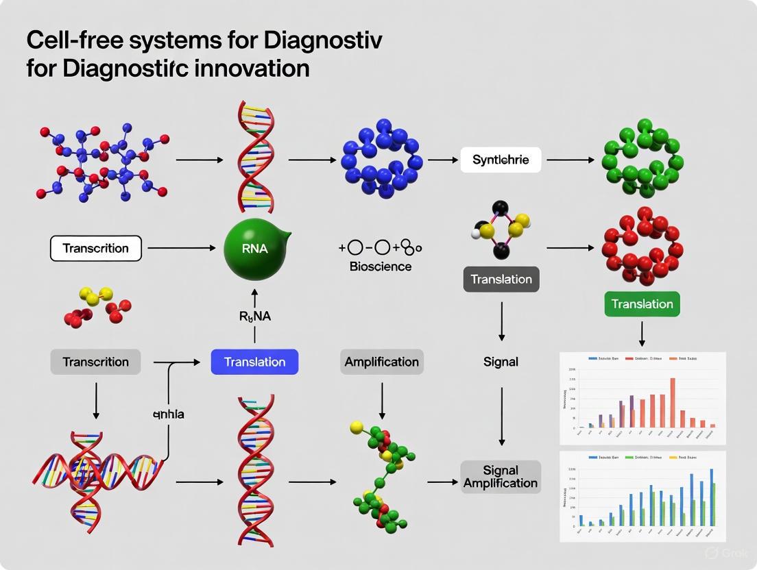
Cell-Free Biosensors: Revolutionizing Diagnostic Innovation for Precision Medicine
This article explores the transformative potential of cell-free systems (CFS) in diagnostic innovation, tailored for researchers, scientists, and drug development professionals.
Cell-Free Biosensors: Revolutionizing Diagnostic Innovation for Precision Medicine
Abstract
This article explores the transformative potential of cell-free systems (CFS) in diagnostic innovation, tailored for researchers, scientists, and drug development professionals. It provides a comprehensive analysis spanning from the foundational principles and unique advantages of CFS over traditional cell-based methods to their cutting-edge methodological applications in detecting pathogens, clinical biomarkers, and environmental contaminants. The scope includes a detailed examination of troubleshooting strategies and optimization techniques for enhancing sensitivity, stability, and cost-effectiveness. Finally, it offers a critical validation and comparative analysis of various CFS platforms, synthesizing key performance metrics to guide platform selection for specific diagnostic intents and outlining future trajectories for biomedical and clinical research.
The Rise of Cell-Free Systems: Core Principles and Diagnostic Advantages
Cell-free protein synthesis (CFPS) is a transformative technology that enables the production of proteins in a controlled, open in vitro environment without the use of living cells [1]. By harnessing the essential transcription and translation machinery from cells, CFPS bypasses the constraints of cell walls and the maintenance of cellular viability, offering researchers direct control over the protein synthesis environment [2] [3]. This platform has evolved from a fundamental research tool into a robust technology for biomanufacturing, therapeutic development, and diagnostic innovation [2] [4]. Its open nature allows for precise manipulation of reaction conditions and the incorporation of non-standard components, making it uniquely suited for applications ranging from rapid prototyping of genetic circuits to the on-demand production of therapeutics and biosensors [5] [6]. This article provides a historical overview of CFPS, details its key components, and presents standardized protocols to equip researchers with the knowledge to leverage this technology for diagnostic innovation.
A Historical Perspective on CFPS Development
The evolution of CFPS spans over six decades, marked by significant breakthroughs that have expanded its capabilities and applications. The following timeline and subsequent analysis detail this journey.
Diagram: The Evolution of Cell-Free Protein Synthesis
The Beginning (1961): Marshall Nirenberg and Heinrich Matthaei used a primitive cell-free system to decode the genetic code. Their experiment demonstrated that a poly-uracil RNA sequence (UUU...) produced a polypeptide chain consisting solely of phenylalanine, thereby identifying UUU as the first codon [2] [1].
Overcoming Limitations (1988): Early batch reactions were hampered by short durations due to energy depletion and byproduct accumulation. Alexander Spirin's team introduced the continuous-flow cell-free (CFCF) system, which used an ultrafiltration membrane to continuously supply energy substrates and remove inhibitory byproducts, extending reaction times to 20 hours and boosting yields a hundredfold [2].
Refinement and Standardization (1990s-2000s): This period saw the development of more practical continuous-exchange (CECF) and bilayer systems that retained the benefits of continuous feeding with simpler setups [2]. A pivotal advancement was the introduction of the Protein synthesis Using Recombinant Elements (PURE) system in 2001. Unlike crude extracts, the PURE system is reconstituted from individually purified components, offering a defined environment free from nucleases and proteases, which is ideal for precise biochemical studies and genetic code expansion [2] [7]. Concurrently, Swartz and colleagues made significant strides in optimizing energy regeneration systems, moving beyond costly substrates to more economical alternatives that supported high-yield production [2].
Modern Maturation (2010s-Present): CFPS has matured into a scalable and versatile platform. Landmark demonstrations included 100-liter reactions producing complex, disulfide-bonded proteins at gram scales, proving its industrial viability [2]. The development of effective lyophilization protocols, using lyoprotectants to stabilize reactions for months at room temperature, enabled portable, cold-chain-independent applications for point-of-care diagnostics and distributed manufacturing [2] [4]. Most recently, the integration of artificial intelligence has revolutionized optimization; for instance, active learning algorithms have explored millions of buffer compositions to achieve 34-fold yield increases with minimal experimental effort, paving the way for data-driven CFPS engineering [2] [6].
Key Components of a CFPS Reaction
A functional CFPS reaction requires the integration of several core modules. The diagram below illustrates their relationships and the central process of protein synthesis.
Diagram: Core Modules of a CFPS System
The Cell Extract (Lysate Module)
The cell extract provides the fundamental enzymatic machinery for transcription and translation, including ribosomes, RNA polymerase, aminoacyl-tRNA synthetases, and translation factors [2] [1]. This is typically prepared by culturing source cells, lysing them using methods like sonication or homogenization, and then clarifying the lysate via centrifugation to remove cell debris and genomic DNA [3]. The choice of extract source depends on the application, as summarized in the table below.
Table 1: Common Cell-Free Expression Systems and Their Characteristics
| System Source | Typical Yield | Key Advantages | Key Limitations | Ideal for Diagnostic Applications |
|---|---|---|---|---|
| E. coli (Prokaryotic) | 0.02 - 1.7 mg/mL [2] | Inexpensive, robust, high yields [3] [1] | Lacks complex eukaryotic PTMs [1] | High-throughput biosensor production; rapid prototyping [4] |
| Wheat Germ (Eukaryotic) | ~100 μg/mL (PURE) [2] | Effective for eukaryotic proteins [1] | Limited glycosylation capability [1] | Producing functional eukaryotic protein domains for assays |
| Rabbit Reticulocyte (Eukaryotic) | Low [8] | Rich in translation machinery; flexible [8] | Limited glycosylation [8] | Functional expression of small eukaryotic enzymes |
| Insect Cell (Eukaryotic) | Information Missing | Supports some PTMs; microsomes for membrane protein folding [6] | More complex preparation | Producing functional membrane proteins (e.g., GPCRs) as diagnostic targets [6] |
| HEK (HeLa/CHO) (Mammalian) | Medium-High [8] | Capable of complex PTMs; functional proteins [8] | Costly; lower yield than E. coli [8] | Producing therapeutically relevant proteins with native modifications |
The Energy Source and Building Blocks (Energy Module)
Protein synthesis is energy-intensive. This module must replenish nucleotides like ATP and GTP, which are consumed during transcription and translation. Common secondary energy sources include phosphoenolpyruvate (PEP), acetyl phosphate, and creatine phosphate [2] [1]. The reaction must also be supplied with all 20 canonical amino acids as the fundamental building blocks for protein assembly [2].
The DNA Template (DNA Module)
The DNA template carries the genetic code for the protein of interest. Both circular plasmids and linear expression templates (LETs) can be used. Plasmids generally yield more protein, but LETs can be rapidly produced via PCR, bypassing time-consuming cloning steps and accelerating the testing of new genetic designs—a significant advantage for diagnostic development [1] [6]. A key challenge with LETs is their susceptibility to degradation by exonucleases present in crude extracts, a problem that can be mitigated using nuclease inhibitors or the PURE system [1].
Essential CFPS Reactor Formats
The performance of a CFPS reaction is heavily influenced by its format, which governs the feeding of substrates and removal of byproducts.
Table 2: Comparison of Common CFPS Reactor Formats
| Reactor Format | Operational Principle | Key Advantages | Limitations | Diagnostic Application Suitability |
|---|---|---|---|---|
| Batch | All components mixed in a single vessel from start [2]. | Simple, fast, inexpensive; easy to parallelize for screening [2]. | Short reaction times; yield limited by byproduct accumulation & energy depletion [2]. | Ideal for high-throughput screening of genetic circuits and sensor components [2]. |
| Continuous-Flow (CFCF) | Reaction mixture is continuously fed with fresh solution through an ultrafiltration membrane, while byproducts are removed [2]. | Greatly extended reaction duration (e.g., 20h); high yields [2]. | Technically complex; membrane fouling can be an issue [2]. | Not commonly used for diagnostics due to complexity. |
| Continuous-Exchange (CECF) | The reaction chamber is separated by a dialysis membrane from a large feeding chamber, allowing passive exchange of small molecules [2]. | Extended reaction lifetime; simpler than CFCF; higher yields than batch [2]. | Requires a membrane; scaling can be challenging. | Suitable for producing larger quantities of diagnostic proteins (e.g., antibodies). |
| Bilayer | A reaction layer is gently layered under a feeding buffer, creating an interface for diffusion [2]. | Simple setup; no membrane required; higher yields than batch [2]. | Limited exchange surface area; less efficient than membrane-based systems. | Good for small-scale, extended production of reagents. |
Experimental Protocols
Protocol 1: Preparation of E. coli Cell Extract
This protocol is adapted from established methodologies for creating a robust, high-yield E. coli lysate [3].
Research Reagent Solutions
| Item | Function in the Protocol |
|---|---|
| 2x YPTG Media | Supports high-density growth of E. coli cells. |
| S30 Buffer (10 mM Tris-OAc, 14 mM Mg(OAc)₂, 60 mM KOAc, 2 mM DTT, pH 8.2) | Washing and resuspension buffer; maintains osmotic stability and provides essential ions and reducing agent. |
| French Press or Sonicator | Equipment for physical cell disruption. |
| DNase I | Enzyme added during runoff to degrade endogenous nucleic acids. |
Procedure:
- Growth: Inoculate E. coli strain (e.g., BL21) into 1L of 2x YPTG media in a 2L baffled flask. Incubate at 37°C with shaking (200 RPM) until the OD₆₀₀ reaches approximately 3.0 [3].
- Harvest and Wash: Centrifuge the culture at 5,000 × g for 10 minutes at 4°C. Resuspend the cell pellet in cold S30 Buffer and repeat the centrifugation and wash steps a total of three times [3].
- Lysis: Resuspend the final pellet in S30 Buffer. Lyse the cells using a French Press at high pressure or via sonication on ice (e.g., 3 cycles of 45 seconds on, 59 seconds off). Keep the suspension cold throughout the process [3].
- Clarification and Runoff: Centrifuge the lysate at 18,000 × g for 30 minutes at 4°C to remove cell debris. Transfer the supernatant (the crude extract) to a new tube. To reduce endogenous background, perform a "runoff" reaction by incubating the extract with DNase I, amino acids, and an energy mix for 60-80 minutes at 37°C [3].
- Dialysis and Storage: Dialyze the extract against fresh S30 buffer to remove small molecules. Aliquot the extract, flash-freeze in liquid nitrogen, and store at -80°C [3].
Protocol 2: Standard Batch CFPS Reaction
This protocol outlines a typical batch reaction for protein production using the prepared E. coli extract.
Procedure:
- Prepare Reaction Mixture: On ice, combine the following components in a microcentrifuge tube to a final volume of 100 μL:
- 30 μL of E. coli cell extract (35-40% of reaction volume) [3].
- DNA template (100-200 ng of plasmid or 5-20 ng/μL of LET) [1].
- Energy Mix: 2mM ATP, GTP, CTP, UTP; 20mM PEP; and a creatine phosphate-based system can also be used [2] [1].
- Amino Acids: A complete mix of all 20 canonical amino acids (2mM each) [2].
- Cofactors: 10-30mM Mg(OAc)₂, 50-150mM KOAc, and other salts as required by the specific system buffer [3].
- Incubate: Place the reaction tube in a thermoshaker or incubator. For E. coli systems, incubate at 30-37°C for 2-8 hours with shaking (if possible) [3].
- Analyze and Purify: After incubation, place the tube on ice. The synthesized protein can be analyzed directly by SDS-PAGE, western blot, or functional assay. For purification, centrifuge the reaction to remove precipitate and use affinity tags (e.g., His-tag) for purification if the template was designed accordingly.
CFPS Applications in Diagnostic Innovation
The unique attributes of CFPS make it a powerful platform for developing next-generation diagnostics. The workflow below illustrates its application in creating a paper-based biosensor.
Diagram: Workflow for a CFPS-Powered Paper-Based Diagnostic
Portable, Low-Cost Biosensors: CFPS reactions can be lyophilized (freeze-dried) on porous materials like paper, creating stable, shelf-ready tests. Upon rehydration with a sample, these tests can detect pathogens (e.g., Zika, Ebola, SARS-CoV-2) or environmental contaminants (e.g., heavy metals) by producing a colorimetric or luminescent signal [2] [4]. For instance, allosteric transcription factors (aTFs) have been used in paper-based systems to detect harmful metals like Hg²⁺ and Pb²⁺ at nanomolar levels, with results readable by smartphones [4].
Rapid Prototyping of Sensor Components: CFPS excels at the high-throughput expression and screening of diagnostic protein components, such as antibody fragments (scFvs, Fabs) and nucleic acid-binding proteins (e.g., CRISPR-Cas). This enables the rapid identification of high-affinity binders against emerging disease biomarkers [6].
On-Demand Therapeutic and Vaccine Production: The open nature of CFPS allows for the direct synthesis of complex biotherapeutics, including virus-like particles (VLPs) for vaccines and conjugated antigens. This paves the way for decentralized manufacturing of diagnostics and therapeutics in resource-limited settings [5] [6].
Cell-free synthetic biology incorporates purified cellular components and/or crude cell extracts to execute metabolic and genetic programs outside of living cells [9]. For diagnostic innovation, this approach provides a disruptive toolkit that bypasses the constraints of cell-based systems. The core advantages driving this adoption are the open reaction environment, exceptional tolerance to toxic substances, and native compatibility with direct sample interfacing [10] [11]. These intrinsic properties enable researchers and drug development professionals to create diagnostic assays with unprecedented speed, flexibility, and functionality, paving the way for next-generation point-of-care (POC) and laboratory-based tests [5] [12]. This document details these advantages within the context of diagnostic research, providing supporting data, experimental protocols, and key resource information.
The Open Reaction Environment
Concept and Diagnostic Impact
The open nature of cell-free systems refers to the absence of a cell wall or membrane, which provides direct, real-time access to the reaction milieu [13]. This eliminates the barrier of transmembrane transport, a significant bottleneck in live-cell biosensors [12]. Researchers can directly manipulate the chemical environment, allowing for sample addition, parameter adjustment, and reaction monitoring without disrupting an ongoing process [10] [13]. This is paramount for diagnostic applications, where the sample matrix itself can be introduced and assay conditions can be finely tuned to optimize sensitivity and specificity.
Key Capabilities and Workflow
The open environment enables two critical capabilities for diagnostics:
- Direct Manipulation and Monitoring: Samples can be added directly, and reagents like substrates, cofactors, or inhibitors can be introduced at any point. Reaction progress can be tracked in real-time via optical methods (e.g., fluorescence, luminescence) [10].
- Component Modularity: The system's biochemical machinery is fully modular. Specific enzymes, chaperones, or synthetic genetic circuits can be added to perform complex functions, such as signal amplification or multi-analyte detection [9] [12].
The workflow below illustrates how a researcher utilizes the open environment to develop and run a cell-free diagnostic assay.
Experimental Protocol: Optimizing a Cell-Free Biosensor
Title: Optimization of Biosensor Dynamic Range by Titrating Transcription Factor Concentration.
Background: The performance of a transcription-factor-based cell-free biosensor is highly dependent on the concentration of its transcription factor (TF). Titrating the TF allows for optimization of the detection limit (LOD) and dynamic range [12].
Materials:
- Cell-free protein synthesis (CFPS) system (e.g., E. coli extract or PURE system)
- Purified DNA plasmid encoding the TF-regulated reporter gene (e.g., sfGFP)
- Purified transcription factor (TF) protein or its DNA template for in-situ expression
- Target analyte (ligand) at known concentrations
- Microplate reader or fluorometer
- 96-well or 384-well reaction plates
Method:
- Master Mix Preparation: Prepare a master mix of CFPS containing all necessary components for transcription and translation (nucleotides, amino acids, energy sources, salts).
- TF Titration: Aliquot the master mix into a multi-well plate. Into each well, add the reporter plasmid and a series of diluted TF protein (or TF DNA template). A negative control should contain no TF.
- Analyte Addition: Add the target analyte (ligand) to experimental wells at a saturating concentration. Include no-analyte controls to measure baseline signal (leakiness).
- Incubation and Monitoring: Incubate the plate at a defined temperature (e.g., 30-37°C) while shaking. Monitor the fluorescence intensity (for sfGFP: Ex/~485 nm, Em/~510 nm) every 5-10 minutes for 4-8 hours.
- Data Analysis: Calculate the maximum fluorescence yield for each condition. Plot the fold-change (Signalwithanalyte / Signal_baseline) against the TF concentration. The TF concentration yielding the highest fold-change represents the optimal condition for sensor dynamic range.
Toxicity Tolerance
Overcoming the Toxicity Barrier
A significant limitation of cell-based biosensors is their inability to handle samples containing components that are toxic to the host organism, as this viability constraint suppresses the signal output [13]. Cell-free systems completely decouple protein synthesis and metabolic activity from cell viability and growth [10] [5]. This allows for the synthesis of proteins that are highly toxic to living cells and, more importantly for diagnostics, the direct analysis of samples containing antimicrobial agents, heavy metals, or other cytotoxic compounds that would kill a living biosensor [13]. This drastically expands the range of sample types that can be tested without pre-processing.
Quantitative Data on Toxicity Tolerance
Table 1: Comparison of Diagnostic-Relevant Protein Production in Cell-Free vs. Cell-Based Systems
| Protein Class | Example | Challenge in Cell-Based Systems | Performance in Cell-Free Systems | Key Citation |
|---|---|---|---|---|
| Toxic Proteins | Antimicrobial peptides, cytotoxic enzymes | Host cell death; low yield | Successful synthesis without viability concerns | [13] |
| Membrane Proteins | G Protein-Coupled Receptors (GPCRs), Porins | Insolubility (inclusion bodies); misfolding | Correct insertion into added liposomes; functional activity | [5] [10] |
| Complex Biologics | Antibody fragments (scFv), disulfide-rich proteins | Improper folding; lack of PTMs | Correct oxidative folding with optimized redox buffers; yields ~hundreds of µg/mL | [5] |
Direct Sample Interfacing
Seamless Integration with Analytical Workflows
The open nature of cell-free systems makes them inherently compatible with direct sampling and analytical techniques. The reaction can be initiated simply by adding a sample to a lyophilized pellet of cell-free reagents, making it ideal for portable, one-pot diagnostic tests [12]. This principle is powerfully illustrated by its integration with paper-based diagnostics and direct sampling mass spectrometry.
Paper-Based Cell-Free Diagnostics
Cell-free reactions can be lyophilized (freeze-dried) onto paper or other porous membranes, creating stable, shelf-ready test strips [12]. Upon rehydration with a liquid sample (e.g., blood, urine, water), the reconstituted system produces a colorimetric, fluorescent, or luminescent signal in the presence of the target analyte.
Interfacing with Direct Sampling Mass Spectrometry
Direct sampling mass spectrometry (MS) techniques, such as paper spray ionization (PSI), allow for rapid analysis of complex biological samples with minimal pre-treatment [14]. A cell-free reaction can be run on a paper cartridge, and the same cartridge can then be used for immediate MS analysis, linking the functional biosensor readout to definitive analyte identification.
Experimental Protocol: Paper-Based Cell-Free Detection of a Small Molecule
Title: Fabrication and Use of Lyophilized Cell-Free Biosensor Strips for Small Molecule Detection.
Background: This protocol describes the creation of a stable, paper-based diagnostic test using a cell-free system that responds to a specific small molecule (e.g., a drug or toxin).
Materials:
- CFPS system (e.g., E. coli extract)
- DNA template for a ligand-activated transcription factor controlling a colorimetric reporter (e.g., lacZ for β-galactosidase)
- Whatman chromatography paper or similar
- Lyophilization stabilizer solution (e.g., trehalose)
- Sample containing the target analyte
- Colorimetric substrate (e.g., chlorophenol red-β-D-galactopyranoside, CPRG)
Method:
- Reaction Assembly: Mix the CFPS reagents with the DNA template and lyophilization stabilizer.
- Spotting and Drying: Spot small aliquots (e.g., 5-10 µL) of the mixture onto paper strips. Immediately flash-freeze the strips using liquid nitrogen or a -80°C freezer.
- Lyophilization: Transfer the frozen strips to a freeze-dryer and lyophilize until completely dry (typically 12-24 hours). Store the dried strips desiccated at -20°C.
- Assay Execution: To run the test, apply the liquid sample (e.g., 20-50 µL) directly onto the paper strip containing the lyophilized pellets.
- Incubation and Readout: Incubate the strip in a humidified chamber at room temperature for 1-2 hours. Observe color development directly or add a substrate like CPRG to enhance sensitivity. The intensity of the color change can be correlated with analyte concentration using a smartphone camera or a simple scanner.
The Scientist's Toolkit: Research Reagent Solutions
Table 2: Essential Materials for Cell-Free Diagnostic Development
| Reagent / Solution | Function in Diagnostic Assays | Key Considerations |
|---|---|---|
| Crude Cell Extract (E. coli) | Provides core transcriptional and translational machinery. | Cost-effective; highly active; limited PTMs. Ideal for prototyping [9] [10]. |
| PURE System | Reconstituted from individually purified E. coli components. | Low background; high specificity; reduced nucleases/proteases. More expensive [10] [13]. |
| Energy Solution | Fuels ATP-dependent reactions (translation, transcription). | Typically contains phosphoenolpyruvate (PEP) or creatine phosphate. Critical for yield and longevity [10]. |
| Lyophilization Stabilizers (Trehalose) | Protects protein machinery during freeze-drying for shelf-stable tests. | Essential for creating point-of-care diagnostics and ensuring long-term stability [12]. |
| Artificial Liposomes / Vesicles | Provide a native-like lipid bilayer for synthesizing functional membrane protein targets (e.g., receptors). | Enhances folding and stability of membrane proteins used as sensing elements [5]. |
| Non-Natural Amino Acids & Modified tRNAs | Enable incorporation of unique chemical handles or labels into synthesized proteins for advanced detection. | Allows for creative sensor design, such as creating novel capture agents or introducing fluorescent probes [10] [13]. |
Biosensors that utilize biological components for detection have become indispensable tools in medical diagnostics, environmental monitoring, and drug development. Traditional whole-cell biosensors, which employ living microorganisms as the sensing element, have been widely used due to their ability to mimic biological responses. However, these cellular systems face fundamental limitations stemming from their reliance on cell viability, including membrane barriers that restrict analyte transport, susceptibility to toxic compounds, and stringent storage requirements that complicate field deployment [15] [16].
Cell-free protein synthesis (CFPS) has emerged as a transformative technology that bypasses these cellular constraints. CFPS harnesses the transcriptional and translational machinery of cells without maintaining cell viability by utilizing cell lysates containing essential components like ribosomes, tRNA, and enzymes [17] [3]. This open system architecture provides direct access to reaction components, eliminates transmembrane transport barriers, and focuses all energy on producing output signals [3]. The resulting biosensing platform offers superior flexibility, stability, and operational convenience compared to whole-cell alternatives, enabling rapid detection of diverse analytes with high sensitivity and specificity [16].
Comparative Analysis: CFPS vs. Whole-Cell Biosensor Performance
Table 1: Performance comparison between whole-cell and CFPS biosensors
| Characteristic | Whole-Cell Biosensors | CFPS Biosensors |
|---|---|---|
| Response Time | Hours to days (requires cell growth and division) | Minutes to hours (direct activation of synthetic machinery) [15] |
| Analyte Transport | Limited by membrane permeability [16] | No barriers; direct access to reaction milieu [15] |
| Toxin Tolerance | Limited by cell viability [15] | High; no viability requirements [18] |
| Storage Stability | Requires strict conditions to maintain viability [15] | Months at room temperature (lyophilized formats) [15] [2] |
| Detection Range | Limited by cytotoxic effects [15] | Expanded range; can detect highly toxic compounds [18] |
| Signal Amplification | Dependent on cellular metabolism | Direct utilization of cascading gene expression (DNA→RNA→protein) [16] |
| Portability | Limited by need for cell culturing | High (paper-based, lyophilized formats) [15] [2] |
Table 2: Experimentally demonstrated detection capabilities of CFPS biosensors
| Target Analyte | Detection Limit | Response Time | Output Signal | Application Area |
|---|---|---|---|---|
| Mercury (Hg²⁺) | 6 μg/L [15], 0.5 nM [18] | ~1 hour | Fluorescence (sfGFP) [15] | Environmental Monitoring |
| Zika Virus RNA | 2 aM [15] | 2.5 hours | Colorimetric (LacZ) [15] | Medical Diagnostics |
| Theophylline | 1 mM [15] | <90 minutes | Colorimetric (LacZ) [15] | Biomedical Research |
| Tetracycline | 0.079-0.47 μM [18] | Not specified | Fluorescence/Luminescence | Food Safety |
| Endocrine Disruptors | 3-30 nM [15] | A few minutes | Colorimetric (β-lactamase) [15] | Clinical/Environmental |
| Lead (Pb²⁺) | 0.1 nM [18] | ~1 hour | Fluorescence | Environmental Monitoring |
| Pathogen 16S rRNA | Femtomolar [18] | Not specified | Fluorescent proteins | Biosecurity |
Mechanisms of Cellular Constraint Bypass in CFPS Biosensors
Elimination of Membrane Barriers and Enhanced Analyte Access
The cellular membrane represents a significant bottleneck in whole-cell biosensors, selectively controlling which compounds enter the cell. This membrane barrier limits both the types of detectable analytes and the detection kinetics. CFPS systems fundamentally overcome this limitation by operating as open systems where all components are freely accessible [15] [3]. Without membrane barriers, analytes directly interact with sensing elements, enabling faster response times and detection of compounds that would be impermeable to cells [15]. This direct access particularly benefits detection of heavy metals like mercury and lead, where CFPS biosensors achieve detection limits as low as 0.1-0.5 nM [18].
Removal of Viability Constraints and Expanded Application Range
Whole-cell biosensors require stringent maintenance of cell viability throughout storage, transport, and operation. This viability requirement limits their practical application in resource-limited settings and for detecting toxic compounds [15]. CFPS biosensors eliminate this constraint entirely since they utilize cellular machinery without requiring viability [16]. The absence of viability constraints enables several key advantages:
- Detection of highly toxic compounds that would kill living cells [18]
- Long-term storage at room temperature through lyophilization [15]
- Operation in harsh environments incompatible with cell survival [18]
- Consistent performance without metabolic fluctuations [17]
Direct Energy Channeling for Enhanced Signal Output
In living cells, energy resources must be allocated across multiple competing processes including growth, maintenance, and reproduction. CFPS systems focus all energy exclusively on the transcription-translation process for signal generation [3]. This direct energy channeling enables:
- Higher protein yields per unit of energy input [3]
- More sensitive detection through stronger output signals [15]
- Reduced background noise from unrelated cellular processes [16]
- Predictable reaction kinetics without metabolic interference [17]
Molecular Design Strategies for CFPS Biosensors
Transcription Factor-Based Detection Systems
Transcription factors (TFs) serve as natural molecular switches that regulate gene expression in response to specific ligands. In CFPS biosensors, TFs are employed to control reporter protein expression in the presence of target analytes [18]. The general mechanism involves a TF that binds to specific DNA sequences upstream of a reporter gene, either activating or repressing transcription based on analyte binding.
TF-Mediated Sensing: This diagram illustrates the working mechanism of transcription factor-based CFPS biosensors, where analyte binding activates reporter gene expression.
Implementation example: Heavy metal detection utilizing natural bacterial transcription factors like MerR (for mercury) and PbrR (for lead) [18]. These TF-based systems have been successfully deployed in paper-based formats for field detection of environmental contaminants, achieving detection limits surpassing WHO guidelines [18].
Nucleic Acid-Based Sensing Mechanisms
Nucleic acid detection represents a major application area for CFPS biosensors, particularly for pathogen identification. These systems employ toehold switches - engineered RNA elements that control translation initiation based on specific trigger RNAs [16].
Toehold Switch Mechanism: This diagram shows how pathogen RNA triggers structural changes in toehold switches, exposing the ribosome binding site and initiating reporter translation.
Implementation example: Zika virus detection using toehold switches that recognize viral RNA sequences. This approach achieved exceptional sensitivity (2 aM detection limit) and the ability to discriminate between viral strains with single-base resolution when combined with CRISPR/Cas9 modules [15] [16].
Allosteric Transcription Factors and Aptamer-Based Designs
Beyond natural transcription factors, CFPS biosensors incorporate engineered allosteric transcription factors (aTFs) and aptamers for detecting analytes without natural regulatory proteins. These designed sensing elements significantly expand the detectable analyte range [18].
Implementation example: Vanillin detection using engineered aTFs created through directed evolution and screening [15]. This approach provides a framework for developing sensors for diverse target compounds that lack natural biosensing components.
Experimental Protocols for CFPS Biosensor Implementation
Core CFPS Biosensor Preparation Protocol
Objective: Prepare functional cell-free biosensor components for analyte detection.
Materials:
- E. coli BL21 Star (DE3) cells
- Luria-Bertani (LB) medium
- S30 Buffer (10 mM Tris-OAc, pH 8.2, 14 mM Mg(OAc)₂, 60 mM KOAc, 2 mM DTT)
- Energy Solution (ATP, GTP, amino acids, phosphoenolpyruvate)
- DNA template containing biosensor genetic circuit
- Paper-based substrate (Whatman Grade 1 filter paper)
Method:
- Cell Culture and Harvest:
- Inoculate E. coli in 50 mL LB medium in a 250 mL baffled flask
- Incubate at 37°C with shaking at 200 rpm until OD₆₀₀ reaches ~3.0
- Centrifuge culture at 5,000 × g for 10 minutes at 4°C
- Wash cell pellet three times with 30 mL S30 buffer [3]
Cell Extract Preparation:
- Resuspend cell pellet in 1 mL S30 buffer per gram of cells
- Lyse cells using sonication on ice (3 cycles of 45 seconds on, 59 seconds off at 50% amplitude)
- Centrifuge lysate at 18,000 × g for 10 minutes at 4°C
- Transfer supernatant to a new tube and perform runoff reaction at 37°C for 60 minutes with shaking
- Centrifuge at 10,000 × g for 10 minutes, aliquot supernatant, and store at -80°C [3]
Biosensor Assembly:
- Prepare master mix containing:
- 30% (v/v) cell extract
- 1.5 mM ATP and GTP
- 0.2 mM of each amino acid
- 50 mM phosphoenolpyruvate
- 25-50 ng/μL DNA template with biosensor genetic circuit
- Apply 10-20 μL aliquots to paper-based substrates or use in solution-based formats
- For field deployment, lyophilize assembled biosensors and store with desiccant [15]
- Prepare master mix containing:
Toehold Switch-Based Pathogen Detection Protocol
Objective: Detect specific pathogen RNA sequences using toehold switch CFPS biosensors.
Materials:
- CFPS system prepared as in Protocol 5.1
- Toehold switch DNA template
- NASBA amplification reagents (for RNA amplification)
- Sample containing target RNA
Method:
- Sample Preparation:
- Extract RNA from sample matrix (serum, water, etc.)
- Amplify target sequence using NASBA isothermal amplification at 41°C for 90 minutes [15]
Biosensor Activation:
Signal Detection:
- For colorimetric output: Measure absorbance at 420 nm (LacZ) or appropriate wavelength
- For fluorescent output: Measure fluorescence with appropriate excitation/emission filters
- Compare to standard curve for quantitative analysis [15]
Transcription Factor-Based Heavy Metal Detection Protocol
Objective: Detect heavy metal contaminants in water samples using TF-based CFPS biosensors.
Materials:
- CFPS system prepared as in Protocol 5.1
- DNA template with metal-responsive promoter (MerR, PbrR, etc.) upstream of reporter gene
- Water samples (filtered through 0.22 μm membrane)
- Standard metal solutions for calibration
Method:
- Biosensor Preparation:
- Prepare CFPS master mix with metal-responsive genetic circuit DNA template
- Apply 10 μL aliquots to paper-based discs and lyophilize for storage [18]
Sample Analysis:
- Reconstitute lyophilized biosensors with 100 μL water samples or standards
- Incubate at 37°C for 1-2 hours
- Measure fluorescent output (sfGFP: Ex 485 nm/Em 510 nm) [15]
Quantification:
- Generate standard curve with known metal concentrations
- Calculate sample concentrations from standard curve
- Report results relative to regulatory limits (e.g., WHO drinking water guidelines) [18]
The Scientist's Toolkit: Essential Reagents and Materials
Table 3: Essential research reagents for CFPS biosensor development
| Reagent/Material | Function | Example Specifications | Key Considerations |
|---|---|---|---|
| Cell Lysates | Source of transcriptional/translational machinery | E. coli BL21 extracts, wheat germ extracts | Choose based on protein folding requirements; eukaryotic extracts enable disulfide bond formation [3] |
| Energy Systems | Drive transcription and translation | ATP/GTP regeneration systems (phosphoenolpyruvate-based) | Critical for extended reaction duration; optimized systems improve yield [2] |
| DNA Templates | Encode biosensor genetic circuit | Plasmid DNA or linear PCR products | Plasmid offers stability; linear DNA enables rapid prototyping [17] |
| Reporter Systems | Generate detectable output signals | sfGFP, LacZ, Luciferase, NanoLuc | Match to detection equipment; colorimetric preferred for field use [15] |
| Lyoprotectants | Stabilize lyophilized biosensors | Trehalose, sucrose, polyethylene glycol | Essential for room temperature storage and field deployment [2] |
| Sensing Elements | Provide analyte recognition | Transcription factors, aptamers, toehold switches | Dictate specificity; can be engineered for novel analytes [18] |
CFPS technology represents a paradigm shift in biosensor design, effectively addressing the fundamental limitations of whole-cell systems. By eliminating cellular constraints including membrane barriers, viability requirements, and metabolic competition, CFPS biosensors achieve superior performance in sensitivity, response time, and application range. The modular nature of these systems enables rapid design iteration for detecting diverse targets from environmental contaminants to pathogen signatures.
Future developments will likely focus on increasing multiplexing capabilities, enhancing field-deployable formats, and integrating with electronic reporting systems such as smartphone-based detection. As CFPS biosensors continue to mature, they hold particular promise for point-of-care diagnostics in resource-limited settings and real-time environmental monitoring networks. The ongoing optimization of lyophilization protocols and room-temperature stability will further expand their practical implementation, ultimately making sophisticated biosensing technology more accessible and deployable across diverse sectors.
The integration of synthetic biology with diagnostic science has catalyzed a paradigm shift, enabling the development of programmable, field-deployable detection systems. Cell-free biosensors harness the selectivity of cellular machinery without the constraints of living cells, offering significant advantages for environmental monitoring, medical diagnostics, and biotechnological applications [4]. These systems leverage purified transcriptional and translational components—ribosomes, transcription factors, energy sources, and cofactors—to create highly tunable platforms that operate in diverse environments, from resource-limited field settings to sophisticated laboratory applications [4]. The elimination of cell viability requirements, combined with rapid response times and minimal metabolic burden, positions cell-free systems as essential tools for addressing global challenges in healthcare, environmental protection, and biosecurity [4].
Within this landscape, three key regulatory elements have emerged as particularly powerful components for the next generation of diagnostic tools: synthetic riboswitches, allosteric transcription factors (aTFs), and synthetic genetic circuits. Each offers distinct mechanisms for detecting target analytes and controlling reporter gene expression, enabling the creation of highly specific, sensitive, and modular biosensing platforms. This article explores the unique advantages and applications of these tools within cell-free systems, providing detailed experimental protocols and performance comparisons to guide researchers in selecting and implementing appropriate technologies for their diagnostic challenges.
Technical Components: Mechanisms and Applications
Synthetic Riboswitches: RNA-Based Regulatory Elements
Synthetic riboswitches are compact, modular RNA elements that unite sensing and regulatory functions within a single molecule, operating entirely at the RNA level without requiring auxiliary proteins [19]. Their modular architecture consists of two key domains: an aptamer domain that specifically binds to a target ligand, and an adjacent regulatory domain that controls gene expression through mechanisms such as translation initiation, mRNA stability, or splicing [19]. Upon ligand binding, the aptamer undergoes a conformational change that is transmitted to the regulatory domain, modifying its activity in a ligand-dependent manner [19].
Key Advantages: Synthetic riboswitches offer several distinct advantages that make them valuable regulatory tools, particularly in cell-free systems. Their completely RNA-based nature imposes substantially lower biosynthetic and degradation demands than protein-based regulators, reducing metabolic burden and enabling more efficient resource allocation [19]. This compact design (often less than 200 nucleotides) suggests good portability, minimal footprint, and low risk of side effects, making them particularly suitable for applications outside model organisms [19]. Additionally, their modularity enables high customizability—aptamer domains can be selected de novo for new target molecules using Systematic Evolution of Ligands by EXponential Enrichment (SELEX), allowing engineering of fully synthetic riboswitches that respond to customized ligands [19].
Diagnostic Applications: Riboswitches have been successfully implemented in various diagnostic contexts. A compelling example is a riboswitch-based cell-free biosensor for broad-spectrum detection of tetracyclines [4]. This system used artificially screened tetracycline RNA aptamers to control reporter gene expression, achieving detection limits of 0.47, 0.079, 0.084, and 0.43 μM for tetracycline, oxytetracycline, chlortetracycline, and doxycycline, respectively, enabling qualitative detection in milk samples at concentrations as low as 1 μM [4]. Furthermore, synthetic riboswitches have been engineered to control complex cellular behaviors, such as regulating metabolic pathways in living cells [19].
Table 1: Performance Characteristics of Riboswitch-Based Biosensors
| Target Analyte | Detection Mechanism | Limit of Detection | Sample Matrix | Reference |
|---|---|---|---|---|
| Tetracyclines | Riboswitch with RNA aptamers | 0.079-0.47 μM | Milk samples | [4] |
| Intracellular metabolites | Aptamer-controlled ribozymes (aptazymes) | Varies by metabolite | Living cells | [19] |
| Synthetic ligands | Orthogonal FMN riboswitch | KD = ~54-75 nM | In vitro, prokaryotic, and eukaryotic systems | [20] |
Allosteric Transcription Factors: Protein-Based Sensing Modules
Allosteric transcription factors (aTFs) are protein-based sensors that undergo conformational changes upon binding to specific ligands, thereby modulating their affinity for target DNA sequences and controlling downstream gene expression. In cell-free systems, aTFs serve as highly specific recognition elements for diverse analytes, including heavy metals, organic pollutants, and clinical biomarkers [4].
Key Advantages: aTFs offer several beneficial characteristics for diagnostic applications. They provide natural specificity for a wide range of biologically relevant targets, can be engineered for improved sensitivity and altered ligand specificity, and enable direct coupling between detection and reporter gene expression [4]. Additionally, aTF-based sensors can be optimized through systematic engineering approaches to achieve detection limits that meet or exceed regulatory standards for environmental contaminants [4].
Diagnostic Applications: aTFs have been successfully deployed for sensitive environmental monitoring applications. Zhang et al. developed a cell-free paper-based biosensor utilizing aTFs for on-site detection of harmful metals, achieving impressive detection limits of 0.5 nM for Hg²⁺ and 0.1 nM for Pb²⁺ in water samples, with recovery rates ranging from 91% to 123% for actual environmental samples [4]. Similarly, Ekas et al. created an engineered aTF platform that achieved a significant improvement in lead detection sensitivity, shifting the limit of detection from 10 μM to 50 nM—sufficient for detecting lead at the legal limit [4]. These systems demonstrate the potential of aTFs for field-deployable environmental monitoring with laboratory-level sensitivity.
Table 2: Performance of aTF-Based Cell-Free Biosensors for Environmental Monitoring
| Target Analyte | Detection System | Limit of Detection | Selectivity/Specificity | Reference |
|---|---|---|---|---|
| Mercury (Hg²⁺) | aTF-based paper biosensor | 0.5 nM | High selectivity for target metals; validated in real water samples | [4] |
| Lead (Pb²⁺) | aTF-based paper biosensor | 0.1 nM | High selectivity for target metals; validated in real water samples | [4] |
| Lead | Engineered PbrR mutants | 50 nM | Selective for lead | [4] |
| Arsenic, Mercury | Optimized transcription factors | As ≤10 μg/L, Hg ≤6 μg/L | Minimal response to nontoxic ions | [4] |
Synthetic Genetic Circuits: Programmable Biological Computing
Synthetic genetic circuits represent the most complex tier of the diagnostic toolbox, enabling sophisticated computation and signal processing within biological systems. These engineered networks of genetic components can perform Boolean logic operations, process multiple input signals, and generate programmable outputs based on predefined rules [21]. Recent advances in circuit design have focused on compression strategies that minimize genetic footprint while maintaining or expanding functionality [21].
Key Advantages: Synthetic genetic circuits offer unprecedented programmability for biological systems. They can integrate multiple input signals using logic gates (AND, OR, NOT), enable higher-state decision-making capabilities, and be designed with minimal parts count to reduce metabolic burden [21]. The development of complementary software tools now allows for predictive design of genetic circuits with quantitatively precise performance setpoints, moving beyond traditional trial-and-error approaches [21].
Diagnostic Applications: Genetic circuits have shown particular utility in multiplexed diagnostic applications. Recent research has highlighted the emergence of wearable multiplexed diagnostic biosensors using synthetic gene circuits and RNA regulators, delivering real-time pathogen detection with minimal infrastructure needs [22]. These systems integrate engineered genetic circuits to provide comprehensive diagnostic profiles, enhancing efficiency in clinical settings [22]. Additionally, circuits capable of 3-input Boolean logic (eight-state decision-making) have been developed, significantly expanding the computational capacity available for diagnostic applications [21].
Research Reagent Solutions: Essential Materials for Diagnostic Development
Table 3: Key Research Reagents for Cell-Free Diagnostic Development
| Reagent Category | Specific Examples | Function in Diagnostic Systems | Key Characteristics |
|---|---|---|---|
| Cell-Free Systems | Commercial extracts (NEB), low-cost homemade extracts | Provide transcriptional/translational machinery | Lyophilization-compatible, customizable, variable cost points |
| Riboswitch Components | Tetracycline aptamers, FMN riboswitch variants, theophylline aptamers | Target recognition and signal transduction | Modular, protein-independent, target-specific |
| Transcription Factors | MerR family metalsensors, TetR variants, engineered PbrR | Protein-based ligand recognition and allosteric regulation | High specificity, engineerable, can be optimized via directed evolution |
| Reporters | Luciferase, fluorescent proteins (eGFP), lacZ β-galactosidase | Generate detectable output signals | Various detection methods (luminescence, fluorescence, colorimetry) |
| Platform Materials | Paper-based matrices, hydrogels, supported lipid bilayers | Immobilization and preservation of components | Field-deployable, room-temperature stable, low-cost |
Experimental Protocols
Protocol 1: Development of a Riboswitch-Based Tetracycline Sensor
Principle: This protocol describes the implementation of a riboswitch-based cell-free biosensor for detection of tetracycline antibiotics, based on the system developed by Dong et al. [4]. The approach utilizes tetracycline-specific RNA aptamers that control expression of a reporter gene in a ligand-dependent manner.
Materials:
- Cell-free protein synthesis system (commercial or homemade extract)
- DNA template encoding the riboswitch-regulated reporter construct
- Tetracycline standards (tetracycline, oxytetracycline, chlortetracycline, doxycycline)
- Reporter assay reagents (luciferase assay system if using luciferase reporter)
- Milk samples for validation (diluted 1:10 in nuclease-free water)
Procedure:
- Template Design: Clone the tetracycline RNA aptamer sequence upstream of the reporter gene (e.g., luciferase or fluorescent protein) in the expression vector. Ensure the riboswitch is positioned in the 5' UTR where it can regulate translation initiation.
- Cell-Free Reaction Assembly: Combine the following components in a microcentrifuge tube on ice:
- 12 μL cell-free extract
- 1 μg DNA template
- 10 μL reaction buffer (containing amino acids, nucleotides, energy sources)
- Tetracycline standard or sample in a volume of 3 μL
- Nuclease-free water to a final volume of 25 μL
- Incubation and Measurement: Incubate the reaction at 37°C for 4-6 hours. Measure reporter output at regular intervals (e.g., every 30 minutes) using appropriate instrumentation (luminometer for luciferase, fluorometer for fluorescent proteins).
- Data Analysis: Plot reporter signal versus tetracycline concentration to generate a standard curve. Calculate tetracycline concentrations in unknown samples by interpolation from the standard curve.
Troubleshooting Tips:
- If sensitivity is insufficient, optimize the riboswitch sequence using SELEX-derived variants with higher binding affinity.
- If dynamic range is limited, modify the sequence context surrounding the riboswitch to improve regulatory efficiency.
- For complex samples like milk, include additional purification steps or control reactions to address matrix effects.
Protocol 2: Implementation of an aTF-Based Heavy Metal Detector
Principle: This protocol outlines the development of a paper-based cell-free biosensor for heavy metal detection using allosteric transcription factors, adapted from Zhang et al. and Ekas et al. [4]. The system leverages the natural specificity of metalloregulatory proteins coupled with reporter gene activation.
Materials:
- Lyophilized cell-free protein synthesis system
- DNA constructs encoding aTF-regulated reporter genes
- Whatman Grade 1 filter paper or nitrocellulose membranes
- Heavy metal standards (Hg²⁺, Pb²⁺, etc.)
- Water samples for testing
- Smartphone or portable detector for readout
Procedure:
- Paper Sensor Fabrication: Spot lyophilized cell-free reactions containing DNA templates encoding aTF-regulated reporters onto filter paper discs (5 mm diameter). Allow to air dry completely, then store with desiccant at 4°C until use.
- Sample Processing: For water samples, filter through 0.45 μm membrane to remove particulate matter. Adjust pH to 7.0-7.5 if necessary.
- Assay Execution: Apply 50 μL of standard or sample to each paper disc. Incubate at room temperature for 30-90 minutes.
- Signal Detection: Visualize color development (for colorimetric reporters) or measure fluorescence/luminescence using a portable detector or smartphone-based imaging system.
- Quantification: Capture images under consistent lighting conditions and analyze using ImageJ or similar software. Convert signal intensity to concentration using a standard curve.
Troubleshooting Tips:
- If background signal is too high, optimize aTF and promoter concentrations during lyophilization.
- If sensitivity does not meet requirements, employ engineered aTF variants with improved detection limits.
- For field applications, ensure proper storage conditions to maintain sensor stability.
Visual Guide: Operational Principles and Workflows
Riboswitch Mechanism and Sensor Assembly
Diagram 1: Riboswitch Operational Mechanism. Illustrates the conformational change in a synthetic riboswitch upon ligand binding, which activates reporter gene expression.
Integrated Workflow for Diagnostic Development
Diagram 2: Diagnostic Development Workflow. Outlines the systematic process for developing cell-free biosensors, from initial tool selection through field deployment.
The expanding diagnostic toolbox, encompassing synthetic riboswitches, allosteric transcription factors, and synthetic genetic circuits, provides researchers with an unprecedented ability to create sensitive, specific, and field-deployable detection systems. Each technology offers distinct advantages: riboswitches with their compact RNA-only architecture and low metabolic burden [19]; aTFs with their high specificity and engineerability for diverse targets [4]; and genetic circuits with their sophisticated computational capabilities and multiplexing potential [21].
The integration of these tools with cell-free systems represents a particularly promising direction for diagnostic innovation, combining the programmability of synthetic biology with the practicality of field-deployable formats. As these technologies continue to mature, we anticipate their increasing adoption for addressing critical challenges in healthcare, environmental monitoring, and biosecurity. Future developments will likely focus on enhancing sensitivity and multiplexing capabilities, improving stability for field use, and reducing costs for global accessibility. By leveraging the protocols and insights presented in this article, researchers can contribute to this rapidly advancing field, developing next-generation diagnostics that deliver precision, programmability, and practical utility across diverse applications.
From Bench to Bedside: Innovative Applications in Medical and Environmental Diagnostics
The rapid and accurate detection of pathogens at the point of care (POC) is a critical component of modern disease control strategies, enabling timely clinical decisions and infection control measures. The COVID-19 pandemic has highlighted both the critical importance and existing limitations of diagnostic technologies, driving innovation in the field [23]. Cell-free systems have emerged as a powerful platform for diagnostic innovation, offering advantages in speed, versatility, and deployment compared to traditional cell-based methods or central laboratory testing [24] [25]. These systems utilize the transcriptional and translational machinery of cells without the constraints of cell membranes or viability maintenance, creating an open environment that can be optimized specifically for diagnostic applications [24]. This application note details the implementation of cell-free biosensing systems for detecting the SARS-CoV-2 Receptor Binding Domain (RBD), providing a framework that can be adapted for detecting biological warfare agents. The protocols and data presented are designed for researchers, scientists, and drug development professionals working in diagnostic development.
Technical Background
The SARS-CoV-2 Spike Protein and Receptor Binding Domain (RBD)
SARS-CoV-2 is an enveloped, positive-sense single-stranded RNA virus with a genome of approximately 29.9 kb [26]. The virion is spherical with a diameter of roughly 125 nm and features prominent club-shaped spike projections on its surface that form its characteristic crown-like appearance [23] [26]. The spike (S) glycoprotein, a key determinant of viral infectivity and pathogenesis, is a homotrimeric complex that mediates host cell attachment and entry [27]. It is divided into S1 and S2 subunits, responsible for receptor binding and membrane fusion, respectively [27]. The Receptor Binding Domain (RBD) located within the S1 subunit specifically mediates viral attachment to the host angiotensin-converting enzyme 2 (ACE2) receptor [27]. This RBD-ACE2 interaction represents a critical neutralization target, as antibodies blocking this interaction can prevent viral infection [27] [28]. The RBD transitions between "up" (receptor-accessible) and "down" (receptor-inaccessible) conformations, influencing its susceptibility to neutralizing antibodies [27].
Cell-Free Systems as a Diagnostic Platform
Cell-free protein synthesis (CFPS) systems provide a flexible alternative to cell-based expression for diagnostic applications. These systems are typically based on S30 extracts (supernatants from cell lysates centrifuged at 30,000× g) or purified enzyme mixtures that retain transcriptional and translational capabilities without cellular homeostasis constraints [24]. The open nature of cell-free platforms allows direct manipulation of reaction conditions and real-time monitoring of molecular interactions—features particularly advantageous for biosensing applications [24]. For POC diagnostics, cell-free systems can be lyophilized for long-term storage and reactivated on-demand, facilitating deployment in resource-limited settings [24] [25]. When integrated with synthetic biology approaches, these systems can be designed to produce visual or measurable signals (colorimetric, luminescent, electrochemical) upon detection of specific pathogen biomarkers like the SARS-CoV-2 RBD.
The following workflow illustrates the typical process for developing a cell-free biosensor for pathogen detection:
Application Note: SARS-CoV-2 RBD Detection Using a Cell-Free Biosensor
This application note describes a rapid, sensitive cell-free biosensing platform for detecting SARS-CoV-2 through its RBD protein. The system leverages a membrane-tethered RBD antigen concept [27] integrated with a cell-free expression system that generates a colorimetric output upon target recognition. This approach addresses key limitations of traditional diagnostics, including labor-intensive processes, time consumption, and requirement for trained personnel in central laboratory settings [23]. The platform demonstrates particular utility for non-laboratory settings such as clinics, airports, and remote locations, with detection times under 10 minutes following sample introduction [26]. The methodology can be adapted for detecting RBD variants and other pathogen antigens with minimal modification.
Key Performance Metrics
Comparative analysis of SARS-CoV-2 detection methods reveals significant advantages of biosensing approaches for point-of-care applications. The table below summarizes the performance characteristics of major diagnostic platforms:
Table 1: Comparison of SARS-CoV-2 Detection Platforms
| Method Category | Specific Technique | Detection Target | Sample Type | Approx. Detection Time | Key Advantages | Key Limitations |
|---|---|---|---|---|---|---|
| Nucleic Acid Amplification | qRT-PCR | Viral RNA | Nasopharyngeal swab | Several hours (including sample prep) | High sensitivity/specificity; gold standard | Labor-intensive; requires trained personnel; central lab equipment [23] |
| Serological Testing | Lateral Flow Immunoassay | IgM/IgG antibodies | Blood/Serum | 10-20 minutes | Rapid; user-friendly; portable | Lower sensitivity in early infection [23] [26] |
| Viral Antigen Detection | Biosensing Devices | Viral antigens (e.g., RBD) | Swab samples | <10 minutes | Extreme speed; portable; minimal sample prep | Emerging technology; validation ongoing [26] |
| Proteomic Analysis | Mass Spectrometry | Viral proteins | Swab samples | Hours | High specificity and precision | Expensive equipment; technical expertise required [26] |
| Cell-Free Biosensing | Integrated CFPS-RBD | RBD antigen | Saliva/Swab | <15 minutes | Rapid; portable; lyophilizable; high adaptability | Limited current scalability |
Quantitative data from the Coronavirus Immunotherapeutic Consortium (CoVIC) analysis of over 400 anti-SARS-CoV-2 spike antibodies provides insights into performance parameters relevant to biosensor design. The following table summarizes key antibody characteristics that inform recognition element selection:
Table 2: Anti-SARS-CoV-2 RBD Antibody Features from CoVIC Analysis [28]
| Antibody Feature | Measurement Range | Correlation with Protection | Implications for Biosensor Design |
|---|---|---|---|
| ACE-2 Binding Blockage | Variable inhibition efficiency | High correlation with neutralization | Preferred recognition element for functional detection |
| Spike Protein Affinity | Wide variability (pM-nM) | Moderate correlation | High-affinity clones preferred for sensitivity |
| Epitope Binning | Distinct communities identified | Specific epitopes associated with durable potency | Epitope selection affects variant detection |
| Variant Neutralization | Variable cross-reactivity | Certain epitopes confer broader protection | Guide selection for pan-variant biosensors |
| In Vivo Protection | Correlated with specific epitopes | Predicts clinical utility | Inform animal model validation studies |
Research Reagent Solutions
The successful implementation of cell-free biosensing platforms requires specific reagents and components optimized for diagnostic applications. The following table details essential materials and their functions:
Table 3: Essential Research Reagents for Cell-Free RBD Detection Systems
| Reagent/Category | Specific Examples | Function in Biosensing Platform | Implementation Notes |
|---|---|---|---|
| Cell-Free System | S30 extracts (E. coli based) | Provides transcriptional/translational machinery | Optimize for lyophilization stability [24] |
| Recognition Elements | Anti-RBD monoclonal antibodies | Specifically binds target antigen with high affinity | Select clones with ACE2 blocking capability [28] |
| Signal Transduction | Nanozymes, Gold nanoparticles | Generates measurable signal upon target binding | Enables colorimetric/electrochemical readout [26] |
| RBD Antigen Standards | Recombinant SARS-CoV-2 RBD | Positive control for assay validation | Include variant RBDs for cross-reactivity testing [27] |
| Sample Collection | Saliva collection devices, Viral transport media | Maintains antigen integrity during collection | Saliva enables non-invasive sampling [23] |
| Output Detection | Lateral flow strips, Electrochemical sensors | Provides user-readable result | Match to POC setting requirements [26] |
Experimental Protocols
Protocol 1: Cell-Free Biosensor Assembly for RBD Detection
Principle: This protocol details the assembly of a lyophilized cell-free biosensor that produces a colorimetric signal upon detection of SARS-CoV-2 RBD, utilizing an antibody-based recognition system coupled with transcription-based signal amplification.
Materials:
- S30 E. coli cell-free extract (commercially available or prepared as in Protocol 2)
- Reaction components: HEPES buffer (pH 7.4), nucleotides (ATP, GTP, UTP, CTP), amino acid mixture, energy regeneration system (phosphoenolpyruvate, pyruvate kinase)
- DNA template encoding reporter enzyme (e.g., luciferase, horseradish peroxidase) under inducible promoter
- Anti-RBD monoclonal antibody with high ACE2 blocking capability [28]
- Saliva collection devices (for clinical validation)
- Lyophilization stabilizers (trehalose, polyethylene glycol)
Procedure:
- Recognition Element Immobilization:
- Incubate anti-RBD antibody (2 µg/mL in phosphate buffer) with magnetic beads functionalized with Protein A for 1 hour at 4°C.
- Wash beads twice with cold PBS to remove unbound antibody.
- Resuspend in cell-free reaction buffer at a concentration of 10 mg beads/mL.
Cell-Free Reaction Mixture Preparation:
- Prepare master mix containing (final concentrations):
- 40% (v/v) S30 extract
- 50 mM HEPES (pH 7.4)
- 1.5 mM each ATP, GTP, UTP, CTP
- 2 mM amino acid mixture
- 30 mM phosphoenolpyruvate
- 0.3 U/µL pyruvate kinase
- 100 ng/µL DNA template encoding reporter enzyme
- 5% (w/v) lyophilization stabilizers (trehalose:PEG, 4:1 ratio)
- Prepare master mix containing (final concentrations):
Biosensor Assembly:
- Combine 90 µL cell-free reaction mixture with 10 µL antibody-coated beads.
- Aliquot 10 µL volumes into reaction tubes or lateral flow device reservoirs.
- Flash-freeze in liquid nitrogen and lyophilize for 24 hours.
Detection Procedure:
- Rehydrate biosensor with 10 µL saliva sample or clinical specimen.
- Incubate at 37°C for 15 minutes.
- Add appropriate substrate for reporter enzyme (e.g., luminescent substrate for luciferase).
- Measure signal output using portable detector or visual assessment.
Validation:
- Include positive control (recombinant RBD, 100 ng/mL) and negative control (RBD-free saliva) in each run.
- Validate against RT-PCR results for clinical specimens.
- Determine limit of detection using serial RBD dilutions.
Protocol 2: S30 Extract Preparation for Diagnostic Applications
Principle: Preparation of high-quality S30 extract from E. coli provides the foundational biochemical machinery for cell-free biosensing systems. This optimized protocol yields extracts with high protein synthesis capability and stability for lyophilization.
Materials:
- E. coli strain A19 (RNase I-deficient) or similar
- 2xYTPG medium: 16 g/L tryptone, 10 g/L yeast extract, 5 g/L NaCl, 7 g/L K2HPO4, 3 g/L KH2PO4, 18 g/L glucose
- S30 buffer A: 10 mM Tris-acetate (pH 8.2), 14 mM magnesium acetate, 60 mM potassium acetate
- S30 buffer B: 10 mM Tris-acetate (pH 8.2), 14 mM magnesium acetate, 60 mM potassium acetate, 1 mM DTT
- DNase I (RNase-free)
- Dialysis membranes (10-14 kDa MWCO)
Procedure:
- Cell Culture:
- Inoculate 5 mL overnight culture of E. coli in 2xYTPG medium.
- Dilute 1:100 into 1L fresh 2xYTPG in 4L flask.
- Incubate at 37°C with vigorous shaking (250 rpm) until OD600 ≈ 0.6-0.8.
- Chill culture rapidly on ice for 15 minutes.
Cell Harvest and Lysis:
- Harvest cells by centrifugation at 5,000 × g for 15 minutes at 4°C.
- Wash cell pellet with 100 mL cold S30 Buffer A.
- Centrifuge again and resuspend pellet in 3 mL S30 Buffer B per gram wet weight.
- Disrupt cells by single passage through French pressure cell at 10,000-15,000 psi.
- Alternatively, use bead beating or sonication for lysis.
Extract Preparation:
- Centrifuge lysate at 12,000 × g for 10 minutes at 4°C to remove debris.
- Transfer supernatant to fresh tube and centrifuge at 30,000 × g for 30 minutes at 4°C (S30 extract).
- Pre-incubate S30 extract with 2 µM DNase I for 45 minutes at 4°C.
- Dialyze against 50 volumes S30 Buffer B for 3 hours at 4°C, with one buffer change.
- Centrifuge again at 4,000 × g for 10 minutes to remove precipitate.
- Aliquot, flash-freeze in liquid nitrogen, and store at -80°C.
Quality Control:
- Determine protein concentration (target: 30-40 mg/mL).
- Test protein synthesis activity using GFP reporter plasmid.
- Confirm absence of RNase contamination by RNA integrity analysis.
Protocol 3: Biosensor Validation Against SARS-CoV-2 Variants
Principle: This protocol validates the cross-reactivity and sensitivity of the cell-free biosensor against SARS-CoV-2 variants of concern, using recombinant RBD proteins and clinical isolates.
Materials:
- Recombinant RBD proteins from variants (Alpha, Beta, Gamma, Delta, Omicron)
- Clinical SARS-CoV-2 isolates (inactivated) or positive clinical specimens
- Standard RT-PCR reagents for comparison
- Cell culture facilities (BSL-2 or BSL-3 as required) [29]
Procedure:
- Analytical Sensitivity Determination:
- Prepare serial dilutions of each recombinant RBD (1 pg/mL to 1 µg/mL) in artificial saliva.
- Test each dilution with cell-free biosensor following Protocol 1.
- Calculate limit of detection (LOD) for each variant using 5-parameter logistic curve fitting.
Clinical Specimen Testing:
- Obtain patient swab samples with known RT-PCR results.
- Test blinded samples with cell-free biosensor.
- Compare results to RT-PCR as gold standard.
- Calculate sensitivity, specificity, positive predictive value, and negative predictive value.
Biosafety Considerations:
- All work with infectious virus must follow BSL-2/BSL-3 guidelines as appropriate [29].
- Use appropriate PPE including lab coats, gloves, and eye protection.
- Perform aerosol-generating procedures in certified Class II Biological Safety Cabinets.
- Decontaminate work surfaces with EPA-registered disinfectants effective against SARS-CoV-2 [29].
The following diagram illustrates the molecular interaction between SARS-CoV-2 RBD and the biosensor detection system:
Adaptation for Biological Warfare Agent Detection
The cell-free biosensing platform developed for SARS-CoV-2 RBD detection can be adapted for detecting biological warfare agents through strategic modification of recognition elements and signal amplification systems. The modular nature of cell-free systems allows for rapid prototyping and deployment of detection capabilities for diverse threat agents.
Table 4: Adaptation Framework for Biological Warfare Agent Detection
| Biological Threat Category | Potential Recognition Element | Required Modifications to Base Platform | Detection Challenge |
|---|---|---|---|
| Toxins (e.g., Ricin, Botulinum) | Toxin-specific aptamers or antibodies | Replace anti-RBD antibody; optimize for toxin epitopes | Extreme sensitivity requirements |
| Bacterial Agents (e.g., Anthrax) | Anti-B. anthracis antibodies or DNA probes | Incorporate pathogen lysis step; multiplexed detection | Sample processing complexity |
| Viral Agents (e.g., Hemorrhagic Fever) | Viral surface protein antibodies | Modify biosafety protocols; enhance signal amplification | High-containment requirements |
| Engineered/Novel Pathogens | Broad-spectrum pattern recognition | Incorporate machine learning for signal interpretation [24] [25] | Unknown target characteristics |
Cell-free systems represent a transformative platform for point-of-care pathogen detection, combining the sensitivity of laboratory-based testing with the speed and convenience required for field deployment. The SARS-CoV-2 RBD detection system detailed in this application note demonstrates the practical application of this technology for addressing immediate public health threats. The modular design enables rapid adaptation for detecting biological warfare agents through substitution of recognition elements and optimization of signal transduction mechanisms. As cell-free systems continue to evolve through integration with synthetic biology, materials science, and artificial intelligence [24] [25], they hold exceptional promise for strengthening global capacity to respond to diverse pathogen threats through decentralized, rapid, and reliable diagnostic solutions.
Endocrine-disrupting chemicals (EDCs) are a class of exogenous substances that can interfere with the normal function of the endocrine system, leading to adverse health effects including cancer, diabetes, infertility, obesity, and neurodevelopmental disorders [30]. The economic burden of EDC exposure is staggering, estimated at $340 billion annually in the United States alone [31]. Detection of these chemicals, particularly estrogenic endocrine disruptors (xenoestrogens) that mimic natural estrogen, has traditionally relied on cell-based assays or chromatographic methods that are time-consuming, equipment-intensive, and unsuitable for field deployment [31] [30].
The Rapid Adaptable Portable In-vitro Detection (RAPID) biosensor platform represents a transformative approach to EDC detection by leveraging cell-free protein synthesis (CFPS) technology [32] [31] [33]. This innovative system eliminates the constraints of maintaining cell viability while providing rapid, sensitive detection of EDCs in complex sample matrices including human blood and urine. The platform's modular design enables adaptation to various nuclear hormone receptors, making it a versatile tool for environmental monitoring, clinical diagnostics, and drug discovery [32] [33].
This application note details the operating principles, experimental protocols, and performance characteristics of the RAPID biosensor platform configured for detecting estrogenic EDCs through the human estrogen receptor β (hERβ).
Fundamental Principles
The RAPID biosensor platform is centered on an engineered allosteric fusion protein that incorporates the ligand-binding domain (LBD) of a target nuclear hormone receptor—in this case, the human estrogen receptor β (hERβ)—fused to a reporter enzyme, β-lactamase [32] [31]. This design exploits the fundamental mechanism of nuclear receptor activation: upon binding of a compatible ligand, the receptor undergoes a conformational change that positions the fused reporter enzyme into an active configuration [31].
The system utilizes cell-free protein synthesis (CFPS), which reconstitutes the essential transcriptional and translational machinery of cells without maintaining cell viability [4]. This approach offers significant advantages over whole-cell biosensors, including faster response times, elimination of cell culture requirements, and enhanced stability in complex sample matrices [4] [31]. The CFPS reaction expresses the fusion protein directly in the presence of the sample, enabling real-time detection of EDCs through a simple colorimetric output generated by β-lactamase activity on its substrate, nitrocefin [32] [31].
Biosensor Mechanism
The detection mechanism of the RAPID platform involves a coordinated molecular process that translates EDC binding into a visible signal, as illustrated below:
The RAPID platform's modular nature enables easy adaptation to different nuclear hormone receptors by simply swapping the ligand-binding domain, facilitating the detection of various classes of endocrine disruptors [31] [33]. This plug-and-play architecture makes the technology exceptionally versatile for multiple applications in research and environmental monitoring.
Performance Characteristics
Detection Capabilities
The RAPID biosensor demonstrates robust performance in detecting established estrogenic EDCs across clinically relevant concentration ranges. The platform successfully detected known hERβ ligands including bisphenol A (BPA), β-estradiol (E2), and diarylpropionitrile (DPN) with similar or improved sensitivity compared to cell-based biosensors, but in a significantly reduced timeframe [32].
Table 1: Detection Performance of RAPID Biosensor for Estrogenic EDCs
| Target Analyte | Detection Range | Sample Matrices Validated | Key Advantages |
|---|---|---|---|
| Bisphenol A (BPA) | Similar or improved vs. cell-based assays | Human blood, urine, environmental samples | Rapid detection (2.5 hours) |
| β-estradiol (E2) | Similar or improved vs. cell-based assays | Human blood, urine | No protein purification required |
| Diarylpropionitrile (DPN) | Similar or improved vs. cell-based assays | Human blood, urine | Works in complex matrices |
The RAPID biosensor achieves detection in approximately 2.5 hours, dramatically faster than conventional cell-based biosensors that require days or weeks of cell culturing before readout [31]. This rapid response time makes the technology particularly valuable for high-throughput screening applications and situations requiring timely intervention.
Performance in Complex Matrices
A significant innovation of the RAPID platform is its ability to function in complex biological samples, including human blood and urine, which typically inhibit conventional bioassays [32] [31]. To overcome the inherent challenges of these matrices, the platform incorporates:
- RNase inhibitors: Added to CFPS reactions to protect essential RNA components from degradation in biological samples [32] [31]
- Engineered CFPS reactions: Optimized to maintain functionality despite inhibitory substances present in blood and urine [32]
This engineering enables direct detection of EDCs in human samples without cumbersome protein purification or extensive sample preparation, streamlining the workflow and enhancing the platform's practical utility for clinical and environmental applications [32].
Table 2: Comparison of RAPID Platform with Conventional EDC Detection Methods
| Method | Assay Time | Equipment Needs | Cost per Sample | Portability | Complex Matrix Compatibility |
|---|---|---|---|---|---|
| RAPID Biosensor | ~2.5 hours | Minimal | Low | High | Excellent (with inhibitors) |
| Cell-Based Assays | Days to weeks | Cell culture facilities | Moderate | Low | Limited |
| LC-MS/GC-MS | Hours to days | ~$190,000 equipment | High | Low | Good (with sample prep) |
Experimental Protocols
Biosensor Preparation
Principle: The core of the RAPID platform is an allosteric fusion protein construct encoding the ligand-binding domain of hERβ fused to β-lactamase [31].
Materials:
- Plasmid DNA encoding hERβ-β-lactamase fusion protein
- E. coli-based cell-free protein synthesis system
- Murine RNase inhibitor
- Nuclease-free water
Procedure:
- Template Preparation: Dilute the plasmid DNA construct to a working concentration of 5-10 ng/μL in nuclease-free water [31].
- Master Mix Preparation: Combine the CFPS reaction components according to manufacturer's instructions, including energy sources, amino acids, and transcription/translation factors [31].
- RNase Inhibitor Addition: Add murine RNase inhibitor to a final concentration of 0.5-1 U/μL to protect RNA components in complex samples [31].
- Sample Addition: Mix 10-20% (v/v) of test sample (blood, urine, or environmental extract) with the CFPS master mix [31].
- Reaction Initiation: Add plasmid DNA template to initiate protein synthesis and incubate at 37°C for 90-120 minutes [31].
Detection and Quantification
Principle: EDC binding activates the fusion protein, enabling β-lactamase to cleave nitrocefin, producing a colorimetric shift from yellow to red [31].
Materials:
- Nitrocefin substrate solution (0.5 mg/mL)
- Microplate reader or visual color comparison cards
- Positive control (10 μM β-estradiol)
- Negative control (vehicle only)
Procedure:
- Substrate Addition: After the CFPS incubation, add nitrocefin substrate solution to a final concentration of 0.1 mg/mL [31].
- Signal Development: Incubate at room temperature for 30 minutes to allow color development [31].
- Signal Measurement:
- Data Analysis: Calculate normalized response relative to positive and negative controls. A statistically significant increase in signal compared to negative control indicates presence of estrogenic EDCs [31].
Troubleshooting:
- Low Signal: Check CFPS component activity; verify RNase inhibitor concentration for complex samples [32] [31].
- High Background: Include additional negative controls; check nitrocefin substrate for degradation [31].
- Matrix Inhibition: Optimize sample dilution factor; increase RNase inhibitor concentration [32].
The Scientist's Toolkit
Table 3: Essential Research Reagent Solutions for RAPID Biosensor Implementation
| Reagent / Material | Function | Specifications | Application Notes |
|---|---|---|---|
| hERβ-β-lactamase Plasmid | DNA template for fusion protein | Contains ligand-binding domain of human estrogen receptor β fused to β-lactamase gene | Core sensing element; modular for other nuclear receptors |
| E. coli CFPS System | Cell-free protein synthesis | Contains ribosomes, transcription/translation factors, energy sources | Enables rapid protein production without cell culture |
| Murine RNase Inhibitor | Protects RNA components | 40 U/μL concentration | Critical for function in blood/urine; use at 0.5-1 U/μL final concentration |
| Nitrocefin Substrate | Colorimetric reporter | 0.5 mg/mL stock solution in DMSO or buffer | β-lactamase substrate; color change from yellow to red upon cleavage |
| Reference EDCs | Positive controls | β-estradiol (E2), bisphenol A (BPA), diarylpropionitrile (DPN) | 10 μM stocks for assay validation and standardization |
Technological Integration
The RAPID platform exemplifies the convergence of cell-free synthetic biology with diagnostic applications [4]. By eliminating the constraints of living cells, the system achieves faster response times, greater stability, and enhanced flexibility compared to whole-cell biosensors [4] [31]. The technology aligns with emerging trends in portable diagnostics and point-of-care testing, potentially enabling:
- Field-deployable EDC monitoring for environmental assessment [34] [4]
- Personalized exposure assessment through direct blood/urine testing [32] [31]
- High-throughput pharmaceutical screening for receptor-targeting drugs [31]
Future developments could integrate the RAPID platform with lyophilization for room-temperature storage [4] [35], paper-based form factors for enhanced portability [4] [35], and multiplexed detection capabilities for comprehensive endocrine disruption profiling [4] [36].
The RAPID biosensor platform represents a significant advancement in EDC detection technology, offering researchers and clinicians a rapid, sensitive, and adaptable tool for assessing endocrine disruption in complex biological samples. Its modular architecture, compatibility with challenging matrices like blood and urine, and minimal equipment requirements position it as a valuable methodology for environmental monitoring, clinical diagnostics, and drug discovery. As cell-free synthetic biology continues to evolve, platforms like RAPID will play an increasingly important role in bridging the gap between laboratory research and real-world diagnostic applications.
Application Notes
This document details the application of advanced sensing platforms for detecting heavy metal ions and organic pollutants, framing these methodologies within the broader, innovative context of cell-free systems for diagnostic research. These systems offer a powerful, controllable, and reproducible environment for prototyping biosensing components, accelerating the development of new environmental monitoring tools.
Advanced Sensing Platforms for Heavy Metal Ions
The accurate detection of toxic heavy metals like Hg²⁺ and Pb²⁺ is paramount for public health, as chronic exposure can lead to neurological damage, kidney dysfunction, and cardiovascular diseases [37]. Cell-free systems provide an ideal platform for engineering the biological recognition elements, such as proteins or nucleic acids, that are central to modern electrochemical sensors.
A notable example is a sensor utilizing a 5-BHAHS@NC/MnO₂ nanocomposite on a carbon paste electrode [37]. This platform is designed for the rapid and simultaneous determination of cadmium (Cd), lead (Pb), and mercury (Hg) ions. The integration of nanocellulose (NC) provides a sustainable, biocompatible, and functional matrix, while the MnO₂ nanoparticles enhance the electrochemical properties. The ionophore 5-BHAHS is responsible for the selective recognition of the target metal ions. This sensor has been successfully applied in complex matrices such as fish samples, tap water, and wastewater, making it a valuable tool for food safety and environmental monitoring [37].
Another innovative approach is an all-in-one electrochemical platform that incorporates an oxygen-filtering electrocatalyst and hybrid nanomaterials [38]. This platform employs anodic stripping voltammetry (ASV) for the ultratrace detection of multiple heavy metals in real water samples. The integrated oxygen-filtering system is crucial for improving the stability and sensitivity of on-site measurements, addressing a common challenge in electrochemical detection.
Beyond electrochemical methods, the Resonance Light Scattering (RLS) technique has emerged as a powerful optical method for heavy metal ion determination [39]. The RLS method is characterized by its high sensitivity, simplicity, and cost-effectiveness. Recent advancements have integrated nanotechnology, using reagents and functionalized materials to enhance selectivity and signal amplification for ions like Hg²⁺ and Pb²⁺ [39].
Table 1: Performance Comparison of Heavy Metal Detection Platforms
| Detection Platform | Target Analytes | Detection Method | Key Features | Reported Applications |
|---|---|---|---|---|
| 5-BHAHS@NC/MnO₂ Nanocomposite [37] | Cd²⁺, Pb²⁺, Hg²⁺ | Voltammetry | High selectivity & portability; Simultaneous detection | Fish samples, tap water, wastewater |
| All-in-One Platform with O₂-filtering [38] | Multiple Heavy Metals | Anodic Stripping Voltammetry (ASV) | Ultratrace detection; On-site capability; Oxygen filtration | Real water samples |
| Resonance Light Scattering (RLS) [39] | Hg²⁺, Pb²⁺, Cd²⁺, As³⁺ | Resonance Light Scattering | High sensitivity & cost-effectiveness | Environmental, biological, industrial samples |
Innovative Detection of Organic Pollutants
Organic pollutants, including pesticides and antibiotics, pose a significant threat to ecosystems and human health. They can interfere with the reproductive and immune systems and contribute to the rise of antibiotic-resistant bacteria [40]. Cell-free transcription-translation systems can be leveraged to rapidly prototype biosensors for these compounds without the constraints of cell-based systems, enabling the design of highly specific recognition elements.
A groundbreaking development is a smartphone-integrated sensor that uses fluorescent uranium-based probes for detecting pesticides and antibiotics in food [41]. This system employs heterometallic uranium-organic frameworks (UOFs) tailored for specific analytes. The detection is based on a dual-step ratiometric sensing test (DRST), which uses changes in fluorescence intensity at two emission wavelengths. This self-calibrating mechanism minimizes environmental noise and enhances precision. A key advantage is its remarkable speed, providing detection within 10 seconds, making it suitable for rapid screening in field settings such as food markets and agricultural sites [41].
The broader field of environmental pollutant detection is also advancing through technologies like biosensors, Surface-Enhanced Raman Spectroscopy (SERS), and multi-omics approaches, which offer promising solutions for rapid and sensitive monitoring [42].
Table 2: Analytical Techniques for Organic Pollutant Detection
| Analytical Technique | Target Pollutants | Key Principle | Advantages | Example Application |
|---|---|---|---|---|
| Smartphone/UOF Fluorescent Probe [41] | Antibiotics, Pesticides | Ratiometric Fluorescence | Ultra-fast (10 s), portable, highly selective | Food samples (vegetables, animal products) |
| SPE-HPLC-DAD [40] | Neonicotinoid Pesticides | Liquid Chromatography | High selectivity & validation for regulated analysis | Wheat, green coffee |
| Magnetic Solid Phase Extraction [40] | Fluoroquinolone Antibiotics | Solid Phase Extraction & HPLC | High pre-concentration efficiency | Milk |
Experimental Protocols
Protocol 1: Simultaneous Voltammetric Detection of Cd²⁺, Pb²⁺, and Hg²⁺ Using a 5-BHAHS@NC/MnO₂ Modified Electrode
This protocol describes the procedure for fabricating a modified carbon paste electrode and using it for the simultaneous detection of heavy metals via voltammetry [37].
Materials and Reagents
- Graphite Powder and Paraffin Oil: For constructing the carbon paste electrode base [37].
- Nanocellulose (NC): Sourced from Whatman filter paper, acts as a sustainable and functional substrate [37].
- MnO₂ Nanoparticles: Enhance the electrochemical properties and conductivity of the sensor [37].
- 5-BHAHS Ionophore: The novel synthetic compound responsible for selectively binding to the target metal ions [37].
- Standard Solutions: Cadmium nitrate (Cd(NO₃)₂), lead nitrate (Pb(NO₃)₂), and mercuric nitrate (Hg(NO₃)₂) for calibration and testing [37].
- Supporting Electrolyte: Potassium hydrogen phthalate (KHP) [37].
Procedure
Step 1: Sensor Fabrication
- Prepare the carbon paste by thoroughly mixing graphite powder and paraffin oil in a specific ratio.
- Immobilize the 5-BHAHS ionophore onto the nanocellulose (NC) to create the 5-BHAHS@NC composite.
- Mix the 5-BHAHS@NC composite with MnO₂ nanoparticles in a determined proportion.
- Incorporate this final mixture into the carbon paste to create the modified electrode material.
- Pack the material into an electrode body to create the working electrode.
Step 2: Sample Preparation
- For water samples, filter through a 0.45 μm membrane and acidify slightly to maintain metal ions in solution.
- For solid food samples (e.g., fish), perform acid digestion to extract metal ions into an aqueous solution.
- Add a known volume of the prepared sample to an electrochemical cell containing the supporting electrolyte (KHP).
Step 3: Voltammetric Measurement
- Insert the modified working electrode, a reference electrode (e.g., Ag/AgCl), and a counter electrode into the cell.
- Apply a predetermined deposition potential to the working electrode for a fixed time, causing the target metal ions to accumulate on the electrode surface.
- Scan the potential in the positive direction using a square wave voltammetry (SWV) protocol.
- Record the resulting voltammogram. The oxidation of each metal (Cd, Pb, Hg) will appear as a distinct peak at a characteristic potential.
Step 4: Data Analysis
- Measure the peak current for each metal ion.
- Use a pre-established calibration curve (peak current vs. concentration) to determine the concentration of each metal in the sample.
The following workflow diagram illustrates the experimental and conceptual process for developing this cell-free system-based sensor:
Protocol 2: Rapid Detection of Antibiotics and Pesticides Using a Smartphone-Based UOF Fluorescent Probe
This protocol outlines the steps for using a smartphone-based ratiometric fluorescent sensor to detect organic pollutants in food samples [41].
Materials and Reagents
- Heterometallic Uranium-Organic Frameworks (UOFs): Synthesized fluorescent probes tailored for specific antibiotics and pesticides [41].
- Smartphone with Custom App: Equipped with a simple attachment for exciting the probe and recording fluorescence changes. The app interprets the signal in real-time [41].
- Solid Phase Extraction (SPE) Cartridges: For cleaning up and pre-concentrating analytes from complex food matrices [40].
- Solvents: High-performance liquid chromatography (HPLC) grade methanol, acetonitrile, and water for sample preparation [40].
Procedure
Step 1: Probe and Sample Preparation
- Synthesize and characterize the three heterometallic UOFs as described in the literature [41].
- For liquid samples (e.g., milk), precipitate proteins and lipids using acetonitrile, then centrifuge to obtain a clear supernatant.
- For solid samples (e.g., vegetables, wheat), homogenize and extract the target analytes with an organic solvent like acetonitrile using shaking or vortexing.
- Clean up the extract using an SPE cartridge to remove interfering substances.
Step 2: Fluorescence Measurement
- Dispense a fixed volume of the prepared sample extract into a cuvette or a multi-well plate.
- Add a specific amount of the UOF probe solution to the sample and mix immediately.
- Immediately place the cuvette in the smartphone attachment.
- Initiate the smartphone app, which triggers the LED to excite the probe and records the fluorescence emission at two different wavelengths over time.
Step 3: Data Analysis
- The app automatically calculates the ratio of fluorescence intensities (I₁/I₂) at the two emission wavelengths.
- The initial emission response within seconds of exposure is used for ultra-fast preliminary screening.
- The unique time-dependent response pattern of the ratio helps identify specific contaminants, reducing false positives.
- The concentration of the target analyte is quantified based on a pre-loaded calibration curve of the ratiometric signal versus concentration.
The Scientist's Toolkit: Key Research Reagent Solutions
The following table catalogs essential materials and reagents used in the featured experiments for environmental contaminant detection.
Table 3: Essential Research Reagents for Sensor Development
| Reagent/Material | Function and Role in Detection | Example Application |
|---|---|---|
| Nanocellulose (NC) [37] | Sustainable, biocompatible nanomaterial that provides a high-surface-area substrate for functionalization; enhances sensor stability. | Heavy metal sensor platform (5-BHAHS@NC/MnO₂) [37]. |
| Functionalized Ionophore (5-BHAHS) [37] | Synthetic molecular recognition element that selectively binds to specific heavy metal ions, providing the sensor's selectivity. | Selective detection of Cd²⁺, Pb²⁺, and Hg²⁺ [37]. |
| MnO₂ Nanoparticles [37] | Nanostructured metal oxide that enhances the electrochemical properties and electron transfer kinetics of the sensor. | Signal amplification in electrochemical heavy metal detection [37]. |
| Heterometallic Uranium-Organic Frameworks (UOFs) [41] | Fluorescent probes with defined pore structures and strong luminescence; interact with analytes to produce a measurable fluorescence change. | Rapid detection of antibiotics and pesticides via ratiometric fluorescence [41]. |
| Magnetic Porous Polymer [40] | A sorbent material used in magnetic solid-phase extraction (MSPE) to pre-concentrate target analytes and remove matrix interferents from complex samples. | Extraction of fluoroquinolone antibiotics from milk [40]. |
The logical relationship between the sensor type, its operating principle, and its output is summarized in the following diagram:
Lyophilization, or freeze-drying, is a critical stabilization technology that converts liquid biological reagents into dry, shelf-stable formats by removing water via sublimation. This process enables the deployment of complex molecular diagnostics outside traditional laboratory settings, forming the foundation for portable and paper-based diagnostic platforms. For cell-free systems (CFPS), which provide a programmable, scalable engine for synthetic biology applications, lyophilization is particularly transformative. It decouples these powerful reaction systems from cold-chain dependencies, allowing their use in point-of-care testing, environmental monitoring, and global health initiatives where refrigeration is impractical [43] [44].
The integration of lyophilization with cell-free systems creates diagnostic tools that are both technically sophisticated and practically accessible. These stabilized reagents can be stored at ambient temperatures for extended periods—often exceeding two years—while maintaining their analytical performance [45]. This stability, combined with simplified workflows requiring only the addition of a sample, positions lyophilized cell-free systems as a cornerstone technology for diagnostic innovation in resource-limited environments.
Scientific Rationale and Technical Advantages
Mechanisms of Stabilization
Lyophilization preserves the functional integrity of biological reagents by removing the water necessary for biochemical degradation reactions. The process involves three critical stages: freezing the liquid formulation, primary drying where ice sublimates under vacuum, and secondary drying that removes bound water through desorption [46]. For cell-free systems, which contain the essential transcription and translation machinery, this water removal effectively pauses all metabolic activity until rehydration, locking in functionality until the moment of use [47].
The stability of lyophilized reagents stems from the formation of a stable, porous cake or bead structure that protects sensitive macromolecules. Excipients play a crucial role in this process, providing both cryo-protection during the freezing phase and lyo-protection during drying. These protective compounds safeguard enzymes, ribosomes, and genetic components from the stresses of ice crystal formation and dehydration, ensuring they remain functional upon rehydration [45] [44].
Benefits for Field-Deployable Diagnostics
The application of lyophilization to cell-free diagnostic systems confers several distinct advantages for field deployment:
*Extended Ambient Stability*: Lyophilized cell-free reagents can maintain functionality for over two years at room temperature, eliminating the need for continuous cold chain infrastructure [45]. This dramatically reduces logistical complexity and cost for diagnostic deployment in remote areas.
*Workflow Simplification*: Multi-component reactions can be pre-mixed and co-lyophilized into single vessels, reducing pipetting steps and potential for user error. A complete molecular assay—including enzymes, substrates, primers, and probes—can be formatted into a single tube or bead requiring only the addition of sample [45] [44].
*Automation Compatibility*: The bead format of lyophilized reagents is particularly compatible with automated systems. Their spherical shape and consistent size enable robotic pick-and-place operations and pneumatic transport, facilitating high-throughput production and integration into automated diagnostic platforms [45].
*Reduced Contamination Risk*: By minimizing liquid handling and enabling closed-system assays, lyophilized formats significantly reduce opportunities for amplicon contamination, a critical concern for nucleic acid amplification tests in field settings [45].
Lyophilization Protocol for Cell-Free Diagnostic Reagents
Formulation Optimization
Successful lyophilization begins with careful formulation design to protect sensitive biological components during freezing and drying:
*Buffer Exchange*: Replace lyo-incompatible components from standard cell-free recipes. Common liquid formulation additives like glycerol, ammonium sulfate, or DMSO must be substituted as they interfere with the lyophilization process [45] [44].
*Excipient Screening*: Systematically test cryoprotectants (e.g., sucrose, trehalose) and lyoprotectants (e.g., dextran, Ficoll) to identify optimal stabilizers for your specific cell-free system. These compounds form amorphous glasses that immobilize and protect macromolecular structures [45]. Excipient selection can impact not only stability but also assay sensitivity, with some excipients potentially enhancing performance [45].
*Component Integration*: Gradually introduce assay components using a sequential optimization approach. Begin with buffer optimization, add the enzyme mixture, then incorporate primers, probes, and synthetic templates once base formulation stability is achieved [44].
Bead Production Process
The production of lyophilized beads for cell-free diagnostics involves a precise, controlled workflow as shown in Figure 1.
Figure 1. Workflow for production of lyophilized beads for cell-free diagnostics.
The bead production process consists of these critical steps:
*Droplet Dispensing*: Precisely dispense formulated cell-free reagents as uniform droplets using automated micro-dispensing systems. Accuracy at this stage ensures bead-to-bead consistency in final reagent content [45].
*Cryogenic Freezing*: Immediately transfer droplets into liquid nitrogen (-196°C) for rapid vitrification. This instant freezing creates small ice crystals that preserve the structural integrity of biological components and produces spherical beads with smooth surfaces [45].
*Primary Drying (Sublimation)*: Transfer frozen beads to a pre-cooled freeze-dryer and initiate primary drying under vacuum. Carefully control temperature and pressure gradients to facilitate ice sublimation while preventing structural collapse. Typical conditions maintain shelf temperatures between -30°C to -45°C with chamber pressures of 100-200 mTorr [45] [48].
*Secondary Drying (Desorption)*: Gradually increase shelf temperature to 20-25°C under continuous vacuum to remove bound water through desorption. This step reduces residual moisture to optimal levels (typically <3%) for long-term stability [46].
*Harvesting and Packaging*: In low-humidity environments, transfer the dried beads to moisture-proof packaging. Use double-sealed, inert-gas-filled (nitrogen) bags with desiccants to prevent moisture ingress during storage. Proper packaging is critical for maintaining stability, as lyophilized products are highly hygroscopic [45] [44].
Quality Control and Validation
Rigorous quality assessment ensures consistent performance of lyophilized cell-free systems:
*Physical Characteristics*: Verify bead integrity, uniform size, and color. Check for structural defects like cracking or collapse that indicate suboptimal lyophilization conditions [46].
*Performance Validation*: Test functionality using control templates to confirm sensitivity, dynamic range, and specificity comparable to liquid formulations. For cell-free biosensors, validate detection limits and response curves against target analytes [47] [46].
*Stability Assessment*: Conduct real-time and accelerated stability studies at various temperatures (4°C, 25°C, 37°C) to establish shelf life and determine optimal storage conditions [46].
*Residual Moisture Analysis*: Use Karl Fischer titration to quantify residual moisture, ensuring levels are sufficiently low (<3%) for long-term stability but not so low as to cause over-drying and activity loss [49] [46].
Essential Reagents and Materials
Successful development of lyophilized cell-free diagnostics requires careful selection of components as detailed in Table 1.
Table 1: Essential Research Reagent Solutions for Lyophilized Cell-Free Diagnostics
| Component Category | Specific Examples | Function in Formulation |
|---|---|---|
| Cell-Free System Source | E. coli S30 extract, wheat germ extract, reconstituted PURE system | Provides core transcriptional/translational machinery for protein synthesis [47]. |
| Energy Regeneration | Phosphoenolpyruvate (PEP), creatine phosphate, maltodextrin | Maintains ATP/GTP levels during reaction; choice impacts yield and longevity [47]. |
| Cryo/Lyoprotectants | Trehalose, sucrose, dextran, Ficoll, polyethylene glycol | Stabilizes enzymes and ribosomes during freezing/drying; forms protective glass matrix [45] [44]. |
| Reaction Buffers | HEPES, Tris, phosphate buffers (lyo-compatible formulations) | Maintains optimal pH; requires removal of incompatible salts [45] [44]. |
| Nucleic Acid Template | Lyophilized linear DNA, PCR products, plasmid DNA, synthetic genes | Encodes output protein (e.g., reporter enzyme, biosensor component) [47]. |
| Detection Components | Fluorescent dyes, lateral flow antibodies, colorimetric substrates | Generates measurable signal upon target detection; must withstand lyophilization [47] [44]. |
Application Workflows for Field-Deployable Diagnostics
The implementation of lyophilized cell-free systems in field settings follows streamlined workflows that maximize accessibility while maintaining analytical robustness. Figure 2 illustrates the complete pathway from production to field deployment.
Figure 2. Field deployment workflow for lyophilized cell-free diagnostic systems.
Diagnostic Implementation
The practical application of lyophilized cell-free diagnostics in field settings involves:
*Rehydration and Activation*: Introduce liquid sample (e.g., urine, serum, water) directly to the lyophilized bead or cake. The sample itself serves as both the analyte source and rehydration medium, creating a true single-step assay [44]. Volume accuracy is critical at this stage, as variations can affect reagent concentrations and assay performance.
*Incubation*: Maintain at ambient temperature (or using simple, portable heating blocks if temperature control is required) for the prescribed reaction duration. Cell-free systems typically produce detectable signals within 30-120 minutes, depending on the application and target concentration [47].
*Signal Detection*: Measure output using appropriate field-compatible methods. For quantitative applications, portable fluorimeters or colorimeters can be used. For qualitative yes/no outputs, visual assessment of color change or lateral flow strips provides simplicity [47].
*Result Interpretation*: Compare signals to predefined thresholds established during validation. For the most simplified applications, smartphone-based imaging with color analysis algorithms can provide objective interpretation while maintaining portability [47].
Representative Applications
Lyophilized cell-free systems have demonstrated particular utility in several field-deployable diagnostic scenarios:
*Infectious Disease Detection*: Platforms for malaria, tuberculosis, and COVID-19 detection have been deployed in remote clinics using lyophilized qPCR reagents [45]. These systems maintain sensitivity comparable to laboratory tests while functioning without refrigeration.
*Environmental Monitoring*: Waterborne pathogens and environmental toxins can be detected in field settings using lyophilized cell-free biosensors [45] [47]. The stability of these reagents enables extended fieldwork without cold chain logistics.
*Veterinary and Agricultural Testing*: Livestock pathogen detection (e.g., avian influenza) and crop disease diagnosis (e.g., banana wilt) benefit from lyophilized reagents that can be used directly on farms or in remote areas [45].
Troubleshooting and Optimization Guidelines
Even with careful development, lyophilization processes may require optimization to address specific challenges. Table 2 outlines common issues and evidence-based solutions.
Table 2: Troubleshooting Guide for Lyophilized Cell-Free Diagnostics
| Observed Issue | Potential Causes | Recommended Solutions |
|---|---|---|
| Poor bead structural integrity | Rapid freezing creating small ice crystals; incorrect excipient ratio | Optimize freezing rate; adjust cryoprotectant to lyoprotectant ratio; introduce matrix formers [45]. |
| Incomplete drying | Inadequate primary drying time; excessive load on condenser | Extend primary drying duration; reduce batch size; optimize shelf temperature ramp rates [48] [49]. |
| Reduced activity post-lyophilization | Protein denaturation during drying; incompatible buffer components | Screen alternative excipients; implement controlled nucleation; modify buffer salts [45] [44]. |
| Short shelf life at ambient temperature | Moisture ingress through packaging; high residual moisture | Improve packaging integrity with metalized bags; optimize secondary drying endpoint [44] [46]. |
| Variable performance between batches | Inconsistent droplet dispensing; non-uniform freezing | Calibrate dispensing equipment; ensure consistent liquid nitrogen exposure [45]. |
Lyophilization represents a critical enabling technology for transforming sophisticated cell-free systems into practical, field-deployable diagnostic tools. The protocols and guidelines presented here provide a roadmap for developing robust, shelf-stable diagnostic platforms that maintain analytical performance without refrigeration requirements. As the field advances, the integration of lyophilized cell-free systems with paper-based microfluidics, smartphone-based detection, and automated manufacturing will further expand access to precision diagnostics across global healthcare, environmental monitoring, and agricultural biosecurity applications. The convergence of lyophilization stability with cell-free programmability creates a powerful platform for diagnostic innovation that bridges the gap between laboratory sophistication and field practicality.
Enhancing Performance: Strategies for Optimizing Sensitivity, Stability, and Cost
Cell-free protein synthesis (CFPS) has emerged as a transformative platform for diagnostic innovation, enabling the on-demand production of proteins and biosensors without the constraints of living cells [4]. For diagnostics, the open nature of CFPS allows for direct integration of detection elements, while its precise controllability is critical for developing sensitive, reproducible, and field-deployable assays [4] [5]. The optimization of core reaction components—including magnesium (Mg²⁺), energy sources, nucleoside triphosphates (NTPs), and the crowding agent polyethylene glycol 8000 (PEG8000)—is a fundamental step to maximize protein yield, sensitivity, and reliability. This application note provides a detailed, actionable protocol for researchers and drug development professionals to systematically optimize these key parameters within the context of diagnostic development, drawing on the latest advances in the field.
Background
CFPS systems harness the transcriptional and translational machinery of cells in a controlled in vitro environment. This platform is particularly suited for diagnostics because it avoids cell viability requirements, reduces response times, and can be lyophilized for portability and long-term shelf-life [4]. Recent applications have demonstrated the power of optimized CFPS systems in creating sensitive biosensors for targets ranging from heavy metals and pathogens to cancer biomarkers [4] [50].
The key reaction components detailed in this protocol each play a critical role:
- Mg²⁺: Serves as an essential cofactor for numerous enzymes in the CFPS system, including those involved in transcription, translation, and energy metabolism. Its concentration directly influences the fidelity and efficiency of protein synthesis.
- Energy Sources (e.g., Phosphoenolpyruvate (PEP), 3-Phosphoglyceric Acid (3-PGA)): Regenerate the adenosine triphosphate (ATP) required for transcription, translation, and aminoacyl-tRNA synthesis.
- Nucleoside Triphosphates (NTPs): ATP, GTP, UTP, and CTP are the direct substrates for RNA polymerase during transcription.
- PEG8000: A macromolecular crowding agent that increases the effective concentration of reaction components, enhancing interaction rates and improving protein synthesis yields.
Optimizing these components is not a one-size-fits-all process; it must be tailored to the specific extract and application requirements to achieve maximum diagnostic performance.
Optimization Data and Reagent Solutions
Quantitative Optimization Ranges for Key Components
Based on current literature and established protocols, the following table summarizes the typical starting points and optimization ranges for critical CFPS components in diagnostic applications.
Table 1: Optimization Ranges for Key CFPS Reaction Components
| Component | Typical Starting Concentration | Optimization Range | Key Function in CFPS |
|---|---|---|---|
| Mg²⁺ (as MgGlutamate) | 8-10 mM | 2 - 20 mM | Essential cofactor for polymerases and ribosomes; stabilizes nucleic acids. |
| Energy Source | 10-30 mM | 5 - 50 mM | Regenerates ATP from ADP; sustains energy-intensive translation process. |
| Phosphoenolpyruvate (PEP) | 30 mM | 10 - 50 mM | High-energy compound for direct ATP regeneration. |
| 3-Phosphoglyceric Acid (3-PGA) | 30 mM | 10 - 50 mM | Substrate for energy regeneration via glycolytic enzymes in the extract. |
| NTPs | 2 mM each | 0.5 - 4 mM each | Direct substrates (ATP, GTP, UTP, CTP) for RNA polymerase during transcription. |
| PEG8000 | 0-2% (w/v) | 0 - 4% (w/v) | Macromolecular crowding agent that increases effective reactant concentrations. |
The Scientist's Toolkit: Essential Research Reagents
A successful CFPS optimization experiment requires a set of core reagents. The following table lists these essential materials and their functions.
Table 2: Key Research Reagent Solutions for CFPS Optimization
| Reagent / Material | Function / Role in Experiment |
|---|---|
| S30 or C321.ΔA.759.T7.D Extract | The core catalytic component of the system, containing ribosomes, transcription/translation factors, tRNAs, and metabolic enzymes [51]. |
| Plasmid DNA or PCR Template | DNA template encoding the gene of interest (e.g., a reporter protein like sfGFP or a diagnostic biosensor component). |
| Amino Acid Mixture | Provides all 20 canonical amino acids as building blocks for protein synthesis. |
| Reaction Buffer (e.g., HEPES-KOH) | Maintains optimal pH (typically 7.0-8.0) for the CFPS reaction. |
| MgGlutamate / MgAcetate | The soluble and bioavailable source of critical Mg²⁺ ions. |
| NTP Set (ATP, GTP, UTP, CTP) | Provides the nucleotide triphosphates required for mRNA synthesis. |
| Energy Regeneration System | A combination of compounds (e.g., PEP/pyruvate kinase or 3-PGA) to maintain high ATP levels. |
| Macromolecular Crowder (PEG8000) | Enhances protein synthesis yield by mimicking the crowded intracellular environment. |
| Reporter System | For rapid yield quantification (e.g., sfGFP for fluorescence, luciferase for luminescence, enzymes for colorimetry) [4] [51]. |
Experimental Protocols
This protocol outlines a structured approach to optimize CFPS reaction components using a reporter protein (e.g., superfolder GFP, sfGFP) for yield quantification.
Materials:
- CFPS extract (e.g., from E. coli BL21 or C321.ΔA.759.T7.D [51])
- 10x Master Mix (see Table 2 for components, minus variables to be optimized)
- 1 M MgGlutamate stock
- 100-500 mM stocks of energy sources (PEP, 3-PGA)
- 100 mM NTP stocks (pH 7.0)
- 40% (w/v) PEG8000 stock
- Nuclease-free water
- Plasmid DNA encoding sfGFP (100-200 ng/µL)
Procedure:
- Prepare Variable Stocks: Create separate stock solutions for each component to be optimized (Mg²⁺, Energy Source, NTPs, PEG8000) at the highest concentration required for the tested range.
- Design Optimization Matrix: Set up a series of reactions where one component is varied while others are kept at their standard or previously optimized concentrations. For instance, to optimize Mg²⁺, prepare reactions with final concentrations of 2, 4, 6, 8, 10, 12, 14, 16, 18, and 20 mM MgGlutamate.
- Assemble Reactions on Ice: For a 10 µL final reaction volume:
- Combine 2 µL of CFPS extract.
- Add 1 µL of 10x Master Mix.
- Add variable volumes of the component stock being tested.
- Add 1 µL of plasmid DNA (e.g., 150 ng).
- Bring the total volume to 10 µL with nuclease-free water.
- Mix gently by pipetting; do not vortex.
- Incubate: Transfer reactions to a thermoshaker or incubator. Incubate at 30-37°C for 4-8 hours with shaking (if possible).
- Quantify Yield: After incubation, measure protein yield.
- For sfGFP: Dilute 2-5 µL of the reaction in PBS or buffer. Measure fluorescence (Ex: 485 nm, Em: 510 nm). Compare to a standard curve of purified sfGFP to determine concentration in g/L [51].
- For Luciferase: Assay using a luminometer and luciferin substrate.
- Data Analysis: Plot the yield (g/L) against the concentration of the varied component. Identify the concentration that yields the maximum protein production.
- Iterate: Use the optimized value from one round as the new baseline for optimizing the next component.
Workflow for Diagnostic Sensor Assembly and Testing
Once the CFPS reaction is optimized, it can be applied to produce functional diagnostic sensors. The following workflow details the steps for developing a cell-free biosensor, such as one based on allosteric transcription factors (aTFs) [4].
Materials:
- Optimized CFPS Master Mix (from Section 4.1)
- DNA template encoding the biosensor (aTF-regulated reporter gene)
- Target analyte (e.g., Hg²⁺, Pb²⁺ for environmental sensing; a protein biomarker for clinical diagnostics)
- Appropriate buffer for the analyte
Procedure:
- Sensor Assembly: In a single tube, combine the optimized CFPS master mix with the DNA template encoding the biosensor genetic circuit. This circuit typically consists of an aTF promoter controlling the expression of a reporter gene (e.g., luciferase, β-galactosidase).
- Analyte Introduction: Spike in a known concentration of the target analyte into the reaction mixture. Include a negative control (no analyte) and positive controls if available.
- Incubation and Reaction: Incubate the reaction at 30°C for a set period (e.g., 2-6 hours) to allow for protein synthesis and sensor activation.
- Signal Detection and Readout:
- Colorimetric/Luminometric: Directly measure the color intensity (absorbance) or luminescence from the reaction tube.
- Paper-based: Apply a sample of the reaction to a paper strip functionalized with detection reagents and capture antibodies for a visual readout [4].
- Data Interpretation: Compare the signal from the sample to the negative control. A significant increase in signal indicates the presence of the target analyte. The signal intensity can be correlated with analyte concentration using a standard curve.
The following diagram illustrates the logical workflow and core mechanism of this diagnostic sensor assembly.
Component Interaction and System Optimization
Understanding how the key reaction components interact is crucial for effective optimization. The concentration of Mg²⁺ is particularly interdependent with NTPs, as Mg²⁺ chelates NTPs to form the biologically active Mg-NTP complex. An imbalance can lead to suboptimal performance. Furthermore, the energy regeneration system must be matched to the enzymatic capabilities of the specific cell extract used. The following diagram maps the logical relationships and interactions between these core components during the CFPS process.
The systematic optimization of Mg²⁺, energy sources, NTPs, and PEG8000 is a foundational step in harnessing the full potential of CFPS for diagnostic innovation. The protocols and data provided here offer a clear roadmap for researchers to rapidly establish high-yielding, robust cell-free reactions. By meticulously tuning these components, scientists can develop next-generation biosensors with the enhanced sensitivity, speed, and portability required for point-of-care diagnostics in both clinical and resource-limited settings [4]. The integration of optimized CFPS systems with synthetic biology tools and novel materials continues to expand the horizons of what is possible in diagnostic research and development.
Template engineering serves as a foundational pillar in the development of sensitive, reliable, and field-deployable diagnostics using cell-free systems. By strategically optimizing the genetic components that drive protein synthesis, researchers can significantly enhance the performance of biosensors designed for pathogen detection, metabolic biomarker monitoring, and environmental contaminant screening. Cell-free protein synthesis (CFPS) platforms extract and repurpose the essential biochemical machinery from cells, enabling protein production without the constraints of cell viability [4]. This technology has emerged as particularly transformative for diagnostic applications, as it allows for the creation of stable, lyophilizable reagents that can be deployed in resource-limited settings [4] [52]. The flexibility of CFPS systems permits extensive tuning of reaction components, making the optimization of the DNA template—through codon optimization, ribosomal binding site (RBS) selection, and careful control of plasmid concentration—a critical determinant of success.
The significance of template engineering extends beyond mere protein yield. In diagnostic applications, the speed, sensitivity, and specificity of detection are paramount. Optimized templates lead to more efficient transcription and translation, resulting in higher reporter protein outputs and lower limits of detection for target analytes. Furthermore, rational design of genetic templates allows for the development of multiplexed detection systems that can identify several pathogens or biomarkers simultaneously [4]. As synthetic biology continues to provide novel biorecognition elements such as CRISPR-based systems, toehold switches, and allosteric transcription factors [52], the role of template engineering in ensuring these components function optimally becomes increasingly vital. This application note details practical protocols and design principles for maximizing the effectiveness of cell-free diagnostic platforms through advanced template engineering.
The Impact of Codon Optimization
Principles and Mechanisms
Codon optimization is a computational strategy that enhances gene expression by tailoring the codon sequence to match the host organism's translational machinery. As most amino acids are encoded by multiple codons, and organisms exhibit distinct codon usage biases, this process selects synonymous codons to maximize translation efficiency and protein yield [53]. The primary metric for optimization is the Codon Adaptation Index (CAI), which quantifies the similarity between a gene's codon usage and the preferred codon usage of highly expressed genes in the target host. CAI values range from 0 to 1, with higher values indicating a greater probability of successful high-level expression [53].
The mechanism underlying codon optimization's effectiveness involves several factors. Firstly, it mitigates issues related to rare codons, which can cause ribosomal stalling, reduced translation rates, and potential translation errors or premature termination. Secondly, optimization can address secondary mRNA structure issues that might obscure RBS or initiation sites, thereby improving ribosomal binding and translation initiation [53] [54]. Furthermore, advanced algorithms now screen for and eliminate cryptic splice sites, internal Shine-Dalgarno sequences, and destabilizing mRNA motifs, contributing to greater mRNA stability and longevity [55].
Quantitative Impact on Protein Expression
The effect of codon optimization on protein expression can be substantial, as demonstrated by several independent studies summarized in the table below.
Table 1: Quantitative Impact of Codon Optimization on Protein Yield
| Protein | Host System | Optimization Tool | CAI Before → After | Fold Increase in Yield | Reference |
|---|---|---|---|---|---|
| JNK3 | CHO Cells | GenSmart | Not Specified | ~8-fold | [55] |
| GFP | CHO Cells | GenSmart | Not Specified | ~18-fold | [55] |
| PiggyBac Transposase | Human Cells | VectorBuilder | 0.69 → 0.93 | Significant (CAI-based prediction) | [53] |
| BE4 Base Editor | HEK293 Cells | GenSmart | Not Specified | 1.8-fold higher editing efficiency | [55] |
Beyond increasing yield, codon optimization can resolve technical challenges in gene synthesis and cloning. For instance, it can adjust the overall GC content of a sequence to an optimal range (e.g., ~60%), which is crucial for successful gene synthesis [53]. It also effectively disrupts repetitive sequences and direct repeats that can cause homologous recombination and plasmid instability in E. coli hosts during cloning [53] [56].
Protocol: Implementing Codon Optimization for Cell-Free Diagnostic Biosensors
Objective: To enhance the expression level of a reporter protein (e.g., luciferase, fluorescent protein) or a key enzyme in a cell-free biosensor through codon optimization.
Materials:
- Gene sequence of the protein of interest.
- Codon optimization tool (e.g., VectorBuilder, GenSmart).
Procedure:
- Sequence Input: Obtain the amino acid or nucleotide sequence of your target protein. If using a nucleotide sequence, ensure it begins with a start codon (ATG) and its length is a multiple of three for a complete coding sequence [53].
- Host Selection: Identify the source of the cell-free extract (e.g., E. coli, wheat germ, CHO cell lysate) and select the corresponding organism in the optimization tool. The choice of host is critical as codon bias is species-specific [57].
- Parameter Setting: Most modern tools integrate complex algorithms automatically. However, if options are available, set parameters to:
- Maximize the CAI for the chosen host.
- Avoid specific restriction enzyme cleavage sites if required for subsequent cloning.
- Adjust GC content if the original sequence is extremenly high or low.
- Sequence Analysis: Run the optimization algorithm. The tool will generate a new nucleotide sequence that encodes the identical amino acid sequence but with optimized codon usage.
- Gene Synthesis and Cloning: Send the optimized sequence for synthesis and clone it into an appropriate plasmid vector for cell-free expression or in vivo use.
- Validation: Express the optimized and non-optimized constructs in parallel cell-free reactions. Quantify protein yield using a relevant method (e.g., fluorescence, luminescence, Western blot) to confirm the enhancement.
The Role of Ribosomal Binding Site (RBS) Selection
Fundamentals of RBS Engineering
The Ribosomal Binding Site (RBS) is a cis-regulatory element in prokaryotic systems located upstream of the start codon that facilitates the recruitment of the ribosome to the mRNA. Its strength directly governs the rate of translation initiation, which is often the rate-limiting step in protein synthesis [54]. The core component of the RBS is the Shine-Dalgarno (SD) sequence, which base-pairs with the anti-SD sequence on the 16S rRNA of the small ribosomal subunit. However, the strength of an RBS is not determined by the SD sequence alone; it is also influenced by the spacing between the SD and the start codon, the nucleotide composition of the spacer region, and the secondary structure of the mRNA in the immediate vicinity [54].
Engineering the RBS allows for precise control over protein expression levels without altering the coding sequence. This is particularly important in cell-free systems for diagnostics, where fine-tuning the expression of multiple components—such as enzymes in a signal amplification cascade or different subunits of a reporter protein—is essential for achieving high sensitivity and a low background signal. Computational tools and libraries of characterized RBS sequences enable the rational design and selection of RBS variants with predicted strengths, moving beyond trial-and-error approaches.
Protocol: RBS Library Design and Screening for Pathway Balancing
Objective: To fine-tune the expression levels of multiple genes in a cell-free biosensor circuit by constructing and screening a library of RBS variants.
Materials:
- Plasmid backbone containing your gene of interest.
- PCR reagents and oligonucleotides for RBS variant generation.
- Cell-free extract (e.g., E. coli S30 extract).
- Reagents for cell-free protein synthesis (energy sources, amino acids, NTPs).
Procedure:
- Design RBS Variants: Using computational RBS calculator tools, design a set of 5-10 oligonucleotides that will replace the native RBS. These should be designed to provide a range of predicted translation initiation strengths.
- Generate Constructs: Use a cloning technique such as site-directed mutagenesis or Golden Gate assembly to create a library of plasmids, each containing your gene of interest but with a different RBS variant.
- Cell-Free Expression:
- Prepare separate cell-free reactions for each RBS variant plasmid. Keep the plasmid concentration consistent across all reactions (a starting point is 10-20 nM).
- Incubate the reactions at the optimal temperature (e.g., 30-37°C for E. coli systems) for 4-8 hours.
- Output Measurement: Quantify the functional output of the biosensor. This could be:
- Enzyme activity (e.g., luciferase luminescence, beta-galactosidase colorimetric assay).
- Fluorescence intensity for fluorescent proteins.
- Color development on a paper-based sensor.
- Data Analysis: Plot the output signal against the predicted RBS strength for each variant. Identify the RBS that provides the optimal signal-to-noise ratio for your application.
- Validation: Use the selected top-performing RBS variant in the final biosensor construct and validate its performance with target analytes in complex matrices (e.g., serum, water).
Optimizing Plasmid Concentration
Plasmid Concentration as a Critical Reaction Parameter
In cell-free protein synthesis, the concentration of the DNA template is a direct and powerful lever controlling the yield of the protein product. The relationship is often linear at lower concentrations, where the transcriptional machinery is not saturated. However, at high concentrations, the system can become resource-limited or inhibited, leading to a plateau or even a decrease in yield [58]. Key resources that can become depleted include nucleotides (NTPs), energy molecules (e.g., ATP, phosphoenolpyruvate), and amino acids.
Optimizing plasmid concentration is therefore not about simply adding more DNA, but about finding the saturation point for a specific CFPS setup. This optimal point maximizes protein output without wasting valuable DNA or triggering inhibitory effects. The optimal concentration can vary significantly based on the plasmid size, the strength of the promoter (e.g., T7, lac), the quality of the cell-free extract, and the overall composition of the reaction mixture [58] [54]. For diagnostic applications, where cost-effectiveness and reproducibility are key, identifying this optimum is essential.
Protocol: Determining the Optimal Plasmid Concentration for CFPS
Objective: To empirically determine the plasmid DNA concentration that maximizes the output of a reporter protein in a cell-free reaction.
Materials:
- Purified plasmid DNA containing the gene of interest under a T7 or other suitable promoter.
- Cell-free protein synthesis system (E. coli lysate, reaction buffer, energy solution, amino acids).
- Spectrophotometer or fluorometer for quantitative measurement.
Procedure:
- DNA Preparation: Prepare a high-quality, endotoxin-free plasmid stock. Verify concentration and purity (A260/A280 ratio of ~1.8-1.9) [59].
- Reaction Setup: Set up a series of identical cell-free master mixes, aliquoted into separate tubes.
- DNA Titration: Spike each tube with plasmid DNA to achieve a final concentration across a wide range (e.g., 0, 5, 10, 20, 40, 60 nM). Include a negative control with no DNA.
- Incubation and Kinetics: Incubate the reactions at the optimal temperature (e.g., 30°C). If possible, monitor the reporter output (e.g., fluorescence) in real-time using a plate reader to observe the synthesis kinetics.
- Endpoint Measurement: After a fixed period (e.g., 6-8 hours), measure the final yield of the synthesized protein.
- For fluorescent proteins, measure fluorescence with appropriate excitation/emission wavelengths.
- For enzymes like luciferase, add substrate and measure luminescence.
- Data Analysis: Plot the protein yield (fluorescence/ luminescence units) against the plasmid concentration. Identify the concentration at which the yield plateaus. This is the optimal plasmid concentration for your system.
Table 2: Troubleshooting Low Yield in CFPS Reactions
| Problem | Potential Cause | Solution |
|---|---|---|
| Low protein yield | Suboptimal codon usage | Perform codon optimization for the specific host lysate [53] [55]. |
| High background noise | Weak RBS leading to poor translation | Screen for a stronger RBS variant to improve initiation [54]. |
| Reaction inhibition | Excessively high plasmid concentration | Titrate DNA to find the optimal concentration [58]. |
| Low DNA quality | Impure plasmid prep (e.g., low A260/280 ratio) | Use endotoxin-free kits and confirm DNA purity [59]. |
| Resource depletion | Incubation time too long for reaction volume | Shorten reaction time or scale up reaction volume. |
Integrated Workflow for Template Engineering
The following diagram illustrates a consolidated experimental workflow for the development and optimization of a cell-free biosensor, integrating the principles of codon optimization, RBS selection, and plasmid concentration tuning.
Integrated Template Engineering Workflow
The Scientist's Toolkit: Essential Research Reagents
The following table lists key reagents and materials required for the successful implementation of the protocols described in this application note.
Table 3: Essential Reagents for Template Engineering in Cell-Free Systems
| Reagent/Material | Function/Description | Example Use Case |
|---|---|---|
| Codon Optimization Tool | Online software that redesigns gene sequences for enhanced expression in a target host. | Optimizing a luciferase reporter gene for high-yield expression in E. coli cell-free extract [53] [55]. |
| RBS Calculator | Computational tool that predicts the translation initiation rate of different RBS sequences. | Designing a set of RBS variants to balance expression levels in a multi-gene biosensor circuit [54]. |
| High-Quality Plasmid Prep Kit | Kit for purifying plasmid DNA with low endotoxin and high purity (A260/280 ~1.8-1.9). | Preparing template DNA for cell-free reactions to avoid inhibition [59] [56]. |
| Cell-Free Extract (Lysate) | The core component of CFPS, containing ribosomes, translation factors, and enzymes. | E. coli S30 extract is commonly used for its high yield and ease of preparation [57] [58]. |
| Energy Regeneration System | A mix of compounds (e.g., phosphoenolpyruvate, creatine phosphate) that regenerate ATP. | Sustaining the energy-intensive process of transcription and translation during prolonged CFPS reactions [57] [54]. |
| Amino Acid Mixture | A solution containing all 20 canonical amino acids. | Providing the building blocks for protein synthesis in the cell-free reaction [58]. |
| Nucleotides (NTPs) | A mix of ATP, GTP, CTP, and UTP. | Providing the substrates for RNA polymerase during the transcription step [54]. |
| Reporter Assay Kit | Reagents for quantifying protein output (e.g., luciferase assay, fluorescence measurement). | Quantifying the success of template engineering optimizations in a diagnostic biosensor [4]. |
Cell-free systems have emerged as a powerful platform for diagnostic innovation, offering the ability to express proteins and operate synthetic gene networks outside of living cells. These abiotic systems provide a rapid, biosafe, and flexible environment for developing diagnostic tools and manufacturing therapeutics [60]. However, two significant challenges impede their reliable translation from research to clinical application: batch-to-batch variability in protein expression yield and short reaction durations due to the inherent instability of liquid reaction components.
This application note details standardized protocols to overcome these hurdles. We focus on reaction optimization to suppress variability and lyophilization techniques to enhance stability, providing researchers with a framework to produce robust, long-lasting, and reproducible cell-free reactions for diagnostic assays.
The Batch Variability Problem in Cell-Free Expression
Inconsistent performance between different batches of cell-free lysate is a common obstacle. Research on the Leishmania tarentolae cell-free system, a cost-effective eukaryotic alternative, has identified a surprising origin for this variability. System activity is exquisitely sensitive to small variations in magnesium ion (Mg²⁺) concentration, where minor pipetting errors can lead to major differences in protein yield [61]. Furthermore, the system's sensitivity to Mg²⁺ fluctuation is itself dependent on the ratio of feed solution to lysate in the reaction mixture [61]. This interplay between component ratios and critical reagent concentrations is a key factor underlying the perceived unpredictability of cell-free systems.
The Instability Challenge and the Lyophilization Solution
The liquid format of traditional cell-free reactions limits their shelf-life and portability. Lyophilization, or freeze-drying, addresses this by removing water from the frozen reaction under vacuum, converting it into a stable solid matrix [60]. This process, widely used in the pharmaceutical industry, preserves the functionality of the delicate biochemical machinery. When needed, the reaction is simply rehydrated with nuclease-free water.
Lyophilized cell-free (FD-CF) reactions offer transformative advantages [60]:
- Room Temperature Storage & Distribution: Eliminates the need for a cold chain.
- Extended Shelf-Life: Poised reactions can be stored for months.
- Enhanced Portability: Enables deployment of diagnostics outside the lab.
- Sterile, Abiotic Format: Ideal for one-time-use diagnostic applications.
Standardized Protocol for Reproducible Cell-Free Protein Expression
This protocol outlines the steps to minimize batch-to-batch variability in cell-free protein expression systems, based on the insights from [61].
Materials
- Leishmania tarentolae cell-free lysate
- Feed solution (containing amino acids, energy sources, etc.)
- Magnesium acetate (Mg(OAc)₂) stock solution
- Plasmid DNA or PCR product encoding the protein of interest
- Nuclease-free water
Optimization of Critical Reaction Parameters
A key finding for standardizing the L. tarentolae system is that a lower ratio of feed solution to lysate increases the system's resilience to Mg²⁺ fluctuations. The following table summarizes the optimized parameters that essentially eliminate batch-to-batch variability [61].
Table 1: Optimized Reaction Parameters for Reproducible L. tarentolae Cell-Free Expression
| Parameter | Typical Suboptimal Condition | Optimized Robust Condition | Impact on Yield & Variability |
|---|---|---|---|
| Feed-to-Lysate Ratio | High | Lower | Reduces system sensitivity to Mg²⁺ concentration changes. |
| Mg²⁺ Concentration | Lower optimum | Higher optimum | Shifts the Mg²⁺ optimum to a more stable operating window. |
| Mg²⁺ Pipetting Accuracy | Standard care | High-precision pipetting | Prevents major yield losses from minor volumetric errors. |
Step-by-Step Workflow
- Setup: Thaw cell-free lysate, feed solution, and Mg(OAc)₂ stock on ice. Keep all components on ice throughout the setup.
- Master Mix: Prepare a master mix containing the cell-free lysate and feed solution at the lower, optimized ratio to minimize volumetric errors across replicates.
- Mg²⁺ Optimization: Precisely add the Mg(OAc)₂ stock solution to the master mix. It is critical to determine the exact optimal concentration for your specific lysate batch and DNA template through a preliminary Mg²⁺ sweep (e.g., 0-3 mM in 0.25 mM increments).
- Initiate Reaction: Aliquot the master mix and add your DNA template. Gently mix by pipetting. Avoid introducing bubbles.
- Incubate: Incubate the reaction at a constant temperature (e.g., 25-30°C) for the desired duration (typically 2-6 hours).
- Analysis: Quantify protein yield using a suitable method (e.g., fluorescence, radioactivity, or immunoassay).
Lyophilization Protocol for Stable, Ready-to-Use Reactions
This protocol describes the freeze-drying of pre-assembled cell-free reactions for long-term storage, adapted from methods in [60] [62].
Materials and Lyoprotectant Formulation
The choice of lyoprotectant is crucial for maintaining the structural integrity and biological activity of the mRNA-LNPs and enzymatic machinery during the dehydration process. A mixture of disaccharides and polyols often outperforms single agents.
Table 2: Research Reagent Solutions for Cell-Free Lyophilization
| Reagent | Function | Example Formulation |
|---|---|---|
| Sucrose | Lyoprotectant | Forms a stable amorphous glassy matrix, protecting biomolecules. |
| Trehalose | Lyoprotectant | Excellent at stabilizing lipids and proteins during dehydration. |
| Mannitol | Bulking Agent | Provides structural rigidity to the lyophilized cake and improves solubility. |
| Optimized Mixture | Composite Lyoprotectant | Sucrose (3.5%) + Trehalose (2.5%) + Mannitol (1.5%) [62] |
Table 3: Key Parameters for an Efficient Lyophilization Cycle
| Process Stage | Temperature | Pressure | Duration | Objective |
|---|---|---|---|---|
| Freezing | -40°C to -50°C | Atmospheric | 2-4 hours | Solidify water into ice crystals. |
| Primary Drying | -30°C to -10°C | < 100 mTorr | 5-12 hours | Sublimate ice under vacuum. |
| Secondary Drying | 0°C to +25°C | < 50 mTorr | 1-2 hours | Remove bound water. |
Step-by-Step Lyophilization Workflow
- Formulation: Assemble the cell-free reaction mixture, including all necessary components (lysate, feed, Mg²⁺, DNA template) and the optimized composite lyoprotectant from Table 2.
- Dispensing: Dispense the homogeneous solution into the final vials (e.g., glass lyophilization vials).
- Freezing: Flash-freeze the vials by immersing them in liquid nitrogen. This rapid freezing creates small ice crystals, which is beneficial for the structure of the final cake and the stability of the nanoparticles [60] [62].
- Primary Drying: Immediately transfer the frozen vials to a pre-cooled (-40°C or lower) freeze-dryer. Apply a vacuum and initiate primary drying by gradually increasing the shelf temperature to the target (e.g., -10°C). This step sublimates the bulk ice.
- Secondary Drying: Once primary drying is complete, gradually increase the shelf temperature to room temperature under high vacuum to desorb the unfrozen, bound water molecules [63].
- Sealing: After drying, back-fill the vials with an inert gas (e.g., argon or nitrogen) and seal them under vacuum or inert atmosphere.
- Storage: The resulting stable, white cake can be stored at 2-8°C or even at room temperature for extended periods. Rehydrate with nuclease-free water immediately before use.
Quality Control and Validation
To ensure the success of the lyophilization process and the functionality of the final product, implement the following QC measures:
- Physical Inspection: The lyophilized cake should be white, with a uniform, porous ("ginger root-shaped") structure, showing no signs of collapse or meltback [62].
- Moisture Content: Measure the residual moisture, ideally below 3%, to guarantee long-term stability [62].
- Rehydration Performance: The cake should rehydrate completely in <10 seconds upon adding water, forming a clear solution without visible particles [62].
- Functional Assay: Post-rehydration, test the biological activity of the cell-free system (e.g., by expressing a reporter protein like GFP) and compare its yield to a non-lyophilized control. The particle size distribution of mRNA-LNPs should be checked after rehydration to confirm no significant aggregation has occurred [62].
Concluding Remarks
The synergistic application of reaction standardization and advanced lyophilization provides a robust solution to the core challenges in cell-free diagnostic development. By meticulously controlling parameters like Mg²⁺ concentration and feed-to-lysate ratios, and by employing optimized freeze-drying protocols with composite lyoprotectants, researchers can produce highly reproducible, stable, and portable cell-free systems. These protocols pave the way for the next generation of deployable, point-of-care diagnostic tools that leverage the full potential of cell-free synthetic biology.
The adoption of cell-free systems for diagnostic innovation represents a paradigm shift in biosensing and rapid assay development. These systems, which utilize the core transcriptional and translational machinery of cells without the constraints of maintaining cell viability, offer unparalleled flexibility for designing field-deployable diagnostics [4]. However, for their potential to be fully realized in resource-limited settings and large-scale applications, a significant reduction in production costs is imperative. The high expense of commercial cell-free protein synthesis (CFPS) reagents often prohibits their widespread use [4]. This application note details the development of low-cost, laboratory-made cell extracts and streamlined preparation processes that reduce costs by two orders of magnitude while maintaining high performance for diagnostic applications [4]. By focusing on open, accessible protocols and the efficient use of resources, these advances make cell-free diagnostic development more accessible to the global research community.
Low-Cost Extract Preparation: Protocols and Characterization
Protocol for the Preparation of Low-Cost E. coli-Based Cell Extract
This protocol describes the creation of a functional cell extract from E. coli, optimized for cost-efficiency without compromising protein synthesis yield. This hands-on method enables labs to produce their own core reagent for cell-free reactions.
Materials & Reagents:
- E. coli strain: BL21(DE3) or other robust, common laboratory strains.
- Growth Media: 2x YTPG Media (16 g/L Tryptone, 10 g/L Yeast Extract, 5 g/L NaCl, 7 g/L K2HPO4, 3 g/L KH2PO4, 18 g/L Glucose). Autoclave separately and add glucose after cooling.
- Lysis Buffer: 10 mM Tris-Acetate pH 8.2, 14 mM Magnesium Acetate, 60 mM Potassium Glutamate, 1 mM DTT. Prepare fresh and keep on ice.
- Equipment: High-pressure homogenizer (or French Press), refrigerated centrifuge capable of 30,000 x g, shaking incubator, 0.22 µm sterile filter unit.
Procedure:
- Inoculum and Cell Growth:
- Inoculate a single colony of E. coli into 50 mL of 2x YTPG media. Incubate overnight at 37°C with vigorous shaking (250 rpm).
- The next day, use this culture to inoculate 1 L of fresh, pre-warmed 2x YTPG media in a 2.5 L baffled flask to an initial OD600 of ~0.1.
- Grow cells at 37°C with shaking until the OD600 reaches 0.6-0.8 (mid-log phase). This typically takes 3-4 hours.
Cell Harvesting and Washing:
- Chill the culture rapidly on an ice-water bath for 15 minutes, swirling periodically.
- Harvest cells by centrifugation at 5,000 x g for 15 minutes at 4°C.
- Carefully decant the supernatant. Resuspend the cell pellet in 50 mL of ice-cold Lysis Buffer.
- Centrifuge again at 5,000 x g for 15 minutes at 4°C. Decant the supernatant completely. The washed cell pellet can be flash-frozen in liquid nitrogen and stored at -80°C or used immediately.
Cell Lysis and Clarification:
- Weigh the cell pellet. Resuspend it in ice-cold Lysis Buffer to a concentration of 0.3 g/mL (wet weight).
- Lyse the cells using a high-pressure homogenizer (two passes at >15,000 psi) or a French Press (one pass at 1,000 psi). Maintain the sample on ice at all times.
- Centrifuge the lysate at 12,000 x g for 10 minutes at 4°C to remove cell debris and unlysed cells.
- Transfer the supernatant to a fresh tube and centrifuge again at 30,000 x g for 30 minutes at 4°C. This step is critical for clarifying the extract.
Run-Off Reaction and Final Processing:
- Incubate the clarified supernatant for 80 minutes at 37°C in a shaking heat block. This "run-off" reaction depletes endogenous mRNA and allows the translation machinery to finish ongoing processes.
- After incubation, dialyze the extract against 50 volumes of fresh Lysis Buffer for 3 hours at 4°C, with one buffer change. Alternatively, use a desalting column equilibrated with Lysis Buffer.
- Pass the dialyzed extract through a 0.22 µm sterile filter, aliquot, flash-freeze in liquid nitrogen, and store at -80°C. Properly stored extracts remain functional for over a year.
Quantitative Performance and Cost Analysis
The performance of laboratory-made low-cost extracts was compared against a commercial CFPS system using a standard reporter gene (superfolder GFP). The table below summarizes the yield, cost, and key performance indicators.
Table 1: Performance and Cost Analysis of Low-Cost Cell Extract vs. Commercial System
| Parameter | Low-Cost Laboratory Extract | Commercial CFPS System |
|---|---|---|
| Protein Yield (μg/mL) | 450 - 600 | 500 - 700 |
| Cost per Reaction (USD) | ~$0.15 | ~$15.00 |
| Cost Reduction Factor | ~100x | - |
| Time-to-Extract (Hands-on) | 2 Days | N/A |
| Key Applications | Biosensor prototyping, educational kits, diagnostic test development [64] | High-yield protein production, standardized research |
Streamlined Processes for Diagnostic Applications
Lyophilization for Room-Temperature Storage and Deployment
To enhance the practicality of low-cost extracts for diagnostic use, especially in field settings, lyophilization (freeze-drying) is a critical step. This process removes water, stabilizing the complex biochemical machinery at room temperature.
Procedure:
- Formulate the Reaction Mix: Combine the following components on ice: 70% (v/v) of the prepared cell extract, 2 mM of each amino acid, 1.5 mM ATP and GTP, 0.9 mM CTP and UTP, 100 mM HEPES buffer pH 7.4, 250 mM Potassium Glutamate, 10 mM Magnesium Glutamate, and 50 mM 3-PGA as an energy source.
- Add Nucleic Acid Template: Include the plasmid DNA or linear PCR template encoding the diagnostic biosensor circuit (e.g., a transcription factor-based sensor for a target analyte).
- Lyophilization: Dispense 10-50 μL aliquots of the complete reaction mixture into sterile vials. Flash-freeze the aliquots in a dry-ice/ethanol bath or a -80°C freezer. Immediately transfer the frozen aliquots to a pre-cooled freeze-dryer. Lyophilize for 16-24 hours until a dry, fluffy pellet is formed.
- Storage and Rehydration: Seal the vials under an inert atmosphere if possible. The lyophilized pellets are stable for months at room temperature. For use, simply add nuclease-free water to reconstitute the original volume and initiate the reaction.
Workflow for Deploying a Low-Cost, Lyophilized Biosensor
The following diagram visualizes the integrated workflow from cell extract preparation to functional diagnostic readout.
Diagram 1: Integrated workflow for low-cost biosensor deployment.
The Scientist's Toolkit: Key Research Reagent Solutions
Successful implementation of low-cost cell-free systems relies on a core set of reagents and materials. The following table details these essential components and their functions.
Table 2: Essential Research Reagents for Low-Cost Cell-Free System Development
| Reagent / Material | Function / Role | Key Characteristics & Alternatives |
|---|---|---|
| E. coli Strain (BL21) | Source of cellular machinery for extract. Provides ribosomes, enzymes, and translation factors. | Robust growth, well-characterized genetics. MRE600 is an alternative with low RNase activity. |
| Energy Source (3-PGA) | Regenerates ATP from ADP during the CFPS reaction, fueling protein synthesis. | 3-Phosphoglyceric Acid is a standard. Phosphoenolpyruvate (PEP) is an alternative, though can lead to inhibitory byproducts. |
| Amino Acid Mixture | Building blocks for protein synthesis. | A complete 20-amino acid mixture is required. Prepared from individual stocks or purchased as a pre-mixed solution. |
| Nucleotides (NTPs) | Building blocks for RNA synthesis (ATP, GTP, UTP, CTP). | ATP and GTP are also energy sources. Critical for transcription of the target gene. |
| Plasmid DNA / Linear Template | Genetic blueprint encoding the desired protein or biosensor circuit. | Plasmid DNA is common. Linear PCR templates with promoters can also be used for rapid prototyping. |
| Lyophilization Protectorants | Stabilize protein machinery during freeze-drying, preventing loss of activity. | Sugars like trehalose are commonly used. They form a glassy matrix that protects molecular structures. |
Experimental Protocol: Testing a Low-Cost Mercury Biosensor
This protocol applies the developed low-cost extract to create a functional, lyophilized biosensor for detecting mercury ions (Hg²⁺), based on the well-characterized merR transcriptional regulator [4].
Principle: The biosensor plasmid contains the merR gene and a promoter it regulates, which drives the expression of a reporter gene (e.g., GFP). In the presence of Hg²⁺, MerR undergoes a conformational change that activates transcription, producing a measurable signal.
Procedure:
- Sensor Formulation: Prepare the CFPS master mix as described in Section 3.1. Include the biosensor plasmid DNA (e.g., pBL1-merR-sfGFP) at a final concentration of 10-15 nM.
- Lyophilization: Dispense 20 µL aliquots of the formulated sensor into a 96-well plate. Follow the lyophilization procedure from Section 3.1.
- Testing and Analysis:
- Reconstitute the lyophilized pellets with 20 µL of nuclease-free water containing either a known concentration of Hg²⁺ (test sample) or pure water (negative control).
- Incubate the plate at 37°C for 4-6 hours.
- Measure fluorescence (excitation 485 nm, emission 520 nm) every 30 minutes using a plate reader.
- Data Analysis: Plot fluorescence versus time. The fold-increase in fluorescence in the Hg²⁺ sample over the negative control indicates sensor activation. A dose-response curve can be generated using a series of Hg²⁺ concentrations, with a typical limit of detection achievable near 1 ppb (part per billion) [4].
The protocols and data presented herein demonstrate a comprehensive framework for producing and implementing low-cost cell-free extracts. By moving from proprietary, expensive commercial systems to open, laboratory-made alternatives, researchers can drastically reduce the economic barrier to entry for cell-free diagnostic innovation. The integration of streamlined processes like lyophilization further enhances the utility of these systems, enabling the development of stable, portable, and affordable diagnostic tests for environmental monitoring, point-of-care clinical diagnostics, and global health security.
Choosing Your Platform: A Critical Comparison of CFPS Systems and Their Diagnostic Fit
Cell-free protein synthesis (CFPS) has become an indispensable research tool in molecular biology and diagnostic innovation [65]. These systems utilize the core translational machinery extracted from cells, allowing for the synthesis of proteins without the constraints of cell viability or complex physiology. The choice between prokaryotic and eukaryotic CFPS platforms is fundamental and dictates the type of proteins that can be effectively produced and studied. This application note provides a detailed comparison of three cornerstone cell-free systems: the prokaryotic E. coli extract and the eukaryotic Wheat Germ Extract (WGE) and Rabbit Reticulocyte Lysate (RRL). We will outline their unique advantages, provide structured experimental protocols, and frame their application within the development of next-generation diagnostics.
The primary distinction between prokaryotic and eukaryotic cells lies in their fundamental organization. Prokaryotes, such as E. coli, are simple, single-celled organisms that lack a membrane-bound nucleus and other membrane-bound organelles. Their DNA is found bundled in a region called the nucleoid [66] [67]. In contrast, eukaryotes (e.g., plants, animals) possess a true nucleus that houses their DNA and a variety of membrane-bound organelles that compartmentalize cellular functions [66] [67]. This structural dichotomy translates directly to the characteristics of their derived cell-free systems.
E. coli, a gram-negative, facultative anaerobic bacterium, is the most widely studied prokaryotic model organism [68]. Its CFPS system is derived from this simple cellular background. Eukaryotic WGE and RRL, however, originate from more complex organisms. WGE is derived from the embryos of wheat seeds [65], while RRL is isolated from the immature red blood cells of rabbits [69]. These eukaryotic systems retain the complex translational and post-translational machinery of their source cells, which is a critical differentiator.
Table 1: Core Characteristics of Cell-Free Expression Systems
| Feature | E. coli Extract | Wheat Germ Extract (WGE) | Rabbit Reticulocyte Lysate (RRL) |
|---|---|---|---|
| System Type | Prokaryotic | Eukaryotic (Plant) | Eukaryotic (Mammalian) |
| Key Advantage | High yield, cost-effective, straightforward scale-up | High success with complex proteins, low codon bias | Authentic mammalian post-translational modifications |
| Optimal Temperature | 37°C | 25-26°C | 30°C [69] |
| Reaction Duration | 1-4 hours | Up to 60+ hours with continuous exchange [65] | ~30 minutes to 1.5 hours [65] [69] |
| Post-Translational Modifications | Limited | Core folding & disulfide bonds [65] [70]; requires supplementation for complex glycosylation | Core folding & disulfide bonds; more likely to support some mammalian PTMs |
| Ideal for | Rapid, high-volume production of soluble, non-toxic proteins; proteins for basic research | Difficult-to-express proteins (membrane proteins [65] [70], protein complexes [65] [70], toxic proteins), structural biology (NMR, Cryo-EM) [65] | Functional study of mammalian proteins, expression of cytosolic proteins |
Table 2: Quantitative Performance Comparison of Cell-Free Systems
| Performance Metric | E. coli Extract | Wheat Germ Extract (WGE) | Rabbit Reticulocyte Lysate (RRL) |
|---|---|---|---|
| Typical Yield (Protein per Reaction) | High (hundreds of µg/mL) | High (preparative scale) [65] [70] | Moderate |
| Protein Synthesis Rate | Very High | High | Moderate |
| Cost per Reaction | Low | Moderate | Moderate to High |
| Throughput Potential | High | High (amenable to automation and high-throughput pipelines) [65] | Moderate |
| Codon Bias Sensitivity | High (often requires optimization) | Low (does not typically require codon optimization) [65] [70] | Moderate |
Detailed Methodologies and Protocols
Prokaryotic System: E. coli Extract Protocol
Principle: This protocol leverages the robust and fast protein synthesis machinery of E. coli. The system is ideal for the high-yield production of prokaryotic proteins, non-glycosylated eukaryotic proteins, and proteins for initial screening and characterization.
Materials:
- S30 Extract: Prepared from E. coli strain B or K-12 [71].
- Reaction Mix: Contains salts (e.g., ammonium, potassium, magnesium), energy source (e.g., phosphoenolpyruvate or creatine phosphate), amino acid mixture, and nucleotide triphosphates.
- DNA Template: Plasmid DNA or PCR product containing a strong bacteriophage promoter (e.g., T7).
- Incubator or Thermocycler: Set to 37°C.
Procedure:
- Reaction Assembly: On ice, combine the following components in a microcentrifuge tube:
- 70% (v/v) S30 Extract
- 10-20% (v/v) Reaction Mix (including energy regeneration system and amino acids)
- 5-10 µg/mL DNA template
- Nuclease-free water to volume.
- Incubation: Incubate the reaction mixture at 37°C for 1-4 hours with gentle shaking (if possible) to ensure oxygenation.
- Reaction Termination: Place the tube on ice. The synthesized protein can be analyzed directly by SDS-PAGE, western blot, or purified via an affinity tag.
Troubleshooting Tip: Low yield can often be attributed to degradation of the energy regeneration system. Ensure fresh reagents are used and consider a continuous-exchange cell-free (CECF) configuration to replenish substrates and remove by-products for longer reactions.
Eukaryotic System: Wheat Germ Extract (WGE) Protocol
Principle: WGE harnesses the translational power of plant embryos, which is highly efficient and shows low codon bias. It is the system of choice for producing complex eukaryotic proteins, including those with multiple transmembrane domains and large multi-subunit complexes [65] [70].
Materials:
- WGE: Commercially available; select versions from pre-washed embryos to remove translational inhibitors [65].
- mRNA Template: In vitro transcribed mRNA with a 5' cap (or cap-analogue) and a 3' poly(A) tail for optimal initiation [65].
- Dialysis Device: For Continuous-Exchange Cell-Free (CECF) translation to extend reaction duration and boost yield [65].
- Feeding Buffer: Contains amino acids, energy sources (creatine phosphate), and salts.
Procedure (Bilayer-Dialysis Method for Membrane Proteins [65]):
- mRNA Preparation: Synthesize mRNA in vitro using a kit that ensures 5' capping and 3' polyadenylation.
- Inner Reaction Mixture: In a dialysis cup or device, combine WGE, mRNA template, and any required supplements (e.g., detergents for membrane protein solubility [65] or liposomes for incorporating membrane proteins [65]).
- Feeding Buffer Setup: Place the inner reaction mixture into a larger container filled with a large volume of feeding buffer. The two solutions are separated by a semi-permeable membrane.
- Incubation: Incubate the entire assembly at 25-26°C for 24-60 hours. Small molecules (substrates, by-products) diffuse across the membrane, maintaining reaction vitality.
- Product Harvesting: After incubation, retrieve the inner reaction mixture for analysis. Membrane proteins can be co-purified with associated liposomes.
Application in Diagnostics: WGE's ability to produce functional membrane proteins, such as G-protein coupled receptors (GPCRs), is pivotal for developing biosensors that detect specific ligands or hormones [65].
Eukaryotic System: Rabbit Reticulocyte Lysate (RRL) Protocol
Principle: RRL provides a mammalian cytoplasmic environment, making it suitable for expressing functional mammalian proteins that may require mammalian-specific folding factors or moderate post-translational modifications.
Materials:
- Untreated RRL: Commercial untreated RRL retains endogenous mRNAs and factors that allow for recapitulation of cap/poly(A) tail synergy, providing a more physiological translational environment [69].
- Supplement Mix: Includes haemin, creatine phosphokinase, creatine phosphate, bovine liver tRNAs, and D-glucose [69].
- mRNA Template: Capped and polyadenylated mRNA for optimal translation efficiency.
Procedure [69]:
- Lysate Supplementation: Thaw untreated RRL on ice. Supplement with:
- 25 µM haemin
- 25 µg/ml creatine phosphokinase
- 5 mg/ml creatine phosphate
- 50 µg/ml bovine liver tRNAs
- 3 mM D-glucose.
- Reaction Assembly: In a tube, mix supplemented RRL with:
- 75 mM KCl
- 0.5 mM MgCl₂
- 20 µM amino acids mix (minus methionine)
- 0.5-1 µg capped/polyadenylated mRNA.
- Incubation and Labeling: Incubate at 30°C for 30-90 minutes. For detection, include [³⁵S]-methionine in the reaction.
- Analysis: Terminate the reaction with SDS-loading buffer. Analyze the products by SDS-PAGE and autoradiography or measure enzymatic activity directly (e.g., luciferase activity [69]).
Workflow Visualization
The following diagram illustrates the key decision points and corresponding protocols for selecting and implementing the appropriate cell-free system.
Cell-Free System Selection and Protocol Workflow
The Scientist's Toolkit: Research Reagent Solutions
Table 3: Essential Reagents for Cell-Free Protein Synthesis
| Reagent / Material | Function / Application | Example Use Case |
|---|---|---|
| BubbleFect (PartitionBio) | A lipid-free, peptide-based delivery system utilizing liquid-liquid phase separation to form cargo-loaded droplets for cell delivery [72]. | Transfection of difficult-to-transfect cells (e.g., human iPSCs, adipocytes) with CRISPR ribonucleoproteins (RNPs) for diagnostic assay development [72]. |
| Creatine Phosphate / Kinase System | A robust ATP-regeneration system that maintains energy levels throughout prolonged cell-free reactions. | Essential for all high-yield CFPS protocols, particularly in eukaryotic WGE and RRL systems to extend reaction lifetime beyond 1 hour [65] [69]. |
| Detergents / Liposomes | To solubilize and stabilize membrane proteins during synthesis, maintaining them in a soluble or lipid-incorporated state. | Synthesis of functional G Protein-Coupled Receptors (GPCRs) in WGE for ligand-binding screening assays [65]. |
| Protein Disulfide Isomerase (PDI) | An enzyme that catalyzes the formation and isomerization of disulfide bonds, ensuring proper protein folding. | Production of correctly folded antibody fragments or viral surface proteins (e.g., SARS-CoV-2 RBD) in WGE for immunoassay development [65]. |
| Capped & Polyadenylated mRNA | The template for eukaryotic CFPS; the 5' cap and 3' poly(A) tail synergistically enhance translation initiation. | Mandatory for efficient protein production in both WGE and RRL systems to recapitulate native eukaryotic translation [65] [69]. |
The strategic selection of a cell-free system is paramount for success in diagnostic innovation. The prokaryotic E. coli system offers speed and volume, the eukaryotic Wheat Germ system excels at producing challenging targets like membrane proteins, and the Rabbit Reticulocyte system provides a mammalian-like environment for specific functional studies. By leveraging the protocols, data, and tools outlined in this application note, researchers can effectively harness these powerful platforms to accelerate the development and deployment of novel diagnostic solutions.
The selection of an appropriate microbial chassis is a critical foundational step in synthetic biology, influencing the success of everything from basic research to industrial-scale bioproduction and diagnostic innovation. While Escherichia coli has long served as the predominant workhorse in biological research and biotechnology, recent advances have highlighted the unique capabilities of other bacterial systems, including Bacillus subtilis, Corynebacterium glutamicum, and Vibrio natriegens. Each organism presents a distinct combination of physiological traits, genetic tools, and application potentials that must be carefully matched to specific research and development goals. This comparative analysis provides a structured framework for selecting chassis organisms optimized for cell-free system development and diagnostic applications, with detailed protocols for their implementation.
Comparative Analysis of Chassis Organisms
Table 1: Fundamental Characteristics of Bacterial Chassis Organisms
| Characteristic | E. coli | B. subtilis | C. glutamicum | V. natriegens |
|---|---|---|---|---|
| Gram Stain | Negative | Positive | Positive | Negative |
| Safety Profile | BSL-1 | BSL-1 (non-pathogenic) [73] | GRAS status [74] | BSL-1 [75] |
| Optimal Growth Temp (°C) | 37 | 37 | 30 | 37 |
| Doubling Time (Minimal Medium) | ~20-60 min | ~30-60 min | ~60 min | ~9.8-15 min (complex medium) [75], ~1.5-1.7 h (minimal medium) [75] |
| Natural Competence | No | Yes [73] | No | No |
| Genome Size (Mbp) | ~4.6 | ~4.2 | ~3.3 | ~5.2 [75] |
| Key Industrial Product | Various recombinant proteins, chemicals | Enzyme production, secretion | Amino acids (L-glutamate, L-lysine) [74] | Emerging host for biotechnology [75] |
Table 2: Biotechnological Applications and Tool Development
| Feature | E. coli | B. subtilis | C. glutamicum | V. natriegens |
|---|---|---|---|---|
| Primary Applications | Recombinant protein production, metabolic engineering, synthetic biology [76] [77] | Protein secretion, industrial enzyme production [73] | Amino acid production, metabolic engineering, bioremediation [74] | Fast protein production, potential for high-throughput biotechnology [78] [75] |
| Genetic Tools Available | Extensive, well-developed | Moderate, improving [73] | CRISPR tools developed, GEMs available [74] | Rapidly expanding toolkit [75] |
| Secretion Efficiency | Moderate, requires specialized systems | High, single membrane advantageous [73] | Engineered secretion systems [74] | Similar to E. coli (Gram-negative) |
| Cell-Free System Potential | High, established protocols | High, secreted components | Moderate, specialized | High, rapid biomass generation [78] |
| Unique Engineering Example | Synthetic methylotrophy for methanol bioconversion [77] | KO7 strain with 7 protease knockouts for improved protein stability [73] | CR101 chassis with prophage and IS element removal [74] | High metabolic rates for accelerated research cycles [75] |
Chassis-Specific Protocols for Diagnostic Development
Glyco-Engineered E. coli for Diagnostic Glycoprotein Production
Application Note: This protocol enables production of humanized N-glycosylated proteins for diagnostic assays requiring specific glycoforms, such as disease biomarker detection reagents or standardized controls for glycan analysis.
Experimental Protocol:
- Chassis Engineering: Integrate the oligosaccharyltransferase PglB and glycosyltransferases LsgCDEF into the E. coli XL1-Blue genome by replacing native ECA and nanKETA gene clusters [76].
- Promoter Optimization: Systematically optimize promoter regions using a range of strengths to regulate transcription levels of integrated pathway genes [76].
- Glycoprotein Production: Transform engineered chassis with plasmid carrying target protein; cultivate in defined medium with appropriate antibiotics.
- Sialylation (Optional): For terminal sialylation, introduce plasmid containing dual-expression cassette of target protein and CMP-sialic acid synthesis genes [76].
- Purification and Validation: Harvest cells at stationary phase, purify glycoproteins via affinity chromatography, and verify glycosylation efficiency using mass spectrometry.
Expected Outcomes: N-glycosylated proteins with 100% tetrasaccharide modification at yields of approximately 320 mg/L; sialylated variants with 40% efficiency at 65 mg/L in flask cultures [76].
Protease-Deficient B. subtilis for Diagnostic Enzyme Production
Application Note: This system addresses protease degradation issues in diagnostic enzyme production, enabling stable production of sensitive proteins for diagnostic kits and biosensors.
Experimental Protocol:
- Strain Selection: Obtain B. subtilis KO7 strain from the Bacillus Genetic Stock Center, which features knockout of 7 genes encoding secreted proteases [73].
- Genetic Modification: Introduce target gene using B. subtilis-specific vectors or integration systems; leverage natural competence for transformation [73].
- Culture Conditions: Grow cultures in osmotically stabilized medium (e.g., BHIS) with appropriate inducers; monitor growth at OD600.
- Protein Harvest: Separate secreted proteins from cells via centrifugation; concentrate supernatant if necessary.
- Quality Control: Assess protein integrity and activity using SDS-PAGE and functional assays; compare to proteins produced in wild-type strains.
Expected Outcomes: Significantly improved half-life and yield of secreted recombinant proteins compared to wild-type B. subtilis, with reduced degradation products [73].
C. glutamicum CR101 Chassis for Metabolic Sensor Applications
Application Note: This prophage-free, IS element-free chassis provides exceptional genetic stability for long-term diagnostic applications and continuous biosensor systems.
Experimental Protocol:
- Strain Acquisition: Obtain C. glutamicum CR101 chassis (prophage-free with all insertion sequence elements removed) [74].
- Pathway Integration: Introduce biosynthetic pathways or sensor components using CRISPR-based genome editing tools optimized for C. glutamicum [74].
- Characterization: Validate genetic stability through serial passaging and sequencing; confirm absence of IS elements via PCR.
- Application Testing: Implement target pathways (e.g., metabolite sensors, production pathways) and evaluate performance under diagnostic-relevant conditions.
Expected Outcomes: Growth characteristics identical to wild-type with increased transformability and genetic stability; ideal host for basic research and biotechnology applications requiring long-term stability [74].
V. natriegens High-Velocity Platform for Rapid Diagnostic Development
Application Note: Leverages exceptionally fast growth for accelerated design-build-test-learn cycles in diagnostic component development, enabling rapid iteration of biosensors and recombinant reagents.
Experimental Protocol:
- Culture Optimization: Maintain V. natriegens ATCC 14048 in medium containing sodium ions (required) and marine salts; optimize aeration due to high oxygen demand [75].
- Genetic Tool Application: Utilize recently developed plasmids and editing tools specifically designed for V. natriegens [75].
- High-Throughput Screening: Employ droplet-based microfluidics or robotic systems to capitalize on rapid growth characteristics [78].
- Biomass Generation for Cell-Free Systems: Harvest cells during exponential growth (doubling time ~15-30 minutes) for cell-free extract preparation [78] [75].
Expected Outcomes: Dramatically reduced development timelines due to faster growth; potential for high-density cultivation; ideal for rapid prototyping of diagnostic components [78] [75].
The Scientist's Toolkit: Essential Research Reagents
Table 3: Key Reagents for Chassis Engineering and Applications
| Reagent/Cell Line | Function/Application | Source/Reference |
|---|---|---|
| E. coli XL1-Blue | Base strain for glyco-engineering; supports integration of glycosylation pathways | [76] |
| B. subtilis KO7 | Protease-deficient strain for stable protein production; 7 extracellular protease genes knocked out | Bacillus Genetic Stock Center [73] |
| C. glutamicum CR101 | Prophage-free, IS element-free chassis for genetically stable applications | [74] |
| V. natriegens ATCC 14048 | Fast-growing platform for rapid prototyping and high-throughput applications | ATCC [75] |
| PglB + LsgCDEF Cassette | Essential for humanized N-glycosylation pathway in E. coli | [76] |
| Sea Salts/NaCl Supplement | Required for V. natriegens growth and viability due to marine origin | [75] |
| CRISPR Tools for C. glutamicum | Enable precise genome editing in the CR101 chassis | [74] |
The expanding repertoire of engineered bacterial chassis provides researchers with specialized tools for diagnostic innovation through cell-free systems. E. coli offers unparalleled versatility and extensive tool development, with recent advances in glycosylation and methylotrophy expanding its application range. B. subtilis excels in protein secretion with its single-membrane architecture, while C. glutamicum provides exceptional genetic stability and metabolic engineering potential. V. natriegens introduces unprecedented speed for rapid prototyping and high-throughput applications. Selection should be guided by specific diagnostic system requirements: genetic stability for long-term applications, secretion efficiency for diagnostic enzymes, glycosylation capabilities for glycoprotein reagents, or development velocity for iterative innovation. This comparative framework enables strategic chassis selection aligned with diagnostic development objectives.
Cell-free systems have emerged as a transformative platform for diagnostic innovation, offering a unique combination of programmability, rapid response, and operational simplicity. These systems leverage the transcriptional and translational machinery of cells without the constraints of cell membranes, enabling the development of highly sensitive biosensors for environmental monitoring, clinical diagnostics, and biosecurity applications. This application note provides a comprehensive evaluation of the key performance metrics—yield, sensitivity, detection limits, and operational simplicity—that researchers must consider when developing cell-free diagnostic systems. We present structured quantitative data comparing various biosensor configurations, detailed protocols for implementing Escherichia coli-based cell-free protein synthesis, visualization of critical workflows, and essential reagent solutions. The information presented herein aims to equip researchers and drug development professionals with the practical knowledge needed to design, optimize, and implement robust cell-free diagnostic assays for both laboratory and field-based settings.
Cell-free protein synthesis (CFPS) systems have revolutionized diagnostic development by providing a highly programmable, rapid, and controllable platform for biosensing applications. These systems utilize cellular extracts containing the essential biochemical machinery for transcription and translation—including ribosomes, RNA polymerases, translation factors, tRNAs, and energy regeneration components—without the constraints of living cells [79]. This open architecture enables direct access to and manipulation of the reaction environment, facilitating the development of highly sensitive biosensors that can detect diverse analytes including heavy metals, pathogens, clinical biomarkers, and organic pollutants [4].
For researchers developing cell-free diagnostic systems, four key performance metrics require careful evaluation and optimization: yield (amount of reporter protein or detectable signal generated), sensitivity (lowest analyte concentration that produces a detectable signal change), detection limits (lowest analyte concentration that can be accurately quantified), and operational simplicity (ease of use, storage stability, and equipment requirements). These metrics are interdependent and often involve trade-offs that must be balanced for specific application requirements. The performance of cell-free biosensors is fundamentally governed by their design, which typically incorporates biological recognition elements such as allosteric transcription factors (aTFs), riboswitches, or aptamers that trigger signal production upon analyte binding [4]. Recent advances in synthetic biology have enabled the development of increasingly sophisticated designs with complex signal processing capabilities, multiplexed detection, and enhanced stability for field deployment [4] [80].
Quantitative Performance Metrics of Cell-Free Biosensors
The performance of cell-free biosensors varies significantly based on their design, recognition elements, and target analytes. The tables below summarize representative performance characteristics for environmental and medical applications, providing researchers with benchmark data for system development and optimization.
Table 1: Performance metrics of cell-free biosensors for environmental monitoring
| Target Analyte | Detection Mechanism | Limit of Detection | Sample Matrix | Key Features |
|---|---|---|---|---|
| Mercury (Hg²⁺) | Paper-based, smartphone readout [4] | 6 μg/L | Water | Dual-filter approach, field-deployable |
| Mercury (Hg²⁺) | merR gene, luciferase/eGFP [4] | 1 ppb | Water | Optical detection, pH optimization enhances specificity |
| Mercury (Hg²⁺) | Allosteric transcription factors [4] | 0.5 nM | Water | Validated in real water samples (91-123% recovery) |
| Lead (Pb²⁺) | Allosteric transcription factors [4] | 0.1 nM | Water | High selectivity, validated with real samples |
| Lead (Pb²⁺) | Engineered PbrR mutants [4] | 50 nM | Water | Engineering approach improved sensitivity |
| Arsenic & Mercury | Optimized transcription factors [4] | As: ≤10 μg/L, Hg: ≤6 μg/L | Water | Below WHO recommended limits |
| Tetracyclines | Riboswitch-based, RNA aptamers [4] | 0.4-0.47 μM | Milk samples | Broad-spectrum detection in food samples |
Table 2: Performance metrics for medical and biosecurity applications
| Application | Detection Mechanism | Limit of Detection | Target | Key Features |
|---|---|---|---|---|
| Pathogen Detection | 16S rRNA detection with retroreflective Janus particles [4] | Femtomolar levels | B. anthracis, F. tularensis, Y. pestis | Multiplexed detection of multiple pathogens |
| Cancer Diagnostics | Cell-free RNA analysis via NGS [81] | Not specified | Various cancers | Liquid biopsy, non-invasive monitoring |
| Point-of-Care Testing | Lyophilized cell-free systems [4] [2] | Varies by target | Various biomarkers | Room temperature stability, portable |
The data reveals that cell-free biosensors can achieve remarkable sensitivity, often detecting targets at nanomolar to picomolar concentrations. The selection of an appropriate biosensor design depends heavily on the specific application requirements, with transcription factor-based systems offering high specificity for environmental contaminants, while nucleic acid-based detection provides exceptional sensitivity for pathogen identification [4].
Experimental Protocols for Cell-Free Biosensor Implementation
E. coli-Based TX-TL Cell-Free Extract Preparation
This protocol describes the preparation of a cost-effective, endogenous E. coli-based transcription-translation (TX-TL) cell-free expression system that preserves native regulatory mechanisms while achieving protein yields comparable to commercial systems at a 98% cost reduction [82].
Day 1: Culture Initiation
- Prepare 2xYT media supplemented with 1% phosphate (2xYT+P): 16 g tryptone, 10 g yeast extract, 5 g NaCl, 5.6 g K₂HPO₄, 1.2 g KH₂PO₄ per liter. Adjust to pH 7.4.
- Prepare 2xYT+P agar plates with 34 μg/mL chloramphenicol (Cm).
- Streak BL21-Rosetta2 strain onto the prepared agar plate and incubate at 37°C for 15-24 hours until colonies form.
Day 2: Culture Expansion and Buffer Preparation
- Prepare S30A buffer: 10 mM Tris-acetate (pH 8.2), 14 mM magnesium acetate, 60 mM potassium acetate, 1 mM dithiothreitol (DTT). DTT should be added just before use.
- Initiate mini-culture 1: Inoculate 4 mL of pre-warmed 2xYT+P media with Cm (1:1000 dilution) using a single colony from Day 1. Incubate at 37°C, 220 rpm for 8 hours.
- After 7.5 hours, prepare mini-culture 2: Add 50 mL of pre-warmed 2xYT+P media with Cm to a 250 mL flask.
- Inoculate mini-culture 2 with 100 μL of mini-culture 1 and incubate at 37°C, 220 rpm for 8 hours.
Day 3: Large-Scale Culture and Cell Harvesting
- Prepare final culture: Transfer 660 mL of pre-warmed 2xYT+P media to each of six 4L Erlenmeyer flasks. Inoculate each with 6.6 mL of mini-culture 2.
- Incubate at 37°C, 220 rpm until OD₆₀₀ reaches 1.5-2.0 (mid-log phase, approximately 3-3.75 hours).
- Immediately transfer cultures to centrifuge bottles and pellet cells at 5,000 × g for 12 minutes at 4°C.
- Decant supernatant and resuspend each pellet in 200 mL of cold S30A buffer. Repeat centrifugation and resuspension twice for thorough washing.
- After final centrifugation, transfer pellets to pre-weighed, chilled 50 mL Falcon tubes. Determine pellet mass and store at -80°C or proceed immediately to lysis.
Day 4: Cell Lysis and Extract Clarification
- Add 1 mL of cold S30A buffer per gram of cell pellet and resuspend completely.
- Add 0.1 mm glass beads (1.5× the pellet mass) to the suspension.
- Lyse cells using a bead-beater with 5 cycles of 30 seconds beating followed by 90 seconds on ice.
- Centrifuge the lysate at 12,000 × g for 10 minutes at 4°C to remove debris.
- Transfer supernatant to a fresh tube and incubate at 37°C for 80 minutes with gentle shaking (run-off reaction).
- Dialyze the extract against 50× volume of S30A buffer for 3 hours at 4°C with one buffer change.
- Centrifuge at 4,000 × g for 10 minutes, aliquot the supernatant (crude cell extract), and flash-freeze in liquid nitrogen. Store at -80°C.
Cell-Free Biosensor Assembly and Testing
Reaction Setup
- Prepare master mix on ice: 5.5 μL crude cell extract, 2.5 μL energy mix (3-PGA as energy source), 1.5 μL amino acid mixture (1 mM final concentration), 1.0 μL transcription-translation buffer, 0.5 μL DNA template (10 nM biosensor plasmid).
- Include appropriate positive and negative controls (e.g., constitutive expression vector for positive, no DNA for negative).
- Incubate reactions at 29-37°C for 4-8 hours depending on the application.
Signal Detection and Quantification
- For fluorescent reporters (e.g., deGFP), measure fluorescence intensity with appropriate excitation/emission wavelengths.
- For colorimetric outputs, measure absorbance at the appropriate wavelength.
- For luciferase reporters, add substrate and measure luminescence.
- Generate a standard curve with known analyte concentrations to quantify detection limits and sensitivity.
- Calculate yield based on standard curves of purified reporter proteins.
Lyophilization for Storage Stability
- Combine cell-free master mix with lyoprotectants (e.g., trehalose).
- Flash-freeze in liquid nitrogen and lyophilize for 24-48 hours.
- Store dried reactions at room temperature or 4°C.
- Reconstitute with nuclease-free water containing the target analyte when needed.
Workflow Visualization
The following diagrams illustrate the core conceptual and experimental workflows for cell-free biosensor operation and implementation.
Biosensor Mechanism
CFPS Extract Preparation
The Scientist's Toolkit: Essential Research Reagent Solutions
Successful implementation of cell-free diagnostic systems requires careful selection of reagents and components. The table below outlines essential materials and their functions in cell-free biosensing applications.
Table 3: Essential research reagents for cell-free biosensor development
| Reagent Category | Specific Examples | Function | Notes for Selection |
|---|---|---|---|
| Cell Extract Sources | E. coli extract [82], Wheat germ extract [79], HeLa cell extract [79] | Provides transcriptional/translational machinery | E. coli: high yield, cost-effective; Eukaryotic: PTM capability |
| Energy Systems | 3-phosphoglyceric acid (3-PGA) [82], Phosphoenolpyruvate (PEP) [2] | Regenerates ATP for prolonged reactions | 3-PGA shows superior protein yields in E. coli systems |
| Template DNA | Plasmid vectors with T7/sigma70 promoters [79] [82] | Encodes biosensor circuitry and reporter | Include proper regulatory elements (riboswitches, promoters) |
| Recognition Elements | Allosteric transcription factors [4], Riboswitches [4], Aptamers [4] | Confers analyte specificity | Engineering can improve sensitivity and dynamic range |
| Reporters | deGFP [82], Luciferase [4], Colorimetric enzymes | Generates detectable signal | Match to detection equipment availability |
| Lyoprotectants | Trehalose [2] | Stabilizes reactions for storage | Enables room temperature storage and field deployment |
| Commercial Systems | Thermo Scientific 1-Step Human Coupled IVT Kit [79] | Ready-to-use solutions | Save time but at higher cost versus homemade extracts |
Cell-free systems represent a versatile and powerful platform for diagnostic innovation, offering researchers unprecedented flexibility in biosensor design and implementation. The performance metrics of yield, sensitivity, detection limits, and operational simplicity provide a critical framework for evaluating and optimizing these systems for specific applications. Through careful selection of biosensor designs, optimization of reaction conditions, and appropriate implementation of the protocols outlined in this application note, researchers can develop robust diagnostic assays capable of detecting diverse targets with high sensitivity and specificity. The ongoing integration of cell-free systems with advanced materials, stabilization techniques, and portable readout platforms will further expand their utility in both laboratory and point-of-care settings, ultimately accelerating the development of next-generation diagnostic tools for global health challenges.
Within the paradigm of diagnostic innovation, cell-free systems have emerged as a powerful platform for accelerating research and development [83]. These systems circumvent the complexities of maintaining living cells, offering unparalleled flexibility for the design and execution of biological reactions. A critical success factor in leveraging this technology is the application-focused selection of the appropriate cell-free platform, matching its inherent strengths to the specific requirements of the diagnostic target. This application note details a novel glycosylation engineering workflow, executed in a cell-free system, for the production of site-specific antibody conjugates—a cornerstone of modern diagnostics and therapeutics [84]. The provided data and protocols are framed within a broader thesis on employing cell-free systems to overcome longstanding challenges in diagnostic development, such as achieving homogeneous conjugate production and maintaining low endotoxin levels for biocompatibility.
The following tables summarize key quantitative data from the development and validation of the Fc-glycovariant antibodies.
Table 1: Expression Yield and Functional Characterization of Fc-Glycovariant Antibodies. HC = Heavy Chain. KD = Equilibrium Dissociation Constant.
| Glycovariant | HC Modifications | Expression Yield (Relative to WT) | Azide Incorporation (Relative to WT) | Antigen Binding (KD, nM) | FcRn Binding Affinity |
|---|---|---|---|---|---|
| Wild-Type (WT) | N297 (canonical) | 100% | 1.0x | ~4 | Similar to WT |
| S1 | N297A + novel N-glycan site 1 | ~100% | >1.0x | ~4 | Similar to WT |
| S2 | N297A + novel N-glycan site 2 | Lower than WT | >1.0x | ~4 | Reduced |
| S3 | N297A + novel N-glycan site 3 | ~100% | >1.0x | ~4 | Reduced |
| S4 | N297A + novel N-glycan site 4 | ~100% | >1.0x | ~4 | Similar to WT |
| S5 | N297A + novel N-glycan site 5 | ~100% | >1.0x | ~4 | Similar to WT |
| S6 | N297A + novel N-glycan site 6 | ~100% | >1.0x | ~4 | Similar to WT |
Table 2: Performance Metrics for Antibody Conjugate Applications.
| Application | Conjugation Partner | Key Performance Metric | Result |
|---|---|---|---|
| Fluorescent Labeling | DBCO-dye | Degree of Labeling (DOL) | Tunable, site-specific labeling achieved for all six glycovariants. |
| Antibody-Drug Conjugate (ADC) | Cytotoxic Drug (via DBCO) | In vitro potency | Effective and specific cell killing demonstrated. |
| Targeted Gene Delivery | Nanoparticles (via DBCO) | Gene delivery efficiency | Successful targeted delivery to cells expressing the target antigen. |
Experimental Protocols
Protocol 1: Design and Cloning of Fc-Glycovariant Antibodies
This protocol describes the computational and molecular biology steps for introducing novel N-linked glycosylation sites into the Fc region of a human IgG1 antibody.
- Principle: Novel N-glycosylation sequons (N-X-S/T, where X ≠ Proline) are designed into the Fc region using a structure-guided approach to ensure they are solvent-exposed, located in flexible loops, and do not disrupt antibody structure or function [84].
- Materials:
- Plasmid DNA encoding the hIgG1 antibody heavy chain (HC) and light chain (LC).
- PyRosetta software or similar for structural modeling [84].
- NetNGlyc server or similar for predicting N-glycosylation probability.
- Site-directed mutagenesis kit.
- Human embryonic kidney (HEK) 293F cells.
- Standard cell culture reagents.
- Procedure:
- In Silico Design: Using the hIgG1 Fc structure (PDB ID: 5JII), screen the HC CH2 and CH3 domain sequences for all potential N-X-S/T sequons [84]. Select sites with a NetNGlyc-predicted glycosylation probability >0.6, high solvent accessibility, and location in flexible loops.
- Introduce Mutations: Using site-directed mutagenesis, install the selected N-X-S/T sequon into the HC gene. Simultaneously, introduce an N297A mutation to abolish the canonical, buried Fc glycan.
- Clone and Verify: Subclone the mutated HC gene and the corresponding LC gene into an appropriate mammalian expression vector.
- Transient Transfection: Transfect the plasmid vectors into HEK293F cells using a standard method like polyethylenimine (PEI). Maintain cells in serum-free medium.
- Antibody Purification: After 5-7 days, harvest the culture supernatant and purify the antibody using Protein A affinity chromatography.
Protocol 2: Metabolic Glycoengineering and Click Chemistry Conjugation
This protocol details the incorporation of non-natural azido-sugars into the engineered glycan sites and the subsequent site-specific conjugation via click chemistry.
- Principle: Cells expressing the glycovariant antibody are supplemented with a high-flux azido-sugar analog (1,3,4-O-Bu3ManNAz), which is metabolically incorporated into the newly installed N-glycans. The azido group then serves as a bioorthogonal handle for copper-free click chemistry with DBCO-functionalized molecules [84].
- Materials:
- Purified glycovariant antibody from Protocol 1.
- 1,3,4-O-Bu3ManNAz (100 µM working concentration in DMSO) [84].
- Dibenzocyclooctyne (DBCO)-functionalized molecule (e.g., dye, drug, nanoparticle).
- Phosphate-buffered saline (PBS) or other non-amine containing buffer (e.g., HEPES).
- Desalting column or dialysis membrane.
- Procedure:
- Metabolic Labeling: During the culture of HEK293F cells expressing the glycovariant antibody (Step 4 of Protocol 1), supplement the culture medium with 100 µM 1,3,4-O-Bu3ManNAz every 48 hours [84].
- Antibody Purification: Purify the antibody as described in Protocol 1.
- Click Conjugation Reaction:
- Incubate the azide-functionalized antibody (0.1-1 mg/mL) with a 2-5 molar excess of the DBCO-functionalized molecule.
- React in PBS at room temperature for 4-16 hours with gentle agitation.
- Purification of Conjugate: Remove unreacted DBCO molecule using a desalting column or dialysis against PBS.
- Characterization: Analyze the conjugate by SDS-PAGE, size-exclusion chromatography, or mass spectrometry to confirm conjugation and determine the degree of labeling.
Workflow and Pathway Visualizations
Diagram 1: Fc-Glycovariant Antibody Production and Conjugation Workflow.
Diagram 2: Metabolic Glycoengineering and Conjugation Logic.
The Scientist's Toolkit: Research Reagent Solutions
Table 3: Essential Materials for Fc Glycoengineering and Conjugation.
| Item | Function/Description | Example/Note |
|---|---|---|
| HEK293F Cells | A mammalian cell line capable of high-density growth in serum-free suspension culture, suitable for transient antibody expression with human-like glycosylation patterns. | Preferred for recombinant antibody production. |
| 1,3,4-O-Bu3ManNAz | A "high-flux" tri-butanoylated azido-sugar analog. Ester groups facilitate cellular uptake, and intracellular esterases cleave them to release the active ManNAz sugar for metabolic incorporation into glycoproteins [84]. | Superior flux into glycosylation pathways compared to earlier analogs. |
| DBCO Reagents | Dibenzocyclooctyne (DBCO) functionalized molecules (e.g., dyes, drugs). DBCO reacts with azido groups via strain-promoted azide-alkyne cycloaddition (SPAAC), a bioorthogonal copper-free click chemistry ideal for sensitive biologics [84]. | Prevents potential toxicity associated with copper catalysts. |
| Protein A Resin | Affinity chromatography resin that binds the Fc region of antibodies. Used for rapid, one-step purification of IgG antibodies from complex culture supernatants. | Critical for obtaining high-purity antibody prior to conjugation. |
| PyRosetta Software | A computational modeling platform used for protein structural analysis. It aids in the rational design of novel glycosylation sites by predicting solvent accessibility and structural impact [84]. | Ensures engineered sites are accessible for conjugation. |
Conclusion
Cell-free systems represent a paradigm shift in diagnostic development, offering a versatile, controllable, and deployable platform that transcends the limitations of traditional cell-based methods. The synthesis of insights from this article confirms that CFPS biosensors achieve remarkable sensitivity for diverse targets—from heavy metals and pathogens to clinical biomarkers—while enabling rapid, equipment-light testing crucial for point-of-care and resource-limited settings. Key to their future success will be the continued optimization of cost, stability, and standardization to facilitate widespread commercial adoption. The convergence of CFPS with advancements in synthetic biology, materials science, and AI-driven design promises a new frontier of intelligent, multiplexed, and closed-loop diagnostic systems. For researchers and drug developers, embracing this technology is imperative to accelerate the creation of next-generation diagnostics that are not only highly precise but also globally accessible, ultimately paving the way for more personalized and proactive healthcare solutions.
References
- 1. Cell-free protein synthesis [https://en.wikipedia.org/wiki/Cell-free_protein_synthesis]
- 2. Cell-Free Protein Synthesis Reactor Formats: A Brief ... [https://www.mdpi.com/2674-0583/3/3/10]
- 3. A User's Guide to Cell-Free Protein Synthesis - PMC [https://pmc.ncbi.nlm.nih.gov/articles/PMC6481089/]
- 4. Recent Advances in Developing Cell-Free Protein ... [https://pmc.ncbi.nlm.nih.gov/articles/PMC12385134/]
- 5. Cell-free protein synthesis and vesicle systems for ... [https://jbioleng.biomedcentral.com/articles/10.1186/s13036-025-00523-x]
- 6. Cell-free protein synthesis platforms for accelerating drug ... [https://pmc.ncbi.nlm.nih.gov/articles/PMC11929937/]
- 7. Cell-Free PURE System: Evolution and Achievements [https://www.sciencedirect.com/science/article/pii/S2693125724000712]
- 8. Cell Free Expression [https://www.creative-biostructure.com/cell-free-expression-524.htm?srsltid=AfmBOoqLTB73XWWaYK--wuMQxTFkmz84QrzfTQ9CTjZz11rKUydrxeQp]
- 9. Cell-Free Systems to Mimic and Expand Metabolism - PMC [https://pmc.ncbi.nlm.nih.gov/articles/PMC11852204/]
- 10. Cell-Free Synthetic Biology: Engineering Beyond the Cell [https://pmc.ncbi.nlm.nih.gov/articles/PMC5131772/]
- 11. Cell-free synthetic biology: Engineering in an open world [https://www.sciencedirect.com/science/article/pii/S2405805X1730008X]
- 12. Advances, Challenges and Future Trends of Cell-Free ... [https://www.mdpi.com/2079-6374/12/5/318]
- 13. Why PUREfrex Cell‑Free Protein Expression Outperforms ... [https://www.cosmobiousa.com/blog/why-cell-free-protein-expression-can-be-better-than-cell-based-systems?srsltid=AfmBOorLLxEWKLn_VkhghfkHeFPeS6EhQVOchoQVTezSQBY0zxj-DJ8-]
- 14. Direct Sampling Mass Spectrometry for Clinical Analysis [https://pmc.ncbi.nlm.nih.gov/articles/PMC6370527/]
- 15. Advances, Challenges and Future Trends of Cell-Free ... [https://pmc.ncbi.nlm.nih.gov/articles/PMC9138237/]
- 16. In Vitro Use of Cellular Synthetic Machinery for Biosensing ... [https://www.frontiersin.org/journals/pharmacology/articles/10.3389/fphar.2019.01166/full]
- 17. Breaking the bounds of biology: innovations in cell-free ... [https://www.tierrabiosciences.com/blog/innovations-in-cell-free-protein-synthesis]
- 18. Recent Advances in Developing Cell-Free Protein ... [https://www.mdpi.com/2079-6374/15/8/499]
- 19. unlocking the full potential of synthetic riboswitches - PMC [https://pmc.ncbi.nlm.nih.gov/articles/PMC12603101/]
- 20. Rational development of FMN-based orthogonal riboswitch ... [https://pmc.ncbi.nlm.nih.gov/articles/PMC12219259/]
- 21. Engineering wetware and software for the predictive ... [https://www.nature.com/articles/s41467-025-64457-0]
- 22. Emerging Applications of Synthetic Biology in Diagnostics [https://www.rootsanalysis.com/key-insights/synthetic-biology-applications.html]
- 23. Point-of-Care Testing—The Key in the Battle against SARS ... [https://pmc.ncbi.nlm.nih.gov/articles/PMC8708595/]
- 24. Biotechnology Applications of Cell-Free Expression Systems [https://pmc.ncbi.nlm.nih.gov/articles/PMC8705439/]
- 25. The future of cell-free synthetic biology [https://www.sciencedirect.com/science/article/pii/S2665906924000151]
- 26. Point-of-Care Diagnostics of COVID-19: From Current Work ... [https://www.mdpi.com/1424-8220/20/15/4289]
- 27. Single-dose replicating poxvirus vector-based RBD vaccine ... [https://pmc.ncbi.nlm.nih.gov/articles/PMC8527104/]
- 28. A global collaboration for systematic analysis of broad- ... [https://www.sciencedirect.com/science/article/pii/S2211124725002700]
- 29. Laboratory Biosafety Guidelines for working with SARS- ... [https://www.cdc.gov/covid/php/lab/index.html]
- 30. Sensor technologies for the detection and monitoring of ... [https://www.frontiersin.org/journals/bioengineering-and-biotechnology/articles/10.3389/fbioe.2023.1141523/full]
- 31. A RAPID cell-free protein synthesis approach [https://www.sciencedirect.com/science/article/abs/pii/S0041008X18300620]
- 32. A RAPID Cell-Free Protein Synthesis Approach [https://pubmed.ncbi.nlm.nih.gov/29499249/]
- 33. Cell-Free Protein Synthesis Approach to Biosensing hTRβ ... [https://pubmed.ncbi.nlm.nih.gov/28238262/]
- 34. RAPID - A Biosensor Platform for Detecting Toxins in the ... [https://techtransfer.byu.edu/a-rapid-adaptable-portable-in-vitro-detection-biosensor-platform-rapid-for-detecting-toxins-in-the-environment]
- 35. Point-of-Care Test Cracks Code for Cell-Free Protein Detection [https://coe.gatech.edu/news/2025/03/point-care-test-cracks-code-cell-free-protein-detection]
- 36. Fueling Diagnostic Innovation with Non-Invasive Biomarker ... [https://blog.avivasysbio.com/fueling-diagnostic-innovation-with-non-invasive-biomarker-detection-technologies]
- 37. Highly selective detection of heavy metal ions in food and ... [https://www.sciencedirect.com/science/article/abs/pii/S1369703X25000336]
- 38. All-in-one Electrochemical Platform with Integrated O₂- ... [https://www.sciencedirect.com/science/article/pii/S0925400525014091]
- 39. A Comprehensive Review on Heavy Metal Ions Determination ... [https://www.jcbms.org/article_718930.html]
- 40. The Analysis and Risk Assessment of Organic Pollutants in ... [https://pmc.ncbi.nlm.nih.gov/articles/PMC12468687/]
- 41. Scientists Develop Smartphone Test to Detect Pesticides ... [https://www.spectroscopyonline.com/view/scientists-develop-smartphone-test-to-detect-pesticides-and-antibiotics-in-food]
- 42. Advances in environmental pollutant detection techniques [https://www.sciencedirect.com/science/article/pii/S0160412025001163]
- 43. Lyophilization Enables Point-of-Care Diagnostics ... [https://www.thermofisher.com/us/en/home/brands/thermo-scientific/molecular-biology/molecular-biology-learning-center/molecular-biology-resource-library/spotlight-articles/lyophilization-in-diagnostics.html]
- 44. Lyophilization Can Increase Accessibility to Point-of-Care ... [https://www.clinicallab.com/lyophilization-can-increase-accessibility-to-point-of-care-testing-28075]
- 45. How Lyophilized Beads Are Changing Diagnostics and ... [https://www.neb.com/en-ca/nebinspired-blog/how-lyophilized-beads-are-changing-diagnostics-and-research?srsltid=AfmBOop98IXqMcoIU-jWtldEjuqhwpZ8iFPajMJfsQbauPQ25qoLrdei]
- 46. Recommended Best Practices for Lyophilization Validation ... [https://pmc.ncbi.nlm.nih.gov/articles/PMC8575750/]
- 47. Automated and Programmable Cell-Free Systems for Scalable ... [https://pmc.ncbi.nlm.nih.gov/articles/PMC12463565/]
- 48. Evaluating Functional Equivalency as a Lyophilization ... [https://www.pharmtech.com/view/evaluating-functional-equivalency-lyophilization-cycle-transfer-tool]
- 49. A Lyophilization Scale-Up Model: Lessons Learned & Best ... [https://drug-dev.com/lyophilization-a-lyophilization-scale-up-model-lessons-learned-best-practices/]
- 50. NAPTUNE: nucleic acids and protein biomarkers testing ... [https://www.nature.com/articles/s41467-025-56653-9]
- 51. A Highly Productive, One-Pot Cell-Free Protein Synthesis ... [https://pmc.ncbi.nlm.nih.gov/articles/PMC7008506/]
- 52. Engineering biosensing platforms for rapid and accurate ... [https://www.sciencedirect.com/science/article/abs/pii/S095656632500822X]
- 53. Codon Optimization Tool [https://en.vectorbuilder.com/tool/codon-optimization.html]
- 54. Computational Strategies to Enhance Cell-Free Protein ... [https://www.mdpi.com/2673-7426/4/3/110]
- 55. Gensmart™ Codon Optimization Tool [https://www.genscript.com/gensmart-free-gene-codon-optimization.html]
- 56. Plasmids 101: Optimizing Plasmid Yields [https://blog.addgene.org/optimizing-plasmids-101-plasmid-yields]
- 57. Optimising protein synthesis in cell‐free systems, a review [https://pmc.ncbi.nlm.nih.gov/articles/PMC9996726/]
- 58. Practical protocols for production of very high yields ... [https://pmc.ncbi.nlm.nih.gov/articles/PMC2771296/]
- 59. Optimization of Plasmid DNA Transfection Protocol [https://www.thermofisher.com/us/en/home/references/gibco-cell-culture-basics/transfection-basics/applications/plasmid-transfection/optimizing-plasmid-dna-transfection.html]
- 60. Perspective: Solidifying the impact of cell-free synthetic ... [https://pmc.ncbi.nlm.nih.gov/articles/PMC6358126/]
- 61. Unexpected instabilities explain batch-to-batch variability ... [https://pubmed.ncbi.nlm.nih.gov/29603735/]
- 62. Lyophilization process optimization and molecular ... [https://www.nature.com/articles/s41541-023-00732-9]
- 63. Optimization and transfer of robust primary drying protocols for ... [https://aapsopen.springeropen.com/articles/10.1186/s41120-025-00115-5]
- 64. Celebrating the Rise of Cell-free Systems in iGEM and ... [https://blog.igem.org/blog/2024/4/24/celebrating-the-rise-of-cell-free-systems-in-igem-and-synthetic-biology]
- 65. The Evolution of Wheat Germ-Based In Vitro Translation ... [https://pmc.ncbi.nlm.nih.gov/articles/PMC12026531/]
- 66. Prokaryotes vs Eukaryotes: What Are the Key Differences? [https://www.technologynetworks.com/cell-science/articles/prokaryotes-vs-eukaryotes-what-are-the-key-differences-336095]
- 67. 3.2 Comparing Prokaryotic and Eukaryotic Cells [https://opentextbc.ca/biology/chapter/3-2-comparing-prokaryotic-and-eukaryotic-cells/]
- 68. Escherichia coli [https://en.wikipedia.org/wiki/Escherichia_coli]
- 69. Back to basics: the untreated rabbit reticulocyte lysate as a ... [https://pmc.ncbi.nlm.nih.gov/articles/PMC2094066/]
- 70. Wheat germ systems for cell-free protein expression [https://www.sciencedirect.com/science/article/pii/S0014579314004712]
- 71. How Escherichia coli Became the Flagship Bacterium of ... [https://pmc.ncbi.nlm.nih.gov/articles/PMC9487582/]
- 72. Cell Delivery System Wins 2025 ELRIG Innovation Award [https://www.technologynetworks.com/drug-discovery/blog/lipid-free-cell-delivery-system-wins-elrig-drug-discovery-2025-innovation-award-405895]
- 73. Chassis/B.subtilis Strains [https://parts.igem.org/Chassis/B.subtilis_Strains]
- 74. Editorial: Engineering Corynebacterium glutamicum ... [https://www.frontiersin.org/journals/bioengineering-and-biotechnology/articles/10.3389/fbioe.2022.923145/full]
- 75. Metabolic engineering of Vibrio natriegens - PMC [https://pmc.ncbi.nlm.nih.gov/articles/PMC8314017/]
- 76. Construction of an Escherichia coli chassis for efficient ... [https://pmc.ncbi.nlm.nih.gov/articles/PMC10987854/]
- 77. A synthetic methylotrophic Escherichia coli as a chassis for ... [https://www.nature.com/articles/s41929-024-01137-0]
- 78. The new chassis in the flask: Advances in Vibrio natriegens ... [https://www.sciencedirect.com/science/article/pii/S0734975024001587]
- 79. Cell-Free Protein Expression [https://www.thermofisher.com/us/en/home/life-science/protein-biology/protein-biology-learning-center/protein-biology-resource-library/pierce-protein-methods/cell-free-protein-expression.html]
- 80. Applications, challenges, and needs for employing ... [https://www.nature.com/articles/s41467-021-21740-0]
- 81. RNA Cell Market Free -2035: Trends... Diagnostics Research 2025 [https://finance.yahoo.com/news/cell-free-rna-diagnostics-market-122800853.html]
- 82. Protocols for Implementing an Escherichia coli Based TX ... [https://pmc.ncbi.nlm.nih.gov/articles/PMC3960857/]
- 83. 3rd Cell Free Systems Conference [https://www.aiche.org/sbe/conferences/cell-free-systems-conference/2025]
- 84. Production of site-specific antibody conjugates using ... [https://pmc.ncbi.nlm.nih.gov/articles/PMC11697773/]