Biosensor Dynamic Range and Operational Range Explained: A Guide for Biomedical Researchers
This article provides a comprehensive guide to the dynamic range and operational range of biosensors, two fundamental performance parameters critical for researchers and drug development professionals.
Biosensor Dynamic Range and Operational Range Explained: A Guide for Biomedical Researchers
Abstract
This article provides a comprehensive guide to the dynamic range and operational range of biosensors, two fundamental performance parameters critical for researchers and drug development professionals. It covers the core definitions and the inherent 81-fold dynamic range limitation of single-site binding, explores advanced engineering strategies for tuning these ranges, and discusses practical methodologies for troubleshooting and optimization. The content also outlines the essential frameworks for validating biosensor performance and comparing different systems, equipping scientists with the knowledge to select, design, and implement biosensors effectively for applications in high-throughput screening, bioproduction monitoring, and precise clinical diagnostics.
Core Concepts: Demystifying Dynamic Range and Operational Range in Biosensors
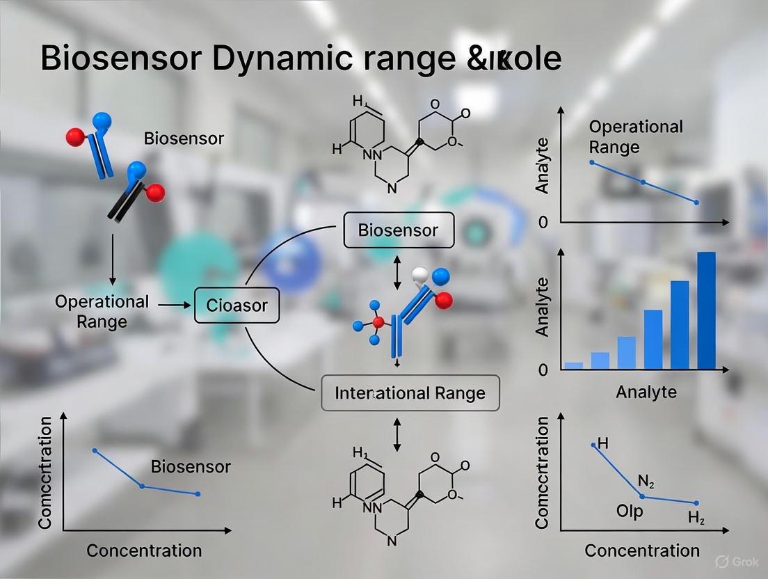
In the development and application of biosensors, two parameters are paramount for evaluating performance: the dynamic range and the operational range. While sometimes used interchangeably, these terms describe fundamentally different characteristics. The dynamic range refers to the concentration span over which a biosensor can produce a quantifiable change in its output signal, effectively defining its sensitivity and limit of detection. In contrast, the operational range describes the full spectrum of environmental conditions—such as pH, temperature, and ionic strength—under which the biosensor maintains its specified performance metrics, including its dynamic range. Understanding and optimizing both parameters is crucial for deploying biosensors in real-world applications, from biomedical diagnostics to environmental monitoring.
This technical guide provides an in-depth examination of both parameters, exploring their definitions, methodologies for enhancement, and interplay within biosensor systems. Framed within broader research on biosensor performance, this analysis equips researchers and drug development professionals with the knowledge to systematically characterize and improve their biosensing platforms.
Dynamic Range: The Concentration Response Window
The dynamic range is a core performance parameter that quantifies a biosensor's ability to respond to varying analyte concentrations. It is typically defined as the range of analyte concentrations between the limit of detection (LOD) and the point of signal saturation, where the output is quantitatively correlated with the analyte level. A wide dynamic range is essential for applications requiring the detection of analytes that can vary significantly in concentration, such as metabolites in physiological fluids or pathogens in environmental samples.
Quantifying Dynamic Range
The dynamic range is most commonly visualized using a dose-response curve, where the sensor's output signal (e.g., fluorescence intensity, voltage, or FRET ratio) is plotted against the logarithm of the analyte concentration. The curve typically has a sigmoidal shape, featuring a linear region where the response is proportional to concentration. The dynamic range is often reported as the span of concentrations across this linear responsive region. The sensitivity of the biosensor is directly derived from the slope of this curve.
Table 1: Quantified Dynamic Range Enhancements in Recent Biosensor Studies
| Biosensor Target | Base Dynamic Range | Enhanced Dynamic Range | Enhancement Method | Key Metric Improvement |
|---|---|---|---|---|
| l-Carnitine [1] | Not specified (Limited) | 10⁻⁴ mM – 10 mM | Directed Evolution (CaiFY47W/R89A variant) | 1000-fold wider range; 3.3-fold higher signal |
| Calcium, ATP, NAD+ [2] | Low (Conventional FRET pairs) | Unprecedented (Specific values not given) | Chemogenetic FRET pairs (e.g., ChemoG5) | Near-quantitative FRET efficiency (≥94%) |
| Lysine [3] | Low dynamic range | Significantly improved | Multilevel optimization (Point mutations, promoter engineering) | Improved dynamic range and lysine response |
Methodologies for Extending Dynamic Range
Research has yielded several powerful strategies for expanding the dynamic range of biosensors.
Directed Evolution and Rational Protein Design: As demonstrated with the l-carnitine biosensor, the transcription factor CaiF was engineered using a "Functional Diversity-Oriented Volume-Conservative Substitution Strategy" on key amino acids. The resulting variant, CaiFY47W/R89A, exhibited a 1000-fold wider concentration response range and a 3.3-fold higher output signal compared to the wild-type control [1]. This approach relies on computer-aided design to model protein structure and identify mutation sites, followed by experimental validation.
Chemogenetic FRET Pair Engineering: A groundbreaking method involves engineering a reversible interaction between a fluorescent protein (FP) and a synthetically labeled HaloTag (HT7). This creates a FRET pair with near-quantitative efficiency (≥94%). By optimizing the interface between the FP donor and the rhodamine-labeled HT7 acceptor, researchers developed biosensors for calcium, ATP, and NAD+ with "unprecedented dynamic ranges" [2]. The protocol involves:
- Fusing a FP (e.g., eGFP) to the N-terminus of HT7.
- Identifying key interface residues (e.g., eGFP Y39, K41, F223) via X-ray crystallography.
- Introducing stabilizing mutations (e.g., eGFP: A206K, T225R; HT7: E143R, E147R, L271E) in a stepwise manner to create variants (ChemoG1 to ChemoG5).
- Labeling the optimized construct (ChemoG5) with cell-permeable rhodamine derivatives (e.g., SiR, JF525, JF669) to achieve spectrally tunable, high-efficiency FRET.
Multilevel Optimization: For the lysine biosensor in E. coli, a low dynamic range was overcome by combining key point mutations in the sensing proteins (LysP and CadC) with engineered modifications to the promoter (Pcad) controlling the output gene [3]. This synergistic approach simultaneously improved the sensor's dynamic range and operational pH stability.
Figure 1: Experimental pathways for enhancing biosensor dynamic range, combining protein engineering and system-level optimization.
Operational Range: The Environmental Stability Domain
The operational range defines the environmental boundaries within which a biosensor functions reliably. It is not a single value but a multi-dimensional space encompassing parameters such as pH, temperature, salinity, and the presence of interfering substances. A biosensor may have an excellent dynamic range under ideal laboratory conditions, but if its performance degrades outside a narrow pH or temperature window, its practical utility is severely limited.
Key Dimensions of the Operational Range
- pH Range: The stability of the biological recognition element (e.g., enzyme, antibody, transcription factor) is often highly pH-dependent. For instance, an initial lysine biosensor in E. coli was found to have a "narrow pH operating range," which was addressed through subsequent engineering [3].
- Temperature Stability: Temperature affects reaction kinetics, protein folding, and the stability of the bioreceptor-transducer complex. The operational temperature range must be specified for applications like industrial bioprocessing or wearable sensors.
- Ionic Strength and Interferents: The concentration of salts and other molecules in the sample matrix can influence biosensor performance by affecting protein structure or generating non-specific signals.
Strategies for Broadening the Operational Range
Engineering a robust operational range focuses on stabilizing the biological components against environmental fluctuations.
- Protein Engineering for Stability: As with dynamic range extension, directed evolution and rational design can be employed to create bioreceptor variants that are functional across a wider pH spectrum or more resistant to denaturation at elevated temperatures. The optimization of the lysine biosensor is a prime example where the operational pH range was broadened [3].
- Material Selection and Sensor Encapsulation: The choice of materials in the sensor's construction can define its operational limits. For example, a glucose sensor designed for stability in interstitial fluid used a nanostructured composite electrode integrated on a printed circuit board, contributing to its excellent durability [4]. Similarly, encapsulating the biological element in a protective polymer hydrogel can shield it from harsh environmental conditions.
Table 2: Research Reagent Solutions for Biosensor Development and Characterization
| Reagent/Material | Function in Development | Example Application |
|---|---|---|
| HaloTag7 (HT7) [2] | Self-labeling protein fusion partner; enables covalent, specific labeling with synthetic fluorophores for FRET-based sensing. | Creating chemogenetic FRET pairs (ChemoX) for tunable biosensors. |
| Silicon Rhodamine (SiR) [2] | Cell-permeable, far-red synthetic fluorophore; used as a FRET acceptor with HaloTag7. | Labeling ChemoG5 to achieve near-quantitative FRET efficiency. |
| CaiF Transcription Factor [1] | Biological recognition element; pivotal transcriptional activator of l-carnitine metabolism. | Core sensing component in an l-carnitine biosensor. |
| LysP & CadC [3] | Lysine transporter and transcription activator; form the core sensing module for lysine. | Constructing a lysine biosensor for dynamic regulation in E. coli. |
| Engineered Promoters [3] | Genetic parts that control the expression level of the output module (e.g., reporter gene). | Fine-tuning the output signal and dynamic range of genetic circuits. |
The Interplay and Trade-offs in Biosensor Design
A critical challenge in biosensor engineering is that optimizing one parameter can inadvertently compromise another. The relationship between dynamic range and operational range is often characterized by trade-offs. For instance, a mutation that increases the affinity of a bioreceptor for its analyte (potentially lowering the LOD and altering the dynamic range) might also make the protein more susceptible to denaturation at higher temperatures (narrowing the operational range). Therefore, a holistic optimization strategy that considers both sets of parameters simultaneously is essential for developing robust biosensors.
The interplay can be visualized as a design space where the goal is to maximize both the dynamic range and the operational range. Successful engineering strategies, such as the multilevel optimization used for the lysine biosensor, address both aspects concurrently by selecting for mutations that improve performance metrics without sacrificing stability under process-relevant conditions [3].
Figure 2: Logical relationships between key biosensor performance parameters, showing synergy and potential trade-offs.
The distinction between dynamic range and operational range is fundamental to the rigorous characterization and successful application of biosensors. The dynamic range defines the sensor's concentration detection window, while the operational range sets the environmental conditions for its reliable use. As evidenced by recent advances, strategies such as directed evolution, chemogenetic FRET pair engineering, and multilevel optimization provide powerful tools to expand both parameters.
Future research will continue to close the gap between laboratory performance and real-world application demands. This will involve developing more sophisticated engineering techniques that minimize trade-offs and create biosensors that are not only exquisitely sensitive across a wide concentration span but also robust enough to function in complex, variable environments like the human body, industrial fermenters, and natural ecosystems. A deep understanding of these key performance parameters is the first step in this engineering journey.
Biomolecular recognition forms the cornerstone of modern biosensing technologies, enabling the highly specific detection of an enormous range of analytes through protein- and nucleic acid-based interactions. The theoretical foundation governing these single-site binding events follows a fundamental hyperbolic relationship between target concentration and receptor occupancy. This relationship, while providing the basis for countless diagnostic applications, imposes a significant inherent constraint known as the 81-fold dynamic range limit. For any biosensor operating under single-site binding conditions, the transition from 10% to 90% site occupancy requires precisely an 81-fold increase in target concentration [5].
This fixed dynamic range presents substantial limitations for real-world applications where target concentrations may vary across several orders of magnitude. Clinical measurements of HIV viral loads, for example, can span from approximately 50 to over 10^6 copies/mL—a range exceeding five orders of magnitude that dwarfs the intrinsic 81-fold dynamic range of single-site binding [5]. Similarly, therapeutic drug monitoring for agents with narrow therapeutic indices (such as cyclosporine and aminoglycosides) requires a precision difficult to achieve with a system necessitating an 81-fold concentration change to transition from minimal to near-maximal signal response [5]. Understanding this fundamental limitation is crucial for researchers and drug development professionals working to optimize biosensor performance for specific applications.
Theoretical Framework and Mathematical Basis
The Law of Mass Action and Binding Isotherm
The 81-fold limit arises directly from the Law of Mass Action applied to reversible bimolecular binding interactions. For a simple equilibrium where a receptor (R) binds to a ligand (L) to form a complex (RL):
R + L ⇌ RL
The dissociation constant Kd is defined as Kd = [R][L]/[RL], where brackets denote concentrations. The fractional occupancy (θ) of receptors is given by:
θ = [L] / (Kd + [L])
This equation generates a hyperbolic binding curve when fractional occupancy is plotted against ligand concentration. The dynamic range is conventionally defined as the concentration interval between 10% and 90% occupancy. Solving the fractional occupancy equation for these values:
When θ = 0.1: [L]₁₀ = Kd/9 When θ = 0.9: [L]₉₀ = 9Kd
The ratio between these concentrations gives the fundamental 81-fold limit:
[L]₉₀ / [L]₁₀ = 9Kd / (Kd/9) = 81
This mathematical relationship demonstrates that the 81-fold dynamic range is an intrinsic property of single-site binding thermodynamics, independent of the specific receptor-ligand pair or absolute affinity [5].
Biosensor Response and Practical Implications
In practical biosensing applications, the receptor occupancy is converted through a transducer into a measurable signal (e.g., optical, electrochemical). The useful dynamic range of the biosensor is therefore constrained by this fundamental 81-fold limit between 10% and 90% signal saturation [5]. The following table summarizes key characteristics of this binding isotherm:
Table 1: Characteristics of Single-Site Binding Isotherm
| Parameter | Symbol/Value | Interpretation |
|---|---|---|
| Binding Curve Shape | Hyperbolic | Non-linear relationship between concentration and occupancy |
| Half-Maximal Effect | EC₅₀ = Kd | Concentration at 50% receptor occupancy equals Kd |
| Lower Detection Limit | [L]₁₀ = Kd/9 | Concentration at 10% occupancy |
| Upper Detection Limit | [L]₉₀ = 9Kd | Concentration at 90% occupancy |
| Useful Dynamic Range | 81-fold | Fixed ratio between upper and lower detection limits |
This fixed dynamic range creates significant challenges for applications requiring either exceptional sensitivity to small concentration changes or the ability to quantify analytes across wide concentration ranges.
Engineering Strategies to Overcome the 81-Fold Limit
Biological Inspiration and Engineering Approaches
Evolution has developed sophisticated mechanisms to overcome the limitations of single-site binding, inspiring parallel engineering approaches for artificial biosensors. Research has identified three primary strategies for editing the dynamic range of biosensors [5]:
- Extended Dynamic Range: Employing pairs or sets of closely related receptors with different affinities for the same target
- Narrowed Dynamic Range: Creating steep, ultrasensitive responses through mechanisms like sequestration
- Complex Dose-Response: Engineering multi-state systems that respond sensitively only within specific concentration windows
These approaches manipulate the fundamental binding curve to better align with application-specific requirements, whether calling for expanded range, heightened sensitivity within a narrow window, or multi-threshold detection capabilities.
Structure-Switching Receptors
A key advancement in overcoming the 81-fold limit involves engineering structure-switching receptors where binding is coupled to a conformational change. This approach enables tuning of apparent affinity without altering the specific chemical interactions that determine specificity [5].
By stabilizing an alternative "non-binding" state and tuning the switching equilibrium constant, researchers can generate receptor variants spanning a wide affinity range while maintaining identical specificity profiles [5]. This strategy mirrors approaches found in nature, such as intrinsically unfolded proteins that reduce affinity by coupling binding to an unfavorable folding event without compromising specificity [5].
Figure 1: Structure-Switching Receptor Mechanism. Receptors exist in equilibrium between non-binding and binding-competent states. Engineering the switching equilibrium constant (Ks) enables tuning of apparent affinity without altering binding site specificity.
Experimental Implementation and Protocols
Generating Receptor Variants with Tunable Affinities
Molecular beacons (DNA stem-loop structures) serve as an excellent model system for demonstrating dynamic range engineering. The following protocol outlines the generation of affinity variants through stem stability modulation [5]:
Materials:
- DNA oligonucleotides for molecular beacon construction
- Fluorescent reporter (e.g., FAM) and quencher (e.g., BHQ1)
- Thermal cycler or water bath for temperature control
- Buffer reagents (Tris-HCl, MgCl₂, NaCl)
- Target DNA sequences (perfect match and mismatch controls)
Method:
- Design molecular beacon variants with identical target-recognition loops but different stem sequences. Vary the stem length and GC content to create a stability gradient.
- Synthesize and purify molecular beacons with 5' fluorophore and 3' quencher modifications.
- Characterize stem stability using thermal denaturation profiles measuring fluorescence vs. temperature.
- Determine binding affinity for each variant by titrating target DNA and measuring fluorescence recovery. Fit data to binding isotherm to extract Kd values.
- Verify specificity by testing each variant against mismatched targets to ensure discrimination is maintained across affinity variants.
Using this approach, researchers have generated molecular beacon sets with dissociation constants spanning from 0.012 μM to 128 μM—a 10,000-fold affinity range—while maintaining similar specificity profiles (on average 35-fold difference in affinity between matched and single-base mismatched targets) [5].
Extending Dynamic Range Through Receptor Mixing
Rationale: Combining multiple receptor variants with different affinities creates a composite sensor with extended dynamic range. The optimal affinity difference between variants is approximately 100-fold to maximize log-linear range while minimizing deviations from linearity [5].
Protocol:
- Select receptor variants with affinity difference near 100-fold (e.g., molecular beacons with Kd values of 0.1 μM and 10 μM).
- Adjust molar ratios to compensate for differences in signal gain. For variants with 83% and 100% maximum fluorescence change, use approximately 59:41 ratio.
- Mix variants in optimized ratios and characterize response across target concentration range.
- Verify specificity across extended dynamic range using mismatch targets.
This approach has demonstrated extension of dynamic range to 8,100-fold with excellent log-linearity (R²=0.995) and 9-fold signal gain [5]. Further extension to ~900,000-fold (more than four orders of magnitude beyond single receptors) can be achieved by combining four appropriately selected variants in optimized ratios [5].
Table 2: Molecular Beacon Variants for Dynamic Range Engineering
| Variant | Dissociation Constant (Kd) | Relative Affinity | Maximum Fluorescence Change | Application in Mixtures |
|---|---|---|---|---|
| 0GC | 0.012 μM | Highest | ~70% | Extended range, Three-state systems |
| 1GC | 0.11 μM | High | 83% | Extended range (paired with 3GC) |
| 2GC | 1.1 μM | Medium | ~90% | Intermediate in 4-variant mixtures |
| 3GC | 11 μM | Low | 100% | Extended range (paired with 1GC) |
| 5GC | 128 μM | Lowest | ~60% | Extended range, Three-state systems |
Creating Narrowed Dynamic Range and Threshold Sensors
Sequestration Approach: Narrowing dynamic range creates steep, ultrasensitive responses valuable for threshold detection. This employs a non-signaling, high-affinity "depletant" receptor that sequesters target until saturation, after which free target concentration rises dramatically [5].
Protocol:
- Design non-signaling receptor with higher affinity than signaling receptor (e.g., DNA sequence complementary to target but without fluorophore/quencher pair).
- Mix signaling and non-signaling receptors at optimized ratio. The non-signaling receptor acts as a sink until saturated.
- Characterize response to observe threshold effect with compressed dynamic range.
This approach can compress the dynamic range by an order of magnitude and rationally tune threshold response to selected target concentrations [5].
Site-Specific Immobilization for Enhanced Performance
Controlled orientation of receptors on biosensor surfaces significantly impacts performance. Site-specific immobilization using bioorthogonal chemistry addresses limitations of random conjugation [6].
Materials:
- Recombinant protein with incorporated noncanonical amino acid (e.g., AzK)
- Orthogonal pyrrolysyl-tRNA synthetase (PylRS)/tRNACUAPyl pair
- Alkyne-functionalized biosensor surface
- Biotin linker for detection
Method:
- Incorporate reactive ncAA using stop codon suppression technology at selected position in protein sequence.
- Conjugate with biotin linker via strain-promoted azide-alkyne cycloaddition (SPAAC).
- Immobilize on sensor surface through biotin-neutravidin interaction.
- Validate orientation and function using binding assays with target molecule.
This approach has demonstrated 12-fold enhancement in binding sensitivity compared to random immobilization methods [6].
Figure 2: Site-Specific vs. Random Immobilization. Directed immobilization through genetic incorporation of noncanonical amino acids enables optimal orientation of binding sites, significantly enhancing biosensor sensitivity compared to random conjugation approaches.
Research Reagents and Materials
Table 3: Essential Research Reagents for Dynamic Range Engineering
| Reagent/Category | Specific Examples | Function and Application |
|---|---|---|
| Structure-Switching Receptors | Molecular beacons with variable stem stability | Generate affinity variants while maintaining specificity for extended dynamic range systems [5] |
| Noncanonical Amino Acids | Nε-((2-azidoethoxy)carbonyl)-l-lysine (AzK) | Enable site-specific immobilization via bioorthogonal chemistry for optimized receptor orientation [6] |
| Orthogonal Translation System | PylRS/tRNACUAPyl pair from M. mazei | Genetic incorporation of ncAAs for controlled surface attachment [6] |
| Bioorthogonal Conjugation | Strain-promoted azide-alkyne cycloaddition (SPAAC) reagents | Directed coupling of receptors to sensor surfaces without interfering with binding function [6] |
| Transcription Factor Scaffolds | CaiF variants for l-carnitine detection | Protein-based biosensors for metabolite detection; platform for directed evolution [1] |
Applications and Recent Advances
Pharmaceutical and Clinical Applications
The ability to engineer biosensor dynamic range has significant implications for pharmaceutical research and clinical diagnostics. Engineered biosensors with extended dynamic ranges are particularly valuable for monitoring therapeutic drugs with narrow therapeutic indices, where precise quantification across clinically relevant concentrations is essential for optimizing efficacy while avoiding toxicity [5]. Similarly, the detection of viral loads that naturally span orders of magnitude, such as HIV monitoring, benefits from expanded dynamic range systems [5].
Threshold sensors with narrowed dynamic ranges offer applications in point-of-care diagnostics where simple "yes/no" or "above/below critical threshold" outputs are clinically sufficient and practically desirable. Three-state dynamic range systems that respond sensitively only when target concentrations fall above or below defined intermediate regimes enable sophisticated monitoring applications in therapeutic drug monitoring and metabolic disorder management [5].
Directed Evolution Approaches
Recent research has complemented the structure-switching and receptor mixing approaches with directed evolution strategies. For example, engineering of the transcription factor CaiF for l-carnitine detection has demonstrated substantial improvements in biosensor performance. Through computational design and functional diversity-oriented substitutions, researchers developed CaiF variants with 1000-fold wider response range (from 10^-4 mM to 10 mM) and 3.3-fold higher output signal intensity compared to the wild-type biosensor [1].
This directed evolution approach involved:
- Computer-aided design of CaiF structure and DNA binding site simulation
- Alanine scanning to validate predicted functional residues
- Diversity-oriented substitution at key positions to generate functional variants
- High-throughput screening to identify variants with improved dynamic range
The success of this methodology highlights the potential for combining rational design with directed evolution to overcome fundamental limitations in biosensor performance [1].
The fundamental 81-fold dynamic range limit represents a significant constraint inherent to single-site binding interactions that underpins many biosensing technologies. Through understanding of this thermodynamic principle and implementation of sophisticated engineering strategies—including structure-switching receptors, optimized receptor mixing, sequestration mechanisms, and site-specific immobilization—researchers can now rationally design biosensors with tailored dynamic ranges optimized for specific applications. These approaches, inspired by natural systems and enhanced through modern protein engineering and directed evolution methodologies, enable unprecedented control over biosensor response characteristics. The continued refinement of these strategies promises to expand further the utility of biosensors in pharmaceutical development, clinical diagnostics, and environmental monitoring applications where precise quantification across wide concentration ranges is required.
Dynamic range, a fundamental performance metric for biosensors, is critically defined as the span between the minimal and maximal detectable signals [7]. In both industrial biomanufacturing and clinical diagnostics, this parameter directly determines the efficacy of high-throughput screening (HTS) campaigns and the accuracy of diagnostic results. This technical review examines the profound implications of dynamic range on biosensor functionality, detailing established and emerging methodologies for its optimization. We present quantitative data linking expanded dynamic range to enhanced screening efficiency and diagnostic reliability, supported by experimental protocols and reagent specifications to provide researchers with a practical framework for biosensor development and implementation.
Biosensors are analytical devices that integrate a biological recognition element with a physicochemical transducer to detect specific analytes [8]. Their performance is characterized by several interdependent parameters:
- Dynamic Range: The concentration interval over which the biosensor produces a measurable and usable signal response, typically bounded by the limit of detection (LOD) at the lower end and signal saturation at the upper end [7].
- Operating Range: The specific concentration window where the biosensor performs with optimal accuracy and precision, often a subset of the full dynamic range [7].
- Sensitivity: The magnitude of output signal change per unit change in analyte concentration.
- Response Time: The speed at which the biosensor reaches its maximum signal after analyte exposure [7].
- Signal-to-Noise Ratio: The ratio between the meaningful signal and the underlying background noise, determining detection clarity [7].
These metrics collectively define biosensor utility across applications ranging from metabolic engineering to point-of-care diagnostics. However, dynamic range holds particular significance as it establishes the fundamental boundaries within which a biosensor can operate effectively.
Table 1: Key Performance Metrics for Biosensor Characterization
| Metric | Definition | Impact on Application |
|---|---|---|
| Dynamic Range | Span between minimal and maximal detectable signals [7] | Determines the concentration window for reliable detection |
| Operating Range | Concentration window for optimal performance [7] | Defines practical utility limits for specific applications |
| Sensitivity | Signal change per unit analyte concentration change | Affects ability to distinguish small concentration differences |
| Response Time | Speed of signal response to analyte exposure [7] | Critical for real-time monitoring and rapid screening |
| Signal-to-Noise Ratio | Ratio of meaningful signal to background noise [7] | Impacts detection reliability and measurement confidence |
The Critical Role of Dynamic Range in High-Throughput Screening
High-throughput screening relies on the rapid assessment of thousands to millions of biological variants to identify candidates with desired properties. In this context, biosensor dynamic range becomes a throughput-determining factor.
Pathway Optimization and Strain Development
In metabolic engineering, biosensors are indispensable for optimizing synthetic pathways and screening strain libraries. A restricted dynamic range can mask superior producers during screening, as variants producing metabolite concentrations beyond the sensor's detection上限 fail to generate discriminable signals [7]. This necessitates multiple screening rounds with sample dilution or different sensors, drastically reducing throughput and increasing resource requirements. Furthermore, biosensors with nonideal dose–response characteristics or high signal noise exacerbate scalability challenges by increasing false positives or masking true high-performing strains [7].
Advanced Screening Methodologies
Emerging technologies are addressing these limitations through sophisticated screening platforms. The BeadScan system combines droplet microfluidics with automated fluorescence imaging to encapsulate, express, and screen vast libraries of biosensor variants [9]. This platform enables parallel evaluation of multiple biosensor features—including dynamic range, affinity, and specificity—across thousands of variants, representing an order-of-magnitude increase in screening throughput [9].
Figure 1: The BeadScan workflow for high-throughput biosensor screening utilizes gel-shell beads (GSBs) as microscale dialysis chambers to assay thousands of variants against multiple analyte conditions in parallel [9].
Dynamic Range and Diagnostic Precision
In diagnostic applications, dynamic range directly influences clinical accuracy and utility across diverse patient populations and disease stages.
Impact on Diagnostic Accuracy
A biosensor with insufficient dynamic range may fail to detect clinically significant analyte concentrations falling outside its detection window, leading to false negatives in hypersecreting conditions or false positives due to nonspecific binding in complex matrices like serum [8]. This is particularly critical for biomarkers with wide physiological and pathological concentration ranges. For example, C-reactive protein (CRP) circulates at concentrations ranging from μg/mL to hundreds of μg/mL during infection [8]. A biosensor with a narrow dynamic range would require multiple dilutions for accurate quantification across this span, increasing assay complexity and potential for error.
Mitigating Nonspecific Binding
Nonspecific binding (NSB) presents a significant challenge in label-free biosensors, particularly when analyzing complex biological samples. NSB from serum constituents can produce signals indistinguishable from specific binding events, effectively reducing the functional dynamic range and compromising diagnostic precision [8]. Implementation of appropriate reference channels with optimized negative controls (e.g., isotype-matched antibodies) enables subtraction of NSB contributions, preserving the integrity of the biosensor's dynamic range in complex media [8].
Enhancing Precision Through Computational Approaches
Innovative computational methods are now addressing dynamic range limitations by extracting more information from biosensor responses. Theory-guided deep learning leverages domain knowledge to improve biosensor accuracy and speed using dynamic signal changes [10]. By training recurrent neural networks with cost function supervision based on biosensing theory, researchers have achieved 98.5% average prediction accuracy for microRNA quantification across nanomolar to femtomolar concentrations—a range spanning six orders of magnitude [10]. This approach significantly reduces false-negative and false-positive results while enabling accurate concentration determination from initial transient responses, effectively expanding the usable dynamic range.
Experimental Approaches for Dynamic Range Optimization
Multiple strategic approaches exist for optimizing the dynamic range of biosensors, from molecular engineering to directed evolution.
Transcription Factor Engineering
Protein-based biosensors, particularly those utilizing transcription factors (TFs), offer numerous engineering handles for dynamic range modulation. Research on the CaiF transcription factor-based biosensor for L-carnitine detection demonstrates that rational engineering of DNA binding domains can dramatically expand dynamic range [1]. Through computer-aided design and alanine scanning, researchers identified key residues affecting DNA binding affinity. The variant CaiF-Y47W/R89A exhibited a 1000-fold wider concentration response range (10⁻⁴ mM to 10 mM) with a 3.3-fold higher output signal intensity compared to the wild-type biosensor [1].
Table 2: Performance Comparison of Engineered CaiF Biosensor Variants
| Biosensor Variant | Dynamic Range | Signal Intensity | Key Structural Modification |
|---|---|---|---|
| Wild-type CaiF | Reference range | Reference intensity | Native DNA-binding domain |
| CaiF-Y47W/R89A | 1000-fold wider [1] | 3.3-fold higher [1] | Dual substitution in DNA interaction interface |
RNA-Based Biosensor Design
RNA-based biosensors, including riboswitches and toehold switches, provide complementary advantages for dynamic range tuning. Their compact size and reversible ligand binding characteristics facilitate integration into metabolic regulation circuits [7]. Toehold switches offer particularly programmable dynamic range control through careful design of the trigger RNA binding region, enabling logic-gated control of metabolic pathways in microbial factories [7].
Integrated Experimental Workflows
Comprehensive dynamic range optimization typically follows an iterative design-build-test cycle, incorporating both rational design and screening approaches.
Figure 2: Integrated workflow for biosensor dynamic range optimization combines rational design with high-throughput screening and data-driven modeling in an iterative refinement cycle [7] [1] [9].
Essential Research Reagents and Materials
Successful development and implementation of biosensors with optimized dynamic ranges requires specific reagent solutions and materials.
Table 3: Essential Research Reagents for Biosensor Development and Analysis
| Reagent/Material | Function/Application | Specific Examples |
|---|---|---|
| Transcription Factors | Protein-based biosensor scaffolds for metabolite detection | CaiF for L-carnitine sensing [1] |
| RNA Scaffolds | Modular, programmable biosensors with tunable response | Riboswitches, toehold switches [7] |
| Cell-Free Expression Systems | High-yield protein synthesis for biosensor production | PUREfrex2.0 system [9] |
| Microfluidic Components | High-throughput screening platform fabrication | Gel-shell beads (GSBs), droplet generators [9] |
| Reference Control Probes | Nonspecific binding correction in label-free biosensors | Isotype-matched antibodies, BSA, anti-FITC [8] |
| Surface Chemistry Reagents | Sensor functionalization and biomolecule immobilization | Silane chemistry for non-planar surfaces [11] |
Dynamic range stands as a critical determinant of biosensor performance in both high-throughput screening and diagnostic applications. The concentration window over which a biosensor operates reliably directly impacts screening efficiency, strain development timelines, diagnostic accuracy, and clinical utility. Through integrated approaches combining molecular engineering, advanced screening platforms, and computational methods, researchers can systematically optimize this fundamental parameter. The continued refinement of biosensors with expanded dynamic ranges will accelerate advancements across synthetic biology, biomanufacturing, and precision medicine, enabling more sensitive detection, more accurate quantification, and more reliable decision-making across biological research and clinical practice.
Biosensors are fundamental biological tools that combine a sensor module, which detects specific intracellular or environmental signals, with an actuator module, which drives a measurable or functional response [7]. These devices are indispensable in metabolic engineering and synthetic biology for enabling real-time monitoring, high-throughput screening, and dynamic regulation of metabolic pathways [7]. The performance of biosensors is critically evaluated through key metrics such as dynamic range (the span between minimal and maximal detectable signals), operating range (the concentration window for optimal performance), response time, and signal-to-noise ratio [7]. Within this framework, transcription factors, riboswitches, and two-component systems represent core natural mechanisms that have been engineered to construct sophisticated synthetic biosensing systems. This review provides an in-depth technical overview of these mechanisms, emphasizing their operational principles, quantitative performance characteristics, and experimental methodologies for engineering enhanced dynamic and operational ranges.
Core Biosensor Mechanisms
Transcription Factor-Based Biosensors
Sensing Principle: Transcription factor (TF)-based biosensors function through ligand-induced allosteric regulation. The binding of a specific target metabolite to the transcription factor induces a conformational change, enabling or disabling its binding to a specific DNA operator sequence, thereby regulating the expression of a downstream reporter gene [7] [12]. This mechanism allows for direct genetic regulation in response to metabolite levels.
Key Characteristics: TFs typically exhibit moderate sensitivity and are suitable for high-throughput screening due to their direct coupling to gene expression [7]. A significant advantage is their broad analyte range, capable of sensing diverse compounds including alcohols, flavonoids, and organic acids [7]. Their performance is often characterized by a dose-response curve, which defines sensitivity and dynamic range.
Engineering Methodologies:
- Dynamic Range Tuning: The dynamic and operational ranges of TF-based biosensors can be tuned by exchanging promoters and ribosome binding sites (RBS), or by altering the number and position of the operator region within the genetic construct [7].
- Specificity Engineering: The chimeric fusion of DNA-binding domains and ligand-binding domains from different transcription factors has been successfully used to engineer novel biosensor specificities [7].
- Directed Evolution: High-throughput techniques like fluorescence-activated cell sorting (FACS), combined with directed evolution strategies, are employed to improve biosensor sensitivity and specificity. This involves creating mutant libraries of the transcription factor and screening for variants with desired response characteristics [7] [12].
Riboswitches
Sensing Principle: Riboswitches are structured RNA elements located in the 5' untranslated regions (UTRs) of mRNAs. They sense intracellular metabolites through direct ligand binding, which induces a conformational change in the RNA structure [7]. This structural switch then affects downstream gene expression by either activating or repressing translation initiation or transcription termination.
Key Characteristics: Riboswitches are compact, tunable, and often reversible, making them ideal for integrating into metabolic regulation circuits [7]. They are particularly useful for sensing fundamental intracellular metabolites such as nucleotides and amino acids, and they generally exhibit faster response times compared to protein-based systems due to the absence of a protein synthesis step [7] [12].
Engineering Methodologies:
- Sequence Rational Design: The sequence of the aptamer domain (responsible for ligand binding) and the expression platform (responsible for the structural switch) can be rationally designed or mutated to alter ligand specificity, affinity, and the dynamic range of the response.
- High-Throughput Screening: As with TFs, generating mutant riboswitch libraries and screening them via FACS or other selection methods is a common approach to isolate variants with improved characteristics, such as a wider dynamic range or altered ligand specificity [12].
Two-Component Systems (TCSs)
Sensing Principle: Two-component systems are modular signaling pathways typically comprising a membrane-associated sensor kinase and a cytoplasmic response regulator. Upon detection of an extracellular or intracellular signal (e.g., ions, pH, small molecules), the sensor kinase autophosphorylates and then transfers the phosphate group to the response regulator [7]. The phosphorylated response regulator then activates the transcription of target genes.
Key Characteristics: TCSs are highly adaptable and specialized for environmental signal detection [7]. Their modular architecture allows for the engineering of hybrid systems where the sensor domain of one kinase is fused to the regulatory domain of another, enabling the creation of novel input-output relationships.
Engineering Methodologies:
- Domain Swapping: The sensor domains of different kinases can be swapped to create chimeric TCSs that respond to new environmental or intracellular cues [7].
- Promoter and RBS Engineering: The output of a TCS can be fine-tuned by engineering the promoter and RBS of the gene(s) controlled by the response regulator, thereby adjusting the amplitude of the output signal within the operational range [7].
Table 1: Comparative Analysis of Core Biosensor Mechanisms
| Category | Biosensor Type | Sensing Principle | Key Advantages | Typical Analyte Examples |
|---|---|---|---|---|
| Protein-Based | Transcription Factors (TFs) | Ligand binding induces DNA interaction to regulate gene expression | Suitable for high-throughput screening; broad analyte range | Alcohols, flavonoids, organic acids [7] |
| Protein-Based | Two-Component Systems (TCSs) | Sensor kinase autophosphorylates and transfers signal to response regulator | High adaptability; environmental signal detection; modular signaling | Ions, pH, small molecules [7] |
| RNA-Based | Riboswitches | Ligand-induced RNA conformational change affects translation | Compact; tunable response; reversible; fast response time | Nucleotides, amino acids [7] |
Quantitative Performance Metrics and Engineering
A critical aspect of biosensor development is the rigorous characterization and engineering of its performance parameters. The following table summarizes the key metrics and common strategies for their optimization.
Table 2: Key Biosensor Performance Parameters and Engineering Strategies
| Performance Parameter | Definition | Engineering & Tuning Strategies |
|---|---|---|
| Dynamic Range | The ratio between the maximal and minimal output signals [7]. | Promoter engineering, RBS tuning, plasmid copy number variation [7]. |
| Operating Range | The concentration window of the analyte where the biosensor performs optimally [7]. | Operator site position/number manipulation, directed evolution of ligand-binding domain [7]. |
| Response Time | The speed at which the biosensor reaches its maximum output signal after analyte exposure [7]. | Using faster-acting components (e.g., riboswitches), hybrid system design [7]. |
| Signal-to-Noise Ratio | The clarity and reliability of the output signal under constant conditions [7]. | Optimizing expression levels of sensor and reporter components to minimize basal expression [7]. |
Experimental Protocol for Characterizing Dose-Response:
- Strain Cultivation: Grow the engineered microbial strain harboring the biosensor to mid-exponential phase.
- Analyte Exposure: Aliquot the culture into multi-well plates and expose to a concentration gradient of the target analyte. Include a negative control (no analyte).
- Output Measurement: For fluorescent reporters, measure fluorescence intensity over time using a plate reader. For other reporters (e.g., colorimetric, luminescent), use the appropriate detector.
- Data Analysis: Normalize the output signal (e.g., fluorescence) to cell density (e.g., OD600). Plot the normalized output against the logarithm of the analyte concentration.
- Curve Fitting: Fit the data to a sigmoidal function (e.g., Hill equation) to extract key parameters: minimum and maximum output (dynamic range), EC50 (sensitivity), and Hill coefficient (cooperativity).
Visualization of Biosensor Mechanisms and Workflows
Transcription Factor Biosensor Mechanism
Two-Component System Biosensor Mechanism
High-Throughput Screening Workflow for Biosensor Engineering
Research Reagent Solutions
The following table details key reagents and materials essential for the construction and testing of genetic biosensors.
Table 3: Essential Research Reagents for Biosensor Development
| Reagent / Material | Function / Application |
|---|---|
| Allosteric Transcription Factors (aTFs) | Core sensing element for protein-based metabolite detection; can be sourced from native hosts or engineered for new specificities [12]. |
| Riboswitch DNA Sequences | Synthetic DNA fragments encoding the aptamer and expression platform; used to construct RNA-based sensors for intracellular metabolites [7] [12]. |
| Plasmid Vectors (various copy numbers) | Backbone for biosensor circuit construction; plasmid copy number is a key variable for tuning biosensor sensitivity and dynamic range [7]. |
| Fluorescent Reporter Proteins (eGFP, mCherry, etc.) | Provide a measurable output signal for high-throughput screening and quantitative characterization of biosensor performance [7] [12]. |
| Microfluidic Droplet Generation Systems | Enable high-throughput screening by encapsulating single cells or biosensor reactions in picoliter droplets for analysis and sorting (e.g., FADS) [12]. |
| Cell-Free Protein Synthesis (CFPS) Systems | In vitro systems for rapid prototyping of biosensors and pathways, bypassing cellular constraints and allowing for direct control of the reaction environment [12]. |
Transcription factors, riboswitches, and two-component systems provide a versatile toolkit for constructing genetic biosensors with applications ranging from metabolic engineering to diagnostics. The effective deployment of these biosensors relies on a deep understanding of their inherent mechanisms and a systematic approach to engineering their performance parameters, particularly the dynamic and operational ranges. By leveraging structured experimental protocols, high-throughput screening methodologies, and computational design, researchers can continually refine these biological devices, enhancing their reliability and expanding their applicability in both industrial and therapeutic contexts.
Engineering and Application: Strategies for Extending and Tailoring Biosensor Ranges
In the fields of therapeutic drug development and basic cellular research, the ability to accurately measure biochemical signals within their native, complex environments is paramount. Biosensors, which convert biological events into measurable signals, are foundational tools for this purpose. A biosensor's dynamic range—the span between the minimum and maximum concentrations of a target molecule it can accurately detect—directly determines its utility. A wide dynamic range is crucial for capturing the full spectrum of physiological fluctuations, from subtle basal signaling to robust, pathway-saturated responses. However, many naturally occurring and first-generation engineered biosensors often possess an insufficient dynamic range, leading to signal saturation and an inability to resolve critical quantitative differences at the upper and lower limits of target concentration.
This technical guide explores the strategy of strategic mixing, a method involving the deliberate combination of receptor variants with distinct ligand-binding affinities to construct a composite biosensor system with a significantly widened effective dynamic range. Framed within a broader research thesis on biosensor operational range, this approach moves beyond the limitations of single-component sensors. By creating a system where different receptor variants are active across different concentration windows, researchers can achieve a more linear and extensive response profile. This guide provides a detailed examination of the underlying principles, quantitative characterization methods, and practical experimental protocols for implementing this powerful technique, equipping scientists with the knowledge to deploy it in advanced drug screening and signaling studies.
Core Principles: Receptor Affinity and Composite Response
The theoretical foundation of strategic mixing rests on the well-defined binding characteristics of individual receptor-ligand pairs. A receptor's affinity for its ligand, typically quantified by its dissociation constant (Kd), defines the concentration range over which it is most sensitive to changes in ligand concentration. A receptor with a low Kd (high affinity) will saturate at relatively low ligand concentrations, making it ideal for detecting subtle changes in low-abundance analytes. Conversely, a receptor with a high Kd (low affinity) will only begin to respond at higher ligand concentrations and will resist saturation, making it suitable for measuring robust signals.
The Strategic Mixing Hypothesis
The central hypothesis of strategic mixing is that by co-deploying multiple receptor variants—each with a deliberately tuned and distinct Kd for the same target ligand—within a single experimental system, their individual responsive ranges can be made to overlap and combine. The resulting composite output is a summation of their individual response curves. When these variants are coupled to the same reporting mechanism (e.g., fluorescence), the system's overall dynamic range is extended to cover a much broader concentration spectrum than any single variant could achieve alone. This method effectively linearizes the sensor's response over a wider interval, enhancing the resolution and accuracy of quantitative measurements across diverse experimental conditions, from the tumor microenvironment to intracellular second-messenger signaling.
Table 1: Characteristics of Receptor Variants for Strategic Mixing
| Variant Name | Theoretical Kd (nM) | Optimal Sensing Range | Primary Role in Composite System |
|---|---|---|---|
| High-Affinity (Variant H) | 10 - 50 nM | Low (Basal) Concentrations | Provides sensitivity to subtle, low-level changes in signaling. |
| Medium-Affinity (Variant M) | 200 - 500 nM | Mid (Physiological) Concentrations | Covers the core range of active biological fluctuations. |
| Low-Affinity (Variant L) | 1000 - 5000 nM | High (Saturated) Concentrations | Prevents saturation and captures data at peak concentrations. |
Quantitative Characterization of Biosensor Performance
To effectively design and implement a strategic mixing protocol, a rigorous quantitative assessment of each receptor variant's performance is non-negotiable. This involves determining key parameters that define the sensor's operational envelope. The most critical of these is the dissociation constant (Kd), which provides a direct measure of binding affinity. Furthermore, the dynamic range of each sensor must be precisely measured as the fold-change in output signal between the minimal (ligand-free) and maximal (saturating ligand) states. For fluorescent biosensors, this is often reported as the ratio of fluorescence intensity or the change in Förster Resonance Energy Transfer (FRET) efficiency.
Recent advances in biosensor engineering, particularly using directed evolution, have demonstrated the dramatic improvements possible in these metrics. For instance, the development of the cdGreen2 biosensor for the bacterial second messenger c-di-GMP involved iterative cycles of mutagenesis and fluorescence-activated cell sorting (FACS). This process yielded a final biosensor variant with a 12-fold increase in fluorescence intensity upon ligand binding and a Kd of 214 nM, showcasing a combination of high sensitivity and a wide dynamic range that is ideal for such applications [13]. Quantitative data, as summarized in the table below, must be collected under standardized conditions to enable rational selection of variants for mixing.
Table 2: Experimentally Determined Biosensor Performance Metrics
| Performance Parameter | Definition & Measurement Method | Target Value for Strategic Mixing |
|---|---|---|
| Dissociation Constant (Kd) | Ligand concentration at half-maximal sensor response. Determined from dose-response curve fitting. | Variants should have Kd values spaced across a log scale (e.g., 50 nM, 500 nM, 5 µM). |
| Dynamic Range (Fold-Change) | Maximal signal output / Minimal signal output. Measured via fluorometry or plate reader. | A high fold-change (>5) is desirable for each variant to ensure a strong composite signal. |
| Brightness (Molecular Brightness) | Fluorescence intensity per sensor molecule. Measured relative to standard fluorescent proteins. | High brightness is critical for signal-to-noise ratio in cell-based assays. |
| Ligand Binding Kinetics (kon, koff) | Rates of ligand association and dissociation. Measured via stopped-flow or surface plasmon resonance. | Variants with similar kon but different koff rates often yield the desired Kd spacing. |
Experimental Protocol: A Step-by-Step Guide to Strategic Mixing
The following section outlines a detailed, actionable protocol for implementing the strategic mixing approach. This methodology is adapted from best practices in protein engineering and biosensor validation, drawing on techniques used in the development of cutting-edge tools like transcription factor-based biosensors [14] and FRET-based signaling reporters [15].
Phase 1: Affinity Variant Generation and Selection
- Generate Receptor Variants: Create a library of receptor variants with a range of affinities for your target ligand. This can be achieved through:
- Site-Directed Mutagenesis: Target residues in the ligand-binding pocket based on structural data (e.g., from PDB files 6E2Q, 4BSK used in computational design [16]).
- Directed Evolution: Use iterative FACS under alternating high/low ligand regimes to select for desired affinities, as successfully demonstrated with the cdGreen2 biosensor [13].
- Characterize Individual Variants: Express and purify each candidate variant. Using a plate reader or fluorometer, generate a dose-response curve by measuring the sensor's output (e.g., fluorescence intensity at 513 nm for GFP-based sensors) across a logarithmic concentration range of the purified ligand (e.g., 1 nM to 100 µM).
- Determine Kd and Dynamic Range: Fit the dose-response data to a sigmoidal curve (e.g., Hill equation) to calculate the Kd and dynamic range (fold-change) for each variant. Select at least three variants whose Kd values are spaced approximately one order of magnitude apart.
Phase 2: Composite System Assembly and Validation
- Define Mixing Ratios: Based on the characterized Kd values and dynamic ranges, establish a theoretical mixing ratio. A starting point is a 1:1:1 total signal contribution ratio, which may require adjusting the actual plasmid DNA or protein amounts based on the relative brightness of each variant.
- Co-Express Variants: In your chosen cellular system (e.g., HEK293T cells for testing, or a relevant therapeutic cell line like engineered T cells [17]), co-transfect the expression plasmids for the selected receptor variants. Ensure the use of a single, unified reporter (e.g., a single fluorescent protein type) to avoid spectral cross-talk.
- Validate Composite Output: Stimulate the cells with a wide range of ligand concentrations and measure the composite output signal using live-cell imaging [15] or flow cytometry. Compare the resulting dose-response curve to those of the individual variants. A successful mix will show a linear response over a significantly wider concentration range than any single component.
The Scientist's Toolkit: Essential Research Reagents and Materials
The successful application of the strategic mixing methodology relies on a suite of specialized reagents and tools. The following table details the key components required for the experiments described in this guide.
Table 3: Essential Research Reagent Solutions for Strategic Mixing
| Reagent / Tool Category | Specific Examples | Critical Function in Protocol |
|---|---|---|
| Protein Engineering & Cloning | CRISPR/Cas9 system [17], site-directed mutagenesis kit, rAAV6 vector [17] | Used for generating and integrating receptor variants with precise genomic modifications. |
| Biosensor Expression & Purification | Mammalian expression vectors (e.g., pcDNA3.4), HEK293T cells, protein purification columns (e.g., Ni-NTA for His-tagged proteins) | For producing and isolating functional receptor variants for in vitro characterization. |
| Ligand Binding & Characterization | Purified target ligand, plate reader (e.g., for FRET/fluorescence), surface plasmon resonance (SPR) instrument | Essential for generating dose-response curves and determining Kd, kon, and koff. |
| Cell-Based Assay & Readout | Flow cytometer, live-cell imaging microscope, deep learning-based image analysis software [15] | Enables high-throughput screening and real-time, multiplexed tracking of biosensor activity in live cells. |
| Data Analysis & Modeling | R package for non-linear mixed models (e.g., SynergyLMM [18]), GraphPad Prism, Python with SciPy | Critical for fitting dose-response data, calculating synergy, and performing statistical power analysis. |
The strategic mixing of receptor variants represents a powerful and rational engineering approach to overcome a fundamental limitation in molecular biosensing: restricted dynamic range. By moving beyond single-component systems to embrace defined heterogeneity, researchers can construct biosensors capable of delivering accurate, quantitative data across the vast spectrum of analyte concentrations found in complex biological settings. This methodology, underpinned by rigorous quantitative characterization and detailed experimental protocols, provides a clear pathway for enhancing drug screening assays, deciphering nuanced cell signaling networks, and developing more robust therapeutic cell products. As the toolbox of protein engineering and computational design continues to expand, the deliberate design of multi-affinity sensor systems will undoubtedly become a standard strategy for pushing the boundaries of measurable biology.
Biosensors are analytical devices that combine a biological recognition element with a physicochemical transducer to detect specific analytes, enabling real-time monitoring in fields from biomedicine to industrial biotechnology [19]. A biosensor's performance is critically dependent on its dynamic range—the concentration interval over which the sensor can produce a quantifiable response—and its operational range, which defines the conditions under which it functions reliably. A restricted dynamic range poses significant limitations in practical applications, potentially leading to inaccurate measurements and missed detections when analyte concentrations fall outside the sensor's narrow detection window [1].
The CaiF-based biosensor for L-carnitine presents a compelling case study in addressing these challenges. L-carnitine is a quaternary amine essential for eukaryotic metabolism, primarily involved in the oxidative decomposition of medium- and long-chain fatty acids to provide cellular energy [1] [20]. Its importance in health and nutrition has led to widespread use in healthcare products and food additives, creating a pressing need for robust detection methods. The transcriptional activator CaiF, a pivotal component in L-carnitine metabolism, is naturally activated by crotonobetainyl-CoA, a key intermediate in the carnitine metabolic pathway [1]. While this natural mechanism provided the foundation for a sophisticated biosensor, its initially restricted detection range limited its utility in practical scenarios, necessitating protein engineering interventions to enhance its performance characteristics.
The Core Engineering Challenge: CaiF's Restricted Detection Range
The fundamental limitation of the native CaiF-based biosensor was its relatively constrained detection range, which created specific practical application barriers [1]. In industrial biotechnology, particularly in optimizing L-carnitine production processes, a narrow dynamic range prevents comprehensive monitoring of metabolic fluxes across varying fermentation conditions. This restriction hinders the ability to capture the full spectrum of analyte concentrations present in complex biological systems, ultimately limiting the biosensor's utility in bioprocess optimization and metabolic engineering.
The core engineering objective was therefore to substantially extend both the dynamic and operational ranges of the CaiF biosensor while simultaneously enhancing its output signal intensity. This multi-faceted improvement would enable more accurate monitoring of L-carnitine concentrations across diverse application scenarios, from industrial bioreactors to research laboratories. The technical approach combined computational protein design with directed evolution methodologies, creating a synergistic engineering strategy that leveraged both rational design and screening-based optimization.
Experimental Strategy: An Integrated Computational and Directed Evolution Approach
Phase 1: Computational Analysis and Target Identification
The protein engineering initiative began with a comprehensive structural analysis of CaiF to identify key residues involved in its DNA-binding and activation mechanisms. Researchers employed computer-aided design to formulate the structural configuration of CaiF and simulate its DNA binding site [1] [20]. This computational modeling provided critical insights into the molecular architecture governing CaiF's function as a transcriptional activator.
To validate the computational predictions experimentally, the team performed alanine scanning mutagenesis—a systematic technique where individual residues are replaced with alanine to assess their functional contribution without altering the protein's main chain conformation. This verification step confirmed the importance of specific residues in DNA binding and provided a foundation for subsequent engineering efforts [1].
Phase 2: Diversity-Oriented Site-Directed Mutagenesis
With key functional residues identified, researchers implemented a Functional Diversity-Oriented Volume-Conservative Substitution Strategy [1]. This sophisticated mutagenesis approach focused on substituting target residues with amino acids of similar molecular volume to minimize structural perturbations while exploring diverse chemical properties. The strategy aimed to create a comprehensive library of CaiF variants with modified functional characteristics, particularly targeting residues involved in the allosteric regulation and DNA-binding affinity.
Phase 3: High-Throughput Screening and Characterization
The resulting mutant library underwent rigorous high-throughput screening to identify variants with improved biosensing capabilities. Screening focused on two primary performance metrics: (1) expanded concentration response range, and (2) enhanced output signal intensity. Successful variants were isolated and characterized to quantify their performance improvements relative to the wild-type CaiF biosensor, leading to the identification of the superior-performing CaiFY47W/R89A double mutant [1].
Diagram Title: CaiF Biosensor Engineering Workflow
Results: Quantifiable Enhancement of Biosensor Performance
The protein engineering effort yielded significant improvements in the CaiF biosensor's performance metrics. The engineered variant CaiFY47W/R89A demonstrated exceptional characteristics that substantially exceeded the capabilities of the wild-type biosensor.
Table 1: Performance Comparison of Wild-Type vs. Engineered CaiF Biosensor
| Performance Metric | Wild-Type CaiF Biosensor | Engineered CaiFY47W/R89A | Improvement Factor |
|---|---|---|---|
| Detection Range | Not specified in available data | 10-4 mM – 10 mM | 1000-fold wider range |
| Output Signal Intensity | Baseline reference | 3.3-fold higher | 3.3-fold enhancement |
| Lower Detection Limit | Not specified in available data | 10-4 mM (0.1 μM) | Substantially improved sensitivity |
| Upper Detection Limit | Not specified in available data | 10 mM | Suitable for high concentration monitoring |
The engineered biosensor achieved a remarkable 1000-fold wider concentration response range compared to the control biosensor, spanning from 10-4 mM to 10 mM [1]. This extraordinary expansion enables the detection of L-carnitine across six orders of magnitude, making the biosensor applicable to diverse scenarios from trace analysis to concentrated industrial samples. Additionally, the 3.3-fold enhancement in output signal intensity significantly improved the biosensor's signal-to-noise ratio, facilitating more accurate and reliable measurements [1].
The strategic mutations Y47W and R89A represent key modifications that altered CaiF's functional properties. The tyrosine to tryptophan substitution at position 47 (Y47W) likely enhanced hydrophobic interactions or π-stacking within the protein structure, while the arginine to alanine change at position 89 (R89A) probably reduced steric hindrance or electrostatic repulsion, collectively optimizing the protein's conformational dynamics and DNA-binding characteristics.
The L-Carnitine/CaiF Signaling Pathway: Molecular Mechanism
Understanding the molecular mechanism of the CaiF biosensor requires examining the natural signaling pathway it exploits. The biosensor capitalizes on the native biological pathway where L-carnitine metabolism generates crotonobetainyl-CoA, which serves as the actual activator for the CaiF transcription factor.
Diagram Title: L-Carnitine/CaiF Biosensor Signaling Pathway
The pathway begins with the metabolic conversion of L-carnitine to crotonobetainyl-CoA, a key intermediate in the carnitine metabolic pathway [1]. This metabolite serves as the natural activator of CaiF, binding to the transcription factor and inducing a conformational change that enables it to bind specific DNA sequences. In the engineered biosensor system, this binding event triggers the expression of a reporter gene, which generates a measurable signal output proportional to the L-carnitine concentration present in the system.
The CaiFY47W/R89A mutations optimize this signaling pathway by enhancing the transcription factor's responsiveness to activator binding and its subsequent DNA-binding efficiency. The expanded dynamic range suggests that the mutations may have altered the allosteric regulation of CaiF, reducing its activation threshold while maintaining specificity, thereby allowing it to respond to a broader concentration range of the activating metabolite.
Research Reagent Solutions: Essential Tools for Biosensor Engineering
The successful engineering of the CaiF biosensor relied on several key research reagents and methodologies that enabled the precise modification and characterization of the protein's function.
Table 2: Essential Research Reagents for Biosensor Engineering
| Research Reagent/Methodology | Function in CaiF Engineering | Technical Application |
|---|---|---|
| Computer-Aided Protein Design | Structural configuration formulation and DNA binding site simulation | Enabled rational identification of mutation targets prior to experimental validation |
| Alanine Scanning Mutagenesis | Functional validation of key residues in DNA binding and allostery | Systematically determined residue-specific contributions to CaiF function |
| Volume-Conservative Substitution Strategy | Diversity-oriented mutagenesis of key functional sites | Maximized functional diversity while minimizing structural perturbations |
| High-Throughput Screening Platforms | Identification of variants with enhanced dynamic range | Enabled rapid assessment of mutant library for desired biosensor properties |
| Reporter Gene Systems | Quantification of biosensor performance and output signal | Provided measurable output correlating with CaiF activation and DNA binding |
These research tools created an integrated engineering pipeline that combined computational prediction with experimental validation. The computer-aided design identified potential optimization targets, while the specialized mutagenesis strategies created diverse variant libraries. Finally, high-throughput screening enabled the identification of superior performers from these libraries, yielding the enhanced CaiFY47W/R89A variant.
Implications and Future Perspectives in Biosensor Technology
The successful engineering of the CaiF biosensor demonstrates the transformative potential of protein engineering in overcoming fundamental limitations in biosensor technology. The 1000-fold expansion in dynamic range achieved through the CaiFY47W/R89A variant represents a significant advancement for L-carnitine detection, with direct implications for improving L-carnitine production processes [1]. Similar engineering approaches could be applied to other biosensor systems facing dynamic range limitations.
The field of biosensor engineering is increasingly incorporating artificial intelligence methodologies to further enhance development capabilities. AI algorithms can process complex biological data to identify patterns and predict optimal mutation sites, potentially accelerating the engineering process [21]. Machine learning approaches are being integrated into biosensor technology to enhance sensitivity, specificity, and stability by filtering out undesirable noise and signals to provide more accurate and reliable measurements [21]. These computational approaches may complement traditional directed evolution methods, creating hybrid engineering strategies that leverage both computational prediction and experimental screening.
Future developments in biosensor technology will likely focus on creating multiplexed sensing systems capable of detecting multiple analytes simultaneously. The color tunability demonstrated by recent biosensor platforms [2] could be integrated with dynamic range engineering to create versatile multi-analyte detection systems. Furthermore, the integration of biosensors with organ-on-chip technologies and microfluidic systems [22] presents opportunities for creating more physiologically relevant screening platforms for drug development and toxicology studies.
As biosensor technology continues to evolve, the protein engineering strategies demonstrated in the CaiF case study provide a valuable template for overcoming performance limitations in molecular sensing systems. The integration of computational design, directed evolution, and AI-assisted optimization represents a powerful paradigm for the next generation of biosensors with enhanced capabilities for biomedical research, industrial biotechnology, and diagnostic applications.
In the field of synthetic biology, the engineering of predictable and high-performance genetic circuits remains a fundamental challenge. Biosensor performance, particularly its dynamic range and operational range, is critically dependent on the precise tuning of its genetic components. The promoter, which regulates transcriptional initiation, and the ribosome binding site (RBS), which controls translational efficiency, serve as the primary genetic dials for modulating circuit behavior [23]. While extensive research has focused on forward engineering approaches using characterized parts, the integration of combinatorial tuning of both promoters and RBSs has emerged as a powerful strategy for achieving user-defined operational specifications [23]. This guide provides an in-depth technical examination of promoter and RBS tuning methodologies, framed within the context of optimizing biosensor performance for applications in research and therapeutic development. We explore systematic approaches for fine-tuning these genetic parts, enabling synthetic biologists to precisely control the transfer function of biosensing systems and achieve predictable, robust performance across diverse biological contexts.
Theoretical Foundations of Genetic Regulation
The Interplay Between Transcriptional and Translational Control
Genetic circuit performance is governed by the complex interplay between transcriptional and translational regulation. Promoter strength determines the rate of transcription initiation, influencing the number of mRNA molecules available for translation. Subsequently, RBS efficiency governs the rate at which ribosomes initiate translation on these mRNA transcripts, directly impacting protein production levels [23]. This hierarchical regulation creates a multi-layered control system that enables sophisticated tuning of gene expression outputs. The inherent coupling between a synthetic circuit and the host's endogenous systems—through mechanisms such as resource competition and regulatory cross-talk—further complicates this relationship, giving rise to the well-documented chassis effect [23]. Strategic exploitation of this interplay allows researchers to access a broader performance space for genetic devices, moving beyond the limitations of traditional single-layer tuning approaches.
Quantitative Metrics for Biosensor Performance
Characterizing biosensor performance requires quantitative assessment of key operational parameters. The dynamic range represents the fold-difference between the fully induced ("on") and uninduced ("off") states, determining the sensor's ability to distinguish between signal presence and absence. The operational range defines the concentration window of the target analyte over which the biosensor responds effectively. Additional critical metrics include sensitivity (the minimal inducer concentration required to trigger a response), leakage (baseline expression in the absence of inducer), and response time (kinetics of signal activation) [23]. Systematic measurement of these parameters during the tuning process enables researchers to iteratively refine biosensor performance toward specific application requirements, whether for high-sensitivity diagnostic applications or broad-range environmental monitoring.
Experimental Methodologies for Systematic Tuning
Combinatorial Library Construction and Design
The construction of combinatorial libraries represents a foundational step in the genetic tuning workflow, enabling high-throughput exploration of the design space defined by promoter-RBS combinations.
BASIC DNA Assembly for RBS Modulation: The DNA-BOT platform utilizing BASIC DNA assembly enables efficient construction of genetic variants through standardized, automatable protocols [23]. This approach incorporates RBSs of known relative translational strengths (e.g., RBS1, RBS2, RBS3 in increasing strength) into BASIC linkers, facilitating rapid generation of circuit variants. A critical design consideration involves the upstream 5'-UTR identity, which can significantly impact gene expression levels—studies have demonstrated up to 6-fold differences in steady-state fluorescence output between UTR1-RBS3 and UTR2-RBS3 variants despite identical RBS sequences [23]. This underscores the importance of maintaining constant intergenic context when specifically assessing RBS contributions to circuit performance.
Host Context Variation: While RBS modulation enables incremental tuning, host context variation can produce more substantial shifts in overall performance profiles [23]. The experimental framework involves transforming identical plasmid libraries into diverse bacterial hosts (e.g., E. coli DH5α, Pseudomonas putida KT2440, and Stutzerimonas stutzeri), creating a multidimensional design space spanning both genetic parts and physiological contexts. This approach reconceptualizes the chassis organism as an active engineering variable rather than a passive container, unlocking auxiliary properties such as inducer tolerance that may be inaccessible through parts tuning alone [23].
Table 1: Key Performance Metrics for Toggle Switch Characterization
| Metric | Symbol | Units | Biological Interpretation |
|---|---|---|---|
| Lag Time | Lag | hours (h) | Delay before fluorescence output begins |
| Rate of Fluorescence Increase | Rate | RFU/h | Exponential increase in signal production |
| Steady-State Fluorescence | Fss | RFU | Maximum output at stationary phase |
| Leakage | - | RFU | Baseline output in non-induced state |
Characterization and Screening Protocols
Performance characterization employs standardized assays to quantify circuit behavior across induction states, generating comprehensive performance profiles for each variant.
Toggling Assay Protocol:
- Culture Preparation: Inoculate circuit variants in appropriate medium with selective antibiotics and grow to mid-log phase.
- Induction Series: Divide cultures and apply varying concentrations of inducers (e.g., cumate/vanillate for toggle switches) to establish dose-response relationships.
- Time-Course Monitoring: Measure optical density (OD600) and fluorescence (RFU) at regular intervals over 12-24 hours to capture growth and production dynamics.
- Data Normalization: Normalize fluorescence measurements by OD600 to account for culture density effects and potential growth burden differences between variants [23].
Fluorescent Reporter Validation: Preliminary assays with constitutive fluorescence reporter constructs verify the relative strengths of modulator parts before their incorporation into complex circuits [23]. This validation step confirms expected translation initiation rates predicted by computational tools such as the Open-Source Translation Initiation Rate (OSTIR) program, ensuring rational design principles guide library construction [23].
Diagram 1: Experimental workflow for combinatorial tuning
Data Analysis and Performance Optimization
Quantitative Analysis of Tuning Effects
Systematic characterization of combinatorial libraries reveals distinct tuning effects associated with different engineering strategies.
RBS Modulation Effects: Varying RBS compositions produces incremental adjustments to circuit performance, particularly affecting the rate of fluorescence increase and steady-state output levels [23]. This granular control enables fine-tuning of biosensor output strength without dramatically altering other operational parameters. The relatively short length of RBSs constrains the design space to a manageable size, and single nucleotide changes can yield significant differences in translational efficiency, enabling a broad spectrum of expression levels from minimal sequence variations [23].
Host Context Effects: In contrast to RBS tuning, variations in host context cause large shifts in overall performance profiles, potentially accessing entirely different operational regimes [23]. These effects arise from differences in host physiology, including growth-mediated dilution of circuit components, resource availability, and unspecific interactions with endogenous systems. The chassis effect can dramatically alter logic function and cause unexpected behaviors, presenting both challenges and opportunities for biodesign [23].
Table 2: Comparison of Tuning Strategies for Genetic Circuits
| Tuning Method | Typical Effect Size | Key Parameters Affected | Implementation Complexity |
|---|---|---|---|
| RBS Composition | Incremental adjustments | Translation rate, Steady-state output | Low (manageable design space) |
| Promoter Strength | Moderate to large shifts | Transcription initiation, Leakage | Moderate (potential cross-talk) |
| Host Context | Large performance shifts | Overall performance profile, Auxiliary properties | High (transformation efficiency) |
| Combined Approach | Broadly tunable | Multiple parameters simultaneously | High (multidimensional optimization) |
Strategic Optimization Framework
A systematic framework for optimizing biosensor performance integrates both modular composition and host context variation.
Sequential Optimization Protocol:
- Initial Characterization: Screen promoter-RBS combinations in a standard laboratory chassis (e.g., E. coli DH5α) to establish baseline performance.
- Incremental Tuning: Select variants with desired characteristics for further refinement through RBS modulation, focusing on specific performance gaps.
- Host Context Exploration: Transform leading candidates into alternative chassis organisms to access expanded performance spaces, including inducer tolerance or signaling strength not achievable through parts tuning alone [23].
- Validation and Iteration: Characterize optimized variants under application-relevant conditions, iterating if necessary to maintain performance in complex environments.
This combined approach enables researchers to strategically navigate the multidimensional design space of genetic circuits, balancing the predictable, incremental control offered by RBS modulation with the transformative potential of host context variation.
Diagram 2: Strategic optimization workflow
Advanced Applications and Implementation
The Scientist's Toolkit: Research Reagent Solutions
Successful implementation of promoter and RBS tuning strategies requires specialized reagents and tools. The following table details essential research reagents and their functions in genetic circuit optimization.
Table 3: Essential Research Reagents for Genetic Parts Tuning
| Reagent/Tool | Function | Application Notes |
|---|---|---|
| BASIC RBS Linkers | Modular DNA parts with varying translational strengths | Enable combinatorial assembly; validated relative strengths (RBS1-3) |
| pVCS Plasmid Series | Toggle switch vectors with modulated RBS combinations | Compatible with DNA-BOT assembly; pBBR1 origin for broad host range |
| DNA-BOT Platform | Automated DNA assembly system | High-throughput construction of genetic variants |
| OSTIR Software | Predicts translation initiation rates from RBS sequences | Informs rational design; validates expected strength hierarchies |
| Broad-Host-Range Vectors | Plasmid systems functional across diverse bacterial species | Enable chassis variation studies (e.g., pBBR1 origin) |
| Dual-Inducer Systems | Complementary inducers for toggle switches (e.g., cumate/vanillate) | Enable characterization of bidirectional response dynamics |
Implementation in Biosensor Development
The integration of promoter and RBS tuning strategies enables precise control over biosensor operational parameters for diverse applications.
Dynamic Range Expansion: Strategic combination of weak promoters with strong RBSs can maintain low leakage while achieving high induced expression, effectively expanding the dynamic range. Conversely, matching strong promoters with moderate RBSs can increase baseline expression while minimizing fold-induction—a valuable approach for metabolic engineering applications where consistent intermediate expression is desirable.
Operational Range Modulation: The dose-response relationship of a biosensor—its operational range—can be shifted toward higher or lower inducer concentrations through coordinated promoter-RBS tuning. Weaker promoters and RBS combinations typically increase inducer sensitivity (shifting the response curve leftward), while stronger combinations require higher inducer concentrations for activation (rightward shift), enabling customization for specific detection thresholds.
Chassis-Specific Optimization: The documented chassis effect necessitates final optimization in the intended application host [23]. A genetic circuit that performs optimally in E. coli may exhibit suboptimal characteristics in Pseudomonas or other non-model hosts due to differences in resource allocation, growth kinetics, or endogenous interference. The combinatorial tuning approach provides a systematic framework for addressing these host-specific challenges, enabling robust performance across diverse implementation contexts.
Promoter and RBS tuning represents a powerful methodology for precisely controlling the operational range of biosensors and other genetic circuits. By integrating combinatorial modulation of genetic parts with strategic host context variation, researchers can access a broad performance space encompassing signaling strength, inducer sensitivity, dynamic range, and auxiliary properties. The experimental frameworks and optimization strategies presented in this technical guide provide a roadmap for systematic engineering of genetic circuits with user-defined specifications. As synthetic biology continues to advance toward more complex and sophisticated applications, the precise tuning of genetic parts will remain an essential capability for achieving predictable, robust performance in both research and therapeutic contexts. The continued development of standardized parts, computational prediction tools, and high-throughput characterization methods will further enhance our ability to fine-tune biological systems for diverse applications in biotechnology, biomedicine, and beyond.
Biosensors, defined as analytical devices that integrate a biological recognition element with a physicochemical transducer, have become indispensable tools across modern biotechnology and medicine [24]. Their ability to convert specific biochemical events into quantifiable signals enables precision applications ranging from high-throughput metabolic engineering to sophisticated living therapeutics. The dynamic range and operational range of a biosensor—the span of analyte concentrations over which it produces a measurable and proportional signal—are fundamental determinants of its practical utility. A restricted range can render a biosensor ineffective in real-world environments where analyte concentrations vary widely, a common scenario in both industrial bioprocessing and physiological systems.
This technical guide explores the latest strategies for enhancing these critical biosensor parameters, documents their quantitative improvements, and details their application in metabolic engineering and living bacterial therapeutics. The convergence of synthetic biology with material science is pushing the boundaries of what engineered biological systems can achieve, with performance-enhanced biosensors serving as the core enabling technology.
Expanding Biosensor Capabilities for Metabolic Engineering
Metabolic engineering relies on the ability to monitor and respond to metabolite concentrations in real-time. Conventional biosensors often fail under industrial-relevant conditions due to saturation effects or ligand toxicity. Recent research has developed innovative strategies to overcome these limitations.
Strategy 1: Directed Evolution of Transcription Factors
Directed evolution represents a powerful protein engineering approach to enhance biosensor performance. A prime example is the development of an L-carnitine biosensor based on the transcription factor CaiF.
- Experimental Protocol: CaiF Biosensor Optimization
- Biosensor Construction: The CaiF-based biosensor was constructed by placing a reporter gene (e.g., GFP) under the control of a promoter regulated by CaiF.
- Structural Analysis: Computer-aided design was used to model the structural configuration of CaiF and simulate its DNA binding site. Key residues for mutation were identified through alanine scanning.
- Diversity Generation: A "Functional Diversity-Oriented Volume-Conservative Substitution Strategy" was applied to the key sites of CaiF to generate a mutant library.
- High-Throughput Screening: The library was screened for variants exhibiting an expanded dynamic range and higher output signal in response to L-carnitine or its metabolic intermediate, crotonobetainyl-CoA.
- Variant Validation: The top-performing variant, CaiFY47W/R89A, was isolated and characterized in detail [1] [20].
Table 1: Performance Metrics of Original vs. Evolved CaiF Biosensor
| Parameter | Original Biosensor | Evolved Biosensor (CaiFY47W/R89A) | Improvement Factor |
|---|---|---|---|
| Detection Range | Not specified in results | 10⁻⁴ mM – 10 mM | 1000-fold wider range |
| Output Signal Intensity | Baseline | 3.3-fold higher | 3.3-fold increase |
| Application | Limited utility | High-value in improving L-carnitine production processes | Enhanced bioprocess monitoring |
Strategy 2: Leveraging Ligand Export Systems
A novel strategy to shift biosensor detection ranges toward higher, often toxic, ligand concentrations involves the use of specific and nonspecific exporters to regulate intracellular ligand levels.
- Experimental Protocol: Exporter-Assisted Biosensor Optimization
- Exporter Identification: Identify a specific exporter for the target ligand (e.g., NisFEG for nisin A) or a nonspecific exporter (e.g., AcrAB-TolC for quorum-sensing molecules like AHLs).
- Genetic Circuit Assembly: Co-express the exporter system alongside the biosensor circuitry within the host cell (e.g., E. coli or Lactococcus lactis).
- Regulator Engineering (if needed): For nonspecific exporters, regulatory proteins like MarA can be used to fine-tune exporter expression levels.
- Characterization: Measure the biosensor's dose-response curve with and without the exporter system to quantify the shift in its operational range and assess the mitigation of ligand toxicity [25].
Table 2: Exporter Systems for Shifting Biosensor Detection Range
| Exporter System | Type | Target Ligand | Key Outcome | Application Context |
|---|---|---|---|---|
| NisFEG | Specific | Nisin A | Expanded detection range 100-fold, reduced toxicity | Screening high-yield nisin A producers [25] |
| AcrAB-TolC | Non-specific | AHLs (Quorum Sensing) | Shifted detection threshold, delayed autolysis | Dynamic control of bioconversion and drug delivery systems [25] |
The following workflow diagram illustrates the two primary strategies for expanding the dynamic range of biosensors for metabolic engineering applications.
Engineered Bacterial Therapeutics Powered by Advanced Biosensing
The precision offered by performance-optimized biosensors is the cornerstone of next-generation living bacterial therapeutics. These engineered systems can diagnose diseases, sense their local microenvironment, and execute sophisticated therapeutic programs in vivo.
Biosensor-Controlled Therapeutic Delivery
Engineered bacteria can be programmed to release therapeutic payloads in response to specific physiological or disease signals.
- Experimental Protocol: Quorum Sensing (QS) Controlled Autolysis
- Chassis Selection: A probiotic chassis with Generally Regarded as Safe (GRAS) status is selected, such as Escherichia coli Nissle 1917 (EcN) [26] [27].
- Circuit Design: A genetic circuit is constructed where the synthesis of a quorum-sensing molecule (e.g., AHL) is constitutive. The QS sensor is designed to activate expression of a bacteriophage lysis gene (e.g., ΦX174E) upon reaching a threshold AHL concentration.
- Payload Integration: A therapeutic protein (e.g., nanobody, enzyme) is expressed intracellularly or secreted via a specialized system.
- Range Tuning: To prevent premature lysis at low cell density, the intracellular AHL concentration is modulated using the nonspecific AcrAB-TolC exporter, thereby shifting the QS biosensor's activation threshold to a higher, therapeutically optimal cell density [25].
- In Vivo Validation: The efficacy of the system is tested in animal models (e.g., tumor-bearing mice), where significant inhibition of tumor growth has been demonstrated [25].
Overcoming Delivery Barriers with Vesicular Systems
For systemic delivery of therapeutics from the gut, engineered bacteria utilize outer membrane vesicles (OMVs) as robust delivery vehicles.
- Experimental Protocol: Protein Delivery via Outer Membrane Vesicles (OMVs)
- Strain Engineering: The nlpI gene is deleted in EcN to enhance OMV yield.
- Protein Loading: Therapeutic proteins (e.g., uricase, catalase) are fused to periplasm-targeting signal peptides (Sec, Tat, or Srp) and expressed in EcNΔnlpI. This directs the proteins to the periplasm for endogenous loading into budding OMVs.
- OMV Isolation & Characterization: OMVs are isolated from the culture medium via ultracentrifugation. Protein loading is confirmed by western blot, and functionality is assessed by in vitro catalytic activity assays (e.g., uric acid degradation rate).
- Efficacy Assessment: The engineered EcN strain is administered orally to disease models (e.g., hyperuricemic mice). Therapeutic OMVs penetrate the gut epithelium, enter circulation, and mediate systemic detoxification, demonstrating superior efficacy compared to bacteria secreting the protein directly [27].
The following diagram illustrates the mechanism of how engineered bacteria function as living therapeutics using these advanced biosensing and delivery systems.
The Scientist's Toolkit: Essential Research Reagents and Materials
Translating these advanced concepts into laboratory practice requires a specific toolkit of reagents, genetic parts, and material solutions.
Table 3: Key Research Reagent Solutions for Biosensor and Living Therapeutic Development
| Reagent / Material | Function / Application | Specific Examples |
|---|---|---|
| Probiotic Chassis | Safe host for engineering in vivo applications. | Escherichia coli Nissle 1917 (EcN), Lactobacillus species, Lactococcus species [26]. |
| Genetic Parts | Construction of biosensor circuits and expression systems. | Plasmids (e.g., BglBrick system), promoter libraries, reporter genes (GFP), secretion signals (Sec, Tat, Srp) [27] [25]. |
| Export Systems | Shifting biosensor dynamic range at high ligand concentrations. | Specific: NisFEG (for nisin A). Non-specific: AcrAB-TolC (regulated by MarA) [25]. |
| Secretion Systems | Enabling display or release of therapeutic payloads. | Type III Secretion System (T3SS), Type 0 Secretion System (T0SS) via Outer Membrane Vesicles (OMVs), Curli fibers for surface display [26] [27]. |
| Therapeutic Payloads | Molecules conferring the desired therapeutic effect. | Bacteriocins (e.g., Microcins), Nanobodies, metabolic enzymes (e.g., Uricase, Lactate Oxidase) [26] [27]. |
| Encapsulation Materials | Enhancing survival, safety, and localized delivery of engineered bacteria. | Hydrogels (e.g., alginate, PEG) for creating living therapeutic materials [28]. |
The strategic expansion of biosensor dynamic and operational range is not merely a technical exercise but a critical enabler for a new class of biotechnological applications. Through directed evolution and sophisticated strategies like exporter engineering, biosensors can now operate effectively in the high-concentration, noisy environments characteristic of industrial bioreactors and complex human physiology. These enhanced sensors form the core intelligence of living bacterial therapeutics, allowing for disease diagnosis and targeted treatment with a precision previously unattainable. As the field progresses, the integration of these advanced biological systems with material science solutions like hydrogel encapsulation promises to further enhance the safety, efficacy, and reliability of these therapies, accelerating their path from the laboratory to the clinic.
Troubleshooting and Optimization: Overcoming Common Biosensor Limitations
The performance of a biosensor is governed by the intricate and often inverse relationships between its core parameters: sensitivity, specificity, and dynamic range. Achieving an optimal balance between these characteristics is a primary challenge in biosensor design for research and drug development. This whitepaper provides a technical analysis of these interdependencies, supported by quantitative data from recent research. We detail experimental methodologies for characterizing and engineering these parameters, with a specific focus on expanding the dynamic and operational range. A practical toolkit of reagents and visualization of critical signaling pathways and workflows are included to guide the development of robust biosensing systems capable of precise measurement in complex biological environments.
In biosensor design, performance is quantified by a set of interdependent parameters that determine its analytical utility and reliability. The dynamic range defines the span between the minimal and maximal detectable analyte concentrations, while the operational range specifies the concentration window where the biosensor functions with optimal accuracy [7]. Sensitivity refers to the smallest change in analyte concentration that the biosensor can discern, often reflected in the steepness of the dose-response curve. Specificity is the biosensor's ability to respond exclusively to its target analyte amidst a background of potential interferents [7] [24]. A fundamental trade-off exists where extending the dynamic range can sometimes necessitate a compromise in peak sensitivity, and enhancing specificity through molecular design can inadvertently constrain the operable concentration window. Navigating these trade-offs is essential for tailoring biosensor performance to specific applications, from monitoring low-abundance biomarkers in serum to tracking high metabolite concentrations in industrial bioprocessing [7] [29].
Quantitative Analysis of Biosensor Performance Trade-Offs
The performance characteristics of different biosensor classes can be quantitatively compared and contrasted. The data below summarize key parameters across various biosensor types, highlighting their typical operational boundaries and inherent design compromises.
Table 1: Performance Characteristics of Major Biosensor Types
| Biosensor Type | Typical Dynamic Range | Key Performance Trade-Offs | Primary Applications |
|---|---|---|---|
| Transcription Factor (TF)-Based | Tunable over several orders of magnitude [7] | Trade-off between dynamic range and response threshold; specificity can be affected by ligand promiscuity [7]. | High-throughput screening, metabolic engineering [7] |
| Silicon Nanowire FET (SiNW-FET) | Detection from attomolar to nanomolar [30] | Ultra-high sensitivity is susceptible to Debye shielding in high-ion strength solutions, limiting use in serum without sample pre-treatment [30]. | Label-free detection of proteins, nucleic acids, viruses [30] |
| Enzyme-Based (e.g., Glucose Oxidase) | ~1–30 mM (for glucose) [31] [32] | High specificity and sensitivity can be limited by enzyme stability and interference from complex sample matrices [31]. | Medical diagnostics (e.g., glucose monitoring), food safety [31] |
| FRET-Based Chemogenetic (e.g., ChemoG5) | Highly tunable; demonstrated over 7 orders of magnitude for miRNAs [30] [2] | Achieving near-quantitative FRET efficiency (>95%) requires precise protein engineering, balancing signal intensity with complex construct stability [2]. | Intracellular metabolite imaging, multiplexed sensing [2] |
Table 2: Impact of Engineering Strategies on Biosensor Parameters
| Engineering Strategy | Effect on Dynamic Range/Sensitivity | Effect on Specificity | Experimental Evidence |
|---|---|---|---|
| Directed Evolution (CaiF TF) | Expanded response range 1000-fold; increased output signal 3.3-fold [1] | Maintained or enhanced through selection pressure during screening. | Functional Diversity-Oriented Volume-Conservative Substitution Strategy [1] |
| Interface Engineering (FRET Pairs) | Achieved near-quantitative FRET efficiency (95.8%) for a large dynamic range [2] | Specificity is delegated to the sensing domain; FRET pair engineering minimizes signal leakage. | Stepwise mutagenesis to stabilize FP-HaloTag interface (e.g., A206K, T225R in eGFP) [2] |
| Nanowire Diameter Reduction | Increased surface-to-volume ratio enhances sensitivity, lowering detection limits [30] | Unaffected by this physical modification; specificity is determined by surface probe chemistry. | Theoretical and experimental correlation between reduced SiNW diameter and increased conductance change [30] |
| Two-Antibody Sandwich Assay | Defines a wide dynamic range for clinical assays (e.g., ELISA) [29] | Extremely high specificity conferred by two distinct epitope-binding events. | Standard clinical methodology for proteins like PSA and CEA [29] |
Experimental Protocols for Characterizing and Engineering Biosensor Range
Protocol: Directed Evolution of a Transcription Factor Biosensor for Expanded Dynamic Range
This protocol outlines the process for extending the dynamic range of a biosensor, as demonstrated for the L-carnitine responsive transcription factor CaiF [1].
Biosensor Construction and Library Generation
- Clone the transcription factor (e.g., CaiF) and its corresponding promoter, fused to a reporter gene (e.g., GFP), into an appropriate plasmid vector.
- Use computer-aided design to model the TF's structure and identify key residues at the DNA or ligand binding interface. Validate residues through alanine scanning.
- Generate a mutant library by employing a "Functional Diversity-Oriented Volume-Conservative Substitution Strategy" at the identified key sites.
High-Throughput Screening (HTS)
- Transform the mutant library into a microbial host and culture in multi-well plates.
- Challenge the library with a wide range of analyte concentrations (e.g., from 10⁻⁴ mM to 10 mM for L-carnitine).
- Use fluorescence-activated cell sorting (FACS) to isolate variants that exhibit a strong reporter signal across the desired high and low concentration ranges, effectively selecting for an expanded dynamic range.
Characterization of Engineered Variants
- Isolate top-performing clones and characterize them in detail.
- Generate a dose-response curve by measuring the output signal across a finely graded concentration series of the analyte.
- Calculate the dynamic range (ratio between maximum and minimum response), operational range (concentrations over which the response is linear and useful), and sensitivity (Hill coefficient) from the fitted curve.
- The successful application of this protocol yielded the CaiFY47W/R89A variant, which exhibited a 1000-fold wider response range and a 3.3-fold higher output signal compared to the wild-type biosensor [1].
Protocol: Tuning Specificity and Dynamic Range in Chemogenetic FRET Biosensors
This protocol describes the engineering of FRET biosensors with large dynamic ranges by creating a reversible interaction between a fluorescent protein and a fluorescently labeled HaloTag [2].
FRET Pair Engineering and Optimization
- Fuse the donor FP (e.g., eGFP) directly to the N-terminus of the HaloTag7 (HT7) acceptor to create the initial construct (ChemoG1).
- To stabilize the interaction and maximize FRET efficiency, introduce interface mutations via site-directed mutagenesis. Key mutations include A206K and T225R in eGFP, and E143R, E147R, and L271E in HT7, culminating in a highly efficient pair (ChemoG5) [2].
- Confirm the structural basis for high FRET efficiency (e.g., 95.8%) via X-ray crystallography, which shows fluorophore proximity of ~15.2 Å.
Sensor Assembly and Spectral Tuning
- Insert the sensing domain (e.g., for calcium, ATP, or NAD⁺) between the optimized FRET pair.
- Tune the spectral properties of the biosensor on demand by labeling the HaloTag with different rhodamine-based fluorophores (e.g., JF525, TMR, SiR, JF669), creating a palette of biosensors from blue to far-red wavelengths.
Validation and Multiplexing
- Transfer the biosensor constructs into mammalian cells for validation.
- Measure the FRET ratio (acceptor/donor emission) upon excitation of the donor across a range of analyte concentrations to establish the dynamic range.
- Utilize the spectrally tunable nature of these biosensors for simultaneous monitoring of multiple analytes in the same cell, as demonstrated for free NAD⁺ in different subcellular compartments [2].
Diagram 1: Chemogenetic FRET biosensor engineering workflow.
The Scientist's Toolkit: Essential Research Reagent Solutions
Successful development and implementation of advanced biosensors require a suite of specialized reagents and tools.
Table 3: Key Research Reagents and Materials for Biosensor Development
| Reagent/Material | Function in Biosensor Development | Example Use Case |
|---|---|---|
| HaloTag Protein & Ligands | Self-labeling protein tag that covalently binds to synthetic fluorophores, enabling precise labeling and spectral tuning [2]. | Creating chemogenetic FRET pairs like ChemoG5 for biosensors with large dynamic ranges [2]. |
| Rhodamine Fluorophores (e.g., JF525, SiR) | Synthetic fluorophores with superior brightness and photostability; used as FRET acceptors or direct labels [2]. | Tuning the emission wavelength of HaloTag-based biosensors for multiplexed imaging [2]. |
| Transcription Factor Plasmids | Engineered genetic constructs containing a TF and its promoter for sensing specific small molecules [7] [1]. | Building biosensors for metabolites like L-carnitine for high-throughput screening [1]. |
| Silicon Nanowire (SiNW) Chips | Nanoscale transducers with high surface-to-volume ratio for ultra-sensitive, label-free electrical detection [30]. | Detecting cancer biomarkers (e.g., PSA, CEA) at femtomolar concentrations [30]. |
| Grating-Coupled Interferometry (GCI) Instruments | Label-free optical biosensors for real-time monitoring of binding kinetics (ka, kd, KD) [33]. | Characterizing affinity and specificity of antibody-antigen interactions during biosensor validation [33]. |
Visualization of Performance Trade-Offs and System Interactions
Understanding the inherent compromises in biosensor design is crucial. The following diagram conceptualizes the primary trade-offs and the engineering strategies used to navigate them.
Diagram 2: Biosensor parameter trade-offs and engineering solutions.
The interdependence of sensitivity, specificity, and dynamic range is not an immutable barrier but a fundamental design consideration in biosensor engineering. As demonstrated by recent advances, strategic approaches like directed evolution, chemogenetic FRET pair design, and nanomaterial integration provide a powerful methodology to navigate these trade-offs. The experimental protocols and toolkit provided herein offer a practical roadmap for researchers to systematically characterize, optimize, and deploy biosensors tailored to the demanding requirements of modern drug development and biological research. By explicitly diagnosing and engineering these core parameters, scientists can develop next-generation biosensors with the precision, breadth, and reliability needed to tackle complex analytical challenges from the single-cell level to clinical diagnostics.
Addressing Signal-to-Noise Ratio and Slow Response Times in Dynamic Environments
In the context of optimizing biosensor dynamic range and operational range, addressing signal-to-noise ratio (SNR) and response time is paramount for applications in dynamic biological environments. Traditional biosensor evaluation has heavily emphasized static parameters like sensitivity and dynamic range, but these metrics prove insufficient for real-world applications where metabolic conditions fluctuate rapidly [7]. In industrial bioproduction, cells experience constant variations in nutrient levels, pH, and dissolved oxygen, while in therapeutic contexts, biosensors must respond precisely to rapidly changing biomarker concentrations [7].
Slow response times significantly hinder controllability by introducing critical delays in feedback mechanisms, while poor SNR obscures meaningful data and increases false positives/negatives [7]. This technical guide examines the fundamental principles and advanced methodologies for optimizing these dynamic performance parameters, providing researchers with actionable strategies for developing next-generation biosensing systems capable of reliable operation in complex, dynamic environments.
Fundamental Biosensor Performance Metrics in Dynamic Contexts
Key Dynamic Performance Parameters
Beyond traditional metrics like sensitivity and selectivity, dynamic environments demand additional characterization parameters:
- Response Time: The speed at which a biosensor reaches its maximum output signal following exposure to its target analyte. Slow response times introduce detrimental delays in feedback control systems [7].
- Signal-to-Noise Ratio (SNR): The ratio between the meaningful information (signal) and the underlying background fluctuation (noise). High SNR provides clearer, more reliable signals and enables detection of smaller concentration changes [7].
- Dynamic Range: The span between the minimal and maximal detectable signals, defining the operational boundaries of the biosensor [7].
- Operating Range: The concentration window where the biosensor performs optimally, typically a subset of the dynamic range [7].
Interdependence and Trade-offs Between Metrics
Critical trade-offs exist between these parameters. Engineering solutions often involve balancing these competing demands based on application requirements:
| Performance Metric | Definition | Impact in Dynamic Environments | Characterization Method |
|---|---|---|---|
| Signal-to-Noise Ratio (SNR) | Clarity and reliability of output signal [7] | Determines detection limits; low SNR masks subtle concentration changes [7] | Dose-response curve analysis under constant conditions [7] |
| Response Time | Speed of reaction to analyte changes [7] | Slow response hinders real-time control in fluctuating conditions [7] | Temporal measurement after analyte exposure [7] |
| Dynamic Range | Span between minimal and maximal detectable signals [7] | Defines upper/lower detection limits and application suitability [7] | Input-output dose-response profiling [7] |
| Operating Range | Concentration window for optimal performance [7] | Must align with expected physiological/industrial concentrations [7] | Linearity assessment of dose-response [7] |
Engineering Strategies for Enhanced SNR and Response Kinetics
Biomolecular Engineering of Sensing Elements
The core sensing element fundamentally constrains performance limits. Both protein-based and nucleic acid-based sensors offer distinct tuning possibilities:
Transcription Factor (TF) Engineering
- Affinity Tuning: Mutating the ligand-binding domain alters specificity and response dynamics. For instance, CaiF TF mutations extended the L-carnitine detection range 1000-fold while increasing signal intensity 3.3-fold [1].
- Expression Optimization: Fine-tuning TF expression levels via promoter/RBS engineering balances sensitivity and dynamic range. Suboptimal expression causes insufficient response or constitutive activation [34].
- Directed Evolution: Using functional diversity-oriented, volume-conservative substitution strategies creates variants with improved characteristics without prior structural knowledge [1].
RNA-Based Sensor Optimization
- Riboswitches: Provide compact, reversible regulation with tunable response characteristics through sequence modification [7].
- Toehold Switches: Enable programmable, highly specific detection of RNA triggers with minimal background expression [7].
Hybrid and Multi-Scale Sensing Architectures
Combining sensing modalities with complementary strengths overcomes individual limitations:
Hybrid Sensor Systems Integrating slower, stable systems with faster-acting components (e.g., riboswitches) creates balanced response profiles optimal for dynamic environments [7]. This approach decouples stability needs from rapid response requirements.
Multi-Layered Circuit Design Implementing serial or parallel regulatory circuits improves sensitivity by 9-fold with minimal leaky expression, while parallel architectures enhance output signal intensity by 3.65-fold [34]. These architectures distribute noise and improve signal fidelity.
Experimental Protocols for Characterizing Dynamic Performance
Quantifying Response Time and SNR
Protocol 1: Temporal Response Characterization
- Objective: Measure biosensor response kinetics following analyte exposure
- Procedure:
- Establish baseline fluorescence/absorbance under non-induced conditions
- Rapidly introduce target analyte at known concentration (ensure rapid mixing)
- Monitor output signal with temporal resolution ≥5x expected response time
- Fit time-course data to exponential model: ( Signal = A(1 - e^{-t/\tau}) )
- Extract time constant (τ) and half-maximal response time (t₁/₂ = ln(2)τ)
- Critical Parameters: Maintain constant temperature, pH, and cell density (if cellular biosensor) throughout measurement [7]
Protocol 2: Signal-to-Noise Quantification
- Objective: Precisely determine SNR across operational range
- Procedure:
- Measure output signal across minimum 8 analyte concentrations (in triplicate)
- At each concentration, collect ≥10 data points under steady-state conditions
- Calculate signal mean (μ) and standard deviation (σ) at each concentration
- Compute SNR = μ/σ for each concentration
- Plot SNR versus analyte concentration to identify optimal operating range
- Noise Sources to Control: Environmental fluctuations, instrumentation variability, biological heterogeneity [7]
Case Study: Directed Evolution of L-Carnitine Biosensor
Experimental Workflow for Dynamic Range Expansion:
Key Research Reagent Solutions:
| Reagent/Material | Function in Experiment | Specific Example |
|---|---|---|
| Transcription Factor | Core sensing element | CaiF for L-carnitine detection [1] |
| Reporter Plasmid | Links sensing to measurable output | Fluorescent protein under TF control [34] |
| Directed Evolution Library | Generates functional diversity | Volume-conservative substitution mutants [1] |
| High-Throughput Screening | Identifies improved variants | FACS or microplate fluorescence screening [34] |
| Surface Functionalization | Improves interfacial properties | Polyvinyl acetate (PVAc) encapsulation [35] |
Advanced Computational and AI-Enhanced Approaches
Machine Learning for Biosensor Optimization
Artificial intelligence (AI) and machine learning (ML) transform biosensor design through data-driven optimization:
- Predictive Modeling: ML algorithms (neural networks, genetic algorithms) analyze complex relationships between surface properties and sensor performance metrics, predicting optimal configurations without exhaustive trial-and-error [36].
- Noise Reduction: AI models process complex spectral data to distinguish signal from noise, enhancing effective SNR in challenging matrices [36].
- Dynamic Response Prediction: ML techniques forecast temporal behavior, enabling pre-emptive compensation for slow response characteristics [36].
OmicSense Framework for Robust Prediction
The OmicSense computational framework addresses multidimensionality and noise challenges in biosensing applications:
- Algorithm Basis: Constructs library of simple regression models between target and each predictor variable, generating conditional probability distributions [37].
- Noise Resilience: Benchmark tests demonstrate accurate, robust prediction against background noise without overfitting [37].
- Biological Interpretability: Weighted gene co-expression network analysis confirms preferential utilization of network hub nodes, validating biological relevance [37].
The implementation uses the equation: [ P(y \mid x)=1/m \cdot \sumi N(f(xi), \sigmai^2) ] where (xi) represents predictor variables, (f(xi)) their regression functions, and (\sigmai^2) associated variances, combining to yield maximum likelihood predictions [37].
Material Science and Surface Engineering Solutions
Nanomaterial-Enhanced Interfaces
Surface functionalization critically influences SNR through multiple mechanisms:
- Increased Probe Density: Nanomaterials like graphene, carbon nanotubes (CNTs), and gold nanoparticles (AuNPs) provide high surface-to-volume ratios, enabling denser immobilization of recognition elements and consequently higher signal amplitudes [36].
- Reduced Non-Specific Binding: Strategic surface engineering with antifouling coatings (PEG, zwitterionic polymers) minimizes background noise from non-specific interactions [36].
- Enhanced Signal Transduction: Nanostructures with unique optoelectronic properties (plasmonic effects, quantum confinement) amplify output signals through physical mechanisms [36].
Functionalization Chemistry Optimization
Covalent and non-covalent immobilization strategies significantly impact biosensor performance:
| Immobilization Strategy | Mechanism | Impact on SNR & Response |
|---|---|---|
| Covalent Binding | Stable chemical linkage to surface | Enhanced stability, reduced signal drift [36] |
| Self-Assembled Monolayers | Molecular spontaneous organization | Controlled probe orientation, improved accessibility [36] |
| Polymer Encapsulation | Entrapment in porous matrices | Protection from interferents, maintained activity [35] |
| Biomimetic Membranes | Native-like environment | Preserved biorecognition element function [38] |
The PVAc-based encapsulation system for cyanocobinamide demonstrates exceptional stability, maintaining functionality after two years of storage while enabling distinct colorimetric response to Pseudomonas aeruginosa biomarkers [35].
Addressing SNR and response time limitations requires multidisciplinary strategies spanning biomolecular engineering, material science, and computational optimization. The most successful implementations combine complementary approaches: engineered sensing elements with enhanced kinetics, noise-reducing surface chemistries, and AI-guided optimization protocols. As biosensors increasingly operate in dynamic environments from industrial bioreactors to clinical diagnostics, these performance parameters will continue to dictate practical utility. Future advancements will likely focus on closed-loop systems where real-time performance monitoring automatically triggers compensatory adjustments, ultimately creating self-optimizing biosensing platforms capable of maintaining precision amid fluctuating environmental conditions.
Fixing Leaky Expression and Context-Dependent Performance in Heterologous Hosts
The reliable function of biosensors and the production of recombinant proteins in heterologous hosts are fundamental to modern biotechnology and drug development. However, two persistent challenges often compromise data integrity and production yields: leaky expression and context-dependent performance. Leaky expression refers to the unintended low-level production of a target protein or genetic circuit component in the absence of an inducer. This can be particularly detrimental when expressing proteins that are toxic to the host cell, as it can inhibit cell growth, reduce final yield, or cause plasmid loss [39] [40]. Meanwhile, context-dependent performance describes how the same genetic construct can behave differently depending on variables such as the host strain, growth medium, and culture conditions. This lack of robustness can make biosensor responses and protein production levels unpredictable when moved from standardized lab conditions to industrially relevant environments [41].
Understanding and mitigating these issues is critical for advancing biosensor applications. A biosensor's utility is defined by its dynamic range (the ratio between its fully "on" and "off" states) and its operational range (the concentration range of the analyte over which it responds effectively). Leaky expression directly erodes the dynamic range by elevating the "off" state signal, while context-dependency can shift the operational range, leading to inaccurate measurements. This guide details practical strategies to overcome these obstacles, providing researchers with methodologies to develop more reliable and efficient biological systems.
Understanding and Quantifying the Problems
The Impact of Leaky Expression
Leaky expression is a common flaw in inducible expression systems. In systems controlled only at the transcriptional level, even a small amount of basal transcription can lead to the production of functional protein. For example, in the classic LacO/I system, leakage can be around 0.1% of fully-induced levels, but this can rise to 2% with stronger derivative promoters like Ptac [40]. While this may seem insignificant, for highly toxic proteins like the colicin E3 enzymatic domain, of which just a few molecules can kill an E. coli cell, this low-level leakage is enough to prevent successful cloning and expression [40]. Leaky expression forces host cells to expend energy on unwanted protein synthesis, can inhibit the growth of the culture, and poses a major barrier to the expression of highly toxic genes for structural or functional studies.
The Pervasiveness of Context-Dependence
A genetic construct does not function in isolation; its performance is modulated by its cellular environment. A 2025 study on naringenin biosensors clearly demonstrated this principle, showing that the same biosensor construct produced significantly different fluorescence outputs when cultured in different media (M9 vs. SOB) or with different carbon sources (glucose vs. glycerol vs. sodium acetate) [41]. These contextual factors alter the internal physiology of the cell, including the availability of key metabolites, energy status, and global transcription and translation rates, all of which can influence the performance of an introduced genetic circuit. For biosensors, this means that a calibration curve generated in one set of conditions may not be valid in another, threatening the accuracy of any measurements taken.
Strategies for Eliminating Leaky Expression
Overcoming leaky expression requires moving beyond single-layer regulation to multi-layered control systems that dramatically lower basal expression levels.
Dual Transcriptional-Translational Control Systems
The most effective strategy for suppressing leakage is to implement control at both the transcriptional and translational levels. In a conventional system regulated only at the transcriptional level, any leaked mRNA is translated into protein. In a dual-control system, even if a small amount of mRNA is produced transcriptionally, a second, independent regulatory mechanism at the translational level prevents its translation into a functional protein [40].
Experimental Protocol: Implementing a HYZEL-like System The HYZEL (High-Yield and ZEro-Leakage) system is a prime example of dual control that combines transcriptional regulation with the incorporation of unnatural amino acids (Uaa) [40].
- Genetic Construct Design: Clone your gene of interest into an expression vector, introducing an amber stop codon (TAG) near the 5' end of the coding sequence.
- Host Strain Selection: Use an E. coli host strain (e.g., BL21-AI) that harbors two inducible systems:
- A T7 RNA polymerase gene under the control of the tightly-regulated arabinose promoter (PBAD/araO/C).
- Genes for a specific Uaa-specific aminoacyl-tRNA synthetase (UaaRS) and its cognate tRNA_CUA pair, typically on a separate plasmid.
- Culture and Induction:
- OFF-state: Grow the culture in the absence of L-arabinose, IPTG, and the Uaa. The addition of D-glucose can provide catabolite repression for the PBAD promoter.
- ON-state: After the culture reaches the desired density, induce by adding L-arabinose (to trigger T7RNAP expression), IPTG (to allow the T7RNAP to transcribe the target gene), and the specific Uaa (e.g., ZK, or Nε-benzyloxycarbonyl-L-lysine).
- Mechanism: In the OFF-state, transcription of the target gene is minimal. Any leaked mRNA is translated, but the ribosome encounters the early amber stop codon and releases an incomplete, non-functional peptide. Only when all three inducers are present is the full-length, functional protein produced [40].
This system has proven effective for expressing extremely toxic proteins like ColE3e and CcdB, which are intolerant to even faint leakage [40].
Table 1: Comparison of Leakage in Expression Systems
| System Type | Regulatory Principle | Example | Typical Leakage | Best For |
|---|---|---|---|---|
| Transcriptional Only | Single operator/repressor system | Ptac/lacO/I | 0.1% - 2% | Non-toxic, high-yield proteins |
| Tight Transcriptional | Strong repression | PBAD/araO/C | < 0.1% | Moderately toxic proteins |
| Translational Control | Unnatural Amino Acid (Uaa) | ZK Incorporation | ~1% | Toxic proteins, all-or-nothing expression |
| Dual Control | Transcription + Uaa | HYZEL System | Nearly Zero | Highly toxic proteins, maximum tightness |
Advanced Host Strains for Tighter Control
Selecting the appropriate bacterial host strain is a critical practical decision that can significantly reduce leakage.
- Controlling Basal T7 Expression: The widely used BL21(DE3) strain has high basal activity of T7 RNA polymerase. Strains like NEB T7 Express and NEB Express Iq address this. The T7 Express strain uses a wild-type lac promoter to express T7 RNA polymerase, resulting in lower basal production compared to DE3 strains [39]. For even tighter control, T7 Express lysY/Iq strains co-express T7 lysozyme, a natural inhibitor of T7 RNA polymerase, and the lacIq gene, which increases Lac repressor production ten-fold [39].
- Tunable Expression with Lemo21(DE3): For toxic proteins, the Lemo21(DE3) strain offers a unique solution. It expresses T7 lysozyme under the control of the rhamnose-inducible PrhaBAD promoter. By titrating the concentration of L-rhamnose (from 0 µM to 2000 µM), you can precisely dial in the level of T7 lysozyme inhibition, tuning the basal expression of your target gene to a level the host can tolerate [39].
Managing Context-Dependent Performance
Achieving consistent biosensor performance across different environments requires a systematic approach to design and validation.
The Design-Build-Test-Learn (DBTL) Cycle
A DBTL pipeline is a powerful framework for engineering robust biological systems. A 2025 study on naringenin biosensors exemplifies this approach [41]:
- Design: Assemble a library of genetic variants. For a transcription factor (TF)-based biosensor, this involves combinatorially pairing different promoters and Ribosome Binding Sites (RBSs) of varying strengths to control the expression of the TF (e.g., FdeR for naringenin) [41].
- Build: Construct the plasmid library, for example, using golden gate assembly.
- Test: Characterize the dynamic response of each biosensor variant under a wide range of environmental conditions. This includes different growth media (e.g., M9, SOB), carbon sources (e.g., glucose, glycerol, acetate), and concentrations of the target analyte [41].
- Learn: Use the collected data to build a predictive model. The naringenin study used a biology-guided machine learning model to understand how genetic parts and environmental factors interact to determine biosensor output. This model can then predict the best genetic combination for a desired performance in a specific context [41].
Table 2: Key Experimental Factors for Testing Context-Dependence
| Factor Category | Specific Variables to Test | Impact on Biosensor/Expression |
|---|---|---|
| Genetic Context | Promoter strength, RBS strength, plasmid copy number | Determines absolute expression levels of circuit components. |
| Physical Environment | Growth medium, temperature, oxygenation | Alters global cell physiology, metabolic state, and growth rate. |
| Nutritional Context | Carbon source (Glucose, Glycerol, Acetate), nitrogen source | Affects energy status (ATP, cAMP) and precursor availability. |
| Chemical Context | Inducer concentration, analyte concentration, supplements | Defines the operational input and can cause non-specific stress. |
Protocol: Characterizing Biosensor Context-Dependence
- Library Preparation: Clone your biosensor construct (e.g., TF and reporter gene) using a set of 3-4 promoters and 3-5 RBSs of different strengths to create a library of 10-20 variants [41].
- Experimental Design: Use a D-optimal design of experiments (DoE) to select a representative subset of conditions (combinations of media, carbon sources, and genetic variants) to test, maximizing information gain while minimizing experimental workload [41].
- Cultivation and Measurement:
- Inoculate cultures of each variant in the selected media.
- Grow in a microplate reader to simultaneously monitor growth (OD600) and reporter signal (e.g., fluorescence/GFP).
- Induce with a range of analyte concentrations to generate dose-response curves for each condition.
- Data Analysis: For each condition, extract key performance parameters: dynamic range (max signal / min signal), operational range (EC50), and Hill coefficient (steepness). Use statistical analysis to identify which factors (genetic, media, etc.) have the most significant impact.
The Scientist's Toolkit: Essential Reagents and Strains
Table 3: Research Reagent Solutions for Tight Control and Robust Performance
| Reagent / Strain | Function / Principle | Key Application |
|---|---|---|
| T7 Express lysY/Iq Strains [39] | Combines T7 lysozyme inhibition and lacIq repressor overexpression. | Reducing basal leakage in T7 promoter-based expression systems. |
| Lemo21(DE3) Strain [39] | Rhamnose-tunable T7 lysozyme expression for controlling basal T7 RNAP activity. | Fine-tuning expression to match host tolerance for toxic proteins. |
| SHuffle Strains [39] | Engineered for disulfide bond formation in the cytoplasm; expresses DsbC isomerase. | Expression of proteins requiring complex disulfide bond formation. |
| Unnatural Amino Acid (Uaa) Systems (e.g., ZK, IY) [40] | Incorporation of Uaa via amber suppression for translational control. | Creating all-or-nothing, inducible switches in dual-control systems like HYZEL. |
| Micro-HEP Platform [42] | A heterologous expression platform using engineered E. coli and Streptomyces with modular RMCE cassettes (Cre-lox, Vika-vox). | Stable, high-copy integration of large biosynthetic gene clusters in heterologous hosts. |
| ONE-GO Biosensors [43] | Single-vector biosensors for direct detection of G-protein activation by GPCRs. | Measuring context-dependent GPCR activity in physiologically relevant systems, including primary cells. |
The challenges of leaky expression and context-dependent performance are significant but surmountable. A strategic approach that combines multi-layered genetic control—such as dual transcriptional-translational systems—to eliminate leakage, with a systematic DBTL framework—leveraging mechanistic and machine learning models—to understand and design for context, is paramount. By adopting these advanced strategies and utilizing the growing toolkit of specialized strains and reagents, researchers can develop next-generation biosensors with high dynamic range and reliable operational ranges, and achieve robust, high-yield heterologous protein production. This rigor is essential for translating laboratory discoveries into predictable and scalable industrial and therapeutic applications.
The optimization of microbial cell factories for the production of valuable chemicals, enzymes, and therapeutic molecules represents a central challenge in modern biotechnology and pharmaceutical development. Traditional metabolic engineering approaches, which often rely on static, rational design, frequently prove inadequate for navigating the complexity of biological systems and the dynamic nature of fermentation environments [7]. Within this context, the integration of directed evolution with high-throughput screening technologies has emerged as a powerful paradigm for overcoming these limitations. This guide details the synergistic application of directed evolution and biosensor-based cell sorting, framing these methodologies within the critical context of biosensor dynamic range and operational range to enable the rapid isolation of superior performers from vast genetic libraries.
The performance of a biosensor is quantified by several key parameters. The dynamic range refers to the ratio between the maximum and minimum output signals of the biosensor, defining its ability to distinguish between different levels of the target metabolite [7]. The operational range defines the concentration window of the target analyte over which the biosensor functions optimally, determining its suitability for detecting the expected intracellular metabolite levels in a given experiment [7]. Other critical metrics include the response time, which is the speed at which the biosensor reacts to changes in analyte concentration, and the signal-to-noise ratio, which reflects the clarity and reliability of the output signal [7]. A thorough characterization of these parameters is essential for designing an effective screening campaign, as a biosensor with a narrow operational range or slow response time can become a bottleneck, leading to high rates of false positives or negatives during screening [7].
Foundations of High-Throughput Screening with Biosensors
Biosensor Architectures for Metabolic Sensing
Biosensors are genetically encoded circuits that convert the concentration of a specific intracellular or extracellular stimulus into a measurable output, typically fluorescence. They function as the critical link between a cell's metabolic state and the experimenter's ability to measure it at scale. The two primary categories are protein-based and RNA-based biosensors, each with distinct sensing principles and application strengths [7].
Table 1: Key Biosensor Types and Their Characteristics in Metabolic Engineering
| Category | Biosensor Type | Sensing Principle | Advantages | Ideal Use Cases |
|---|---|---|---|---|
| Protein-Based | Transcription Factors (TFs) | Ligand binding induces DNA interaction to regulate gene expression [7]. | Suitable for high-throughput screening; broad analyte range [7]. | Screening for metabolites like alcohols, flavonoids, and organic acids [7]. |
| Protein-Based | Two-Component Systems (TCSs) | Sensor kinase autophosphorylates and transfers signal to a response regulator [7]. | High adaptability; environmental signal detection [7]. | Detecting extracellular signals such as ions, pH, or small molecules [7]. |
| RNA-Based | Riboswitches | Ligand-induced RNA conformational change affects translation [7]. | Compact; integrates well into metabolic regulation; reversible [7]. | Sensing intracellular metabolites like nucleotides and amino acids [7]. |
| RNA-Based | Toehold Switches | Base-pairing with a trigger RNA activates translation of downstream genes [7]. | High specificity; programmable; enables logic-gated control [7]. | RNA-level diagnostics and production; logic-based pathway control [7]. |
| Hybrid | RNA-Protein | An RNA riboswitch acts as the sensor, coupled with a protein-based inverter and output module [44]. | High specificity; can be designed for positive correlation between metabolite and output [44]. | Screening for metabolites like adenosylcobalamin where high specificity is required [44]. |
High-Throughput Screening Modalities
The selection of a screening platform is dictated by the required throughput, the biosensor's characteristics, and the library size. Each method offers a different balance of these factors [45].
- Fluorescence-Activated Cell Sorting (FACS): This is one of the most powerful and widely used methods for high-throughput screening. It allows for the quantitative analysis and sorting of individual cells based on biosensor-driven fluorescence intensity at rates exceeding thousands of cells per second [45]. FACS is ideal for screening library sizes ranging from 10^5 to 10^8 variants and enables the selection of cells based on precise fluorescence thresholds, directly linking intracellular metabolite concentration to sortability [45] [46].
- Droplet-Based Microfluidics: This ultrahigh-throughput method involves encapsulating single cells in picoliter-sized aqueous droplets within an oil phase. Each droplet acts as a discrete microreactor, and the biosensor signal (e.g., from a fluorescent product) is contained and quantified. This system is particularly useful for screening enzymatic activities and has been successfully applied to evolve enzymes like α-amylase, leading to variants with a 48.3% improvement in activity [46]. It can screen libraries of up to 10^10 variants [46].
- Agar Plate Screening: A lower-throughput but accessible method where cells are grown on solid agar media. Biosensor output is visualized as fluorescence or color (e.g., blue-white screening) around colonies. While simple, its throughput is limited to thousands of clones, and quantification is less precise than with liquid-based methods [45].
Table 2: Comparison of High-Throughput Screening Methods
| Screen Method | Throughput | Library Size | Key Advantage | Example Application |
|---|---|---|---|---|
| Agar Plate | Low (10^3 - 10^4) | Thousands | Low equipment cost; simple execution [45]. | Screening RBS libraries for mevalonate production [45]. |
| Well Plate | Medium (10^3 - 10^4) | Thousands | Compatible with automated liquid handling [45]. | Screening metagenomic libraries for lignin-degrading clones [45]. |
| FACS | High (10^5 - 10^8) | Millions to hundreds of millions | Quantitative, single-cell resolution and sorting [45] [46]. | Evolution of resveratrol biosynthetic pathway (1.7-fold increase) [46]. |
| Droplet Microfluidics | Ultrahigh (10^7 - 10^10) | Billions | Extremely high throughput; minimal cross-talk [46]. | Evolution of α-amylase (48.3% activity improvement) [46]. |
Integrated Experimental Workflows
An In Vivo Continuous Directed Evolution Platform
This protocol describes an integrated system for continuous evolution in E. coli, which reduces the need for iterative manual cycles of DNA extraction, mutagenesis, and transformation [46].
1. System Construction:
- Mutator Plasmid: A low-copy plasmid (e.g., pSC101 origin) harbors an engineered error-prone DNA polymerase I (Pol I) and a thermal-responsive repressor (cI857). The pol I* gene is under the control of the λPR promoter, which is tightly repressed by cI857* at 30°C [46].
- Target Plasmid: A high-copy ColE1-based plasmid (e.g., pET28a) carries the gene(s) of interest to be evolved. The replication of this plasmid is specifically dependent on Pol I, ensuring that mutations are targeted to this plasmid and not the host genome [46].
2. Evolution Cycle:
- Mutagenesis Phase: The culture temperature is shifted to 37–42°C. This inactivates the cI857* repressor, inducing the expression of Pol I. The high-copy target plasmid is then replicated with an increased error rate, generating a diverse mutant library *in vivo [46].
- Screening/Selection Phase: The culture is returned to the permissive temperature (e.g., 30°C) to repress further mutagenesis. Cells are then subjected to high-throughput screening via FACS or droplet microfluidics using a pre-validated biosensor to isolate improved variants [46].
- Enrichment and Iteration: Sorted cells are regrown, and the cycle of temperature-induced mutagenesis and screening is repeated to accumulate beneficial mutations.
Diagram 1: In vivo continuous evolution workflow.
Case Study: Engineering an Adenosylcobalamin Biosensor for HTS
This protocol details the development and use of a hybrid RNA-protein biosensor for screening adenosylcobalamin overproducers [44].
1. Biosensor Assembly and Optimization:
- Input Module: An endogenous adenosylcobalamin-responsive riboswitch serves as the sensor component [44].
- Inverter Module: An orthogonal transcriptional repressor is incorporated to invert the signal from the riboswitch, creating a positive correlation between metabolite concentration and output [44].
- Output Module: A gene for a fluorescent protein (e.g., GFP) is placed under the control of the inverter, creating a quantifiable readout [44].
- Dynamic Range Tuning: The operational and dynamic ranges of the biosensor are systematically optimized by engineering promoter strength, ribosome binding sites (RBS), and plasmid copy number to ensure the output signal is sensitive to the expected physiological range of the metabolite [7] [44].
2. Library Screening via FACS:
- A library of adenosylcobalamin-producing strain variants is generated via methods like error-prone PCR or whole-genome mutagenesis [46].
- The library is cultured under production conditions, and the biosensor is allowed to respond to intracellular adenosylcobalamin levels.
- Cells are analyzed and sorted by FACS. The top fluorescent fraction (e.g., top 1-5%), representing the highest producers, is collected [44].
- Sorted cells are regrown, and the process is repeated for 1-2 additional rounds to enrich for the best performers. Individual clones are then isolated and validated using gold-standard analytical methods like HPLC [44].
Diagram 2: Hybrid RNA-protein biosensor architecture.
The Scientist's Toolkit: Research Reagent Solutions
Table 3: Essential Reagents for Directed Evolution and Biosensor Screening
| Reagent / Tool | Function | Example & Notes |
|---|---|---|
| Error-Prone DNA Polymerase I | Drives targeted mutagenesis of plasmid-borne genes in E. coli [46]. | Pol I D424A I709N A759R (Pol I*); expressed from a thermo-inducible promoter [46]. |
| Thermal-Responsive Repressor | Enables tight, temperature-controlled induction of the mutator system [46]. | Engineered cI857* mutant (ΔT57, A400T, T418A) provides low leakage and high expression at 37°C [46]. |
| Transcription Factor (TF) Biosensors | Converts metabolite concentration into transcriptional activation of a reporter gene [7] [45]. | Can be evolved for novel ligand specificity or tuned for sensitivity and dynamic range [45]. |
| Riboswitches | RNA-based sensors that undergo structural changes upon ligand binding to regulate translation [7] [44]. | Used in hybrid biosensors for high specificity (e.g., for adenosylcobalamin) [44]. |
| Microfluidic Droplet Generator | Encapsulates single cells and assay reagents in picoliter droplets for ultrahigh-throughput screening [46]. | Enables screening of libraries >10^7; ideal for secreted enzymes and toxic metabolites [46]. |
| Fluorescence-Activated Cell Sorter (FACS) | Measures and sorts individual cells based on biosensor-driven fluorescence [45] [44]. | Enables screening of millions of cells; requires a biosensor with a high signal-to-noise ratio [45]. |
Validation and Selection: A Framework for Benchmarking Biosensor Performance
In both drug development and biosensor research, the dose-response experiment is a fundamental tool for quantifying the relationship between the concentration of a substance and the magnitude of its effect on a biological system. For biosensor applications, this relationship directly characterizes the dynamic range and operational range, which are defined as the span between the minimal and maximal detectable signals and the concentration window where the biosensor performs optimally, respectively [7]. Accurate quantification of this relationship is equally crucial in pharmacology for determining drug potency and efficacy [47]. The reliability of these quantitative assessments hinges on the standardized evaluation of the dose-response curve using a consistent set of metrics. This guide provides an in-depth technical overview of the essential metrics and methodologies for dose-response analysis, framed within the context of biosensor characterization and drug discovery.
Fundamental Parameters of Dose-Response Curves
Dose-response relationships are typically modeled using nonlinear regression, most commonly the four-parameter logistic (4PL) model, also known as the Hill Equation [47]. This model estimates four key parameters that define the sigmoidal curve, providing a complete quantitative description of the response.
Table 1: Core Parameters of the Four-Parameter Logistic (4PL) Model
| Parameter | Symbol | Description | Biosensor Context |
|---|---|---|---|
| Bottom Plateau | Bottom | The minimum response value, or the baseline signal in the absence of the analyte [47]. | Corresponds to the baseline output or background signal of the biosensor. |
| Top Plateau | Top | The maximum response value, representing the saturation point of the system [47]. | Represents the maximum output signal when the biosensor is fully saturated with the analyte. |
| Hill Slope | Hill Slope | The steepness of the curve at its midpoint. A steeper slope indicates a more sensitive, cooperative system [47]. | Indicates the sensitivity and cooperativity of the biosensing element (e.g., transcription factor). |
| Potency (EC₅₀/IC₅₀) | EC₅₀/IC₅₀ | The concentration that produces a half-maximal response. For inhibitors, this is the Half Maximal Inhibitory Concentration (IC₅₀) [47]. | Defines the midpoint of the biosensor's dynamic range and is a key measure of its sensitivity to the analyte [7]. |
Beyond the 4PL parameters, other critical metrics are used to characterize the performance and quality of the dose-response relationship, particularly in high-throughput screening and biosensor applications.
Table 2: Key Performance and Quality Metrics
| Metric | Description | Interpretation and Importance |
|---|---|---|
| Dynamic Range | The span between the minimal and maximal detectable signals [7]. | In biosensors, a wider dynamic range allows for the detection of a broader concentration of analytes without sample dilution [1] [20]. |
| Operating Range | The concentration window where the biosensor performs optimally [7]. | The most useful and reliable portion of the biosensor's response, often where the curve is steepest. |
| Z'-Factor | A measure of assay quality and robustness in high-throughput screening [48]. | Values >0.5 indicate an excellent assay suitable for screening, reflecting the separation between positive and negative controls. |
| Signal-to-Noise Ratio | The clarity and reliability of the output signal [7]. | A high ratio is critical for distinguishing the true signal from background noise, improving the limit of detection. |
Advanced Response Metrics for Specific Applications
In complex biological systems, such as cancer drug sensitivity screening, simple end-point measurements can be biased by varying cellular growth rates. This has led to the development of advanced, normalized metrics.
- Normalized Drug Response (NDR): This metric utilizes both the start-point and end-point measurements of an experiment, incorporating signals from the drug-treated condition, a negative control (untreated cells), and a positive control (completely killed cells). This formulation allows it to account for differences in cell growth rates and experimental artifacts like background noise [48]. It reliably captures a wider spectrum of drug-induced effects, from complete cell death to growth stimulation, and demonstrates improved consistency across replicates and different seeding densities compared to traditional metrics like Percent Inhibition (PI) or Growth Rate (GR) inhibition [48].
- GR Value: A growth-rate-based metric designed to correct for varying division rates of different cell lines under normal conditions. It aims to distinguish between cytostatic and cytotoxic drug effects [48].
- Benchmark Dose (BMD): Used in toxicological studies, the BMD is a model-based estimate of the dose that produces a predetermined, small change in response (benchmark response, BMR), offering a more robust alternative to the No-Observed-Adverse-Effect-Level (NOAEL) [49].
Experimental Design for Robust Dose-Response Analysis
The reliability of dose-response metrics is heavily dependent on the quality of the experimental design. Optimal design theory can be used to select dose levels that maximize the precision of parameter estimates while minimizing the number of required measurements [49].
Optimal Design and Dose Selection
Statistical optimal design theory, particularly D-optimal design, recommends selecting dose levels to obtain the most precise estimates of the curve parameters. For common nonlinear models like the log-logistic, log-normal, and Weibull functions, this often results in designs that require a control plus only three optimally chosen dose levels [49]. These designs are intuitively plausible, with dose levels typically placed near the anticipated lower asymptote, upper asymptote, and EC₅₀/IC₅₀ region to best define the curve's shape.
Practical Experimental Protocol
The following workflow, derived from standard practices and the REAP-2 tool, outlines a robust method for conducting a dose-response experiment [50] [47].
- Plate Setup and Seeding: Seed cells or prepare the biosensing system in a multi-well plate. Include control wells: negative controls (untreated or vehicle-treated) and positive controls (cells treated with a cytotoxic agent like staurosporine for viability assays, or a saturating analyte concentration for biosensors).
- Compound Treatment and Dispensing: Treat with the compound or analyte of interest. A well-designed experiment should include 5-10 concentrations distributed evenly on a logarithmic scale across a broad range to adequately define the bottom plateau, top plateau, and the central linear portion of the curve [47].
- Incubation and Signal Measurement: Incubate for the desired duration. For cell-based assays, measure the response at the start-point (T0) and the end-point (Tend) using a viability readout (e.g., luminescence, fluorescence). This allows for the calculation of advanced metrics like NDR [48].
- Data Normalization: Normalize the response data from the treated wells using the control values. A common method is:
- Normalized Response (%) = [(Treated - Positive Control) / (Negative Control - Positive Control)] × 100. This transformation allows for comparison across different experiments by setting the negative control to 100% and the positive control to 0% [47].
- Nonlinear Regression and Curve Fitting: Fit the normalized data to the 4PL model using nonlinear regression software (e.g., GraphPad Prism, REAP-2). The model estimates the Top, Bottom, Hill Slope, and EC₅₀/IC₅₀.
- Quality Control and Potency Estimation: Assess the quality of the fit. The curve should be sigmoidal, and the EC₅₀/IC₅₀ should fall within the range of the tested concentrations. Calculate the desired potency metrics (e.g., IC₅₀, GR₅₀, NDR₅₀) from the fitted curve.
Research Reagent Solutions for Dose-Response Studies
The following table details essential materials and their functions in a typical dose-response experimental workflow.
Table 3: Key Research Reagents and Materials
| Item | Function in Dose-Response Experiments |
|---|---|
| Cell Lines (e.g., MCF-7, MDA-MB-231) | Model biological systems used for in vitro drug sensitivity screening and biosensor validation [48]. |
| Compound Libraries (e.g., 131 oncology drugs) | Collections of chemical entities screened at multiple concentrations to generate dose-response data and determine potency [48]. |
| Viability Assay Kits (e.g., RealTime-Glo) | Luminescence or fluorescence-based assays used to quantify cell number or viability as the response (Y-value) in end-point measurements [48]. |
| Positive Control Compounds (e.g., Staurosporine) | Cytotoxic agents used to define the minimum response (0% viability) in normalized dose-response calculations [48]. |
| Software (e.g., REAP-2, GraphPad Prism) | Tools for nonlinear regression analysis, curve fitting, and calculation of key metrics like EC₅₀/IC₅₀ and Hill Slope [50] [47]. |
| Transcription Factors (e.g., CaiF) | Protein-based bioreceptors used in biosensors; their engineering can extend the dynamic range of the sensing system [1] [20] [7]. |
Critical Considerations and Best Practices
Assumptions and Limitations of Nonlinear Regression
Nonlinear regression analysis for dose-response curves operates under several key assumptions: the exact concentration (X-value) is known; the variability of the response (Y-values) follows a Gaussian distribution; the scatter of the data is uniform across the curve (homoscedasticity); and all observations are independent [47]. Violations of these assumptions can lead to inaccurate parameter estimates. Biological complexity also means that parameters like EC₅₀ can be influenced by both a drug's affinity for its target and its efficacy, which may not be distinguishable from the curve alone [47].
The Challenge of Background Noise and Normalization
In high-throughput screening and biosensing, background noise and varying growth rates are major sources of inconsistency. Metrics that fail to account for the positive control (background signal) and the starting cell density can produce biased results. As demonstrated in simulations and experimental data, the Normalized Drug Response (NDR) metric, which dynamically models signal changes in the drug-treated, negative control, and positive control conditions, provides superior consistency and accuracy compared to traditional Percent Inhibition (PI) and GR metrics [48]. This is particularly important for achieving reproducible results in biosensor characterization and large-scale drug screens.
Optimizing Biosensor Dynamic Range
In biosensor development, the dose-response curve is a direct measure of performance. Key parameters like dynamic range and operational range can be engineered and optimized. Strategies include:
- Directed Evolution: Using high-throughput techniques like cell sorting to select for biosensor variants with improved characteristics. For example, a CaiF-based biosensor was engineered to have a 1000-fold wider concentration response range [1] [20].
- Genetic Tuning: Modulating promoter strength, ribosome binding sites, and the copy number of the biosensor's genetic parts to tune the response sensitivity and threshold, often involving a trade-off between dynamic range and detection threshold [7].
The standardized evaluation of dose-response curves using a well-defined set of metrics is fundamental to advancing research in drug discovery and biosensor development. A comprehensive understanding of core parameters—Bottom, Top, Hill Slope, and EC₅₀/IC₅₀—provides a foundational description of the system under investigation. Incorporating advanced, normalized metrics like NDR addresses critical challenges of biological variability and experimental noise, leading to more accurate and reproducible potency assessments. Furthermore, the application of optimal experimental design and robust engineering strategies ensures that the resulting data is both reliable and informative, ultimately enabling the development of more sensitive biosensors with wider dynamic ranges and more effective therapeutic agents.
Biosensors are analytical devices that integrate a biological recognition element (BRE) with a transducer to detect specific analytes, converting a biological response into a quantifiable signal [51] [52]. Their application spans critical fields including clinical diagnostics, drug development, environmental monitoring, and synthetic biology. The performance of any biosensor is critically dependent on the properties of its BRE, which largely determine its specificity, sensitivity, and operational characteristics [52]. Among the diverse BREs available, protein-based and RNA-based sensors represent two major classes with distinct advantages and limitations. This review provides a comparative analysis of these platforms, with a specific focus on their dynamic range and operational range—key performance parameters for their effective application in research and industrial scale-up. The dynamic range defines the span between the minimal and maximal detectable signals, while the operational range specifies the concentration window where the biosensor performs optimally [7]. Understanding these characteristics is essential for selecting the appropriate biosensor platform for specific applications, from metabolic engineering to therapeutic development.
Fundamental Principles and Classification of Biosensors
Biosensors function through the coupling of a biorecognition event to a transducing element that generates a measurable output. The BRE can be classified into two primary types: biocatalytic (BioCat-BREs), which typically use enzymes to catalyze a reaction with the target analyte, and bioaffinity (BioAff-BREs), which rely on binding molecules like antibodies or nucleic acids to capture the target [52]. The transducer, which can be optical, electrochemical, mass-sensitive, or thermal, then converts this interaction into a quantifiable signal [51] [53].
Table 1: Fundamental Components of Biosensors
| Component | Description | Examples |
|---|---|---|
| Biological Recognition Element (BRE) | The biological molecule that selectively interacts with the target analyte. | Enzymes, Antibodies, Transcription Factors, RNA Aptamers, Whole Cells. |
| Transducer | The element that converts the biological interaction into a measurable signal. | Optical, Electrochemical, Piezoelectric, Thermal. |
| Biosensor Platform | The complete integrated device, including signal processing and readout. | Continuous Glucose Monitors, Paper-based Lateral Flow Strips, SPOC Platform. |
The most commercially successful biosensor to date is the enzymatic glucose sensor used for diabetes management, which leverages the oxidoreductase activity of glucose oxidase [54]. Its success is attributed to three key factors: the use of a stable and catalytic BRE, a high physiological concentration of the target analyte (glucose in the mM range), and a clear market need [52]. This model provides a benchmark against which emerging biosensor platforms can be evaluated.
Protein-Based Biosensors: Design, Performance, and Engineering
Types and Mechanisms
Protein-based biosensors utilize proteins such as transcription factors (TFs), two-component systems (TCSs), G-protein coupled receptors (GPCRs), and enzymes as their sensing modules [7]. These sensors operate on principles like ligand-induced conformational changes, phosphorylation cascades, and catalytic activity.
- Transcription Factors (TFs): These proteins regulate gene expression by binding to DNA in response to specific small molecules or metabolites. In biosensor design, TF activity is often linked to reporter genes like fluorescent proteins, enabling high-throughput screening of strain libraries in metabolic engineering [7].
- Two-Component Systems (TCSs): TCSs involve a sensor kinase that autophosphorylates upon detecting an environmental signal and then transfers the phosphate group to a response regulator, which subsequently activates gene expression. Their modular nature makes them adaptable to various sensing applications [7].
- Enzyme-Based Sensors: These sensors exploit the high specificity of enzymes for their substrates. The catalytic conversion of the target analyte generates a product (e.g., a change in pH, release of electrons, or a colored compound) that is detected by the transducer. Glucose oxidase is the quintessential example [7] [52].
Performance Characteristics and Engineering Strategies
The performance of protein-based biosensors is characterized by several key metrics, with dynamic range and operational range being paramount for applications requiring quantitative detection. Engineering these parameters often involves molecular techniques to alter the interaction between the protein and its ligand.
A study on the CaiF transcription factor, a biosensor for L-carnitine, exemplifies this approach. The wild-type biosensor had a restricted detection range. Researchers used a computer-aided design to model CaiF's structure and identify key residues at the DNA binding site, which were validated by alanine scanning. A "Functional Diversity-Oriented Volume-Conservative Substitution Strategy" was then applied to these key sites. This directed evolution effort yielded a variant, CaiFY47W/R89A, with a dramatically expanded operational range from 10⁻⁴ mM to 10 mM—a 1000-fold widening—and a 3.3-fold higher output signal intensity compared to the original biosensor [1].
Beyond directed evolution, other standard protein engineering approaches include:
- Promoter and RBS Engineering: Modifying the promoter strength and ribosome binding site (RBS) sequences to tune the expression levels of the sensor protein [7].
- Chimeric Fusions: Creating fusion proteins between the ligand-binding domain of one protein and the DNA-binding domain of another to engineer novel specificities [7].
- High-Throughput Screening: Using techniques like fluorescence-activated cell sorting (FACS) in combination with directed evolution to rapidly isolate variants with improved sensitivity and specificity from large mutant libraries [7].
Figure 1: A generalized workflow for engineering protein-based biosensors to extend dynamic and operational range, incorporating strategies from directed evolution and high-throughput screening [7] [1].
RNA-Based Biosensors: Design, Performance, and Engineering
Types and Mechanisms
RNA-based biosensors are engineered nucleic acids that undergo structural or functional changes upon binding a target ligand or nucleic acid sequence. Key platforms include riboswitches, toehold switches, and CRISPR-Cas-based systems [7] [55].
- Riboswitches: These are naturally occurring or engineered RNA elements located in the 5' untranslated region (UTR) of mRNA. They undergo a ligand-induced conformational change that affects translation initiation or transcription termination, allowing for real-time regulation of metabolic fluxes [7] [55].
- Toehold Switches: Synthetic RNA devices designed to control translation. In the absence of a trigger RNA, the ribosome binding site (RBS) is sequestered in a hairpin structure. Upon binding to a complementary trigger RNA via a "toehold" region, the switch undergoes a strand displacement reaction that exposes the RBS and initiates translation of a reporter or output gene. They are highly programmable and enable logic-gated control of pathways [7].
- RADAR Sensors: A more recent platform, RADAR (RNA sensing using Adenosine Deaminases Acting on RNA), utilizes endogenous ADAR enzymes. The sensor RNA is designed to be reverse complementary to a target RNA and contains a premature stop codon. When the target RNA is present, it forms a double-stranded RNA (dsRNA) hybrid, recruiting ADAR. The enzyme catalyzes an A-to-I edit in the stop codon, converting it to a sense codon and allowing translation of a downstream output protein [56].
Performance Characteristics and Engineering Strategies
A significant challenge for RNA-based biosensors, particularly when detecting low-abundance endogenous transcripts, is achieving high sensitivity and a wide dynamic range. For instance, while RADAR sensors are highly modular, they are characterized by low sensitivity and a narrow dynamic range, limiting their ability to detect subtle changes in transcript levels [56].
A key engineering strategy to overcome this is the integration of the RNA sensor with a genetic signal amplification circuit. In one study, a standard RADAR sensor producing GFP was compared to a RADAR sensor designed to produce a transcription factor that acts as the master regulator of an amplifier circuit. This amplifier incorporates orthogonal regulatory elements at both the transcriptional and post-translational levels. The integrated RADAR-amplifier platform was shown to enhance both the sensitivity and the dynamic range of the output signal, enabling the monitoring of small fluctuations in endogenous RNA levels under physiological conditions [56].
Other engineering approaches for RNA biosensors include:
- Optimizing Sequence Complementarity: Fine-tuning the base-pairing between the sensor (e.g., toehold switch) and its trigger RNA to maximize specificity and minimize off-target activation [7].
- Modulating RNA Stability: Engineering the secondary structure and incorporating stability elements to control the half-life of the sensor RNA, which can affect response times [7].
Figure 2: Mechanism of the RADAR RNA sensor and the strategy for enhancing its performance by coupling it to a genetic signal amplification circuit [56].
Comparative Analysis: Protein vs. RNA Biosensors
The choice between protein-based and RNA-based biosensors is application-dependent. The table below provides a direct comparison of their core characteristics, with a focus on performance metrics critical for research and development.
Table 2: Comparative Analysis of Protein-Based and RNA-Based Biosensors
| Feature | Protein-Based Biosensors | RNA-Based Biosensors |
|---|---|---|
| Sensing Principle | Ligand-binding induced conformational change; Catalytic activity [7]. | Ligand-binding induced structural switching; Complementary base pairing [7] [55]. |
| Key Examples | Transcription Factors (CaiF), Two-Component Systems, GPCRs, Enzymes (Glucose Oxidase) [7] [52]. | Riboswitches, Toehold Switches, RADAR sensors [7] [56]. |
| Dynamic & Operational Range | Tunable over a wide range; Can be significantly extended via protein engineering (e.g., 1000x range extension for CaiF) [7] [1]. | Often inherently narrower; Can be expanded using genetic amplifier circuits [7] [56]. |
| Response Time | Can be limited by protein turnover and transcription/translation; Moderate speed [7]. | Typically faster; Directly acts at the RNA level, bypassing protein synthesis for sensing [7]. |
| Advantages | • Broad analyte range (ions, metabolites, peptides) [7].• High sensitivity and catalytic signal amplification possible [52].• Well-established engineering methods (directed evolution). | • High programmability via base-pairing rules [56].• Compact genetic size [7].• Suitable for logic operations and multiplexing [7].• Non-integrating and modular [56]. |
| Disadvantages & Challenges | • Can be large and complex [7].• Limited modularity; often require extensive engineering for new analytes [7].• Stability issues during storage and operation [54]. | • Sensitivity to nucleases and RNA degradation [7].• Limited to mostly nucleic acid and some small molecule targets.• Performance can be hindered by low sensitivity for endogenous transcripts [56]. |
| Ideal Applications | • High-throughput screening in metabolic engineering [7].• Continuous monitoring of metabolites (e.g., glucose) [52] [54].• Detection of non-nucleic acid targets. | • Real-time intracellular RNA imaging [55].• Cell-state classification in therapeutics [56].• Diagnostic applications for pathogen RNA [7]. |
Experimental Protocols for Key Evaluations
Protocol for Characterizing Biosensor Dynamic Range
This protocol is essential for evaluating both protein and RNA biosensors and should be performed after the initial sensor construct is introduced into the host system (e.g., via plasmid transfection or chromosomal integration).
- Sample Preparation: Prepare a series of samples (e.g., cell cultures, purified sensor solutions) containing a defined and broad range of analyte concentrations. For a small molecule, this may span from nanomolar (nM) to millimolar (mM) levels, ensuring coverage below and above the expected detection threshold [7].
- Signal Measurement: For each analyte concentration, measure the output signal of the biosensor (e.g., fluorescence intensity, luminescence, enzymatic activity) using an appropriate instrument (plate reader, flow cytometer). Ensure measurements are taken in the linear range of the detector and that control samples (without analyte) are included to establish background signal [7].
- Data Analysis: Plot the measured output signal against the logarithm of the analyte concentration. Fit a dose-response curve (e.g., sigmoidal curve) to the data. From this curve, determine:
- Dynamic Range: Calculated as the ratio between the maximum and minimum output signals (Signalmax / Signalmin) [7].
- Operational Range: Often defined as the linear portion of the dose-response curve or the concentration range between EC₁₀ and EC₉₀ (effective concentration for 10% and 90% of maximal response) [7].
- Response Sensitivity (EC₅₀ or Kₐ): The analyte concentration that produces half of the maximal sensor response, derived from the curve fit.
Protocol for Enhancing RNA Sensor Range via Signal Amplification
This protocol outlines the integration of a signal amplifier with an RNA sensor, as demonstrated for the RADAR platform [56].
- Amplifier Circuit Cloning: Clone the genetic amplifier circuit into a suitable expression vector. This circuit is typically designed such that a master transcription factor (TF) activates the expression of the output gene (e.g., a bright fluorescent protein) through a positive feedback loop or a multi-stage cascade [56].
- Sensor- Amplifier Integration: Genetically fuse the coding sequence for the master TF from the amplifier circuit downstream of the RNA sensor's editing site, replacing a standard reporter like GFP. This creates a single genetic construct where activation of the RNA sensor leads to the production of the TF.
- Cell Transfection and Validation: Co-transfect the integrated sensor-amplifier plasmid along with the plasmid expressing the required ADAR enzyme (for RADAR) into the target cell line.
- Performance Comparison: Measure the output signal in the presence of varying concentrations of the target RNA. Directly compare the dynamic range and sensitivity (limit of detection) of the integrated sensor-amplifier system against a control sensor that directly produces a simple reporter (e.g., GFP) without amplification.
The Scientist's Toolkit: Essential Research Reagents
The following table lists key reagents and tools frequently employed in the development and application of advanced biosensor platforms.
Table 3: Essential Research Reagents for Biosensor Development
| Reagent / Tool | Function | Example Use Case |
|---|---|---|
| HaloTag | A self-labeling protein tag that covalently binds to synthetic ligands. Used for protein immobilization and creating chemogenetic FRET pairs [2] [57]. | SPOC platform for capture-purifying HaloTag-fused proteins on biosensor chips [57]. |
| Cell-Free Expression System (IVTT) | An in vitro transcription-translation lysate for rapid protein synthesis without living cells [57]. | High-throughput production of proteins directly in SPOC platform nanowells from printed DNA [57]. |
| ADAR Enzyme (p150 isoform) | An adenosine deaminase acting on RNA that catalyzes A-to-I editing in dsRNA regions. | The core enzyme component for activating RADAR RNA sensors by editing a stop codon upon target recognition [56]. |
| Silicon Rhodamine (SiR) & Janelia Fluor (JF) Dyes | Cell-permeable, synthetic fluorophores with superior brightness and photostability. | Serve as tunable FRET acceptors in chemogenetic FRET pairs (e.g., with a fluorescent protein donor) for building biosensors with large dynamic ranges [2]. |
| Signal Amplifier Genetic Circuit | A genetic module that amplifies a weak input signal to produce a stronger output signal. | Integrated with RADAR sensors to enhance sensitivity and dynamic range for detecting low-abundance endogenous transcripts [56]. |
Protein-based and RNA-based biosensors offer complementary strengths for the quantitative detection of analytes in research and therapeutic contexts. Protein sensors, exemplified by engineered transcription factors and enzymes, provide a robust platform for detecting a wide array of small molecules and metabolites, with dynamic ranges that can be powerfully optimized through directed evolution. In contrast, RNA biosensors, such as toehold switches and RADAR systems, offer unparalleled programmability and direct access to RNA biology, with their performance limitations being actively addressed through innovative strategies like genetic amplification circuits.
The critical importance of dynamic and operational range in determining the utility of a biosensor cannot be overstated. These parameters dictate the concentration window over which a sensor can function effectively, impacting its suitability for applications ranging from high-throughput screening of industrial microbial strains to the precise detection of disease-specific biomarkers in complex cellular environments. Future advancements will likely involve the further integration of computational design, machine learning-guided engineering, and multi-modal sensing to create next-generation biosensors with unprecedented performance, specificity, and scalability.
The accurate quantification of analytes in complex biological matrices represents a significant challenge in biosensor development. Validation protocols ensure that biosensors provide reliable, accurate data across their entire operational range. Cross-validation, which involves testing biosensor performance against established reference methods and in realistic sample matrices, is essential for verifying biosensor reliability in practical applications. These protocols are particularly critical for applications in biomedical diagnostics, environmental monitoring, and food safety where matrix effects can substantially impact sensor performance [58].
Biosensor performance can be compromised by numerous factors in complex matrices, including background noise, interferents, and the hook effect—a phenomenon where sensor response decreases at very high analyte concentrations due to saturation effects [59]. The dynamic range of a biosensor, which spans from the limit of detection to the upper limit of quantification, must be thoroughly characterized against reference methods to establish credibility for real-world applications. This technical guide provides comprehensive methodologies for validating biosensor performance across different operational contexts.
Fundamental Principles of Biosensor Cross-Validation
Core Definitions and Concepts
Cross-validation in biosensor development refers to the practice of employing multiple complementary techniques to verify sensor accuracy, reliability, and robustness. This approach is particularly valuable for identifying and compensating for matrix effects that may not be apparent in simple buffer systems [58]. The key rationale is that while single-mode biosensors often suffer from susceptibility to interference and narrow detection ranges, multi-verification approaches provide cross-validation and wider dynamic range for complex samples [58].
Essential terminology in biosensor validation includes:
- Dynamic Range: The concentration interval over which a biosensor provides quantitative responses to an analyte
- Operational Range: The actual usable measurement range under specific experimental conditions
- Hook Effect: The paradoxical decrease in sensor response at high analyte concentrations due to saturation phenomena [59]
- Matrix Effects: The influence of sample components other than the analyte on the measurement signal
The Multi-Modal Validation Approach
Triple-mode biosensing strategies have emerged as powerful tools for cross-validation, integrating three distinct detection mechanisms into a single system to provide self-validation capabilities [58]. These platforms typically combine colorimetric and fluorescence techniques with complementary detection methods such as electrochemical, photothermal, or surface-enhanced Raman spectroscopy (SERS). The integration of multiple detection modalities provides built-in cross-validation, as each technique covers a different concentration range and together they present a wide dynamic range for analyzing complex samples [58].
For example, in photothermal/colorimetric/fluorescence triple-mode biosensors, the photothermal technique serves as a complementary method while both colorimetric and fluorescence methods provide indispensable detection capabilities. This multi-signal approach provides high detection accuracy and sensitivity, with different signals offering varying sensitivities to enhance overall sensing applicability [58].
Table 1: Comparison of Biosensor Validation Approaches
| Validation Type | Key Characteristics | Advantages | Limitations |
|---|---|---|---|
| Single-Mode | Relies on one detection mechanism | Simple implementation; Lower cost | Susceptible to interference; Limited self-validation |
| Dual-Mode | Employs two detection mechanisms | Cross-validation; Reduced false positives | Limited dynamic range; Challenges in ultra-trace detection |
| Triple-Mode | Integrates three detection mechanisms | Built-in self-validation; Wide dynamic range; High reliability | Integration complexity; Higher development cost |
Experimental Protocols for Comprehensive Validation
Testing in Complex Matrices
Preparation of Complex Matrices
When validating biosensors for biomedical applications, complex matrices should include biologically relevant fluids such as blood, serum, urine, saliva, or sweat [60]. For food safety applications, matrices like milk, fruit juices, or homogenized tissues are appropriate. Environmental applications may require testing in water samples with varying turbidity and organic content.
Protocol: Milk-Based Matrix Validation (Progesterone Biosensor)
- Sample Preparation: Collect fresh milk samples and centrifuge at 3,000 × g for 15 minutes to remove fat components [59].
- Spiking Procedure: Prepare progesterone stock solutions in ethanol and spike into processed milk samples to create calibration standards covering the expected dynamic range (e.g., 0-5 ng/mL for progesterone) [59].
- Matrix Matching: Prepare standard solutions in both buffer and milk matrices to quantify matrix effects.
- Incubation Conditions: Perform competitive binding assays by incubating samples with alkaline-phosphatase-labeled progesterone conjugate for specified durations (typically 30-60 minutes) under controlled temperature [59].
- Signal Development: Add p-nitrophenylphosphate substrate and measure colorimetric absorbance at 405 nm or perform cyclic voltammetry measurements [59].
Controlling for Cross-Talk in Cellular Systems
When working with cellular biosensors or analyzing cellular responses, cross-talk between producing and non-producing cells can lead to false positives. This occurs when metabolites diffuse from strong producing cells to weak or non-producing variants [61].
Protocol: Minimizing Biosensor Cross-Talk
- Culture Optimization: Implement conditions that minimize metabolite diffusion between cells, including adjusted cell densities and optimized expression conditions [61].
- Separation Techniques: Use fluorescence-activated cell sorting (FACS) with appropriate gating strategies to isolate single cells [61].
- Validation Sequencing: Perform Sanger sequencing of sorted clones to confirm genuine producers versus false positives [61].
Validation Against Reference Methods
Reference Method Correlation Studies
Protocol: Method Comparison Experiments
- Sample Set Selection: Collect or prepare 30-50 samples spanning the entire claimed dynamic range of the biosensor.
- Parallel Testing: Analyze all samples using both the biosensor and reference method (e.g., ELISA, HPLC, MS) within the same time frame to minimize degradation effects.
- Statistical Analysis: Calculate correlation coefficients (Pearson's r), regression parameters (slope, intercept), and agreement metrics (Bland-Altman analysis).
- Precision Assessment: Perform replicate measurements (n≥5) at low, medium, and high concentrations within the dynamic range to determine intra-assay and inter-assay precision.
Hook Effect Characterization
The hook effect presents a significant challenge in biosensor validation, particularly affecting the upper limit of the dynamic range [59].
Protocol: Identifying and Quantifying Hook Effects
- Extended Calibration: Prepare analyte standards covering 3-5 orders of magnitude concentration range, extending well above the expected operational range.
- Response Mapping: Measure sensor response across the full concentration series, paying particular attention to regions where response plateau or decrease occurs.
- Model Fitting: Fit mathematical models to the response data to identify concentration zones where hook effects manifest.
- Mitigation Strategies: Implement design modifications such as sample dilution protocols, adjusted antibody concentrations, or kinetic measurements to minimize hook effect impacts [59].
Table 2: Quantitative Parameters for Hook Effect Characterization
| Parameter | Description | Measurement Method | Progesterone Biosensor Example [59] |
|---|---|---|---|
| Hook Point Concentration | Analyte concentration where response plateau begins | Identification of deviation from linearity in response curve | 2-3 ng/mL in milk progesterone biosensor |
| Maximum Signal Response | Peak signal before hook effect decrease | Maximum value in response vs. concentration curve | ~0.8 absorbance units at 405 nm |
| Dynamic Range Compression | Ratio of hook point to maximum measurable concentration | Comparison of practical vs. theoretical range | 60-70% of theoretical range compromised |
Data Analysis and Computational Methods
Cross-Validation Statistical Approaches
Machine learning algorithms play an increasingly important role in biosensor data validation, particularly for analyzing complex datasets from multi-mode biosensors [60].
Protocol: Machine Learning-Assisted Validation
- Feature Selection: Identify relevant parameters from biosensor responses (peak current, fluorescence intensity, colorimetric values, etc.) [60].
- Model Training: Implement supervised learning algorithms using reference method results as ground truth:
- Support Vector Machines (SVM): Effective for high-dimensional data, finding optimal hyperplanes to separate classes [60]
- k-Nearest Neighbor (KNN): Classifies samples based on similarity to k-nearest training examples [60]
- Decision Trees: Creates hierarchical decision rules for classification tasks [60]
- Performance Validation: Use k-fold cross-validation to assess model robustness, partitioning data into k subsets and iteratively using k-1 folds for training and one fold for testing [62].
Computer Modeling of Biosensor Performance
Computational models provide valuable tools for understanding and optimizing biosensor performance before extensive experimental validation [59].
Protocol: Developing Predictive Biosensor Models
- Parameter Estimation: Determine key parameters including:
- Model Implementation: Develop equations describing competitive binding between native and labeled analytes for immobilized binding sites [59].
- Validation Against Experimental Data: Fine-tune model parameters by comparing predictions with experimental results from well-characterized systems [59].
- Performance Prediction: Use validated models to predict biosensor behavior under different design and operational conditions.
The Scientist's Toolkit: Essential Research Reagents and Materials
Table 3: Key Research Reagent Solutions for Biosensor Validation
| Reagent/Material | Function in Validation | Example Specifications |
|---|---|---|
| Screen-Printed Carbon Electrodes (SPCEs) | Transducer platform for electrochemical biosensors | 3mm diameter working area; D14 ink; 20μm thickness [59] |
| Capture Antibodies (e.g., rIgG) | Immobilized recognition element for competitive assays | Rabbit anti-sheep IgG; 25ng loading per electrode [59] |
| Detection Antibodies (e.g., mAb) | Analyte-specific binding element | Sheep anti-progesterone monoclonal antibody; 1:50 dilution [59] |
| Enzyme-Labeled Conjugates | Signal generation for competitive assays | Alkaline-phosphatase-labeled progesterone [59] |
| Enzyme Substrates | Chemical signal amplification | p-nitrophenylphosphate for colorimetric detection [59] |
| Fluorescent Proteins | Reporter molecules for genetic biosensors | Enhanced Yellow Fluorescent Protein (EYFP); excitation 510nm, emission 532nm [61] |
| Microplate Platforms | High-throughput screening format | 48-well or 96-well FlowerPlates for online monitoring [61] |
Implementation Workflows and Visual Protocols
Comprehensive Cross-Validation Workflow
The following diagram illustrates the integrated workflow for biosensor cross-validation encompassing both complex matrix testing and reference method comparison:
Triple-Mode Biosensor Validation Concept
Triple-mode biosensors provide built-in cross-validation capabilities through integrated detection mechanisms:
Robust cross-validation protocols are essential for establishing the reliability and accuracy of biosensors in real-world applications. The integration of testing in complex matrices with validation against reference methods provides comprehensive assessment of biosensor performance across the claimed dynamic range. Multi-modal biosensing approaches, particularly triple-mode strategies, offer built-in validation mechanisms that enhance reliability in complex sample environments. Computational modeling and machine learning approaches further strengthen validation protocols by enabling predictive performance assessment and sophisticated data analysis. The protocols outlined in this guide provide a framework for rigorous biosensor validation that supports their translation from research tools to practical analytical solutions in biomedical, environmental, and industrial applications.
Biosensors, defined as analytical devices comprising a biorecognition element and a transducer, have become indispensable tools in bioproduction and diagnostic applications [63] [64]. Their ability to provide specific, quantitative data for target analytes ranges from industrial metabolites to disease biomarkers. The performance of any biosensor is governed by key characteristics, with dynamic range (the span between minimal and maximal detectable signals) and operational range (the concentration window for optimal performance) being particularly crucial for application success [7]. These parameters directly determine a biosensor's utility in real-world scenarios, where analyte concentrations can vary significantly.
Selecting the appropriate biosensor requires a systematic approach that aligns technical specifications with project requirements. This guide provides a comprehensive checklist and framework for researchers and drug development professionals to methodically evaluate and select biosensor technologies based on defined application goals, whether for high-throughput strain screening, bioprocess monitoring, or clinical diagnostics. The integration of biosensors with advanced engineering approaches and machine learning algorithms is paving the way for the next generation of scalable and adaptive metabolic engineering solutions [7].
Core Biosensor Characteristics and Performance Metrics
Fundamental Biosensor Properties
Biosensor performance is evaluated through several critical characteristics that collectively determine application suitability:
- Selectivity: The ability of a bioreceptor to detect a specific analyte in samples containing admixtures and contaminants. This is primarily determined by the biorecognition element's affinity and specificity for its target [64].
- Sensitivity: The minimum amount of analyte that can be reliably detected, often expressed as the limit of detection (LOD). Highly sensitive biosensors are essential for detecting trace biomarkers or contaminants [64].
- Reproducibility: The capacity to generate identical responses for duplicated experimental setups, reflecting precision and reliability across multiple sensors or measurements [64].
- Stability: The degree of susceptibility to ambient disturbances and degradation over time, particularly important for applications requiring long incubation or continuous monitoring [64].
- Response Time: The speed at which the biosensor reacts to changes in analyte concentration, critical for real-time monitoring and dynamic control applications [7].
Dynamic and Operational Range Explained
The dynamic range and operational range are distinct but interrelated parameters fundamental to biosensor performance:
- Dynamic Range: Defined as the span between the minimal and maximal detectable signals, representing the entire concentration spectrum over which the biosensor can generate a measurable response [7].
- Operational Range: The concentration window where the biosensor performs optimally, typically where the dose-response curve exhibits the desired sensitivity and linearity for accurate quantification [7].
These parameters are characterized through dose-response curves, which map the output signal as a function of analyte concentration. Engineering these ranges is a primary focus of biosensor optimization, as extending them enhances utility across diverse applications. For instance, research on a transcription factor (CaiF) based biosensor for l-carnitine demonstrated a 1000-fold expansion of the concentration response range through directed evolution, significantly improving its practical application value [1].
Biosensor Types and Their Characteristic Performance
Classification of Biorecognition Elements
Biorecognition elements can be categorized into natural, synthetic, and pseudo-natural modalities, each with distinct performance attributes [63]:
- Natural Biorecognition Elements: Include antibodies and enzymes that leverage biologically evolved interactions. Antibodies provide high specificity through unique 3D recognition patterns, while enzymes offer catalytic conversion of target analytes [63].
- Synthetic Biorecognition Elements: Such as molecularly imprinted polymers (MIPs), create templated polymer matrices with synthetic recognition patterns, offering enhanced stability and customization [63].
- Pseudo-Natural Biorecognition Elements: Including aptamers and phage display systems, combine natural subunits with artificial selection processes (e.g., SELEX for aptamers) to generate recognition elements with tailored specificities [63].
Performance Comparison of Major Biosensor Types
Table 1: Characteristics of major biosensor types and their typical performance metrics
| Biosensor Type | Sensing Principle | Dynamic Range | Selectivity | Stability | Response Time | Primary Applications |
|---|---|---|---|---|---|---|
| Antibody-Based | Affinity-based binding to form immunocomplexes | Moderate to Wide | Very High | Moderate (subject to degradation) | Minutes to Hours | Clinical diagnostics, protein detection [63] |
| Enzyme-Based | Catalytic conversion of analyte | Moderate | High | Moderate | Seconds to Minutes | Metabolite monitoring (e.g., glucose) [63] |
| Nucleic Acid-Based | Complementary base pairing | Wide | Very High | High | Minutes | Genetic analysis, pathogen detection [63] |
| Aptamer-Based | Folding-dependent molecular recognition | Wide | High | High | Minutes | Diverse targets including cells, proteins [63] |
| MIP-Based | Size inclusion and non-covalent interactions | Moderate to Wide | Moderate to High | Very High | Minutes | Environmental monitoring, small molecules [63] |
| Transcription Factor-Based | Ligand-induced DNA binding regulation | Tunable | High | Moderate | Minutes | Metabolic engineering, high-throughput screening [7] |
| Riboswitches | Ligand-induced RNA conformational changes | Tunable | High | Moderate | Seconds to Minutes | Real-time metabolic regulation [7] |
| Toehold Switches | RNA strand displacement | Wide | Very High | High | Minutes | Logic-gated pathway control [7] |
Selection Checklist: Matching Biosensors to Project Goals
Bioproduction and Metabolic Engineering Applications
For bioproduction applications such as microbial strain development and biomanufacturing, consider the following selection criteria:
- ☐ Dynamic Range Requirement: Select biosensors with dynamic ranges encompassing expected intracellular metabolite concentrations (typically µM to mM). Transcription factors and riboswitches offer tunable ranges suitable for these applications [7].
- ☐ Response Time Needs: For real-time metabolic monitoring, prioritize RNA-based sensors (riboswitches, toehold switches) with response times of seconds to minutes versus protein-based sensors with potentially slower responses [7].
- ☐ Integration Capability: Ensure compatibility with existing genetic circuits and regulatory networks. Toehold switches excel at implementing logic operations for sophisticated pathway control [7].
- ☐ High-Throughput Compatibility: For strain screening, select biosensors with high signal-to-noise ratios and reliable outputs (e.g., TF-based systems coupled with fluorescence) [7].
- ☐ Operational Stability: For extended bioprocesses, prioritize robust biosensors (e.g., RNA-based) that maintain performance over prolonged periods despite environmental fluctuations [7].
Diagnostic and Clinical Applications
For diagnostic applications, including point-of-care testing and clinical biomarker detection, prioritize these factors:
- ☐ Sensitivity Threshold: Verify the biosensor's limit of detection meets clinical requirements (often ng/ml or lower for disease biomarkers). Antibody and aptamer-based sensors typically provide necessary sensitivities [63] [64].
- ☐ Selectivity in Complex Matrices: Confirm minimal cross-reactivity with similar biomarkers or matrix components in blood, urine, or saliva. Antibodies offer exceptional specificity when properly validated [64].
- ☐ Sample Volume Requirements: Match biosensor format to available sample volumes. Micro/nano-scale biosensors enable testing with minimal sample requirements [64].
- ☐ Result Interpretation Simplicity: For point-of-care applications, select biosensors with straightforward signal outputs (e.g., colorimetric) that require minimal processing or instrumentation [64].
- ☐ Regulatory Compliance Potential: Consider a clear path to clinical validation, favoring established platforms (e.g., immunoassays) or emerging technologies with robust performance data [65].
Engineering and Optimization Methodologies
Experimental Protocols for Biosensor Characterization
Standardized methodologies are essential for reliable biosensor evaluation:
Protocol 1: Dose-Response Characterization
- Prepare analyte solutions across a concentration range spanning at least 6 orders of magnitude (e.g., 1 nM to 1 mM)
- Expose biosensor to each concentration in triplicate, allowing equilibrium binding
- Measure output signal (fluorescence, electrochemical, etc.) for each concentration
- Plot normalized response versus log(concentration) to generate sigmoidal curve
- Calculate dynamic range (10%-90% of maximum response) and operational range (linear portion of curve) [7]
Protocol 2: Response Time Determination
- Rapidly introduce saturating analyte concentration to biosensor while continuously monitoring output
- Record time interval from analyte introduction to 90% of maximum signal attainment
- Repeat with sub-saturating concentrations to establish concentration dependence
- For reversible biosensors, measure recovery time after analyte removal [7]
Protocol 3: Specificity Assessment
- Test biosensor against structural analogs of target analyte at equivalent concentrations
- Evaluate cross-reactivity in complex matrices (serum, cell lysate) versus buffer controls
- Calculate specificity ratio (response to target vs. closest analog) [63]
Strategies for Extending Dynamic and Operational Range
Several engineering approaches can optimize biosensor performance characteristics:
- Directed Evolution: Implement iterative mutagenesis and screening cycles to generate variants with improved characteristics, as demonstrated with the CaiF biosensor which achieved a 1000-fold wider response range [1].
- Promoter and RBS Engineering: Modify regulatory elements to tune expression levels of sensing components, adjusting sensitivity and dynamic range [7].
- Chimeric Protein Design: Create fusion proteins combining DNA and ligand-binding domains from different sources to engineer novel specificities [7].
- High-Throughput Screening: Utilize fluorescence-activated cell sorting (FACS) to isolate improved biosensor variants from large libraries [7].
- Computational Design: Employ structure-based modeling to identify key residues for mutagenesis, as demonstrated in alanine scanning studies of DNA binding sites [1].
Table 2: Research Reagent Solutions for Biosensor Development and Optimization
| Reagent/Material | Function | Examples/Specifications |
|---|---|---|
| Octet BLI Biosensors | Label-free biomolecular interaction analysis | Surface chemistries: Protein A, Anti-Human Fc, Streptavidin; Used for affinity characterization, quantitation [66] |
| SELEX Library | Aptamer discovery | Random oligonucleotide sequences (typically 40-70 nt); Used for in vitro selection of target-specific aptamers [63] |
| Molecular Imprinting Polymers | Synthetic recognition elements | Functional monomers (e.g., methacrylic acid), crosslinkers, template molecules; Form synthetic binding cavities [63] |
| Cell-Free Expression Systems | Rapid biosensor prototyping | Reconstituted transcription-translation systems; Enable testing without cellular context [7] |
| Silicon Nanowire Sensors | Highly sensitive transduction | Functionalized with antibodies; Used for electrical detection of proteins [65] |
| Fluorescent Reporters | Biosensor output signal | GFP, RFP variants; Enable high-throughput screening via FACS [7] |
Signaling Pathways and Engineering Workflows
Biosensor Signaling Mechanisms
Biosensor Engineering Workflow
Selecting the optimal biosensor for bioproduction and diagnostics requires systematic evaluation of performance characteristics against specific application requirements. The dynamic range and operational range serve as critical differentiators, particularly in applications where analyte concentrations vary significantly. By applying the structured checklist and engineering methodologies outlined in this guide, researchers can make informed decisions that align biosensor capabilities with project goals, ultimately enhancing development efficiency and success rates across biotechnology applications.
Future directions in biosensor development will likely focus on increasing integration with machine learning approaches for predictive modeling, expanding the repertoire of detectable analytes through de novo design, and enhancing compatibility with multi-analyte detection platforms. As these technologies mature, the systematic selection framework presented here will enable researchers to leverage biosensors as powerful tools for advancing bioproduction and diagnostic applications.
Conclusion
Mastering the dynamic and operational range of biosensors is not merely an academic exercise but a practical necessity for advancing biomedical research and drug development. As synthesized from the core intents, overcoming the innate 81-fold dynamic range limit through sophisticated engineering strategies like receptor mixing and protein design is crucial for applications demanding wide quantification windows, such as monitoring metabolite production or viral loads. Concurrently, the ability to narrow this range enables the precise detection required for drugs with narrow therapeutic indices. Future progress hinges on standardizing validation protocols, improving the robustness of biosensors for use in complex biological fluids, and integrating these biological tools with electronic systems and machine learning algorithms. This will pave the way for the next generation of 'smart' biosensors capable of real-time, precise control in scalable biomanufacturing and personalized medicine, ultimately closing the gap between laboratory innovation and commercial clinical application.
References
- 1. Extending dynamic and operational range of the biosensor ... [https://www.sciencedirect.com/science/article/pii/S2405805X25000596]
- 2. A general method for the development of multicolor ... [https://www.nature.com/articles/s41589-023-01350-1]
- 3. Development of a lysine biosensor for the dynamic ... [https://pubmed.ncbi.nlm.nih.gov/40544235/]
- 4. Biosensors, Volume 15, Issue 9 (September 2025) [https://www.mdpi.com/2079-6374/15/9]
- 5. Engineering biosensors with extended, narrowed, or ... [https://pmc.ncbi.nlm.nih.gov/articles/PMC3522460/]
- 6. Site-Specific Immobilization Boosts the Performance of a ... [https://pmc.ncbi.nlm.nih.gov/articles/PMC11660155/]
- 7. Challenges and Opportunities in Smart Biosensing for ... [https://pmc.ncbi.nlm.nih.gov/articles/PMC12455640/]
- 8. Everything's under Control: Maximizing Biosensor ... [https://pmc.ncbi.nlm.nih.gov/articles/PMC11840803/]
- 9. A high-throughput multiparameter screen for accelerated ... [https://www.nature.com/articles/s41467-022-30685-x]
- 10. Improving biosensor accuracy and speed using dynamic ... [https://www.sciencedirect.com/science/article/abs/pii/S0956566323007716]
- 11. Performance metrics and enabling technologies for ... [https://www.nature.com/articles/s41467-018-06419-3]
- 12. Cell-free systems and genetic biosensors for accelerating ... [https://www.sciencedirect.com/science/article/abs/pii/S2452310023000586]
- 13. A genetically encoded biosensor to monitor dynamic ... [https://www.nature.com/articles/s41467-024-48295-0]
- 14. Transcription factor-based biosensors for screening and ... [https://pmc.ncbi.nlm.nih.gov/articles/PMC9939652/]
- 15. Deciphering cell signaling networks with massively ... [https://www.sciencedirect.com/science/article/pii/S0092867421013209]
- 16. Computational design of synthetic receptors with ... [https://www.nature.com/articles/s41551-025-01532-3]
- 17. Recent advances in universal chimeric antigen receptor T cell ... [https://jhoonline.biomedcentral.com/articles/10.1186/s13045-025-01737-8]
- 18. Improved analysis of in vivo drug combination experiments ... [https://www.nature.com/articles/s41467-025-65218-9]
- 19. Biosensors, Artificial Intelligence Biosensors, False Results ... [https://www.mdpi.com/2075-4418/15/8/1037]
- 20. Extending dynamic and operational range of the biosensor ... [https://pubmed.ncbi.nlm.nih.gov/40386442/]
- 21. The Role of Artificial Intelligence in Advancing Biosensor ... [https://pmc.ncbi.nlm.nih.gov/articles/PMC12392873/]
- 22. Future advances of artificial biosensor technology in ... [https://www.sciencedirect.com/science/article/pii/S2666831924000158]
- 23. Fine-Tuning Genetic Circuits via Host Context and RBS ... [https://pmc.ncbi.nlm.nih.gov/articles/PMC11744933/]
- 24. A Closer Look at Biosensors: A Practical Tutorial for ... [https://www.spectroscopyonline.com/view/a-closer-look-at-biosensors-a-practical-tutorial-for-spectroscopists]
- 25. Shifting the detection range of cell biosensors toward high ... [https://www.sciencedirect.com/science/article/abs/pii/S0167779925002057]
- 26. Living Engineered Bacterial Therapeutics: Emerging ... [https://pmc.ncbi.nlm.nih.gov/articles/PMC11584976/]
- 27. Oral delivery of therapeutic proteins by engineered ... [https://www.nature.com/articles/s41467-025-57153-6]
- 28. Engineered bacterial therapeutics with material solutions [https://www.sciencedirect.com/science/article/pii/S0167779924001744]
- 29. Biosensors Designed for Clinical Applications - PMC [https://pmc.ncbi.nlm.nih.gov/articles/PMC8301448/]
- 30. 硅纳米线场效应管生物传感器的医学应用- PMC [https://pmc.ncbi.nlm.nih.gov/articles/PMC9935460/]
- 31. Advances in Enzyme-Based Biosensors: Emerging Trends ... [https://www.mdpi.com/2673-4591/106/1/5]
- 32. 什么是生物传感器? - 现实的理想 [https://indaydream.com/biosensor-introduction/]
- 33. Label-free interaction analysis [https://www.malvernpanalytical.com/en/products/measurement-type/label-free-analysis]
- 34. Applications and Tuning Strategies for Transcription Factor ... [https://pmc.ncbi.nlm.nih.gov/articles/PMC10136082/]
- 35. A simple and specific colorimetric biosensor for early ... [https://www.sciencedirect.com/science/article/pii/S095656632500572X]
- 36. AI-Enhanced Surface Functionalization in Biosensors [https://www.sciencedirect.com/science/article/abs/pii/S0165993625003887]
- 37. A quantitative prediction method utilizing whole omics data ... [https://www.nature.com/articles/s41598-024-84323-1]
- 38. Biosensor for Bacterial Detection Through Color Change in ... [https://pmc.ncbi.nlm.nih.gov/articles/PMC12384555/]
- 39. Avoid Common Obstacles in Protein Expression [https://www.neb.com/en-us/tools-and-resources/feature-articles/bypassing-common-obstacles-in-protein-expression?srsltid=AfmBOoop3-kDwnnubQiQVr-49Rhg6y1-dBHRT-Dqw6Ysrg-iEBJzjO1t]
- 40. Extremely Low Leakage Expression Systems Using Dual ... [https://pmc.ncbi.nlm.nih.gov/articles/PMC7037476/]
- 41. Context-Aware Biosensor Design Through Biology-Guided ... [https://pmc.ncbi.nlm.nih.gov/articles/PMC12186671/]
- 42. A highly efficient heterologous expression platform to facilitate ... [https://microbialcellfactories.biomedcentral.com/articles/10.1186/s12934-025-02722-z]
- 43. Direct interrogation of context-dependent GPCR activity ... [https://pmc.ncbi.nlm.nih.gov/articles/PMC10947893/]
- 44. A hybrid RNA-protein biosensor for high-throughput ... [https://www.sciencedirect.com/science/article/pii/S2405805X24000590]
- 45. Effective use of biosensors for high-throughput library ... [https://pmc.ncbi.nlm.nih.gov/articles/PMC8788864/]
- 46. Ultrahigh-throughput screening-assisted in vivo directed ... [https://biotechnologyforbiofuels.biomedcentral.com/articles/10.1186/s13068-024-02457-w]
- 47. How Do I Perform a Dose-Response Experiment? [https://www.graphpad.com/support/faq/how-do-i-perform-a-dose-response-experiment/]
- 48. A normalized drug response metric improves accuracy and ... [https://www.nature.com/articles/s42003-020-0765-z]
- 49. Optimal experimental designs for dose–response studies with ... [https://pmc.ncbi.nlm.nih.gov/articles/PMC4655015/]
- 50. An interactive quantitative tool for robust and efficient dose ... [https://pmc.ncbi.nlm.nih.gov/articles/PMC10643933/]
- 51. Biosensor Platform - an overview [https://www.sciencedirect.com/topics/engineering/biosensor-platform]
- 52. In-vivo continuous monitoring with biosensors based on ... [https://www.frontiersin.org/journals/sensors/articles/10.3389/fsens.2025.1579359/full]
- 53. Biosensor nanoengineering: Design, operation, and ... [https://www.sciencedirect.com/science/article/pii/S2666351120300401]
- 54. The Example of Blood Glucose Meters [https://www.mdpi.com/2227-9040/13/8/300]
- 55. Potentials of RNA biosensors in developmental biology [https://www.sciencedirect.com/science/article/pii/S0012160625002027]
- 56. Establishing an RNA sensor with high sensitivity and ... [https://pmc.ncbi.nlm.nih.gov/articles/PMC12458279/]
- 57. Multiplexed proteomic biosensor platform for label-free real ... [https://pmc.ncbi.nlm.nih.gov/articles/PMC11928563/]
- 58. Triple-mode biosensing strategies: A critical review of ... [https://www.sciencedirect.com/science/article/abs/pii/S0026265X25023550]
- 59. Quantitative analysis of the response of an electrochemical ... [https://www.sciencedirect.com/science/article/abs/pii/S095656630400421X]
- 60. Synergizing Biosensors and Machine Learning for Early ... [https://pmc.ncbi.nlm.nih.gov/articles/PMC10526782/]
- 61. Displaced by Deceivers: Prevention of Biosensor Cross- ... [https://pmc.ncbi.nlm.nih.gov/articles/PMC6702586/]
- 62. Cross-Validation Visualized: A Narrative Guide to ... [https://www.mdpi.com/2504-4990/6/2/65]
- 63. Guide to Selecting a Biorecognition Element for Biosensors [https://pmc.ncbi.nlm.nih.gov/articles/PMC6416154/]
- 64. Introduction to biosensors - PMC [https://pmc.ncbi.nlm.nih.gov/articles/PMC4986445/]
- 65. Startup's biosensor makes drug development and ... [https://news.mit.edu/2025/startup-biosensor-makes-drug-development-and-manufacturing-cheaper-0615]
- 66. Octet BLI Biosensors & Kits [https://www.sartorius.com/en/products/biolayer-interferometry/biosensors-kits]