A Step-by-Step Prime Editing Protocol: From Basic Concepts to Advanced Applications in Biomedical Research
This comprehensive guide provides researchers, scientists, and drug development professionals with a complete framework for implementing prime editing technology.
A Step-by-Step Prime Editing Protocol: From Basic Concepts to Advanced Applications in Biomedical Research
Abstract
This comprehensive guide provides researchers, scientists, and drug development professionals with a complete framework for implementing prime editing technology. Covering foundational principles through advanced optimization strategies, we detail step-by-step protocols for precise genome manipulation without double-strand breaks. The article explores cutting-edge applications including disease-agnostic therapeutic approaches like PERT for nonsense mutations, benchmarking data on editing efficiencies, and comparative analysis with other genome editing platforms. With practical troubleshooting guidance and validation methodologies, this resource equips scientists to harness prime editing's potential for both basic research and therapeutic development.
Understanding Prime Editing: Core Principles and Technological Evolution
Prime editing represents a significant leap in precision genome editing, enabling targeted corrections to DNA without inducing double-strand breaks. This application note details the fundamental mechanism of prime editors, which uniquely combine a Cas9 nickase with an engineered reverse transcriptase. Framed within a broader thesis on prime editing protocols, this document provides researchers, scientists, and drug development professionals with a detailed explanation of the mechanism, a comparative analysis of editor systems, and a foundational protocol for mammalian cells to support therapeutic development and functional genomics.
The Core Components and Mechanism of Prime Editing
The prime editing system functions as a complex molecular machine composed of two primary parts: a prime editor protein and a prime editing guide RNA (pegRNA) [1] [2]. The editor protein is a fusion of a Cas9 nickase and an engineered reverse transcriptase (RT) enzyme. The Cas9 nickase (specifically the H840A variant) is catalytically impaired, capable of cutting only one strand of the DNA duplex—the non-complementary strand bound by the pegRNA—to create a "nick" [2]. Fused to this nickase is the Moloney Murine Leukemia Virus (M-MLV) reverse transcriptase, an enzyme that synthesizes DNA using an RNA template [1] [3].
The pegRNA is an extended guide RNA that performs two critical functions: it directs the editor complex to the specific target genomic locus, and it encodes the desired genetic edit [1] [3]. Beyond the standard CRISPR guide RNA sequence (spacer and scaffold), the pegRNA contains two additional key regions at its 3' end:
- The Primer Binding Site (PBS): A short sequence (typically 10-15 nucleotides) that is complementary to the DNA region immediately adjacent to the nick site. This hybridizes with the nicked DNA strand to prime the reverse transcription reaction [3] [4].
- The Reverse Transcription Template (RTT): A longer sequence (often 25-40 nucleotides) that contains the desired edit(s) flanked by homologous sequence to facilitate integration into the genome [3] [4].
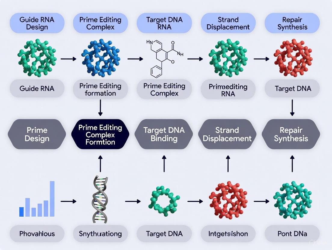
The mechanism of prime editing can be broken down into a series of discrete molecular steps, as illustrated below.
Diagram 1: The step-by-step molecular mechanism of prime editing.
- Target Recognition & Binding: The prime editor protein complexed with the pegRNA binds to the target DNA sequence. The spacer region of the pegRNA base-pairs with the complementary DNA strand, positioning the Cas9 nickase at the correct location [3] [2].
- Strand Nicking & Priming: The Cas9 H840A nickase cleaves the non-complementary DNA strand, creating a nick and exposing a 3' hydroxyl (3' OH) group on the DNA. This 3' end then hybridizes with the primer binding site (PBS) on the pegRNA, forming a primer-template complex for the reverse transcriptase [1] [4].
- Reverse Transcription: The reverse transcriptase domain uses the RNA sequence of the RTT as a template to synthesize a new DNA strand. This newly synthesized "edited flap" is directly polymerized onto the 3' end of the nicked DNA strand and contains the desired genetic alteration [1] [3].
- Flap Resolution & Repair: The resulting DNA structure is a branched intermediate with two flaps: the newly synthesized, edited 3' flap and the original, unedited 5' flap. Cellular repair machinery, particularly structure-specific endonucleases like FEN1, preferentially cleaves the 5' flap. The edited 3' flap is then ligated into the DNA backbone, creating a heteroduplex where one strand contains the edit and the other remains unedited [1] [2].
- Heteroduplex Resolution: The cell's DNA mismatch repair (MMR) system detects the base mismatches in the heteroduplex. The outcome of this repair is critical; it can either permanently install the edit by using the edited strand as a template to correct the complementary strand, or it can revert the edit back to the original sequence [1] [4]. Subsequent engineering efforts like the PE3 and PE4 systems were developed specifically to bias MMR towards the desired outcome.
The Evolution of Prime Editing Systems
Since the initial development of PE1, the prime editing system has undergone significant engineering to enhance its efficiency and precision. These improvements have targeted the reverse transcriptase enzyme, strategies to manipulate cellular DNA repair, and the overall architecture of the editor.
Table 1: Evolution and Characteristics of Prime Editing Systems
| System | Key Components & Modifications | Primary Mechanism of Action | Key Advantages / Use Cases |
|---|---|---|---|
| PE1 [1] [2] | Cas9(H840A) nickase fused to wild-type M-MLV RT. | Basic proof-of-concept; demonstrates search-and-replace editing. | Not recommended for current use; prototype system. |
| PE2 [1] [2] [4] | Cas9(H840A) nickase fused to engineered M-MLV RT (5 mutations for higher efficiency/thermostability). | Improved reverse transcription efficiency. | Simpler system; preferred if nicking sgRNAs cause unacceptable indels or long-term MMR inhibition is not desired [4]. |
| PE3/PE3b [1] [2] [4] | PE2 + an additional sgRNA to nick the non-edited strand. | The additional nick biases cellular MMR to use the edited strand as a repair template. | Higher editing efficiency than PE2; preferred when optimal efficiency is needed without inhibiting cellular MMR. PE3b reduces indels by using a strand-specific nicking sgRNA [1] [4]. |
| PE4/PE5 [1] [2] [4] | PE2 (PE4) or PE3 (PE5) + co-expression of a dominant-negative MLH1 (MLH1dn) protein. | Transient inhibition of the mismatch repair pathway, preventing repair of the edit back to the original sequence. | Increases editing efficiency and reduces indels; particularly beneficial in MMR-proficient cell types. PE5 combines strand nicking and MMR inhibition [1] [5]. |
| PEmax [1] [6] | Optimized PE2 architecture with codon-optimized RT, additional nuclear localization signals, and mutations in Cas9 for improved activity. | Enhanced expression, nuclear localization, and nicking activity in human cells. | A high-performance editor that can be used with any PE2-PE5 strategy; often the basis for the most advanced systems [1] [6]. |
Further innovations continue to expand the toolkit. epegRNAs incorporate structured RNA motifs at their 3' end to protect against degradation, significantly improving stability and editing efficiency [1] [6]. More recently, the PE6 system introduced specialized reverse transcriptases evolved from bacterial retrons and retrotransposons, offering smaller sizes for viral delivery and improved efficiency for certain edits [1].
Quantitative Data and Performance
The performance of prime editing is quantitatively assessed by its efficiency (the percentage of sequencing reads with the intended edit) and its purity (the ratio of desired edits to unwanted byproducts like indels).
Table 2: Prime Editing Performance Metrics Across Systems and Cell Types
| Editor System / Condition | Cell Type / Context | Typical Editing Efficiency Range | Key Factors Influencing Outcome |
|---|---|---|---|
| PE2 [1] [4] | HEK293T cells | ~20-50% (original study) | Underperforms PE3/PE4/PE5 but is simpler. Efficiency is highly dependent on pegRNA design and target locus. |
| PE3 [1] | HEK293T cells | 2-3x increase over PE2 | Increases efficiency but can also slightly increase indel formation compared to PE2. |
| PE4/PE5 + PEmax [5] [6] | MMR-deficient K562 cells (PEmaxKO) | Up to ~95% (at optimized loci) | Combining MMR inhibition (PE4/5) with an optimized editor (PEmax) and epegRNAs in a stable expression system yields the highest reported efficiencies. |
| With MMR [7] [5] | MMR-proficient cells (e.g., K562) | Lower efficiency for small edits; edits like G>C evade MMR better. | MMR negatively affects small edits; editing patterns differ (e.g., 4-5bp insertions are more efficient than 1bp insertions in MMR-proficient cells) [7]. |
| In Vivo [7] | Mouse hepatocytes | Higher variability | Editing patterns more closely resemble those in MMR-proficient cell lines, highlighting the critical role of cellular context. |
Machine learning models like PRIDICT2.0 have been developed to predict pegRNA efficiency, accounting for factors such as edit type, length, local sequence context (e.g., polyT tracts), and GC content, which are critical for experimental planning [7].
Detailed Experimental Protocol for Mammalian Cells
The following protocol provides a framework for conducting prime editing experiments in mammalian cells, utilizing the highly efficient PE4max system to maximize the probability of success.
Stage 1: pegRNA Design and Vector Construction
Objective: To design and clone pegRNAs that effectively encode the desired edit.
Materials & Reagents:
- pegRNA Expression Plasmid: e.g., pU6-pegRNA-GG-acceptor (Addgene #132777) [8].
- Prime Editor Expression Plasmid: e.g., pCMV-PEmax-P2A-hMLH1dn for PE4max (Addgene #174828) [4] [8].
- Software Tools: For pegRNA design (e.g., pegRNA design tools from the Liu lab or web-based interfaces).
Methodology:
- pegRNA Design:
- Identify the target genomic sequence and ensure the presence of an appropriate PAM (NGG for SpCas9) [4].
- Design the pegRNA spacer sequence (typically 20 nt) to target the desired location.
- Define the edit within the Reverse Transcription Template (RTT). The RTT should be long enough to include the edit and sufficient homologous sequence on both sides (typically 10-15 nt total). A common RTT length is 13-16 nucleotides [4].
- Design the Primer Binding Site (PBS) to be complementary to the 3' end of the nicked DNA strand. Test different PBS lengths (typically 8-15 nt) in silico as this is a critical optimization parameter [4].
- Consider using an epegRNA design by adding a 3' structural motif (e.g., tevopreQ1) to the pegRNA to enhance RNA stability and editing efficiency [1] [6].
- Vector Construction:
- Synthesize and clone the designed pegRNA sequence into the pegRNA expression plasmid using standard molecular biology techniques (e.g., In-Fusion cloning, Golden Gate assembly) [8].
Stage 2: Cell Transfection and Editing
Objective: To deliver the prime editing components into mammalian cells and allow editing to occur.
Materials & Reagents:
- Mammalian Cells: e.g., HEK293T, HeLa, or K562 cells. Adherent or suspension cells can be used with appropriate transfection methods.
- Transfection Reagent: Polymer-based transfection reagent (e.g., PolyJet) for HEK293Ts or electroporation for hard-to-transfect cells [8].
- Cell Culture Media: Appropriate complete media for the cell line used (e.g., DMEM with 10% FBS for HEK293Ts).
Methodology:
- Cell Seeding: Seed cells into a multi-well plate (e.g., 24-well) to reach 60-80% confluency at the time of transfection [8].
- Transfection Mixture Preparation:
- Transfection: Add the DNA-transfection reagent complexes dropwise to the cells.
- Incubation: Incubate the cells for 48-72 hours to allow for expression of the editor and installation of the edit.
Stage 3: Analysis and Validation of Editing Outcomes
Objective: To isolate genomic DNA and quantify prime editing efficiency.
Materials & Reagents:
- Genomic DNA Isolation Kit: e.g., QIAamp DNA Mini Kit (Qiagen) [8].
- PCR Reagents: High-fidelity PCR master mix (e.g., KOD One PCR Master Mix) and primers flanking the target site [8].
- Sequencing Service: Sanger sequencing or next-generation amplicon sequencing.
Methodology:
- Genomic DNA Extraction: Harvest transfected cells and extract genomic DNA using a commercial kit [8].
- Target Locus Amplification: Design primers to amplify a 300-500 bp region surrounding the target site. Perform PCR using the extracted genomic DNA as a template.
- Editing Efficiency Analysis:
- Bulk Sanger Sequencing: Submit the PCR amplicon for Sanger sequencing. Use trace data decomposition software (e.g., EditR or TIDE) to quantify the efficiency of the intended edit [8].
- High-Confidence NGS: For a more accurate and quantitative measurement, especially for complex edits, perform deep amplicon sequencing (NGS) on the PCR product. This allows for the precise quantification of the intended edit, error rates, and indel byproducts [7] [5].
- Clonal Isolation (Optional): If a clonal cell line is required (e.g., for iPS cell line generation), single cells must be sorted or diluted after transfection, expanded into colonies, and genotyped individually to identify clones homozygous for the edit [8].
The Scientist's Toolkit: Essential Research Reagents
Table 3: Key Reagents for Prime Editing Experiments
| Reagent / Tool | Function / Role in Experiment | Example Source / Identifier |
|---|---|---|
| PEmax Plasmid | Optimized prime editor protein (Cas9-H840A nickase + engineered RT). Backbone for PE2max experiments. | Addgene #174828 [8] |
| PE4max Plasmid | PEmax + dominant-negative MLH1dn for mismatch repair inhibition. All-in-one plasmid for the PE4max system. | Addgene #174828 [8] |
| pegRNA Acceptor Plasmid | Backbone vector for cloning and expressing custom pegRNAs. | Addgene #132777 [8] |
| Engineered pegRNA (epegRNA) | pegRNA with 3' RNA motif (e.g., tevopreQ1) to protect against degradation, improving stability and efficiency. | Designed into pegRNA sequence [1] [6] |
| MLH1dn (Dominant-Negative MLH1) | Protein used to transiently inhibit cellular mismatch repair, boosting prime editing efficiency (key in PE4/PE5). | Encoded in PE4max plasmid [1] [4] |
| Polymer-based Transfection Reagent | Chemical method for delivering plasmid DNA into adherent mammalian cells (e.g., HEK293Ts). | e.g., PolyJet [8] |
Prime editing represents a significant advancement in precision genome editing, enabling targeted nucleotide substitutions, insertions, and deletions without requiring double-strand breaks (DSBs) or donor DNA templates [9]. This technology centers on a complex of three core components: a specialized prime editing guide RNA (pegRNA), a Cas9 nickase enzyme (commonly the H840A variant), and a reverse transcriptase domain [10]. These elements work in concert to directly copy genetic information from the pegRNA into the target genomic locus. The precision of this system reduces unwanted byproducts typical of earlier CRISPR-Cas systems, such as indels resulting from non-homologous end joining (NHEJ) [11] [10]. This application note provides a detailed breakdown of these key components, supported by quantitative data, experimental protocols, and visualization tools to facilitate robust implementation in research and therapeutic development.
Component 1: pegRNA Structure and Design
The pegRNA is the central targeting and template molecule in prime editing. It combines the functions of a standard single-guide RNA (sgRNA) with those of a reverse transcription template.
Structural Elements
The pegRNA consists of four critical regions:
- Spacer Sequence: A 20-nucleotide guide segment that determines DNA target specificity through Watson-Crick base pairing with the genomic target site.
- scaffold: The secondary structure that binds the Cas9 nickase protein.
- Primer Binding Site (PBS): A short sequence (typically 8-15 nucleotides) that hybridizes to the 3' end of the nicked DNA strand to initiate reverse transcription.
- Reverse Transcription Template (RTT): The region encoding the desired edit(s), which is copied into the genomic DNA by the reverse transcriptase [9].
Design Parameters and Optimization
Optimal pegRNA design is critical for editing efficiency. Key parameters include:
- PBS Length: A length of 10-16 nucleotides is generally effective, with 13 nucleotides often providing optimal balance between binding stability and editing efficiency [9].
- RTT Length: Must be sufficiently long to encode the desired edit; templates of 10-16 nucleotides are standard for point mutations.
- Structural Considerations: The pegRNA extension should be designed to minimize intramolecular secondary structures that could impede function, particularly interactions between the PBS and spacer sequences [9].
Table 1: pegRNA Design Specifications for Point Mutations
| Component | Optimal Length Range | Function | Design Consideration |
|---|---|---|---|
| Spacer | 20 nt | Target recognition | Ensure uniqueness in genome; minimize off-target potential |
| PBS | 10-16 nt (13 nt optimal) | Primer binding | Avoid complementarity to RTT; moderate GC content (40-60%) |
| RTT | 10-16 nt | Edit template | Encode desired mutation; position edit centrally when possible |
Component 2: Cas9 H840A Nickase
The Cas9 nickase serves as the programmable DNA-binding component that precisely positions the editing machinery.
Mechanism and Engineering
The native Streptococcus pyogenes Cas9 enzyme contains two nuclease domains: RuvC and HNH, which together generate DSBs. The H840A mutation inactivates the HNH domain while retaining the RuvC domain's ability to cleave the non-target DNA strand [12] [10]. This creates a nickase that induces a single-strand break in the DNA, which serves as the initiation point for prime editing.
Recent research has revealed that the canonical H840A mutation does not completely abolish HNH domain activity, potentially leading to low-frequency DSB formation and unwanted indel formation [10]. To address this, enhanced nickase variants with additional mutations (e.g., H840A+N863A) have been developed, showing reduced DSB formation while maintaining efficient nicking activity [10].
Performance Characteristics
Table 2: Comparison of Cas9 Nickase Variants
| Nickase Variant | Active Domain | Cleavage Strand | DSB Formation | Relative Indel Frequency |
|---|---|---|---|---|
| nCas9 (D10A) | HNH | Target strand | Minimal | Very low |
| nCas9 (H840A) | RuvC | Non-target strand | Low-level | Moderate (1.5-3.5%) |
| nCas9 (H840A+N863A) | RuvC | Non-target strand | Minimal | Low (0.5-1.2%) |
Component 3: Reverse Transcriptase Domain
The reverse transcriptase (RT) domain catalyzes the central editing reaction by copying genetic information from the pegRNA into the target DNA.
Biochemical Properties
The RT domain used in prime editors is typically derived from Moloney Murine Leukemia Virus (M-MLV) [10]. This enzyme possesses several biochemical activities essential for prime editing:
- RNA-dependent DNA polymerase activity: Synthesizes a DNA strand complementary to the RTT portion of the pegRNA [13].
- RNase H activity: Degrades the RNA strand in RNA-DNA hybrids, though engineered versions often have reduced RNase H activity to prevent premature degradation of the pegRNA template [14] [13].
- DNA-dependent DNA polymerase activity: Can extend DNA primers using DNA templates, potentially contributing to second-strand synthesis [13].
Engineering for Enhanced Performance
Wild-type M-MLV reverse transcriptase has been engineered for improved performance in prime editing applications:
- Thermostability: Engineered variants withstand higher temperatures (up to 55°C), enabling better access to structured genomic regions [14].
- Processivity: Enhanced variants incorporate 65 times more nucleotides per binding event than wild-type enzymes, improving efficiency for longer edits [14].
- Reduced RNase H activity: Minimizes pegRNA degradation during reverse transcription [14].
- Fidelity: M-MLV reverse transcriptase has an error rate of approximately 1 in 15,000-27,000 bases, though this is generally acceptable for most editing applications given the short template lengths [14].
Diagram: Prime Editing Component Assembly. The pegRNA, Cas9 H840A nickase, and reverse transcriptase form a complex that nicks target DNA and initiates reverse transcription.
Integrated Prime Editing Mechanism
The prime editing process involves a coordinated, multi-step mechanism:
- Complex Formation: The pegRNA, Cas9 H840A nickase, and reverse transcriptase form the prime editor complex [9].
- DNA Binding and Nicking: The complex binds the target genomic locus through spacer sequence complementarity, and the Cas9 H840A nickase cleaves the non-target DNA strand [10].
- Primer Binding: The 3' end of the nicked DNA strand hybridizes with the PBS region of the pegRNA [9].
- Reverse Transcription: The reverse transcriptase extends the 3' DNA end using the RTT as a template, creating an edited DNA flap [9].
- Flap Resolution and Integration: Cellular enzymes resolve the DNA flap structure, with the edited strand preferentially integrated [9].
- DNA Repair: The resulting mismatch is repaired by cellular machinery, permanently incorporating the edit into the genome [9].
Experimental Protocol for Prime Editing
Component Preparation
pegRNA Design and Synthesis
- Identify Target Site: Select a target site with an appropriate PAM (NGG for SpCas9) sequence adjacent to the desired edit location.
- Design pegRNA: Using the parameters in Table 1, design the pegRNA with the edit encoded in the RTT.
- Synthesize pegRNA: Chemically synthesize the full pegRNA sequence or produce via in vitro transcription.
Prime Editor Expression Construct
- Vector Selection: Use a suitable expression vector (e.g., plasmid, viral vector) for your cell type.
- Clone Prime Editor: Insert genes encoding the Cas9 H840A nickase-reverse transcriptase fusion protein.
- Clone pegRNA Expression Cassette: Include the pegRNA under an appropriate RNA polymerase III promoter.
Cell Transfection and Editing
Day 1: Cell Seeding
- Plate HEK293T cells (or your target cell line) at 60-70% confluence in appropriate culture vessels.
Day 2: Transfection
- For a 6-well plate format, prepare:
- Prime editor plasmid: 1.0 µg
- pegRNA plasmid: 0.5 µg
- Transfection reagent: According to manufacturer's protocol
- Incubate cells with transfection complex for 24-48 hours.
Day 4: Analysis and Selection
- Harvest cells for genomic DNA extraction.
- Amplify target region by PCR and analyze editing efficiency by sequencing.
- For stable edits, apply appropriate selection (e.g., antibiotics for resistance markers).
Diagram: Prime Editing Experimental Workflow. Timeline and key steps for implementing prime editing in cell culture.
Advanced Protocol: proPE System
The recently developed proPE (prime editing with prolonged editing window) system addresses several limitations of standard prime editing [9]. This approach uses two distinct sgRNAs:
- Essential Nicking Guide RNA (engRNA): A standard sgRNA that directs the prime editor to nick the target DNA.
- Template Providing Guide RNA (tpgRNA): Contains the PBS and RTT sequences with a truncated spacer (11-15 nt) that prevents DNA cleavage but enables target binding.
proPE Transfection Protocol:
- Prepare separate complexes for engRNA and tpgRNA rather than mixing before transfection.
- Test 2-3 different engRNA plasmid quantities (e.g., 0.25 µg, 0.5 µg, 1.0 µg) to identify optimal nicking activity.
- Maintain tpgRNA plasmid at a constant amount (e.g., 0.5 µg).
The Scientist's Toolkit: Research Reagent Solutions
Table 3: Essential Reagents for Prime Editing Research
| Reagent Category | Specific Examples | Function | Implementation Notes |
|---|---|---|---|
| Prime Editor Constructs | PE2, PE3, PE4, proPE systems [9] [10] | Core editing machinery | PE2: Basic editor; PE3: Includes additional nicking sgRNA; proPE: Separate engRNA/tpgRNA |
| Control Elements | Dead Cas9 (dCas9) controls [11], Nuclease-active Cas9 | Experimental controls | dCas9 validates nickase-dependent editing; WT Cas9 controls for DSB-induced effects |
| Delivery Tools | Plasmid vectors, RNP complexes, Viral vectors (AAV, Lentivirus) | Component delivery | RNP complexes reduce off-target effects; AAV for in vivo applications |
| Detection & Analysis | Next-generation sequencing, T7E1 assay, Tracking of Indels by DEcomposition (TIDE) | Edit verification | Amplicon sequencing provides quantitative efficiency data |
| Enhanced Fidelity Nickases | nCas9 (H840A+N863A) [10] | Reduced DSB formation | Minimizes unwanted indel formation (0.5-1.2% vs 1.5-3.5%) |
| Efficiency Enhancers | Alt-R HDR Enhancer V2 [15], Engineered pegRNAs (epegRNAs) [10] | Increase editing rates | HDR Enhancer improves homology-directed repair efficiency |
Troubleshooting and Optimization Guidelines
Addressing Low Editing Efficiency
- Optimize PBS Length: Test PBS lengths between 8-15 nucleotides; 13 nt often optimal [9].
- Adjust Component Ratios: Titrate the amount of engRNA plasmid (0.25-1.0 µg in 6-well format) while keeping tpgRNA constant [9].
- Modify RTT Design: Position edits in the center of the RTT when possible, and ensure the template is long enough to accommodate the edit.
- Utilize proPE System: For persistently low efficiency, implement the proPE system with separate engRNA and tpgRNA components [9].
Reducing Unwanted Byproducts
- Implement High-Fidelity Nickases: Use nCas9 (H840A+N863A) instead of standard H840A to minimize DSB formation and indel rates [10].
- Optimize Nickase Expression: Reduce engRNA amount to minimize re-nicking of edited DNA [9].
- Employ Engineered pegRNAs: Incorporate stability modifications to reduce pegRNA degradation [10].
The precision and versatility of prime editing stem from the sophisticated interplay of its three core components: the pegRNA that provides targeting and template information, the Cas9 H840A nickase that enables programmable DNA recognition and nicking, and the reverse transcriptase that copies genetic information into the genome. Ongoing refinements, including the development of proPE systems [9] and high-fidelity nickase variants [10], continue to enhance the efficiency and specificity of this technology. The protocols and guidelines presented here provide researchers with a foundation for implementing prime editing in diverse experimental systems, supporting advancements in functional genomics, disease modeling, and therapeutic development.
Prime editing represents a transformative advancement in the field of genome engineering, offering a versatile and precise method for modifying DNA without inducing double-strand breaks (DSBs). Developed from the CRISPR-Cas9 system, prime editing functions as a "search-and-replace" genomic tool, capable of introducing all 12 possible base-to-base conversions, small insertions, deletions, and combinations thereof without requiring donor DNA templates [16] [3]. This technology addresses critical limitations of earlier gene-editing platforms, including the unpredictable repair outcomes associated with DSBs and the restricted editing scope of base editors, which are confined to specific nucleotide transitions and often exhibit bystander editing [16] [17].
The fundamental prime editing system consists of two core components: (1) a prime editor protein, which is a fusion of a Cas9 nickase (H840A) and an engineered reverse transcriptase (RT), and (2) a prime editing guide RNA (pegRNA) that both specifies the target genomic locus and encodes the desired edit [16] [3]. The editing process initiates when the pegRNA directs the prime editor to the target DNA sequence. The Cas9 nickase cleaves only one DNA strand, and the released 3'-hydroxyl end serves as a primer for the reverse transcriptase to synthesize new DNA using the pegRNA's template region [16]. The resulting DNA flap containing the edit is then incorporated into the genome through cellular repair mechanisms, achieving precise genetic modifications with significantly reduced risks of unwanted mutations compared to earlier technologies [16] [17].
Chronological Evolution of Prime Editing Systems
First-Generation Systems: PE1 to PE3
The development of prime editing began with PE1, the foundational proof-of-concept system that established the core architecture of a nCas9 (H840A) fused to a wild-type Moloney Murine Leukemia Virus reverse transcriptase (M-MLV RT) [16] [17]. While PE1 successfully demonstrated the "search-and-replace" capability, its editing efficiency remained relatively limited, typically achieving ~10-20% editing frequency in HEK293T cells [17].
PE2 emerged as a significant improvement through protein engineering of the reverse transcriptase component. By introducing specific mutations that enhanced thermostability, processivity, and affinity for RNA-DNA hybrid substrates, researchers developed an optimized RT that substantially improved editing outcomes [16] [17]. The PE2 system demonstrated ~20-40% editing efficiency in HEK293T cells, effectively doubling the performance of PE1 while maintaining high fidelity and reducing undesired byproducts [17].
Building on PE2's success, PE3 incorporated an additional strategic innovation: a second sgRNA designed to nick the non-edited DNA strand opposite the pegRNA-guided nick [16] [17]. This dual-nicking approach encourages the cellular repair machinery to use the newly synthesized edited strand as a template for repairing the nicked complementary strand, thereby increasing the likelihood of stable edit incorporation [16]. The PE3 system boosted editing efficiency further to ~30-50% in HEK293T cells, particularly in challenging genomic contexts where higher editing efficiency was required [17].
Figure 1: Evolution of Prime Editing Systems from PE1 to PE7, Showing Progressive Efficiency Improvements
Advanced Systems: PEmax, PE4/5, and PE6/7 Variants
The PEmax system represents a substantial optimization of PE2 through codon optimization of the reverse transcriptase, addition of two nuclear localization signals (NLS), and incorporation of mutations that enhance SpCas9 nuclease activity [18]. These modifications improved nuclear targeting and overall editor performance, making PEmax the currently recommended protein for most prime editing applications, as it matches or surpasses PE2 efficiency across multiple genomic loci [18].
The PE4 and PE5 systems address a critical cellular barrier to prime editing efficiency: the mismatch repair (MMR) pathway. PE4 incorporates a dominant-negative MLH1 protein (MLH1dn) to transiently inhibit MMR, ensuring that edits are not reversed before stable integration [17] [18]. This approach increases editing efficiency to ~50-70% in HEK293T cells while reducing indel formation. PE5 combines the MMR inhibition strategy with the PE3 dual-nicking approach, achieving ~60-80% editing efficiency and representing one of the most efficient systems for challenging edits [17].
The most recent advancements include the PE6 suite and PE7 systems. The PE6 editors incorporate multiple innovations, including modified RT variants (PE6a, PE6b, PE6c, PE6d), enhanced Cas9 variants (PE6e, PE6f, PE6g), and engineered pegRNAs (epegRNAs) that resist degradation [17] [18]. These comprehensive optimizations enable ~70-90% editing efficiency in HEK293T cells. The PE7 system further enhances performance by fusing the La(1-194) protein to the prime editor complex, improving pegRNA stability and editing outcomes in challenging cell types to achieve ~80-95% efficiency [17].
Comparative Analysis of Prime Editor Versions
Table 1: Comparative Characteristics of Major Prime Editing Systems
| Editor Version | Core Components | Editing Efficiency (HEK293T) | Key Innovations | Applications & Advantages |
|---|---|---|---|---|
| PE1 | nCas9 (H840A) + wild-type M-MLV RT | ~10-20% | Foundational proof-of-concept | Initial demonstration of search-and-replace editing |
| PE2 | nCas9 (H840A) + engineered M-MLV RT | ~20-40% | Optimized reverse transcriptase | Higher efficiency than PE1, maintained precision |
| PE3 | PE2 system + additional nicking sgRNA | ~30-50% | Dual nicking strategy | Enhanced efficiency via strand-biased repair |
| PEmax | Codon-optimized PE2 + extra NLSs + enhanced Cas9 | Matches or surpasses PE2 | Improved nuclear localization & activity | Current recommended system for most applications |
| PE4 | PE2 + dominant-negative MLH1 | ~50-70% | MMR inhibition | Reduced edit reversal, higher efficiency |
| PE5 | PE3 + dominant-negative MLH1 | ~60-80% | Combined MMR inhibition & dual nicking | Maximum efficiency for challenging edits |
| PE6 Suite | Modified RT/Cas9 variants + epegRNAs | ~70-90% | Compact RTs, stabilized pegRNAs | Better delivery, reduced degradation |
| PE7 | PE6 system + La(1-194) fusion | ~80-95% | pegRNA stabilization complex | Enhanced outcomes in difficult cell types |
pegRNA Engineering and Delivery Optimization
pegRNA Design and Stabilization
The prime editing guide RNA (pegRNA) is a sophisticated molecular construct that serves dual functions: targeting the editor to specific genomic loci and templating the desired edit. A standard pegRNA consists of four essential components: (1) a spacer sequence (~20 nucleotides) that directs Cas9 binding through complementarity to the target DNA; (2) a scaffold sequence that enables Cas9 nickase binding; (3) a reverse transcription template (RTT) containing the desired edit and flanking homology (typically 25-40 nucleotides); and (4) a primer binding site (PBS) (10-15 nucleotides) that anchors the reverse transcription process [3]. The complete pegRNA typically ranges from 120-145 nucleotides in length, with more complex edits requiring longer constructs up to 170-190 nucleotides [3].
A significant challenge with early pegRNAs was their susceptibility to cellular degradation, which limited editing efficiency. This prompted the development of engineered pegRNAs (epegRNAs) that incorporate structured RNA motifs at their 3' ends to enhance stability [16]. These protective motifs include evopreQ and mpknot structures, Zika virus exoribonuclease-resistant RNA motifs (xr-pegRNA), G-quadruplexes (G-PE), and stem-loop aptamers [16]. These epegRNAs demonstrate 3-4-fold improvements in prime editing efficiency across multiple human cell lines and primary human fibroblasts without increasing off-target effects [16].
Delivery Methods and Challenges
The substantial size and structural complexity of prime editing components present significant delivery challenges for therapeutic applications. The prime editor protein and pegRNA combined exceed the packaging capacity of standard adeno-associated virus (AAV) vectors, which have a ~4.7 kb limit [16] [18]. Researchers have developed multiple strategies to overcome this limitation:
- Dual AAV Systems: Splitting the prime editor components across two separate AAV vectors that reassemble in target cells [16]
- Non-Viral Delivery: Using lipid nanoparticles (LNPs) or electroporation to deliver prime editor mRNA or ribonucleoprotein (RNP) complexes [3] [18]
- Split Prime Editors (sPE): Engineering systems where nCas9 and RT function as separate polypeptides that assemble intracellularly [16]
Recent innovations include the development of circular RNA RT templates and truncated Cas9 variants that reduce system size while maintaining functionality [16]. Additionally, virus-like particles (VLPs) and advanced LNPs are being explored for tissue-specific delivery in therapeutic contexts [3] [18].
Experimental Protocols for Prime Editing
Protocol 1: Prime Editing in Mammalian Cells Using PEmax
This protocol outlines the standard procedure for implementing prime editing in mammalian cell lines using the PEmax system, which offers superior efficiency compared to earlier versions [18].
Materials Required:
- PEmax expression plasmid (Addgene #174828) or mRNA
- pegRNA expression plasmid (Addgene #174820) or synthetic pegRNA
- Mammalian cell line (HEK293T, HeLa, HCT116, etc.)
- Transfection reagent (Lipofectamine 3000, PEI MAX, or electroporation system)
- DNA purification kit
- Next-generation sequencing (NGS) reagents for analysis
Procedure:
pegRNA Design and Preparation
- Identify the target genomic locus and desired edit
- Design pegRNA spacer sequence (20 nt) complementary to target site
- Design RTT sequence encoding the desired edit with appropriate flanking homology (10-16 nt)
- Design PBS sequence (13 nt) complementary to the DNA flap created by nicking
- Clone pegRNA into expression vector or order as synthetic RNA with 3' stabilization motifs
Cell Culture and Transfection
- Culture mammalian cells in appropriate medium until 60-80% confluent
- For plasmid transfection: co-transfect 1-2 μg PEmax plasmid and 1-2 μg pegRNA plasmid per well in 6-well plate using preferred transfection reagent
- For RNP transfection: complex 2-4 μg PEmax protein with 1-2 μg synthetic pegRNA and transfect using electroporation
- Include controls: non-transfected cells, pegRNA-only transfection
Harvest and Analysis (48-72 hours post-transfection)
- Extract genomic DNA using standard purification methods
- Amplify target region by PCR with barcoded primers for NGS
- Prepare sequencing libraries and perform high-coverage amplicon sequencing (>10,000x coverage)
- Analyze sequencing data using prime editing-specific analysis tools (PE-Analyzer, CRISPResso2)
Validation
- Clone edited cells by limiting dilution or FACS sorting
- Expand single-cell clones and validate edits by Sanger sequencing
- Functional validation through relevant phenotypic assays
Troubleshooting:
- Low editing efficiency: Optimize PBS length (try 10-15 nt), adjust RTT homology arm length, test different pegRNA scaffolds, consider PE3 or PE5 systems with nicking sgRNA
- High indel formation: Switch to PE4/5 system with MLH1dn, reduce nCas9 expression level, use engineered nCas9 with N863A mutation to minimize DSBs
- Cellular toxicity: Titrate down editor expression, use RNP delivery instead of plasmids, employ transient expression systems
Protocol 2: Assessing Prime Editing Efficiency and Specificity
Accurate measurement of prime editing outcomes requires sensitive detection methods and careful assessment of both on-target and off-target effects.
Materials:
- High-fidelity DNA polymerase for PCR
- NGS library preparation kit
- Off-target prediction software (CCTop, Cas-OFFinder)
- Mismatch repair inhibitor (MLH1dn plasmid for PE4/5 systems)
On-Target Efficiency Analysis:
- Amplify target locus with barcoded primers incorporating unique molecular identifiers (UMIs)
- Sequence with minimum 10,000x read depth to detect low-frequency edits
- Quantify: (1) precise intended edits, (2) unpredicted edits within RTT region, (3) indels at target site
- Calculate editing efficiency as: (reads with precise edit / total reads) × 100%
Off-Target Assessment:
- Perform in silico prediction of potential off-target sites using target sequence
- Amplify top predicted off-target loci (5-10 sites) by PCR
- Sequence with high coverage (minimum 50,000x read depth)
- Compare mutation frequencies in edited vs. control samples
- For comprehensive assessment, utilize GUIDE-seq or CIRCLE-seq methods
Optimization Strategies:
- For difficult edits, test multiple pegRNA designs with varying PBS lengths and RTT configurations
- Implement PE4/5 system with MLH1dn for edits prone to MMR-mediated reversal
- Consider temperature optimization (cold shock at 32°C has shown ~22% improvement in some systems) [18]
- For large insertions (>100 bp), utilize twinPE systems with paired pegRNAs and recombinases [18]
The Scientist's Toolkit: Essential Research Reagents
Table 2: Key Research Reagents for Prime Editing Applications
| Reagent/Category | Specific Examples | Function & Application | Considerations |
|---|---|---|---|
| Prime Editor Proteins | PE2, PEmax, PE6 variants | Core editing machinery with optimized reverse transcriptase | PEmax recommended for new studies; PE6 for enhanced efficiency |
| pegRNA Expression Systems | pegRNA plasmids, synthetic epegRNAs | Target localization and edit templating | epegRNAs with 3' stabilization motifs improve efficiency 3-4 fold |
| Delivery Tools | Lipid nanoparticles (LNPs), Electroporation systems, AAV vectors | Cellular delivery of editing components | Dual AAV systems overcome size limitations; LNPs suitable for mRNA delivery |
| Efficiency Enhancers | MLH1dn (for PE4/5), La protein fusions (PE7) | Suppress mismatch repair, stabilize pegRNA | MLH1dn increases efficiency 1.5-2x by preventing edit reversal |
| Analysis Tools | NGS platforms, PE-Analyzer, CRISPResso2 | Quantify editing outcomes and specificity | UMIs essential for accurate efficiency measurement |
| Specialized Systems | TwinPE, Cas12a-PE, bi-PE | Large edits, alternative PAM targeting, specific applications | TwinPE with recombinases enables large DNA integration |
Future Perspectives and Applications
Emerging Technologies and Clinical Translation
Recent innovations continue to expand prime editing capabilities. The PERT (Prime Editing-mediated Readthrough of Premature Termination Codons) system represents a novel approach that addresses nonsense mutations responsible for approximately 30% of rare genetic diseases [19]. Rather than correcting individual mutations, PERT installs a suppressor tRNA that enables readthrough of premature stop codons, potentially allowing a single editing agent to treat multiple different genetic diseases [19]. This approach has demonstrated success in restoring protein function in cell and animal models of Batten disease, Tay-Sachs disease, Niemann-Pick disease type C1, and Hurler syndrome [19].
The proPE (prime editing with prolonged editing window) system addresses five key bottlenecks in traditional prime editing by using two distinct sgRNAs: an essential nicking guide RNA (engRNA) and a template-providing guide RNA (tpgRNA) [9]. This separation of functions enhances editing efficiency, particularly for modifications beyond the typical prime editing range, and expands targeting capabilities to encompass a major portion of human pathogenic single nucleotide polymorphisms [9].
Additional advancements include vPE systems with dramatically reduced error rates (from ~1/7 edits to ~1/101 for standard mode) through Cas9 protein engineering [20], and pvPE systems utilizing porcine endogenous retrovirus reverse transcriptase showing high efficiency across mammalian species [21].
Figure 2: Prime Editing Workflow from Target Identification to Functional Validation
Therapeutic Applications and Commercial Development
Prime editing shows remarkable potential for treating diverse genetic disorders. Clinical applications are advancing rapidly, with the first successful use of prime editing in a human patient reported for chronic granulomatous disease (CGD) [20]. Additional therapeutic candidates target sickle cell disease, beta-thalassemia, transthyretin amyloidosis, hereditary angioedema, and various rare genetic conditions [22] [20].
The commercial landscape for prime editing is expanding, with companies like Beam Therapeutics, Prime Medicine, and Caribou Biosciences developing therapeutic platforms based on precision genome editing [22]. Beam's BEAM-101 for sickle cell disease and beta-thalassemia represents the most advanced base editing program, demonstrating durable increases in fetal hemoglobin in clinical trials [22]. As delivery technologies improve and editing efficiency increases, prime editing is poised to become a cornerstone of genetic medicine, potentially enabling one-time treatments for hundreds of genetic diseases.
The evolution of prime editing systems from the initial PE1 to sophisticated variants like PEmax and PE6 represents a remarkable trajectory of innovation in precision genome engineering. Each generation has addressed specific limitations—improving efficiency through reverse transcriptase optimization, enhancing specificity via strategic nicking approaches, overcoming cellular barriers through mismatch repair inhibition, and expanding applicability with compact designs and stabilized components. The development of comprehensive experimental protocols and specialized reagents has enabled researchers to implement these systems across diverse biological contexts. As prime editing continues to mature, with ongoing enhancements in efficiency, specificity, and delivery, this technology holds exceptional promise for both basic research and therapeutic applications, potentially enabling precise correction of diverse genetic mutations underlying human disease.
Traditional CRISPR-Cas9 genome editing operates by introducing targeted double-strand breaks (DSBs) in DNA, relying on endogenous cellular repair mechanisms to achieve genetic modifications [23]. While revolutionary, this approach carries significant limitations for therapeutic applications, primarily due to the unpredictable nature of DSB repair. The non-homologous end joining (NHEJ) pathway frequently results in insertions or deletions (indels) that can disrupt gene function, while homology-directed repair (HDR) is inefficient in many therapeutically relevant cell types [16] [17]. Furthermore, DSB formation can trigger p53-mediated cellular stress responses, apoptosis, and chromosomal rearrangements, posing substantial safety risks [16] [17].
Prime editing represents a transformative advance in genome engineering that fundamentally addresses these limitations. As a "search-and-replace" editing technology, it enables precise genetic modifications without inducing DSBs or requiring donor DNA templates [16] [24] [17]. This paradigm shift from cutting to rewriting DNA expands the scope of possible edits while significantly reducing unwanted byproducts, making it particularly valuable for therapeutic development and precise disease modeling where accuracy is paramount.
Mechanisms: How Prime Editing Achieves Precision Without Double-Strand Breaks
Core Architecture of the Prime Editing System
The prime editing system consists of two primary components: (1) a prime editor protein and (2) a specialized prime editing guide RNA (pegRNA) [16] [3]. The prime editor is a fusion protein comprising a Cas9 nickase (H840A) connected to an engineered reverse transcriptase (RT) from the Moloney Murine Leukemia Virus (M-MLV) [16] [17]. The Cas9 nickase is capable of cutting only one DNA strand, unlike the wild-type Cas9 which creates double-strand breaks, while the reverse transcriptase synthesizes DNA using an RNA template [3].
The pegRNA is an engineered guide RNA that serves dual functions: target site recognition and edit encoding [3]. Beyond the standard CRISPR guide RNA components (spacer sequence and scaffold), the pegRNA contains a 3' extension with two critical elements:
- Primer binding site (PBS): A 10-15 nucleotide sequence that anneals to the nicked DNA strand to prime reverse transcription [3]
- Reverse transcriptase template (RTT): A template sequence encoding the desired genetic edit, typically 25-40 nucleotides in length [3]
This sophisticated architecture enables prime editing to perform all 12 possible base-to-base conversions, as well as targeted insertions and deletions, without DSB formation [16] [24].
The Stepwise Prime Editing Mechanism
The prime editing mechanism proceeds through a series of coordinated molecular events, visualized in the diagram below:
Figure 1: The stepwise mechanism of prime editing, from target binding to edit installation.
Target Recognition and Binding: The prime editor-pegRNA complex binds to the target DNA sequence through standard Cas9-DNA interactions guided by the pegRNA's spacer sequence [3].
Strand Nicking: The Cas9 nickase (H840A) cleaves the non-target DNA strand, creating a single-strand break with an exposed 3'-hydroxyl group [16] [17].
Primer Binding and Reverse Transcription: The PBS region of the pegRNA anneals to the nicked DNA strand. The reverse transcriptase then uses the 3'-OH end as a primer and the RTT region of the pegRNA as a template to synthesize a new DNA flap containing the desired edit [16] [3].
Flap Resolution and Edit Installation: Cellular repair machinery processes the resulting DNA structure where the newly synthesized edited flap competes with the original unedited flap. The edited strand is preferentially incorporated through a series of enzymatic steps involving flap endonucleases and DNA ligases [16] [25].
Complementary Strand Correction (in PE3 system): To increase editing efficiency, an additional sgRNA can be used to nick the non-edited strand, encouraging the cell to use the edited strand as a repair template, resulting in a fully edited DNA duplex [16] [17].
Quantitative Comparisons: Efficiency and Specificity Metrics
Direct Performance Comparison with Traditional Genome Editing Tools
The advantages of prime editing become evident when examining quantitative performance metrics compared to traditional editing technologies. The following table summarizes key comparative data:
Table 1: Performance comparison of major genome editing technologies
| Editing Technology | DSB Formation | Edit Types Supported | Typical Editing Efficiency | Indel Frequency | Therapeutic Safety Profile |
|---|---|---|---|---|---|
| CRISPR-Cas9 (HDR) | Yes | All (with donor template) | 1-10% (varies by cell type) [23] | High (5-60%) [16] | Moderate (DSB risks) |
| Base Editing | No | C•G to T•A, A•T to G•C [16] | 50-70% [3] | Low (<1.5%) [16] | High (bystander edits possible) |
| Prime Editing | No | All 12 base conversions, insertions, deletions [16] [24] | 20-50% (PE2), 30-60% (PE3) [17] | Very low (0.1-1.5%) [16] [25] | Very high |
The data reveal prime editing's unique combination of versatility and safety. While base editing offers high efficiency for specific transitions, prime editing supports all possible genetic modifications while maintaining low indel rates comparable to base editing [16]. Next-generation prime editors show further improvements, with the recently developed vPE system demonstrating edit:indel ratios as high as 543:1, representing up to 60-fold reduction in indel errors compared to earlier versions [25].
Evolution of Prime Editing Systems and Their Performance
The continuous refinement of prime editing systems has yielded successive generations with improved characteristics:
Table 2: Development timeline and features of prime editor generations
| Prime Editor Version | Key Components | Editing Efficiency | Notable Features | Indel Reduction Strategies |
|---|---|---|---|---|
| PE1 | nCas9(H840A)-RT, pegRNA | ~10-20% [17] | Proof-of-concept system | Foundation without optimization |
| PE2 | Engineered RT, optimized pegRNA | ~20-40% [17] | Improved RT processivity | 2-3x reduction vs PE1 [16] |
| PE3 | PE2 + additional nicking sgRNA | ~30-50% [17] | Dual nicking enhances efficiency | Similar to PE2 with proper design |
| PE4/PE5 | PE2/PE3 + MLH1dn | ~50-80% [17] | MMR inhibition boosts efficiency | Reduced MMR-mediated indels [17] |
| vPE/pPE | Engineered Cas9 variants | Comparable to PE3 | Relaxed nick positioning | Up to 60x lower indels [25] |
Recent engineering efforts have focused specifically on minimizing genomic errors while maintaining high editing efficiency. The precise Prime Editor (pPE) incorporates mutations (K848A-H982A) that relax nick positioning and promote degradation of the competing 5' strand, reducing indel errors by 7.6-26 fold compared to previous editors [25]. This error-suppressing strategy represents a significant advancement for therapeutic applications where unwanted mutations could have serious consequences.
Experimental Protocols: Implementing Prime Editing in Research
Workflow for Prime Editing in Human Induced Pluripotent Stem Cells
The following workflow diagram outlines a validated protocol for generating human induced pluripotent stem (iPS) cell lines with precise single nucleotide variants using prime editing:
Figure 2: Experimental workflow for prime editing in human iPS cells.
pegRNA Design and Vector Construction
pegRNA Design Considerations:
- Design pegRNAs with spacer sequences (typically 20 nt) complementary to the target site [3] [8]
- Incorporate structured RNA motifs (evopreQ, mpknot, or G-quadruplex) at the 3' end of pegRNA to protect against degradation and improve editing efficiency by 3-4 fold [16]
- Optimize primer binding site (PBS) length (typically 10-15 nt) and reverse transcriptase template (RTT) length (25-40 nt) based on target sequence [3] [8]
- For the PE3 system, design an additional sgRNA to nick the non-edited strand at a distance of 40-100 bp from the pegRNA nick site [16] [17]
Vector Assembly Protocol:
- Clone pegRNA expression cassettes into appropriate vectors under U6 promoters [8]
- For iPS cell editing, use the pCMV-PEmax-P2A-hMLH1dn vector (Addgene #174828) which incorporates a dominant-negative MLH1 to suppress mismatch repair and improve efficiency [8]
- Include selection markers (e.g., puromycin resistance) for enrichment of transfected cells [8]
- Utilize In-Fusion cloning with overlap extension PCR for efficient assembly of pegRNA components [8]
Cell Transfection and Screening
Efficiency Screening in HEK293T Cells:
- Transfect HEK293T cells with prime editor and pegRNA vectors using polymer-based transfection reagents (e.g., PolyJet) [8]
- Harvest cells 72 hours post-transfection and extract genomic DNA
- Assess editing efficiency via bulk Sanger sequencing and tracking of indels by decomposition (TIDE) analysis [8]
- Select the most efficient pegRNAs for iPS cell experiments
iPS Cell Transfection and Selection:
- Culture human iPS cells (e.g., 201B7 line) in StemFit medium on iMatrix-511-coated plates [8]
- Pre-treat cells with Y-27632 (ROCK inhibitor) for 1 hour before transfection to enhance viability [8]
- Transfert cells at 70-80% confluence using polymer-based transfection reagents
- Begin puromycin selection (0.5-1 μg/mL) 24 hours post-transfection for 48-72 hours [8]
- Allow recovery in drug-free medium for 3-5 days before single-cell cloning
Clone Validation and Characterization
Isolation and Expansion:
- Harvest transfected cells using TrypLE Select enzyme and seed as single cells in conditioned medium with Y-27632 [8]
- Expand individual clones for 2-3 weeks until colonies are suitable for genotyping
- Transfer portions of each clone for genomic DNA extraction while maintaining the remainder
Genotypic Validation:
- Extract genomic DNA using commercial kits (e.g., QIAamp DNA Mini Kit) [8]
- Amplify target regions by PCR and confirm edits by Sanger sequencing
- Verify the absence of unwanted mutations at potential off-target sites predicted by in silico tools
- For complete characterization, perform whole-genome sequencing on selected clones to confirm specificity
This protocol typically enables establishment of precisely edited iPS cell lines within 6-8 weeks while preserving genomic integrity [8].
The Scientist's Toolkit: Essential Research Reagents
Successful implementation of prime editing requires carefully selected molecular tools and reagents. The following table outlines key components and their functions:
Table 3: Essential research reagents for prime editing experiments
| Reagent Category | Specific Examples | Function | Considerations |
|---|---|---|---|
| Prime Editor Plasmids | pCMV-PEmax-P2A-hMLH1dn (Addgene #174828) [8] | Expresses optimized prime editor protein with MMR suppression | PE5 system enhances efficiency in difficult-to-edit loci |
| pegRNA Backbones | pU6-pegRNA-GG-acceptor (Addgene #132777) [8] | Enables cloning of custom pegRNA sequences | Compatible with various synthesis methods |
| Delivery Reagents | PolyJet DNA transfection reagent [8] | Facilitates plasmid delivery into cells | Polymer-based reagents show high reproducibility in iPS cells |
| Cell Culture Supplements | Y-27632 (ROCK inhibitor) [8] | Enhances cell survival after dissociation | Critical for single-cell cloning of iPS cells |
| Selection Agents | Puromycin [8] | Enriches for successfully transfected cells | Concentration must be optimized for each cell type |
| Extraction & Analysis Kits | QIAamp DNA Mini Kit [8] | Extracts high-quality genomic DNA for genotyping | Enables PCR amplification of target loci |
| Structured RNA Motifs | evopreQ, mpknot sequences [16] | Stabilizes pegRNA against degradation | Improves editing efficiency 3-4 fold |
Applications and Therapeutic Translation
The precision of prime editing has enabled diverse research applications from disease modeling to therapeutic development. In disease modeling, researchers have successfully generated isogenic iPS cell lines harboring precise disease-relevant single nucleotide variants, providing improved models for studying conditions like normal-tension glaucoma [8]. The technology has demonstrated particular value for modeling disorders where single base-pair changes drive pathology, as it avoids confounding indels that could complicate phenotypic analysis.
In therapeutic development, prime editing has shown promise in preclinical models of various genetic disorders. Researchers have corrected mutations associated with alternating hemiplegia of childhood in patient-derived stem cells and mouse models [26]. In vision research, virus-like particle-delivered prime editors improved editing efficiency by 65-fold and corrected vision loss in a mouse model of genetically inherited retinal degeneration [26]. These advances highlight the therapeutic potential of prime editing for treating monogenic disorders.
The translation of prime editing to clinical applications reached a significant milestone with the US Food and Drug Administration's Investigational New Drug (IND) clearance for PM359, the first prime editing-based therapeutic to enter clinical trials [24]. This ex vivo therapy corrects mutations in the NCF1 gene in patient-derived hematopoietic stem cells for the treatment of chronic granulomatous disease, marking a historic advancement for the field.
Despite these promising developments, therapeutic delivery remains a key challenge. The large size of prime editing components complicates packaging into delivery vectors such as adeno-associated viruses [16] [24]. Innovative solutions including virus-like particles, lipid nanoparticles, and split systems are under active investigation to overcome these limitations and unlock the full therapeutic potential of prime editing.
The advent of CRISPR-mediated genome editing has revolutionized molecular biology, yet traditional approaches relying on double-strand breaks (DSBs) face significant limitations including low efficiency of homology-directed repair (HDR) and unintended indel formation [27]. Base editing and prime editing represent two transformative technologies that enable precise genome modification without inducing DSBs, yet they differ fundamentally in their mechanisms and capabilities [27] [3]. While base editors facilitate direct chemical conversion of one base to another, prime editing operates as a "search-and-replace" system capable of installing virtually any small-scale genetic change [3] [16]. This application note examines the technical distinctions between these platforms, with particular emphasis on prime editing's dramatically expanded targeting scope beyond the transition mutations accessible to base editing technologies.
Molecular Mechanisms: Fundamental Operational Differences
Base Editing Architecture and Limitations
Base editors consist of a catalytically impaired Cas protein (nickase or dead Cas9) fused to a deaminase enzyme that performs direct chemical conversion on DNA bases [28]. Cytosine base editors (CBEs) convert cytosine to thymine (C→T) through a uracil intermediate, while adenine base editors (ABEs) convert adenine to guanine (A→G) via an inosine intermediate [27] [28]. These systems operate within a constrained editing window of approximately 4-5 nucleotides and are restricted to transition mutations (purine-to-purine or pyrimidine-to-pyrimidine changes) [27] [29]. This fundamental limitation means conventional base editors can only achieve 4 of the 12 possible base-to-base conversions [29].
Table 1: Base Editor Types and Capabilities
| Editor Type | Key Components | Base Conversion | Primary Mechanism | Limitations |
|---|---|---|---|---|
| Cytosine Base Editors (CBEs) | nCas9/dCas9 + cytidine deaminase (APOBEC) + UGI | C→T (G→A on opposite strand) | Deamination of cytosine to uracil | Restricted to transition mutations; bystander edits |
| Adenine Base Editors (ABEs) | nCas9/dCas9 + engineered tRNA adenosine deaminase (TadA) | A→G (T→C on opposite strand) | Deamination of adenine to inosine | Restricted to transition mutations; requires complex engineering |
Prime Editing Architecture and Expanded Capabilities
Prime editing employs a more complex but versatile architecture consisting of a Cas9 nickase (H840A) fused to an engineered reverse transcriptase (RT) enzyme, programmed with a specialized prime editing guide RNA (pegRNA) [17] [3]. The pegRNA serves dual functions: targeting the genomic locus and encoding the desired edit through its reverse transcriptase template (RTT) and primer binding site (PBS) components [3]. This system creates a nicked DNA strand that primes reverse transcription of the edited sequence, which is then incorporated into the genome through cellular repair processes [17] [16]. Unlike base editors, prime editors introduce no double-strand breaks and require no donor DNA templates [16].
Figure 1: Prime Editing Mechanism - The prime editor complex binds target DNA directed by the pegRNA, nicks one strand, and reverse transcribes the edited sequence encoded in the pegRNA
Quantitative Comparison: Editing Scope and Efficiency
Mutation Type Accessibility
The most significant distinction between these technologies lies in their accessible editing scope. While base editors are restricted to transition mutations (C→T, G→A, A→G, T→C), prime editing enables all 12 possible base substitutions, in addition to small insertions, deletions, and combinations thereof [3] [29]. This expanded scope is clinically relevant, as approximately 50% of disease-causing single nucleotide variants (SNVs) require transversion mutations (purine-to-pyrimidine or pyrimidine-to-purine changes) that conventional base editors cannot address [29].
Table 2: Mutation Type Accessibility Across Editing Platforms
| Mutation Type | Base Editing | Prime Editing | Representative Pathogenic Variants |
|---|---|---|---|
| Transition Mutations (4 types) | Yes | Yes | 25% of known genetic disease variants [27] |
| Transversion Mutations (8 types) | No* | Yes | 50% of known genetic disease variants [29] |
| Small Insertions | No | Yes | Frameshift corrections, tag insertions |
| Small Deletions | No | Yes | In-frame deletion corrections |
| Combination Edits | No | Yes | Multiple adjacent corrections |
Note: Specialized base transversion editors are in early development but not widely available [29]
Efficiency and Specificity Metrics
Editing efficiency varies substantially between systems and across target sites. Second-generation prime editors (PE2) typically achieve 20-40% editing efficiency in human cell lines, while third-generation systems (PE3) reach 30-50% efficiency through incorporation of an additional nicking sgRNA to enhance editing strand incorporation [17]. The latest PE6 systems demonstrate dramatically improved efficiency of 70-90% through optimized reverse transcriptase variants and engineered pegRNAs (epegRNAs) with improved stability [17]. By comparison, base editors typically achieve 30-60% efficiency for preferred target sequences but produce significant bystander edits within the editing window [27] [29].
Experimental Protocol: Prime Editing Workflow
pegRNA Design and Optimization
The success of prime editing experiments critically depends on optimal pegRNA design [3]. A standard pegRNA consists of four essential components:
- Spacer sequence (∼20 nt): Targets the Cas9 nickase to the genomic locus
- Scaffold sequence: Binds the Cas9 protein
- Reverse transcriptase template (RTT) (∼25-40 nt): Encodes the desired edit with flanking homology
- Primer binding site (PBS) (∼10-15 nt): Primes the reverse transcription reaction [3]
Critical Protocol Parameters:
- PBS length: Optimize between 10-15 nucleotides through empirical testing
- RTT design: Ensure the desired edit is positioned to minimize secondary structure
- pegRNA stabilization: Incorporate evolved preQ1 (epegRNA) or similar motifs at the 3' terminus to prevent degradation [17] [16]
- Edit positioning: Place the edit 1-10 nucleotides upstream of the nick site for optimal efficiency
Delivery and Validation Methods
Effective delivery of prime editing components remains technically challenging due to the large size of the editor and complexity of pegRNAs [3]. For mammalian cell editing:
Delivery Options:
- Plasmid transfection: Co-deliver PE and pegRNA expression constructs
- Viral delivery: Utilize dual AAV systems with split-intein reconstitution to accommodate size constraints [27] [16]
- RNA delivery: Deliver PE as mRNA and pegRNA as synthetic RNA
- Ribonucleoprotein (RNP): Complex purified PE protein with in vitro transcribed pegRNA
Validation Workflow:
- Transfection: Deliver editing components to target cells (e.g., via lipofection)
- Harvest genomic DNA: 72-96 hours post-transfection
- PCR amplification: Target edited genomic region
- Sequencing analysis: Utilize Sanger sequencing with EditR or next-generation sequencing for quantitative efficiency assessment [30]
- Functional validation: Assess phenotypic consequences through relevant assays (e.g., Western blot, functional assays)
Research Reagent Solutions
Table 3: Essential Reagents for Prime Editing Implementation
| Reagent Category | Specific Examples | Function/Application | Considerations |
|---|---|---|---|
| Prime Editor Constructs | PE2, PE3, PE6 variants [17] | Catalytic editing machinery | PE2 offers simplicity; PE3 provides higher efficiency; PE6 represents latest generation |
| pegRNA Expression Systems | U6-promoter vectors, synthetic pegRNAs [3] | Encode targeting and edit information | Synthetic pegRNAs enable rapid testing; plasmid vectors suitable for stable expression |
| Delivery Vehicles | Lentiviral particles, AAV vectors [27] [16] | Component delivery to cells | AAV preferred for in vivo; lipid nanoparticles emerging for clinical translation |
| Editing Enhancers | epegRNA scaffolds, MMR inhibitors (MLH1dn) [17] [16] | Improve editing efficiency | MMR suppression critical for maintaining edits in dividing cells |
| Validation Tools | Sanger sequencing, NGS platforms, EditR software [30] | Assess editing outcomes and efficiency | NGS required for comprehensive off-target profiling |
Figure 2: Prime Editing Experimental Workflow - Step-by-step protocol from pegRNA design to functional validation
Applications and Future Directions
Prime editing's expanded targeting scope enables correction of up to 89% of known genetic variants associated with human diseases, compared to approximately 25% addressable by conventional base editors [27] [16]. This includes therapeutic applications for:
- Autosomal dominant disorders: Where silencing mutant alleles is insufficient [27]
- Large gene corrections: Where full-gene delivery exceeds viral packaging capacity [27]
- Regulatory element engineering: Precise modification of promoter elements without coding sequence alteration [30]
- Multiplexed editing: Simultaneous correction of multiple pathogenic variants [31]
Current research focuses on enhancing prime editing efficiency through:
- Engineered reverse transcriptases with improved processivity and fidelity [17] [16]
- Cas9 variants with expanded PAM compatibility and reduced off-target activity [27] [12]
- Novel delivery strategies including virus-like particles and lipid nanoparticles [3] [32]
- System miniaturization through split-intein systems and compact editors [16]
As these enhancements mature, prime editing is poised to become the preferred platform for precise genome modification, particularly for mutations inaccessible to base editing technologies.
Application Notes: Prime Editing in Therapeutic Development
Prime editing is a versatile genome editing technology that enables precise correction of genetic mutations without requiring double-strand DNA breaks (DSBs) or donor DNA templates [33] [1]. This "search-and-replace" editing approach uses a catalytically impaired Cas9 nickase fused to a reverse transcriptase (RT) and a prime editing guide RNA (pegRNA) that specifies the target locus and encodes the desired edit [1] [4]. The technology has demonstrated potential for therapeutic correction of a broad spectrum of genetic diseases, offering significant advantages over previous editing platforms in versatility, precision, and safety profile.
Current Therapeutic Applications and Validation
The therapeutic application of prime editing has expanded rapidly, with validation across multiple disease models demonstrating its potential for clinical translation.
Table 1: Validated Therapeutic Applications of Prime Editing
| Disease Model | Genetic Defect | Editing Approach | Correction Efficiency | Key Outcome | Citation |
|---|---|---|---|---|---|
| Hurler syndrome | IDUA p.W392X nonsense mutation | Endogenous tRNA conversion to sup-tRNA (PERT) | ~6% IDUA enzyme activity restoration | Near-complete rescue of disease pathology in mice | [34] |
| Batten disease, Tay–Sachs disease, Niemann–Pick disease | TPP1, HEXA, NPC1 nonsense mutations | PERT strategy | 20–70% of normal enzyme activity | Functional protein rescue across multiple genes | [34] |
| Sickle cell disease, Tay–Sachs disease | Point mutations | Prime editing in human cell lines | Not specified | Correction of pathogenic mutations | [35] |
| Cystic fibrosis | CFTR nonsense mutations | PERT strategy | Not specified | Demonstration of disease-agnostic approach | [34] |
The PERT (Prime Editing-mediated Readthrough of Premature Termination Codons) strategy represents a particularly innovative disease-agnostic approach [34]. By using prime editing to convert a dispensable endogenous tRNA into an optimized suppressor tRNA (sup-tRNA), this method enables readthrough of premature stop codons regardless of the specific gene affected. This approach could potentially address the ~24% of pathogenic alleles in ClinVar that are nonsense mutations using a single therapeutic agent, dramatically simplifying treatment development for multiple diseases [34].
Advantages Over Alternative Genome Editing Technologies
Prime editing offers distinct advantages that make it particularly suitable for therapeutic applications:
- Versatility: Capable of installing all 12 possible base-to-base conversions, small insertions, and deletions without DSBs [33] [1]. This contrasts with base editors, which are limited to specific transition mutations [33] [4].
- Precision and Purity: Achieves higher ratios of desired edits to indel byproducts compared to Cas9-initiated homology-directed repair (HDR) [1] [4]. PE4 and PE5 systems further enhance purity by transiently inhibiting mismatch repair (MMR) to reduce undesired outcomes [1] [4].
- Reduced Off-Target Effects: Requires three distinct DNA hybridization events (spacer, PBS, and 3′ homology), providing multiple opportunities to reject off-target sequences [4]. Whole-genome sequencing studies have detected minimal Cas9-independent off-target effects in edited cells [4].
- Flexible Targeting: Less constrained by protospacer adjacent motif (PAM) availability, with effective editing up to 30+ base pairs from the PAM site [1].
Table 2: Comparison of Major Genome Editing Technologies
| Technology | Editing Capabilities | DSB Formation | Key Limitations | Therapeutic Advantages |
|---|---|---|---|---|
| Prime Editing | Substitutions, insertions, deletions (typically <50 bp) | No | Variable efficiency requiring optimization | High precision, minimal indel formation, versatile correction |
| Cas9 Nuclease | Gene disruption via indels | Yes | High indel rates, large deletions, translocations | Potent gene knockout |
| Base Editing | C•G to T•A, A•T to G•C, C•G to G•C | No | Restricted to specific transitions, bystander editing | High efficiency for point mutations within activity window |
| HDR with DSBs | Any change with donor template | Yes | Low efficiency, cell-cycle dependent, requires donor DNA | Precise incorporation of large sequences |
Experimental Protocols
This section provides detailed methodologies for implementing prime editing in therapeutic development contexts, from initial design to functional validation.
Prime Editing Workflow for Therapeutic Correction
The following diagram illustrates the complete experimental workflow for therapeutic gene correction using prime editing:
Protocol: cliPE for Multiplexed Functional Assay of Variants
The curated loci Prime Editing (cliPE) protocol enables functional assessment of variants of uncertain significance (VUS) at scale, providing a pathway for resolving the >1 million VUS currently in ClinVar [36]. This 2-4 week protocol is optimized for HAP1 cells but transferable to other cell lines with appropriate optimization.
Reagent Design and Library Cloning
Materials:
- cliPE companion Shiny app (http://home.clipe-mave.org/)
- pCMV-PEmax-P2A-GFP (Addgene #180020)
- pEF1a-hMLH1dn (Addgene #174824)
- pU6-tevopreq1-GG-acceptor (Addgene #174038)
- Oligonucleotide library encoding desired variants
- Q5 Hot Start High-Fidelity DNA polymerase
- BsmBI-v2 and BsaI-HFv2 restriction enzymes
- Gel DNA recovery kit
Procedure:
pegRNA Design:
- Input target gene coordinates and variant list into the cliPE Shiny app
- Select variants from ClinVar and gnomAD with appropriate population frequency filters
- Download output files including candidate epegRNA libraries, archetypal epegRNAs, and nicking gRNAs
- For each variant, design epegRNAs with 10-15 nt primer binding site (PBS) and 12-18 nt reverse transcription template (RTT)
Archetypal epegRNA Validation:
- Clone 5-10 representative epegRNAs into expression plasmid
- Transfect HEK293T cells and culture for 72 hours
- Extract genomic DNA and amplify target region
- Sequence amplicons to estimate editing efficiency (aim for >10% for library inclusion)
Library Cloning:
- Pool oligonucleotides by resuspending individual variants and mixing in equimolar ratios
- Amplify library with flanking primers adding Golden Gate assembly sites
- Digest acceptor vector and insert library with BsmBI-v2
- Ligate using T4 DNA ligase and transform into electrocompetent E. coli
- Recover library plasmids using midiprep kit, aiming for >1000x coverage
Cell Culture and Editing
Materials:
- HAP1 cells (Horizon #C669)
- DMEM with 10% FBS and 1% PenStrep
- TurboFectin 8.0 transfection reagent
- Neon electroporation system (where applicable)
- Fluorescence-activated cell sorting (FACS) equipment
Procedure:
Cell Preparation:
- Culture HAP1 cells in DMEM with 10% FBS at 37°C, 5% CO₂
- Passage cells at 70-80% confluence to maintain exponential growth
- For transfection, seed 2×10⁵ cells per well in 6-well plates 24 hours prior to editing
Prime Editing Transfection:
- Prepare DNA mixture for each sample:
- 1.5 μg pCMV-PEmax-P2A-GFP
- 1.5 μg pEF1a-hMLH1dn
- 1.0 μg epegRNA library plasmid
- 0.5 μg nicking gRNA plasmid (if using PE3/PE5 approach)
- Complex DNA with 9 μL TurboFectin in 300 μL serum-free DMEM
- Incubate 15 minutes at room temperature, then add dropwise to cells
- Replace medium after 6-8 hours
- Prepare DNA mixture for each sample:
Cell Selection and Expansion:
- At 48-72 hours post-transfection, harvest cells using TrypLE
- Sort GFP-positive cells using FACS or apply functional selection based on gene of interest
- Expand sorted cells for 7-10 days, maintaining >1000x library coverage
- Split cells into selected and unselected pools for functional assays
Sequencing and Analysis
Materials:
- Genomic DNA extraction kit
- iProof high-fidelity PCR master mix
- AMPure XP size selection beads
- Next-generation sequencing platform
Procedure:
Sequencing Library Preparation:
- Extract genomic DNA from 1×10⁶ cells each from selected and unselected pools
- Amplify edited target region using nested PCR with Illumina adapter sequences
- Purify amplicons using AMPure XP beads (0.8x ratio)
- Quantify libraries using Qubit high-sensitivity dsDNA reagent
- Sequence on Illumina platform to achieve >500x coverage
Variant Enrichment Analysis:
- Demultiplex sequencing reads and align to reference sequence
- Count reads for each variant in selected and unselected pools
- Calculate enrichment scores using binomial test or similar statistical framework
- Classify variants as functional (depleted), non-functional (enriched), or neutral (no change)
- Validate top hits using orthogonal functional assays
Protocol: PERT for Disease-Agnostic Nonsense Mutation Correction
The Prime Editing-mediated Readthrough of Premature Termination Codons (PERT) protocol enables correction of diverse nonsense mutations using a single therapeutic agent [34]. This approach is particularly valuable for addressing the 24% of pathogenic alleles that are premature stop codons.
sup-tRNA Design and Optimization
Materials:
- mCherry-STOP-GFP reporter constructs
- tRNA sequencing database (e.g., GtRNAdb)
- HEK293T cells (ATCC CRL-321)
Procedure:
sup-tRNA Screening:
- Design prime editing reagents to convert anticodon loops of endogenous tRNAs to complement premature termination codons (TAG, TAA, TGA)
- Create mCherry-STOP-GFP reporters with premature stop codons in various sequence contexts
- Transfert HEK293T cells with sup-tRNA editors and reporter constructs
- Analyze by flow cytometry for GFP expression 72 hours post-transfection
sup-tRNA Engineering:
- Iteratively optimize tRNA sequence through saturation mutagenesis
- Test variants with modified 5' leader sequences (40 bp) and 3' terminators
- Evaluate sup-tRNA potency using % GFP-positive cells and relative protein yield compared to wild-type controls
Endogenous Installation:
- Select optimal sup-tRNA variant based on readthrough efficiency and cellular tolerance
- Design prime editing reagents to install sup-tRNA at endogenous tRNA locus
- Validate installation efficiency by targeted sequencing (aim for >20% conversion)
In Vivo Validation
Materials:
- Animal model of genetic disease with nonsense mutation
- AAV or lipid nanoparticle delivery vectors
- Prime editor components (PEmax, MLH1dn, pegRNA)
Procedure:
Therapeutic Vector Preparation:
- Package prime editing system into delivery vector (AAV for in vivo studies)
- For AAV, use serotype with tropism for target tissue (e.g., AAV9 for liver)
- Purify and titer vector stocks to appropriate concentration (>1×10¹³ vg/mL for AAV)
In Vivo Delivery:
- Administer prime editing vector to disease model mice via appropriate route (IV for systemic delivery, local injection for tissue-specific correction)
- Include control groups receiving empty vector or non-targeting pegRNA
- Monitor animals for 4-12 weeks to assess durability of editing
Efficacy Assessment:
- Harvest target tissues at endpoint for molecular analysis
- Quantify sup-tRNA installation efficiency by amplicon sequencing
- Measure functional protein restoration by Western blot or enzymatic assay
- Evaluate histological improvement and rescue of disease pathology
- Assess potential toxicity through readthrough of natural termination codons (transcriptomic/proteomic analysis)
The Scientist's Toolkit: Research Reagent Solutions
Table 3: Essential Reagents for Prime Editing Research
| Reagent Category | Specific Examples | Function | Therapeutic Application |
|---|---|---|---|
| Prime Editor Proteins | PEmax, PE2, PE4, PE5, PE6 variants | Catalyze the prime editing reaction | Different versions offer varying efficiency/size trade-offs for specific applications |
| pegRNA Expression Systems | pU6-tevopreq1-GG-acceptor, epegRNAs | Encode target specificity and desired edit | Engineered pegRNAs (epegRNAs) with RNA pseudoknots enhance stability and efficiency |
| MMR Modulation | pEF1a-hMLH1dn (dominant negative MLH1) | Temporarily inhibit mismatch repair | Enhances editing efficiency in PE4/PE5 systems, particularly for certain edit types |
| Delivery Vehicles | AAV vectors, lipid nanoparticles, electroporation | Enable intracellular delivery of editing components | Different delivery methods suited for ex vivo vs. in vivo applications |
| Efficiency Prediction Tools | PRIDICT2.0, ePRIDICT | Computational prediction of editing efficiency | Guides pegRNA design and identifies optimal editing conditions |
| Validation Assays | Amplicon sequencing, functional protein assays | Confirm editing outcomes and functional correction | Essential for quantifying editing efficiency and therapeutic efficacy |
Selection Guide for Prime Editing Systems
The following diagram illustrates the decision process for selecting the appropriate prime editing system for a specific therapeutic application:
Advanced Applications and Methodologies
Machine Learning for pegRNA Optimization
Recent advances in machine learning have dramatically improved the ability to predict prime editing outcomes, addressing one of the major challenges in therapeutic development.
PRIDICT2.0 Implementation:
- Train on diverse edit types (1-5 bp replacements, 1-15 bp insertions/deletions)
- Account for mismatch repair status using separate models for MMR-deficient (HEK293T) and MMR-proficient (K562) cells
- Incorporate epigenetic context through ePRIDICT module for chromatin-aware predictions
- Key features for prediction: edit type and length, polyT sequences in spacer/extension, RTT overhang length, melting temperature, and GC content [7]
Application Workflow:
- Input target sequence and desired edit into PRIDICT2.0
- Generate multiple pegRNA designs with varying PBS/RTT lengths
- Rank designs by predicted efficiency scores
- Select top 3-5 designs for experimental validation
- Iterate based on empirical results to refine predictions
Specialized Prime Editing Modalities
proPE (Prime Editing with Prolonged Editing Window):
- Uses separate engRNA (essential nicking guide) and tpgRNA (template providing guide)
- Enables editing beyond typical range and improves efficiency for challenging targets
- Particularly beneficial when PBS-spacer interactions inhibit standard PE [9]
Twin Prime Editing:
- Uses two pegRNAs to generate larger edits (>100 bp)
- Enables precise deletions, insertions, and inversions
- Can be combined with recombinases for gene-sized (>5 kb) insertions [4]
Quantitative Assessment of Editing Outcomes
Table 4: Key Metrics for Therapeutic Prime Editing Assessment
| Assessment Metric | Methodology | Therapeutic Benchmark | Notes |
|---|---|---|---|
| Editing Efficiency | Amplicon sequencing | >10% for most therapeutic applications | Varies by target locus and cell type |
| Indel Rate | Amplicon sequencing | <1% for clinical applications | PE4/PE5 systems typically achieve this |
| Variant Correction Accuracy | Clonal sequencing | >95% precise correction | Essential to avoid unintended mutations |
| Protein Restoration | Western blot, enzymatic assay | >10-20% wild-type level | Often sufficient for phenotypic rescue |
| Off-Target Editing | Whole-genome sequencing | Undetectable or <0.1% frequency | Prime editing shows minimal off-target effects |
The therapeutic landscape for genetic disease correction using prime editing has expanded rapidly, with multiple approaches now available for both mutation-specific and disease-agnostic applications. The protocols outlined here provide a framework for researchers to implement these technologies, with careful consideration of the tradeoffs between different editing systems and optimization strategies. As prime editing continues to evolve with improvements in efficiency, delivery, and specificity, its potential to address previously untreatable genetic disorders appears increasingly promising.
Practical Implementation: Step-by-Step Prime Editing Workflow and Experimental Design
Prime editing is a "search-and-replace" genome editing technology that enables precise genetic modifications without introducing double-strand DNA breaks (DSBs) or requiring donor DNA templates [16] [3]. This revolutionary technology combines a Cas9 nickase (H840A) with a reverse transcriptase enzyme, programmed by a specialized prime editing guide RNA (pegRNA) that specifies both the target site and encodes the desired edit [16] [37]. Since its initial development, prime editing has evolved through several generations of systems—PE2, PE3, PE4, PE5, and PEmax—each offering distinct advantages and limitations for research and therapeutic applications [16] [38] [1]. This guide provides a comprehensive comparison of these systems to assist researchers in selecting the optimal architecture for their specific experimental needs.
The core mechanism of prime editing involves multiple coordinated steps: the pegRNA directs the Cas9 nickase-reverse transcriptase fusion protein to the target DNA site, where it nicks one DNA strand; the reverse transcriptase then uses the pegRNA's template to synthesize a new DNA flap containing the desired edit; finally, cellular repair mechanisms incorporate this edit into the genome [3] [37]. Different prime editing systems manipulate this process and the cellular environment to enhance efficiency and precision.
Table 1: Overview of Prime Editing System Generations
| System | Year | Key Components | Primary Innovation | Typical Editing Efficiency |
|---|---|---|---|---|
| PE2 | 2019 | nCas9-H840A + engineered M-MLV RT (pentamutant) | Optimized reverse transcriptase | 1.5-5.1× higher than PE1 [16] [1] |
| PE3 | 2019 | PE2 + additional nicking sgRNA | Nicks non-edited strand to bias repair | 2-3× higher than PE2 [16] [1] |
| PE4 | 2021 | PE2 + MLH1dn co-expression | Temporary MMR inhibition | 7.7× higher than PE2 [38] [1] |
| PE5 | 2021 | PE3 + MLH1dn co-expression | Combines strand nicking with MMR inhibition | 2.0× higher than PE3 [38] [1] |
| PEmax | 2021 | Optimized PE2 architecture | Codon optimization, additional NLS, Cas9 mutations | 2.8× higher than PE2 in HeLa cells [38] [1] |
Comparative Analysis of Prime Editing Architectures
PE2: The Foundation of Modern Prime Editing
The PE2 system represented a significant advancement over the original PE1 architecture by incorporating an engineered reverse transcriptase with five mutations (D200N, T306K, W313F, T330P, L603P) that enhance DNA-RNA binding affinity, thermostability, and processivity [16] [1]. This optimization resulted in a 2.3- to 5.1-fold improvement in editing efficiency across various genomic loci compared to PE1, with some targets showing up to 45-fold enhancement [1]. PE2 operates through a relatively simple mechanism: the pegRNA directs the fusion protein to the target site, where a single nick is made, reverse transcription occurs, and the cellular repair machinery resolves the heteroduplex containing the edited and non-edited strands.
PE2 is particularly suitable for applications where minimal cellular disturbance is desired, as it does not involve additional strand nicking or manipulation of DNA repair pathways. However, its efficiency can be limited by cellular mismatch repair (MMR) systems that often favor the original sequence over the edited one [38]. PE2 typically shows moderate editing efficiency (often 1-20% depending on the target site and cell type) but produces very low indel rates (typically 1-10%) [16] [1]. This system serves as the foundational architecture upon which later systems are built.
PE3: Enhancing Efficiency Through Strand Nicking
The PE3 system builds upon PE2 by incorporating an additional sgRNA that directs nicking of the non-edited DNA strand, creating a double-nicked intermediate that encourages the cellular repair machinery to use the edited strand as a template [16] [37]. This strategy increases editing efficiencies by approximately 2-3 fold compared to PE2 alone [1]. The additional nick is typically placed 40-90 base pairs away from the pegRNA nick site to avoid creating a double-strand break [16].
A variant called PE3b was developed to reduce indel formation by designing the additional sgRNA to target only after the edit has been incorporated, though this requires the edit to disrupt the sgRNA binding site [1]. While PE3 enhances editing efficiency, it can slightly increase indel formation compared to PE2 due to the creation of a double-nicked system [16] [1]. PE3 is particularly valuable for challenging targets where PE2 efficiency is insufficient, and the potentially slightly higher indel rate is acceptable for the application.
PE4/PE5: Manipulating Cellular Repair Pathways
The PE4 and PE5 systems represent a paradigm shift in prime editing by addressing a fundamental limitation: the counterproductive activity of cellular mismatch repair systems. Through CRISPRi screens, researchers discovered that MMR machinery frequently recognizes the prime editing heteroduplex as damaged DNA and excises the edited strand, reducing efficiency and increasing indel formation [38]. PE4 and PE5 address this by co-expressing a dominant-negative version of the MLH1 protein (MLH1dn) to temporarily inhibit the MutLα complex of the MMR pathway [38] [1].
PE4 combines the PE2 editor with MLH1dn co-expression and enhances editing efficiency by an average of 7.7-fold compared to PE2 while improving the edit-to-indel ratio by 3.4-fold in MMR-proficient cells [38]. PE5 similarly enhances the PE3 system with MLH1dn co-expression, providing a 2.0-fold improvement over PE3 [38] [1]. These systems are particularly beneficial in MMR-proficient cell types and for edits that create strong MMR substrates. The inhibition is temporary, reducing long-term genomic instability concerns [38].
PEmax: Optimized Editor Architecture
PEmax represents a comprehensive optimization of the PE2 protein architecture rather than a fundamentally new mechanism. This system incorporates three key improvements: (1) codon optimization of the reverse transcriptase for better expression in human cells; (2) addition of nuclear localization signals to both termini of the editor for improved nuclear import; and (3) incorporation of the R221K and N394K mutations in Cas9 that have been shown to improve nuclease activity [38] [1]. These combined enhancements resulted in a 2.8-fold average increase in editing efficiency compared to PE2 in HeLa cells [38].
The PEmax architecture is compatible with all previous systems (creating PE2max, PE3max, PE4max, and PE5max) and represents the current state-of-the-art backbone for prime editor proteins [1]. Its improvements are particularly valuable in challenging-to-transfect cells where editor expression may be limiting, and in applications requiring maximal editing efficiency.
Table 2: Performance Comparison Across Editing Systems
| System | Average Efficiency Gain | Indel Formation | MMR Dependence | Best Use Cases |
|---|---|---|---|---|
| PE2 | Baseline (1-20% range) | Very Low | High | Basic edits, low indel requirements, MMR-deficient cells |
| PE3 | 2-3× over PE2 | Low to Moderate | High | Efficiency-limited targets, accepting slightly higher indels |
| PE4 | 7.7× over PE2 | Low | Reduced | MMR-proficient cells, high-fidelity applications |
| PE5 | 2.0× over PE3 | Low to Moderate | Reduced | Maximum efficiency in MMR-proficient cells |
| PEmax | 2.8× over PE2 (in HeLa) | Comparable to base system | Comparable to base system | All applications, especially challenging cells/targets |
Experimental Protocol for Prime Editing System Evaluation
pegRNA and sgRNA Design
Effective prime editing begins with careful pegRNA design. The pegRNA consists of four key elements: (1) a 20-nucleotide spacer sequence that targets the editor to the specific genomic locus; (2) the Cas9 scaffold sequence; (3) the reverse transcriptase template (RTT) containing the desired edit(s), typically 10-25 nucleotides in length; and (4) the primer binding site (PBS), generally 8-15 nucleotides long, which hybridizes to the nicked DNA to initiate reverse transcription [3] [8]. For PE3 systems, an additional nicking sgRNA is designed to target the non-edited strand, with its cleavage site typically 40-90 bp from the pegRNA nick site [16].
To enhance pegRNA stability and efficiency, engineered pegRNAs (epegRNAs) incorporating 3' RNA pseudoknot motifs (such as evopreQ or mpknot) can be used to protect against exonuclease degradation [16]. These structured motifs improve editing efficiency by 3-4-fold across multiple human cell lines without increasing off-target effects [16]. For the PBS region, designs with melting temperatures of approximately 30°C are generally optimal, and the RTT should be long enough to contain the edit with sufficient homology on both sides (typically 8-12 nucleotides of homology beyond the edit) [8].
Vector Assembly and Delivery
Prime editing components are typically delivered via plasmid vectors. The following protocol outlines vector construction for PE4 and PE5 systems, which can be adapted for other architectures:
Materials:
- Prime editor expression vector (e.g., pCMV-PEmax-P2A-hMLH1dn for PE4/PE5 systems [8])
- pegRNA expression vector (e.g., pU6-pegRNA-GG-acceptor [8])
- Additional nicking sgRNA expression vector (for PE3/PE5 systems)
- Cell line-specific transfection reagents (e.g., PolyJet for HEK293T cells [8])
- Selection markers (e.g., puromycin resistance gene [8])
Procedure:
- Clone pegRNA sequences into the appropriate expression vector using In-Fusion cloning or Golden Gate assembly [8].
- For PE3/PE5 systems, clone the nicking sgRNA into a separate expression vector.
- Co-transfect the prime editor plasmid, pegRNA plasmid, and (for PE3/PE5) nicking sgRNA plasmid into target cells using polymer-based transfection reagents (e.g., PolyJet) or electroporation [8].
- For stable integration or enrichment of edited cells, include a selectable marker (e.g., puromycin resistance) and apply selection pressure 24-48 hours post-transfection [8].
- Harvest cells 72-96 hours post-transfection for initial efficiency assessment.
Editing Efficiency Assessment and Validation
Editing efficiency is typically evaluated 3-7 days post-transfection using a combination of molecular techniques:
- Genomic DNA Extraction: Harvest transfected cells and extract genomic DNA using commercial kits (e.g., QIAamp DNA Mini Kit [8]).
- PCR Amplification: Amplify the target region using locus-specific primers (e.g., OPTNgenotypingforward and OPTNgenotypingreverse for glaucoma-related edits [8]).
- Sequencing Analysis: Subject PCR amplicons to Sanger or next-generation sequencing. For bulk transfections, sequence the mixed population and quantify editing efficiency using decomposition of sequencing chromatograms or specialized analysis tools [8].
- Clone Isolation: For homogeneous cell lines, isolate single-cell clones by limiting dilution or FACS sorting, expand for 2-3 weeks, and screen individual clones by sequencing [8].
- Off-Target Assessment: Perform whole-genome sequencing or targeted sequencing of predicted off-target sites to validate editing specificity.
The entire process from vector design to validated clonal cell lines typically requires 6-8 weeks [8].
Visual Guide to Prime Editing System Selection
Prime Editing System Selection Workflow
The Scientist's Toolkit: Essential Research Reagents
Table 3: Essential Reagents for Prime Editing Research
| Reagent/Category | Specific Examples | Function & Application Notes |
|---|---|---|
| Prime Editor Plasmids | pCMV-PEmax-P2A-hMLH1dn [8], pLV[Exp]-EF1A>hCas9(ns):T2A:Puro [8] | Express optimized prime editor proteins; PE4/PE5 systems include MLH1dn for MMR inhibition |
| pegRNA Expression Vectors | pU6-pegRNA-GG-acceptor [8] | Express pegRNAs with required structural elements; U6 promoter typically used |
| Cell Culture Reagents | StemFit medium (iPS cells) [8], DMEM (HEK293T) [8], iMatrix-511 [8] | Cell-type specific culture systems; coating matrices for adherent cells |
| Transfection Reagents | PolyJet DNA [8], Lipofectamine kits | Chemical transfection; optimize for specific cell type |
| Selection Agents | Puromycin [8], Geneticin (G418) | Enrich for transfected cells; concentration requires titration |
| Efficiency Enhancers | epegRNA modifications [16], MLH1dn [38] | Improve editing outcomes through RNA stability or MMR inhibition |
The selection of an appropriate prime editing system involves careful consideration of experimental goals, target cells, and desired outcomes. PE2 and PEmax provide solid foundations for most applications, while PE3 and PE5 offer enhanced efficiency at the potential cost of slightly increased indel formation. PE4 and PE5 systems are particularly valuable in MMR-proficient cells, where they can dramatically improve editing outcomes. As prime editing continues to evolve, researchers should consider starting with PEmax-based systems and incorporating MMR inhibition (PE4/PE5) for challenging targets. The optimal system ultimately depends on the specific research context, with the selection workflow provided in this guide serving as a logical starting point for experimental design.
The prime editing guide RNA (pegRNA) is the central molecular blueprint that directs the prime editor to a specific genomic locus and encodes the desired genetic alteration. Its design is fundamentally more complex than that of a standard CRISPR single-guide RNA (sgRNA) [3]. A pegRNA consists of two main functional segments: the conventional sgRNA component, which includes a ~20-nucleotide spacer sequence that specifies the target site, and the 3' extension, which is unique to pegRNAs. This 3' extension itself contains two critical elements: the Primer Binding Site (PBS), a short sequence complementary to the DNA flanking the nick site, and the Reverse Transcriptase Template (RTT), which contains the desired edit(s) and flanking homology [39] [1].
The success of a prime editing experiment is highly dependent on the optimal design of both the PBS and RTT. These elements work in concert during the prime editing process: after the Cas9 nickase creates a single-strand break, the exposed 3' end of the DNA hybridizes with the PBS. This interaction serves as a primer for the reverse transcriptase, which then uses the RTT as a template to synthesize a new DNA flap containing the desired edit [40] [1]. This newly synthesized DNA is then incorporated into the genome through cellular repair processes. This protocol will detail the principles and methods for optimizing the RTT and PBS to achieve efficient and precise genome editing.
The schematic below illustrates the key components of the pegRNA and their roles in the prime editing mechanism.
Core Design Principles for the RTT and PBS
Foundational Design Parameters
The initial design of the RTT and PBS requires careful consideration of length and sequence composition to ensure efficient hybridization, reverse transcription, and editing.
Table 1: Foundational Design Parameters for PBS and RTT
| Component | Initial Parameter | Rationale & Optimization Strategy |
|---|---|---|
| PBS Length | ~13 nucleotides (nt) [39] | Start with a length of ~13 nt and test a range (e.g., 8-16 nt). The PBS must be long enough for stable hybridization but short enough to avoid impeding flap resolution [39]. |
| PGC Content | 40–60% [39] | PBS sequences with GC content within this range are most likely to be successful. Sequences outside this range can still be optimized but may require more extensive screening. |
| RTT Length | 10-16 nt [39] | The length depends on the type of edit. For point mutations, start with a minimal length. For insertions, the RTT must be long enough to include the new sequence. For longer templates, systematic testing of different lengths is crucial to avoid inhibitory secondary structures [39]. |
| RTT 5' Nucleotide | Avoid 'C' [39] | The first base of the RTT (immediately 5' of the PBS) should not be a cytosine. A 5' C can base-pair with G81 of the sgRNA scaffold, disrupting the canonical structure and Cas9 binding [39]. |
Strategic Sequence Design for Enhanced Efficiency
Beyond the basic parameters, strategic design of the encoded sequence can significantly improve editing outcomes by leveraging cellular repair mechanisms.
Editing the PAM Sequence: Whenever possible, the RTT should be designed to install a silent mutation that disrupts the protospacer adjacent motif (PAM) sequence. This prevents the Cas9 nickase from re-binding and re-nicking the newly synthesized, edited strand, which can lead to undesired indel formation and reduce editing efficiency [39].
Evading Mismatch Repair (MMR): Cellular MMR systems tend to favor the removal of single-base mismatches, which can reverse the intended prime edit. To counteract this, the RTT can be designed to incorporate 3-base (or longer) "bubbles" of edited sequence. This is achieved by adding silent mutations near the primary point mutation. MMR is less efficient at identifying and repairing these larger heterologies, thereby increasing the likelihood that the edit is permanently incorporated [39].
Advanced Optimization and Stabilization Strategies
pegRNA Stabilization: epegRNAs and La Fusion
A significant bottleneck in prime editing efficiency is the degradation of the pegRNA's 3' extension by cellular exonucleases. Truncated pegRNAs can still bind the target site but are incompetent for editing, thereby poisoning the process [41]. Two primary strategies have been developed to address this:
- Engineered pegRNAs (epegRNAs): Appending structured RNA motifs—such as the evopreQ1 riboswitch aptamer or the mpknot pseudoknot from Moloney murine leukemia virus (MMLV)—to the 3' terminus of the pegRNA protects it from degradation. This strategy has been shown to improve prime editing efficiency by an average of 1.5 to 5.6-fold across various mammalian cell lines [41]. When using large motifs like mpknot, it is crucial to use computational tools like pegLIT to design linkers that minimize unwanted base-pairing with the PBS [39] [41].
- PE7 System: This approach involves fusing the endogenous RNA-binding protein La to the prime editor (PEmax). La stabilizes pegRNAs by binding to their 3' ends. For this system, adding 3' polyU tracts to standard pegRNAs (but not epegRNAs) can further enhance La binding and improve editing efficiency [39] [1].
Managing pegRNA Misfolding
The extended length of pegRNAs increases the risk of internal complementarity, particularly between the 5' spacer and the 3' PBS/RTT regions. These interactions can cause the pegRNA to misfold, reducing its ability to complex with the Cas9 protein and decreasing editing efficiency [42].
Two simple experimental solutions can mitigate this issue:
- pegRNA Refolding: Prior to formation of ribonucleoprotein (RNP) complexes, subject the pegRNA to a heat denaturation and slow cooling protocol. This refolding step can rescue Cas9-binding capability and has been shown to increase prime editing efficiencies in zebrafish embryos by up to 24.7-fold for some targets [42].
- Introducing Point Mutations in the RTT: Strategically introducing mutations at the +1 and +2 positions of the RTT (the first nucleotides 5' of the PBS) can disrupt complementary interactions with the spacer without altering the encoded edit. This approach has been demonstrated to further boost editing efficiency, in some cases by up to 6.7-fold, even after refolding [42].
The following diagram summarizes the advanced optimization pathways and their mechanisms of action.
Experimental Protocol for pegRNA Design and Validation
This section provides a detailed, step-by-step protocol for designing, cloning, and testing pegRNAs in human induced pluripotent stem cells (iPS cells), adapted from a peer-reviewed method [8].
pegRNA Design and Vector Construction
Target Analysis and pegRNA Component Definition:
- Identify the genomic target sequence and the specific edit(s) to be introduced.
- Spacer Sequence: Design a 20-nt sequence targeting the non-complementary DNA strand adjacent to an NGG PAM.
- RTT Design: Define the RTT sequence to include the desired edit(s). For point mutations, include silent mutations to create a 3-base mismatch "bubble" where possible. Ensure the first nucleotide of the RTT (5' of the PBS) is not a 'C'. The final RTT length should be sufficient to include the edit and necessary homology (typically 10-16 nt as a starting point).
- PBS Design: Design a PBS sequence of ~13 nt that is complementary to the 3' end of the nicked genomic DNA strand. Verify that its GC content is ideally between 40-60%.
In-Silico Folding Check: Use RNA folding prediction software (e.g., ViennaRNA) to check for potential secondary structures or extensive complementarity between the spacer and the PBS/RTT. If problematic interactions are predicted, consider implementing the RTT +1/+2 point mutation strategy [42].
Vector Assembly via Overlap Extension PCR and In-Fusion Cloning:
- Synthesize the three pegRNA components as oligonucleotides: the spacer, the spCas9 scaffold, and the 3' extension (containing RTT + PBS, and an evopreQ1 motif for an epegRNA).
- Perform an overlap extension PCR to assemble these three fragments into a single "pegRNA insert".
- Digest the recipient pegRNA expression vector (e.g., pU6-pegRNA-GG-acceptor, Addgene #132777) with the appropriate restriction enzyme (e.g., BsaI).
- Clone the assembled pegRNA insert into the linearized vector using the In-Fusion Snap Assembly Master Mix.
- Transform the reaction into competent DH5α cells, plate on ampicillin-containing LB agar, and incubate overnight.
- Pick colonies, culture them, and isolate plasmid DNA using a miniprep kit. Verify the pegRNA sequence by Sanger sequencing.
Testing and Validation in Cell Culture
Initial Efficiency Testing in HEK293T Cells:
- Co-transfect HEK293T cells (in a 24-well plate format) with the following plasmids using a polymer-based transfection reagent:
- Prime Editor: pCMV-PEmax-P2A-hMLH1dn (Addgene #174828)
- pegRNA vector: The constructed pegRNA expression plasmid
- Include a transfection with a non-targeting pegRNA as a negative control.
- Incubate for 72 hours.
- Co-transfect HEK293T cells (in a 24-well plate format) with the following plasmids using a polymer-based transfection reagent:
Harvest and Analysis:
- Harvest genomic DNA from the transfected cells using a kit (e.g., QIAamp DNA Mini Kit).
- Amplify the target genomic locus by PCR.
- Analyze editing efficiency by Sanger sequencing of the bulk PCR product. The efficiency can be estimated by analyzing the chromatogram data with software like TIDE or EditR, or more accurately by subsequent next-generation sequencing (NGS).
Prime Editing in Human iPS Cells:
- For a pegRNA demonstrating high efficiency in HEK293T cells, proceed to iPS cell editing.
- Culture human iPS cells (e.g., line 201B7) on iMatrix-511-coated plates in StemFit medium.
- Co-transfect iPS cells with the prime editor and verified pegRNA plasmids using a polymer-based transfection reagent.
- At 48 hours post-transfection, add puromycin (e.g., 0.5 µg/mL) to the culture medium to select for transfected cells for 48 hours.
- Return cells to standard culture conditions and allow them to recover.
- After 5-7 days, harvest a portion of the bulk population for genomic DNA extraction and analysis by NGS to confirm editing.
- To isolate isogenic clonal lines, dissociate the edited population into single cells and seed them at a very low density. Manually pick individual colonies after 10-14 days, expand them, and screen for the desired edit by PCR and sequencing.
The Scientist's Toolkit: Essential Reagents
Table 2: Key Research Reagents for Prime Editing Experiments
| Reagent / Tool Type | Specific Example(s) | Function & Application Notes |
|---|---|---|
| Prime Editor Plasmids | pCMV-PEmax-P2A-hMLH1dn (Addgene #174828) [8] | Expresses the optimized PEmax editor and a dominant-negative MLH1 to transiently inhibit MMR, enhancing efficiency (PE4max system). |
| pegRNA Cloning Vectors | pU6-pegRNA-GG-acceptor (Addgene #132777) [8] | Backbone for expressing pegRNAs or epegRNAs from the U6 promoter. |
| pegRNA Design Software | PRIDICT; pegLIT [39] [41] | Computational tools to predict efficient pegRNA designs and to identify non-interfering linkers for epegRNAs, respectively. |
| Cloning Kit | In-Fusion Snap Assembly Master Mix [8] | Enables highly efficient, seamless assembly of pegRNA inserts into the expression vector. |
| Delivery Reagent | PolyJet DNA In Vitro Transfection Reagent [8] | A polymer-based reagent for efficient plasmid delivery into hard-to-transfect cells, including iPS cells. |
| Selection Agent | Puromycin [8] | Allows for the selection of successfully transfected cells when used with a co-expressed resistance marker. |
Within the advancing field of genetic engineering, the development of stable cell lines is a cornerstone for biopharmaceutical production, functional genomics, and therapeutic discovery. This process involves the integration of a gene of interest into a host cell's genome, enabling long-term, consistent protein expression for research and industrial applications [43] [44]. Among the various techniques available, lentiviral transduction has emerged as a particularly powerful and versatile delivery strategy. Its ability to efficiently transduce both dividing and non-dividing cells and achieve stable integration makes it exceptionally suitable for challenging applications, including the development of cell-based therapies and the establishment of reliable in vitro models [45]. This article details the application of lentiviral vectors for stable cell line development, providing detailed protocols and quantitative data analysis framed within the context of prime editing research. The methodologies outlined are designed to assist researchers and drug development professionals in streamlining their cell line generation processes, thereby supporting critical work in therapeutic discovery.
Key Advantages of Lentiviral Transduction
Lentiviral vectors offer distinct advantages that make them a preferred delivery system for stable cell line generation. A primary benefit is their capacity for stable gene expression. Unlike transient transfection methods, lentiviruses integrate the transgene into the host cell's genome. This allows the genetic modification to be passed on to daughter cells during division, enabling long-term studies and consistent protein production [46] [45]. Furthermore, lentiviruses are renowned for their high transduction efficiency. Protocols can be optimized using reagents like polybrene, which enhances infection by neutralizing charges between viral particles and the cell membrane, and "spinoculation," to achieve robust delivery into a high percentage of the target cell population [47] [48] [45].
Another significant advantage is their broad tropism, or the ability to infect a wide range of cell types. This includes primary cells, stem cells, and other hard-to-transfect cell lines that are often resistant to conventional transfection methods [45]. Finally, the process is highly scalable. From a research perspective, the same fundamental protocol can be applied to generate cell lines in multi-well plates or expanded to larger tissue culture vessels, providing a clear path from initial experimentation to larger-scale applications [46].
Table 1: Key Advantages of Lentiviral Vectors for Stable Cell Line Development
| Advantage | Description | Impact on Research and Development |
|---|---|---|
| Stable Integration | Integrates transgene into host genome, enabling long-term expression. | Eliminates need for repeated transfections; ensures consistent protein production for biopharmaceutical manufacturing and functional studies [46] [45]. |
| High Efficiency | Capable of transducing a high percentage of the target cell population. | Reduces time and resources needed for antibiotic selection; yields a more homogenous polyclonal cell population [47] [48]. |
| Broad Cellular Tropism | Effectively infects both dividing and non-dividing cells. | Enables genetic modification of challenging primary cells and stem cells, which are crucial for advanced therapy development [45]. |
| Scalability | Protocols are easily adapted from small-scale to larger vessels. | Supports seamless transition from basic research and clone screening to pre-clinical and manufacturing scales [46] [44]. |
Quantitative Data and Market Context
The global market for stable cell line generation services underscores its critical role in the biopharmaceutical industry. Valued at USD 884 million in 2024, the market is projected to grow at a compound annual growth rate (CAGR) of 5.7%, reaching USD 1,291 million by 2031 [43]. This growth is primarily fueled by the increasing demand for biologics, such as monoclonal antibodies, and the expanding applications of cell therapies. Mammalian cell lines dominate this market segment due to their capability to produce complex therapeutic proteins with human-like post-translational modifications [43].
From a technical perspective, the success of stable cell line generation is quantifiable through various metrics. A key parameter is the Multiplicity of Infection (MOI), which is the ratio of transducing viral particles to target cells. Using the correct MOI is crucial for optimizing efficiency and ensuring a high percentage of transduced cells. The formula for calculating the volume of lentivirus needed is:
(Total number of cells per well) x (Desired MOI) / (Viral Titer in TU/mL) = Volume of Lentivirus (mL) [48].
During the subsequent antibiotic selection phase, the health and confluency of cells must be monitored quantitatively. For example, when seeding cells for transduction in a 6-well plate, a typical protocol involves seeding 50,000 cells per well [46]. The selection process itself, which eliminates non-transduced cells, typically begins 48-72 hours post-transduction and can last from 10 to 14 days, until all cells in the untransduced control well have died [46] [48].
Table 2: Quantitative Parameters in Stable Cell Line Generation
| Parameter | Typical Range/Value | Application Note |
|---|---|---|
| Market Value (2024) | USD 884 million | Highlights the economic significance and widespread adoption of these services [43]. |
| Projected Market Value (2031) | USD 1,291 million (CAGR 5.7%) | Indicates sustained growth and future demand in the biopharma sector [43]. |
| Cells Seeded (6-well plate) | 50,000 cells/well | A common seeding density for initiating transduction experiments [46]. |
| Polybrene Concentration | 8–10 µg/mL | Enhances transduction efficiency; cell-type specific sensitivity should be tested [46] [48]. |
| Virus Incubation Time | 24–72 hours | Time for virus-cell interaction; can be adjusted based on toxicity concerns [46] [48] [45]. |
| Antibiotic Selection Start | 48–72 hours post-transduction | Allows time for transgene integration and expression before applying selective pressure [46]. |
| Selection Duration | 10–14 days | Continues until all cells in the negative control (untransduced) well are dead [46]. |
Detailed Experimental Protocols
Protocol for Lentiviral Transduction
This protocol describes the process of transducing target cells with lentiviral particles to initiate stable cell line development [46] [48] [45].
Day 0: Seed Cells
- Seed the target cells (e.g., HEK293T or your specific cell line) into an appropriate tissue culture vessel. For a 24-well plate, seed at approximately 50,000 to 100,000 cells per well to achieve 50% confluency at the time of transduction.
- Incubate the cells for 18–24 hours at 37°C in a humidified 5% CO₂ incubator.
Day 1: Transduction
- Calculate and Prepare Virus: Thaw the lentiviral aliquot on ice. Calculate the volume of virus needed based on the desired MOI and the viral titer (Transducing Units, TU/mL) using the formula:
(Number of cells) x (MOI) / (TU/mL) = Virus volume (mL)[48]. - Prepare Transduction Mixture: Aspirate the medium from the pre-seeded cells. Prepare a mixture of fresh complete medium, the calculated volume of lentivirus, and polybrene at a final concentration of 8–10 µg/mL. Gently swirl the plate to mix.
- Incubate: Return the cells to the incubator for 18–24 hours. Note: Incubation can be as short as 4-6 hours if viral toxicity is a concern [48] [45].
Day 2: Refresh Medium
- Carefully aspirate the medium containing the viral particles and replace it with fresh, pre-warmed complete culture medium.
Day 3 Onwards: Selection and Expansion
- Initiate Antibiotic Selection: 48-72 hours post-transduction, replace the medium with fresh complete medium containing the appropriate selection antibiotic (e.g., puromycin, blasticidin). The antibiotic concentration should be predetermined by a kill curve experiment on untransduced cells.
- Monitor and Maintain: Observe cells daily, changing the selection media every 2-3 days. The untransduced control well should show complete cell death, confirming effective selection.
- Expand Polyclonal Populations: Once a resistant, polyclonal population proliferates and becomes confluent, expand the cells into larger culture vessels for further analysis and cryopreservation [46] [48].
Protocol for Generating a Stable Cell Pool
The following workflow outlines the key stages in establishing a stable polyclonal cell line following lentiviral transduction, from initial seeding to final expansion and analysis.
The Scientist's Toolkit: Research Reagent Solutions
Successful lentiviral transduction and stable cell line development depend on a suite of essential reagents, each serving a critical function in the process.
Table 3: Essential Reagents for Lentiviral Transduction and Stable Cell Line Development
| Reagent / Material | Function / Application | Key Considerations |
|---|---|---|
| Lentiviral Particles | Delivery vector for stable integration of the transgene into the host genome. | Aliquot to avoid freeze-thaw cycles; titer must be determined for accurate MOI calculation [46] [48]. |
| Polybrene | A cationic polymer that neutralizes charge repulsion between viral particles and the cell membrane, enhancing transduction efficiency. | Test for cell line sensitivity; final concentration typically 8-10 µg/mL; avoid for sensitive cells like primary neurons [46] [48] [45]. |
| Selection Antibiotics | Selects for successfully transduced cells by eliminating cells that do not express the resistance gene. | Must perform a kill curve to determine the optimal concentration for each cell line and antibiotic batch [46] [45]. |
| HEK293T Cells | A highly transfectable cell line commonly used for production of lentiviral particles. | Maintain in log-phase growth; do not over-grow; passage at 80-90% confluency for optimal health [45]. |
| Packaging & Envelope Plasmids | Third-generation systems use separate plasmids (e.g., gag/pol, rev, VSV-G) to package the vector plasmid into functional viral particles. | Using a third-generation system improves biosafety by splitting necessary viral functions [45]. |
| Transfection Reagent | Facilitates the delivery of packaging plasmids into HEK293T cells during viral production. | Use serum-free media (e.g., Opti-MEM) during the complex formation step to maximize transfection efficiency [45]. |
Critical Controls and Strategic Considerations for Success
Incorporating the correct controls is non-negotiable for validating results and troubleshooting failed experiments. Essential controls include [45]:
- Positive Control: Use a lentivirus expressing a fluorescent marker (e.g., GFP). Successful fluorescence confirms the transduction process itself worked.
- Negative Control (Blank Lentivirus): Transduce cells with virus lacking the transgene. This controls for effects caused by the transduction process or viral infection alone.
- Untransduced Control: Cells not exposed to any virus. This is the baseline for comparing cell health and is essential for monitoring antibiotic selection.
Strategic planning is vital for efficient cell line development. Industry leaders emphasize early engagement and collaboration with experienced partners or CDMOs to streamline processes and ensure productivity, stability, and scalability from the outset [44]. Furthermore, the field is evolving towards high-throughput screening technologies to rapidly identify optimal clones, accelerating the timeline from DNA to Research Cell Banks (RCBs) [44]. As therapeutic modalities expand to include atypical molecules like bispecific antibodies, developing agile and flexible processes that can accommodate diverse products is paramount for future success [44].
Prime editing is a versatile genome-editing technology that enables the precise installation of substitutions, insertions, and deletions in mammalian cells without requiring double-strand DNA breaks (DSBs). This precision makes it particularly valuable for both basic research and therapeutic development, as it minimizes unwanted byproducts such as indels and other genomic rearrangements associated with DSBs [4] [33]. The method uses a fusion protein consisting of a Cas9 nickase and an engineered reverse transcriptase, along with a prime editing guide RNA (pegRNA) that specifies the target locus and templates the desired edit [4].
This application note provides a detailed, step-by-step protocol designed to be completed within a 2 to 4-week timeframe, from initial design to final analysis [4]. It is structured to guide researchers, scientists, and drug development professionals through the critical stages of a prime editing experiment, incorporating best practices and recent advancements to enhance efficiency and success.
Prime Editing Systems and Selection
Selecting the appropriate prime editing system is a critical first step, as the optimal choice depends on the specific application, desired editing efficiency, and the need to minimize indel byproducts. The systems have evolved from the initial proof-of-concept PE1 to more sophisticated versions that manipulate cellular DNA repair pathways to improve outcomes [4] [33].
The table below summarizes the key prime editing systems and their recommended applications to guide your selection.
Table 1: Guide to Prime Editing Systems and Their Applications
| PE System | Key Components | Key Features | Recommended Use Cases |
|---|---|---|---|
| PE2 | Cas9(H840A)–engineered RT [4] | Simpler system; lower editing than PE3/PE4/PE5; requires only a pegRNA [4] | Creating stable cell lines; when high editing efficiency is not critical; when nicking sgRNAs generate unacceptable indels [4] |
| PE3 / PE3b | PE2 + additional nicking sgRNA (ngRNA) [4] | Higher editing efficiency than PE2; PE3b uses an ngRNA that overlaps with the edit for higher specificity [4] | When high efficiency is needed without MMR inhibition; screening multiple ngRNAs is feasible [4] |
| PE4 / PE5 | PE2 + dominant-negative MLH1 (MLH1dn) to inhibit MMR [4] [6] | Increased editing efficiency; reduced indel byproducts; particularly effective for small edits [4] [6] | When indels must be minimized; in difficult-to-edit cell types; for installing small substitutions [4] [6] |
| PEmax | Optimized PE2 architecture [6] | Improved editor expression and nuclear localization [6] | General-purpose use; can be combined with PE3/PE4/PE5 strategies for superior performance [6] |
The following workflow diagram outlines the key decision points for selecting and executing a prime editing experiment.
The Scientist's Toolkit: Research Reagent Solutions
A successful prime editing experiment relies on a set of core reagents. The table below details these essential components and their functions.
Table 2: Essential Reagents for Prime Editing Experiments
| Reagent / Tool | Function / Description | Key Considerations |
|---|---|---|
| Prime Editor Plasmid | Expresses the fusion protein (e.g., PE2, PEmax, PE4) [4] [6]. | The PEmax architecture is recommended for improved expression and nuclear localization [6]. |
| pegRNA Expression Plasmid | Guides the editor to the target and templates the edit [4] [3]. | Requires careful design of PBS and RTT; epegRNAs with tevopreQ1 motif improve stability [6]. |
| Nicking sgRNA (ngRNA) Plasmid | For PE3/PE3b systems; nicks non-edited strand to boost efficiency [4] [49]. | PE3b designs (ngRNA spacer overlaps edit) can offer higher product purity [4] [49]. |
| Design & Prediction Software | Computational tools for designing and ranking pegRNAs [49] [7] [50]. | Tools like PrimeDesign [49] and Easy-Prime [50] automate design. PRIDICT [7] predicts efficiency. |
| MMR-Inhibiting Component | MLH1dn protein for PE4/PE5 systems to evade mismatch repair [4] [6]. | Crucial for achieving high efficiency with small edits in MMR-proficient cells [6]. |
Detailed Experimental Protocol
Week 1: Design and Cloning
Days 1-2: pegRNA and ngRNA Design The design of the pegRNA is the most critical factor for success. It requires a spacer sequence (for targeting), a primer binding site (PBS), and a reverse transcription template (RTT) that encodes your desired edit [4] [3].
- Input Sequences: Define the wild-type genomic sequence and the desired edited sequence.
- Use Design Tools: Utilize web applications like PrimeDesign [49] or Easy-Prime [50] to automate the generation of candidate pegRNAs. These tools consider parameters such as:
- PBS length: Typically 10-16 nucleotides [4].
- RTT length: Must be long enough to encode the edit and include necessary downstream homology; often 10-30+ nucleotides [4].
- PAM Disruption: Designs that disrupt the PAM sequence through the edit can improve efficiency and product purity by preventing re-nicking [4] [49].
- RNA Secondary Structure: Avoid pegRNA designs where the extension sequence base-pairs with the scaffold, as this can impair efficiency. Machine learning tools like Easy-Prime can score this [50].
- Select ngRNA (for PE3/PE3b): If using PE3, design an ngRNA that binds ~50-150 bp from the pegRNA nick site. For PE3b, the ngRNA spacer should be complementary to the edited sequence and overlap the pegRNA spacer, ensuring it only nicks after a successful edit [4] [49].
- Rank and Order: Rank candidates using prediction models like PRIDICT [7] or Easy-Prime [50] and order oligonucleotides for cloning.
Days 3-4: Molecular Cloning Clone the selected pegRNA and ngRNA (if applicable) sequences into appropriate expression vectors, typically using BsaI or BsmBI restriction sites for Golden Gate assembly [49].
Week 2: Delivery and Editing
Day 5: Cell Seeding Seed the mammalian cells (e.g., HEK293T, K562, or other relevant cell types) into multi-well plates. The cell density should be such that they are 60-80% confluent at the time of transfection the next day [49].
Day 6: Transfection Deliver the prime editing components into the cells. A common transfection mix for a single well of a 96-well plate contains [49]:
- 30 ng PE2/PEmax/PE4 editor plasmid
- 10 ng pegRNA plasmid
- 3.3 ng ngRNA plasmid (for PE3 systems)
- Transfection reagent (e.g., TransIT-X2)
The diagram below illustrates the key molecular steps of how these components work together inside a cell to create a precise edit.
Week 3: Expansion and Harvest
Days 7-13: Cell Expansion and Editing Maturation
- Culture Cells: Allow the cells to grow and divide for several days. For stable editor expression systems, this extended time allows for the accumulation of precise edits, with efficiencies potentially increasing over 2-4 weeks [6] [5].
- Monitor Transfection Efficiency: If using a fluorescent reporter (e.g., P2A-eGFP fused to the editor), you can monitor transfection efficiency via fluorescence microscopy or flow cytometry [49].
- Optional: Apply Selection: If your vectors contain antibiotic resistance genes, you can add the appropriate antibiotic (e.g., puromycin) 1-2 days post-transfection to select for successfully transfected cells and enrich the edited population.
Day 14: Harvesting Harvest the cells and extract genomic DNA using a commercial kit for subsequent analysis.
Week 4: Analysis and Validation
Days 15-16: Target Amplification and Sequencing Amplify the targeted genomic locus from the extracted DNA via PCR. The choice of analysis method depends on the required depth of characterization.
Table 3: Methods for Analyzing Prime Editing Outcomes
| Method | Throughput | Information Gained | Best For |
|---|---|---|---|
| Sanger Sequencing | Low | Sequence of the edited locus; qualitative. | Quick confirmation of editing success. |
| TIDE/TIDER | Medium | Estimates editing efficiency and indel rates from Sanger data. | Rapid, quantitative assessment of simple edits. |
| Amplicon Deep Sequencing | High | Exact sequence of thousands of alleles; quantifies precise editing, indels, and other byproducts. | Comprehensive analysis; publication-quality data [6] [49]. |
Days 17-18: Data Analysis and Interpretation
- Process Sequencing Data: Use specialized tools to analyze deep sequencing data, which can quantify the percentage of reads containing the precise intended edit, those with errors, and unedited sequences [6].
- Evaluate Success: A successful experiment will show a clear peak of reads with the desired edit. If efficiency is low, proceed to the troubleshooting section.
Anticipated Results and Troubleshooting
When optimized, prime editing can achieve remarkably high efficiencies. In MMR-deficient cells with stable editor expression, precise editing rates can exceed 80-95% for certain targets over several weeks [6] [5]. However, initial experiments may yield lower efficiencies.
Common Challenges and Solutions:
- Low Editing Efficiency:
- High Indel Rates:
- Solution: Use the PE4 or PE5 system, which is specifically designed to minimize indels. For PE3, try a PE3b ngRNA design [4].
- Difficulty with Delivery:
- Solution: The large size of the editor and pegRNA can be challenging. Consider using optimized delivery systems like engineered viral vectors or lipid nanoparticles (LNPs) designed for large payloads [3].
By following this structured protocol and utilizing the recommended tools and systems, researchers can reliably apply prime editing to install precise genomic modifications in mammalian cells within a standardized 2-4 week timeline.
Prime Editing-mediated Readthrough of Premature Termination Codons (PERT) represents a transformative approach in therapeutic genome editing that addresses a fundamental limitation of precision genetic medicines: the need to develop unique therapies for each specific pathogenic mutation. Nonsense mutations, which convert a sense codon into a premature termination codon (PTC), account for approximately 24% of pathogenic alleles in the ClinVar database and underlie hundreds of genetic disorders [34]. Traditional allele-specific therapeutic genome editing strategies require the development of distinct treatments for each of the over 200,000 known pathogenic mutations, creating an impractical development pipeline despite technological capabilities [34]. PERT circumvents this limitation by creating a universal therapeutic that can potentially treat diverse genetic diseases caused by the same type of nonsense mutation using a single composition of matter [34] [51].
The conceptual foundation of PERT leverages the natural function of transfer RNAs (tRNAs) in protein translation while employing advanced prime editing technology for permanent genomic installation. Instead of correcting individual disease-causing genes directly, PERT permanently converts a dispensable endogenous tRNA gene into an optimized suppressor tRNA (sup-tRNA) capable of reading through PTCs during translation [34] [52]. This approach enables a single installed sup-tRNA to rescue protein production across multiple genes harboring premature stop codons, making it disease-agnostic rather than disease-specific [53]. The installed sup-tRNA incorporates an amino acid at the PTC site rather than terminating translation, thereby restoring production of full-length, functional proteins [54]. This strategy significantly expands the potential reach of therapeutic genome editing, potentially benefiting large patient populations across multiple rare diseases that share the common molecular pathology of nonsense mutations.
Technical Foundation and Molecular Mechanism
Prime Editing Platform
Prime editing represents a significant advancement in precision genome editing technology that enables targeted insertions, deletions, and all possible base-to-base conversions without requiring double-strand DNA breaks (DSBs) or donor DNA templates [1]. The system employs an engineered prime editor protein consisting of a Cas9 nickase (H840A) fused to a reverse transcriptase, along with a specialized prime editing guide RNA (pegRNA) that both specifies the target site and encodes the desired edit [1]. The technology has evolved through several generations of optimization:
- PE1: The original construct featuring wild-type Moloney Murine Leukemia Virus (M-MLV) reverse transcriptase fused to Cas9 H840A nickase
- PE2: Incorporates a pentamutant version of M-MLV RT with 2.3- to 5.1-fold higher editing efficiency
- PE3/PE3b: Utilizes an additional sgRNA to nick the non-edited strand, improving efficiency 2-3-fold
- PE4/PE5: Incorporates a dominant-negative mutant of MLH1 to temporarily inhibit mismatch repair, improving efficiency 7.7-fold and 2.0-fold respectively
- PEmax: Features codon optimization for human cells, additional nuclear localization signals, and Cas9 mutations that improve nuclease activity
- epegRNAs: Include engineered RNA pseudoknots that protect the 3' extension from degradation, improving stability and editing efficiency [1]
For PERT applications, the prime editing system is programmed to target endogenous tRNA genes in the genome and precisely rewrite their anticodon sequences to create suppressor tRNAs, leveraging the technology's ability to make specific nucleotide changes without damaging DNA [34] [54].
Suppressor tRNA Engineering and Optimization
The human genome encodes 418 high-confidence tRNA genes distributed across 47 isodecoder families, providing substantial genetic redundancy that can be leveraged for PERT [34]. The engineering of optimized sup-tRNAs requires systematic evaluation of multiple tRNA structural domains to achieve maximal PTC readthrough efficiency. Researchers conducted iterative screening of thousands of variants of all human tRNAs, optimizing three critical components [34]:
- The 40-bp leader sequence of the tRNAs
- The tRNA sequence via saturation mutagenesis
- The terminator sequence of the tRNAs
This comprehensive approach identified specific tRNA variants with exceptional sup-tRNA potential, enabling efficient nonsense suppression even when expressed from a single genomic locus without overexpression [34]. The engineering process revealed that different tRNA families require distinct optimization strategies tailored to the physicochemical properties of their cognate amino acids [55]. For example, sup-tRNAs charged with serine (a stabilizing amino acid) benefit from modest stabilization of interactions with elongation factor eEF1A, while arginine-charged sup-tRNAs (with nearly neutral thermodynamic contribution) perform better with stabilized TΨC-stem interactions [55].
Table 1: Optimization Parameters for High-Efficacy Suppressor tRNAs
| tRNA Domain | Engineering Strategy | Functional Impact |
|---|---|---|
| Anticodon loop | Anticodon substitution to complement PTC | Directs sup-tRNA to premature stop codons |
| Anticodon stem | Position-specific mutations | Modulates decoding accuracy at stop codons |
| TΨC stem | Stability engineering (ΔΔG optimization) | Optimizes eEF1A binding affinity |
| Leader sequence | 40-bp leader optimization | Enhances transcription and processing |
| Terminator sequence | Sequence engineering | Improves transcription termination |
Molecular Workflow of PERT
The following diagram illustrates the comprehensive PERT workflow from genomic editing to functional protein rescue:
Experimental Protocols and Methodologies
Endogenous tRNA Conversion via Prime Editing
The installation of suppressor tRNAs at endogenous genomic loci requires careful design and optimization of prime editing components. The following protocol details the systematic approach for converting endogenous tRNAs into optimized sup-tRNAs:
Step 1: Selection of Target Endogenous tRNA Genes
- Identify redundant tRNA genes using genomic tRNA databases (e.g., 418 high-confidence human tRNA genes)
- Prioritize tRNA isodecoder families with known dispensability (e.g., tRNA-Lys-CUU deleted in ~50% of humans without phenotypic consequences) [54]
- Verify target tRNA dispensability through literature review and redundancy analysis
Step 2: pegRNA Design for Anticodon Conversion
- Design pegRNAs to rewrite the endogenous tRNA anticodon to complement the target PTC:
- For amber (TAG) suppression: engineer 5'-CUA-3' anticodon
- For ochre (TAA) suppression: engineer 5'-UUA-3' anticodon
- For opal (TGA) suppression: engineer 5'-UCA-3' anticodon [54]
- Extend editing beyond the anticodon to include optimized sequences identified through screening (leader, terminator, structural motifs)
- Utilize epegRNA designs with 3' RNA pseudoknots to enhance stability [1]
Step 3: Prime Editor Delivery and Editing
- Formulate prime editing components according to target cell type:
- For in vitro models: Transfect with PEmax editor plasmid and epegRNA using appropriate transfection reagents
- For in vivo applications: Package prime editing machinery in delivery vehicles (LNP, AAV) [34]
- Employ PE5 system (PEmax + MLH1dn) to enhance editing efficiency through temporary mismatch repair inhibition [1]
- Determine optimal editor:pegRNA ratios through dose-response testing
Step 4: Validation of Editing Efficiency
- Extract genomic DNA from treated cells
- Amplify target tRNA locus using PCR with locus-specific primers
- Quantify editing efficiency via deep sequencing (Illumina platform)
- Confirm expected conversion rates of 19-37% at endogenous loci [34]
Step 5: Functional Assessment of sup-tRNA Activity
- Transfert edited cells with PTC reporter constructs (e.g., mCherry-STOP-GFP)
- Quantify readthrough efficiency via flow cytometry (% GFP+ cells and mean fluorescence intensity)
- Compare to wild-type GFP controls to calculate relative protein yield [34]
In Vitro Disease Modeling and Functional Rescue
The therapeutic potential of installed sup-tRNAs must be validated across multiple disease-relevant models using standardized protocols:
Cell Line Engineering and Disease Modeling
- Generate isogenic disease model cell lines using CRISPR-Cas9 to introduce pathogenic PTCs into relevant genes
- Alternatively, utilize patient-derived fibroblasts harboring endogenous nonsense mutations (e.g., GM14867 for Xeroderma Pigmentosum) [56]
- Establish clonal lines with verified homozygous PTC alleles through sequencing
Functional Rescue Assessment
- Culture edited and control cells under standardized conditions
- Measure disease-specific functional endpoints:
- Enzyme activity assays for lysosomal storage disorders (Batten disease, Tay-Sachs)
- Chloride flux measurements for cystic fibrosis (CFTR function)
- Protein quantification via Western blot for full-length protein restoration
- Normalize measurements to wild-type controls to determine percentage of functional rescue
Safety and Specificity Profiling
- Perform ribosome profiling to assess readthrough at natural termination codons
- Conduct transcriptomic (RNA-seq) and proteomic (mass spectrometry) analyses to identify potential off-target effects
- Evaluate global translation impacts through polysome profiling [34]
In Vivo Therapeutic Efficacy Evaluation
Translation of PERT to therapeutic applications requires rigorous in vivo validation:
Animal Model Selection and Editing
- Select appropriate disease models (e.g., Hurler syndrome mice with IDUA p.W392X mutation) [34]
- Package prime editing components in delivery-optimized formats:
- Lipid nanoparticles (LNPs) for intravenous administration
- Adeno-associated viruses (AAVs) for tissue-specific delivery
- Determine optimal dosing regimen based on preliminary pharmacokinetic studies
Therapeutic Endpoint Analysis
- Measure restoration of functional protein in target tissues (e.g., IDUA enzyme activity in Hurler model)
- Quantify reduction of disease biomarkers (e.g., glycosaminoglycan accumulation in Hurler syndrome)
- Assess histopathological improvement in affected tissues
- Evaluate behavioral or functional recovery in disease-specific paradigms
Biodistribution and Safety Assessment
- Quantify editing efficiency across major organs via deep sequencing of target tRNA locus
- Assess potential off-target editing through genome-wide sequencing methods (WHOLE-Genome Sequencing)
- Monitor clinical pathology parameters (serum chemistry, hematology)
- Evaluate immune responses to editing components or sup-tRNAs [34] [55]
Quantitative Assessment of PERT Efficacy
In Vitro Rescue of Disease-Relevant Models
The therapeutic potential of PERT has been quantitatively demonstrated across multiple human disease models, showing significant protein rescue despite modest editing efficiencies at the genomic level. The following table summarizes key efficacy data from published studies:
Table 2: Quantitative Efficacy of PERT Across Disease Models
| Disease Model | Gene Mutation | PTC Type | Editing Efficiency | Functional Rescue |
|---|---|---|---|---|
| Batten disease | TPP1 p.L211X | TAG | 19-37% endogenous tRNA conversion | 20-70% normal enzyme activity [34] |
| Batten disease | TPP1 p.L527X | TAG | 19-37% endogenous tRNA conversion | 20-70% normal enzyme activity [34] |
| Tay-Sachs disease | HEXA p.L273X | TAG | 19-37% endogenous tRNA conversion | 20-70% normal enzyme activity [34] |
| Tay-Sachs disease | HEXA p.L274X | TAG | 19-37% endogenous tRNA conversion | 20-70% normal enzyme activity [34] |
| Niemann-Pick C1 | NPC1 p.Q421X | TAG | 19-37% endogenous tRNA conversion | Not specified [34] |
| Niemann-Pick C1 | NPC1 p.Y423X | TAG | 19-37% endogenous tRNA conversion | Not specified [34] |
| Cystic fibrosis | CFTR R1162X | UGA | Not specified | Surpassed therapeutic threshold for CF [57] |
| Xeroderma pigmentosum | XPC 1840C>T | TGA | 3.4% (ABE7.10), >10% (ABEmax) | Partial functional rescue [56] |
In Vivo Therapeutic Efficacy
The translational potential of PERT is demonstrated by robust rescue in animal models, even with relatively modest editing rates:
Table 3: In Vivo Efficacy of PERT in Disease Models
| Model System | Intervention | Editing Efficiency | Therapeutic Outcome |
|---|---|---|---|
| GFP reporter mice | sup-tRNA installation | Not specified | ~25% full-length GFP production [34] |
| Hurler syndrome mouse (IDUA p.W392X) | sup-tRNA installation | Not specified | ~6% IDUA enzyme activity restoration, nearly complete pathology rescue [34] |
| CFTR R1162X model | LNP-sup-tRNA delivery | Not specified | Restoration of airway volume homeostasis [55] |
The quantitative data demonstrates that PERT achieves substantial functional rescue across diverse disease models, with particularly promising results in nonsense mutations involving the amber (TAG) stop codon. The dissociation between editing efficiency (19-37% at endogenous loci) and functional rescue (20-70% of normal activity) suggests that even modest sup-tRNA installation can yield therapeutic benefits, possibly due to the catalytic nature of tRNA function in translation [34].
The Scientist's Toolkit: Essential Research Reagents
Implementation of PERT requires carefully selected molecular tools and reagents. The following table details essential components for establishing PERT workflows:
Table 4: Essential Research Reagents for PERT Implementation
| Reagent Category | Specific Examples | Function and Application Notes |
|---|---|---|
| Prime Editor Systems | PEmax, PE5, PE6 variants | Engineered Cas9-reverse transcriptase fusions optimized for efficiency and specificity [1] |
| pegRNA/epegRNA | Custom-designed sequences | Guide RNAs encoding both target location and desired edit; epegRNAs offer enhanced stability [1] |
| Delivery Vehicles | Lipid nanoparticles (LNPs), AAV vectors | In vivo delivery of editing components; LNPs preferred for tRNA delivery [55] |
| Reporter Systems | mCherry-STOP-GFP, luciferase-PTC fusions | Quantitative assessment of readthrough efficiency [34] [57] |
| tRNA Optimization Tools | Saturation mutagenesis libraries, tRNA variant arrays | High-throughput screening of tRNA efficacy [34] |
| Validation Assays | Deep sequencing, ribosome profiling, mass spectrometry | Comprehensive assessment of editing efficiency and specificity [34] [57] |
| Cell Models | Patient-derived fibroblasts, engineered cell lines | Disease-relevant contexts for testing therapeutic efficacy [56] |
| Animal Models | Hurler syndrome mice, GFP reporter mice | In vivo validation of therapeutic potential [34] |
Critical Technical Considerations and Optimization Strategies
Determinants of sup-tRNA Efficacy
The efficiency of installed sup-tRNAs in mediating PTC readthrough is influenced by multiple molecular factors that must be considered during experimental design:
Translation Velocity Modulation Recent research has revealed that translation velocity in the upstream region of PTCs significantly impacts sup-tRNA efficacy. Analysis of ribosome profiling (Ribo-seq) data demonstrates that PTCs most refractory to suppression are embedded in sequence contexts translated with abrupt reversals of translation speed, leading to ribosomal collisions [57]. Modeling translation velocity using Ribo-seq data can accurately predict suppression efficacy at PTCs, providing a valuable tool for anticipating therapeutic potential [57].
PTC Sequence Context While short sequence contexts flanking PTCs influence readthrough efficiency, their impact appears less deterministic than initially hypothesized. Systematic analysis revealed almost no correlation between similarity to efficient readthrough contexts (ERC) and actual sup-tRNA efficacy [57]. This suggests that broader mRNA structural and translational dynamics outweigh local sequence effects in determining readthrough success.
Cellular tRNA Abundance and Composition The compositional variation of the translation apparatus across different cell types significantly impacts sup-tRNA efficacy. Tissue-specific differences in tRNA abundance create distinct translational environments that can either facilitate or hinder sup-tRNA function [57]. This cellular context dependence underscores the importance of validating sup-tRNA efficacy in disease-relevant cell types rather than relying solely on standardized laboratory cell lines.
Safety and Specificity Profiles
Comprehensive assessment of PERT safety reveals a favorable molecular profile:
Natural Stop Codon Readthrough A critical safety consideration for sup-tRNA therapies is potential readthrough at natural termination codons (NTCs), which could produce extended proteins with aberrant functions. Multiple biological mechanisms protect against NTC readthrough:
- Distinct distribution of stop codons for PTCs versus NTCs [34]
- Presence of redundant in-frame stop codons following NTCs [34]
- Recruitment of polypeptide chain release factors to 3' UTR regions near NTCs [34]
- Non-stop decay pathways that degrade proteins translated past NTCs [34]
Empirical testing confirmed that PERT-installed sup-tRNAs do not induce detectable readthrough of natural stop codons or cause significant transcriptomic or proteomic changes [34].
Cellular Homeostasis Preservation Unlike sup-tRNA overexpression approaches, which can perturb global translation, PERT maintains installed sup-tRNAs at endogenous expression levels, minimizing disruption to cellular processes [34]. This single-copy genomic installation approach avoids the potential toxicity associated with tRNA overexpression while maintaining therapeutic efficacy.
Visualizing the Molecular Mechanism of PERT
The following diagram illustrates the molecular mechanism of prime editing installation of sup-tRNAs and their function in premature termination codon readthrough:
PERT represents a paradigm shift in therapeutic genome editing, moving from mutation-specific corrections to disease-agnostic interventions that leverage shared molecular pathology across diverse genetic disorders. The installation of optimized suppressor tRNAs at endogenous genomic loci enables permanent production of therapeutic molecules that can read through premature termination codons regardless of their genomic context [34] [51]. This approach substantially expands the potential reach of genetic medicines, particularly for ultra-rare diseases where developing individualized therapies is economically challenging.
The quantitative success of PERT across multiple disease models, achieving 20-70% of normal enzyme activity with a single composition of matter, demonstrates the viability of this strategy [34]. The favorable safety profile, with minimal off-target effects and no detected readthrough at natural stop codons, further supports its therapeutic potential [34]. As delivery technologies continue to advance and prime editing efficiency improves, PERT-based therapies may offer hope for the thousands of patients suffering from nonsense mutation-mediated genetic diseases who currently lack effective treatments.
Future development will likely focus on expanding PERT to address all three stop codon types (currently most efficacious for TAG), optimizing delivery to target tissues, and establishing safety profiles in long-term models. The modular nature of the platform suggests that as new sup-tRNAs are developed and optimized, they can be incorporated into the same therapeutic backbone, creating a scalable platform for addressing nonsense mutations across the spectrum of genetic diseases [34] [51].
Prime editing represents a significant advancement in precision genome editing, enabling precise genetic modifications without inducing double-strand breaks or requiring donor DNA templates [16]. This technology combines a Cas9 nickase (H840A) with an engineered reverse transcriptase, programmed by a prime editing guide RNA (pegRNA) that specifies the target site and encodes the desired edit [16]. The versatility and precision of prime editing have opened new avenues for therapeutic development, particularly for genetic disorders that have been challenging to address with previous technologies.
A recent innovative application of prime editing, termed Prime Editing-mediated Readthrough of Premature Termination Codons (PERT), offers a promising disease-agnostic strategy for treating genetic disorders caused by nonsense mutations [34] [19]. These mutations, which account for approximately 24% of pathogenic alleles in the ClinVar database, create premature stop codons that halt protein synthesis prematurely, resulting in truncated, non-functional proteins [34] [19]. The PERT approach uses prime editing to permanently convert a dispensable endogenous tRNA into an optimized suppressor tRNA (sup-tRNA) that can read through premature termination codons, allowing production of full-length functional proteins [34]. This single editing strategy has demonstrated efficacy across multiple disease models, including Batten disease, Tay-Sachs disease, and Hurler syndrome, showcasing its potential as a broad therapeutic platform [34] [19] [58].
Quantitative Outcomes of PERT in Disease Models
The PERT platform has been quantitatively evaluated in both cellular and animal models of several genetic diseases. The table below summarizes the key efficacy outcomes reported in these studies.
Table 1: Quantitative Efficacy Outcomes of PERT Application in Disease Models
| Disease Model | Model Type | Specific Mutation | Editing Efficiency | Functional Rescue | Reference |
|---|---|---|---|---|---|
| Batten disease | Human cell model | TPP1 p.L211X and p.L527X | Not specified | 20-70% of normal enzyme activity restored | [34] [19] |
| Tay-Sachs disease | Human cell model | HEXA p.L273X and p.L274X | Not specified | 20-70% of normal enzyme activity restored | [34] [19] |
| Niemann-Pick disease type C1 | Human cell model | NPC1 p.Q421X and p.Y423X | Not specified | 20-70% of normal enzyme activity restored | [34] [19] |
| Hurler syndrome | Mouse model | IDUA p.W392X | Not specified | ~6% of normal enzyme activity restored, nearly complete rescue of disease pathology | [34] [19] |
| Reporter system | Mouse model | GFP nonsense mutation | Not specified | ~25% production of full-length GFP | [34] |
Table 2: sup-tRNA Engineering and Optimization Parameters
| Engineering Parameter | Screening Scale | Optimization Outcome | Safety Findings |
|---|---|---|---|
| tRNA leader sequence | Thousands of variants across 418 human tRNAs | Highly active TAG-targeting sup-tRNA | No significant transcriptomic or proteomic changes detected |
| tRNA sequence via saturation mutagenesis | Iterative screening | Efficient readthrough even with single genomic copy | No detected readthrough of natural stop codons |
| Terminator sequence | tRNA variants tested | Function at sub-endogenous expression levels | Minimal impact on normal protein synthesis |
Protocol: Prime Editing-Installed Suppressor tRNA for Nonsense Mutation Rescue
sup-tRNA Design and Optimization
Objective: Engineer highly efficient suppressor tRNAs capable of reading through premature termination codons when expressed at endogenous levels.
Materials:
- Library of all 418 high-confidence human tRNA sequences
- mCherry-STOP-GFP reporter constructs [34]
- Prime editing components (PE2 or PE3 system) [16]
- HEK293T cell line
Procedure:
- Iterative tRNA Screening:
- Design pegRNAs to systematically modify the anticodon loops of endogenous tRNAs (e.g., tRNA-Gln-CTG-6-1 and tRNA-Arg-CCG-2-1) to target premature stop codons.
- Test tens of thousands of tRNA variants with modifications in: (1) the 40-bp leader sequence, (2) the tRNA sequence via saturation mutagenesis, and (3) the terminator sequence [34].
- Use mCherry-STOP-GFP reporters to quantify readthrough efficiency, measuring both percentage of GFP-positive cells and relative GFP fluorescence intensity compared to wild-type controls [34].
- sup-tRNA Validation:
- Transfer selected sup-tRNA candidates to endogenous genomic loci using prime editing in HEK293T cells.
- Quantify conversion efficiency (expected range: 19-37% of endogenous tRNA converted to sup-tRNA) [34].
- Evaluate sup-tRNA performance using both overexpressed reporters (plasmid transfection) and single-copy reporters (lentiviral transduction) to assess potency at different expression levels [34].
Prime Editing Installation in Disease Models
Objective: Install optimized sup-tRNA into disease-relevant cell and animal models to rescue protein function.
Materials:
- Optimized prime editing components (PE2 or PE3 with engineered pegRNAs) [16]
- Disease-specific cell models (Batten, Tay-Sachs, Niemann-Pick) [34]
- Hurler syndrome mouse model (IDUA p.W392X) [34]
- AAV delivery vectors for in vivo applications [34]
Procedure:
- In Vitro Editing in Human Cell Models:
- Program prime editing agents to convert a dispensable endogenous tRNA into the optimized sup-tRNA at a single genomic locus.
- Transfert disease-model cells (e.g., Batten disease: TPP1 p.L211X/p.L527X; Tay-Sachs: HEXA p.L273X/p.L274X; Niemann-Pick: NPC1 p.Q421X/p.Y423X) with prime editing components [34].
- Assess editing efficiency via DNA sequencing of the target tRNA locus.
- Quantify functional rescue by measuring restoration of enzyme activity (expected range: 20-70% of normal levels) [34].
- In Vivo Editing in Animal Models:
- Package the prime editor components into AAV vectors for in vivo delivery [34].
- Administer to Hurler syndrome mice (IDUA p.W392X) via appropriate route for target tissue exposure.
- Analyze tissues (brain, liver, spleen) for:
- Evaluate potential off-target effects through transcriptomic and proteomic analyses [34].
Safety and Specificity Assessment
Objective: Confirm that PERT mediates specific readthrough of premature termination codons without affecting natural stop codons or global cellular processes.
Materials:
- Transcriptomic profiling tools (RNA-seq)
- Proteomic analysis platforms
- Controls for natural stop codon readthrough
Procedure:
- Specificity Validation:
- Test sup-tRNA-mediated readthrough across a panel of all clinically relevant TAG premature termination codons from the ClinVar database [34].
- Compare readthrough efficiency between premature termination codons and natural termination codons using specialized reporter constructs.
- Global Impact Assessment:
- Perform RNA sequencing to evaluate transcriptome-wide changes in gene expression.
- Conduct proteomic analyses to detect potential alterations in protein synthesis or processing.
- Monitor for activation of cellular stress pathways or immune responses.
Diagram 1: PERT Workflow for sup-tRNA Engineering and Application. This workflow illustrates the three major stages of the PERT platform implementation, from initial suppressor tRNA engineering through functional rescue of disease phenotypes.
Research Reagent Solutions
Table 3: Essential Research Reagents for Prime Editing Applications in Disease Modeling
| Reagent Category | Specific Examples | Function in Protocol |
|---|---|---|
| Prime Editor Systems | PE2, PE3 [16] | Core editing machinery combining nCas9 (H840A) with reverse transcriptase |
| EnginepegRNAs | epegRNAs with evopreQ, mpknot, or xr-pegRNA motifs [16] | Enhanced stability and editing efficiency through 3' RNA structure motifs |
| Delivery Vectors | AAV vectors for in vivo delivery [34] | Safe and efficient delivery of editing components to target tissues |
| Reporter Systems | mCherry-STOP-GFP reporters [34] | Quantitative assessment of premature stop codon readthrough efficiency |
| Cell Models | Disease-specific cell lines (Batten, Tay-Sachs, Niemann-Pick) [34] | Context-specific assessment of editing efficacy and functional rescue |
| Animal Models | Hurler syndrome mice (IDUA p.W392X) [34] | In vivo validation of therapeutic efficacy and safety |
Diagram 2: Molecular Mechanism of sup-tRNA Mediated Readthrough. This diagram illustrates how engineered suppressor tRNAs recognize premature termination codons and incorporate amino acids, enabling production of full-length functional proteins that would otherwise be truncated.
The PERT platform represents a transformative approach in therapeutic genome editing, demonstrating that a single prime editing system can potentially address multiple genetic diseases caused by nonsense mutations. By leveraging the natural redundancy of the human tRNA system and the precision of prime editing, this strategy achieves significant functional rescue across diverse disease models including Batten disease, Tay-Sachs disease, and Hurler syndrome. The protocols detailed herein provide researchers with a roadmap for implementing this technology, from initial sup-tRNA optimization through in vivo validation. As prime editing technology continues to evolve with improvements in efficiency and delivery, disease-agnostic approaches like PERT offer the potential to dramatically expand the therapeutic applications of genome editing for genetic disorders.
Prime editing is a versatile "search-and-replace" genome editing technology that enables precise installation of all 12 possible single-nucleotide changes, as well as small insertions and deletions, without requiring double-strand DNA breaks or donor DNA templates [59]. This system utilizes a prime editing guide RNA (pegRNA) that both directs the editor to a specific genomic locus and encodes the desired genetic modification [60].
The key innovation in multiplexed screening approaches is the coupling of each pegRNA with a synthetic "sensor" target site—an artificial copy of the endogenous target sequence that recapitulates its native architecture [60]. This sensor strategy links pegRNA identity to editing outcomes, enabling high-throughput quantification of editing efficiency and functional impact simultaneously across thousands of genetic variants [60] [61].
Key Performance Metrics and Experimental Data
Recent studies have demonstrated the substantial impact of optimized prime editing systems on editing efficiency. The table below summarizes key quantitative findings from recent large-scale screening efforts.
Table 1: Performance Metrics of Optimized Prime Editing Systems in Large-Scale Screens
| Editing Condition | Target Site | Precise Editing Efficiency | Error Rate | Application Scale | Citation |
|---|---|---|---|---|---|
| PEmax + epegRNA (MMR-deficient) | HEK3 +1 T>A | 68.9% (Day 7) → ~95% (Day 28) | Minimal | ~240,000 epegRNAs targeting ~17,000 codons | [6] |
| PEmax + epegRNA (MMR-deficient) | DNMT1 +6 G>C | 81.1% (Day 7) → ~95% (Day 28) | Minimal | ~240,000 epegRNAs targeting ~17,000 codons | [6] |
| PEmax (MMR-proficient) | HEK3 +1 T>A | 2.3% | Not specified | ~240,000 epegRNAs targeting ~17,000 codons | [6] |
| +5 G>H Library (PEmaxKO) | 1,453 edits | ≥75% (Day 28) | Median <4% | 2,000 epegRNA-target pairs | [6] |
| +5 G>H Library (PEmax) | 388 edits | ≥75% (Day 28) | Median <4% | 2,000 epegRNA-target pairs | [6] |
| TP53 Sensor Screen | Oligomerization Domain Variants | Identified misclassified pathogenic variants | High specificity | ~30,000 pegRNAs for >1,000 variants | [60] [61] |
Detailed Experimental Protocol
This protocol outlines the complete workflow for executing a multiplexed prime editing screen with sensor libraries, from initial library design to final data analysis.
pegRNA and Sensor Library Design
- Computational Design: Utilize the Prime Editing Guide Generator (PEGG), a publicly available Python package, for high-throughput design of pegRNA-sensor libraries [60] [62]. PEGG accepts multiple input formats, including datasets from cBioPortal, ClinVar identifiers, and custom mutations.
- pegRNA Design Parameters: For each variant, generate multiple pegRNA designs (e.g., 30 designs per variant) with varying Reverse Transcription Template (RTT) lengths (typically 10–30 nucleotides) and Primer Binding Site (PBS) lengths (typically 10–15 nucleotides) [60].
- Sensor Integration: Each pegRNA expression cassette is physically linked to a synthetic DNA sequence ("sensor") that is a perfect copy of the endogenous target site but contains the desired edit. This allows for quantitative tracking of editing outcomes via next-generation sequencing [60] [61].
- Control Elements: Include essential controls in the library:
Delivery and Editing in Cell Culture
- Cell Line Engineering:
- Establish a clonal cell line (e.g., K562, HAP1, or hPSCs) that constitutively expresses an optimized prime editor (PE2 or PEmax) [6] [59]. The PEmax variant often shows superior performance [6].
- For enhanced editing efficiency of small edits, use a MMR-deficient cell line (e.g., with MLH1 genetically disrupted, termed PEmaxKO) to prevent repair of the edited strand [6] [63].
- Library Delivery:
- Transduce the sensor library into the prime editor-expressing cells using lentiviral delivery at a low multiplicity of infection (MOI ~0.3-0.7) to ensure most cells receive a single pegRNA-sensor construct [6] [60].
- Select transduced cells with appropriate antibiotics to generate a pooled population for screening.
- Editing Period:
- Maintain the pooled, transduced cells for an extended period (e.g., 3–4 weeks) to allow for accumulation of precise edits. Sample cells at multiple time points (e.g., 7, 14, 21, and 28 days post-transduction) to monitor editing kinetics [6].
Screening and Outcome Analysis
- Sequencing Library Preparation:
- Harvest cells at each time point and at the endpoint of the screen.
- Prepare next-generation sequencing libraries by amplifying both the sensor sites (to quantify editing outcomes and efficiencies) and the pegRNA expression cassettes (to quantify pegRNA abundance for enrichment/depletion analysis) [6] [60].
- Functional Screening:
- For negative selection screens (e.g., identifying loss-of-function variants in essential genes), monitor the depletion of specific pegRNAs over time in a proliferating cell population [6] [63].
- For positive selection screens, apply a selective pressure (e.g., 6-thioguanine for variants affecting MLH1 function) and monitor the enrichment of pegRNAs conferring a survival advantage [63].
- Data Deconvolution:
- Editing Efficiency Calibration: Use the sequencing data from the sensor sites to filter out pegRNAs with low editing efficiency and to calibrate the phenotypic data based on actual editing rates [60].
- Variant Phenotyping: Correlate pegRNA abundance with editing outcomes to identify variants that confer a fitness effect. High-quality hits should show a phenotype that is specific to the intended edit as confirmed by codon-matched controls [6].
Diagram 1: Multiplexed prime editing screen workflow
The Scientist's Toolkit: Essential Research Reagents
Successful implementation of a multiplexed prime editing screen requires several key reagents and resources, as cataloged in the table below.
Table 2: Essential Research Reagents for Multiplexed Prime Editing Screens
| Reagent / Resource | Function and Key Features | Examples / Specifications |
|---|---|---|
| Optimized Prime Editor | Engineered Cas9 nickase-reverse transcriptase fusion protein for precise editing. | PEmax (enhanced version over PE2) [6] |
| epegRNA | Engineered pegRNA with 3' structural motif (e.g., tevopreQ1) that enhances stability and editing efficiency. | Contains tevopreQ1 motif [6] |
| Sensor Library | Pooled construct library pairing each pegRNA with its synthetic target site for outcome quantification. | Custom-designed; >28,000 pegRNAs demonstrated [60] |
| MMR-Deficient Cell Line | Host cell line with knocked-out DNA mismatch repair gene (e.g., MLH1) to dramatically boost editing efficiency. | PEmaxKO (PEmax with MLH1 disruption) [6] [63] |
| Design Software (PEGG) | Computational pipeline for high-throughput, automated design and ranking of pegRNA-sensor pairs. | Prime Editing Guide Generator (PEGG) Python package [60] [62] |
| Delivery Vector | Viral vector for efficient, stable delivery of the large sensor library into the target cell population. | Lentiviral vector system [6] [63] |
| Drive-and-Process Array | A strategy for multiplexed gRNA expression using tRNA arrays processed by endogenous cellular machinery. | tRNA-gRNA array (e.g., using engineered hCtRNA) [64] |
Troubleshooting and Technical Considerations
- Editing Efficiency: If editing efficiencies are low, confirm stable expression of the prime editor, utilize MMR-deficient cell lines, and employ epegRNAs with optimized PBS and RTT lengths [6].
- Library Representation: Maintain a high library coverage (typically >500x) throughout the screen to prevent stochastic loss of pegRNAs.
- Specificity Controls: Always include synonymous mutations and codon-matched controls to distinguish true functional impacts from sequence-specific or efficiency-related confounding effects [6].
- Multiplexing Scale: While sensor libraries can scale to tens of thousands of targets, the physical delivery of the entire library via a single vector can be a bottleneck. The DAP (drive-and-process) array architecture can help streamline the expression of multiple guides [64].
Diagram 2: Prime editing sensor library component architecture
Maximizing Efficiency: Advanced Optimization Strategies and Troubleshooting Guide
Prime editing is a versatile "search-and-replace" genome editing technology that enables the precise installation of substitutions, insertions, and deletions without requiring double-strand DNA breaks (DSBs) or donor DNA templates [4] [3] [1]. The core components of the prime editing system include a prime editor protein—typically a Cas9 nickase (H840A) fused to an engineered reverse transcriptase (RT)—and a prime editing guide RNA (pegRNA) [4]. The pegRNA not only directs the editor to the target genomic locus but also encodes the desired edit within its 3' extension, which comprises a primer binding site (PBS) and a reverse transcription template (RTT) [3].
A significant technical challenge in prime editing is the suboptimal efficiency often observed across different target sites and cell types [41]. Research has identified that the 3' extension of pegRNAs, being unprotected by the Cas9 protein, is susceptible to exonucleolytic degradation in cells [41]. This degradation produces truncated pegRNAs that remain capable of binding the target site and the editor protein but cannot mediate productive editing, thereby acting as competitive inhibitors that further reduce overall editing efficiency [41].
To address this vulnerability, engineered pegRNAs (epegRNAs) were developed by incorporating structured RNA motifs at the 3' end of the pegRNA [41]. These motifs protect the 3' extension from degradation, significantly enhancing pegRNA stability and prime editing efficiency. Among several tested motifs, the tevopreQ1 motif (a modified prequeosine-1 riboswitch aptamer) has emerged as a particularly effective and compact stabilizer, leading to substantial improvements in editing outcomes across diverse mammalian cell lines and genomic loci [41] [65].
Mechanism of Action: How epegRNAs Enhance Stability and Efficiency
The functional superiority of epegRNAs stems from their ability to overcome the inherent instability of canonical pegRNAs. The mechanism can be broken down into a series of critical steps, as illustrated in the following diagram and elaborated in the subsequent sections.
Diagram 1: Mechanism of epegRNA versus canonical pegRNA. epegRNAs with tevopreQ1 motifs resist degradation, enabling efficient reverse transcription and higher editing yields.
Vulnerability of Canonical pegRNAs
In canonical pegRNAs, the lengthy 3' extension containing the PBS and RTT is exposed and vulnerable to cellular exonucleases [41]. Degradation from the 3' end, particularly of the PBS, renders the pegRNA incompetent for the reverse transcription step, as it can no longer prime the RT reaction [41]. Crucially, these truncated pegRNAs still form ribonucleoprotein (RNP) complexes with the prime editor protein and retain the ability to bind to the target DNA site. This results in non-productive complexes that occupy the target locus and compete with functional, full-length pegRNAs, thereby poisoning the editing reaction [41].
Stabilization by the tevopreQ1 Motif
The tevopreQ1 motif is a small, naturally derived RNA pseudoknot of approximately 42 nucleotides that adopts a defined tertiary structure [41]. When appended to the 3' end of the pegRNA—often via a short, optimized linker sequence—this structured motif acts as a physical barrier, mechanically impeding the progression of 5'→3' exoribonucleases and thereby protecting the upstream PBS and RTT from degradation [41] [66]. This stabilization ensures that a higher proportion of pegRNAs remain intact and functionally competent within the cell.
The small size of tevopreQ1 is a distinct advantage, as it minimizes the potential for forming interfering secondary structures with the functional elements of the pegRNA and facilitates chemical synthesis and delivery [41]. The stability provided by this motif leads to increased concentrations of functional pegRNA:editor complexes at the target site, which in turn promotes more efficient hybridization of the PBS to the nicked DNA strand and subsequent reverse transcription of the edited sequence, ultimately resulting in higher editing efficiencies [41].
Quantitative Performance Data
The enhancement of prime editing efficiency through tevopreQ1-epegRNAs has been quantitatively demonstrated across multiple cell lines and target loci. The table below summarizes key experimental data from foundational studies.
Table 1: Quantitative Enhancement of Prime Editing by tevopreQ1-epegRNAs
| Cell Line | Edit Type | Genomic Loci Tested | Avg. Fold Improvement vs. pegRNA | Key Findings and Notes |
|---|---|---|---|---|
| HEK293T [41] | 24-bp FLAG insertion | 5 loci (e.g., HEK3, FANCF) | ~2.1-fold | Improvement observed with PE3 system. |
| HEK293T [41] | Point mutations & deletions | 7 loci, 148 total pegRNAs | ~1.5-fold | Broad efficacy across diverse edits and templates. |
| K562 [41] | 24-bp FLAG insertion, 15-bp deletion, transversion | HEK3, DNMT1, RNF2 | ~2.4-fold | Consistent enhancement in hematopoietic cells. |
| HeLa [41] | 24-bp FLAG insertion, 15-bp deletion, transversion | HEK3, DNMT1, RNF2 | ~3.1-fold | Strong improvement in this cell line. |
| U2OS [41] | 24-bp FLAG insertion, 15-bp deletion, transversion | HEK3, DNMT1, RNF2 | ~5.6-fold | Very substantial gain in editing efficiency. |
| Primary Human Fibroblasts [41] | Disease-relevant mutations | N/A | 3 to 4-fold | Demonstrates utility in therapeutically relevant primary cells. |
Beyond the tevopreQ1 motif, other RNA motifs have been explored for pegRNA stabilization. The following table provides a comparative overview of these different stabilization strategies.
Table 2: Comparison of 3' Stabilization Motifs for pegRNAs
| Stabilization Motif | Origin | Approx. Size (nt) | Reported Avg. Fold Improvement | Pros and Cons |
|---|---|---|---|---|
| tevopreQ1 [41] | Bacterial riboswitch | ~42 nt | 1.5 to 5.6 (varies by cell line) | Pro: Small size, minimal interference. Con: May require an optimized linker. |
| mpknot (MMLV) [41] | Moloney Murine Leukemia Virus | Larger than tevopreQ1 | Similar to tevopreQ1 | Pro: Native template for MMLV RT. Con: Larger size may complicate delivery. |
| xrRNA (e.g., Zika) [66] | Flaviviruses (e.g., Zika virus) | ~70 nt | Up to ~2.8-fold (in reporter assay) | Pro: Highly stable knot-like structure. Con: Larger than tevopreQ1. |
| Csy4-Binding Site [66] | Bacterial CRISPR system | N/A | Comparable to epegRNAs | Pro: Strong stabilization. Con: Requires co-expression of Csy4 protein, adding complexity. |
Integrated Experimental Protocol for Implementing tevopreQ1-epegRNAs
This section provides a detailed, step-by-step protocol for designing and testing tevopreQ1-epegRNAs in mammalian cell cultures, incorporating best practices from the literature.
Stage 1: Design and Cloning of tevopreQ1-epegRNAs
Objective: To computationally design and molecularly clone pegRNAs incorporating the tevopreQ1 stability motif.
Table 3: Research Reagent Solutions for epegRNA Experiments
| Item Name | Function/Description | Example Source / Identifier |
|---|---|---|
| PEmax Plasmid | An optimized prime editor (Cas9 nickase-RT fusion) with improved expression and nuclear localization. | Addgene #... |
| pU6-tevopreQ1-GG-acceptor Backbone | A plasmid backbone for expressing epegRNAs with the tevopreQ1 motif already incorporated. | Addgene #174038 [65] |
| MLH1dn Plasmid | Expresses a dominant-negative version of MLH1 to transiently inhibit MMR and boost editing efficiency (for PE4/PE5 systems). | [4] |
| Nicking sgRNA Expression Plasmid | For PE3/PE5 systems, expresses the sgRNA that nicks the non-edited strand to bias repair towards the edit. | Standard sgRNA cloning vector |
| Lipid-Based Transfection Reagent | For delivering plasmid DNA into mammalian cells (e.g., HEK293T, HeLa). | Commercially available (e.g., Lipofectamine) |
Procedure:
- pegRNA Spacer and Edit Design: Design the spacer sequence (typically 20 nt) to target the desired genomic locus. Define the desired edit (substitution, insertion, deletion) within the RTT. The RTT length is typically 10-16 nt for point mutations and should be extended for larger insertions. The PBS is usually designed to be 10-15 nt long with a melting temperature (Tm) of approximately 30°C [4].
- Incorporate the tevopreQ1 Motif: Clone the pegRNA sequence (spacer, scaffold, RTT, PBS) into a specialized plasmid backbone such as pU6-tevopreQ1-GG-acceptor (Addgene #174038), which already contains the tevopreQ1 motif and an 8-nt linker [65]. The linker (e.g., sequence 'GGGGAUAU') is designed using tools like ViennaRNA to prevent base-pairing interactions with the functional regions of the pegRNA [41].
- Design a Nicking sgRNA (for PE3/PE5): If using a dual-nicking system (PE3/PE5), design a standard sgRNA to target the non-edited strand. The optimal nicking site is typically 50-100 bp away from the pegRNA nick site to avoid creating a double-strand break. PE3b systems, where the nicking sgRNA spacer overlaps with the edit, often yield the highest efficiencies and fewest indels [4].
The following workflow diagram summarizes the key experimental stages from design to analysis.
Diagram 2: Experimental workflow for tevopreQ1-epegRNA evaluation. The process involves design and cloning, delivery into cells, and rigorous sequencing-based analysis.
Stage 2: Cell Culture and Transfection
Objective: To deliver the prime editing components into mammalian cells efficiently.
Procedure:
- Cell Culture: Maintain appropriate mammalian cell lines (e.g., HEK293T, HeLa, K562, U2OS, primary fibroblasts) in their recommended growth media and conditions.
- Transfection Preparation: For a standard 24-well plate format, seed cells to reach 60-80% confluency at the time of transfection.
- Plasmid Mixture: Prepare a plasmid transfection mixture containing:
- Prime editor plasmid (e.g., PEmax): 500 ng
- tevopreQ1-epegRNA plasmid: 500 ng
- (For PE4/PE5) MLH1dn plasmid: 300 ng
- (For PE3/PE5) Nicking sgRNA plasmid: 200 ng
- Note: Ratios and total amounts should be optimized for specific cell lines and transfection reagents.
- Transfection: Use a suitable transfection method. For adherent cells like HEK293T, use a lipid-based transfection reagent according to the manufacturer's protocol. For hard-to-transfect cells like K562 or primary cells, consider nucleofection.
Stage 3: Harvest and Analysis of Editing Outcomes
Objective: To quantitatively assess prime editing efficiency and precision.
Procedure:
- Harvest Genomic DNA: Harvest cells 48-72 hours post-transfection. Extract genomic DNA using a commercial kit.
- PCR Amplification: Design primers to amplify a genomic region of 300-500 bp surrounding the target site. Perform PCR with high-fidelity DNA polymerase.
- Next-Generation Sequencing (NGS): Purify the PCR amplicons and prepare NGS libraries. Sequence on an Illumina MiSeq or similar platform to obtain deep, high-resolution data. A minimum coverage of 10,000x per sample is recommended.
- Data Analysis: Process the NGS data using bioinformatic tools designed for prime editing analysis (e.g.,
prime-editing-sequencing-analysistool from the Liu lab). Key metrics to calculate include:- Intended Editing Efficiency: (% of reads containing the precise desired edit).
- Indel Frequency: (% of reads containing insertions or deletions at the target site).
- Edit:Indel Ratio: (Intended Editing Efficiency / Indel Frequency), a critical measure of precision.
The integration of the tevopreQ1 motif into pegRNAs to create epegRNAs represents a significant and practical advancement in prime editing technology. By mitigating the central vulnerability of pegRNA—3' end degradation—this strategy robustly enhances editing efficiency across a wide spectrum of edits, loci, and cell types, including therapeutically relevant primary human cells. The detailed protocol provided here, encompassing design, cloning, delivery, and rigorous NGS-based analysis, offers researchers a reliable framework to implement this improved system. As prime editing continues to evolve towards therapeutic applications, the use of stabilized epegRNAs will remain a cornerstone strategy for achieving high-efficiency, precise genomic modifications.
Prime editing is a versatile "search-and-replace" genome editing technology that enables precise genetic modifications without inducing double-strand DNA breaks (DSBs) or requiring donor DNA templates [3] [17]. This system utilizes a prime editor protein consisting of a Cas9 nickase (H840A) fused to an engineered reverse transcriptase (RT) from the Moloney Murine Leukemia Virus (M-MLV), which is programmed by a specialized prime editing guide RNA (pegRNA) [67] [1]. The pegRNA both directs the complex to the target DNA sequence and encodes the desired edit within its 3' extension, serving as a template for the RT [3]. The foundational systems, PE1, PE2, and PE3, established the proof-of-concept and basic efficiency of prime editing, with PE3 introducing an additional sgRNA to nick the non-edited strand, enhancing editing efficiency approximately 2-3 fold over PE2, though with a slight increase in indel formation [67] [1].
A significant cellular barrier to efficient prime editing is the DNA mismatch repair (MMR) system [68] [69]. After the prime editor introduces the edited sequence into one DNA strand, a heteroduplex DNA structure is formed, containing a mismatch between the newly edited strand and the original unedited strand [1]. The cellular MMR machinery, particularly the MutSα–MutLα complex, recognizes this heteroduplex as an error [67] [69]. Critically, because the edited strand is often the one that was initially nicked by the Cas9 nickase, the MMR system tends to preferentially excise and repair this strand, using the unedited strand as a template, thereby reversing the intended edit and restoring the original sequence [67] [69]. This activity significantly limits the efficiency of prime editing systems like PE2 and PE3.
The PE4 and PE5 Systems: Engineering Solutions to Bypass MMR
To overcome the limitation imposed by the MMR system, researchers developed the PE4 and PE5 systems. These systems are built upon the PE2 and PE3 architectures, respectively, but are co-expressed with a dominant-negative version of the MLH1 protein (MLH1dn), a key component of the MutLα MMR complex [67] [68] [1]. The MLH1dn is an engineered truncation mutant (lacking amino acids D754–756) that disrupts the endogenous MutLα complex's endonuclease activity, thereby transiently inhibiting the MMR pathway [67] [69]. This inhibition creates a window of opportunity for the prime editing machinery to incorporate the desired edits before permanent DNA repair actions can reverse them [67].
The following diagram illustrates the mechanism by which the PE4/PE5 system with MLH1dn evades the MMR system to achieve successful editing.
Quantitative Performance of PE4 and PE5 Systems
The transient inhibition of MMR via MLH1dn co-expression significantly enhances prime editing outcomes. The table below summarizes the performance improvements of PE4 and PE5 over their predecessors.
Table 1: Performance Enhancement of PE4 and PE5 Systems over PE2 and PE3
| System | Components | Average Efficiency Increase | Edit/Indel Ratio Improvement | Key Characteristics |
|---|---|---|---|---|
| PE2 | nCas9-RT + pegRNA | Baseline | Baseline | Foundational system without MMR inhibition [1] |
| PE4 | PE2 + MLH1dn | 7.7-fold over PE2 [68] [1] | 3.4-fold increase in outcome purity [68] | Enhanced substitution, small insertion, and deletion edits; reduced indels [67] [68] |
| PE3 | PE2 + additional nicking sgRNA | 2-3 fold over PE2 [1] | Lower than PE4/PE5 | Higher editing efficiency but also higher indel rate than PE2 [67] |
| PE5 | PE3 + MLH1dn | 2.0-fold over PE3 [68] [1] | 3.4-fold increase in outcome purity [68] | Combines strand nicking strategy with MMR inhibition for maximal efficiency [67] |
These enhancements have been demonstrated across various cell types, including induced pluripotent stem cells (iPSCs) and primary T cells, establishing PE4 and PE5 as robust systems for research applications [68].
Detailed Experimental Protocol
This protocol outlines the key steps for implementing the PE4 system to correct a nonsense mutation in a human cell line, providing a template for various precise editing applications.
Materials and Reagents
Table 2: Essential Research Reagent Solutions for PE4/PE5 Editing
| Item | Function / Description | Example or Source |
|---|---|---|
| Prime Editor Plasmid | Expresses the nCas9(H840A)-Reverse Transcriptase fusion protein. | PEmax (codon-optimized version with enhanced nuclear localization) is recommended [68] [1] |
| pegRNA Plasmid | Guides the editor to the target locus and provides the template for the new sequence. | Must be designed to contain the target spacer, PBS, RTT with the desired edit, and a 3' pseudoknot (epegRNA) for stability [1] |
| MLH1dn Plasmid | Expresses the dominant-negative MLH1 protein to transiently inhibit MMR. | Plasmid encoding truncated human MLH1 (ΔD754-756) [67] [68] |
| Delivery Vehicle | Introduces genetic material into cells. | Lipofection reagents (e.g., Lipofectamine 3000) or electroporation (e.g., Neon System) [3] |
| Cell Culture Reagents | Supports the growth and maintenance of the target cells. | Appropriate medium, serum, antibiotics, and trypsin for the chosen cell line (e.g., HEK293T, HeLa, K562) |
| Genomic DNA Extraction Kit | Isolates DNA for genotyping post-editing. | Commercial kits (e.g., from QIAGEN or Thermo Fisher) |
| PCR & Sequencing Reagents | Amplifies and sequences the target locus to assess editing outcomes. | High-fidelity DNA polymerase, primers flanking the target site, Sanger or NGS services |
Step-by-Step Workflow
The following diagram maps the complete experimental workflow from initial design to final validation.
Phase 1: Design and Preparation (Days 1-2)
pegRNA Design: Design the pegRNA to target the specific genomic locus.
- The spacer sequence (typically 20 nucleotides) must be complementary to the target DNA site immediately 5' of the protospacer adjacent motif (PAM) [3].
- The Reverse Transcriptase Template (RTT) must encode the desired edit(s). The length can vary from 25–40 nucleotides depending on the complexity of the edit [3] [17].
- The Primer Binding Site (PBS) should be 10–15 nucleotides long and complementary to the DNA sequence 3' of the nick site to facilitate hybridization [9] [3].
- For improved stability and efficiency, use an engineered pegRNA (epegRNA) architecture that includes a 3' RNA pseudoknot to protect the pegRNA from degradation [1].
Plasmid Preparation: Obtain the necessary plasmids—PEmax (or PE2), the plasmid expressing your designed pegRNA, and the plasmid expressing MLH1dn. Confirm plasmid sequences and concentrations.
Phase 2: Cell Culture and Transfection (Days 3-5)
Cell Seeding: Seed an appropriate number of mammalian cells (e.g., HEK293T) into a 24-well plate. Culture the cells so they are 70-80% confluent at the time of transfection.
Plasmid Transfection: Transfect the cells with the plasmid mixture. A recommended starting ratio is a 1:1:1 mass ratio of PEmax, pegRNA, and MLH1dn plasmids.
- Critical Note: The total DNA amount should be optimized for your specific cell line and transfection method.
- Controls: Always include a negative control (e.g., cells transfected with a non-targeting pegRNA) and, if possible, a PE2-only control to directly assess the benefit of MLH1dn.
Phase 3: Analysis and Validation (Days 6-8)
Cell Harvest and Genotyping: Harvest cells 72-96 hours post-transfection to allow for edit stabilization and protein turnover. Extract genomic DNA using a commercial kit.
Efficiency Assessment:
- Perform PCR amplification of the target genomic region using high-fidelity polymerase and primers flanking the edit site.
- For initial qualitative assessment, use Sanger sequencing of the PCR amplicon.
- For robust, quantitative evaluation of editing efficiency, purity, and byproducts, perform amplicon deep sequencing [68]. This allows you to calculate the precise percentage of reads containing the desired edit and the frequency of any undesired indels.
Troubleshooting and Optimization
- Low Editing Efficiency: Ensure the pegRNA components (PBS and RTT length) are optimally designed for your target site. Testing multiple pegRNAs for the same edit can be beneficial. Verify the expression of all system components (PE, pegRNA, MLH1dn) in your cells.
- High Indel Rates: While PE4/PE5 generally reduce indels, high rates may indicate issues with the pegRNA design or excessive nicking activity. Consider using the PE4 system (without the additional nicking sgRNA) if purity is the primary concern [67] [1].
- Cell Type-Specific Variability: Editing efficiency can vary significantly across different cell types, partly due to inherent differences in MMR activity [69]. Titrating the amount of MLH1dn plasmid or using the more recent PE7-SB2 system, which incorporates a compact, AI-generated MLH1 binder, may improve results in recalcitrant cell types [70].
The strategic inhibition of MMR via the PE4 and PE5 systems represents a significant leap in prime editing technology, dramatically enhancing efficiency and outcome purity. Recent advancements continue to build on this approach. The PE7-SB2 system, for instance, uses a generative AI-designed small protein binder (MLH1-SB) to disrupt MLH1, achieving an 18.8-fold increase in efficiency over PEmax and a 3.4-fold increase over PE7 in mouse models, offering a more compact and potent alternative to MLH1dn [70].
Furthermore, alternative strategies like "proPE" (prime editing with a prolonged editing window) use a second, non-cleaving sgRNA to position the reverse transcriptase template more effectively, overcoming different bottlenecks in the PE process [9]. For therapeutic applications, systems like PERT (Prime Editing-mediated Readthrough of premature termination codons) demonstrate how a single prime editor can be designed to treat multiple genetic diseases caused by a common class of mutation, showcasing the powerful applicability of these advanced systems [19].
In conclusion, the PE4 and PE5 protocols provide a reliable and highly effective method for achieving precise genome edits. By understanding and manipulating the cellular DNA repair landscape, researchers can now overcome a major barrier to efficient prime editing, opening new avenues for functional genomics and the development of genetic therapeutics.
Prime editing represents a significant advancement in precision genome editing, enabling the installation of precise genetic modifications without inducing double-strand DNA breaks (DSBs) or requiring donor DNA templates [17]. This technology utilizes a catalytically impaired Cas9 nickase (H840A) fused to a reverse transcriptase (RT) and a specialized prime editing guide RNA (pegRNA) that specifies both the target site and the desired edit [17]. Despite its precision, the initial adoption of prime editing for high-throughput applications has been hampered by variable and often low editing efficiencies [6]. A primary cellular barrier to efficient prime editing is the DNA mismatch repair (MMR) pathway, which recognizes and eliminates the heteroduplex DNA structures formed during the prime editing process [6].
MMR is a highly conserved system that corrects DNA replication errors, including base-base mismatches and insertion-deletion loops [71]. The system primarily relies on the MutSα complex (MSH2-MSH6) for mismatch recognition and the MutLα complex (MLH1-PMS2) to initiate the excision and resynthesis of the erroneous strand [71]. To overcome this barrier, researchers have developed MMR-deficient cellular systems, such as the PEmaxKO cell line, which is derived from a PEmax-expressing line with genetic disruption of the essential MMR gene MLH1 [6]. This application note details the protocols and considerations for utilizing MMR-deficient systems to achieve high-efficiency prime editing.
The PEmaxKO System: Rationale and Components
The Critical Role of MMR in Editing Suppression
The MMR system acts as a major suppressor of small prime edits by recognizing the DNA heteroduplex formed when the pegRNA-encoded edited strand invades the genomic DNA. The edited DNA strand is often treated as the erroneous strand by the repair machinery, leading to its rejection and the restoration of the original sequence [6]. Disruption of key MMR components, particularly MLH1, cripples this correction pathway, allowing the newly synthesized DNA flap containing the intended edit to be preferentially incorporated into the genome [6]. This results in a dramatic increase in precise editing efficiency, as demonstrated by editing rates reaching ~95% in PEmaxKO cells compared to significantly lower rates in MMR-proficient lines [6].
System Components and Reagents
The following table summarizes the key reagents essential for establishing and working with the PEmaxKO system.
Table 1: Essential Research Reagents for MMR-Deficient Prime Editing Systems
| Reagent Category | Specific Example | Function and Importance |
|---|---|---|
| Prime Editor Construct | PEmax (PE2 with enhanced RT) | Optimized editor with improved nuclear localization and stability for higher efficiency [6]. |
| MMR-Deficient Cell Line | PEmaxKO (MLH1-disrupted) | Disrupts the MutLα complex, preventing mismatch repair and dramatically boosting editing yields [6]. |
| Guide RNA Format | epegRNA (e.g., with tevopreQ1 motif) | Engineered pegRNA with 3' RNA motif enhancing stability and editing efficiency [6]. |
| Delivery System | piggyBac Transposon System | Enables stable genomic integration of large editor constructs for sustained expression [72]. |
| MMR Inhibition | Dominant-negative MLH1 (MLH1dn) | Used in PE4/PE5 systems to transiently suppress MMR in MMR-proficient cells [17]. |
Quantitative Performance Benchmarking
The efficacy of the PEmaxKO system is demonstrated through direct comparisons with MMR-proficient systems across multiple genomic loci and edit types. The data below consolidate performance metrics from published studies.
Table 2: Benchmarking Prime Editing Efficiency in MMR-Deficient vs. MMR-Proficient Systems
| Cell Line / System | MMR Status | Target Locus / Edit Type | Precise Editing Efficiency | Key Observations |
|---|---|---|---|---|
| PEmaxKO + epegRNA [6] | Deficient (MLH1-/-) | HEK3 +1 T>A | ~95% (Day 28) | Near-complete editing; minimal unwanted byproducts. |
| PEmaxKO + epegRNA [6] | Deficient (MLH1-/-) | DNMT1 +6 G>C | ~95% (Day 28) | High efficiency sustained across different targets. |
| PEmax + epegRNA [6] | Proficient | HEK3 +1 T>A | ~30% (Day 28) | Lower efficiency due to MMR-mediated rejection. |
| PE3 System [17] | Proficient | Various in HEK293T | ~30-50% | Standard efficiency range in MMR-proficient cells. |
| PE5 System [17] | Proficient + MLH1dn | Various in HEK293T | ~60-80% | MLH1dn co-expression enhances editing significantly. |
| Optimized piggyBac Delivery [72] | Varied | Multiple Loci | Up to 80% | Stable integration and sustained expression boost output. |
Experimental Protocols
Protocol 1: Establishing a Clonal PEmaxKO Cell Line
This protocol outlines the generation of a clonal cell line stably expressing the prime editor in an MMR-deficient background, ensuring consistent, high-level editor expression.
Materials:
- Parental cell line (e.g., K562, HEK293T)
- Plasmids: pB-pCAG-PEmax-P2A-hMLH1dn-T2A-mCherry, pCAG-hyPBase (hyperactive piggyBac transposase)
- Transfection reagent
- Culture media and supplements
- Flow cytometer and sorter
- Lenti-X p24 Rapid Titer Kit (or similar)
Procedure:
- Cell Preparation: Seed parental cells to achieve 60-80% confluency at the time of transfection.
- Co-transfection: Co-transfect the cells with the PEmax transposon donor plasmid (pB-pCAG-PEmax-P2A-hMLH1dn-T2A-mCherry) and the transposase helper plasmid (pCAG-hyPBase) at a molar ratio of 1:1.
- Transient Expression: Culture transfected cells for 48-72 hours to allow for transposase-mediated genomic integration of the editor construct.
- Clonal Selection: Based on the mCherry reporter signal, use fluorescence-activated cell sorting (FACS) to isolate single cells into 96-well plates.
- Clone Expansion: Expand single-cell clones for 2-3 weeks, replenishing media regularly.
- Clone Validation: Screen expanded clones for:
- Editor Expression: Validate via Western blotting for Cas9 and RT domains, and/or mCherry fluorescence.
- MMR Deficiency: Confirm MLH1 knockout via genomic sequencing or Western blot.
- Functional Testing: Transduce a validated epegRNA and measure editing efficiency at a control locus (e.g., HEK3) via next-generation sequencing (NGS). Select the highest-performing clone for future work.
Establishing a Clonal PEmaxKO Cell Line
Protocol 2: High-Efficiency Editing in PEmaxKO Cells via Lentiviral epegRNA Delivery
This protocol describes the delivery of epegRNAs via lentivirus to the validated PEmaxKO clonal line for highly efficient, multiplexed editing.
Materials:
- Validated PEmaxKO clonal line
- Lenti-TevopreQ1-Puro backbone (for epegRNA cloning)
- Lentiviral packaging plasmids (psPAX2, pMD2.G)
- Polybrene
- Puromycin
Procedure:
- epegRNA Cloning: Design and clone epegRNAs targeting desired loci into the lentiviral vector. The design should include the spacer sequence, reverse transcription template (RTT) with the intended edit, and primer binding site (PBS).
- Lentivirus Production: Generate high-titer lentivirus by co-transfecting HEK293T cells with the epegRNA transfer plasmid and packaging plasmids (psPAX2, pMD2.G). Collect virus-containing supernatant at 48 and 72 hours post-transfection.
- Virus Transduction: Transduce the PEmaxKO cells with the harvested lentivirus at a low multiplicity of infection (MOI ~0.7) in the presence of polybrene (e.g., 8 µg/mL) to ensure most cells receive only one epegRNA.
- Selection and Expansion: 24 hours post-transduction, add puromycin to the culture medium to select for successfully transduced cells. Maintain selection for 3-5 days.
- Phenotyping and Analysis: Culture the selected cell population for up to 28 days, sampling at regular intervals (e.g., days 7, 14, 21, 28).
- Genotypic Analysis: Harvest genomic DNA and perform NGS to quantify precise editing efficiency and byproduct formation.
- Phenotypic Analysis: For dropout screens, monitor epegRNA abundance over time via NGS of the integrated cassette to determine fitness effects.
High-Efficiency Editing Workflow in PEmaxKO Cells
Advanced Technical Considerations
Optimizing Editing Specificity and Safety
While MMR-deficiency dramatically increases editing efficiency, it is crucial to consider potential trade-offs. The use of the original nCas9 (H840A) nickase has been associated with the generation of unwanted double-strand breaks and subsequent indels due to residual activity in the HNH nuclease domain [10]. To enhance the purity of editing outcomes, consider incorporating next-generation nickase variants like nCas9 (H840A + N863A). This double-mutant demonstrates minimal DSB-inducing behavior, both on-target and genome-wide, leading to a significant reduction in unwanted indels without compromising prime editing efficiency [10].
Pathway Visualization: MMR and Prime Editing
The following diagram illustrates the molecular interplay between the prime editing machinery and the MMR pathway, highlighting the mechanism of action in PEmaxKO systems.
MMR Inhibition Mechanism in Prime Editing
Within the context of a broader thesis on prime editing protocol step-by-step research, understanding the method by which the editing machinery is delivered to cells is paramount. The choice between stable expression and transient delivery is not merely a matter of convenience; it fundamentally influences the duration and amount of editor exposure, directly impacting the accumulation of on-target edits, the risk of off-target effects, and the ultimate success of an experiment [73]. This article provides detailed application notes and protocols to guide researchers, scientists, and drug development professionals in selecting and implementing the optimal delivery strategy for their prime editing goals.
Stable expression involves the genomic integration of DNA encoding the prime editor components, leading to long-term, persistent expression [74] [75]. In contrast, transient delivery introduces the editor as pre-formed complexes or mRNA, resulting in a short, high-intensity burst of editor activity without genomic integration [74] [73]. The strategic decision between these approaches governs the kinetics of editing accumulation over time.
Core Concepts and Key Comparisons
Defining the Systems
- Stable Expression: This process begins transiently but employs selective pressure to isolate rare clones where the foreign DNA has integrated into the host cell's genome. Descendants of these successfully transfected cells permanently express the foreign gene, resulting in a stably transfected cell line [75]. This system is characterized by long-term, persistent expression of the editing machinery.
- Transient Delivery: In this approach, the foreign genetic material (DNA or RNA) is introduced into the host cells but does not integrate into the host genome. Consequently, the encoded proteins are expressed only for a limited time, and the genetic material is eventually lost through cell divisions [74] [75]. This system provides a short-term, high-intensity burst of editor activity.
Impact on Editing Accumulation: A Comparative Analysis
The choice between stable and transient delivery directly affects the temporal profile of editor presence and, consequently, the accumulation of edits in a cell population.
Table 1: Impact of Delivery Method on Editing Accumulation and Experimental Outcomes
| Feature | Stable Expression | Transient Delivery |
|---|---|---|
| Genetic Integration | Foreign DNA integrates into the host genome [74] [75]. | No integration of genetic material [74] [73]. |
| Expression Duration | Long-term, persistent; passed to cell progeny [75]. | Short-term (typically several days) [75] [73]. |
| Kinetics of Editing Accumulation | Editing can accumulate over multiple cell cycles as the editor is continuously present. Risk of editing accumulation over extended time, increasing off-target potential. | Editing occurs in a narrow time window; accumulation plateaus quickly after editor degradation. Limits ongoing editing, reducing off-target risks [73]. |
| Typical Workflow Timeline | Several weeks to months for selection, clonal isolation, and validation [75]. | Rapid; protein production can often be achieved within 6-10 days post-transfection [75]. |
| Ideal Application | Large-scale bioproduction, generation of stable knockout cell lines, long-term functional studies [75]. | Rapid protein production [75], testing multiple gRNAs/systems [73], and therapeutic applications where transient presence is safer. |
| Key Consideration for Prime Editing | Continuous editor presence may be counterproductive for a "hit-and-run" technology like prime editing, increasing off-target risks without improving on-target efficiency. | The transient nature aligns well with prime editing's mechanism, limiting the window for undesired activity while allowing sufficient time for precise editing. |
Experimental Protocols
Protocol A: Achieving Stable Expression of Prime Editors
This protocol outlines the generation of a mammalian cell line that stably expresses prime editor components.
1. Vector Design and Preparation:
- Utilize a plasmid vector encoding the prime editor fusion (e.g., nickase Cas9-reverse transcriptase) and a selectable marker (e.g., antibiotic resistance gene like puromycin N-acetyltransferase) [75] [73].
- The prime editor and selection marker may be expressed from a single vector or from two separate vectors co-transfected.
2. Transfection:
- Transfect the target cells (e.g., HEK293, CHO) using an appropriate method such as lipofection (e.g., Lipofectamine LTX) [76] or electroporation [73].
- For controls, transfert cells with an empty vector.
3. Selection and Clonal Isolation:
- 48 hours post-transfection, begin applying the appropriate selection agent (e.g., puromycin) [73].
- Maintain the selection pressure for 1-2 weeks, replenishing the drug every 2-3 days, until distinct colonies form.
- Isolate individual clones using cloning rings or by seeding cells at low density into 96-well plates.
- Expand each clonal cell line and cryopreserve.
4. Validation of Stable Expression:
- Confirm the genomic integration of the prime editor construct via genomic PCR and sequencing.
- Verify protein expression using western blotting with antibodies against Cas9 or the reverse transcriptase domain.
- Assess functionality by transfecting a pegRNA targeting a reporter gene and measuring editing efficiency.
Diagram 1: Stable cell line generation workflow.
Protocol B: Transient Delivery of Prime Editing Ribonucleoproteins (RNPs)
This protocol describes the delivery of pre-assembled prime editor ribonucleoprotein (RNP) complexes via electroporation, a highly efficient method for primary and difficult-to-transfect cells [73].
1. Complex Assembly:
- Purify the prime editor protein (e.g., PE2, PEmax) or use a commercial source.
- Synthesize and purify the pegRNA.
- Working Solution: In a nuclease-free tube, complex the prime editor protein with a 1.2-1.5 molar ratio of pegRNA. Incubate at room temperature for 10-20 minutes to form the RNP.
2. Cell Preparation:
- Harvest and count the target cells (e.g., HEK293T, HCT116, K562) [76].
- Centrifuge the required number of cells and resuspend them in an appropriate electroporation buffer.
3. Electroporation:
- Mix the cell suspension with the pre-assembled RNP complex.
- Transfer the mixture to an electroporation cuvette.
- Electroporate using a pre-optimized program (e.g., for HCT116 cells: 1130 V, 30 ms, 2 pulses; for K562 cells: 1450 V, 10 ms, 3 pulses) [76].
- Immediately after pulsing, transfer the cells to pre-warmed culture medium.
4. Analysis of Editing Outcomes:
- Harvest cells 48-72 hours post-electroporation.
- Extract genomic DNA from the transfected cell population.
- Analyze editing efficiency at the target locus using next-generation sequencing (NGS) or the qEva-CRISPR method, which is quantitative and detects all mutation types without bias [76].
Diagram 2: Transient RNP delivery and analysis workflow.
The Scientist's Toolkit: Essential Reagents and Materials
Successful execution of prime editing experiments relies on a suite of specialized reagents and tools.
Table 2: Key Research Reagent Solutions for Prime Editing
| Item | Function/Description | Example/Citation |
|---|---|---|
| Prime Editor Plasmids | DNA vectors for stable or transient expression of the editor (e.g., PE2, PEmax). Often contain selection markers. | PEmax plasmid [77] |
| pegRNA | Specialized guide RNA that directs the PE to its target and contains the template for the new genetic information. | Chemically synthesized [3] |
| Purified Prime Editor Protein | Recombinantly produced editor protein for RNP formation and transient delivery. | PE2 protein [17] |
| Delivery Reagents | Chemical carriers (e.g., lipofectamine, PEI) or physical methods (electroporator) for intracellular delivery. | Lipofectamine LTX [76], PEI [75], Neon Transfection System [76] |
| Selection Antibiotics | Reagents for selecting and maintaining stably transfected cell pools (e.g., Puromycin). | Puromycin [73] |
| Editing Efficiency Assay | Method to quantitatively evaluate the success and precision of prime editing. | qEva-CRISPR [76] |
The temporal control of editor presence, dictated by the choice between stable expression and transient delivery, is a critical variable in prime editing experimental design. Stable expression leads to persistent editor activity, allowing editing to accumulate over time, which is beneficial for creating stable cell lines but poses a significant risk for the accumulation of off-target edits. In contrast, transient delivery, particularly via RNP complexes, provides a short, defined window of editing activity. This "hit-and-run" approach aligns perfectly with the mechanism of prime editing, often yielding high on-target efficiency while minimizing off-target effects [73].
For most in vitro research applications, especially those utilizing innovative tools like prime editing, transient RNP delivery is the recommended starting point. It offers a superior combination of efficiency, speed, and safety. Stable expression remains a powerful tool for large-scale bioproduction and specific long-term studies. The protocols and data presented herein provide a framework for researchers to make an informed decision, optimizing the accumulation of precise edits over time for their specific scientific goals.
{# The Primer Binding Site (PBS) and Reverse Transcription Template (RTT) are fundamental components of the prime editing guide RNA (pegRNA). Their precise length and sequence are critical determinants of prime editing success, directly influencing the efficiency and accuracy of the edit [3]. This protocol provides detailed methodologies for the systematic optimization of PBS and RTT length to achieve robust editing outcomes. ## PBS and RTT Parameter Optimization
Optimizing the lengths of the PBS and RTT is a primary step in pegRNA design. The tables below summarize established and effective starting parameters for this process.
Table 1: General Optimization Ranges for PBS and RTT Lengths
| Component | Function | Recommended Starting Length (Nucleotides) | Optimal GC Content |
|---|---|---|---|
| Primer Binding Site (PBS) | Binds the nicked DNA strand to initiate reverse transcription [3] | ~13 nt [39] | 40–60% [39] |
| Reverse Transcription Template (RTT) | Encodes the desired edit(s) and provides a homology arm [3] | 10–16 nt (for short edits) [39] | N/A |
Table 2: Advanced Considerations for RTT Design
| Design Factor | Recommendation | Rationale |
|---|---|---|
| Edit Position | Position edits closer to the nick site. Efficiency can decay with distance [78]. | In a DMD gene correction study, editing efficiency for a target at +13 from the nick site was significantly lower than for closer targets and required extensive optimization to reach 22% [78]. |
| PAM Disruption | Incorporate silent mutations to disrupt the PAM sequence [39]. | Prevents re-binding and re-nicking of the edited strand by the prime editor, reducing indel byproducts [39]. |
| MMR Evasion | Include additional silent mutations to create a "bubble" of 3 or more mismatches [39]. | Makes the heteroduplex less recognizable to the cellular mismatch repair (MMR) system, favoring the retention of the edit [39]. |
pegRNA Design and Assembly Workflow
The following diagram outlines the key steps for designing, constructing, and testing pegRNAs.
Experimental Protocol: Systematic Testing of PBS and RTT Lengths
This protocol describes a screen to identify the most effective PBS and RTT combinations for a specific edit in mammalian cells.
Materials and Reagents
Table 3: Research Reagent Solutions for Prime Editing
| Item | Function / Description | Example Sources / Identifiers |
|---|---|---|
| Prime Editor Expression Plasmid | Expresses the Cas9 nickase-reverse transcriptase fusion protein (e.g., PEmax). | pCMV-PEmax-P2A-hMLH1dn (Addgene #174828) [8] |
| pegRNA Expression Backbone | Plasmid with a U6 promoter for cloning and expressing pegRNAs. | pU6-pegRNA-GG-acceptor (Addgene #132777) [8] |
| Cell Line | Mammalian cells for initial pegRNA screening. | HEK293T cells [78] |
| Transfection Reagent | Polymer-based reagent for plasmid delivery. | PolyJet (SignaGen) [8] |
| Genomic DNA Isolation Kit | For extracting DNA from transfected cells for analysis. | QIAamp DNA Mini Kit (Qiagen) [8] |
Procedure
pegRNA Library Construction
- Design a matrix of pegRNA constructs. For example, create variants with PBS lengths of 8, 10, 13, and 15 nucleotides, each combined with RTT lengths of 10, 13, 16, and 20 nucleotides [39]. This creates a 4x4 matrix (16 constructs) for a single edit.
- Synthesize and clone each pegRNA variant into the expression backbone. High-efficiency cloning can be achieved using overlap extension PCR and In-Fusion cloning [8].
Cell Transfection
- Culture HEK293T cells in appropriate medium (e.g., DMEM with 10% FBS) [8].
- Co-transfect cells in a multi-well plate with a constant amount of the prime editor plasmid (e.g., PEmax) and each individual pegRNA plasmid from the library. Include a negative control (editor plasmid with a non-targeting pegRNA).
- Use a polymer-based transfection reagent according to the manufacturer's instructions [8].
Harvest and Analysis
- Incubate cells for 48-72 hours post-transfection.
- Harvest cells and extract genomic DNA using a commercial kit [8].
- Amplify the target genomic locus by PCR and quantify editing efficiency using Sanger sequencing (analyzed with decomposition tools like EditR or Synthego ICE) or next-generation amplicon sequencing for higher accuracy [79] [8].
Application Note: Correction of the DMD c.8713C>T Mutation
A study aiming to correct a Duchenne Muscular Dystrophy (DMD) point mutation provides a concrete example of PBS/RTT optimization in action.
- Challenge: The target nucleotide (C>T correction) was located at a suboptimal distance of +13 nucleotides from the Cas9 nick site, where prime editing efficiency is typically low [78].
- Optimization Strategy: Researchers screened multiple pegRNAs with different RTT and PBS combinations (e.g., RTT15/PBS12, RTT16/PBS14) [78].
- Outcome: Through systematic testing, they identified an optimal pegRNA design that increased the editing efficiency by 3.8-fold, achieving a 22% correction rate in patient-derived myoblasts. This genetic correction was sufficient to restore dystrophin protein expression to 42% of normal levels as detected by western blot [78].
Troubleshooting and Best Practices
- Low Efficiency: If initial editing is low, expand the screen to include a wider range of PBS (8-16 nt) and RTT lengths. Consider using engineered pegRNAs (epegRNAs) with 3' RNA pseudoknot motifs to protect against exonuclease degradation and enhance efficiency [4] [1].
- High Indel Rate: This can result from re-nicking of the edited strand. To mitigate this, design the RTT to include a silent mutation that disrupts the PAM sequence [39]. Alternatively, employ the PE3b/PE5b system, which uses a second nicking sgRNA that only targets the edited sequence [4] [39].
- Cell-Type Specific Considerations: Be aware that optimal pegRNA designs can vary across cell types. A design optimized in HEK293T cells should be validated in therapeutically relevant primary cells [79]. For difficult-to-transfect cells like iPSCs, consider using lentiviral delivery of pegRNAs or stable integration of the editor via the piggyBac transposon system to ensure sustained expression [80].
The advancement of prime editing from a revolutionary concept to a reliable tool for therapeutic development hinges on one critical factor: minimizing off-target effects. Prime editing was explicitly designed to be a precise "search-and-replace" genome editing technology that avoids double-strand DNA breaks (DSBs), thereby reducing the unwanted insertions, deletions, and chromosomal rearrangements commonly associated with earlier CRISPR-Cas9 nucleases [16] [33]. Despite this inherent advantage, no gene-editing technology is perfectly specific, and off-target effects remain a significant concern for clinical applications where unintended edits could pose critical safety risks to patients, including potential oncogene activation [81]. For researchers and drug development professionals, implementing a multi-layered strategy to predict, detect, and minimize off-target activity is therefore not merely a best practice but an essential component of the experimental and therapeutic workflow. This document outlines a comprehensive, practical framework for maintaining specificity throughout prime editing experiments, incorporating the latest technological innovations and validation methodologies.
Foundational Strategies for Enhancing Prime Editing Specificity
The specificity of a prime editing experiment is influenced by choices made at the earliest stages of experimental design. The selection of editing components and their delivery method forms the first line of defense against off-target effects.
Choice of Editor and Fusion Protein: The core prime editor protein is a fusion of a Cas9 nickase (nCas9) and a reverse transcriptase (RT). Using high-fidelity Cas9 variants or alternative Cas proteins with more stringent PAM requirements can reduce off-target binding and nicking [81]. Furthermore, the configuration of this fusion protein impacts specificity. Recent research has demonstrated that embedding enzymes within the Cas9 protein, rather than fusing them to its N-terminus, can dramatically reduce off-target effects without compromising on-target efficiency, a strategy known as the "Cas-embedding strategy" [82].
pegRNA Design and Engineering: The prime editing guide RNA (pegRNA) is the most complex component to design, and its optimization is crucial for both efficiency and specificity. Several strategies have proven effective:
- Stabilized pegRNAs: The original pegRNAs are prone to degradation, which can reduce editing efficiency and increase variability. Incorporating structured RNA motifs (e.g., evopreQ1 or mpknot) at the 3' end of the pegRNA creates engineered pegRNAs (epegRNAs) that protect against exonuclease degradation. This stabilization increases on-target efficiency by 3–4 fold without increasing off-target effects [16].
- Optimal Length: The primer binding site (PBS) and reverse transcriptase template (RTT) should be designed to be as short as functionally possible. Shorter pegRNAs have a lower risk of off-target activity [81].
- GC Content: Designs with higher GC content in the spacer sequence stabilize the DNA:RNA duplex, which can increase on-target editing and reduce off-target binding [81].
Delivery Method and Temporal Control: The delivery vehicle and the form of the CRISPR cargo significantly influence how long the editing components remain active in cells. The longer the components are active, the greater the window for off-target events to occur.
- Transient Expression: Using transient delivery methods, such as ribonucleoprotein (RNP) complexes or mRNA, rather than plasmid DNA, can shorten the exposure time of the cell to the editing machinery. Plasmid DNA can lead to persistent expression over many cell divisions, substantially increasing the risk of off-target edits [81].
- Non-Viral Delivery: Lipid nanoparticles (LNPs) are an emerging and promising method for the transient delivery of prime editing components, as they avoid the risk of genomic integration and long-term expression associated with some viral vectors [3].
Leveraging Advanced Editor Systems: For particularly challenging targets or when maximum specificity is required, consider using advanced prime editor systems:
- proPE (prime editing with prolonged editing window): This system uses two distinct sgRNAs: an essential nicking guide RNA (engRNA) and a template-providing guide RNA (tpgRNA). This separation allows for independent optimization of the nicking and templating functions. A key advantage of proPE is that requiring two target sites for successful editing inherently reduces the likelihood of off-target effects at any single, spurious site [9].
- vPE (High-Fidelity Prime Editor): A recently developed editor that incorporates mutations in the Cas9 domain to destabilize the original DNA strand after nicking, favoring the incorporation of the newly synthesized, edited strand. This approach has been shown to dramatically lower the error rate of prime editing from about one error in seven edits to one in 101 for the most-used editing mode [20].
Table 1: Summary of Foundational Specificity Strategies
| Strategy Category | Specific Approach | Key Mechanism | Considerations |
|---|---|---|---|
| Editor Protein | High-fidelity Cas9 variants | Reduced off-target binding & nicking | May slightly reduce on-target efficiency |
| Cas-embedding strategy [82] | Relocates enzyme to middle of Cas9; reduces steric freedom for off-target activity | Maintains on-target efficiency while reducing RNA/DNA off-targets | |
| pegRNA Design | epegRNAs [16] | RNA motifs prevent degradation; increase efficiency | Now a standard practice for most applications |
| Optimal length/GC content | Stabilizes on-target binding | Requires careful in silico design | |
| Delivery & Control | Transient RNP/mRNA delivery | Shortens editor half-life; narrows editing window | Can be more challenging to deliver than plasmids |
| Lipid Nanoparticles (LNPs) | Enables transient in vivo delivery | Packaging large PE components can be complex | |
| Advanced Systems | proPE system [9] | Dual-guide requirement increases specificity | Requires design and delivery of two RNAs |
| vPE system [20] | Mutations in Cas9 prevent re-binding of old strand | Latest generation; significantly reduces errors |
The following diagram illustrates the logical decision-making pathway for selecting and applying these foundational strategies to enhance the specificity of a prime editing experiment.
Experimental Protocol for Off-Target Assessment and Validation
Once a prime editing experiment is designed and executed, a rigorous and multi-faceted approach to off-target assessment is essential. The following protocol provides a detailed methodology for quantifying editing outcomes and identifying potential off-target sites.
Protocol: A Multi-Tiered Workflow for Off-Target Analysis
This protocol combines targeted and comprehensive methods to balance cost, throughput, and depth of analysis.
I. Pre-Experimental In Silico Prediction
- Tool: Use specialized software like PrimeDesign or CRISPOR during the pegRNA design phase to rank guides based on their predicted on-target to off-target activity [81] [83].
- Action: Generate a list of top candidate off-target sites for downstream experimental validation. These are genomic loci with high sequence similarity to your intended target, allowing for a few mismatches.
II. Primary Efficiency and Specificity Screening with a Reporter Assay
- Principle: Before moving to expensive and complex genomic methods, a cellular reporter assay provides a rapid, quantitative, and cost-effective way to compare the performance of different pegRNAs and editor configurations.
- Recommended Method: Bioluminescence Resonance Energy Transfer (BRET)-based editing sensor [83].
- Procedure:
- Reporter Construction: Clone your target DNA sequence (wild-type or mutant) into a BRET reporter plasmid. This plasmid contains an intron-interrupted fluorescent protein sequence that only regains activity upon successful prime editing, restoring a correct splice site.
- Cell Transfection: Co-transfect HEK293T cells (or your relevant cell line) with:
- The BRET reporter plasmid (e.g., 500 ng).
- The prime editor plasmid (e.g., pCMV-PE2, 750 ng).
- The pegRNA plasmid (e.g., 250 ng).
- Optional: A nicking gRNA (ngRNA) plasmid for PE3 systems (e.g., 83 ng).
- Use a standard transfection reagent like polyethylenimine (PEI).
- Incubation and Lysis: Incubate cells for 24-72 hours post-transfection. Discard media, wash cells with ice-cold PBS, and lyse cells using a commercial luciferase assay lysis buffer.
- Measurement and Analysis: Measure the BRET signal (e.g., RLuc8 and GFP2 emissions) using a microplate reader. The BRET ratio normalizes for transfection efficiency and cell number fluctuations, providing a robust and quantitative measure of editing efficiency [83].
III. Targeted Sequencing of Candidate Off-Target Sites
- Amplicon Sequencing: Design PCR primers to amplify the ~10-20 top candidate off-target sites identified in Step I.
- Next-Generation Sequencing (NGS): Perform deep sequencing (e.g., Illumina MiSeq) on these amplicons from your edited cell population.
- Analysis: Use bioinformatic tools to quantify the frequency of insertions, deletions, or base substitutions at these sites compared to an unedited control. This confirms whether predicted off-target sites are genuinely affected.
IV. Comprehensive, Unbiased Off-Target Discovery
- Purpose: To identify off-target edits at unknown sites across the genome.
- Recommended Method: Whole Genome Sequencing (WGS).
- Procedure:
- Sample Preparation: Perform WGS on your edited cell population and a matched, unedited control. A clonal population derived from a single edited cell is ideal for this analysis.
- Sequencing and Analysis: Sequence to a high coverage (e.g., 30x-50x). Use sophisticated variant calling pipelines to identify single-nucleotide variants (SNVs) and small indels that are unique to the edited sample.
- Note: WGS is the only method that provides a truly comprehensive analysis, including the detection of large chromosomal aberrations, but it is significantly more expensive and data-intensive than targeted methods [81]. It is often reserved for final therapeutic candidate validation.
The following workflow diagram summarizes this multi-tiered experimental protocol.
The Scientist's Toolkit: Key Reagents and Materials
Successful and specific prime editing requires a suite of well-characterized reagents. The table below details essential materials and their functions.
Table 2: Research Reagent Solutions for Prime Editing Specificity
| Reagent / Material | Function / Description | Specificity Consideration |
|---|---|---|
| Prime Editor Plasmids | Mammalian expression vectors for PE2, PE3, or advanced systems (e.g., vPE, proPE). | High-fidelity or engineered versions (e.g., with N863A mutation) minimize DSB formation and indel byproducts [16] [20]. |
| epegRNA Cloning Vector | Plasmid (e.g., pU6-pegRNA-GG-acceptor) for cloning pegRNAs with stabilizing 3' motifs. | Protects pegRNA from degradation, increasing on-target efficiency and reducing noise from truncated guides [16]. |
| Synthetic pegRNAs | Chemically modified, in vitro transcribed pegRNAs. | Modifications like 2'-O-methyl analogs (2'-O-Me) and 3' phosphorothioate bonds (PS) can reduce off-target edits and increase stability [81]. |
| BRET Reporter Plasmid | Plasmid containing an intron-interrupted fluorescent protein for efficiency quantification. | Allows rapid, ratiometric pre-screening of pegRNA performance, enabling selection of the most specific guide before genomic experiments [83]. |
| Delivery Reagents | PEI / Lipofectamine: For plasmid delivery.LNPs / Electroporation: For RNP/mRNA delivery. | Transient delivery methods (mRNA, RNP) shorten editor activity window, a key factor in reducing off-target effects [81] [3]. |
| NGS Library Prep Kit | Kits for preparing amplicon sequencing libraries from targeted genomic loci. | Essential for quantifying on-target efficiency and indels, and for profiling candidate off-target sites. |
Prime editing represents a significant advancement in precision genome engineering, enabling targeted insertions, deletions, and all 12 possible base-to-base conversions without requiring double-strand breaks (DSBs) or donor DNA templates [17]. Despite its considerable potential, researchers often encounter three persistent challenges: low editing efficiency, high rates of unintended insertions and deletions (indels), and difficulties in delivering the large editing components [17] [72]. This protocol provides a structured framework to diagnose, troubleshoot, and resolve these common issues, incorporating the latest engineered systems and delivery strategies to enhance experimental outcomes.
Troubleshooting Low Editing Efficiency
Low editing efficiency remains a primary bottleneck in prime editing applications. The following sections outline systematic approaches to identify and address specific factors limiting efficiency.
Identification of Causes
- Suboptimal pegRNA Design: Inefficient primer binding site (PBS) length or reverse transcription template (RTT) design can severely limit editing rates [3].
- Cellular Mismatch Repair (MMR) Activity: The cellular MMR pathway can actively reverse prime edits, lowering overall efficiency [25] [3].
- Inefficient Editor Delivery: Transient expression systems may not sustain editor components long enough for efficient editing [72].
- Challenging Genomic Loci: Chromatin compaction and transcriptional status can create barriers to editing at specific targets [9].
Solutions and Protocols
2.2.1 Advanced Editor Systems
Implement next-generation prime editors engineered for enhanced performance. The recently developed vPE system combines mutations that relax Cas9 nick positioning with RNA-binding proteins that stabilize pegRNA ends, resulting in dramatically improved editing outcomes [20] [25].
Table 1: Advanced Prime Editing Systems for Enhanced Efficiency
| System | Key Features | Reported Efficiency Gains | Primary Application |
|---|---|---|---|
| proPE [9] | Uses separate nicking and template-providing sgRNAs | 6.2-fold increase for low-performing edits (<5%) | Targets with extensive secondary structure |
| PE5/PE6 [17] | Incorporates MMR inhibition (MLH1dn) and optimized reverse transcriptase | Up to 80% in mammalian cell lines | Therapeutically relevant edits requiring high efficiency |
| PEn [84] | Utilizes Cas9 nuclease (not nickase) for insertion | Higher efficiency for 3-30 bp insertions | Short DNA fragment integration |
| pvPE [21] | Employs porcine retrovirus reverse transcriptase | Enhanced efficiency across mammalian cell lines | Cross-species applications |
2.2.2 Optimized Delivery Protocol for Sustained Expression
The following protocol utilizes the piggyBac transposon system for stable genomic integration of prime editor components, ensuring robust and sustained expression [72].
- Step 1: Clone your prime editor (PEmax recommended) into a piggyBac transposon vector under control of the CAG promoter for high-level expression.
- Step 2: Co-transfect cells with the piggyBac-PE vector and a hyperactive piggyBac transposase vector (hyPBase) at a 3:1 ratio using an appropriate transfection reagent.
- Step 3: At 48 hours post-transfection, begin antibiotic selection (e.g., puromycin) to eliminate non-transfected cells.
- Step 4: Continue selection for 7-10 days, then isolate single-cell clones and expand them.
- Step 5: Validate prime editor expression in individual clones via Western blotting or functional assays.
- Step 6: Deliver pegRNA via lentiviral transduction to selected clones, using a multiplicity of infection (MOI) of 5-10.
- Step 7: Harvest cells at 7-14 days post-transduction for analysis of editing outcomes.
This combined approach has demonstrated up to 80% editing efficiency in various mammalian cell lines and approximately 50% efficiency in challenging human pluripotent stem cells [72].
Diagram 1: Multi-factor strategy for enhancing prime editing efficiency.
Addressing High Indel Rates
Unintended insertion and deletion (indel) mutations represent a significant safety concern in therapeutic applications of prime editing. Recent research has identified specific mechanisms underlying indel formation and developed effective countermeasures.
Mechanism of Indel Formation
Indel errors in prime editing primarily occur due to the competition between the newly synthesized edited 3' DNA flap and the original 5' DNA flap. If the original 5' flap outcompetes the edited 3' flap for incorporation into the genome, the displaced edited flap may be incorporated at random locations, leading to indels [20] [25].
Solutions and Experimental Protocols
3.2.1 Implementing High-Fidelity Editor Variants
The engineered variant precise Prime Editor (pPE), which contains K848A and H982A mutations in Cas9, demonstrates significantly reduced indel rates. These mutations relax nick positioning, destabilizing the original 5' DNA strand and promoting its degradation, thereby favoring incorporation of the edited strand [25].
Table 2: Performance Comparison of Prime Editor Systems for Indel Suppression
| Editor System | Indel Rate (pegRNA only) | Indel Rate (pegRNA + ngRNA) | Edit:Indel Ratio |
|---|---|---|---|
| Original PE [25] | ~1 in 7-121 edits | ~1 in 122 edits | Up to 121:1 |
| PEmax [25] | Baseline | Baseline | Baseline |
| pPE (K848A-H982A) [25] | 7.6-fold reduction | 26-fold reduction | Up to 361:1 |
| vPE [20] [25] | 60-fold reduction | 60-fold reduction | Up to 543:1 |
3.2.2 Experimental Protocol for Evaluating Indel Rates
- Step 1: Design pegRNAs targeting your locus of interest with 10-13 nt PBS and 25-40 nt RTT sequences.
- Step 2: Clone pPE (K848A-H982A) or vPE editor into your preferred expression system.
- Step 3: Transfect cells with editor plasmid and pegRNA using optimized delivery methods.
- Step 4: At 72 hours post-transfection, harvest cells and extract genomic DNA.
- Step 5: Amplify the target region by PCR using high-fidelity DNA polymerase.
- Step 6: Perform deep sequencing (150x300 bp paired-end recommended) of the amplified products.
- Step 7: Analyze sequencing data using computational tools like PE-Analyzer to quantify precise editing and indel frequencies.
The vPE system has demonstrated remarkable improvement, reducing indel rates from approximately 1 error in 7 edits to 1 error in 101 edits for the most-used editing mode, and from 1 in 122 edits to 1 in 543 for high-precision mode [20] [25].
Diagram 2: Mechanism of indel suppression in advanced prime editing systems.
Overcoming Delivery Challenges
The large size of prime editing components presents significant delivery obstacles, particularly for therapeutic applications. This section outlines strategies to overcome these limitations.
Delivery Bottlenecks
- Large Component Size: The prime editor protein (Cas9 nickase-reverse transcriptase fusion) exceeds 6 kb, while pegRNAs are significantly longer than standard sgRNAs [3].
- Vector Packaging Limitations: Adeno-associated virus (AAV) vectors have limited packaging capacity (~4.7 kb), requiring split systems [85].
- Cellular Degradation: Long RNA molecules are susceptible to cellular nucleases, reducing functional half-life [72].
Solutions and Protocols
4.2.1 Optimized Delivery Systems
Table 3: Delivery Strategies for Prime Editing Components
| Delivery Method | Advantages | Limitations | Ideal Use Cases |
|---|---|---|---|
| PiggyBac Transposon [72] | High cargo capacity, sustained expression, minimal immunogenicity | Random genomic integration | Research applications, in vitro studies |
| Lentiviral Vectors | Broad tropism, high transduction efficiency | Limited packaging capacity, random integration | Difficult-to-transfect cells |
| Lipid Nanoparticles (LNPs) [85] [3] | Transient delivery, reduced immunogenicity, clinical compatibility | Variable efficiency across cell types | Therapeutic applications, in vivo delivery |
| Virus-Like Particles (VLPs) | Capsid-mediated delivery, no viral genome integration | Lower efficiency than viral vectors | Preclinical therapeutic development |
4.2.2 Dual-Vector Delivery Protocol for AAV
For in vivo applications requiring viral delivery, this protocol enables efficient prime editing using dual AAV vectors:
- Step 1: Split the prime editor into two fragments: Cas9 nickase in one vector and reverse transcriptase fused to a split intein in the second vector.
- Step 2: Package each fragment into separate AAV vectors with appropriate serotypes for your target tissue.
- Step 3: Design a compact pegRNA expression cassette using minimal promoters.
- Step 4: Co-deliver both AAV vectors at appropriate titers (typically 1:1 ratio).
- Step 5: The intein fragments facilitate post-translational splicing, reconstituting the full prime editor protein.
- Step 6: Analyze editing outcomes 2-4 weeks post-transduction to allow for protein expression and editing.
This approach has successfully demonstrated prime editing in multiple animal models and shows promise for therapeutic applications [21].
The Scientist's Toolkit: Research Reagent Solutions
Table 4: Essential Reagents for Optimized Prime Editing
| Reagent/Category | Specific Examples | Function | Source/Reference |
|---|---|---|---|
| High-Efficiency Editors | PEmax, PE5, PE6 | Enhanced editing efficiency with MMR inhibition | [17] [72] |
| Low-Indel Editors | pPE (K848A-H982A), vPE | Significantly reduces unintended indel mutations | [20] [25] |
| Stable Integration Systems | piggyBac transposon system | Enables sustained editor expression | [72] |
| pegRNA Stabilization | epegRNA, TevopreQ1 motif | Reduces degradation, improves efficiency | [72] |
| MMR Inhibition | MLH1dn | Blocks mismatch repair to prevent edit reversal | [17] [3] |
| Delivery Vehicles | LNPs, AAV, Lentivirus | Efficient component delivery to cells | [85] [72] |
| Analysis Tools | PE-Analyzer, amplicon sequencing | Quantifies editing efficiency and indel rates | [25] [86] |
Prime editing continues to evolve as a powerful precision genome engineering tool. By implementing the troubleshooting strategies outlined in this protocol - including optimized editor selection, pegRNA design, delivery systems, and MMR inhibition - researchers can significantly enhance editing efficiency while minimizing unwanted byproducts. The latest editor variants like vPE and pPE demonstrate that substantial improvements are possible, with error rates reduced by up to 60-fold compared to earlier systems. As delivery methods continue to advance, these optimized approaches will further expand the applications of prime editing in both basic research and therapeutic development.
Validation and Benchmarking: Assessing Editing Outcomes and Comparative Technology Analysis
The advent of prime editing has revolutionized precision gene therapy by enabling targeted corrections, insertions, and deletions without introducing double-stranded DNA breaks [3]. This groundbreaking technology, which utilizes a Cas9 nickase-reverse transcriptase fusion protein and a specialized prime editing guide RNA (pegRNA), represents a significant advancement over traditional CRISPR-Cas9 systems [9]. However, the efficacy of any prime editing experiment hinges on accurately quantifying editing efficiency, a critical parameter that determines experimental success and therapeutic potential. As prime editing continues to evolve with enhanced systems like proPE (prime editing with prolonged editing window), which increases editing efficiency 6.2-fold for previously low-performing edits, robust quantification methods become increasingly essential for evaluating these technological improvements [9].
Next-generation sequencing (NGS)-based amplicon sequencing has emerged as the gold standard for quantifying gene editing outcomes due to its exceptional sensitivity, specificity, and capacity for absolute digital quantification [87]. Unlike traditional Sanger sequencing, which lacks sensitivity and reproducibility—particularly when editing efficiencies are modest (<20%) or high (>80%)—amplicon sequencing provides single-nucleotide resolution with the capability to detect editing frequencies as low as 0.02% and quantitatively measure edits down to 1% [88] [87]. This level of precision is indispensable for characterizing novel prime editors, optimizing delivery systems, and validating therapeutic candidates during preclinical development. The following application notes provide detailed methodologies for implementing amplicon sequencing and NGS analysis to accurately quantify prime editing efficiency, complete with standardized protocols, data analysis workflows, and quality control measures tailored for research and drug development applications.
Amplicon Sequencing Fundamentals and Experimental Design
Core Principles of Amplicon Sequencing for Editing Analysis
Amplicon sequencing, also known as targeted amplicon sequencing, involves the PCR amplification of specific genomic regions of interest followed by high-throughput sequencing to detect genetic variations [87]. This method provides a highly sensitive approach for identifying edited sequences amidst a background of wild-type DNA, enabling precise quantification of editing efficiency. The fundamental principle relies on deep sequencing coverage, where each amplified molecule is sequenced multiple times to ensure statistical significance in variant detection. This digital quantification approach allows researchers to calculate editing efficiency as the percentage of sequenced reads containing the desired edit relative to the total reads covering the target locus [87].
The exceptional sensitivity of NGS amplicon sequencing makes it particularly valuable for prime editing applications, where editing efficiencies may vary considerably based on cell type, delivery method, and target locus. Studies have demonstrated that amplicon sequencing can reliably achieve a lower limit of detection (LOD) of 0.02% for edited sequences, with a lower limit of quantification (LLOQ) established at 1% using custom synthetic controls spanning 1%-100% edited DNA mixtures [87]. This sensitivity range is crucial for detecting rare editing events and accurately quantifying efficiency across diverse experimental conditions. Furthermore, amplicon sequencing provides information beyond simple efficiency metrics, enabling simultaneous assessment of editing precision, potential off-target effects, and the spectrum of editing outcomes within heterogeneous cell populations.
Comparison of NGS Platforms for Amplicon Sequencing
Table 1: Comparison of NGS Platforms for Amplicon Sequencing Applications
| Platform | Sequencing Technology | Read Length | Key Advantages | Limitations | Suitability for Editing Analysis |
|---|---|---|---|---|---|
| Illumina | Sequencing-by-synthesis | 36-300 bp | High accuracy (Q30+), low error rate (~0.1%), high throughput | Short reads limit detection of large edits | Excellent for targeted editing efficiency quantification |
| Oxford Nanopore | Electrical impedance detection | 10,000-30,000 bp average | Real-time sequencing, long reads, portable | Higher error rate (up to 15%) [89] | Ideal for large insertions/deletions and complex edits |
| PacBio SMRT | Sequencing-by-synthesis | 10,000-25,000 bp average | Long reads, minimal GC bias | Higher cost, lower throughput | Suitable for haplotype phasing of edits |
| Ion Torrent | Semiconductor sequencing | 200-400 bp | Rapid sequencing, simple workflow | Homopolymer sequencing errors | Appropriate for rapid screening of editing efficiency |
Selecting the appropriate NGS platform depends on specific experimental requirements. Illumina platforms remain the predominant choice for routine editing efficiency quantification due to their high accuracy and cost-effectiveness for targeted sequencing [89]. However, Oxford Nanopore Technologies (ONT) offers distinct advantages for certain applications, including real-time sequencing capabilities and the ability to sequence long amplicons exceeding 10,000 base pairs [88] [89]. Recent advancements such as TIP (Target-Indexed-PCR) sequencing leverage ONT's long-read capabilities for digital quantification of RNA editing events, demonstrating the platform's versatility for diverse editing applications [88].
Key Considerations for Experimental Design
Proper experimental design is paramount for obtaining reliable, reproducible editing efficiency data. Several critical factors must be addressed during the planning phase:
Coverage Requirements: Sufficient sequencing depth is essential for detecting low-frequency editing events. For routine editing efficiency quantification where edits are expected to be present in >1% of alleles, a minimum coverage of 1,000x per amplicon is recommended. For detecting rare editing events or in heterogeneous samples, coverage should be increased to 10,000x or higher to ensure statistical significance [87].
Replication Strategy: Biological and technical replicates are necessary to account for variability in editing efficiency across samples and library preparation. Include at least three biological replicates per experimental condition to enable statistical analysis of editing efficiency differences.
Control Design: Appropriate controls are critical for assay validation and data interpretation. Synthetic DNA controls with known editing frequencies (e.g., 0%, 1%, 5%, 10%, 50%, 100% edited) should be included in each sequencing run to establish standard curves for quantification and verify assay sensitivity [87]. Negative controls (untransfected/uninjected samples) are essential for identifying background signals and potential contamination.
Amplicon Design: Primer design should ensure specific amplification of the target region while avoiding secondary structures and repetitive elements. Amplicon length should be optimized for the selected sequencing platform, typically 200-400 bp for Illumina and up to several kilobases for long-read platforms. Primers must be positioned to maintain sufficient distance from the edited site to ensure complete coverage while avoiding potential primer-binding issues.
Materials and Reagents
Essential Research Reagent Solutions
Table 2: Key Research Reagents for Amplicon Sequencing-Based Editing Efficiency Analysis
| Item | Function | Considerations |
|---|---|---|
| High-Fidelity DNA Polymerase | PCR amplification of target regions | Essential for minimizing amplification errors; select enzymes with proofreading capability |
| NGS Library Preparation Kit | Preparing sequencing libraries from amplicons | Platform-specific; select based on read length and throughput requirements |
| DNA Quantitation Kits | Accurate nucleic acid quantification | Fluorescence-based (e.g., PicoGreen, Qubit) preferred over spectrophotometry for library quantification |
| Synthetic DNA Controls | Assay validation and standard curve generation | Custom-designed sequences with known edits at varying frequencies (1%-100%) |
| Indexed Adapters | Sample multiplexing | Enable sequencing of multiple samples in a single run; ensure compatibility with sequencing platform |
| Size Selection Beads | Library fragment size selection | Critical for removing primer dimers and optimizing library size distribution |
| Quality Control Assays | Assessing DNA and library quality | Electrophoresis (TapeStation, Fragment Analyzer) and fluorometric methods |
| Prime Editor Components | Experimental editing system | pegRNA, Cas9 nickase-reverse transcriptase fusion, additional sgRNAs for PE3/PE3b systems |
Specialized Reagents for Prime Editing Research
Prime editing experiments require specialized molecular tools beyond standard NGS reagents. The core prime editing system consists of two fundamental components: (1) the prime editor protein, a fusion of Cas9 nickase and reverse transcriptase, and (2) the pegRNA, which directs the editor to the target site and templates the desired edit [3]. Recent advancements have led to improved systems such as PE2, which incorporates mutations to enhance binding strength and thermostability, and PE3/PE3b, which utilize an additional guide RNA to improve editing efficiency by addressing mismatch repair issues [3]. Furthermore, the emerging proPE system employs two distinct sgRNAs—essential nicking guide RNA and template providing guide RNA—to enhance editing efficiency, particularly for modifications beyond the typical prime editing range [9].
pegRNA design presents unique challenges due to their extended length (typically 120-145 nucleotides) and complex secondary structures [3]. Specialized services or synthesis platforms capable of producing long RNA molecules with high fidelity are essential for successful prime editing experiments. Additionally, researchers should consider incorporating mismatch repair inhibitors such as MLH1dn (used in the PE5 system) to prevent reversal of edits by cellular repair mechanisms, thereby enhancing editing persistence [3].
Step-by-Step Protocol for Amplicon Sequencing
Sample Preparation and DNA Extraction
Procedure:
- Harvest Cells or Tissue: Collect samples at appropriate time points post-transfection/delivery of prime editing components. Include untreated controls.
- Extract Genomic DNA: Use optimized, tissue-specific protocols for genomic DNA extraction. Mechanical disruption may be necessary for tough tissues.
- Quantify DNA: Assess DNA concentration and purity using fluorescence-based methods (e.g., PicoGreen, Qubit) for accurate quantification, supplemented with spectrophotometry (Nanodrop) to check for contaminants [87]. A260/A280 ratios of ~1.8 indicate high-purity DNA.
- Assess DNA Integrity: Verify DNA integrity via agarose gel electrophoresis or automated electrophoresis systems (e.g., Agilent TapeStation). High-molecular-weight DNA without smearing indicates minimal degradation.
Critical Step: Consistent DNA quality across samples is essential for reproducible amplification. Degraded DNA may lead to biased amplification and inaccurate editing efficiency measurements.
Target Amplification and Library Preparation
Procedure:
- Design PCR Primers: Create primers flanking the target edit site with overhangs complementary to platform-specific adapter sequences. Maintain amplicon length appropriate for your sequencing platform.
- Perform High-Fidelity PCR:
- Set up reactions with 10-100 ng genomic DNA template
- Use high-fidelity DNA polymerase with proofreading capability
- Optimize cycle number to minimize amplification bias
- Include synthetic DNA controls with known editing frequencies
- Purify Amplicons: Use solid-phase reversible immobilization (SPRI) beads or column-based purification to remove primers, enzymes, and non-specific products.
- Index PCR: Add platform-specific adapters and dual indices via limited-cycle PCR to enable sample multiplexing.
- Purify Final Library: Perform size selection with SPRI beads to remove primer dimers and optimize library size distribution.
- Assess Library Quality: Validate library size distribution using capillary electrophoresis (e.g., Agilent TapeStation) and quantify using fluorometric methods.
Critical Step: Minimize cross-contamination between samples during library preparation by using dedicated workspace, aerosol-resistant tips, and including negative controls.
Sequencing and Quality Control
Procedure:
- Pool Libraries: Combine indexed libraries in equimolar ratios based on accurate quantification.
- Denature and Dilute: Prepare library pool according to platform-specific recommendations for cluster generation.
- Sequence: Load onto appropriate sequencing platform (e.g., Illumina MiSeq, NovaSeq; Oxford Nanopore) following manufacturer's instructions.
- Perform Base Calling: Convert raw signals to nucleotide sequences using platform-specific software.
- Assess Run Quality: Monitor key quality metrics including cluster density, error rate, phasing/prephasing, and Q scores [90].
Quality Control Considerations:
- Q Score: A score above 30 indicates high-quality data, corresponding to a base call accuracy of 99.9% [90].
- Cluster Density: Optimal density varies by platform but typically falls within specific ranges (e.g., 170-220 K/mm² for MiSeq).
- Error Rate: Monitor for sudden increases, which may indicate technical issues.
- Adapter Content: Should be minimal (<5%) in final library.
Diagram 1: Amplicon sequencing workflow for editing efficiency analysis. This comprehensive workflow illustrates the key steps from sample preparation through data analysis, highlighting critical quality control checkpoints.
Data Analysis and Bioinformatics Pipeline
Primary Data Processing and Quality Control
Procedure:
- Assess Raw Read Quality: Use FastQC to generate quality control reports on raw sequencing data. Examine per-base sequence quality, adapter content, and GC distribution [90].
- Trim and Filter Reads:
- Remove adapter sequences using tools like CutAdapt or Trimmomatic
- Quality-trim reads by removing low-quality bases from 3' ends (typically Q<20)
- Filter out reads below minimum length threshold (e.g., <50 bp)
- Align to Reference Genome:
- Map processed reads to reference genome using aligners such as BWA-MEM or Bowtie2
- Generate sorted BAM files and mark PCR duplicates
- Verify Target Coverage:
- Calculate coverage statistics at target loci
- Ensure minimum coverage requirements are met (typically >1000x)
Critical Step: For long-read sequencing data (Oxford Nanopore), use specialized quality control tools like Nanoplot or PycoQC, and trimming tools like Nanofilt or Porechop [90].
Variant Calling and Editing Efficiency Calculation
Procedure:
- Identify Variants:
- Use targeted variant callers optimized for amplicon sequencing (e.g., GATK HaplotypeCaller, LoFreq)
- Apply base quality score recalibration if necessary
- Filter Variants:
- Remove low-quality calls (Q<30)
- Exclude variants with strand bias or low supporting read count
- Calculate Editing Efficiency:
- For each sample, calculate editing efficiency as (Number of reads with edit / Total reads covering locus) × 100
- Apply correction using standard curve from synthetic controls if available
- Generate Reports: Create comprehensive reports including editing efficiency, read counts, and quality metrics for each sample.
Critical Step: Distinguish true editing events from sequencing errors or PCR artifacts by applying stringent filters and comparing to negative controls.
Diagram 2: Bioinformatics pipeline for editing efficiency analysis. This workflow outlines the sequential steps for processing sequencing data, from initial quality assessment through final efficiency calculation, with key quality metrics highlighted.
Advanced Analysis for Prime Editing Specific Applications
Prime editing experiments often require specialized analytical approaches beyond standard variant calling:
Precise Edit Verification: Confirm that observed edits match the exact changes templated by the pegRNA, including specific nucleotide conversions, insertions, or deletions.
Byproduct Analysis: Identify and quantify common prime editing byproducts such as indels at the target site, which may result from non-ideal editing outcomes.
Multiple Edit Detection: For systems introducing complex edits or multiple adjacent changes, ensure analytical methods can accurately detect and quantify all intended modifications.
Allele-Specific Analysis: In proPE systems or when targeting specific alleles, implement analysis pipelines that can distinguish between edited versions of different alleles.
Applications and Case Studies
Preclinical Validation of Gene Editing Therapies
Amplicon sequencing for editing efficiency quantification plays a critical role in preclinical development of gene editing therapies. A comprehensive case study demonstrates this application in validating an in vivo gene editing therapy in rat models [87]. Researchers used a validated NGS amplicon sequencing approach to quantify editing efficiency across multiple tissues, including blood, spleen, and bone marrow. The study achieved sensitive detection of editing frequencies as low as 1% with robust reproducibility, enabling precise determination of editing percentages per tissue type and identification of variability between tissues and animals [87]. This data informed critical go/no-go decisions during preclinical development and provided essential supporting evidence for regulatory submissions.
The case study highlights several best practices: (1) implementation of a comprehensive validation including specificity, accuracy, precision, and linearity across the dynamic range; (2) use of synthetic DNA controls with defined editing frequencies from 1%-100% to establish assay performance characteristics; and (3) completion of the full analysis within a 12-week timeframe, demonstrating the method's efficiency for supporting drug development timelines [87].
Optimization of Prime Editing Systems
Amplicon sequencing provides essential quantitative data for optimizing prime editing systems. Recent research utilizing proPE (prime editing with prolonged editing window) demonstrates how efficiency quantification enables comparison of editing system performance [9]. The study showed that proPE increased overall editing efficiency 6.2-fold (up to 29.3%) for edits that previously exhibited low efficiency (<5% with standard PE) [9]. Such quantitative comparisons are essential for selecting the most effective editing platforms for specific applications.
Furthermore, amplicon sequencing facilitates the optimization of editing conditions by quantifying how variables such as guide RNA design, delivery methods, and cellular context influence editing outcomes. This enables systematic improvement of editing efficiency, which is particularly important for therapeutic applications where high efficiency is crucial for clinical efficacy.
Troubleshooting and Technical Considerations
Common Challenges and Solutions
Table 3: Troubleshooting Guide for Amplicon Sequencing in Editing Efficiency Analysis
| Problem | Potential Causes | Solutions |
|---|---|---|
| Low Sequencing Quality | Degraded reagents, instrument issues, poor library quality | Verify library quality before sequencing, use fresh reagents, consult platform provider for instrument issues |
| High Duplication Rates | Insufficient input DNA, overamplification | Increase input DNA, reduce PCR cycles, optimize amplification |
| Low Coverage at Target | Poor primer design, amplification bias | Redesign primers, optimize annealing temperature, validate amplification efficiency |
| Inconsistent Results Between Replicates | Variable DNA quality, technical errors | Standardize DNA extraction methods, include additional replicates, verify technical consistency |
| Background in Negative Controls | Contamination, index hopping | Use dedicated workspace, include UMI, check for cross-contamination |
| Discrepancy Between Expected and Measured Editing | PCR bias, suboptimal variant calling | Use synthetic controls for standardization, optimize variant calling parameters |
Quality Assurance and Validation
Rigorous quality assurance is essential for generating reliable editing efficiency data. Implement the following practices:
Assay Validation: Comprehensively validate the amplicon sequencing assay by establishing specificity, accuracy, precision, linearity, and limits of detection and quantification using synthetic controls [87].
Inter-assay Reproducibility: Demonstrate consistency across different operators, instruments, and days to establish assay robustness.
Standard Curve Implementation: Include synthetic DNA controls with known editing frequencies (0%, 1%, 5%, 10%, 50%, 100%) in each sequencing run to create standard curves for quantitative accuracy assessment.
Cross-platform Verification: When possible, verify critical findings using an alternative method or platform to confirm results.
By implementing these comprehensive protocols and quality control measures, researchers can confidently quantify prime editing efficiency with the precision and accuracy required for rigorous scientific research and therapeutic development.
Within the broader scope of prime editing protocol research, a critical phase involves rigorously assessing whether the precise genetic correction translates into meaningful biological recovery. This assessment hinges on two core principles: enzyme activity restoration, which confirms functional protein recovery at a biochemical level, and phenotypic reversal, which demonstrates correction of disease-associated traits at a cellular or organismal level [34] [91]. For researchers and drug development professionals, establishing robust, quantitative protocols for these assessments is paramount for validating therapeutic prime editing outcomes. This application note provides detailed methodologies and data frameworks for evaluating functional rescue, drawing on recent breakthroughs in disease-agnostic and neurological disorder models.
Key Experimental Findings and Data
Recent studies have demonstrated the efficacy of prime editing in restoring protein function across multiple disease models. The table below summarizes key quantitative data on enzyme activity restoration and phenotypic reversal from pivotal experiments.
Table 1: Quantitative Functional Rescue Following Prime Editing Intervention
| Disease Model | Gene / Mutation | Editing Efficiency | Enzyme Activity Restoration | Key Phenotypic Reversal Observations | Source |
|---|---|---|---|---|---|
| Hurler Syndrome (in vivo mouse model) | IDUA p.W392X | Not Specified | ~6% of normal levels | Nearly complete rescue of disease pathology in mice [34]. | [34] |
| Batten Disease (human cell model) | TPP1 p.L211X, p.L527X | Not Specified | 20–70% of normal enzyme activity | Rescue of lysosomal enzyme function [34]. | [34] |
| Tay-Sachs Disease (human cell model) | HEXA p.L273X, p.L274X | Not Specified | 20–70% of normal enzyme activity | Rescue of lysosomal enzyme function [34]. | [34] |
| Alternating Hemiplegia of Childhood (AHC) (in vivo mouse model) | ATP1A3 (D801N, E815K) | Up to 48% of DNA; 73% of mRNA in cortex | Restoration of ATPase activity | Significant improvements in motor function, cognitive performance, and lifespan; rescue of complex neurological phenotypes [91]. | [91] |
| Niemann-Pick Disease Type C1 (human cell model) | NPC1 p.Q421X, p.Y423X | Not Specified | 20–70% of normal enzyme activity | Rescue of lysosomal enzyme function [34]. | [34] |
Detailed Experimental Protocols
Protocol 1: Assessing Enzyme Activity Rescue in Lysosomal Storage Disease Models
This protocol is adapted from studies rescuing nonsense mutations in models of Batten, Tay-Sachs, and Niemann-Pick diseases using the PERT (Prime Editing-mediated Readthrough of Premature Termination Codons) strategy [34].
Key Research Reagent Solutions:
- Prime Editor System: PEmax prime editor [1] [8].
- pegRNA: Engineered pegRNA (epegRNA) designed to install a suppressor tRNA (sup-tRNA) at a dispensable endogenous tRNA locus [34].
- Cell Lines: Patient-derived or engineered human cell lines harboring the pathogenic nonsense mutation (e.g., in TPP1, HEXA, or NPC1 genes).
- Control Cells: Isogenic wild-type cells for normalization of 100% enzyme activity.
Procedure:
- Cell Editing: Transfect the target cell model with the PEmax editor and the specific epegRNA using a polymer-based transfection reagent (e.g., PolyJet) [8]. Include appropriate controls (untransfected mutant cells, wild-type cells).
- Selection and Expansion: Apply puromycin selection (e.g., 1–2 µg/mL for 48 hours) post-transfection to enrich for transfected cells. Expand the edited population for 7–14 days [8].
- Cell Lysis: Harvest cells and lyse using a RIPA buffer or a specific lysis buffer compatible with the enzyme assay. Clarify the lysate by centrifugation.
- Enzyme Activity Assay: Perform a fluorometric or colorimetric enzyme activity assay specific to the target protein (e.g., TPP1, Hexosaminidase A). The assay typically uses a fluorogenic or chromogenic substrate that is cleaved by the active enzyme.
- Example for TPP1: Incubate cell lysate with the substrate Ala-Pro-Ala-7-amido-4-methylcoumarin in a citrate-phosphate buffer (pH 4.5) for 30-60 minutes at 37°C. Stop the reaction and measure the fluorescence (excitation 355 nm, emission 460 nm).
- Protein Quantification: Determine the total protein concentration of each lysate using a Bradford or BCA assay.
- Data Analysis: Normalize the raw enzyme activity values to the total protein content. Express the enzyme activity in edited cells as a percentage of the activity measured in wild-type control cells.
Protocol 2: Evaluating Phenotypic Reversal in a Neurological Disease Model
This protocol is based on the in vivo rescue of a mouse model of Alternating Hemiplegia of Childhood (AHC) via prime editing [91].
Key Research Reagent Solutions:
- Prime Editor Delivery System: Dual AAV9 vectors packaging the prime editing components (PEmax and epegRNA) for in vivo delivery [91].
- Animal Model: A mouse model harboring the pathogenic Atp1a3 mutation (e.g., D801N or E815K).
- Behavioral Assays: Apparatus for rotarod, open field, and contextual fear conditioning tests.
Procedure:
- In Vivo Editing: Administer the AAV9 prime editor vectors to postnatal mice (e.g., day 1-5) via intracerebroventricular (ICV) injection [91].
- Tissue Collection: After a sufficient period for editing and protein expression (e.g., 3-8 weeks), sacrifice a subset of animals and dissect brain regions (e.g., cortex, hippocampus).
- Molecular Analysis:
- Editing Assessment: Extract genomic DNA from brain tissue. Amplify the target region by PCR and sequence to determine the percentage of alleles with the correct edit.
- Transcript Analysis: Isolate total RNA, synthesize cDNA, and perform droplet digital PCR (ddPCR) to quantify the percentage of corrected mRNA transcripts [91].
- Functional Phenotypic Assessment:
- Motor Function (Rotarod Test): Place mice on an accelerating rotarod and record the latency to fall. Improved performance indicates rescue of motor coordination deficits [91].
- Cognitive Function (Fear Conditioning): Place mice in a novel context, administer a mild footshock, and assess freezing behavior 24 hours later. Increased freezing indicates rescued associative learning and memory [91].
- Lifespan Monitoring: Monitor and record the survival of treated versus untreated mutant mice to assess rescue of lethality.
Workflow and Strategy Visualization
Diagram 1: PERT Strategy for Disease-Agnostic Rescue
Diagram 2: In Vivo Functional Rescue Workflow
Prime editing represents a significant advancement in precision genome editing by enabling targeted insertions, deletions, and all base-to-base conversions without inducing double-strand DNA breaks (DSBs) [16] [3]. While this mechanism inherently reduces risks associated with traditional CRISPR-Cas9 systems, comprehensive safety profiling remains essential for therapeutic translation. Off-target effects can potentially alter gene expression, modify gene function, or cause genomic rearrangements, raising concerns for clinical applications [92]. Safety assessment must encompass both genome-wide identification of unintended editing events and transcriptomic analyses to detect broader functional impacts.
The unique molecular mechanism of prime editors, which utilize a Cas9 nickase fused to a reverse transcriptase, means that traditional off-target detection methods designed for nuclease-based systems may not be directly applicable [92]. This application note details standardized protocols for detecting prime editing-specific off-target effects and transcriptomic consequences, providing researchers with a framework for rigorous safety evaluation.
Genome-Wide Off-Target Site Identification Using PE-tag
Principles of the PE-tag Method
The PE-tag method enables genome-wide identification of potential prime editor off-target sites by leveraging the intrinsic ability of prime editors to insert or attach specific DNA sequences at sites of editing activity [92]. This approach involves engineering a pegRNA that encodes an "amplification tag" within its reverse transcriptase template (RTT) region. When prime editing occurs at either on-target or off-target sites, this tag is incorporated into the genome, serving as a molecular handle for subsequent detection and amplification [92].
Unlike indirect assessment methods or computational prediction of near-cognate sequences, PE-tag directly captures active prime editing sites throughout the genome, providing higher specificity and reduced false positive rates [92]. The method can be performed efficiently in vitro using purified genomic DNA, in mammalian cell lines, and in vivo, as demonstrated in adult mouse liver studies [92].
Quantitative Data on PE-tag Performance
Table 1: Influence of Homology Arm Length on Prime Editing Efficiency
| Homology Arm Length | On-Target Editing Efficiency | Off-Target Editing Efficiency | Fold Change (On-target) | Fold Change (Off-target) |
|---|---|---|---|---|
| 7 bp | Baseline | Baseline | 1x | 1x |
| 20 bp | Significantly enhanced | Predominantly decreased | ~4x increase | ~2.5x decrease |
Table 2: Effect of PBS Mismatches on 3' Flap Generation Efficiency
| Mismatch Position in PBS | Effect on 3' Flap Generation |
|---|---|
| 5' end | Dramatically decreased efficiency |
| 3' end | Modestly affected efficiency |
Experimental Protocol: PE-tag for In Vitro Off-Target Detection
Key Reagent Solutions:
- Purified PE2 protein (affinity chromatography-purified from E. coli)
- Synthetic pegRNA with 20-nucleotide amplification tag preceded by 7-nucleotide homology arm (20-7 design)
- Tn5 transposase programmed with adaptors containing unique molecular identifiers (UMIs)
- PCR reagents for targeted amplification
- Next-generation sequencing (NGS) library preparation reagents
Step-by-Step Procedure:
Prime Editing Reaction:
- Extract genomic DNA (gDNA) from target cells or tissues.
- Treat purified gDNA with PE2 protein complexed with synthetic 20-7 pegRNA targeting the genomic sequence of interest.
- Incubate under optimized biochemical conditions to facilitate 3' flap generation at potential off-target sites [92].
Tagmentation:
- Treat the edited gDNA with Tn5 transposase programmed with adaptors encoding UMIs, a pooling index, and i5 primer site for Illumina sequencing.
- This step fragments the DNA while adding necessary sequencing adaptors.
Selective Amplification:
- Perform PCR amplification using primers complementary to the Tn5-added adaptor and the PE2-introduced tag sequence.
- This selectively amplifies genomic regions where prime editing activity has occurred.
Sequencing and Analysis:
- Sequence amplicons using short-read NGS platforms.
- Map sequences to the reference genome to identify locations of prime editor activity.
- Count unique prime editing events using UMI information to eliminate PCR duplicates.
- Analyze the distribution of editing sites to distinguish on-target from off-target events.
Figure 1: Workflow for PE-tag genome-wide off-target detection. UMI: unique molecular identifier.
Assessing Transcriptomic Effects
Principles of Transcriptomic Analysis
While prime editing does not typically introduce double-strand breaks, comprehensive safety assessment requires evaluation of transcriptomic effects to detect potential unintended consequences on gene expression [93]. Transcriptomic profiling can identify changes in expression patterns resulting both from off-target editing and from cellular responses to the editing process itself.
Advanced omics technologies, including bulk and single-cell RNA sequencing, provide powerful methods for characterizing these effects [93]. Integration of transcriptomic data with off-target site identification offers a comprehensive safety profile, particularly important for therapeutic applications where precise control over genetic outcomes is critical.
Experimental Protocol: Transcriptomic Profiling After Prime Editing
Key Reagent Solutions:
- RNA extraction kit (high-quality, DNAse-treated)
- Library preparation kit for RNA sequencing
- Single-cell RNA sequencing platform (optional)
- Bioinformatics tools for differential expression analysis
Step-by-Step Procedure:
Cell Preparation and Editing:
- Perform prime editing in target cells using optimized delivery methods.
- Include appropriate controls (non-edited cells and mock-treated cells).
- Allow sufficient time for editing and gene expression responses (typically 48-96 hours post-editing).
RNA Extraction:
- Extract high-quality total RNA using standardized methods.
- Treat with DNAse to remove genomic DNA contamination.
- Assess RNA quality using appropriate methods (e.g., Bioanalyzer).
Library Preparation and Sequencing:
- Prepare RNA sequencing libraries using poly-A selection or ribosomal RNA depletion.
- Use unique dual indexing to enable sample multiplexing.
- Sequence on an appropriate NGS platform to sufficient depth (typically 25-50 million reads per sample).
Bioinformatic Analysis:
- Perform quality control on raw sequencing data.
- Align reads to the reference genome/transcriptome.
- Quantify gene expression levels.
- Identify differentially expressed genes between edited and control samples.
- Conduct pathway analysis to identify affected biological processes.
Integration with Off-Target Data:
- Cross-reference differentially expressed genes with identified off-target editing sites.
- Determine if expression changes correlate with specific off-target events.
Figure 2: Workflow for transcriptomic analysis after prime editing.
Research Reagent Solutions
Table 3: Essential Reagents for Prime Editing Safety Profiling
| Reagent/Category | Specific Examples | Function and Application Notes |
|---|---|---|
| Prime Editor Systems | PE2, PE3, PE3b, PEmax [16] | Engineered editors with improved efficiency and specificity; PE3 systems incorporate additional nicking for enhanced efficiency. |
| pegRNA Design | 20-7 tag design (20nt tag, 7nt homology) [92] | Optimized for off-target detection; shorter homology arms increase sensitivity for off-target identification. |
| Off-Target Detection | PE-tag system [92] | Genome-wide identification of off-target sites through tag incorporation and amplification. |
| Delivery Methods | Lipid nanoparticles, AAV vectors [3] | Optimized for prime editor component delivery; dual AAV systems can accommodate large editor constructs. |
| Control Elements | Mismatch repair inhibitors (MLH1dn) [16] | Enhance editing efficiency by blocking cellular pathways that reverse edits. |
| Structured RNA Motifs | evopreQ, mpknot, xr-pegRNA [16] | Protect pegRNA from degradation, significantly improving editing efficiency (3-4 fold enhancement). |
| Analysis Tools | NGS platforms, UMI systems [92] | Enable precise mapping and quantification of editing events while eliminating PCR amplification biases. |
Comprehensive safety profiling through genome-wide off-target detection and transcriptomic analysis represents a critical component of therapeutic prime editing development. The PE-tag method provides a direct, sensitive approach for identifying potential off-target sites, while transcriptomic assessment reveals broader functional impacts. The protocols detailed herein establish a standardized framework for rigorous safety evaluation, supporting the advancement of prime editing toward clinical applications. As the field progresses, integration of these safety assessment methods with ongoing improvements in prime editing efficiency and specificity will be essential for developing safe, effective genetic therapies.
The advent of CRISPR-Cas technology has revolutionized biological research and therapeutic development, providing unprecedented capability for modifying genomes. Among the various CRISPR-based systems, three primary platforms have emerged for introducing precise genetic changes: CRISPR-Cas9 nuclease with Homology-Directed Repair (HDR), base editing, and the more recently developed prime editing. Each technology offers distinct advantages and limitations in terms of editing precision, versatility, and efficiency. Understanding these trade-offs is essential for selecting the optimal strategy for specific research or therapeutic applications, particularly as the field moves toward correcting disease-causing mutations with minimal genotoxic risk.
CRISPR-Cas9 nucleases introduce double-strand breaks (DSBs) at targeted genomic locations, which can be repaired via non-homologous end joining (NHEJ) or homology-directed repair (HDR). While HDR can achieve precise edits using donor DNA templates, this pathway competes with error-prone NHEJ, resulting in variable efficiency and unwanted indel byproducts [33]. Base editors, developed to address these limitations, catalyze direct chemical conversion of one DNA base to another without inducing DSBs, enabling efficient point mutation correction with significantly fewer indel byproducts [94] [95]. Prime editors represent a further evolution, combining a Cas9 nickase with a reverse transcriptase to enable targeted insertions, deletions, and all base-to-base conversions without DSBs or donor DNA templates [33] [1]. This application note provides a comparative benchmarking of these technologies, with a specific focus on their efficiencies and optimal applications within a prime editing research framework.
Technology Comparison and Quantitative Benchmarking
Performance Characteristics of Editing Technologies
The table below summarizes the key performance metrics, advantages, and limitations of CRISPR-Cas9/HDR, base editing, and prime editing based on current literature.
Table 1: Comparative Analysis of Precision Genome Editing Technologies
| Technology | Editing Scope | Typical Efficiency Range | Key Advantages | Primary Limitations |
|---|---|---|---|---|
| CRISPR-Cas9/HDR | Point mutations, insertions, deletions (theoretically unlimited size) | Often <10% HDR in many systems [33]; Can reach ~20% with optimized ssODN templates [96] | High versatility for large insertions; Well-established protocols | Low efficiency due to competition with NHEJ; Requires donor DNA; High indel rates |
| Base Editing | C•G to T•A (CBEs), A•T to G•C (ABEs), C•G to G•C (CGBEs) [33] | 44-100% (median ~82%) in various models [96]; Varies by sequence context | High efficiency; No DSBs; Minimal indel formation [95] | Restricted to specific transition mutations; Bystander editing within activity window; Limited by PAM availability |
| Prime Editing | All 12 possible base substitutions, small insertions, deletions [33] [1] | Originally <5% (PE1); 20-50% in HEK293T cells (PE2) [1]; Up to 7.7-fold improvement with PE4 [1] | No DSBs; Highly versatile; Minimal indel byproducts; Less constrained by PAM location [1] | Complex pegRNA design; Variable efficiency across targets; Large cargo size challenges delivery |
HDR Efficiency Optimization Strategies
Substantial research has focused on enhancing HDR efficiency for CRISPR-Cas9 editing. The table below summarizes several validated optimization approaches and their quantitative impacts based on recent studies.
Table 2: Strategies for Enhancing HDR Efficiency in CRISPR-Cas9 Editing
| Optimization Strategy | Experimental Approach | Impact on HDR Efficiency |
|---|---|---|
| Donor Template Engineering | Use of 5'-biotin-modified dsDNA donors [97] | Increased single-copy integration up to 8-fold [97] |
| Donor Template Engineering | 5'-C3 spacer modification on donor DNA [97] | Up to 20-fold increase in correctly edited mice [97] |
| Donor Template Engineering | Denaturation of long 5'-monophosphorylated dsDNA templates [97] | Enhanced precise editing and reduced unwanted template multiplications [97] |
| Protein Co-Delivery | Supplementation with RAD52 protein [97] | Nearly 4-fold increase in ssDNA integration efficiency [97] |
| Computational Design | Machine learning-based target selection (CUNE tool) [96] | 83% improvement in HDR efficiency compared to traditionally chosen targets [96] |
| Repair Pathway Modulation | Inhibition of key NHEJ components (e.g., DNA-PKcs) [98] | Increased HDR but risk of exacerbated genomic aberrations including megabase-scale deletions [98] |
Structural Variation Risks Across Editing Platforms
Recent studies have revealed that CRISPR editing can induce unintended structural variations, with significant implications for therapeutic safety assessment.
Table 3: Unintended Genomic Alterations Associated with Editing Technologies
| Technology | Common Unintended Outcomes | Risk Level | Notes |
|---|---|---|---|
| CRISPR-Cas9/HDR | Indels at on-target site; Kilobase to megabase-scale deletions; Chromosomal translocations [98] | High (DSB-dependent) | Large deletions may go undetected with standard amplicon sequencing, leading to HDR overestimation [98] |
| Base Editing | Bystander editing within activity window; Potential for off-target DNA and RNA edits [33] | Moderate (DSB-independent) | Engineered variants with reduced off-target effects available (e.g., AccuBase) [95] |
| Prime Editing | Low frequency indels; Incomplete editing leading to heteroduplex intermediates [33] | Low (DSB-independent) | Mismatch repair inhibition (e.g., PE4/PE5) improves efficiency and reduces indels [1] |
Experimental Protocols for Technology Benchmarking
Protocol: HDR Efficiency Enhancement Using Modified Donor Templates
This protocol describes methodology for improving HDR efficiency in mouse zygotes using 5'-modified donor DNA templates, adapted from [97].
Materials:
- CRISPR-Cas9 components (Cas9 protein, crRNAs, tracrRNA)
- Long dsDNA donor template (approximately 600 bp for conditional knockout models)
- 5'-biotin or 5'-C3 spacer modification reagents
- RAD52 protein (for supplementation condition)
- Microinjection equipment for mouse zygotes
Procedure:
- Design donor DNA template: For conditional knockout models, design a donor fragment containing exon flanked by LoxP sites with short homologous arms (60 bp and 58 bp). Incorporate restriction enzyme sites adjacent to LoxP sequences to facilitate Southern blot analysis.
- Implement 5'-end modifications: Chemically modify donor DNA with 5'-biotin or 5'-C3 spacer using standard conjugation chemistry. Purify modified DNA using ethanol precipitation.
- Prepare experimental conditions:
- Condition A: Standard dsDNA donor template
- Condition B: Heat-denatured dsDNA template (95°C for 5 minutes, followed by immediate cooling)
- Condition C: Denatured DNA template with RAD52 protein supplementation (add to final concentration of 100 nM in injection mix)
- Condition D: 5'-biotin-modified dsDNA template
- Condition E: 5'-C3 spacer-modified dsDNA template
- Microinjection: Co-inject CRISPR-Cas9 ribonucleoprotein complex with donor templates into pronuclear stage mouse zygotes. Use approximately 300 zygotes per condition to achieve statistical power.
- Analysis: Transfer injected zygotes to pseudopregnant females. Genotype resulting founders (typically 12-47 pups per condition) via Southern blot and PCR to quantify precise HDR, template multiplication, and locus modification rates.
Troubleshooting: High rates of template concatemerization indicate need for further donor DNA optimization. If HDR remains low despite modifications, test alternative crRNAs targeting the antisense strand, which has shown improved HDR precision in transcriptionally active genes [97].
Protocol: Machine Learning-Guided HDR Target Selection
This protocol utilizes computational prediction to identify high-efficiency targets for HDR-mediated nucleotide editing, as described in [96].
Materials:
- CUNE (Computational Universal Nucleotide Editor) web tool (available at https://gt-scan.csiro.au/cune)
- Target genomic sequence of interest
- ssODN design software
Procedure:
- Input target sequence: Access the CUNE web interface and input the genomic sequence containing the desired nucleotide change.
- Parameter specification: Define the specific nucleotide conversion required (e.g., A>G, C>T).
- Efficiency prediction: Run the CUNE algorithm which utilizes a Random Forest machine learning model trained on guide nucleotide composition and ssODN sequence features.
- Target selection: Review the predicted high-efficiency target sites ranked by the algorithm. Select the target with the highest predicted HDR efficiency score.
- ssODN design: Design single-stranded oligodeoxynucleotide (ssODN) repair templates with approximately 25-40 nucleotide homology arms flanking the desired mutation. The 3' homology arm composition is particularly critical for HDR efficiency [96].
- Experimental validation: Validate predictions by comparing HDR efficiency between top-ranked and lower-ranked targets in your experimental system.
Validation: In original studies, this approach yielded 83% improvement in HDR efficiency compared to traditionally selected targets [96].
Pathway and Workflow Visualizations
Technology Mechanism Comparison
Diagram 1: Comparative mechanisms of precision genome editing technologies.
Prime Editing Workflow Optimization
Diagram 2: Prime editing workflow with key optimization points.
Research Reagent Solutions
Table 4: Essential Reagents for Precision Genome Editing Research
| Reagent Category | Specific Examples | Function & Application Notes |
|---|---|---|
| Editor Proteins | PE2max, PEmax [1] | Optimized prime editor architectures with improved nuclear localization and activity |
| Editor Proteins | eeCas9 [99] | Efficiency-enhanced Cas9 with HMG-D domain fusion showing 1.4-2.6× improved editing |
| Editor Proteins | AccuBase Base Editor [95] | High-fidelity cytosine base editor with reported near-zero off-target effects |
| Guide RNAs | pegRNA [1] | Prime editing guide RNA with spacer + scaffold + RT template + PBS (120-145 nt total) |
| Guide RNAs | epegRNA [1] | Engineered pegRNA with 3' RNA pseudoknot for enhanced stability and efficiency |
| Template Donors | 5'-biotin-modified dsDNA [97] | Enhanced HDR efficiency (up to 8×) and reduced concatemerization |
| Template Donors | 5'-C3 spacer-modified DNA [97] | Substantially improved HDR efficiency (up to 20×) in mouse models |
| Template Donors | ssODN with optimized homology arms [96] | Machine learning-designed templates for improved HDR efficiency |
| Enhancer Proteins | RAD52 [97] | Increases ssDNA integration efficiency (near 4×) but may raise template multiplication |
| Enhancer Proteins | MLH1dn [1] | Dominant-negative mismatch repair protein to improve prime editing efficiency (PE4/PE5) |
| Delivery Systems | AAV vectors (dual for large editors) [32] | Common viral delivery for in vivo applications; limited cargo capacity |
| Delivery Systems | Lipid Nanoparticles (LNPs) [32] [1] | Emerging non-viral delivery method for editors and RNA components |
The benchmarking data presented in this application note demonstrates a clear trade-off between editing versatility and efficiency across current precision genome editing platforms. CRISPR-Cas9/HDR remains valuable for large sequence insertions but suffers from low efficiency and high indel rates. Base editing offers superior efficiency for specific transition mutations but lacks versatility. Prime editing represents the most versatile platform, capable of installing all possible point mutations and small indels with high precision, though with variable efficiency that requires careful optimization.
For researchers designing therapeutic gene editing strategies, the following recommendations emerge from current evidence:
- For specific point mutations within base editing windows: Base editors provide the highest efficiency with minimal byproducts.
- For complex edits beyond transition mutations: Prime editing should be the preferred approach despite requiring more extensive optimization.
- When using HDR-based approaches: Implement 5'-modifications on donor templates and consider computational target selection to significantly enhance efficiency.
- For all therapeutic applications: Employ comprehensive analysis methods capable of detecting structural variations, as these genotoxic risks may be underestimated with standard amplicon sequencing.
Each technology continues to evolve rapidly, with ongoing improvements in efficiency, specificity, and delivery. The optimal editing solution depends critically on the specific genetic change required, the target sequence context, and the therapeutic safety profile.
The transition from preclinical research to clinical applications for prime editing therapies requires rigorous validation within biologically relevant disease models. This process ensures that therapeutic genome editing agents are not only efficacious but also safe, paving the way for first-in-human trials. The development pathway is structured around defined Technology Readiness Levels (TRLs), which provide a systematic framework for advancing medical countermeasures from basic research to clinical approval [100]. For gene editing therapies, this involves demonstrating robust editing efficiency, functional recovery, and phenotypic rescue in animal models that accurately recapitulate human disease pathology. The growing success of prime editing in treating neurological, metabolic, and monogenic disorders underscores its potential as a transformative therapeutic modality, provided that comprehensive in vivo validation is successfully completed [101] [34] [32].
Table 1: Technology Readiness Levels (TRLs) for Therapeutic Development
| TRL | Stage of Development | Key Activities and Milestones |
|---|---|---|
| TRL 3-4 | Candidate Identification & Preliminary Validation | Target identification; Preliminary in vivo proof-of-concept (non-GLP); Candidate optimization [100]. |
| TRL 5 | Advanced Characterization | Non-GLP in vivo studies for PK/PD; Initiation of GMP process development; Draft Target Product Profile [100]. |
| TRL 6 | IND-Enabling Studies | GLP non-clinical toxicology and pharmacology studies; GMP pilot lot production; Phase 1 clinical trial submission and initiation [100]. |
| TRL 7-8 | Clinical Validation & Approval | Pivotal GLP animal efficacy studies; Phase 2/3 clinical trials; Scale-up and validation of GMP manufacturing; FDA approval/licensure [100]. |
A critical aspect of this validation is the adoption of the "V3 Framework" (Verification, Analytical Validation, and Clinical Validation), originally established for clinical digital measures and adapted for preclinical research [102]. This framework ensures the reliability and relevance of data generated in animal models. Verification confirms that the technologies and assays accurately capture raw data. Analytical validation assesses the precision and accuracy of algorithms or methods that process this data into meaningful biological metrics. Finally, clinical validation confirms that the measured outcomes accurately reflect the biological or functional state in the animal model, within a specific context of use [102]. For prime editing, this translates to verifying editing tools, analytically validating the measurement of editing efficiency, and clinically validating the therapeutic effect on the disease phenotype.
In Vivo Validation of Prime Editing: Case Studies
Recent pioneering studies have demonstrated the profound therapeutic potential of in vivo prime editing by rescuing severe genetic diseases in mouse models. The following case studies highlight the key parameters for achieving clinical translation readiness.
Rescue of Alternating Hemiplegia of Childhood (AHC)
A landmark study published in 2025 established the efficacy of in vivo prime editing in treating AHC, a neurodevelopmental disorder caused by mutations in the ATP1A3 gene [101]. Researchers developed prime editing strategies to correct the prevalent D801N and E815K mutations in Atp1a3 in two mouse models of AHC.
The methodology involved intracerebroventricular injection of AAV9 vectors encoding the prime editing machinery into neonatal mice. This delivery approach enabled efficient targeting of the central nervous system. Quantitative analysis of the results demonstrated compelling evidence of clinical translation readiness:
- Molecular Efficacy: The treatment achieved up to 48% DNA correction and 73% mRNA correction in the bulk brain cortex, demonstrating robust editing efficiency in therapeutically relevant tissues [101].
- Functional Rescue: The correction led to the restoration of ATPase activity, confirming that the edited gene produced a functional protein [101].
- Phenotypic Rescue: Treated mice showed significant amelioration of disease-specific phenotypes, including a reduction in paroxysmal spells, improvement in motor and cognitive deficits, and a dramatic extension of lifespan [101].
This study provides a comprehensive template for validation, linking molecular correction to functional protein restoration and ultimately to meaningful clinical outcomes in a severe neurological disorder.
Disease-Agnostic Treatment via Suppressor tRNA Installation
To address the challenge of treating thousands of distinct genetic mutations, a disease-agnostic strategy termed PERT (Prime Editing-mediated Readthrough of Premature Termination Codons) was developed [34]. This approach uses prime editing to permanently convert a dispensable endogenous tRNA into an optimized suppressor tRNA (sup-tRNA) that can read through premature stop codons, which account for approximately 24% of pathogenic alleles.
The protocol involved:
- Screening and Engineering: Iterative screening of thousands of variants of all 418 human tRNAs to identify and optimize sup-tRNAs with high potency.
- Genomic Installation: Using prime editing to install the optimized sup-tRNA gene at a single genomic locus, avoiding the need for overexpression and maintaining native regulation.
- In Vivo Testing: Validation was performed in a mouse model of Hurler syndrome, a severe lysosomal storage disease caused by a premature stop codon (IDUA p.W392X) [34].
The outcomes were significant:
- Treatment with a single prime editor composition resulted in approximately 6% restoration of IDUA enzyme activity, which was sufficient to drive nearly complete rescue of disease pathology [34].
- The strategy did not induce significant readthrough of natural stop codons or cause detectable transcriptomic or proteomic changes, indicating a favorable safety profile [34].
This work demonstrates the viability of "one-to-many" therapeutic genome editing, where a single drug product can potentially treat multiple genetic diseases.
Table 2: Quantitative Outcomes from Prime Editing In Vivo Studies
| Disease Model | Target Gene / Mutation | Delivery Method | Editing Efficiency | Functional/Phenotypic Rescue |
|---|---|---|---|---|
| Alternating Hemiplegia of Childhood [101] | Atp1a3 (D801N, E815K) | AAV9 intracerebroventricular | Up to 48% (DNA), 73% (mRNA) | Restoration of ATPase activity; Amelioration of motor/cognitive deficits; Dramatically extended lifespan. |
| Hurler Syndrome [34] | IDUA (p.W392X) via PERT | Not Specified | ~6% IDUA enzyme activity restored | Nearly complete rescue of disease pathology. |
| Chronic Granulomatous Disease [20] | Not Specified | Not Specified | Successful human treatment reported | Restoration of white blood cell function. |
Detailed Experimental Protocols for In Vivo Validation
This section provides a step-by-step methodology for assessing the clinical translation readiness of prime editing therapies in mouse disease models, based on the principles and case studies reviewed.
Protocol: Comprehensive In Vivo Efficacy and Phenotypic Rescue Assessment
Objective: To evaluate the therapeutic efficacy, functional improvement, and safety of a prime editor in a murine disease model through a multi-faceted analysis pipeline.
Materials:
- Animal Model: Age-matched mice with the target genetic disease (e.g., AHC or Hurler syndrome models).
- Prime Editor Components: Formulated prime editor (e.g., AAV9-PE, LNP-PE).
- Control Groups: Untreated mutant mice, wild-type mice, and vehicle-treated mutant mice.
- Equipment: HPLC/MS for enzyme activity, behavioral apparatus, DNA/RNA sequencer, histology equipment.
Procedure:
Treatment Administration:
- Randomize disease model mice into treatment and control groups.
- Administer the prime editor via the appropriate route (e.g., intracerebroventricular injection for CNS targets, systemic delivery for peripheral targets) at the determined therapeutic window. Ensure control groups receive a vehicle or sham treatment.
- Monitor animals closely for acute adverse effects.
Tissue Collection and Sampling:
- At predetermined endpoints (e.g., 4, 8, and 12 weeks post-treatment), euthanize a subset of animals from each group.
- Collect relevant tissues (e.g., brain regions, liver, blood). Snap-freeze aliquots in liquid nitrogen for molecular analyses and preserve other aliquots in formalin for histology.
Molecular Efficacy Analysis (Tissue Homogenates):
- Genomic DNA Extraction: Isolate gDNA from frozen tissue samples.
- Editing Efficiency Quantification: Amplify the target genomic region by PCR and perform deep sequencing (amplicon sequencing). Calculate the percentage of reads containing the desired edit.
- RNA Expression and Correction: Extract total RNA, synthesize cDNA, and perform quantitative PCR (qPCR) or RNA sequencing to measure the correction rate at the transcript level and overall expression of the target gene.
Functional Biochemical Analysis:
- Protein Extraction: Prepare protein lysates from frozen tissues.
- Enzyme Activity Assay: Perform a standardized enzymatic activity assay specific to the target protein (e.g., ATPase assay for ATP1A3, IDUA enzyme activity assay for Hurler syndrome). Compare activity levels in treated groups to wild-type and untreated controls. Express results as a percentage of wild-type activity restored.
Phenotypic and Behavioral Assessment:
- Longitudinal Monitoring: For the remaining animals, conduct regular, blinded behavioral assessments relevant to the disease phenotype.
- Paroxysmal Event Logging: For neurological disorders like AHC, record the frequency and duration of seizures or paralytic episodes.
- Motor Function Tests: Utilize rotarod, open field, or grip strength tests to assess coordination, activity, and strength.
- Cognitive Function Tests: Employ tests like the Morris water maze or fear conditioning to evaluate learning and memory.
- Survival Study: Monitor all animals for longevity, recording lifespan data.
Histopathological Examination:
- Process formalin-fixed tissues, embed in paraffin, and section.
- Stain sections with Hematoxylin and Eosin (H&E) or disease-specific stains (e.g., stains for lysosomal storage material in Hurler syndrome).
- Score histopathology in a blinded manner to assess rescue of cellular and tissue-level disease hallmarks.
Protocol: Analytical Validation of Editing Efficiency using Amplicon Sequencing
Objective: To precisely quantify prime editing efficiency and specificity at the target genomic locus.
Materials:
- Purified gDNA from treated and control tissues.
- High-fidelity DNA polymerase, primers flanking the target site, NGS library preparation kit, and sequencing platform.
Procedure:
- PCR Amplification: Design primers to generate a 200-400 bp amplicon encompassing the target edit site. Perform PCR amplification on sample gDNA using a high-fidelity polymerase to minimize amplification errors.
- NGS Library Preparation: Clean the PCR amplicons and prepare sequencing libraries using a dual-indexing strategy to allow for sample multiplexing.
- High-Throughput Sequencing: Pool libraries at equimolar concentrations and sequence on an Illumina MiSeq or similar platform to achieve high coverage (>10,000x per sample).
- Bioinformatic Analysis:
- Demultiplexing: Assign reads to respective samples based on their index sequences.
- Alignment: Map quality-filtered reads to the reference genomic sequence.
- Variant Calling: Quantify the frequency of the desired edit versus the original mutation and identify any potential undesired mutations (indels, base substitutions) at the target site.
- Statistical Analysis: Compare editing efficiencies between treatment groups and tissues using appropriate statistical tests (e.g., t-test, ANOVA).
The Scientist's Toolkit: Essential Reagents and Materials
Table 3: Key Research Reagent Solutions for In Vivo Prime Editing Validation
| Reagent / Material | Function / Description | Example Use Case |
|---|---|---|
| Prime Editor (PE) Construct | Fusion of Cas9 nickase and reverse transcriptase; the core editing protein. | PE2 with engineered mutations for higher fidelity and efficiency [20]. |
| pegRNA / Dual-guide System | Guide RNA specifying target site and encoding the desired edit; or separate engRNA and tpgRNA in proPE systems [9]. | Directs PE to genomic target and templates reverse transcription for correction. |
| AAV Vectors (e.g., AAV9) | In vivo delivery vehicle with tropism for specific tissues (e.g., CNS). | Intracerebroventricular delivery for brain-wide editing in neurological disorders [101]. |
| Lipid Nanoparticles (LNPs) | Non-viral delivery system for encapsulating and delivering RNA or RNP complexes. | Systemic delivery of prime editing components to hepatocytes or other tissues [32]. |
| Next-Generation Sequencing (NGS) Kit | Reagents for amplicon sequencing to quantify on-target editing efficiency and byproducts. | Deep sequencing of target locus to calculate correction rates and identify indels [101] [34]. |
| qPCR/RTPCR Assays | TaqMan or SYBR Green assays for quantifying gene expression, vector biodistribution, and RNA correction. | Measuring relative expression of corrected mRNA transcripts [101]. |
| Activity Assay Kits | Biochemical kits for measuring the enzymatic activity of the rescued protein. | Confirming functional restoration of ATP1A3 ATPase or IDUA enzyme activity [101] [34]. |
Prime Editing-mediated Readthrough of premature Termination codons (PERT) represents a transformative approach in therapeutic genome editing that transcends traditional one-drug, one-disease paradigms. This strategy addresses a fundamental limitation of precision gene-editing technologies—their allele-specific nature—by creating a disease-agnostic platform capable of treating numerous genetic disorders with a single therapeutic agent [34] [103]. The PERT platform achieves this by leveraging the versatility of prime editing to permanently convert a dispensable endogenous tRNA gene into an optimized suppressor tRNA (sup-tRNA) that reads through premature termination codons (PTCs) [34] [104].
Nonsense mutations, which create PTCs, account for approximately 24% of pathogenic alleles in the ClinVar database and contribute to nearly one-third of known Mendelian disorders [34] [103]. The PERT platform theoretically enables treatment of any disease caused by a specific type of PTC (e.g., TAG, TAA, or TGA) regardless of which gene contains the mutation [103]. This approach fundamentally shifts the therapeutic development paradigm from mutation-specific corrections to a platform-based strategy where "one composition of matter, one syringe of stuff"—one prime editing agent plus one pegRNA—can permanently treat multiple patients with unrelated genetic diseases [103].
Platform Foundation: Prime Editing and tRNA Engineering
Prime Editing Mechanism
The PERT platform builds upon prime editing technology, a "search-and-replace" genome-editing system that combines a Cas9 nickase with a reverse transcriptase to mediate targeted insertions, deletions, and all base-to-base conversions without double-strand breaks [3] [1]. The system utilizes a specialized prime editing guide RNA (pegRNA) that both specifies the target site and encodes the desired edit [3]. The editing process involves: (1) target recognition and binding, (2) creation of a nicked DNA strand, (3) primer binding and reverse transcription, (4) insertion of edited DNA and flap repair, and (5) correction of the unedited strand [3].
Advanced prime editing systems like PE3/PE3b and PEmax have been optimized for improved efficiency through strategies including nicking the non-edited strand and engineering the editor architecture for enhanced expression and activity in human cells [1]. For the PERT platform, these optimized systems enable highly efficient conversion of endogenous tRNA genes into sup-tRNAs at their native genomic loci, ensuring the edited sup-tRNA remains under natural regulatory control [34].
Systematic tRNA Engineering
The development of highly potent sup-tRNAs required overcoming the inherent limitation of low suppression efficiency from single-copy genomic tRNA expression [34]. Researchers conducted iterative screening of thousands of variants across all 418 high-confidence human tRNAs to identify optimal sup-tRNA configurations [34] [104]. This systematic optimization process involved three key sequential improvements:
- Leader sequence optimization: Engineering the 40-bp leader sequence of tRNAs to enhance expression and processing [34]
- tRNA saturation mutagenesis: Comprehensive mutagenesis of tRNA sequences to identify variants with enhanced suppression capability [34]
- Terminator sequence optimization: Refining terminator sequences to improve transcription efficiency and stability [34]
This engineering pipeline yielded "super-suppressor" tRNAs that produced full-length proteins at levels roughly fivefold higher than anticodon-only designs, achieving sufficient potency to mediate efficient nonsense mutation suppression even when expressed from a single genomic copy [34] [103]. The optimized sup-tRNAs maintained this efficacy while being expressed at sub-endogenous levels, minimizing potential disruption to global cellular translation [34].
Experimental Validation of Multi-Disease Efficacy
In Vitro Disease Model Rescue
The therapeutic potential of the PERT platform was rigorously validated across multiple human cell models of genetic diseases caused by nonsense mutations. In each case, treatment involved the same prime editor composition programmed to install an optimized TAG-targeting sup-tRNA, demonstrating true disease-agnostic functionality [34].
Table 1: In Vitro Efficacy of PERT Platform Across Disease Models
| Disease Model | Gene Mutation | Protein Function Assessed | Rescue Efficiency (% Normal Activity) |
|---|---|---|---|
| Batten disease | TPP1 p.L211X, TPP1 p.L527X | Enzyme activity | 20-70% |
| Tay-Sachs disease | HEXA p.L273X, HEXA p.L274X | Enzyme activity | 20-70% |
| Niemann-Pick disease type C1 | NPC1 p.Q421X, NPC1 p.Y423X | Enzyme activity | 20-70% |
| Cystic fibrosis | CFTR nonsense mutations | Protein function | 20-70% |
The consistent rescue of 20-70% of normal enzyme activity across these diverse disease models is particularly significant as this range frequently exceeds the threshold required for therapeutic benefit in monogenic diseases [34] [104]. Furthermore, the platform successfully mediated readthrough for the vast majority of clinically relevant TAG PTCs when tested against sequences from the ClinVar database, confirming its broad applicability [34].
In Vivo Therapeutic Efficacy
The PERT platform was further evaluated in two distinct mouse models, demonstrating both functional protein rescue and meaningful correction of disease pathology.
Table 2: In Vivo Efficacy of PERT Platform in Mouse Models
| Mouse Model | Targeted Mutation | Delivery Method | Editing Efficiency | Functional Rescue |
|---|---|---|---|---|
| GFP reporter mouse | Nonsense mutation in GFP | AAV-mediated delivery | 10-20% editing across brain hemispheres | ~25% of normal GFP production |
| Hurler syndrome (MPS I) | IDUA p.W392X | AAV-mediated delivery | Not specified | ~6% IDUA enzyme activity restoration |
In the Hurler syndrome model, the approximately 6% restoration of normal IDUA enzyme activity—achieved with a single editor composition—mediated nearly complete rescue of disease pathology [34] [104]. This remarkable outcome underscores that even modest restoration of functional protein can produce profound therapeutic effects for lysosomal storage disorders, and demonstrates the platform's potential to address severe genetic diseases with a single agent [34].
Detailed Experimental Protocols
Protocol 1: Prime Editing Installation of Endogenous sup-tRNA
This protocol details the methodology for permanently converting an endogenous tRNA into an optimized sup-tRNA using prime editing, based on the approaches used to validate the PERT platform [34] [103].
Reagent Preparation
- Prime Editor Expression Vector: Utilize optimized prime editor systems such as PEmax (Addgene #174828), which incorporates codon-optimized reverse transcriptase, additional nuclear localization sequences, and Cas9 mutations to improve activity [1] [8].
- pegRNA Design and Cloning: Design pegRNAs to target the desired endogenous tRNA locus (e.g., tRNA-Gln-CTG-6-1 or tRNA-Arg-CCG-2-1) with the engineered sup-tRNA sequence in the template region [34]. For improved stability, use epegRNAs with 3' RNA pseudoknot structures to protect against degradation [1]. Clone into appropriate expression vectors (e.g., pU6-pegRNA-GG-acceptor, Addgene #132777) [8].
- Cell Line Selection: HEK293T cells were used for initial efficiency testing, followed by disease-relevant cell models [34]. Human iPS cells (e.g., 201B7 line) can be employed for disease modeling applications [8].
Transfection and Editing
- Transfection Method: Use polymer-based transfection reagents (e.g., PolyJet) at optimized reagent:DNA ratios [8]. For hard-to-transfect cells, consider nucleofection approaches.
- Dosing: Co-deliver prime editor and pegRNA plasmids at a 1:2 ratio (e.g., 1μg PE:2μg pegRNA for a 6-well plate format) [8].
- Incubation: Harvest cells 72-96 hours post-transfection for efficiency analysis [34] [8].
Efficiency Validation
- Genomic DNA Extraction: Use commercial kits (e.g., QIAamp DNA Mini Kit) to isolate genomic DNA [8].
- Editing Efficiency Quantification: Amplify target locus by PCR and sequence using next-generation sequencing or Sanger sequencing with decomposition analysis to quantify conversion rates [34] [8]. The original PERT validation achieved 19-37% conversion efficiency of endogenous tRNAs into sup-tRNAs [34].
Protocol 2: Functional Assessment of PTC Readthrough
This protocol describes the methodology for evaluating the functional consequences of sup-tRNA installation through PTC readthrough assays.
Reporter Construction
- mCherry-STOP-GFP Reporter: Construct a dual-fluorescence reporter where mCherry is expressed constitutively, followed by a PTC, then GFP [34]. Successful readthrough results in GFP expression.
- Control Reporters: Include wild-type (no STOP) and negative control (STOP with no edit) constructs [34].
- Delivery: Transfert reporter via plasmid for overexpression or integrate as single copy via lentiviral transduction for more physiologically relevant assessment [34].
Readthrough Quantification
- Flow Cytometry Analysis: Perform dual-color flow cytometry 48-72 hours post-reporter delivery [34].
- Metrics: Calculate two key parameters:
- % GFP-positive cells: Percentage of cells exhibiting GFP fluorescence above background
- Relative protein yield: Mean fluorescence intensity of GFP relative to wild-type control [34]
- Validation: In initial PERT validation, sup-tRNAs demonstrated 7.8% GFP-positive cells and 10% relative GFP protein yield with overexpressed reporter, but significantly higher efficacy with optimized designs [34].
Protocol 3: Disease-Specific Functional Rescue Assessment
This protocol outlines the methodology for evaluating therapeutic efficacy in disease-specific models.
Enzyme Activity Assays
For lysosomal storage disorders (Batten disease, Tay-Sachs disease, Hurler syndrome):
- Cell Homogenate Preparation: Lyse edited cells in appropriate buffer containing protease inhibitors [34].
- Substrate-Based Assays:
- Normalization: Express activity as percentage of wild-type control enzyme activity [34].
In Vivo Delivery and Analysis
For animal model studies:
- Vector Preparation: Package prime editor and pegRNA in appropriate AAV serotypes for target tissue tropism [34] [103].
- Dosing: Administer single systemic injection at optimized titer (e.g., 1e12-1e13 vg/mouse for AAV9) [34].
- Tissue Analysis: Harvest tissues 4-8 weeks post-injection for:
- Genomic DNA editing efficiency by sequencing
- Enzyme activity assays in tissue homogenates
- Histopathological examination of disease markers [34]
Research Reagent Solutions
The following table details essential reagents and resources for implementing PERT platform validation studies.
Table 3: Essential Research Reagents for PERT Platform Studies
| Reagent Category | Specific Examples | Function/Purpose | Sources/References |
|---|---|---|---|
| Prime Editor Systems | PEmax, PE3, PE5 | Engineered Cas9 nickase-reverse transcriptase fusions for precise genome editing | Addgene #174828 [8] |
| pegRNA Vectors | pU6-pegRNA-GG-acceptor | Backbone for expressing pegRNAs with desired sup-tRNA sequences | Addgene #132777 [8] |
| sup-tRNA Sequences | Optimized leucine, arginine, tyrosine, serine backbones | Engineered tRNA variants with enhanced suppression potency | [34] [103] |
| Reporter Plasmids | mCherry-STOP-GFP constructs | Quantitative assessment of PTC readthrough efficiency | [34] |
| Cell Lines | HEK293T, human iPS cells (201B7) | Validation and disease modeling platforms | RIKEN BRC HPS0063 [8] |
| Delivery Reagents | PolyJet, lipid nanoparticles (LNPs) | Efficient intracellular delivery of editing components | [34] [8] |
| AAV Vectors | Serotypes for target tissue tropism | In vivo delivery of editing components | [34] [103] |
Safety and Specificity Assessment
A comprehensive safety profile was established for the PERT platform through multiple orthogonal assessments evaluating potential off-target effects [34] [103].
Specificity Analyses
- Off-target Editing Assessment: Using two complementary, genome-wide detection assays, researchers found no detectable off-target edits when replacing the endogenous tRNA with the optimized TAG-suppressor tRNA [103].
- Natural Stop Codon Readthrough: Targeted mass spectrometry analysis searching for peptides resulting from readthrough of natural TAG stop codons in over 4,000 human genes found no statistically significant evidence of unintended readthrough at native termination signals, with only one very faint signal detected (YARS gene) [103].
- Transcriptomic and Proteomic Profiling: Global RNA sequencing and proteomic analyses revealed no significant changes in transcript or protein abundance (exceeding common twofold threshold for biological significance) between treated and untreated cells, indicating minimal cellular perturbation [34] [103].
These comprehensive safety assessments suggest that converting a single endogenous tRNA to a sup-tRNA is less disruptive to global cellular translation than overexpression methods, potentially mitigating toxicity concerns associated with conventional sup-tRNA approaches [103] [104].
The PERT platform validation establishes a robust foundation for disease-agnostic therapeutic genome editing, demonstrating that a single prime editing composition can rescue diverse genetic disorders caused by nonsense mutations [34] [104]. By systematically engineering highly potent sup-tRNAs and permanently installing them at endogenous genomic loci via prime editing, this approach achieves therapeutic levels of protein restoration (typically 20-70% of normal activity) across multiple disease models while avoiding the pitfalls of sup-tRNA overexpression [34].
The platform's validation across in vitro models of Batten disease, Tay-Sachs disease, Niemann-Pick disease type C1, and cystic fibrosis, coupled with successful in vivo rescue of pathology in a Hurler syndrome model, provides compelling evidence for its broad therapeutic potential [34] [104]. Comprehensive safety assessments further support its translational feasibility by demonstrating minimal off-target effects, negligible readthrough of natural stop codons, and no significant perturbation of global gene expression or protein abundance [34] [103].
Future development of PERT agents targeting all three stop codons with various amino acid specificities will further expand the platform's therapeutic reach, potentially enabling treatment of the substantial proportion of genetic diseases caused by nonsense mutations with a small set of defined editing agents [103]. This approach represents a paradigm shift from mutation-specific corrections toward platform-based genome editing strategies that could substantially increase the number of patients benefiting from a single genome-editing drug [34] [104].
Prime editing is a versatile "search-and-replace" genome editing technology that enables precise genetic modifications without introducing double-strand DNA breaks (DSBs) or requiring donor DNA templates [16] [1]. This revolutionary system, derived from CRISPR-Cas9 systems, significantly expands the scope of programmable genome editing by enabling all 12 possible base-to-base conversions, targeted insertions, deletions, and combinations thereof with high precision and minimal off-target effects [16] [77]. The precision and versatility of prime editing make it particularly valuable for therapeutic development, as it can potentially correct a vast majority of known pathogenic genetic variants, including those responsible for many rare diseases [1] [105].
The core prime editing system consists of two main components: (1) a prime editor protein, which is a fusion of a Cas9 nickase (H840A) and an engineered reverse transcriptase (RT), and (2) a prime editing guide RNA (pegRNA) that both specifies the target site and encodes the desired edit [3] [77]. Since its initial development in 2019, prime editing has evolved through several generations of improvements, with enhanced efficiency, specificity, and delivery capabilities that position it as a leading technology for next-generation genetic therapies [16] [77].
Technological Advances in Prime Editing Systems
Evolution of Prime Editor Proteins
The development of prime editing systems has progressed through multiple generations, each offering improved editing efficiency and specificity [16] [1]. The original PE1 system, which fused a Cas9 nickase to a wild-type reverse transcriptase, demonstrated the proof-of-concept but with limited efficiency [4]. The subsequent PE2 system incorporated an engineered pentamutant M-MLV reverse transcriptase (D200N/L603W/T330P/T306K/W313F) that significantly enhanced editing efficiency by 2.3- to 5.1-fold on average, and up to 45-fold at some genomic sites [16] [1].
Further enhancements led to the PE3 and PE3b systems, which employ an additional nicking sgRNA to target the non-edited DNA strand, encouraging cellular repair machinery to use the edited strand as a template [16] [4]. This strategy increases editing efficiency by 2-4-fold but may slightly increase indel formation [4]. The most recent iterations, PE4 and PE5, transiently inhibit the cellular mismatch repair (MMR) pathway by co-expressing a dominant-negative version of the MLH1 protein (MLH1dn), which prevents the reversal of prime edits and improves efficiency by 7.7-fold and 2.0-fold compared to PE2 and PE3, respectively [1] [4].
The PEmax architecture represents another significant advancement, featuring codon optimization for human cells, additional nuclear localization signals, and mutations in Cas9 (R221K/N394K) known to improve nuclease activity [1] [4]. These modifications enhance editor expression and nuclear localization, further boosting editing efficiency across various cell types [4].
Table 1: Evolution of Prime Editing Systems
| System | Key Features | Editing Efficiency | Primary Applications |
|---|---|---|---|
| PE1 | Cas9(H840A)-wildtype RT | Low (prototype) | Proof-of-concept |
| PE2 | Cas9(H840A)-engineered RT (pentamutant) | 2.3-5.1× higher than PE1 | Basic editing where high efficiency not critical |
| PE3/PE3b | PE2 + nicking sgRNA (PE3b overlaps with edit) | 2-4× higher than PE2 | Applications requiring higher efficiency where indels are acceptable |
| PE4/PE5 | PE2/PE3 + MLH1dn to inhibit MMR | 7.7× (PE4) and 2.0× (PE5) higher than predecessors | Editing with minimal indels; difficult-to-edit cell types |
| PEmax | Codon-optimized, additional NLS, Cas9 mutations | Further improved over PE2-PE5 | Broad applications across mammalian cell types |
| PE6(a-d) | Evolved RT domains from various sources | Varies by target; specialized for different edit types | Complex edits; size-constrained applications |
pegRNA Engineering and Stability Enhancements
The prime editing guide RNA (pegRNA) is a critical component that directs the editing system to specific genomic loci and encodes the desired genetic modification [3]. A standard pegRNA consists of four key elements: (1) a ~20 nucleotide spacer sequence that targets the Cas9 nickase to the DNA site, (2) a scaffold sequence that binds Cas9 nickase, (3) a reverse transcription template (RTT) containing the desired edit (typically 25-40 nucleotides), and (4) a primer-binding site (PBS) that anchors the reverse transcriptase (typically 10-15 nucleotides) [3]. The complete pegRNA generally ranges from 120-145 nucleotides, though more complex designs can extend to 170-190 nucleotides or longer [3].
A significant challenge with early pegRNAs was their susceptibility to degradation by cellular exonucleases, particularly at the 3' extension containing the RTT and PBS sequences [16]. To address this limitation, researchers developed engineered pegRNAs (epegRNAs) that incorporate structured RNA motifs such as evopreQ or mpknot at the 3' end, which protect against degradation and improve prime editing efficiency by 3-4-fold across multiple human cell lines [16] [4]. Alternative stabilization approaches include the use of Zika virus exoribonuclease-resistant RNA motifs (xr-pegRNA), G-quadruplex structures (G-PE), and the fusion of the La protein to prime editors (PE7) to further enhance pegRNA stability and editing outcomes [16] [1].
Specialized Prime Editors and Delivery Optimization
Recent advances have focused on developing specialized prime editors tailored for specific applications and delivery constraints [1] [77]. The PE6 series of editors (PE6a-PE6g) feature evolved reverse transcriptase domains from various sources, including E. coli (Ec48) and S. pombe (Tf1), offering compact size and specialized functionality for different types of edits [1] [77]. For instance, PE6a excels at single-base pair insertions, while PE6d demonstrates high processivity for complex edits requiring longer RTTs [77].
Delivery efficiency remains a critical consideration for therapeutic applications [3] [105]. The substantial size of prime editing components presents challenges for packaging into delivery vectors such as adeno-associated viruses (AAVs) [16] [77]. Innovative solutions include the development of split prime editing systems (sPE) where nCas9 and RT function as separate proteins, dual-AAV delivery strategies, and the use of non-viral delivery methods such as lipid nanoparticles (LNPs) [16] [106]. These advances have enabled efficient prime editing in various therapeutically relevant cell types, including hematopoietic stem cells, T cells, and human pluripotent stem cells (hPSCs) [4] [105].
Diagram 1: Prime Editing Mechanism. This diagram illustrates the stepwise molecular mechanism of prime editing, from complex formation to edit integration.
Therapeutic Applications and Clinical Trial Landscape
Current Clinical Trials Involving Gene Editing Technologies
The field of gene editing therapeutics has progressed rapidly from promise to clinical reality, with the first CRISPR-based therapy (CASGEVY) receiving regulatory approval in 2023 for sickle cell disease and beta thalassemia [107]. As of February 2025, the clinical landscape includes approximately 250 gene editing clinical trials, with over 150 currently active [107]. These trials encompass a diverse array of editing technologies, including CRISPR-Cas nucleases, base editors, prime editors, zinc finger nucleases (ZFNs), TALENs, and newer systems like CAS-CLOVER and RNA editors [107].
The therapeutic areas under investigation are extensive, with blood disorders and hematological malignancies representing the most advanced categories [107]. Phase 3 trials are currently underway not only for sickle cell disease and beta thalassemia but also for hereditary amyloidosis and immunodeficiencies [107]. Other active areas of clinical investigation include solid cancers, viral diseases, metabolic disorders, autoimmune diseases, inherited eye diseases, cardiovascular diseases, and various rare inherited conditions [107].
Table 2: Selected Gene Editing Clinical Trials as of February 2025
| Therapeutic Area | Condition | Editing Technology | Sponsor | Phase |
|---|---|---|---|---|
| Autoimmune Diseases | Systemic Lupus Erythematosus | Not specified | Century Therapeutics | Phase I |
| Autoimmune Diseases | Multiple Sclerosis | Not specified | Genentech | Phase I |
| Cardiovascular Diseases | Familial Hypercholesterolemia | Base Editing | Verve Therapeutics | Phase I |
| Hematological Malignancies | B-cell Acute Lymphoblastic Leukemia | CRISPR-Cas9 | Servier | Phase I/II |
| Hematological Malignancies | Multiple Myeloma | CRISPR-Cas9 | University of Pennsylvania | Phase I |
| Metabolic Disorders | Type I Tyrosinemia | Prime Editing | Preclinical | Preclinical |
| Rare Genetic Diseases | Hurler Syndrome | Prime Editing (PERT) | Preclinical | Preclinical |
Emerging Prime Editing Therapeutics
While most current clinical trials utilize earlier generation editing technologies, prime editing approaches are advancing rapidly through preclinical development toward clinical translation [19] [77]. Several promising therapeutic applications of prime editing have demonstrated proof-of-concept in disease models:
The PERT (Prime Editing-mediated Readthrough of Premature Termination Codons) system represents a particularly innovative approach that addresses nonsense mutations, which account for approximately 30% of all rare genetic diseases [19]. Rather than correcting individual mutations, PERT installs a engineered suppressor tRNA gene that enables readthrough of premature stop codons, potentially allowing a single editing agent to treat multiple different genetic diseases caused by nonsense mutations [19]. This system has shown efficacy in cell and animal models of Batten disease, Tay-Sachs disease, Niemann-Pick disease type C1, and Hurler syndrome, restoring protein function to therapeutic levels (6-70% of normal activity) with minimal off-target effects [19].
Other promising applications in preclinical development include prime editing approaches for cystic fibrosis, where prime editing demonstrated superior precision compared to base editing and homology-directed repair for correcting the R785X mutation [77], and Duchenne muscular dystrophy, where prime editing has been used to precisely correct exon deletion mutations in patient-derived cells [105]. Additionally, prime editing strategies are being explored for hereditary amyloidosis, inherited eye diseases, and metabolic liver disorders [77] [107].
The twinPE system expands the capabilities of prime editing beyond point mutations to larger sequence modifications [4]. This approach uses two pegRNAs to simultaneously edit both strands of DNA, enabling precise insertions or deletions of hundreds of base pairs [4]. When combined with recombinase systems, twinPE can facilitate gene-sized insertions (>5 kb) and chromosomal inversions, opening possibilities for therapeutic applications requiring more extensive genomic rearrangements [4].
Experimental Protocols and Methodologies
Prime Editing Workflow for Mammalian Cells
Successful prime editing requires careful experimental design and optimization across multiple parameters. The following protocol outlines key steps for conducting prime editing experiments in mammalian cells, with an expected timeline of 2-4 weeks [4]:
Stage 1: Pre-experimental Planning and Design (Days 1-3)
- Target Analysis: Identify the specific edit(s) required and analyze the genomic context, including PAM availability and potential off-target sites. The PAM (NGG) should be located 3' of the target edit, with optimal editing typically occurring within 30 base pairs of the PAM site [1] [4].
- pegRNA Design: Design multiple pegRNAs (typically 3-5) with varying PBS lengths (8-15 nt) and RTT lengths (10-30 nt, plus the edit). The RTT should encode the desired edit and include sufficient homology (typically 10-16 nt) beyond the edit site to support flap equilibration [4]. Utilize computational design tools such as pegFinder or PrimeDesign for optimized pegRNA sequences [4] [105].
- Prime Editor Selection: Choose an appropriate prime editor system based on the application. PE3 is recommended for maximal efficiency when some indels are acceptable, while PE4/PE5 are preferred when indels must be minimized [4]. For initial testing, PEmax combined with epegRNAs provides a robust starting point [4].
Stage 2: Delivery and Editing (Days 4-7)
- Delivery Method Selection: Choose an appropriate delivery method based on the cell type. For easily transfectable cells (HEK293T, HeLa), plasmid transfection using lipid-based methods is effective. For difficult-to-transfect cells (hPSCs, primary T cells), ribonucleoprotein (RNP) delivery via electroporation is recommended [4] [105].
- Component Delivery: Co-deliver the prime editor and pegRNA constructs at optimized ratios. For plasmid-based delivery, use a 1:2 ratio of editor:pegRNA plasmid. For RNP delivery, complex purified prime editor protein with in vitro-transcribed pegRNA at 1:3 molar ratio prior to electroporation [4].
- MMR Inhibition (PE4/PE5): When using PE4 or PE5 systems, co-express MLH1dn to temporarily inhibit mismatch repair and improve editing efficiency [1] [4].
Stage 3: Analysis and Validation (Days 8-21)
- Harvest and Extract DNA: Allow 3-7 days for editing to occur, then harvest cells and extract genomic DNA using standard methods [4].
- Editing Efficiency Assessment: Quantify editing efficiency using targeted next-generation sequencing (NGS) of the edited locus. Sanger sequencing with decomposition analysis provides an alternative for rapid screening [4] [105].
- Off-target Analysis: Perform whole-genome sequencing on clonal isolates or use GUIDE-seq for comprehensive off-target profiling, though prime editing typically demonstrates minimal off-target effects [4].
- Functional Validation: For therapeutic applications, validate functional correction through protein expression analysis, enzymatic assays, or phenotypic recovery as appropriate for the target gene [105].
Diagram 2: Prime Editing Experimental Workflow. This diagram outlines the key stages in a typical prime editing experiment, from design to validation.
The Scientist's Toolkit: Essential Research Reagents
Table 3: Essential Research Reagents for Prime Editing Experiments
| Reagent Category | Specific Examples | Function | Considerations |
|---|---|---|---|
| Prime Editor Plasmids | PE2, PEmax, PE4max, PE5max | Express the prime editor protein | PEmax offers improved efficiency; PE4/PE5 include MMR inhibition |
| pegRNA Expression Vectors | pegRNA-encoding plasmids, epegRNA vectors | Express pegRNAs with desired edits | epegRNAs with 3' pseudoknots improve stability and efficiency |
| Delivery Reagents | Lipofectamine, electroporation systems, LNPs | Facilitate cellular entry of editing components | RNP electroporation optimal for sensitive cell types |
| MMR Inhibition Components | MLH1dn expression plasmids | Temporarily suppress mismatch repair | Critical for PE4/PE5 systems; improves efficiency 2-7.7× |
| Control Constructs | Nuclease-active Cas9, fluorescent reporters | Assess delivery efficiency and editing specificity | Essential for experimental validation and optimization |
| Analysis Tools | NGS libraries, Sanger sequencing primers | Quantify editing efficiency and specificity | Amplicon sequencing most accurate for efficiency assessment |
| Cell Culture Media | Cell-specific optimized media | Maintain cell health during and after editing | Critical for primary cells and stem cells |
Challenges and Future Directions
Despite significant progress, several challenges remain in the clinical translation of prime editing technologies [3] [105]. Delivery efficiency continues to be a primary obstacle, as the large size of prime editing components complicates packaging into viral vectors with limited cargo capacity [16] [77]. Creative solutions such as split systems (sPE), dual-vector approaches, and nanoparticle-based delivery are under active investigation to address this limitation [16] [106].
Editing efficiency varies considerably across genomic loci and cell types, with particularly challenging environments including human pluripotent stem cells (hPSCs) where transfection efficiency is low and cellular contexts can suppress editing outcomes [105]. The development of cell-type specific optimization strategies, including the use of cell-specific promoters and delivery method optimization, is helping to address these limitations [105].
Potential immune responses to bacterial-derived Cas9 components represent another consideration for therapeutic applications [3]. Strategies to mitigate immunogenicity include using humanized Cas9 variants, transient delivery methods such as RNA or RNP delivery, and patient screening for pre-existing immunity [3].
Looking forward, the field is moving toward more specialized prime editors tailored for specific applications, as evidenced by the PE6 series with editors optimized for different types of edits [1] [77]. The integration of prime editing with other technologies, such as recombinases for large DNA insertions (twinPE) and epigenetic modifiers for broader regulatory control, represents another exciting direction [4]. As the technology matures, the therapeutic landscape for prime editing is expected to expand dramatically, potentially enabling the correction of a vast majority of known pathogenic mutations across diverse genetic diseases [19] [105].
The continued refinement of prime editing systems, coupled with advances in delivery technologies and manufacturing processes, positions this versatile genome editing platform to make significant contributions to genetic medicine in the coming years. With multiple therapeutic applications advancing through preclinical development and toward clinical trials, prime editing represents a promising frontier in the development of precise, safe, and effective genetic therapies for a wide range of human diseases.
Conclusion
Prime editing represents a transformative advancement in precision genome engineering, offering researchers and therapeutic developers an unprecedented ability to correct diverse genetic mutations without double-strand breaks. The technology's evolution from basic PE systems to optimized PEmax architectures with enhanced efficiency has enabled both targeted corrections and innovative disease-agnostic approaches like PERT. As validation studies demonstrate successful phenotypic rescue in multiple disease models with minimal off-target effects, prime editing stands poised to address the bottleneck of developing individualized therapies for thousands of rare genetic diseases. Future directions will focus on further optimizing delivery systems, expanding in vivo applications, and advancing toward clinical trials, potentially enabling single therapeutic agents to benefit patient populations across multiple genetic disorders. The continued refinement of prime editing protocols and their integration into biomedical research pipelines will accelerate both basic scientific discovery and the development of next-generation genetic medicines.
References
- 1. : Adding Precision and Flexibility to CRISPR Prime Editing Editing [https://blog.addgene.org/prime-editing-crisp-cas-reverse-transcriptase]
- 2. - Wikipedia Prime editing [https://en.wikipedia.org/wiki/Prime_editing]
- 3. Prime Editing as a Precision Gene Editing Tool [https://www.synthego.com/guide/crispr-methods/prime-editing/]
- 4. Designing and executing prime editing experiments in ... [https://pmc.ncbi.nlm.nih.gov/articles/PMC9799714/]
- 5. A benchmarked, high-efficiency prime editing platform for ... [https://pmc.ncbi.nlm.nih.gov/articles/PMC10996517/]
- 6. A benchmarked, high-efficiency prime editing platform for ... [https://www.nature.com/articles/s41592-024-02502-4]
- 7. Machine learning prediction of prime editing efficiency ... [https://pmc.ncbi.nlm.nih.gov/articles/PMC7617539/]
- 8. Precise Generation of Human Induced Pluripotent Stem ... [https://en.bio-protocol.org/en/bpdetail?id=5191&type=0]
- 9. ProPE expands the prime editing window and enhances ... [https://www.nature.com/articles/s41929-025-01406-6]
- 10. Prime editing with genuine Cas9 nickases minimizes ... [https://www.nature.com/articles/s41467-023-37507-8]
- 11. Precise genome-wide base editing by the CRISPR Nickase ... [https://pmc.ncbi.nlm.nih.gov/articles/PMC5437071/]
- 12. CRISPR Guide [https://www.addgene.org/guides/crispr/]
- 13. Reverse Transcription: An In-Depth Exploration of Structure ... [https://www.cd-genomics.com/blog/reverse-transcription-structure-function-application/]
- 14. Reverse Transcriptase Properties [https://www.thermofisher.com/us/en/home/life-science/cloning/cloning-learning-center/invitrogen-school-of-molecular-biology/rt-education/reverse-transcriptase-attributes.html]
- 15. HDR and Cas9 nickase design: What you should know [https://sg.idtdna.com/page/support-and-education/decoded-plus/hdr-and-cas9-nickase-design-what-you-should-know/]
- 16. Emerging trends in prime editing for precision genome ... [https://www.nature.com/articles/s12276-025-01463-8]
- 17. Emerging trends in prime editing for precision genome ... [https://pmc.ncbi.nlm.nih.gov/articles/PMC12322275/]
- 18. Prime editing in mammals: From promise to practicalities - PMC [https://pmc.ncbi.nlm.nih.gov/articles/PMC12550301/]
- 19. Single prime editing system could potentially treat multiple ... [https://www.broadinstitute.org/news/single-prime-editing-system-could-potentially-treat-multiple-genetic-diseases]
- 20. A more precise way to edit the genome [https://news.mit.edu/2025/more-precise-way-edit-genome-0917]
- 21. Highly efficient prime editors for mammalian genome ... [https://www.sciencedirect.com/science/article/pii/S0167779925003142]
- 22. Ten CRISPR companies to watch in 2025 [https://www.labiotech.eu/best-biotech/crispr-companies/]
- 23. Precision gene editing: The power of CRISPR-Cas in ... [https://pmc.ncbi.nlm.nih.gov/articles/PMC12590234/]
- 24. Prime Editing: The Next Frontier in Precision Gene Therapy [https://pubmed.ncbi.nlm.nih.gov/41038616/]
- 25. Engineered prime editors with minimal genomic errors [https://www.nature.com/articles/s41586-025-09537-3]
- 26. Prime Editing Brings Precision and Breadth to Genome Editing [https://www.the-scientist.com/prime-editing-brings-precision-and-breadth-to-genome-editing-73253]
- 27. CRISPR-Cas9 DNA Base-Editing and Prime-Editing - PMC [https://pmc.ncbi.nlm.nih.gov/articles/PMC7503568/]
- 28. CRISPR Base Editing Explained | ABE & CBE in Gene ... [https://www.synthego.com/crispr-base-editing-guide/]
- 29. Advances in base editing: A focus on base transversions [https://www.sciencedirect.com/science/article/abs/pii/S1383574224000280]
- 30. Protocol for permanent gene repression by CRISPR-adenine ... [https://pmc.ncbi.nlm.nih.gov/articles/PMC12491521/]
- 31. Multimodal scanning of genetic variants with base and ... [https://www.nature.com/articles/s41587-024-02439-1]
- 32. In vivo precision base editing to rescue mouse models of ... [https://www.sciencedirect.com/science/article/pii/S2162253125001763]
- 33. Prime editing for precise and highly versatile genome ... [https://pmc.ncbi.nlm.nih.gov/articles/PMC10989687/]
- 34. Prime editing-installed suppressor tRNAs for disease ... [https://www.nature.com/articles/s41586-025-09732-2]
- 35. Prime Editing - Wellcome Sanger Institute Blog [https://sangerinstitute.blog/2023/07/17/prime-editing-explainer/]
- 36. to perform multiplexed assays of variant effect using curated... Protocol [https://pmc.ncbi.nlm.nih.gov/articles/PMC12159904/]
- 37. Prime editing: therapeutic advances and mechanistic insights [https://pmc.ncbi.nlm.nih.gov/articles/PMC11946880/]
- 38. Enhanced prime editing systems by manipulating cellular ... [https://www.sciencedirect.com/science/article/pii/S0092867421010655]
- 39. Design Tips for Prime Editing [https://blog.addgene.org/design-tips-for-prime-editing]
- 40. Structural basis for pegRNA-guided reverse transcription ... [https://www.nature.com/articles/s41586-024-07497-8]
- 41. Engineered pegRNAs improve prime editing efficiency - PMC [https://pmc.ncbi.nlm.nih.gov/articles/PMC8930418/]
- 42. Enhancing CRISPR prime editing by reducing misfolded ... [https://elifesciences.org/articles/90948]
- 43. Stable Cell Line Generation Services Market Outlook 2025- ... [https://www.intelmarketresearch.com/diagnostic-and-biotech/6039/stable-cell-line-generation-services-2025-2032-908]
- 44. Strategic Approaches for Successful Cell Line Development [https://www.agcbio.com/biopharma-blog/strategic-approaches-for-successful-cell-line-development]
- 45. Easy 3rd Gen Lentiviral & Top Tips Transduction Protocol [https://bitesizebio.com/62960/easy-lentiviral-transduction-protocol/]
- 46. Generating Stable Cell Lines with Lentivirus [https://www.addgene.org/protocols/generating-stable-cell-lines/]
- 47. for the simultaneous activation and Protocol of... lentiviral transduction [https://pubmed.ncbi.nlm.nih.gov/40067824/]
- 48. | OriGene Technologies Inc. Lentiviral Transduction Protocol [https://www.origene.com/support/learning-resources/protocols/lentiviral-transduction-protocol]
- 49. PrimeDesign software for rapid and simplified design of ... [https://www.nature.com/articles/s41467-021-21337-7]
- 50. Easy-Prime: a machine learning–based prime editor design tool [https://genomebiology.biomedcentral.com/articles/10.1186/s13059-021-02458-0]
- 51. Inside Biotech: Could one gene edit treat thousands of rare ... [https://www.proactiveinvestors.com.au/companies/news/1082937/inside-biotech-could-one-gene-edit-treat-thousands-of-rare-diseases-1082937.html]
- 52. Prime editing-installed suppressor tRNAs for disease ... [https://pubmed.ncbi.nlm.nih.gov/41261131/]
- 53. Prime editing-installed suppressor tRNAs for disease- ... [https://www.broadinstitute.org/publications/broad1370811]
- 54. WO2024155741A9 - Prime editing-mediated readthrough ... [https://patents.google.com/patent/WO2024155741A9/en]
- 55. Engineered tRNAs suppress nonsense mutations in cells ... [https://www.nature.com/articles/s41586-023-06133-1]
- 56. CRISPR-Pass: Gene Rescue of Nonsense Mutations Using ... [https://pmc.ncbi.nlm.nih.gov/articles/PMC6698196/]
- 57. Translation velocity determines the efficacy of engineered ... [https://www.nature.com/articles/s41467-024-47258-9]
- 58. Prime Editing Breakthrough Enables Broad Genetic Disease ... [https://globalfocus24.com/en/news/analysis/Prime-Editing-Breakthroug-xseNat1764291734198]
- 59. Comprehensive analysis of prime editing outcomes in ... [https://pmc.ncbi.nlm.nih.gov/articles/PMC8789035/]
- 60. High-throughput evaluation of genetic variants with prime ... [https://www.nature.com/articles/s41587-024-02172-9]
- 61. Scaling Up Precision Genome Editing [https://www.hhmi.org/news/scaling-precision-genome-editing]
- 62. Using Prime Editing Guide Generator (PEGG) for high- ... [https://pubmed.ncbi.nlm.nih.gov/40121083/]
- 63. High-throughput screening of human genetic variants by ... [https://www.sciencedirect.com/science/article/pii/S2666979X25000709]
- 64. Multiplex base- and prime-editing with drive-and-process ... [https://www.nature.com/articles/s41467-022-30514-1]
- 65. pU6-tevopreq1-GG-acceptor (Plasmid #174038) [https://www.addgene.org/174038/]
- 66. Enhancement of prime editing via xrRNA motif-joined ... [https://pmc.ncbi.nlm.nih.gov/articles/PMC8986804/]
- 67. Prime Editing and DNA Repair System: Balancing Efficiency with Safety - PMC [https://pmc.ncbi.nlm.nih.gov/articles/PMC11120019/]
- 68. Enhanced prime editing systems by manipulating cellular ... [https://pmc.ncbi.nlm.nih.gov/articles/PMC8584034/]
- 69. Enhancing prime editing via inhibition of mismatch repair ... [https://pmc.ncbi.nlm.nih.gov/articles/PMC8864037/]
- 70. AI-generated MLH1 small binder improves prime editing ... [https://pubmed.ncbi.nlm.nih.gov/40769155/]
- 71. Colorectal cancers associated with mismatch repair ... [https://www.frontiersin.org/journals/medicine/articles/10.3389/fmed.2025.1649565/full]
- 72. Systematic optimization of prime editing for enhanced ... [https://pmc.ncbi.nlm.nih.gov/articles/PMC12069386/]
- 73. CRISPR Transfection Protocols Guide: How To Select The ... [https://www.synthego.com/guide/how-to-use-crispr/transfection-protocols/]
- 74. Stable vs transient expression: Which to use and when? [https://www.eppendorf.com/us-en/lab-academy/life-science/cell-biology/stable-vs-transient-expression/]
- 75. Transient vs. Stable Expression [https://www.genscript.com/transient-vs-stable-expression.html]
- 76. qEva-CRISPR: a method for quantitative evaluation of ... [https://pmc.ncbi.nlm.nih.gov/articles/PMC6158505/]
- 77. Prime editing: therapeutic advances and mechanistic insights [https://www.nature.com/articles/s41434-024-00499-1]
- 78. Prime editing optimized RTT permits the correction of the c. ... [https://pmc.ncbi.nlm.nih.gov/articles/PMC9587501/]
- 79. Systematic optimization of prime editing for the efficient ... [https://www.nature.com/articles/s41551-024-01233-3]
- 80. Systematic optimization of prime editing for enhanced ... [https://www.frontiersin.org/journals/cell-and-developmental-biology/articles/10.3389/fcell.2025.1589034/full]
- 81. CRISPR Off-Target Editing: Prediction, Analysis, and More [https://www.synthego.com/blog/crispr-off-target-editing/]
- 82. A Cas-embedding strategy for minimizing off-target effects ... [https://www.nature.com/articles/s41467-020-19690-0]
- 83. Rapid and Reliable Quantification of Prime Editing Targeting ... [https://pmc.ncbi.nlm.nih.gov/articles/PMC10278032/]
- 84. Optimised genome editing for precise DNA insertion and ... [https://elifesciences.org/reviewed-preprints/107475]
- 85. Overcoming the Delivery Challenges in CRISPR/Cas9 ... [https://www.medsci.org/v22p3625.htm]
- 86. Protocol for the design, conduct, and evaluation of prime ... [https://www.sciencedirect.com/science/article/pii/S2666166723005506]
- 87. Case Study: Quantifying In Vivo Gene Editing in Preclinical ... [https://www.avancebio.com/case-study-quantifying-in-vivo-gene-editing-in-preclinical-tissues-via-ngs-amplicon-seq-news/]
- 88. Rapid and Cost‐Effective Digital Quantification of RNA Editing ... [https://pmc.ncbi.nlm.nih.gov/articles/PMC12537063/]
- 89. Next-Generation Sequencing Technology: Current Trends ... [https://pmc.ncbi.nlm.nih.gov/articles/PMC10376292/]
- 90. How-to: NGS Quality Control [https://frontlinegenomics.com/how-to-ngs-quality-control/]
- 91. Prime Editing of Common Variants Reverses Pediatric ... [https://www.insideprecisionmedicine.com/topics/translational-research/prime-editing-of-common-variants-reverses-pediatric-brain-disorder-in-mice/]
- 92. Genome-wide Profiling of Prime editor off-target sites in vitro ... [https://pmc.ncbi.nlm.nih.gov/articles/PMC11708963/]
- 93. 2025 Course: Optimizing Gene Editing with Advanced Omics [https://www.genehumdi.eu/2025-course-optimizing-gene-editing-with-advanced-omics-safety-off-target-detection-and-cell-phenotyping/]
- 94. Methods and applications of CRISPR-mediated base editing ... [https://pmc.ncbi.nlm.nih.gov/articles/PMC5997582/]
- 95. Base Editing vs. CRISPR: Precision in Gene Therapy [https://kactusbio.com/blogs/gene-editing/base-editing-vs-crispr-navigating-precision-gene-therapy?srsltid=AfmBOoqBPrp1HQ0EVZkLhTQ4x_Nv2mnWszEL4ok9_5gEZ1-SurSSNuuP]
- 96. Unlocking HDR-mediated nucleotide editing by identifying ... [https://www.nature.com/articles/s41598-019-39142-0]
- 97. CRISPR-Cas9 HDR optimization: RAD52, denatured, and 5 [https://pmc.ncbi.nlm.nih.gov/articles/PMC12616026/]
- 98. The hidden risks of CRISPR/Cas: structural variations and ... [https://www.nature.com/articles/s41467-025-62606-z]
- 99. Engineering of efficiency-enhanced Cas9 and base editors ... [https://www.sciencedirect.com/science/article/pii/S1525001622006724]
- 100. Integrated Technology Readiness Levels (TRLs) [https://medicalcountermeasures.gov/trl/integrated-trls]
- 101. In vivo prime editing rescues alternating hemiplegia ... - PubMed [https://pubmed.ncbi.nlm.nih.gov/40695277/]
- 102. Validation framework for in vivo digital measures - PMC [https://pmc.ncbi.nlm.nih.gov/articles/PMC11751004/]
- 103. Reporter's Notebook: The Dawn of Disease-Agnostic Gene ... [https://www.insideprecisionmedicine.com/topics/translational-research/reporters-notebook-the-dawn-of-disease-agnostic-gene-editing-therapies/]
- 104. A New Therapeutic Paradigm: Disease-Agnostic Genome ... [https://www.linkedin.com/pulse/new-therapeutic-paradigm-disease-agnostic-genome-via-mohammed-ph-d--5rfoe]
- 105. Advances and optimization strategies in prime editing of ... [https://www.sciencedirect.com/science/article/abs/pii/S0167779925002562]
- 106. Advancing gene editing therapeutics: Clinical trials and ... [https://www.sciencedirect.com/science/article/pii/S2162253125002203]
- 107. Overview CRISPR Clinical Trials 2025 - Learn | Innovate [https://crisprmedicinenews.com/clinical-trials/]