A Comprehensive Guide to Validating CRISPR Editing Outcomes: From Foundational Principles to Advanced Clinical Applications
This article provides a definitive guide for researchers and drug development professionals on validating CRISPR editing outcomes. It covers the foundational principles of DNA repair in diverse cell types, a critical comparison of modern validation methodologies, strategies for troubleshooting and optimization, and a framework for rigorous, clinically relevant validation. With the increasing clinical translation of CRISPR therapies, this resource synthesizes the latest advances—including insights into editing in non-dividing cells, novel off-target detection tools, and precision control systems—to empower scientists with the knowledge to ensure the accuracy, efficiency, and safety of their genome editing workflows.
A Comprehensive Guide to Validating CRISPR Editing Outcomes: From Foundational Principles to Advanced Clinical Applications
Abstract
This article provides a definitive guide for researchers and drug development professionals on validating CRISPR editing outcomes. It covers the foundational principles of DNA repair in diverse cell types, a critical comparison of modern validation methodologies, strategies for troubleshooting and optimization, and a framework for rigorous, clinically relevant validation. With the increasing clinical translation of CRISPR therapies, this resource synthesizes the latest advances—including insights into editing in non-dividing cells, novel off-target detection tools, and precision control systems—to empower scientists with the knowledge to ensure the accuracy, efficiency, and safety of their genome editing workflows.
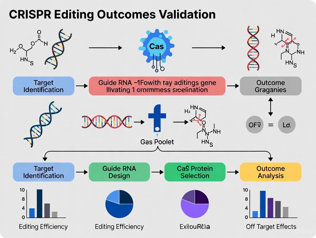
Understanding CRISPR Editing Outcomes: The Impact of DNA Repair and Cellular Context
The advent of CRISPR-Cas9 genome editing has revolutionized biological research and therapeutic development, but the ultimate editing outcome is not determined by the cutting tool itself. Instead, cellular DNA repair pathways ultimately dictate the genetic result, making their understanding critical for predicting and controlling editing outcomes. When the CRISPR-Cas9 system induces a double-strand break (DSB), the cell primarily engages one of three major repair pathways: non-homologous end joining (NHEJ), microhomology-mediated end joining (MMEJ), or homology-directed repair (HDR) [1] [2]. Each pathway possesses distinct mechanisms, efficiencies, and resulting mutational profiles that directly impact the success of genome editing experiments.
The competition between these pathways presents both challenges and opportunities for researchers. NHEJ dominates the repair process in most mammalian cells and operates throughout the cell cycle, but its error-prone nature often introduces semi-random insertions or deletions (indels) [1] [3]. By contrast, HDR enables precise, template-directed repair but functions primarily in replicating cells during the S and G2 phases of the cell cycle, making it inefficient in non-dividing cells [4] [1]. The recently characterized MMEJ pathway represents an intermediate mechanism that utilizes microhomology regions flanking the break site, typically resulting in deletions [5] [3]. For researchers aiming to achieve specific editing outcomes—whether gene knockouts via disruptive indels or precise knock-ins via HDR—understanding and manipulating these pathways is essential for experimental success.
Comparative Analysis of DNA Repair Pathways
The three major DNA repair pathways differ fundamentally in their mechanisms, key protein components, and resulting editing outcomes. The table below provides a comprehensive comparison of these pathways, highlighting their distinct characteristics and applications in CRISPR genome editing.
Table 1: Comparison of Major DNA Double-Strand Break Repair Pathways
| Feature | NHEJ (Non-Homologous End Joining) | MMEJ (Microhomology-Mediated End Joining) | HDR (Homology-Directed Repair) |
|---|---|---|---|
| Repair Mechanism | Direct ligation of broken ends | Annealing of microhomology sequences (2-20 nt) flanking the break | Uses homologous DNA template for precise repair |
| Key Protein Components | Ku70/Ku80, DNA-PKcs, XRCC4, DNA Ligase IV | POLQ (Polθ), PARP1, DNA Ligase I/III | RAD51, BRCA1/2, RAD52 (in SSA sub-pathway) |
| Template Requirement | None | None | Required (donor DNA with homology arms) |
| Cell Cycle Phase | All phases (G1, S, G2, M) | Primarily S/G2 phases | S and G2 phases |
| Mutation Profile | Small insertions/deletions (indels) | Characteristic deletions | Precise, predetermined sequence changes |
| Editing Efficiency | High (dominant pathway) | Intermediate | Low (typically 0.5-20% without enhancement) |
| Primary Applications in CRISPR | Gene knockouts, disruption | Gene knockouts, specific deletions | Precise knock-ins, base corrections, endogenous tagging |
| Advantages | Efficient in both dividing and non-dividing cells | Can produce predictable deletion patterns | High fidelity, precise editing |
| Limitations | Error-prone, random outcomes | Still introduces mutations | Inefficient in non-dividing cells, requires donor template |
This comparative analysis reveals why HDR presents the greatest challenge for researchers seeking precise edits, as it must compete against the more dominant and active NHEJ and MMEJ pathways [5] [1]. Recent studies have demonstrated that even with NHEJ inhibition, imprecise repair still accounts for nearly half of all integration events, highlighting the significant contribution of alternative pathways like MMEJ and single-strand annealing (SSA), a sub-pathway of HDR that uses longer homologous sequences [5]. The complex interplay between these pathways necessitates sophisticated experimental strategies to achieve desired editing outcomes, particularly for therapeutic applications where precision is paramount.
Experimental Data on Pathway Manipulation for Enhanced Editing
Strategic inhibition of competing repair pathways has emerged as a powerful approach to enhance the efficiency of precise genome editing. Quantitative data from recent studies demonstrates how targeted pathway manipulation can significantly shift repair outcomes toward desired HDR events.
Table 2: Quantitative Effects of DNA Repair Pathway Inhibition on Editing Outcomes
| Experimental Condition | Target Gene/Locus | Perfect HDR Efficiency | Indel Frequency | Key Findings |
|---|---|---|---|---|
| NHEJ inhibition only | HNRNPA1 (Cpf1-mediated) | 16.8% | Significant reduction in small deletions (<50 nt) | 3-fold increase in knock-in efficiency compared to control (5.2% to 16.8%) |
| MMEJ inhibition (ART558) | HNRNPA1 | Significant increase | Reduction in large deletions (≥50 nt) and complex indels | Decreased nucleotide deletions around cut site |
| SSA inhibition (D-I03) | HNRNPA1 | No substantial effect on overall HDR | Reduced asymmetric HDR and imprecise donor integration | Specifically reduced asymmetric HDR patterns |
| Combined NHEJ + MMEJ inhibition (HDRobust) | TTLL5 | 80% (from 21% in wildtype) | Reduced from 82% to 1.7% | Outcome purity above 91% for all four genes tested |
| Combined NHEJ + MMEJ inhibition (HDRobust) | RB1CC1 | 63% to 41% with Polθ V896* alone | Drastic reduction | Excessive cell death (≥95%) when editing without donor template |
The data reveal that combined inhibition of NHEJ and MMEJ produces the most dramatic improvements in HDR efficiency. The HDRobust approach, which transiently inhibits both pathways, achieved remarkably high precise editing rates of up to 93% (median 60%) across 58 different target sites [6]. This strategy not only enhanced precise editing but also largely abolished indels, large deletions, rearrangements, and off-target editing events at the target site [6]. Importantly, pathway manipulation effects are consistent across different CRISPR systems, including both Cas9 and Cpf1 (Cas12a) nucleases, highlighting the broad applicability of these findings [5] [6].
Experimental Protocols for Pathway Manipulation
HDRobust Method for High-Precision Editing
The HDRobust protocol represents a cutting-edge approach for achieving high-efficiency precise editing through combined pathway inhibition. The methodology involves either genetic or chemical inhibition of both NHEJ and MMEJ pathways:
- Genetic Inhibition: Introduction of specific mutations in key repair genes: DNA-PKcs K3753R (NHEJ inhibition) combined with Polθ V896* (MMEJ inhibition) [6].
- Chemical Inhibition: Transient treatment with small molecule inhibitors targeting both pathways simultaneously [6].
- Cell Handling: Perform editing in H9 human embryonic stem cells (hESCs) carrying an inducible (iCRISPR) Cas9D10A gene or in human myelogenous leukemia line (K562) using ribonucleoprotein (RNP) delivery [6].
- Editing Template: Design single-stranded DNA donors with blocking mutations to prevent recutting after successful incorporation [6].
- Validation: After isolation of DNA, perform PCR amplicon sequencing of targeted regions and score HDR as the fraction of amplified molecules carrying the intended nucleotide substitutions [6].
This method has been validated for multiple targets including TTLL5, RB1CC1, VCAN, and SSH2, with outcome purity exceeding 91% for all tested genes [6].
Pathway-Specific Inhibitor Treatment Protocol
For researchers seeking to manipulate DNA repair pathways without genetic modification, the following chemical inhibition protocol has demonstrated efficacy:
- Inhibitor Preparation: Prepare stock solutions of Alt-R HDR Enhancer V2 (NHEJi), ART558 (MMEJi), and/or D-I03 (SSA inhibitor) in appropriate solvents [5].
- Cell Treatment: Electroporate Cas nuclease RNP complexes along with donor DNA into human non-transformed diploid RPE1 cells, then immediately treat with specific pathway inhibitors for 24 hours [5].
- Concentration Optimization: Perform dose-response experiments to determine optimal inhibitor concentrations that maximize HDR efficiency while minimizing cytotoxicity.
- Timing Considerations: Limit treatment duration to 24 hours post-electroporation, as HDR typically occurs within this timeframe after Cas9 protein delivery [5].
- Outcome Analysis: At 4 days post-electroporation, perform flow cytometric analysis to quantify knock-in efficiency and long-read amplicon sequencing using PacBio for comprehensive genotyping of repair outcomes [5].
This approach has been successfully applied to both Cpf1-mediated C-terminal tagging and Cas9-mediated N-terminal tagging with fluorescent proteins, demonstrating its versatility across different editing scenarios [5].
Pathway Diagrams and Molecular Mechanisms
The following diagram illustrates the complex interplay between the three major DNA repair pathways and their key protein components:
This diagram illustrates the competitive relationship between the three major repair pathways and highlights key intervention points where specific inhibitors can be applied to shift the balance toward desired outcomes. The visual representation clarifies why NHEJ typically dominates—it involves fewer steps and can proceed throughout the cell cycle—while also demonstrating why HDR requires more specific cellular conditions but produces superior precision.
The Scientist's Toolkit: Essential Research Reagents
Successful manipulation of DNA repair pathways requires carefully selected reagents and inhibitors. The following table compiles key research tools mentioned in recent literature for controlling CRISPR repair outcomes.
Table 3: Essential Research Reagents for DNA Repair Pathway Manipulation
| Reagent/Inhibitor | Target Pathway | Mechanism of Action | Key Applications | Reported Efficacy |
|---|---|---|---|---|
| Alt-R HDR Enhancer V2 | NHEJ | Potent inhibitor of non-homologous end joining | Improving knock-in efficiency in endogenous tagging | ~3-fold increase in knock-in efficiency (5.2% to 16.8% for HNRNPA1) |
| ART558 | MMEJ | Inhibits POLQ, the key enzyme in MMEJ pathway | Reducing large deletions (≥50 nt) and complex indels | Significant increase in perfect HDR frequency |
| D-I03 | SSA (HDR sub-pathway) | Specific inhibitor of Rad52-mediated annealing | Reducing asymmetric HDR and imprecise donor integration | Specifically reduces asymmetric HDR patterns |
| HDRobust Substance Mix | NHEJ & MMEJ | Combined transient inhibition of both pathways | Achieving high-precision editing in multiple cell types | Up to 93% HDR (median 60%) across 58 target sites |
| Virus-Like Particles (VLPs) | Delivery system | Efficient RNP delivery to hard-to-transfect cells | Editing in postmitotic cells (neurons, cardiomyocytes) | Up to 97% delivery efficiency in human neurons |
These reagents represent the current state-of-the-art in DNA repair pathway manipulation for CRISPR genome editing. When selecting reagents for experimental use, researchers should consider cell type specificity, delivery method compatibility, and potential cytotoxic effects. The HDRobust approach, utilizing combined inhibition, has demonstrated particularly robust results across diverse cell types and target sites [6]. For editing in challenging primary cells or non-dividing cell types, VLPs provide an efficient delivery alternative to traditional transfection methods [4].
The systematic comparison of NHEJ, MMEJ, and HDR pathways reveals a complex competitive landscape that directly determines CRISPR editing outcomes. While NHEJ dominates the repair process and often undermines precision editing efforts, recent advances in pathway-specific inhibition—particularly combined NHEJ and MMEJ suppression—have dramatically improved HDR efficiency from typical rates of 0.5-20% to over 90% in some cases [6]. The growing toolkit of chemical inhibitors, genetic approaches, and delivery methods now enables researchers to strategically steer DNA repair toward desired outcomes based on their specific experimental goals.
For researchers validating CRISPR editing outcomes, these findings highlight the critical importance of accounting for cell-type-specific repair behaviors, particularly the dramatically different pathway activities observed in dividing versus non-dividing cells [4]. The experimental protocols and reagent frameworks presented here provide a foundation for designing editing experiments with predictable outcomes, whether the goal is efficient gene disruption via NHEJ/MMEJ or precise template-directed editing via HDR. As CRISPR-based therapies continue their progression toward clinical applications [7], mastering these DNA repair pathways will remain essential for translating cutting-edge gene editing capabilities into reliable research tools and effective treatments.
A cornerstone challenge in therapeutic genome editing is that the same CRISPR intervention produces dramatically different outcomes depending on the target cell's division status. Research reveals that postmitotic cells like neurons and cardiomyocytes repair CRISPR-induced DNA damage through fundamentally different mechanisms and timelines than their dividing counterparts [4]. This comparative guide analyzes the key experimental data, protocols, and reagent solutions essential for validating CRISPR outcomes across these distinct cellular contexts.
Comparative Analysis of Editing Outcomes
Efficiency and Kinetics
| Parameter | Dividing Cells (iPSCs) | Non-Dividing Cells (Neurons) |
|---|---|---|
| Indel Accumulation Timeline | Plateaus within days [4] | Continues increasing for up to 2 weeks [4] |
| Primary Repair Pathways | Microhomology-Mediated End Joining (MMEJ), Nonhomologous End Joining (NHEJ) [4] | Predominantly classical NHEJ (cNHEJ) [4] |
| Indel Size Distribution | Broad range, larger deletions [4] | Narrow distribution, smaller indels [4] |
| Insertion-to-Deletion Ratio | Lower [4] | Significantly higher [4] |
| DSB Repair Half-Life | 1-10 hours [4] | Extended, not specified but resolution is slower [4] |
Outcome Distribution by Repair Pathway
| Repair Pathway | Prevalence in Dividing Cells | Prevalence in Non-Dividing Cells | Characteristic Indels |
|---|---|---|---|
| Classical NHEJ (cNHEJ) | Lower [4] | High [4] | Small indels, perfect repair [4] |
| Microhomology-Mediated End Joining (MMEJ) | High (predominant larger deletions) [4] | Low [4] | Larger deletions [4] |
| Homology Directed Repair (HDR) | Active in S/G2 phases [8] | Largely inactive (cell cycle exit) [4] [8] | Precise edits (requires donor template) |
Experimental Protocols for Cell-Type-Specific Editing
Core Workflow for Comparative Studies
The following diagram illustrates a key experimental workflow for directly comparing editing outcomes between dividing and non-dividing isogenic cells.
Key Methodological Details
- Cell Models: The definitive protocol uses genetically identical iPSCs and iPSC-derived neurons to isolate the effect of cell state from genetic background [4]. For immune cells, primary human T cells in activated (dividing) versus resting (non-dividing) states serve as another isogenic model [4].
- Delivery to Challenging Cells: For postmitotic neurons, which are notoriously difficult to transfect, Virus-Like Particles (VLPs) pseudotyped with VSVG and/or BaEVRless (BRL) envelope proteins achieve up to 97% delivery efficiency of Cas9 ribonucleoprotein (RNP) [4]. Electroporation is suitable for delivering RNP directly into resting and activated T cells [4].
- Timeline and Sampling: In dividing cells, editing outcomes can typically be assessed within a few days post-delivery. For non-dividing cells, sequencing and analysis must extend over at least 16 days to capture the full spectrum of slowly accumulating indels [4].
- Pathway Analysis: To characterize repair outcomes, perform next-generation sequencing (NGS) and analyze the spectra of insertion and deletion mutations. The prevalence of larger deletions (> several base pairs) suggests MMEJ activity, while a dominance of small indels (1-5 bp) points to cNHEJ [4].
DNA Repair Pathway Utilization
The diagram below summarizes how the availability and activity of different DNA repair pathways diverge between dividing and non-dividing cells after a CRISPR-induced double-strand break (DSB).
The Scientist's Toolkit: Research Reagent Solutions
| Reagent / Tool | Function | Application Context |
|---|---|---|
| iPSC-derived Neurons | Clinically relevant postmitotic cell model [4] | Studying editing in neurons; isogenic control for iPSCs [4] |
| Virus-Like Particles (VLPs) | Protein cargo delivery (e.g., Cas9 RNP) [4] | Efficient transduction of neurons (VSVG/BRL pseudotype) [4] |
| B2Mg1 sgRNA | sgRNA with diverse indel outcomes [4] | Testing compatibility with multiple DSB repair pathways [4] |
| Lipid Nanoparticles (LNPs) | In vivo delivery of editing components [7] | Liver-targeted therapies; allows for re-dosing [7] |
| Alt-R HDR Enhancer Protein | Boosts HDR efficiency [9] | Improving precise edits in challenging cells (iPSCs, HSCs) [9] |
| Graph-CRISPR | AI model for editing efficiency prediction [10] | Predicting sgRNA on-target activity across systems [10] |
| CRISPR-GPT | AI-assisted experimental design [11] | Optimizing gRNA design and troubleshooting edits [11] |
Advanced Technologies: AI and Machine Learning
The integration of artificial intelligence (AI) and machine learning (ML) is revolutionizing the prediction and control of cell-type-specific editing outcomes.
- Efficiency Prediction: Models like Graph-CRISPR leverage graph neural networks to integrate both sgRNA sequence and secondary structure information, enhancing the prediction of on-target editing efficiency for CRISPR-Cas9, base editing, and prime editing systems [10]. Other models, including DeepSpCas9 and CRISPRon, have been developed using large-scale screening data to improve generalizability across different cell types and targets [12].
- Experimental Design: CRISPR-GPT is a large language model trained on over a decade of CRISPR literature and expert discussions. It functions as an AI "copilot" to help researchers design experiments, predict off-target effects, and troubleshoot protocols, thereby accelerating the research timeline [11].
- Off-Target Prediction: A significant challenge in CRISPR technology is unintended off-target editing. ML and deep learning tools are becoming the leading methods for predicting these off-target activities, although their accuracy continues to improve with the availability of larger training datasets [13].
The therapeutic promise of CRISPR gene editing extends far beyond the creation of the double-strand break. The ultimate editing outcome—whether a successful gene correction, a disruptive indel, or an unproductive repair—is determined by a complex interplay between the delivery method employed and the cellular repair kinetics it elicits. For researchers and drug development professionals, understanding this relationship is paramount for designing effective therapies. This guide objectively compares the performance of major delivery platforms and analyzes how their interaction with DNA repair pathways in different cell types influences experimental and therapeutic outcomes, providing a structured framework for validating CRISPR editing efficiency.
Delivery Platform Performance Comparison
The choice of delivery method directly impacts editing efficiency, precision, and applicability across cell types. The table below summarizes the performance characteristics of current delivery technologies.
Table 1: Performance Comparison of Major CRISPR Delivery Platforms
| Delivery Method | Editing Efficiency Range | Key Advantages | Key Limitations | Ideal Use Cases |
|---|---|---|---|---|
| Lipid Nanoparticles (LNPs) | Variable; ~90% protein reduction (hATTR trial) [7] | Low immunogenicity, repeat dosing possible, scalable manufacturing [7] [9] | Primarily liver-tropic, can be trapped in endosomes [7] [14] | In vivo liver-targeted therapies (e.g., hATTR, HAE) [7] |
| Virus-Like Particles (VLPs) | Up to 97% transduction (iPSC-derived neurons) [4] | Efficient delivery to hard-to-transfect cells (e.g., neurons), protein cargo avoids DNA integration [4] | Complex production, potential for pre-existing immunity | Delivery to post-mitotic cells (neurons, cardiomyocytes) [4] |
| Electroporation | High in amenable cells (e.g., lymphocytes) | High efficiency for ex vivo editing, direct RNP delivery | Cytotoxicity, only suitable for ex vivo applications | Ex vivo editing of immune cells, stem cells |
| Spherical Nucleic Acids (SNAs) | 3x increase vs. standard LNPs [14] | Enhanced cellular uptake, reduced toxicity, improved precision [14] | Novel technology, further in vivo validation ongoing [14] | Potential as a broad, next-generation delivery platform [14] |
The Impact of Delivery on Repair Kinetics and Outcomes
The method of delivery influences not just if the editing components reach the cell, but also how and when the cell responds to the induced DNA break, with profound implications for the resulting repair outcomes.
Repair Kinetics Differ Dramatically in Dividing vs. Non-Dividing Cells
The cell's proliferation status is a major determinant of repair kinetics. A seminal study comparing genetically identical human induced pluripotent stem cells (iPSCs) and iPSC-derived neurons revealed stark contrasts [4].
- In Dividing Cells (iPSCs): Cas9-induced double-strand breaks are resolved rapidly, with indel frequencies plateauing within a few days. Dividing cells predominantly use resection-dependent repair pathways like microhomology-mediated end joining (MMEJ), resulting in a broader distribution of larger deletion outcomes [4].
- In Non-Dividing Cells (Neurons, Cardiomyocytes): DSB repair is a much slower process. Indels in neurons continue to accumulate for up to 16 days post-transduction. These post-mitotic cells rely more heavily on non-homologous end joining (NHEJ), yielding a narrower distribution of smaller indels. This prolonged timeline is consistent across multiple sgRNAs and delivery particles, indicating a fundamental biological difference in repair kinetics [4].
Table 2: Comparison of CRISPR Repair Kinetics and Outcomes by Cell Type
| Characteristic | Dividing Cells (e.g., iPSCs) | Non-Dividing Cells (e.g., Neurons) |
|---|---|---|
| Primary Repair Pathways | MMEJ, NHEJ [4] | NHEJ [4] |
| Time to Indel Plateau | A few days [4] | Up to 2 weeks [4] |
| Predominant Indel Type | Larger deletions [4] | Small insertions/deletions [4] |
| Implication for Editing | Faster outcome assessment, prone to MMEJ signatures | Requires extended validation timelines, favors NHEJ outcomes |
The following diagram illustrates the divergent repair pathways and outcomes in dividing and non-dividing cells.
Delivery Method Influences Repair Pathway Engagement
The delivery platform itself can influence the repair process. A key advantage of non-viral methods like LNPs is the possibility of redosing. Unlike viral vectors, which often trigger immune responses that prevent re-administration, LNPs do not share this limitation. Clinical trials have demonstrated that patients can safely receive multiple doses of an LNP-delivered CRISPR therapy, with each dose increasing the percentage of edited cells and further reducing symptoms [7]. This capability allows researchers to modulate the overall editing efficiency in a way that is not feasible with single-administration viral vectors.
Experimental Protocols for Validating Repair Outcomes
Robust validation of editing outcomes is critical. Below are detailed methodologies for key experiments cited in this guide.
Protocol 1: Assessing Editing Kinetics in Non-Dividing Cells
This protocol is adapted from research comparing repair in iPSCs and iPSC-derived neurons [4].
- Cell Differentiation: Differentiate human iPSCs into cortical-like excitatory neurons using a established protocol. Validate post-mitotic status via immunocytochemistry (e.g., >99% Ki67-negative, ~95% NeuN-positive) [4].
- CRISPR Delivery via VLPs:
- Produce VSVG-pseudotyped HIV VLPs or VSVG/BRL-co-pseudotyped FMLV VLPs loaded with Cas9 ribonucleoprotein (RNP).
- Transduce both iPSCs and neurons with equal doses of Cas9 VLP. Include a non-transduced control.
- Longitudinal Sampling: Harvest cells at multiple time points post-transduction (e.g., days 1, 2, 4, 7, 11, and 16).
- DNA Extraction and Analysis: Extract genomic DNA from samples at each time point. Amplify the target locus by PCR and use next-generation sequencing (NGS) to quantify indel frequencies and spectra.
- Data Interpretation: Plot indel frequency versus time. Expect to see a rapid plateau in iPSCs versus a slow, linear increase over two weeks in neurons [4].
Protocol 2: Validating Delivery Efficiency with SNA Nanoparticles
This protocol is based on the study demonstrating enhanced delivery with spherical nucleic acids [14].
- Nanostructure Synthesis: Synthesize Lipid Nanoparticle Spherical Nucleic Acids (LNP-SNAs) by forming an LNP core carrying Cas9 RNP and a DNA repair template. Decorate the surface with a dense shell of short, engineered DNA strands [14].
- In Vitro Testing: Apply both standard LNPs and the novel LNP-SNAs to various human cell cultures (e.g., skin cells, white blood cells, bone marrow stem cells).
- Efficiency and Toxicity Measurement:
- Uptake Efficiency: Use flow cytometry or fluorescence microscopy to quantify cellular internalization of particles.
- Cytotoxicity: Measure cell viability using a standard assay (e.g., MTT) 48-72 hours post-treatment.
- Editing Assessment: Extract genomic DNA and perform NGS to determine on-target editing efficiency and the precision of DNA repairs (HDR rate).
- Comparison: The LNP-SNAs are expected to show significantly higher uptake (up to 3x), reduced toxicity, and a threefold increase in gene-editing efficiency compared to standard LNPs [14].
Essential Research Reagent Solutions
The following table catalogues key reagents and their functions for research in CRISPR delivery and repair validation.
Table 3: Essential Research Reagents for CRISPR Delivery and Repair Studies
| Reagent / Tool | Function / Application |
|---|---|
| Virus-Like Particles (VLPs) | Protein-based delivery of CRISPR RNP to difficult-to-transfect cells like neurons; avoids DNA integration [4]. |
| Lipid Nanoparticles (LNPs) | In vivo delivery vehicle with low immunogenicity; particularly effective for liver-targeted therapies [7] [9]. |
| Spherical Nucleic Acids (SNAs) | Next-generation delivery nanostructure that enhances cellular uptake, editing efficiency, and precision while reducing toxicity [14]. |
| Alt-R HDR Enhancer Protein | Recombinant molecule that increases homology-directed repair (HDR) efficiency up to two-fold in challenging primary cells [9]. |
| proPE (prime editing system) | Improved prime editing system that uses a second sgRNA to enhance editing efficiency for low-performing edits, reducing optimization needs [9]. |
| T7 Endonuclease I Assay | Enzyme-based mismatch cleavage assay for initial, rapid detection of indel mutations following genome editing [15]. |
The journey "beyond the break" reveals that the delivery method is not merely a vehicle but an active determinant of CRISPR repair kinetics and outcomes. The data clearly shows that non-viral platforms like LNPs and VLPs enable safe and effective editing, with emerging technologies like SNAs poised to significantly boost efficiency. Furthermore, the cellular context—specifically whether a cell is dividing or post-mitotic—dictates a fundamental repair timeline and pathway preference. For researchers, this underscores the necessity of selecting a delivery platform compatible with the target cell's biology and of employing longitudinal validation protocols that account for potentially slow repair kinetics, as this integrated approach is critical for unlocking the full therapeutic potential of CRISPR medicine.
The Validation Toolbox: A Practical Guide to CRISPR Analysis Methods
The advent of CRISPR-Cas9 technology has revolutionized genome engineering, enabling precise genetic modifications across diverse biological systems. However, the success of any CRISPR experiment hinges on accurately confirming that the intended edits have been introduced without unwanted, off-target mutations. While various methods exist to analyze editing outcomes, targeted next-generation sequencing (NGS) has emerged as the gold standard for comprehensive validation, providing unparalleled resolution and sensitivity. This approach allows researchers to obtain deep, quantitative insights into both on-target editing efficiency and off-target effects, which is crucial for therapeutic development and basic research applications.
Targeted NGS addresses critical limitations of alternative methods. Techniques such as the T7E1 assay, while rapid and inexpensive, provide only qualitative estimates of editing efficiency without revealing the specific sequence alterations. Sanger sequencing-based tools like TIDE and ICE offer more detail but lack the sensitivity to detect low-frequency edits or complex heterogeneous outcomes in mixed cell populations. In contrast, targeted NGS delivers nucleotide-level resolution across all modified alleles in a sample, enabling researchers to fully characterize the spectrum of insertions, deletions, and more complex rearrangements generated by CRISPR-mediated editing [16]. This comprehensive insight is indispensable for applications ranging from the development of genetically modified cell lines to clinical-grade therapeutic editing where safety and precision are paramount.
Comparative Analysis of Targeted NGS Methodologies
Targeted NGS employs different enrichment strategies to isolate genomic regions of interest prior to deep sequencing. The two primary approaches—amplicon-based and hybridization capture-based enrichment—each present distinct advantages and limitations that researchers must consider when validating CRISPR experiments.
Enrichment Strategy Performance Comparison
The table below summarizes the core characteristics of these enrichment methodologies:
Table 1: Comparison of Targeted NGS Enrichment Strategies for CRISPR Validation
| Feature | Amplicon-Based Enrichment | Hybridization Capture-Based Enrichment |
|---|---|---|
| Principle | Multiplex PCR amplification of target regions using specific primer pools [17] | Hybridization of biotinylated oligonucleotide baits to genomic regions followed by magnetic pull-down [18] |
| Workflow Complexity | Simple, fast (as little as 3 hours), minimal hands-on time [17] | Complex, multi-step process requiring specialized equipment and 2-3 days [17] |
| DNA Input Requirements | Low (can work with degraded samples like FFPE) [17] | High (typically 50-200 ng of high-quality DNA) [19] |
| Panel Flexibility | Highly flexible; easily customized with new targets [17] | Less flexible; redesign requires new bait synthesis |
| Uniformity of Coverage | Moderate to high (modern systems achieve >95% uniformity) [17] | High, especially across exonic regions [20] |
| Best Applications | Focused panels (dozens to hundreds of targets), CRISPR validation [17] | Large genomic regions (whole exomes, large gene panels) [17] |
Bait Chemistry and Performance Metrics
Further considerations in enrichment methodology include the choice of bait chemistry, which significantly impacts performance. Recent comparative studies have evaluated different bait types, including single-stranded RNA (Agilent SureSelect), single-stranded DNA (IDT xGEN), double-stranded DNA (Twist Bioscience), and double-stranded RNA (Dynegen QuarXeq). These platforms demonstrate that RNA baits generally exhibit stronger binding affinity, while DNA baits perform better in high-GC regions [20]. Double-stranded RNA baits have shown particularly balanced capture performance, with high sensitivity for variant detection. Platforms with RNA baits also demonstrate lower AT dropout rates (reduced loss of AT-rich regions), whereas single-stranded DNA baits can exhibit AT dropout rates up to 10% [20].
Experimental Design and Protocol for CRISPR Validation
A robust targeted NGS workflow for CRISPR validation requires careful experimental planning and execution across multiple stages, from sample preparation to data analysis.
Sample Preparation and Library Generation
The initial phase involves processing samples to create sequencing-ready libraries:
- DNA Extraction: Isolate high-quality genomic DNA from CRISPR-treated cells using standardized extraction protocols. For cell lines, typical yields of 50-100 ng/μL are sufficient. The TTSH-oncopanel validation study determined that ≥50 ng of DNA input was necessary for reliable detection of all expected mutations [19].
- Target Enrichment: Select appropriate enrichment strategy based on experimental needs. For most CRISPR validation studies focusing on specific target sites and predicted off-target loci, amplicon-based approaches provide the optimal balance of efficiency and cost-effectiveness. The CleanPlex technology, for example, employs a three-step process: (1) multiplex PCR with specifically designed primers, (2) background cleaning to remove primer dimers and non-specific products, and (3) indexing PCR to barcode samples for multiplexed sequencing [17].
- Library Quality Control and Sequencing: Assess library quality and quantity using methods such as Bioanalyzer or TapeStation, then sequence on an appropriate NGS platform (Illumina, Ion Torrent, or MGI platforms) to achieve sufficient depth. For CRISPR editing analysis, recommended coverage is typically 1000x or higher to detect low-frequency editing events [19].
Analytical Performance Metrics and Thresholds
Establishing rigorous performance metrics ensures reliable variant detection in CRISPR-edited samples:
Table 2: Key Analytical Performance Metrics for Targeted NGS in CRISPR Validation
| Performance Metric | Target Specification | Experimental Validation |
|---|---|---|
| Sensitivity | >97% for variant detection | 97.14% sensitivity observed in TTSH-oncopanel validation [19] |
| Specificity | >99.9% | 99.99% specificity demonstrated in replicate analyses [19] |
| Limit of Detection (VAF) | ≤3% variant allele frequency | Reliable detection of SNVs and INDELs at 2.9% VAF confirmed [19] |
| Reproducibility | >99.9% concordance between replicates | 99.99% reproducibility demonstrated in inter-run precision tests [19] |
| Coverage Uniformity | >95% of targets at ≥0.2x mean coverage | >96% uniformity achieved across various panel sizes [17] |
| On-target Rate | >80% of reads mapping to target regions | >96% on-target rate demonstrated with optimized panels [17] |
Workflow Visualization for CRISPR Validation
The following diagram illustrates the complete targeted NGS workflow for comprehensive CRISPR validation:
Diagram Title: Targeted NGS Workflow for CRISPR Validation
Advanced Applications in CRISPR Research
Targeted NGS enables sophisticated analysis of CRISPR editing outcomes that extends far beyond basic indel detection, providing critical insights for therapeutic development.
Single-Cell Resolution of Editing Outcomes
Emerging technologies now combine targeted NGS with single-cell analysis to resolve complex editing patterns in heterogeneous cell populations. The Tapestri platform, for example, employs single-cell DNA sequencing to characterize editing outcomes simultaneously at dozens to hundreds of loci across thousands of individual cells [21]. This approach enables researchers to determine the co-occurrence of edits (which edits happen together in the same cell), assess editing zygosity (whether one or both alleles are modified), and correlate genomic edits with protein expression through combined DNA-protein profiling. In validation studies, this method demonstrated 99.77% sensitivity and 99.93% specificity for detecting editing events at the single-cell level [21].
Ultrasensitive Off-Target Detection
Conventional NGS methods often miss low-frequency off-target edits that occur below 0.5% variant allele frequency. To address this limitation, advanced methods like CRISPR amplification have been developed to dramatically enhance detection sensitivity [22]. This technique uses CRISPR nucleases to selectively cleave wild-type DNA at potential off-target sites, thereby enriching for mutated alleles through multiple rounds of amplification. This approach can detect off-target mutations with frequencies as low as 0.00001%—a 1.6 to 984-fold improvement over standard targeted amplicon sequencing [22]. When combined with genome-wide off-target prediction tools, this method provides a comprehensive safety profile for therapeutic CRISPR applications.
Essential Research Reagents and Solutions
Successful implementation of targeted NGS for CRISPR validation requires specific reagents and computational tools. The following table catalogues key solutions utilized in the field:
Table 3: Essential Research Reagent Solutions for Targeted NGS in CRISPR Validation
| Category | Specific Examples | Function and Application |
|---|---|---|
| Target Enrichment | CleanPlex Custom Panels (Paragon Genomics) [17] | Ultra-high multiplex PCR-based target enrichment with background cleaning chemistry |
| Hybridization Capture | SureSelect (Agilent), xGEN (IDT), Twist Bioscience Panels [20] | Solution-based or solid-phase hybridization capture using DNA or RNA baits |
| Reference Materials | Genome in a Bottle (GIAB) Reference Materials [18] | Benchmarking and validation of NGS workflows with ground-truth variant calls |
| Variant Calling & Analysis | Sophia DDM [19], GA4GH Benchmarking Tools [18] | Automated variant detection, annotation, and classification with clinical interpretation |
| CRISPR Analysis Software | ICE (Synthego), TIDE [16] | Computational tools for analyzing CRISPR editing efficiency from sequencing data |
| Single-Cell Analysis | Tapestri Platform (Mission Bio) [21] | High-throughput single-cell DNA sequencing for resolving editing heterogeneity |
Targeted next-generation sequencing represents the definitive standard for comprehensive characterization of CRISPR editing outcomes, providing researchers with the resolution and sensitivity needed to advance both basic science and therapeutic applications. Through appropriate selection of enrichment strategies, rigorous validation of analytical performance, and implementation of advanced applications such as single-cell analysis and ultrasensitive off-target detection, targeted NGS delivers the critical insights necessary to understand the full spectrum of CRISPR-induced genetic modifications. As CRISPR technology continues to evolve toward clinical applications, targeted NGS will remain an indispensable component of the validation workflow, ensuring both the efficacy and safety of genome-editing interventions across diverse research and therapeutic contexts.
The advent of CRISPR-based genome editing has revolutionized biological research and therapeutic development, making the accurate validation of editing outcomes a critical step in the workflow. Confirming the efficiency and specificity of gene edits is essential for attributing phenotypic changes to genotypic alterations and for ensuring the safety and efficacy of potential therapies [23]. While next-generation sequencing (NGS) is considered the gold standard for comprehensive variant detection due to its high sensitivity and discovery power, it remains cost-prohibitive and computationally intensive for many routine applications, particularly when analyzing a small number of targets [24]. In this context, Sanger sequencing-based computational tools have emerged as accessible yet powerful alternatives for quantifying editing efficiencies.
Among these, Tracking of Indels by Decomposition (TIDE) and Inference of CRISPR Edits (ICE) have gained significant popularity. These tools leverage the ubiquity and low cost of Sanger sequencing by applying sophisticated decomposition algorithms to sequencing chromatograms from edited samples. By comparing these to wild-type sequences, they can deconvolute complex mixtures of indels (insertions and deletions) and provide quantitative estimates of editing efficiency and the spectrum of resulting mutations [25] [16]. This guide provides a objective comparison of ICE and TIDE, enabling researchers to select the most appropriate tool for their specific CRISPR validation needs.
Tool Comparison: ICE vs. TIDE
Core Functionality and Algorithmic Approach
Both ICE and TIDE analyze Sanger sequencing trace data from PCR amplicons covering the CRISPR target site. The fundamental principle involves computational decomposition of the complex sequencing chromatogram from a heterogeneous, edited cell population into its constituent sequences (wild-type and various indel-containing sequences).
TIDE (Tracking of Indels by Decomposition): This older method aligns the guide RNA (gRNA) sequence to both unedited (wild-type) and edited sample sequences. Its algorithm then decomposes the sequencing trace data from the edited sample using the wild-type sequence as a reference template. This process estimates the relative abundance and size of insertions and deletions, providing a goodness-of-fit (R²) value and assessing the statistical significance of each identified indel [16]. TIDE can infer the identity of single-base pair insertions but requires manual adjustment of settings for more complex indels [16].
ICE (Inference of CRISPR Edits): Synthego's ICE tool also performs alignment of unedited samples to the original sgRNA sequence, followed by alignment between unedited and edited samples to determine differences. ICE calculates an editing efficiency score (the ICE score, corresponding to indel frequency) and provides detailed information on the distribution and types of indels generated. A key feature is its reported ability to detect unexpected editing outcomes, such as large insertions or deletions, without additional user input [16].
Performance and Accuracy Analysis
Recent systematic comparisons using artificial sequencing templates with predetermined indels have shed light on the relative performance of these tools.
Table 1: Performance Comparison of ICE and TIDE based on Experimental Data
| Performance Metric | ICE (Inference of CRISPR Edits) | TIDE (Tracking of Indels by Decomposition) |
|---|---|---|
| Overall Accuracy | Highly comparable to NGS (R² = 0.96) [16]. Provides the most accurate estimations for most samples in a 2024 study [25]. | Acceptable accuracy for simple indels; estimates become more variable with complex indels [25]. |
| Indel Frequency Estimation | Accurate for indels with a few base changes; more variable for complex indels or knock-ins [25]. | Reasonable accuracy in the mid-range of indel frequencies; more variable in low or high ranges [25]. |
| Indel Sequence Deconvolution | Effectively estimates net indel sizes; capability to deconvolute specific sequences has variability and limitations [25]. | Effectively estimates net indel sizes; its ability to resolve specific indel sequences is more limited compared to ICE [25] [16]. |
| Key Limitations | Accuracy diminishes with highly complex indel mixtures or knock-in sequences [25]. | Limited capability for predicting sequences of longer insertions; requires manual setting adjustments for complex edits [16]. |
A critical finding from independent research is that both tools perform with acceptable accuracy when indels are simple and involve only a few base changes. However, their estimated values show greater variation when sequencing templates contain more complex indels or knock-in sequences [25]. While both tools effectively estimate the net sizes of indels, their capability to deconvolute the exact sequences of these indels has demonstrated certain limitations [25]. A comparative study concluded that DECODR, another web tool, provided the most accurate estimations for the majority of samples, though ICE and TIDE remain widely used and effective for routine analysis [25].
Practical Considerations for Researchers
When integrating these tools into a workflow, practical aspects beyond raw performance are crucial.
Table 2: Practical Considerations for ICE and TIDE
| Consideration | ICE | TIDE |
|---|---|---|
| User Interface & Experience | Noted for a user-friendly interface and easier interpretation of results [16]. | Interface is functional, but analysis parameters can be difficult for the average user to modify confidently [16]. |
| Additional Features | Includes a "Knockout Score" focusing on edits causing frameshifts, batch sample uploads, and multi-guide analysis [16]. | Provides a goodness-of-fit R² value and statistical significance for identified indels. TIDER, a TIDE-based tool, outperforms others for knock-in efficiency estimation [25]. |
| Ideal Use Case | Routine, quantitative analysis of editing efficiency and indel distribution where user-friendliness and detection of larger indels are priorities. | Quick assessment of editing efficiency, particularly for projects involving simple indels or knock-in validation via TIDER. |
Experimental Protocols for Tool Validation
To ensure reliable results from ICE or TIDE, a robust experimental workflow from sample preparation to data analysis is essential. The following protocol, commonly used in comparative studies, outlines the key steps.
Sample Preparation and Sequencing
- CRISPR Delivery and Culture: Introduce the CRISPR-Cas9 components (e.g., as a ribonucleoprotein complex) into your target cells using your preferred method (e.g., electroporation, lipofection) [25]. Culture the cells for a sufficient duration to allow for DNA repair and the manifestation of edits.
- Genomic DNA (gDNA) Extraction: Harvest cells and extract high-quality gDNA using a standard kit or phenol-chloroform method. Ensure DNA integrity and purity (A260/280 ratio ~1.8-2.0).
- PCR Amplification: Design primers that flank the CRISPR target site, ideally generating an amplicon of 300-600 bp. Use a high-fidelity DNA polymerase to minimize PCR errors.
- PCR Product Purification: Clean up the PCR reaction using a commercial gel and PCR clean-up kit to remove primers, enzymes, and salts. Elute in nuclease-free water or a low-EDTA buffer.
- Sanger Sequencing: Submit the purified PCR product for Sanger sequencing using one of the PCR primers. It is critical to also sequence a wild-type (unmodified) control sample from the same genetic background using the same primer.
Data Analysis with ICE and TIDE
- File Preparation: Obtain the sequencing chromatogram files (.ab1 format) for both the wild-type and edited samples.
- Analysis with TIDE:
- Access the TIDE web tool (http://shinyapps.datacurators.nl/tide/).
- Upload the wild-type and edited sample .ab1 files.
- Input the target sequence and specify the cut site (typically 3 bp upstream of the PAM sequence).
- Select an analysis window (e.g., 100-200 bp around the cut site) and run the decomposition analysis [26].
- Analysis with ICE:
- Access the ICE web tool (Synthego).
- Upload the reference sequence (wild-type) and the edited sample .ab1 file, or provide the sequence trace data.
- Input the gRNA sequence to define the target site.
- Run the analysis to receive the ICE score (indel frequency) and a detailed breakdown of the inferred indel landscape.
- Result Interpretation and Validation: Compare the indel frequency and spectra reported by both tools. For critical experiments, or if the results are ambiguous, consider validating the findings by NGS on a subset of samples, which provides a high-resolution ground truth [25] [24].
The Scientist's Toolkit: Essential Reagents and Materials
Table 3: Key Research Reagent Solutions for CRISPR Validation Workflows
| Item | Function/Description | Example |
|---|---|---|
| High-Fidelity DNA Polymerase | Amplifies the target genomic locus with minimal errors, ensuring accurate sequencing templates. | Q5 Hot Start High-Fidelity Master Mix [26] |
| PCR Clean-Up Kit | Purifies amplification products by removing excess primers, dNTPs, and enzymes before sequencing. | Gel and PCR Clean-Up Kit [26] |
| Sanger Sequencing Service/Kit | Generates the chromatogram trace data (.ab1 files) required for indel analysis by ICE and TIDE. | Services from providers like GENEWIZ [27] or Macrogen [26] |
| CRISPR RNP Complex | Pre-assembled ribonucleoprotein complex for highly specific and efficient genome editing. | Alt-R S.p. Cas9 Nuclease with crRNA and tracrRNA [25] |
| Digital PCR System | Provides an orthogonal, highly quantitative method for validating editing efficiencies. | Droplet Digital PCR (ddPCR) [26] |
ICE and TIDE have democratized quantitative analysis of CRISPR editing outcomes by leveraging the widespread accessibility of Sanger sequencing. While NGS remains the gold standard for its unparalleled sensitivity and ability to detect novel variants, ICE and TIDE offer an excellent balance of cost, speed, and accuracy for most routine applications [24]. Performance data indicates that ICE generally provides a more user-friendly experience and more accurate estimations for a wider range of indel types, making it suitable for most researchers seeking a robust primary analysis tool [25] [16]. TIDE remains a valuable tool, particularly for quick assessments, and its derivative TIDER is specifically advantageous for analyzing knock-in efficiencies [25]. The choice between them should be guided by the specific editing context, the required level of detail, and the need for orthogonal validation in downstream applications.
The T7 Endonuclease I (T7E1) assay has been a widely used enzymatic method for detecting CRISPR-Cas9 editing efficiency in the era of genome engineering. As a legacy mismatch cleavage assay, it offers a technically straightforward and cost-effective approach for initial validation of nuclease activity [28]. The assay operates by recognizing and cleaving heteroduplex DNA structures that form when wild-type and indel-containing strands hybridize after PCR amplification of the target region [29]. This cleavage produces DNA fragments of predictable sizes that can be visualized via gel electrophoresis, providing a semi-quantitative estimate of editing efficiency.
However, as CRISPR validation methodologies have advanced, significant limitations of the T7E1 system have emerged. Current research demonstrates that T7E1 often provides inaccurate reflections of true editing activity, with particular shortcomings in dynamic range, sensitivity, and detection capability [28] [30]. This comparative guide examines the performance constraints of T7E1 against modern validation alternatives, providing experimental data and methodologies to inform appropriate assay selection for rigorous CRISPR editing validation.
Core Limitations of the T7E1 Assay
Mechanism-Inherent Constraints
The fundamental limitations of T7E1 stem from its biochemical mechanism. T7E1 recognizes structural deformations in DNA heteroduplexes rather than specific sequence changes, which creates several analytical constraints [31]. The enzyme's cleavage efficiency depends heavily on the type and size of DNA mismatches, with larger insertion-deletion mutations (indels) being detected more reliably than single nucleotide polymorphisms [31] [29]. This bias means the assay can overlook single-base edits while preferentially detecting larger structural alterations.
Additionally, the requirement for heteroduplex formation means that samples with predominantly homozygous edits (where mutant strands primarily hybridize with other mutant strands) may show minimal cleavage despite high editing efficiency [29]. The assay's performance is further influenced by reaction conditions including incubation temperature, salt concentration, and enzyme concentration, requiring careful optimization for different experimental contexts [29].
Quantitative Inaccuracy and Dynamic Range Limitations
Comparative studies consistently demonstrate that T7E1 significantly underestimates editing efficiency, particularly with highly active guide RNAs. Research comparing T7E1 with targeted next-generation sequencing (NGS) across 19 genomic loci revealed that T7E1 reported an average editing efficiency of 22%, while NGS detected an average of 68% for the same targets [28]. This underestimation was most pronounced with highly active sgRNAs: those with >90% editing efficiency by NGS appeared only modestly active by T7E1 [28].
The T7E1 assay exhibits a compressed dynamic range, with performance plateauing at approximately 30% editing efficiency [28]. Guide RNAs with substantially different activities can appear similar when evaluated by T7E1, potentially leading to incorrect conclusions about reagent efficacy. In one documented case, two sgRNAs showing ~28% activity by T7E1 actually demonstrated dramatically different true efficiencies of 40% versus 92% when assessed by NGS [28].
Table 1: Performance Comparison of T7E1 Versus Targeted Next-Generation Sequencing
| Metric | T7E1 Assay | Targeted NGS |
|---|---|---|
| Average Reported Editing Efficiency | 22% | 68% |
| Detection of Poorly Performing Guides (<10% editing) | Often appears inactive | Accurately detects low activity |
| Detection of Highly Active Guides (>90% editing) | Appears modestly active | Accurately quantifies high activity |
| Differentiation of Guides with Different Efficiencies | Poor resolution | High resolution |
| Dynamic Range | Limited, plateaus around 30% | Linear across full range |
Detection Specificity and Sensitivity Constraints
The T7E1 assay demonstrates variable sensitivity depending on mutation type. While it effectively detects deletions, particularly larger indels, it performs poorly at identifying single nucleotide changes [31] [29]. Comparative analysis with Surveyor mismatch cleavage assays revealed that T7E1 outperforms Surveyor for deletion detection but is substantially less effective for identifying single-base substitutions [31].
The sensitivity of T7E1 depends on the sequence context surrounding the mismatch and the specific base pairs involved in the mismatch [31]. The optimal amplicon size for T7E1 detection ranges from 400-800 bp, with cleavage products needing to be >100 bp for clear resolution [29]. These requirements can limit experimental design flexibility, particularly for compact genomic regions.
Comparative Analysis of CRISPR Validation Methods
Modern CRISPR validation has moved beyond enzymatic mismatch cleavage to more precise methodologies. The table below compares key features of current validation approaches:
Table 2: Method Comparison for Assessing CRISPR-Cas9 Editing Efficiency
| Method | Detection Principle | Quantitative Capability | Key Advantages | Key Limitations |
|---|---|---|---|---|
| T7E1 | Enzyme cleavage of heteroduplex DNA | Semi-quantitative | Low cost, technically simple, rapid results [29] [16] | Limited dynamic range, poor SNP detection, underestimates efficiency [28] [30] |
| TIDE | Decomposition of Sanger sequencing chromatograms | Quantitative | Cost-effective, provides indel sequence information [16] | Can miscall alleles in clones, accuracy depends on sequencing quality [28] [30] |
| ICE | Algorithmic analysis of Sanger sequencing data | Quantitative | High correlation with NGS (R² = 0.96), detects large indels [16] | Requires good quality sequencing data [30] |
| ddPCR | Fluorescent probe detection in partitioned samples | Highly quantitative | Exceptional precision, discriminates between edit types [30] | Requires specific probe design, limited to known edits [30] |
| Targeted NGS | High-throughput sequencing of target regions | Highly quantitative | Comprehensive view of all edits, high sensitivity [28] [16] | Expensive, time-consuming, requires bioinformatics expertise [16] |
Quantitative Performance Comparisons
Direct comparisons between methods demonstrate clear performance differences. When evaluating editing efficiencies in cellular pools, TIDE and Inference of CRISPR Edits (ICE) show strong correlation with targeted NGS results [28]. However, both TIDE and Indel Detection by Amplicon Analysis (IDAA) can miscall specific alleles in edited clones, with IDAA accurately predicting only 25% of both indel sizes and frequencies in tested clones [28].
Droplet digital PCR (ddPCR) provides highly precise quantification of editing efficiencies and can discriminate between different types of edits, such as non-homologous end joining versus homology-directed repair products [30]. This method is particularly valuable in therapeutic contexts where precise quantification of specific edit types is critical.
Experimental Protocols and Methodologies
Standard T7E1 Assay Protocol
The T7E1 protocol begins with PCR amplification of the target region from genomic DNA using high-fidelity polymerase. Recommended amplicon size is 400-800 bp, with primers binding approximately 250 bp upstream and downstream of the target site [29]. The PCR product is purified using standard gel extraction or PCR clean-up kits.
For heteroduplex formation, the purified PCR product is subjected to a denaturation and reannealing process: heat to 95°C for 5 minutes, then slowly cool to 25°C at a rate of 0.1°C per second [28]. The reannealed product is digested with T7 Endonuclease I (typically 1 μL enzyme in 10 μL reaction with appropriate buffer) at 37°C for 30 minutes [30]. Some protocols recommend adding MnCl₂ to enhance cleavage efficiency [29].
The digestion products are separated by agarose gel electrophoresis (1-2% agarose), and band intensities are quantified using densitometry. The editing efficiency is estimated using the formula: % indel = [1 - (1 - (a + b)/(a + b + c))^0.5] × 100, where c is the intensity of the undigested PCR product, and a and b are the intensities of the cleavage products [28].
Methodological Considerations for Accurate T7E1 Results
Several strategies can improve T7E1 reliability. Pre-digesting genomic DNA with restriction enzymes recognizing the wild-type sequence can reduce wild-type background [29]. Using ice-COLD-PCR (co-amplification at lower denaturation temperature) with reduced denaturation temperature can enrich mutated sequences while suppressing wild-type amplification [29]. Including appropriate controls is essential: both unedited samples and samples with known editing efficiency should be run in parallel.
Despite these optimizations, the fundamental limitations of T7E1 remain. For critical applications, confirmation with orthogonal methods is strongly recommended, particularly when quantitative accuracy is essential.
T7E1 Assay Workflow and Heteroduplex Formation
The following diagram illustrates the core experimental workflow of the T7E1 assay and the critical heteroduplex formation process:
Research Reagent Solutions for CRISPR Validation
Table 3: Essential Research Reagents for CRISPR Editing Validation
| Reagent/Category | Specific Examples | Function in Validation |
|---|---|---|
| Mismatch Cleavage Enzymes | T7 Endonuclease I, Surveyor Nuclease | Recognizes and cleaves heteroduplex DNA at mismatch sites [31] [29] |
| PCR Components | High-fidelity DNA Polymerase (Q5 Hot Start), Target-specific Primers | Amplifies target genomic region for downstream analysis [30] |
| Nucleic Acid Analysis | Agarose Gels, Ethidium Bromide/GelRed, Electrophoresis Systems | Separates and visualizes DNA fragments by size [30] |
| Sequencing Services | Sanger Sequencing, Next-Generation Sequencing Platforms | Provides nucleotide-level resolution of editing outcomes [28] [16] |
| Analysis Software | TIDE, ICE, NGS Analysis Pipelines | Quantifies editing efficiency and characterizes indel spectra [30] [16] |
The T7E1 assay remains a useful tool for initial screening during CRISPR guide RNA optimization when rapid, cost-effective assessment is prioritized over quantitative accuracy [16]. However, its significant limitations in dynamic range, sensitivity, and detection specificity necessitate complementary validation with more rigorous methods for definitive characterization of editing outcomes.
For most research applications, Sanger sequencing-based approaches like ICE or TIDE provide an optimal balance of cost, throughput, and information content [30] [16]. In therapeutic development or clinical applications where precision is paramount, targeted next-generation sequencing or ddPCR offer the quantitative accuracy and comprehensive variant detection required for rigorous validation [28] [30].
The selection of appropriate validation methodology should be guided by experimental context, required precision, and resource constraints. While T7E1 maintains a place in the CRISPR validation toolkit, researchers should interpret its results with appropriate understanding of its methodological constraints and supplement it with more quantitative approaches for critical applications.
The clinical translation of CRISPR-based genome editing necessitates rigorous assessment of editing fidelity, as off-target effects can lead to unintended genomic modifications with potentially deleterious consequences, including disruption of essential genes or activation of oncogenes [32] [33]. The first FDA-approved CRISPR therapy, exagamglogene autotemcel (exa-cel) for sickle cell disease, underwent extensive regulatory review where off-target analysis was a critical consideration [33]. This has spurred the development and refinement of numerous detection methodologies, which broadly fall into two categories: empirical assays that experimentally identify off-target sites and in silico tools that computationally predict them. Empirical methods can be further classified as biochemical (using purified genomic DNA), cellular (using living cells), or in situ (in a native cellular context) [33]. This guide provides a comparative analysis of these approaches, focusing on established and emerging methods like GUIDE-seq, CIRCLE-seq, and in silico frameworks such as CCLMoff and DNABERT-Epi, to inform researchers in the validation of CRISPR editing outcomes.
Empirical Detection Methods: Experimental Workflows and Protocols
Empirical methods directly detect Cas9-induced double-strand breaks (DSBs) or their repair products, offering an unbiased, genome-wide perspective on nuclease activity.
Cellular Methods: Capturing Biological Context
Cellular methods assess nuclease activity within living cells, thereby incorporating the influences of native chromatin structure, DNA repair pathways, and cellular physiology on editing outcomes [33].
GUIDE-seq (Genome-wide, Unbiased Identification of DSBs Enabled by Sequencing) is a prominent cellular assay. Its protocol involves co-delivering a CRISPR-Cas9 system (as plasmid, RNA, or ribonucleoprotein) with a short, double-stranded oligonucleotide tag into cells [34] [33]. When a DSB occurs, this tag is integrated into the genome via the non-homologous end joining (NHEJ) repair pathway. After genomic DNA extraction, tag-specific primers are used to amplify and sequence the regions flanking the integrated tag, allowing for the genome-wide identification of DSB loci [34].
Diagram 1: GUIDE-seq workflow for identifying DSBs in cells.
Recent advancements include GUIDE-seq-2, which offers improved scalability and accuracy, enabling population-scale studies that can reveal how human genetic variation affects Cas9 off-target activity [35].
DISCOVER-seq (Discovery of in situ Cas Off-targets and verification by sequencing) is another cellular method that leverages a different biological principle. It identifies off-target sites by performing ChIP-seq for the MRE11 DNA repair protein, which is recruited to DSBs shortly after they occur [34] [33]. This method captures the immediate cellular response to Cas9 cleavage within the native chromatin environment.
Biochemical Methods: Maximizing Sensitivity
Biochemical methods utilize purified genomic DNA and Cas9 nuclease in a controlled in vitro setting, eliminating cellular variables. This allows for ultra-sensitive detection but may overestimate biologically relevant editing [33].
CIRCLE-seq (Circularization for In Vitro Reporting of Cleavage Effects by Sequencing) is a highly sensitive biochemical technique. The protocol begins by fragmenting and circularizing genomic DNA [36]. This circularized DNA is then treated with the Cas9 protein and a guide RNA of interest. Subsequent exonuclease digestion degrades linear DNA, enriching for the circular molecules. Any DNA that was cleaved by Cas9 becomes linearized and is thus protected from digestion. This enriched, cleaved DNA is then prepared as a sequencing library, allowing for the identification of cleavage sites with high sensitivity and low background noise [36]. The entire process, from cell growth to sequencing data, can be completed within two weeks [36].
Diagram 2: CIRCLE-seq in vitro cleavage detection workflow.
CHANGE-seq (Circularization for High-throughput Analysis of Nuclease Genome-wide Effects by Sequencing) is an improved version of CIRCLE-seq that incorporates a tagmentation-based library preparation step (using Tn5 transposase), which increases sensitivity and reduces sequencing bias [32] [33]. A recently described variant, CHANGE-seq-R, is a massively parallel biochemical assay designed to measure Cas9 activity across millions of mismatched target sites, facilitating the training of deep learning models like CHANGE-net [35].
Hybrid and In Situ Methods
GUIDE-tag is a more recent in vivo method that combines strengths from multiple approaches. It utilizes a tethering strategy between a SpyCas9-monomeric streptavidin (mSA) fusion protein and a biotinylated dsDNA donor to significantly increase the capture efficiency of DSBs in mouse tissues like liver and lung [34]. It also incorporates Unique Molecular Identifiers (UMIs) via Tn5 tagmentation to mitigate PCR bias. This system can detect off-target sites with editing rates as low as 0.2% and can be coupled with UDiTaS (Uni-Directional Targeted Sequencing) analysis to identify translocations and large deletions [34].
In Silico Prediction Methods: The Rise of Deep Learning
In silico methods predict off-target sites computationally, offering a fast and inexpensive approach for guide RNA design and risk assessment. Early tools were primarily alignment-based or used hand-crafted scoring formulae [37]. The current state-of-the-art leverages deep learning models trained on large-scale experimental data, which can automatically extract relevant features from DNA sequences and context.
CCLMoff is a deep learning framework that incorporates a pre-trained RNA language model (RNA-FM from RNAcentral) to capture mutual sequence information between the sgRNA and potential target sites [37]. It is trained on a comprehensive dataset compiled from 13 genome-wide off-target detection techniques, which forces the model to learn general off-target patterns rather than those specific to a single assay. This enables CCLMoff to demonstrate strong generalization performance across diverse datasets [37]. An enhanced version, CCLMoff-Epi, integrates epigenetic features such as chromatin accessibility (ATAC-seq) and histone modifications (H3K4me3, H3K27ac) to further improve predictive accuracy by accounting for chromatin context [37].
DNABERT-Epi represents another advanced in silico approach. It is based on DNABERT, a model pre-trained on the entire human genome, allowing it to learn the fundamental "language" of DNA [32]. DNABERT-Epi integrates the same key epigenetic features (H3K4me3, H3K27ac, ATAC-seq) that are enriched at off-target sites identified by cellular assays like GUIDE-seq. Ablation studies have quantitatively confirmed that both genomic pre-training and the integration of epigenetic features are critical for its high predictive accuracy [32].
Diagram 3: Architecture of deep learning-based in silico prediction models.
Comparative Analysis of Methods
The table below summarizes the key characteristics, strengths, and limitations of the major off-target detection approaches.
Table 1: Comparison of Major Off-Target Detection Methods
| Method | Category | Principle | Sensitivity | Biological Context | Key Strengths | Primary Limitations |
|---|---|---|---|---|---|---|
| GUIDE-seq [34] [33] | Cellular | NHEJ-mediated tag integration | High (≥0.2%) [34] | Yes (Native chromatin & repair) | Detects biologically relevant edits; medium throughput | Requires efficient RNP/delivery; may miss rare sites |
| DISCOVER-seq [34] [33] | Cellular | MRE11 ChIP-seq at DSBs | High | Yes (Native chromatin & repair) | Captures DSBs in temporal window; no exogenous tag | Cumbersome; temporally restricted [34] |
| CIRCLE-seq [36] [33] | Biochemical | Cas9 cleavage of circularized DNA | Very High | No (Purified DNA) | Ultra-sensitive; low background; standardized | May overestimate cleavage; lacks biological context |
| CHANGE-seq [32] [33] | Biochemical | CIRCLE-seq with tagmentation | Very High | No (Purified DNA) | Higher sensitivity & lower bias than CIRCLE-seq | May overestimate cleavage; lacks biological context |
| GUIDE-tag [34] | In Situ / In Vivo | Tethered donor & tagmentation | Very High (≥0.2%) | Yes (In vivo) | Direct in vivo detection; captures large deletions | Technically complex; lower throughput |
| CCLMoff / DNABERT-Epi [32] [37] | In Silico | Deep Learning + Epigenetics | N/A (Predictive) | Partial (Via epigenetic features) | Fast, inexpensive; guides sgRNA design; incorporates context | Predictions only; model-dependent accuracy |
Table 2: Typical Input Requirements and Outputs
| Method | Input Material | Typical Input Amount | Detects Indels | Detects Translocations | Workflow Duration |
|---|---|---|---|---|---|
| GUIDE-seq | Living Cells | Varies with delivery | Yes [33] | No [33] | 1-2 weeks |
| CIRCLE-seq | Purified Genomic DNA | Nanograms [33] | N/A (Cleavage sites) | N/A (Cleavage sites) | ~2 weeks [36] |
| CHANGE-seq | Purified Genomic DNA | Nanograms [33] | N/A (Cleavage sites) | N/A (Cleavage sites) | ~2 weeks |
| GUIDE-tag | Cells or Tissue | Varies | Yes | Yes (with UDiTaS) [34] | Several weeks |
| In Silico Tools | Genome Sequence & sgRNA | Computational | N/A (Predictive) | N/A (Predictive) | Minutes to hours |
Successful off-target assessment relies on a suite of specialized reagents and computational resources.
Table 3: Key Research Reagent Solutions
| Reagent / Resource | Function | Example Application / Note |
|---|---|---|
| SpyCas9-mSA RNP [34] | A fusion of Cas9 and monomeric streptavidin for tethering biotinylated donors. | Enhances DSB capture efficiency in GUIDE-tag. |
| Biotin-dsDNA Donor [34] | A double-stranded DNA tag for integration into DSBs. | Serves as the capture moiety in GUIDE-tag and related methods. |
| Tn5 Transposase [34] [33] | An enzyme that simultaneously fragments DNA and adds sequencing adapters ("tagmentation"). | Used in CHANGE-seq and GUIDE-tag for efficient library prep. |
| UMI Adapters [34] | Oligonucleotide adapters containing Unique Molecular Identifiers. | Reduces PCR amplification bias and improves quantitative accuracy in NGS. |
| MRE11 Antibody [33] | For immunoprecipitation of the MRE11 DNA repair protein. | Essential for DISCOVER-seq to pull down Cas9-induced DSBs. |
| Pre-trained Model Weights (e.g., DNABERT, RNA-FM) [32] [37] | The parameters of a foundation model pre-trained on genomic or RNA data. | Provides a head start for in silico tools, capturing fundamental sequence rules. |
| Epigenetic Data Tracks (H3K4me3, H3K27ac, ATAC-seq) [32] | Genome-wide maps of histone modifications and chromatin accessibility. | Integrated into models like DNABERT-Epi to account for chromatin context. |
The evolving landscape of CRISPR off-target detection is characterized by a trend towards multi-modal validation, where a combination of sensitive biochemical assays, biologically relevant cellular methods, and sophisticated in silico predictions is employed to build a comprehensive safety profile [33]. The FDA's emphasis on genome-wide unbiased assays for therapeutic development underscores the importance of these methods [33]. Future directions include the integration of population-scale genetic variation into off-target profiling, as demonstrated by GUIDE-seq-2 and CHANGE-seq-R [35], and the continued refinement of explainable deep learning models that not only predict but also interpret the factors leading to off-target activity [32] [37]. For researchers validating CRISPR outcomes, the selection of a method should be guided by the specific application: biochemical assays for maximum sensitivity during initial guide screening, cellular assays to confirm biological relevance in the target cell type, and in silico tools for rapid design and prioritization, with the most rigorous applications likely requiring data from multiple complementary approaches.
In the rapidly advancing field of CRISPR-based gene editing, robust validation workflows are paramount for distinguishing transformative therapeutic breakthroughs from experimental artifacts. As CRISPR technologies evolve from basic research tools into clinical therapeutics, the scientific community faces increasing pressure to develop standardized, rigorous validation methodologies, particularly in complex pre-clinical models and challenging cellular contexts. The validation process extends far beyond simple confirmation of target modification to comprehensive assessment of editing efficiency, specificity, functional correction, and physiological relevance. This comparative guide examines current validation frameworks through the lens of cutting-edge CRISPR platforms, with a specialized focus on their application in hard-to-edit cells and sophisticated animal models that more accurately recapitulate human disease.
The emergence of next-generation editing platforms like CRISPR Therapeutics' SyNTase system demonstrates how technological innovations are creating new validation paradigms while addressing persistent challenges in the field [38] [39]. This analysis synthesizes experimental data, methodological approaches, and technical solutions to provide researchers with a practical framework for validating CRISPR outcomes across diverse experimental contexts, from initial discovery research to IND-enabling pre-clinical studies.
Comparative Analysis of CRISPR Editing Platforms
Platform Mechanisms and Technical Specifications
Different CRISPR platforms employ distinct molecular mechanisms that directly influence validation requirements and methodological approaches. The table below compares four major editing platforms across key technical parameters.
Table 1: Comparison of Major CRISPR Editing Platforms
| Editing Platform | Molecular Mechanism | Primary Repair Pathway | Size Constraints | Theoretical Max Efficiency | Key Technical Differentiators |
|---|---|---|---|---|---|
| SyNTase Editing | Cas9 + engineered polymerase with synthetic templates | Bias toward HDR | Moderate (fits in LNP) | 95% (demonstrated) [38] | AI-guided structural modeling; engineered nucleotide templates |
| CRISPR-Cas9 | Wild-type Cas9 + gRNA | NHEJ-dominated | ~4.2 kb Cas9 cDNA [40] | Variable (40-80%) | RNA-guided DNA endonuclease activity |
| Base Editing | Cas9 nickase + deaminase | Single-base substitution without DSBs | Larger than Cas9 | High in optimal contexts | Chemical conversion of base pairs without double-strand breaks |
| Prime Editing | Cas9-reverse transcriptase + pegRNA | Reverse transcription of edit template | Significant size | Moderate (varies by target) | Versatile edits without donor DNA templates |
Performance Metrics in Pre-clinical Models
Quantitative assessment of editing outcomes provides critical insights for platform selection. The following table compares performance metrics across recent pre-clinical studies.
Table 2: Performance Metrics of CRISPR Platforms in Pre-clinical Models
| Editing Platform | Disease Model | Editing Efficiency | Off-Target Effects | Functional Correction | Delivery Method |
|---|---|---|---|---|---|
| SyNTase [38] [39] | AATD (humanized mouse/rat) | Up to 95% in hepatocytes; >70% mRNA correction | <0.5% detectable | >3-fold serum AAT increase; >99% M-AAT:Z-AAT ratio | LNP (single IV dose ≤0.5 mg/kg) |
| CRISPR-Cas9 [7] | hATTR (human trial) | ~90% protein reduction | Managed | Disease symptom stability/improvement | LNP (single IV dose) |
| Base Editing [41] | Sickle cell disease (murine) | Higher than CRISPR-Cas9 | Fewer genotoxicity concerns | Reduced red cell sickling | Lentiviral HSPC transduction |
| Prime Editing [41] | Junctional epidermolysis bullosa | Up to 60% in keratinocytes | Minimal | Type XVII collagen restoration; selective advantage | Viral vector |
Validation Workflows for Next-Generation Editing Platforms
Case Study: SyNTase Platform Validation for AATD
CRISPR Therapeutics' SyNTase editing platform represents a significant advance in gene correction technology, combining compact Cas9 proteins with a novel class of engineered polymerases [38] [39]. The validation workflow for this platform in Alpha-1 Antitrypsin Deficiency (AATD) models provides an exemplary framework for rigorous pre-clinical assessment.
Experimental Protocol 1: Comprehensive In Vivo Validation
Animal Model Preparation:
- Utilize both established NSG-PiZ mouse model (carrying human SERPINA1 Z alleles) and novel humanized PiZ rat model with human mutant SERPINA1 E342K variant replacing the rat ortholog [39]
- Confirm baseline characterization including serum AAT levels (~5-6 μM) compared to normal levels (>20 μM)
LNP Formulation and Dosing:
- Encapsulate SyNTase editing components in proprietary lipid nanoparticles
- Administer single intravenous dose at ≤0.5 mg/kg in volume-appropriate vehicle
- Include control cohorts receiving empty LNPs or non-targeting editors
Temporal Analysis and Tissue Collection:
- Collect serum samples at predetermined intervals (e.g., 1, 3, 7 weeks post-administration)
- Harvest liver tissue at endpoint (7-9 weeks) for DNA, RNA, and protein analysis
- Preserve samples for histopathological examination
Molecular Validation Tier 1 - DNA Editing Assessment:
- Extract genomic DNA from homogenized liver tissue
- Amplify target SERPINA1 region using PCR with flanking primers
- Quantify editing efficiency via next-generation sequencing of amplicons
- Calculate percentage correction of E342K mutation
Molecular Validation Tier 2 - Transcriptional Analysis:
- Isolate total RNA from liver samples
- Perform RT-qPCR with allele-specific probes distinguishing wild-type M-AAT and mutant Z-AAT
- Calculate mRNA correction percentage (>70% target)
Molecular Validation Tier 3 - Functional Protein Assessment:
- Measure total serum AAT levels via ELISA
- Determine M-AAT:Z-AAT ratio using quantitative immunoassays
- Target >3-fold increase in total AAT and >99% M-AAT:Z-AAT ratio [39]
Safety and Specificity Validation:
- Assess potential off-target effects through whole-genome sequencing
- Evaluate liver histology for evidence of toxicity or inflammation
- Monitor serum biomarkers of liver function
The following diagram illustrates the integrated validation workflow for the SyNTase platform in AATD models:
Advanced Validation Methodologies
Experimental Protocol 2: Off-Target Assessment Using NGS
Comprehensive off-target profiling remains essential for therapeutic development. The following protocol adapts methodologies from the SyNTase validation [38] and established best practices [40] [42]:
In Silico Prediction:
- Identify potential off-target sites using multiple algorithms with different scoring metrics
- Include sites with up to 5 nucleotide mismatches and bulges
Amplicon Sequencing:
- Design primers flanking predicted off-target sites
- Amplify regions from treated and control animal genomic DNA
- Sequence using Illumina platforms with minimum 100,000x coverage
Bioinformatic Analysis:
- Map reads to reference genome using optimized aligners
- Detect indels using specialized variant callers (e.g., GATK)
- Establish threshold of <0.5% frequency as background level
Whole Genome Sequencing:
- Perform WGS on treated and control samples at 30-40x coverage
- Analyze sheared DNA for comprehensive indel profiling
- Compare variant profiles between experimental groups
Addressing Challenges in Hard-to-Edit Cells
Biological Barriers to Efficient Editing
Certain cell types and genomic contexts present significant challenges for CRISPR editing, requiring specialized validation approaches. The molecular factors influencing editing difficulty are visualized below:
Specialized Workflows for Challenging Contexts
Experimental Protocol 3: Editing Validation in Essential Genes
Essential genes require specialized validation approaches since complete knockout is lethal [43]:
Pre-Editing Essentiality Assessment:
- Consult DepMap (Dependency Map) database to determine gene essentiality scores
- Identify common essential genes with CERES scores <-1
- Design validation around partial rather than complete disruption
Heterozygous Knockout Strategy:
- Design gRNAs targeting specific alleles while preserving wild-type copy
- Use FACS sorting to isolate single-cell clones
- Validate heterozygous status using ICE analysis or TIDE
Alternative Functional Assessment:
- Implement CRISPR interference (CRISPRi) with dCas9-KRAB fusions
- Measure transcript reduction rather than protein absence via RT-qPCR
- Assess functional consequences through phenotypic assays specific to gene function
Viability-Coupled Validation:
- Monitor cell proliferation for 5-10 passages post-editing
- Compare growth rates between edited and control populations
- Use competitive growth assays with mixed populations
Experimental Protocol 4: High GC Content and Repetitive Regions
Genomic regions with unusual sequence composition present unique validation challenges [43]:
PCR Optimization for GC-Rich Targets:
- Incorporate betaine or DMSO to reduce secondary structures
- Use polymerase blends with enhanced processivity
- Implement touchdown PCR protocols
Sequencing Strategy Adaptation:
- Utilize long-read technologies (Oxford Nanopore, PacBio) for repetitive regions
- Design primers outside repetitive elements when possible
- Validate sequencing results with multiple primer sets
Editing Efficiency Normalization:
- Account for reduced amplification efficiency in quantitative assessments
- Include internal control amplicons with normal GC content
- Use spike-in controls for absolute quantification
The Scientist's Toolkit: Essential Research Reagents
Successful validation requires appropriate tools and reagents. The following table details essential components for comprehensive CRISPR validation workflows.
Table 3: Research Reagent Solutions for CRISPR Validation
| Reagent/Category | Specific Examples | Function in Validation Workflow | Application Context |
|---|---|---|---|
| Mismatch Detection Assays | Surveyor, T7E1 | Initial rapid screening of editing efficiency | Early-stage validation; high-throughput screening |
| Sanger Sequencing Analysis Tools | TIDE, ICE | Estimate editing efficiency and specific indel proportions | Intermediate validation; quality control |
| Next-Generation Sequencing | Illumina amplicon sequencing | Detect low-frequency edits; characterize specific indels | Comprehensive on-target analysis |
| Whole Genome Sequencing | Illumina NovaSeq, PacBio | Comprehensive on/off-target detection; indel profiling | Preclinical safety assessment; regulatory submissions |
| Lipid Nanoparticles (LNPs) | Proprietary formulations | In vivo delivery of editing components | Therapeutic development; animal studies |
| Viral Vectors | AAV, Lentivirus | Efficient delivery to hard-to-transfect cells | Cell line engineering; in vivo delivery |
| Cell Viability Assays | MTT, ATP-based | Assess toxicity in essential gene editing | Functional validation; safety assessment |
| Antibodies for Protein Detection | Allele-specific anti-AAT | Quantify functional protein correction | Therapeutic efficacy assessment |
The evolving CRISPR landscape demands increasingly sophisticated validation workflows, particularly as editing technologies advance toward clinical application. The SyNTase platform demonstrates how next-generation editors can achieve unprecedented efficiency and specificity in therapeutically relevant contexts, while established technologies like base editing and prime editing continue to find application in specific challenging scenarios. Successful validation requires a multi-tiered approach encompassing DNA, RNA, and protein-level assessments, coupled with rigorous safety profiling.
For researchers navigating this complex landscape, the integration of standardized validation protocols with platform-specific optimization remains critical. As artificial intelligence and machine learning increasingly guide editor design and outcome prediction [40], validation workflows must similarly evolve to incorporate these computational advances while maintaining rigorous experimental standards. Through continued refinement of these specialized workflows, the research community can accelerate the development of transformative CRISPR-based therapies while ensuring their safety and efficacy.
Optimizing Precision and Safety: Strategies for Enhanced CRISPR Workflows
Selecting High-Activity Guides and Cas Variants for Improved Specificity
Performance Comparison of CRISPR Systems and Tools
Selecting the optimal CRISPR system is paramount for achieving high on-target activity while minimizing off-target effects. The table below provides a comparative overview of key systems and the AI-driven tools available for their selection and optimization.
Table 1: Comparison of CRISPR Systems and AI-Guided Design Tools
| System / Tool | Key Features | Reported Performance Advantage | Primary Application |
|---|---|---|---|
| OpenCRISPR-1 [44] | AI-generated Cas9-like effector; 400 mutations away from SpCas9. | Comparable or improved activity & specificity relative to SpCas9; compatible with base editing. | High-fidelity genome editing. |
| AncBE4max-AI-8.3 [45] | AI-engineered Cas9 variant with 8 point mutations for base editing. | 2-3 fold increase in average base editing efficiency; stable enhancement in 7 cancer cell lines & hESCs. | High-efficiency cytosine and adenine base editing. |
| HF-Cas9 Variants [45] | Rationally designed mutants (e.g., HF1) with reduced non-specific DNA binding. | Significantly reduced off-target effects; often at the cost of reduced on-target efficiency. | Applications where specificity is the highest priority. |
| CRISPR-GPT [46] | LLM-based agent for end-to-end experiment design, including system selection and gRNA design. | Automates selection of high-specificity systems; validates gRNA designs against off-target effects. | Automated, optimized experiment planning for non-experts. |
| ProMEP [45] | AI model predicting mutation effects on Cas9 fitness landscape from sequence and structure. | Accurately identifies single-point mutations (e.g., G1218R, C80K) that enhance editing efficiency. | In silico engineering of high-performance Cas9 variants. |
Experimental Protocols for Validation
Accurately quantifying editing outcomes is a critical step in validating the performance of any CRISPR system. The following section details standard protocols for assessing on-target efficiency and specificity.
Protocol: Quantifying On-Target Editing Efficiency with Amplicon Sequencing
Targeted amplicon sequencing (AmpSeq) is widely considered the "gold standard" for sensitive and accurate quantification of CRISPR edits [47].
Step 1: DNA Extraction and Amplification
- Method: Extract genomic DNA from CRISPR-treated cells or tissues using a standard kit. Design and synthesize PCR primers flanking the target site to create amplicons ideally 300-500 bp in length.
- Rationale: High-quality, pure genomic DNA is essential for unbiased PCR amplification. Proper primer design ensures specific amplification of the target locus.
Step 2: Library Preparation and Sequencing
- Method: Prepare the PCR amplicons into a sequencing library using a commercial kit (e.g., Illumina). Sequence the library on a next-generation sequencing (NGS) platform to achieve high coverage (recommended >10,000x read depth per sample).
- Rationale: High read depth is necessary to detect low-frequency editing events and provide a quantitative measure of efficiency [47].
Step 3: Data Analysis
- Method: Process the raw sequencing data. Align reads to a reference sequence to identify insertions, deletions (indels), or base substitutions at the target site. Calculate the editing efficiency as the percentage of reads containing non-wild-type sequences at the target locus.
- Rationale: This provides a precise, quantitative measure of on-target activity for different guide RNAs or Cas variants [47].
Protocol: Profiling Off-Target Effects with Genome-Wide Assays
Understanding and quantifying off-target activity is crucial for assessing specificity.
Step 1: Guide RNA Design
- Method: Use computational tools (e.g., CRISPOR) to predict potential off-target sites in the reference genome. These are genomic loci with sequence similarity to the guide RNA, especially in the "seed" region [48].
Step 2: Off-Target Interrogation
- Method 1 (Targeted): For a limited number of high-risk predicted off-target sites, perform targeted AmpSeq as described in Protocol 2.1 [47].
- Method 2 (Genome-Wide): For an unbiased survey, use methods like GUIDE-seq or CIRCLE-seq. These experimental techniques identify potential off-target sites in a genome-wide manner without prior prediction [49].
Step 3: Specificity Calculation
- Method: Quantify the indel or edit frequency at each verified off-target site using AmpSeq. The specificity of a guide RNA or Cas variant can be summarized by the total number of off-target sites with editing above a background threshold or the frequency of editing at the most promiscuous off-target site.
The following workflow diagram illustrates the key steps in designing and validating a high-specificity CRISPR experiment.
The Scientist's Toolkit: Essential Research Reagents
Table 2: Key Reagents for High-Specificity CRISPR Experiments
| Reagent / Tool | Function | Example Use-Case |
|---|---|---|
| High-Fidelity Cas9 Variants | Engineered Cas9 protein with reduced off-target binding. | Replacing SpCas9 in applications where specificity is critical [45]. |
| AI-Designed Effectors (e.g., OpenCRISPR-1) | Novel, highly functional editors generated by language models. | Achieving high activity and specificity at challenging genomic targets [44]. |
| Optimized Base Editor (e.g., AncBE4max-AI-8.3) | Cas9 variant engineered for enhanced base editing efficiency. | Performing efficient C->T or A->G base conversions with minimal indels [45]. |
| Lipid Nanoparticles (LNPs) | Non-viral delivery vehicle for CRISPR components. | Enabling efficient in vivo delivery and potential for re-dosing [7]. |
| dCas9-Activator/Repressor | Catalytically dead Cas9 fused to transcriptional modulators. | For CRISPRa/i experiments to reversibly tune gene expression without altering DNA [50] [51]. |
| Guide RNA Design Tools | Computational algorithms for predicting sgRNA efficacy and specificity. | Selecting guides with high on-target and low off-target activity during experiment planning [48] [46]. |
| Validated sgRNA Libraries | Pre-designed sets of sgRNAs with known high performance. | Running high-confidence genetic screens (e.g., Avana library) [49]. |
| Amplicon Sequencing Kits | Reagents for preparing NGS libraries from PCR amplicons. | Gold-standard quantification of CRISPR editing efficiency and specificity [47]. |
The therapeutic application of CRISPR genome editing hinges on the efficient and safe delivery of editing machinery to target cells. The choice of delivery strategy is not one-size-fits-all; it is profoundly influenced by the physiology of the target cell, such as its division status and transfection efficiency. Furthermore, as the field advances toward clinical applications, validating editing outcomes with precision has become a critical component of the research and development workflow. This guide objectively compares the three primary delivery platforms—viral vectors, electroporation, and lipid nanoparticles (LNPs)—by synthesizing recent experimental data, with a specific focus on their performance across diverse cell types within the context of rigorous CRISPR outcome validation.
Comparative Performance of Delivery Platforms
The table below summarizes the key characteristics of each delivery platform based on current research and clinical applications.
Table 1: Comparison of CRISPR-Cas9 Delivery Strategies for Different Cell Types
| Delivery Method | Mechanism of Action | Key Applications & Cell Types | Efficiency & Performance Data | Key Advantages | Major Limitations |
|---|---|---|---|---|---|
| Viral Vectors (e.g., AAV, Lentivirus, VLPs) | Engineered viruses infect cells and deliver genetic cargo [52]. | • In vivo therapy (e.g., AAV for retinal diseases [53])• Ex vivo therapy (e.g., Lentivirus for CAR-T cells [54])• Hard-to-transfect cells (e.g., VSVG/BRL-VLPs for neurons, 97% transduction [4]) | • AAV: Efficient delivery to non-dividing cells [54].• Lentivirus: Stable integration in dividing cells.• VLPs: Up to 97% transduction in iPSC-derived neurons [4]. | • High transduction efficiency.• Proven clinical success.• Suitable for in vivo and ex vivo use. | • Cargo size constraints (e.g., AAV ~4.7 kb [54]).• Risk of insertional mutagenesis [54].• Pre-existing immunity concerns. |
| Electroporation | Electric pulses create temporary pores for cargo entry [52]. | • Ex vivo editing of immune cells [55] [52] and hematopoietic stem cells (e.g., Casgevy [52]). | • High editing efficiency in amenable cells.• Direct RNP delivery possible. | • Wide cargo compatibility (DNA, RNA, RNP).• Direct intracellular delivery. | • High cell toxicity, low viability [55] [52].• Limited to ex vivo use.• Can damage sensitive primary cells. |
| Lipid Nanoparticles (LNPs) | Lipid vesicles encapsulate and deliver nucleic acids via endocytosis [56]. | • In vivo mRNA/CRISPR delivery (e.g., NTLA-2001 to liver [52] [53]).• Ex vivo cell engineering (e.g., CAR-T cells [55]). | • CAR-T cell editing: 76% (PD-1), 86% (TRAC), 80% (B2M) knockout [55].• Superior cell viability vs. electroporation [55].• Pancreatic islet cells: Efficient transfection via biliary duct infusion [57]. | • Suitable for in vivo use.• Transient expression, safer profile.• High viability in sensitive cells.• Potential for re-dosing [57]. | • Primarily targets liver after IV injection [54].• Challenges with large/compact genetic cargo. |
Experimental Insights into Cell-Type-Specific Outcomes
Dividing vs. Non-Dividing Cells Exhibit Divergent DNA Repair
A critical study highlights how DNA repair pathways—and thus CRISPR outcomes—differ dramatically between cell types. Research comparing induced pluripotent stem cells (iPSCs) to genetically identical iPSC-derived neurons found that post-mitotic neurons resolve Cas9-induced double-strand breaks over a much longer timeframe (up to two weeks) compared to dividing cells [4]. Neurons predominantly utilized non-homology directed repair pathways like non-homologous end joining, resulting in a narrower distribution of small insertions/deletions, whereas iPSCs showed a broader range of outcomes, including larger deletions associated with microhomology-mediated end joining [4]. This fundamental difference underscores the necessity of tailoring delivery and analysis to the target cell's physiology.
Advancing Safety and Precision with Novel Systems
Beyond delivery, controlling CRISPR activity is vital for safety. A recent development is a cell-permeable anti-CRISPR protein system, LFN-Acr/PA, designed to deactivate Cas9 after editing is complete [58]. This system, derived from anthrax toxin components, can be delivered via protein-based delivery into human cells rapidly and at low concentrations, reducing off-target effects and boosting editing specificity by up to 40% [58]. This represents a significant tool for improving the clinical safety of gene editing across all delivery platforms.
Essential Experimental Protocols
Protocol: Assessing Delivery Efficiency and Editing Outcomes in Neurons Using VLPs
This protocol is adapted from studies using virus-like particles to edit human iPSC-derived neurons [4].
- VLP Production and Transduction:
- Produce VLPs (e.g., VSVG-pseudotyped HIV VLPs or VSVG/BRL-co-pseudotyped FMLV VLPs) loaded with Cas9 ribonucleoprotein (RNP) and a fluorescent reporter (e.g., mNeonGreen) [4].
- Differentiate human iPSCs into post-mitotic cortical neurons, confirming purity with immunocytochemistry for Ki67 (negative) and NeuN (positive) [4].
- Transduce neurons with a calibrated dose of Cas9-VLPs. Include controls with VLPs containing only the fluorescent reporter.
- Efficiency and DSB Validation:
- Editing Outcome Analysis:
- Time-Course Sampling: Harvest genomic DNA at multiple time points post-transduction (e.g., days 1, 4, 7, 14) to track the slow accumulation of indels characteristic of neurons [4].
- Next-Generation Sequencing (NGS): Amplify the target genomic region by PCR and perform NGS. Analyze the sequences for insertion/deletion (indel) types and frequencies, comparing the distribution to outcomes in isogenic iPSCs edited with the same sgRNA [4].
Protocol: Multiplexed Gene Editing in CAR-T Cells Using LNPs
This protocol is based on recent work using LNPs for multiplexed RNA delivery in primary human T cells [55].
- LNP Formulation and T Cell Activation:
- LNP Transfection and Control:
- Transfect activated T cells with the multiplexed LNPs. As a benchmark control, deliver the same RNA payloads via electroporation, a traditional method for T cell engineering [55].
- Culture the transfected T cells and monitor cell viability and expansion over several days.
- Functional and Genotypic Validation:
- Cell Viability Assay: Compare viability between LNP-transfected and electroporated cells. LNPs consistently demonstrate superior viability [55].
- Flow Cytometry: Confirm surface expression of the CD19 CAR using a protein-specific label.
- Editing Efficiency Analysis:
- Use the Tapestri single-cell sequencing platform to simultaneously genotype edited cells at multiple loci. This allows for the assessment of editing efficiency, zygosity (biallelic vs. monoallelic edits), and the correlation of multiplexed edits in individual cells [59].
- Alternatively, use bulk NGS to quantify knockout efficiencies at each target gene.
Visualizing Workflows and Pathways
CRISPR Delivery and Outcome Analysis Workflow
The following diagram illustrates the general workflow for delivering CRISPR components and validating editing outcomes, integrating key steps from the protocols above.
Figure 1: CRISPR Delivery and Analysis Workflow. This diagram outlines the decision-making process for selecting a delivery method based on target cell type and the subsequent steps for validating editing outcomes.
DNA Repair Pathways in Dividing vs. Non-Dividing Cells
The core cellular pathways that resolve CRISPR-induced DNA breaks differ significantly between cell types, directly influencing editing outcomes.
Figure 2: DNA Repair Pathways by Cell Type. Diagram showing the dominant DNA repair pathways in dividing versus non-dividing cells, leading to distinct CRISPR editing outcomes and repair kinetics [4].
The Scientist's Toolkit: Key Research Reagent Solutions
The table below lists essential materials and tools used in the experiments cited in this guide.
Table 2: Key Research Reagents and Solutions for CRISPR Delivery Studies
| Item Name | Type/Function | Specific Application Example |
|---|---|---|
| Virus-Like Particles (VLPs) | Protein-based delivery vehicle for Cas9 RNP [4]. | Delivery of CRISPR components to hard-to-transfect human iPSC-derived neurons [4]. |
| Ionizable Lipids (e.g., LP01, ALX-184) | Critical component of LNPs for nucleic acid encapsulation and endosomal release [52]. | Formulating LNPs for in vivo CRISPR therapy (NTLA-2001) or transfecting pancreatic islet cells [52] [57]. |
| Microfluidic Nanoparticle Synthesizer | Instrument for consistent, scalable LNP production via rapid mixing [52]. | Synthesizing nucleic acid-encapsulated LNPs for research and pre-clinical development (e.g., PreciGenome's NanoGenerator) [52]. |
| Tapestri Single-Cell Sequencing Platform | Platform for high-throughput single-cell DNA sequencing [59]. | Characterizing multiplexed editing (zygosity, clonality) in engineered CAR-T cells at single-cell resolution [59]. |
| Anti-CRISPR Protein System (LFN-Acr/PA) | Cell-permeable protein for rapid inhibition of Cas9 activity [58]. | Reducing off-target effects by turning off Cas9 after successful editing in human cells [58]. |
The precision of CRISPR-based gene editing is fundamentally constrained by the duration of activity of the editing machinery within the cell. Extended exposure of the genome to CRISPR nucleases increases the probability of off-target effects, where unintended genomic sites are cleaved, leading to potentially detrimental mutations [60]. The emerging paradigm for mitigating this risk focuses on controlling editing kinetics, specifically through the development of novel systems that enable precise temporal shut-off of CRISPR activity immediately after successful target editing is achieved. This approach is critical for validating true editing outcomes, as it minimizes the confounding variables introduced by off-target modifications. By limiting the window of opportunity for off-target cleavage, these systems enhance the specificity and safety of CRISPR applications, a concern paramount to researchers, scientists, and drug development professionals engaged in therapeutic development and functional genomics [60] [61].
This guide provides an objective comparison of the most current systems designed for the temporal control of CRISPR activity. We will dissect the mechanisms, experimental performance data, and implementation protocols for three primary strategies: small-molecule inducible systems, self-inactivating vectors, and degron-based rapid degradation. The comparison is framed within the critical context of validating authentic CRISPR editing outcomes, where reducing off-targets is not merely an optimization but a necessity for data integrity and therapeutic safety.
Comparative Analysis of Temporal Shut-off Systems
The following table summarizes the core characteristics and performance metrics of the leading temporal shut-off systems, based on recent experimental findings.
Table 1: Performance Comparison of Key Temporal Shut-off Systems
| System Name | Core Mechanism | Key Components | Reported Off-Target Reduction | Activation/Shut-off Kinetics | Primary Applications Cited |
|---|---|---|---|---|---|
| Doxycycline-inducible dCas9 (iCRISPRi/a) [62] | Small-molecule controlled transcription of dCas9-effector fusions. | rtTA, dCas9-KRAB (for i), dCas9-VPR (for a), doxycycline. | Not explicitly quantified, but tight control shown via flow cytometry (e.g., CXCR4 modulation). | Rapid protein degradation upon doxycycline withdrawal (demonstrated in days). | Precise temporal regulation of endogenous gene expression in primary human 3D organoids [62]. |
| Doxycycline-inducible Cas9 for Non-Dividing Cells [9] | Small-molecule controlled transcription of functional Cas9. | rtTA, Cas9 nuclease, doxycycline. | Enables screening in senescent/quiescent cells, reducing indirect phenotypic effects. | Cas9 induced after non-dividing state is established; temporal control prevents interference with phenotype establishment. | CRISPR screens in non-proliferative cell states (senescence, quiescence, terminal differentiation) [9]. |
| Self-Inactivating Vectors (eVLP) [9] | Delivery of pre-assembled Cas9 RNP via virus-like particles; transient activity. | Engineered virus-like particles (eVLP), Cas9 ribonucleoprotein (RNP). | Inferred from transient delivery; no viral vector integration or persistent expression. | Single injection; transient activity (duration not specified). Achieved 16.7% average editing efficiency in mouse retinal pigment epithelium. | Safe in vivo gene therapy; demonstrated for wet age-related macular degeneration [9]. |
| TadA-8e Mutant ABE (H52L/D53R) [9] | Engineering of base editor protein to minimize RNA off-target editing. | Adenine Base Editor (ABE) variant with reduced RNA binding. | Substantially reduced RNA off-target editing while retaining efficient on-target DNA editing. | Not a temporal shut-off system; a protein engineering solution to a specific off-target class. | Therapeutic base editing applications where RNA-editing-related toxicity is a concern [9]. |
Experimental Protocols for Key Systems
Protocol for Doxycycline-Inducible CRISPRi/a in 3D Organoids
This protocol, adapted from a large-scale organoid screening study, details the establishment of a temporally controlled CRISPR interference/activation (CRISPRi/a) system [62].
- Step 1: Cell Line Engineering. Begin with the target cell line, such as TP53/APC double knockout (DKO) gastric organoids. Use a sequential two-vector lentiviral transduction approach. First, transduce with a lentiviral vector constitutively expressing the reverse tetracycline-controlled transactivator (rtTA). Select and expand stable rtTA-expressing cells.
- Step 2: Inducible dCas9 Effector Expression. Introduce a second lentiviral vector containing a doxycycline-inducible promoter driving the expression of a dCas9 fusion protein—either dCas9-KRAB (for CRISPRi) or dCas9-VPR (for CRISPRa)—and a fluorescent reporter (e.g., mCherry).
- Step 3: Cell Sorting and Validation. After induction with doxycycline (e.g., 1-2 µg/mL for 48 hours), sort the mCherry-positive cell population to establish a stable iCRISPRi or iCRISPRa organoid line. Validate the tight control of the system by Western blotting, confirming the presence of the dCas9 fusion protein only upon doxycycline induction and its degradation upon withdrawal.
- Step 4: Functional Validation. Design and deliver sgRNAs targeting a gene with a measurable output, such as the CXCR4 receptor. After doxycycline induction, analyze the cell population via antibody staining and flow cytometry 5 days post-induction. Successful temporal control is demonstrated by a decrease in CXCR4-positive cells (iCRISPRi) or an increase (iCRISPRa) compared to uninduced controls.
Protocol for Engineered Virus-Like Particle (eVLP) Delivery
This protocol outlines the use of self-inactivating eVLPs for transient, temporally constrained delivery of CRISPR machinery in vivo [9].
- Step 1: eVLP Production. Produce engineered virus-like particles by co-transfecting mammalian producer cells (e.g., HEK293) with plasmids encoding the structural proteins (Gag, Pol), the envelope protein (VSV-G), and a plasmid encoding the Cas9 protein fused to a peptide that binds to the eVLP scaffold and a guide RNA (sgRNA) targeting the gene of interest (e.g., Vegfa).
- Step 2: Purification and Titration. Harvest the cell culture supernatant and purify the eVLPs using ultracentrifugation. Determine the viral titer and Cas9 concentration using methods like ELISA or quantitative PCR.
- Step 3: In Vivo Administration. Administer a single, subretinal injection of the eVLP preparation into the target organism (e.g., a mouse model of wet age-related macular degeneration).
- Step 4: Assessment of Editing and Off-Targets. After a predetermined period (e.g., 2-4 weeks), harvest the retinal tissue. Analyze on-target editing efficiency by sequencing the Vegfa locus. To confirm transient activity and assess off-targets, perform whole-exome or targeted deep sequencing of potential off-target sites predicted by bioinformatic tools, comparing to negative controls. The system's transient nature is inherent to the RNP delivery and does not require active shut-off.
Visualization of System Mechanisms and Workflows
The following diagrams illustrate the logical relationships and workflows of the described temporal control systems.
Mechanism of Inducible dCas9 Systems
Diagram 1: Doxycycline-Inducible dCas9 System. This diagram shows how doxycycline (Dox) activates the rtTA protein, which then turns on the expression of the dCas9-effector fusion. Once expressed, the dCas9 complex is guided by sgRNA to modulate the expression of a specific endogenous gene. Withdrawing Dox stops new dCas9 production, providing temporal control.
Workflow for Temporal Screening in Non-Dividing Cells
Diagram 2: Workflow for State-Specific CRISPR Screening. This workflow demonstrates how inducible Cas9 enables functional genomics in non-dividing cells. Cells are first transduced with the guide RNA library, then the non-dividing state (e.g., senescence) is established. Only after this state is stable is Cas9 expression induced, ensuring that gene editing occurs specifically in the context of the desired cellular state, which prevents confounding effects during phenotypic validation.
The Scientist's Toolkit: Essential Research Reagents
The following table details key reagents and tools required for implementing the temporal control systems discussed in this guide.
Table 2: Essential Reagents for Temporal Control Experiments
| Reagent/Tool Name | Function in Experiment | Key Characteristics |
|---|---|---|
| Doxycycline-inducible dCas9-KRAB/VPR System [62] | Enables temporal and reversible knockdown (i) or overexpression (a) of endogenous genes. | Components often split across two lentiviral vectors (rtTA + inducible dCas9). Allows tight, dose-dependent control. |
| Doxycycline-inducible Cas9 [9] | Enables temporal control of gene knockout in diverse cell states, including non-dividing cells. | Critical for screens in senescent, quiescent, or terminally differentiated cells to isolate direct phenotypic effects. |
| Engineered Virus-Like Particles (eVLPs) [9] | Delivers pre-assembled Cas9 ribonucleoprotein (RNP) for transient, high-specificity editing in vivo. | Avoids persistent nuclease expression; reduces off-target risk and immunogenicity compared to viral vectors. |
| Alt-R HDR Enhancer Protein [9] | Increases homology-directed repair (HDR) efficiency during CRISPR editing. | Recombinant molecule; boosts HDR efficiency up to 2-fold in challenging cells (e.g., iPSCs, hematopoietic stem cells). |
| ProPE System [9] | An improved prime editing approach for installing precise edits. | Uses a second sgRNA to enhance editing efficiency 6.2-fold for previously low-performing edits, reducing optimization time. |
| Validated sgRNA Library [62] | A pooled collection of guide RNAs for large-scale genetic screens. | Used in organoid screens; requires high representation (>1000 cells/sgRNA) for robust results. |
The objective comparison of temporal shut-off systems reveals a landscape of complementary technologies, each with distinct advantages for specific experimental or therapeutic contexts. Small-molecule inducible systems, such as the doxycycline-controlled iCRISPRi/a, offer unparalleled reversibility and are ideal for modeling dynamic biological processes in sophisticated systems like primary human organoids [62]. In contrast, self-inactivating delivery systems like eVLPs provide a inherently transient activity profile, making them particularly suitable for in vivo therapeutic applications where persistent nuclease expression poses a significant safety risk [9].
From the perspective of validating true CRISPR editing outcomes, the ability to restrict editing to a specific temporal window—such as after the establishment of a quiescent cell state—is transformative [9]. It allows researchers to deconvolute the primary effects of a gene knockout from secondary, adaptive responses, thereby strengthening the causal link between genotype and phenotype. While protein engineering solutions like the TadA-8e mutant for reducing RNA off-targets address a critical aspect of specificity, they function independently of kinetic control [9].
In conclusion, the move towards precise temporal control of CRISPR activity is a fundamental advancement in the field. It directly addresses the persistent challenge of off-target effects, thereby enhancing the reliability of functional genomics data and the safety profile of emerging gene therapies. The choice of system is not one of superiority but of strategic alignment with the research goal: inducible systems for mechanistic studies in complex models, and transient delivery systems for streamlined therapeutic development. As these technologies continue to mature, they will undoubtedly become standard tools in the arsenal of researchers dedicated to validating precise and specific genome editing outcomes.
In the realm of CRISPR-Cas9 genome editing, achieving precise genetic modifications via Homology-Directed Repair (HDR) remains a significant challenge, primarily due to the competing and highly efficient Non-Homologous End Joining (NHEJ) pathway [63]. The design and delivery of the donor template, which provides the corrective DNA sequence, are critical determinants of HDR success. This guide objectively compares the performance of various donor template designs and delivery strategies, providing a structured analysis of experimental data to inform researchers and drug development professionals. Framed within the broader thesis of validating CRISPR editing outcomes, this comparison underscores that optimizing the donor template is not merely a preliminary step but a central factor in ensuring the reliability and interpretability of experimental results.
Donor Template Design: A Comparative Analysis of Strategies and Performance
The choice of donor template—encompassing its form, chemical modifications, and sequence design—profoundly impacts the frequency of precise gene knock-ins. The table below summarizes key design strategies and their quantitative outcomes based on recent research.
Table 1: Comparison of Donor Template Design Strategies and Their Impact on HDR Efficiency
| Design Strategy | Donor Type | Key Experimental Findings | Reported HDR Efficiency | Advantages | Limitations/Considerations |
|---|---|---|---|---|---|
| 5'-End Modifications | dsDNA or ssDNA with 5'-biotin or 5'-C3 spacer | 5′-biotin increased single-copy integration up to 8-fold; 5′-C3 spacer produced up to a 20-fold rise in correctly edited mice [64]. | Up to 20-fold increase | Enhances single-copy integration; reduces template multimerization [64]. | Requires chemical synthesis; optimal modification may be context-dependent. |
| Template Denaturation | Heat-denatured long dsDNA (creating ssDNA) | Denaturation of 5′-monophosphorylated dsDNA enhanced precise editing and reduced unwanted template multiplications [64]. | ~4-fold increase over dsDNA [64] | Boosts precision; reduces concatemer formation [64]. | Can increase rates of aberrant template integration [64]. |
| Single-Stranded DNA (ssDNA) Donors | Synthetic single-stranded oligodeoxynucleotides (ssODNs) | Favored over dsDNA due to lower cytotoxicity, higher specificity, and greater efficiency [65]. A length of ~120 nucleotides is often optimal [65]. | Highly efficient for small edits | Lower cytotoxicity; high efficiency for point mutations and short inserts [65]. | Limited carrying capacity; can form secondary structures [65]. |
| Homology Arm Optimization | ssDNA or dsDNA with defined homology arms | Homology arms of at least 40 bases are typically required for robust HDR [65]. For plasmid donors, arms of 800-1000 bp are common [66]. | Critical for robust HDR | Well-established design rules; can be optimized for arm length and symmetry. | Longer arms are necessary for large insertions, complicating synthesis and delivery. |
| Stabilizing Modifications | ssDNA with phosphorothioate (PS) linkages | PS bonds on the 5’ and 3’ ends provide a >2-fold improvement in knock-in efficiency versus non-modified oligos [66]. | >2-fold improvement | Protects the donor from exonuclease degradation [66]. | Adds cost to donor synthesis. |
Experimental Protocols for HDR Enhancement
To ensure the reproducibility of HDR efficiency studies, detailed methodologies are essential. The following protocols are derived from cited experiments.
Protocol: Evaluating 5'-End Modifications in Mouse Zygotes
This protocol is based on the comprehensive testing of donor modifications for generating conditional knockout mouse models [64].
- Donor Template Preparation: Design a long double-stranded DNA (dsDNA) donor template (~600 bp) containing homologous arms, LoxP sites, and the desired genetic alteration. Synthesize donors with 5′-monophosphate, 5′-biotin, or 5′-C3 spacer modifications.
- Experimental Groups: Prepare multiple injection mixes for microinjection into mouse zygotes. Groups should include:
- dsDNA donor (5′-P) as a control.
- Denatured dsDNA donor (heat-treated to create ssDNA).
- Modified donors (5'-biotin or 5'-C3), both double-stranded and denatured.
- Groups supplemented with RAD52 protein (e.g., 200-500 ng/µL) to test synergistic effects.
- Zygote Injection and Transfer: Co-inject CRISPR-Cas9 components (Cas9 protein, crRNAs) with the donor templates into over 2000 zygotes. Culture the injected zygotes and transfer viable embryos into pseudopregnant females.
- Genotype Analysis: Screen the resulting founder animals (F0) for precise HDR events using Southern blot analysis to distinguish single-copy integration from template multimerization (head-to-tail integrations). Use PCR and sequencing to confirm the integrity of the edit.
- Data Quantification: Calculate the percentage of born pups with correct HDR, template multiplication, and overall locus modification for each experimental group.
Protocol: Testing ssDNA Donors with Stabilizing Modifications in Cell Culture
This protocol outlines a standard workflow for testing ssODN donors in human cell lines [66] [65].
- Guide RNA and Donor Design: Use a web-based tool (e.g., IDT's Alt-R HDR Design Tool or Horizon Discovery's HDR Donor Designer) to design a sgRNA targeting the gene of interest [67] [66]. Design an ssDNA donor with 30-40 nt homology arms and the desired edit. Include a test group with phosphorothioate linkages on the two terminal bases at both the 5' and 3' ends.
- Cell Transfection: Culture the target cells (e.g., HEK293T, induced pluripotent stem cells) and transfect with a ribonucleoprotein (RNP) complex comprising Cas9 protein and sgRNA, along with the ssDNA donor. Include appropriate controls:
- Positive Editing Control: A validated sgRNA with known high efficiency.
- Negative Editing Control: Cells transfected with sgRNA only or a non-targeting scramble sgRNA.
- Mock Control: Cells subjected to the transfection reagent without any CRISPR components [68].
- Harvest and Analysis: Harvest genomic DNA 48-72 hours post-transfection. Amplify the target region by PCR and quantify HDR efficiency using next-generation amplicon sequencing (the "gold standard") or droplet digital PCR (ddPCR) for sensitive and absolute quantification [47].
- Data Analysis: Compare the HDR rates between modified and unmodified donor groups, normalized to the transfection efficiency and positive control.
Visualization of Workflows and Pathway Logic
The following diagrams illustrate the key experimental workflow and strategic decision-making process for optimizing HDR.
HDR Donor Optimization Workflow
This diagram outlines the core experimental process for testing and validating different donor templates.
Donor Template Selection Logic
This diagram provides a strategic framework for selecting the appropriate donor template based on the experimental goal.
Validating CRISPR Editing Outcomes
Robust validation is paramount, especially when employing HDR-boosting strategies that might inadvertently increase off-target effects or introduce unwanted on-target artifacts like template multimerization [64].
- Addressing Template Multimerization: A key advantage of 5'-modified and denatured donors is the reduction of head-to-tail concatemers. Validation must therefore distinguish single-copy integration from multi-copy insertions. Southern blot analysis remains a definitive method for this purpose, as it can detect changes in restriction fragment sizes caused by single-copy integration versus multimerization [64].
- Quantification Methods: While T7E1 and RFLP assays offer quick estimates, they lack the sensitivity for low-frequency HDR events. For accurate quantification, especially in heterogeneous cell populations, targeted amplicon sequencing (AmpSeq) is considered the "gold standard" due to its high sensitivity and accuracy. Droplet digital PCR (ddPCR) has also been shown to be highly accurate and sensitive for quantifying editing frequencies [47].
- Control Experiments: Proper controls are the foundation for validating that an observed phenotype is due to the precise HDR event and not a byproduct of the experimental process. Essential controls include:
- Positive Editing Control: A sgRNA with proven high cutting efficiency to confirm the CRISPR system is working.
- Negative Editing Controls: These include cells transfected with only the sgRNA (no Cas9) or a non-targeting "scramble" sgRNA. They establish a baseline for cellular responses to transfection stress and confirm that edits are CRISPR-induced [68].
The Scientist's Toolkit: Key Research Reagent Solutions
The table below lists essential tools and reagents for designing, executing, and analyzing HDR optimization experiments.
Table 2: Essential Research Reagents and Tools for HDR Experiments
| Tool/Reagent | Function | Example Use Case |
|---|---|---|
| HDR Donor Design Tools (e.g., IDT Alt-R, Horizon HDR Donor Designer) [67] [66] | Online software for designing optimal ssDNA and dsDNA donor templates with homology arms. | Designing a donor for a point mutation in a human cell line, ensuring correct homology arm placement. |
| Chemically Modified ssODNs (5'-biotin, 5'-C3, phosphorothioate bonds) [64] [66] | Synthetic single-stranded donors with modifications to enhance HDR efficiency and stability. | Boosting knock-in rates for a fluorescent tag insertion in mouse embryos or primary cell cultures. |
| RAD52 Protein [64] | A recombination factor that can be co-delivered to enhance the integration of ssDNA donors. | Increasing HDR efficiency in contexts where endogenous repair is low, such as in certain primary cells. |
| NHEJ Inhibitors (e.g., SCR7, DNA-PKcs inhibitors) [63] [65] | Small molecules that temporarily inhibit the competing NHEJ repair pathway. | Shifting the repair balance toward HDR in cell culture models to improve precise editing yields. |
| Validated Positive Control gRNAs (e.g., targeting human TRAC/RELA, mouse ROSA26) [68] | sgRNAs with known high editing efficiency used to control for delivery and cellular health. | Verifying that transfection and editing workflows are optimized before using a novel research sgRNA. |
| Sensitive Quantification Assays (AmpSeq, ddPCR, PCR-CE/IDAA) [47] | High-fidelity methods for detecting and quantifying low-frequency HDR events amidst a background of unedited and NHEJ-edited alleles. | Accurately measuring the success rate of a precise editing experiment in a stably transfected cell pool. |
Addressing Unique Challenges in Neurons, Cardiomyocytes, and Primary Immune Cells
This guide objectively compares the performance of CRISPR-Cas9 genome editing across three critical, yet challenging, primary human cell types: neurons, cardiomyocytes, and primary immune cells. Research reveals that the DNA repair machinery and editing outcomes are fundamentally different in these non-dividing or hard-to-transfect cells compared to standard immortalized cell lines. The data synthesized below underscores that effective genome editing requires cell-type-specific protocols, accounting for distinct delivery constraints, repair kinetics, and molecular responses to double-strand breaks. This comparative analysis is essential for validating CRISPR editing outcomes and advancing therapeutic applications.
Quantitative Comparison of Editing Outcomes Across Cell Types
The table below summarizes key performance metrics for CRISPR-Cas9 editing, highlighting the unique challenges and outcomes in each cell type.
Table 1: Comparative Analysis of CRISPR-Cas9 Editing in Challenging Cell Types
| Cell Type | Proliferation Status | Preferred Delivery Method | Editing Kinetics (Indel Accumulation) | Dominant DNA Repair Pathway | Key Challenges |
|---|---|---|---|---|---|
| Neurons (iPSC-derived) [4] [69] [70] | Postmitotic (Non-dividing) | Virus-like particles (VLPs), Enveloped Delivery Vehicles [4] [70] | Slow; continues for up to 2 weeks post-transduction [4] [69] | Non-homologous end joining (NHEJ) [4] | Long-lived Cas9 activity, limited repair pathway options, difficult transfection [70] |
| Cardiomyocytes (iPSC-derived) [4] [71] | Postmitotic (Non-dividing) | Lentivirus (reverse transduction), Lipid Nanoparticles [4] [71] | Slow; similar weeks-long timeline as neurons [4] | Information missing from search results | Low native transduction efficiency, maintaining cell viability and differentiation post-editing [71] |
| Primary T Cells [4] [72] | Can be activated (dividing) or rested (non-dividing) | Electroporation of RNP [4] [72] | Varies with state; slower in resting (non-dividing) T cells [4] | Information missing from search results | Cytotoxicity, pre-existing immunity to Cas9, variability between donors [72] [73] |
| iPSCs (Reference) [4] | Dividing | Electroporation, Lentivirus [71] | Fast; plateaus within a few days [4] | Microhomology-mediated end joining (MMEJ) [4] | Used as a baseline for comparing dividing vs. non-dividing cells. |
Detailed Experimental Data and Protocols
Experimental Workflow for Neurons and Cardiomyocytes
The following diagram outlines a generalized workflow for genome editing and analysis in iPSC-derived neurons and cardiomyocytes, integrating methods from the cited research.
DNA Repair Pathway Choices
A critical finding from recent research is that postmitotic cells utilize different DNA repair pathways than dividing cells in response to CRISPR-induced double-strand breaks, which directly influences editing outcomes.
Key Methodologies from Cited Research
1. Protocol for CRISPR Delivery to Human iPSC-Derived Neurons using VLPs [4] [70]
- Cell Culture: Differentiate human iPSCs into cortical-like excitatory neurons. Validate that >99% of cells are Ki67-negative (postmitotic) and >95% are NeuN-positive (neuronal marker).
- VLP Production: Generate Friend Murine Leukemia Virus (FMLV) or HIV-based VLPs pseudotyped with VSVG and/or BaEVRless (BRL) envelope proteins. Engineer VLPs to contain Cas9 ribonucleoprotein (RNP) complexes as cargo.
- Transduction: Transduce neurons with VLPs. Flow cytometry can confirm delivery efficiencies of up to 97%.
- Validation: Confirm successful Cas9 delivery and DSB induction by immunocytochemistry for DNA damage markers like γH2AX and 53BP1.
- Outcome Analysis: Harvest genomic DNA at multiple time points (up to 16 days post-transduction). Analyze indel spectra and efficiency via next-generation sequencing (e.g., TIDE, NGS).
2. Protocol for Genome-Wide Screening in iPSC-Derived Cardiomyocytes [71]
- Lentiviral Library Transduction (Reverse Transduction):
- Critical Step: Use reverse transduction for efficient lentiviral delivery. Mix the genome-wide CRISPR/Cas9 lentiviral library (e.g., containing ~75,000 sgRNAs) with human iPSCs during plating, rather than infecting adherent cells.
- Culture and expand transduced iPSCs under puromycin selection. Validate retention of library representation (e.g., <10% sgRNA loss) and normal karyotype.
- Differentiation: Differentate the pooled, library-infected iPSCs into highly pure cardiomyocytes. Confirm differentiation via expression of cardiac markers (cardiac troponin T, MLC2) and rhythmic contraction.
- Phenotypic Screening: Treat cardiomyocytes with a selective agent (e.g., 3 µM Doxorubicin for 72 hours) to induce cardiotoxicity.
- Screen Deconvolution: Isolate genomic DNA from surviving cells. Amplify and sequence the integrated sgRNAs. Compare sgRNA abundance to an untreated control pool to identify genes whose knockout confers resistance or sensitivity.
3. Protocol for CRISPR Editing in Primary Human T Cells [4] [72]
- Cell Preparation: Isolate primary T cells from human blood. Use in either an activated (dividing) or resting (non-dividing) state.
- CRISPR Delivery by Electroporation: For both resting and activated T cells, deliver preassembled Cas9-gRNA ribonucleoprotein (RNP) complexes via electroporation. This method avoids the use of viral vectors and allows for transient Cas9 expression, reducing off-target risks and immune responses.
- Analysis: Track indel formation over time in both cell states. Note that resting T cells exhibit slower kinetics of indel accumulation compared to their activated counterparts [4].
The Scientist's Toolkit: Essential Research Reagents
The table below lists key reagents and their functions, as utilized in the experiments cited herein.
Table 2: Essential Reagents for CRISPR Editing in Challenging Cell Types
| Reagent / Tool | Function | Application Notes |
|---|---|---|
| Virus-Like Particles (VLPs) [4] [70] | Protein-based delivery vehicle for Cas9 RNP. | Pseudotyping with VSVG/BRL enables high-efficiency transduction of neurons (up to 97%). Avoids DNA integration. |
| Lipid Nanoparticles (LNPs) [74] [70] | Synthetic particles for delivering mRNA, gRNA, or proteins. | Can be engineered as "all-in-one" particles to co-deliver Cas9 and modulators (e.g., siRNAs) to direct repair outcomes. |
| Cas9 Ribonucleoprotein (RNP) [4] [72] | Precomplexed Cas9 protein and guide RNA. | Gold standard for primary T cell editing via electroporation. Reduces off-target effects and immune activation due to transient activity. |
| iPSCs [4] [70] [71] | Source for generating genetically identical, human-derived neurons and cardiomyocytes. | Provides a scalable and reproducible system for studying editing in otherwise inaccessible human cell types. |
| GMP-grade gRNAs & Nucleases [75] | Critical reagents for clinical-grade therapy development. | Ensures purity, safety, and efficacy. Procurement of true GMP (not "GMP-like") reagents is a major hurdle for clinical translation. |
| siRNAs / Chemical Inhibitors [4] [69] | To manipulate DNA repair pathways (e.g., inhibit RRM2). | Used to shift editing outcomes in neurons towards more desirable profiles (e.g., increasing deletion efficiency). |
Ensuring Clinical Rigor: A Comparative Framework for CRISPR Validation
The application of CRISPR-Cas systems has revolutionized biological research and therapeutic development. However, verifying the accuracy of editing outcomes and identifying unintended off-target effects remain critical challenges for research reproducibility and clinical safety. A comprehensive validation strategy is essential, as unanticipated changes can include large deletions, exon skipping, inter-chromosomal fusions, and the transcriptional alteration of neighboring genes, many of which are not detectable by standard PCR amplification of the target site [76]. This guide provides an objective comparison of current validation methodologies, enabling researchers to select the most appropriate techniques for their specific experimental and regulatory contexts.
Comparative Performance of Validation Methods
The sensitivity, specificity, and practicality of CRISPR validation methods vary significantly. The table below summarizes the key characteristics of major detection techniques.
Table 1: Comparison of Major CRISPR Off-Target and Outcome Validation Methods
| Method Category | Method Name | Key Principle | Reported Sensitivity | Advantages | Disadvantages |
|---|---|---|---|---|---|
| In vitro Cell-Free | Digenome-seq [77] [78] | In vitro Cas9-digested whole genome sequencing (WGS) | High (requires high sequencing coverage) | Unbiased, genome-wide | Expensive; high background; does not account for cellular context |
| CIRCLE-seq [77] [78] | Cas nuclease cleavage of circularized genomic DNA followed by sequencing | Highly sensitive (low background) | Very high sensitivity; reduced sequencing depth needed | Specialized protocol; may identify pseudo off-target sites | |
| SITE-seq [77] [78] | Biotin-streptavidin enrichment of cleaved DNA fragments | High (minimal read depth needed) | Lower sequencing cost than Digenome-seq | Lower validation rate than other methods | |
| In cellulo/Cell-Based | GUIDE-seq [77] [78] | Captures DSBs via integration of double-stranded oligodeoxynucleotides | Highly sensitive, low false positive rate | Highly sensitive; captures cellular context | Limited by transfection efficiency of dsODN |
| BLESS/BLISS [77] [78] | Direct in situ capture of DSBs with biotinylated adaptors | High (BLISS requires low input) | Captures DSBs at the time of detection | Only provides a snapshot in time; complex protocol | |
| DISCOVER-seq [77] [78] | ChIP-seq using DNA repair protein MRE11 as bait | Highly sensitive and precise in cells | Utilizes natural DNA repair machinery; in vivo applicable | Can have false positives | |
| Sequencing & Analysis | RNA-seq [76] | Transcriptome sequencing to characterize changes post-editing | Varies with sequencing depth | Detects transcriptional changes, exon skipping, fusions missed by DNA analysis | Does not directly detect DNA-level off-targets |
| CRISPR Amplification [22] | Target-specific enrichment of mutant DNA using CRISPR effectors | Extremely high (can detect 0.00001% indel frequency) | Highest sensitivity for low-frequency mutations; validates in silico predictions | Relies on initial in silico prediction; multi-step process | |
| In silico Prediction | Cas-OFFinder, CFD, etc. [77] [78] | Computational nomination of off-target sites based on sequence homology | N/A | Fast, inexpensive, convenient | Biased toward sgRNA-dependent effects; misses chromatin-influenced sites |
Quantitative data highlights the stark differences in sensitivity. For example, in one study, the CRISPR amplification method detected off-target mutations at a significantly higher rate (1.6 to 984-fold increase) than conventional targeted amplicon sequencing [22]. This method can detect indel frequencies as low as 0.00001%, far below the typical detection limit of standard sequencing [22]. In comparisons of nucleic acid amplification techniques, LAMP demonstrated a sensitivity of 0.01 ng/μL, making it 10 times more sensitive than real-time PCR (0.1 ng/μL) and 100 times more sensitive than conventional PCR (1.0 ng/μL) for pathogen detection, a metric relevant to validation workflows [79].
Detailed Experimental Protocols
GUIDE-seq (Genome-wide, Unbiased Identification of DSBs Enabled by Sequencing)
GUIDE-seq is a widely adopted cell-based method for the genome-wide profiling of off-target sites [77] [78].
Workflow:
- Transfection: Co-deliver the CRISPR-Cas components (e.g., Cas9/sgRNA RNP) and a linear, blunt, double-stranded oligodeoxynucleotide (dsODN) tag into the target cells.
- Tag Integration: During repair of CRISPR-induced double-strand breaks (DSBs), the dsODN tag is integrated into the break sites.
- Genomic DNA Extraction & Shearing: Harvest cells and extract genomic DNA. Fragment the DNA by sonication or enzymatic digestion.
- Library Preparation & Sequencing: Prepare a next-generation sequencing (NGS) library. Use a primer specific to the integrated dsODN tag to selectively amplify and sequence the off-target regions.
- Data Analysis: Map the sequencing reads back to the reference genome to identify the locations of DSBs, which represent both on-target and off-target cleavage events.
The following diagram illustrates the core workflow and principle of GUIDE-seq:
CIRCLE-seq
CIRCLE-seq is an in vitro, biochemical method known for its high sensitivity and low background [77] [78].
Workflow:
- Genomic DNA Extraction and Shearing: Extract high-molecular-weight genomic DNA from cells of interest and shear it into linear fragments.
- Circularization: Use intramolecular ligation to circularize the sheared genomic DNA fragments.
- In vitro Cleavage: Incubate the circularized DNA library with the pre-assembled Cas9/sgRNA ribonucleoprotein (RNP) complex. The RNP will linearize any circular DNA molecules that contain a compatible target sequence (on- or off-target).
- Adapter Ligation and Amplification: Ligate sequencing adapters to the ends of the newly linearized fragments and amplify them via PCR.
- High-Throughput Sequencing and Analysis: Sequence the amplified products and bioinformatically identify the sites of cleavage by mapping the linearized fragment ends to the reference genome.
The diagram below outlines the key steps of the CIRCLE-seq protocol:
RNA-seq for Transcriptional Characterization
RNA-seq provides a crucial layer of validation by identifying unexpected transcriptional changes resulting from CRISPR editing [76].
Workflow:
- RNA Extraction: Extract total RNA from both edited and unedited control cells.
- Library Preparation and Sequencing: Prepare an RNA-seq library. While standard poly-A enrichment is common, for a more comprehensive view of transcriptional anomalies, ribosomal RNA depletion is recommended. Sequence at a sufficient depth (e.g., >50 million reads per sample).
- Differential Expression and Transcript Assembly: Perform standard differential gene expression analysis. Additionally, use de novo transcript assembly tools (e.g., Trinity) to reconstruct transcripts without relying on a reference genome. This can reveal novel fusion genes, exon skipping, and other aberrant splicing events.
- Variant Calling: Analyze the RNA-seq data for allele-specific expression or single-nucleotide variants that may indicate unintended editing.
- Validation: Confirm any critical unexpected findings (e.g., gene fusions) using an independent method such as RT-PCR and Sanger sequencing.
The Scientist's Toolkit: Essential Research Reagents
Successful validation requires careful selection of reagents and tools. The following table details key materials used in the featured methods.
Table 2: Key Research Reagent Solutions for CRISPR Validation
| Reagent/Tool | Function | Examples & Notes |
|---|---|---|
| Cas9 Nuclease | Creates double-strand breaks at target DNA sites. | High-fidelity variants (e.g., SpCas9-HF1) are recommended to reduce off-target cleavage [80]. |
| Guide RNA (sgRNA/crRNA) | Directs the Cas nuclease to a specific genomic locus. | Design using tools that minimize off-target potential (e.g., MIT specificity score, CFD score) [77]. |
| dsODN Tag (for GUIDE-seq) | Serves as a marker integrated into DSB sites for genome-wide identification. | A key component of the GUIDE-seq protocol; must be blunt-ended and phosphorylated [77]. |
| Ribonucleoprotein (RNP) Complex | Pre-complexed Cas protein and guide RNA. | Using RNP instead of plasmid DNA can reduce off-target effects and is essential for in vitro methods like CIRCLE-seq [78]. |
| NGS Library Prep Kits | Prepare DNA or RNA fragments for high-throughput sequencing. | Select kits compatible with the input material (e.g., gDNA for CIRCLE-seq, RNA for transcriptome analysis). |
| In silico Prediction Software | Computationally predicts potential off-target sites. | Cas-OFFinder [77], CCTop [77], and DeepCRISPR [77] are commonly used. Results require experimental validation. |
| Lipid Nanoparticles (LNPs) | Delivery vehicle for CRISPR components in vivo. | Novel designs like LNP-SNAs show 2-3-fold higher cellular uptake and improved editing efficiency [81]. |
No single method provides a complete picture of CRISPR editing outcomes. The choice of validation strategy must be guided by the experimental goal. For therapeutic development, a tiered approach is necessary, starting with highly sensitive in silico and in vitro methods (e.g., CIRCLE-seq) to nominate off-target candidates, followed by confirmation with cell-based methods (e.g., GUIDE-seq) that account for chromosomal context. Crucially, RNA-seq analysis should be incorporated as a standard practice to capture unintended transcriptional consequences that DNA-based methods miss [76]. As CRISPR technology evolves toward base and prime editing, validation methodologies must similarly advance to address new safety profiles and ensure the fidelity of these powerful genetic tools.
Establishing a Multi-Tiered Validation Pipeline for Robust Safety Profiling
The transition of CRISPR-Cas9 technology from a powerful research tool to a clinical therapeutic, evidenced by the first FDA-approved CRISPR-based medicine, Casgevy, has necessitated an unparalleled focus on its safety profile [82] [7]. The core safety challenge lies in off-target effects—unintended genetic modifications at sites in the genome similar to the intended target sequence. While the target specificity of CRISPR-Cas nuclease is determined by the guide RNA, Cas proteins can bind and cleave partially complementary off-target sequences through various noncanonical base-pairing interactions [83]. A single validation method is insufficient to capture the full spectrum of these potential off-target events, as each technique possesses inherent strengths and blind spots. Consequently, establishing a multi-tiered validation pipeline is not merely a best practice but a fundamental requirement for robust safety profiling. This pipeline must integrate computational prediction, empirical screening in biologically relevant contexts, and rigorous final product verification to provide a comprehensive risk assessment, thereby ensuring the reliability of research data and the safety of therapeutic products [84].
Phase 1: In Silico Design and Pre-Experimental Planning
The foundation of a safe CRISPR experiment is laid before any wet-lab work begins. Meticulous in silico design and planning constitute the most cost-effective and efficient stage for risk mitigation, focusing on selecting optimal guide RNAs (sgRNAs) and high-fidelity editing tools to minimize off-target potential from the outset.
Rational sgRNA Design: Selecting the Genomic GPS
The sgRNA acts as the guidance system for the Cas9 nuclease, and its sequence characteristics directly dictate editing accuracy. A rational design strategy involves several key considerations:
- Comprehensive Algorithmic Screening: sgRNA selection should leverage multiple authoritative bioinformatics tools—such as CRISPOR, CRISPR-PLANT v2, CHOPCHOP, and CRISPR-direct—to generate a consensus prediction [85] [84]. These tools evaluate on-target activity and perform genome-wide comparisons to list potential off-target sites with risk scores. The golden rule is to prioritize sgRNAs with clean off-target spectra, even if their predicted on-target efficiency is not the highest. An sgRNA with 70% on-target efficiency and minimal off-target predictions is generally preferable to one with 90% efficiency but multiple high-risk off-target sites [84].
- Optimization of Physicochemical Properties: The guide sequence itself should be optimized for specificity and function. The GC content should ideally be maintained between 40% and 60%; excessively high GC content can promote non-specific binding, while overly low GC content compromises sgRNA stability [84]. Sequences containing four or more consecutive identical bases (e.g., "TTTT") should be avoided, as should those prone to forming stable secondary structures that could interfere with the Cas9-sgRNA DNA heteroduplex formation [85].
- Integration of Epigenomic Data: Consulting public databases like ENCODE for information on chromatin state (e.g., DNase I hypersensitive sites, histone modifications) allows for the selection of targets in open chromatin regions [84]. Cas9 accesses and edits these regions more efficiently, enabling researchers to use lower doses of CRISPR components to achieve the desired editing, thereby indirectly reducing off-target risks associated with high concentration and prolonged expression.
Selection of High-Fidelity Editing Tools
The inherent mismatch tolerance of wild-type SpCas9 is a primary source of off-target effects. A critical design choice is the use of engineered high-fidelity Cas9 variants.
- First-Generation Variants: Proteins like eSpCas9(1.1) and SpCas9-HF1 were engineered through specific mutations that destabilize the Cas9-sgRNA-DNA complex in the presence of base mismatches, making them more likely to dissociate before cleavage occurs [86] [84].
- Newer Generation Variants: Variants like SpCas9-HiFi have demonstrated a superior balance, maintaining high on-target activity while exhibiting significantly reduced off-target effects in challenging systems like human primary cells [84]. Before committing to a variant for critical experiments, benchmarking wild-type Cas9 against one or two high-fidelity variants in the researcher's specific cell model provides invaluable empirical performance data.
Strategic Delivery and Dose Control
The method used to deliver CRISPR components into cells profoundly impacts their expression kinetics and peak concentration, which are directly linked to off-target activity.
- Ribonucleoprotein (RNP) Complex Delivery: The delivery of pre-assembled Cas9 protein and sgRNA complexes (RNPs) is considered the gold standard for minimizing off-target effects [84]. RNP delivery results in a rapid, transient activity window, limiting the time available for off-target cleavage events. This method offers superior kinetics and dose control compared to nucleic acid-based delivery.
- Avoidance of Long-Term Expression Systems: Plasmid DNA vectors, which lead to sustained intracellular expression of Cas9 and sgRNA, should be avoided where possible. This prolonged expression significantly amplifies the risk of off-target editing [84].
Table 1: Key Research Reagent Solutions for the In Silico and Design Phase
| Reagent / Tool Type | Specific Examples | Primary Function | Key Selection Criteria |
|---|---|---|---|
| sgRNA Design Tools | CRISPOR, CHOPCHOP, CRISPR-PLANT v2 [85] | Predict on-target efficiency & genome-wide off-target sites | Consensus across multiple tools; few, low-risk off-target sites [84] |
| High-Fidelity Cas9 | eSpCas9(1.1), SpCas9-HF1, SpCas9-HiFi [86] [84] | Reduce cleavage at off-target sites with sequence mismatches | Balance of high on-target efficiency and low off-target activity in target cell type |
| Delivery Modality | Cas9-gRNA RNP Complexes [84] | Enable transient, dose-controlled editing activity | Rapid kinetics; avoids persistent nuclease expression |
Diagram 1: In Silico sgRNA Selection Workflow
Phase 2: Empirical Off-Target Profiling in Biologically Relevant Contexts
Computational predictions, while invaluable, cannot fully replicate the complex cellular environment, including 3D chromatin organization, cell-cycle status, and DNA repair factor availability [84]. Therefore, unbiased empirical methods applied in relevant cell models are an indispensable second tier for discovering off-target sites that in silico tools might miss.
Methodologies for Genome-Wide Off-Target Discovery
A range of powerful experimental methods has been developed to identify off-target cleavage sites genome-wide. These can be broadly categorized into in cellulo (within cells) and in vitro (cell-free) approaches, each with distinct advantages [83].
- In Cellulo Methods (Label-and-Capture): These methods capture double-strand breaks (DSBs) within the native cellular context. Techniques like GUIDE-seq (Genome-wide, Unbiased Identification of DSBs Enabled by Sequencing) work by introducing a short, double-stranded oligonucleotide tag into edited cells. This tag is incorporated into DSB sites during repair, and subsequent sequencing pinpoints these labeled loci across the genome [83]. The key strength of these methods is their high sensitivity and, crucially, their ability to reflect the influence of the physiological chromatin state on editing outcomes.
- In Vitro Methods (Cell-Free): These approaches utilize purified genomic DNA and offer high sensitivity without the need for cell culture. Digenome-seq involves digesting linear genomic DNA with the Cas9-gRNA RNP complex in vitro, followed by whole-genome sequencing to map cleavage sites [83]. CIRCLE-seq is an even more sensitive in vitro method where genomic DNA is circularized before in vitro cleavage; this process linearizes cleaved molecules, which are then selectively amplified and sequenced [83] [84]. While these methods do not account for chromatin state, their high sensitivity and lower cost make them excellent for initial, broad screening.
Table 2: Comparison of Empirical Off-Target Discovery Methods
| Method | Principle | Sensitivity | Reflects Chromatin State | Throughput | Relative Cost |
|---|---|---|---|---|---|
| GUIDE-seq [83] | In cellulo; oligonucleotide tag integration into DSBs | High | Yes | Medium | Medium |
| CIRCLE-seq [83] [84] | In vitro; cleavage of circularized genomic DNA | Very High | No | High | Medium |
| Digenome-seq [83] [84] | In vitro; WGS of nuclease-digested linear DNA | High | No | High | Medium |
| Whole Genome Sequencing (WGS) | In cellulo; direct sequencing of edited cell genome | Medium (Depth-dependent) | Yes | Low | High |
Recommended Empirical Profiling Strategy
A robust and efficient strategy involves a two-step empirical process. First, perform a rapid, high-sensitivity, cell-free whole-genome cleavage screen (e.g., CIRCLE-seq or Digenome-seq) using purified genomic DNA from the target cell type. This initial step generates a list of high-confidence candidate off-target sites. Subsequently, the top candidate sites from this list should be validated using an in cellulo method like GUIDE-seq to confirm which sites are genuinely susceptible to editing within the native chromatin environment of the relevant cells [84]. This combined approach leverages the strengths of both method types to achieve comprehensive coverage with practical feasibility.
Diagram 2: Empirical Profiling Strategy
Phase 3: Final Product Validation and Targeted Sequencing
Empirical profiling identifies potential off-target sites, but the final edited cell product—whether a monoclonal line for research or a therapeutic lot for administration—requires definitive verification. This third tier focuses on targeted, high-confidence assessment of the final product's genomic integrity.
The Gold Standard: Monoclonal Verification
For applications requiring a uniform genetic background, such as the generation of clonal cell lines or cell therapy products, verification of individual clones is essential.
- Rationale for Monoclonality: In a mixed, heterogeneous cell population, a low-frequency off-target event (e.g., present in 0.1% of cells) can be obscured by sequencing background noise. In a single clone, however, any true off-target editing that occurred in the progenitor cell will be present in all daughter cells, presenting at an allele frequency of 0%, 50%, or 100%, and is therefore unambiguously detectable [84].
- Targeted Deep Sequencing: For the list of potential off-target sites identified in Phases 1 and 2, researchers should design specific PCR primers to amplify these loci from the clonal genomic DNA. Subsequent high-depth next-generation sequencing (NGS) (>10,000x read depth) provides a quantitative and sensitive measure of indel frequency at each site [84]. This is the most common and cost-effective method for final clone validation.
- Whole Genome Sequencing (WGS): As the ultimate unbiased verification method, performing high-depth WGS (≥30x coverage) on a few key clones can confirm known off-target events and also detect unexpected off-target effects or structural variations (e.g., large deletions, translocations) that previous targeted approaches may have missed [84]. Although costly, WGS is increasingly considered the verification standard for clinical products.
Validation of Mixed Cell Populations
For some research applications, such as CRISPR-based genetic screens, a clonal population is not required. In these cases, the entire edited cell population can be subjected to targeted deep sequencing. The critical step is a statistical comparison of the indel frequency at each candidate site between the edited population and an unedited control population to identify variations that are significantly enriched in the edited group [84].
Supporting Validation Techniques
While NGS is the cornerstone of modern validation, other techniques remain useful for specific applications.
- TIDE (Tracking of Indels by Decomposition): TIDE is a rapid, accessible method that uses Sanger sequencing trace files from PCR-amplified target sites of both edited and control cells. A decomposition algorithm quantifies the spectrum and frequency of insertions and deletions in the bulk population [86] [87]. It is well-suited for initial efficiency checks but lacks the sensitivity for low-frequency or off-target detection.
- Restriction Fragment Length Polymorphism (RFLP) Analysis: If the CRISPR edit alters a natural restriction enzyme site, or if a silent "passenger" mutation is engineered to create or destroy one, PCR followed by restriction enzyme digest and gel electrophoresis can provide a quick, low-cost assessment of editing efficiency [86]. However, it is not quantitative for complex indel mixtures and is unsuitable for off-target analysis.
Table 3: Final Validation Methods for Edited Cells
| Validation Method | Key Principle | Detection Sensitivity | Application Context | Key Limitation |
|---|---|---|---|---|
| Targeted Deep Sequencing [84] | Amplicon sequencing of specific on-/off-target loci | High (e.g., >0.1%) | Gold standard for monoclonal and polyclonal verification | Requires a priori knowledge of target sites |
| Whole Genome Sequencing (WGS) [84] | Unbiased sequencing of the entire genome | Medium (depends on depth) | Ultimate unbiased check for clones; detects structural variants | High cost; data analysis complexity |
| TIDE Assay [86] [87] | Decomposition of Sanger sequencing traces | Low (~1-5%) | Rapid, low-cost initial check of on-target efficiency | Unsuitable for low-frequency or off-target events |
| CRISPR-detector [88] | Bioinformatics pipeline for WGS/NGS data | High | Co-analysis of treated/control samples to remove background | Specialized computational requirement |
A multi-tiered validation pipeline is no longer optional for rigorous CRISPR research and is absolutely mandatory for clinical development. This comprehensive strategy, integrating in silico design, empirical profiling, and final product verification, provides the most robust framework for safety profiling. By systematically addressing off-target risks from computational prediction through to final validation, researchers can generate high-confidence data on genomic integrity, thereby underpinning the reliability of their functional conclusions and, critically, ensuring the safety of future CRISPR-based therapeutics. As the field advances with new editors and delivery systems, the principles of this layered safety approach will remain paramount, ensuring that CRISPR technology realizes its full potential as a precise and safe tool for genetic medicine.
The therapeutic application of CRISPR-based genome editing represents a paradigm shift in modern medicine. However, the journey from a conceptual edit to a validated therapy is complex, hinging on robust validation strategies that confirm both the intended genomic modification and the resulting therapeutic effect. As the field progresses, distinct validation approaches have emerged for different therapeutic contexts—in vivo editing where changes are made inside the body, ex vivo editing of cells outside the body, and preclinical modeling in biologically relevant systems. This guide objectively compares the performance of validation strategies across recent successful pre-clinical and clinical case studies, providing a framework for researchers to select and implement appropriate confirmation methodologies.
Case Study 1: Clinical Validation of In Vivo CRISPR Therapeutics
In vivo CRISPR therapies, where editing components are delivered directly into the patient's body, present unique validation challenges. Success requires demonstrating not only editing at the molecular level but also a corresponding physiological effect.
A. Hereditary Transthyretin Amyloidosis (hATTR) – Intellia Therapeutics
Experimental Protocol & Therapeutic Strategy:
- Therapy: NTLA-2001, a systemically administered CRISPR-Cas9 therapy delivered via Lipid Nanoparticles (LNPs) targeting the TTR gene in hepatocytes.
- Primary Endpoint: Reduction in serum transthyretin (TTR) protein levels.
- Validation Workflow: Phase I clinical trial employing a multi-tiered validation approach [7].
- Molecular Confirmation: DNA sequencing of target locus from liver biopsies or cell-free DNA to quantify indel percentage and characterize edit profiles.
- Biomarker Correlation: Measurement of TTR protein levels in patient serum via validated immunoassays.
- Clinical Outcome Assessment: Monitoring of neuropathy or cardiomyopathy symptoms via standardized functional and quality-of-life surveys.
Quantitative Outcomes and Validation Data [7]:
| Validation Tier | Metric | Result (Phase I/II) |
|---|---|---|
| Molecular Editing | Indel Frequency at Target Locus | ~90% reduction in TTR protein (correlate for editing) |
| Biochemical Efficacy | Serum TTR Protein Reduction | Sustained ~90% decrease |
| Durability | Sustained Response (27 patients, 2 yrs) | 100% showed sustained TTR reduction |
| Clinical Benefit | Functional / Quality-of-Life | Disease stability or improvement |
B. Hereditary Angioedema (HAE) – Intellia Therapeutics
This case study highlights the validation of a similar in vivo platform for a different disease, demonstrating a consistent biomarker-driven approach [7].
Experimental Protocol & Therapeutic Strategy:
- Therapy: LNP-delivered CRISPR-Cas9 targeting the KLKB1 gene to reduce plasma kallikrein.
- Primary Endpoint: Reduction in plasma kallikrein and HAE attack rate.
- Key Validation Method: Use of non-invasive biomarker (kallikrein level) as a direct surrogate for editing efficacy.
Quantitative Outcomes and Validation Data [7]:
| Validation Tier | Metric | Result (Phase I/II) |
|---|---|---|
| Molecular/Biomarker | Kallikrein Reduction (Higher Dose) | 86% average reduction |
| Clinical Efficacy | Attack-Free Patients (16 weeks) | 8 of 11 patients (higher dose) |
Case Study 2: Pre-clinical Validation in Neurological Disease Models
Validating edits in post-mitotic cells like neurons is notoriously difficult. A 2025 Nature Communications study provides a seminal case for cell-type-specific validation [4].
Experimental Protocol & Therapeutic Strategy:
- Aim: Compare CRISPR-Cas9 repair outcomes in human iPSCs vs. iPSC-derived neurons.
- Key Challenge: Neurons are resistant to standard transfection methods.
- Delivery Solution: Use of engineered Virus-Like Particles (VLPs) pseudotyped with VSVG/BRL glycoproteins to deliver Cas9 ribonucleoprotein (RNP) with >95% efficiency [4].
- Validation Workflow:
- Efficient Delivery Confirmation: Flow cytometry (for mNeonGreen reporter) and immunocytochemistry (for γH2AX/53BP1 foci) confirmed VLP transduction and DSB induction.
- Editing Kinetics & Outcome Analysis: Targeted Next-Generation Sequencing (NGS) of PCR amplicons over a multi-week time course to track indel accumulation and spectrum.
- Pathway Manipulation: Chemical and genetic perturbations of DNA repair pathways (e.g., MMEJ, NHEJ) to direct editing outcomes.
Key Findings and Validation Data [4]:
| Cell Type | Predominant Repair Pathway | Indel Accumulation Timeline | Characteristic Indel Spectrum |
|---|---|---|---|
| iPSCs (Dividing) | MMEJ | Plateau within days | Larger deletions |
| Neurons (Post-Mitotic) | NHEJ | Continued increase for up to 16 days | Small insertions/deletions |
Case Study 3: Comparative Analysis of Validation Methodologies
The choice of validation technique is critical and depends on the required balance of quantitative accuracy, throughput, and cost. A 2018 Scientific Reports study provides a foundational comparison, which remains highly relevant in current workflows [28].
Experimental Protocol:
- Aim: Evaluate the accuracy and sensitivity of common sgRNA validation assays against a targeted NGS gold standard.
- Methods Compared: T7 Endonuclease 1 (T7E1) assay, Tracking of Indels by Decomposition (TIDE), Indel Detection by Amplicon Analysis (IDAA), and Targeted NGS.
- Test System: 19 sgRNAs tested in human (K562) and mouse (N2a) cell pools. Editing efficiency estimates from each method were benchmarked against NGS data. Clonal analysis was performed for resolution at the single-allele level.
Performance Comparison of Key Validation Methods [28]:
| Method | Principle | Throughput | Quantitative Accuracy | Key Limitation |
|---|---|---|---|---|
| T7E1 Assay | Heteroduplex cleavage | High | Low (Poor dynamic range, underestimates high efficiency) | Fails to accurately differentiate sgRNAs with >30% or <10% efficiency [28] |
| TIDE | Decomposition of Sanger traces | Medium-High | Medium (Deviated >10% from NGS in 50% of clones) | Can miscall specific alleles in edited clones [28] |
| IDAA | Fragment analysis of amplicons | Medium-High | Medium (Accurate for 25% of clones vs. NGS) | Can miscall specific alleles in edited clones [28] |
| Targeted NGS | High-depth sequencing | Medium (Lower throughput) | High (Gold Standard) | Higher cost and data analysis burden [28] |
The Scientist's Toolkit: Essential Reagents & Materials
Successful validation requires a suite of high-quality, purpose-built reagents. The following table details key solutions for different stages of therapeutic development.
| Research Reagent / Solution | Function in Validation | Application Context |
|---|---|---|
| INDe gRNAs (Synthego) | High-quality guides for IND-enabling pre-clinical studies; comply with GLP guidelines [89]. | Pre-clinical Research |
| GMP gRNAs | Guides with extensive documentation for use in human clinical trials [89]. | Clinical Manufacturing |
| Virus-Like Particles (VLPs) | Efficient delivery of Cas9 RNP to hard-to-transfect cells (e.g., neurons) [4]. | Pre-clinical / In vivo |
| Lipid Nanoparticles (LNPs) | In vivo systemic delivery of CRISPR components; tropism for liver [7]. | Clinical / In vivo |
| T7 Endonuclease I | Enzyme for mismatch detection in the T7E1 cleavage assay [28]. | Initial Screening |
| NGS Kits for Amplicon Seq | Target enrichment and library prep for deep sequencing of edited loci [28]. | Gold-Standard Validation |
The path to a successful CRISPR-based therapeutic is paved with rigorous, multi-layered validation. Key takeaways from recent successes indicate that in vivo therapies are effectively validated through a combination of NGS-based molecular confirmation and correlated, non-invasive biomarker assessment [7]. In contrast, pre-clinical development for novel targets like neurons must account for cell-type-specific biological differences, such as repair pathway dominance and editing kinetics, requiring extended timelines and specialized delivery systems like VLPs [4]. Finally, the choice of validation methodology itself is critical; while rapid, low-cost methods like T7E1 have a role in initial screening, targeted NGS remains the gold standard for accurately quantifying editing efficiency and characterizing edits, especially as therapies move toward the clinic [28].
The transformative potential of CRISPR technology in biological research and therapeutic development is undeniable. However, its power is entirely dependent on the reproducibility of experimental outcomes, a challenge that has become increasingly prominent as the technology is adopted worldwide. A 2019 consortium study evaluating the reproducibility of a specific CRISPR method across 20 laboratories revealed startling inconsistencies, with a success rate of only 0.87% in generating conditional knockout alleles using one particular approach [90]. Such findings underscore the critical importance of standardized practices, rigorous controls, and comprehensive reporting in CRISPR research. This guide examines the current best practices that underpin reproducible CRISPR experiments, providing researchers with a framework for validating editing outcomes with confidence.
Foundational Principles for Reproducible CRISPR Experiments
The Critical Role of Experimental Controls
CRISPR controls are not optional but form the essential backbone of any reliable gene-editing experiment. They are fundamental for optimizing conditions, troubleshooting workflows, distinguishing true biological effects from technical artifacts, and ultimately ensuring data integrity [91].
Positive Controls: These pre-validated sgRNAs with known high editing efficiency establish performance baselines across different cell types and workflows. Large-scale projects like the Cancer Dependency Map (DepMap) rely on standardized positive control sgRNAs across hundreds of cell lines to calibrate knockout efficiency and minimize false positives [91].
Negative Controls: Non-targeting sgRNAs designed not to induce genomic edits are crucial for identifying background noise and nonspecific effects. In genome-wide CRISPR screens, such as one identifying modulators of Tau protein levels, negative controls help confirm that observed phenotypes result from specific knockouts rather than experimental artifacts [91].
AAVS1 Safe Harbor Controls: Targeting the validated "safe harbor" locus AAVS1 serves a dual purpose, acting as both a positive control for editing validation and a negative control for phenotypic neutrality. These controls are particularly valuable in stem cell and T cell engineering, where they establish a baseline for editing efficiency without disrupting cellular function [91].
Lethal Controls: sgRNAs targeting essential genes like PLK1 produce clear, rapid phenotypes (e.g., cell death within 48-72 hours), providing visual confirmation of editing success and enabling efficient optimization of transfection conditions [91].
Table 1: Essential CRISPR Controls and Their Applications
| Control Type | Description | Primary Application | Example Use Case |
|---|---|---|---|
| Positive Control | Pre-validated sgRNA with high known efficiency | Establish editing baselines | Calibrating knockout efficiency across cell lines in DepMap project [91] |
| Negative Control | Non-targeting sgRNA | Identify background effects | Distinguishing specific vs. nonspecific phenotypes in Tau protein screens [91] |
| AAVS1 Safe Harbor | Targets neutral genomic site | Dual-purpose editing and phenotype reference | Baseline for T cell engineering in CAR-T therapy development [91] |
| Lethal Control | Targets essential gene (e.g., PLK1) | Visual confirmation of editing | Optimizing transfection conditions with clear cell death readout [91] |
Standardized Experimental Design and Reagent Selection
The foundation of reproducible CRISPR research begins with careful experimental design and reagent selection. The basic CRISPR system requires two components: a guide RNA (gRNA) and a CRISPR-associated endonuclease (Cas enzyme) [92]. The gRNA consists of a scaffold sequence for Cas-binding and a user-defined ~20-nucleotide spacer that determines the genomic target, while the Cas enzyme (most commonly SpCas9 from Streptococcus pyogenes) requires a specific Protospacer Adjacent Motif (PAM) sequence adjacent to the target site [92].
Engineering the Cas enzyme itself can significantly enhance reproducibility. High-fidelity Cas9 variants (e.g., eSpCas9(1.1), SpCas9-HF1, HypaCas9) incorporate specific mutations that reduce off-target editing by weakening non-specific DNA interactions or increasing proofreading capabilities [92]. For experiments requiring precise editing near specific genomic features, PAM-flexible Cas9 variants (e.g., xCas9, SpCas9-NG, SpRY) expand the targeting range beyond the traditional NGG PAM sequence [92].
Methodological Standards for CRISPR Workflows
Optimized Delivery Methods and Experimental Protocols
Delivery method optimization is critical for reproducible outcomes. The large consortium study that revealed poor reproducibility of the "two-donor floxing" method also found that newer "one-donor" approaches demonstrated 10- to 20-fold higher efficiency, as they rely on a single recombination event rather than two simultaneous events [90]. This highlights how fundamental methodological choices dramatically impact success rates.
For in vivo applications, delivery systems significantly influence editing efficiency and safety. Lipid nanoparticles (LNPs) have emerged as a particularly promising delivery vehicle, especially for liver-targeted therapies. Unlike viral vectors, LNPs don't trigger the same immune concerns, opening the possibility for re-dosing, as demonstrated in Intellia Therapeutics' hereditary transthyretin amyloidosis (hATTR) trial and the personalized CRISPR treatment for infant KJ with CPS1 deficiency [7].
Table 2: Standardized Experimental Protocols for Key CRISPR Applications
| Application | Core Methodology | Critical Steps for Standardization | Validation Approach |
|---|---|---|---|
| Gene Knockout | NHEJ-mediated indel formation | gRNA design optimization, Cas9-gRNA RNP delivery, control inclusion | NGS validation of indel spectrum, Western blot for protein loss [92] |
| Conditional Knockout (Floxed Alleles) | HDR with loxP donor templates | Use of one-donor (not two-donor) approach, ssODN or long dsDNA donors | PCR screening for 5' and 3' loxP integration, sequencing to verify correct orientation [90] |
| In Vivo Therapeutic Editing | LNP-mediated delivery | Dose optimization, organ targeting efficiency assessment | Protein level reduction (e.g., TTR reduction in hATTR), functional improvement [7] |
| CRISPR Screening | Multiplexed gRNA libraries | Complexity maintenance, non-targeting control distribution | Z-score calculation against controls, hit confirmation with individual guides [91] |
Validation and Detection Methods
Comprehensive validation of editing outcomes is non-negotiable for reproducible CRISPR research. CRISPR-detector represents a significant advancement in this area—a bioinformatic tool specifically designed for detecting on- and off-target editing events in sequencing data. This tool incorporates co-analysis of treated and control samples to remove background variants, utilizes haplotype-based variant calling to handle sequencing errors, and provides integrated structural variation calling with functional annotations [88].
For research involving RNA detection in tissue samples, standardized pretreatment protocols are essential. The RNAscope platform specifies distinct pretreatment reagents (Protease Plus, Protease III, or Protease IV) optimized for different sample types—formalin-fixed paraffin-embedded (FFPE) tissue, fresh-frozen tissue, or cell preparations—ensuring consistent target accessibility and detection sensitivity across experiments [93].
Reporting Standards and Data Documentation
Essential Metadata and Experimental Parameters
Consistent reporting of CRISPR experiments requires comprehensive documentation of key parameters. The analysis of the failed two-donor floxing method revealed that none of the factors analyzed—including mouse strains, nature of loci, distance between guides, delivery method, or reagent concentrations—could predict success, highlighting the need to report all methodological details to enable proper troubleshooting [90].
Recent guidelines for CRISPR diagnostics have identified widespread gross errors in reports of kinetic rate constants of CRISPR-Cas enzymes, with many studies providing insufficient data to assess consistency or calibration [94]. Proper experimental procedures, including signal calibration, are critical for the assessment, design, and future development of CRISPR assays.
Emerging Tools and Computational Approaches
Artificial intelligence tools are emerging to standardize and accelerate CRISPR experimental design. CRISPR-GPT, an AI tool developed at Stanford Medicine, acts as a gene-editing "copilot" that helps researchers generate designs, analyze data, and troubleshoot flaws based on years of published data and expert discussions [11]. Such tools have demonstrated the ability to flatten CRISPR's steep learning curve, enabling even novice researchers to successfully design experiments on their first attempt [11].
Additionally, proper calibration of fluorescence-based CRISPR assays has been identified as particularly critical, as incorrect calibration is implicated in high-profile errors in the field. Enzymatic kinetic rates and reporter molecule degradation represent the major factors limiting CRISPR assay sensitivity, and these parameters must be consistently reported [94].
The Research Toolkit: Essential Reagents and Controls
Table 3: Essential Research Reagent Solutions for Reproducible CRISPR Research
| Reagent Type | Function | Examples & Specifications | Application Context |
|---|---|---|---|
| High-Fidelity Cas9 Variants | Reduce off-target effects while maintaining on-target activity | eSpCas9(1.1), SpCas9-HF1, HypaCas9 [92] | Experiments requiring maximal specificity, especially therapeutic applications |
| PAM-Flexible Cas9 Variants | Expand targeting range beyond NGG PAM | xCas9 (NG, GAA, GAT PAMs), SpCas9-NG (NG PAM), SpRY (NRN/NYN PAMs) [92] | Targeting genomic regions with limited PAM availability |
| Validated Control sgRNAs | Benchmark editing efficiency and specificity | Positive controls (high efficiency), non-targeting controls, AAVS1 safe harbor, lethal controls (PLK1) [91] | All CRISPR experiments for normalization and troubleshooting |
| Specialized Pretreatment Reagents | Optimize sample preparation for detection assays | RNAscope Protease Plus (FFPE), Protease III (standard), Protease IV (strong) [93] | RNA in situ hybridization applications in diverse sample types |
| Lipid Nanoparticles (LNPs) | In vivo delivery with reduced immunogenicity | Liver-targeting LNPs, organ-specific LNP formulations in development [7] | Therapeutic applications, particularly for liver-targeted disorders |
Visualizing the CRISPR Validation Workflow
The following workflow diagram outlines a standardized approach for validating CRISPR editing outcomes, incorporating essential controls and validation steps discussed in this guide:
CRISPR Validation Workflow: This diagram outlines the essential steps for validating CRISPR experiments, highlighting critical phases where standardization is most crucial for reproducibility.
Reproducible CRISPR research demands meticulous attention to experimental design, implementation, and reporting. The consistent implementation of appropriate controls, standardized validation methodologies, and comprehensive reporting of experimental parameters forms the foundation of reliable gene-editing research. As CRISPR technology continues to evolve toward more sophisticated therapeutic applications, these practices become increasingly critical for translating laboratory discoveries into clinical breakthroughs. By adopting these best practices, researchers can contribute to a culture of reproducibility that accelerates scientific progress while maintaining the rigor required for responsible genome engineering.
Conclusion
Validating CRISPR outcomes is not a single checkpoint but an integrated process that begins with experimental design and continues through final analysis. A successful strategy must account for the profound influence of cellular context—especially in clinically relevant non-dividing cells—and employ a multi-faceted validation pipeline that combines NGS for depth with accessible tools like ICE for routine checks. As the field advances, the adoption of high-fidelity enzymes, precision control systems like anti-CRISPR proteins, and standardized off-target profiling will be paramount for translating CRISPR safely into therapies. Future directions will be shaped by Al-enhanced prediction tools and a growing emphasis on long-term follow-up to understand the persistence of edits, solidifying CRISPR's role in the next generation of genetic medicine.
References
- 1. CRISPR-Cas9-mediated homology-directed repair for ... [https://pmc.ncbi.nlm.nih.gov/articles/PMC11531618/]
- 2. Precision gene editing: The power of CRISPR-Cas in ... [https://pmc.ncbi.nlm.nih.gov/articles/PMC12590234/]
- 3. Small-molecule enhancers of CRISPR-induced homology ... [https://www.sciencedirect.com/science/article/abs/pii/S1359644622002768]
- 4. Characterizing and controlling CRISPR repair outcomes in ... [https://www.nature.com/articles/s41467-025-66058-3]
- 5. Comparative analysis of multiple DNA double-strand break ... [https://pmc.ncbi.nlm.nih.gov/articles/PMC12075812/]
- 6. Efficient high-precision homology-directed repair ... [https://www.nature.com/articles/s41592-023-01949-1]
- 7. CRISPR Clinical Trials: A 2025 Update [https://innovativegenomics.org/news/crispr-clinical-trials-2025/]
- 8. Advances in large-scale DNA engineering with the ... [https://www.nature.com/articles/s12276-025-01530-0]
- 9. CMN Weekly (10 October 2025) [https://crisprmedicinenews.com/news/cmn-weekly-10-october-2025-your-weekly-crispr-medicine-news/]
- 10. Graph-CRISPR: a gene editing efficiency prediction model ... [https://pmc.ncbi.nlm.nih.gov/articles/PMC12354951/]
- 11. AI-powered CRISPR could lead to faster gene therapies ... [https://med.stanford.edu/news/all-news/2025/09/ai-crispr-gene-therapy.html]
- 12. Revolutionizing CRISPR technology with artificial intelligence [https://www.nature.com/articles/s12276-025-01462-9]
- 13. Review Advancing CRISPR with deep learning [https://www.sciencedirect.com/science/article/pii/S2162253125002458]
- 14. Scientists just made CRISPR three times more effective [https://www.sciencedaily.com/releases/2025/09/250907024543.htm]
- 15. A simple validation and screening method for CRISPR/Cas9 ... [https://journals.plos.org/plosone/article?id=10.1371/journal.pone.0312722]
- 16. How to Pick the Best CRISPR Data Analysis Method for ... [https://www.synthego.com/guide/how-to-use-crispr/analysis-methods/]
- 17. NGS target enrichment method comparison [https://www.paragongenomics.com/cleanplex-technology-ultra-fast-simple-and-scalable-targeted-sequencing-solution-for-illumina-ion-torrent-and-mgiseq/]
- 18. Determining Performance Metrics for Targeted Next ... [https://pmc.ncbi.nlm.nih.gov/articles/PMC6172655/]
- 19. A targeted next-generation sequencing panel for ... [https://www.nature.com/articles/s41598-025-08039-6]
- 20. Performance comparison of four types of target enrichment ... [https://hereditasjournal.biomedcentral.com/articles/10.1186/s41065-021-00171-3]
- 21. Precise measurement of CRISPR genome editing ... [https://pmc.ncbi.nlm.nih.gov/articles/PMC11987616/]
- 22. Prediction-based highly sensitive CRISPR off-target ... [https://www.nature.com/articles/s41467-020-17418-8]
- 23. CRISPR Gene Editing Confirmation with Sanger Sequencing [https://www.thermofisher.com/us/en/home/life-science/sequencing/sanger-sequencing/applications/crispr-talen-genome-editing-sanger-sequencing.html]
- 24. NGS vs Sanger Sequencing [https://www.illumina.com/science/technology/next-generation-sequencing/beginners/advantages/ngs-vs-sanger.html]
- 25. Systematic Comparison of Computational Tools for Sanger ... [https://pmc.ncbi.nlm.nih.gov/articles/PMC10854981/]
- 26. Comparative Analysis of Methods for Assessing On-Target ... [https://www.mdpi.com/2409-9279/8/2/23]
- 27. Tools for Viewing Sanger Sequencing Data [https://www.genewiz.com/public/resources/tools-for-viewing-sequencing-data]
- 28. A Survey of Validation Strategies for CRISPR-Cas9 Editing [https://www.nature.com/articles/s41598-018-19441-8]
- 29. Genome Editing Validation using T7 Endonuclease I [https://www.goldbio.com/blogs/articles/genome-editing-validation-using-t7-endonuclease?srsltid=AfmBOooeTDjepBIGCm-sDq77f-gZVmjt1UUM_XkRYXZvyiViHzo5vIsI]
- 30. Comparative Analysis of Methods for Assessing On-Target ... [https://pmc.ncbi.nlm.nih.gov/articles/PMC11932317/]
- 31. Comparison of T7E1 and Surveyor Mismatch Cleavage ... [https://pmc.ncbi.nlm.nih.gov/articles/PMC4349094/]
- 32. Improved CRISPR/Cas9 off-target prediction with DNABERT ... [https://pmc.ncbi.nlm.nih.gov/articles/PMC12611124/]
- 33. BLOG: Selecting the Right Gene Editing Off-Target Assay [https://seqwell.com/guide-to-selecting-right-gene-editing-off-target-assay/]
- 34. Genome-wide detection of CRISPR editing in vivo using ... [https://www.nature.com/articles/s41467-022-28135-9]
- 35. Population-scale cellular GUIDE-seq-2 and biochemical ... [https://pubmed.ncbi.nlm.nih.gov/39990392/]
- 36. CIRCLE-Seq for Interrogation of Off-Target Gene Editing [https://pubmed.ncbi.nlm.nih.gov/39555799/]
- 37. A versatile CRISPR/Cas9 system off-target prediction tool ... [https://www.nature.com/articles/s42003-025-08275-6]
- 38. Press Release [https://ir.crisprtx.com/news-releases/news-release-details/crispr-therapeutics-present-preclinical-data-alpha-1-antitrypsin]
- 39. CRISPR Therapeutics Presents New Preclinical… [https://crisprtx.com/about-us/press-releases-and-presentations/crispr-therapeutics-presents-new-preclinical-data-for-ctx460-demonstrating-in-vivo-gene-correction-of-alpha-1-antitrypsin-deficiency-aatd-utilizing-novel-syntase-editing-platform]
- 40. Challenges and Opportunities in the Application of CRISPR ... [https://pmc.ncbi.nlm.nih.gov/articles/PMC12503735/]
- 41. CMN Weekly (31 October 2025) [https://crisprmedicinenews.com/news/cmn-weekly-31-october-2025-your-weekly-crispr-medicine-news/]
- 42. CRISPR Validation Tools Selection Chart [https://www.neb.com/en-us/tools-and-resources/selection-charts/crispr-validation-tools-selection-chart?srsltid=AfmBOorN9J7k4J00jVSLGlQohnv7xY8DArhnVaPiZa5OkT9U42mbpQm0]
- 43. Why Are Some Genes Difficult to CRISPR Edit? [https://www.synthego.com/blog/genes-difficult-crispr-edit/]
- 44. Design of highly functional genome editors by modelling ... [https://www.nature.com/articles/s41586-025-09298-z]
- 45. AI-guided Cas9 engineering provides an effective strategy ... [https://pmc.ncbi.nlm.nih.gov/articles/PMC12583830/]
- 46. CRISPR-GPT for agentic automation of gene-editing ... [https://www.nature.com/articles/s41551-025-01463-z]
- 47. Comprehensive benchmarking of genome editing ... [https://pmc.ncbi.nlm.nih.gov/articles/PMC12144422/]
- 48. Computational approaches for effective CRISPR guide ... [https://www.sciencedirect.com/science/article/pii/S2001037019303551]
- 49. Optimized sgRNA design to maximize activity and minimize ... [https://pmc.ncbi.nlm.nih.gov/articles/PMC4744125/]
- 50. CRISPR activation: identifying and using novel genes for ... [https://pmc.ncbi.nlm.nih.gov/articles/PMC12450905/]
- 51. CRISPRa and CRISPRi [https://www.synthego.com/guide/crispr-methods/crispri-crispra/]
- 52. Lipid Nanoparticle Applications in Gene Editing Therapies [https://www.precigenome.com/post/lipid-nanoparticle-applications-in-gene-editing-therapies?srsltid=AfmBOooKVt0UEwE3rcLdrgGm90tdNUYRYvgJEVfXBmYoGwEMnmL-jDUi]
- 53. CRISPR-Cas9: The gene editing tool changing the world [https://www.labiotech.eu/in-depth/crispr-cas9-review-gene-editing-tool/]
- 54. The State and Future of Viral, Nonviral Vectors for Gene ... [https://www.ajmc.com/view/the-state-and-future-of-viral-non-viral-vectors-for-gene-therapy]
- 55. Co-Delivery of Multiple RNAs via Lipid Nanoparticles ... [https://pubmed.ncbi.nlm.nih.gov/40879055/]
- 56. The Hope for Lipid-Based Nanoparticles in Blood Disorders [https://www.sciencedirect.com/science/article/pii/S2452318625000546]
- 57. Genprex Collaborators Present Research on Non-Viral ... [https://www.genprex.com/2025/06/23/genprex-collaborators-present-research-on-non-viral-approach-to-diabetes-gene-therapy-using-lipid-nanoparticle-delivery-system-at-the-2025-american-diabetes-association-85th-scientific-sessi/]
- 58. A boost for the precision of genome editing [https://news.mit.edu/2025/boost-precision-genome-editing-0820]
- 59. Precise measurement of CRISPR genome editing ... [https://www.sciencedirect.com/science/article/pii/S2329050125000440]
- 60. Recent applications, future perspectives, and limitations of ... [https://www.sciencedirect.com/science/article/pii/S216225312500188X]
- 61. Comparing Gene Editing Platforms: CRISPR vs. Traditional ... [https://blog.crownbio.com/comparing-gene-editing-platforms-crispr-vs-traditional-methods]
- 62. Large-scale CRISPR screening in primary human 3D ... [https://www.nature.com/articles/s41467-025-62818-3]
- 63. Methodologies for Improving HDR Efficiency - PMC [https://pmc.ncbi.nlm.nih.gov/articles/PMC6338032/]
- 64. CRISPR-Cas9 HDR optimization: RAD52, denatured, and 5 [https://pmc.ncbi.nlm.nih.gov/articles/PMC12616026/]
- 65. Review Optimizing homology-directed repair for gene editing [https://www.sciencedirect.com/science/article/abs/pii/S0168952525001064]
- 66. HDR Donor Designer Workflow [https://horizondiscovery.com/en/ordering-and-calculation-tools/hdr-donor-designer-workflow]
- 67. Alt-R CRISPR HDR Design Tool | IDT [https://www.idtdna.com/pages/tools/alt-r-crispr-hdr-design-tool]
- 68. Ensure Proper Controls in Your CRISPR Experiments [https://www.synthego.com/blog/controls-crispr-experiments/]
- 69. Neurons repair CRISPR damage with unique timing [https://crisprmedicinenews.com/news/neurons-repair-crispr-damage-with-unique-timing/]
- 70. The Genome Editing Playbook Is Different in Neurons [https://gladstone.org/news/genome-editing-playbook-different-neurons]
- 71. Genome-wide CRISPR/Cas9 screening in human iPS ... [https://www.nature.com/articles/s41598-021-92988-1]
- 72. Challenges of CRISPR-Based Gene Editing in Primary T Cells [https://pmc.ncbi.nlm.nih.gov/articles/PMC8835901/]
- 73. Cas9 immunity creates challenges for CRISPR gene ... [https://www.nature.com/articles/s41467-018-05843-9]
- 74. CRISPR-Cas9 in Cardiovascular Medicine: Unlocking New ... [https://pmc.ncbi.nlm.nih.gov/articles/PMC11763404/]
- 75. CRISPR Cell and Gene Therapies: Key Challenges in ... [https://www.synthego.com/blog/crispr-cell-gene-therapy-obstacles/]
- 76. Techniques for Validating CRISPR Changes Using RNA- ... [https://pmc.ncbi.nlm.nih.gov/articles/PMC12027141/]
- 77. Off-target effects in CRISPR/Cas9 gene editing - PMC [https://pmc.ncbi.nlm.nih.gov/articles/PMC10034092/]
- 78. Assessing and advancing the safety of CRISPR-Cas tools [https://www.nature.com/articles/s41467-023-35886-6]
- 79. Comparative Evaluation of PCR-Based, LAMP and RPA ... [https://pmc.ncbi.nlm.nih.gov/articles/PMC11172161/]
- 80. Methods for Optimizing CRISPR-Cas9 Genome Editing ... [https://pmc.ncbi.nlm.nih.gov/articles/PMC4976696/]
- 81. CMN Weekly (5 September 2025) [https://crisprmedicinenews.com/news/cmn-weekly-5-september-2025-your-weekly-crispr-medicine-news/]
- 82. Therapies Clinical Trial CRISPR Shows Potential with Active... Pipeline [https://www.linkedin.com/pulse/crispr-therapies-clinical-trial-pipeline-shows-potential-ankit-nigam-ln7kc]
- 83. Assessing and advancing the safety of CRISPR-Cas tools [https://pmc.ncbi.nlm.nih.gov/articles/PMC9838544/]
- 84. CRISPR Safety Workflow: Mitigate Off-Target Risks [https://www.cd-genomics.com/biomedical-ngs/resource/crispr-cas9-safety-gene-workflow.html]
- 85. Guidelines for Performing CRISPR/Cas9 Genome Editing ... [https://pmc.ncbi.nlm.nih.gov/articles/PMC10610170/]
- 86. CRISPR 101: Validating Your Genome Edit [https://blog.addgene.org/crispr-101-validating-your-genome-edit]
- 87. Validating CRISPR: How to Confirm Successful Editing [https://bitesizebio.com/47275/validate-crispr-experiment/]
- 88. CRISPR-detector: fast and accurate detection, visualization ... [https://www.sciencedirect.com/science/article/pii/S1673852723000747]
- 89. Understanding CRISPR Clinical Trials [https://www.synthego.com/blog/understanding-crispr-clinical-trials/]
- 90. Reproducibility of CRISPR-Cas9 methods for generation of ... [https://pmc.ncbi.nlm.nih.gov/articles/PMC6709553/]
- 91. CRISPR Confidence: The Power of Controls in Genome ... [https://www.editco.bio/blog/crispr-confidence-the-power-of-controls-in-genome-editing]
- 92. CRISPR Guide [https://www.addgene.org/guides/crispr/]
- 93. RNAscope™ Pretreatment | In Situ Hybridization, RNA-ISH [https://acdbio.com/rnascope%E2%84%A2-pretreatment]
- 94. Engineering guidelines for CRISPR diagnostics [https://pubmed.ncbi.nlm.nih.gov/41178730/]