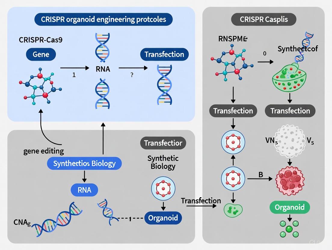
A Comprehensive Guide to CRISPR Organoid Engineering: Protocols, Applications, and Troubleshooting
This article provides a complete roadmap for researchers and drug development professionals seeking to implement CRISPR-based genome editing in human organoid models.
A Comprehensive Guide to CRISPR Organoid Engineering: Protocols, Applications, and Troubleshooting
Abstract
This article provides a complete roadmap for researchers and drug development professionals seeking to implement CRISPR-based genome editing in human organoid models. It covers the foundational principles of why 3D organoids offer superior physiological relevance over traditional 2D cultures for functional genomics. The guide details step-by-step protocols for key techniques—including knockout, interference (CRISPRi), activation (CRISPRa), and knock-in—in various organoid systems like gastric, intestinal, and brain. A strong emphasis is placed on troubleshooting common challenges such as delivery efficiency and mosaicism. Finally, it explores validation strategies and comparative analyses that demonstrate the power of integrated CRISPR-organoid platforms for dissecting gene-drug interactions, modeling cancer, and advancing personalized medicine.
Why CRISPR-Engineered Organoids Are Revolutionizing Disease Modeling
For decades, two-dimensional (2D) cell culture has been the standard workhorse in biological research, enabling foundational discoveries in cell biology, drug development, and disease modeling. However, the inherent limitations of growing cells as flat monolayers on plastic surfaces have become increasingly apparent. These 2D models fail to capture the three-dimensional architectural complexity, cell-cell interactions, and physiological microenvironment of human tissues, leading to poor predictive value for human drug responses and disease mechanisms [1] [2].
The transition to three-dimensional (3D) organoids represents a paradigm shift in experimental biology. Organoids are self-organizing 3D structures derived from stem cells that recapitulate key aspects of native tissue architecture and function. Unlike 2D cultures, organoids preserve tissue-specific stem cell activity, enable multilineage differentiation, and maintain genomic alterations, histology, and pathology of primary tissues [3]. This technological advance has created unprecedented opportunities for studying human development, disease modeling, and drug discovery in systems that genuinely mirror human physiology.
The integration of CRISPR genome editing with 3D organoid models has particularly revolutionized functional genomics, enabling researchers to systematically dissect gene function and gene-drug interactions in physiologically relevant human systems [3] [4]. This combination represents a powerful toolkit for addressing fundamental biological questions and accelerating therapeutic development.
Comparative Analysis: 2D vs. 3D Culture Systems
Table 1: Fundamental differences between 2D and 3D cell culture systems
| Feature | 2D Cell Culture | 3D Organoid Culture |
|---|---|---|
| Growth Pattern | Single layer on flat surfaces [1] | Three-dimensional expansion in all directions [1] |
| Cell-ECM Interaction | Limited, unnatural adhesion [2] | Complex, physiologically relevant ECM interactions [1] |
| Cell-Cell Signaling | Primarily lateral connections | Natural 3D spatial organization and signaling gradients [1] |
| Gene Expression Profiles | Often aberrant due to artificial environment [1] | More in vivo-like gene expression patterns [1] |
| Drug Response Prediction | Often overestimates efficacy [1] | Better predicts in vivo drug responses, including resistance [1] |
| Tissue Architecture | Lacks structural complexity [2] | Mimics organ-specific microarchitecture [3] |
| Throughput & Cost | High-throughput, inexpensive [1] [2] | Medium throughput, higher cost [1] |
| Technical Ease | Simple handling, standardized protocols [1] | More complex culture requirements [5] |
Table 2: Functional outcomes in research applications
| Research Application | 2D Culture Performance | 3D Organoid Performance |
|---|---|---|
| Tumor Modeling | Poor representation of tumor microenvironment [1] | Recapitulates tumor heterogeneity and drug penetration gradients [1] |
| Drug Toxicity Screening | Limited predictive value for human toxicity [6] | Improved prediction of hepatotoxicity and cardiotoxicity [6] [7] |
| Stem Cell Differentiation | Limited differentiation potential | Enhanced differentiation and maturation [1] |
| Personalized Medicine | Limited clinical correlation | Strong correlation with patient drug responses in PDOs [6] |
| High-throughput Screening | Excellent for early-stage compound elimination [1] | Improving with automation and AI integration [8] |
| Gene Editing Efficiency | High efficiency for CRISPR manipulations [1] | More challenging but physiologically more relevant [3] |
The fundamental differences between these systems translate directly to research outcomes. A compelling example comes from cancer research, where 3D tumor organoids maintain the cellular heterogeneity and structural complexity of original tumors, enabling more accurate prediction of drug responses [6]. Similarly, in neurodegenerative disease modeling, 3D midbrain organoids recapitulate key pathological hallmarks of Parkinson's disease—including dopaminergic neuron loss and Lewy body-like formation—that cannot be adequately modeled in 2D systems [9].
CRISPR-Organoid Integration: Technical Protocols
The fusion of CRISPR technologies with 3D organoid cultures has created powerful experimental platforms for functional genomics. Below are detailed protocols for implementing CRISPR screening in gastric organoids, based on established methodologies [3].
Protocol: Large-Scale CRISPR Knockout Screening in Human Gastric Organoids
Principle: This protocol enables genome-wide identification of genes essential for cell growth and drug response using a pooled CRISPR knockout approach in primary human gastric organoids.
Materials & Reagents:
- TP53/APC double knockout (DKO) human gastric organoid line
- Lentiviral Cas9 expression vector
- Pooled lentiviral sgRNA library (e.g., 12,461 sgRNAs targeting 1093 membrane proteins + 750 non-targeting controls)
- Puromycin selection antibiotic
- Advanced DMEM/F12 culture medium
- Recombinant growth factors (EGF, Noggin, R-spondin)
- Matrigel or similar extracellular matrix
- Next-generation sequencing platform
Procedure:
- Stable Cas9 Organoid Generation:
- Lentivirally transduce TP53/APC DKO gastric organoids with Cas9 expression vector.
- Select with appropriate antibiotics for 7-10 days until control cells are eliminated.
- Validate Cas9 activity using GFP reporter assay (should achieve >95% knockout efficiency).
Library Transduction:
- Dissociate organoids to single cells using enzyme-free dissociation buffer.
- Transduce Cas9-expressing organoids with pooled sgRNA lentiviral library at MOI of 0.3-0.5 to ensure majority receive single sgRNA.
- Maintain cellular coverage of >1000 cells per sgRNA throughout the experiment.
- Culture transduced organoids in complete growth medium for 48 hours.
Selection and Expansion:
- Initiate puromycin selection (2-5 μg/mL) 48 hours post-transduction.
- Continue selection for 5-7 days until non-transduced control organoids are eliminated.
- Harvest reference sample (T0) for genomic DNA extraction.
- Culture remaining organoids for 28 days, maintaining >1000x coverage.
Sample Processing and Sequencing:
- Harvest organoids at endpoint (T1) and extract genomic DNA.
- Amplify integrated sgRNA sequences using PCR with barcoded primers.
- Sequence amplified products using next-generation sequencing.
- Quantify sgRNA abundance by mapping sequences to reference library.
Data Analysis:
- Normalize read counts across samples.
- Calculate fold-change (T1/T0) for each sgRNA.
- Use statistical frameworks (e.g., MAGeCK) to identify significantly depleted or enriched sgRNAs.
- Gene-level scores are computed by aggregating signals from multiple sgRNAs per gene.
Troubleshooting Notes:
- Low infection efficiency: Optimize viral titer and include polybrene (4-8 μg/mL).
- Poor organoid viability after transduction: Reduce centrifugation speed during infection.
- Library representation loss: Maintain minimum 1000x coverage throughout culture.
- High false-positive rates: Include sufficient non-targeting control sgRNAs and perform biological replicates.
Protocol: Inducible CRISPRi/a in Gastric Organoids
Principle: This protocol enables targeted gene repression (CRISPRi) or activation (CRISPRa) using doxycycline-inducible dCas9 systems for temporal control of gene expression.
Materials & Reagents:
- TP53/APC DKO gastric organoid line
- Lentiviral rtTA expression vector
- Doxycycline-inducible dCas9-KRAB (for CRISPRi) or dCas9-VPR (for CRISPRa) vectors
- Target-specific sgRNAs (e.g., CXCR4 or SOX2 promoters)
- Doxycycline (1 μg/mL working concentration)
- Flow cytometry antibodies for target validation
Procedure:
- Stable Cell Line Generation:
- Sequential lentiviral transduction: first introduce rtTA, then inducible dCas9 fusion.
- Sort mCherry-positive cells after 48 hours of doxycycline induction to establish stable pools.
- Confirm dCas9 expression by Western blot using anti-Cas9 antibodies.
Gene Expression Modulation:
- Design sgRNAs targeting promoter regions of genes of interest.
- Transduce stable inducible organoids with sgRNA vectors.
- Induce dCas9 activity with doxycycline (1 μg/mL) for 5-7 days.
- For temporal control, withdraw doxycycline to turn off system.
Validation and Phenotyping:
- Analyze target gene expression by qRT-PCR, flow cytometry, or immunostaining.
- For CXCR4 targeting, assess CXCR4-positive population by flow cytometry 5 days post-induction.
- Expected outcomes: CRISPRi should decrease CXCR4-positive population (e.g., from 13.1% to 3.3%), while CRISPRa should increase it (e.g., to 57.6%) [3].
- Monitor organoid growth and morphology for functional phenotypes.
Applications:
- Acute gene function loss without compensation
- Gene essentiality screens in specific contexts
- Modeling dosage-sensitive gene functions
- Dynamic studies of gene expression effects
Advanced Applications and Case Studies
Dissecting Gene-Drug Interactions in Cancer
A landmark application of CRISPR-organoid technology demonstrated how large-scale screening in primary human 3D gastric organoids enables comprehensive dissection of gene-drug interactions [3]. Researchers performed multiple CRISPR modalities—including knockout, interference, activation, and single-cell approaches—to identify genes modulating sensitivity to cisplatin, a common chemotherapy drug.
The screens revealed unexpected connections, including a link between fucosylation pathways and cisplatin sensitivity, and identified TAF6L as a key regulator of cell recovery from cisplatin-induced damage. These findings were enabled by the physiological relevance of the 3D organoid model, which preserved the tissue architecture and cellular heterogeneity of gastric epithelium.
Key Experimental Insights:
- Pooled screens identified 68 significant dropout genes whose disruption caused growth defects
- Pathway enrichment revealed genes involved in transcription, RNA processing, and nucleic acid metabolism
- Tumor suppressor LRIG1 was identified as a top hit whose knockout enhanced proliferation
- Single-cell CRISPR screens resolved how genetic alterations interact with cisplatin at individual cell level
Disease Modeling and Drug Discovery
The application of 3D organoids extends beyond cancer to numerous disease areas. In neurodegenerative disease research, 3D midbrain organoids (MOs) have emerged as transformative tools for modeling Parkinson's disease (PD) [9]. These organoids recapitulate key pathological hallmarks including dopaminergic neuron loss and Lewy body formation, enabling mechanistic studies and drug screening.
Notable Advances:
- MOs with PD-linked mutations (LRRK2, GBA1, DNAJC6) model disease-specific phenotypes
- Optogenetics-assisted α-synuclein aggregation systems enable study of protein pathology
- High-throughput drug testing platforms identify potential neuroprotective compounds
- Successful integration and functional recovery demonstrated in animal PD models
The pharmaceutical industry is increasingly adopting organoid models for preclinical testing. Roche uses 3D tumor spheroids to model hypoxic tumor cores and test immunotherapies, while Memorial Sloan Kettering employs patient-derived organoids to match therapies to drug-resistant pancreatic cancer patients [1].
The Scientist's Toolkit: Essential Research Reagents
Table 3: Key reagents and solutions for CRISPR-organoid research
| Reagent Category | Specific Examples | Function & Application |
|---|---|---|
| CRISPR Systems | Cas9, dCas9-KRAB (CRISPRi), dCas9-VPR (CRISPRa) [3] | Genome editing, gene repression, gene activation |
| Delivery Vectors | Lentiviral sgRNA libraries, Lipid nanoparticles (LNPs) [10] | Efficient delivery of CRISPR components to organoids |
| Extracellular Matrices | Matrigel, synthetic hydrogels, engineered scaffolds [6] [7] | 3D structural support mimicking native tissue microenvironment |
| Culture Media | Advanced DMEM/F12 with tissue-specific growth factors [3] | Support organoid growth and maintenance |
| Selection Agents | Puromycin, Geneticin (G418), Blasticidin | Selection of successfully transduced organoids |
| Induction Systems | Doxycycline-inducible cassettes, rtTA [3] | Temporal control of CRISPR activity |
| Analysis Tools | Single-cell RNA sequencing, HCS-3DX AI imaging [8] | High-content screening and analysis at single-cell resolution |
| Specialized Equipment | CERO 3D bioreactor, OrganoPlate, microfluidic chips [7] [5] | Scalable, reproducible organoid culture systems |
Current Challenges and Future Directions
Despite the considerable promise of CRISPR-engineered organoids, several challenges remain. Batch-to-batch variability, limited scalability, and incomplete microenvironmental complexity can undermine reliability and translational potential [6] [5]. The absence of vascularization in most current organoid models restricts nutrient delivery and organoid size, while the fetal phenotype of iPSC-derived organoids may limit their utility for modeling adult diseases [5].
The "Organoid Plus and Minus" framework has emerged as a strategic approach to address these limitations [6]. This integrated strategy combines technological augmentation ("Plus") with culture system refinement ("Minus") to improve screening accuracy, throughput, and physiological relevance.
Key Future Developments:
- Vascularization strategies: Co-culture with endothelial cells to create perfusable networks
- Immune system integration: Incorporation of microglia and other immune components
- Automation and AI: HCS-3DX and other automated systems for reproducible high-content screening [8]
- Organ-on-chip integration: Combining organoids with microfluidic platforms for enhanced physiological relevance [5]
- Multi-omics characterization: Comprehensive molecular profiling to validate model fidelity
The regulatory landscape is also evolving to accommodate these advanced models. The FDA's recent policy shift phasing out traditional animal testing in favor of human-relevant systems like organoids for drug safety evaluation signals growing acceptance of these technologies [6]. This regulatory transformation, combined with ongoing technical innovations, positions CRISPR-engineered organoids as cornerstone platforms for personalized drug discovery and therapeutic optimization in the coming years.
The convergence of CRISPR genome engineering with 3D organoid technology represents a transformative advance in biomedical research, enabling the creation of highly physiologically relevant human disease models. Organoids, which are three-dimensional in vitro cultures derived from adult stem cells (ASCs) or induced pluripotent stem cells (iPSCs), replicate the structural and functional complexity of native tissues [11]. When combined with the precision of CRISPR-based genetic perturbations, researchers can generate isogenic disease models to elucidate the functional impact of genetic variants in a human tissue context [12]. This powerful synergy allows for systematic dissection of gene function, drug mechanisms, and therapeutic vulnerabilities in human tissue environments that were previously inaccessible.
The expanding CRISPR toolkit now extends far beyond simple gene knockout, encompassing CRISPR interference (CRISPRi) for transcriptional repression and CRISPR activation (CRISPRa) for gene activation, in addition to base editing and prime editing technologies [3] [13]. These tools are revolutionizing functional genomics in organoid models, particularly through large-scale pooled screens that identify genes influencing disease processes and drug responses [3]. This application note provides a comprehensive overview of the current CRISPR toolkit for organoid engineering, with detailed protocols and experimental frameworks for implementing these technologies in a research setting.
The Expanded CRISPR Toolkit: Mechanisms and Applications
The CRISPR toolkit has evolved to encompass diverse functionalities for precise genetic manipulation, each with distinct mechanisms and applications in organoid research. The table below summarizes the key CRISPR technologies and their primary applications in organoid engineering.
Table 1: CRISPR Technologies and Their Applications in Organoid Research
| Technology | Cas Enzyme | Mechanism of Action | Primary Applications in Organoids | Key Advantages |
|---|---|---|---|---|
| CRISPR Knockout | Cas9, Cas9 nickase | Creates double-strand breaks (DSBs) repaired by error-prone NHEJ | Generating loss-of-function mutations; creating isogenic disease models [12] | Permanent gene disruption; well-established protocols |
| CRISPRi | dCas9-KRAB fusion | Blocks transcription initiation/elongation without DNA cleavage [3] | Reversible gene silencing; studying essential genes [3] | No DNA damage; tunable repression; fewer off-target effects |
| CRISPRa | dCas9-VPR fusion | Recruits transcriptional activators to gene promoters [3] | Gene activation; studying gene dosage effects | Precise transcriptional activation without DSBs |
| Base Editing | Base editor (dCas9 or Cas9 nickase fused to deaminase) | Directly converts one DNA base to another without DSBs [13] | Introducing point mutations; disease modeling | High efficiency; minimal indel formation |
| Prime Editing | Cas9 nickase-reverse transcriptase fusion | Uses pegRNA to directly copy edited sequence into genome [13] | Precise insertions, deletions, and point mutations | Versatile; no DSBs; wider editing window than base editors |
Experimental Workflow for CRISPR Screening in Organoids
The following diagram illustrates the generalized workflow for conducting CRISPR screens in organoid models, from establishment to hit validation:
Figure 1: Generalized workflow for CRISPR screening in organoid models, adapted from large-scale screening approaches [3] [11].
Detailed Experimental Protocols
Protocol 1: Generating Isogenic Disease Models in Organoids Using Base and Prime Editors
This protocol describes the creation of precise genetic variants in organoids using next-generation CRISPR tools that avoid double-strand breaks, enabling modeling of genetic diseases with higher efficiency and reduced cellular toxicity [12].
sgRNA Design and Cloning
- Design sgRNAs targeting the genomic region of interest using computational tools, considering the editing window of base editors (typically 4-8 nucleotides upstream of PAM for cytosine base editors and 5-10 nucleotides for adenine base editors) or the positioning requirements for prime editing guide RNAs (pegRNAs) [13].
- For base editing: Select sgRNAs with the target base within the optimal activity window for the specific base editor being used.
- For prime editing: Design pegRNAs with reverse transcriptase templates encoding the desired edit and appropriate primer binding sites.
- Clone sgRNAs into appropriate expression vectors using restriction-ligation cloning or Golden Gate assembly. For pooled screens, clone sgRNA libraries into lentiviral vectors with appropriate selection markers.
Electroporation-based Transfection
- Culture organoids to 60-80% confluence in Matrigel or similar extracellular matrix.
- Dissociate organoids into single cells using enzyme-free dissociation reagents or gentle enzymatic digestion.
- Prepare electroporation mixture containing:
- 1-5 µg base editor or prime editor mRNA/protein
- 500 ng-1 µg sgRNA expression plasmid or synthetic sgRNA
- 1-2 million dissociated organoid cells in electroporation buffer
- Electroporate using optimized parameters for your organoid type (typical conditions: 1200-1500 V, 20 ms pulse width for 2-3 pulses).
- Plate transfected cells in fresh Matrigel with organoid culture medium and allow to recover for 48 hours.
Selection and Clonal Expansion
- Initiate antibiotic selection 72 hours post-transfection if using plasmid-based systems with selection markers (e.g., puromycin at 1-5 µg/mL for 5-7 days).
- For fluorescence-activated cell sorting (FACS), use co-expressed fluorescent markers (e.g., GFP) to isolate successfully transfected cells 96 hours post-transfection.
- Plate sorted/selected cells at clonal density (500-1000 cells per well in 96-well plates) in Matrigel with organoid growth medium.
- Expand individual clones for 2-3 weeks, refreshing medium every 2-3 days.
Validation of Genome Editing
- Extract genomic DNA from expanded clonal organoid lines using standard protocols.
- Amplify target region by PCR using flanking primers.
- Validate edits by Sanger sequencing and tracking of indels by decomposition (TIDE) analysis for base editors or specific allele-PCR for prime editors.
- For comprehensive characterization, perform additional validation such as:
- Western blotting to confirm protein-level changes
- RT-qPCR to assess transcriptional effects
- Functional assays relevant to the specific genetic variant
Protocol 2: Establishing Inducible CRISPRi and CRISPRa Systems in Gastric Organoids
This protocol details the implementation of inducible CRISPR interference and activation systems in human gastric organoids, enabling temporal control of gene expression for studying dynamic biological processes [3].
Generation of Stable dCas9-Expressing Organoid Lines
- Engineer TP53/APC double knockout (DKO) gastric organoid line to provide a homogeneous genetic background with minimal variability [3].
- Generate organoid lines expressing rtTA using lentiviral transduction followed by antibiotic selection.
- Introduce doxycycline-inducible dCas9 fusion protein:
- For CRISPRi: dCas9-KRAB (transcriptional repressor)
- For CRISPRa: dCas9-VPR (VP64-p65-Rta transcriptional activator)
- Transduce with lentiviral vectors containing the inducible dCas9 cassette and mCherry reporter.
- Sort mCherry-positive cells by FACS after doxycycline induction (1 µg/mL for 72 hours) to establish stable iCRISPRi and iCRISPRa organoid lines.
- Confirm expression of dCas9-KRAB or dCas9-VPR by Western blotting using anti-Cas9 antibodies.
Testing System Functionality
- Design sgRNAs targeting gene promoters of control genes (e.g., CXCR4, SOX2) with known expression patterns in gastric organoids.
- Transduce stable dCas9 organoid lines with lentiviral sgRNA vectors.
- Induce dCas9 expression with doxycycline (1 µg/mL) for 5-7 days.
- Analyze gene expression changes:
- For surface markers like CXCR4: Use antibody staining and flow cytometry
- For intracellular proteins: Perform immunofluorescence or Western blotting
- For transcript levels: Conduct RT-qPCR or RNA-seq
- Expected outcomes: iCRISPRi-sgCXCR4 organoids should show decreased CXCR4-positive population (e.g., from 13.1% to 3.3%), while iCRISPRa-sgCXCR4 should show increased positive population (e.g., to 57.6%) [3].
Protocol 3: Large-Scale Pooled CRISPR Screening in 3D Gastric Organoids
This protocol enables genome-wide functional genetic screens in organoids to identify genes involved in specific biological processes or drug responses, as demonstrated in gastric cancer organoids treated with cisplatin [3].
Screening Setup and Library Delivery
- Select appropriate CRISPR library based on screening goal:
- Knockout: Cas9-based sgRNA library
- CRISPRi: dCas9-KRAB-compatible sgRNA library targeting promoters
- CRISPRa: dCas9-VPR-compatible sgRNA library targeting promoters
- Validate Cas9 activity in organoid line using GFP reporter assay (≥95% GFP-negative cells indicates robust Cas9 activity) [3].
- Transduce organoids with pooled lentiviral library at low MOI (MOI=0.3-0.5) to ensure most cells receive single sgRNA.
- Maintain cellular coverage of >1000 cells per sgRNA throughout the screening process to maintain library representation.
- Harvest reference sample 2 days post-puromycin selection (T0) for baseline sgRNA distribution.
Selection and Phenotypic Interrogation
- Apply selective pressure relevant to biological question:
- For gene-drug interaction studies: Add drug (e.g., cisplatin for gastric cancer screens) at appropriate concentration
- For essential gene identification: Culture without selective pressure and identify dropout sgRNAs
- For fitness genes: Monitor growth over 2-4 weeks
- Maintain consistent cellular coverage (>1000 cells per sgRNA) throughout selection period.
- Harvest endpoint sample (T1) after 14-28 days of selection, depending on experimental design.
Sequencing and Hit Identification
- Extract genomic DNA from T0 and T1 samples using scalable methods.
- Amplify sgRNA regions by PCR with barcoded primers for multiplexing.
- Sequence amplified libraries using next-generation sequencing (Illumina platforms).
- Quantify sgRNA abundance by counting reads mapping to each sgRNA in the library.
- Calculate phenotype scores for each gene by comparing sgRNA abundance between T0 and T1 samples, normalized to control sgRNAs.
- Identify significant hits using statistical frameworks (e.g., MAGeCK, RSA) with thresholds for significance (e.g., FDR < 0.05).
Essential Controls and Validation Strategies
Proper experimental controls are critical for interpreting CRISPR screening results and validating candidate hits. The table below outlines essential controls for different types of CRISPR experiments in organoids.
Table 2: Essential Controls for CRISPR Experiments in Organoids
| Control Type | Composition | Purpose | Expected Outcome |
|---|---|---|---|
| Transfection Control | Fluorescent reporter (GFP mRNA/plasmid) | Assess delivery efficiency | Visual confirmation of successful transfection |
| Positive Editing Control | Validated sgRNA with known high efficiency (e.g., targeting TRAC, RELA) [14] | Verify editing capability under optimized conditions | High editing efficiency in target gene |
| Negative Editing Control (Scramble) | Scramble sgRNA + Cas nuclease [14] | Establish baseline for non-specific effects | No specific editing; phenotype similar to wildtype |
| Negative Editing Control (Guide Only) | sgRNA without Cas nuclease [14] | Control for sgRNA-specific off-target effects | No editing; identifies sgRNA toxicity |
| Negative Editing Control (Cas Only) | Cas nuclease without sgRNA [14] | Control for Cas nuclease toxicity | No specific editing; identifies Cas toxicity |
| Mock Control | Cells undergoing transfection with no nucleases or guides [14] | Control for transfection stress | Phenotype similar to wildtype |
Hit Validation Strategies
- Individual sgRNA validation: Test 2-3 independent sgRNAs targeting significant hit genes in arrayed format to confirm phenotype reproducibility [3].
- Rescue experiments: For CRISPRi/CRISPRa hits, demonstrate that phenotype is reversible by withdrawing doxycycline to turn off dCas9 activity.
- Multi-modal validation: Combine complementary approaches (e.g., CRISPR knockout with RNAi) to confirm phenotype is not technology-specific.
- Functional assays: Perform pathway-specific functional assays relevant to the screened phenotype (e.g., DNA repair assays for cisplatin sensitivity hits).
Research Reagent Solutions
Successful implementation of CRISPR tools in organoid research requires carefully selected reagents and delivery systems. The following table outlines key solutions for establishing CRISPR-organoid workflows.
Table 3: Essential Research Reagents for CRISPR-Organoid Experiments
| Reagent Category | Specific Examples | Function | Application Notes |
|---|---|---|---|
| CRISPR Nucleases | Cas9, Base editors, Prime editors, dCas9-KRAB, dCas9-VPR | Genome editing, transcriptional regulation | Select based on desired genetic manipulation (see Table 1) |
| Delivery Vectors | Lentiviral vectors, Electroporation systems, Lipid nanoparticles (LNPs) | Deliver CRISPR components into cells | Lentiviruses for stable integration; electroporation for transient expression [12] |
| Organoid Culture Matrix | Matrigel, Synthetic hydrogels | Provide 3D extracellular environment for organoid growth | Matrigel is most common; synthetic alternatives improve reproducibility |
| Selection Markers | Puromycin, Blasticidin, Fluorescent proteins (GFP, mCherry) | Enumerate and select successfully transfected cells | Antibiotic resistance for stable lines; fluorescent markers for FACS |
| sgRNA Libraries | Genome-wide knockout, CRISPRi, CRISPRa libraries | Enable large-scale genetic screens | Ensure high coverage (>1000 cells/sgRNA) and library representation [3] |
| Validation Tools | Sanger sequencing, Next-generation sequencing, ICE analysis | Verify editing efficiency and specificity | ICE analysis for quantifying editing efficiency from Sanger data [14] |
The integration of advanced CRISPR technologies with 3D organoid models has created a powerful platform for human disease modeling and functional genomics. The protocols and frameworks presented here provide researchers with comprehensive guidance for implementing these tools, from generating precise isogenic models to conducting genome-wide screens. As both technologies continue to evolve, their combination will undoubtedly yield deeper insights into human biology and disease mechanisms, accelerating the development of novel therapeutic strategies. The key to success lies in careful experimental design, appropriate control selection, and rigorous validation of genetic perturbations and their phenotypic consequences.
Organoid biobanks represent a transformative resource in biomedical research, enabling the study of human development, disease modeling, and high-throughput drug screening. The fundamental choice researchers face is between patient-derived organoid (PDO) biobanks, which preserve native human genetic and phenotypic diversity, and engineered organoid biobanks, which offer defined genetic backgrounds and tailored modifications for specific research questions [15] [16]. Patient-derived organoids are three-dimensional (3D) cell culture systems derived directly from patient tumor tissue that retain the genetic variability and phenotypic diversity of the primary tumor, effectively recapitulating the structural, functional, and heterogeneous characteristics of original tissues [15]. In contrast, engineered organoid biobanks utilize genetic engineering tools like CRISPR-Cas9 to introduce specific mutations into pluripotent or adult stem cells, generating genetically defined models for systematic investigation of gene function and disease mechanisms [17] [16]. This Application Note provides a structured comparison of these complementary approaches and detailed protocols for their establishment and application within CRISPR organoid engineering research.
Comparative Analysis: PDO vs. Engineered Organoid Biobanks
The selection between patient-derived and engineered organoid models should be guided by research objectives, required throughput, and available resources. Each approach offers distinct advantages and limitations, which are summarized in Table 1 below.
Table 1: Key Characteristics of Patient-Derived vs. Engineered Organoid Biobanks
| Characteristic | Patient-Derived Organoid (PDO) Biobanks | Engineered Organoid Biobanks |
|---|---|---|
| Genetic Background | Native patient genetics; preserves tumor heterogeneity [15] | Defined genetic background; enables isogenic controls [17] |
| Primary Applications | Drug screening, personalized medicine, studying tumor heterogeneity [15] | Functional genomics, disease mechanism studies, gene function validation [17] [16] |
| Development Timeline | 2-3 weeks for establishment [15] | 2-4 months including genetic engineering [18] |
| Throughput Potential | Medium; limited by patient sample availability | High; scalable from single stem cell lines |
| Technical Complexity | Medium; requires optimization of culture conditions [15] | High; requires expertise in genetic engineering [17] |
| Representative Examples | Colorectal, pancreatic, breast cancer PDOs [15] | CRISPR-engineered fetal brain organoids, intestinal organoid knockout biobanks [18] [17] |
Establishment Protocols and Methodologies
Protocol for Patient-Derived Organoid Biobank Development
Principle: Generate organoids directly from patient tissue samples while preserving original tissue architecture and genetic heterogeneity.
Workflow:
- Tissue Collection and Processing: Obtain fresh tumor tissue via biopsy or surgical resection. Mechanically dissociate and enzymatically digest tissue into small fragments or single cells [15].
- Extracellular Matrix Embedding: Suspend tissue fragments in Basement Membrane Extract (e.g., Matrigel) and plate as domes. Polymerize at 37°C for 30-60 minutes [15].
- Organoid Culture Medium: Overlay with defined medium containing:
- Essential growth factors (EGF, Noggin, R-spondin)
- Tissue-specific patterning factors
- Wnt pathway agonists for stem cell maintenance
- Antibiotic/Antimycotic solution [15]
- Expansion and Biobanking: Culture at 37°C with 5% CO₂, passaging every 1-2 weeks via mechanical disruption and re-embedding. Cryopreserve in freezing medium containing 10% DMSO and serum alternatives [15].
Quality Control: Validate organoids through genomic sequencing, histology, and immunostaining to confirm retention of original tissue characteristics [19].
Protocol for CRISPR-Engineered Organoid Biobank Development
Principle: Introduce specific genetic modifications into stem cell-derived organoids using CRISPR-Cas9 technology for controlled functional studies.
Workflow:
- Stem Cell Culture: Maintain human pluripotent stem cells (hPSCs) or adult stem cells in feeder-free conditions with daily medium changes [18].
- CRISPR-Cas9 Design: Design and validate sgRNAs targeting genes of interest. Select Cas9 system (wild-type, nickase, or dead Cas9) based on desired edit type [17].
- Delivery and Transfection: Deliver CRISPR components via electroporation or lipofection. Use fluorescence-activated cell sorting (FACS) to isolate successfully transfected cells based on reporter expression (e.g., EGFP) [17].
- Clonal Selection and Expansion: Plate single cells and expand into clonal organoid lines. Screen for desired mutations via sequencing and functional assays [17].
- Biobank Curation: Cryopreserve validated clones with detailed genetic characterization. Establish 2-6 mutated clonal lines per target gene to account for potential clonal variation [17].
Validation: Confirm genetic modifications through Sanger sequencing, functional assays, and western blotting. Verify absence of off-target effects through whole-genome sequencing where necessary [17].
Figure 1: CRISPR-engineered organoid biobank development workflow.
Integrated Research Applications
Functional Genomics through CRISPR Screening in PDOs
Principle: Combine the genetic diversity of PDOs with systematic CRISPR screening to identify patient-specific genetic vulnerabilities and therapeutic targets [15].
Workflow:
- Library Design: Select CRISPR knockout, activation, or base-editing libraries targeting genes of interest (e.g., cancer driver genes, drug targets).
- Viral Transduction: Package sgRNA library into lentiviral vectors. Transduce PDO cultures at low MOI to ensure single integration events.
- Selection and Expansion: Apply selection pressure (e.g., antibiotics, drug treatment) and expand organoids for 2-3 weeks.
- Genomic DNA Extraction and Sequencing: Harvest organoids, extract gDNA, amplify integrated sgRNAs, and sequence via next-generation sequencing.
- Bioinformatic Analysis: Identify enriched or depleted sgRNAs using specialized algorithms (e.g., MAGeCK, BAGEL) to reveal essential genes under experimental conditions [15].
Application Example: CRISPR screens in colorectal cancer PDOs have identified novel genetic dependencies and mechanisms of resistance to targeted therapies, highlighting pathways not revealed in conventional 2D models [15].
Tumor Microenvironment and Immunotherapy Modeling
Principle: Engineer PDOs to include stromal and immune components for studying tumor-immune interactions and immunotherapy responses.
Workflow:
- Stromal Co-culture: Isolate cancer-associated fibroblasts (CAFs) from patient tissue and co-culture with matched PDOs in 3D matrices.
- Immune Cell Integration: Expand autologous tumor-infiltrating lymphocytes (TILs) or engineer immune cells (e.g., CAR-T cells) for incorporation into PDO cultures.
- Treatment and Monitoring: Treat co-cultures with immunotherapeutic agents (e.g., immune checkpoint inhibitors) and monitor tumor cell killing via live-cell imaging [15].
- Analysis: Quantify immune cell-mediated organoid killing, cytokine production, and changes in immune cell phenotypes [15].
Figure 2: Tumor microenvironment modeling with PDO co-cultures.
Essential Research Reagents and Tools
Table 2: Key Research Reagent Solutions for Organoid Biobanking
| Reagent Category | Specific Examples | Function and Application |
|---|---|---|
| Extracellular Matrices | Matrigel, Basement Membrane Extract | Provide 3D scaffold for organoid growth and polarization [15] |
| Growth Factors | EGF, Noggin, R-spondin, Wnt-3a | Maintain stemness and support tissue-specific differentiation [15] |
| CRISPR Components | Cas9 nucleases, sgRNA libraries, HDR templates | Enable precise genetic editing for engineered biobanks [17] |
| Delivery Systems | Lentiviral vectors, electroporation systems | Facilitate efficient introduction of genetic elements [17] |
| Cell Culture Supplements | B-27, N-2, N-acetylcysteine | Support organoid growth and viability in defined media [15] |
| Analysis Tools | CrisprVi software, single-cell RNA seq | Enable visualization and analysis of CRISPR and sequencing data [20] |
Analytical Methods and Data Interpretation
Quantitative Analysis of Organoid Drug Responses
Principle: Employ high-content imaging and computational analysis to quantify organoid-level and cell-level responses to therapeutic interventions.
Methodology:
- Live-Cell Imaging: Label organoids with nuclear markers (e.g., H2B-GFP) and vital dyes (e.g., DRAQ7) to track cell birth and death events dynamically [19].
- Morphometric Analysis: Quantify organoid volume, sphericity, and ellipticity to assess growth patterns and structural changes [19].
- Dose-Response Modeling: Calculate IC₅₀ values and growth inhibition percentages from longitudinal size measurements.
- Response Classification: Differentiate cytotoxic vs. cytostatic effects based on live cell counts and death rates over time [19].
Data Interpretation: Organoid volume strongly correlates with live cell number, enabling both parameters as reliable metrics for dose-response studies. Morphological heterogeneity within and between patients can be quantified through sphericity and ellipticity indices [19].
Genetic Validation and Visualization
Principle: Implement computational tools for verification and visualization of genetic modifications in engineered organoid biobanks.
Methodology:
- CRISPR Array Analysis: Use specialized software (e.g., CrisprVi) to visualize and analyze CRISPR direct repeats and spacers [20].
- Sequence Alignment: Conduct multiple sequence alignment of spacer arrays to verify intended modifications.
- Consensus Sequence Finding: Identify consensus sequences of direct repeats and spacers across modified organoid lines using BLAST-based clustering [20].
- Variant Calling: Detect on-target and potential off-target edits through whole-genome or targeted sequencing.
Application: CrisprVi provides graphical representation of CRISPR loci, statistical analysis of direct repeats and spacers, and heatmap visualization of consensus sequences across multiple engineered organoid lines [20].
The strategic selection between patient-derived and engineered organoid biobanks should be guided by specific research goals. PDO biobanks offer unparalleled clinical relevance for personalized medicine and drug screening applications, preserving native genetic heterogeneity and tumor microenvironment interactions [15]. Engineered organoid biobanks provide powerful platforms for functional genomics and mechanistic studies, enabling systematic investigation of gene function in defined genetic backgrounds [17] [16]. The integration of these approaches through CRISPR engineering of PDOs represents the cutting edge of organoid research, combining physiological relevance with genetic tractability to advance precision oncology and therapeutic development [15]. As these technologies continue to evolve, they will increasingly bridge the gap between in vitro models and clinical applications, accelerating the development of personalized cancer therapies and our understanding of human disease mechanisms.
Application Notes
CRISPR-engineered organoids represent a transformative platform that bridges the gap between traditional 2D cell cultures and in vivo models. By combining the physiological relevance of three-dimensional tissue structures with precise genome editing, these models enable unprecedented investigation of disease mechanisms, drug responses, and personalized therapeutic strategies [4] [11].
Table 1: Key Applications of CRISPR-Engineered Organoids
| Application Domain | Specific Uses | Key Advantages | Representative Examples |
|---|---|---|---|
| Personalized Medicine | • Modeling patient-specific genetic variants• Ex vivo therapeutic testing• Predicting individual drug response | • Preserves patient genomic background• Enables "clinical trials in a dish"• Recapitulates tumor microenvironment | • Gastric cancer organoids for cisplatin response testing [3]• Biobanks of patient-derived tumoroids [11] |
| Cancer Research | • Functional genomics screens• Oncogene/tumor suppressor validation• Tumor evolution studies | • Identifies gene-drug interactions• Models cancer hallmarks in 3D context• Reveals therapeutic vulnerabilities | • Genome-wide CRISPR screens in gastric organoids [3]• PTEN knockout modeling in colorectal cancer [21] |
| Drug Development | • Target identification & validation• Preclinical efficacy & toxicity testing• Mechanism of action studies | • More predictive than 2D models• Reduces reliance on animal models• High-throughput compatible | • Identification of TAF6L in cisplatin response [3]• Drug screening in colorectal carcinoma organoids [11] |
Advancing Personalized Medicine
The integration of CRISPR with patient-derived organoids (PDOs) enables the creation of bespoke disease models. These isogenic models allow researchers to study the specific impact of individual genetic variants against a consistent genetic background, pinpointing causal mutations and their functional consequences [22]. This approach is particularly valuable for assessing inter-patient variability in drug response, a technique known as pharmacotyping, which has been successfully demonstrated in pancreatic, ovarian, and colorectal cancer organoids [11]. The ability to maintain the original patient's genomic context while introducing specific edits makes these models powerful tools for predicting individual therapeutic outcomes.
Transforming Cancer Research
CRISPR-engineered organoids have significantly advanced cancer research by enabling systematic investigation of oncogenic processes in a physiologically relevant context. Large-scale CRISPR screens in 3D gastric organoids have identified novel genes modulating chemotherapy response, uncovering previously unappreciated connections such as the link between fucosylation and cisplatin sensitivity [3]. The use of complex CRISPR tools—including knockout, interference (CRISPRi), activation (CRISPRa), and single-cell approaches—in organoid models provides comprehensive insights into gene-drug interactions that were previously inaccessible using conventional models [3].
Streamlining Drug Development
The pharmaceutical industry benefits from CRISPR-engineered organoids through improved target validation and more predictive preclinical testing. Organoid models demonstrate higher clinical translatability compared to 2D cell lines, better recapitulating therapeutic vulnerabilities observed in patients [3] [4]. Recent regulatory changes, including the FDA's updated stance on animal testing requirements, have accelerated the adoption of these human-based models in drug development pipelines [4]. The high editing efficiencies achieved with non-viral RNP-based methods (up to 98%) enable rapid generation of engineered models for functional studies without the need for clonal selection, significantly reducing development timelines [21].
Experimental Protocols
Protocol: Large-Scale CRISPR Screening in Human Gastric Organoids
This protocol enables genome-wide CRISPR screening in primary human 3D gastric organoids to systematically identify genes affecting drug sensitivity, as demonstrated in recent studies investigating cisplatin response [3].
Materials
- Primary human gastric organoids (TP53/APC DKO model)
- Lentiviral Cas9 expression vector
- Pooled lentiviral sgRNA library (e.g., 12,461 sgRNAs targeting 1093 genes + 750 non-targeting controls)
- Puromycin selection antibiotic
- Cisplatin (or other chemotherapeutic agent for modulation studies)
- Next-generation sequencing platform
Methodology
1. Cas9-Expressing Organoid Line Generation
- Generate stable Cas9-expressing TP53/APC double knockout (DKO) gastric organoids using lentiviral transduction.
- Validate Cas9 activity through GFP reporter assay (>95% knockout efficiency recommended).
2. Pooled Library Transduction
- Transduce Cas9-expressing organoids with pooled lentiviral sgRNA library at MOI ensuring >1000 cells per sgRNA.
- Maintain cellular coverage >1000 cells per sgRNA throughout screening.
- Initiate puromycin selection 48 hours post-transduction; continue for 5-7 days.
3. Experimental Timeline & Selection
- Harvest reference sample (T0) 2 days post-selection for baseline sgRNA distribution.
- Culture remaining organoids under experimental conditions (e.g., cisplatin treatment) for 28 days (T1).
- Maintain consistent cellular coverage throughout culture period.
4. Analysis & Hit Identification
- Extract genomic DNA from T0 and T1 organoid populations.
- Amplify integrated sgRNA sequences and perform next-generation sequencing.
- Calculate sgRNA abundance changes between T0 and T1 using specialized analysis pipelines.
- Identify significantly depleted or enriched sgRNAs (compared to non-targeting controls) indicating growth disadvantages or advantages under selection.
5. Validation
- Select top hits for independent validation using individual sgRNAs.
- Confirm phenotype recapitulation in separate experiments.
- Perform downstream functional studies to characterize mechanism.
Protocol: High-Efficiency RNP-Based Editing of Human Intestinal Organoids
This non-viral protocol achieves up to 98% editing efficiency in human intestinal organoids using ribonucleoprotein (RNP) complexes, eliminating the need for clonal selection [21].
Materials
- Human intestinal organoids (adult or fetal-derived)
- Recombinant Cas9 protein
- Synthetic sgRNAs (designed using Benchling/Indelphi)
- Electroporation system (Lonza Nucleofector recommended)
- P3 Primary Cell Solution (Lonza)
- Matrigel or similar extracellular matrix
- Intestinal organoid culture medium with growth factors
Methodology
1. Guide RNA Design & Validation
- Design 3 sgRNAs per target gene targeting early exons.
- Select guides with on-target score >40, off-target score >80, frameshifting score >80.
- Pre-test guides in cell lines and analyze editing efficiency using ICE analysis.
2. Organoid Preparation & Dissociation
- Culture intestinal organoids to appropriate size (~100-200μm diameter).
- Dissociate organoids to single cells using enzymatic digestion.
- Count cells and aliquot 100,000 cells per electroporation condition.
3. RNP Complex Formation
- Complex recombinant Cas9 protein (5μg) with synthetic sgRNA (100pmol).
- Incubate at room temperature for 10-15 minutes to form RNP complexes.
4. Electroporation
- Use Lonza Nucleofector with DS-138 program and P3 buffer.
- Electroporate RNP complexes with 100,000 cells per condition.
- Immediately transfer cells to pre-warmed recovery medium.
5. Organoid Reformation & Analysis
- Embed transfected cells in Matrigel and culture with intestinal organoid medium.
- Analyze editing efficiency 3-7 days post-electroporation using ICE or NGS.
- Validate protein knockout by Western blot, immunohistochemistry, or WesTM.
6. Functional Characterization
- Assess phenotypic consequences (e.g., budding frequency, proliferation rates).
- Perform transcriptional profiling for pathway analysis.
- Validate functional outcomes (e.g., p-AKT elevation in PTEN KO).
Table 2: Research Reagent Solutions for CRISPR Organoid Engineering
| Reagent Type | Specific Examples | Function & Application | Technical Notes |
|---|---|---|---|
| CRISPR Editors | • SpCas9 (wild-type)• dCas9-KRAB (CRISPRi)• dCas9-VPR (CRISPRa)• Base editors (ABE, CBE)• Prime editors | • Gene knockout, repression, or activation• Single-nucleotide editing without DSBs• Diverse editing modalities for different applications | • Use SpCas9 for NGG PAM sites [22]• Inducible systems enable temporal control [3]• Base editors preferred for point mutations [22] |
| Delivery Systems | • Lentiviral vectors• Electroporation (RNP)• Lipofection | • Stable integration for long-term expression• High efficiency with minimal off-target effects• Alternative non-viral method | • RNP achieves >95% efficiency in intestinal organoids [21]• Viral methods enable pooled library screens [3] |
| Organoid Culture | • Matrigel/ECM substitutes• Growth factor cocktails• R-spondin, EGF, Noggin, Wnt3a | • Provides 3D structural support• Maintains stem cell niche signaling• Essential for organoid growth and differentiation | • Composition critical for phenotype retention [11]• Growth factor independence can enable selection [22] |
| Selection Tools | • Antibiotic resistance (puromycin)• Fluorescent reporters (GFP/mCherry)• Growth factor independence | • Enriches for successfully edited cells• Enables tracking and sorting of edited populations• Functional selection based on edited phenotype | • FACS sorting for inducible systems [3]• -WNT/Rspo1 selection for APC KO [22] |
Advanced Methodologies
Inducible CRISPR Systems for Temporal Control
The development of inducible CRISPR systems (iCRISPR) enables precise temporal control over gene expression in organoids. These systems utilize doxycycline-inducible dCas9-KRAB (CRISPRi) or dCas9-VPR (CRISPRa) constructs for reversible gene repression or activation [3]. The tight regulation of these systems allows investigators to study gene function at specific developmental timepoints or to model the sequential acquisition of mutations, mirroring the natural progression of diseases like cancer.
Next-Generation CRISPR Tools for Precision Editing
Beyond conventional CRISPR-Cas9, base editors and prime editors offer more precise genome engineering capabilities without introducing double-strand breaks (DSBs) [22]. These tools are particularly valuable for introducing specific patient-derived point mutations or for correcting pathogenic variants in disease modeling. The editing workflow follows a strategic approach: selection of the appropriate editor based on the desired nucleotide change, careful sgRNA design to maximize efficiency, and delivery via electroporation for optimal results in organoid systems [22].
Step-by-Step Protocols for Successful CRISPR Editing in Organoids
The integration of CRISPR-based genome editing with three-dimensional (3D) organoid culture represents a transformative approach in biomedical research, enabling the development of highly physiologically relevant human disease models. Organoids are in vitro 3D cultures derived from pluripotent or adult stem cells that self-organize to recapitulate the structural, genetic, and functional characteristics of native organs [11]. The essence of a successful organoid system lies in its ability to replicate the in vivo tissue environment through presence of heterogeneous cell populations and mechanical connections with adjacent cells and the intercellular matrix [11]. When combined with CRISPR screening technologies, organoids become powerful platforms for investigating gene function, oncogenic vulnerabilities, developmental pathways, and therapeutic responses in a human physiological context [11] [3]. The fidelity of these models, however, is critically dependent on the optimization of culture conditions, extracellular matrix (ECM) scaffolds, and growth media formulations, which together provide the necessary biochemical and biophysical cues to support stem cell maintenance, differentiation, and 3D organization.
Critical Components for Organoid Culture
Extracellular Matrix (ECM) Scaffolds
The ECM serves as the fundamental scaffold for 3D organoid growth, providing structural support and essential biochemical signals that regulate cell behavior, including proliferation, differentiation, and spatial organization.
- Matrigel: This reconstituted basement membrane extract derived from Engelbreth-Holm-Swarm (EHS) mouse sarcomas remains the gold standard ECM for many organoid cultures. It is rich in laminin, collagen IV, and other ECM proteins, along with growth factors that support cell adhesion and morphogenesis [11] [23]. Despite its widespread use, Matrigel has significant limitations: its murine origin and tumor derivation raise translational concerns, its complex and undefined composition leads to batch-to-batch variability, and its mechanical properties are suboptimal for certain tissues like bone [23] [24] [25].
- Animal-Free Alternatives: Recent advances have focused on developing defined, synthetic, or human-derived matrices to overcome Matrigel's limitations, enhancing reproducibility and clinical applicability.
- Fibrin-Based Hydrogels: Composed of fibrinogen and thrombin, these hydrogels offer excellent biocompatibility, support angiogenic sprouting, and are particularly effective for vascular organoid culture [23].
- PIC-Invasin Gel: A fully synthetic alternative combining polyisocyanopeptide (PIC) with the bacterial protein invasin supports long-term 3D growth of various human and mouse organoids without animal components [26].
- Vitronectin: This recombinant human protein serves as an effective xeno-free substrate for 2D culture of induced pluripotent stem cells (iPSCs) prior to 3D differentiation, maintaining pluripotency and supporting subsequent vascular organoid formation [23].
Growth Media and Signaling Molecules
Precisely formulated growth media are indispensable for directing stem cell differentiation and maintaining organoid phenotype. These media typically contain defined cocktails of growth factors, signaling molecules, and supplements that mimic the niche signaling pathways active during organ development and homeostasis [27]. The specific combination and concentration of these factors must be meticulously optimized for each organoid type. Commonly used components include Wnt agonists (e.g., R-spondin-1), growth factors (e.g., Epidermal Growth Factor), BMP inhibitors (e.g., Noggin), and TGF-β pathway modulators [11] [28]. The transition to animal-free culture conditions also necessitates the use of recombinant growth factors to eliminate variability and ethical concerns associated with animal-derived components.
Advanced Culture Techniques
Ensuring adequate oxygen and nutrient supply throughout 3D organoids remains a significant challenge. Traditional static culture systems can create diffusion-limited gradients, leading to necrotic cores in larger organoids. Advanced dynamic culture systems, such as spinning bioreactors and microfluidic devices, improve mass transfer and mimic physiological flow, promoting more uniform growth and enhanced maturation [27]. For specialized applications like bone organoids, the integration of biomechanical stimulation via bioreactors that apply cyclic stress is crucial for replicating the native bone microenvironment and promoting osteogenic differentiation [25].
Quantitative Comparison of Culture Formulations
Table 1: Comparison of Extracellular Matrix (ECM) Scaffolds for Organoid Culture
| Matrix Type | Composition | Origin | Key Advantages | Key Limitations | Demonstrated Applications |
|---|---|---|---|---|---|
| Matrigel | Complex mixture of laminin, collagen IV, entactin, growth factors | Mouse tumor (EHS sarcoma) | High biocompatibility; supports diverse organoid types | Batch-to-batch variability; undefined composition; animal origin | Intestinal, breast, gastric, renal, tumoroid cultures [11] [23] |
| Fibrin Hydrogel | Fibrinogen polymerized with thrombin | Human (recombinant) | Animal-free; biocompatible; supports angiogenesis | May require optimization of stiffness and degradation | Vascular organoids, endothelial cell sprouting [23] |
| PIC-Invasin Gel | Synthetic PIC polymer functionalized with invasin protein | Synthetic (animal-free) | Fully defined and synthetic; thermo-reversible; transparent | Relatively new technology | Long-term 3D culture of mouse intestinal and human organoids [26] |
| Vitronectin | Recombinant human vitronectin protein | Human (recombinant) | Xeno-free; defined composition; supports iPSC pluripotency | Primarily for 2D culture prior to 3D differentiation | iPSC culture and expansion for subsequent vascular organoid differentiation [23] |
Table 2: Growth Media Components for Various Organoid Models
| Organoid Type | Essential Base Medium | Critical Growth Factors & Supplements | Function of Key Components | References |
|---|---|---|---|---|
| General Tumor Organoids | Varies by tissue type; often growth factor-reduced | Wnt3A, R-spondin-1, Noggin, EGF, TGF-β inhibitor | Supports stem cell expansion and maintains tumor heterogeneity | [11] [28] |
| Human Intestinal Organoids | IntestiCult Organoid Growth Medium | As per commercial formulation; often includes Wnt agonist, R-spondin-1 | Maintains crypt-villus structure and stem cell compartment | [29] |
| Gastric Organoids (for CRISPR screens) | Not specified (Custom formulation) | Growth factor cocktail inducing proliferation | Supports expansion of stem cell compartment in 3D culture | [11] [3] |
| Vascular Organoids (BVOs) | Custom differentiation medium | Specific induction factors for mesoderm and endothelial lineage | Directs hiPSC differentiation into endothelial and mural cells | [23] |
Experimental Protocols
Protocol: CRISPR-Cas9 Genome Editing in Human Intestinal Organoids
This protocol outlines the steps for efficient CRISPR-Cas9-mediated gene editing in human intestinal organoids cultured in IntestiCult Organoid Growth Medium using a ribonucleoprotein (RNP)-based delivery system [29].
Part I: Preparation of sgRNA Working Solution
- Centrifuge the vial of ArciTect sgRNA briefly before opening.
- Resuspend the sgRNA in nuclease-free water to a final concentration of 100 µM.
- Mix thoroughly, aliquot, and store at -20°C for up to 6 months. Avoid repeated freeze-thaw cycles.
Part II: Preparation of Culture Media
- Complete IntestiCult Medium: Thaw the Human Basal Medium and Organoid Supplement components. Mix 50 mL of Organoid Supplement with 50 mL of Human Basal Medium. Add antibiotics (e.g., 50 µg/mL gentamicin) immediately before use.
- DMEM + 1% BSA: Add 2 mL of 25% Bovine Serum Albumin (BSA) to 48 mL of DMEM/F-12 with 15 mM HEPES. Mix well by inversion and keep on ice.
Part III: Preparation of Organoid Single-Cell Suspension
- Warm a 24-well tissue culture plate and complete IntestiCult medium to 37°C. Thaw Matrigel on ice.
- Carefully remove and discard the medium from the organoid culture well without disturbing the Matrigel dome.
- Add 500 µL of pre-warmed ACCUTASE containing 10 µM Y-27632 (a ROCK inhibitor) onto the dome and incubate for 1 minute at room temperature.
- Using a pre-wetted pipette tip, rinse and scrape the dome free from the well bottom, pipetting up and down to break up the organoids. Transfer the mixture to a tube.
- Repeat the washing step with another 500 µL of ACCUTASE + Y-27632 and pool the contents.
- Incubate the tube in a 37°C water bath for 20 minutes, pipetting the mixture vigorously every 5 minutes to achieve a single-cell suspension.
- Centrifuge at 300 × g for 5 minutes. Resuspend the cell pellet in 1 mL of DMEM + 1% BSA and filter through a 70 µm strainer. Count the cells.
- Prepare 1 × 10^5 cells per electroporation reaction, pellet again, and proceed immediately to electroporation.
Part IV: Electroporation with CRISPR-Cas9 RNP Complex
- For Neon Transfection System: Prepare the RNP complex mix by combining 6.0 µL Resuspension Buffer T, 0.90 µL ArciTect Cas9 Nuclease (4 µg/µL), and 0.60 µL of the 100 µM sgRNA per reaction (Table 2).
- Incubate the RNP complex at room temperature for 10-20 minutes.
- Resuspend the cell pellet in the RNP complex mix.
- Electroporate using the Neon system (typically 1100-1700 V, 20-30 ms, 1-2 pulses; optimize for specific cell source).
- Immediately transfer the electroporated cells into pre-warmed complete IntestiCult medium containing Y-27632.
Part V: Post-Electroporation Culture and Analysis
- Mix the cells with cold Matrigel and plate as domes in the pre-warmed 24-well plate. Allow the Matrigel to polymerize for 10-20 minutes at 37°C.
- Carefully overlay each dome with 750 µL of complete IntestiCult medium + Y-27632.
- Culture the organoids at 37°C, changing the medium every 2-3 days. Y-27632 can be removed after 2-3 days.
- Monitor organoid growth and harvest for downstream genomic DNA extraction and editing efficiency analysis (e.g., T7 Endonuclease I assay, Sanger sequencing, or next-generation sequencing) after 5-10 days.
Workflow: Establishing a CRISPR Screen in Gastric Organoids
This workflow, adapted from a large-scale screen in primary human 3D gastric organoids [3], illustrates the key steps for identifying genes that modulate responses to stimuli like chemotherapeutic drugs. Critical parameters include maintaining a cellular coverage of >1000 cells per sgRNA to ensure library representation and determining an appropriate endpoint based on the selective condition applied.
The Scientist's Toolkit: Essential Research Reagents
Table 3: Key Reagent Solutions for CRISPR-Organoid Research
| Reagent / Kit | Supplier / Reference | Primary Function | Application Notes |
|---|---|---|---|
| IntestiCult Organoid Growth Medium (Human) | STEMCELL Technologies [29] | Supports the growth and maintenance of human intestinal organoids | Used as a complete, optimized medium in the detailed CRISPR editing protocol. |
| ArciTect CRISPR-Cas9 System | STEMCELL Technologies [29] | Ribonucleoprotein (RNP) complex for precise genome editing | Allows for direct delivery of precomplexed Cas9 and sgRNA, reducing off-target effects. |
| Corning Matrigel Matrix, GFR | Corning [29] | Standard basement membrane matrix for 3D organoid culture | Growth Factor Reduced (GFR) and phenol-red-free versions are often preferred. |
| PIC-Invasin Gel | Hubrecht Institute [26] | Fully synthetic, animal-free hydrogel for 3D organoid culture | Emerging alternative to Matrigel; offers defined composition and reduced variability. |
| Vitronectin XF | Various [23] | Recombinant human protein for xeno-free 2D culture of iPSCs | Serves as a feeder-free substrate for pluripotent stem cell culture prior to organoid differentiation. |
| Y-27632 (ROCK inhibitor) | Various [29] | Improves viability of single stem cells after dissociation | Crucial for enhancing cell survival after passaging or electroporation. |
| ACCUTASE | STEMCELL Technologies [29] | Enzyme blend for gentle cell dissociation | Used to generate single-cell suspensions from organoids for electroporation. |
| Neon Transfection System / 4D-Nucleofector X | Thermo Fisher / Lonza [29] | Electroporation devices for efficient RNP delivery into cells | Essential for introducing CRISPR RNP complexes into hard-to-transfect primary organoid cells. |
The successful integration of CRISPR technologies with organoid models hinges on the meticulous optimization of the culture microenvironment. While Matrigel remains a widely used and effective ECM, the field is steadily moving toward defined, animal-free alternatives like fibrin hydrogels and PIC-invasin gels to enhance reproducibility and clinical translation [23] [26]. Similarly, the development of standardized, xeno-free media formulations is critical for reducing batch variability. Future advancements will likely focus on increasing cellular complexity through assembloid approaches, improving vascularization to support larger organoids, and incorporating biomechanical cues via specialized bioreactors [25] [28]. The combination of optimized culture conditions, sophisticated ECM scaffolds, and precise genome editing will continue to elevate organoids as indispensable tools for decoding human development, disease mechanisms, and personalized therapeutic screening.
The development of robust CRISPR organoid engineering protocols is a cornerstone of modern biomedical research, enabling precise disease modeling and drug development. The efficacy of these protocols is profoundly influenced by the choice of gene delivery method. This Application Note provides a detailed comparative analysis of three central techniques—electroporation, lentiviral transduction, and the delivery of ribonucleoprotein (RNP) complexes—within the context of adult stem cell (ASC)-derived organoid engineering. We summarize key performance metrics, provide actionable protocols optimized for organoid systems, and delineate a decision-making framework to guide researchers in selecting the most appropriate method for their experimental goals.
The table below provides a high-level comparison of the three delivery methods, synthesizing data from recent organoid studies.
Table 1: Key Characteristics of CRISPR Delivery Methods for Organoid Engineering
| Feature | Electroporation | Lentiviral Transduction | RNP Complex Delivery |
|---|---|---|---|
| Typical Cargo | Plasmid DNA, mRNA, RNP complexes [21] [30] | DNA plasmids encoding Cas9 and gRNA (integrated into host genome) [31] | Pre-complexed Cas9 protein and gRNA (RNP) [32] [33] |
| Editing Efficiency | Up to 98% (with RNP cargo) [21] | 30-50%, up to 80-100% with optimized protocols [21] | Consistently high, often >70-98% [33] [21] |
| Mechanism of Action | Physical application of an electrical field to create transient pores in the cell membrane [21] | Viral infection leading to genomic integration of the CRISPR cassette [31] | Direct delivery of active editing complex; transient activity [32] |
| Off-Target Rate | Lower (especially with RNP cargo) [21] | Higher (due to prolonged Cas9/gRNA expression) [31] [33] | Lowest (due to transient cellular presence) [33] [21] |
| Cytotoxicity | Variable, can be high depending on parameters [33] | Variable, can trigger immune responses [31] | Low cytotoxicity [33] |
| Indel Pattern | Clean, predominantly on-target with RNP [21] | Complex, potential for on- and off-target plasmid integration [31] [33] | Clean, minimal non-specific indels [32] [21] |
| Experimental Timeline | Days to a few weeks [21] | Weeks to months (due to viral production) [21] | Shortest; reduced by 50% compared to plasmids [33] |
Detailed Methodologies and Protocols
High-Efficiency RNP Delivery via Electroporation
This protocol describes the optimal procedure for achieving high-efficiency gene editing in human intestinal organoids using CRISPR RNP complexes delivered by electroporation, achieving knockout efficiencies up to 98% [21].
Key Reagent Solutions
Table 2: Essential Reagents for RNP Electroporation
| Reagent / Material | Function / Description |
|---|---|
| Recombinant Cas9 Protein | Purified nuclease for RNP complex formation. |
| Synthetic sgRNA | High-quality, research-grade single-guide RNA; chemically modified to enhance stability [33]. |
| Lonza P3 Primary Cell 4D-Nucleofector X Kit | Contains optimized electroporation buffer and cuvettes. |
| Nucleofector Device (e.g., 4D-Nucleofector System) | Instrument for applying predefined electrical programs. |
| Organoid Culture Media | Advanced DMEM/F12 supplemented with essential growth factors (e.g., EGF, Noggin, R-spondin) [22]. |
| Extracellular Matrix (e.g., Matrigel) | 3D scaffold to support organoid growth and development. |
Step-by-Step Protocol
- sgRNA Design and Validation: Design sgRNAs using online tools (e.g., Benchling) with high on-target (>40) and off-target (>80) scores. Target an early exon to ensure frameshift mutations. Recommendation: Design and test three guides per gene in a cell line (e.g., HEK293T) using the ICE (Inference of CRISPR Edits) tool to identify the highest-performing guide before proceeding to organoids [21].
- RNP Complex Assembly: For 100,000 dissociated organoid cells, complex 5 µg of recombinant Cas9 protein with 100 pmol of synthetic sgRNA. Incubate at room temperature for 10-15 minutes to allow RNP formation [21].
- Organoid Dissociation: Harvest and dissociate human intestinal organoids into single cells or small clumps using a gentle cell dissociation reagent. Centrifuge and resuspend the cell pellet in Lonza P3 Nucleofector Solution [21].
- Electroporation: Mix the cell suspension with the pre-assembled RNP complex. Transfer the entire mixture into a Nucleofector cuvette. Electroporate using the DS-138 program on the 4D-Nucleofector System [21].
- Recovery and Seeding: Immediately after electroporation, add pre-warmed culture medium to the cuvette. Seed the transfected cells in a droplet of extracellular matrix and overlay with organoid culture medium.
- Validation and Expansion: After 5-7 days of growth, extract genomic DNA and analyze editing efficiency via Sanger sequencing and ICE analysis, or next-generation sequencing (NGS). Expand edited organoid cultures for functional studies [21].
Lentiviral Transduction for Organoid Engineering
This protocol is adapted for delivering CRISPR components via lentiviral vectors, which is suitable for long-term expression studies but requires careful biosafety considerations.
Key Reagent Solutions
Table 3: Essential Reagents for Lentiviral Transduction
| Reagent / Material | Function / Description |
|---|---|
| Lentiviral Transfer Plasmid | Plasmid encoding Cas9 and sgRNA expression cassettes, with LTRs and packaging signal. |
| Packaging Plasmids (psPAX2, pMD2.G) | Provide viral structural proteins and envelope glycoprotein for virus production. |
| HEK293T Cells | Production cell line for generating high-titer lentiviral particles. |
| Polybrene | Polycation that enhances viral infection efficiency by neutralizing charge repulsion. |
| Puromycin or other Antibiotics | For selection of successfully transduced organoids, if the vector contains a resistance gene. |
Step-by-Step Protocol
- Viral Particle Production: Co-transfect HEK293T cells with the lentiviral transfer plasmid and packaging plasmids (psPAX2, pMD2.G) using a standard transfection method. Harvest the viral supernatant at 48 and 72 hours post-transfection. Concentrate the supernatant and determine the viral titer [31] [21].
- Organoid Infection: Dissociate organoids into single cells or small clusters. Incubate the cells with the concentrated lentivirus in the presence of 4-8 µg/mL Polybrene. Centrifuge the plate (e.g., at 600 x g for 60-120 minutes) to enhance infection (spinoculation) [21].
- Selection and Expansion: After 24-48 hours, begin antibiotic selection (e.g., Puromycin) if applicable to enrich for transduced cells. Culture the selected organoids for expansion. The integrated CRISPR cassette allows for continuous expression, which is useful for difficult-to-edit targets but increases off-target risks [31] [34].
- Validation: Validate editing efficiency as described in section 3.1.2. Note that the constant presence of the nuclease may require outgrowth or single-cell cloning to isolate pure edited populations.
The Scientist's Toolkit: Research Reagent Solutions
Table 4: Essential Reagents for CRISPR Organoid Engineering
| Category | Reagent | Function in Protocol |
|---|---|---|
| Nuclease & Guides | Recombinant SpCas9 Protein | Core enzyme for DNA cleavage in RNP delivery [32] [21]. |
| Synthetic sgRNA | Programmable RNA guide; synthetic version offers high quality and consistency [33]. | |
| Delivery & Transfection | Lonza 4D-Nucleofector System & Kits | Instrumentation and optimized buffers for electroporation of sensitive primary cells [21]. |
| Lipid Nanoparticles (LNPs) | A non-viral delivery vehicle for encapsulating and delivering RNP complexes [31] [30]. | |
| Cell Culture & Organoids | Growth Factor-Reduced Matrigel | Standard extracellular matrix for 3D organoid culture and embedding. |
| Essential Growth Factors (EGF, Noggin, R-spondin) | Niche factors critical for sustaining stemness and growth in intestinal organoid media [22]. | |
| Selection & Analysis | Puromycin Dihydrochloride | Antibiotic for selecting cells transduced with vectors containing a puromycin resistance gene [22]. |
| Inference of CRISPR Edits (ICE) Tool | Software for deconvoluting Sanger sequencing traces to calculate editing efficiency [21]. |
Technical Decision Framework
Selecting the optimal delivery method requires balancing efficiency, precision, and experimental timeline. The following diagram outlines a logical decision pathway based on project goals.
Pathway to Selection:
- Prioritize high efficiency and low off-targets: The primary recommendation is RNP delivery via electroporation. This combination leverages the transient nature of RNPs to minimize off-target effects while achieving knockout efficiencies upwards of 95% in various organoid systems [33] [21].
- Require stable, long-term expression: If the experimental goal necessitates persistent Cas9/gRNA expression (e.g., for in vivo screening or continuous gene repression), lentiviral transduction is the appropriate choice, despite its longer timeline and higher risk of off-target effects [31] [34].
- Balance timeline and ease-of-use: For projects where timeline is less critical and viral production is feasible, lentiviral vectors remain a viable option. However, for most applications requiring precise, transient editing, RNP electroporation offers a superior balance of speed, efficiency, and precision [33] [21].
CRISPR-based functional genomics in primary human organoids represent a significant advancement over traditional 2D cell line models, as they preserve tissue architecture, stem cell activity, and genomic alterations of primary tissues [3]. This protocol details the establishment of large-scale CRISPR screening platforms in human gastric organoids, enabling comprehensive dissection of gene-drug interactions through knockout (CRISPR-KO), interference (CRISPRi), and activation (CRISPRa) approaches [3]. The methodologies described herein were developed within the broader context of thesis research on CRISPR organoid engineering protocols, with particular focus on their application for investigating therapeutic vulnerabilities in gastric cancer and repair of disease-causing mutations [3] [35].
Key Research Reagent Solutions
Table 1: Essential research reagents for CRISPR organoid screening
| Reagent Category | Specific Examples | Function and Application |
|---|---|---|
| Organoid Models | TP53/APC double knockout (DKO) human gastric organoids [3] | Provides homogeneous genetic background for screening; models gastric adenocarcinoma |
| CRISPR Systems | Cas9, dCas9-KRAB (CRISPRi), dCas9-VPR (CRISPRa) [3] | Enables gene knockout, transcriptional repression, or activation |
| Library Resources | Pooled lentiviral sgRNA libraries (e.g., 12,461 sgRNAs targeting 1093 membrane proteins) [3] | High-representation screening at scale with >1000 cells per sgRNA coverage |
| Delivery Vectors | Lentiviral vectors with puromycin resistance [3] [11] | Stable integration and selection of CRISPR constructs in organoids |
| Detection Tools | CRISPR-detector [36] | Bioinformatics pipeline for detecting on/off-target editing events in sequencing data |
| Editing Efficiency Validation | GFP-reported Cas9 cleavage assay [3] | Measures functional Cas9 activity (>95% knockout efficiency) |
Establishment of CRISPR Screening Platform in 3D Organoids
Organoid Line Engineering
The foundation of successful CRISPR screening in organoids depends on proper line establishment. Generate stable Cas9-expressing TP53/APC DKO gastric organoids using lentiviral transduction [3]. Validate Cas9 functionality through GFP reporter assays, where >95% of Cas9-expressing cells should become GFP-negative when transduced with GFP-targeting sgRNA [3]. For inducible CRISPRi/a systems, employ a sequential two-vector lentiviral approach: first introduce rtTA, followed by doxycycline-inducible cassettes containing dCas9-KRAB (for CRISPRi) or dCas9-VPR (for CRISPRa) fused with mCherry reporters [3]. Sort mCherry-positive cells after induction to establish stable lines, confirming minimal growth defects and tight control of dCas9 fusion protein expression via Western blotting [3].
Pilot Screening Validation
In a representative pilot screen, researchers transduced a validated pooled lentiviral library of 12,461 sgRNAs targeting 1093 membrane proteins alongside 750 negative control non-targeting sgRNAs into Cas9-expressing TP53/APC DKO organoids [3]. Maintain cellular coverage of >1000 cells per sgRNA throughout the screening timeline. Harvest a subpopulation 2 days post-puromycin selection (T0) and continue culturing remaining organoids until day 28 (T1) [3]. Measure relative sgRNA abundance by next-generation sequencing to identify genes affecting cellular growth, with significant hits validated using individual sgRNAs in arrayed format [3].
Quantitative Screening Data and Outcomes
Table 2: Performance metrics from representative CRISPR-KO screen in gastric organoids
| Screening Parameter | Performance Result | Experimental Detail |
|---|---|---|
| Library Representation | 99.9% at T0 (1092/1093 target genes) [3] | Indicates comprehensive coverage |
| Significant Hits Identified | 68 dropout genes (growth defect) [3] | Under-represented sgRNAs compared to controls |
| Key Biological Pathways | Transcription, RNA processing, nucleic acid metabolism [3] | Enriched in growth defect genes |
| Top Growth Advantage Hit | LRIG1 knockout [3] | Negative regulator of ERBB receptor tyrosine kinases |
| Validation Success Rate | 4/4 selected hits confirmed [3] | CD151, KIAA1524, TEX10, RPRD1B |
Detailed Methodologies for Key Experiments
CRISPR-KO Screening for Gene-Drug Interactions
Experimental Timeline: Day 0: Seed Cas9-expressing organoids in Matrigel; Day 1: Transduce with pooled sgRNA library at appropriate MOI; Day 3: Begin puromycin selection; Day 5: Harvest T0 reference sample; Day 7: Apply drug treatment (e.g., cisplatin); Day 28: Harvest T1 endpoint sample [3].
Critical Optimization Parameters:
- Library Complexity: Maintain >1000 cells per sgRNA throughout experiment to prevent library bottlenecking [3]
- Drug Treatment: For cisplatin sensitivity screens, determine appropriate IC50 values in preliminary dose-response experiments [3]
- Organoid Culture: Use defined growth factor cocktails appropriate for gastric organoid maintenance [11]
Downstream Analysis: Extract genomic DNA from T0 and T1 samples using standardized protocols. Amplify integrated sgRNA sequences with indexing primers for multiplexed sequencing. Process sequencing data through bioinformatics pipelines such as MAGeCK for essential gene identification [37]. Calculate phenotype scores based on sgRNA fold-change (T1 vs T0), with significant depletion or enrichment determined relative to control sgRNA distributions [3].
Inducible CRISPRi/a System for Endogenous Gene Regulation
System Components:
- dCas9 Effectors: dCas9-KRAB-MeCP2 (for CRISPRi), dCas9-VPR (for CRISPRa) [3]
- Induction System: Doxycycline-controlled rtTA expression [3]
- Reporter: mCherry fluorescence for tracking dCas9 expression [3]
Protocol for Gene Expression Modulation:
- Design sgRNAs targeting promoter regions of genes of interest (e.g., CXCR4, SOX2)
- Transduce stable iCRISPRi or iCRISPRa organoid lines with lentiviral sgRNA vectors
- Induce dCas9 expression with 1-2 μg/mL doxycycline
- Analyze gene expression changes 5-7 days post-induction via flow cytometry (for surface markers) or qRT-PCR (for transcript levels) [3]
Efficiency Metrics: In validation experiments, iCRISPRi-sgCXCR4 reduced CXCR4-positive populations from 13.1% to 3.3%, while iCRISPRa-sgCXCR4 increased positive populations to 57.6% [3].
Single-Cell CRISPR Screening with Transcriptomic Readout
Experimental Integration: Combine pooled CRISPR screening with single-cell RNA sequencing (scRNA-seq) to resolve how genetic perturbations influence transcriptional networks in individual cells [3]. This approach enables deconvolution of heterogeneous responses to genetic perturbations and drug treatments within organoid populations.
Workflow:
- Transduce organoids with a pooled sgRNA library at low MOI to ensure single integrations
- Treat with compounds of interest (e.g., cisplatin) or maintain under control conditions
- Dissociate organoids to single-cell suspension and partition using droplet-based scRNA-seq platforms
- Sequence both transcriptomes and sgRNA barcodes from individual cells
- Map sgRNA identities to transcriptional profiles to identify perturbation-specific signatures [3]
Application Insights: This method has revealed DNA repair pathway-specific transcriptomic convergence in cisplatin-treated organoids and uncovered unexpected connections between fucosylation and cisplatin sensitivity [3]. Additionally, TAF6L was identified as a key regulator of cell recovery from cisplatin-induced cytotoxicity [3].
Technical Considerations and Troubleshooting
Optimization of Lentiviral Transduction in Organoids
- Organoid Size: Optimal transduction efficiency requires careful control of organoid size and integrity. Gently disrupt larger organoids into smaller fragments (3-10 cells) without complete dissociation to single cells [11].
- Transduction Enhancers: Include polybrene (4-8 μg/mL) in transduction media to enhance viral entry [3].
- Multiplicity of Infection (MOI): Titrate lentivirus to achieve MOI of 0.3-0.4 to minimize multiple sgRNA integrations per cell [3].
Addressing Screening-Specific Challenges
- Library Representation: Maintain minimum 500-1000x coverage throughout culture period to prevent stochastic loss of sgRNAs [3].
- Control sgRNAs: Include 500-750 non-targeting control sgRNAs distributed throughout library to establish baseline sgRNA distribution [3].
- Viability Assessment: Monitor organoid viability and growth kinetics throughout screening timeline, as extended culture (28 days) may induce spontaneous differentiation without proper maintenance of stem cell niche factors [11].
Bioinformatics and Data Analysis Pipeline
Implement comprehensive bioinformatics workflow for screen deconvolution:
- Sequence Processing: Demultiplex raw sequencing data and align sgRNA sequences to reference library
- Quality Control: Assess read depth distribution and evenness across samples
- Count Normalization: Apply median ratio normalization or similar approaches to account for sequencing depth variation
- Hit Identification: Use robust statistical algorithms (MAGeCK, CERES) to identify significantly enriched/depleted sgRNAs [37]
- Pathway Analysis: Perform gene set enrichment analysis on significant hits to identify affected biological processes [3]
Concluding Remarks
The integration of CRISPR screening platforms with primary human organoid technology represents a powerful approach for functional genomics in physiologically relevant systems. These protocols enable systematic dissection of gene function and gene-drug interactions while preserving the cellular heterogeneity and tissue architecture of native epithelium. The methodologies detailed herein—spanning CRISPR-KO, CRISPRi, CRISPRa, and single-cell modalities—provide a comprehensive toolkit for investigating biological mechanisms and therapeutic vulnerabilities in gastrointestinal cancers and other diseases. As these technologies continue to evolve, particularly with the emergence of AI-designed CRISPR tools [38] and enhanced safety profiles through base and prime editing [35], they hold immense promise for advancing precision medicine and therapeutic development.
In the field of developmental biology and disease modeling, understanding the lineage relationships and differentiation trajectories of individual cells is paramount. Cell lineage tracing is a key technology for describing the developmental history of individual progenitor cells and assembling them to form a lineage development tree [39]. Traditional methods, including direct microscopic observation, dye labeling, and early genetic markers, have been limited by poor stability, insufficient resolution, and the dilution of labels over time [39] [40].
The advent of CRISPR-Cas9 genome editing has revolutionized this field. CRISPR-Cas9-based knock-in (KI) approaches now enable precise cell lineage tracing and live imaging by inserting fluorescent reporter genes or DNA barcodes into specific genomic loci [41] [39]. While the homology-directed repair (HDR) pathway has been traditionally used for precise gene insertion, its low efficiency, particularly in slow-dividing or primary cells, has been a major bottleneck [41] [42].
This application note focuses on homology-independent knock-in, which leverages the more active non-homologous end joining (NHEJ) pathway to integrate reporter constructs. This method outperforms HDR for KI in epithelial organoids and other human cell types, enabling robust, frame-accurate KI with minimal cloning steps [41] [42]. Herein, we provide detailed protocols and application contexts for implementing this powerful technique for lineage tracing in organoid models.
Key Concepts and Rationale
The Principle of Homology-Independent Knock-In
In CRISPR/Cas9-based editing, a targeted double-strand break (DSB) is introduced into the genome. While HDR requires a homologous DNA template for precise repair, the NHEJ pathway repairs breaks by directly ligating the broken ends, a process that is active throughout the cell cycle and is the dominant repair mechanism in human cells [42] [43].
Homology-independent knock-in co-opts this pathway. A double-strand break is generated simultaneously in the target genomic locus and in the donor plasmid carrying the reporter construct. The cellular NHEJ machinery then ligates these broken ends, resulting in the integration of the reporter into the genome [41] [43]. This method is highly efficient for inserting large DNA fragments, such as fluorescent protein genes, and permits multiallelic gene disruption and tagging in diploid or aneuploid cells [43].
Advantages over HDR-Based Methods
Quantitative comparisons have demonstrated that the NHEJ-based knock-in is more efficient than HDR-mediated gene targeting in all human cell types examined, including human embryonic stem cells (ESCs) [42]. For instance, in human somatic LO2 cells, the integration of a 4.6 kb promoterless reporter into the GAPDH locus yielded up to 20% GFP+ cells via NHEJ, a significant increase over HDR efficiency [42]. The method avoids the need for complex donor plasmids with long homology arms, simplifying cloning and enabling faster construct generation [41].
Research Reagent Solutions
The following table catalogues the essential reagents required for implementing homology-independent knock-in for lineage tracing, as derived from established protocols [41].
Table 1: Key Research Reagents for Homology-Independent Knock-In
| Reagent Category | Specific Examples | Function and Application Notes |
|---|---|---|
| sgRNA Cloning Vector | pSPgRNA | Backbone for cloning and expressing gene-specific single-guide RNAs (sgRNAs) [41]. |
| Reporter Donor Plasmids | CRISPaint-TagBFP-PuroR (Addgene #80969); CRISPaint-TagmNEON-PuroR (Addgene #174090) | Donor plasmids containing fluorescent reporter genes (e.g., BFP, mNeon) and a puromycin resistance marker for selection. These are designed for homology-independent integration [41]. |
| Frame-Selector Plasmids | pCAS9-mCherry-Frame +0/+1/+2 (Addgene #66939-41) | A set of three plasmids that express Cas9 and an sgRNA to linearize the CRISPaint donor plasmid in one of three possible reading frames. Ensures frame-accurate fusion of the reporter to the endogenous gene [41]. |
| Restriction Enzymes & Cloning Reagents | BbsI-HF, FASTAP phosphatase, T4 Polynucleotide Kinase (PNK), T4 DNA Ligase | Enzymes for digesting the sgRNA vector, processing oligonucleotides, and ligating the sgRNA insert into the backbone [41]. |
| Electroporation System | NEPA21 electroporator, 2 mm gap cuvettes | Device for delivering plasmid DNA into organoid-derived single cells with high efficiency [41]. |
| Culture Reagents | Matrigel, EMEOM medium, ROCK inhibitor (Y-27632), Trypsin-EDTA | Supports the 3D culture, passaging, and recovery of esophageal organoids before and after electroporation [41]. |
Quantitative Data and Performance
The performance of homology-independent knock-in has been quantitatively assessed in various human cell types and organoids. The data below summarizes key efficiency metrics.
Table 2: Quantitative Performance of Homology-Independent Knock-In
| Cell Type/Model | Target Locus | Integrated Reporter | Knock-In Efficiency | Key Finding |
|---|---|---|---|---|
| Human Somatic LO2 cells [42] | GAPDH | 4.6 kb promoterless ires-eGFP | Up to 20% GFP+ cells | NHEJ-mediated knock-in was more efficient than HDR in all human cell types tested. |
| Human Embryonic Stem Cells (ESCs) [42] | GAPDH | 4.6 kb promoterless ires-eGFP | 1.70% GFP+ cells | Demonstrated the feasibility of efficient homology-independent knock-in in pluripotent stem cells. |
| Murine Esophageal Organoids [41] | Krt13 and Sox2 | BFP and mNeon | Robust, frame-accurate KI achieved | The method enables the generation of dual-reporter organoids for direct monitoring of differentiation trajectories. |
| Hyperploid Human LO2 cells [43] | ULK1 (4 alleles) | ires-GFP / ires-tdTomato | Simultaneous disruption of all four alleles | One-step generation of cells carrying complete disruption of target genes at multiple alleles. |
Detailed Experimental Protocol
This protocol outlines the steps to generate fluorescent knock-in murine esophageal organoids for dual-color lineage tracing, tagging the differentiation marker Krt13 with BFP and the progenitor marker Sox2 with mNeon [41].
Protocol Workflow
The following diagram illustrates the major stages of the homology-independent knock-in protocol for lineage tracing.
Step-by-Step Procedures
Phase 1: sgRNA and Plasmid Preparation (Timing: 1–2 days)
- sgRNA Design: Select cut sites within ~10–20 bp upstream of the stop codon in the last exon of your target genes (e.g., Krt13 and Sox2). This enables C-terminal tagging of the native protein with a fluorescent reporter [41].
- sgRNA Cloning:
- Oligo Annealing: Mix forward and reverse oligos (100 µM each) with T4 PNK and ligation buffer. Incubate at 37°C for 30 min, then 95°C for 5 min, and ramp down to 25°C at 5°C/min. Dilute the annealed oligo product 1:200 in nuclease-free water [41].
- Vector Digestion: Digest the pSPgRNA plasmid with BbsI-HF and dephosphorylate with FASTAP phosphatase. Purify the linearized vector via gel extraction [41].
- Ligation: Ligate the diluted, annealed oligos into the digested pSPgRNA backbone. Transform the ligation reaction into competent bacteria and purify the plasmid using a Mid- or Maxi-prep kit [41].
- Plasmid Selection: Choose appropriate reporter donor plasmids (e.g., CRISPaint-TagBFP-PuroR for Krt13, CRISPaint-TagmNEON-PuroR for Sox2). Prepare the three frame-selector plasmids (pCAS9-mCherry-Frame +0, +1, +2) which are crucial for ensuring the reporter is integrated in the correct reading frame [41].
Phase 2: Preparation of Single Cells from Organoids (Timing: 30–60 min)
- Culture Maintenance: Use 5–7-day-old murine esophageal organoids cultured in EMEOM medium for optimal viability post-electroporation. Replace the medium 24 hours before transfection to ensure cells are in a proliferative state [41].
- Harvesting and Dissociation:
- Harvest organoids and wash with ice-cold PBS.
- Dissociate into single cells using 0.5% Trypsin-EDTA, incubating for 10 minutes.
- Inactivate trypsin with complete medium (DMEM + 10% FBS).
- Filter the cell suspension through a 40 µm strainer, count the cells, and keep them on ice.
- Centrifuge at 1000 RPM for 4 minutes and resuspend the pellet in 600 µL of Opti-MEM. The optimal cell number for each electroporation is between 1×10^5 and 1×10^6 cells [41].
Phase 3: Electroporation (Timing: 1–2 hours)
- DNA-Cell Mix Preparation: Aliquot 150 µL of cell suspension per electroporation. Mix with the following plasmids for each of the three frame-selector conditions [41]:
- Total DNA per sample: 10 µg (3 µg frame-selector plasmid, 4 µg CRISPaint donor, 3 µg pSPgRNA with target sgRNA).
- Electroporation:
- Transfer the cell-DNA mixture to a 2 mm gap cuvette.
- Electroporate using a NEPA21 electroporator with the following settings [41]:
- Poring Pulse: Voltage: 175 V, Pulse length: 5 ms, Number of pulses: 2, Interval: 50 ms, Decay rate: 10%.
- Transfer Pulse: Voltage: 20 V, Pulse length: 50 ms, Number of pulses: 5, Interval: 50 ms, Decay rate: 10%, Polarity: +-.
- Post-Electroporation Recovery:
- Combine the samples from the three frame-selector conditions and add 600 µL of Opti-MEM.
- Incubate at room temperature for 30 minutes.
- Centrifuge at 600 g for 3 minutes, remove the supernatant, and resuspend the cell pellet in a mixture of EMEOM medium and Matrigel (2:3 ratio).
- Seed the cells in a 48-well plate (20 µL/well). After the Matrigel solidifies, add 500 µL of EMEOM medium per well containing 10 µM Y-27632 (ROCK inhibitor) for 24–48 hours to enhance cell survival [41].
Phase 4: Recovery, Clonal Selection, and Amplification (Timing: 1–3 weeks)
- Culture and Expression: Culture the organoids for 5–10 days to allow for robust reporter expression. The mCherry from the frame-selector plasmid can be an initial indicator of transfection success but will be lost if not integrated [41].
- Clonal Isolation:
- Expand the organoids until the 3rd passage to obtain sufficient material.
- Transfer all organoids to a round-bottom 96-well plate, diluting them to have approximately 10–20 organoids per well.
- Using a fluorescence microscope, identify and mark wells containing organoids with the correct fluorescent reporter expression.
- Manually pick the positive organoids and transfer them to a new tube for expansion in Matrigel with EMEOM medium [41].
- Validation: Expand candidate clones for genotyping (e.g., PCR, sequencing) to confirm correct reporter integration and for long-term banking [41].
- Second Round of Editing: To create dual-reporter organoids, repeat the entire process (Steps 5.2.2 to 5.2.4) using the established Krt13-BFP organoid line and the Sox2-targeting CRISPaint-TagmNEON-PuroR donor plasmid [41].
Critical Experimental Considerations
Optimization and Troubleshooting
- Cell State is Critical: Editing efficiency is highest in proliferative cells. Avoid using organoid cultures that have become overly differentiated or confluent [41].
- Frame Selection: Using a pool of the three frame-selector plasmids is essential to ensure that, regardless of the NHEJ repair outcome at the genomic junction, one-third of integration events will result in the reporter being in the correct reading frame for the target gene [41].
- Low Efficiency: If knock-in efficiency is low, optimize the electroporation conditions for your specific cell type. Ensure the sgRNA has high cutting efficiency, which can be tested separately using T7E1 assay or sequencing.
- Fluorescence-Activated Cell Sorting (FACS): As an alternative to manual picking, FACS can be used to enrich for fluorescent-positive cells after allowing sufficient time for reporter expression, which can significantly streamline the isolation of pure clonal populations [43].
Applications in Research
The dual-reporter organoids generated through this protocol allow direct monitoring of growth dynamics and differentiation trajectories. For example, in murine esophageal organoids, the Krt13-BFP marker identifies differentiated cells, while the Sox2-mNeon marker labels basal/stem progenitor compartments [41]. This enables real-time, live imaging of basal-to-differentiated cell fate decisions. Beyond lineage tracing, this homology-independent knock-in methodology can be broadly applied to damage-response studies, cancer modeling, and the precise functional study of genes via insertional disruption [41] [43].
Human fetal brain organoids (FeBOs) represent a significant advancement in the field of brain development and disease modeling. Unlike traditional brain organoids derived from pluripotent stem cells, FeBOs are established directly from healthy human fetal brain tissue, preserving its intrinsic cellular heterogeneity and complex tissue architecture [18] [44]. This protocol outlines their establishment and CRISPR-Cas9-mediated engineering to create scalable, bottom-up models for studying brain tumor initiation and progression [45] [46].
FeBOs are characterized by their ability to be long-term expanded in culture while broadly retaining the original regional identity of the central nervous system (CNS) area from which they were derived [44]. Critically, their growth depends on the maintenance of tissue integrity, which ensures the production of a tissue-like extracellular matrix (ECM) niche that endows FeBO expansion [44] [46]. This self-organizing, tissue-derived model constitutes a complementary platform to study human brain development and disease [46].
Key Reagents and Experimental Setup
Research Reagent Solutions
The table below details the essential materials required for the successful establishment and genetic engineering of FeBOs.
Table 1: Essential Research Reagents for FeBO Generation and CRISPR Engineering
| Reagent Category | Specific Examples / Components | Function and Application |
|---|---|---|
| Starting Biological Material | Healthy human fetal brain tissue (small pieces) [46] | Preservation of native tissue integrity, cell-cell interactions, and regional identity for FeBO establishment. |
| CRISPR-Cas9 System | Cas9 nuclease, sgRNA (synthetic guide RNA) [47] [48] | Introduction of targeted double-strand breaks (DSBs) in the genome for gene knock-out or knock-in. |
| Genome Editing Templates | Single-stranded oligodeoxynucleotides (ssODNs), double-stranded DNA donor vectors [47] | Serve as homologous recombination templates for introducing specific mutations or reporter genes. |
| Cell Culture Supplements | Fibroblast Growth Factors (e.g., FGF2) [18] | Promote neural progenitor expansion and long-term growth of FeBOs as mitogenic regulators. |
| Extracellular Matrix (ECM) | Tissue-derived ECM proteins [44] [46] | Provides critical scaffolding and biochemical signals that support self-organization and expansion. |
Establishment of Fetal Brain Organoid Cultures
Protocol: Derivation and Long-Term Expansion of FeBOs
The following protocol describes the process of generating FeBOs directly from fetal brain tissue, which can be completed by scientists with tissue culture experience within 2–3 weeks [18].
- Step 1: Tissue Collection and Preparation. Obtain human fetal brain tissue from consented sources under appropriate ethical guidelines. Instead of dissociating into single cells, preserve tissue integrity by cutting into small pieces (approximately 1-2 mm³) [46]. This is crucial for maintaining native cell-cell interactions.
- Step 2: Initial Culture Setup. Place the tissue fragments in a specialized 3D culture environment. The medium should support stem/progenitor cell expansion, typically containing growth factors like basic Fibroblast Growth Factor (FGF2), which is known to be necessary for cell proliferation and neurogenesis in the developing cerebral cortex [18].
- Step 3: Organoid Formation and Expansion. The tissue fragments will self-organize into 3D organoid structures within the matrix-rich environment. Within 20–30 days, stably expanding FeBO lines are established [46]. These organoids can be reliably passaged by cutting an entire organoid into pieces, each of which will reform a new organoid [46].
- Step 4: Characterization. Validate the resulting FeBO lines for:
- Cellular Heterogeneity: Presence of neural stem cells, progenitors, neurons, and outer radial glia [46].
- Regional Identity: Confirmation of dorsal or ventral forebrain characteristics if derived from specific CNS areas [44].
- ECM Production: Verification of a native-like extracellular matrix niche [44].
Diagram: Workflow for Establishing FeBOs from Tissue
CRISPR-Cas9 Engineering of FeBOs for Tumor Modeling
Protocol: Genome Engineering of FeBOs
The process of introducing tumor-related mutations into FeBOs using CRISPR-Cas9 takes approximately 2–4 months [18]. The workflow involves careful design, delivery, and validation steps.
Step 1: sgRNA Design and Validation.
- Design: Use online bioinformatic tools (e.g., CHOPCHOP) to design sgRNAs with high predicted on-target activity and minimal off-target effects [47]. The sgRNA should be located within 30 base pairs of the target site [47].
- Target Genes: For glioblastoma modeling, target genes include TP53, PTEN, and NF1 [46].
- Validation: Test sgRNA efficiency using an in vitro cutting assay with purified Cas9 protein before cellular delivery [47].
Step 2: Delivery of CRISPR-Cas9 Components.
Step 3: Selection and Expansion of mutant clones.
Step 4: Genotypic and Phenotypic Validation.
- Genotypic Analysis: Use genomic DNA extraction, barcoded deep sequencing, and Sanger sequencing to confirm the introduction of the desired mutations and assess editing efficiency [47].
- Phenotypic Analysis: Monitor for cancer-related phenotypes, such as hyperproliferation and altered morphology [46].
Diagram: CRISPR Engineering Workflow for Tumor Modeling
Quantitative Data on Engineering and Tumor Phenotypes
The table below summarizes key experimental data and outcomes from CRISPR-engineered FeBO tumor models.
Table 2: Experimental Data from CRISPR-Engineered FeBO Tumor Models
| Parameter | Control / Wild-type FeBOs | CRISPR-Engineered FeBOs (e.g., TP53-/-, TP53/PTEN/N F1-/-) | Experimental Context / Citation |
|---|---|---|---|
| Time to Establish Culture | 2-3 weeks [18] | N/A (derived from established FeBOs) | Protocol timeline [18] |
| Time for Genome Engineering | N/A | 2-4 months [18] | Protocol timeline [18] |
| Growth Dynamics | Stable long-term expansion [44] | Growth advantage; TP53-/- cells overtake healthy organoid in ~3 months [46] | Observation of cancer cell phenotype [46] |
| Key Applications | Study of brain development, cellular heterogeneity [46] | Mutation-drug sensitivity assays, scalable bottom-up cancer modeling [44] | Functional downstream application [44] |
Downstream Applications: Drug Sensitivity Assays
A primary application of CRISPR-engineered FeBOs is the systematic evaluation of therapeutic responses. The isogenic nature of these models—where the only genetic difference is the engineered mutation—allows researchers to attribute phenotypic differences, such as drug sensitivity, directly to that mutation [47] [4].
- Protocol: Mutation-Drug Sensitivity Assay
- Treatment: Expose CRISPR-mutated FeBOs (e.g., TP53/PTEN/NF1 null) and control FeBOs to a panel of therapeutic compounds, such as standard-of-care chemotherapeutics or targeted inhibitors [46].
- Endpoint Analysis: After a defined period, assess organoid viability, size, and morphology. Viability can be quantified using assays like ATP-based luminescence.
- Data Analysis: Compare dose-response curves between mutant and control FeBOs to identify gene-drug interactions. A significant difference in IC₅₀ values indicates that the mutation confers sensitivity or resistance to the drug.
- Validation: The results from these in vitro assays can inform subsequent preclinical studies, helping to prioritize promising therapeutic strategies [4] [3].
FeBOs derived directly from human fetal tissue, combined with precise CRISPR-Cas9 genome engineering, provide a powerful and physiologically relevant platform for modeling brain tumors. This approach captures key aspects of native tissue architecture and cellular diversity that are often lost in traditional 2D cell cultures [44]. The protocols detailed herein—from organoid establishment and genetic modification to functional drug screening—provide a robust framework for studying brain tumor biology and therapy development in a human, tissue-specific context. These "bottom-up" cancer models are poised to enhance our understanding of tumorigenesis and accelerate the discovery of novel treatments for brain cancers [18] [46].
Solving Common CRISPR Organoid Challenges: Mosaicism, Delivery, and Reproducibility
The convergence of CRISPR-based genome editing and 3D organoid technology represents a paradigm shift in biomedical research, enabling the creation of highly physiologically relevant human disease models. These engineered organoids are transforming drug discovery and the development of personalized therapeutic strategies. However, the path to robust and reproducible organoid engineering is fraught with significant technical challenges. This document outlines the five primary technical hurdles, provides detailed protocols for their mitigation, and supplies a toolkit of reagents and solutions to support researchers in this rapidly advancing field.
The Five Major Technical Hurdles
The table below summarizes the core technical challenges in CRISPR-organoid engineering, their impact on research outcomes, and the primary strategies researchers are employing to overcome them.
Table 1: Core Technical Hurdles in CRISPR-Organoid Engineering
| Technical Hurdle | Description & Impact | Primary Mitigation Strategies |
|---|---|---|
| 1. Delivery Efficiency | Inefficient delivery of CRISPR components (RNP, mRNA) into the core of 3D organoid structures, leading to low editing rates and unpredictable results [49]. | Lentiviral transduction; Non-viral delivery (lipoplex nanoparticles, electroporation) [50] [3]. |
| 2. Off-Target Effects | Non-specific editing at genomic sites with sequence similarity to the target, confounding experimental phenotypes and raising safety concerns for therapies [51] [52]. | High-fidelity Cas variants; Optimized gRNA design; Chemical modifications; RNP delivery; Post-editing validation (GUIDE-seq, WGS) [51]. |
| 3. Mosaicism | A mixture of edited and unedited cells within a single organoid, resulting from editing after cell division, which compromises phenotypic consistency [53]. | Early delivery (e.g., prior to organoid formation); Using Cas9 protein (RNP) for rapid activity; Selection methods (antibiotic, FACS). |
| 4. Editing Efficiency | Variable rates of successful gene modification, influenced by delivery method, chromatin accessibility, and the specific Cas nuclease used [3]. | Stable Cas9-expressing organoid lines; Optimized delivery protocols; Cas9 engineering for improved performance [3]. |
| 5. Immunogenicity & Cellular Toxicity | Immune responses to bacterial Cas proteins or cellular damage from prolonged nuclease expression, leading to reduced cell viability and fitness [53] [54]. | Transient delivery (RNP, mRNA); Using minimal immunogenic Cas proteins; Employing non-cutting editors (e.g., Base Editors, Prime Editors) [50]. |
Detailed Experimental Protocols
Protocol for Pooled CRISPR Knockout Screening in Gastric Organoids
This protocol, adapted from a recent Nature Communications study, enables genome-wide functional screening in a physiologically relevant 3D model [3].
Step 1: Generate Cas9-Expressing Organoid Line
- Method: Lentivirally transduce the oncogene-engineered human gastric TP53/APC DKO organoid line with a vector for stable Cas9 expression.
- Validation: Confirm Cas9 activity by transducing a second lentiviral construct with a GFP reporter and a GFP-targeting sgRNA. A >95% loss of GFP signal indicates robust Cas9 activity [3].
Step 2: Library Transduction and Selection
- Reagent: Use a pooled lentiviral sgRNA library (e.g., 12,461 sgRNAs targeting 1,093 membrane proteins).
- Procedure: Transduce the Cas9-expressing organoids at a low MOI to ensure most cells receive a single sgRNA. Maintain a cellular coverage of >1,000 cells per sgRNA throughout the screen to ensure library representation.
- Selection: Apply puromycin selection 48 hours post-transduction. Harvest a subset of cells as the "Time Point 0" (T0) reference.
Step 3: Apply Selective Pressure and Harvest
- Culture: Continue culturing the organoids under normal conditions or with a challenge (e.g., a chemotherapeutic drug like cisplatin).
- Harvest: After a predetermined period (e.g., 28 days), harvest the organoids as the "Time Point 1" (T1) sample.
Step 4: Sequencing and Hit Identification
- Genomic DNA Extraction: Isolate gDNA from both T0 and T1 samples.
- Sequencing & Analysis: Amplify and sequence the integrated sgRNA cassettes via next-generation sequencing (NGS). Calculate phenotype scores by comparing sgRNA abundance between T1 and T0. sgRNAs that are significantly depleted indicate genes essential for growth or drug resistance.
Figure 1: Workflow for pooled CRISPR knockout screening in 3D organoids [3].
Protocol for Minimizing Off-Target Effects
This protocol outlines a multi-pronged approach to ensure editing fidelity, critical for both basic research and clinical applications.
Step 1: In Silico gRNA Design and Selection
- Tool: Use specialized software like CRISPOR for guide design.
- Parameters: Select gRNAs with high predicted on-target activity and a high "off-target score" indicating low similarity to other genomic sites. Prioritize gRNAs with higher GC content and a length of 17-20 nucleotides to stabilize the DNA:RNA duplex and reduce off-target risk [51].
Step 2: Employ High-Fidelity Editing Systems
- Nuclease Choice: Use high-fidelity Cas9 variants (e.g., eSpCas9, SpCas9-HF1) engineered to reduce off-target cleavage.
- Advanced Editors: For single-base changes, use Base Editors or Prime Editors. These systems do not create double-strand breaks (DSBs), significantly lowering the risk of off-target indels [51] [50]. A recent study demonstrated successful mutation correction in retinal organoids using prime editing with no detected off-target effects [50].
Step 3: Optimize Delivery for Transient Activity
- Cargo Form: Deliver CRISPR components as a preassembled Ribonucleoprotein (RNP) complex. RNP has a short intracellular half-life, limiting the window for off-target activity [51].
- Chemical Modifications: When using synthetic gRNAs, incorporate 2'-O-methyl analogs (2'-O-Me) and 3' phosphorothioate bonds (PS) to improve stability and reduce off-target effects [51].
Step 4: Post-Editing Off-Target Validation
- Methods: Utilize sensitive, genome-wide methods to profile off-target sites.
- GUIDE-seq: Identifies in vivo DSB sites.
- CIRCLE-seq: An in vitro method for comprehensive off-target site identification.
- Whole Genome Sequencing (WGS): The gold standard for detecting all genomic alterations, including large structural variations [51].
- Methods: Utilize sensitive, genome-wide methods to profile off-target sites.
The Scientist's Toolkit: Research Reagent Solutions
The table below catalogs essential reagents and their critical functions for successfully executing CRISPR-organoid experiments.
Table 2: Essential Reagents for CRISPR-Organoid Engineering
| Reagent / Tool | Function & Application | Key Features & Notes |
|---|---|---|
| Lentiviral sgRNA Libraries | Enables pooled genetic screens by delivering thousands of distinct perturbations to a cell population. | Ensure high coverage (>1000 cells/sgRNA); Include non-targeting control sgRNAs [3]. |
| Stable Cas9-Expressing Organoid Lines | Provides consistent, high-level nuclease expression, improving editing efficiency. | Generated via lentiviral transduction and selection; Validated by functional assays [3]. |
| Inducible CRISPRi/a Systems (dCas9-KRAB/VPR) | Allows for reversible gene knockdown (CRISPRi) or activation (CRISPRa) without altering the DNA sequence. | Enables study of essential genes; Uses a doxycycline-inducible system for temporal control [3]. |
| High-Fidelity Cas Nucleases | Engineered variants of Cas9 that drastically reduce off-target editing. | Examples: eSpCas9, SpCas9-HF1. Trade-off: may have slightly reduced on-target efficiency [51]. |
| Chemically Modified sgRNAs | Synthetic guide RNAs with altered chemical structures to enhance performance. | 2'-O-Me and PS modifications increase nuclease resistance and reduce immune responses and off-target effects [51]. |
| Ribonucleoprotein (RNP) Complexes | Pre-complexed Cas9 protein and sgRNA. | Enables rapid, transient editing activity; Reduces off-target effects and cellular toxicity; Ideal for minimizing mosaicism [51]. |
Advanced Workflows: Integrating Single-Cell Analysis
The integration of single-cell RNA sequencing (scRNA-seq) with pooled CRISPR screening represents a powerful high-content method. This approach, as applied in gastric organoids, allows researchers to not only identify which genes are essential but also to resolve how their perturbation alters the transcriptional landscape at a single-cell resolution [3].
- Workflow: After a pooled CRISPR screen is conducted, organoids are dissociated into single cells. The cells are then processed using a platform like 10x Genomics, which enables the simultaneous capture of the transcriptome and the sgRNA barcode from each individual cell.
- Outcome: This reveals the full spectrum of cellular states (e.g., different differentiation stages, stress responses) induced by each genetic perturbation, providing deep mechanistic insights into gene function and drug response [3].
Figure 2: Single-cell CRISPR screening workflow for high-content analysis in organoids [3].
The systematic engineering of organoids using CRISPR is a cornerstone of modern functional genomics and personalized medicine. While significant hurdles related to delivery efficiency, mosaicism, and off-target effects persist, the protocols and tools detailed here provide a robust framework for overcoming them. The field is rapidly advancing through innovations such as high-fidelity editors, sophisticated delivery platforms, and high-content screening methods. By adhering to these detailed application notes, researchers can enhance the precision and reproducibility of their CRISPR-organoid models, thereby accelerating the translation of biological insights into transformative therapies.
Optimizing Electroporation Parameters and Viral Transduction Efficiency
The development of CRISPR-engineered organoids represents a significant advancement for disease modeling, drug discovery, and regenerative medicine. A critical factor in this process is the efficient delivery of genetic material, primarily achieved through two principal methods: viral transduction and electroporation. Viral transduction utilizes engineered viruses to introduce genetic material, while electroporation uses electrical pulses to create temporary pores in cell membranes for payload entry. Optimizing these techniques is essential for achieving high editing efficiency while preserving organoid viability and function. This application note provides a detailed, evidence-based framework for optimizing electroporation parameters and viral transduction efficiency specifically within the context of CRISPR organoid engineering, synthesizing current research and established protocols to guide researchers and drug development professionals.
Viral Transduction for Organoid Engineering
Fundamentals of Viral Transduction
Viral transduction involves using engineered viral vectors to deliver transgenes into target cells. The process is critical for stable genomic integration or transient expression of CRISPR components. Key viral vectors used in organoid research include:
- Lentiviruses (LVs): Capable of transducing both dividing and non-dividing cells, enabling stable genomic integration. They are particularly valuable for long-term persistence of therapeutic cells. Pseudotyping with vesicular stomatitis virus-G (VSV-G) envelope proteins broadens their tropism for diverse cell types [55].
- Adeno-Associated Viruses (AAVs): Known for their favorable safety profile and ability to transduce non-dividing cells, making them suitable for delicate structures like organoids. Their relatively low immunogenicity allows for repeated administration. However, their limited payload capacity (~4.7 kb) can be a constraint for some CRISPR applications [55].
- Adenoviruses (AVs): Non-integrating viruses that provide high transduction efficiency and rapid production, suitable for transient expression needs. Their pronounced immunogenicity and payload limitations (~8 kb) often preclude their use in engineered cell therapy products [55].
Critical Process Parameters and Optimization Strategies
The efficiency of viral transduction is governed by several Critical Process Parameters (CPPs). Systematic optimization of these parameters is required to achieve high transduction rates while maintaining cell health.
Table 1: Key Parameters for Optimizing Viral Transduction in Organoids
| Parameter | Description | Optimization Strategy | Typical Range/Examples |
|---|---|---|---|
| Multiplicity of Infection (MOI) | The ratio of infectious viral particles to target cells. | Titrate to balance efficiency with safety (avoiding toxicity from excessive viral load). Lower MOI ranges can reduce the incidence of high vector copy numbers [55]. | Varies by cell type and vector; clinical CAR-T manufacturing often uses MOIs resulting in 30-70% efficiency [55]. |
| Cell Quality & Pre-activation | The health, viability, and state of the target cells. | Pre-activate cells to upregulate viral receptor expression. Use cells at an optimal passage and culture stage [55]. | T cells activated via CD3/CD28 stimulation show improved transduction [55]. |
| Vector Pseudotyping | Engineering the viral envelope protein to alter cell tropism. | Select envelope proteins (e.g., VSV-G) that enhance entry into specific organoid cell types [55]. | VSV-G-pseudotyped LVs for broad tropism [55]. |
| Transduction Enhancers | Chemical compounds or polymers that increase viral entry. | Add enhancers like polybranes or protamine sulfate to the transduction medium [55]. | Polyprene, protamine sulfate [55]. |
| Cell-Vector Contact Method | The technique used to facilitate virus-cell interaction. | Use spinoculation (centrifugation during transduction) to enhance contact. Optimize incubation time and temperature [55]. | Spinoculation at 1200 × g for 30-120 minutes [55]. |
Protocol: Lentiviral Transduction of Gastric Organoids for CRISPR Screening
The following protocol is adapted from large-scale CRISPR screening experiments in primary human 3D gastric organoids [3].
Materials:
- Cas9-expressing human gastric organoid line (e.g., TP53/APC double knockout model)
- Lentiviral vector (e.g., sgRNA library or individual sgRNA constructs)
- Polybrene or other transduction enhancers
- Organoid culture medium
- Matrigel or other extracellular matrix
- Puromycin or appropriate selection antibiotic
Procedure:
- Organoid Preparation: Dissociate organoids into single cells or small clumps using a gentle dissociation reagent. Ensure high cell viability (>90%) post-dissociation.
- Pre-stimulation (Optional): Depending on the organoid type, a short period of activation with growth factors may be beneficial to increase susceptibility to transduction.
- Vector Preparation: Thaw the lentiviral supernatant on ice and dilute it in the organoid culture medium to the desired MOI. Add polybrene to a final concentration of 5-8 µg/mL.
- Transduction:
- Mix the cell suspension with the prepared viral medium.
- Seed the cell-virus mixture in Matrigel droplets as per standard organoid culture protocol.
- Alternatively, for spinoculation, plate the mixture in a low-attachment plate and centrifuge at 800 × g for 30-60 minutes at 32°C. After centrifugation, collect the cells and embed them in Matrigel.
- Incubate for 12-24 hours.
- Virus Removal & Selection: After incubation, carefully remove the medium containing the viral particles. Wash the organoids with fresh culture medium and continue culturing. Add the appropriate selection antibiotic (e.g., puromycin, 0.5-2 µg/mL) 48-72 hours post-transduction to select for successfully transduced cells.
- Expansion & Analysis: Maintain the organoids under selection for several days before expanding them for downstream applications. Analyze transduction efficiency via flow cytometry for reporter genes, PCR for vector copy number, or functional assays.
Electroporation for CRISPR Delivery in Organoids
Fundamentals of Electroporation
Electroporation is a non-viral physical method that uses controlled electrical pulses to create transient pores in the cell membrane, allowing nucleic acids (plasmid DNA, mRNA, sgRNA) or preassembled CRISPR Ribonucleoproteins (RNPs) to enter the cell. RNP electroporation is particularly favored for CRISPR editing due to its rapid activity, reduced off-target effects, and minimal impact on cell viability [56]. However, the electroporation process itself can impact cell health and the integrity of biological molecules, necessitating careful parameter optimization [57].
Critical Electroporation Parameters and Optimization
Successful electroporation requires a delicate balance between achieving high delivery efficiency and maintaining acceptable cell viability. The key parameters are interdependent and must be optimized for specific cell types.
Table 2: Key Parameters for Optimizing Electroporation in Organoids and Embryos
| Parameter | Description | Optimization Strategy | Example Data from Literature |
|---|---|---|---|
| Voltage & Waveform | The strength and shape of the electrical pulse. | Use square wave pulses for mammalian cells. Optimize voltage to ensure membrane permeabilization without irreversible damage. | In bovine zygotes, Neon electroporation at 700 V, 20 ms, 1 pulse achieved 65.2% editing efficiency [56]. |
| Pulse Duration & Number | The length and quantity of electrical pulses. | Shorter pulse durations and fewer pulses are generally gentler. Increasing pulse number/width can enhance delivery but reduce viability. | In bovine zygotes, NEPA21 parameters (225 V, 1-5 ms, 2 pulses) resulted in ~40% editing efficiency with good blastocyst development [56]. |
| Buffer Conductivity & Osmolarity | The ionic composition and solute concentration of the electroporation buffer. | Use low-conductivity, iso-osmotic buffers to minimize arcing and maintain cell volume. Optimize with inert proteins or sugars. | Commercial, pre-optimized electroporation buffers (e.g., MaxCyte) are tailored for specific instruments and cell types [58]. |
| Cell Preparation | The state and concentration of the target cells. | Use single-cell suspensions from dissociated organoids with high viability. Optimal cell densities prevent arcing and ensure efficient payload delivery. | -- |
| Payload Form | The type of molecule being delivered (RNP, DNA, RNA). | For CRISPR, RNP delivery is often most efficient and least toxic. The concentration and purity of the payload are critical. | Lipofectamine CRISPRMAX transfection of RNPs into bovine zygotes yielded 27% blastocyst rate with 50% editing when combined with NEPA21 electroporation [56]. |
Protocol: RNP Electroporation of Dissociated Organoids
This protocol outlines a generalized workflow for delivering CRISPR/Cas9 RNPs into organoids via electroporation, synthesizing principles from current research.
Materials:
- High-quality, dissociated organoid cells (single-cell suspension)
- Recombinant Cas9 protein and synthetic sgRNA (or pre-complexed RNP)
- Pre-optimized electroporation buffer (e.g., MaxCyte buffer)
- Electroporation system and compatible processing assemblies (e.g., Neon, NEPA21, MaxCyte ExPERT)
- Organoid recovery medium, supplemented with Rho-associated protein kinase (ROCK) inhibitor
- Matrigel and organoid culture medium
Procedure:
- RNP Complex Formation: Resuspend purified Cas9 protein and sgRNA in nuclease-free buffer. Incubate at room temperature for 10-20 minutes to form the RNP complex.
- Cell Preparation: Dissociate organoids into a single-cell suspension using enzyme-free dissociation reagents or gentle protease treatment. Quantify cells and ensure viability exceeds 80%. Pellet the required number of cells (e.g., 1x10^5 to 1x10^6).
- Cell-RNP Resuspension: Thoroughly resuspend the cell pellet in the pre-optimized electroporation buffer. Add the pre-formed RNP complex to the cell suspension.
- Electroporation: Transfer the cell/RNP mixture into an electroporation cuvette or processing assembly. Apply the pre-defined electrical parameters. For example, using the Neon system, parameters of 700 V, 20 ms, and 1 pulse have been effective, though conditions must be optimized per cell type [56].
- Post-Electroporation Recovery: Immediately transfer the electroporated cells into pre-warmed recovery medium containing a ROCK inhibitor. Incubate the cells at 37°C for 10-15 minutes.
- Organoid Reformation: Pellet the cells and resuspend them in cold Matrigel. Seed as droplets in a culture plate and allow the Matrigel to polymerize. Overlay with organoid culture medium.
- Culture and Analysis: Culture the organoids, changing the medium every 2-3 days. Allow 3-7 days for recovery and expansion before analyzing editing efficiency via genomic DNA extraction and sequencing (T7E1 assay, Sanger sequencing, or NGS).
The Scientist's Toolkit: Essential Reagents and Materials
Table 3: Key Research Reagent Solutions for Organoid Engineering
| Item | Function | Example Application |
|---|---|---|
| Lentiviral Vectors (VSV-G) | Stable delivery of CRISPR components (e.g., Cas9, sgRNAs) for long-term expression. | Generating stable Cas9-expressing gastric organoid lines for pooled CRISPR screens [3]. |
| rAAV Serotypes (e.g., 2/2) | High-efficiency transduction with low immunogenicity; useful for delicate cells. | Transducing liver progenitor cells with a reporter gene at high efficiency (93.6%) [59]. |
| CRISPR/Cas9 RNP Complex | Direct delivery of pre-assembled Cas9 and sgRNA; rapid editing, minimal off-targets. | Efficient gene knockout in bovine embryos via electroporation [56]. |
| Electroporation Buffer | A low-conductivity, iso-osmotic solution that maintains cell viability during electrical pulses. | Used in all electroporation protocols to ensure high cell survival and editing efficiency [58]. |
| ROCK Inhibitor (Y-27632) | A small molecule that improves the survival of single cells and dissociated organoids post-transfection. | Added to recovery medium after electroporation to enhance organoid re-formation [59]. |
| Matrigel / BME | A basement membrane extract providing a 3D scaffold that supports organoid growth and differentiation. | Used for embedding dissociated organoid cells after transduction or electroporation [3] [59]. |
Workflow and Pathway Diagrams
Decision Pathway for Gene Delivery Method Selection
The following diagram outlines a logical workflow for choosing between viral transduction and electroporation for CRISPR organoid engineering, based on experimental goals and constraints.
Diagram 1: Gene delivery method selection.
Integrated Workflow for CRISPR Organoid Generation
This diagram illustrates the comprehensive experimental workflow for generating CRISPR-engineered organoids, integrating both viral and electroporation methods.
Diagram 2: CRISPR organoid generation workflow.
Concluding Remarks
The successful application of CRISPR technology in organoids is critically dependent on the choice and optimization of the gene delivery method. Viral transduction offers the benefit of stable integration and is well-suited for long-term studies and complex genetic screens, whereas electroporation with RNPs provides a rapid, precise, and potentially safer alternative for knockout and knock-in strategies. The protocols and parameters detailed herein, derived from recent advancements in the field, provide a robust starting point for researchers. However, optimization remains an empirical process, and initial pilot experiments tailored to specific organoid types and genetic targets are indispensable for achieving high editing efficiency and maintaining physiological relevance in these sophisticated 3D models.
Strategies to Minimize Mosaicism and Ensure Clonal Purity
In the field of CRISPR organoid engineering, mosaicism—the coexistence of multiple genetically distinct cell populations within a single organoid—and impure clonal lines represent significant bottlenecks that compromise experimental reproducibility and translational potential [60]. The inherent multicellularity and stem cell dynamics within three-dimensional (3D) organoid systems create a unique set of challenges for genetic manipulation. A foundational understanding of these challenges is critical for developing effective strategies to mitigate them.
Organoids are dynamic structures where long-term genetic stability is maintained only when the self-renewing stem cell compartment is successfully targeted [60]. When genetic modifications are introduced to multicellular organoids, the resulting organoids often become genetically mosaic, containing a mixture of edited and unedited stem cells [60]. Furthermore, the process of organoid passaging itself can influence clonal outcomes; passaging as single cells accelerates monoclonality, while passaging as larger fragments preserves diversity for longer periods [60]. Therefore, the methodological approach to genetic manipulation and subsequent culture must be strategically chosen based on the desired experimental outcome.
The primary challenges in achieving clonal purity stem from both biological and technical constraints. Biological hurdles include the low efficiency of homology-directed repair (HDR) in human induced pluripotent stem cells (iPSCs) compared to immortalized cell lines, and the inherent vulnerability of dissociated organoid cells to anoikis (cell death due to loss of cell-cell contact) [60] [61]. Technically, a major obstacle has been the difficulty in high-throughput isolation of viable single-cell clones from sensitive iPSC cultures [62].
The table below summarizes the key causes of mosaicism and the corresponding strategic goals for mitigation.
Table 1: Core Challenges and Strategic Goals for Minimizing Mosaicism in CRISPR-Engineered Organoids
| Challenge | Impact on Clonal Purity | Strategic Goal |
|---|---|---|
| Multicellular Targeting [60] | Introducing edits to intact organoids leads to mosaicism, as only a fraction of stem cells are modified. | Ensure the genetic edit is introduced into and propagated by a founding stem cell. |
| Low HDR Efficiency [61] | In iPSCs, the preferred HDR pathway for precise edits is inefficient, favoring error-prone repair that creates indels. | Enhance HDR rates or implement robust strategies to isolate the rare correctly edited clones. |
| Cell Death Post-Dissociation [62] [60] | Dissociating organoids into single cells for editing causes high mortality, reducing the pool of editable stem cells. | Optimize survival protocols using Rho-kinase inhibitors and optimized niche factors. |
| Inefficient Clone Isolation [62] | Traditional manual picking of clones is low-throughput and stressful for cells, limiting analysis and scale. | Implement automated, high-throughput robotic isolation to efficiently pick and expand clonal lines. |
Quantitative Comparison of Clonal Isolation Strategies
Different methodological approaches offer varying degrees of efficiency, scalability, and technical demand. The selection of a strategy often involves a trade-off between the thoroughness of clonal validation and the required experimental throughput. The following table compares two primary isolation strategies and a novel screening approach.
Table 2: Comparison of Clonal Isolation and Screening Strategies
| Strategy | Key Methodological Features | Reported Efficiency / Outcome | Key Advantages |
|---|---|---|---|
| Robotic Picking of iPSC Clumps [62] | Single cells are embedded in Matrigel domes to form clumps, which are then isolated by a cell-handling robot (e.g., CELL HANDLER). | Analysis of over 1,000 iPS cell clones revealed a high frequency of homozygous editing with identical mutations on both alleles. | Avoids the high mortality of single-cell dissociation; enables high-throughput, automated clone isolation. |
| Single-Cell Dispensing | Isolation of true single cells via automated dispensing systems. | Applied to cultured cell lines (HEK293T, HeLa) yielding >2,600 clones; not suitable for iPS cells due to high mortality [62]. | Ensures monoclonality at the single-cell level. Ideal for robust, adherent cell lines. |
| Barcoded Monoclonal Embryoids [63] | Each embryoid body is derived from a single, genetically barcoded mouse embryonic stem cell (mESC) to trace clonal origin. | A proof-of-concept study demonstrated reduced confounding bottlenecks and enabled quantification of inter-individual heterogeneity. | Solves the "mosaic screen" problem; allows precise tracking of perturbation effects in a monoclonal context. |
Detailed Experimental Protocols
Protocol 1: High-Throughput Robotic Isolation of Genome-Edited iPSC Clones
This protocol, adapted from a 2025 study, leverages robotic assistance to overcome the low survival rate of fully dissociated iPSCs, enabling the systematic genotyping of over 1,000 clones [62].
Workflow Overview:
Step-by-Step Procedure:
- Transfection: Seed WTC11 iPS cells at 4x10^4 cells/well in a Matrigel-coated 24-well plate. The next day, transfert using Lipofectamine Stem with a combination of 400 ng of Cas9 plasmid (e.g., px459-HypaCas9) and 100 ng of single-stranded DNA (ssDNA) donor if performing HDR [62].
- Single-Cell Dissociation and Embedding: 4-6 days post-transfection, upon reaching confluence, dissociate the iPS cell pool into single cells using Accutase. Resuspend the cell pellet in mTeSR Plus medium supplemented with 10 µM Y-27632 (Ri) and count. Gently mix the cell suspension with GFR Matrigel on ice to achieve a final concentration of 10-30 cells/µL. Aliquot 50 µL of this mixture per well of a 6-well plate to form domes. Warm the plate briefly to gel the domes, then invert and incubate at 37°C for 2 hours for solidification. Afterward, add 3 mL of mTeSR Plus medium with Ri [62].
- Robotic Clump Picking: Culture the domes until the cell clumps reach 100-200 µm in diameter. Use a cell-handling robot (e.g., Yamaha Motor's CELL HANDLER) to acquire Z-stack images of each dome and automatically identify and pick clumps that match the predefined size and morphology parameters. The robot transfers each picked clump into a separate well of a Matrigel-coated 96-well plate containing 100 µL of preheated mTeSR Plus with Ri [62].
- Clone Expansion and Genotyping: Culture the isolated clones for 2-3 weeks, expanding them to obtain sufficient cells. Split each clone, using half for genomic DNA extraction and the other half for cryopreservation. Prepare multiplexed amplicon sequencing libraries from the genomic DNA to determine the genotype of each individual iPS cell clone [62].
- Cryopreservation: Cryopreserve the expanded clones using a standard freezing medium (e.g., 10% DMSO in FBS) layered with mineral oil and stored at -80°C [62].
Protocol 2: Optimized Gene Knockout in hPSCs with Inducible Cas9
This protocol focuses on maximizing editing efficiency in human pluripotent stem cells (hPSCs) using an inducible Cas9 system, thereby reducing the burden of screening by increasing the proportion of successfully edited cells [64].
Workflow Overview:
Step-by-Step Procedure:
- Cell Line Generation: Generate a stable hPSC line with doxycycline-inducible spCas9 (hPSCs-iCas9) by targeting the Cas9-puromycin cassette to a safe-harbor locus like AAVS1. Maintain cells in Pluripotency Growth Medium (e.g., PGM1) on Matrigel-coated plates [64].
- Nucleofection Optimization: Prior to nucleofection, induce Cas9 expression by adding doxycycline to the culture medium for at least 24 hours. Dissociate the cells using EDTA and pellet them. For nucleofection, use a fixed amount of chemically synthesized and modified (CSM) sgRNA—which features 2'-O-methyl-3'-thiophosphonoacetate modifications at both ends to enhance stability—with an optimized cell-to-sgRNA ratio (e.g., 5 µg sgRNA for 8x10^5 cells). Perform nucleofection using a 4D-Nucleofector system with the CA137 program and a P3 Primary Cell kit [64].
- Enhancing Efficiency: To further increase the INDEL efficiency, perform a second nucleofection with the same sgRNA 3 days after the first round, following the same procedure [64].
- Efficiency Assessment and Validation: Expand the edited cell pool for at least 5-7 days post-nucleofection. Extract genomic DNA and amplify the target region by PCR. Analyze the Sanger sequencing chromatograms of the PCR products using algorithms like ICE (Inference of CRISPR Edits) or TIDE (Tracking of Indels by Decomposition) to quantify the INDEL percentage. Crucially, validate the functional knockout at the protein level using Western blotting to identify and discard ineffective sgRNAs that produce high INDELs but fail to ablate protein expression [64].
The Scientist's Toolkit: Essential Reagents and Equipment
Table 3: Key Research Reagent Solutions for Clonal Purity
| Item | Specific Example / Brand | Function in Protocol |
|---|---|---|
| Rho-kinase (ROCK) Inhibitor | Y-27632 [62] [60] | Suppresses anoikis, dramatically improving survival of dissociated single iPS cells and single-cell-derived clumps. |
| Extracellular Matrix | GFR Matrigel Matrix (Corning) [62] | Provides a 3D environment for organoid growth and clump formation. The "growth factor reduced" formulation is preferred for controlled differentiation. |
| Cell Dissociation Reagent | Accutase [62] [60] | A gentle enzyme blend for dissociating iPS cells and organoids into single cells with high viability. |
| Chemically Modified sgRNA | 2'-O-methyl-3'-thiophosphonoacetate modifications (e.g., from GenScript) [64] | Enhanced stability within cells compared to in vitro transcribed (IVT) sgRNA, leading to higher and more consistent editing efficiency. |
| Nucleofection System | 4D-Nucleofector X Kit (Lonza) [64] | Enables efficient delivery of CRISPR ribonucleoproteins (RNPs) or sgRNAs into hard-to-transfect hPSCs. |
| Inducible Cas9 System | Tet-on 3G spCas9 system [64] | Allows precise temporal control of Cas9 expression, improving cell health and editing efficiency by minimizing prolonged Cas9 activity. |
| Cell-Handling Robot | CELL HANDLER (Yamaha Motor) [62] | Automates the high-throughput, gentle picking of iPS cell clumps from Matrigel domes, enabling large-scale clonal analysis. |
Within the expanding field of CRISPR-organoid engineering, the success of complex genetic manipulations is fundamentally dependent on the viability of the organoid cultures. Post-transfection, organoids undergo significant cellular stress, making the subsequent culture conditions and recovery media not merely a maintenance step, but a critical determinant of experimental outcome. The ability to maintain a representative cell population and minimize selection bias is paramount for high-fidelity functional genomics screens. This application note details standardized protocols for optimizing culture conditions and recovery media to maximize viability in CRISPR-engineered organoids, providing a essential framework for robust and reproducible research.
Critical Culture Conditions for Organoid Viability
The transition from a 2D cell culture system to 3D organoids introduces unique demands on the culture environment. The conditions outlined below are essential for preserving the complex architecture and function of organoids, especially following the stress of CRISPR-Cas9 transfection.
- Three-Dimensional Extracellular Matrix (ECM): A 3D ECM, such as Matrigel or Basement Membrane Extract, is indispensable. It provides a physiologically relevant scaffold that supports polarized cell growth, self-organization, and protects organoids from anoikis. Studies have shown that the use of Matrigel as a scaffold enhances the structural integrity and growth of patient-derived organoids (PDOs) [15]. The ECM recapitulates the in vivo stem cell niche, which is critical for maintaining stemness and facilitating proper differentiation.
- Growth Factor Cocktails: Tailored combinations of growth factors are required to mimic the signaling milieu of the native tissue. These factors suppress differentiation and promote the proliferation of stem and progenitor cells during the expansion phase.
- ROCK Inhibition Post-Dissociation: The process of organoid passaging or single-cell dissociation for transfection induces massive apoptosis. Incorporating a Rho-associated coiled-coil containing protein kinase (ROCK) inhibitor, such as Y-27632, into the recovery media is a well-established protocol to dramatically improve the survival of single cells and small organoid fragments by suppressing actomyosin hyperactivation [65].
- Tight Control of Physiological Conditions: Maintaining precise temperature (37°C), CO₂ (5%), and O₂ levels is more critical for 3D organoids than for 2D cultures. The internal architecture of organoids can create diffusion gradients, and deviations can lead to necrotic cores. Furthermore, the osmolality and pH of the culture medium must be rigorously controlled to ensure consistent organoid growth and differentiation.
Table 1: Essential Components of a Basal Organoid Culture Medium
| Component | Final Concentration | Function | Example & Source |
|---|---|---|---|
| Advanced DMEM/F12 | Base medium | Nutrient and energy source | Thermo Fisher Scientific, Cat# 12634-010 [65] |
| HEPES | 10 mM | pH buffering | Thermo Fisher Scientific, Cat# 15630080 [65] |
| GlutaMax | 1x | Stable source of L-glutamine | Thermo Fisher Scientific, Cat# 35050061 [65] |
| N-Acetylcysteine | 1.25 mM | Antioxidant; promotes growth | Sigma Aldrich, Cat# A9165 [65] |
| B-27 Supplement | 1x | Hormones, growth factors | Thermo Fisher Scientific, Cat# 17504044 [65] |
| Nicotinamide | 10 mM | Promotes progenitor expansion | Sigma Aldrich, Cat# N0636 [65] |
| Primocin | 100 µg/mL | Antibiotic | Invivogen, Cat# ant-pm-2 [65] |
Formulation and Application of Recovery Media
Recovery media are specialized formulations designed to mitigate the acute stress and cell death associated with experimental procedures like CRISPR transfection, single-cell sorting, or organoid passaging. The protocol below is adapted from established methods for genetic manipulation of human intestinal organoids (hIOs) [65].
Experimental Protocol: Post-Transfection Recovery of CRISPR-Engineered Organoids
Objective: To enhance the survival and regrowth of organoids following lentiviral transduction or lipofection of CRISPR-Cas9 components.
Materials:
- LeviCell system or standard cell culture centrifuge [66]
- Complete organoid growth medium (See Table 1 for basal components)
- Recovery Medium Additives (See Table 2)
- ROCK inhibitor (Y-27632 dihydrochloride), 10 mM stock [65]
- TrypLE Express Enzyme [65]
- Matrigel, growth factor reduced
- Pre-warmed 24-well culture plates
Method:
- Transfection & Stress Induction: Following the delivery of CRISPR-Cas9 ribonucleoproteins or lentiviral vectors to dissociated organoid cells, pellet the cells by gentle centrifugation.
- Medium Formulation: Prepare the recovery medium by supplementing the complete organoid growth medium with the critical additives listed in Table 2. The ROCK inhibitor is particularly vital at this stage.
- Resuspension and Plating: Carefully resuspend the pelleted, transfected cell in the prepared recovery medium. Mix this cell suspension with cold Matrigel on ice and plate as droplets in a pre-warmed 24-well plate. Allow the Matrigel to polymerize for 20-30 minutes in a 37°C incubator.
- Overlay and Culture: Gently overlay each Matrigel droplet with 500 µL of the recovery medium.
- Incubation: Culture the organoids for a minimum of 48-72 hours, monitoring daily for the formation of new, viable organoid structures.
- Medium Transition: After the initial 72-hour recovery period, carefully replace the recovery medium with standard complete growth medium without the ROCK inhibitor to encourage normal growth and differentiation.
Table 2: Key Additives for Organoid Recovery Media
| Additive | Final Concentration | Function in Recovery | Preparation |
|---|---|---|---|
| Y-27632 (ROCK inhibitor) | 10 µM | Inhibits dissociation-induced apoptosis; enhances single-cell survival [65] | 10 mM stock in MilliQ water; aliquot and store at -20°C [65] |
| EGF | 50-100 ng/mL | Stimulates epithelial proliferation and repair | 500 µg/mL stock in 0.1% BSA/PBS; store at -20°C [65] |
| A83-01 (TGF-β Inhibitor) | 500 nM | Inhibits TGF-β signaling to reduce epithelial senescence and fibrosis | 5 mM stock in DMSO; store at -20°C [65] |
| PGE2 | 1-10 nM | Promotes stem cell expansion and tissue repair | 10 mM stock in DMSO; store at -20°C [65] |
| Wnt3a Surrogate | 1-20% v/v | Activates Wnt/β-catenin signaling for stem cell maintenance | Commercially available or conditioned medium [65] |
The following workflow diagram summarizes the critical steps in the post-transfection recovery protocol:
Figure 1: Workflow for Post-Transfection Organoid Recovery. Critical recovery phase with specialized medium lasts 48-72 hours.
The Scientist's Toolkit: Research Reagent Solutions
Successful CRISPR-organoid engineering relies on a suite of essential reagents. The table below details key materials, their functions, and application notes based on protocols from recent literature.
Table 3: Essential Research Reagents for CRISPR-Organoid Workflows
| Reagent Category | Specific Product/Component | Function in Workflow | Application Note |
|---|---|---|---|
| CRISPR Delivery | Lentiviral sgRNA vectors; Cas9 protein | Enables high-efficiency genetic perturbation (KO/i/a) in organoids [3] | Use a doxycycline-inducible system for tight temporal control of gene expression [3]. |
| Cell Enrichment | LeviCell System | Label-free isolation of viable cells for cleaner organoid initiation post-transfection [66] | Maximizes yield from precious samples; avoids antibody-induced cellular stress. |
| 3D Scaffold | Growth Factor Reduced Matrigel | Provides a basement membrane matrix for 3D organoid growth and polarization [65] [15] | Keep on ice during handling to prevent premature polymerization. Batch variability should be characterized. |
| Stem Cell Niche Factors | R-spondin-1, Noggin, Wnt3a | Critical for maintaining stemness and enabling long-term organoid culture [67] [65] | Use conditioned media or recombinant proteins. Withdrawal often initiates differentiation. |
| Cryopreservation | DMSO (10%) + FBS | Preserves organoid lines and CRISPR-engineered clones for biobanking | Use controlled-rate freezing. Recovery is significantly improved with ROCK inhibitor in the thawing medium. |
The integration of optimized culture conditions and purpose-formulated recovery media is not an ancillary technique but a core component of robust CRISPR-organoid engineering. The protocols and reagents detailed herein provide a foundational framework for researchers to enhance the viability and reliability of their organoid models. This, in turn, ensures that high-throughput functional genomics screens, such as those identifying genes like TAF6L in cell recovery from cisplatin-induced damage, are conducted with minimal bias and maximal physiological relevance [3]. As the field progresses toward more complex co-culture systems and high-throughput drug screening, standardized and effective protocols for maintaining organoid health will be indispensable for translating CRISPR-based discoveries into meaningful therapeutic insights.
Best Practices for Scalability and Reproducible Organoid Generation
The integration of organoid technology with CRISPR-based genome editing represents a transformative advance in biomedical research, enabling unprecedented modeling of human development and disease. A primary challenge in the field remains the establishment of scalable and reproducible protocols that maintain physiological relevance while allowing for high-throughput genetic screening. This application note details best practices for generating organoids capable of supporting large-scale CRISPR screens, drawing from established methodologies in gastric, hepatic, and intestinal organoid systems. Adherence to these standardized protocols ensures the generation of consistent, high-quality organoids suitable for functional genomics and preclinical drug development.
The tables below summarize key quantitative metrics from published organoid CRISPR screening studies, providing benchmarks for scalability and performance.
Table 1: Scaling CRISPR Screens in 3D Organoid Systems
| Organoid Type | Screening Scale | Key Quantitative Outcomes | Reference |
|---|---|---|---|
| Mouse Gastric Organoids | Genome-scale CRISPR-KO (GeCKO library A: ~63,000 gRNAs) | 80-100 organoids/well continued growth under low-Wnt selective pressure; Identified 3 novel Wnt suppressors (Alk, Bclaf3, Prkra) | [68] |
| Human Gastric Tumor Organoids (TP53/APC DKO) | Focused CRISPR-KO (12,461 sgRNAs targeting 1,093 membrane proteins) | 99.9% library representation at T0; 68 significant dropout genes identified; >95% GFP knockout efficiency with validation | [3] |
| Mouse Liver Organoids (mICOs) | Single-cell CROP-seq (22 sgRNAs) | 20,046 cells analyzed (8,812 EM, 11,234 DM); >70% cell assignment rate; ~50% unique sgRNA assignment | [69] |
| Human Intestinal Organoids | Genome-scale CRISPR screening for TGF-β resistance | Drivers of TGF-β resistance identified via SWI/SNF complex | [70] |
Table 2: Performance Metrics for CRISPR Modalities in Organoids
| CRISPR Modality | Application in Organoids | Efficiency / Outcome | Reference |
|---|---|---|---|
| CRISPR Knockout (KO) | Identification of essential genes and growth factor dependencies | Robust dropout of essential genes; validation of hits with individual sgRNAs (e.g., CD151, TEX10) | [3] |
| CRISPR Interference (CRISPRi) | Tunable gene repression (e.g., CXCR4) | Reduction of CXCR4+ population from 13.1% to 3.3% within 5 days of induction | [3] |
| CRISPR Activation (CRISPRa) | Targeted gene activation (e.g., CXCR4, SOX2) | Increase of CXCR4+ population to 57.6%; successful SOX2 activation | [3] |
| Single-cell CRISPR Screens | Linking perturbations to transcriptomic profiles | Identification of regulators (e.g., Fos, Ubr5) of hepatocyte differentiation using regulon activity (OSCAR method) | [69] |
Experimental Protocols
Protocol: Genome-Scale CRISPR Knockout Screening in Human Gastric Organoids
This protocol establishes a robust pipeline for pooled CRISPR knockout screens in oncogene-engineered human gastric organoids, adapted from [3].
I. Pre-screening Preparation: Cell Line Engineering
- Generate Cas9-Expressing Organoids:
- Utilize a lentiviral vector encoding spCas9.
- Culture TP53/APC double knockout (DKO) human gastric organoids in Matrigel domes with appropriate gastric organoid culture medium.
- Transduce organoids with lentivirus at a suitable multiplicity of infection (MOI). A pilot GFP reporter assay is recommended to confirm >95% editing efficiency [3].
- Select transduced organoids with puromycin (e.g., 2-5 µg/mL) for 5-7 days to establish a stable Cas9-expressing line.
II. Primary Screening Workflow
- Library Transduction:
- Dissociate Cas9-expressing organoids into single cells.
- Transduce cells with the pooled lentiviral sgRNA library (e.g., a library targeting 1,093 genes) at an MOI ensuring ~1000x cellular coverage per sgRNA.
- Maintain cells in puromycin-containing medium for 48 hours to select successfully transduced cells. Harvest a representative sample at this stage as the "Time Point 0" (T0) reference.
Phenotypic Selection and Passaging:
- Culture the remaining selected organoids for the duration of the phenotypic selection period (e.g., 28 days as in [3]).
- Maintain >1000x cellular coverage per sgRNA throughout the entire screen by scaling organoid culture appropriately during each passage.
- Passage organoids as needed, typically every 5-10 days, using standard mechanical or enzymatic dissociation techniques.
Endpoint Analysis and Sequencing:
- At the endpoint (T1), harvest organoids and extract genomic DNA.
- Amplify integrated sgRNA sequences via PCR and subject them to next-generation sequencing (NGS) to determine sgRNA abundance.
III. Post-screening Validation
- Data Analysis:
- Compare sgRNA abundance at T1 versus T0.
- Calculate gene-level phenotype scores using specialized algorithms (e.g., MAGeCK) to identify significantly depleted (essential genes) or enriched (growth advantage) sgRNAs.
- Hit Validation:
- Clone individual sgRNAs against top hits into lentiviral vectors.
- Transduce parental Cas9-expressing organoids and perform competitive growth assays or directly assess phenotypic changes to confirm screening results.
Diagram 1: CRISPR-KO screening workflow in organoids.
Protocol: Inducible CRISPRi/CRISPRa for Gene Expression Modulation
This protocol enables precise, temporal control of gene expression in human gastric organoids using doxycycline-inducible systems [3].
I. Stable Cell Line Generation
- Engineer rtTA-Expressing Organoids:
- Transduce organoids with a lentivirus constitutively expressing the reverse tetracycline-controlled transactivator (rtTA).
- Select with the appropriate antibiotic (e.g., blasticidin) to create a stable line.
- Introduce Inducible dCas9 Effector:
- Transduce the rtTA-organoid line with a second lentivirus containing a doxycycline-inducible dCas9-KRAB (for CRISPRi) or dCas9-VPR (for CRISPRa) cassette coupled with a fluorescent reporter (e.g., mCherry).
- Induce with doxycycline (e.g., 1 µg/mL) for 24-48 hours and use Fluorescence-Activated Cell Sorting (FACS) to isolate mCherry-positive cells, establishing the stable iCRISPRi or iCRISPRa organoid line.
II. Functional Validation and Screening
- Test System Functionality:
- Design sgRNAs targeting the promoter of a gene with a measurable surface marker (e.g., CXCR4).
- Transduce iCRISPRi/a organoids with lentiviral sgRNAs and induce with doxycycline.
- After 5-7 days, analyze cells via flow cytometry. iCRISPRi should reduce the CXCR4+ population, while iCRISPRa should expand it [3].
- Application in Screens:
- For large-scale screens, transduce iCRISPRi/a organoids with a pooled sgRNA library targeting gene promoters.
- Apply doxycycline to induce perturbation and subject the organoids to the desired selective condition (e.g., drug treatment).
- Proceed with NGS-based sgRNA abundance quantification as in the knockout screen protocol.
The Scientist's Toolkit: Essential Research Reagents
Table 3: Key Reagent Solutions for CRISPR-Organoid Research
| Reagent / Solution | Function and Critical Role | Examples / Specifications |
|---|---|---|
| Extracellular Matrix (ECM) | Provides the 3D scaffold for organoid growth and self-organization; critical for maintaining polarity and complex tissue architecture. | Matrigel (GFR, Cultrex), synthetic hydrogels [11] [4]. |
| Stem Cell Progenitors | Source for generating organoids; choice determines genetic background and potential applications. | Adult Stem Cells (ASCs), Induced Pluripotent Stem Cells (iPSCs), Embryonic Stem Cells (ESCs) [11] [71]. |
| Defined Growth Factor Cocktails | Directs stem cell fate towards target organ lineage and maintains the stem cell niche. | Combinations of Wnt agonists (e.g., R-spondin), BMP antagonists (e.g., Noggin), EGF, FGF10, etc. [68] [72]. |
| CRISPR-Cas9 System | Enables precise genome editing for gene knockout, interference, or activation. | Lentiviral vectors for stable delivery of spCas9, dCas9-KRAB (CRISPRi), dCas9-VPR (CRISPRa) [68] [3]. |
| sgRNA Library | Contains guides targeting genes of interest for functional genomic screens. | Genome-wide (e.g., GeCKO) or focused libraries; design includes non-targeting control sgRNAs [68] [73] [3]. |
| Lentiviral Delivery System | Efficient method for stably introducing CRISPR components into organoid cells. | Third-generation packaging systems; titer must be optimized for organoid transduction (e.g., MOI of 0.3 for single-cell screens) [3] [69]. |
Signaling Pathways in Organoid Biology and CRISPR Screening
Understanding the core signaling pathways that govern stem cell maintenance and differentiation is paramount for designing effective CRISPR screens, as these pathways often form the basis for the selective pressures applied.
Diagram 2: Wnt pathway regulation by screen hit Alk.
Validating Your Models and Comparing CRISPR-Organoid Platforms
CRISPR-engineered organoids have emerged as a transformative preclinical model that closely mirrors the physiological and genomic complexity of native tissues. Their ability to preserve tissue architecture, stem cell activity, and genomic alterations of primary tissues makes them particularly valuable for investigating gene function and drug responses [3] [11]. However, the full potential of these models is only realized through rigorous multi-tier validation strategies that interrogate genetic modifications at multiple molecular levels.
Comprehensive validation ensures that CRISPR-induced edits produce the intended functional consequences, ruling out confounding factors such as off-target effects or incomplete editing. This application note details an integrated framework for validating CRISPR organoid engineering experiments through coordinated genotyping, transcriptomic profiling, and phenotypic analysis, providing researchers with a standardized approach for generating reliable, reproducible data in functional genomics and drug discovery applications.
Genotyping Methods for CRISPR Edit Verification
Genotyping constitutes the foundational tier of CRISPR validation, confirming the presence and efficiency of intended genetic modifications at the DNA level. Several established methods enable researchers to characterize editing outcomes with varying levels of precision and throughput.
Methodological Comparison
The selection of an appropriate genotyping method depends on experimental requirements for accuracy, throughput, and resource availability. The table below summarizes the key characteristics of major genotyping approaches:
Table 1: Comparison of CRISPR Genotyping Methods
| Method | Principle | Information Provided | Throughput | Relative Cost | Best Applications |
|---|---|---|---|---|---|
| Next-Generation Sequencing (NGS) | High-throughput sequencing of amplified target regions | Comprehensive sequence-level data on all indel variants; precise quantification of editing efficiency | High | High | Gold-standard validation; publication-quality data; heterogeneous population analysis |
| Inference of CRISPR Edits (ICE) | Computational analysis of Sanger sequencing chromatograms | Indel spectrum and frequency; ICE score correlates with editing efficiency | Medium | Low-medium | Routine validation; labs without bioinformatics support; large-scale screening triage |
| Tracking Indels by Decomposition (TIDE) | Decomposition of Sanger sequencing trace files | Estimation of indel frequencies and types; statistical significance assessment | Medium | Low-medium | Quick efficiency assessment; simple editing experiments |
| T7 Endonuclease 1 (T7E1) Assay | Enzyme cleavage of heteroduplex DNA at mismatch sites | Presence/absence of editing; semi-quantitative efficiency estimate | Low | Low | Initial editing confirmation; budget-constrained projects |
NGS remains the gold standard for comprehensive genotyping, providing base-pair resolution of all induced mutations and their relative frequencies in a heterogeneous cell population [74]. For large-scale CRISPR screening in organoids, such as the membrane protein library screen in gastric organoids described by [3], NGS enables precise quantification of sgRNA enrichment or depletion through sequencing read counts.
For laboratories without access to NGS capabilities, ICE analysis offers a compelling alternative that achieves high correlation with NGS results (R² = 0.96) while utilizing more accessible Sanger sequencing technology [74]. The ICE platform provides detailed information on indel diversity and a knockout score that emphasizes frameshift mutations likely to cause gene disruption.
Protocol: Next-Generation Sequencing for CRISPR Validation
Purpose: To comprehensively characterize CRISPR-induced mutations in organoid populations at single-base resolution.
Materials:
- DNA extraction kit (e.g., DNeasy Blood & Tissue Kit)
- High-fidelity DNA polymerase for PCR
- NGS library preparation kit
- Quantitation reagents (e.g., Qubit dsDNA HS Assay)
- Bioanalyzer or TapeStation reagents
Procedure:
- DNA Extraction: Extract genomic DNA from CRISPR-edited and control organoids using standard protocols. Ensure DNA quality (A260/A280 ratio of ~1.8) and quantity (minimum 100 ng for PCR amplification).
- Target Amplification: Design primers flanking the CRISPR target site (amplicon size: 300-500 bp). Perform PCR amplification using high-fidelity polymerase to minimize amplification errors.
- Library Preparation: Utilize a commercial NGS library preparation kit according to manufacturer instructions. Include barcodes for sample multiplexing.
- Sequencing: Perform sequencing on an appropriate platform (e.g., Illumina MiSeq) with sufficient coverage (minimum 10,000 reads per sample for low-frequency variant detection).
- Bioinformatic Analysis:
- Quality control of raw reads (FastQC)
- Alignment to reference sequence (BWA, Bowtie2)
- Variant calling and indel quantification (CRISPResso2, MAGeCK)
Troubleshooting Tip: If editing efficiency appears low, ensure adequate coverage depth and verify primer binding sites do not overlap with common SNP regions that might impair amplification.
Transcriptomic Analysis of Functional Consequences
Transcriptomic profiling provides the second validation tier, revealing how genetic perturbations alter gene expression patterns, splicing, and pathway regulation in CRISPR-edited organoids.
Advanced Transcriptomic Approaches
Single-cell RNA sequencing (scRNA-seq) has emerged as a particularly powerful method for CRISPR organoid validation, enabling simultaneous capture of transcriptomic states and sgRNA identities in complex, heterogeneous organoid populations [3]. This approach allows researchers to:
- Resolve how genetic alterations interact with environmental stimuli (e.g., drug treatments) at individual cell resolution
- Identify distinct cellular subpopulations and their specific responses to genetic perturbation
- Map transcriptional trajectories following gene editing
In practice, [3] successfully combined single-cell CRISPR screening with transcriptomic profiling in gastric organoids to resolve how genetic alterations interact with cisplatin treatment at cellular resolution, uncovering previously unappreciated links between fucosylation and cisplatin sensitivity.
Bulk RNA-seq remains valuable for assessing overall transcriptional changes in edited organoid populations, particularly for identifying differentially expressed genes and pathway enrichment. This method provides greater sequencing depth per sample at lower cost, making it suitable for time-series experiments or dose-response studies.
Protocol: Single-Cell RNA Sequencing with CRISPR sgRNA Capture
Purpose: To correlate CRISPR-mediated genetic perturbations with transcriptomic profiles at single-cell resolution.
Materials:
- Single-cell suspension of CRISPR-edited organoids
- Chromium Controller (10X Genomics)
- Single Cell 3' Reagent Kits (10X Genomics)
- sgRNA detection additives (feature barcoding technology)
- Bioanalyzer or TapeStation
- Library quantification kit
Procedure:
- Organoid Dissociation:
- Wash organoids with PBS and dissociate to single cells using enzyme-free dissociation buffer or gentle enzymatic treatment (e.g., TrypLE Express for 5-10 minutes at 37°C)
- Pass through 40μm cell strainer to remove aggregates
- Assess viability (should be >80% via trypan blue exclusion)
Single-Cell Partitioning and Library Preparation:
- Follow manufacturer protocol for Single Cell 3' Reagent Kits
- Include feature barcoding for sgRNA detection to link genetic perturbations to transcriptomic profiles
- Adjust cell concentration to target 5,000-10,000 cells per sample
Sequencing:
- Perform sequencing on Illumina platform with recommended read lengths:
- Read 1: 28 cycles (cell barcode and UMI)
- i7 index: 10 cycles (sample index)
- i5 index: 10 cycles (sample index)
- Read 2: 90 cycles (transcript)
- Perform sequencing on Illumina platform with recommended read lengths:
Data Analysis:
- Cell Ranger (10X Genomics) for initial processing and quantification
- Seurat or Scanpy for downstream analysis
- sgRNA assignment to individual cells based on feature barcoding
- Differential expression analysis between sgRNA-containing populations
Troubleshooting Tip: Over-digestion during organoid dissociation can reduce cell viability and induce stress responses that confound transcriptomic analysis. Always perform viability assessment and optimize dissociation conditions for each organoid type.
Phenotypic Readouts in Organoid Models
The third validation tier assesses functional consequences of genetic edits through phenotypic assays tailored to organoid biology, connecting molecular perturbations to measurable functional outcomes.
Phenotypic Assay Portfolio
CRISPR-edited organoids enable diverse phenotypic assessments that mirror in vivo biology more accurately than traditional 2D cultures. Key phenotypic readouts include:
Growth and Viability Phenotypes: Essential for determining gene function in cell proliferation and survival. In CRISPR screens conducted in gastric organoids, [3] measured sgRNA abundance changes over time to identify genes whose disruption impaired organoid growth. These growth defects were independently validated using individual sgRNAs, confirming phenotypes for hits like CD151, KIAA1524, TEX10, and RPRD1B.
Drug Response Profiling: Organoids uniquely model patient-specific therapeutic responses. CRISPR-engineered organoids can identify genetic modifiers of drug sensitivity, as demonstrated by [3] who uncovered genes modulating cisplatin response in gastric cancer models. The 3D architecture of organoids incorporates physiological barriers to drug penetration not captured in 2D models, providing more clinically relevant drug response data.
Differentiation Capacity: For organoids containing multiple cell types, genetic perturbations can alter differentiation trajectories. Immunofluorescence staining for lineage-specific markers enables quantification of these changes.
Morphological Phenotypes: Bright-field microscopy can reveal structural alterations in organoid morphology resulting from genetic edits, including changes in size, lumen formation, and budding patterns.
Protocol: High-Content Growth and Viability Screening
Purpose: To quantitatively assess growth and viability phenotypes in CRISPR-edited organoids.
Materials:
- CRISPR-edited organoids in extracellular matrix (Matrigel, BME)
- Organoid culture medium with appropriate growth factors
- 96-well imaging plates
- Live-cell imaging system or high-content microscope
- Viability stains (e.g., Calcein AM for live cells, propidium iodide for dead cells)
- Image analysis software (e.g., ImageJ, CellProfiler)
Procedure:
- Organoid Seeding:
- Seed dissociated organoids in ECM in 96-well imaging plates at standardized density (500-1,000 cells/well)
- Allow 15-30 minutes for ECM polymerization at 37°C
- Add pre-warmed organoid culture medium
Time-Course Imaging:
- Place plates in live-cell imaging system maintained at 37°C, 5% CO2
- Acquire bright-field and fluorescence images every 24 hours for 5-7 days
- For viability assessment, add Calcein AM (1μM) and propidium iodide (2μM) 2 hours before imaging
Image Analysis:
- Segment individual organoids using bright-field contrast
- Quantify organoid size (projected area), number, and circularity
- Calculate viability metrics from fluorescence channels
- Normalize measurements to control organoids at each timepoint
Data Analysis:
- Generate growth curves from size measurements over time
- Calculate area under the curve for quantitative comparison
- Perform statistical testing between experimental and control conditions
Troubleshooting Tip: Maintain consistent ECM batch and concentration across experiments, as variation in matrix composition can significantly influence organoid growth patterns independent of genetic perturbations.
Integrated Workflow and Experimental Design
Successful multi-tier validation requires careful integration of genotyping, transcriptomic, and phenotypic analyses within a cohesive experimental framework. The workflow below illustrates how these validation tiers interconnect in CRISPR organoid engineering:
Figure 1: Integrated multi-tier validation workflow for CRISPR-engineered organoids, connecting molecular verification with functional assessment.
Temporal Considerations
A well-designed validation protocol staggers analytical approaches to inform subsequent experiments:
- Week 1-2: Initial genotyping (ICE/TIDE) to confirm editing efficiency
- Week 2-4: Phenotypic assessment (growth, morphology)
- Week 4-5: Advanced genotyping (NGS) and transcriptomic profiling
- Week 5-6: Data integration and secondary validation experiments
This staggered approach ensures that resource-intensive transcriptomic analyses are performed only on successfully edited organoids with established phenotypic profiles.
Essential Research Reagents and Tools
Successful implementation of multi-tier validation requires specific reagents and computational tools optimized for CRISPR-organoid applications. The table below catalogues essential resources:
Table 2: Essential Research Reagents and Computational Tools for CRISPR Organoid Validation
| Category | Specific Product/Tool | Application | Key Features |
|---|---|---|---|
| CRISPR Delivery | Lentiviral sgRNA libraries | Large-scale screening | High coverage (>1000x); optimized sgRNA designs; puromycin selection |
| Organoid Culture | Matrigel/BME | 3D extracellular matrix | Basement membrane extract supporting organoid growth and polarity |
| Genotyping | CRISPResso2 | NGS data analysis | Quantifies editing efficiency and identifies precise indels |
| Genotyping | ICE (Synthego) | Sanger sequence analysis | User-friendly interface; NGS-comparable accuracy |
| Transcriptomics | Cell Ranger (10X Genomics) | scRNA-seq analysis | Processes feature barcoding for sgRNA assignment |
| Transcriptomics | Seurat | scRNA-seq analysis | Differential expression; dimensionality reduction; clustering |
| Phenotypic Screening | High-content imaging systems | Growth and morphology | Automated image acquisition and analysis |
| Bioinformatics | MAGeCK | CRISPR screen analysis | Identifies enriched/depleted sgRNAs in pooled screens |
The multi-tier validation framework presented here—spanning genotyping, transcriptomics, and phenotypic readouts—provides a comprehensive approach for robust characterization of CRISPR-engineered organoids. By implementing this integrated strategy, researchers can move beyond simple verification of DNA edits to understanding the functional consequences of genetic perturbations in physiologically relevant models.
The power of this approach is exemplified by recent research [3] that combined large-scale CRISPR screening with single-cell transcriptomics in primary human gastric organoids, uncovering novel gene-drug interactions and resolving how genetic alterations shape transcriptional responses to chemotherapeutic agents. Such insights would remain inaccessible using single-validation approaches.
As CRISPR-organoid technologies continue evolving—with emerging capabilities in base editing, epigenetic modification, and multiplexed perturbation—comprehensive validation strategies will become increasingly essential for extracting meaningful biological insights from these complex experimental systems.
Functional assays using CRISPR-engineered organoids represent a transformative approach in biomedical research, enabling the systematic dissection of gene-drug interactions within physiological human model systems. The convergence of CRISPR-based genetic screens and 3D primary human organoids has created a powerful platform for identifying genetic determinants of therapy responses and discovering novel therapeutic targets. These assays move beyond conventional 2D cell cultures by preserving tissue architecture, stem cell activity, multilineage differentiation, and genomic alterations of primary tissues, thereby offering unprecedented translational potential for precision oncology and drug development [3] [15]. This application note details standardized protocols for implementing functional assays using CRISPR-organoid models, with specific emphasis on quantitative readouts and experimental workflows tailored for research and drug development applications.
Quantitative Data from Functional Genetic Screens in Organoids
Large-scale genetic screens in organoid models generate complex datasets requiring careful statistical analysis and interpretation. The tables below summarize key quantitative parameters and validated screening outcomes from published studies employing CRISPR-based functional assays in organoid systems.
Table 1: Key Parameters from a Pilot CRISPR Knockout Screen in Gastric Organoids
| Screening Parameter | Specification | Experimental Value |
|---|---|---|
| Library Size | Number of sgRNAs | 12,461 sgRNAs |
| Gene Targets | Membrane protein-coding genes | 1,093 genes |
| Controls | Non-targeting sgRNAs | 750 sgRNAs |
| Library Representation | Coverage at T0 | 99.9% (1092/1093 genes) |
| Cellular Coverage | Cells per sgRNA | >1000x |
| Screen Duration | Culture timepoint | 28 days (T1) |
| Significant Hits | Growth-defect genes | 68 genes |
Table 2: Validation Outcomes for Selected Screening Hits
| Gene Target | Phenotype in Primary Screen | Validation Outcome |
|---|---|---|
| CD151 | Growth defect | Confirmed |
| KIAA1524 | Growth defect | Confirmed |
| TEX10 | Growth defect | Confirmed |
| RPRD1B | Growth defect | Confirmed |
| LRIG1 | Growth advantage | Confirmed (Top hit) |
Table 3: In Vivo CRISPR Screening Parameters with "Stealth" Method
| Parameter | Conventional Approach | "Stealth" Method |
|---|---|---|
| Immune Recognition | High (attacked as foreign) | Minimal immune response |
| Metastasis Formation | Reduced (due to immune clearance) | Accurate modeling |
| Cas9 Persistence | Persistent in modified cells | Transient exposure only |
| Reporter Genes | Standard fluorescent proteins | Mouse protein-mimicking versions |
| Key Application | Targets with limited immune context | Discovery of metastasis genes (e.g., AMH, AMHR2) |
Experimental Protocols for CRISPR-Based Functional Assays in Organoids
Protocol: Establishment of Cas9-Expressing Primary Human Gastric Organoids
This protocol outlines the generation of stable Cas9-expressing organoid lines from primary human gastric tissue, forming the foundation for subsequent genetic screens.
Materials:
- Primary human gastric organoids (normal or tumor-derived)
- Lentiviral vector containing Cas9 expression cassette
- Polybrene (8 μg/mL working concentration)
- Organoid culture medium with appropriate growth factors
- Matrigel or similar extracellular matrix
- Puromycin (concentration to be determined by kill curve)
Method:
- Culture Expansion: Maintain primary human gastric organoids in Matrigel domes with complete culture medium supplemented with EGF, Noggin, R-spondin, and other tissue-specific factors.
- Lentiviral Transduction:
- Dissociate organoids into single cells or small clusters using enzymatic digestion.
- Resuspend cells in organoid medium containing 8 μg/mL polybrene.
- Add lentiviral particles at a pre-optimized multiplicity of infection (MOI) to achieve ~30% infection efficiency.
- Centrifuge at 600 × g for 60 minutes at 32°C (spinoculation).
- Incubate for 6 hours at 37°C, 5% CO₂.
- Selection and Expansion:
- After 48 hours, begin selection with puromycin (typically 1-5 μg/mL).
- Maintain selection for 5-7 days until control (non-transduced) organoids are completely eliminated.
- Expand Cas9-expressing organoid pools and verify Cas9 activity using a GFP reporter assay.
- Quality Control:
- Confirm Cas9 expression by Western blotting.
- Validate editing efficiency by transducing with GFP-targeting sgRNA and assessing GFP loss via flow cytometry (>95% efficiency expected).
- Cryopreserve early-passage aliquots for long-term storage [3].
Protocol: Pooled CRISPR Knockout Screen for Gene-Drug Interactions
This protocol describes the implementation of a large-scale pooled CRISPR screen to identify genetic modifiers of drug response in organoid models, using cisplatin sensitivity in gastric cancer as an example.
Materials:
- Cas9-expressing gastric organoids (from Protocol 3.1)
- Pooled lentiviral sgRNA library (e.g., membrane protein-targeting library with 12,461 sgRNAs)
- Cisplatin (or other chemotherapeutic agent of interest)
- Puromycin
- DNA extraction kit
- Next-generation sequencing library preparation reagents
- PCR purification kit
Method:
- Library Transduction:
- Expand Cas9-expressing organoids and dissociate to single cells.
- Transduce with pooled sgRNA library at MOI of ~0.3 to ensure most cells receive only one sgRNA.
- Use spinoculation as described in Protocol 3.1.
- After 24 hours, begin puromycin selection for 5-7 days.
- Screen Execution:
- Harvest a reference sample (T0) 2 days post-selection with >1000x cellular coverage.
- Split remaining organoids into two treatment arms: vehicle control and cisplatin treatment.
- Culture organoids for 28 days (T1), maintaining >1000x coverage throughout.
- Refresh cisplatin-containing medium every 3-4 days.
- Harvest genomic DNA from T0 and T1 samples using a commercial kit.
- sgRNA Amplification and Sequencing:
- Amplify integrated sgRNA sequences from 5-10 μg genomic DNA per sample using 18-22 PCR cycles.
- Use barcoded primers to enable sample multiplexing.
- Purify PCR products and quantify by fluorometry.
- Sequence pooled libraries on an appropriate next-generation sequencing platform to achieve >100x coverage per sgRNA.
- Data Analysis:
- Align sequencing reads to the reference sgRNA library.
- Count sgRNA reads for each sample and timepoint.
- Normalize counts and calculate fold-changes (T1/T0) for each sgRNA.
- Use statistical frameworks (e.g., MAGeCK, DESeq2) to identify significantly enriched or depleted sgRNAs in drug-treated versus control conditions.
- Perform gene-level analysis by aggregating data from multiple sgRNAs targeting the same gene [3].
Protocol: Inducible CRISPRi/CRISPRa System for Temporal Gene Regulation
This protocol details the implementation of inducible CRISPR interference (CRISPRi) and CRISPR activation (CRISPRa) systems for controlled manipulation of gene expression in organoids.
Materials:
- TP53/APC double knockout (DKO) gastric organoid line
- Lentiviral vectors for rtTA and inducible dCas9-KRAB (iCRISPRi) or dCas9-VPR (iCRISPRa)
- Doxycycline hyclate (1-2 μg/mL working concentration)
- Flow cytometry sorting capabilities
- Antibodies for target protein detection (e.g., anti-CXCR4)
Method:
- Stable Cell Line Generation:
- Transduce TP53/APC DKO organoids with rtTA-expressing lentivirus and select with appropriate antibiotic.
- Subsequently transduce with inducible dCas9-KRAB or dCas9-VPR construct containing mCherry reporter.
- Induce with doxycycline (1 μg/mL) for 48 hours and sort mCherry-positive cells by FACS.
- Expand sorted populations and verify tight control of dCas9 fusion protein expression by Western blotting after doxycycline withdrawal and re-induction.
- Gene Expression Modulation:
- Design sgRNAs targeting promoter regions of genes of interest (e.g., CXCR4, SOX2).
- Transduce iCRISPRi/a organoids with lentiviral sgRNA vectors.
- Indce with doxycycline (1 μg/mL) for 5 days to initiate gene repression or activation.
- Phenotypic Validation:
- For surface markers like CXCR4, analyze protein expression by antibody staining and flow cytometry.
- Compare iCRISPRi-sgCXCR4 (expected decrease to 3.3% positive) and iCRISPRa-sgCXCR4 (expected increase to 57.6% positive) against parental iCRISPRi organoids (baseline ~13.1% positive) [3].
The Scientist's Toolkit: Essential Research Reagent Solutions
Successful implementation of functional assays in CRISPR-engineered organoids requires carefully selected reagents and tools. The table below catalogues essential research solutions with specific functions and applications.
Table 4: Essential Research Reagents for CRISPR-Organoid Functional Assays
| Reagent/Tool | Function | Application Notes |
|---|---|---|
| Primary Human Organoids | Physiologically relevant 3D model system | Retain tissue architecture, heterogeneity, and patient-specific genetics [15] |
| Lentiviral sgRNA Libraries | Delivery of CRISPR guide RNAs | Pooled formats (e.g., 12,461 sgRNAs) enable high-throughput screening [3] |
| Inducible dCas9 Systems (KRAB/VPR) | Precise temporal control of gene expression | Enables reversible gene repression (CRISPRi) or activation (CRISPRa) [3] |
| Extracellular Matrix (Matrigel) | 3D scaffold for organoid growth | Provides structural support and biochemical cues for proper organoid development [15] |
| Lipid Nanoparticles (LNPs) | In vivo delivery of CRISPR components | Enables therapeutic editing; allows re-dosing due to low immunogenicity [10] |
| "Stealth" CRISPR System | Minimizes immune recognition in vivo | Uses transient Cas9 exposure and mouse protein-mimicking reporters [75] |
| Single-cell RNA Sequencing | Transcriptomic profiling at cellular resolution | Resolves genetic networks and heterogeneity in edited organoids [3] |
Visualizing Experimental Workflows and Biological Pathways
The following diagrams illustrate key experimental workflows and signaling pathways relevant to functional assays in CRISPR-engineered organoids, generated using DOT language with adherence to the specified color and contrast guidelines.
Within CRISPR-organoid engineering research, selecting a physiologically relevant model is paramount for generating preclinical data that reliably predicts clinical outcomes. Traditional two-dimensional (2D) cell cultures and animal models often fail to recapitulate human-specific pathophysiology, contributing to high attrition rates in drug development [76]. This application note provides a detailed protocol for benchmarking CRISPR-edited organoids against conventional models, using quantitative metrics to validate their superior predictive value, particularly for therapy response modeling.
Quantitative Benchmarking of Model Systems
The table below summarizes key performance indicators of various preclinical models, highlighting the advantages of organoid systems in mimicking human physiology and predicting clinical results.
Table 1: Benchmarking of Preclinical Models for Predictive Drug Development
| Model Type | Predictive Value for Clinical Efficacy | Predictive Value for Toxicity | Genetic & Cellular Complexity | Key Limitations |
|---|---|---|---|---|
| 2D Cell Cultures | Low to Moderate [76] | Low to Moderate [6] | Low: Lacks tissue architecture and cellular heterogeneity [76] [4]. | Poorly recapitulates human tissue physiology and drug responses [76]. |
| Animal Models | Moderate, but species-specific differences common [76] | Variable: High risk of false positives/negatives in human toxicity prediction [76] | High, but not human [4]. | Significant physiological and genetic differences from humans; ethical concerns [76] [4]. |
| Organoids (Non-Vascularized) | High: Strong correlation between patient-derived organoid (PDO) responses and clinical outcomes in oncology [76] [6]. | High: e.g., hPSC-derived hepatocytes and cardiomyocytes improve human toxicity prediction [76] [6]. | High: Recapitulates human tissue architecture, cellular heterogeneity, and patient-specific genetics [76] [4]. | Limited maturity (e.g., fetal phenotype), lack of vascularization and immune components, variability [6] [77] [5]. |
| Advanced/Engineered Organoids | Very High: Enhanced physiological relevance for pharmacokinetics and pharmacodynamics [76] [78]. | Very High: e.g., Liver organoids-on-chip for improved hepatotoxicity and metabolism assessment [76] [78]. | Very High: Can incorporate vasculature, immune cells, and multi-tissue interactions via organ-on-chip systems [78] [5]. | Technically complex, high cost, lack of standardized protocols [78] [6]. |
A concrete example of this benchmarking comes from gene therapy research. The table below compares CRISPR/Cas9 editing efficiency of the RHO gene across different models, demonstrating that retinal organoids provide a human-relevant model with editing outcomes more predictive of in vivo results than conventional 2D cells.
Table 2: Benchmarking CRISPR Editing Efficiency: Retinal Organoids vs. Other Models
| Model System | Observed Editing Efficiency | Physiological Relevance |
|---|---|---|
| HEK293T Cells (2D) | High | Low |
| Retinal Organoids | Moderate, aligned with in vivo outcomes [79] | High: Recapitulates human retinal architecture and delivery barriers [79]. |
| Humanized Mouse Model (In Vivo) | Moderate [79] | High, but not human [79]. |
Experimental Protocol: CRISPR-Organoid Screening for Gene-Drug Interactions
This protocol details a methodology for performing large-scale CRISPR screens in human gastric organoids to identify genes modulating response to chemotherapeutics like cisplatin [3]. The workflow is designed to benchmark organoid performance against genetic perturbations.
Figure 1: Workflow for CRISPR screening in organoids.
Materials and Equipment
- Biological Materials: Human pluripotent stem cell (hPSC)-derived or primary human gastric organoids (e.g., TP53/APC double knockout line) [3].
- CRISPR Reagents:
- Cell Culture:
- Drug: Cisplatin (or other chemotherapeutic of interest).
- Equipment: Class II biosafety cabinet, tissue culture incubator, centrifuge, flow cytometer (for sorting if needed), next-generation sequencer.
Step-by-Step Procedure
Generate Cas9-Expressing Organoid Line:
- Transduce the target organoid line (e.g., TP53/APC DKO gastric organoids) with a lentivirus encoding Cas9 and a selectable marker (e.g., puromycin resistance) [3].
- Select transduced cells with puromycin (e.g., 1-2 µg/mL) for 5-7 days.
- Validate Cas9 activity by transducing a control GFP-reporter sgRNA and confirming GFP loss via flow cytometry (>95% efficiency is ideal) [3].
Perform Pooled CRISPR Screen:
- Transduce the Cas9-expressing organoids with the pooled sgRNA library at a low Multiplicity of Infection (MOI ~0.3) to ensure most cells receive only one sgRNA. Use a cellular coverage of >1,000 cells per sgRNA in the library to maintain representation [3].
- 48 hours post-transduction, begin puromycin selection to eliminate non-transduced cells.
- After selection, harvest a representative sample of organoids (≥1,000 cells per sgRNA) as the baseline time point (T0). Extract genomic DNA and store at -20°C [3].
- Split the remaining organoids into control and experimental arms. Treat the experimental arm with a predetermined IC50 dose of cisplatin, while the control arm remains untreated. Culture organoids for 3-4 weeks, passaging as needed while maintaining >1000x coverage [3].
- Harvest the final organoids (T1) and extract genomic DNA.
Sequencing and Data Analysis:
- Amplify the integrated sgRNA sequences from the genomic DNA of T0 and T1 samples by PCR for next-generation sequencing (NGS) [3].
- Map the sequenced reads to the sgRNA library to determine the abundance of each guide in T0 and T1 samples.
- Use specialized bioinformatics tools (e.g., MAGeCK) to identify sgRNAs and genes that are significantly enriched or depleted in the cisplatin-treated group compared to the control or T0 baseline. Depleted sgRNAs indicate genes whose knockout sensitizes cells to cisplatin [3].
Validation of Hits:
- Select top candidate genes from the screen.
- Design and clone individual sgRNAs targeting these genes into lentiviral vectors.
- Transduce Cas9-expressing organoids with these individual sgRNAs (using a non-targeting sgRNA as a control) and subject them to the same cisplatin treatment.
- Quantitatively assess organoid growth and viability (e.g., via cell titer glow assays, imaging) to confirm the phenotype [3].
The Scientist's Toolkit: Key Research Reagent Solutions
Table 3: Essential Reagents for CRISPR-Organoid Benchmarking Studies
| Reagent / Solution | Function | Example & Notes |
|---|---|---|
| Defined Synthetic Hydrogel | Provides a chemically defined, reproducible 3D scaffold for organoid growth, replacing variable basement membrane extracts (e.g., Matrigel) [6]. | Engineered polyethylene glycol (PEG)-based hydrogels with tunable adhesive and mechanical properties. |
| Pooled sgRNA Library | Enables simultaneous knockout, inhibition (CRISPRi), or activation (CRISPRa) of thousands of genes in a single experiment for unbiased screening [3]. | Libraries targeting membrane proteins, essential genes, or custom disease-specific gene sets. |
| dCas9 Effector Systems | Allows for reversible, tunable gene modulation without cutting DNA, useful for studying essential genes and dose-dependent effects. | Doxycycline-inducible dCas9-KRAB (CRISPRi) for gene repression or dCas9-VPR (CRISPRa) for gene activation [3]. |
| Organ-on-a-Chip Microfluidic Device | Integrates organoids into a dynamic system with fluid flow, enhancing maturity, enabling vascularization, and permitting multi-tissue interaction studies [78] [5]. | Devices with multiple channels lined by endothelial cells, supporting co-culture of different organoids. |
| Lipid Nanoparticles (LNPs) | A non-viral method for delivering CRISPR components in vivo; allows for re-dosing and has a natural tropism for the liver [10]. | Used in clinical trials for systemic delivery of CRISPR therapies targeting the liver (e.g., for hATTR) [10]. |
Supporting Protocol: Integrating Inducible CRISPRi/a in Organoids
For refined dissection of gene function, inducible CRISPR interference/activation (CRISPRi/a) systems are ideal. This protocol supplements the core knockout screen.
Figure 2: Protocol for inducible CRISPRi/a in organoids.
- Stable Line Generation: Generate a gastric organoid line stably expressing the reverse tetracycline-controlled transactivator (rtTA) [3].
- dCas9 Integration: Transduce the rtTA-positive organoids with a lentivirus containing a doxycycline-inducible cassette encoding either dCas9-KRAB (for CRISPRi) or dCas9-VPR (for CRISPRa) and a fluorescent reporter (e.g., mCherry) [3].
- Cell Sorting: After induction with doxycycline (e.g., 1 µg/mL for 48-72 hours), sort mCherry-positive cells using Fluorescence-Activated Cell Sorting (FACS) to establish a stable polyclonal line. Confirm dCas9 fusion protein expression by Western blot [3].
- Functional Validation: Design sgRNAs targeting the promoter of a gene of interest (e.g., CXCR4). Transduce the inducible dCas9 organoid line with the sgRNA and induce with doxycycline. After 5-7 days, assess knockdown (CRISPRi) or activation (CRISPRa) efficiency via flow cytometry (for surface markers) or qPCR [3].
- Phenotypic Assay: Use the validated line for downstream assays, such as testing sensitivity to a drug in the presence or absence of the genetic perturbation.
CRISPR interference (CRISPRi) has emerged as a powerful tool for interrogating gene function in physiologically relevant models. Unlike CRISPR-Cas9 knockout which introduces DNA double-strand breaks, CRISPRi uses a deactivated Cas9 (dCas9) fused to transcriptional repressors like KRAB to temporarily block gene transcription without permanent genomic alterations [80] [81]. This reversible, highly specific inhibition makes it particularly valuable for studying essential genes and for use in sensitive model systems like human organoids and stem cells [3] [82].
Recent advances have enabled the application of CRISPRi screening to increasingly complex biological systems, from primary human 3D organoids to differentiated cell types. These models preserve tissue architecture and cellular heterogeneity that are lost in conventional 2D cell lines, providing unprecedented insights into how genetic dependencies shift across cellular contexts [83] [3]. This protocol focuses on comparative CRISPRi screening approaches that reveal how gene essentiality is rewired during differentiation and in disease states, with particular emphasis on organoid and stem cell models.
Key Insights from Comparative Screens
Cell-Type Specific Dependency Patterns
Comparative CRISPRi screens across multiple cell types have revealed that while core cellular machinery remains universally essential, specialized quality control pathways and regulatory factors exhibit striking cell-type specificity.
Table 1: Comparative Essentiality of Translation Machinery Components Across Cell Types [83]
| Gene Category | hiPS Cells | Neural Progenitors | Neurons | HEK293 |
|---|---|---|---|---|
| Core ribosomal proteins | 99% essential | 99% essential | Similar broad essentiality | 99% essential |
| Translation factors | 99% essential | 99% essential | Similar broad essentiality | 99% essential |
| Translation-coupled quality control | 76% show context-dependent essentiality | 67% show context-dependent essentiality | 55% recovered known neuronal essentials | 67% show context-dependent essentiality |
| Specialized dependencies | ZNF598 (start site collision resolution) | Cell-type specific rescue pathways | NAA11 (neuron-specific essential) | CARHSP1, EIF4E3 (HEK293-specific) |
The data reveal that human induced pluripotent stem cells (hiPS cells) demonstrate heightened sensitivity to perturbations in mRNA translation machinery, with 200 of 262 (76%) genes scoring as essential compared to 67% in neural progenitors and HEK293 cells [83]. This may be linked to the exceptionally high global protein synthesis rates in pluripotent states. The screens identified remarkably few genes essential in only a single cell type, with only one gene (NAA11) specifically essential for neuron survival and four genes specifically essential in HEK293 cells [83].
Organoid Screening Reveals Disease-Relevant Dependencies
The application of CRISPRi to primary human 3D organoids has enabled the dissection of gene-drug interactions in tissue-relevant contexts. In gastric organoid models, inducible CRISPRi systems (iCRISPRi) have successfully identified genes modulating response to chemotherapeutic agents like cisplatin [3]. These systems utilize doxycycline-controlled dCas9-KRAB expression coupled with sgRNA libraries to temporally regulate endogenous gene expression, enabling the study of essential genes that would be lethal in permanent knockout models [3].
Table 2: CRISPRi Applications Across Biological Models
| Model System | Application | Key Findings | Technical Considerations |
|---|---|---|---|
| hiPS cells and derivatives [83] | Study developmental transitions and cell-type specific essential genes | Stem cells depend on mRNA translation-coupled quality control; ZNF598 resolves ribosome collisions | Avoids p53-mediated toxicity from DNA breaks; enables study of differentiation |
| Gastric organoids [3] | Gene-drug interactions in tissue context | Identified TAF6L regulating recovery from cisplatin-induced DNA damage | Requires optimized lentiviral transduction in 3D culture; maintain >1000x sgRNA coverage |
| Noncoding screens (K562) [82] | Functional characterization of regulatory elements | 4.0% of perturbed bases showed regulatory function; epigenetic marks predict active elements | Tiling vs. cCRE-targeted approaches require different analytical methods; strand bias considerations |
| Fetal brain organoids [18] | Brain development and tumor modeling | Preserves tissue integrity and cellular heterogeneity for studying neural development | Establishment takes 2-3 weeks; engineering takes 2-4 months |
Experimental Protocols
Inducible CRISPRi System Establishment
Protocol: Establishing Inducible CRISPRi in Organoids and Stem Cells
Principle: A doxycycline-inducible dCas9-KRAB system allows temporal control of gene repression, essential for studying genes involved in differentiation and cell survival [83] [3].
Materials:
- Lentiviral vectors for rtTA and inducible dCas9-KRAB (with mCherry reporter)
- Target cells (organoids or stem cells)
- Polybrene (8 μg/mL)
- Doxycycline (1-2 μg/mL)
- Flow cytometer for mCherry+ cell sorting
Procedure:
Sequential Viral Transduction:
- Day 1: Seed cells at 50-60% confluence in 6-well plates
- Day 2: Transduce with rtTA lentivirus in presence of 8 μg/mL polybrene
- Day 5: Select with appropriate antibiotic (e.g., puromycin 1-2 μg/mL) for 5-7 days
- Day 12: Transduce stable rtTA cells with inducible dCas9-KRAB-mCherry lentivirus
- Day 15: Induce with doxycycline (1-2 μg/mL) for 48 hours
- Day 17: Sort mCherry+ population using FACS to establish polyclonal iCRISPRi line [3]
Validation:
- Confirm dCas9-KRAB expression by Western blot after doxycycline induction
- Test knockdown efficiency using control sgRNAs (e.g., targeting CXCR4)
- Verify tight control by measuring protein degradation after doxycycline withdrawal [3]
Library Design and Screening
Protocol: Designing and Executing Comparative CRISPRi Screens
Principle: sgRNA libraries targeting genes of interest are transduced at low multiplicity to ensure single sgRNA integration, followed by phenotypic screening across multiple cell contexts [83].
Materials:
- CRISPRi sgRNA library (e.g., 3,000 sgRNAs with 10% non-targeting controls)
- Lentiviral packaging plasmids
- Polybrene (8 μg/mL)
- Puromycin for selection
- Cell culture media for each cell type
Procedure:
Library Design:
- Use computational tools (CRISPRiaDesign) to design 3-5 sgRNAs per gene target
- Include 10% non-targeting control sgRNAs for normalization
- Include cell-specific marker gene targets as positive controls [83]
Library Amplification and Titering:
- Amplify library in Endura electrocompetent cells
- Determine functional titer using HEK293T cells
- Aim for >1000x coverage of library representation [3]
Screening Across Cell Types:
- Transduce iCRISPRi cells at MOI~0.3 to ensure single integration
- Select with puromycin for 5-7 days (confirm >90% killing in non-transduced controls)
- Split cells into different differentiation conditions (neural, cardiac, etc.)
- Harvest genomic DNA at T0 (post-selection) and after 10 population doublings
- Amplify integrated sgRNAs and sequence with Illumina platforms [83]
Data Analysis:
- Align sequencing reads to sgRNA library
- Calculate sgRNA abundance changes using MAGeCK or similar tools
- Determine gene-level essentiality scores using robust rank aggregation [80]
The Scientist's Toolkit
Table 3: Essential Research Reagents for Comparative CRISPRi Screening
| Reagent/Category | Specific Examples | Function & Application Notes |
|---|---|---|
| dCas9 Systems | dCas9-KRAB (repression), dCas9-VPR (activation) [3] | Transcriptional regulation without DNA cleavage; KRAB domain recruits repressive complexes |
| Inducible Systems | Doxycycline-inducible dCas9, rtTA transactivator [83] [3] | Temporal control of CRISPRi activity; essential for studying genes involved in differentiation |
| sgRNA Design Tools | CRISPRiaDesign [83], Benchling (on-target prediction) [81] | Algorithmic selection of high-efficiency sgRNAs with minimal off-target effects |
| Analytical Software | MAGeCK [80], CASA [82] | Statistical analysis of screen data; CASA particularly robust for noncoding screens |
| Validation Methods | RT-qPCR, Western blot, flow cytometry for surface markers [83] [3] | Confirm target gene knockdown and functional consequences |
| Specialized Models | hiPS cells, primary organoids (gastric, brain) [83] [18] [3] | Physiologically relevant systems for cell-type specific dependency mapping |
Data Analysis and Interpretation
Bioinformatics Analysis Pipeline
Protocol: Analyzing Comparative CRISPRi Screen Data
Principle: Specialized computational tools identify significantly depleted or enriched sgRNAs across cell types, revealing context-specific genetic dependencies [80].
Materials:
- High-performance computing environment
- Sequencing data (FASTQ files)
- Reference sgRNA library manifest
- Analysis tools: MAGeCK, CASA, or custom pipelines
Procedure:
Sequence Processing:
- Quality control: FastQC for read quality assessment
- Alignment: Bowtie2 or BWA for mapping reads to sgRNA library
- Count generation: Count sgRNA reads per sample [80]
Essentiality Calling:
- Normalize read counts using median scaling or DESeq2
- Calculate fold-changes between T0 and endpoint
- Use robust rank aggregation (RRA) to identify gene-level essentiality
- Apply false discovery rate (FDR) correction (typically 5-10% FDR) [80]
Comparative Analysis:
- Compute cell-type specificity scores for each gene
- Perform hierarchical clustering of dependency profiles
- Conduct pathway enrichment analysis on cell-type specific hits [83]
Interpretation Guidelines
When analyzing comparative CRISPRi data, several factors require special consideration:
- Differentiation State Confounders: Essentiality differences may reflect varying proliferation rates rather than fundamental biological differences; normalize using cell cycle genes as reference [83].
- Knockdown Efficiency: Non-dividing cells (e.g., neurons) may show fewer hits due to lack of protein dilution by cell division; include positive controls to assess efficiency [83].
- Cell-Type Specific Validation: Always confirm hits with individual sgRNAs in the relevant cell type; correlation between screen and validation should exceed R=0.5 [83].
Troubleshooting and Optimization
Common Challenges and Solutions:
- Low Knockdown Efficiency: Optimize sgRNA binding proximity to transcription start sites (typically -50 to +300 bp); test multiple sgRNAs per gene; verify dCas9 expression by Western blot [3] [82].
- Poor Organoid Transduction: Use high-titer lentivirus (>10^8 IU/mL); incorporate spinfection (centrifugation at 1000×g for 30-60 minutes); optimize organoid dissociation protocol to maintain viability [3].
- High False Positive/Negative Rates: Increase library coverage (>1000x); include more non-targeting controls; use redundant sgRNAs (5-10 per gene); apply stringent statistical cutoffs [83] [82].
- Cell-Type Specific Toxicity: Titrate doxycycline concentration; use inducible systems to minimize prolonged dCas9 expression; include cell health assays alongside proliferation readouts [83].
The protocols and insights presented here provide a framework for designing and executing comparative CRISPRi screens that reveal how genetic dependencies are rewired across cellular contexts. These approaches are particularly powerful for identifying therapeutic targets that selectively affect disease cells while sparing healthy tissues.
Integrating with Single-Cell RNA-Seq and Organ-on-a-Chip Technologies
This application note provides a detailed protocol for integrating CRISPR-engineered organoids with single-cell RNA sequencing (scRNA-seq) and Organ-on-a-Chip (OoC) technologies. This integrative approach enables unprecedented resolution in studying cellular heterogeneity, gene function, and tissue-level responses to genetic perturbations in a physiologically relevant context. The methodology is particularly valuable for investigating gastroesophageal tissues, advancing disease modeling, and streamlining drug development pipelines.
Table 1: Key Advantages of Integrated Technologies
| Technology | Key Advantage | Application in Integrated Workflow |
|---|---|---|
| CRISPR-Engineered Organoids | Recapitulates tissue architecture and patient-specific genetics [3] [11] | Provides a physiologically relevant, genetically tractable tissue model for perturbation studies. |
| Organ-on-a-Chip (OoC) | Introduces physiological fluid flow, shear stress, and mechanical forces [84] | Enhances organoid maturation and enables modeling of systemic interactions (e.g., drug ADME). |
| Single-Cell RNA-Seq | Reveals cellular heterogeneity and rare cell populations [85] [86] | Deciphers cell-type-specific transcriptomic responses to CRISPR perturbations at a high resolution. |
Integrated Experimental Workflow
The following workflow outlines the core procedures for integrating these technologies, from initial cell isolation to final multi-omics analysis.
Detailed Protocols
Protocol 1: Generation of Gastroesophageal Organoids
This protocol is adapted from robust methods for culturing organoids from the gastroesophageal tract [87].
- Tissue Dissociation: Isolate epithelial stem cells from mouse or human gastroesophageal tissues (esophagus, GEJ, stomach). For embryonic/newborn tissue, use TrypLE alone for 10-15 minutes at 37°C. For dense adult tissue, use a two-step enzymatic process involving Collagenase II pretreatment (1-2 mg/mL, 30-60 min) followed by TrypLE to improve cell yield and viability [87].
- 3D Culture Setup: Resuspend the isolated single cells or glandular fragments in Basement Membrane Extract (e.g., Matrigel) and plate as droplets. Upon polymerization, overlay with organoid culture medium.
- Culture Media Formulation:
- Base Medium: Advanced DMEM/F12.
- Essential Supplements: B27, N-Acetylcysteine, Nicotinamide, [87] [84].
- Growth Factors:
- Esophageal Organoids: EGF (50 ng/mL), Noggin (100 ng/mL), FGF10 (100 ng/mL), A83-01 (TGFβ inhibitor, 500 nM), Forskolin (10 µM). Notably, canonical WNT agonists are dispensable and may even inhibit squamous differentiation [87].
- Stomach Organoids: Require WNT agonists (e.g., R-spondin-1) and WNT-3a for columnar epithelial growth [87].
- Maintenance: Culture at 37°C with 5% CO2. Passage organoids every 1-2 weeks by mechanical breaking and enzymatic dissociation (e.g., with Dispase) [87] [84].
Protocol 2: CRISPR Screening in Human Gastric Organoids
This protocol enables large-scale functional genomics in a physiologically relevant model [3].
- CRISPR System Selection: Choose the appropriate CRISPR modality based on the experimental goal. Use:
- CRISPRn (Knockout): For complete gene ablation.
- CRISPRi (Interference): For reversible transcriptional repression using dCas9-KRAB.
- CRISPRa (Act activation): For gene activation using dCas9-VPR [3].
- Stable Cell Line Generation:
- Establish a Cas9-expressing gastric organoid line (e.g., TP53/APC DKO model) via lentiviral transduction.
- Validate Cas9 activity using a GFP-reporter assay, where >95% GFP loss indicates robust activity [3].
- Pooled Library Delivery:
- Transduce the organoids with a pooled lentiviral sgRNA library (e.g., a library targeting ~1,000 genes) at a high cellular coverage (>1,000 cells per sgRNA) to maintain library representation.
- Select transduced cells with puromycin for 2-5 days [3].
- Screening and Hit Identification:
- Harvest a reference cell population 2 days post-selection (T0).
- Apply the desired selective pressure (e.g., drug treatment like cisplatin, or continued growth) for a defined period (e.g., 28 days, T1).
- Harvest genomic DNA from T0 and T1 populations.
- Amplify and sequence the integrated sgRNAs by next-generation sequencing (NGS).
- Identify hits by comparing sgRNA abundance between T0 and T1. Depleted sgRNAs indicate genes essential for growth/survival; enriched sgRNAs indicate genes conferring resistance [3].
Protocol 3: Integration with Organ-on-a-Chip and scRNA-seq
This protocol combines the physiological relevance of OoC with the analytical power of scRNA-seq.
- Organoid Integration into OoC:
- Device Fabrication: Fabricate microfluidic devices using polydimethylsiloxane (PDMS) via soft lithography or use commercially available chips [84].
- Loading: Seed a single-cell suspension or fragmented organoids into the tissue chamber of the OoC device, pre-coated with an appropriate extracellular matrix (e.g., Matrigel, synthetic PEG hydrogels) [84].
- Perfusion Culture: Connect the device to a perfusion system to circulate culture medium at a physiologically relevant flow rate. This enhances nutrient delivery, waste removal, and exposes cells to fluid shear stress [84].
- Stimulus and Perturbation: After the tissue matures, introduce experimental stimuli directly via the microfluidic channels (e.g., small molecule drugs, cytokines, bacterial co-culture) [84].
- Single-Cell Suspension Preparation for scRNA-seq:
- At the experimental endpoint, stop perfusion and flush the device channels with a buffer.
- Apply a digestion enzyme (e.g., TrypLE, Accutase) into the tissue chamber to dissociate the organoids into a single-cell suspension.
- Recover the cell suspension, wash, and resuspend in a suitable buffer for scRNA-seq.
- Count cells and assess viability (>80% is critical) [87] [85].
- scRNA-seq Library Preparation and Analysis:
- Use a droplet-based platform (e.g., 10x Genomics) according to the manufacturer's instructions to capture single cells and prepare barcoded cDNA libraries.
- Sequence the libraries on an appropriate Illumina platform.
- Process the data using a standard bioinformatics pipeline (Cell Ranger -> Seurat/Scanpy) for quality control, normalization, clustering, and differential expression to identify cell types and transcriptional states affected by the CRISPR perturbation [86].
The Scientist's Toolkit: Essential Research Reagents
Table 2: Key Reagent Solutions for CRISPR-scRNA-seq-OoC Integration
| Reagent / Material | Function | Example & Notes |
|---|---|---|
| Extracellular Matrix (ECM) | Provides a 3D scaffold for organoid growth and self-organization. | Matrigel: Gold standard, but undefined and batch-variable. Synthetic PEG Hydrogels: Defined, tunable stiffness, xeno-free alternative [84]. |
| CRISPR Modulators | Enables precise genetic perturbations in organoids. | Inducible dCas9-KRAB (iCRISPRi) / dCas9-VPR (iCRISPRa): For reversible gene repression/activation [3]. Pooled sgRNA Libraries: For high-throughput functional screens [3]. |
| Microfluidic Chip | Provides a perfusable, physiologically relevant microenvironment. | PDMS-based Chips: Most common for research; allow gas exchange and are optically clear for imaging [84]. |
| scRNA-seq Kit | Enables high-throughput profiling of single-cell transcriptomes. | 10x Genomics Chromium Single Cell 3' Kit: Widely used for droplet-based encapsulation and barcoding [88]. |
| Cell Multiplexing Oligos | Allows pooling of samples (e.g., different conditions) in one scRNA-seq run, reducing batch effects and cost. | CellPlex Kit (10x Genomics): Uses lipid-tagged barcode oligonucleotides to tag cells from different samples prior to pooling [88]. |
Signaling Pathways in Gastroesophageal Patterning
Understanding the signaling environment is crucial for directing organoid differentiation and interpreting CRISPR screening results. Key pathways active in the gastroesophageal junction (GEJ) are summarized below.
Data Integration and Analysis Framework
Following scRNA-seq, data integration is essential for robust analysis, especially when combining datasets from multiple experiments or conditions.
- Reference Atlas Mapping: Project your organoid scRNA-seq data onto primary tissue reference atlases (fetal or adult) to assess the fidelity of the model, identify "on-target" versus "off-target" cell types, and validate the maturity of the organoids [86]. For example, adult stem cell (ASC)-derived intestinal organoids show a median of 98.14% on-target mapping to adult intestine, whereas pluripotent stem cell (PSC)-derived organoids often show lower on-target percentages and a more fetal-like character [86].
- Multi-Dataset Integration: Use computational tools like scPoli to integrate multiple organoid datasets, correcting for batch effects arising from different protocols, stem cell sources (PSC, ASC), or sequencing platforms. This enables the creation of a unified embedding for comparative analysis [86].
- Differential Expression and Trajectory Inference: Following integration and clustering, perform differential expression analysis to identify genes altered by specific CRISPR perturbations. Use trajectory inference algorithms (e.g., PAGA, Monocle3) to reconstruct differentiation lineages and model the impact of gene perturbations on cellular fate decisions [85] [86].
Conclusion
The integration of CRISPR technology with human organoid models creates a powerful, physiologically relevant platform that is transforming biomedical research. This synergy enables the precise dissection of gene function, the modeling of complex diseases like cancer, and the high-throughput screening of therapeutics in a human context. Success hinges on a meticulous approach—from selecting the appropriate CRISPR tool and delivery method to rigorous validation and troubleshooting. Future directions will focus on overcoming current limitations in vascularization, immune component integration, and standardization. As automation, AI, and organ-on-chip technologies mature, CRISPR-engineered organoids are poised to accelerate the drug discovery pipeline and usher in a new era of precision medicine, ultimately reducing reliance on animal models and improving patient-specific therapeutic outcomes.
References
- 1. 3D vs 2D Cell Culture: Which Model Suits Your Lab in 2025? [https://www.pharmanow.live/knowledge-hub/research/3d-vs-2d-cell-culture-comparison]
- 2. 2D Versus 3D Cell Cultures: Advantages and Disadvantages [https://www.mimetas.com/en/blogs/315/2d-versus-3d-cell-cultures-advantages-and-disadvantages.html]
- 3. Large-scale CRISPR screening in primary human 3D ... [https://www.nature.com/articles/s41467-025-62818-3]
- 4. Advances in CRISPR Disease Modeling: Cells, Organoids, ... [https://www.synthego.com/blog/crispr-disease-modeling-trends/]
- 5. 2025 Trends: Organoids [https://www.genengnews.com/topics/genome-editing/2025-trends-organoids/]
- 6. Iteration of Tumor Organoids in Drug Development [https://pmc.ncbi.nlm.nih.gov/articles/PMC12566727/]
- 7. From Bench to Breakthrough: Organoids Get Real in 3D ... [https://cellmicrosystems.com/blog/from-bench-to-breakthrough-organoids-get-real-in-3d-suspension-culture/]
- 8. HCS-3DX, a next-generation AI-driven automated 3D-oid high ... [https://pmc.ncbi.nlm.nih.gov/articles/PMC12504613/]
- 9. Three-dimensional midbrain organoids: a next-generation tool ... [https://stemcellres.biomedcentral.com/articles/10.1186/s13287-025-04660-4]
- 10. CRISPR Clinical Trials: A 2025 Update [https://innovativegenomics.org/news/crispr-clinical-trials-2025/]
- 11. Integration of Organoids With CRISPR Screens - PMC - NIH [https://pmc.ncbi.nlm.nih.gov/articles/PMC11995251/]
- 12. Protocol to create isogenic disease models from adult stem ... [https://www.sciencedirect.com/science/article/pii/S266616672400354X]
- 13. Plan Your CRISPR Experiment [https://www.addgene.org/guides/crispr/plan-your-experiment/]
- 14. Ensure Proper Controls in Your CRISPR Experiments [https://www.synthego.com/blog/controls-crispr-experiments/]
- 15. integrating patient-derived organoids and CRISPR screening ... [https://pmc.ncbi.nlm.nih.gov/articles/PMC11975957/]
- 16. Evolution of organoid genetics [https://www.sciencedirect.com/science/article/pii/S0171933525000068]
- 17. A CRISPR/Cas9 genetically engineered organoid biobank ... [https://pmc.ncbi.nlm.nih.gov/articles/PMC8448725/]
- 18. Generation of human fetal brain organoids and their ... [https://pubmed.ncbi.nlm.nih.gov/39915620/]
- 19. Comparison of Cell and Organoid-Level Analysis of Patient ... [https://www.sciencedirect.com/science/article/pii/S2472555222066059]
- 20. CrisprVi: a software for visualizing and analyzing CRISPR ... [https://pmc.ncbi.nlm.nih.gov/articles/PMC9128103/]
- 21. Efficient genetic editing of human intestinal organoids ... [https://pmc.ncbi.nlm.nih.gov/articles/PMC10565108/]
- 22. Protocol to create isogenic disease models from adult stem ... [https://pmc.ncbi.nlm.nih.gov/articles/PMC11298932/]
- 23. Animal-free alternatives for Matrigel in human iPSC- ... [https://www.nature.com/articles/s41598-025-20091-w]
- 24. Rethinking Matrigel: The Complex Journey to Matrix ... [https://pubmed.ncbi.nlm.nih.gov/41204762/]
- 25. Innovative strategies for bone organoid - PubMed Central - NIH [https://pmc.ncbi.nlm.nih.gov/articles/PMC12362404/]
- 26. an animal-free gel to grow organoids [https://www.hubrecht.eu/bacteria-to-the-rescue-again/]
- 27. Optimizing Organoid Culture Conditions: Paving the Way ... [https://www.assaygenie.com/blog/optimizing-organoid-culture-conditions?srsltid=AfmBOoq2cSdi8VeLhuuLE19LmVvbHPgIGO9jjlAQVYKWjrwwEU2cGNT4]
- 28. Tumor organoid-immune co-culture models: exploring a ... [https://www.nature.com/articles/s41420-025-02407-x]
- 29. CRISPR-Cas9 Genome Editing of Human Intestinal ... [https://www.stemcell.com/crispr-cas9-genome-editing-of-intestinal-organoids-using-arcitect-and-intesticult.html]
- 30. CRISPR/Cas9 Delivery Systems to Enhance Gene Editing ... [https://www.mdpi.com/1422-0067/26/9/4420]
- 31. CRISPR Delivery Methods: Cargo, Vehicles, and Challenges [https://www.synthego.com/blog/delivery-crispr-cas9/]
- 32. CRISPR RNP-Mediated Transgene-Free Genome Editing ... [https://pubmed.ncbi.nlm.nih.gov/40923633/]
- 33. 5 Reasons Why Ribonucleoproteins Are a Better ... [https://www.synthego.com/blog/crispr-plasmid-pitfalls/]
- 34. Overcoming the Delivery Challenges in CRISPR/Cas9 Gene ... [https://pmc.ncbi.nlm.nih.gov/articles/PMC12434829/]
- 35. Thesis defense Maarten Geurts: CRISPR engineering in ... [https://www.hubrecht.eu/thesis-defense-maarten-geurts/]
- 36. CRISPR-detector: fast and accurate detection, visualization ... [https://www.sciencedirect.com/science/article/pii/S1673852723000747]
- 37. CRISPR-Cas9 and Its Bioinformatics Tools: A Systematic ... [https://pmc.ncbi.nlm.nih.gov/articles/PMC12109910/]
- 38. Design of highly functional genome editors by modelling ... [https://www.nature.com/articles/s41586-025-09298-z]
- 39. Advances in CRISPR‐Cas9 in lineage tracing of model animals [https://pmc.ncbi.nlm.nih.gov/articles/PMC12205011/]
- 40. Next generation lineage tracing and its applications ... [https://www.nature.com/articles/s41540-025-00542-w]
- 41. CRISPR-Based Knock-in for Cell Lineage Tracing - PMC [https://pmc.ncbi.nlm.nih.gov/articles/PMC12582399/]
- 42. Knock-in of large reporter genes in human cells via ... [https://pmc.ncbi.nlm.nih.gov/articles/PMC4872082/]
- 43. Homology-independent multiallelic disruption via CRISPR ... [https://bmcbiol.biomedcentral.com/articles/10.1186/s12915-018-0616-2]
- 44. Human fetal brain self-organizes into long-term expanding ... [https://www.sciencedirect.com/science/article/pii/S0092867423013442]
- 45. Generation of human fetal brain organoids and ... - Nature [https://www.nature.com/articles/s41596-024-01107-7]
- 46. Brain Organoids from Fetal Tissue Take CRISPR Changes, ... [https://www.genengnews.com/topics/translational-medicine/brain-organoids-from-fetal-tissue-take-crispr-changes-enable-tumor-modeling/]
- 47. Comprehensive protocols for CRISPR/Cas9-based gene ... [https://pmc.ncbi.nlm.nih.gov/articles/PMC4988528/]
- 48. How To Use CRISPR: Your Guide to Successful Genome ... [https://www.synthego.com/guide/how-to-use-crispr/]
- 49. High-throughput CRISPR approaches: New solutions for ... [https://www.sciencedirect.com/science/article/abs/pii/S016599362500113X]
- 50. CMN Weekly (17 October 2025) [https://crisprmedicinenews.com/news/cmn-weekly-17-october-2025-your-weekly-crispr-medicine-news/]
- 51. CRISPR Off-Target Editing: Prediction, Analysis, and More [https://www.synthego.com/blog/crispr-off-target-editing/]
- 52. Off-target effects in CRISPR-Cas genome editing for ... [https://www.sciencedirect.com/science/article/pii/S2162253125001908]
- 53. CRISPR Technology in Disease Management: An Updated ... [https://pmc.ncbi.nlm.nih.gov/articles/PMC12584872/]
- 54. Recent applications, future perspectives, and limitations of ... [https://www.sciencedirect.com/science/article/pii/S216225312500188X]
- 55. Optimizing viral transduction in immune cell therapy ... [https://pmc.ncbi.nlm.nih.gov/articles/PMC12046913/]
- 56. Optimizing the delivery of CRISPR/Cas9 ... [https://www.sciencedirect.com/science/article/abs/pii/S0378111925005049]
- 57. Electroporation induced changes in extracellular vesicle ... [https://pubmed.ncbi.nlm.nih.gov/40981531/]
- 58. Electroporation Process [https://maxcyte.com/our-technology/guide-to-electroporation/]
- 59. The Optimization of a Protocol for the Directed ... [https://pmc.ncbi.nlm.nih.gov/articles/PMC12189164/]
- 60. Strategies for genetic manipulation of adult stem cell- ... [https://www.nature.com/articles/s12276-021-00609-8]
- 61. Overcoming Barriers of CRISPR Gene Editing in iPS Cells [https://www.synthego.com/blog/ipsc-crispr-editing/]
- 62. High-throughput robotic isolation of human iPS cell clones ... [https://stemcellres.biomedcentral.com/articles/10.1186/s13287-025-04414-2]
- 63. Barcoded monoclonal embryoids are a potential solution to ... [https://pubmed.ncbi.nlm.nih.gov/40475436/]
- 64. Optimization of gene knockout approaches and sgRNA ... [https://www.nature.com/articles/s41598-025-24184-4]
- 65. Differentiation and CRISPR-Cas9-mediated genetic ... [https://pmc.ncbi.nlm.nih.gov/articles/PMC9420536/]
- 66. Beyond the Cage: Enabling Organoid-Based Drug ... [https://levitasbio.com/beyond-the-cage-enabling-organoid-based-drug-development/]
- 67. Organoids: development and applications in disease models ... [https://pmc.ncbi.nlm.nih.gov/articles/PMC11416091/]
- 68. A genome-scale CRISPR screen reveals factors regulating ... [https://pmc.ncbi.nlm.nih.gov/articles/PMC7848749/]
- 69. In-organoid single-cell CRISPR screening reveals ... [https://genomebiology.biomedcentral.com/articles/10.1186/s13059-023-03084-8]
- 70. Genome-Scale CRISPR Screening in Human Intestinal ... [https://www.sciencedirect.com/science/article/pii/S1934590920300606]
- 71. Human organoids: model systems for human biology and ... [https://www.nature.com/articles/s41580-020-0259-3]
- 72. A review of protocols for brain organoids and applications for ... [https://pmc.ncbi.nlm.nih.gov/articles/PMC9803834/]
- 73. Benchmarking genetic interaction scoring methods for ... [https://pmc.ncbi.nlm.nih.gov/articles/PMC12464814/]
- 74. How to Pick the Best CRISPR Data Analysis Method for ... [https://www.synthego.com/guide/how-to-use-crispr/analysis-methods/]
- 75. New stealth CRISPR method reduces immune interference ... [https://www.drugtargetreview.com/news/190813/new-stealth-crispr-method-reduces-immune-interference-in-tumours/]
- 76. Stem Cells and Organoids: A Paradigm Shift in Preclinical ... [https://pmc.ncbi.nlm.nih.gov/articles/PMC12299584/]
- 77. Navigating Brain Organoid Maturation: From Benchmarking ... [https://pmc.ncbi.nlm.nih.gov/articles/PMC12383812/]
- 78. Advancements, Strategies, and Challenges in Organoid ... [https://www.sciencedirect.com/science/article/pii/S2095809925006083]
- 79. Retinal organoids mirror CRISPR/Cas9 gene editing ... [https://www.sciencedirect.com/science/article/pii/S2329050125002220]
- 80. Bioinformatics approaches to analyzing CRISPR screen data [https://pmc.ncbi.nlm.nih.gov/articles/PMC10019185/]
- 81. Lambert-GA | CRISPR-interference [https://2025.igem.wiki/lambert-ga/therapeutics-crispri]
- 82. Multicenter integrated analysis of noncoding CRISPRi ... [https://www.nature.com/articles/s41592-024-02216-7]
- 83. Comparative CRISPRi screens reveal a human stem cell ... [https://www.nature.com/articles/s41594-025-01616-3]
- 84. Organ-on-a-Chip: A Roadmap for Translational Research in ... [https://pmc.ncbi.nlm.nih.gov/articles/PMC12610883/]
- 85. Single-cell RNA Sequencing: Current Progresses and ... [https://openbiotechnologyjournal.com/VOLUME/18/ELOCATOR/e18740707311249/FULLTEXT/]
- 86. An integrated transcriptomic cell atlas of human endoderm- ... [https://www.nature.com/articles/s41588-025-02182-6]
- 87. Integrated protocol for single-cell RNA sequencing, organoid ... [https://bmcmethods.biomedcentral.com/articles/10.1186/s44330-025-00035-6]
- 88. A single cell RNAseq benchmark experiment embedding “ ... [https://www.nature.com/articles/s41597-024-03002-y]