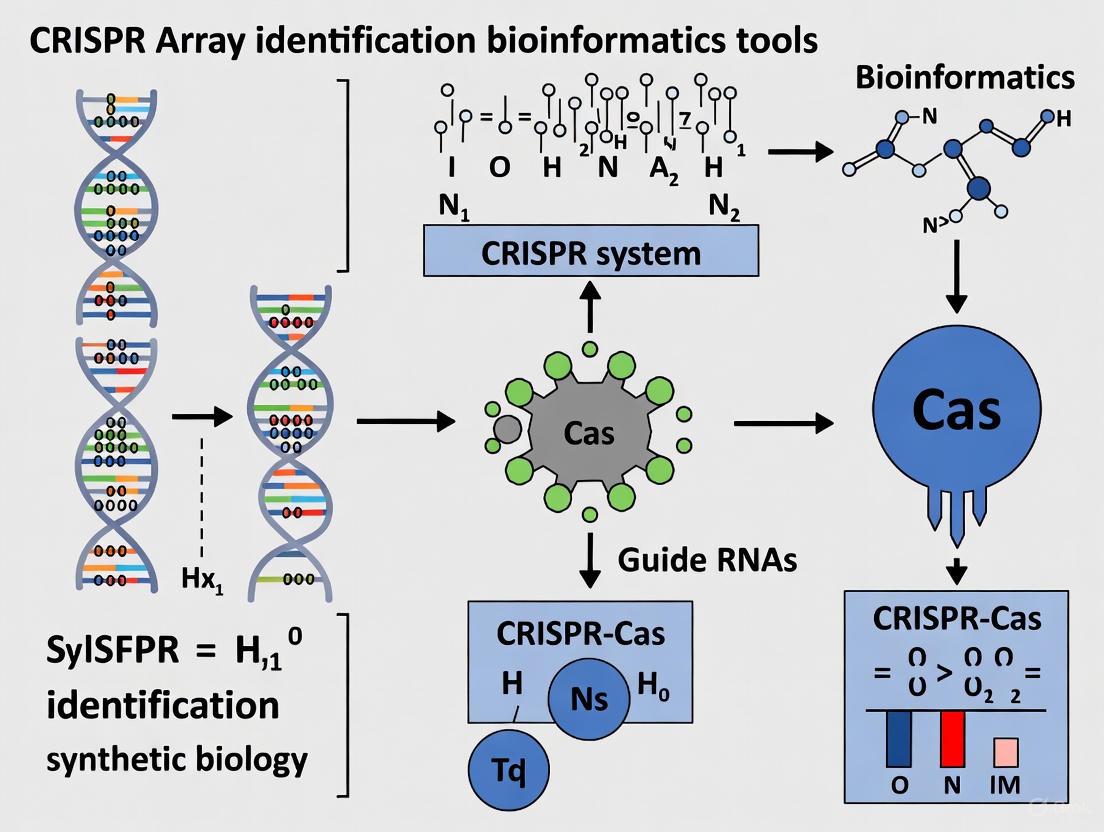
A Comprehensive Guide to CRISPR Array Identification Tools: From Foundational Concepts to Clinical Applications
This article provides a systematic overview of the bioinformatics tools essential for identifying and analyzing CRISPR arrays, a cornerstone of prokaryotic adaptive immunity and genome-editing technologies.
A Comprehensive Guide to CRISPR Array Identification Tools: From Foundational Concepts to Clinical Applications
Abstract
This article provides a systematic overview of the bioinformatics tools essential for identifying and analyzing CRISPR arrays, a cornerstone of prokaryotic adaptive immunity and genome-editing technologies. Tailored for researchers, scientists, and drug development professionals, we explore the foundational principles of CRISPR array structure and the evolutionary classification of CRISPR-Cas systems. The content details a practical workflow for array detection, visualization, and orientation prediction, addressing common challenges and optimization strategies. Furthermore, we present a comparative analysis of tool performance, validation methodologies, and the growing role of machine learning. This guide synthesizes current knowledge to empower the selection and application of the most effective computational resources for precision genome editing and therapeutic development.
Understanding CRISPR Arrays: Structure, Function, and Evolutionary Classification
Clustered Regularly Interspaced Short Palindromic Repeats (CRISPR) arrays are distinctive genetic elements that function as the adaptive immune memory in prokaryotes, enabling bacteria and archaea to defend against invading mobile genetic elements like viruses and plasmids [1] [2]. These arrays, together with CRISPR-associated (cas) genes, form the CRISPR-Cas system that provides sequence-specific immunity against foreign nucleic acids [3]. The fundamental architecture of a CRISPR array consists of direct repeats (DRs)—highly conserved short DNA sequences—separated by similarly sized but highly variable spacer sequences that are derived from foreign genetic elements [3] [4]. This review examines the structural organization of CRISPR arrays within the broader context of bioinformatics tools research, highlighting how computational approaches have been essential for deciphering their architecture, evolution, and function. For researchers and drug development professionals, understanding this architecture is crucial not only for grasping prokaryotic immunity but also for leveraging CRISPR systems in biomedical applications, including the development of novel therapeutics and diagnostic tools [3] [5].
Core Architectural Components of CRISPR Arrays
Structural Anatomy of Direct Repeats and Spacers
The defining feature of a CRISPR array is its repetitive structure composed of alternating repeats and spacers. The direct repeats typically range from 25 to 45 nucleotides in length, though some studies report a broader range of approximately 10-100 base pairs [3] [4]. These repeats are largely identical within an array but exhibit significant variation between different CRISPR-Cas systems and organisms [1]. Flanking these repeats are spacer sequences of similar length (approximately 10-100 bp) that represent captured fragments of foreign DNA from previous encounters with mobile genetic elements [4] [2]. The spacer sequences are highly variable and unique within an array, serving as a molecular memory of past infections. A minimum of three repeat-spacer units is generally required to define a CRISPR array [4].
The repeats play crucial functional roles beyond mere structural components. They contain recognition motifs for Cas proteins involved in pre-crRNA processing and form specific RNA secondary structures essential for the maturation and function of CRISPR RNAs (crRNAs) [1] [6]. The conservation of repeat sequences within an array is a critical feature that bioinformatics tools leverage for detection, as these patterns of regularity amidst non-repetitive spacer sequences create a distinctive signature in genomic data [1] [4].
Leader Sequence: The Regulatory Hub
Adjacent to the CRISPR array lies the leader sequence, a non-coding region of variable length (up to several hundred base pairs) that plays essential regulatory roles [6]. The leader is typically located at the 5' end of the array and contains promoters for pre-crRNA transcription as well as signals for spacer acquisition [6]. This region is characterized by a relatively high AT content compared to the surrounding genomic regions, a feature exploited by several bioinformatics tools for orientation prediction [6]. The leader sequence serves as the site for the integration of new spacers during the adaptation phase of CRISPR immunity, with new spacers almost always being inserted at the end proximal to the leader in a polarized manner [6] [7]. This polarized insertion process creates a chronological record of spacer acquisition, with the most recently acquired spacers positioned closest to the leader sequence and older spacers progressively farther away [7].
Table 1: Quantitative Features of CRISPR Array Components Based on Genomic Analyses
| Component | Typical Size Range | Key Characteristics | Functional Role |
|---|---|---|---|
| Direct Repeat | 25-45 bp (range: 10-100 bp) [3] [4] | Highly conserved within array; forms stable RNA secondary structures [1] | Processing signals for crRNA maturation; Cas protein binding [6] |
| Spacer | 25-45 bp (range: 10-100 bp) [3] [4] | Highly variable; derived from foreign DNA [2] | Immune memory; guides Cas proteins to specific targets [1] |
| Leader Sequence | Up to several hundred bp [6] | AT-rich; contains promoters and integration signals [6] | Regulates transcription and spacer acquisition [6] |
| Complete Array | 3 to hundreds of units (mean: 40; median: 25) [8] | Polarized spacer insertion; chronological record of infections [7] | Provides adaptive immunity through sequence-specific targeting [2] |
Quantitative Distribution and Genomic Organization
Statistical analyses of CRISPR arrays in bacterial Class I systems reveal that while arrays can expand to hundreds of spacers, their size typically follows a geometric distribution with mean and median sizes of approximately 40 and 25 spacers respectively, reflecting rather modest acquisition and/or retention overall [8]. This distribution indicates that most arrays are relatively small, with a decreasing probability of observing larger arrays. The geometric distribution parameter for Class I systems was estimated at 0.025 [8]. When multiple arrays occur within a single genome, the array closest to the cas operon is typically larger than distal loci, reflecting acquisition and expansion biases related to proximity to the molecular machinery [8].
The genomic distribution of CRISPR arrays is non-random, with a higher probability of occurrence at clustered locations along both DNA strands [8]. In bacterial Class I systems, CRISPR loci show preferential positioning between 200-240 degrees on the negative strand and between 60-120 degrees on the positive strand when mapping frequency along a standardized chromosome plot [8]. This non-uniform distribution suggests potential functional or evolutionary constraints on CRISPR array locations within genomes.
Table 2: Statistical Distribution of CRISPR Array Sizes in Bacterial Class I Systems [8]
| System Category | Mean Array Size (Spacers) | Median Array Size (Spacers) | Distribution Type | Sample Size (Observations) |
|---|---|---|---|---|
| Class I (Overall) | 40 | 25 | Geometric | 811 |
| Type I | 42 | 26 | Geometric | 558 |
| Type III | 36 | 23 | Geometric | 172 |
| Subtype I-B | 54 | 36 | Geometric | 77 |
| Subtype I-C | 30 | 14 | Geometric | 103 |
| Subtype I-E | 38 | 25 | Geometric | 213 |
| Subtype I-F | 35 | 22 | Geometric | 79 |
Bioinformatics Tools for CRISPR Array Identification
Detection Algorithms and Methodologies
The identification of CRISPR arrays in genomic sequences relies heavily on computational approaches that exploit their characteristic repetitive architecture. Early bioinformatics tools such as CRT, PILER-CR, and CRISPRFinder employed algorithms based on detecting repetitive patterns through self-alignment or sliding window approaches [1] [3] [4]. These tools typically identify pairs of maximal repeats, join them into consensus repeat sequences, and then score potential arrays using built-in evaluation functions that consider features like repeat length, spacer length, similarity between repeats, and regularity of spacing [1]. While these methods demonstrated reasonable sensitivity, they often suffered from high false positive rates and limited ability to precisely define array boundaries [1] [4].
More recent approaches have incorporated machine learning techniques to improve detection accuracy. CRISPRidentify, for example, implements a data-driven pipeline that performs three key steps: detection of repetitive elements, feature extraction, and classification using manually curated sets of positive and negative examples of CRISPR arrays [1]. This tool extracts multiple features including repeat similarity, AT-content, stability of repeat hairpin structures, and spacer uniqueness, then applies classifiers such as Support Vector Machines, Random Forests, and Neural Networks to distinguish true CRISPR arrays from false positives [1] [3]. This machine learning approach has demonstrated a drastically reduced false positive rate compared to earlier methods while maintaining high sensitivity [1].
FindCrispr represents another algorithmic approach that combines feature extraction with a scoring system based on properties such as repeat length, copy number, starting position sequences, and repeat sequence characteristics [4]. This tool is particularly sensitive for identifying CRISPR arrays with a small number of repeats and has low tolerance for long, scattered repeats, making it complementary to other detection methods [4].
Orientation Prediction and Array Annotation
Determining the correct orientation of CRISPR arrays is crucial for understanding their functionality, as it enables identification of leader sequences, transcription initiation sites, and the direction of spacer acquisition [6]. Multiple computational approaches have been developed for orientation prediction, each leveraging different features of CRISPR architecture:
- Repeat-based orientation: Tools like CRISPRstrand analyze mutation patterns along the array, as repeats tend to accumulate mutations at their 3' ends due to the polarized nature of spacer insertion and deletion processes [6] [3]. These tools often use graph kernel models trained on curated datasets of repeat sequences [6].
- Leader-based orientation: Methods implemented in CRISPRDirection and CRISPRCasFinder identify leader sequences by detecting relative AT richness at array ends and comparing distances to adjacent coding genes [6]. These approaches combine multiple features including sequence motifs, RNA secondary structure, and repeat degeneracy [6].
- Cas gene orientation: Some methods predict array orientation based on the transcription direction of nearby cas genes, assuming close linkage between arrays and their associated cas operons [6].
- Evolutionary approaches: CRISPR-evOr represents a novel method that leverages the polarized acquisition of spacers to reconstruct and compare the likelihood of evolutionary histories under different orientation hypotheses [6]. This approach is particularly valuable for arrays where traditional markers like leaders are absent or ambiguous [6].
Table 3: Bioinformatics Tools for CRISPR Array Analysis and Their Key Features
| Tool Name | Primary Function | Methodology | Key Features | Applications |
|---|---|---|---|---|
| CRISPRidentify [1] [3] | Array detection & annotation | Machine learning (SVM, Random Forest, Neural Networks) | Low false positive rate; detailed annotation; certainty scoring | Comprehensive array identification in genomic sequences |
| CRISPRDetect [3] | Array detection & orientation | Repeat pattern analysis + leader detection | Precise repeat-spacer boundaries; orientation prediction; cas gene annotation | Automated CRISPR annotation in prokaryotic genomes |
| CRISPRCasFinder [6] | Array detection & classification | Combined leader + repeat orientation | Evidence-level scoring; subtype classification; web interface | CRISPR system characterization and classification |
| CRISPR-evOr [6] | Orientation prediction | Evolutionary history reconstruction | Independent of Cas type, leader existence; resolves conflicting predictions | Orientation of challenging arrays |
| CCTK [7] | Array comparison & phylogeny | Network analysis + maximum parsimony | Visualizes spacer sharing; infers evolutionary relationships | Strain typing; evolutionary studies of related arrays |
| FindCrispr [4] | Array detection | Feature extraction + scoring | Sensitive for small arrays; visualizes results | Identification of CRISPRs with few repeats |
Experimental Protocols for CRISPR Array Analysis
Computational Workflow for Array Identification
A standard pipeline for identifying and analyzing CRISPR arrays from genomic sequences involves multiple steps, each leveraging specific bioinformatics tools:
Sequence Preprocessing: Assemble raw sequencing reads into contigs using tools like SPAdes with careful error correction [7]. For metagenomic datasets, perform binning to obtain metagenome-assembled genomes (MAGs) followed by dereplication to identify non-redundant genomes [2].
CRISPR Array Detection: Process assembled sequences through multiple detection tools to maximize sensitivity. A recommended approach includes:
- Run CRISPRidentify with default parameters to leverage its machine learning classification and low false positive rate [1] [3].
- Process sequences through CRISPRDetect to identify arrays with precise repeat-spacer boundaries and orientation information [3].
- For comprehensive analysis, additionally use CRISPRCasFinder with evidence level thresholds (typically level 4 for high-confidence arrays) [2].
Array Orientation and Annotation: Determine the correct orientation of identified arrays using a consensus approach:
- Apply CRISPRstrand or CRISPRDirection for repeat-based and leader-based orientation predictions [6].
- For arrays with ambiguous predictions or lacking leader sequences, use CRISPR-evOr to leverage evolutionary information [6].
- Annotate associated cas genes using hidden Markov models (HMMs) of known Cas protein families [3].
Comparative Analysis: For multiple arrays from related organisms, use the CRISPR Comparison Toolkit (CCTK) to:
- Identify homologous arrays based on shared spacers using network analysis [7].
- Visualize relationships between arrays with CRISPRdiff, which color codes shared and unique spacers [7].
- Infer evolutionary relationships with CRISPRtree using maximum parsimony to reconstruct ancestral arrays and evolutionary events [7].
Spacer-Protospacer Matching Protocol
Identifying the targets of CRISPR spacers is essential for understanding their biological function and ecological impact:
Database Preparation: Compile comprehensive databases of potential protospacer sources, including:
Similarity Search: Perform spacer-protospacer alignment using BLASTN or similar tools with appropriate threshold (typically 80-90% similarity over at least 80% of spacer length) [2].
Filtering and Validation: Apply stringent filters to eliminate false positives:
Statistical Analysis: Quantify the prevalence of different protospacer sources and perform statistical tests to identify biases related to taxonomic relationships, genomic proximity, or environmental factors [2].
Table 4: Key Research Reagent Solutions for CRISPR Array Studies
| Reagent/Resource | Function | Application Examples |
|---|---|---|
| CRISPRidentify [1] [3] | Machine learning-based array detection | Accurate identification of true CRISPR arrays with minimal false positives; provides certainty scores |
| CRISPRCasFinder [6] [2] | Integrated array and Cas gene detection | Comprehensive CRISPR-Cas system annotation; evidence-level classification |
| CCTK (CRISPR Comparison Toolkit) [7] | Comparative analysis of multiple arrays | Phylogenetic analysis of array evolution; visualization of spacer relationships |
| CRISPR-evOr [6] | Evolutionary orientation prediction | Determining array orientation without relying on traditional markers |
| MinCED [7] | CRISPR array detection in genomes | Identification of arrays without prior knowledge of CRISPR subtypes |
| CRISPRDetect [3] | Web-based array detection and annotation | Precise boundary identification; orientation prediction; compatible with other analysis tools |
Visualization of CRISPR Array Architecture and Analysis Workflow
The architecture of CRISPR arrays, with their precisely organized direct repeats and spacers, represents a sophisticated system for storing immunological memory in prokaryotes. Understanding this architecture is fundamental not only for deciphering prokaryotic immunity but also for leveraging CRISPR systems in biomedical applications. The development of sophisticated bioinformatics tools has been instrumental in characterizing these arrays, enabling researchers to identify their components, determine their orientation, and reconstruct their evolutionary history. For drug development professionals, this knowledge provides the foundation for harnessing CRISPR systems as programmable gene-editing tools, with applications ranging from functional genomics screens to therapeutic genome engineering [3]. As bioinformatics tools continue to evolve, incorporating more advanced machine learning approaches and leveraging the growing wealth of genomic data, our ability to decipher the complex architecture and evolutionary dynamics of CRISPR arrays will continue to improve, driving innovations in both basic research and applied biotechnology.
{INTRODUCTION}
The Natural Function: CRISPR-Cas as an Adaptive Immune System in Prokaryotes
Clustered Regularly Interspaced Short Palindromic Repeats (CRISPR) and CRISPR-associated (Cas) proteins constitute an adaptive immune system that confers sequence-specific protection to prokaryotes against invasive mobile genetic elements (MGEs) such as viruses and plasmids [9] [10]. This system provides a heritable record of infections, allowing cells to recognize and clear subsequent invasions by the same genetic elements [9] [11]. The fundamental principle of CRISPR-Cas immunity—RNA-guided targeting of nucleic acids—has not only revolutionized our understanding of virus-host interactions in prokaryotes but has also provided the foundational machinery for the development of powerful genome-editing technologies [12] [13]. For researchers focused on bioinformatics tool development, a deep understanding of this natural biological function is critical for the accurate identification of CRISPR arrays, prediction of their potential targets, and the rational design of guide RNAs for experimental applications.
{BIOLOGY AND MECHANISMS}
Molecular Architecture and Mechanism of Action
CRISPR-Cas systems function through a structured, three-stage process that provides adaptive, heritable immunity. The process initiates with adaptation, where new spacers are acquired from invading nucleic acids. This is followed by crRNA biogenesis, where the CRISPR locus is transcribed and processed into functional guide RNAs. The final interference stage involves sequence-specific recognition and cleavage of the target invader [10] [11].
Stage 1: Adaptation – Acquiring Immunological Memory
During adaptation, the Cas1-Cas2 protein complex integrates short fragments (~30-40 base pairs) of foreign DNA, known as protospacers, into the host's CRISPR genomic locus [10]. This locus consists of short, partially palindromic repeats separated by variable "spacer" sequences derived from past invasions. The integration occurs at the leader end of the array, creating a chronological record of encounters [9] [10]. A critical requirement for spacer acquisition is the presence of a short, conserved protospacer adjacent motif (PAM) flanking the protospacer in the invader's genome. The PAM sequence is system-specific and enables the machinery to distinguish between self and non-self DNA, thus preventing autoimmunity [10].
Stage 2: crRNA Biogenesis – Generating Guide Molecules
In the second stage, the CRISPR locus is transcribed as a long precursor CRISPR RNA (pre-crRNA). This precursor is then processed by Cas proteins into short, mature CRISPR RNAs (crRNAs). Each crRNA contains a single spacer sequence that serves as a guide for locating complementary foreign nucleic acids [10] [11].
Stage 3: Interference – Executing the Immune Response
In the final interference stage, the mature crRNA assembles with one or multiple Cas proteins to form an effector complex. This complex scans the cell for nucleic acid sequences complementary to the crRNA spacer. Upon recognizing a matching sequence adjacent to a valid PAM, the effector complex cleaves and degrades the invading DNA or RNA, thereby neutralizing the threat [10] [11].
Table 1: Core Functional Stages of the CRISPR-Cas Adaptive Immune System
| Stage | Key Process | Primary Components | Outcome |
|---|---|---|---|
| 1. Adaptation | Spacer acquisition from invader DNA | Cas1, Cas2 complex | Integration of new spacer into CRISPR array, creating immunological memory |
| 2. crRNA Biogenesis | Processing of CRISPR transcript into guide RNAs | Cas6, RNase III (Type II systems) | Generation of mature crRNA guides for target recognition |
| 3. Interference | Target recognition and cleavage | crRNA & Effector Complex (e.g., Cas9, Cascade) | Sequence-specific degradation of invading nucleic acids |
Diversity and Classification of CRISPR-Cas Systems
CRISPR-Cas systems are highly diverse and have been classified into two major classes based on their effector complex architecture. Class 1 systems (Types I, III, and IV) utilize multi-subunit effector complexes, while Class 2 systems (Types II, V, and VI) rely on a single, large Cas protein for crRNA processing and interference [11]. This classification is fundamental for bioinformatics, as the type of system dictates the PAM requirements, guide RNA structures, and cleavage mechanisms that computational tools must account for.
Table 2: Major Types of CRISPR-Cas Systems and Their Key Characteristics
| System Type | Class | Signature Nuclease | Target | Key Feature |
|---|---|---|---|---|
| Type I | 1 | Cas3 | DNA | Multi-protein CASCADE complex surveys DNA; recruits Cas3 for degradation [10] [11] |
| Type II | 2 | Cas9 | DNA | Requires a trans-activating crRNA (tracrRNA); single protein creates DSBs [10] [11] |
| Type III | 1 | Cas10 | RNA / ssDNA | Targets transcriptionally active RNA; can also cleave ssDNA [10] [11] |
| Type IV | 1 | Unknown | DNA (plasmid) | Minimal system often plasmid-borne; involved in plasmid competition [11] |
| Type V | 2 | Cas12 (Cpf1) | DNA | Single RuvC domain cleaves both DNA strands; some subtypes target RNA [13] [11] |
| Type VI | 2 | Cas13 | RNA | Targets RNA; exhibits collateral RNase activity upon activation [11] |
The following diagram illustrates the generalized, three-stage functional mechanism of the CRISPR-Cas adaptive immune system.
Diagram 1: The Three-Stage CRISPR-Cas Adaptive Immune Pathway.
{EXPERIMENTAL VALIDATION}
A Foundational Experiment: Demonstrating Adaptive Immunity inS. thermophilus
The first definitive biological evidence establishing CRISPR-Cas as an adaptive immune system was published in 2007 by Barrangou et al. [10]. This seminal study used the bacterium Streptococcus thermophilus as a model to demonstrate that exposure to bacteriophages leads to the acquisition of new spacers from the viral genome, which in turn confers specific resistance to subsequent phage attacks.
Experimental Protocol and Methodology
Phage Challenge and Survivor Isolation: A population of S. thermophilus was exposed to a lytic bacteriophage. The few surviving bacterial colonies were isolated for further analysis.
CRISPR Locus Analysis: The CRISPR loci of both the original phage-sensitive strain and the phage-resistant survivor strains were amplified by polymerase chain reaction (PCR) and sequenced. The sequences were compared to identify any changes.
Spacer Acquisition and Source Verification: The study found that the resistant strains had acquired one or more new spacers within their CRISPR arrays. These new spacer sequences were identical to segments (protospacers) of the infecting phage's genome. This was confirmed by aligning the spacer sequences against the known phage genome sequence.
Resistance Specificity Validation: To prove that the acquired spacers were responsible for immunity, the researchers challenged the resistant strains with phages that had mutations in the protospacer or the adjacent PAM sequence. These mutated phages were able to evade the CRISPR system and successfully infect the bacteria, demonstrating that immunity is highly sequence-specific.
Table 3: Key Research Reagent Solutions for CRISPR-Cas Functional Studies
| Reagent / Material | Function in Experimental Research |
|---|---|
| Cas Protein Expression Vectors | Plasmids for producing Cas nucleases (e.g., Cas9, Cas12) in heterologous hosts for interference studies [14] |
| Guide RNA Cloning Plasmids | Vectors with promoters (e.g., U6) for expressing custom crRNA and tracrRNA molecules for target guidance [14] [13] |
| CRISPR Array Libraries | Collections of knock-in constructs for endogenous gene tagging, enabling functional investigation of protein localization and dynamics [14] |
| Bioinformatics Tools (e.g., CRISPOR, CHOPCHOP) | Computational platforms for designing highly efficient and specific guide RNAs and predicting potential off-target effects [12] [13] |
| Next-Generation Sequencing (NGS) | Gold-standard method for comprehensive analysis of CRISPR editing outcomes, including indel spectrum and off-target assessment [15] |
{IMPLICATIONS FOR BIOINFORMATICS}
Connecting Natural Biology to Computational Tool Development
The biological principles of the native CRISPR-Cas system directly inform the design and application of bioinformatics tools for CRISPR array identification and guide RNA selection.
CRISPR Array Identification: The characteristic structure of alternating repeats and spacers is the primary feature used by bioinformatics algorithms (e.g., CRISPRFinder, CRISPRDetect) to identify and annotate CRISPR loci in genomic sequences [12]. Understanding that spacers are derived from MGEs allows these tools to predict the potential targets of a given CRISPR system by querying spacer sequences against viral and plasmid databases.
Guide RNA Design and Off-Target Prediction: The requirement for a PAM sequence is a critical constraint built into all guide RNA design tools (e.g., CRISPOR, CHOPCHOP) [13]. Furthermore, the biological reality that mismatches between the crRNA and target DNA can lead to promiscuous cleavage or failed immunity drives the development of sophisticated off-target prediction algorithms. These tools assess genome-wide potential binding sites to maximize on-target efficiency and minimize off-target effects in gene-editing applications [12] [13].
The following diagram outlines the logical workflow from the natural immune function to the development of applied bioinformatics tools.
Diagram 2: From Biological Principle to Bioinformatics Application.
{CONCLUSION}
The CRISPR-Cas system is a sophisticated adaptive immune mechanism in prokaryotes that provides sequence-specific, heritable defense against genetic parasites. Its operation through a clearly defined cycle of adaptation, expression, and interference showcases a remarkable form of molecular memory. The quantitative parameters of this system—such as spacer and repeat lengths, PAM sequences, and the structural diversity of effector complexes—provide the essential data around which bioinformatics tools are built. A rigorous understanding of this natural function is therefore indispensable for driving innovation in computational biology, from the accurate annotation of CRISPR arrays in genomic data to the rational design of specific and efficient guides for contemporary genome engineering.
The classification of Clustered Regularly Interspaced Short Palindromic Repeats (CRISPR) and CRISPR-associated (Cas) systems has undergone significant expansion to encompass growing diversity, with the current evolutionary classification now encompassing 2 classes, 7 types, and 46 subtypes [16] [17]. This updated taxonomy represents a substantial increase from the 6 types and 33 subtypes documented in the previous major survey conducted five years ago, reflecting remarkable discoveries in the field of prokaryotic adaptive immunity [16]. The relentless discovery of novel variants, particularly through extensive mining of genomic and metagenomic databases, has necessitated this revised framework, which now includes rare systems that constitute the "long tail" of CRISPR-Cas distribution in prokaryotes and their viruses [16] [18].
This classification system, essential for accurate description and characterization of CRISPR-cas loci in newly sequenced bacterial and archaeal genomes, employs a complex polythetic approach that combines analyses of CRISPR-cas locus architecture and gene composition with sequence similarity clustering and phylogenetic analysis of conserved Cas proteins [16]. The classification framework organizes CRISPR-Cas systems based on their effector module compositions, with Types delineated by unique effector signatures and subtypes defined through a combination of phylogenetic analysis and gene composition criteria [16]. The evolving complexity of this system underscores the dynamic nature of CRISPR research and highlights the critical importance of bioinformatics tools in identifying, classifying, and understanding these complex molecular systems.
Updated Classification Framework
Hierarchical Organization: From Classes to Subtypes
The updated classification organizes CRISPR-Cas systems into a hierarchical structure that begins with two fundamental classes based on effector complex architecture, then divides into types distinguished by their signature genes and effector mechanisms, and further differentiates into subtypes based on more subtle variations in gene composition and locus organization [16].
Table 1: CRISPR-Cas System Classification Hierarchy
| Classification Level | Key Defining Characteristics | Current Count |
|---|---|---|
| Class | Effector module architecture | 2 |
| Type | Signature effector proteins | 7 |
| Subtype | Gene composition & locus organization | 46 |
Class 1 and Class 2: Fundamental Architectural Division
CRISPR-Cas systems are primarily partitioned into two classes based on their effector module organization:
Class 1 Systems: Characterized by multisubunit effector complexes that require multiple Cas protein subunits to form functional CRISPR machinery [18]. This class includes Types I, III, IV, and the newly added Type VII [16]. Class 1 systems represent the majority of known CRISPR-Cas diversity and are phylogenetically more widespread among prokaryotes.
Class 2 Systems: Employ single, large Cas proteins in their effector complexes, making them structurally simpler but functionally diverse [18]. This class includes Types II, V, and VI, which have been predominantly harnessed for genome engineering applications due to their simpler architecture.
Table 2: CRISPR-Cas System Classes and Types
| Class | Types | Signature Effector/Features |
|---|---|---|
| Class 1 | Type I | Multi-subunit complex: Cas3 (helicase-nuclease), Cas5, Cas6, Cas7, Cas8 |
| Type III | Multi-subunit complex: Cas10 (large subunit with polymerase/cyclase activity) | |
| Type IV | Minimal adaptation modules; variable effector complexes | |
| Type VII | Newly identified; Cas14 effector with metallo-β-lactamase (β-CASP) domain | |
| Class 2 | Type II | Single effector: Cas9; utilizes tracrRNA for maturation |
| Type V | Single effector: Cas12 family; includes DNA and RNA targeting variants | |
| Type VI | Single effector: Cas13 family; specialized for RNA targeting |
Detailed Analysis of Class 1 Systems
Type I Systems: Diversity and Modularity
Type I systems represent one of the most diverse and prevalent CRISPR-Cas types, characterized by the presence of a Cas3 protein that possesses both helicase and nuclease activities. These systems employ a multi-subunit effector complex known as Cascade (CRISPR-associated complex for antiviral defense) for target recognition and Cas3 for DNA degradation. The updated classification recognizes eight subtypes within Type I (A-F, U, and recently identified variants), with ongoing discoveries revealing additional functional variations [16].
Recent investigations have identified novel Type I variants with unique characteristics, including I-E2 and I-F4 systems that incorporate an HNH nuclease fused to Cas5 and Cas8f proteins, respectively [16]. These variants typically lack the canonical Cas3 helicase-nuclease yet demonstrate robust crRNA-guided double-stranded DNA cleavage activity, challenging previous assumptions about Type I functional requirements [16]. The discovery of such variants illustrates the remarkable evolutionary plasticity of CRISPR-Cas systems and underscores the importance of continued database mining and classification refinement.
Type III Systems: Complex Signaling and Defense Mechanisms
Type III systems represent some of the most complex CRISPR-Cas variants, characterized by the presence of Cas10 as the large subunit of their effector complexes. These systems can target both RNA and DNA and often incorporate sophisticated signaling pathways involving cyclic oligoadenylate (cOA) second messengers that activate ancillary effector proteins. The updated classification expands Type III to include nine subtypes (A-I), with recent additions including III-G, III-H, and III-I [16].
Notably, subtypes III-G and III-H exhibit evidence of reductive evolution, with inactivated polymerase/cyclase domains in their Cas10 proteins [16]. This loss of cOA generation capacity correlates with the absence of genes encoding ancillary proteins containing cOA-binding domains (such as CARF or SAVED domains) fused to effector domains like HEPN RNase, which are characteristic of most Type III systems [16]. Subtype III-G, specifically identified in Sulfolobales, typically lacks adaptation modules, and no CRISPR arrays have been found associated with its loci, suggesting these systems may recruit crRNAs from other CRISPR-cas loci in trans [16].
The newly described subtype III-I, identified in over 160 genomes primarily from the phyla Thermodesulfobacteriota and Chloroflexota, features an extremely diverged Cas10 protein lacking the N-terminal polymerase/cyclase domain and a unique multidomain effector protein with architecture resembling Cas7-11 of subtype III-E but apparently originating independently from a different variant of subtype III-D [16]. Based on conserved catalytic residues, this subtype is predicted to cleave RNA targets [16].
Type IV Systems: Minimalist and Enigmatic
Type IV systems represent minimalist CRISPR-Cas variants that typically lack adaptation modules and often have degenerate repeats in their associated CRISPR arrays. Previously considered somewhat anomalous, the updated classification now recognizes three subtypes within Type IV (A-C) and has characterized additional variants with unique functionalities [16]. Recent research has identified Type IV variants that cleave target DNA, expanding the functional repertoire of this type [16] [17]. The streamlined architecture of Type IV systems, coupled with their demonstrated interference capabilities, makes them intriguing subjects for both basic research and potential biotechnological applications.
Type VII: The Newest Addition to Class 1
The identification and characterization of Type VII represents one of the most significant updates to the CRISPR-Cas classification. This newly defined type is found mostly in taxonomically diverse archaeal genomes and contains a metallo-β-lactamase (β-CASP) effector nuclease designated Cas14 [16]. According to established CRISPR-Cas classification principles, this unique signature effector qualifies these loci as a distinct type [16].
Type VII loci typically lack adaptation modules, and repeats in their associated CRISPR arrays often contain multiple substitutions, suggesting limited incorporation of new spacers [16]. Analysis of the limited number of spacer hits indicates these systems target transposable elements [16]. Structural analysis reveals that Cas14 contains a C-terminal domain structurally resembling the C-terminal domain of Cas10, the large subunit of Type III effector modules, suggesting an evolutionary connection between these types [16]. This relationship is further supported by specific similarity between the Cas5 proteins of Type VII and subtype III-D [16].
Unlike Type III systems that target RNA, Type VII systems have been shown to target RNA in a crRNA-dependent manner, cleaving targets via the nuclease activity of Cas14 [16]. Despite their apparently simple organization, recent cryogenic-electron microscopy structures reveal that Type VII effector complexes can contain up to 12 subunits, with Cas14 binding to the Cas7 backbone via its Cas10 remnant domain, making this complex one of the largest among Class 1 systems [16].
Class 2 Systems: Structural Simplicity and Functional Diversity
Type II: The Genome Engineering Revolution
Type II systems, characterized by the single-protein effector Cas9, have become the most widely utilized CRISPR system in biotechnology and therapeutic development. These systems employ a dual RNA structure comprising crRNA and trans-activating crRNA (tracrRNA) that can be engineered into a single-guide RNA (sgRNA) for simplified genome editing applications [12]. The canonical Type II system from Streptococcus pyogenes recognizes a 5'-NGG-3' protospacer adjacent motif (PAM) and creates blunt-ended double-strand breaks 3 base pairs upstream of the PAM sequence [19].
While Type II was among the first CRISPR systems to be characterized and harnessed for genetic engineering, the updated classification continues to recognize its phylogenetic diversity and functional variations across bacterial species. The relative simplicity of Type II systems, with their single-protein effectors, has facilitated their rapid adoption and engineering for diverse applications, from gene knockout to transcriptional regulation and epigenetic modification.
Type V: DNA Targeting Beyond Cas9
Type V systems encompass a growing family of Cas12 effectors that recognize T-rich PAM sequences and create staggered DNA cuts with sticky ends. The updated classification reveals substantial expansion within Type V, with multiple subtypes now recognized. These systems have been engineered for diverse applications, including DNA detection, gene editing, and diagnostic platforms.
Recent research has identified novel Type V variants that inhibit target replication without cleavage, expanding the functional capabilities of this type beyond traditional nucleases [16] [17]. These alternative functionalities demonstrate the evolutionary innovation within CRISPR-Cas systems and provide new molecular tools for precise genetic manipulation without introducing double-strand breaks.
Type VI: RNA-Targeting Specialists
Type VI systems utilize Cas13 effectors that target RNA rather than DNA, making them unique among the primarily DNA-targeting CRISPR systems. These proteins contain two Higher Eukaryotes and Prokaryotes Nucleotide-binding (HEPN) domains that confer RNase activity upon target recognition. Type VI systems have been harnessed for RNA knockdown, tracking, and detection applications, with the recently described Cas13d variant offering particularly compact architecture beneficial for delivery applications.
The classification of Type VI continues to expand as new variants are discovered through database mining and functional characterization. The RNA-targeting capability of Type VI systems complements the DNA-targeting functions of other Class 2 effectors, providing researchers with a comprehensive toolkit for genetic manipulation at multiple molecular levels.
Evolutionary Dynamics and Distribution Patterns
Analysis of CRISPR-Cas variant abundance in genomes and metagenomes reveals a consistent pattern: previously defined and well-characterized systems are relatively common, while the more recently characterized variants are comparatively rare [16]. These low-abundance variants comprise the "long tail" of the CRISPR-Cas distribution in prokaryotes and their viruses, with many remaining to be characterized experimentally [16] [17].
The evolutionary dynamics shaping CRISPR-Cas diversity involve multiple processes, including gene duplication, domain shuffling, horizontal gene transfer, and reductive evolution. Class 1 systems appear to be evolutionarily older and more diverse, while Class 2 systems likely evolved from simpler transposon-encoded ancestors on multiple independent occasions. The updated classification reveals complex patterns of evolutionary relationships between types, such as the connection between Type III and Type VII systems through their shared structural features [16].
The continuous discovery of rare variants suggests that the current classification, while dramatically expanded, represents an ongoing effort rather than a complete catalog. As sequencing technologies advance and exploration of diverse microbial habitats expands, additional CRISPR-Cas types and subtypes will likely be identified, further refining our understanding of prokaryotic immunity evolution.
Bioinformatics Tools for CRISPR Array Identification and Analysis
Computational Identification of CRISPR Systems
The expanding diversity of CRISPR-Cas systems has driven development of sophisticated bioinformatics tools for their identification and characterization. These tools employ various algorithms to detect the hallmark signatures of CRISPR arrays—direct repeats interspersed with variable spacers—in genomic sequences [3]. Early tools like CRT, PILER-CR, and CRISPRFinder established the foundation for computational CRISPR detection, while contemporary tools have incorporated machine learning and evolutionary approaches to improve accuracy and functionality [3] [4].
Table 3: Bioinformatics Tools for CRISPR Identification and Analysis
| Tool | Primary Function | Key Features | Limitations |
|---|---|---|---|
| CRISPRDetect | CRISPR array detection and refinement | Precise repeat-spacer boundaries; orientation detection; cas gene annotation | Limited information about Cas proteins |
| CRISPRidentify | Machine learning-based array identification | Multiple classifiers (SVM, Random Forest, etc.); lower false positive rate | Requires curated training data |
| CRISPRFinder | Web-based CRISPR identification | User-friendly interface; historical significance | Older algorithm; less accurate than newer tools |
| CRISPRCasFinder | Integrated CRISPR and Cas detection | Combines leader- and repeat-orientation methods | Complex output for novice users |
| FindCrispr | Accurate CRISPR identification | Feature extraction and scoring model; sensitive to arrays with few repeats | Lower tolerance for long, scattered repeats |
| CRISPR-evOr | Evolutionary orientation prediction | Independent of Cas type, leader existence; resolves conflicting predictions | Requires multiple related arrays for analysis |
Orientation Prediction and Evolutionary Analysis
Determining the orientation of CRISPR arrays is crucial for understanding their functionality, as new spacers are almost always inserted at the leader end in a polarized manner [20]. Multiple computational approaches have been developed to predict array orientation:
- Leader-based orientation: Identifies the leader sequence, typically located at one end of the array, through sequence features like AT richness and proximity to coding genes [20].
- Repeat-based orientation: Analyzes mutation patterns along repeats, which tend to degrade in the 5'-to-3' direction due to the polarized insertion process [20].
- Cas-based orientation: Determines orientation based on the transcription direction of nearby Cas genes [20].
- Evolutionary approaches: Tools like CRISPR-evOr leverage evolutionary patterns by reconstructing and comparing the likelihood of evolutionary histories with respect to both possible acquisition orientations, making them particularly valuable for arrays where traditional methods provide conflicting results [20].
Integrated Platforms and Specialized Databases
Comprehensive bioinformatics resources have been developed to support CRISPR research, integrating multiple analytical functions and providing curated databases:
- CRISPRdb and CRISPRCasdb: Specialized databases storing annotated CRISPR data from bacterial and archaeal genomes, enabling comparative analyses and classification [12] [3].
- CRISPRminer and CRISPRBank: Platforms that utilize various programs to identify both CRISPR arrays and Cas genes, providing classification into types and identifying self-targeting regions [3].
- CRISPI: A database and annotation tool that allows users to view all CRISPR arrays identified in microbial genomes, with associated Cas genes indicated using Hidden Markov Model profiles [3].
These resources collectively provide the bioinformatics infrastructure necessary to navigate the expanding diversity of CRISPR-Cas systems, enabling researchers to identify novel variants, classify them within the established taxonomic framework, and hypothesize about their functional capabilities based on comparative genomics.
Research Reagent Solutions for CRISPR Studies
Table 4: Essential Research Reagents and Computational Tools for CRISPR Analysis
| Reagent/Tool Category | Specific Examples | Function/Application |
|---|---|---|
| CRISPR Identification Tools | CRISPRDetect, CRISPRFinder, FindCrispr | Computational detection of CRISPR arrays in genomic sequences |
| Orientation Prediction Tools | CRISPRstrand, CRISPRDirection, CRISPR-evOr | Determination of CRISPR array orientation and transcriptional direction |
| Classification Databases | CRISPRdb, CRISPRCasdb, CRISPRBank | Reference databases for comparative analysis and subtype classification |
| Machine Learning Frameworks | CRISPRidentify (SVM, Random Forest, Neural Network) | Distinguishing true CRISPR arrays from false positives with high specificity |
| Evolutionary Analysis Tools | CRISPR-evOr, SpacerPlacer | Reconstruction of spacer acquisition history and evolutionary relationships |
| Cas Gene Annotation | HMMER, Custom HMM profiles | Identification and classification of Cas proteins in genomic sequences |
The updated evolutionary classification of CRISPR-Cas systems, encompassing 2 classes, 7 types, and 46 subtypes, represents a significant milestone in our understanding of prokaryotic adaptive immunity [16] [17]. This expanded framework captures the remarkable diversity of these molecular defense systems while revealing complex evolutionary relationships between seemingly distinct types. The continuous discovery of rare variants highlights the importance of ongoing genomic and metagenomic mining, suggesting that the current classification represents a snapshot of an ever-expanding universe.
Bioinformatics tools play an indispensable role in this taxonomic endeavor, enabling researchers to identify novel systems, determine their orientation and transcriptional direction, classify them within established frameworks, and hypothesize about their functional capabilities [3] [20]. As these tools evolve to incorporate more sophisticated machine learning approaches and evolutionary analyses, they will undoubtedly facilitate the discovery and characterization of additional CRISPR-Cas variants, particularly from the "long tail" of rare systems that remain to be discovered and experimentally characterized [16].
The expanded classification system provides not only a taxonomic framework but also a roadmap for biotechnological innovation, as each newly characterized system offers potential for engineering novel genome editing tools with unique properties and specificities. From the compact RNA-targeting Cas13 variants to the multi-subunit Type VII complexes, the diversity of natural CRISPR systems continues to inspire and enable new applications across basic research, therapeutic development, and diagnostic technologies.
The discovery and characterization of Clustered Regularly Interspaced Short Palindromic Repeats (CRISPR) systems in prokaryotic genomes represents a fundamental research area that has paved the way for revolutionary genome-editing technologies. Within the context of a broader thesis on CRISPR array identification bioinformatics tools research, specialized databases play an indispensable role as centralized knowledge repositories. These resources systematically organize identified CRISPR arrays, Cas genes, and their associated metadata, enabling researchers to explore natural diversity, evolutionary relationships, and functional properties of these systems [3] [21]. This technical guide provides an in-depth analysis of three core databases—CRISPRdb, CRISPRCasdb, and CRISPRBank—that have become essential resources for researchers, scientists, and drug development professionals working in the CRISPR field.
The exponential growth of genomic data has created both opportunities and challenges for the identification of CRISPR systems. While computational tools can detect potential CRISPR arrays in genomic sequences, curated databases provide the critical framework for annotation, classification, and comparative analysis [3] [22]. These resources have evolved from early collections of CRISPR observations to sophisticated platforms that integrate multiple prediction algorithms, classification systems, and visualization tools. For professionals engaged in drug development, these databases offer insights into CRISPR system functionality that can inform therapeutic strategies, including the selection of appropriate Cas proteins for specific applications and the identification of anti-CRISPR proteins that may enable safer therapeutic approaches [3] [23].
Database Comparative Analysis
The following table provides a systematic comparison of the key technical features and functionalities of the three databases, highlighting their respective strengths and specializations.
Table 1: Comparative Analysis of CRISPR Databases
| Feature | CRISPRdb | CRISPRCasdb | CRISPRBank |
|---|---|---|---|
| Primary Focus | CRISPR arrays and spacer sequences [22] | Integrated CRISPR arrays and Cas genes with system classification [3] [24] | Comprehensive repository of CRISPR and Cas genes [3] |
| Organism Coverage | Bacteria and Archaea [22] | Prokaryotic genomes [3] | Prokaryotic genomes [3] |
| Core Functionality | Identifies CRISPRs and spacers; provides visualization tools [22] | Identifies and classifies complete CRISPR-Cas systems; includes typing by subtype [3] [21] | Database containing CRISPR cas genes and arrays from 2733 strains [3] |
| Classification System | Limited to array identification [22] | Classifies systems into 6 types and identifies self-targeting regions [3] | Utilizes various programs to identify both CRISPR and Cas [3] |
| User Interface | Web-based query system [22] | Integrated with CRISPRCasFinder tool [24] | Web interface with multiple analytical tools [3] |
| Key Limitation | Limited to CRISPR arrays; does not design guide RNA [22] | Dependent on underlying CRISPRCasFinder algorithm [24] | Less specialized in system classification [3] |
Database-Specific Profiles and Applications
CRISPRdb
CRISPRdb serves as a foundational resource specifically focused on the identification of CRISPR arrays and their constituent spacer sequences in bacterial and archaeal genomes [22]. The database employs specialized algorithms to detect the hallmark repeating patterns of CRISPR arrays, which consist of direct repeats separated by variable spacer sequences. This focused approach enables researchers to quickly identify the presence and genomic location of CRISPR arrays, providing initial insights into the adaptive immune capabilities of the studied microorganisms.
The technical implementation of CRISPRdb centers on its visualization tools, which allow researchers to graphically represent identified CRISPR arrays and examine the sequence characteristics of individual repeats and spacers [22]. This functionality is particularly valuable for evolutionary studies, as spacer sequences can reveal historical encounters with mobile genetic elements such as plasmids and viruses. While the database's limitation to array identification without integrated Cas gene annotation represents a constraint for comprehensive system characterization, its specialized focus makes it particularly useful for initial screening and comparative analysis of CRISPR distribution across taxonomic groups.
CRISPRCasdb
CRISPRCasdb represents a significant advancement in database functionality by integrating both CRISPR array identification and Cas gene annotation within a unified classification framework [3] [24]. This integrated approach enables the database to classify complete CRISPR-Cas systems according to established taxonomic principles, organizing them into two classes, six types, and numerous subtypes based on the complement of Cas genes and the architecture of the CRISPR locus [3].
The database is tightly integrated with CRISPRCasFinder, a computational tool that implements the current classification standards for CRISPR system identification and typing [24]. This integration ensures that annotations remain current with evolving understanding of CRISPR system diversity. A particularly advanced feature of CRISPRCasdb is its ability to identify self-targeting spacers—sequences within the CRISPR array that match genomic regions of the host organism [3]. This capability has important implications for understanding CRISPR regulation and potential autoimmunity effects. The database's comprehensive approach makes it particularly valuable for researchers seeking to identify novel CRISPR systems with specific properties for biotechnological application, such as Cas proteins with unique PAM specificities or functional characteristics.
CRISPRBank
CRISPRBank functions as a comprehensive repository that consolidates CRISPR array and Cas gene information from multiple computational prediction sources [3]. The database contains annotated CRISPR-Cas systems from 2,733 microbial strains, providing broad coverage of prokaryotic diversity [3]. Unlike more specialized resources, CRISPRBank employs various prediction algorithms to identify both CRISPR arrays and associated Cas genes, creating a synthesized resource that leverages the strengths of multiple computational approaches.
The database's interface provides access to diverse analytical tools, allowing researchers to explore CRISPR system components from different perspectives [3]. While potentially less specialized in system classification compared to CRISPRCasdb, the integrative nature of CRISPRBank makes it a valuable starting point for exploratory research and meta-analyses. The breadth of genomic coverage enables comparative studies across taxonomic boundaries and facilitates the identification of evolutionary patterns in CRISPR system distribution and architecture.
Experimental Workflow for CRISPR System Identification
The following diagram illustrates the generalized computational workflow for identifying and characterizing CRISPR systems using specialized databases and bioinformatics tools, representing a standard methodology in the field.
Diagram 1: CRISPR System Identification Workflow
This workflow initiates with genomic sequence input, followed by sequential stages of computational analysis that progressively characterize different aspects of the CRISPR system. The integration of database resources at the intermediate stages enables researchers to contextualize their findings within existing knowledge, while the final stages focus on comparative and functional assessment to identify systems with novel or useful properties.
Research Reagent Solutions for CRISPR Identification
The experimental identification and characterization of CRISPR systems relies on a suite of computational tools and resources that constitute the essential "research reagents" for bioinformatics investigations in this field.
Table 2: Essential Computational Resources for CRISPR System Research
| Resource Category | Specific Tools | Primary Function | Application Context |
|---|---|---|---|
| CRISPR Array Predictors | CRISPRDetect, CRISPRFinder, PILER-CR, CRT [3] [22] | Identify CRISPR repeats and spacers in genomic sequences | Initial detection of CRISPR arrays; determines orientation and repeat-spacer boundaries |
| Cas Gene Identifiers | HMM profiles, BLAST, CRISPR-Cas Atlas [3] [25] | Detect Cas proteins through sequence similarity | Identification of associated Cas genes; essential for system classification |
| Classification Systems | CRISPRstrand, CRISPR-Cas Atlas [3] [25] | Determine transcribed strand and classify system type | Functional annotation; evolutionary studies; tool selection for applications |
| Database Platforms | CRISPRdb, CRISPRCasdb, CRISPRBank [3] [22] | Centralized repositories for annotated systems | Comparative analysis; meta-studies; reference for experimental design |
| Advanced Analysis | CRISPRidentify, Machine Learning Classifiers [3] [26] | Distinguish true CRISPR arrays from false positives | Validation of predictions; analysis of complex datasets |
Emerging Trends and Future Perspectives
The field of CRISPR database development is rapidly evolving, with several emerging trends shaping future directions. The integration of machine learning approaches represents a significant advancement, with tools like CRISPRidentify employing sophisticated classification algorithms to reduce false positive rates in array identification [3]. Similarly, the application of large language models to protein sequence analysis is opening new possibilities for generating novel CRISPR systems with optimized properties, as demonstrated by the development of OpenCRISPR-1 through AI-based design [25].
Another important trend is the expansion of database content through systematic mining of diverse genomic and metagenomic datasets. The CRISPR-Cas Atlas initiative, which has curated over 1.2 million CRISPR-Cas operons from 26 terabases of sequence data, exemplifies this scaling effort and has dramatically expanded the known diversity of systems beyond what is available in traditional curated databases [25]. This expansion is particularly valuable for drug development professionals seeking novel Cas proteins with specific functional characteristics for therapeutic applications.
Future developments will likely focus on enhanced integration between databases and analytical tools, creating more seamless workflows for researchers. Additionally, as structural information for Cas proteins accumulates, the incorporation of structural annotations and predictions will provide deeper insights into the molecular mechanisms of CRISPR system function. These advances will further solidify the role of specialized databases as indispensable resources for unlocking the potential of CRISPR systems in basic research and therapeutic applications.
The Critical Role of Bioinformatics in CRISPR Discovery and Development
The advent of CRISPR-Cas systems has ushered in a revolutionary era in genetic research and biotechnology. Since its discovery as a programmable gene-editing tool in 2012, CRISPR-Cas9 has transformed molecular biology and biomedical research by enabling precise modifications to genomic sequences [12]. This technology, often described as "molecular scissors," functions by utilizing a guide RNA (gRNA) that directs the Cas9 enzyme to a specific DNA sequence, where it creates a double-strand break [12]. This break activates the cell's natural DNA repair mechanisms—either error-prone non-homologous end joining (NHEJ) or precise homology-directed repair (HDR)—allowing researchers to disrupt, insert, or modify genes with unprecedented precision [12] [15].
The complexity, sheer volume of genomic data, and precision required in CRISPR-mediated genome editing have driven the rapid development of an extensive ecosystem of bioinformatics tools [12]. These computational resources are indispensable for designing CRISPR experiments, predicting off-target effects, analyzing screening data, and ensuring the accuracy and efficiency of the editing process. This review systematically examines the critical role of bioinformatics in CRISPR discovery and development, with particular emphasis on CRISPR array identification tools and their applications in advancing genome editing capabilities.
Bioinformatics for CRISPR Array Identification and Analysis
CRISPR arrays, consisting of direct repeats (DRs) and spacers, are fundamental components of prokaryotic CRISPR-Cas systems that provide adaptive immunity against mobile genetic elements [27]. Bioinformatics tools for identifying and analyzing these arrays form the foundation for understanding CRISPR system diversity, evolution, and function.
Core Computational Tools for CRISPR Array Detection
Specialized algorithms have been developed to identify CRISPR arrays in genomic sequences, each employing distinct computational approaches:
Table 1: Bioinformatics Tools for CRISPR Array Identification
| Tool Name | Methodology | Key Features | Applications |
|---|---|---|---|
| CRISPRDetect | Automated detection with refinement | Identifies repeat-spacer boundaries, substitutions, insertions, deletions; provides annotated cas genes [3] | Precise CRISPR array detection, target prediction [3] |
| CRISPRidentify | Machine learning (SVM, Random Forest, Neural Networks) | Distinguishes genuine CRISPR arrays from false positives; three-stage process: detection, extraction, classification [3] | High-specificity CRISPR array identification with lower false positive rates [3] |
| CRISPRstrand | Machine learning for orientation prediction | Predicts correct orientation of repeats within CRISPR loci [3] | Identifies strand for mature crRNA production; classification and annotation [3] |
| CCTK (CRISPR Comparison Toolkit) | Combines MinCED or BLASTN with specialized algorithms | Identifies arrays, analyzes relationships using CRISPRdiff and CRISPRtree; infers phylogenetic relationships [7] | Evolutionary analysis of array relationships; strain typing [7] |
| CRISPRFinder | Early prediction algorithm | Identifies regularly spaced repeats [12] [3] | Basic CRISPR array detection [12] |
| CRISPRCasFinder | Integrated detection and classification | Identifies CRISPR arrays and classifies Cas proteins [27] | Comprehensive CRISPR-Cas system characterization [27] |
Visualization and Comparative Analysis Tools
Visualization platforms enable researchers to intuitively analyze and compare CRISPR arrays across multiple genomes:
CrisprVi is a Python package with a graphic user interface (GUI) that visually presents information of CRISPR direct repeats and spacers, including their genomic locations, orders, IDs, and coordinates [27]. The tool provides interactive operations for displaying, labeling, and aligning CRISPR sequences, enabling researchers to investigate the locations, orders, and components of CRISPR sequences in a global view [27]. Compared to other visualization tools like CRISPRviz and CRISPRStudio, CrisprVi offers enhanced interactivity, basic statistics of CRISPR sequences, and consensus sequence analysis through clustering heatmaps based on BLAST results [27].
CCTK includes CRISPRdiff, which visualizes arrays and highlights similarities between them, and CRISPRtree, which infers phylogenetic relationships using a maximum parsimony approach [7]. This toolkit automates the process of reconstructing strain histories using CRISPR spacers, which are highly variable between microbial strains and can be acquired rapidly, making them well-suited for typing closely related organisms [7].
Experimental Protocol: CRISPR Array Identification and Analysis Workflow
Objective: Identify and characterize CRISPR arrays in prokaryotic genomes to understand system diversity and evolutionary relationships.
Methodology:
- Sequence Acquisition: Obtain prokaryotic genome sequences of interest in FASTA format.
- Array Identification: Process sequences through CRISPR detection tools (e.g., CRISPRDetect, CRISPRidentify, or MinCED) to identify putative CRISPR arrays.
- Data Formatting: Convert output to General Feature Format (GFF) for visualization and further analysis.
- Visualization and Manipulation: Load GFF files into CrisprVi for interactive visualization of DRs and spacers.
- Comparative Analysis: Use CCTK's CRISPRdiff to identify shared spacers and structural similarities between arrays.
- Phylogenetic Inference: Apply CCTK's CRISPRtree to reconstruct evolutionary relationships based on array similarities.
- Consensus Sequence Identification: Utilize CrisprVi's BLAST-based heatmap functionality to identify conserved DR and spacer sequences across strains.
Validation: Compare results across multiple detection tools to minimize false positives. Manually inspect arrays with atypical structures or spacer compositions.
Bioinformatics for Guide RNA Design and Off-Target Prediction
The success of CRISPR experiments heavily depends on the careful design of guide RNAs and comprehensive prediction of potential off-target effects.
Key Tools for gRNA Design and Optimization
CRISPOR and CHOPCHOP represent versatile platforms that provide robust guide RNA design for several species, integrated off-target scoring, and intuitive genomic locus visualization [3]. These tools incorporate multiple algorithms to predict gRNA efficiency and specificity, considering factors such as GC content, position-specific nucleotide preferences, and self-complementarity.
MAGeCK (Model-based Analysis of Genome-wide CRISPR/Cas9 Knockouts) employs a negative binomial model to prioritize sgRNAs, genes, and pathways in genome-scale CRISPR/Cas9 knockout screens across different experimental conditions [28]. The algorithm begins by median-normalizing raw read counts corresponding to sgRNAs, then models the mean-variance relationship to capture the relationship of mean and variance in replicates [28].
Off-Target Effect Prediction and Analysis
Off-target effects remain a significant challenge in CRISPR applications. Tools like Cas-OFFinder provide comprehensive prediction of potential off-target sites by allowing user-defined mismatch numbers and positions [12]. Advanced computational methods increasingly incorporate machine learning approaches to improve prediction accuracy based on experimental data from genome-wide off-target assessment studies.
Comparative analyses of off-target discovery tools, such as those performed following ex vivo editing of CD34+ hematopoietic stem and progenitor cells, provide valuable insights into the performance and limitations of existing prediction algorithms [29]. These evaluations help researchers select appropriate tools based on their specific experimental systems and requirements.
Analytical Tools for CRISPR Screening Data
The emergence of CRISPR-mediated genetic screens has driven the development of specialized computational methods for analyzing screening data to identify genetic dependencies and interactions.
Table 2: Computational Methods for Analysis of Pooled CRISPR Screens
| Algorithm | Statistical Approach | Key Features | Applications |
|---|---|---|---|
| MAGeCK | Negative binomial model [28] | Prioritizes sgRNAs, genes, and pathways; robust performance across conditions [28] | Genome-wide CRISPR knockout screens; pathway analysis [28] |
| BAGEL | Bayesian analysis [28] | Uses core essential and nonessential gene sets as references [28] | Identification of essential genes from pooled screens [28] |
| CERES | Copy number correction [28] | Estimates gene dependency while correcting for copy number effects [28] | Unbiased interpretation of gene dependency across copy number variations [28] |
| DrugZ | Chemogenetic interaction analysis [28] | Identifies synergistic and suppressor drug-gene interactions [28] | CRISPR-based chemogenetic screens for drug discovery [28] |
| CRISPhieRmix | Mixture modeling [28] | Fits broad-tailed null distribution using negative control sgRNAs [28] | Gene-level significance testing in CRISPR screens [28] |
| CRISPRcleanR | Copy number correction [28] | Circular binary segmentation algorithm; corrects gene-independent responses [28] | Genome-wide CRISPR knockout screens with copy number variation [28] |
Experimental Protocol: Analysis of Pooled CRISPR Knockout Screens
Objective: Identify genes essential for cell fitness or drug response in a genome-wide CRISPR knockout screen.
Methodology:
- Data Preprocessing: Obtain raw sequencing reads from pre- and post-selection samples.
- Read Alignment and Counting: Align reads to the sgRNA library reference and count sgRNA abundances using tools like MAGeCK count.
- Quality Control: Assess screen quality metrics including library representation, replicate correlation, and negative control distributions.
- Normalization: Apply median normalization or other normalization methods to account for variations in sequencing depth.
- Gene Ranking: Calculate gene essentiality scores using robust rank aggregation (RRA) in MAGeCK or Bayesian frameworks in BAGEL.
- Pathway Analysis: Identify enriched biological pathways among hit genes using integrated pathway databases.
- Copy Number Correction: Apply CERES or CRISPRcleanR to correct for copy number-specific biases in essentiality scores.
Validation: Compare results with known essential gene sets. Perform secondary validation using individual sgRNAs and orthogonal assays.
Analytical Tools for CRISPR Editing Validation
After performing CRISPR edits, validation is crucial to confirm the intended modifications and assess potential off-target effects. Bioinformatics tools play an essential role in analyzing validation data.
Inference of CRISPR Edits (ICE) from Synthego uses Sanger sequencing data to determine the relative abundance and levels of indels resulting from CRISPR editing [15]. ICE software aligns unedited samples to the original sgRNA sequence, followed by alignment of unedited and edited samples to determine differences [15]. The tool calculates editing efficiency (producing an ICE score corresponding to indel frequency) and provides detailed information on different types and distributions of indels generated in samples [15]. When compared to next-generation sequencing (NGS), ICE analysis results were highly comparable (R² = 0.96), providing NGS-level results with Sanger sequencing costs [15].
Tracking of Indels by Decomposition (TIDE) is another method that analyzes CRISPR editing results using Sanger sequencing data [15]. Similar to ICE, TIDE software aligns sgRNA sequences to unedited and edited samples, then decomposes sequencing data using the unedited sequence as a template to estimate relative abundance and levels of insertions or deletions [15]. However, TIDE has limitations in predicting longer insertions and requires manual parameter adjustments that may challenge average users [15].
For comprehensive assessment of editing outcomes, next-generation sequencing (NGS) remains the gold standard, providing extremely sensitive detection of editing outcomes with high-throughput sequence-based data that offers a comprehensive view of indels generated [15]. However, NGS is time-consuming, labor-intensive, and requires bioinformatics support, making it less practical for smaller labs or small-scale CRISPR studies [15].
Table 3: Key Research Reagent Solutions for CRISPR Bioinformatics
| Reagent/Resource | Function | Application in CRISPR Research |
|---|---|---|
| CRISPR Nuclease Vectors | Delivery of Cas9 protein | Enable targeted DNA cleavage; available with fluorescent reporters for transfection efficiency monitoring [30] |
| Lentiviral gRNA Particles | Efficient delivery of guide RNAs | Enable stable integration of gRNA constructs; compatible with high-throughput screening [30] |
| Genomic Cleavage Detection Kit | T7 endonuclease-based assay | Quickly confirm CRISPR insertions, deletions, or gene modulations; results in 4 hours [30] |
| Anti-Cas9 Antibodies | Immunodetection of Cas9 protein | Verify Cas9 expression and localization via western blot or immunocytochemistry [30] |
| Positive Control gRNAs | Validation of editing efficiency | Provide benchmark for optimizing editing conditions; target well-characterized loci like HPRT [30] |
| Fluorescent Reporters | Visualization of transfection/transduction | Assess delivery efficiency via flow cytometry or microscopy [30] |
| CRISPR Bioinformatic Databases | Curated genomic information | Provide reference data for guide design and off-target prediction [12] [3] |
Visualization of CRISPR Bioinformatics Workflows
CRISPR Bioinformatics Workflow: This diagram illustrates the integrated workflow of bioinformatics tools in CRISPR research, from initial array detection to final validation and visualization.
CRISPR Screen Analysis Pipeline: This workflow details the key computational steps in analyzing pooled CRISPR screening data, highlighting specialized approaches used by different algorithms.
Future Directions and Challenges
The landscape of CRISPR bioinformatics continues to evolve rapidly, with several emerging trends and persistent challenges. There is a growing need for integrated platforms that combine multiple functionalities, reducing reliance on fragmented workflows [12]. Current tools often address narrow tasks, complicating their practical application [12]. Future development should focus on comprehensive, multitasking tools to improve accessibility and streamline research processes [12].
Machine learning and artificial intelligence are increasingly being incorporated into CRISPR bioinformatics tools. For instance, CRISPRidentify uses various machine learning approaches including Support Vector Machine, K-nearest Neighbours, Naive Bayes, Decision Tree, Fully Connected Neural Network, Random Forest, and Extra Trees classifiers to accurately distinguish true CRISPR arrays from false positives [3]. This data-driven approach significantly enhances the precision and reliability of CRISPR array identification.
Another challenge is the need for better standardization and experimental validation of computational predictions [12]. Most tools lack experimental validation, and those developed by their authors may introduce potential bias [12]. As CRISPR applications expand into therapeutic domains, improving the accuracy of off-target prediction and developing more comprehensive validation frameworks will be essential for clinical translation.
The development of specialized tools for emerging CRISPR technologies, such as base editing, prime editing, and CRISPR activation/inhibition systems, represents another frontier for bioinformatics innovation. These advanced applications require specialized computational approaches that account for their unique mechanisms and potential artifacts.
Bioinformatics tools have become indispensable components of the CRISPR technology ecosystem, playing critical roles in guide design, experimental planning, data analysis, and result interpretation. From foundational CRISPR array identification to sophisticated analysis of genome-wide screens, these computational resources enable researchers to harness the full potential of CRISPR systems with greater precision, efficiency, and reliability.
As CRISPR applications continue to expand across basic research, therapeutic development, and agricultural biotechnology, the symbiotic relationship between experimental and computational approaches will only grow stronger. Future advances in both CRISPR technology and bioinformatics methodologies will further enhance our ability to precisely manipulate genetic information, opening new frontiers in biological research and therapeutic intervention.
A Practical Workflow for CRISPR Array Detection and Analysis
The computational identification of Clustered Regularly Interspaced Short Palindromic Repeats (CRISPR) arrays is a foundational step in prokaryotic genomics, enabling research into bacterial immunity, evolutionary biology, and the development of genome-editing tools [22] [31]. CRISPR arrays, consisting of short direct repeats separated by variable spacer sequences, are genetic signatures of adaptive immune systems in archaea and bacteria. Detecting these arrays through sequence analysis is crucial for classifying CRISPR-Cas systems, understanding host-virus interactions, and discovering new editing mechanisms [21].
The field has evolved from early pattern-matching programs to sophisticated algorithms that account for biological nuances like repeat degeneracy and transcriptional directionality [31] [21]. Among the numerous tools developed, CRISPRFinder, CRISPRDetect, and MinCED have emerged as core computational resources for reliable CRISPR discovery. This whitepaper provides an in-depth technical analysis of these three pivotal tools, detailing their operational principles, methodological workflows, and practical applications to guide researchers in selecting and implementing the appropriate tool for their investigative needs.
CRISPR detection tools employ distinct algorithms to identify the hallmark periodicity of repeats and spacers within genomic sequences. The table below summarizes the core characteristics and output of CRISPRFinder, CRISPRDetect, and MinCED.
Table 1: Core Features of CRISPR Array Detection Tools
| Feature | CRISPRFinder | CRISPRDetect | MinCED |
|---|---|---|---|
| Primary Function | Web-based identification and annotation of CRISPRs [22] | Flexible algorithm to define and refine CRISPR arrays [31] | Command-line CRISPR mining in genomes and metagenomes [32] |
| Core Algorithm | Not specified in search results | Regular expressions followed by multiple refinement subroutines [31] | Derived from CRT; sliding window search [7] [32] |
| Typical Input | Microbial genome sequences [22] | FASTA/MultiFASTA, GBK files [33] | FASTA files (genomes, assembled contigs) [32] |
| Key Outputs | Annotated CRISPR arrays [22] | CRISPR arrays with direction, quality scores, GFF files [31] [33] | Table and GFF format array coordinates [32] |
| Key Strengths | Integrated with CRISPRdb database [22] | High accuracy; determines array direction; integrates with CRISPRTarget [31] | Fast; suitable for large datasets and metagenomes [7] [32] |
| Limitations | Less accurate for degenerate arrays [31] | Requires more user parameterization [33] | Focuses on array detection, less refinement [31] |
Tool-Specific Methodologies in Detail
CRISPRDetect: A Refinement-Based Algorithm
CRISPRDetect implements a multi-stage, flexible pipeline that surpasses simple repeat identification. Its workflow involves putative array detection, tandem repeat removal, and several refinement steps to produce high-quality, biologically relevant annotations [31].
Table 2: Key Parameters and Defaults in CRISPRDetect
| Parameter | Function | Default Value |
|---|---|---|
| Word Size | Length of the initial repeating sequence to search for | 11 nucleotides [33] |
| Minimum Word Repetition | Minimum number of repeats required for a putative array | 3 [33] |
| Maximum Gap Between CRISPRs | Maximum allowed spacer length; used for joining arrays | 125 nucleotides [33] |
| CRISPR Likelihood Score | Quality threshold for filtering poor-quality predictions | 4.0 [33] |
A critical differentiator of CRISPRDetect is its suite of interactive refinement modules, which can be applied post-prediction to improve accuracy [33]. These include:
- Array Extension: Dynamically extends arrays at both ends by identifying degenerated repeats with identity as low as 55%, using an adaptive search that compares with the closest repeat rather than the consensus to handle propagated mutations [31] [33].
- Directionality Determination: Uses the CRISPRDirection algorithm to predict the transcriptional orientation of the array, which is crucial for correctly identifying the leader sequence and spacer boundaries [31] [33].
- Boundary Correction: Corrects repeat-spacer boundaries by detecting when parts of repeats have been misassigned to adjacent spacers, ensuring precise spacer sequences for downstream target prediction [31].
- Handling Indels: Identifies insertion/deletion events in near-identical repeats, providing a more realistic representation of array evolution and degeneration, particularly at the trailer end [31].
MinCED: Command-Line Efficiency for Large-Scale Analysis
MinCED is designed for high-throughput CRISPR identification, especially in large genomic and metagenomic datasets. As a command-line tool derived from the CRISPR Recognition Tool (CRT), it uses a sliding window search to identify regularly spaced repeats without requiring prior knowledge of CRISPR subtypes [7] [32].
Its primary strength is speed and simplicity. A basic command to find CRISPRs in an E. coli genome is ./minced ecoli.fna [32]. For analyzing metagenomic data with short sequences where finding more than two repeats is unlikely, the minimum repeat parameter can be adjusted: minced -minNR 2 metagenome.fna [32]. MinCED can output both a human-readable table and a GFF file for genome browsers simultaneously: minced ecoli.fna out.txt out.gff [32].
CRISPRFinder: A Web-Based Platform with Database Integration
CRISPRFinder provides a user-friendly web interface for identifying CRISPR arrays and is integrated with the CRISPRdb database, which allows users to view all CRISPRs identified in published bacterial and archaeal genomes [22]. While the search results do not detail its internal algorithm, it is recognized as a key tool in the ecosystem, particularly for users who prefer a web application over a command-line tool and benefit from seeing their results in the context of known CRISPRs from public databases [22].
Experimental Protocol: A Standard Workflow for CRISPR Identification
The following workflow diagram and protocol outline a standard approach for identifying CRISPR arrays in a prokaryotic genome using these tools.
Step-by-Step Guide
- Input Preparation: Obtain the genomic sequence of the target organism in FASTA format. For complete genomes, a GBK file can also be used directly by CRISPRDetect [33].
- Tool Selection and Execution:
- For Large-Scale or Metagenomic Analysis: Use MinCED from the command line. Its speed and simplicity make it ideal for processing many genomes or assembled contigs. Example:
./minced -minNR 2 input.fna output_table.txt output.gff[32]. - For High-Accuracy Annotation and Directionality: Use CRISPRDetect via its web server or command line. Specify key parameters such as word size (default: 11), minimum repeats (default: 3), and apply refinement modules like automatic direction finding and array extension [31] [33].
- For Web-Based Analysis and Database Comparison: Use the CRISPRFinder web interface. Submit the FASTA sequence and retrieve annotated arrays, which can be compared against known CRISPRs in the integrated CRISPRdb [22].
- For Large-Scale or Metagenomic Analysis: Use MinCED from the command line. Its speed and simplicity make it ideal for processing many genomes or assembled contigs. Example:
- Output Interpretation: Analyze the results. MinCED and CRISPRDetect provide GFF files for visualization in genome browsers. CRISPRDetect's output includes a quality score and directional information, which helps filter and validate arrays [31] [33] [32].
- Downstream Analysis: Use the identified spacer sequences for further investigation. Spacers can be searched against viral and plasmid databases using tools like CRISPRTarget to predict invaders and study host-virus dynamics [31]. The array data can also be used for phylogenetic analysis or strain typing with tools like the CRISPR Comparison Toolkit (CCTK) [7].
Table 3: Key Computational Reagents for CRISPR Identification Research
| Resource Name | Type | Primary Function in Research |
|---|---|---|
| FASTA/GBK File | Data Input | The genomic sequence of the target bacterium or archaea, serving as the primary input for all detection tools [33]. |
| CRISPRDetect Web Server | Analysis Tool | Provides a user-friendly interface for accurate array detection, directionality assignment, and boundary refinement without local installation [31] [33]. |
| MinCED (Command Line) | Analysis Tool | A fast, efficient program for identifying CRISPRs in large batches of genomes or metagenomic assemblies via the terminal [32]. |
| GFF (Generic Feature Format) File | Data Output | A standard file format used by MinCED and CRISPRDetect to store the genomic coordinates and features of predicted arrays, compatible with genome browsers [33] [32]. |
| BLAST Suite | Analysis Tool | Used for downstream validation and analysis, such as comparing predicted spacers against nucleotide databases to identify potential protospacers and targets [34] [7]. |
| CRISPRTarget | Analysis Tool | A dedicated tool that directly uses spacer outputs from CRISPRDetect to predict targets in viral and plasmid databases, elucidating the immune history of the array [31]. |
CRISPRFinder, CRISPRDetect, and MinCED form a critical toolkit for the bioinformatics-driven discovery of CRISPR arrays. While they share a common goal, their methodological approaches and optimal use cases differ. CRISPRFinder offers accessibility and database context, MinCED provides speed and efficiency for large-scale analyses, and CRISPRDetect delivers high accuracy and detailed biological insights through its sophisticated refinement pipeline. The choice of tool should be guided by the specific research objectives, the scale of the data, and the required level of annotation detail. As CRISPR research continues to expand into diverse and complex microbiomes, these core detection tools will remain indispensable for unlocking the functional and evolutionary secrets encoded within these remarkable genetic elements.
The accurate identification of Clustered Regularly Interspaced Short Palindromic Repeats (CRISPR) arrays in genomic sequences is a fundamental prerequisite for studying prokaryotic adaptive immunity and harnessing CRISPR systems for genome engineering [3]. CRISPR arrays, consisting of short direct repeats separated by similarly-sized spacers, serve as the genetic memory of prokaryotes, recording fragments of previously encountered foreign DNA [4]. Traditional computational tools for CRISPR identification, including CRISPRFinder, PILER-CR, and CRT, have provided valuable services to the research community but often result in ambiguous CRISPR arrays or struggle with precisely defining repeat-spacer boundaries and transcriptional orientation [3] [4].
These limitations are particularly problematic for advanced applications requiring precise knowledge of the strand from which crRNAs are derived, which is crucial for tasks including CRISPR conservation classification, leader region detection, protospacer identification, and protospacer-adjacent motif (PAM) characterization [3]. The emergence of machine learning (ML) approaches has introduced a paradigm shift in bioinformatics tool development, offering enhanced precision and reliability for complex pattern recognition tasks in genomic sequences. Among these advanced tools, CRISPRidentify represents a significant breakthrough, employing a sophisticated machine learning framework to distinguish genuine CRISPR arrays from false positives with unprecedented accuracy [35] [3]. This technical guide examines the methodological foundation, implementation, and advantages of the CRISPRidentify approach within the broader context of CRISPR array identification bioinformatics tools.
CRISPRidentify: A Machine Learning Framework
Core Architecture and Workflow
CRISPRidentify implements a structured three-stage computational pipeline that combines traditional detection algorithms with advanced machine learning classification [35]. This multi-layered approach enables the tool to achieve a drastically reduced false-positive rate compared to conventional methods while maintaining high sensitivity for true CRISPR arrays.
Table 1: Three-Stage Computational Pipeline of CRISPRidentify
| Stage | Process | Key Function | Output |
|---|---|---|---|
| 1. Detection | Initial screening | Identifies candidate CRISPR arrays using similarity search | Candidate repeat-spacer regions |
| 2. Feature Extraction | Quantitative characterization | Calculates distinctive features from candidate arrays | Numerical feature vectors |
| 3. Classification | Machine learning evaluation | Applies classifier to distinguish true from false CRISPR arrays | Certainty score and final classification |
The workflow begins with the detection phase, where the algorithm scans input genomic sequences for potential CRISPR arrays using similarity-based approaches. This initial screening identifies regions exhibiting the characteristic repeat-spacer pattern of CRISPR loci [35]. The subsequent feature extraction phase represents a critical innovation, where each candidate array is quantitatively characterized through a comprehensive set of computational descriptors that capture the essential properties of bona fide CRISPR arrays. Finally, in the classification phase, the extracted feature vectors are evaluated by a machine learning ensemble to generate a definitive classification and associated certainty score [35] [3].
Machine Learning Classifiers and Feature Selection
CRISPRidentify employs a diverse ensemble of machine learning classifiers to achieve robust performance across different genomic contexts and CRISPR types [35] [3]. The tool utilizes seven distinct ML approaches: Support Vector Machine (SVM), K-nearest Neighbors (KNN), Naive Bayes, Decision Tree, Fully Connected Neural Network, Random Forest, and Extra Trees classifiers. This multi-algorithm strategy enhances the system's reliability through complementary strengths of different classification paradigms.
The feature extraction process encompasses both primary properties (absolute characteristics of repeater sequences themselves) and senior properties (relative characteristics comparing sequence segments to ideal CRISPR repeaters) [4]. Key features include repeat length, copy number, sequence conservation, spacer uniqueness, and structural patterns distinctive to validated CRISPR arrays. The algorithm is trained on carefully curated datasets of confirmed CRISPR arrays as positive examples and non-CRISPR sequences as negative examples, enabling the model to learn the subtle signatures that distinguish true biological CRISPR arrays from similar-looking genomic structures [35].
Performance Comparison with Traditional Tools
Accuracy and Specificity Advantages
CRISPRidentify demonstrates significantly enhanced performance compared to established CRISPR identification tools, particularly in reducing false positive rates while maintaining high sensitivity [35]. The machine learning approach enables the tool to identify not only previously detected CRISPR arrays but also novel candidates that escape detection by other methods. In comparative assessments, CRISPRidentify maintains high agreement with conventional tools like PILER-CR and CRT while identifying hundreds of additional validated arrays that these methods miss [3].
Table 2: Performance Comparison of CRISPR Identification Tools
| Tool | Methodology | Key Strength | Limitation | Certainty Metric |
|---|---|---|---|---|
| CRISPRidentify | Machine learning ensemble | Low false positive rate, high specificity | Computational intensity | Certainty score (0-1) |
| CRISPRDetect | Automated detection & refinement | Precise repeat-spacer boundaries | Limited Cas protein information | Quality assessment |
| CRISPRFinder | Pattern-based screening | Established, widely used | Ambiguous array orientation | Subjective rating |
| PILER-CR | Pile identification | High sensitivity & specificity | Boundary inaccuracy | Not provided |
| CRT | Repeat recognition | Early established method | Basic functionality | Not provided |
A distinctive feature of CRISPRidentify is its certainty score, an intuitive quantitative measure (ranging from 0 to 1) representing the classifier's confidence that a identified genomic region constitutes a bona fide CRISPR array [35]. This probabilistic output provides researchers with valuable guidance for prioritizing experimental validation efforts, focusing resources on high-probability candidates. The tool categorizes results into "Bona-Fide Candidates" (certainty score >0.75), "Possible Candidates" (score 0.4-0.75), and low-score candidates (<0.4) for further investigation [35].
Handling of Complex Cases
CRISPRidentify exhibits particular advantages in challenging identification scenarios that frequently confound traditional algorithms. The tool effectively addresses common issues such as arrays with partially identical spacers, a feature that typically causes failure in other identification programs [3]. By specifically focusing on arrays with limited spacer repetition while appropriately weighting this characteristic within its classification framework, CRISPRidentify achieves more biologically plausible identifications without sacrificing sensitivity for genuine arrays with some degree of spacer similarity.
The algorithm's multi-classifier approach also enhances its performance across the diverse spectrum of natural CRISPR systems, which exhibit substantial variation in repeat length, spacer size, conservation patterns, and organizational structures [35] [3]. This flexibility represents a significant advancement over rigid, rule-based systems that inevitably struggle with the natural diversity of CRISPR architectures across different prokaryotic species and CRISPR-Cas types.
Practical Implementation Guide
Installation and Environment Configuration
Implementing CRISPRidentify requires establishing a specific computational environment with necessary dependencies. The tool is distributed through GitHub and requires Python and several bioinformatics libraries for optimal operation [35]. The following protocol outlines the installation process:
Prerequisite Installation: Begin by installing Miniconda for Python 3, which facilitates environment management and dependency resolution.
Environment Creation: Establish a dedicated Conda environment for CRISPRidentify to isolate its dependencies from other computational workflows:
Tool Acquisition: Download the CRISPRidentify package and associated model files:
Model Installation: Obtain the pre-trained machine learning models from the designated Google Drive repository (due to GitHub file size limitations) and place them in the CRISPRidentify directory [35].
Execution Modalities and Parameters
CRISPRidentify supports multiple operational modes to accommodate different research scenarios and input types [35]. The basic execution command follows the structure:
Table 3: Input Mode Options for CRISPRidentify
| Execution Mode | Command Flag | Use Case | Output Structure |
|---|---|---|---|
| Folder of single-sequence files | --input_folder <path> |
Batch processing of individual sequences | Separate results per file |
| Single multi-sequence file | --file <path> |
Analysis of assembled genomes or metagenomic contigs | Combined report with source tracking |
| Folder of multi-sequence files | --input_folder_multifasta <path> |
Large-scale comparative genomics | Hierarchical results organization |
Key operational parameters include:
--model [8,9,10,ALL]: Specifies the classification model version, with "ALL" computing an average certainty score across all available models for enhanced robustness [35].--fast_run [True/False]: When enabled, skips the repeat set enhancement step to dramatically accelerate processing at a potential cost to recall quality, particularly valuable for large metagenomic datasets [35].--fasta_report True: Generates additional FASTA files containing array sequences, repeat sequences, and spacer sequences with comprehensive header annotations, enabling downstream analysis and experimental design.
Output Interpretation and Analysis
CRISPRidentify generates comprehensive, annotated outputs designed to support biological interpretation and experimental planning [35]. The primary result files include:
Bona-Fide_Candidates: Contains high-confidence CRISPR arrays accompanied by detailed feature annotations, orientation predictions, and supporting information including potential leader regions, associated Cas genes, and IS-elements when corresponding detection flags are enabled.
Alternative_Candidates: Presents alternative representations of high-scoring arrays that received slightly lower certainty scores, often corresponding to variations in repeat length or boundaries while representing the same genomic region.
PossibleCandidates and PossibleDiscarded_Candidates: Archive intermediate-confidence candidates (scores 0.4-0.75) for potential manual inspection and validation.
Lowscorecandidates: Documents identified genomic structures with low certainty scores (<0.4) that exhibit some CRISPR-like characteristics but are unlikely to represent genuine arrays.
The algorithm additionally produces a comprehensive CSV summary file containing essential array statistics including genomic coordinates, consensus repeat sequence, repeat length, spacer characteristics, array orientation, and classification category [35]. For metagenomic analyses, the tool automatically generates consolidated summaries for all identified arrays and annotated Cas genes across all input sequences.
Research Reagent Solutions and Computational Requirements
Table 4: Essential Research Resources for CRISPR Array Identification
| Resource Type | Specific Solution | Function/Application | Implementation Note |
|---|---|---|---|
| Genomic Data | FASTA-formatted sequences | Input material for CRISPR identification | May include complete genomes, contigs, or metagenomic assemblies |
| Reference Databases | CRISPR-Casdb, CRISPRdb | Comparative annotation and validation | Provides evolutionary context and functional predictions |
| ML Models | Pre-trained classifiers | Core classification capability | Downloaded separately due to size constraints |
| Alignment Tools | Vmatch, BLAST | Similarity assessment and feature extraction | Integrated within pipeline |
| Computational Environment | Miniconda (Python 3) | Dependency management and execution | Requires specific library versions |
Integration with Broader CRISPR Bioinformatics Workflow
CRISPRidentify represents one component in the expanding ecosystem of CRISPR bioinformatics tools, which collectively address the complete spectrum of CRISPR research applications from natural system discovery to engineered genome editing [12] [3]. While tools like CHOPCHOP, CRISPOR, and Cas-OFFinder focus primarily on guide RNA design and off-target prediction for engineered CRISPR applications, identification tools like CRISPRidentify provide the foundational understanding of natural CRISPR systems that informs protein engineering and tool development [12].
The integration of machine learning approaches for CRISPR identification reflects a broader trend in bioinformatics toward data-driven, intelligent algorithms that leverage growing volumes of annotated genomic data to improve predictive accuracy [35] [3]. As CRISPR databases expand and structural knowledge advances, the performance of ML-based tools like CRISPRidentify is expected to progressively improve through retraining with enhanced datasets and incorporation of additional feature domains.
Future Directions and Development
The CRISPRidentify development team maintains an active improvement cycle, with researchers encouraged to submit bug reports or identification errors through the GitHub issue tracking system [35]. Anticipated future directions include enhanced classification accuracy through deep learning approaches, expanded capability for detecting atypical and minimal CRISPR systems, and improved integration with Cas protein prediction algorithms for comprehensive CRISPR-Cas locus annotation.
The established machine learning framework also provides a foundation for specialization toward specific CRISPR types or taxonomic groups, potentially offering even greater precision within defined biological contexts. As single-nucleotide resolution of CRISPR arrays becomes increasingly important for tracking and phylogenetic analyses, the precision offered by ML-based identification tools will become increasingly essential to rigorous CRISPR research.
The exploration of Clustered Regularly Interspaced Short Palindromic Repeats (CRISPR)-Cas systems has fundamentally transformed molecular biology, providing unprecedented capabilities for precision genome editing across diverse organisms [3]. Central to these adaptive immune systems in prokaryotes are the CRISPR arrays, which consist of short, partially palindromic repeats separated by unique spacer sequences [21]. The identification and characterization of these arrays represent a critical first step in understanding CRISPR system function and repurposing them for biotechnological applications. Within this research context, bioinformatics tools that specialize in comparing and visualizing relationships between CRISPR arrays—such as CRISPRdiff and CrisprVi—provide indispensable capabilities for researchers investigating CRISPR system evolution, diversity, and function.
The analysis of array relationships offers valuable insights into the evolutionary history of CRISPR systems, enabling researchers to trace spacer acquisition events and understand how prokaryotes adapt to invading genetic elements [3]. Visualization tools transform complex array data into interpretable graphical representations, revealing patterns that might remain obscured in tabular data formats. As the field progresses toward more integrated platforms, the ability to visually compare arrays across different organisms or within metagenomic samples has become increasingly important for both basic research and the development of novel genome-editing tools [21]. This technical guide examines the current landscape of CRISPR array analysis tools, with particular emphasis on visualization methodologies that facilitate the comparison of array relationships.
The Bioinformatics Landscape for CRISPR Array Analysis
CRISPR Array Identification Tools
Before relationships between arrays can be visualized, the arrays must first be accurately identified within genomic sequences. Numerous computational tools have been developed for this purpose, employing various algorithms to detect the characteristic repeat-spacer architecture of CRISPR arrays (Table 1).
Table 1: Bioinformatics Tools for CRISPR Array Identification
| Tool Name | Primary Function | Methodology | Key Features |
|---|---|---|---|
| CRISPRFinder [3] | CRISPR array detection | Sequence similarity | Web-based tool with visualization of repeats and spacers |
| PILER-CR [21] | CRISPR array detection | Pattern-based algorithm | First specialized tool for CRISPR detection |
| CRISPRDetect [21] [3] | CRISPR array detection & analysis | Automated refinement | Determines array orientation, identifies repeat-spacer boundaries |
| CRISPRidentify [3] | CRISPR array detection | Machine learning | Lower false positive rate, uses multiple classifiers |
| CRT [3] | CRISPR array detection | Algorithm-based | Among early tools for CRISPR identification |
| CCTK [36] | Array comparison & visualization | Python-based toolkit | Publication-quality images, fits into existing workflows |
These tools vary in their underlying algorithms, with earlier approaches relying on sequence similarity searches and later implementations incorporating more sophisticated machine learning methods to reduce false positives [3]. For instance, CRISPRidentify employs multiple machine learning approaches—including Support Vector Machine, Random Forest, and Fully Connected Neural Network classifiers—to distinguish genuine CRISPR arrays from false positives with higher specificity than previous tools [3]. The accuracy of these initial detection tools is paramount, as any errors in array identification will propagate through subsequent comparative analyses.
From Identification to Comparison: The Need for Specialized Tools
While identification tools locate CRISPR arrays within genomes, comparison tools like CRISPRdiff and visualization tools like CrisprVi serve the distinct purpose of analyzing relationships between identified arrays. The CRISPR Comparison Toolkit (CCTK) represents one such specialized framework for comparing CRISPR arrays, providing Python-based tools that transform genomic assemblies into publication-quality visualizations [36]. These tools operate on the fundamental principle that arrays sharing similar repeat sequences and spacer organizations likely have evolutionary relationships or functional similarities.
The comparison process typically involves multiple analytical steps: (1) array identification and annotation, (2) repeat sequence alignment and classification, (3) spacer content comparison, and (4) phylogenetic relationship inference. Visualization tools then integrate these analyses into coherent graphical representations that highlight similarities and differences between arrays. This capability is particularly valuable for tracking the evolutionary trajectory of CRISPR systems across related bacterial strains or investigating the adaptation of CRISPR immunity against specific viral challenges.
Experimental Protocols for CRISPR Array Analysis
Standard Workflow for Array Comparison Studies
Research investigating relationships between CRISPR arrays typically follows a standardized computational workflow (Figure 1). The initial phase involves genomic sequence acquisition, which may include completely sequenced genomes from public databases or metagenomically assembled contigs from environmental samples. The next critical step is comprehensive CRISPR array identification using multiple detection tools to maximize sensitivity while minimizing false positives.
Figure 1: Experimental Workflow for CRISPR Array Comparison Studies
Following identification, arrays undergo detailed annotation to characterize their features, including repeat sequence conservation, spacer length distributions, and presence of associated Cas genes. The comparative analysis phase examines relationships between arrays through multiple sequence alignments of repeat regions, assessment of spacer content similarity, and identification of shared versus unique spacers. Visualization tools then transform these analyses into interpretable formats, enabling researchers to draw biological conclusions about CRISPR system evolution and function.
Protocol for Multi-Tool Array Identification and Comparison
A robust approach for comprehensive CRISPR array analysis involves utilizing multiple complementary tools to maximize detection sensitivity. The following protocol outlines a standardized methodology:
Sequence Preprocessing: Obtain genomic sequences in FASTA format. For metagenomic data, perform assembly using appropriate algorithms before CRISPR detection.
Array Identification with Multiple Tools: Process sequences through at least three distinct detection tools (e.g., CRISPRFinder, CRISPRDetect, and PILER-CR) to identify candidate CRISPR arrays [3]. Each tool employs different algorithms, providing complementary sensitivity.
Result Integration and Validation: Consolidate results from different tools, giving preference to arrays identified by multiple methods. For arrays identified by only one tool, verify using additional evidence such as presence of associated Cas genes or characteristic repeat conservation.
Array Annotation: Determine array orientation using tools like CRISPRstrand or CRISPRDirection, which predict the transcription strand for crRNA production [21]. Identify repeat-spacer boundaries and annotate conserved repeat motifs.
Comparative Analysis: For relationship analysis, extract repeat sequences and spacer content from annotated arrays. Perform multiple sequence alignment of repeat regions using algorithms such as MUSCLE or MAFFT. Compare spacer content between arrays using BLAST-based approaches with identity thresholds typically set at >95% over the full spacer length.
Visualization Generation: Input annotated arrays and comparison results into visualization tools such as CCTK to generate publication-quality diagrams [36]. These visualizations typically represent repeats as conserved symbols and spacers as colored boxes, with connecting lines indicating sequence similarity.
This multi-tool approach mitigates the limitations of individual algorithms and provides more comprehensive array identification, forming a solid foundation for subsequent relationship analysis.
Visualization Approaches for CRISPR Array Relationships
Data Representation Strategies
Effective visualization of CRISPR array relationships requires thoughtful representation of complex genetic information. The most common approach represents each array as a linear sequence of graphical elements, with repeats depicted as conserved symbols (e.g., diamonds or circles) and spacers as colored rectangles (Figure 2). This representation allows researchers to quickly identify patterns in repeat conservation and spacer organization across multiple arrays.
Figure 2: Common Representation of CRISPR Array Relationships
Relationship visualization typically highlights shared spacers using consistent coloring across arrays while employing distinct colors for unique spacers. This approach immediately draws attention to conservation patterns that suggest common evolutionary history or shared selective pressures. Some advanced visualization platforms incorporate interactive elements, allowing researchers to click on specific spacers to retrieve additional information such as sequence data or potential protospacer matches.
Comparative Analysis of Visualization Tools
The landscape of tools capable of visualizing relationships between CRISPR arrays includes both specialized comparison toolkits and broader platforms that incorporate comparative features (Table 2). Each tool offers distinct advantages depending on the specific research context and analytical requirements.
Table 2: Comparison of CRISPR Array Visualization and Analysis Tools
| Tool Name | Primary Focus | Visualization Capabilities | Relationship Analysis | Integration with Workflows |
|---|---|---|---|---|
| CCTK (CRISPR Comparison Toolkit) [36] | Array comparison | Publication-quality images | Specialized in array relationships | Designed to fit existing workflows |
| CRISPR-GATE [21] | Comprehensive resource repository | Limited direct visualization | Indirect through tool collection | Gateway to multiple resources |
| CRISPRDetect [21] [3] | Array detection & annotation | Basic array diagrams | Limited comparison features | Compatible with other analysis tools |
| CRISPI [3] | CRISPR database with analysis | Tabular data presentation | Limited visualization | Web-based interface |
| CRISPRstrand [3] | Array orientation prediction | Minimal visualization | Infers transcriptional relationship | Specialized functionality |
Tools specifically designed for array comparison, such as CCTK, typically provide the most sophisticated visualization capabilities for revealing relationships between arrays [36]. These specialized tools often incorporate multiple display options, allowing researchers to emphasize different aspects of array relationships depending on their specific research questions. In contrast, broader platforms like CRISPR-GATE serve as gateways to multiple resources but may offer less specialized visualization functionality [21].
Successful investigation of CRISPR array relationships requires both computational tools and biological resources. The following table details key reagents and their functions in array comparison studies:
Table 3: Essential Research Reagents and Resources for CRISPR Array Studies
| Resource Category | Specific Examples | Function in Array Analysis |
|---|---|---|
| Genomic Data Sources | NCBI GenBank, RefSeq, PATRIC | Provide genomic sequences for CRISPR array identification and comparison |
| Sequence Alignment Tools | MUSCLE, MAFFT, BLAST | Enable comparison of repeat sequences and spacer content between arrays |
| CRISPR-Specific Databases | CRISPRdb, CRISPR-Casdb, CRISPRBank [3] | Store annotated CRISPR arrays for comparative studies |
| Programming Environments | Python, R, BioPython | Facilitate custom analysis scripts and integration of different tools |
| Visualization Libraries | Matplotlib, ggplot2, Graphviz | Generate publication-quality figures of array relationships |
| Specialized CRISPR Tools | CCTK, CRISPRDetect, CRISPRidentify [36] [3] | Perform specific tasks in array identification, comparison, and visualization |
These resources collectively enable the end-to-end analysis of CRISPR array relationships, from initial sequence acquisition through final visualization. The selection of appropriate tools and databases depends on the specific research goals, with some studies requiring broad comparative analyses across diverse organisms while others focus on detailed relationships within specific bacterial lineages.
Future Directions and Emerging Technologies
The field of CRISPR array analysis is rapidly evolving, with several emerging technologies poised to enhance relationship visualization capabilities. Machine learning approaches are increasingly being incorporated into tools like CRISPRidentify, improving the accuracy of array identification and reducing false positives that can complicate comparative analyses [3]. As these algorithms become more sophisticated, they may extend to predicting functional relationships between arrays based on sequence features and organizational patterns.
The development of integrated platforms represents another significant trend. CRISPR-GATE, for instance, aims to consolidate diverse CRISPR tools into a unified repository, potentially streamlining the workflow from array identification through relationship visualization [21]. Such integrations reduce the analytical burden on researchers and promote more comprehensive approaches to array comparison.
Perhaps the most transformative development is the emergence of AI-powered assistants like CRISPR-GPT, which combine large language models with domain-specific knowledge to guide researchers through complex experimental designs [37]. While not specifically designed for array visualization, this technology demonstrates the potential for more intuitive interfaces that could eventually interpret natural language queries to generate customized visualizations of array relationships.
These advancements collectively point toward a future where relationship visualization becomes more automated, accurate, and accessible to researchers with varying levels of bioinformatics expertise. As visualization capabilities improve, so too will our understanding of the evolutionary dynamics and functional significance of CRISPR array relationships across diverse biological contexts.
The visualization of relationships between CRISPR arrays represents a specialized but crucial capability within the broader landscape of CRISPR bioinformatics. Tools like CRISPRdiff and CrisprVi, alongside more general frameworks such as CCTK, provide researchers with the means to transform complex genetic data into interpretable visual representations that reveal evolutionary patterns and functional relationships [36]. As the field advances, the integration of machine learning, more sophisticated visualization algorithms, and user-friendly interfaces will further enhance our ability to extract biological insights from array comparisons. These developments will continue to support diverse research applications, from understanding prokaryotic immunity to engineering novel CRISPR systems for biotechnology and therapeutic applications.
Predicting Array Orientation and Leader Sequences with CRISPRstrand and CRISPR-evOr
The accurate determination of CRISPR array orientation is a fundamental prerequisite for understanding the functionality and evolutionary dynamics of prokaryotic adaptive immune systems. CRISPR-Cas systems protect bacteria and archaea from mobile genetic elements by incorporating fragments of foreign DNA, known as spacers, into the host genome at the CRISPR array [20] [6]. These arrays consist of short, partially palindromic repeats separated by unique spacer sequences. The orientation of these arrays dictates the direction of transcription for CRISPR-derived RNAs (crRNAs), which guide Cas proteins to recognize and cleave complementary foreign nucleic acids [20]. Correct orientation prediction is therefore essential for identifying leader sequences, determining protospacer adjacent motifs (PAMs), understanding interference mechanisms, and reconstructing ecological evolutionary histories [20] [6].
Despite its biological importance, determining CRISPR orientation presents significant challenges. Existing prediction tools utilize different biological features and often yield conflicting results, particularly for rare CRISPR subtypes or arrays with atypical characteristics [20] [6]. Some methods rely on the presence and transcription direction of adjacent Cas genes, while others analyze leader sequence properties, repeat sequence motifs, or PAM sequences [6]. However, these features are not always present or detectable, limiting the applicability of these methods. Within this context, CRISPRstrand represents an established machine learning approach, while CRISPR-evOr introduces a novel evolutionary method that leverages the polarized insertion pattern of spacers to overcome these limitations [20] [6]. This technical guide examines both approaches within the broader framework of CRISPR bioinformatics tool research, providing researchers with methodologies to confidently determine array orientation.
Comparative Analysis of CRISPR Orientation Prediction Concepts
Various computational approaches have been developed to predict CRISPR array orientation, each leveraging different biological signals and computational techniques. [6] provides a comprehensive overview of these orientation concepts, which are summarized in Table 1 below.
Table 1: Orientation Prediction Concepts and Tools for CRISPR-Cas Systems
| Orientation Concept | Core Methodology | Key Tools/References | Primary Applications |
|---|---|---|---|
| Acquisition Orientation | Reconstructs evolutionary history by comparing likelihood of spacer insertion patterns | CRISPR-evOr [20] [6] | Confirming orientation when other methods disagree; rare subtypes |
| Repeat Orientation | Analyzes mutation patterns, sequence motifs, and RNA secondary structure in repeats | CRISPRstrand [6], CRISPRidentify [6] | Standard orientation prediction; crRNA strand identification |
| Leader + Repeat Orientation | Combines leader sequence detection with repeat sequence analysis | CRISPRDirection [20] [6], CRISPRCasFinder [20] [6] | Comprehensive array annotation; leader identification |
| Cas Orientation | Determines orientation based on transcription direction of adjacent Cas genes | Milicevic et al. [6] | Arrays with complete, nearby cas gene clusters |
| PAM Orientation | Identifies protospacer adjacent motifs (PAMs) through spacer matches to foreign elements | Vink et al. [20] [6] | Functional validation; PAM characterization |
| Transcriptome Orientation | Directly maps CRISPR transcripts using RNA sequencing data | TOP [6] | Experimental confirmation of transcription |
Performance Characteristics and Limitations
Each orientation prediction method exhibits distinct strengths and limitations. Methods relying on Cas gene orientation frequently fail when Cas genes are absent, distantly located, or organized in reverse orientation relative to the array [6]. Leader-based approaches struggle with arrays that lack identifiable leader sequences or contain atypical leader architectures, particularly in certain Type II-C systems that acquire spacers at the 3' end rather than the expected leader end [6]. Repeat-based methods like CRISPRstrand may encounter difficulties with very short arrays or repeats that exhibit unusual mutation patterns [6]. The PAM-based approach requires successful identification of protospacers in foreign genetic elements, which is not always feasible due to database limitations or spacer divergence [6].
CRISPR-evOr addresses several limitations by employing an evolutionary acquisition-based approach that is independent of Cas type, leader existence, and transcription orientation [20] [6]. This method currently confidently predicts the orientation of 28.3% of arrays in CRISPRCasdb that other tools like CRISPRDirection and CRISPRstrand cannot reliably orient [20] [6]. As genomic databases expand, the performance of this evolutionary approach is expected to improve due to its reliance on comparative analysis of related arrays [6].
CRISPRstrand: Machine Learning for Repeat Orientation Prediction
Theoretical Foundation and Algorithm Design
CRISPRstrand operates on the biological principle that CRISPR repeats contain specific sequence motifs and exhibit characteristic mutation patterns that correlate with transcriptional direction [6] [3]. The tool utilizes an advanced machine learning framework that incorporates domain expert knowledge about repeat structures. The algorithm partitions consensus repeats into different blocks based on divergent mutation patterns along the repeat sequence [6]. These patterns emerge from the molecular mechanisms of polarized spacer insertion and deletion processes, which cause repeats to preferentially accumulate mutations in the 5'-to-3' direction [6].
The core innovation of CRISPRstrand lies in its representation of consensus repeats as graphs that encode information about mutations and their precise positions within the repeat structure [6]. This graph-based representation captures both sequence conservation patterns and positional mutation information, which serves as input for a graph kernel model trained on a curated dataset of CRISPR arrays with known orientation [6]. The model effectively learns the features that distinguish the correct transcriptional orientation, enabling accurate prediction for novel arrays.
Experimental Implementation Protocol
Input Data Requirements:
- Genomic sequence in FASTA format containing predicted CRISPR arrays
- Pre-identified CRISPR array coordinates (from tools like CRISPRDetect or CRISPRFinder)
- For optimal performance, arrays should contain at least 3 repeats
Processing Workflow:
- Array Identification: Use CRISPR detection tools (CRISPRDetect, CRISPRFinder, or CRISPRidentify) to identify CRISPR loci and generate consensus repeat sequences [3] [31].
- Repeat Processing: Extract and align individual repeats from the array; generate a consensus repeat sequence representing the array.
- Feature Extraction: Decompose the consensus repeat into blocks and encode as a graph structure incorporating mutation position information.
- Model Application: Process the graph representation through the pre-trained graph kernel model to generate orientation prediction.
- Output Interpretation: Receive prediction of the crRNA-encoding strand (+ or - orientation) with associated confidence metrics.
Table 2: Key Research Reagents and Computational Tools for CRISPRstrand Analysis
| Resource Type | Specific Tool/Resource | Primary Function | Access Method |
|---|---|---|---|
| CRISPR Detection | CRISPRDetect [31] | Identifies CRISPR arrays and refines repeat-spacer boundaries | Web server or command line |
| CRISPR Detection | CRISPRFinder [38] | Detects CRISPR arrays with repeat-spacer patterns | Web server (CrisprCasFinder) |
| CRISPR Detection | CRISPRidentify [6] [3] | Machine learning-based CRISPR identification with false positive reduction | Command line tool |
| Sequence Database | CRISPRCasdb [3] | Repository of annotated CRISPR arrays and Cas genes | Online database |
| Implementation | CRISPRstrand [6] | Predicts CRISPR orientation using machine learning | Integrated within CRISPRidentify |
CRISPRstrand Workflow: The machine learning pipeline for repeat-based orientation prediction.
CRISPR-evOr: Evolutionary Acquisition-Based Orientation Prediction
Theoretical Foundation in CRISPR Array Evolution
CRISPR-evOr introduces a paradigm shift in orientation prediction by leveraging the nearly universal property of polarized spacer acquisition in CRISPR-Cas systems [20] [6]. Unlike methods that depend on specific sequence features, CRISPR-evOr operates on the evolutionary principle that new spacers are consistently incorporated at one specific end of the array (typically the leader end) in a time-ordered manner [20] [6]. This polarized insertion process creates evolutionary patterns in groups of related CRISPR arrays that can be analyzed phylogenetically.
The method reconstructs and compares the likelihood of evolutionary histories under both possible acquisition orientations [20] [6]. By analyzing the pattern of shared spacers in related arrays and their positional conservation, CRISPR-evOr identifies which orientation produces a more plausible evolutionary scenario where spacer acquisitions occur sequentially at a single end of the array [6]. This approach is particularly powerful because it utilizes the fundamental property of CRISPR array evolution rather than relying on potentially variable or absent sequence features.
Experimental Implementation Protocol
Input Data Requirements:
- Multiple related CRISPR arrays from closely related prokaryotic strains
- Arrays should share some common spacers with positional conservation
- Genomic context information is beneficial but not required
Processing Workflow:
- Array Collection: Identify a set of related CRISPR arrays from evolutionarily close prokaryotic strains using databases like CRISPRCasdb or custom sequencing data.
- Spacer Alignment: Map shared spacers across different arrays and record their relative positions.
- Evolutionary Modeling: Reconstruct the most likely evolutionary history of spacer acquisition and loss for both possible array orientations.
- Likelihood Comparison: Calculate and compare the statistical likelihood of the observed spacer patterns under both orientation hypotheses.
- Orientation Determination: Select the orientation that yields the more probable evolutionary history as the correct prediction.
Table 3: Performance Comparison of Orientation Prediction Tools
| Tool | Methodology | Confidence Rate | Key Advantages | Limitations |
|---|---|---|---|---|
| CRISPR-evOr | Evolutionary acquisition pattern analysis | 28.3% of previously unorientable arrays [20] [6] | Independent of leader, Cas genes, and PAMs; resolves conflicts | Requires multiple related arrays |
| CRISPRstrand | Machine learning on repeat mutation patterns | High for arrays with characteristic repeats [6] | Works on single arrays; incorporates biological features | Limited for short or atypical arrays |
| CRISPRDirection | Combined leader and repeat analysis | Varies based on leader detectability [6] | Integrates multiple evidence types | Requires identifiable leader sequence |
| Cas-based Methods | Cas gene transcription direction | High when complete cas operon present [6] | Simple implementation | Fails with distant or missing Cas genes |
CRISPR-evOr Workflow: The comparative evolutionary analysis pipeline for acquisition-based orientation prediction.
Integrated Experimental Framework for Orientation Validation
Complementary Application of Multiple Methods
For comprehensive orientation validation, researchers should implement a sequential workflow that leverages the complementary strengths of both CRISPRstrand and CRISPR-evOr. Begin with CRISPRstrand analysis, which requires only a single array and provides rapid orientation prediction based on repeat characteristics [6]. For arrays where CRISPRstrand returns low confidence predictions, or when analyzing multiple related strains, apply CRISPR-evOr to leverage evolutionary patterns [20] [6]. This integrated approach is particularly valuable for rare CRISPR subtypes where knowledge about repeats and leaders is limited [6].
When different methods yield conflicting predictions, consider the biological context and data quality. CRISPR-evOr predictions generally have higher reliability when the method provides confident calls, as they are based on fundamental evolutionary principles rather than potentially variable sequence features [6]. However, when CRISPR-evOr cannot make a confident prediction (due to insufficient related arrays), prioritize consensus among multiple established methods like CRISPRstrand and CRISPRDirection [6].
Technical Implementation Considerations
Computational Requirements:
- CRISPRstrand: Standard computational resources; integration available through CRISPRidentify pipeline
- CRISPR-evOr: Available within the SpacerPlacer framework (https://github.com/fbaumdicker/SpacerPlacer) [20]
Data Quality Considerations:
- For CRISPRstrand: Ensure accurate repeat-spacer boundary identification using tools like CRISPRDetect
- For CRISPR-evOr: Verify evolutionary relationships between input arrays through phylogenetic analysis
- For both methods: Manual inspection of array annotations is recommended to prevent propagation of detection errors
Validation Approaches:
- Experimental confirmation through transcriptome analysis (where feasible)
- Consistency check with Cas gene orientations when available
- PAM sequence analysis for spacers with identified targets
- Comparison with published annotations in reference databases
This integrated methodological framework provides researchers with a robust approach for accurate CRISPR orientation prediction, enhancing subsequent analyses including leader sequence identification, PAM characterization, and evolutionary studies of CRISPR-mediated immunity.
Resolving Common Challenges in Accurate CRISPR Array Identification
The accurate identification of Clustered Regularly Interspaced Short Palindromic Repeats (CRISPR) arrays in genomic sequences represents a fundamental bioinformatics challenge with profound implications for genomic research and therapeutic development. CRISPR-Cas systems, functioning as adaptive immune mechanisms in prokaryotes, have been harnessed as revolutionary genome-editing tools, yet their effective utilization depends heavily on precise computational identification [12] [3]. The core problem lies in distinguishing bona fide CRISPR arrays from genomic regions that merely exhibit repetitive patterns, as false positives can significantly compromise downstream analyses, including guide RNA design, off-target prediction, and functional characterization of CRISPR systems [1].
Within the context of broader thesis research on CRISPR array identification bioinformatics tools, this technical guide addresses the persistent specificity challenges that continue to plague both conventional and machine learning-based detection approaches. While existing tools generally demonstrate high sensitivity in detecting potential arrays, they frequently suffer from elevated false positive rates due to their reliance on repetitive pattern recognition without sufficient biological context integration [1]. The ramifications of these false positives extend across multiple research domains, potentially leading to misannotation of genomic features, incorrect evolutionary inferences, and flawed experimental designs in therapeutic development pipelines.
This whitepaper provides researchers, scientists, and drug development professionals with a comprehensive framework for implementing specificity-focused strategies in CRISPR array identification. By synthesizing current methodological advances, quantitative performance assessments, and practical experimental protocols, we aim to equip the scientific community with standardized approaches for maximizing predictive accuracy in CRISPR genomics.
Core Computational Challenges in CRISPR Array Identification
Fundamental Limitations of Pattern-Based Detection
Traditional CRISPR detection algorithms primarily rely on identifying repetitive sequences separated by non-repetitive spacers, an approach that inherently generates false positives from other genomic repeats. Early bioinformatics tools such as CRT, PILER-CR, and CRISPRFinder employ fixed-parameter systems that search for direct repeats of specific lengths with intervening spacer sequences [31] [1]. These methods utilize built-in scoring functions that evaluate array candidates based on features like repeat similarity, spacer regularity, and length distributions. However, this fundamental approach proves insufficient because numerous non-CRISPR genomic elements, including tandem repeats, transposable elements, and low-complexity regions, exhibit similar structural patterns, leading to significant false positive rates [1].
The technical limitations of conventional detection methods become particularly evident when analyzing genomes with high repeat content or when working with metagenomic assemblies where sequence quality may be compromised. These tools often fail to adequately account for biological realities such as degenerate repeats at array termini, the presence of insertion/deletion mutations, or partially deleted repeat-spacer units [31]. Consequently, arrays with unusual architectures or significant repeat divergence frequently escape detection, while non-CRISPR repetitive regions are misclassified as putative arrays.
Biological Complexities Computational Identification
The natural diversity of CRISPR systems introduces substantial complications for computational detection. CRISPR arrays exhibit remarkable variation in repeat length (typically 21-50 nucleotides but extending up to 50 nucleotides in arrays with "extra-large" repeats), spacer composition, and overall array length [31]. Additionally, the polarized acquisition of new spacers at the leader end of arrays creates evolutionary patterns where repeats accumulate mutations at their 3' ends, leading to degenerate sequences that challenge simple repeat-finding algorithms [6] [31].
The accurate determination of array orientation presents another critical challenge with significant implications for functional annotation. Correct orientation is essential for identifying leader sequences, predicting transcription initiation sites, characterizing protospacer adjacent motifs (PAMs), and understanding the evolutionary history of spacer acquisition [6]. Traditional methods rely on features such as leader sequence identification, Cas gene orientation, repeat-based mutation patterns, or PAM identification, but these features are not always present or detectable, leading to unreliable orientation predictions for a substantial subset of arrays [6].
Table 1: Common Sources of False Positives in CRISPR Array Identification
| False Positive Source | Description | Impact on Specificity |
|---|---|---|
| Simple Tandem Repeats | Short repeating units without biological CRISPR characteristics | High false positive rate in repeat-rich genomic regions |
| Degenerate Terminal Repeats | Mutated repeats at array ends incorrectly extending into non-CRISPR sequence | Artificial inflation of array size and incorrect repeat definition |
| Non-CRISPR Direct Repeats | Genomic elements with regular spacing but non-CRISPR function | Misannotation of functional domains as CRISPR arrays |
| Partial Arrays | Incomplete CRISPR-like structures from recombination events | Overestimation of CRISPR system prevalence |
| Low-Complexity Regions | Sequences with biased nucleotide composition | False array detection in archaeal genomes with extreme GC content |
Advanced Bioinformatics Strategies for Enhanced Specificity
Machine Learning-Based Classification Approaches
The incorporation of machine learning classifiers represents a paradigm shift in CRISPR array identification, moving from fixed-parameter systems to data-driven evaluation. CRISPRidentify implements a sophisticated pipeline that combines initial array detection with feature extraction and machine learning classification based on manually curated sets of confirmed CRISPR arrays and negative examples [1]. This approach utilizes multiple features—including repeat similarity metrics, AT-content, repeat hairpin stability, spacer similarity, and array length—to compute a classification score that reliably distinguishes true CRISPR arrays from false positives [1].
The machine learning framework within CRISPRidentify employs multiple classifiers, including Support Vector Machine, K-nearest Neighbors, Naive Bayes, Decision Tree, Fully Connected Neural Network, Random Forest, and Extra Trees classifiers, which collectively achieve a significantly reduced false positive rate compared to conventional tools [3] [1]. This multi-classifier approach leverages the strengths of different algorithmic strategies to create a robust consensus, with the system providing a certainty score that quantifies the likelihood that a identified genomic region constitutes a bona fide CRISPR array [1]. This probability estimate offers researchers a practical metric for prioritizing array candidates for experimental validation or inclusion in downstream analyses.
Evolutionary Methods for Orientation Confidence
Evolutionary approaches leverage the nearly universal polarized insertion of new spacers at the leader end of CRISPR arrays to resolve orientation ambiguities. CRISPR-evOr represents an innovative method that reconstructs and compares the likelihood of evolutionary histories for groups of related CRISPR arrays with respect to both possible acquisition orientations [6]. This method operates independently of Cas type, leader existence, transcription orientation, or repeat sequence motifs, making it particularly valuable for resolving challenging cases where conventional orientation methods produce conflicting or low-confidence predictions [6].
The power of CRISPR-evOr stems from its foundation in the fundamental biology of CRISPR array evolution. As arrays evolve through sequential spacer acquisition and occasional deletion events, they preserve a historical record that, when compared across related arrays, provides strong evidence for the correct orientation [6]. This method has demonstrated particular utility for rare CRISPR subtypes and arrays where leader sequences are absent or degenerate, confidently predicting the orientation for 28.3% of arrays in CRISPRCasdb that other tools could not reliably orient [6]. As genomic databases expand, providing more closely related arrays for comparative analysis, the applicability of this evolutionary approach is expected to increase substantially.
Table 2: Performance Comparison of Specificity-Focused CRISPR Detection Tools
| Tool | Methodology | Key Specificity Features | Reported Advantages |
|---|---|---|---|
| CRISPRidentify | Machine learning classification | 13 array-derived features; multiple classifier ensemble | Drastically reduced false positive rate; certainty scoring [1] |
| CRISPRDetect | Interactive refinement with biological validation | Repeat-spacer boundary correction; tandem repeat filtering | Accurate direction assignment; handling of degenerate repeats [31] |
| CRISPR-evOr | Evolutionary history comparison | Polarized spacer insertion analysis | Independent of genetic markers; resolves conflicting predictions [6] |
| CRISPRstrand | Machine learning with graph kernels | Repeat mutation pattern analysis | Accurate orientation prediction for transcript identification [3] [6] |
| CRISPRDirection | Multi-factor leader identification | Combines sequence motifs, AT content, repeat degeneration | Reliable orientation prediction for spacer acquisition studies [6] |
Experimental Validation Frameworks and Protocols
Integrated Computational-Experimental Workflow for Array Verification
A robust validation framework combining computational predictions with experimental confirmation is essential for establishing true array functionality. The following workflow outlines a systematic approach for verifying putative CRISPR arrays identified through bioinformatics analyses:
Phase 1: Computational Triangulation Initiate with parallel analysis using at least three distinct detection algorithms (e.g., CRISPRDetect, CRISPRidentify, CRISPRCasFinder) to identify consensus predictions [31] [1]. For arrays with conflicting predictions, apply specialized resolution tools like CRISPR-evOr for orientation confirmation [6]. Subsequently, analyze genomic context by verifying the presence of associated cas genes within a reasonable genomic distance and searching for conserved leader sequences using tools like CRISPRLeader.
Phase 2: in silico Functional Analysis Perform spacer similarity searches against viral and plasmid databases using CRISPRTarget to identify potential protospacers, which provides evidence of functional history [31]. For arrays with identified targets, analyze flanking sequences for appropriate PAM sequences corresponding to the predicted CRISPR-Cas type.
Phase 3: Experimental Confirmation Design PCR primers flanking the putative array and within conserved repeat regions to amplify the array from genomic DNA. Sequence amplified products to verify the computational predictions. For transcriptional validation, conduct RT-PCR assays using primers specific to predicted crRNA products to confirm processing of the array into mature CRISPR RNAs. For systems with intact Cas machinery, perform interference assays by introducing plasmids containing protospacer sequences with appropriate PAMs to test immune functionality.
Diagram 1: Array Verification Workflow
Benchmarking and Validation Standards
Establishing standardized benchmarking approaches is critical for evaluating tool performance and comparing specificity across methods. The field has converged on several key metrics and datasets for meaningful comparisons:
Standardized Performance Metrics: Utilize precision (specificity), recall (sensitivity), and F1-score (harmonic mean of precision and recall) as core evaluation metrics. The accuracy of orientation prediction should be measured separately using arrays with experimentally confirmed orientation. Additionally, employ per-array certainty scores when available to assess prediction confidence thresholds.
Reference Datasets: Leverage manually curated sets of experimentally verified CRISPR arrays from both bacterial and archaeal genomes, ensuring phylogenetic diversity [1]. Complement these positive examples with carefully constructed negative datasets containing non-CRISPR repetitive elements that commonly generate false positives in initial screens. For clinical and applied research, incorporate cancer cell line genomes with known amplification patterns to test for false positives in complex genomic contexts [39].
Cross-Platform Validation: Implement cross-tool validation where predictions from one algorithm are verified against results from methodologically distinct tools. Functional validation through spacer target identification provides orthogonal evidence for array functionality, while experimental confirmation through transcriptome sequencing (RNA-seq) offers definitive evidence of array expression and processing [6].
Implementation Toolkit for Research Applications
Integrated Bioinformatics Pipelines for Specificity
Deploying integrated bioinformatics pipelines that combine multiple specificity-focused tools provides the most robust approach for accurate CRISPR array identification in research settings. The following pipeline represents a consensus strategy derived from current best practices:
Multi-Tool Detection Layer: Implement parallel execution of CRISPRDetect for comprehensive array identification with accurate repeat-spacer boundary definition [31], CRISPRidentify for machine learning-based false positive filtering [1], and CRISPRCasFinder for additional annotation features. Each tool contributes unique strengths to the detection process, with CRISPRDetect excelling in handling degenerate repeats, CRISPRidentify providing superior false positive discrimination, and CRISPRCasFinder offering detailed system classification.
Specificity Filtering Layer: Apply certainty score thresholds from CRISPRidentify to remove low-probability candidates [1]. Filter out arrays lacking associated cas genes within a defined genomic distance, though note that some systems utilize trans-acting Cas components. Remove candidates with significant similarity to known non-CRISPR repetitive elements in reference databases.
Biological Validation Layer: For remaining candidates, perform spacer similarity searches to identify potential targets in viral and plasmid databases [31]. Verify orientation predictions using CRISPR-evOr, particularly for arrays where conventional methods yield low-confidence results [6]. Annotate predicted PAM sequences based on spacer matches and associated Cas type.
Table 3: Research Reagent Solutions for CRISPR Array Validation
| Reagent/Tool | Function | Application Context |
|---|---|---|
| CRISPRidentify | Machine learning-based array classification | False positive reduction in genomic annotations [1] |
| CRISPRDetect | CRISPR array detection with boundary refinement | Accurate repeat-spacer definition for functional studies [31] |
| CRISPR-evOr | Evolutionary orientation prediction | Resolving ambiguous array directions [6] |
| CRISPRTarget | Spacer target identification | Functional validation of immune history [31] |
| CRISPRCasFinder | System classification and annotation | Typing and contextual analysis of CRISPR systems [6] |
| CRISPRstrand | Orientation prediction from repeat features | Transcript direction inference for crRNA studies [6] |
Domain-Specific Implementation Guidelines
Tailoring CRISPR array identification strategies to specific research domains optimizes the balance between sensitivity and specificity for particular applications:
Therapeutic Development Applications: In drug development contexts prioritizing target safety, implement stringent specificity filters with CRISPRidentify certainty scores >0.8 to minimize false positives that could compromise target validation [39]. Conduct comprehensive off-target assessment by analyzing identified arrays for potential human genome homology to preclude unintended immune activation. Deploy multiple orientation prediction methods with consensus requirement to ensure correct transcript direction for guide RNA designs.
Basic Research and Genomics: For genome annotation projects seeking comprehensive cataloging, utilize more sensitive detection parameters while maintaining multi-tool verification. Implement evolutionary orientation methods like CRISPR-evOr to resolve challenging cases where standard methods conflict [6]. Apply phylogenetic analysis of spacer content to infer evolutionary relationships and ecological interactions of the host organisms.
Metagenomics and Microbiome Studies: In metagenomic applications with fragmented assemblies, adjust detection parameters to identify shorter arrays while maintaining strict machine learning classification. Leverage spacer similarity analysis to track viral-bacterial interactions across microbial communities. Deploy array orientation analysis to understand the temporal dynamics of spacer acquisition in complex ecosystems.
Diagram 2: Domain-Specific Implementation Framework
Future Directions and Concluding Perspectives
The field of CRISPR array identification continues to evolve with several promising avenues for further enhancing specificity. Integration of multi-omics data, particularly transcriptomic evidence of array expression, provides orthogonal validation that could significantly reduce false positives [6]. The application of more sophisticated deep learning architectures, including convolutional and recurrent neural networks, may capture subtle sequence features that distinguish true CRISPR arrays from mimics. Development of unified platforms that combine the strengths of multiple current tools into integrated workflows would address the current fragmentation in CRISPR bioinformatics resources [12].
As CRISPR-based therapeutic applications advance, the demands on array identification specificity will intensify, particularly with growing recognition of false positive patterns in specific genomic contexts like cancer cell lines with amplified genomic regions [39]. The research community would benefit from establishing standardized benchmark datasets and evaluation metrics to facilitate direct comparison of emerging tools. Additionally, increased emphasis on experimental validation protocols will be essential for grounding computational predictions in biological reality.
The strategies outlined in this technical guide provide a roadmap for maximizing specificity in CRISPR array identification while maintaining sensitivity for novel system discovery. By implementing multi-tool consensus approaches, incorporating machine learning classification, leveraging evolutionary methods for orientation resolution, and adhering to rigorous experimental validation frameworks, researchers can significantly enhance the reliability of CRISPR array annotations for both basic research and therapeutic development applications.
Handling Incomplete Arrays and Degraded Repeats in Trailer-End Sequences
The accurate identification of Clustered Regularly Interspaced Short Palindromic Repeats (CRISPR) arrays and their associated cas genes is fundamental to understanding prokaryotic adaptive immune systems and repurposing these systems for genome editing [21]. However, the process of detecting and characterizing CRISPR arrays in genomic sequences is often complicated by biological reality—arrays are frequently incomplete, contain degraded repeats, or have poorly defined boundaries, particularly at the trailer-end (the end distal to the leader sequence) [4]. These irregularities pose significant challenges for conventional bioinformatics tools, which often rely on the presence of perfectly conserved repeats and clear repeat-spacer boundaries.
The identification of the correct transcriptional orientation of an array is crucial for exploring non-canonical functions and accurately identifying leader regions [21]. When arrays are incomplete or repeats are degraded, this determination becomes substantially more difficult. In the context of a broader thesis on CRISPR array identification bioinformatics tools research, understanding and overcoming these challenges is paramount. This guide provides an in-depth technical examination of the core issues surrounding incomplete arrays and degraded repeats, offering robust computational strategies, detailed experimental protocols, and comprehensive tool comparisons to advance the field.
Computational Tools and Algorithms for Handling Sequence Degradation
Specialized Bioinformatics Tools and Their Capabilities
Several computational tools have been developed to identify CRISPR arrays, but they vary significantly in their ability to handle degraded repeats and incomplete arrays. CRISPRDetect employs a automated approach to detect, predict, and refine CRISPR arrays, providing precise identification of their orientation, repeat-spacer boundaries, and substitutions, insertions, or deletions within the repeats [3]. Its comparison with other programs demonstrated its ability to identify hundreds of additional arrays that other methods missed. Conversely, CRISPRidentify leverages multiple machine learning classifiers (Support Vector Machine, K-nearest Neighbours, Naive Bayes, Decision Tree, Fully Connected Neural Network, Random Forest, and Extra Trees) to distinguish genuine CRISPR arrays from false positives with a significantly lower false positive rate [3]. This data-driven approach is particularly valuable for arrays with unusual features that might be discarded by less sophisticated algorithms.
The tool FindCrispr represents a specialized algorithm sensitive to finding CRISPR with a small number of duplicates but has low tolerance for long, scattered repeats [4]. It utilizes a scoring system based on extracted features including repeater length, copy number, starting position sequence, and the repeater sequence itself. This method has demonstrated a tendency to identify more repeaters than traditional tools like PilerCR in archaeal genomes, showing particular utility for arrays with multiple calibration repeaters that may be missed by other approaches [4].
Table 1: Comparison of CRISPR Identification Tools and Their Handling of Degraded Sequences
| Tool | Primary Methodology | Strengths for Degraded Repeats | Limitations |
|---|---|---|---|
| CRISPRDetect | Automated detection and refinement | Identifies arrays with substitutions, insertions, or deletions; determines correct orientation | Primarily focuses on CRISPR arrays with less information about Cas proteins [3] |
| CRISPRidentify | Multiple machine learning classifiers | Distinguishes true arrays from false positives; handles arrays with few identical spacers | Requires careful curation of training datasets [3] |
| FindCrispr | Feature extraction and scoring model | Sensitive to CRISPR with small numbers of duplicates; finds more repeats than PilerCR | Low tolerance for long, scattered repeats [4] |
| CRISPRstrand | Machine learning for orientation prediction | Predicts transcribed strand; useful for arrays with ambiguous repeats | Focuses on classification, not experimental design [3] |
| PilerCR | Pile-based local alignments | High sensitivity and specificity for canonical arrays | Often identifies boundaries incorrectly, especially with end cuts [4] |
Algorithmic Strategies for Incomplete Array Detection
Advanced algorithms address incomplete arrays through sophisticated feature classification. The approach can be divided into primary properties (absolute characteristics that are independent of other sequences) and senior properties (relative characteristics that determine resemblance to CRISPR repeaters) [4]. Primary properties include the similarity of repeaters, length of spacers, length of repeaters, number of repeater copies, and uniqueness of spacers. Senior properties encompass the length of repeaters, number of repeater copies, arithmetic attribute, and distance to be a CRISPR repeater, which collectively determine how closely a sequence segment resembles a true CRISPR repeater.
For handling trailer-end degradation specifically, tools must account for the minimum crossing criterion (the maximum allowed distance between repeat start points, often set to 3.5 times the mean length) and the isometric attribute criterion (limiting length variation between repeats, typically to within 10 base pairs) [4]. These parameters help distinguish true degraded arrays from random repetitive sequences while accommodating natural variation that occurs at array ends.
Experimental Design and Validation Protocols
Integrated Workflow for Array Identification and Validation
The following diagram illustrates a comprehensive experimental workflow for identifying and validating CRISPR arrays, with particular emphasis on handling incomplete structures and degraded repeats:
Advanced Detection and Validation Methodologies
When standard CRISPR identification tools produce ambiguous results for degraded arrays, advanced detection methods are necessary. GUIDE-seq (Genome-wide, Unbiased Identification of DSBs Enabled by Sequencing) employs a double-stranded oligonucleotide (dsODN) that integrates into double-strand breaks, serving as a priming site for sequencing to identify Cas9 cleavage sites genome-wide [40]. This method is highly sensitive with low false positive rates, though it requires efficient dsODN delivery. Digenome-seq (in vitro nuclease-digested whole genome sequencing) involves digesting purified genomic DNA with Cas9/gRNA ribonucleoprotein complexes followed by whole-genome sequencing, providing high sensitivity but requiring high sequencing coverage [41].
For assessing CRISPR system activity in the context of incomplete arrays, BLESS (Direct in situ Breaks Labeling, Enrichment on Streptavidin, and Next-Generation Sequencing) captures DNA double-strand breaks in situ by biotinylated adaptors, allowing direct detection of cleavage events at the time of fixation [40]. This method can be applied to tissue samples from in vivo models but requires a relatively large number of cells. CIRCLE-seq circularizes sheared genomic DNA, incubates it with Cas9/gRNA ribonucleoprotein complexes, then linearizes the DNA for next-generation sequencing, creating a highly sensitive off-target detection method that can be adapted for validating putative arrays identified in silico [41].
Table 2: Key Research Reagent Solutions for CRISPR Array Validation
| Reagent/Resource | Function | Application Context |
|---|---|---|
| CRISPR-GATE Repository | Consolidated web repository of CRISPR tools | One-stop access to categorized tools for genome editing research [21] |
| Double-stranded Oligonucleotide (dsODN) | Marker integration into double-strand breaks | GUIDE-seq detection of CRISPR system activity [40] |
| Biotinylated Adaptors | In situ capture of DNA breaks | BLESS protocol for direct DSB detection [40] |
| Cas9 Ribonucleoprotein (RNP) | Pre-complexed Cas9 and guide RNA | Digenome-seq and CIRCLE-seq for precise cleavage mapping [41] |
| CRISPR-Casdb | Specialized database of annotated CRISPR systems | Reference for comparing identified arrays against known systems [3] |
| Anti-CRISPR Proteins | Naturally occurring CRISPR-Cas inhibitors | Experimental control to validate CRISPR system functionality [3] |
Analysis and Interpretation of Complex Results
Classification and Evolutionary Context
When analyzing incomplete arrays and degraded repeats, understanding their place within the broader CRISPR-Cas classification system provides valuable context. The updated evolutionary classification of CRISPR-Cas systems now includes 2 classes, 7 types, and 46 subtypes [16]. This expanded classification encompasses rare variants that often exhibit atypical array structures. Class 1 systems (types I, III, IV, and VII) utilize multi-protein effector complexes, while Class 2 systems (types II, V, and VI) employ single effector proteins [16]. Degraded arrays may be associated with systems undergoing reductive evolution, such as type III-G and III-H systems that have lost their adaptation modules and associated CRISPR arrays [16].
The transcriptional orientation of an array is critical for accurate bioinformatics analysis. Tools such as CRISPRDirection and CRISPRstrand predict an array's transcriptional direction, which is essential for identifying leader regions and understanding the biological functionality of the system [21]. CRISPRstrand utilizes machine learning to accurately predict the correct orientation of repeats within CRISPR loci, facilitating identification of the strand from which mature crRNAs are produced [3]. This is particularly important for degraded arrays where repeat conservation may be insufficient for orientation determination through conventional means.
Addressing Tool-Specific Limitations and Biases
Each CRISPR identification tool has specific limitations that can affect performance with degraded sequences. PILER-CR, while having both high sensitivity and specificity, often identifies boundaries incorrectly, especially when they have end cuts [4]. CRISPRDetect focuses primarily on CRISPR arrays with less information about Cas proteins, which can limit classification of more diverse subtypes [3]. CRISPRidentify addresses common issues encountered by previous tools, including the existence of identical spacers inside the array, by focusing on arrays with few repeated spacers—unlike other tools that do not assess spacer similarity [3].
To mitigate these limitations, a tiered approach is recommended: initial screening with multiple tools followed by consensus analysis and manual curation. This strategy leverages the strengths of individual tools while minimizing their respective weaknesses. Special attention should be paid to the trailer-end regions where degradation is most likely to occur, utilizing the feature extraction parameters such as spacer length range (typically 20-120 bp), repeater length range (typically 30-300 bp), and minimum copies of repeater (typically 3) to filter false positives while retaining legitimate degraded arrays [4].
The accurate identification of incomplete arrays and degraded repeats in trailer-end sequences remains a significant challenge in CRISPR bioinformatics, but substantial progress has been made through specialized algorithms, machine learning approaches, and sophisticated validation methodologies. As the CRISPR field continues to evolve with the discovery of new types and subtypes—many of which exhibit atypical features—the development of more robust computational tools that can handle sequence degradation will be essential. The integration of multiple detection methods, comprehensive feature classification, and experimental validation provides a pathway toward more accurate characterization of these complex genetic elements. For researchers investigating CRISPR array identification, embracing these advanced strategies will be crucial for unlocking the full diversity and functional potential of CRISPR-Cas systems in prokaryotic genomes.
Addressing Ambiguity from Ectopic Spacer Acquisition and Horizontal Gene Transfer
The accurate identification of Clustered Regularly Interspaced Short Palindromic Repeats (CRISPR) arrays and their associated cas genes is fundamental to understanding prokaryotic adaptive immunity and harnessing it for biotechnological applications. CRISPR-Cas systems function as sophisticated defense mechanisms in approximately 40% of bacteria and 90% of archaea, providing sequence-specific immunity against mobile genetic elements (MGEs) like phages and plasmids [42] [4]. These systems incorporate short DNA segments from invaders as "spacers" within CRISPR arrays, creating a molecular fossil record of past infections [43]. However, the accurate bioinformatic identification of these arrays is complicated by two significant biological phenomena: ectopic spacer acquisition and horizontal gene transfer (HGT).
Ectopic spacer acquisition represents a fundamental deviation from the canonical polarized insertion pattern, wherein new spacers integrate into middle positions within CRISPR arrays rather than at the leader end [43] [44]. This process challenges core assumptions in CRISPR identification algorithms and can lead to misinterpretation of array structure and function. Simultaneously, horizontal gene transfer contributes to the complex evolutionary dynamics of CRISPR-Cas systems, facilitating their distribution across diverse prokaryotic lineages and creating intricate phylogenetic patterns that complicate computational identification [42]. This technical guide examines how these phenomena introduce ambiguity into CRISPR array detection and presents advanced bioinformatic strategies to address these challenges within the broader context of CRISPR research tools and methodologies.
Core Challenges in CRISPR Array Identification
Ectopic Spacer Acquisition: Mechanisms and Impact
Ectopic spacer acquisition occurs when new spacers integrate into non-canonical positions within CRISPR arrays, primarily due to mutations in specific leader sequence elements. Experimental studies in Type II-A systems of Streptococcus pyogenes and Streptococcus thermophilus have demonstrated that deletions or mutations in the Leader Anchoring Sequence (LAS), particularly a conserved 5'-GAG-3' motif at the 3' end of the leader, redirect spacer integration to internal array positions [43] [44]. In one landmark study, mutation of the LAS in S. pyogenes resulted in >99% of new spacers integrating at the fifth repeat position rather than the first repeat, with only ~0.65% of integration events occurring at the canonical leader-proximal location [43].
The biological significance of this phenomenon extends beyond rare anomalies, as ectopic acquisition has been documented across multiple CRISPR-Cas types and bacterial species. Research indicates that while spacers added through ectopic acquisition can still provide phage resistance, polarized acquisition confers more robust immunity, particularly during high phage titers [43]. From a bioinformatic perspective, ectopic acquisition disrupts the chronological record of immunological encounters and challenges algorithms that rely on strict polarity for array orientation prediction and leader sequence identification.
Horizontal Gene Transfer and CRISPR System Dynamics
Horizontal gene transfer significantly influences CRISPR-Cas system evolution and distribution, creating complex patterns that introduce ambiguity into computational identification. Studies of Bacteroides fragilis populations in human gut microbiomes reveal substantial inter-individual variation in CRISPR-Cas system presence and spacer content, with limited shared spacers between hosts [42]. This distribution pattern reflects both vertical inheritance and horizontal transfer events, creating challenges for phylogenetic analyses and spacer conservation metrics.
The dynamic nature of CRISPR arrays through HGT is further evidenced by observations of "radical spacer acquisition" during certain periods, co-existence of diverse CRISPR arrays within the same individuals, and complex host-MGE interaction networks [42]. These evolutionary dynamics can result in CRISPR arrays with heterogeneous repeat sequences, atypical spacer patterns, and unexpected associations with cas genes, all of which present challenges for standardized identification algorithms.
Limitations of Conventional Bioinformatics Tools
Traditional CRISPR identification tools primarily employ strategies based on detecting repetitive patterns and evaluating fixed scoring functions. Tools like CRT, PILER-CR, and CRISPRFinder identify candidate arrays by searching for direct repeats and then apply manually curated scoring metrics based on features such as repeat length, spacer regularity, and sequence conservation [1] [4]. However, these approaches demonstrate limited capability to distinguish genuine CRISPR arrays complicated by ectopic acquisition from false positives with similar repetitive structures.
Table 1: Performance Comparison of CRISPR Identification Tools
| Tool | Methodology | Strengths | Limitations with Ectopic/HGT Cases |
|---|---|---|---|
| CRISPRFinder | Repeat pattern detection with fixed scoring | High sensitivity for canonical arrays | Limited orientation prediction capability |
| PILER-CR | Pile-up of local alignments | Fast processing speed | Incorrect boundary identification for degenerate ends |
| CRISPRDetect | Multiple feature analysis | Identifies repeat-spacer boundaries precisely | Lower performance on arrays with identical spacers |
| CRISPRidentify | Machine learning classification | Low false positive rate; handles spacer similarity | Requires substantial training data |
| CRISPR-evOr | Evolutionary history likelihood | Independent of leader sequence; resolves conflicts | Currently predicts orientation for only 28.3% of challenging arrays |
Conventional tools particularly struggle with distinguishing genuine CRISPR arrays from "CRISPR artifacts" - repetitive structures that superficially resemble CRISPR arrays but lack functional characteristics. Analysis of B. fragilis genomes revealed that a putative fourth CRISPR-Cas system previously reported was actually a CRISPR artifact present in 61.7% of reference genomes, containing protein-coding genes for transcriptional regulators rather than authentic immune memory function [42].
Advanced Computational Strategies
Machine Learning Approaches
Machine learning-based tools represent a significant advancement in addressing CRISPR identification challenges. CRISPRidentify employs a data-driven approach using multiple classifiers (Support Vector Machine, Random Forest, Neural Network, etc.) trained on carefully curated sets of positive and negative examples [1] [3]. This system evaluates 13 array-derived features including repeat similarity, AT-content, repeat hairpin stability, and spacer heterogeneity to distinguish true CRISPR arrays from false positives with a drastically reduced false positive rate compared to conventional methods [1].
Unlike tools with manually curated scoring functions, CRISPRidentify adapts to growing databases and explicitly controls the balance between sensitivity and specificity. It specifically addresses challenges like arrays with identical spacers, which are typically excluded by other tools but may represent biologically relevant cases of convergent spacer acquisition against pervasive threats [3]. The tool provides a certainty score that quantifies the likelihood of a genomic region being a genuine CRISPR array, offering researchers a practical measure of confidence for downstream analyses.
Evolutionary History Reconstruction
CRISPR-evOr introduces a novel approach that leverages evolutionary patterns to predict CRISPR array orientation, independent of Cas type, leader existence, transcription direction, or PAM sequences [45]. This method reconstructs and compares the likelihood of evolutionary histories with respect to both possible acquisition orientations, exploiting the polarized insertion pattern that remains fundamental to most CRISPR systems despite occasional ectopic events.
This evolutionary approach is particularly valuable for arrays where traditional tools like CRISPRDirection and CRISPRstrand cannot reliably predict orientation or yield conflicting results. CRISPR-evOr currently provides confident orientation predictions for 28.3% of arrays in the CRISPRCasdb subset that other tools cannot reliably orient, with expected performance improvements as more closely related arrays become available [45]. The method demonstrates special utility for rare CRISPR subtypes where knowledge about repeats and leaders is limited, offering an alternative reasoning framework when standard features are insufficient or ambiguous.
Integrated Multi-Tool Frameworks
Given the limitations of individual tools, robust CRISPR identification increasingly requires integrated frameworks that combine multiple detection methods with complementary strengths. CRISPRDetect provides automated detection with precise determination of array orientation, repeat-spacer boundaries, and leader sequences, while CRISPRCasdb integrates CRISPR array identification with Cas protein annotation and system classification [3] [42]. These platforms enable researchers to cross-validate predictions and leverage the respective strengths of different algorithms.
For comprehensive analysis, a recommended workflow incorporates both pattern-based detection (CRISPRFinder, PILER-CR) and machine learning classification (CRISPRidentify), followed by evolutionary orientation prediction (CRISPR-evOr) for ambiguous cases. This multi-layered approach increases confidence in identification results, particularly for non-canonical arrays affected by ectopic acquisition or evolutionary mosaicism resulting from horizontal gene transfer.
Experimental Validation and Methodologies
Key Experimental Models and Protocols
Experimental characterization of ectopic spacer acquisition has primarily utilized Type II-A CRISPR-Cas systems in model organisms including Streptococcus pyogenes, Streptococcus thermophilus, and heterologous hosts like Staphylococcus aureus [43] [44]. These Gram-positive bacteria offer genetically tractable systems for investigating leader sequence requirements and spacer integration mechanisms.
Table 2: Essential Research Reagents for Studying Spacer Acquisition
| Reagent/Cell Line | Function in Research | Key Characteristics/Applications |
|---|---|---|
| S. pyogenes SF370 | Native model for Type II-A systems | Contains 6 spacers; 102 bp leader sequence |
| S. aureus RN4220 | Heterologous host for genetic studies | Lacks endogenous CRISPR; enables functional tests |
| ϕNM4γ4 phage | Selective pressure for spacer acquisition | Lytic staphylococcal phage for challenge experiments |
| pC194 plasmid | CRISPR-Cas system cloning | Staphylococcal plasmid for genetic manipulation |
| BIMs (Bacteriophage-Insensitive Mutants) | Spacer acquisition analysis | Result from phage challenge; contain new spacers |
The fundamental protocol for investigating spacer acquisition involves challenging bacterial cultures with virulent phages at defined multiplicities of infection (typically MOI=1), followed by isolation and PCR analysis of CRISPR loci from surviving colonies [43] [44]. Primer sets designed to amplify either the leader-end specifically or the entire array enable discrimination between canonical and ectopic integration events. Next-generation sequencing of amplified arrays provides comprehensive assessment of integration positions and frequencies.
Leader Sequence Manipulation
Defining leader sequence requirements involves systematic manipulation of the region immediately upstream of CRISPR arrays, particularly the Leader Anchoring Sequence (LAS). Experimental approaches include:
- Deletion analysis: Progressive deletions (25 bp, 15 bp, 5 bp) at the 3' end of the leader sequence [43]
- Nucleotide transversions: Specific base substitutions in defined regions (-10 to -6, -5 to -1 relative to first repeat) [43]
- Spacer position swapping: Exchanging spacer locations to test "pseudo-LAS" functionality [43]
These manipulations have established that the -5 to -1 region (especially the GAG motif) constitutes the critical LAS, with mutations in this region sufficient to redirect >99% of integration events to ectopic positions while maintaining overall acquisition capability [43].
Computational-Experimental Integration
Robust validation of bioinformatic predictions requires tight integration with experimental data. CRISPRDetect and CRISPRidentify both support annotation of arrays with experimental evidence, including transcribed strands and leader sequences [1] [3]. Longitudinal studies of natural isolates, such as those conducted with B. fragilis from human gut microbiomes, provide valuable ground-truth data for evaluating tool performance on genuine biological examples affected by evolutionary processes like HGT and ectopic acquisition [42].
Visualization of Ectopic Spacer Acquisition Impact
The following diagram illustrates how ectopic spacer acquisition disrupts the chronological immunological record in CRISPR arrays, creating challenges for bioinformatic analysis:
The expanding landscape of CRISPR bioinformatics continues to evolve toward more sophisticated integration of machine learning and evolutionary approaches. Deep learning methods show particular promise for predicting CRISPR activity and specificity, though their accuracy is currently limited by available training data [46]. As more sequence features are identified and incorporated into predictive models, computational tools are expected to better align with experimental results, even for challenging cases involving ectopic acquisition or complex evolutionary histories.
Emerging resources like the B. fragilis CRISPR-Cas web resource (https://omics.informatics.indiana.edu/CRISPRone/Bfragilis) demonstrate the value of centralized databases integrating CRISPR systems with their target MGEs and interaction networks [42]. Such resources provide essential ground-truth data for refining identification algorithms and understanding the ecological and evolutionary dynamics of CRISPR-Cas systems in natural environments.
In conclusion, addressing the ambiguities introduced by ectopic spacer acquisition and horizontal gene transfer requires moving beyond conventional pattern-matching approaches toward integrated frameworks that combine multiple computational strategies with experimental validation. Machine learning classification, evolutionary history reconstruction, and multi-tool consensus approaches offer complementary strengths for distinguishing authentic CRISPR arrays from artifacts and accurately interpreting their biological significance. As these methods continue to mature, they will enhance both our understanding of prokaryotic immunity and our ability to harness CRISPR systems for biotechnological applications.
Optimizing Parameters for Diverse Genomic Contexts and Rare CRISPR Subtypes
The identification of CRISPR-Cas systems represents a critical bioinformatics challenge with profound implications for genome editing, microbial ecology, and evolutionary biology. While numerous computational tools have been developed for CRISPR array identification, their performance varies significantly across different genomic contexts, particularly for rare CRISPR subtypes. The parameter settings within these tools substantially impact detection sensitivity, specificity, and the ability to characterize systems at the extremes of the diversity spectrum. The expansion of known CRISPR-Cas diversity to include 2 classes, 7 types, and 46 subtypes underscores the pressing need for optimized analytical approaches that can capture both common and rare variants [16].
This technical guide addresses the critical gap between tool availability and optimal implementation by providing evidence-based parameter optimization strategies. We synthesize recent advances in CRISPR detection algorithms, classification systems, and validation methodologies to establish a comprehensive framework for researchers navigating the complex landscape of CRISPR bioinformatics. By focusing specifically on parameter optimization for diverse genomic contexts—including assembled genomes, unassembled metagenomic reads, and complex microbial communities—this guide aims to enhance the detection and characterization of rare CRISPR subtypes that constitute the "long tail" of CRISPR diversity [16].
Fundamental Optimization Principles Across Genomic Contexts
Core Parameter Classes and Their Biological Significance
Effective parameter optimization requires understanding how algorithmic settings correspond to biological features of CRISPR systems. Four parameter classes universally influence detection performance across tools and genomic contexts:
Sequence similarity thresholds control the identification of conserved repeats amid genomic noise. Stricter thresholds minimize false positives but may miss divergent repeats in novel subtypes [3].
Array architecture parameters define the expected structure of CRISPR arrays, including minimum and maximum repeat lengths, spacer sizes, and the number of repeating units required for confident detection [47].
K-mer based detection leverages de Bruijn graph properties, where k-mer size selection directly impacts the ability to resolve repeats and spacers in graph-based methods [47].
Quality filtering criteria separate bona fide CRISPR arrays from pseudo-repeats using features like repeat conservation, spacer diversity, and array completeness [3].
The optimal configuration of these parameter classes depends heavily on the genomic context and research objectives. Tools designed for assembled genomes typically employ different default parameters than those optimized for metagenomic data, reflecting the distinct challenges of these applications [47] [3].
Genomic Context Considerations for Parameter Selection
Table 1: Optimal Parameter Ranges by Genomic Context
| Genomic Context | Recommended K-mer Size | Minimum Array Length | Repeat Identity Threshold | Key Considerations |
|---|---|---|---|---|
| Assembled Genomes | 23-31 bp | 3 repeats | 85-95% | Higher specificity possible due to complete sequences |
| Metagenomic Assemblies | 21-27 bp | 2 repeats | 80-90% | Addresses assembly fragmentation and incomplete arrays |
| Unassembled Metagenomic Reads | 19-23 bp | 2 repeats | 75-85% | Optimized for short read length and coverage variation |
| Rare Subtype Detection | 23-28 bp | 2 repeats | 70-82% | Permissive settings to capture divergent systems |
Tool-Specific Parameter Optimization Strategies
Graph-Based Detection Algorithms
Graph-based approaches have emerged as particularly powerful for analyzing unassembled metagenomic data, where traditional assembly-based methods fail to capture significant portions of CRISPR diversity. The Metagenomic CRISPR Array Analysis Tool (MCAAT) leverages the fundamental property that CRISPR arrays form multicycles in de Bruijn graphs, enabling assembly-free detection with high sensitivity [47].
For MCAAT, the following parameter optimizations are recommended:
K-mer size: Default 23 bp corresponds to the minimum length of repeats and spacers. For datasets with longer reads (≥150 bp), increasing to 27-31 bp can improve specificity without substantial sensitivity loss [47].
Multiplicity threshold: Default 20 (product of repeat frequency and sequencing coverage). Lower to 10-15 for low-biomass or low-diversity samples; increase to 25-30 for complex communities with high coverage variation [47].
Cycle enumeration limits: The maximum cycle length should be adjusted based on the expected maximum array size in the target microbiome. For environments with previously characterized systems, set this parameter to 1.5× the largest known array [47].
The MCAAT algorithm employs a sophisticated start node detection system that identifies nodes with multiple incoming edges in the de Bruijn graph—a topological pattern indicative of repetitive sequences. The subsequent fast bounded cycle enumeration systematically explores these graph structures to identify candidate arrays, with parameter-controlled bounds on search depth and complexity [47].
Machine Learning-Enhanced Detection
Machine learning approaches have significantly advanced the discrimination of true CRISPR arrays from pseudo-repeats. CRISPRidentify implements a multi-stage classification pipeline that combines multiple machine learning models—including Support Vector Machine, Random Forest, and Fully Connected Neural Network classifiers—to evaluate candidate arrays [3].
Key parameter optimization considerations for CRISPRidentify include:
Feature selection: The tool incorporates over 20 features spanning sequence composition, array regularity, and repeat conservation. Users can prioritize features based on their specific genomic context, though the default balanced feature set performs well across most applications [3].
Classification threshold: The default threshold provides balanced sensitivity and specificity. For discovery-focused applications targeting rare subtypes, lowering the classification threshold increases sensitivity at the cost of more false positives that require manual curation [3].
Spacer similarity assessment: Unlike many tools, CRISPRidentify explicitly evaluates spacer similarity within arrays. For rare subtype detection, relaxing the spacer diversity threshold can help capture recently acquired or expanding arrays [3].
Table 2: Optimization Guidelines for Major CRISPR Detection Tools
| Tool | Core Algorithm | Key Optimizable Parameters | Recommended Settings for Rare Subtypes |
|---|---|---|---|
| MCAAT | De Bruijn graph cycle detection | K-mer size, multiplicity threshold, max cycle length | k=23, multiplicity=15, max length=40 nodes |
| CRISPRidentify | Machine learning classification | Feature weights, classification threshold, spacer similarity | Balanced features, threshold=0.4, spacer similarity=0.7 |
| CRISPRDetect | Pattern recognition + annotation | Repeat stability score, subunit substitution tolerance | Stability threshold=0.6, substitution tolerance=0.3 |
| CRISPRCasFinder | Repeat identification + Cas association | Repeat quality threshold, Cas gene proximity | Quality level=2, extended Cas search region |
| CHOOSER | Protein language model | Embedding similarity, functional prediction confidence | ESM-2 embeddings, confidence=0.6 |
AI-Driven Discovery Frameworks
The CHOOSER framework represents a paradigm shift in CRISPR discovery by leveraging protein language models (ESM-2) for alignment-free identification of Cas homologs [48]. This approach is particularly valuable for detecting rare and highly divergent CRISPR systems that lack close sequence similarity to known systems.
For CHOOSER implementation, critical parameter optimizations include:
Embedding similarity thresholds: Controls the identification of potential Cas homologs based on learned protein representations rather than sequence identity. For novel subtype discovery, moderate thresholds (0.5-0.6) balance novelty capture with functional relevance [48].
Functional prediction confidence: Specifically predicts pre-crRNA self-processing capability in Cas12 homologs. Lower confidence thresholds (0.5-0.6) enable discovery of functional variants with atypical domain architectures [48].
Phylogenetic placement parameters: Guide the classification of newly identified systems within the established CRISPR taxonomy. Permissive tree-building parameters allow for the creation of new subtypes when sequence divergence warrants it [48].
Experimental Validation and Benchmarking Methodologies
Reference Dataset Curation for Parameter Optimization
Robust parameter optimization requires carefully curated benchmark datasets that represent the diversity of target genomic contexts. The following dataset types serve distinct purposes in optimization workflows:
Positive control sets: Well-characterized CRISPR arrays from reference databases (CRISPRCasDB, CRISPRdb) provide ground truth for sensitivity measurements [3]. For comprehensive benchmarking, ensure representation across all major types and subtypes.
Negative control sets: Genomic regions with pseudo-repeats (transposon termini, structural RNAs) assess false positive rates. Include high-GC and low-complexity sequences to stress-test parameter settings [3].
Mixed complexity communities: Synthetic metagenomes with known composition enable precision-recall calculations across abundance gradients. The 57-genome benchmark used in MCAAT development provides a standardized assessment framework [47].
Performance evaluation should employ multiple metrics including sensitivity (recall), precision, F1-score, and subtype-specific detection rates. For rare subtypes, weighted metrics that emphasize detection capability for low-prevalence systems provide more meaningful optimization guidance than overall accuracy [47] [3].
Experimental Validation Workflows
Computational predictions require experimental validation, particularly for novel or rare subtypes identified through parameter-optimized detection. A tiered validation approach balances throughput and confidence:
In vitro transcription and processing assays: Validate predicted self-processing capability for Cas12 effectors using synthetic pre-crRNA arrays [48].
DNA cleavage assays: Confirm interference function using plasmid targets containing protospacer sequences flanked by candidate PAMs [48].
Host-based editing efficiency: Quantify genome editing activity in model systems (E. coli, human cell lines) for the most promising candidates [25].
The following diagram illustrates the complete computational and experimental workflow for rare subtype discovery and validation:
Advanced Applications and Future Directions
The Scientist's Toolkit: Research Reagent Solutions
Table 3: Essential Research Reagents for CRISPR System Validation
| Reagent Category | Specific Examples | Function in Validation Pipeline | Optimization Considerations |
|---|---|---|---|
| Expression Vectors | pET-based (E. coli), pcDNA3 (mammalian) | Recombinant protein production | Codon optimization, purification tag selection |
| Cell-Free Systems | PURE, wheat germ extract | In vitro activity assessment | Magnesium concentration, temperature optimization |
| Target Substrates | Linear dsDNA, supercoiled plasmids, synthetic oligos | Cleavage activity quantification | Substrate topology, concentration gradients |
| Detection Reagents | FRET reporters, fluorescent nucleases | Real-time activity monitoring | Probe design, buffer compatibility |
| Host Organisms | E. coli, S. cerevisiae, HEK293T | Functional characterization | Transformation efficiency, growth conditions |
Emerging Technologies and Methodologies
The CRISPR discovery landscape is rapidly evolving with several emerging technologies poised to impact parameter optimization strategies:
Protein language models: Frameworks like CHOOSER demonstrate that protein foundation models can identify distant Cas homologs without multiple sequence alignments, enabling discovery of previously missed systems [48].
Structure-aware prediction: Integrating AlphaFold2-predicted structures with sequence-based detection helps validate the functional potential of divergent systems identified through permissive parameter settings [25].
Deep learning architectures: Models trained on expanded CRISPR diversity datasets show improved performance in distinguishing functional systems from pseudo-CRISPRs, potentially reducing the parameter sensitivity of detection tools [25] [48].
Single-cell metagenomics: Emerging methods for analyzing CRISPR systems at single-cell resolution create new opportunities and challenges for parameter optimization in low-input and mixed-population contexts [16].
As these technologies mature, parameter optimization will increasingly focus on balancing exploration of sequence space with functional confidence, moving beyond simple sequence similarity toward structure-aware and function-predictive metrics.
Optimizing parameters for CRISPR array identification across diverse genomic contexts requires a nuanced understanding of both computational algorithms and biological diversity. The strategies outlined in this guide provide a roadmap for enhancing detection sensitivity for rare subtypes while maintaining acceptable specificity. As CRISPR classification expands to encompass increasingly diverse systems, continued refinement of these parameter optimization approaches will be essential for fully mapping the functional and evolutionary landscape of prokaryotic adaptive immune systems. The integration of machine learning and protein language models represents a particularly promising direction for future method development, potentially reducing the parameter sensitivity that currently challenges many CRISPR discovery workflows.
Benchmarking Tool Performance and Validation Strategies
Predicting the orientation of CRISPR arrays is a critical step in understanding their functionality, evolutionary history, and application in genome editing. This technical guide provides an in-depth evaluation of three prominent bioinformatics tools—CRISPRstrand, CRISPRDirection, and CRISPR-evOr—each employing distinct methodological concepts for determining CRISPR array orientation. CRISPRstrand utilizes repeat sequence analysis, CRISPRDirection integrates leader and repeat features, and CRISPR-evOr leverages evolutionary history of spacer acquisition. Based on comprehensive analysis of performance metrics, integration capabilities, and operational principles, we provide a structured framework to assist researchers in selecting appropriate tools based on their specific experimental contexts and data availability. Our evaluation reveals that while CRISPRstrand and CRISPRDirection offer robust solutions for arrays with well-characterized features, CRISPR-evOr provides a unique evolutionary approach particularly valuable for resolving conflicting predictions and analyzing rare CRISPR subtypes where conventional markers are insufficient.
In prokaryotic CRISPR-Cas systems, the orientation of CRISPR arrays is not arbitrary but functionally significant. The acquisition of new spacers occurs in a polarized manner, almost exclusively at one end of the array known as the leader end [6]. This polarized insertion creates a chronological record where the most recently acquired spacers are located at the leader end, while older spacers are progressively positioned toward the distal end. Accurate determination of array orientation is therefore fundamental for understanding spacer acquisition dynamics, identifying leader sequences, determining transcription initiation sites, and predicting protospacer adjacent motifs (PAMs) [6] [49].
Incorrect orientation prediction can lead to misinterpretation of CRISPR system functionality, flawed evolutionary inference, and practical issues in experimental applications where arrays inserted in the wrong orientation may be characterized as non-functional [6]. Despite its importance, reliable orientation prediction remains challenging due to several factors: leader sequences may be absent or poorly conserved, Cas genes are sometimes reversed or distant from arrays, and some CRISPR types exhibit atypical behaviors that contradict general patterns [6]. These challenges have spurred the development of computational tools that employ diverse strategies to address the orientation prediction problem.
Tool Methodologies and Experimental Protocols
CRISPRstrand: Repeat-Based Orientation Prediction
Core Concept: CRISPRstrand predicts orientation by analyzing sequence patterns and mutation profiles within CRISPR repeats, leveraging the observation that repeats tend to accumulate mutations in a directional manner due to the polarized spacer insertion process [6] [49].
Experimental Protocol:
- Input Data Preparation: Obtain genomic sequences containing CRISPR arrays in FASTA format. Pre-identified CRISPR arrays from tools like CRISPRDetect or CRISPRCasFinder can be used as input.
- Consensus Repeat Generation: Calculate consensus repeat sequences for each array using multiple sequence alignment of all repeats within the array.
- Feature Extraction: Decompose each consensus repeat into predefined blocks representing distinct functional regions. Encode mutation patterns and positional information across these blocks.
- Graph-Based Representation: Transform the encoded repeat information into graph structures capturing sequence features and mutation profiles.
- Machine Learning Classification: Apply a pre-trained graph kernel model to analyze the graph representations and predict the crRNA-encoding strand, which determines array orientation [6] [3].
- Output Interpretation: The tool outputs the predicted transcriptional direction, indicating which end of the array represents the leader sequence.
CRISPRDirection: Integrated Leader and Repeat Analysis
Core Concept: CRISPRDirection employs a weighted combination of features related to both leader sequences and repeat characteristics, recognizing that leaders are typically AT-rich and located adjacent to the 5' end of arrays, while repeats exhibit specific mutation patterns along the array length [6].
Experimental Protocol:
- Input Data: Provide genomic sequences with putative CRISPR arrays. The tool can be used standalone or as part of the CRISPRCasFinder pipeline.
- Leader Identification: Scan regions flanking both ends of the CRISPR array to identify potential leader sequences based on AT content comparison and proximity to coding genes.
- Repeat Analysis: Analyze repeats for specific sequence motifs, RNA secondary structure stability, AT content patterns, and degeneracy accumulation at the 3' end of the array.
- Feature Weighting and Integration: Combine evidence from leader identification and repeat analysis using a predefined scoring system that assigns weights to different feature types.
- Orientation Call: Generate a confidence-based prediction of array orientation by evaluating the combined evidence from both feature categories [6].
- Result Validation: Cross-reference predictions with Cas gene orientations when available to assess biological consistency.
CRISPR-evOr: Evolutionary History Reconstruction
Core Concept: CRISPR-evOr takes a fundamentally different approach by reconstructing and comparing the likelihood of evolutionary histories for groups of related CRISPR arrays with respect to both possible acquisition orientations, leveraging the nearly universal polarized insertion of spacers [6].
Experimental Protocol:
- Dataset Compilation: Identify and compile a set of evolutionarily related CRISPR arrays from homologous loci across multiple bacterial or archaeal strains.
- Spacer Content Analysis: Perform all-against-all comparison of spacer sequences to identify shared spacers and their relative positions within arrays.
- Evolutionary Model Application: Reconstruct the most probable sequence of spacer acquisition and loss events for the group of arrays under two competing hypotheses: forward orientation and reverse orientation.
- Likelihood Calculation: Calculate the statistical likelihood of the observed spacer patterns under each orientation hypothesis using probabilistic models of CRISPR array evolution.
- Orientation Determination: Select the orientation that yields the more likely evolutionary history, indicating the end where spacer acquisition occurs [6].
- Confidence Assessment: Evaluate prediction confidence based on the strength of statistical support and consistency across related arrays.
Comparative Performance Analysis
Table 1: Comparative Features of CRISPR Orientation Prediction Tools
| Feature | CRISPRstrand | CRISPRDirection | CRISPR-evOr |
|---|---|---|---|
| Core Concept | Repeat sequence analysis & mutation patterns [6] | Integrated leader & repeat features [6] | Evolutionary history of spacer acquisition [6] |
| Key Input Requirements | CRISPR repeat sequences | Genomic regions flanking CRISPR arrays | Multiple related CRISPR arrays |
| Primary Methodology | Graph kernel machine learning [6] | Weighted combination of multiple features | Evolutionary likelihood comparison |
| Dependency on Leader Sequence | No | Yes | No |
| Dependency on Cas Genes | No | No | No |
| Applicability to Rare Subtypes | Limited | Limited | Strong [6] |
| Typical Integration | CRISPRidentify, CRISPRmap [3] [21] | CRISPRCasFinder, CRISPRDetect [6] [21] | Standalone |
| Key Strength | Works without leader sequences | Combines multiple evidence types | Resolves cases where other methods fail or disagree |
Table 2: Performance and Application Scope Comparison
| Performance Metric | CRISPRstrand | CRISPRDirection | CRISPR-evOr |
|---|---|---|---|
| Confidently Predictable Arrays | Moderate | Moderate | 28.3% of arrays other tools cannot reliably orient [6] |
| Cas Type Dependency | Low | Low | None |
| Leader Sequence Requirement | Not required | Required | Not required |
| Transcription Data Requirement | Not required | Not required | Not required |
| Best For | Standard arrays with characteristic repeats | Arrays with identifiable leader sequences | Arrays without clear leaders, rare subtypes, resolving conflicts |
| Limitations | May struggle with highly conserved repeats or short arrays | Fails when leader cannot be identified | Requires multiple related arrays |
Table 3: Key Bioinformatics Resources for CRISPR Orientation Analysis
| Resource Category | Specific Tools/Databases | Function in Orientation Analysis |
|---|---|---|
| CRISPR Identification | CRISPRDetect [3], CRISPRCasFinder [6] [21], MinCED [7] | Preliminary detection of CRISPR arrays before orientation prediction |
| CRISPR Databases | CRISPRCasdb [6] [3], CRISPRdb [3] [27] | Reference data for comparing array structures and spacer content |
| Visualization Tools | CrisprVi [27], CRISPRviz [7] [27], CRISPRStudio [27] | Visual assessment of array structures and spacer relationships |
| Sequence Analysis | BLAST [50] [21], Multiple Sequence Alignment tools | Identifying homologous spacers and analyzing repeat conservation |
| Genomic Data Sources | NCBI RefSeq [50], CRISPRCasdb [6] | Source genomes for identifying related CRISPR arrays |
Implementation Guide and Best Practices
Tool Selection Framework
Choosing the appropriate orientation prediction tool depends on multiple factors related to the specific research context and available data:
For Standard Arrays with Flanking Sequences: When analyzing individual CRISPR arrays with available flanking genomic sequence, CRISPRDirection provides a robust solution by integrating multiple lines of evidence from both leaders and repeats.
For Arrays Without Clear Leaders: When leader sequences cannot be reliably identified or are absent, CRISPRstrand offers an effective alternative by focusing exclusively on repeat characteristics.
For Resolving Conflicting Predictions: When different tools yield contradictory results or when analyzing rare CRISPR subtypes with atypical features, CRISPR-evOr's evolutionary approach provides an independent method for verification.
For Population Genomics Studies: When multiple related strains are available, CRISPR-evOr leverages comparative genomics to make high-confidence predictions while simultaneously reconstructing evolutionary relationships.
Integrated Workflow for High-Confidence Predictions
For critical applications requiring maximum confidence in orientation predictions, we recommend a consensus-based approach:
- Initial Screening: Process target arrays through both CRISPRstrand and CRISPRDirection using standard parameters.
- Agreement Assessment: When both tools agree, accept the consensus prediction with high confidence.
- Conflict Resolution: When tools disagree or provide low-confidence predictions, apply CRISPR-evOr to a set of related arrays if available.
- Biological Validation: Where possible, verify predictions using additional biological evidence such as Cas gene orientation, transcriptome data, or PAM sequence analysis.
The accurate determination of CRISPR array orientation remains a challenging but essential aspect of CRISPR system analysis. CRISPRstrand, CRISPRDirection, and CRISPR-evOr represent distinct methodological approaches to this problem, each with particular strengths and optimal application domains. CRISPRstrand's repeat-focused method provides valuable insights when leader sequences are unavailable, while CRISPRDirection's integrated approach leverages multiple feature types for robust prediction on standard arrays. Most significantly, CRISPR-evOr introduces a novel evolutionary paradigm that can resolve previously intractable cases and confidently predict orientation for nearly one-third of arrays that other tools cannot reliably orient.
As genomic databases continue to expand with increasingly diverse CRISPR systems, evolutionary approaches like CRISPR-evOr are expected to become more powerful and widely applicable. Future developments will likely focus on hybrid methods that combine the strengths of these different concepts, along with improved machine learning models trained on broader datasets. The integration of orientation prediction with comprehensive CRISPR analysis platforms will further streamline the characterization of these complex immune systems, accelerating both basic research and biotechnological applications.
The analysis of Clustered Regularly Interspaced Short Palindromic Repeats (CRISPR) arrays is fundamental to understanding prokaryotic adaptive immunity and harnessing it for biotechnological applications. CRISPR arrays, composed of direct repeats (DRs) and spacers, evolve rapidly and provide a record of past encounters with mobile genetic elements [34] [51]. This dynamic nature makes comparative visualization of arrays across multiple microbial strains essential for research in epidemiology, ecology, and evolution [51] [7]. While several computational tools predict CRISPR arrays, few specialize in their intuitive and comparative visualization. This whitepaper provides an in-depth technical assessment of three specialized platforms—CrisprVi, CRISPRviz, and CRISPRStudio—framed within the broader context of bioinformatics research on CRISPR array identification tools. We evaluate their architectures, capabilities, and performance to guide researchers and drug development professionals in selecting the appropriate tool for their experimental needs.
Tool Architectures and Technical Frameworks
The three platforms are built on distinct technological foundations, which directly influence their functionality, interoperability, and user experience.
CrisprVi is implemented as a comprehensive Python package. Its architecture is multi-layered, consisting of a Graphic User Interface (GUI) built with PyQt5 for visualization, a module for command parsing and data transmission, local SQLite and BLAST databases for data storage, and a functions layer for core data processing [34] [27]. This local database structure allows users to efficiently store, query, and manipulate CRISPR annotation information. The processing layer leverages widely adopted scientific Python packages—including pandas, NumPy, matplotlib, seaborn, and Biopython—for data computation, statistical analysis, and visualization [34]. CrisprVi requires users to pre-compute CRISPR annotations using external prediction tools (e.g., CRISPRCasFinder, CRISPRDetect) and load them via General Feature Format (GFF) files, making it an analysis and visualization suite rather than a primary detector [27].
CRISPRviz operates primarily as a web-based application, promoting accessibility by eliminating local installation [34] [27]. Its pipeline is more integrated for detection and visualization; it directly incorporates MinCED for the extraction of CRISPR direct repeats and spacers from raw genomic sequences [27]. This tight coupling streamlines the initial workflow but also creates a dependency that may affect visualization accuracy, as MinCED does not natively determine array orientation [51] [27]. The tool then converts the identified spacer sequences into colored symbols for comparative visualization within a web browser.
CRISPRStudio is designed as a command-line tool, appealing to users comfortable with terminal-based workflows and script-based automation [51]. It is implemented as a Python script reliant on several dependencies: the fasta36 software for sensitive local alignments of spacers, and Python packages like SciPy, pandas, NumPy, and scikit-bio for data handling and clustering [51]. Unlike CRISPRviz, CRISPRStudio is decoupled from a specific detection algorithm. It is designed to work with the output of CRISPRDetect, which is considered highly accurate and provides crucial array orientation information [51]. CRISPRStudio focuses on post-detection analysis, taking pre-identified spacers, clustering them based on user-defined sequence similarity, and generating publication-ready figures in Scalable Vector Graphics (SVG) format.
Table 1: Core Architectural Overview
| Feature | CrisprVi | CRISPRviz | CRISPRStudio |
|---|---|---|---|
| Primary Interface | Graphical User Interface (GUI) | Web Interface | Command-Line |
| Implementation | Python Package (PyQt5) | Web-based | Python Script (Command-line) |
| Core Dependency | PyQt5, SQLite, BLAST | MinCED | CRISPRDetect, fasta36 |
| Input Requirements | Pre-computed GFF annotation files | Raw genomic sequences | CRISPRDetect GFF3 output or FASTA |
| Data Storage | Local SQLite Database | Not Specified | Flat files |
Comparative Functional Analysis and Visualization Capabilities
A critical differentiator among these tools is their approach to visualizing spacer similarity and their analytical depth.
CrisprVi offers the most interactive and multi-faceted visualization experience. Its GUI allows users to manipulate graphics directly, such as zooming, labeling, and dynamically displaying sequence information on click [34] [27]. It provides three distinct viewing modes ('DRs and Spacers', 'Spacers', and 'DRs') and supports the alignment of spacer arrays using a custom algorithm, SpacerAlign, which employs a progressive multiple alignment guided by a UPGMA tree [34]. A unique analytical feature is its consensus sequence finding module, which uses BLAST to identify identical or similar DRs/spacers across input genomes and presents the results as a clustering heatmap, revealing patterns of CRISPR consensus sequences [34]. It also includes functions for basic statistical analysis, such as counting DRs/spacers and calculating GC content [27].
CRISPRviz utilizes a nucleotide-to-color algorithm for visualization. This method transforms the nucleotide sequence of each spacer directly into a Red-Green-Blue (RGB) color value [51]. While automated, this approach has a significant drawback: even single-nucleotide differences can result in completely distinct colors, which may not reflect biological relatedness or be intuitive for interpreting similarity between spacers in large, complex datasets [51] [27]. Its interactivity is confined to its web interface, and it lacks advanced analytical features like statistical analysis or consensus finding [34].
CRISPRStudio employs a cluster-based color-coding system that is more biologically meaningful. It first aligns all spacers using fasta36 and clusters them based on a user-defined similarity threshold (default is ≤2 mismatches) [51]. Spacers within the same cluster are assigned the same color, making it easy to visually track identical or highly similar spacers across different strains and arrays. This method is particularly useful for identifying shared phage infection histories or conserved regions [51]. The tool also includes a feature to automatically sort strains based on a guide tree generated from hierarchical clustering of their spacer content, facilitating rapid phylogenetic inference [51].
Table 2: Core Visualization and Analysis Capabilities
| Feature | CrisprVi | CRISPRviz | CRISPRStudio |
|---|---|---|---|
| Spacers | Supported | Supported | Supported |
| Direct Repeats (DRs) | Supported | Not specified | Not the primary focus |
| Color Assignment | Not specified in detail | Nucleotide-to-integer-to-RGB | Cluster-based (sequence similarity) |
| Key Analytical Features | Spacer alignment, statistics, consensus sequence heatmaps | Spacer array alignment | Shared spacer identification, automatic strain sorting |
| Interactivity | High (GUI-based manipulation) | Moderate (Web-based) | Low (Output is static SVG file) |
| Output Format | GUI display, statistical plots, heatmaps | Web graphics | Scalable Vector Graphics (SVG) |
Performance and Experimental Application
Performance metrics and suitability vary significantly across the tools, dependent on dataset size and research goals.
In a benchmark test with a dataset of 206 Salmonella genomes containing 4,705 spacers, CRISPRStudio demonstrated high efficiency, generating visualizations in under five minutes [51]. This speed, combined with its informative clustering-based visualization, makes it well-suited for large-scale CRISPR typing studies aimed at strain differentiation and tracking outbreak origins [51].
CrisprVi was evaluated on two datasets: a smaller set of 24 Campylobacter strains and a larger set of 100 prokaryotic sequences [27]. While specific timing data was not provided in the results, its developers position it as a tool for inspecting novel CRISPR-Cas systems and performing more in-depth, interactive analysis on multiple genomes, rather than for the highest-throughput applications [34] [27]. Its strength lies in its analytical depth and interactivity for complex datasets.
CRISPRviz is noted for its rapid visualization capabilities via a web interface [27]. However, a key limitation is its dependency on MinCED, which does not identify array orientation. This can force users to manually verify reverse complement sequences, a process that becomes tedious and error-prone with large numbers of strains [51] [27]. Furthermore, its color-coding scheme can become confusing with many strains and complex spacer compositions [27].
The following diagram summarizes the standard workflow for processing and visualizing CRISPR arrays, integrating the role of detection tools with the visualization platforms.
Diagram 1: CRISPR Array Visualization Workflow. The process begins with genome sequences, moves through detection by specialized tools, and culminates in visualization. The choice of detection tool (e.g., CRISPRDetect, MinCED) influences the data available for the visualization platforms.
Table 3: Performance and Experimental Suitability
| Aspect | CrisprVi | CRISPRviz | CRISPRStudio |
|---|---|---|---|
| Reported Speed | Not explicitly quantified | Fast web processing | ~5 mins for 4,705 spacers |
| Scalability | Suitable for multiple genomes | Can be confusing with many/complex strains [27] | Efficient for large datasets (e.g., 206 genomes) [51] |
| Typical Use Case | Interactive inspection, consensus analysis, novel system investigation [34] | Rapid, basic visualization for smaller datasets | Large-scale CRISPR typing studies, publication-ready figures [51] |
| Key Limitation | Requires pre-processed GFF files | Dependent on MinCED; color scheme can be non-intuitive [51] [27] | Command-line only; less interactive |
The Scientist's Toolkit: Essential Research Reagents and Materials
The following table details key computational reagents and data sources essential for working with CRISPR visualization platforms.
Table 4: Essential Research Reagents and Computational Resources
| Item / Resource | Function / Purpose | Relevant Context |
|---|---|---|
| CRISPRDetect | A bioinformatics tool for precise detection, orientation prediction, and annotation of CRISPR arrays from genomic sequences. | Provides the recommended input (GFF3 files) for CRISPRStudio and CrisprVi [51]. |
| MinCED | A computational program that rapidly identifies CRISPR arrays by searching for regularly spaced repeats. | Integrated directly into the CRISPRviz pipeline for spacer and repeat extraction [27]. |
| GFF/GFF3 File | A standard file format (General Feature Format) for storing genomic feature annotations, including CRISPR arrays, DRs, and spacers. | Serves as the primary input for CrisprVi and CRISPRStudio, enabling interoperability [34] [51]. |
| BLAST Suite | A toolkit for comparing primary biological sequence information, such as amino acid or nucleotide sequences. | Used by CrisprVi to find consensus DR/spacer sequences across genomes and build local databases [34]. |
| SQLite Database | A lightweight, file-based database management system. | Used by CrisprVi for local storage, efficient querying, and management of CRISPR annotation data [34] [27]. |
The choice between CrisprVi, CRISPRviz, and CRISPRStudio is not a matter of overall superiority but depends on the specific research objectives and technical context. CRISPRStudio excels in high-throughput, publication-focused CRISPR typing studies, offering rapid, informative, and standardized figures via the command line. CrisprVi provides the most powerful and interactive desktop environment for researchers seeking to perform deep, multi-faceted analysis, including statistical profiling and consensus discovery. CRISPRviz offers the most accessible entry point for quick visualizations of smaller datasets via a web browser, albeit with limitations in analytical depth and color interpretation. By aligning their needs with the strengths of each platform, researchers can effectively leverage these tools to unlock the rich biological information encoded within CRISPR arrays.
The Role of Experimental Validation and Integration with NGS Data
The landscape of Clustered Regularly Interspaced Short Palindromic Repeats (CRISPR) bioinformatics has expanded dramatically, with tools like CRISPRidentify, CRISPRCasFinder, and CRISPRDetect enabling researchers to identify CRISPR arrays and associated Cas genes from genomic data [3] [21] [1]. These computational tools form the foundation for discovering novel CRISPR-Cas systems that can be repurposed as genome editing technologies. However, the proliferation of these tools has created a critical validation gap—many lack rigorous experimental confirmation of their predictions, potentially compromising their reliability for downstream applications [12]. This whitepaper examines how the integration of Next-Generation Sequencing (NGS) methodologies and experimental validation frameworks addresses this gap, creating a more robust paradigm for CRISPR tool development and application.
The challenge is particularly acute in clinical and biotechnological contexts where precision is paramount. While computational tools employ sophisticated algorithms—from simple repeat detection to advanced machine learning—their predictions inherently remain hypothetical without experimental confirmation [12] [1]. The integration of NGS provides the essential bridge between computational prediction and biological reality, offering base-level resolution for verifying CRISPR edits and detecting unintended effects [52]. This convergence of computational prediction, NGS validation, and functional characterization represents a transformative approach in the field, ensuring that bioinformatic tools not only predict CRISPR elements accurately but that these systems function as intended in biological contexts.
Validation Frameworks for CRISPR Bioinformatics Tools
Benchmarking Standards and Performance Metrics
Establishing robust validation frameworks begins with standardized benchmarking against curated datasets. Tools like CRISPRidentify employ machine learning models trained on manually verified positive and negative examples of CRISPR arrays, achieving significantly reduced false positive rates compared to earlier tools [1]. This data-driven approach replaces manually curated scoring functions with classifiers learned from known CRISPR array features, including repeat similarity, AT-content, and repeat hairpin stability [1].
Performance is quantitatively assessed using several key metrics:
- Sensitivity: The ability to correctly identify true CRISPR arrays
- Specificity: The ability to reject false positives
- Accuracy: Overall correctness of predictions
- Certainty scoring: Practical measures of prediction likelihood [1]
For metagenomic applications, tools like the Metagenomic CRISPR Array Analysis Tool (MCAAT) are validated against synthetic and real metagenomes, with performance comparisons to assembly-based methods and other assembly-free approaches [47]. This benchmarking is particularly challenging due to the fragmented nature of metagenomic data but is essential for tools aimed at expanding the known diversity of CRISPR-Cas systems.
Table 1: Key Performance Metrics for CRISPR Identification Tools
| Tool Name | Primary Methodology | Validation Approach | Reported Advantage |
|---|---|---|---|
| CRISPRidentify | Machine learning classification | Manually curated datasets of 400 archaeal and 600 bacterial arrays [1] | Drastically reduced false positive rate [1] |
| MCAAT | de Bruijn graph cycle detection | Synthetic and real metagenomes; 57 genomes from CRISPRCasDB [47] | High sensitivity in unassembled metagenomic data [47] |
| FindCrispr | Feature extraction and scoring | Comparison with PILER-CR on 302 archaeal genomes [4] | Identifies more CRISPRs with small numbers of repeats [4] |
| CCTK | Comparative array analysis | Pseudomonas aeruginosa isolates from cystic fibrosis patients [7] | Enables phylogenetic analysis of array relationships [7] |
Experimental Validation Protocols
Experimental validation of computationally predicted CRISPR arrays follows several established methodologies that provide direct biological confirmation:
PCR Amplification and Sequencing: Candidate CRISPR arrays identified through tools like CRISPRDetect or CRISPRCasFinder are amplified using primers flanking the predicted array region. The resulting amplicons are sequenced using Sanger or NGS methods to confirm the presence of direct repeats and spacers [3] [21].
RT-PCR for Expression Validation: To confirm functional activity, researchers isolate RNA from the host organism and perform reverse transcription PCR (RT-PCR) targeting the predicted repeat-spacer array. Detection of processed crRNAs indicates transcriptional activity and functional maturation of the CRISPR system [1].
Interference Assays: For comprehensive functional validation, predicted systems are tested for immune capability through plasmid clearance assays. A plasmid containing a protospacer sequence matching a spacer in the candidate array is introduced into the host, with successful interference demonstrated by reduced transformation efficiency [53].
These experimental protocols provide the essential biological grounding that transforms computational predictions into validated CRISPR systems, creating a feedback loop that improves subsequent tool development and refinement.
NGS Integration for Enhanced CRISPR Analysis
NGS Workflows for CRISPR Validation
Next-Generation Sequencing provides powerful capabilities for validating CRISPR system predictions and characterizing their editing outcomes. Integrated NGS workflows deliver comprehensive analysis through multiple approaches:
Amplicon Sequencing for Edit Verification: Targeted amplification of CRISPR array loci followed by NGS provides base-level resolution of repeats and spacers, confirming the structure predicted by bioinformatic tools. This approach captures precise indels, substitutions, and structural variations while quantifying mutation frequencies across alleles [52].
Whole Genome Sequencing for Off-Target Characterization: WGS identifies unintended edits at genomic locations beyond the target array, revealing off-target effects that might result from cross-reactive Cas proteins. This is particularly important when assessing specific CRISPR systems for potential genome editing applications [52].
RNA Sequencing for Functional Analysis: RNA-seq validates the expression of predicted CRISPR arrays and cas genes, providing evidence of mature crRNA production and potential identification of leader sequences through transcription start site analysis [52].
Table 2: NGS Strategies for CRISPR Analysis and Validation
| NGS Approach | Primary Application | Data Output | Bioinformatic Analysis Tools |
|---|---|---|---|
| Amplicon Sequencing | On-target edit verification | Indel profiles, allele frequency | CRISPResso2, alignment tools [52] |
| Whole Genome Sequencing | Off-target effect profiling | Genome-wide variant calls | Variant annotation, off-target scoring [52] |
| RNA Sequencing | Expression validation | Transcript abundance, splice variants | Differential expression analysis |
| Metagenomic Sequencing | Novel system discovery | Assembled contigs or read graphs | MCAAT, CRISPRCasFinder, CRISPRidentify [47] |
Analysis of NGS Data for CRISPR Validation
The interpretation of NGS data for CRISPR validation relies on specialized bioinformatic pipelines that process sequencing outputs into biologically meaningful insights:
Edit Characterization: Tools like CRISPResso2 process amplicon sequencing data to quantify editing efficiencies, characterize indel patterns, and determine zygosity states in modified cell populations [52]. These analyses confirm whether predicted CRISPR systems function as intended and quantify their activity levels.
Variant Calling: For WGS data, standardized variant calling pipelines identify single nucleotide variations (SNVs) and insertions/deletions (indels) across the genome. Comparison of edited versus control samples distinguishes true off-target effects from background mutations [52].
sgRNA Quantification: In screening applications, NGS quantifies guide RNA abundance from pooled libraries, with tools like MAGeCK identifying statistically enriched or depleted guides that correlate with phenotypic outcomes [12] [52].
The integration of these NGS validation approaches creates a comprehensive framework for verifying computational predictions, moving from simple sequence confirmation to functional characterization of predicted CRISPR systems.
NGS and Experimental Validation Workflow for CRISPR Identification
Integrated Workflows and Future Directions
Unified Platforms and Tool Repositories
The growing complexity of CRISPR bioinformatics has spurred development of integrated platforms that unify multiple tools and workflows. Resources like CRISPR-GATE (Gateway for Accessing Tools and Resources) provide categorized repositories of publicly available CRISPR tools, enabling researchers to efficiently locate appropriate resources for specific experimental needs [21]. These platforms address the current fragmentation in the field, where researchers must often navigate multiple disconnected tools for a complete analysis pipeline.
The CRISPR Comparison Toolkit (CCTK) represents another integrated approach, unifying tools for array identification (CCTK Minced, CCTK Blast), visualization (CRISPRdiff), and phylogenetic analysis (CRISPRtree) [7]. Such toolkits streamline the analytical process while ensuring compatibility between workflow stages, reducing the technical burden on researchers and promoting more comprehensive analyses.
Emerging Trends and Future Developments
The future of CRISPR bioinformatics points toward several promising developments that will further strengthen the integration of computational prediction with experimental validation:
Artificial Intelligence Integration: Machine learning and deep learning approaches are being increasingly incorporated into CRISPR tool development, improving prediction accuracy for gRNA efficiency, off-target effects, and editing outcomes [12] [54]. These data-driven methods will continue to evolve as more experimental data becomes available for training.
Single-Cell and Spatial Omics: The integration of single-cell sequencing and spatial transcriptomics with CRISPR screening (e.g., Perturb-seq) enables high-resolution functional characterization of CRISPR perturbations in complex cellular environments [54] [52].
Multi-Omics Data Integration: Combining CRISPR screening data with other functional genomics datasets (epigenomics, proteomics) provides systems-level insights into gene regulatory networks and pathway interactions [52].
Long-Read Sequencing Technologies: Platforms like PacBio and Oxford Nanopore enable direct sequencing of repetitive CRISPR arrays that are difficult to assemble with short reads, improving the discovery of novel systems [52].
These emerging approaches will continue to close the validation gap, creating a more integrated and reliable framework for CRISPR discovery and application.
Table 3: Key Research Reagents and Computational Tools for CRISPR Identification and Validation
| Tool/Resource | Type | Primary Function | Application Context |
|---|---|---|---|
| CRISPRidentify | Computational Tool | Machine learning-based CRISPR array identification | Initial genome scanning for CRISPR arrays [1] |
| CRISPRCasFinder | Computational Tool | CRISPR and Cas gene detection with classification | System typing and annotation [21] |
| MCAAT | Computational Tool | CRISPR array detection in unassembled metagenomes | Metagenomic discovery of novel systems [47] |
| CCTK | Computational Toolkit | Comparative analysis of CRISPR arrays | Evolutionary studies and strain typing [7] |
| CRISPR-GATE | Tool Repository | Categorized access to CRISPR bioinformatics tools | Resource discovery and selection [21] |
| CRISPResso2 | Analysis Tool | NGS data analysis for CRISPR editing outcomes | Experimental validation of editing efficiency [52] |
| MAGeCK | Analysis Tool | Statistical analysis of CRISPR screening data | Functional validation through phenotypic screening [52] |
Integrated Validation Paradigm for CRISPR Bioinformatics
The integration of experimental validation and NGS data analysis has become indispensable for advancing CRISPR bioinformatics beyond computational prediction into biologically relevant applications. As the field evolves, the feedback loop between prediction, validation, and tool refinement will grow increasingly sophisticated, driven by emerging technologies in sequencing, genome engineering, and artificial intelligence. For researchers in drug development and biotechnology, this integrated approach provides a more rigorous foundation for translating computational discoveries into therapeutic and biotechnological applications. The future of CRISPR bioinformatics lies not in standalone tools, but in validated, integrated workflows that bridge the digital and biological realms with increasing fidelity and functional relevance.
The revolutionary CRISPR-Cas9 system has emerged as a powerful tool for targeted genome editing, enabling researchers to modify an organism's genomic DNA at precise locations with unprecedented simplicity and versatility [12]. However, a significant challenge hindering its broader clinical application is the off-target effect, where the single-guide RNA (sgRNA) directs the Cas9 enzyme to cut DNA fragments other than the intended target, potentially leading to unintended genetic consequences [46] [55]. Accurately predicting both on-target efficiency and off-target activity before attempting clinical applications is therefore essential for developing safe and effective gene-editing therapies [46] [56].
Traditional scoring methods for off-target prediction, such as CFD score, MIT score, and CCTop score, are limited by their inability to improve predictive performance with increasing data volume and their failure to discover complex relationships between mismatched and matched sites [55]. The integration of Machine Learning (ML) and Deep Learning (DL) represents a paradigm shift, offering data-driven solutions that learn directly from expanding CRISPR datasets. These models are projected to become the leading methods for predicting CRISPR on-target and off-target activity, with their accuracy continuously improving as more sequence features are identified and incorporated [46] [56]. This technical guide explores the cutting-edge integration of ML and DL models within CRISPR bioinformatics, framing it as the essential future of prediction.
Current Landscape of ML/DL Models in CRISPR
The application of ML and DL in CRISPR has evolved to address several critical tasks in the gene-editing workflow. Current AI-driven applications span at least ten distinct tasks, including CRISPR array and loci identification, Cas protein classification, and the prediction of on-target and off-target activity [26]. The landscape is characterized by a diverse array of model architectures, each suited to different aspects of the prediction challenge.
Table 1: Key Deep Learning Architectures for CRISPR Prediction
| Model Architecture | Primary Application in CRISPR | Key Advantage | Example Implementation |
|---|---|---|---|
| Convolutional Neural Networks (CNNs) | On/Off-target prediction [57] [55] | Identifies local sequence motifs and patterns | CRISPR-ONT, CRISPR-OFFT [55] |
| Recurrent Neural Networks (RNNs) | Off-target prediction with sequence context [55] | Models sequential dependencies in DNA | RNN-GRU models [55] |
| Feedforward Neural Networks (FNNs) | General prediction tasks [55] | High performance on structured data | 5-layer FNN variants [55] |
| Multilayer Perceptrons (MLPs) | Efficiency and outcome prediction [55] | Robust foundational architecture | MLP variants [55] |
A major frontier in the field is the move beyond single-model, single-dataset approaches. For instance, researchers have developed "dataset-aware" training strategies that simultaneously train models on multiple experimental datasets while explicitly labeling each data point's origin. This approach, exemplified by the CRISPRon-ABE and CRISPRon-CBE models for base-editing, overcomes the challenge of data incompatibility caused by different experimental conditions, base editor versions, and cell types. The model architecture uses deep convolutional neural networks with multiple filter sizes to process the 30-nucleotide target sequence, alongside molecular features like gRNA-DNA binding energy and predicted Cas9 efficiency [57].
Technical Approaches for Enhanced Prediction
Transfer Learning for Data-Scarce Scenarios
A significant technical challenge in applying DL to CRISPR is that deep learning models, with thousands of parameters, require substantial training data, while many CRISPR-Cas9 benchmark datasets contain an insufficient number of samples [55]. Transfer Learning (TL) has emerged as a powerful solution, leveraging knowledge from large source datasets to improve prediction accuracy and avoid overfitting on smaller target datasets [55].
The critical innovation for effective TL is a principled method for source dataset selection. A 2025 study proposed a dual-layer framework that integrates similarity-based pre-evaluation with transfer learning. This framework uses distance metrics—cosine, Euclidean, and Manhattan distances—to evaluate the similarity between source and target datasets based on their sgRNA-DNA sequence patterns before initiating transfer learning. The results indicate that cosine distance is a more effective metric for this pre-selection than Euclidean or Manhattan distances. Models like RNN-GRU, a 5-layer FNN, and specific MLP variants have demonstrated the best overall prediction results within this framework [55].
Diagram 1: A similarity-based transfer learning framework for CRISPR. This workflow systematically selects the best source dataset for pre-training before fine-tuning on a smaller target dataset, improving off-target prediction accuracy.
Multi-Dataset Training and Large Language Models
Another advanced approach addresses data heterogeneity directly. Instead of pooling datasets and assuming a unified scale, novel deep learning models are trained on multiple datasets simultaneously while tracking their origins. Each guide RNA (gRNA) is labeled by its dataset of origin, allowing the model to learn systematic differences between experimental conditions. This strategy effectively calibrates the data without forcing it into a single scale, enabling users to tailor predictions to specific base editors and experimental setups by assigning weights to different datasets during inference [57].
Concurrently, large language models (LLMs) are being adapted as specialized copilots for CRISPR experimental design. Tools like CRISPR-GPT are trained on over a decade of expert discussions and scientific literature. They can generate experimental plans, predict off-target edits, and troubleshoot design flaws through a conversational interface. This application of AI not only accelerates the design process but also democratizes access to complex CRISPR design, allowing even novices to achieve successful edits on their first attempt [58].
Experimental Protocols and Methodologies
Protocol for Multi-Dataset Model Training
The following protocol outlines the methodology for training a dataset-aware deep learning model for base-editing prediction, as described in recent high-impact research [57].
Data Acquisition and Curation:
- Generate novel data: Use high-throughput technologies like SURRO-seq to create libraries pairing gRNAs with their target sequences integrated into the genome. Measure base-editing efficiency for thousands of gRNAs (e.g., ~11,500 each for ABE and CBE editors) in a consistent cell line (e.g., HEK293T).
- Collate public datasets: Identify and gather relevant published datasets (e.g., from studies by Song, Arbab, and Kissling). Preserve all metadata, including the specific base editor variant (e.g., ABE7.10 vs. ABE8e) and cell type.
Data Preprocessing and Labeling:
- Apply quality filtering to obtain robust measurements (e.g., >11,000 gRNAs per editor).
- Encode the 30-nucleotide target sequences as numerical tensors.
- Crucially, create a dataset-origin label for each gRNA, representing its source study as a feature vector. This allows the model to learn systematic inter-dataset biases.
Model Architecture and Training:
- Network Design: Employ a Deep Convolutional Neural Network (CNN) with multiple filter sizes to capture sequence motifs of varying lengths in the target site.
- Input Features: Integrate both the raw sequence data and additional molecular features, including gRNA-DNA binding energy and predicted Cas9 efficiency.
- Multi-task Training: Train the model to simultaneously predict two key outputs: a) the overall gRNA editing efficiency, and b) the frequency of specific editing outcomes (e.g., accounting for bystander edits within the editing window).
- Validation: Use a hold-out test set or cross-validation, ensuring that data from the same experimental origin is kept together in splits to evaluate generalizability fairly.
Model Deployment and Inference:
- Deploy the trained model (e.g., CRISPRon-ABE for Adenine Base Editors) as a web server or standalone software.
- During inference, users can provide the target sequence and specify the experimental context (e.g., by weighting the influence of different source datasets) to obtain tailored predictions.
Protocol for Similarity-Based Transfer Learning
This protocol is designed for improving prediction on small CRISPR datasets using the similarity-based transfer learning framework [55].
Dataset Preparation:
- Target Dataset: The small, target dataset for the specific CRISPR task (e.g., off-target prediction from a new cell line).
- Source Candidate Pool: Multiple large, publicly available CRISPR datasets (e.g., CRISPOR with >18,000 samples, or other high-throughput functional datasets).
Similarity-Based Source Selection:
- Extract the sgRNA-DNA sequence pairs from both the target dataset and all candidate source datasets.
- Use a representation learning method to convert these sequence pairs into numerical vectors.
- Calculate the similarity between the target dataset and each source candidate using distance metrics. Empirical evidence suggests prioritizing cosine distance over Euclidean or Manhattan for this task.
- Select the source dataset with the highest similarity (smallest cosine distance) to the target for the transfer learning process.
Model Training with Transfer Learning:
- Pre-training: Pre-train a suitable deep learning model (e.g., RNN-GRU, FNN, or MLP) on the selected, similar source dataset.
- Fine-tuning: Use the pre-trained model as a starting point and perform additional training (fine-tuning) on the smaller target dataset. This allows the model to adapt its general knowledge to the specific patterns in the target data.
- Benchmarking: Compare the performance of the transfer-learned model against a model trained from scratch only on the target dataset. Use evaluation metrics like AUC-ROC, precision, and recall.
Table 2: Key Research Reagent Solutions for ML-Driven CRISPR Experiments
| Reagent / Resource | Function in ML/CRISPR Workflow | Specification Notes |
|---|---|---|
| SURRO-seq Library | High-throughput measurement of base-editing outcomes for thousands of gRNAs [57]. | Essential for generating robust, quantitative training data for base-editor-specific models. |
| Curated Public Datasets (e.g., CRISPOR) | Large-scale source data for pre-training models or for similarity analysis [55]. | Must include metadata on experimental conditions (cell type, editor variant). |
| Pre-trained Model Weights (e.g., CRISPRon) | Starting point for transfer learning or for making predictions in a specific experimental context [57]. | Available via web server or academic license for CRISPRon-ABE/CBE. |
| Cosine Distance Metric | A computational tool for pre-evaluating dataset similarity to guide optimal transfer learning [55]. | More effective than Euclidean or Manhattan distance for sgRNA-DNA sequence data. |
Implementation and Tool Integration
The latest ML/DL tools are being integrated into user-friendly platforms to bridge the gap between AI and wet-lab applications. For instance, the CRISPRon models are available both as a web server and standalone software, allowing researchers to input target sequences and receive predictions for gRNAs with the highest editing efficiency and intended outcome [57]. Similarly, the CRISPR-GPT platform offers a conversational AI agent that guides users through experimental design, functioning as an "ever-available lab partner" for experts and novices alike [58].
These tools are also evolving to become more comprehensive. A systematic review of CRISPR bioinformatics tools highlighted a trend towards the development of multi-tasking platforms that consolidate functionalities like gRNA design, off-target prediction, and data analysis, which are often fragmented across specialized tools [12]. This integration is critical for streamlining research workflows and improving the practical application of AI-driven predictions in both basic research and therapeutic development.
Diagram 2: An integrated AI-assisted workflow for CRISPR experiment design. This system uses a central AI agent to interpret user goals and coordinate a suite of specialized prediction models, providing a comprehensive and user-friendly design output.
The integration of machine learning and deep learning models is fundamentally reshaping the predictive capabilities within CRISPR bioinformatics. The future of prediction lies not in isolated models but in sophisticated, integrated frameworks that leverage multi-dataset training, principled transfer learning, and user-friendly AI assistants. As these technologies mature, they promise to significantly compress the timeline from genetic target identification to viable therapy, accelerating the development of lifesaving treatments for a wide range of genetic diseases. The key to success will be the continued collaboration between computational and biological scientists, ensuring that these powerful AI tools are grounded in robust experimental data and are accessible to the researchers who need them most.
Conclusion
The landscape of bioinformatics tools for CRISPR array identification is both rich and rapidly evolving. A successful analysis hinges on understanding the foundational biology of CRISPR-Cas systems and strategically selecting from a suite of complementary tools for detection, orientation prediction, and visualization. While established tools like CRISPRFinder and CRISPRDetect provide robust starting points, emerging methods leveraging machine learning and evolutionary models, such as CRISPRidentify and CRISPR-evOr, are pushing the boundaries of accuracy for complex or rare variants. The future of the field points toward more integrated, intelligent platforms that seamlessly combine multiple functionalities, reducing reliance on fragmented workflows. For biomedical and clinical research, these advanced computational resources are not merely supportive but are critical for unlocking the full potential of CRISPR technologies, from tracking pathogenic strains and understanding host-virus dynamics to ensuring the safety and efficacy of next-generation gene therapies by comprehensively characterizing editing outcomes.
References
- 1. identification of CRISPR arrays using machine learning ... [https://pmc.ncbi.nlm.nih.gov/articles/PMC7913763/]
- 2. Acquisition of Spacers from Foreign Prokaryotic Genomes ... [https://pmc.ncbi.nlm.nih.gov/articles/PMC12612501/]
- 3. Unlocking the potential of CRISPR tools and databases for ... [https://www.frontiersin.org/journals/plant-science/articles/10.3389/fpls.2025.1563711/full]
- 4. Tool FindCrispr: An Accurate Identification of Crisprs [https://openbioinformaticsjournal.com/VOLUME/18/ELOCATOR/e18750362401268/FULLTEXT/]
- 5. CRISPR-powered quantitative keyword search engine in ... [https://www.nature.com/articles/s41467-024-46767-x]
- 6. An evolutionary approach to predict the orientation of ... [https://pmc.ncbi.nlm.nih.gov/articles/PMC12638026/]
- 7. Rapid Identification, Visualization, and Analysis of CRISPR ... [https://pmc.ncbi.nlm.nih.gov/articles/PMC10457644/]
- 8. On the global CRISPR array behavior in class I systems [https://biologydirect.biomedcentral.com/articles/10.1186/s13062-017-0193-2]
- 9. The CRISPR-Cas immune system: Biology, mechanisms ... [https://www.sciencedirect.com/science/article/pii/S0300908415001042]
- 10. CRISPR-Cas systems: prokaryotes upgrade to adaptive ... [https://pmc.ncbi.nlm.nih.gov/articles/PMC4025954/]
- 11. The CRISPR-Cas Mechanism for Adaptive Immunity and ... [https://www.frontiersin.org/journals/cellular-and-infection-microbiology/articles/10.3389/fcimb.2020.619763/full]
- 12. CRISPR-Cas9 and Its Bioinformatics Tools: A Systematic ... [https://pmc.ncbi.nlm.nih.gov/articles/PMC12109910/]
- 13. Computational Tools and Resources for CRISPR/Cas ... [https://pmc.ncbi.nlm.nih.gov/articles/PMC10372911/]
- 14. ORANGE: A CRISPR/Cas9-based genome editing toolbox ... [https://pmc.ncbi.nlm.nih.gov/articles/PMC7176289/]
- 15. How to Pick the Best CRISPR Data Analysis Method for ... [https://www.synthego.com/guide/how-to-use-crispr/analysis-methods/]
- 16. An updated evolutionary classification of CRISPR–Cas ... [https://www.nature.com/articles/s41564-025-02180-8]
- 17. An updated evolutionary classification of CRISPR-Cas ... [https://pubmed.ncbi.nlm.nih.gov/41198952/]
- 18. New Evolutionary Classification of Rare CRISPR–Cas ... [https://bioengineer.org/new-evolutionary-classification-of-rare-crispr-cas-variants/]
- 19. Current Bioinformatics Tools to Optimize CRISPR/Cas9 ... [https://www.mdpi.com/1422-0067/24/7/6261]
- 20. An evolutionary approach to predict the orientation of CRISPR ... [https://journals.plos.org/ploscompbiol/article?id=10.1371/journal.pcbi.1013706]
- 21. CRISPR-GATE: a one-stop repository and guide to ... [https://pmc.ncbi.nlm.nih.gov/articles/PMC12576957/]
- 22. Unlocking the potential of CRISPR tools and databases for ... [https://pmc.ncbi.nlm.nih.gov/articles/PMC12319022/]
- 23. CRISPR Clinical Trials: A 2025 Update [https://innovativegenomics.org/news/crispr-clinical-trials-2025/]
- 24. CRISPR-CAS++ [https://crisprcas.i2bc.paris-saclay.fr/]
- 25. Design of highly functional genome editors by modelling ... [https://www.nature.com/articles/s41586-025-09298-z]
- 26. Transitioning from wet lab to artificial intelligence: a systematic ... [https://translational-medicine.biomedcentral.com/articles/10.1186/s12967-024-06013-w]
- 27. CrisprVi: a software for visualizing and analyzing CRISPR ... [https://bmcbioinformatics.biomedcentral.com/articles/10.1186/s12859-022-04716-9]
- 28. Common computational tools for analyzing CRISPR screens [https://pmc.ncbi.nlm.nih.gov/articles/PMC8786280/]
- 29. Comparative analysis of CRISPR off-target discovery tools ... [https://www.sciencedirect.com/science/article/pii/S1525001623000746]
- 30. TALEN and CRISPR Validation Tools [https://www.thermofisher.com/us/en/home/life-science/genome-editing/crispr-validation.html]
- 31. CRISPRDetect: A flexible algorithm to define CRISPR arrays [https://bmcgenomics.biomedcentral.com/articles/10.1186/s12864-016-2627-0]
- 32. ctSkennerton/minced: Mining CRISPRs in Environmental ... [https://github.com/ctSkennerton/minced]
- 33. CRISPRDetect help [https://crispr.otago.ac.nz/CRISPRDetect/CRISPRDetect_help.html]
- 34. CrisprVi: a software for visualizing and analyzing CRISPR ... [https://pmc.ncbi.nlm.nih.gov/articles/PMC9128103/]
- 35. BackofenLab/CRISPRidentify [https://github.com/BackofenLab/CRISPRidentify]
- 36. CRISPR Comparison Toolkit (CCTK) — cctk 1.0.2 ... [https://crispr-comparison-toolkit.readthedocs.io/]
- 37. CRISPR-GPT — The Future of Bioengineering [https://ersgenomics.com/crispr-gpt-the-future-of-bioengineering/]
- 38. CRISPRCasFinder [online] [https://crisprcas.i2bc.paris-saclay.fr/CrisprCasFinder/Index]
- 39. Genomic amplifications cause false positives in CRISPR ... [https://pmc.ncbi.nlm.nih.gov/articles/PMC4975541/]
- 40. Methods for Optimizing CRISPR-Cas9 Genome Editing ... [https://pmc.ncbi.nlm.nih.gov/articles/PMC4976696/]
- 41. Off-target effects in CRISPR/Cas9 gene editing - PMC [https://pmc.ncbi.nlm.nih.gov/articles/PMC10034092/]
- 42. Diversity and dynamics of the CRISPR-Cas systems ... [https://bmcgenomics.biomedcentral.com/articles/10.1186/s12864-022-08770-8]
- 43. CRISPR-Cas systems optimize their immune response by ... [https://pmc.ncbi.nlm.nih.gov/articles/PMC5096952/]
- 44. Ectopic Spacer Acquisition in Streptococcus thermophilus ... [https://pmc.ncbi.nlm.nih.gov/articles/PMC7999890/]
- 45. An evolutionary approach to predict the orientation of ... [https://pubmed.ncbi.nlm.nih.gov/41252463/?utm_source=FeedFetcher&utm_medium=rss&utm_campaign=None&utm_content=0GaYCevddEDAfC5HA0wlElkFqaKqOUgRmbLrsDcYiaD&fc=None&ff=20251118192844&v=2.18.0.post22+67771e2]
- 46. Review Advancing CRISPR with deep learning [https://www.sciencedirect.com/science/article/pii/S2162253125002458]
- 47. Metagenomic CRISPR Array Analysis Tool: a novel graph ... [https://pmc.ncbi.nlm.nih.gov/articles/PMC12342471/]
- 48. Discovering CRISPR-Cas system with self-processing pre- ... [https://www.nature.com/articles/s41467-024-54365-0]
- 49. Predicting and visualizing features of CRISPR–Cas systems [https://www.sciencedirect.com/science/article/abs/pii/S0076687918304300]
- 50. Visualization and prediction of CRISPR incidence in microbial ... [https://pmc.ncbi.nlm.nih.gov/articles/PMC6776019/]
- 51. CRISPRStudio: A User-Friendly Software for Rapid CRISPR Array Visualization - PMC [https://www.ncbi.nlm.nih.gov/pmc/articles/PMC6267562/]
- 52. Explore CRISPR Editing Analysis with NGS [https://www.cd-genomics.com/genome-editing-sequencing.html]
- 53. CRISPR-based diagnostics | Nature Biomedical Engineering [https://www.nature.com/articles/s41551-021-00760-7]
- 54. CRISPR libraries: A powerful screening tool for function ... [https://www.sciencedirect.com/science/article/pii/S3050538025000389]
- 55. Similarity-based transfer learning with deep networks for... learning [https://journals.plos.org/ploscompbiol/article?id=10.1371/journal.pcbi.1013606]
- 56. Advancing CRISPR with deep learning: A comprehensive ... [https://pubmed.ncbi.nlm.nih.gov/41017815/]
- 57. Breaking: Deep learning multi-dataset training improves ... [https://crisprmedicinenews.com/news/breaking-deep-learning-multi-dataset-training-improves-crispr-base-editing-predictions/]
- 58. AI-powered CRISPR could lead to faster gene therapies ... [https://med.stanford.edu/news/all-news/2025/09/ai-crispr-gene-therapy.html]