The Invisible Switch: How a Tiny DNA Element Controls a Cancer Giant
The c-Myc Conundrum
In the shadowy realm of our DNA, a microscopic battle rages over one of oncology's most infamous actors: the c-Myc oncogene. This master regulator controls ~15% of human genes, driving cell growth and division. Yet in >70% of cancers—from aggressive leukemias to solid tumors—c-Myc runs amok, its expression spiraling out of control 4 . For decades, the question haunted researchers: How does this gene's "on/off" switch work? The answer lies in a cryptic DNA stretch called the nuclease-hypersensitive element (NHE III₁)—a region smaller than a hundred nucleotides that holds the key to taming this cancer giant.
I. Decoding the NHE III₁: Genomics' Master Regulator
Architecture of Control
Buried 142 bases upstream of c-Myc's primary promoter (P1), the NHE III₁ resembles a molecular pressure point. Its defining feature? Extreme sensitivity to DNase I, an enzyme that chews up loose, uncoiled DNA. This hypersensitivity signals its role as a landing pad for regulatory proteins 1 2 . But the real magic lies in its sequence:
- A guanine-rich (G-rich) strand with five consecutive "GGG" repeats
- A cytosine-rich (C-rich) strand mirroring it
This duality allows the region to flip between classic B-DNA and exotic folded structures: G-quadruplexes (G4) on the G-rich strand and i-motifs on the C-rich strand 3 5 .
Structural Chameleons
| Structure | Strand | Formation Trigger | Effect on Transcription |
|---|---|---|---|
| B-DNA double helix | Both | Protein-bound state | ACTIVATION (transcription ON) |
| G-quadruplex (G4) | G-rich | Low pH, K⁺ ions, crowding | REPRESSION (transcription OFF) |
| i-motif | C-rich | Molecular crowding, acidic pH | REPRESSION (transcription OFF) |
When folded into G4, DNA twists into guanine tetrads—square planar arrays held by Hoogsteen hydrogen bonds. These stacks create knobby structures that physically block RNA polymerase 3 . Intriguingly, molecular crowding (mimicked by 40% PEG200) stabilizes G4 and i-motifs even at neutral pH—hinting at their biological relevance in the packed nucleus 3 5 .

G-quadruplex
Four-stranded DNA structure formed by stacked G-tetrads stabilized by Hoogsteen hydrogen bonds.

i-motif
C-rich DNA structure formed under acidic conditions through intercalated hemiprotonated C:C+ base pairs.
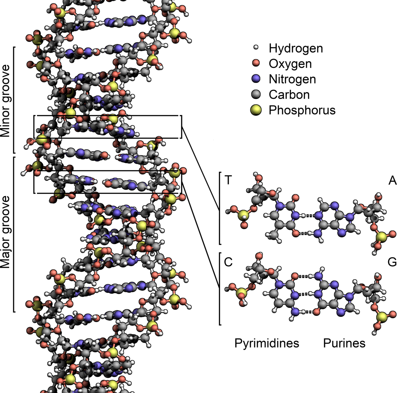
B-DNA
The classic right-handed double helix structure with major and minor grooves.
II. The Discovery of PuF: The Transcription Whisperer
In 1989, a landmark study cracked open the NHE III₁'s secrets by identifying its first protein partner: the Purine Factor (PuF) 1 2 . Here's how the detective work unfolded:
Step-by-Step: The Key Experiment
Fraction Hunting
Scientists partially purified nuclear proteins from human cells, isolating fractions with c-Myc promoter-binding activity.
DNA Mobility Shift Assays
They incubated these fractions with a radioactive NHE III₁ DNA probe (-142 to -115 region). PuF's binding slowed the probe's movement through a gel, revealing direct interaction.
Precision Mapping
Methylation interference pinpointed exact contact points: the palindromic sequence "GGGTGGG" at positions -128 to -122. Mutating this site abolished binding.
Functional Test
Adding synthetic "GGGTGGG" oligonucleotides as decoys completely shut down c-Myc transcription in vitro—proving PuF's essential role.
| Experimental Approach | Finding | Significance |
|---|---|---|
| Deletion of -142/-115 region | ↓ Transcription efficiency by >80% | Confirmed NHE III₁'s regulatory role |
| Methylation interference | Identified GGGTGGG as PuF contact site | Revealed sequence-specific binding |
| Oligonucleotide decoy competition | Complete repression of c-Myc transcription | Proved PuF is essential for activation |
"Our data suggest pur/pyr sequences serve as transcription factor landing pads—a paradigm shift for gene control."
PuF Binding to NHE III₁

Click image to enlarge. Illustration shows PuF (purple) binding to the GGGTGGG sequence in the NHE III₁ region.
III. The Scientist's Toolkit: Cracking the NHE Code
Studying ephemeral DNA structures demands ingenious tools. Key reagents that powered this field:
| Reagent/Tool | Function | Key Insight Enabled |
|---|---|---|
| DNase I | Digests "open" chromatin regions | Identified hypersensitive sites in c-Myc promoter |
| Cationic Porphyrins (TMPyP4) | Stabilizes G-quadruplex structures | Proved G4 folding represses c-Myc transcription 6 |
| Polyethylene Glycol (PEG) | Mimics molecular crowding in nuclei | Revealed G4/i-motif stability at neutral pH 3 5 |
| QW10 Peptide | Binds and stabilizes c-Myc G4 | Achieved 2.5-fold c-Myc downregulation in breast cancer cells 3 |
| PARP-1 Inhibitors | Block G4-unfolding enzyme | Confirmed PARP-1's role in switching c-Myc ON 6 |
DNase I
An endonuclease that cleaves DNA at sites not protected by proteins, revealing "open" chromatin regions like NHE III₁.
Chromatin Analysis Hypersensitivity MappingTMPyP4
A porphyrin compound that selectively binds and stabilizes G-quadruplex structures, used to probe their biological roles.
G4 Stabilizer Therapeutic LeadPEG
Polymer used to mimic the crowded intracellular environment, revealing that G4/i-motifs form under physiological conditions.
Molecular Crowding Biophysical ToolQW10 Peptide
A designed peptide that specifically targets the c-Myc G4 structure, showing promise as a cancer therapeutic.
G4 Binder c-Myc InhibitorIV. Beyond PuF: The Expanding Universe of G4 Regulators
PuF was just the first soldier in an army of NHE III₁-interacting proteins:
NME2
A nucleoside diphosphate kinase that activates c-Myc by binding G4 and recruiting transcription machinery .
PIWIL2
Partners with NME2 to boost c-Myc expression—linking stem cell proteins to cancer .
PARP-1
The "molecular origami master" that unfolds G4 into B-DNA, flipping c-Myc ON. Inhibiting PARP-1 locks DNA in repressive G4 6 .
Genomic Significance
Remarkably, 17 genomic cousins of the c-Myc NHE III₁ sequence exist across human chromosomes. These "Pu27-HS" sites regulate genes like SOX2 (critical for stemness), suggesting a universal regulatory code 4 .
V. Therapeutic Horizons: Silencing c-Myc with Structure
Harnessing NHE III₁ biology offers brilliant anti-cancer strategies:
G4-Stabilizing Drugs
Compounds like QW10 peptide bind c-Myc G4, suppressing transcription and slashing breast cancer cell growth (IC₅₀ = 6.44 μM at 96 hours) while sparing healthy cells 3 .
PARP-1 Inhibitors
Used in BRCA-mutant cancers, they may also trap c-Myc in "OFF" mode by preventing G4 unfolding 6 .
NME2 Disruptors
Could block PIWIL2/NME2 complexes from over-activating c-Myc in tumors .
"Molecular crowding stabilizes the i-motif at pH 6.7—within the physiological range. These structures ARE biology."
Therapeutic Targeting of c-Myc Regulation

VI. Future Codebreakers: Unanswered Mysteries
Despite progress, puzzles endure:
- How do environmental cues (pH, ions, metabolites) dynamically flip the NHE III₁ between structures in vivo?
- Can we design tumor-specific G4 drugs avoiding off-target effects on "good" G4s (e.g., in telomeres)?
- Do Pu27-HS elements "cross-talk" to coordinate gene networks?
Conclusion: The Smallest Switches Cast the Longest Shadows
The story of the c-Myc NHE III₁ is a testament to biology's elegance: a vanishingly small DNA segment controls an oncogene that looms over cancer biology. From PuF's discovery to G-quadruplex-targeted therapies, this field exemplifies how understanding fundamental mechanisms unlocks transformative medicine. As research illuminates the dance between DNA structure and transcription factors, we edge closer to flipping c-Myc's switch from "destroy" to "cure."