Molecular Scissors and Glue: How Restriction Enzymes and DNA Ligase Power Modern Cloning
This article provides a comprehensive overview of the indispensable roles restriction enzymes and DNA ligase play in molecular cloning, a cornerstone technique for researchers, scientists, and drug development professionals.
Molecular Scissors and Glue: How Restriction Enzymes and DNA Ligase Power Modern Cloning
Abstract
This article provides a comprehensive overview of the indispensable roles restriction enzymes and DNA ligase play in molecular cloning, a cornerstone technique for researchers, scientists, and drug development professionals. It covers the foundational science, from the historical discovery of restriction-modification systems to the precise molecular mechanisms of DNA cutting and joining. The scope extends to detailed methodological workflows for traditional and advanced cloning techniques, practical troubleshooting guides for common laboratory challenges, and a comparative analysis of modern assembly methods. By synthesizing established protocols with current applications in genomics and therapeutic development, this resource aims to be a definitive guide for efficiently designing, executing, and optimizing cloning experiments.
The Discovery and Mechanism: Understanding the Biology of DNA Cutting and Joining
The discovery of restriction and modification (R-M) systems represents a foundational pillar of modern molecular biology. What began as the observation of a puzzling bacteriophage behavior, termed "host-induced variation," evolved into the understanding of a sophisticated bacterial immune mechanism. This enzymatic system, capable of distinguishing between self and non-self DNA, provided the very tools that enabled the recombinant DNA revolution. This review details the historical trajectory of this discovery, from its genetic origins to the biochemical characterization of the enzymes, and frames its profound significance within the context of molecular cloning. The precise DNA cleavage by restriction endonucleases, coupled with the DNA-joining activity of DNA ligases, created the "cut-and-paste" methodology that underlies most genetic engineering and drug development workflows today.
In the early 1950s, scientists studying bacteriophages encountered a curious and non-hereditary phenomenon. Bacteriophages isolated from one bacterial strain showed a dramatically reduced ability to reproduce in a different strain, yet readily regained this ability after a single growth cycle on the original host [1]. This reversible change, initially termed host-controlled variation or host-induced variation, was first reported by Luria and Human in 1952 and by Bertani and Weigle in 1953 [2] [1]. The efficiency of plating (EOP) of phages on a restricting host could drop to values between 10⁻¹ and 10⁻⁵, indicating a potent, yet reversible, barrier to viral propagation [1]. This phenomenon, which would later be recognized as the genetic manifestation of R-M systems, set the stage for a series of investigations that would unravel a fundamental bacterial defense mechanism and ultimately provide the key tools for manipulating DNA in vitro.
The Genetic Foundation: Key Early Experiments
The initial genetic observations paved the way for more systematic studies that began to outline the molecular nature of host-controlled variation.
Establishing the DNA as the Carrier of Specificity
A critical leap in understanding came from the work of Werner Arber and Daisy Dussoix in 1962 [3]. Using bacteriophage λ as a model, they demonstrated that the phage DNA itself carried the host-range imprint. They observed that phage λ grown on E. coli K12 was restricted when plated on a K12(P1) lysogen (a strain lysogenized by bacteriophage P1), whereas the few progeny phage that did manage to grow were thereafter modified and could grow efficiently on the K12(P1) lysogen [3]. This modification was lost upon passage through a non-lysogenic strain, confirming that the effect was not due to mutation but to a reversible alteration of the DNA. Their work showed that bacterial cells could impart a specific "modification" to DNA that replicated within them, protecting it from "restriction" upon subsequent infection of the same cell type.
Discovery of the EcoP15 and EcoP1 Systems
Parallel genetic studies in E. coli 15T− revealed the presence of multiple, independent R-M systems. Kenneth Stacey and later Werner Arber's group identified that this strain possessed the chromosomal EcoA (Type I) system and a second, plasmid-borne system [3]. This second system, dubbed EcoP15, was found on the p15B plasmid and was closely related to the R-M system of bacteriophage P1 (EcoP1) [3]. These systems, later classified as Type III R-M systems, demonstrated that R-M genes could be encoded on mobile genetic elements and were not solely chromosomal attributes [3] [4].
Table 1: Key Early Experiments in the Discovery of R-M Systems
| Year | Researchers | Experimental System | Key Finding |
|---|---|---|---|
| 1952-1953 | Luria & Human; Bertani & Weigle | Various bacteriophages | Discovery of host-controlled variation as a non-hereditary phenomenon [2] [1]. |
| 1962 | Arber & Dussoix | Phage λ in E. coli K12 and K12(P1) | Identified DNA as the target of host-specific restriction and modification [3] [1]. |
| 1960s | Arber & Linn; Arber & Wauters-Williems | E. coli 15T− and plasmid p15B | Discovery of the EcoP15 R-M system on a plasmid, distinct from the chromosomal EcoA system [3]. |
| 1960s | Arber | Methionine-starved E. coli | Implicated S-adenosylmethionine (SAM) and DNA methylation in the modification process [3]. |
| 1970 | Smith & Wilcox | Haemophilus influenzae | Iscribed the first Type II restriction enzyme (HindII), which cleaves DNA at specific sequences [2] [1]. |
| 1971 | Danna & Nathans | SV40 DNA + Endonuclease R | First use of a restriction enzyme to generate a physical map of a genome [2]. |
The Role of Methionine and DNA Methylation
Gunther Stent's suggestion that DNA methylation might be the basis for modification led to a crucial experiment. Arber and colleagues used methionine mutants (met⁻) of E. coli, which are deficient in the synthesis of S-adenosyl-L-methionine (SAM), the methyl group donor for most DNA methyltransferases [3]. They found that methionine starvation inhibited the modification function of the R-M system, thereby directly linking SAM-dependent DNA methylation to the host-specific modification of DNA [3] [1]. This finding connected the genetic phenomenon to a tangible biochemical process.
The Biochemical Revolution: From Concept to Enzyme
The theoretical framework for R-M systems was established by Werner Arber in 1965, who postulated that restriction enzymes would recognize specific DNA sequences and cleave them, while modification enzymes would methylate the same sequences to protect them [5] [1]. The subsequent isolation of these enzymes confirmed this model and opened a new era of molecular biology.
The First Restriction Enzymes
The first restriction enzymes to be isolated, such as those from E. coli K and B (EcoKI and EcoBI), were classified as Type I R-M systems [2] [1]. These complex enzymes recognized specific sequences but cleaved DNA at variable, often distant, locations, making them unsuitable for practical applications in DNA manipulation [2].
The pivotal breakthrough came in 1970 when Hamilton Smith and Kent Wilcox isolated a restriction enzyme from Haemophilus influenzae serotype d [2] [1]. This enzyme, initially called "endonuclease R," was found to cleave bacteriophage T7 DNA into specific fragments. It was the first characterized Type II restriction enzyme, defined by its ability to recognize a specific DNA sequence and cleave at a fixed location within or adjacent to that sequence [2]. Smith and Kelly soon determined its recognition sequence to be GTY↓RAC (where Y is C or T, R is A or G, and ↓ indicates the cleavage site) [2]. This enzyme was later named HindII.
A Powerful Combination: Restriction Enzymes and Gel Electrophoresis
The full potential of Type II enzymes was realized through the work of Daniel Nathans and Kathleen Danna. Using the enzyme preparation provided by Smith (which was later found to contain both HindII and HindIII), they digested the DNA of simian virus 40 (SV40) [2]. Instead of using sucrose gradients for analysis, they employed polyacrylamide gel electrophoresis, a technique adapted from RNA separation methods [2]. This allowed them to resolve the digestion products into discrete, reproducible bands, creating the first "restriction map" [2]. This landmark 1971 paper demonstrated that restriction enzymes could be used to dissect genomes into specific fragments for mapping and analysis, a foundational technique for molecular cloning [2].
Table 2: Classification of Major Restriction-Modification System Types
| Type | Subunit Composition | Recognition Sequence | Cleavage Position | Cofactors | Primary Use |
|---|---|---|---|---|---|
| Type I | 3 subunits (R, M, S) | Bipartite (e.g., EcoKI: AACNNNNNNGTGC) | Variable, >1000 bp away [1] | ATP, SAM, Mg²⁺ [4] | Research |
| Type II | Separate R (homodimer) and M (monomer) enzymes | Short, palindromic (e.g., EcoRI: GAATTC) | Within or close to recognition site [4] | Mg²⁺ [4] | Molecular cloning, DNA analysis |
| Type IIS | Separate R and M enzymes | Asymmetric (e.g., BsaI: GGTCTC) | Outside ( downstream) of recognition site [6] | Mg²⁺ | Advanced DNA assembly (e.g., Golden Gate) |
| Type III | Heterocomplex (R₂M₂ or M₂R) [3] | Short, asymmetric (e.g., EcoP15I: CAGCAG) | 25-27 bp downstream of site [3] | ATP, (SAM?), Mg²⁺ [3] | Research |
| Type IV | Restriction enzyme only | Methylated DNA (e.g., McrBC: RmC) | Variable | GTP (for some) | Research on modified DNA |
Diagram 1: The Historical Workflow from Phenomenon to Tool. This diagram outlines the key stages in the discovery and development of restriction-modification systems, from initial genetic observations to their application as essential tools in molecular biology.
The Core Technology for Molecular Cloning
The unique properties of Type II restriction enzymes made them ideal for recombinant DNA technology. Their simplicity—a restriction endonuclease for cutting and a separate, sequence-specific methyltransferase for protection—allowed scientists to use the REase in vitro to create defined DNA fragments.
The "Cut and Paste" Paradigm
Molecular cloning via restriction enzymes is a multi-step process that fundamentally relies on the specific cutting and pasting of DNA [7]:
- DNA Digestion: A plasmid vector and the DNA fragment of interest (insert) are cut with the same restriction enzyme(s).
- Ligation: The vector and insert are mixed and joined together by DNA ligase, an enzyme that catalyzes the formation of phosphodiester bonds, creating a recombinant DNA molecule.
- Transformation and Selection: The recombinant plasmid is introduced into a bacterial host (transformation). Bacteria containing the plasmid are selected using antibiotic resistance markers, and those with successful insertions are often identified via cloning selection markers (e.g., blue-white screening) [7].
This process, often called restriction cloning, allows for the precise insertion of a gene into a self-replicating vector for amplification and study [7].
Directional Cloning and Advanced Applications
Early cloning often used a single restriction enzyme, which led to inserts ligating in either orientation. Directional cloning, which uses two different restriction enzymes to create non-compatible ends on the vector and insert, ensures the insert is ligated in the correct orientation and significantly reduces background [7]. Further advancements leveraged Type IIS restriction enzymes, which cut outside of their recognition sequence. This property is exploited in techniques like Golden Gate Assembly, which allows for the seamless, one-pot assembly of multiple DNA fragments without incorporating the restriction site into the final construct [6] [5].
Diagram 2: The Cloning Workflow and its Applications. This diagram illustrates the core "cut and paste" process of restriction enzyme cloning, from digestion and ligation to the creation of a recombinant plasmid, and outlines its major applications in biotechnology and medicine.
The Scientist's Toolkit: Essential Reagents and Protocols
The practical application of this historical discovery relies on a standardized set of reagents and methodologies.
Key Research Reagent Solutions
Table 3: Essential Reagents for Restriction Enzyme-Based Cloning
| Reagent / Tool | Function in Cloning | Example |
|---|---|---|
| Type IIP Restriction Enzymes | Cut vector and insert DNA at specific palindromic sequences to generate compatible ends. | EcoRI, HindIII, BamHI [6] [7] |
| Type IIS Restriction Enzymes | Cut outside recognition site; enable advanced, seamless assembly of DNA fragments. | BsaI, BsmBI, Esp3I [6] |
| DNA Ligase | Joins the sugar-phosphate backbones of the digested vector and insert fragments. | T4 DNA Ligase [7] |
| Plasmid Cloning Vector | A circular DNA molecule containing an Origin of Replication (ORI), Selectable Marker (e.g., Amp⁺), and a Multi-Cloning Site (MCS). | pBR322, pUC19 [7] |
| Competent Cells | Chemically or electroporation-treated bacterial cells capable of taking up foreign DNA. | E. coli DH5α, BL21(DE3) |
| Selection Media | Growth media containing an antibiotic to select for bacteria that have taken up the plasmid vector. | LB + Ampicillin [7] |
Detailed Protocol: Basic Restriction Cloning
The following methodology outlines a standard restriction enzyme cloning procedure for inserting a DNA fragment into a plasmid vector [7].
Experimental Design and In Silico Simulation:
- Select a plasmid vector with a Multi-Cloning Site (MCS).
- Choose restriction enzymes that cut uniquely in the vector MCS and at the ends of your insert. For directional cloning, use two different enzymes.
- Use software to simulate the digestion and confirm the final recombinant plasmid sequence.
Digestion of Vector and Insert:
- Set up separate reactions for the vector and insert DNA.
- Reaction Mix: 1 µg DNA, 1X Restriction Enzyme Buffer, 10-20 units of each restriction enzyme, Nuclease-free water to final volume.
- Incubation: 1 hour at the enzyme's optimal temperature (typically 37°C).
Purification of Digested Products:
- Run the digestion products on an agarose gel.
- Excise the bands corresponding to the linearized vector and the insert.
- Purify the DNA from the gel slices using a gel extraction kit. Quantify the purified DNA.
Ligation:
- Reaction Mix: 50 ng linearized vector, insert DNA (at a 3:1 molar ratio of insert:vector), 1X DNA Ligase Buffer, 1 µL T4 DNA Ligase, Water to final volume.
- Incubation: 10-30 minutes at room temperature or 16°C for 1-2 hours.
Transformation and Selection:
- Add the ligation mixture to chemically competent E. coli cells. Incubate on ice, heat-shock at 42°C for 30-45 seconds, and return to ice.
- Add recovery media and incubate with shaking for 1 hour.
- Plate the transformation mixture onto agar plates containing the appropriate antibiotic.
- Incubate plates overnight at 37°C.
Screening and Verification:
- Pick individual colonies and grow in small cultures.
- Isolate plasmid DNA (mini-prep).
- Verify the presence and correctness of the insert by restriction digest (analytical gel) and DNA sequencing.
The journey from the observation of host-induced variation to the elucidation of the restriction-modification system is a testament to the power of fundamental scientific research. The discovery of Type II restriction enzymes provided the precise molecular scissors that, when combined with DNA ligase, enabled the controlled manipulation of DNA. This "cut-and-paste" methodology lies at the heart of molecular cloning, forming the technical foundation for the entire biotechnology industry. The cloning and production of therapeutic proteins like human insulin, the mapping of disease genes, and the development of advanced gene therapies all trace their origins to the bacterial immune system and the curious scientists who sought to understand it. As new cloning techniques emerge, the historical and practical framework established by restriction enzymes remains a cornerstone of genetic engineering and drug development.
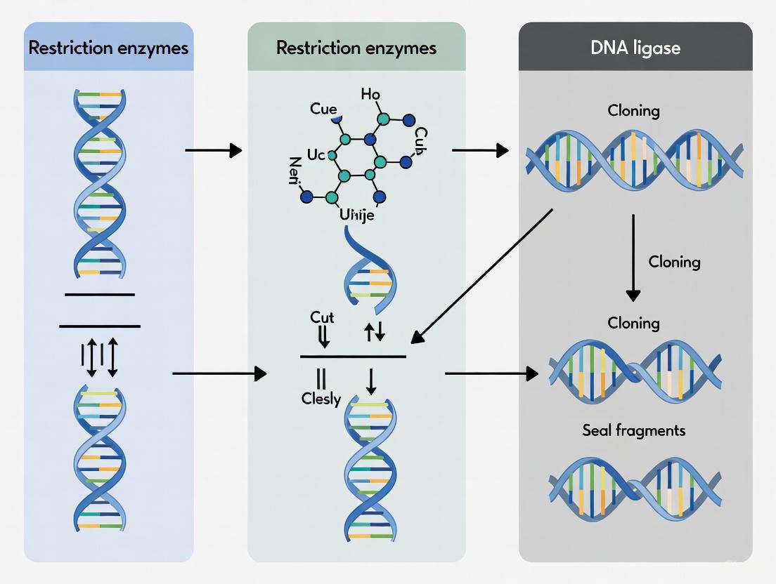
Restriction endonucleases, often termed "molecular scissors," are enzymatic tools that recognize specific DNA sequences and catalyze the cleavage of double-stranded DNA. These enzymes form the foundation of recombinant DNA technology, enabling the precise manipulation of genetic material essential for cloning research. This technical guide explores the molecular mechanisms underlying their sequence-specific recognition and cleavage activities, detailing how the ends they generate are leveraged by DNA ligase to construct novel recombinant molecules. We provide a comprehensive analysis of enzyme classes, quantitative cleavage data, standardized experimental protocols, and essential reagent solutions to support research and drug development applications.
Restriction endonucleases are fundamental tools in molecular biology, with their natural function serving as a defense mechanism in bacterial cells against invading viral DNA (bacteriophages). The restriction-modification (R-M) system functions through the coordinated activity of two enzymes: a restriction enzyme that cleaves foreign, unmethylated DNA, and a methyltransferase that modifies the host's own DNA by adding methyl groups to specific bases within the recognition sequences, thereby protecting it from cleavage [8] [9]. This selective cleavage ensures that only foreign DNA is targeted and degraded, while the host DNA remains intact. The functionality of restriction enzymes extends beyond this native bacterial immune role, with pivotal applications in genetic engineering processes where they provide the foundational mechanism for DNA manipulation [8].
The discovery of restriction enzymes dates back to the 1950s and 1960s, when researchers observed that bacteriophages exhibited host-controlled variation in their ability to infect different bacterial strains [9] [2]. Werner Arber proposed the restriction-modification system, suggesting that host DNA was protected through methylation while foreign viral DNA was cleaved by restriction enzymes. The full potential of restriction enzymes became apparent with the discovery of Type II restriction enzymes by Hamilton Smith and Kent Wilcox, which cleave DNA at specific symmetrical sequences within their recognition sites [9] [2]. This pioneering work, which earned Daniel Nathans, Hamilton Smith, and Werner Arber the 1978 Nobel Prize in Physiology or Medicine, laid the groundwork for modern molecular cloning techniques [9].
Classification and Mechanisms of Restriction Enzymes
Restriction enzymes are categorized based on their structural complexity, recognition sequence characteristics, cleavage site position, and cofactor requirements. Understanding these classifications is crucial for selecting appropriate enzymes for specific experimental applications.
Enzyme Classes and Characteristics
Table 1: Classification and characteristics of restriction endonucleases
| Enzyme Class | Recognition Sequence | Cleavage Site | Cofactor Requirements | Primary Applications |
|---|---|---|---|---|
| Type I | Asymmetric and bipartite | Variable distance from recognition site (≥1000 bp) | ATP, Mg²⁺, AdoMet | Limited research applications |
| Type II | Specific palindromic sequences (4-8 bp) | Within or close to recognition site | Mg²⁺ | Molecular cloning, DNA analysis, RFLP |
| Type IIS | Asymmetric sequences | Outside of recognition site (1-20 bp away) | Mg²⁺ | Golden Gate Assembly, advanced DNA assembly |
| Type III | Short asymmetric sequences | Specific distance (24-26 bp) from recognition site | ATP, Mg²⁺ | Limited research applications |
| Type IV | Methylated DNA | Approximately 30 bp from recognition site | Mg²⁺ | Methylation studies |
Molecular Mechanisms of Sequence Recognition and Cleavage
Type II restriction enzymes, the most commonly used class in molecular biology, achieve sequence-specific recognition through intimate interactions between protein domains and specific nucleotide bases within their target sequences. These enzymes typically recognize short palindromic sequences of 4-8 base pairs in length, reading the DNA sequence through a combination of hydrogen bonding, van der Waals forces, and structural complementarity with the DNA double helix [9]. The precise molecular recognition process varies among enzyme families, with some employing a "direct readout" mechanism where amino acid side chains form specific contacts with nucleotide bases, while others use "indirect readout" by sensing sequence-dependent DNA conformation and flexibility.
The cleavage mechanism involves coordinated hydrolysis of the phosphodiester bonds in both DNA strands. Most Type II enzymes generate breaks that produce either sticky ends (5′ or 3′ protruding termini) or blunt ends (evenly cut ends without overhangs) [9]. Sticky ends are particularly valuable in cloning applications as the complementary overhangs facilitate specific annealing of DNA fragments before ligation. The cleavage reaction requires Mg²⁺ as a cofactor, which activates water molecules for nucleophilic attack on the phosphate groups in the DNA backbone and stabilizes the transition state during hydrolysis.
Type IIS restriction enzymes represent a particularly valuable subclass that recognize asymmetric sequences and cleave outside of their recognition site [8]. This unique property enables greater flexibility in DNA assembly, as the cleavage site can be designed independently of the recognition sequence. As explained by Bill Jack, an Emeritus Scientist at New England Biolabs: "Many Type IIS enzymes, when they cut, leave a staggered overhang. Since the single-stranded overhang is not defined by the restriction site, it can be essentially any set of nucleotides, and so, there's a great ability to order the type of sequence or the type of overhang that will be there to allow assembly" [8]. This characteristic makes Type IIS enzymes particularly valuable for advanced cloning techniques such as Golden Gate Assembly, which enables complex DNA assemblies with high efficiency and accuracy [8].
Experimental Applications and Methodologies
Core Restriction Enzyme-Based Protocols
Standard Restriction Digestion and Ligation Protocol
The foundational protocol for molecular cloning involves digesting both the vector and insert DNA with restriction enzymes that generate compatible ends, followed by ligation with DNA ligase to create recombinant molecules.
Materials Required:
- Purified DNA vector and insert
- Appropriate restriction enzymes with compatible buffers
- DNA ligase (typically T4 DNA ligase)
- ATP (cofactor for ligation)
- Thermostable DNA polymerase with proofreading capability for amplification
- Agarose gel electrophoresis equipment
- DNA purification kits or reagents
Methodology:
- Experimental Design: Select a cloning vector with an appropriate multiple cloning site (MCS) containing unique restriction sites not present in your insert DNA [10]. Choose restriction enzymes that generate compatible ends for both vector and insert.
- DNA Digestion: Set up separate digestion reactions for vector and insert DNA using the selected restriction enzymes. A typical 50 μL reaction contains:
- 1 μg DNA
- 1× restriction enzyme buffer
- 10-20 units of each restriction enzyme
- Nuclease-free water to volume
- Incubate at the recommended temperature (usually 37°C) for 1-2 hours [9]
- Fragment Purification: Separate digested fragments by agarose gel electrophoresis and extract the desired bands using gel extraction kits to isolate vector and insert fragments.
- Ligation: Combine the purified vector and insert fragments in a molar ratio (typically 1:3 vector:insert) with DNA ligase. A standard 20 μL ligation reaction contains:
- 50-100 ng vector DNA
- Appropriate molar amount of insert DNA
- 1× ligation buffer
- 1 mM ATP
- 1-5 units T4 DNA ligase
- Incubate at 16°C for 4-16 hours [10]
- Transformation and Screening: Transform the ligation reaction into competent bacterial cells, plate on selective media, and screen resulting colonies for correct recombinants using colony PCR or restriction analysis.
Advanced Application: HIV-1 Gag Gene Cloning for Replication Studies
This specialized protocol demonstrates how restriction enzyme-based cloning can be applied to study viral replication capacity, specifically for HIV-1 subtype C gag genes [11].
Materials Required:
- Viral RNA from HIV-1 infected plasma
- MJ4 subtype C HIV-1 proviral backbone
- High-fidelity DNA polymerase with proofreading capability
- Restriction enzymes for insertion into MJ4 vector
- CEM-CCR5 T-cell line for replication assays
- Radiolabeled reverse transcriptase assay components
Methodology:
- RNA Extraction and cDNA Synthesis: Extract viral RNA from 140 μL thawed HIV-1 infected plasma using a commercial extraction kit. Perform reverse transcription immediately after extraction to maximize RNA integrity [11].
- Nested PCR Amplification: Amplify the gag gene using a two-step nested PCR approach with high-fidelity DNA polymerase to minimize PCR-introduced errors:
- First-round PCR: Use one-step RT-PCR with outer primers
- Second-round PCR: Use 1 μL of first-round product as template with nested primers containing appropriate restriction sites for cloning [11]
- Restriction Digestion and Cloning: Digest both the PCR-amplified gag gene and MJ4 proviral backbone with selected restriction enzymes. Purify fragments and ligate using T4 DNA ligase.
- Virus Production: Transfect recombinant proviral constructs into 293T cells using appropriate transfection reagents. Harvest virus supernatants after 72 hours.
- Replication Capacity Assay: Infect CEM-CCR5 T-cells with normalized virus stocks and monitor replication over 7-10 days using radiolabeled reverse transcriptase assays to quantify virus production [11].
Figure 1: HIV-1 Gag Gene Cloning and Assessment Workflow. This diagram illustrates the key steps in the restriction enzyme-based cloning method for studying HIV-1 replication capacity, from RNA extraction to functional assessment [11].
Essential Research Reagent Solutions
Table 2: Key research reagents for restriction enzyme-based experiments
| Reagent Category | Specific Examples | Function and Application | Technical Considerations |
|---|---|---|---|
| Restriction Enzymes | HindII, EcoRI, BamHI, BsaI-HFv2, BsmBI-v2 | Sequence-specific DNA cleavage for fragmentation and vector linearization | Select based on recognition sequence, cutting frequency, and end type (sticky vs blunt) |
| DNA Ligases | T4 DNA Ligase | Joins compatible DNA ends by catalyzing phosphodiester bond formation | Requires ATP cofactor; works optimally with complementary overhangs |
| DNA Polymerases | High-fidelity proofreading enzymes (e.g., Q5, Phusion) | Amplify DNA fragments with minimal errors for cloning | Critical for PCR amplification of inserts prior to restriction digestion |
| Cloning Vectors | Plasmid vectors with MCS (Multiple Cloning Sites) | Serve as carrier molecules for DNA fragments | MCS contains multiple unique restriction sites for flexible cloning strategies |
| Competent Cells | E. coli strains (DH5α, BL21) | Host organisms for plasmid propagation after ligation | Transformation efficiency critical for obtaining sufficient recombinant clones |
Advanced DNA Assembly Techniques
Golden Gate Assembly
Golden Gate Assembly represents a significant advancement in restriction enzyme-based cloning, leveraging Type IIS restriction enzymes to enable efficient, one-pot assembly of multiple DNA fragments. This technique exploits the unique property of Type IIS enzymes, which cleave outside of their recognition sequence, allowing custom overhangs to be designed for precise fragment assembly [8]. The method typically uses enzymes such as BsaI-HFv2 or BsmBI-v2, which create 4-base overhangs that can be specifically designed for each fragment junction.
The Golden Gate reaction combines the Type IIS restriction enzyme and DNA ligase in a single-tube reaction, with cycles of digestion and ligation enabling directional assembly of multiple fragments. As Bill Jack explains: "Since the single-stranded overhang is not defined by the restriction site, it can be essentially any set of nucleotides, and so, there's a great ability to order the type of sequence or the type of overhang that will be there to allow assembly" [8]. This approach overcomes limitations of traditional cloning by placing restriction sites outside the fragments of interest, allowing seamless assembly without incorporating extra nucleotides at junctions.
The development of new restriction enzymes and improved understanding of ligase fidelity have made Golden Gate Assembly remarkably efficient, with fragment assemblies achieving >90% accuracy and high efficiency [8]. NEB's Ligase Fidelity Tools further aid in designing high-fidelity Golden Gate Assemblies by optimizing experimental conditions. These advances have enabled sophisticated applications in synthetic biology, including construction of complex genetic circuits and metabolic pathways.
Restriction Enzyme Applications Beyond Cloning
While essential for molecular cloning, restriction enzymes serve critical functions in other molecular biology applications:
Restriction Fragment Length Polymorphism (RFLP) Analysis: RFLP was one of the first DNA fingerprinting methods, relying on restriction enzyme digestion to detect variations in DNA sequences between individuals [12]. The technique involves digesting genomic DNA with restriction enzymes, separating fragments by gel electrophoresis, transferring to membranes (Southern blotting), and hybridizing with labeled probes to reveal unique fingerprint patterns. RFLP markers have been widely used in genetic counseling, forensic analysis, and studying genetic diversity.
DNA Methylation Analysis: Some restriction enzymes are sensitive to DNA methylation, enabling their use in epigenetic studies [12]. Enzymes such as HpaII, which cleaves unmethylated CCGG sites but not methylated ones, allow researchers to map methylation patterns across genomes. This application is particularly valuable for studying epigenetic regulation in development and disease, including cancer research where aberrant methylation patterns are common.
Amplified Fragment Length Polymorphism (AFLP): AFLP combines restriction digestion with PCR amplification to generate genetic fingerprints without prior sequence knowledge [12]. Genomic DNA is digested with two restriction enzymes (typically a rare-cutter like EcoRI and a frequent-cutter like MseI), followed by ligation of adapters and selective PCR amplification. The resulting fragment patterns provide robust markers for genetic mapping, phylogenetic studies, and population genetics.
Restriction endonucleases remain indispensable tools in molecular biology, providing the foundation for DNA manipulation through their precise sequence-specific cleavage activities. From their initial discovery as bacterial defense mechanisms to their current status as workhorses of genetic engineering, these molecular scissors have continuously evolved to meet the demands of increasingly sophisticated research applications. Their synergy with DNA ligase enables the construction of recombinant DNA molecules that form the basis of modern cloning technologies, from basic plasmid construction to advanced assembly methods like Golden Gate cloning.
The continued development of novel restriction enzymes, particularly Type IIS variants with their enhanced flexibility, ensures that these molecular tools will remain relevant in the era of synthetic biology and precision genome engineering. As research progresses, the fundamental principles of restriction and ligation continue to underpin new methodologies, maintaining the central role of these enzymes in advancing both basic research and therapeutic development. For drug development professionals and researchers, understanding the mechanisms and applications of restriction endonucleases remains essential for designing effective genetic engineering strategies and interpreting experimental results in molecular cloning research.
In molecular cloning, the precise manipulation of DNA fragments relies fundamentally on the nature of their terminal ends. Restriction endonucleases, often described as "molecular scissors," cleave DNA at specific sequences to generate these defined ends [13] [9]. DNA ligase then functions as the "molecular glue," catalyzing the formation of phosphodiester bonds to join compatible ends together [14]. This restriction-ligation process forms the cornerstone of recombinant DNA technology, enabling researchers to create novel genetic constructs for applications ranging from basic biological research to pharmaceutical development [9]. The efficiency and outcome of these enzymatic reactions are profoundly influenced by whether the DNA fragments possess sticky ends or blunt ends—a fundamental distinction that dictates experimental design, efficiency, and success in cloning workflows [15] [14]. This guide provides an in-depth technical examination of these DNA end types, their generation, and their strategic implications for research and drug development.
Defining DNA Ends: Structural Characteristics and Formation
Sticky Ends (Cohesive Ends)
Sticky ends, or cohesive ends, are characterized by short, single-stranded DNA overhangs that result from staggered cuts made by restriction enzymes in the double-stranded DNA backbone [13] [15]. These overhangs are typically complementary palindromic sequences, allowing fragments from different origins to associate through hydrogen bonding before being permanently sealed by DNA ligase [13] [15]. The overhangs can be either 5' or 3' protrusions, depending on the specific restriction enzyme used [14]. The discovery of sticky ends by Ronald W. Davis as a product of EcoRI action revolutionized molecular biology by providing a mechanism for precise and efficient joining of DNA fragments [15].
Blunt Ends (Non-Cohesive Ends)
Blunt ends occur when both strands of a DNA molecule are cut at equivalent positions, resulting in terminal ends with no unpaired bases [13] [15] [16]. This straight-across cleavage pattern generates fragments that are universally compatible but lack the stabilizing hydrogen bonds that facilitate fragment association in sticky-end ligations [13] [16]. The absence of these complementary interactions makes the ligation process significantly less efficient and more challenging to control [16] [17].
Table 1: Comparative Characteristics of Sticky Ends vs. Blunt Ends
| Characteristic | Sticky Ends | Blunt Ends |
|---|---|---|
| Structure | Short, single-stranded overhangs (5' or 3') | No overhangs; both strands end at same base position |
| Formation | Staggered cuts by restriction enzymes (e.g., EcoRI, BamHI) | Straight-across cuts by restriction enzymes (e.g., SmaI, EcoRV) [16] |
| Complementarity | Sequence-specific; must be compatible for ligation | Universally compatible with any blunt end |
| Ligation Efficiency | High (due to hydrogen bonding stabilization) | Low (10-100 times less efficient) [16] [17] |
| Directional Cloning | Possible with non-complementary overhangs | Not inherently possible without additional modifications [16] [14] |
| Common Applications | Standard cloning, directional insertion | PCR product cloning, library construction when sticky ends are unavailable |
Enzymatic Formation of DNA Ends
Restriction endonucleases are classified into four major types (I-IV) based on their structural complexity, recognition sites, cleavage positions, and cofactor requirements [13] [9]. Type II restriction enzymes are the workhorses of molecular cloning, as they recognize specific palindromic sequences and cleave at predictable positions within or near these recognition sites [9]. While most Type II enzymes (Type IIP) recognize and cut within palindromic sequences, Type IIS enzymes recognize asymmetric sequences and cut at a defined distance outside them, a property exploited in advanced techniques like Golden Gate assembly [13].
The following diagram illustrates how different restriction enzymes generate sticky versus blunt ends through their distinct cleavage patterns:
Practical Implications for Cloning Research and Drug Development
Ligation Efficiency and Experimental Design
The choice between sticky-end and blunt-end cloning strategies has profound implications for experimental efficiency and design. Sticky-end ligations benefit from hydrogen bonding between complementary overhangs, which stabilizes the DNA fragment association and increases ligation efficiency by 10 to 100-fold compared to blunt-end ligations [16] [17]. This efficiency advantage translates to higher transformation yields and reduced screening effort. For blunt-end ligations, the absence of this stabilizing interaction means successful ligation depends on transient associations between 5' phosphate and 3' hydroxyl groups being captured at the right moment by DNA ligase [17]. This necessitates optimized reaction conditions including higher DNA concentrations, increased ligase amounts, longer incubation times, and often the use of crowding agents like polyethylene glycol (PEG) to improve efficiency [18] [17].
Directional Cloning and Vector Considerations
Directional cloning, which ensures inserts are oriented correctly in vectors, is readily achievable with sticky ends by using two different restriction enzymes that generate incompatible ends on each side of the insert [14]. This approach forces the insert into the vector in a single orientation, saving significant time in downstream screening and validation. In contrast, blunt ends are universally compatible but lack inherent directionality, resulting in a 50% chance of incorrect insertion orientation for each transformant [16]. Advanced techniques such as using monophosphorylated vectors and inserts or implementing selection systems like Staby technology can overcome this limitation, but they add complexity to the experimental design [16].
Vector re-circularization presents another significant consideration. In blunt-end cloning, intramolecular ligation (vector self-ligation) competes effectively with the desired insert-vector intermolecular ligation [16] [17]. This background can be minimized by dephosphorylating the vector ends using alkaline phosphatases (e.g., CIP, SAP, or BAP) to remove 5'-phosphate groups, preventing recircularization [16] [17]. Concurrently, ensuring the insert contains 5'-phosphate groups is essential for successful ligation, which may require phosphorylation of PCR-generated inserts using T4 polynucleotide kinase [16] [17].
Strategic Applications in Research and Drug Development
The different properties of sticky and blunt ends make them suitable for distinct applications in research and pharmaceutical development:
Sticky-end cloning is preferred for standard molecular biology applications where efficiency and directional control are priorities, such as constructing expression vectors for recombinant protein production—a critical step in biopharmaceutical development [14].
Blunt-end cloning is invaluable when working with DNA fragments that lack convenient restriction sites or when multiple internal restriction sites preclude the use of traditional sticky-end approaches [16]. This versatility makes blunt-end cloning essential for library construction and cloning PCR products, particularly those generated with proofreading polymerases that produce blunt-ended fragments [16].
Golden Gate assembly represents an advanced application that leverages Type IIS restriction enzymes, which create custom sticky ends outside their recognition sites [13]. This technique enables seamless assembly of multiple DNA fragments in a single reaction and is particularly valuable in synthetic biology applications, including metabolic pathway engineering for therapeutic compound production [13].
Table 2: Optimization Strategies for Different DNA End Types
| Parameter | Sticky-End Ligation | Blunt-End Ligation |
|---|---|---|
| Insert:Vector Ratio | 1:1 to 3:1 (standard) [18] | 10:1 or higher to favor intermolecular ligation [14] |
| Ligase Type/Amount | Standard T4 DNA Ligase (1-1.5 Weiss units) [18] | High-concentration T4 DNA Ligase (1.5-5.0 Weiss units) [14] |
| Reaction Additives | Standard buffer components | PEG 4000 as molecular crowding agent [14] [17] |
| Incubation Time | 10 minutes to 1 hour at 22°C [14] | Up to 24 hours to increase collision probability [17] |
| Phosphorylation State | Standard phosphorylation usually sufficient | Often requires vector dephosphorylation and insert phosphorylation [16] [17] |
| Typical Efficiency | High (benchmark) | 10-100 times lower than sticky ends [16] [17] |
Essential Methodologies and Protocols
Standard Restriction Digestion Protocol
The following protocol provides a reliable method for generating either sticky or blunt ends through restriction enzyme digestion [19]:
Reaction Setup: In a sterile microcentrifuge tube, combine the following components:
- 1 μg DNA (plasmid or genomic)
- 2 μL 10X restriction enzyme buffer
- 1 μL restriction enzyme (10 units)
- Nuclease-free water to 20 μL total volume
Digestion: Mix thoroughly by pipetting and centrifuge briefly. Incubate at the enzyme's optimal temperature (typically 37°C) for 1-4 hours.
Verification: Analyze digestion completeness by agarose gel electrophoresis alongside undigested DNA and appropriate size markers.
Critical Considerations:
- Ensure DNA is free from contaminants such as phenol, ethanol, or detergents that may inhibit enzyme activity [19].
- For double digests with two different enzymes, verify compatibility of buffer and temperature requirements [19].
- For enzymes that do not heat-inactivate effectively, purify DNA after digestion using spin columns or phenol/chloroform extraction before proceeding to ligation [19].
Optimized Ligation Workflow
The ligation process differs significantly between sticky-end and blunt-end fragments. The following workflow outlines a standardized approach adaptable to both scenarios [18] [14] [17]:
Blunt-End Specific Optimizations:
- Implement a two-step incubation: 1 hour with high insert concentration followed by dilution (1:20) and extended incubation to favor complete circularization while minimizing concatemer formation [17].
- For PCR-generated inserts, verify the phosphorylation status. Fragments amplified with proofreading polymerases lack 5'-phosphates and require treatment with T4 polynucleotide kinase before ligation [16] [14].
- When using Taq polymerase-generated fragments with 3'A overhangs for blunt-end cloning, remove the overhangs using proofreading polymerase treatment (e.g., Pfu) before ligation [16].
The Scientist's Toolkit: Essential Reagents
Table 3: Essential Research Reagents for DNA End Manipulation
| Reagent | Function | Key Applications |
|---|---|---|
| Type II Restriction Enzymes | Recognize and cleave specific DNA sequences | Generating sticky or blunt ends for cloning [13] [9] |
| T4 DNA Ligase | Catalyzes phosphodiester bond formation | Joining DNA fragments with compatible ends [18] [14] |
| Alkaline Phosphatase (CIP, SAP) | Removes 5'-phosphate groups | Preventing vector self-ligation in blunt-end cloning [16] [17] |
| T4 Polynucleotide Kinase | Adds 5'-phosphate groups | Phosphoryrating PCR inserts for ligation [16] [14] |
| PEG 4000 | Molecular crowding agent | Increasing effective concentration in blunt-end ligations [14] [17] |
| DNA Polymerases (T4, Klenow) | Fills or removes single-stranded DNA | Converting sticky ends to blunt ends; end repair [16] |
Advanced Applications and Future Perspectives
Type IIS Restriction Enzymes and Golden Gate Assembly
Type IIS restriction enzymes represent a powerful tool for advanced cloning strategies. Unlike conventional Type IIP enzymes that cut within their recognition sites, Type IIS enzymes recognize asymmetric DNA sequences and cleave at a defined distance outside these sequences [13]. This unique property enables Golden Gate assembly, a method that allows seamless assembly of multiple DNA fragments in a single reaction [13]. In this technique, both inserts and destination vectors contain compatible cleavage sites that generate custom complementary overhangs, enabling precise assembly of up to 35 DNA fragments in the desired order [13]. The recognition sites are positioned such that they are removed from the final construct, eliminating the need for scar sequences and making this approach particularly valuable for sophisticated genetic engineering applications in pharmaceutical development and synthetic biology [13].
Isoschizomers and Neoschizomers in Experimental Design
The availability of isoschizomers (different enzymes that recognize and cleave the same sequence) and neoschizomers (enzymes that recognize the same sequence but cleave at different positions) provides valuable flexibility in experimental design [13]. For example, SmaI and XmaI both recognize the sequence 5'-CCCGGG-3', but SmaI generates blunt ends (CCC↓GGG) while XmaI produces sticky ends (C↓CCGGG) [13]. Isoschizomers may offer advantages such as improved stability, reduced cost, different methylation sensitivities, or absence of star activity (relaxed specificity under suboptimal conditions) [13]. This enzyme diversity enables researchers to select the most appropriate restriction endonuclease based on the specific type of DNA end required for their application.
Implications for Drug Development and Biotechnology
The precise manipulation of DNA ends underpins many advanced applications in biotechnology and pharmaceutical development. In plant genetic engineering, Golden Gate assembly facilitates the construction of complex synthetic constructs for metabolic pathway engineering and genome editing [13]. For therapeutic protein production, control over DNA end joining ensures correct open reading frame maintenance and optimal expression cassette design. The emerging field of gene therapy relies on sophisticated vector construction where the specificity of DNA end joining ensures proper transgene integration and expression. As these technologies advance, the fundamental principles governing DNA end recognition and joining continue to inform the development of more efficient and precise genetic engineering tools for therapeutic applications.
The strategic manipulation of DNA ends represents a foundational skill in molecular biology with far-reaching implications for research and drug development. Sticky ends offer efficiency and directional control through complementary hydrogen bonding, while blunt ends provide versatility at the cost of reduced ligation efficiency. The choice between these approaches depends on multiple factors, including the source DNA, available restriction sites, desired insert orientation, and downstream applications. Mastery of both standard and specialized techniques—from basic restriction cloning to advanced Golden Gate assembly—enables researchers to tackle increasingly complex genetic engineering challenges. As molecular techniques continue to evolve, the precise control over DNA fragment joining remains central to innovations across biological research, therapeutic development, and biotechnology.
This technical guide explores the critical role of DNA ligase in molecular cloning, framing it as the indispensable "paste" function that complements the "cut" function of restriction enzymes. We delve into the enzymatic mechanism of phosphodiester bond reformation, detail the types and applications of DNA ligases in research and drug development, and provide optimized protocols for modern cloning workflows. The integral partnership between restriction enzymes and DNA ligase has powered the recombinant DNA revolution, enabling the construction of novel genetic entities for therapeutic and research purposes. This whitepaper provides researchers with a comprehensive resource on ligase biology and practical methodologies to enhance cloning efficiency.
The revolutionary development of molecular cloning rests on a foundational paradigm: the ability to specifically cut and paste DNA sequences. Restriction enzymes serve as precise molecular scissors, recognizing and cleaving DNA at specific palindromic sequences to generate defined fragments [20] [2]. These catalytic proteins, originally discovered as a bacterial defense mechanism against invading bacteriophages, create either blunt ends or cohesive "sticky" ends with short, single-stranded overhangs that enable specific re-association through base-pair complementarity [20] [10].
However, cleavage alone is insufficient for recombinant DNA technology. The final and crucial step—the "pasting" function—is catalyzed by DNA ligase, an enzyme that seals the sugar-phosphate backbone between adjacent nucleotides [21] [10]. In living organisms, DNA ligases are essential for DNA replication, particularly in joining Okazaki fragments on the lagging strand, and for various DNA repair pathways [21] [22]. In vitro, this sealing function enables the stable insertion of DNA fragments into cloning vectors, forming recombinant molecules that can be amplified and expressed in host organisms [22] [10]. The synergistic partnership between restriction enzymes and DNA ligase has thus been the cornerstone of genetic engineering for decades, enabling everything from basic gene mapping to the production of life-saving biologic drugs [2] [10].
The Molecular Mechanism of DNA Ligase
The Phosphodiester Bond: Foundation of DNA Integrity
A phosphodiester bond is a covalent linkage in which a phosphate group forms two ester-like connections, bridging the 3' hydroxyl (-OH) group of one nucleotide to the 5' phosphate (PO₄) group of the adjacent nucleotide [23] [24]. This creates the characteristic sugar-phosphate backbone of DNA and RNA, with the nucleotide bases projecting from this backbone to form the genetic code [23]. The stability of this bond is crucial for genetic integrity, though its susceptibility to hydrolysis varies depending on flanking nucleotides; for instance, phosphodiester bonds adjacent to pyrimidine-purine sequences (e.g., UA and CA) demonstrate notably higher instability [24].
The Enzymatic Steps of Bond Reformation
DNA ligase catalyzes the formation of phosphodiester bonds in a three-step mechanism that requires energy from either ATP (eukaryotic and T4 ligases) or NAD+ (prokaryotic ligases) [21] [25] [22]. The reaction proceeds as follows:
- Adenylation: The ligase reacts with ATP (or NAD+), releasing pyrophosphate (or nicotinamide mononucleotide) and forming a covalent intermediate where AMP becomes attached to a conserved lysine residue within the enzyme's active site [21] [22].
- Transfer to DNA: The AMP group is subsequently transferred from the enzyme to the 5' phosphate terminus of the "donor" DNA strand, forming a high-energy DNA-adenylate intermediate (AppDNA) [21] [25].
- Nucleophilic Attack and Ligation: The 3' hydroxyl group of the "acceptor" DNA strand initiates a nucleophilic attack on the activated 5' phosphate. This results in the formation of a phosphodiester bond, sealing the DNA nick and releasing AMP [21] [25] [22].
This mechanism is conserved across DNA ligases, though cofactor requirements and specific biological roles differ among enzyme types.
Mechanism of DNA Ligase
The following diagram illustrates the three-step enzymatic mechanism by which DNA ligase reforms the phosphodiester bond, from initial adenylation to final bond formation.
Types of DNA Ligases and Their Applications
Classification and Characteristics
Various DNA ligases have been isolated and are utilized in molecular biology, each with distinct properties that make them suitable for specific applications. The following table summarizes key ligases and their characteristics.
Table 1: Types of DNA Ligases and Their Properties
| Ligase Type | Natural Source | Cofactor | Primary Applications | Key Features |
|---|---|---|---|---|
| T4 DNA Ligase | Bacteriophage T4 | ATP | General molecular cloning, blunt & sticky-end ligation, RNA ligation [21] | Most commonly used in labs; ligates blunt ends and cohesive ends; can join RNA-DNA hybrids [21] |
| E. coli DNA Ligase | Escherichia coli | NAD+ | Cohesive-end ligation, in vivo repair [21] | Efficient for sticky ends; less efficient for blunt ends unless under molecular crowding conditions [21] |
| DNA Ligase 1 | Mammals | ATP | Okazaki fragment joining, nuclear DNA repair [21] | Essential for DNA replication; seals nicks in the lagging strand [21] |
| DNA Ligase 3 | Mammals | ATP | Base excision repair, mitochondrial DNA repair [21] | Complexes with XRCC1; only mammalian ligase found in mitochondria [21] |
| DNA Ligase 4 | Mammals | ATP | Non-homologous end joining, V(D)J recombination [21] | Complexes with XRCC4; critical for double-strand break repair and immune system development [21] |
| Thermostable Ligase | Thermophilic bacteria | ATP/NAD+ | PCR-based ligation, detection methods [21] [22] | Stable at high temperatures (>65°C); essential for techniques requiring thermal cycling [21] |
DNA Ligase in Advanced Cloning Techniques
While traditional cloning relies on complementary ends generated by restriction enzymes, advanced techniques leverage the precision of specialized ligases. Golden Gate Assembly is a prominent example that uses Type IIS restriction enzymes, which cut outside their recognition sequence, in conjunction with T4 DNA ligase to enable efficient one-pot assembly of multiple DNA fragments [20]. This method allows for the creation of complex genetic constructs with high efficiency and accuracy, often exceeding 90% success rates [20]. The technique's success depends on the synchronized activity of the restriction enzyme and DNA ligase, facilitated by thermal cycling between their optimal activity temperatures.
Practical Guide: Experimental Protocols for DNA Ligation
Standard Ligation Reaction Setup
A typical ligation reaction involves combining the vector, insert, ligase enzyme, and reaction buffer under optimal conditions. The following table provides a standardized protocol for both sticky-end and blunt-end ligations, which require different optimization strategies.
Table 2: Standardized Ligation Reaction Setup and Conditions
| Reaction Component | Sticky-End Ligation | Blunt-End Ligation | Notes |
|---|---|---|---|
| Vector DNA | 20–100 ng | 20–100 ng | Determine concentration spectrophotometrically |
| Insert DNA | x ng (see ratio calc.) | x ng (see ratio calc.) | 5'-phosphorylation is critical [14] |
| 10X Ligation Buffer | 2 µL | 2 µL | Contains ATP, DTT; freeze-thaw sensitive [14] |
| 50% PEG 4000 | Optional | 2 µL | Crowding agent critical for blunt-end efficiency [14] |
| T4 DNA Ligase | 1.0–1.5 Weiss Units | 1.5–5.0 Weiss Units | Higher concentration needed for blunt ends [14] |
| Nuclease-free Water | to 20 µL | to 20 µL | Dilutes potential inhibitors |
| Incubation | 10 min–1 hr at 22°C | 10 min–1 hr at 22°C | Overnight not typically required [14] |
Critical Optimization Parameters
- Insert:Vector Molar Ratio: The optimal ratio is contingent upon the downstream application and must be determined empirically [14]. A 3:1 ratio is a common starting point for sticky-end ligations, while blunt-end ligations often require higher ratios (e.g., 10:1) to drive the less efficient reaction [14]. The required mass of insert for a 1:1 molar ratio can be calculated as: (ng of vector × length of insert (bp)) ÷ length of vector (bp) [14].
- End Compatibility: For successful ligation, DNA ends must be properly prepared. Sticky ends must be complementary, and 5'-phosphate groups must be present on at least one strand to be joined [14]. PCR products generated by proofreading polymerases lack 5' phosphates and require treatment with T4 Polynucleotide Kinase (PNK) prior to ligation [14].
- Reaction Temperature and Time: While T4 DNA ligase has optimal activity at 37°C, ligation of cohesive ends is often performed at lower temperatures (16–25°C) to stabilize the annealing of short overhangs [21] [14]. Prolonged incubations are generally unnecessary due to the enzyme's high efficiency, with 10 minutes to one hour typically sufficient [14].
Molecular Cloning Workflow
The following diagram outlines the complete workflow for a standard restriction-ligation cloning experiment, from initial DNA preparation to verification of the recombinant construct.
The Scientist's Toolkit: Essential Reagents for DNA Ligation
Successful DNA ligation experiments require a suite of specialized reagents and enzymes. The following table catalogs essential solutions for the molecular biologist's toolkit.
Table 3: Essential Research Reagent Solutions for DNA Ligation
| Reagent / Enzyme | Function / Application | Key Considerations |
|---|---|---|
| T4 DNA Ligase | Joins double-stranded DNA fragments with cohesive or blunt ends [21] [14] | Requires ATP and Mg²⁺ as cofactors; most versatile for laboratory use [21] |
| Restriction Enzymes (Type II) | Generate specific cleavage patterns in DNA to create compatible ends for ligation [20] [2] | Selection determines end type (blunt or sticky); unique site in vector is essential [10] |
| T4 Polynucleotide Kinase (PNK) | Adds 5' phosphate groups to DNA fragments, essential for ligating PCR products [14] | Critical when using DNA fragments synthesized by proofreading polymerases [14] |
| PEG 4000 | Molecular crowding agent that dramatically increases ligation efficiency, especially for blunt ends [14] | Included in specialized ligation buffers; promotes macromolecular association [14] |
| ATP | Essential cofactor for T4 DNA ligase activity; provides energy for phosphodiester bond formation [21] [25] | Degrades upon freeze-thaw cycles; requires stable buffer aliquots [14] |
| Alkaline Phosphatase (CIP, SAP) | Removes 5' phosphate groups from vectors to prevent self-ligation [14] | Used for vector dephosphorylation to reduce background during cloning [14] |
| DNA Ligase Buffer | Provides optimal ionic conditions (Mg²⁺), ATP, and DTT (reducing agent) for ligase activity [14] | DTT is oxygen-sensitive; aliquot storage is recommended to maintain efficacy [14] |
Troubleshooting and Quality Control
Common Challenges and Solutions
Even well-designed ligation experiments can encounter obstacles. Key troubleshooting considerations include:
- Low Efficiency: This can result from insufficient 5' phosphorylation, suboptimal insert:vector ratios, or inadequate ligase concentration. Verify fragment phosphorylation status and perform a ratio titration series (e.g., 1:1, 3:1, 10:1) [14]. For blunt-end ligations, ensure the inclusion of PEG and higher ligase concentrations [14].
- Inhibitor Presence: Common contaminants like salts, EDTA, phenol, ethanol, or excess glycerol can inhibit ligase activity [14]. Ensure proper DNA purification and use recommended reaction volumes (e.g., 20 µL) to dilute potential inhibitors [14].
- Vector Self-Ligation: High background from re-circularized vector without insert can be mitigated by dephosphorylating the vector with alkaline phosphatase prior to ligation [14].
Verification of Ligation Products
Confirming successful ligation is a critical step before proceeding to cellular transformation. Multiple analytical methods can be employed:
- Agarose Gel Electrophoresis: Allows for visualization of ligation products based on size shift; the ligated vector-insert construct should migrate more slowly than the linearized vector alone [22].
- PCR and Sequencing: Colony PCR using insert-specific primers can screen for successful recombinants. Sanger sequencing or Next-Generation Sequencing (NGS) provides definitive confirmation of the correct ligation product and reading frame [22].
- Enzymatic Assays: Bioluminescent-based assays can quantitatively measure ligation activity and efficiency by detecting ATP consumption or other reaction products [22].
DNA ligase, functioning as the molecular paste that reforms the phosphodiester bond, remains an indispensable component of the genetic engineering toolkit. Its synergy with restriction enzymes has enabled the cloning and manipulation of DNA sequences, forming the technical foundation for modern biologic drug development, functional genomics, and synthetic biology. As cloning techniques evolve toward more complex and high-throughput assemblies, such as Golden Gate and other modular methods, the precision and efficiency of DNA ligation continue to be paramount. A deep understanding of ligase mechanics, types, and reaction optimization—as detailed in this guide—empowers researchers to design and execute robust cloning strategies that accelerate scientific discovery and therapeutic innovation.
In the realm of molecular biology, plasmid vectors serve as fundamental vehicles for gene cloning and manipulation, enabling researchers to study and engineer genetic material. These small, circular DNA molecules replicate independently of the host's chromosomal DNA and have become indispensable tools for life scientists and bioengineers [26]. The process of molecular cloning, which involves making multiple copies of a specific DNA fragment, relies heavily on plasmid vectors to receive, replicate, and express foreign DNA inserts in host cells such as bacteria [7]. This technical guide examines the three core characteristics of plasmid vectors—the origin of replication, selectable markers, and multiple cloning site (MCS)—and frames their function within the essential biochemical context of restriction enzymes and DNA ligase, the enzymes that make recombinant DNA technology possible.
The significance of plasmid vectors extends across diverse applications, from basic research to therapeutic development. Historically, the discovery of restriction enzymes and DNA ligase in the 1970s enabled the creation of the first recombinant DNA molecules, revolutionizing biological research [7]. Today, more than 70% of all molecular biology experiments begin with the restriction cloning of DNA fragments into plasmid vectors [7]. These experiments underpin advancements such as the production of therapeutic proteins like human insulin, the development of CRISPR-based genome editing tools, and the generation of disease-resistant crops [7]. Understanding the key features of plasmid vectors is therefore crucial for researchers, scientists, and drug development professionals seeking to leverage genetic engineering in their work.
Fundamental Components of Plasmid Vectors
Origin of Replication (ORI)
The origin of replication (ORI) is a specific DNA sequence that enables the initiation of plasmid replication within a host cell by recruiting the necessary replication machinery proteins [26]. This element controls two critical parameters: host range (which organisms can replicate the plasmid) and copy number (the number of plasmid copies maintained per cell) [26]. The copy number varies significantly between different ORI types, directly influencing plasmid yield. High-copy-number plasmids (e.g., pUC series with 500-700 copies/cell) are preferred when large quantities of DNA are required, while low-copy-number plasmids (e.g., pSC101 with ~5 copies/cell) offer greater stability for maintaining hard-to-clone inserts [27]. The choice of ORI must align with experimental goals, as it impacts both DNA yield and the metabolic burden placed on the host cell [27].
Table 1: Common Origin of Replication Types and Their Characteristics
| Origin Type | Approximate Copy Number | Key Features | Common Applications |
|---|---|---|---|
| pUC | 500-700 | High-copy | High-yield DNA preparation |
| pBR322 | 15-20 | Medium-copy | General cloning |
| pSC101 | ~5 | Low-copy | Stable maintenance of large inserts |
| ColE1 | 15-60 | Medium-copy | General molecular biology |
Selectable Marker (Antibiotic Resistance Gene)
The selectable marker, typically an antibiotic resistance gene, enables researchers to identify and isolate cells that have successfully taken up the plasmid after transformation [27]. This selection occurs when transformed cells are grown on media containing a specific antibiotic—only those expressing the resistance gene will survive and form colonies [28]. Common antibiotic resistance genes include those conferring resistance to ampicillin (AmpR), kanamycin (KanR), and tetracycline (TetR) [27]. The choice of selection marker depends on the host system; while antibiotic resistance is standard for bacterial systems, other selection principles apply to different organisms. For example, in yeast, markers often complement nutritional deficiencies by encoding enzymes for biosynthesis of essential nutrients, allowing growth in selective media [28].
Table 2: Common Selectable Markers in Plasmid Vectors
| Antibiotic | Resistance Gene | Mechanism of Action | Selection Principle |
|---|---|---|---|
| Ampicillin | bla (AmpR) | Inhibits cell wall synthesis | Only resistant bacteria grow |
| Kanamycin | KanR (nptII) | Disrupts protein synthesis | Only resistant bacteria grow |
| Tetracycline | TetR | Inhibits protein synthesis | Only resistant bacteria grow |
Multiple Cloning Site (MCS)
The multiple cloning site (MCS), also known as a polylinker, is a short DNA segment containing numerous unique restriction enzyme recognition sequences [28]. This feature provides flexibility for inserting foreign DNA fragments at precise locations within the plasmid [27]. In expression vectors, the MCS is strategically positioned downstream of a promoter region, ensuring that any inserted gene is properly oriented and positioned for transcription [26]. The MCS typically contains restriction sites for 5-20 different enzymes, with each site appearing only once in the entire plasmid to ensure specific and predictable cutting [7]. While traditional restriction cloning relies on MCS, some modern cloning methods like Golden Gate Assembly or Gibson Assembly may not require a conventional MCS [28].
The Enzymatic Machinery: Restriction Enzymes and DNA Ligase
The utility of plasmid vectors depends entirely on two classes of enzymes: restriction enzymes that cut DNA at specific sequences, and DNA ligases that join DNA fragments together. These enzymes provide the molecular "scissors and glue" that enable precise DNA manipulation.
Restriction Enzymes: Molecular Scissors
Restriction enzymes, also known as restriction endonucleases, recognize specific short DNA sequences (typically 4-8 base pairs) and cleave both strands of the DNA molecule at or near these recognition sites [28]. These enzymes are categorized into three main types based on their cleavage characteristics, with Type IIP enzymes serving as the workhorses of molecular cloning due to their predictable cutting at fixed positions relative to their recognition sites [7]. Restriction enzymes generate three types of DNA ends: 5' protruding ends (overhangs), 3' protruding ends, or blunt ends with no overhangs [7]. The complementary nature of "sticky ends" (5' or 3' overhangs) allows DNA fragments from different sources cut with the same enzyme to anneal through specific base pairing.
Table 3: Types of Restriction Enzymes and Their Applications
| Enzyme Type | Cleavage Characteristics | Ends Generated | Common Examples |
|---|---|---|---|
| Type IIP | Cut at specific, fixed positions within recognition site | 5' overhang, 3' overhang, or blunt | EcoRI (5' overhang), PstI (3' overhang), SmaI (blunt) |
| Type IIS | Cut at defined positions outside recognition site | Custom overhangs | BsaI, BbsI |
| Type IIB | Cut on both sides of recognition site | Fragments without recognition site | BcgI |
DNA Ligase: Molecular Glue
DNA ligase catalyzes the formation of a phosphodiester bond between adjacent 3'-hydroxyl and 5'-phosphate termini in DNA strands, effectively "gluing" DNA fragments together [21]. The ligation mechanism proceeds through three steps: adenylylation of a lysine residue in the enzyme's active site, transfer of AMP to the 5' phosphate of the DNA donor fragment, and finally formation of the phosphodiester bond between the donor and acceptor fragments [21]. Different DNA ligases have varying properties and applications: T4 DNA ligase (from bacteriophage T4) is most common in research, can ligate both cohesive and blunt ends, and requires ATP as a cofactor [21]. E. coli DNA ligase uses NAD+ instead of ATP and is less efficient with blunt ends, while thermostable DNA ligases from thermophilic bacteria remain stable at high temperatures, enabling specialized applications [21].
Diagram: Restriction enzymes cut plasmid and insert DNA, while DNA ligase joins them to form a recombinant plasmid.
Experimental Framework: Restriction Cloning Protocol
The following section provides detailed methodologies for performing restriction cloning, from initial planning to verification of the final construct.
Experimental Planning and Vector Selection
Successful restriction cloning begins with careful experimental design. Researchers must select an appropriate plasmid backbone containing the necessary elements for their application: origin of replication compatible with the host, relevant selectable marker, and MCS with suitable restriction sites [7]. Two primary cloning strategies are employed: single enzyme cloning uses one restriction enzyme to cut both vector and insert, but does not control insert orientation; dual enzyme (directional) cloning uses two different enzymes to ensure the insert is placed in the correct orientation and reduces background from vector self-ligation [7] [29]. Directional cloning is generally preferred as it guarantees proper orientation, which is critical for gene expression applications [29].
Step-by-Step Protocol
Restriction Digest: Set up separate restriction digest reactions for the plasmid backbone (1μg) and insert DNA (1.5-2μg) using the selected restriction enzymes. Ensure digestion proceeds to completion by following manufacturer recommendations for duration and conditions. Fast-digest enzymes may complete digestion in 10 minutes, while conventional enzymes may require several hours [29].
Gel Purification: After digestion, separate the DNA fragments by agarose gel electrophoresis. Visualize DNA using stains like SYBR Safe (sensitivity: 0.5ng), GelRed (sensitivity: 0.1ng), or ethidium bromide (sensitivity: 0.5ng) [29]. Excise the gel slices containing the linearized vector and insert fragments, then purify using a gel extraction kit. This critical step removes enzymes, buffer, and unwanted DNA fragments while allowing quantification of recovered DNA [29].
Ligation: Mix the purified vector and insert fragments in a molar ratio typically between 1:3 to 1:10 (vector:insert), with approximately 100ng total DNA in the reaction. For blunt-end ligations or very small inserts, higher insert ratios (10:1 to 20:1) may be necessary [7] [29]. Include a negative control with vector alone to assess background. Add DNA ligase (T4 DNA ligase is standard) and incubate at 16°C for several hours or overnight. For single-enzyme cloning, treat the vector with phosphatase (CIP or SAP) prior to ligation to prevent self-ligation [29].
Transformation: Introduce the ligation reaction into competent bacterial cells (e.g., DH5α, TOP10) following manufacturer protocols. For most applications, 1-2μL of ligation reaction transformed into chemically competent cells is sufficient. For large constructs (>10kb) or when using very little DNA, consider electro-competent cells for higher efficiency [29].
Selection and Screening: Plate transformed cells on antibiotic-containing agar plates corresponding to the plasmid's resistance marker. Incubate overnight at 37°C. A successful ligation typically shows many colonies on the vector+insert plate and few on the vector-only control plate [29]. Pick 3-10 colonies for further analysis.
Verification: Purify plasmid DNA from selected colonies via miniprep. Verify successful cloning through diagnostic restriction digest (cutting with the original enzymes should release the insert) [29] and sequence critical regions (especially insert-vector junctions) using primers flanking the MCS [7].
Diagram: Restriction cloning workflow from planning to verification.
Research Reagent Solutions
Table 4: Essential Reagents for Restriction Cloning Experiments
| Reagent Category | Specific Examples | Function in Cloning |
|---|---|---|
| Restriction Enzymes | EcoRI, HindIII, BamHI, XhoI | Cut DNA at specific sequences to generate compatible ends |
| DNA Ligase | T4 DNA Ligase | Joins vector and insert DNA fragments covalently |
| Competent Cells | DH5α, TOP10, BL21 | Host cells for plasmid transformation and propagation |
| Antibiotics | Ampicillin, Kanamycin | Selection of successfully transformed cells |
| DNA Purification Kits | Gel extraction, Miniprep kits | Purify DNA fragments from gels or bacterial cultures |
| DNA Ladders | 1kb DNA ladder, 100bp ladder | Size standards for agarose gel electrophoresis |
Troubleshooting and Optimization
Even with careful planning, restriction cloning can encounter challenges. Common issues include insufficient colonies, high background (colonies without insert), or incorrect constructs. If few or no colonies appear, verify transformation efficiency with a positive control, ensure antibiotic is fresh and correct, and confirm DNA quality and concentration [29]. High background on the vector-only control indicates insufficient phosphatase treatment or incomplete digestion—optimize restriction enzyme concentration and duration, and ensure phosphatase is properly inactivated [29]. For verification failures, sequence the entire insert and junction regions to identify mutations, deletions, or orientation problems. Always use fresh, high-quality reagents and consider using higher-fidelity enzymes for critical applications. Band purification of fragments after restriction digest is the most effective way to eliminate uncut vector and small fragments, significantly improving ligation efficiency [7].
Plasmid vectors, with their precisely engineered components—origin of replication, selectable markers, and multiple cloning site—remain fundamental tools in molecular biology and biotechnology. Their functionality is intrinsically linked to the enzymatic actions of restriction enzymes and DNA ligase, which together enable the precise cutting and joining of DNA fragments that underpin recombinant DNA technology. As detailed in this guide, successful implementation of restriction cloning requires careful experimental planning, optimization of reaction conditions, and thorough verification of final constructs. While newer cloning methods have emerged, restriction cloning continues to be widely used due to its simplicity, reliability, and the extensive resources available to support it. Understanding these core principles and components empowers researchers to design and execute effective genetic engineering strategies across diverse applications, from basic research to therapeutic development.
From Theory to Bench: A Step-by-Step Guide to Restriction/Ligation Cloning and Its Applications
Molecular cloning, a cornerstone technique of modern biological research and drug development, relies fundamentally on the precise activities of restriction enzymes and DNA ligase. These enzymes provide the molecular tools for cutting and reassembling DNA, enabling researchers to create recombinant DNA molecules for a vast array of applications, from protein expression to gene therapy development [30]. Restriction enzymes serve as highly specific molecular scissors, while DNA ligase acts as a molecular glue, covalently joining DNA fragments [30] [31]. The strategic selection of restriction enzymes is therefore a critical first step in experimental design, directly influencing the efficiency, orientation, and ultimate success of the cloning procedure. This guide provides an in-depth technical framework for selecting restriction enzymes and designing robust directional cloning strategies, with a focus on applications relevant to research scientists and drug development professionals.
Core Principles of Restriction Enzyme Selection
The selection of appropriate restriction enzymes extends beyond merely identifying sites that flank a DNA insert. A strategic approach involves evaluating several key enzyme properties and their compatibility with the experimental goal.
Types of Restriction Ends and Their Compatibility
Restriction enzymes are categorized based on the type of ends they generate, which dictates how DNA fragments can be joined [30] [7]. The table below summarizes the primary types of ends and their ligation compatibility.
Table 1: Types of Restriction Enzyme Ends and Their Characteristics
| End Type | Description | Ligation Compatibility | Key Considerations |
|---|---|---|---|
| 5' Protruding (Overhang) | Creates a short, single-stranded sequence at the 5' end of the DNA strand [7]. | Joins only to a complementary 5' overhang generated by the same or a different enzyme that produces the same sequence [7]. | Offers high efficiency; complementary overhangs stabilize the association before ligation [31]. |
| 3' Protruding (Overhang) | Creates a short, single-stranded sequence at the 3' end of the DNA strand [7]. | Joins only to a complementary 3' overhang [7]. | Similar efficiency to 5' overhangs, though less common. |
| Blunt | Cuts both DNA strands at the same position, leaving no overhang [7]. | Compatible with any other blunt end [7]. | Ligation is less efficient than sticky-end ligation due to lack of stabilizing base pairing [31]. |
Strategic Considerations for Enzyme Selection
- Ensuring Unique Sites: The chosen restriction sites must be unique within both the insert and the plasmid backbone to prevent unintended internal cleavage [7]. Software tools are indispensable for in silico analysis of potential recognition sites.
- Directional Cloning with Dual Enzymes: Using two different restriction enzymes that generate incompatible ends ensures the insert is ligated into the vector in a single, predetermined orientation [29] [7]. This is crucial for experiments where the orientation of the gene is critical, such as when cloning into an expression vector downstream of a promoter [29].
- Buffer Compatibility: When performing a double digest (simultaneous cutting with two enzymes), it is essential to verify that both enzymes are active in the same buffer [32]. Many commercial suppliers provide high-efficiency buffers designed for this purpose.
- Preventing Vector Re-circularization: In single-enzyme cloning, or if two enzymes produce compatible ends, the linearized vector can easily re-ligate without an insert, creating high background [29]. Treating the cut vector with a phosphatase (e.g., CIP or SAP) to remove 5' phosphate groups prevents this self-ligation [29] [32].
Experimental Protocol for Directional Cloning
The following section provides a detailed, step-by-step methodology for a standard directional cloning experiment using restriction enzymes and DNA ligase.
The diagram below outlines the key stages of the directional cloning workflow, from initial planning to verification of the final construct.
Detailed Step-by-Step Methodology
Step 1: In Silico Planning and Digest Design
- Vector and Insert Analysis: Use molecular biology software to identify a multiple cloning site (MCS) in the vector. Select two unique restriction sites that are not present within your gene of interest (insert) [29] [7].
- Enzyme Selection: Choose enzymes that produce non-compatible ends to enforce directionality. For example, using EcoRI (5'-G∧AATTC-3') and HindIII (5'-A∧AGCTT-3') ensures the insert cannot ligate in the reverse orientation [29].
- Primer Design (if using PCR): If amplifying the insert via PCR, design primers to add the selected restriction sites to the 5' ends of the amplicon.
Step 2: Restriction Digest
- Setup: Set up separate digest reactions for the vector and the insert DNA. A typical reaction might include:
- 1 µg of DNA (vector or insert)
- 1X restriction enzyme buffer
- 10-20 units of each restriction enzyme
- Nuclease-free water to final volume
- Incubation: Incubate at the temperature specified by the enzyme manufacturer (usually 37°C) for 1-2 hours or as recommended to ensure complete digestion [29]. Using "fast-digest" enzymes can reduce this time to 10-15 minutes.
- Critical Tip: To prevent vector re-circularization, the plasmid backbone can be dephosphorylated using a phosphatase like Shrimp Alkaline Phosphatase (SAP) after the restriction digest [29].
Step 3: Gel Purification of Fragments
- Electrophoresis: Load the digested DNA onto an agarose gel to separate the fragments. For the vector digest, you should see a single band corresponding to the linearized backbone. For the insert digest, you should see a band at the expected size of your insert [29].
- Purification: Excise the correct DNA bands from the gel and purify the DNA using a gel extraction kit. This step removes enzymes, buffers, and any uncut or mis-cut DNA fragments, which is critical for a clean ligation [30] [29].
- Quantification: Precisely measure the concentration of the purified DNA fragments using a spectrophotometer [29].
Step 4: Ligation of Vector and Insert
- Reaction Setup: Combine the purified vector and insert fragments in a ligation reaction. A standard reaction using T4 DNA Ligase includes:
- 50-100 ng of total DNA
- 1X T4 DNA Ligase Buffer (contains ATP)
- 1 µL (e.g., 5-10 Weiss units) of T4 DNA Ligase
- Nuclease-free water to final volume
- Molar Ratio: The optimal molar ratio of insert:vector is typically 3:1. This can be adjusted based on fragment sizes; for blunt-end ligations or very small inserts, a higher ratio (e.g., 10:1) may be necessary [29] [7].
- Incubation: Incubate the reaction at 16°C for 1 hour or overnight at 4°C. The lower temperature stabilizes the association of complementary sticky ends, improving efficiency [31].
- Negative Control: Always include a control ligation with vector alone and no insert to assess the background from self-ligated vector [29].
Step 5: Transformation and Selection
- Transformation: Introduce 1-2 µL of the ligation reaction into chemically competent E. coli cells via heat shock or electroporation [30] [29].
- Outgrowth and Plating: After transformation, incubate the cells in recovery media for 1 hour, then plate onto LB agar plates containing the appropriate antibiotic for selection [30].
- Screening: The next day, successful ligations will show significantly more colonies on the insert + vector plate compared to the vector-only control plate. Colonies can be initially screened by colony PCR or analytical restriction digest of miniprepped DNA [30] [29].
Step 6: Verification of Recombinant Clone
- Analytical Digest: Purify plasmid DNA from selected colonies and perform a diagnostic restriction digest using the same enzymes. A correct clone will yield two bands: one for the vector and one for the insert [29] [7].
- Sequencing: Ultimately, the cloned construct must be verified by Sanger sequencing across the insertion site to confirm the correct sequence and orientation of the insert [30] [7].
Optimization and Troubleshooting
Even a well-designed experiment can benefit from optimization. The table below outlines common challenges and solutions to improve cloning efficiency.
Table 2: Troubleshooting Guide for Directional Cloning
| Problem | Potential Cause | Solutions and Optimization Tips |
|---|---|---|
| High background (many colonies on vector-only control) | Incomplete digestion; vector self-ligation. | - Gel purify digested vector [29]. - Dephosphorylate vector with phosphatase [29]. - Add a restriction enzyme that cuts within the discarded MCS fragment to the ligation mix just before transformation [31]. |
| Few or no colonies on insert + vector plate | Low ligation efficiency; inefficient transformation. | - Verify DNA concentrations and molar ratios [29]. - Include PEG 8000 (5-15% final concentration) in the ligation to increase macromolecular crowding [31]. - Ensure ligase buffer is fresh (ATP degrades with freeze-thaw) [31]. - Heat DNA fragments to 65°C for 5 min before setting up ligation to disrupt sticky-end aggregates [31]. |
| Incorrect insert orientation | Use of enzymes that create compatible ends. | - Use two different enzymes that create non-compatible ends for directional cloning [29] [7]. - Screen more colonies by analytical digest. |
| Low ligation efficiency (especially blunt ends) | Lack of stabilizing cohesive ends. | - Use a high concentration of T4 DNA Ligase. - Increase the insert:vector ratio to 10:1 or higher [29]. - Extend ligation time (e.g., overnight at 4°C) [31]. |
The Scientist's Toolkit: Essential Reagents and Materials
Successful cloning requires a suite of reliable reagents and tools. The following table catalogs the essential components for a restriction cloning workflow.
Table 3: Key Research Reagent Solutions for Restriction Cloning
| Reagent/Material | Function and Role in the Workflow | Examples and Key Features |
|---|---|---|
| Type IIP Restriction Enzymes | Site-specific cleavage of DNA to generate defined ends for ligation [30] [7]. | High-purity enzymes (e.g., EcoRI, HindIII, BamHI); High-Fidelity (HiFi) enzymes for complex digests; fast-digest enzymes for rapid workflow [30]. |
| DNA Ligase | Catalyzes the formation of a phosphodiester bond between the 3'-OH and 5'-PO₄ of adjacent nucleotides, sealing the backbone [30] [31]. | T4 DNA Ligase is most common, capable of ligating both cohesive and blunt ends [30] [31]. |
| Competent E. coli Cells | Host cells for plasmid propagation following ligation and transformation [30]. | Chemically competent (e.g., DH5α, TOP10) for heat shock; electrocompetent cells for large constructs (>10 kb); cloning strains with recA mutations to improve plasmid stability [30] [29]. |
| DNA Purification Kits | Isolation and concentration of high-quality DNA free from contaminants for downstream reactions [30] [32]. | Silica-membrane spin columns for plasmid minipreps and gel extraction; magnetic bead-based purification for automation compatibility [30]. |
| Agarose Gel Electrophoresis System | Separation, identification, and size-based purification of DNA fragments post-digestion [29] [32]. | Agarose, gel tanks, power supplies, and DNA stains (e.g., SYBR Safe, GelRed) for visualization [29]. |
| Plasmid Vector with MCS | A cloning vehicle designed to replicate in a host cell, containing the necessary elements for selection and propagation [30] [7]. | Vectors with antibiotic resistance genes, origins of replication, and a Multiple Cloning Site (MCS) with numerous unique restriction sites [7]. |
Strategic planning in selecting restriction enzymes and designing directional cloning experiments remains a foundational skill in molecular biology. A meticulous approach—incorporating careful in silico design, the use of two non-compatible enzymes to enforce directionality, and adherence to optimized protocols for digestion and ligation—dramatically increases the likelihood of success. As cloning technologies continue to evolve with methods like Golden Gate assembly, the principles governing restriction enzyme-based cloning continue to underpin recombinant DNA technology, enabling critical advances in basic research and therapeutic development [30] [33]. By leveraging this technical guide and its associated toolkit, researchers can efficiently generate high-quality constructs to drive their scientific inquiries forward.
In molecular cloning, the successful insertion of a DNA fragment of interest into a plasmid vector is a cornerstone technique for a multitude of applications, including gene expression studies and drug development. This process relies on the coordinated activity of two key enzymatic workhorses: restriction enzymes and DNA ligase [34] [21]. Restriction enzymes function as highly precise molecular scissors, cutting DNA at specific recognition sequences, while DNA ligase acts as the molecular glue, sealing the DNA backbone by catalyzing the formation of phosphodiester bonds [14] [21]. The preparation of the vector—the vehicle that will carry the foreign DNA into a host cell—is a critical step that dictates the entire experiment's success. Inefficient or incorrect vector preparation can lead to a high background of empty vectors that have simply recircularized without an insert, a process known as self-ligation [35]. This technical guide details the core procedures of vector digestion, dephosphorylation, and purification, framing them within the essential biochemical context of how restriction enzymes and DNA ligase collaborate and compete in a classic cloning workflow. By optimizing these steps, researchers can significantly enhance the efficiency of obtaining the desired recombinant DNA molecule.
The Role of Restriction Enzymes and DNA Ligase in Cloning
Restriction Enzymes: Molecular Scissors
Restriction enzymes are fundamental tools in genetic engineering, originally discovered as part of the bacterial immune system against bacteriophages [34]. For molecular cloning, Type II restriction enzymes are primarily used because they recognize specific palindromic sequences (typically 4-8 base pairs in length) and cut within or at a defined position relative to this site, generating predictable DNA ends [34] [2]. The two primary types of ends produced are:
- Sticky (Cohesive) Ends: These are created when the enzyme makes a staggered cut, resulting in short, single-stranded overhangs. These overhangs are complementary to each other, facilitating the annealing of the insert and vector before ligation. Sticky-end ligation is generally more efficient than blunt-end ligation [14] [35].
- Blunt Ends: These are produced when the enzyme cuts both strands of DNA at the same position, leaving no overhang. While more versatile for cloning fragments without compatible ends, ligation of blunt ends is less efficient and requires optimized conditions [14] [35].
A powerful strategy is directional cloning, which uses two different restriction enzymes to generate non-compatible ends on the vector and insert. This ensures the insert is ligated in the correct orientation, which is crucial for maintaining an open reading frame in protein expression studies [36] [35].
DNA Ligase: The Molecular Glue
DNA ligase is the enzyme responsible for joining DNA fragments by catalyzing the formation of a phosphodiester bond between the 3'-hydroxyl end of one nucleotide and the 5'-phosphate end of another [14] [21]. The most commonly used ligase in research is T4 DNA Ligase, which requires ATP as a cofactor and can ligate both sticky and blunt ends, though the latter requires higher enzyme concentrations and longer incubation times [14] [21]. The ligation mechanism involves a series of steps where the enzyme becomes adenylylated and then transfers the AMP group to the 5'-phosphate of the DNA donor, finally forming the phosphodiester bond with the 3'-OH of the DNA acceptor [21].
Table 1: Key DNA Ligases and Their Properties in Cloning
| Ligase Type | Source | Cofactor | Key Features | Common Applications |
|---|---|---|---|---|
| T4 DNA Ligase | Bacteriophage T4 | ATP | Ligates sticky and blunt ends; most versatile for cloning [14] [21]. | Standard restriction-ligation cloning [36]. |
| E. coli DNA Ligase | Escherichia coli | NAD | Less efficient for blunt-end ligation; requires molecular crowding agents like PEG [21]. | Specific protocols where NAD is preferred. |
| Quick Ligase | Engineered | ATP | Rapid reaction times (5-15 minutes) at room temperature [36]. | High-throughput cloning. |
| DNA Ligase 1 | Mammalian | ATP | Involved in DNA replication; not typically used for in vitro cloning [21]. | DNA repair in cellular contexts. |
The Problem of Vector Self-Ligation
Self-ligation occurs when the two ends of a linearized vector molecule are joined back together by DNA ligase without incorporating the insert DNA [35]. This happens with high efficiency because the vector is a single, self-complementary molecule. Self-ligation is a major competitor to the desired insert-vector ligation, leading to a high background of non-recombinant colonies during transformation, which wastes time and resources on colony screening. The strategies to prevent self-ligation are the central focus of effective vector preparation and are detailed in the following sections.
Core Techniques for Vector Preparation
Restriction Digestion of the Vector
The first step is to linearize the circular plasmid vector using restriction enzymes. A double digest with two different enzymes that produce incompatible ends is ideal for directional cloning.
Detailed Protocol: Vector Restriction Digest
Reaction Setup: Combine the following components in a nuclease-free microcentrifuge tube [36]:
- Plasmid DNA: 1 µg
- 10X Restriction Enzyme Buffer: 5 µL (Always use the buffer recommended for the specific enzymes)
- Restriction Enzyme 1: 1 µL (typically 10 units)
- Restriction Enzyme 2: 1 µL (typically 10 units)
- Nuclease-free Water: to a final volume of 50 µL
Incubation: Incubate the reaction at the temperature specified by the enzyme manufacturer (usually 37°C) for 1 hour. For time-saving, enzymes qualified for 5-15 minute incubations can be used [36].
Verification: To confirm complete digestion, run an aliquot (e.g., 5 µL) of the reaction on an analytical agarose gel alongside uncut plasmid. The linearized vector should migrate differently from the supercoiled or nicked circular forms of the uncut plasmid.
Table 2: Standard vs. Time-Saver Restriction Digest Protocols
| Component / Condition | Standard Protocol [36] | Time-Saver Protocol [36] |
|---|---|---|
| DNA | 1 µg | 1 µg |
| 10X NEBuffer | 5 µL | 5 µL |
| Restriction Enzyme(s) | 1 µL each (10 units) | 1 µL each |
| Total Volume | 50 µL | 50 µL |
| Incubation Time | 60 minutes | 5-15 minutes |
| Incubation Temperature | Enzyme-dependent | Enzyme-dependent |
Dephosphorylation of the Vector
Dephosphorylation is a key biochemical intervention to prevent self-ligation. It involves the removal of the 5' phosphate groups from the linearized vector using a phosphatase enzyme, such as Calf Intestinal Alkaline Phosphatase (CIP) or its heat-labile variants [36] [35]. Since DNA ligase requires a 5'-phosphate to form a phosphodiester bond, a dephosphorylated vector cannot self-ligate. However, the insert, which retains its 5'-phosphates, can still donate a phosphate to ligate with the vector's 3'-OH group, forming a single phosphodiester bond per junction. The ligase can then seal the nicks in the host cell after transformation, yielding a stable recombinant plasmid [35].
Detailed Protocol: Vector Dephosphorylation with Quick CIP
Direct Addition: After the restriction digest is complete, add the following directly to the reaction tube without prior purification [36]:
- 10X rCutSmart Buffer: 2 µL
- Quick CIP: 1 µL
- Nuclease-free Water: to a final volume of 20 µL
Incubation: Incubate at 37°C for 10 minutes [36].
Heat Inactivation: Heat the reaction to 80°C for 2 minutes to inactivate the phosphatase [36]. Other phosphatases, like Shrimp Alkaline Phosphatase (rSAP), are inactivated at 65°C for 5 minutes.
Purification of the Linearized Vector
Purifying the digested and dephosphorylated vector is crucial to remove enzymes, salts, and cofactors (like ATP from ligase buffer) that can inhibit subsequent ligation and transformation steps [36] [14]. Gel purification is highly recommended as it not only removes enzymes and buffers but also separates the linearized vector from any undigested or partially digested plasmid, ensuring that only the correctly processed vector is used in the ligation.
Detailed Protocol: Gel Purification
- Gel Electrophoresis: Load the entire digestion/dephosphorylation reaction onto an agarose gel for separation. Use a low-percentage gel (e.g., 0.8% - 1.0%) for larger vectors to facilitate separation and recovery.
- Visualization and Excision: Visualize the DNA using a low-energy UV light source (e.g., longwave 360 nm) to minimize DNA damage [36]. Quickly excise the gel slice containing the linearized vector band.
- DNA Recovery: Use a commercial gel extraction kit (e.g., Monarch Spin DNA Gel Extraction Kit) following the manufacturer's instructions. These kits typically involve dissolving the agarose gel slice, binding the DNA to a silica membrane, washing away impurities, and eluting the pure DNA in water or a low-salt buffer [36].
- Quantification: Measure the concentration of the purified vector using a spectrophotometer (e.g., Nanodrop). Pure DNA should have an A260/A280 ratio of ~1.8 [35].
The Scientist's Toolkit: Essential Reagents
Table 3: Key Research Reagent Solutions for Vector Preparation
| Reagent / Kit | Function / Application | Example Products |
|---|---|---|
| Restriction Enzymes | Site-specific cleavage of DNA to create vector and insert ends [34]. | EcoRI, HindIII, BamHI, BsaI-HFv2 (NEB) |
| DNA Ligase | Joins compatible DNA ends by forming phosphodiester bonds [36] [21]. | T4 DNA Ligase, Quick Ligation Kit (NEB), Instant Sticky-end Ligase Master Mix (NEB) |
| Phosphatases | Removes 5'-phosphate groups from DNA to prevent vector self-ligation [36] [35]. | Quick CIP (NEB), Shrimp Alkaline Phosphatase (rSAP) |
| Gel Extraction Kits | Purifies DNA fragments from agarose gels after electrophoresis [36]. | Monarch DNA Gel Extraction Kit (NEB) |
| Competent Cells | Genetically engineered E. coli for efficient uptake of ligated DNA during transformation [36]. | NEB 5-alpha, NEB Stable, NEB-10 beta (NEB) |
Workflow and Data Visualization
The following diagram summarizes the logical sequence and key decision points in the vector preparation workflow to prevent self-ligation.
Meticulous vector preparation is a non-negotiable prerequisite for efficient molecular cloning. By understanding the biochemical roles of restriction enzymes and DNA ligase, researchers can strategically employ techniques like directional digestion and enzymatic dephosphorylation to effectively suppress vector self-ligation. Coupled with rigorous purification, these methods dramatically increase the proportion of recombinant clones, thereby streamlining the workflow for researchers and drug development professionals. This foundational process, powered by the specific cutting of restriction enzymes and the judicious control of ligation substrates, continues to be a critical enabling technology in modern biological research.
Within molecular cloning, the preparation of the DNA insert is a critical upstream step that determines the efficiency and success of downstream recombinant DNA generation [37]. This technical guide details PCR-based methods for adding restriction sites to DNA fragments, a foundational technique enabling the precise assembly of plasmids for applications ranging from recombinant protein production to the development of gene therapies [38]. The process hinges on the coordinated activity of two core enzymatic tools: restriction endonucleases, which act as molecular scissors for precise DNA cleavage, and DNA ligases, which serve as molecular glue to seal DNA fragments together [39] [14]. By framing this insert preparation within the broader context of restriction enzyme and ligase function, this guide provides researchers with the mechanistic understanding necessary to design and troubleshoot robust cloning workflows.
Primer Design for Restriction Site Addition
The successful addition of restriction sites via PCR is entirely dependent on meticulous primer design. The primers must not only amplify the correct target sequence but also append the necessary sequences to facilitate subsequent digestion and ligation.
Core Primer Components
A well-designed primer for this application consists of three distinct regions [40]:
- 5' Leader Sequence: A short, non-complementary sequence (typically 3-6 bp) added to the 5' end to ensure efficient restriction enzyme binding and cleavage. Without this leader, enzymes often cleave inefficiently at the terminal of DNA fragments.
- Restriction Site: The exact recognition sequence (usually 6-8 bp) for the chosen Type IIP or Type IIS restriction enzyme.
- Gene-Specific Hybridization Sequence: The region (usually 18-21 bp) that anneals to the template DNA to be amplified, ensuring specific and efficient PCR.
Selection of Restriction Enzymes
Choosing appropriate restriction enzymes is a critical strategic decision. Key considerations include [40]:
- Unique Sites: The selected restriction sites must not be present internally within the gene of interest.
- Vector Compatibility: The sites must be available in the multiple cloning site (MCS) of the recipient plasmid.
- Buffer Compatibility: For double digestions, the enzymes should be active in the same buffer to allow simultaneous digestion.
- Directional Cloning: Using two different restriction enzymes enables directional insertion of the fragment, ensuring correct orientation in the final construct.
Table 1: Key Considerations for Restriction Enzyme Selection in Primer Design
| Consideration | Description | Experimental Impact |
|---|---|---|
| Site Uniqueness | Recognition sequence must not appear within the insert. | Prevents internal cleavage and fragmentation of the insert. |
| Vector Compatibility | Sites must be present in the recipient plasmid's MCS. | Enables ligation of the digested insert into the prepared vector. |
| Buffer Compatibility | Enzymes should function in a common buffer for double digestion. | Allows simultaneous digestion in a single tube, streamlining the workflow. |
| Overhang Type | Sticky ends (cohesive) vs. blunt ends. | Sticky ends increase ligation efficiency and enable directional cloning. |
Experimental Workflows and Protocols
The following section provides detailed methodologies for preparing inserts via PCR, from initial amplification through to final digestion.
The general pathway from template DNA to a digested insert ready for ligation involves sequential steps of amplification, purification, and enzymatic digestion, as visualized below.
Detailed Step-by-Step Protocols
PCR Amplification
The goal of this step is to produce a high-fidelity, high-yield amplicon of the gene of interest with the restriction sites incorporated at each end.
Reaction Setup:
- Use a high-fidelity DNA polymerase (e.g., Pfu Ultra II) to minimize introduction of mutations during amplification [41].
- Assemble the reaction according to the manufacturer's instructions. A typical 50 µL reaction may contain:
- 1x High-Fidelity PCR Buffer
- 200 µM of each dNTP
- 0.5 µM of each forward and reverse primer (designed as in Section 2)
- 10–50 ng of template DNA (plasmid, cDNA, or genomic DNA)
- 1–2 units of high-fidelity DNA polymerase
- Nuclease-free water to 50 µL.
Thermocycling Parameters:
- Initial Denaturation: 95°C for 2–4 minutes.
- Amplification Cycles (25–35 cycles):
- Denaturation: 95°C for 30 seconds.
- Annealing: Temperature calculated based on the gene-specific portion of the primer (typically 55–65°C) for 30 seconds [40].
- Extension: 72°C (1 minute per 1 kb of product).
- Final Extension: 72°C for 5–10 minutes.
- Hold: 4°C.
PCR Product Purification
After amplification, the PCR product must be isolated from enzymes, primers, and salts that could inhibit downstream restriction digestion.
- Use a commercial PCR purification kit (e.g., QIAquick PCR Purification Kit) according to the manufacturer's protocol [40].
- Elute the purified DNA in nuclease-free water or a low-EDTA TE buffer to avoid chelating magnesium ions required for restriction enzyme activity.
- Determine the DNA concentration using a spectrophotometer.
Restriction Enzyme Digestion
This step cleaves the amplified product, releasing the insert with compatible ends for ligation into the prepared vector.
Reaction Setup:
- Combine the following components in a nuclease-free microcentrifuge tube:
- Purified PCR product (up to 1 µg recommended for efficient visualization on a gel)
- 1x appropriate restriction enzyme buffer (check for compatibility if doing a double digest)
- 5–10 units of each restriction enzyme
- Nuclease-free water to a final volume of 20–50 µL.
- Incubate at the recommended temperature (usually 37°C) for 4 hours to overnight. Longer incubations help ensure complete digestion, especially for PCR products [40].
- Combine the following components in a nuclease-free microcentrifuge tube:
Post-Digestion Purification:
- Run the entire digestion reaction on an agarose gel.
- Excise the band corresponding to the insert DNA.
- Purify the DNA from the gel slice using a gel extraction kit.
- Quantify the final, digested insert before proceeding to ligation.
Advanced Strategy: Expanded Golden Gate Assembly
While the above protocol is standard for Type IIP enzymes, advanced strategies like Expanded Golden Gate (ExGG) Assembly have been developed to enhance flexibility and efficiency [42]. ExGG allows for Golden Gate-like, one-pot assembly using common vectors with Type IIP sites, overcoming a major limitation of traditional Golden Gate which requires specialized Type IIS vectors.
Mechanism of ExGG
In ExGG, the insert is prepared with Type IIS restriction sites (e.g., BsaI) via PCR. Upon digestion, the Type IIS enzyme generates custom overhangs that are compatible with the protruding ends of a vector digested with Type IIP enzymes (e.g., EcoRI, XhoI). A critical feature is the "recut blocker"—a single-base mutation in the primer that alters the original Type IIP recognition site (e.g., GAATTC to GAATTA) after ligation, preventing re-digestion and allowing the reaction to proceed in a single tube [42].
Table 2: Key Reagent Solutions for PCR-Based Insert Preparation
| Reagent / Tool | Function | Example Products & Notes |
|---|---|---|
| High-Fidelity DNA Polymerase | Amplifies insert from template with minimal errors. | Pfu Ultra II [41]; Essential for long or mutation-sensitive inserts. |
| Type IIP Restriction Enzymes | Cleaves at recognition sites within their sequence to generate defined ends. | EcoRI-HF, XhoI-HF, NotI-HF; "HF" denotes high fidelity, reducing star activity [42] [39]. |
| Type IIS Restriction Enzymes | Cleaves outside recognition site, enabling seamless assembly and custom overhangs. | BsaI-HFv2, BsmBI-v2, Esp3I; Used in Golden Gate and ExGG assembly [42] [43]. |
| T4 DNA Ligase | Joins compatible ends of insert and vector via phosphodiester bonds. | Requires ATP and Mg²⁺; active in same buffers as many REs for one-pot reactions [42] [14]. |
| T4 Polynucleotide Kinase (PNK) | Adds 5' phosphate groups to PCR products for ligation. | Critical if a non-proofreading polymerase (e.g., Taq) is used, as these products lack 5'-phosphates [14]. |
Troubleshooting and Optimization
- Low PCR Yield: Optimize annealing temperature using a gradient thermocycler. Ensure primer design is specific and check template quality and concentration [40].
- Inefficient Restriction Digestion: Ensure the 5' leader sequence is present on primers. Check for enzyme inhibition by salts or EDTA from the PCR purification step; perform additional clean-up if necessary. Verify that the recognition site is correctly designed and not dam/dcm methylated [14] [40].
- High Background in Ligation: Perform rigorous gel purification of the digested insert to remove any uncut PCR product. Use a phosphatase (e.g., CIP) on the digested vector backbone to prevent self-ligation [40].
- Unexpected Mutation: Always sequence the final cloned insert. PCR errors, though minimized by high-fidelity polymerases, can occur. Screening multiple colonies is recommended [40].
Molecular cloning, a cornerstone technique of modern genetic engineering, allows scientists to replicate and manipulate DNA sequences for a vast array of applications, from basic research to the production of therapeutic drugs like human insulin [7]. At the heart of this process lies the synergistic action of two classes of enzymes: restriction enzymes and DNA ligases. Restriction enzymes act as molecular scissors, precisely cutting DNA at specific sequences, while DNA ligases function as molecular glue, seamlessly joining the DNA fragments back together [44] [10]. This catalytic partnership enables the insertion of a gene of interest into a plasmid vector, creating recombinant DNA that can be propagated in bacterial hosts. The final and often most critical step in this assembly is the ligation reaction, whose efficiency profoundly impacts the success of the entire cloning experiment. This guide delves into the optimization of this crucial step, focusing on the strategic use of T4 DNA ligase and the pivotal role of the insert-to-vector molar ratio in achieving high-efficiency cloning.
Foundational Concepts: Restriction Enzymes and DNA Ligase
The Role of Restriction Enzymes in Cloning
Restriction enzymes are a bacterial defense mechanism that cleaves invading viral DNA. Their discovery and application revolutionized molecular biology, providing the first tools for dissecting and mapping DNA [2]. For cloning, Type IIP restriction enzymes are most commonly used as they recognize and cut within specific palindromic sequences, generating defined ends [7]. These ends can be:
- Sticky (or Cohesive) Ends: These have short, single-stranded overhangs that are complementary to each other. They facilitate a more efficient ligation because the ends can base-pair, holding the fragments together before ligation [14] [7].
- Blunt Ends: These possess no overhang, with the cut occurring between complementary base pairs. While more versatile, their ligation is inherently less efficient [7].
The choice of restriction enzymes dictates the cloning strategy. Directional cloning, which uses two different restriction enzymes to create non-compatible ends on the insert and vector, ensures the insert is oriented correctly in the final construct and minimizes vector self-ligation [7].
T4 DNA Ligase: The Molecular Glue
T4 DNA Ligase, isolated from bacteriophage T4, is the workhorse enzyme for joining DNA fragments in vitro [45]. It catalyzes the formation of a phosphodiester bond between the 3'-hydroxyl end of one DNA fragment and the 5'-phosphate end of another [14]. This reaction is ATP-dependent and requires Mg²⁺ as a cofactor [14] [46].
The enzyme's mechanism involves three key steps [45]:
- Adenylation: The T4 DNA ligase is activated by the addition of an AMP (adenosine monophosphate) group from ATP.
- AMP Transfer: The AMP is transferred from the enzyme to the 5'-phosphate group of the DNA fragment, creating a high-energy DNA-AMP intermediate.
- Ligation: The 3'-OH group of the adjacent DNA fragment attacks the activated 5'-phosphate, displacing the AMP and forming the stable phosphodiester bond.
T4 DNA Ligase is versatile and can ligate both sticky and blunt ends, though the efficiency for the latter is significantly lower [47] [45].
The Critical Factor: Insert-to-Vector Molar Ratio
The Theory Behind Molar Ratios
The insert-to-vector molar ratio is a critical variable because it directly influences the probability of a productive collision between the correct DNA ends. Using a 1:1 molar ratio often leads to a high background of re-ligated, empty vector because the cyclic plasmid is a more favorable substrate for ligation than the linear vector-insert combination [14].
Therefore, an excess of insert is used to statistically drive the reaction toward the formation of the desired recombinant plasmid. This increases the likelihood that the ends of a linearized vector molecule will encounter and anneal to an insert molecule rather than to the other end of itself [14] [48].
Quantitative Guidance for Ratio Optimization
The optimal molar ratio depends on the type of DNA ends being ligated. The following table summarizes recommended starting ratios and the underlying rationale.
Table 1: Optimizing Insert-to-Vector Molar Ratios for Different Cloning Strategies
| Cloning Strategy | Recommended Molar Ratio (Insert:Vector) | Rationale and Considerations |
|---|---|---|
| Sticky-End Ligation | 1:1 to 3:1 [14] [48] | Complementary overhangs facilitate annealing, requiring a lower excess of insert. A 3:1 ratio is a standard starting point [14]. |
| Blunt-End Ligation | 10:1 to 20:1 [14] [7] | The lack of stabilizing overhangs makes the reaction less efficient. A higher insert concentration is needed to drive the ligation forward. |
| Multiple Fragment Ligation | Up to 6:1 per insert [48] | When assembling more than one insert into a vector, even higher ratios are recommended to promote the simultaneous incorporation of all fragments. |
Calculating the Mass of Insert Required
To achieve a specific molar ratio, the amount of insert in nanograms (ng) must be calculated based on the size of the DNA fragments. The standard formula is [14]:
*
ng of insert = (size of insert (bp) / size of vector (bp)) × ng of vector × desired molar ratio
*
For example, to ligate a 500 bp insert into a 3,000 bp vector at a 3:1 ratio using 100 ng of vector:
ng of insert = (500 bp / 3000 bp) × 100 ng × 3 = 50 ng
Given the sensitivity of the reaction, it is considered good practice to set up multiple parallel ligations testing a range of ratios (e.g., 1:1, 3:1, and 5:1) to empirically determine the optimal condition for a specific system [48].
A Comprehensive Protocol for Ligation with T4 DNA Ligase
Experimental Workflow
The following diagram outlines the key stages of a standard restriction-ligation cloning experiment, from planning to verification.
Step-by-Step Ligation Methodology
This protocol assumes the vector and insert have already been digested with the appropriate restriction enzymes and gel-purified.
- Prepare DNA Fragments: Quantify the concentration of the purified vector and insert DNA using a spectrophotometer (e.g., Nanodrop) [46]. Ensure the DNA is clean and free of contaminants like salts, EDTA, or phenol, which can inhibit ligase activity [14] [45].
Calculate and Assemble the Reaction: Based on the calculations from Section 3.3, assemble the ligation reaction on ice. A typical 20 µL reaction is shown below.
Table 2: Sample Ligation Reaction Setup
Component Sticky-End Ligation Blunt-End Ligation Vector DNA 20-100 ng [14] 20-100 ng [14] Insert DNA X ng (calculated for 3:1 ratio) X ng (calculated for 10:1 ratio) 10X T4 DNA Ligase Buffer 2 µL 2 µL T4 DNA Ligase 1 Weiss Unit [48] 1.5-5.0 Weiss Units [14] Nuclease-free Water to 20 µL to 20 µL Notes:
- T4 DNA Ligase Buffer contains ATP and DTT, which are prone to degradation with multiple freeze-thaw cycles. Aliquot the buffer into single-use portions to maintain efficacy [14].
- For blunt-end ligations, the addition of a crowding agent like Polyethylene Glycol (PEG) is recommended. It increases the effective concentration of DNA, significantly improving ligation efficiency [14]. Note that many optimized "Quick Ligation" or "Master Mix" formulations already include PEG [47] [48].
- The enzyme should always be added last.
Incubate the Reaction:
- Temperature and Time: For standard T4 DNA Ligase, incubate at 16°C for 4-16 hours (overnight). While shorter incubations (10 minutes to 1 hour) at room temperature (~22°C) are possible for cohesive ends, longer incubations at 16°C generally yield higher efficiency, especially for blunt-end ligations [14] [45].
- Post-Ligation Handling:
- Heat Inactivation: After incubation, the enzyme can be heat-inactivated at 65°C for 20 minutes [48]. This step is particularly recommended if the ligation mix will be stored or used for electroporation, as it can improve transformation efficiency [45].
- Transformation: Use 1-5 µL of the ligation reaction to transform competent E. coli cells [48].
Troubleshooting and Advanced Reagent Solutions
Addressing Common Ligation Failures
- No Colonies or Very Few Colonies:
- High Background (Many colonies with no insert):
- Cause: Incomplete digestion of the vector or insufficient dephosphorylation (if performed).
- Solution: Ensure restriction digests are complete by running an analytical gel. For single-enzyme cloning, treat the vector with a phosphatase (e.g., Antarctic Phosphatase or CIP) to remove 5' phosphates and prevent vector re-circularization [14] [48].
- Unexpected Banding Pattern in Diagnostic Digests:
- Cause: Incomplete ligation or multi-insertion events.
- Solution: Optimize the insert concentration to avoid a vast excess that can promote multi-insert ligation. Ensure the ligation reaction is incubated for a sufficient duration.
The Scientist's Toolkit: Essential Reagents
Table 3: Key Research Reagent Solutions for Efficient Ligation
| Reagent / Kit | Primary Function | Application Notes |
|---|---|---|
| T4 DNA Ligase (Standard) | Joins sticky and blunt ends. | The versatile, go-to enzyme for most cloning applications [47] [45]. |
| Quick Ligation Kit / Master Mixes | Pre-mixed, optimized formulations for rapid ligation. | Enables 5-minute ligations at room temperature. Ideal for high-throughput workflows [47] [48]. |
| Blunt/TA Ligase Master Mix | Optimized for efficient ligation of blunt ends or single-base overhangs. | Contains proprietary enhancers and PEG for maximum yield of difficult ligations [47]. |
| Electro Ligase | A PEG-free formulation for ligation reactions. | Essential for direct transformation via electroporation, as PEG is incompatible with this method [47] [48]. |
| Gel Extraction Kit | Purifies DNA fragments from agarose gels. | Critical for removing uncut vector and isolating correctly sized inserts, dramatically improving ligation success [7] [46]. |
| NEBioCalculator | Online tool for calculating molar ratios and DNA masses. | Simplifies reaction setup and ensures accurate calculations [48]. |
The meticulous optimization of the ligation reaction, particularly the insert-to-vector molar ratio, is not merely a technical exercise but a fundamental determinant of success in molecular cloning. By understanding the biochemical principles of T4 DNA ligase and applying systematic optimization strategies, researchers can significantly increase their cloning efficiency, saving valuable time and resources. The robust and reliable techniques of restriction enzyme cloning, powered by the precise cutting of restriction enzymes and the faithful joining of DNA ligase, continue to form the foundation upon which advanced DNA assembly methods are built. Mastering this core competency empowers scientists in drug development and basic research to reliably construct the genetic tools necessary to drive discovery and innovation forward.
The successful in vitro ligation of a gene of interest into a plasmid vector using restriction enzymes and DNA ligase is a pivotal first step in molecular cloning [49] [7]. However, the full potential of this recombinant DNA is only realized upon its introduction into a living host cell, a process central to a broader thesis on the role of restriction enzymes and DNA ligase in cloning research. This guide details the critical downstream steps that follow enzymatic ligation: the transformation of the recombinant molecule into competent bacterial cells and the subsequent screening of transformants via blue-white selection. These procedures are fundamental to isolating and amplifying clones containing the desired plasmid, enabling further research and applications in drug development and protein production [37] [50].
Transformation into Competent Cells
Transformation is the process by which foreign DNA is introduced into a bacterial host, enabling its replication and propagation [37]. Special preparation of bacterial cells to make them permeable to DNA creates competent cells [51].
Key Materials and Reagents
The following reagents are essential for a successful transformation experiment.
Table 1: Essential Research Reagent Solutions for Transformation
| Reagent/Solution | Function | Key Considerations |
|---|---|---|
| Competent Cells [51] | Engineered E. coli strains (e.g., TOP10, DH5α) with high transformation efficiency and genotypes suitable for cloning (e.g., lacZΔM15, endA1, recA1). |
Select strains compatible with blue-white screening and plasmid propagation. |
| Selective Agar Plates [51] | LB agar containing an antibiotic (e.g., ampicillin, kanamycin). Allows selective growth of only those bacteria that have taken up the plasmid, which carries the corresponding resistance gene. | Pre-warm ampicillin plates for rapid transformation protocols. |
| Recovery Medium (S.O.C.) [51] | A nutrient-rich medium used after the heat shock or electroporation step to allow bacterial cell recovery and expression of the antibiotic resistance gene. | Essential for obtaining high transformation efficiency. |
Transformation Methodologies
Two primary methods are employed for transforming competent E. coli: chemical transformation and electroporation.
Table 2: Comparison of Chemical Transformation vs. Electroporation
| Parameter | Chemical Transformation | Electroporation |
|---|---|---|
| Principle | Chemical treatment (e.g., calcium chloride) permeabilizes the cell membrane, allowing DNA entry during a brief heat shock [37]. | A short, high-voltage pulse creates temporary pores in the cell membrane through which DNA enters [37]. |
| Procedure Duration | ~1.5 hours (Regular Protocol) [51] | ~1 hour [51] |
| Transformation Efficiency | >1 x 10⁹ cfu/µg (for high-efficiency cells) [51] | >1 x 10⁹ cfu/µg (for high-efficiency cells); generally higher than chemical methods [51]. |
| Key Advantage | Economical and requires no specialized equipment [37]. | Higher efficiency, preferred for large plasmids or library construction [51]. |
| Key Disadvantage | Lower efficiency than electroporation [37]. | Requires an electroporator and specialized cuvettes [37]. |
Detailed Protocol: Chemical Transformation of One Shot TOP10 Cells
This protocol is adapted for routine, high-efficiency cloning [51].
- Thaw: Remove a 50 µL vial of chemically competent TOP10 cells from storage and thaw on ice.
- Add DNA: Pipette 1-5 µL of the ligation reaction (containing ~10-100 ng of plasmid DNA) directly into the vial of competent cells. Mix gently by tapping. Do not pipette up and down.
- Incubate on Ice: Incubate the cell-DNA mixture on ice for 30 minutes.
- Heat Shock: Transfer the vial to a precisely 42°C water bath for exactly 30 seconds. Do not shake.
- Recovery: Immediately place the vial back on ice.
- Outgrowth: Add 250 µL of pre-warmed S.O.C. medium to the vial.
- Shake Incubate: Secure the vial horizontally in a shaking incubator and shake at 225 rpm for 1 hour at 37°C.
- Plate: Spread 20-200 µL of the transformation culture onto a pre-warmed LB agar plate containing the appropriate antibiotic.
- Incubate: Invert the plate and incubate at 37°C overnight (16-24 hours).
Colony Screening (Blue-White Selection)
Following transformation, blue-white screening provides a powerful visual method to distinguish between colonies harboring recombinant plasmids and those with the empty vector backbone [52] [53].
Molecular Mechanism of Blue-White Screening
The system is based on the α-complementation of the β-galactosidase gene (lacZ) [52] [53].
- The host E. coli strain (e.g., TOP10, DH5α) carries a mutant
lacZgene (lacZΔM15) that encodes an inactive fragment of the β-galactosidase enzyme (the ω-peptide) [51] [53]. - The plasmid vector contains a multiple cloning site (MCS) embedded within a gene segment (
lacZα) that codes for another fragment of the enzyme (the α-peptide) [52] [53]. - When the plasmid is taken up by the host cell, the α-peptide it produces complements the ω-peptide produced by the host, forming a functional β-galactosidase enzyme [53].
- If a DNA fragment is successfully ligated into the MCS, it disrupts the
lacZαgene, preventing production of the functional α-peptide and, consequently, the functional enzyme [52]. - The substrate X-gal is included in the agar plates. Functional β-galactosidase cleaves X-gal, producing an insoluble blue pigment. Colonies with a non-recombinant plasmid turn blue; those with a successful insert remain white [52] [50].
Diagram: Mechanism of Blue-White Screening via α-Complementation
Practical Protocol and Considerations
For reliable results, specific reagents and controls are necessary [52].
Table 3: Key Reagents for Blue-White Screening
| Reagent | Purpose |
|---|---|
| IPTG [52] | Inducer of the lac operon; enhances expression of the lacZ gene. |
| X-gal [52] | Colorimetric substrate cleaved by β-galactosidase to yield a blue precipitate. |
- Preparation: Spread X-gal (and often IPTG) onto the surface of the pre-warmed, antibiotic-containing LB agar plates before plating the transformation mixture. X-gal is light-sensitive and must be stored in the dark [53].
- Plating and Incubation: Plate the transformed cells and incub the plates at 37°C overnight (16-20 hours). For enhanced blue color development, refrigerate the plates at 4°C for a few hours after incubation [52].
- Interpretation:
- Blue colonies indicate cells containing the empty vector plasmid (non-recombinant).
- White colonies are potential candidates containing the recombinant plasmid with an inserted DNA fragment [50].
- Controls: Always run a control by transforming the backbone plasmid without an insert. All colonies on this control plate should be blue, confirming the system is working correctly [52].
Limitations and Troubleshooting
While highly useful, blue-white screening has limitations [52] [53]:
- False Positives: White colonies only confirm disruption of the
lacZαgene, not the presence of the correct insert. Cloning artifacts, vector dimers, or failed ligations can also produce white colonies. - False Negatives: If a small insert is ligated in-frame without a stop codon, it may produce a fusion protein that retains β-galactosidase activity, resulting in a blue or light blue colony.
- Satellite Colonies: White colonies with no plasmid can appear if the antibiotic in the plate is degraded.
Therefore, blue-white screening is a screening tool, not a definitive selection method. Putative positive (white) colonies must be verified by colony PCR, restriction digest, or sequencing for final confirmation [37] [52].
Transformation and blue-white screening are indispensable downstream steps that complete the cloning workflow initiated by restriction enzymes and DNA ligase. Mastery of these techniques allows researchers to efficiently introduce recombinant DNA into a biological system and rapidly identify successful clones. This process is a cornerstone of molecular biology, facilitating the study of gene function and the production of recombinant proteins for therapeutic and industrial applications.
While restriction enzymes and DNA ligase form the foundational mechanics of traditional molecular cloning, their role in scientific research extends far beyond the assembly of recombinant DNA. The classic paradigm of using these enzymes to cut and paste DNA fragments into plasmid vectors has revolutionized biology since the 1970s [7]. However, scientific innovation has transformed these biochemical tools into sophisticated instruments for mapping complex genomes and deciphering the epigenetic code.
This technical guide explores the advanced applications of restriction enzymes and ligase in modern research contexts, focusing specifically on their critical functions in DNA mapping and epigenetic studies. Where traditional cloning utilizes these enzymes for constructing recombinant DNA molecules, contemporary applications leverage their sequence specificity and cleavage precision to generate genomic landmarks and probe chromatin modifications at nucleotide resolution. We present detailed methodologies, data analysis frameworks, and reagent toolkits to equip researchers with practical resources for implementing these techniques in both basic research and drug development pipelines.
Fundamental Tools: Restriction Enzymes and DNA Ligase
Biochemical Foundations
Restriction enzymes (restriction endonucleases) are bacterial defense proteins that recognize specific DNA sequences and catalyze phosphodiester bond cleavage [54]. DNA ligase functions as the molecular "glue," rejoining these fragments by catalyzing the formation of phosphodiester bonds between adjacent 3'-hydroxyl and 5'-phosphate ends [37]. This cut-and-paste mechanism enabled the first recombinant DNA molecules in 1972 and continues to underpin countless molecular biology techniques [38] [7].
Type IIP restriction enzymes (e.g., EcoRI, HindIII) recognize palindromic sequences and cut within these sites, making them ideal for traditional cloning [7]. In contrast, Type IIS enzymes (e.g., BsaI, BsmBI) recognize asymmetric sequences and cut outside of their recognition sites, enabling seamless assembly without residual "scar" sequences [54] [55]. This property makes Type IIS enzymes particularly valuable for advanced applications like Golden Gate Assembly, which allows simultaneous, ordered assembly of multiple DNA fragments [38] [55].
Engineering Enhanced Enzyme Function
Protein engineering has significantly expanded the utility of restriction enzymes for research applications. High Fidelity (HF) enzymes have been engineered to minimize star activity (cleavage at non-cognate sites), enabling more specific digestion under diverse reaction conditions [55]. Additionally, engineering efforts have created strand-specific nicking enzymes (NEases) from wild-type restriction enzymes that normally cleave both strands [55]. These specialized enzymes enable sophisticated applications including epigenetic mapping and DNA library construction.
Restriction Enzyme-Based DNA Mapping
Principles and Methodologies
DNA mapping utilizes restriction enzymes as sequence-specific landmarks to generate physical maps of DNA molecules. The foundational technique involves digesting DNA with single or multiple restriction enzymes, separating fragments by size via gel electrophoresis, and reconstructing their original order based on fragment patterns [55].
Restriction mapping has evolved into sophisticated methodologies for detecting single nucleotide polymorphisms (SNPs) and insertions/deletions (indels) by exploiting sequence-specific cleavage patterns [55]. These applications enable researchers to identify genetic disorder loci, assess population genetic diversity, and perform parental testing.
Advanced Restriction Mapping Workflow
The following diagram illustrates a modern restriction mapping workflow integrating next-generation sequencing:
Experimental Protocol: Restriction Landmark Genome Scanning (RLGS)
RLGS employs rare-cutting restriction enzymes (e.g., NotI, AscI, EagI, BssHII) to interrogate genome-wide methylation patterns [55]:
- First-Dimension Digestion and Labeling: Digest high-molecular-weight genomic DNA with a rare-cutting restriction enzyme (recognition site typically 8+ bp). Label restriction sites radioactively or fluorescently.
- First-Dimension Separation: Separate large DNA fragments (10-2,000 kb) by pulse-field gel electrophoresis (PFGE).
- In-Gel Digestion: Perform secondary digestion in the gel with a frequent-cutter enzyme (e.g., 4-6 bp recognition site).
- Second-Dimension Separation: Rotate gel 90° and separate fragments by conventional agarose gel electrophoresis.
- Detection and Analysis: Visualize spot patterns via autoradiography or fluorescence imaging. Compare spot intensities and positions between samples to identify differential methylation states.
Data Interpretation and Analysis
RLGS generates complex two-dimensional spot patterns where each spot represents a specific genomic locus bounded by the primary and secondary restriction sites. Spot intensity variations indicate differential methylation status at the primary enzyme's recognition site, as methylation prevents cleavage and subsequent detection [55].
Epigenetic Applications
Mapping DNA Methylation Patterns
Restriction enzymes provide a powerful tool for detecting and quantifying cytosine modifications, particularly 5-methylcytosine (5-mC) and 5-hydroxymethylcytosine (5-hmC), which play crucial roles in gene regulation and disease pathogenesis [55].
Methylation-Sensitive Restriction Enzymes exhibit differential cleavage activity based on the methylation status of their recognition sequences. The isoschizomers MspI and HpaII both recognize CCGG sequences but display distinct methylation sensitivities:
Table: Methylation-Sensitive Restriction Enzymes for Epigenetic Analysis
| Enzyme | Recognition Site | Methylation Sensitivity | Primary Application |
|---|---|---|---|
| HpaII | C▼CGG | Sensitive to internal C methylation | Detection of 5-mC at CCGG sites |
| MspI | C▼CGG | Cleaves regardless of methylation status | Control for presence of CCGG sites |
| NotI | GC▼GGCCGC | Sensitive to C methylation | RLGS for genome-wide methylation |
| AbaSI | Not well-defined | Preferentially cleaves 5-hmC | Hydroxymethylation mapping |
Advanced Epigenetic Mapping Workflow
The following diagram illustrates a comprehensive workflow for differential methylation analysis using restriction enzymes:
Experimental Protocol: Methylation-Sensitive Amplification Polymorphism (MSAP)
MSAP leverages the differential sensitivity of MspI and HpaII to identify 5-mC and 5-hmC patterns [55]:
- Parallel Digestion: Split each genomic DNA sample into two aliquots. Digest one with HpaII (methylation-sensitive) and the other with MspI (methylation-insensitive control).
- Adapter Ligation: Ligate double-stranded adapters to the restriction fragment ends.
- Pre-Selective Amplification: Perform PCR with primers complementary to adapter sequences with one additional selective nucleotide.
- Selective Amplification: Conduct touchdown PCR with fluorescently labeled primers containing 2-3 additional selective nucleotides.
- Fragment Analysis: Separate amplification products by capillary electrophoresis and detect size polymorphisms.
- Data Analysis: Compare HpaII and MspI profiles. Fragments present in MspI but absent in HpaII indicate methylated CCGG sites.
Advanced Epigenetic Mapping Technologies
The EpiMark 5-hmC and 5-mC Analysis Kit exploits the properties of MspI and HpaII on 5-glucosyl hydroxymethylcytosine (5-ghmC) to differentiate 5-hmC from 5-mC [55]. This system provides refined identification and quantitation of epigenetic markers.
Recently discovered restriction enzymes (e.g., MspJI, FspEI, LpnPI) recognize and cleave DNA at 5-mC and 5-hmC sites, while others (e.g., PvuRts1I, AbaSI) preferentially cleave 5-hmC or 5-ghmC over 5-mC or unmodified C [55]. These enzymes enable high-throughput mapping of cytosine-based epigenetic markers in methylated genomes.
Research Reagent Solutions
Table: Essential Research Reagents for Restriction Enzyme-Based Applications
| Reagent Category | Specific Examples | Research Application | Technical Notes |
|---|---|---|---|
| Type IIP Restriction Enzymes | EcoRI-HF, HindIII-HF | Traditional cloning, DNA mapping | High-fidelity versions minimize star activity |
| Type IIS Restriction Enzymes | BsaI-HFv2, BsmBI-v2 | Golden Gate Assembly, seamless cloning | Cleavage outside recognition site enables scarless assembly |
| Methylation-Sensitive Enzymes | HpaII, NotI, AciI | Epigenetic methylation mapping | Differential cleavage based on methylation status |
| DNA Ligases | T4 DNA Ligase, E. coli DNA Ligase | Fragment joining, library construction | T4 DNA Ligase works with both sticky and blunt ends |
| Specialty Mapping Kits | EpiMark 5-hmC and 5-mC Analysis Kit | Hydroxymethylation vs. methylation analysis | Uses enzymatic properties to differentiate modifications |
| High-Efficiency Cloning Kits | Gibson Assembly Cloning Kit, NEBuilder HiFi DNA Assembly | Ligation-independent cloning | Enable assembly of multiple fragments in single reaction |
Restriction enzymes and DNA ligase have evolved far beyond their original application in traditional molecular cloning. These enzymes now serve as precision tools for genome mapping and epigenetic profiling, enabling researchers to decipher complex genetic and epigenetic landscapes. The continued engineering of novel enzyme specificities and enhanced fidelity ensures these molecular tools will remain indispensable for basic research and therapeutic development. As epigenetic targeting emerges as a promising therapeutic strategy, restriction enzyme-based profiling methods will play an increasingly critical role in drug discovery and development pipelines.
Solving Common Cloning Problems: A Troubleshooting Guide for Reliable Results
In molecular cloning, the precise cleavage of DNA by restriction enzymes is a foundational step. This process, which allows for the fragmentation and subsequent reassembly of genetic material, is critical for a vast array of applications, from basic gene characterization to the development of advanced biologics and cell therapies [38]. Incomplete digestion, the failure of restriction enzymes to cleave all recognition sites in a DNA sample, directly compromises the accuracy and efficiency of these downstream workflows [56]. It can lead to erroneous experimental results, failed cloning attempts, and significant losses of time and resources. This guide provides an in-depth analysis of the causes of incomplete digestion and offers detailed, actionable solutions to ensure robust and reliable DNA cleavage for the research and drug development community.
Core Concepts and the Cloning Workflow
Restriction enzymes are indispensable tools in the molecular biologist's toolkit. Their discovery and characterization, a breakthrough that earned Werner Arber, Hamilton Smith, and Daniel Nathans the 1978 Nobel Prize, paved the way for modern genetic engineering [38]. In a standard cloning workflow, a restriction enzyme is used to cut a plasmid vector and a DNA fragment of interest, and a DNA ligase is then used to join these pieces together, forming a novel recombinant DNA molecule that can be propagated in a host cell [14] [57]. The fidelity of this entire process hinges on the restriction enzyme's ability to perform complete and specific cleavage. Incomplete digestion disrupts this process by producing a heterogeneous mixture of DNA fragments, including undesired partially digested products and uncut DNA, which can lead to failed ligations and incorrect clone construction [56].
The following diagram illustrates the critical role of complete digestion within the broader restriction-ligation cloning context, and the consequences when it fails.
Systematic Troubleshooting of Incomplete Digestion
Diagnosing the root cause of incomplete digestion requires a systematic approach. The problems can be broadly categorized into issues related to the enzyme, the reaction setup, and the DNA substrate itself.
Enzyme-Related Issues
The restriction enzyme is a critical variable. Its activity can be compromised by several factors related to handling and storage.
Table 1: Troubleshooting Enzyme-Related Issues
| Possible Cause | Recommended Solutions |
|---|---|
| Inactive Enzyme | Check the expiration date. Store enzymes stably at –20°C; avoid frost-free freezers. Limit freeze-thaw cycles to no more than three. Use a benchtop cooler during handling [56] [58]. |
| Improper Dilution | Avoid pipetting very small volumes (<0.5 µL). For accuracy, create a larger working stock using the manufacturer's recommended dilution buffer, not water or reaction buffer [56]. |
| Excess Glycerol | The final glycerol concentration in the reaction should be <5%. Ensure the enzyme volume does not exceed 1/10 of the total reaction volume [56] [59]. |
Reaction Condition Issues
The environment in which the digestion occurs is crucial for optimal enzyme activity. Even a viable enzyme will perform poorly in suboptimal conditions.
Table 2: Troubleshooting Reaction Condition Issues
| Possible Cause | Recommended Solutions |
|---|---|
| Incorrect Buffer | Always use the recommended reaction buffer supplied with the enzyme. For double digests, use a validated compatible buffer or a universal buffer system [56] [60]. |
| Missing Cofactors | Verify the reaction contains all necessary additives like DTT, Mg²⁺, or ATP, as required by specific enzymes (e.g., DTT for Esp3I) [56]. |
| Suboptimal Incubation | Perform digestion at the enzyme's optimal temperature. For sequential double digests, perform the lower-temperature digestion first [56]. |
| Insufficient Enzyme or Time | Use 5–10 units of enzyme per µg of DNA, increasing for supercoiled plasmids. Extend incubation time; 1–2 hours is typical, but longer times can help [59] [60]. |
DNA Substrate Issues
The quality, structure, and sequence of the DNA substrate itself are frequent culprits in incomplete digestion.
Table 3: Troubleshooting DNA Substrate Issues
| Possible Cause | Recommended Solutions |
|---|---|
| DNA Contaminants | Remove inhibitors (SDS, EDTA, salts, phenol, ethanol) by silica spin-column purification or ethanol precipitation. For PCR products, ensure the PCR mixture is ≤1/3 of the final reaction volume [56] [60]. |
| Methylation Blocking | Check the enzyme's sensitivity to Dam/Dcm or CpG methylation. Propagate plasmids in E. coli dam-/dcm- strains (e.g., GM2163) or use a methylation-insensitive isoschizomer [56] [59]. |
| Substrate Structure | Supercoiled plasmid DNA may require more enzyme (5–10 units/µg). For sites near DNA ends, verify the enzyme has sufficient flanking bases for efficient cleavage [56] [59]. |
| Missing or Blocked Site | Re-verify the DNA sequence. For enzymes requiring two sites (e.g., SfiI, NaeI), add spermidine or a short oligonucleotide containing the recognition site to activate cleavage [56] [61]. |
Advanced Considerations and Special Cases
Beyond general troubleshooting, several nuanced scenarios require specific experimental strategies.
The Multi-Site Enzyme Challenge
A significant but often overlooked cause of incomplete digestion involves a specific class of restriction enzymes that require binding to more than one recognition site to cleave DNA optimally [61]. These "multi-site" enzymes (e.g., SfiI, SgrAI, NgoMIV) form higher-order complexes and are common among Type IIS and some Type IIP enzymes. They exhibit characteristic behaviors:
- Poor cleavage of single-site substrates
- Inhibition at high enzyme-to-DNA ratios (site saturation)
- Cleavage efficiency dependent on DNA concentration
For these enzymes, standard troubleshooting can be counterproductive. Instead, if you suspect your enzyme is multi-site, consult the manufacturer's documentation and apply these specific protocols [61]:
- Enzyme Titration: Perform a 2-fold serial dilution of the enzyme while keeping the DNA concentration constant. Using too much enzyme can inhibit the reaction.
- Activator Oligonucleotides: Add double-stranded oligonucleotides containing the recognition sequence to provide the additional binding sites needed for activation. This requires careful stoichiometric optimization.
The Double Digest Dilemma
Simultaneous digestion with two enzymes ("double digest") introduces additional complexity. A common problem is inefficient cleavage when recognition sites are very close together in the multiple cloning site [59]. One enzyme may cut first and sterically hinder the second enzyme from accessing its site.
Solution: Perform a Sequential Digest
- Determine which enzyme's site is closer to the end of the DNA fragment after the first cut. Enzymes differ in their ability to cut near DNA ends.
- Use the enzyme that is less efficient at cutting near an end first.
- After the first digestion, clean up the DNA using a spin column to change the buffer.
- Set up the second digestion with the optimal buffer for the second enzyme.
Essential Reagents for Reliable Digestion
A successful experiment depends on high-quality reagents. The following toolkit is essential for troubleshooting and preventing incomplete digestion.
Table 4: Research Reagent Solutions for Restriction Digestion
| Reagent / Material | Function / Application |
|---|---|
| High-Fidelity (HF) Restriction Enzymes | Engineered enzymes that minimize star activity (off-target cleavage) and offer robust performance under a wider range of conditions, improving reliability [60] [62]. |
| Methylation-Free E. coli Strains | Strains such as GM2163 (dam-/dcm-) are used for plasmid propagation to prevent methylation from blocking enzyme recognition sites [59] [60]. |
| DNA Cleanup Kits (Spin Columns) | For rapid removal of contaminants like salts, enzymes, or solvents from DNA samples post-PCR or miniprep, eliminating common reaction inhibitors [59] [60]. |
| Control DNA (e.g., Lambda DNA) | A well-characterized DNA substrate used to test enzyme activity and validate reaction conditions, distinguishing between enzyme failure and substrate issues [59]. |
| BSA or Recombinant Albumin | A reaction component for some restriction enzymes that stabilizes the enzyme and prevents its adhesion to tube walls. Note that many modern buffers now include rAlbumin [60]. |
| Time-Saver Qualified Enzymes | Enzymes that can achieve complete digestion in 5-15 minutes, reducing the opportunity for enzyme decay or contaminant interference during long incubations [60]. |
Future Directions: Beyond Traditional Restriction-Ligation Cloning
While restriction enzymes and DNA ligase remain fundamental, modern cloning strategies have evolved to overcome some of their inherent limitations. Methods like Golden Gate Assembly use Type IIS restriction enzymes, which cut outside their recognition sequence, enabling the seamless, scarless assembly of multiple DNA fragments in a single reaction [38] [62]. Similarly, ligation-independent cloning (LIC) and related techniques (e.g., Gibson Assembly) bypass the need for restriction enzymes and ligase altogether, using instead exonuclease and polymerase activities to assemble DNA fragments [38] [41]. These advanced methods are increasingly used in synthetic biology for constructing complex genetic circuits and pathways, representing the next evolutionary step in the precise engineering of DNA.
In the foundational ecosystem of molecular cloning, which hinges upon the precise cutting and pasting of DNA fragments by restriction enzymes and DNA ligases, star activity emerges as a critical adversary to experimental fidelity. Also known as "relaxed" activity, star activity is an inherent property of restriction endonucleases wherein, under suboptimal conditions, the enzyme loses its stringent specificity and cleaves DNA at recognition sequences that bear minor, often singular, differences from their canonical sites [63]. This phenomenon poses a direct threat to the core principle of cloning—precision. A cloning experiment designed to incorporate a specific DNA fragment can be compromised if the vector backbone is cleaved at unintended, "star" sites, leading to complex and uninterpretable ligation products, failed subcloning, and significant losses of valuable time and resources. The role of DNA ligase, the essential enzyme that catalyzes the formation of phosphodiester bonds to seal DNA strands together [38], is rendered ineffective if its substrates are the erroneous fragments generated by star activity. As the DNA ligases market expands, driven by applications in genomics, genetic engineering, and drug discovery [64], the demand for reproducibility and accuracy intensifies, making the understanding and mitigation of star activity more crucial than ever for researchers and drug development professionals.
The Molecular Mechanisms and Causes of Star Activity
At its core, star activity is a manifestation of reduced enzymatic specificity. Under ideal buffer conditions, a restriction enzyme exhibits high fidelity for its canonical recognition sequence, with the rate of cleavage at even closely related "star" sites being approximately 10^5–10^6 times slower [63]. However, this delicate balance is disrupted when the reaction environment deviates from the manufacturer's specifications, effectively lowering the energy barrier required for the enzyme to engage and cleave non-canonical sequences.
The molecular causes for this relaxation in specificity are well-characterized and primarily relate to conditions that alter the enzyme's interaction with its DNA substrate. The following diagram illustrates the key experimental factors that trigger star activity and their downstream impact on cloning workflows.
The table below provides a detailed summary of the primary causative factors and their specific effects on enzyme behavior.
Table 1: Primary Causes of Star Activity and Their Effects
| Causal Factor | Specific Effect on Enzyme | Common Scenario |
|---|---|---|
| High Glycerol Concentration (>5% v/v) | Disrupts hydration shells and promotes non-specific binding. | Using undiluted enzyme stock directly in a small-volume reaction. |
| Prolonged Incubation | Increases probability of low-frequency cleavage events. | Extending digestion "to completion" overnight. |
| Excess Enzyme | Increases molecular collisions, forcing engagement with suboptimal sites. | Adding more than 10% of the total reaction volume as enzyme. |
| Non-optimal pH | Alters the ionization state of amino acids critical for specific DNA binding. | Using an incorrect buffer or a poorly buffered system. |
| Incorrect Salt/Ionic Strength | Disrupts electrostatic interactions that stabilize the specific enzyme-DNA complex. | Using a buffer intended for a different enzyme. |
| Presence of Organic Solvents (e.g., DMSO, ethanol) | Denatures the enzyme, reducing its structural specificity. | Adding DMSO to aid in digestion of complex DNA. |
The specific outcomes of these factors can be illustrated with canonical examples. For instance, EcoRI, which strictly recognizes 5'-GAATTC-3' under optimal conditions, may cleave sequences such as 5'-TAATTC-3' or 5'-CAATTC-3' under star-inducing conditions. Similarly, BamHI (canonical site: 5'-GGATCC-3') may exhibit activity against 5'-NGATCC-3', 5'-GPuATCC-3', or 5'-GGNTCC-3' [63]. These subtle changes in sequence recognition can generate a complex mixture of DNA fragments that are unsuitable for downstream ligation by DNA ligase, undermining the entire cloning workflow.
Distinguishing Star Activity from Other Experimental Anomalies
A critical skill in troubleshooting cloning experiments is the ability to differentiate the unexpected cleavage patterns caused by star activity from those resulting from other common issues, such as incomplete digestion. Both can produce extra bands on an agarose gel, but their distinguishing features and appropriate remedies are fundamentally different. Misdiagnosis can lead to the application of an incorrect troubleshooting strategy, further exacerbating the problem.
Table 2: Diagnostic Guide: Star Activity vs. Incomplete Digestion
| Characteristic | Star Activity | Incomplete Digestion |
|---|---|---|
| Primary Cause | Suboptimal reaction conditions leading to relaxed specificity. | Insufficient enzyme, insufficient time, or impurities inhibiting the enzyme. |
| Band Pattern on Gel | Appearance of new, unexpected bands that are lower than the smallest predicted band. | Presence of larger, partial products and disappearance of expected bands. |
| Response to Increased Incubation Time | Unexpected bands become more intense and distinct. | Partial products diminish, and the correct band pattern emerges. |
| Corrective Action | Optimize reaction conditions: reduce glycerol, shorten time, use correct buffer. | Increase enzyme units, extend incubation time, repurify DNA substrate. |
The following experimental workflow provides a systematic approach for diagnosing the cause of unexpected cleavage patterns in the laboratory.
It is also crucial to rule out other potential causes of unexpected patterns. For example, some restriction enzymes, such as FokI and TauI, can bind tightly to the cleaved DNA, resulting in an apparent gel shift or smearing during electrophoresis [63]. This issue can often be resolved by adding a loading dye containing SDS and heating the sample to dissociate the enzyme from the DNA. Furthermore, mutations in the DNA substrate itself can destroy a known restriction site or create a new one, leading to a pattern that differs from expectations. In such cases, Sanger sequencing of the DNA substrate is the definitive method for identification [63].
A Proactive Toolkit for Preventing Star Activity
The most effective strategy for managing star activity is proactive prevention. Adherence to manufacturer-recommended protocols and a disciplined laboratory approach can virtually eliminate its occurrence. The following guidelines form the cornerstone of star-free restriction digests.
Established Best Practices
- Follow Manufacturer Protocols Diligently: Always use the recommended buffer, temperature, and incubation time. Manufacturers often provide "single" buffers optimized for specific enzymes to minimize star activity, even in prolonged incubations [63].
- Minimize Glycerol Concentration: A fundamental rule is to keep the final glycerol concentration in the reaction below 5%. This typically involves using diluted enzyme aliquots rather than adding the stock enzyme directly. For example, in a 20 µL reaction, adding 1 µL of enzyme from a 50% glycerol stock results in a 2.5% final concentration, which is generally safe.
- Avoid Excessive Enzyme and Overnight Incubations: Do not exceed a 10-fold excess of enzyme over the amount required for complete digestion. While convenient, overnight incubations should be avoided unless specifically validated for the enzyme in use. Many modern, high-fidelity enzymes are designed for complete digestion in 15-60 minutes.
- Ensure DNA Purity: Contaminants from DNA preparation, such as salts, alcohols, detergents, or residual proteins, can inhibit enzyme activity or promote star activity. Use high-quality DNA purification methods and ensure the DNA is resuspended in a neutral buffer like TE or nuclease-free water [63].
Advanced and Alternative Strategies
For critical applications or when working with notoriously star-prone enzymes, consider these advanced strategies:
- Sequential Digestion: When performing a double digest with two enzymes that are incompatible in a single buffer, avoid compromising the buffer for both. Instead, perform the digestion sequentially, with a purification step (e.g., column purification or ethanol precipitation) in between to adjust the buffer conditions for the second enzyme.
- Use of High-Fidelity (HF) Enzymes: Many suppliers now offer engineered "High-Fidelity" restriction enzymes that have been specifically selected or mutated for reduced star activity under standard conditions, even with extended incubation times.
- Adoption of Modern Cloning Techniques: To circumvent the limitations of restriction enzymes entirely, many researchers are turning to seamless cloning methods. Techniques such as Gibson Assembly, NEBuilder HiFi DNA Assembly, and Golden Gate Assembly (which uses Type IIS restriction enzymes that cleave outside their recognition site) offer scarless, multi-fragment assembly with minimal sequence constraints, thereby eliminating the risk of star activity [38] [65].
The Scientist's Toolkit: Essential Reagents for Research
Table 3: Research Reagent Solutions for Restriction Digestion and Cloning
| Reagent / Material | Core Function | Technical Notes & Applications |
|---|---|---|
| Restriction Endonucleases | Precise cleavage of DNA at specific nucleotide sequences. | Select HF (High-Fidelity) versions to minimize star activity. Critical for RE-based cloning. |
| DNA Ligase | Catalyzes the joining of DNA strands by forming phosphodiester bonds. | Essential for ligating insert and vector fragments post-digestion. T4 DNA Ligase is most common [64]. |
| 10X Reaction Buffers | Provides optimal pH, ionic strength, and co-factors (e.g., Mg²⁺) for enzyme activity. | Always use the buffer supplied with the enzyme. "Universal" buffers can sometimes induce star activity. |
| Agarose Gel Electrophoresis System | Separates DNA fragments by size for analysis and purification. | The primary tool for visualizing digestion completeness and diagnosing star activity. |
| Rapid DNA Dephosphorylation Kit | Removes 5' phosphate groups to prevent vector re-circularization. | Used in conjunction with alkaline phosphatase (e.g., CIP, SAP) for reducing background in ligations. |
| Seamless Cloning Kit (e.g., Gibson Assembly, NEBuilder) | Enables scarless, orientation-specific assembly of multiple DNA fragments without reliance on restriction sites. | Modern alternative to bypass restriction enzyme limitations and star activity [65]. |
In the precise world of molecular cloning, where the predictable actions of restriction enzymes and DNA ligase form the bedrock of genetic engineering, star activity remains a persistent menace. Its potential to derail experiments through non-specific cleavage demands respect and understanding. However, by comprehending its molecular triggers, implementing rigorous diagnostic protocols, and adhering to a disciplined, preventive laboratory practice, researchers can effectively neutralize this threat. Furthermore, the continued evolution of molecular tools, from high-fidelity restriction enzymes to sophisticated seamless assembly methods, provides a powerful arsenal to ensure cloning fidelity. As the fields of genomics and drug discovery advance, driving growth in the DNA ligases market [64], the principles of rigorous enzyme management and experimental design detailed in this guide will remain fundamental to achieving reliable and reproducible results.
Molecular cloning, a cornerstone technique in genetic engineering and drug development, fundamentally relies on the precise enzymatic functions of restriction enzymes and DNA ligase. These enzymes facilitate the cutting and pasting of DNA fragments, enabling the construction of recombinant DNA molecules. However, the entire process can fail at the final step—transformation—when bacterial colonies simply do not appear on the selective plate. This failure, often manifesting as "no colonies after transformation," frequently originates in the preceding ligation and digestion steps. Within the context of a broader thesis on the role of these enzymes in cloning research, this guide provides a systematic framework for diagnosing these failures. We will dissect the critical parameters for successful ligation, provide quantitative data from key studies, and outline definitive protocols to rescue your experiments, ensuring that the foundational tools of cloning effectively serve advanced applications in synthetic biology and therapeutic development.
Diagnosing Ligation Failures: The Core of the Problem
A failed ligation reaction, resulting in no viable recombinant plasmid for bacterial uptake, is a primary culprit behind a barren transformation plate. The ligation efficiency is governed by several interdependent factors, each of which must be optimized.
Essential DNA End Compatibility and Modification
The success of any ligation reaction is predicated on the chemical compatibility of the DNA ends being joined.
- 5' Phosphate Requirement: T4 DNA ligase, the workhorse enzyme for cloning, requires a 5' phosphate group on at least one of the DNA strands to be joined to catalyze the formation of a phosphodiester bond [66]. DNA generated by restriction enzyme digestion naturally possesses these 5' phosphates. In contrast, PCR products generated by proofreading polymerases lack 5' phosphates and must be phosphorylated using T4 Polynucleotide Kinase (T4 PNK) prior to ligation [14].
- End Compatibility: The structure of the DNA ends dictates the ligation strategy and its efficiency.
- Sticky-End Ligation: Fragments with complementary overhangs (e.g., those generated by the same restriction enzyme) ligate with high efficiency due to the stable base-pairing that holds the fragments together [14].
- Blunt-End Ligation: Joining two blunt ends is inherently less efficient and requires a higher concentration of DNA and ligase, often supplemented with crowding agents like polyethylene glycol (PEG) [14] [67].
- Directional Cloning: Using two different restriction enzymes that generate incompatible ends on the vector and insert prevents vector self-ligation and ensures the insert is cloned in a specific orientation. After digestion, the vector must be purified to remove the small stuffer fragment between the two restriction sites to maximize the efficiency of recombinant molecule formation.
Reaction Component Optimization
Beyond the DNA ends, the precise setup of the ligation reaction is critical. The table below summarizes optimal conditions for different ligation types.
Table 1: Optimized Reaction Conditions for DNA Ligation
| Reaction Component | Sticky-End Ligation | Blunt-End Ligation |
|---|---|---|
| Vector DNA | 20–100 ng | 20–100 ng |
| Insert:Vector Molar Ratio | 1:1 to 3:1 (a good starting point) | 5:1 to 10:1 (higher to favor insertion) |
| T4 DNA Ligase | 1.0–1.5 Weiss Units | 1.5–5.0 Weiss Units |
| PEG 4000 | Optional | Recommended (acts as a molecular crowding agent) |
| Incubation | 10 min to 1 hr at 22°C (room temperature) | 10 min to 1 hr at 22°C (or overnight for difficult fragments) |
| ATP Integrity | Ensure ligation buffer is fresh; avoid repeated freeze-thaw cycles that degrade ATP [14] | Ensure ligation buffer is fresh; avoid repeated freeze-thaw cycles that degrade ATP [14] |
The following workflow diagrams the logical process for diagnosing a "no colonies" result, focusing on the ligation and transformation steps.
Quantitative Insights from Lentiviral Vector Cloning
The critical impact of vector dephosphorylation was quantitatively demonstrated in a study on lentiviral transfer vector construction. Researchers ligating large (~11.4 kb) vectors with inserts such as EGFP and hPlk2 genes found that treating the BamHI-digested vector with Calf Intestinal Phosphatase (CIP) to remove 5' phosphates drastically reduced background from self-ligated empty vectors [68]. This step, while decreasing the total number of transformants, radically increased the percentage of colonies containing the desired recombinant plasmid from a low baseline to 93.7% [68]. This underscores that a high number of colonies is not always indicative of success; the quality of those colonies is paramount.
Table 2: Impact of Vector Dephosphorylation on Cloning Efficiency
| Insert Gene | Percentage of Positive Clones After CIP Treatment |
|---|---|
| EGFP | 97% ± 5.5% |
| hPlk2 Wild Type | 95% ± 10.5% |
| hPlk2 K111M | 91% ± 10.9% |
| hPlk2 T239D | 95% ± 6.4% |
| hPlk2 T239V | 93% ± 5.2% |
| Total Average | 93.7% |
Troubleshooting the Upstream Restriction Digestion
An incomplete or failed restriction digest is a major upstream cause of downstream ligation failure. If the vector is not linearized completely, it will re-circularize efficiently during ligation, yielding colonies that contain empty plasmid and outcompete the less efficient recombinant product.
Common Causes of Incomplete Digestion
- Methylation Sensitivity: Many restriction enzymes are inhibited by specific methylation patterns (e.g., Dam, Dcm, or CpG methylation). If a recognition site is methylated in the plasmid prep, the enzyme will not cut [69] [56]. The solution is to propagate the plasmid in a dam-/dcm- E. coli strain or select a methylation-insensitive isoschizomer.
- Enzyme Inhibition and Star Activity: Contaminants in the DNA preparation, such as salts, SDS, or EDTA, can inhibit enzyme activity. Always clean up DNA after purification or PCR using spin columns [69] [56]. Furthermore, using excessive units of enzyme, extended incubation times, or suboptimal buffers (e.g., high glycerol concentration) can induce "star activity," where the enzyme loses specificity and cleaves at degenerate sites, damaging the insert or vector ends [56].
- Site-Specific Issues: Some enzymes cut inefficiently near the end of a DNA molecule. It is generally recommended to include at least 6 base pairs between the recognition site and the DNA terminus [69]. Additionally, a few enzymes require two copies of their recognition site to cut efficiently, as they function as oligomers [56].
Transformation and Final Verification
Even with a successful ligation, the transformation step itself can be a point of failure.
Transformation-Specific Issues
- Competent Cell Quality: The efficiency of chemically competent cells drops significantly with improper handling. Always thaw cells on ice, avoid vortexing, and do not subject them to multiple freeze-thaw cycles [70]. For large constructs (>10 kb) or library construction, high-efficiency electrocompetent cells are preferred.
- Ligation Mixture Toxicity: Adding excessive amounts of the ligation reaction (more than 5 µL per 50 µL of competent cells) can introduce inhibitory amounts of salts and ligase, reducing transformation efficiency. For electroporation, the DNA must be purified from the ligation mixture to remove all salts [70] [67].
- Toxic Inserts and Antibiotic Selection: If the cloned gene product is toxic to the E. coli host, cells containing the recombinant plasmid may fail to grow. This can be mitigated by using tightly regulated expression strains, low-copy-number vectors, or growing transformations at a lower temperature (e.g., 30°C) [70]. Furthermore, always verify the antibiotic is correct and active, and do not incubate plates for more than 16 hours to prevent the growth of satellite colonies lacking the plasmid.
The Critical Role of Controls
Implementing a rigorous control experiment is the most powerful tool for diagnosing a "no colonies" problem. The table below outlines essential ligation controls.
Table 3: Essential Ligation Controls for Diagnostic Troubleshooting
| Control Reaction | Ligase Added? | Interpretation of Results (If Colonies Are Present) |
|---|---|---|
| Cut Backbone Alone | No | Colonies indicate an incomplete digest (uncut vector is present). |
| Cut Backbone Alone | Yes | Colonies indicate vector self-ligation is occurring. Treat vector with phosphatase. |
| Cut Insert Alone | Yes | Colonies suggest insert is contaminated with uncut plasmid. |
| Uncut Vector (positive control) | No | Verifies transformation efficiency and antibiotic selection are working. |
The Scientist's Toolkit: Essential Research Reagents
The following table details key reagents and their functions for successfully navigating cloning experiments, from restriction digestion through transformation.
Table 4: Key Reagents for Successful Cloning and Transformation
| Reagent / Kit | Primary Function | Key Application Note |
|---|---|---|
| T4 DNA Ligase | Joins 5' phosphate and 3' OH ends of DNA. | Use standard T4 for cohesive ends; use master mixes or higher concentrations for blunt ends [14] [67]. |
| Calf Intestinal Phosphatase (CIP) | Removes 5' phosphates from DNA. | Critical for preventing vector self-ligation after single-enzyme digestion [68]. |
| T4 Polynucleotide Kinase (PNK) | Adds 5' phosphate to DNA termini. | Essential for phosphorylating PCR products made by proofreading polymerases before ligation [14]. |
| Spin Column Purification Kits | Removes salts, enzymes, and other impurities from DNA. | Vital for cleaning up digests and PCR reactions to prevent inhibition of downstream enzymes [69] [56]. |
| High-Efficiency Competent Cells | Facilitate plasmid uptake into E. coli. | Essential for challenging ligations (e.g., large plasmids, low DNA yield). Avoid freeze-thaw cycles [70]. |
| dam-/dcm- E. coli Strains | Produce DNA devoid of Dam and Dcm methylation. | Used for plasmid propagation when restriction sites are sensitive to these methylation types [69]. |
The absence of colonies after transformation is a formidable but surmountable challenge in molecular cloning. As detailed in this guide, the solution requires a methodical approach that honors the biochemical requirements of restriction enzymes and DNA ligase. By rigorously verifying DNA end compatibility, optimizing reaction conditions with quantitative insights, implementing essential controls, and ensuring the integrity of the transformation system, researchers can systematically overcome this hurdle. Mastering these diagnostic techniques not only rescues individual experiments but also deepens fundamental understanding of the enzymatic tools that underpin all recombinant DNA technology, from basic research to the development of next-generation biologics and cell therapies.
In molecular cloning, the creation of recombinant DNA relies on the precise activity of two key enzymatic actors: restriction enzymes and DNA ligase. Restriction enzymes function as highly specific molecular scissors, while DNA ligase acts as the molecular glue. The challenge of high background colonies arises directly from the competing and sometimes imperfect nature of these enzymatic processes. When a cloning vector re-circularizes without an insert—a phenomenon known as self-ligation—it leads to the formation of empty vector colonies that obscure the desired recombinant clones, wasting time and resources [71] [35]. This technical guide examines the mechanisms behind this common problem and presents proven solutions framed within the critical interplay of restriction enzymes and DNA ligase biochemistry, providing researchers with strategic approaches to significantly improve cloning efficiency.
The Core Problem: Mechanisms of Vector Self-Ligation
Biochemical Basis of Recircularization
Vector self-ligation occurs when the ends of a linearized plasmid vector are compatible and can be rejoined by DNA ligase. For ligation to proceed, T4 DNA ligase requires both a 5' phosphate terminus and a 3' hydroxyl terminus to form a phosphodiester bond [71] [35]. In a typical restriction enzyme digestion, these ends are created precisely, leaving the vector susceptible to recircularization. The problem intensifies when using a single restriction enzyme for linearization, as all resulting ends are compatible by definition [7].
The efficiency of this unwanted process depends on several factors, including:
- End compatibility: Cohesive ends with perfect complementarity ligate far more efficiently than blunt ends [7].
- Vector concentration: Higher DNA concentrations favor intermolecular ligation, including self-circularization.
- Ligase activity: T4 DNA ligase is remarkably efficient at joining both cohesive and blunt ends, contributing to the background problem [72].
Table 1: Common Cloning Scenarios and Self-Ligation Risk
| Cloning Scenario | End Configuration | Self-Ligation Risk | Primary Cause |
|---|---|---|---|
| Single Enzyme Digest | Compatible cohesive ends | Very High | All vector ends match perfectly |
| Dual Enzyme Digest | Different cohesive ends | Low | Non-compatible ends prevent recircularization |
| Blunt-End Cloning | No overhangs | Moderate | All blunt ends are technically compatible |
Impact on Research and Drug Development
In drug development pipelines, where cloning often serves as a gateway to protein expression and functional studies, high background colonies can significantly delay projects. The necessity to screen numerous colonies to identify correct recombinants creates bottlenecks in critical pathways, including the production of therapeutic proteins, monoclonal antibodies, and CRISPR-based editing tools [38]. Understanding and addressing vector self-ligation is therefore not merely a technical concern but a fundamental requirement for efficient research progression.
Strategic Solutions: From Molecular Biology to Practical Protocols
Enzymatic Inhibition of Recircularization
Vector Dephosphorylation
The most direct approach to prevent self-ligation involves removing the essential 5' phosphate groups from the linearized vector using phosphatases. Without these phosphate groups, DNA ligase cannot catalyze the phosphodiester bond formation required for recircularization [71] [35].
Protocol: Rapid Dephosphorylation Using Calf Intestinal Alkaline Phosphatase (CIP)
Set up the reaction:
- 1 pmol of vector DNA ends
- 2 µL of 10X rCutSmart Buffer
- 1 µL of Quick CIP
- Nuclease-free water to 20 µL final volume [71]
Incubate at 37°C for 10 minutes.
Heat inactivation at 80°C for 2 minutes. [71]
Alternative phosphatases include Shrimp Alkaline Phosphatase (rSAP) and Antarctic Phosphatase (AP), both offer the advantage of simpler heat inactivation without the need for purification steps before subsequent reactions [71].
Directional Cloning
Directional cloning utilizes two different restriction enzymes that generate non-compatible ends on both the vector and insert. This elegant strategy ensures the insert can only be ligated in one orientation while physically preventing vector self-ligation due to end incompatibility [35] [7].
Key considerations for success:
- Verify that both enzymes are active in the same buffer system.
- Ensure the recognition sites for both enzymes are not present within your insert.
- Confirm the final construct will maintain the correct reading frame if expressing a protein.
Table 2: Comparison of Self-Ligation Prevention Strategies
| Strategy | Mechanism of Action | Advantages | Limitations |
|---|---|---|---|
| Dephosphorylation | Removes 5' phosphate required for ligation | Highly effective; works with any restriction enzyme | Requires additional purification step; can reduce overall efficiency |
| Directional Cloning | Creates incompatible ends on vector | Prevents self-ligation without additional steps; controls orientation | Requires two unique restriction sites; more complex planning |
| Gel Purification | Physically separates linear from circular DNA | Removes uncut vector; cleanest approach | DNA loss during extraction; time-consuming |
Advanced Assembly Methods
Golden Gate Assembly
Golden Gate Assembly represents a significant advancement in cloning methodology that inherently minimizes background through the use of Type IIS restriction enzymes. These unique enzymes cleave DNA outside of their recognition sequence, enabling the creation of custom overhangs and the seamless assembly of multiple fragments in a single reaction [73] [42]. The reaction typically cycles between digestion and ligation, progressively driving the assembly toward the desired product while the original vector sites are destroyed in the process [42].
Expanded Golden Gate (ExGG) Assembly
The recently developed Expanded Golden Gate (ExGG) method extends the benefits of Golden Gate to conventional vectors. By incorporating Type IIS sites into PCR primers and including a "recut blocker" single-base change, ExGG prevents re-cleavage after ligation while maintaining compatibility with traditional Type IIP-restricted vectors [42]. This innovative approach combines the high efficiency of Golden Gate with the flexibility to use existing plasmid collections, all while maintaining low background through the strategic design of incompatible ends post-ligation.
The Scientist's Toolkit: Essential Reagents and Protocols
Research Reagent Solutions
Table 3: Essential Reagents for Preventing Vector Self-Ligation
| Reagent/Solution | Function | Example Products |
|---|---|---|
| Alkaline Phosphatase | Removes 5' phosphates to prevent vector self-ligation | Quick CIP, rSAP, Antarctic Phosphatase [71] |
| Type IIP Restriction Enzymes | Cut within palindromic recognition sites to generate specific overhangs | EcoRI, HindIII, BamHI, etc. [7] |
| Type IIS Restriction Enzymes | Cut outside recognition site for advanced assembly methods | BsaI, BsmBI, BbsI [73] [42] |
| T4 DNA Ligase | Joins compatible DNA ends by forming phosphodiester bonds | T4 DNA Ligase, Quick Ligation Kit [72] [71] |
| Thermostable Ligase | Maintains activity at higher temperatures for fidelity | Hi-T4 DNA Ligase [42] |
| Gel Extraction Kits | Purify linearized vector from uncut or partially cut plasmid | Various commercial kits [71] |
Comprehensive Experimental Workflow
The following diagram illustrates a strategic workflow for minimizing background colonies through integrated experimental design:
Detailed Protocol: Directional Cloning with Dephosphorylation
Day 1: Vector and Insert Preparation
Restriction Digest Setup:
- Vector DNA: 1 µg
- Restriction Enzyme 1: 1 µL (10 units)
- Restriction Enzyme 2: 1 µL (10 units)
- 10X Restriction Buffer: 5 µL
- Nuclease-free water: to 50 µL final volume
- Incubate at recommended temperature for 1 hour [71]
Dephosphorylation Reaction:
- Add directly to restriction digest: 1 µL Quick CIP
- Incubate at 37°C for 10 minutes
- Heat inactivate at 80°C for 2 minutes [71]
Gel Purification:
- Run entire sample on agarose gel
- Excise linear vector band under longwave UV (360 nm) to minimize DNA damage [71]
- Purify using gel extraction kit
Insert Preparation:
- Digest 1-2 µg of source DNA with the same restriction enzymes
- Gel purify insert fragment as above
Day 2: Ligation and Transformation
Ligation Reaction:
- Prepare multiple reactions with varying vector:insert molar ratios (1:1, 1:3, 1:5)
- Typical 10 µL reaction:
- Vector DNA: 50 ng (0.020 pmol for 4 kb vector)
- Insert DNA: 37.5 ng (0.060 pmol for 1 kb insert)
- 2X Quick Ligation Buffer: 5 µL
- Quick T4 DNA Ligase: 1 µL
- Water: to 10 µL [71]
- Incubate at room temperature for 5 minutes
Transformation:
- Use 50 µL of high-efficiency competent cells (>1×10^8 CFU/µg)
- Add 1-5 µL ligation mixture
- Heat shock at 42°C for 30 seconds
- Add 950 µL room temperature SOC medium
- Incubate at 37°C for 60 minutes with shaking
- Plate 100-200 µL on selective plates [71]
Tackling the challenge of vector self-ligation and high background requires a comprehensive understanding of the enzymatic principles governing molecular cloning. By strategically employing methods such as directional cloning, vector dephosphorylation, and potentially adopting modern techniques like Golden Gate assembly, researchers can dramatically reduce empty vector colonies and improve screening efficiency. The most successful outcomes typically result from combining multiple approaches—such as using directional cloning with careful gel purification and optimized ligation conditions. As cloning methodologies continue to evolve, with innovations like ExGG expanding compatibility between traditional and advanced methods [42], the fundamental goal remains unchanged: to harness the specific capabilities of restriction enzymes and DNA ligase to efficiently create accurate recombinant DNA constructs that drive biomedical research and therapeutic development forward.
In molecular biology, the elegant simplicity of using restriction enzymes to cut DNA for cloning can be complicated by an inherent bacterial defense system: DNA methylation. Dam (DNA adenine methyltransferase) and Dcm (DNA cytosine methyltransferase) are two methylases in Escherichia coli that add methyl groups to specific DNA sequences, protecting the host bacterium from its own restriction enzymes [74]. These enzymes are part of restriction-modification (R-M) systems where methyltransferases modify host DNA, while companion endonucleases recognize and cleave unmodified foreign DNA [75]. For molecular biologists, this natural system presents a significant challenge when Dam or Dcm methylation sites overlap with the recognition sequences of restriction enzymes used for cloning, potentially blocking digestion and leading to failed experiments or misinterpreted results [74].
Understanding Dam and Dcm methylation is crucial within the broader context of restriction enzyme and DNA ligase function in cloning research. Restriction enzymes, powerful tools that enabled the first molecular cloning techniques, recognize specific DNA sequences and cleave them, generating fragments with cohesive or blunt ends for assembly [76]. DNA ligases then covalently join these fragments, completing the recombinant DNA molecule [77] [78]. However, when restriction sites are obscured by Dam or Dcm methylation, this carefully orchestrated process fails. This technical guide explores the mechanisms by which methylation blocks restriction sites, provides methodologies for identifying and overcoming this challenge, and details practical solutions to ensure successful cloning outcomes.
Understanding Dam and Dcm Methylation
Fundamental Mechanisms and Recognition Sequences
Dam and Dcm methylases are orphan enzymes not associated with a specific restriction enzyme counterpart, playing roles in DNA replication, mismatch repair, and gene expression regulation [75] [74]. Dam methylase transfers a methyl group to the adenine residue in the sequence 5′-GATC-3′, creating N6-methyladenine [79] [74]. Dcm methylase methylates the internal cytosine residue in the sequences 5′-CCAGG-3′ and 5′-CCTGG-3′, forming C5-methylcytosine [79] [75]. These modifications do not alter base pairing but significantly affect how proteins interact with the DNA sequence.
Most standard laboratory E. coli strains, such as DH5α, are derivatives of K-12 and possess both Dam and Dcm methylases [79]. Consequently, any plasmid DNA propagated in these strains will carry the corresponding methylation pattern. However, derivatives of E. coli B strains (such as BL21(DE3)) naturally lack Dcm methylation, while still maintaining Dam activity [79].
How Methylation Blocks Restriction Enzyme Cleavage
Restriction enzyme inhibition occurs when a Dam or Dcm methylation site overlaps with the enzyme's recognition sequence. The methyl group protrudes into the major groove of DNA, where most restriction enzymes make specific contacts with base pairs. This steric hindrance can prevent the restriction enzyme from recognizing its binding site or forming a productive complex for cleavage [74].
The degree of blockage depends on the precise nature of the overlap:
- Complete Overlap: The methylation site is entirely within the restriction site.
- Partial Overlap: The methylation site overlaps the flanking region of the restriction site.
Table: Restriction Enzymes Affected by Dam or Dcm Methylation
| Restriction Enzyme | Recognition Sequence | Methylation Type | Effect |
|---|---|---|---|
| ClaI | 5′-AT↓CGAT-3′ | Dam | Blocked |
| XbaI | 5′-T↓CTAGA-3′ | Dam | Blocked |
| MboI | 5′-↓GATC-3′ | Dam | Blocked |
| ApaI | 5′-GGGCC↓C-3′ | Dcm | Blocked |
| BsaI | 5′-GGTCTC(1/5)-3′ | Dcm | Blocked |
| DpnI | 5′-G↓A*TC-3′ | Dam | Required |
Note: * indicates methylated adenine. Arrow indicates cleavage site. [74]
A classic example is XbaI (TCTAGA), which is blocked when preceded by GA or followed by TC, creating a GATC Dam methylation site that overlaps the restriction site [74]. Conversely, DpnI uniquely requires Dam methylation for activity, cleaving only at methylated GATC sequences, a property exploited in site-directed mutagenesis to digest the methylated template while leaving the newly synthesized, unmethylated PCR product intact [74].
Experimental Approaches and Workflows
Determining Methylation Status and Its Impact
Before initiating cloning experiments, researchers should bioinformatically analyze their DNA sequences to identify potential methylation conflicts:
- Sequence Analysis: Use sequence visualization software (e.g., SnapGene, Geneious) to scan for all occurrences of GATC (Dam) and CCWGG (Dcm, where W = A or T) sites.
- Overlap Identification: Check for overlaps between these methylation sites and the recognition sequences of restriction enzymes planned for use.
- Digest Prediction: Run in silico digests with and without methylation filtering to predict fragment patterns.
The following workflow illustrates the decision process for managing methylation-sensitive restriction sites in cloning experiments:
Generating Unmethylated DNA in Dam-/Dcm- Strains
When methylation-sensitive restriction sites cannot be avoided, producing unmethylated DNA becomes essential. Specialized E. coli strains lacking functional Dam and Dcm methylases are required for this purpose [79]:
Protocol: Producing Unmethylated Plasmid DNA
Strain Selection: Choose an appropriate Dam-/Dcm- strain:
- NEB's dam-/dcm- Competent E. coli (C2925): Ready-to-use for routine applications [79]
- JM110: Suitable for M13 propagation or when LacI repression is needed [79]
- GM2929: recF mutant for reducing recombination between repeated sequences [79]
- BL21(DE3): Naturally Dcm- while maintaining Dam+ activity [79]
Transformation and Growth:
- Transform the desired plasmid into the methylation-deficient strain using standard protocols
- Plate on LB agar with appropriate antibiotics
- For strains like C2925 (CamR), include chloramphenicol (15 µg/mL) to maintain selection for the dam mutation [79]
Culture and Plasmid Isolation:
- Inoculate a single colony into liquid media with antibiotics
- Grow overnight at 37°C with shaking
- Isolate plasmid DNA using standard miniprep or maxiprep protocols
Important Considerations:
- Dam-/Dcm- strains are not recommended for primary cloning/ligation due to increased mutation rates and reduced transformation efficiency [79]
- Always start with a single colony to minimize the emergence of Dam+ revertants
- Use these strains specifically for producing unmethylated DNA, not for long-term storage [79] [74]
Methylation-Sensitive Restriction Digestion Analysis
This protocol verifies whether methylation is affecting restriction enzyme digestion:
Materials:
- Plasmid DNA isolated from Dam+/Dcm+ strain (e.g., DH5α)
- Plasmid DNA isolated from Dam-/Dcm- strain (e.g., C2925)
- Restriction enzyme suspected to be methylation-sensitive and its appropriate buffer
- Control restriction enzyme not affected by methylation
- T4 DNA Ligase Buffer (if testing ligation compatibility) [77]
Method:
- Set up parallel digestion reactions:
- Reaction A: DNA from Dam+/Dcm+ strain + methylation-sensitive enzyme
- Reaction B: DNA from Dam-/Dcm- strain + methylation-sensitive enzyme
- Reaction C: DNA from Dam+/Dcm+ strain + control enzyme
- Reaction D: DNA from Dam-/Dcm- strain + control enzyme
Incubate according to the enzyme manufacturer's specifications
Analyze digestion completeness by agarose gel electrophoresis:
- Incomplete digestion in Reaction A but complete digestion in Reaction B indicates methylation interference
- Complete digestion in both Reactions C and D confirms general enzyme activity
For advanced cloning methods like Golden Gate Assembly, test enzyme activity in T4 DNA Ligase Buffer, as several type IIP and type IIS enzymes maintain functionality in this buffer [42]
The Researcher's Toolkit: Essential Reagents and Strains
Table: Key Research Reagents for Methylation Management
| Reagent/Strain | Function/Application | Key Features |
|---|---|---|
| Dam-/dcm- Competent E. coli (e.g., NEB #C2925) | Production of unmethylated plasmid DNA | Ready-to-use, chloramphenicol-resistant, contains Tn9 insertion in dam gene [79] |
| JM110 E. coli Strain | Production of unmethylated DNA for M13 vectors | Dam-/Dcm-, carries F' with lacIq, complements α-fragment of β-galactosidase [79] |
| BL21(DE3) E. coli Strain | Protein expression with natural Dcm- background | Derivate of E. coli B, naturally Dcm-, suitable for transforming organisms sensitive to Dcm methylation [79] [80] |
| T4 DNA Ligase (NEB #M0202) | Ligation of DNA fragments | Standard for cloning; works with cohesive or blunt ends; requires ATP [77] |
| Quick Ligation Kit (NEB #M2200) | Rapid ligation of DNA fragments | Optimized for 5-minute reactions at room temperature [77] |
| ElectroLigase (NEB #M0369) | Ligation compatible with electroporation | No need for heat inactivation before electroporation [77] |
| Type IIS Restriction Enzymes (e.g., BsaI-HFv2, BsmBI-v2) | Golden Gate Assembly; cut outside recognition site | Avoid methylation conflicts by cleaving distant from recognition sequence [76] [42] |
Advanced Applications and Broader Implications
Methylation in Specialized Cloning Techniques
Modern cloning methods have incorporated strategies to circumvent methylation limitations:
Golden Gate Assembly uses type IIS restriction enzymes (e.g., BsaI, BsmBI) that cleave outside their recognition sequences, preventing restoration of the site after ligation and enabling one-pot, multi-fragment assembly [76]. Since cleavage occurs distantly from the recognition site, methylation within the recognition sequence doesn't necessarily affect the resulting overhang used for ligation.
Expanded Golden Gate (ExGG) assembly extends this convenience to vectors with conventional type IIP restriction sites by introducing "recut blocker" mutations that prevent restoration of the original restriction site after ligation, allowing digestion and ligation in a single pot [42].
Biological Implications Beyond E. coli Cloning
The impact of Dam and Dcm methylation extends beyond standard E. coli cloning systems:
Transformation Efficiency in Other Bacteria: Some bacteria, including Clostridium thermocellum, show dramatically different transformation efficiencies based on plasmid methylation status. Studies demonstrate that Dam methylation increases transformation efficiency, while Dcm methylation can decrease it by up to 500-fold [80]. Properly methylated plasmid DNA (Dam+, Dcm-) is therefore crucial for efficient genetic manipulation in these systems.
Methylation-Sensitive Genome Scanning: In eukaryotic systems, methylation-sensitive restriction enzymes are used to identify epigenetically modified regions, such as imprinted genes where methylation patterns differ based on parental origin [81].
Dam and Dcm methylation present both challenges and opportunities in molecular cloning research. Understanding how these bacterial methylation systems interact with restriction enzymes is crucial for successful experimental design and interpretation. By employing bioinformatic analysis, utilizing appropriate bacterial strains for plasmid propagation, selecting methylation-insensitive enzymes or advanced assembly methods, and systematically testing digestion efficiency, researchers can effectively navigate methylation-related obstacles. As cloning technologies continue to evolve, the integration of methylation awareness into standard molecular biology practice ensures that these natural bacterial defense mechanisms no longer hinder progress but become manageable variables in the sophisticated workflow of genetic engineering.
Restriction enzyme cloning remains a foundational technique in molecular biology, with over 70% of all molecular biology experiments beginning with the restriction cloning of DNA fragments [7]. This in-depth technical guide provides researchers and drug development professionals with a proven optimization checklist to overcome common challenges and achieve high-efficiency cloning. Within the broader context of enzymatic tools for genetic engineering, we detail how the precise cutting activity of restriction enzymes combined with the sealing function of DNA ligase enables the construction of recombinant DNA molecules that drive pharmaceutical discovery and basic research. The following sections provide detailed methodologies, structured data, and visual workflows to enhance experimental success rates.
Since their pioneering application in the 1970s, restriction enzymes have served as indispensable tools for genetic engineering [2]. These bacterial defense proteins recognize specific DNA sequences and cleave them at precise locations, enabling researchers to dissect and reassemble DNA molecules with defined fragments [82]. When combined with DNA ligase—the enzyme that covalently joins DNA ends—restriction enzymes form the core of a cloning methodology that continues to power modern biotechnology, from therapeutic protein production to CRISPR-based gene editing [38] [7].
The fundamental process involves cutting both the insert DNA (gene of interest) and plasmid vector with the same restriction enzyme(s), creating compatible ends that can be annealed and ligated to form a stable recombinant molecule [83]. Despite the development of ligation-independent cloning methods [84], restriction enzyme cloning maintains widespread popularity due to its rich resource ecosystem, extensive vector systems, and well-characterized protocols [7]. This guide synthesizes current optimization strategies into seven actionable tips to maximize efficiency and reliability.
Optimization Checklist: Seven Proven Tips
Tip 1: Implement Directional Cloning Whenever Possible
Directional cloning using two different restriction enzymes ensures proper orientation of your insert and significantly reduces background colonies from re-ligated empty vectors [7].
- Experimental Protocol: Select two restriction enzymes with incompatible recognition sequences that generate non-complementary overhangs. Digest vector and insert simultaneously or sequentially, ensuring both enzymes are active in the same buffer. Purify digested DNA to remove enzymes before ligation [83].
- Technical Considerations: Verify that the selected enzyme recognition sites are not present elsewhere in your insert or vector sequence using analysis tools such as NEBcutter or SnapGene [83] [7]. Ensure adequate spacing (typically 12 bp) between restriction sites in the multiple cloning site to allow efficient enzyme binding and cleavage [83].
Tip 2: Optimize Restriction Digestion Conditions
Incomplete digestion is a primary cause of cloning failure. Several factors critically influence digestion efficiency.
- Reaction Composition: Maintain glycerol concentration below 5% of the total reaction volume to prevent star activity (non-specific cutting) [85]. Ensure adequate buffer capacity by limiting DNA solution volume to no more than 25% of the total reaction when working with salt-sensitive enzymes [85].
- Experimental Protocol: Use 1 unit of enzyme per µg of DNA with incubation times of 1 hour for standard enzymes or 5-15 minutes for Time-Saver qualified enzymes [85]. For overnight digestions, use sufficient reaction volumes (≥50 µL) in small tubes with limited surface area to prevent evaporation-induced star activity [83].
- Troubleshooting: Clean up PCR products before restriction digestion using kits such as the Monarch PCR & DNA Cleanup Kit to remove inhibitors and prevent polymerase-mediated blunting of newly created ends [85].
Tip 3: Control for DNA Methylation Sensitivity
Some restriction enzymes cannot cleave methylated recognition sites, potentially leading to incomplete digestion [85].
- Experimental Protocol: Check manufacturer documentation for methylation sensitivity of your enzymes. If sensitive to Dam or Dcm methylation, propagate substrate DNA in methylation-deficient E. coli strains [85] [83].
- Technical Considerations: Bacterial methyltransferases modify specific sequence contexts: Dam methylates GATC sequences, while Dcm modifies CCAGG and CCTGG sequences [83]. Plan cloning strategies accounting for potential methylation sites within your recognition sequences.
Tip 4: Implement Appropriate Purification and Dephosphorylation Steps
Post-digestion processing significantly reduces background and increases ligation efficiency.
- Vector Dephosphorylation: Treat digested vector with alkaline phosphatase (CIP, SAP, or rSAP) to remove 5' phosphate groups and prevent vector self-ligation [83]. Do not dephosphorylate insert DNA.
- Gel Purification: Separate digested fragments by agarose gel electrophoresis and excise bands of interest using kits such as the Monarch DNA Gel Extraction Kit [85]. This removes uncut vector, small DNA fragments, and enzymes that could interfere with ligation.
- Experimental Protocol: After digestion, heat-inactivate enzymes when possible or purify DNA using column-based, gel extraction, or phenol-chloroform methods, especially for enzymes that cannot be heat-inactivated (e.g., BamHI, BclI, HpaI) [83].
Tip 5: Optimize Ligation Reaction Parameters
Proper ligation conditions are critical for efficient recombinant molecule formation.
- Molar Ratios: Test insert:vector ratios from 1:1 to 15:1, with 3:1 as a starting point for cohesive ends and 10:1 for blunt ends [14] [7]. Calculate amounts using the formula: ng of insert = (length of insert (bp) / length of vector (bp)) × ng of vector [14].
- Reaction Components: Use 1.0-1.5 Weiss units of T4 DNA ligase for sticky ends and 1.5-5.0 Weiss units for blunt ends [14]. Include 5% PEG 4000 in blunt-end ligations to enhance efficiency through molecular crowding [14].
- Incubation Conditions: Incubate at room temperature (22°C) for 10 minutes to 1 hour; prolonged incubations are generally unnecessary [14]. For challenging ligations, temperature cycling between optimal ligase temperature (typically 16-22°C) and overhang annealing temperature may improve results [14].
Table 1: Ligation Reaction Setup Guide
| Component | Sticky-End Ligation | Blunt-End Ligation |
|---|---|---|
| Vector DNA | 20-100 ng | 20-100 ng |
| 10X Ligation Buffer | 2 µL | 2 µL |
| 50% PEG 4000 | Not required | 2 µL |
| T4 DNA Ligase | 1.0-1.5 Weiss units | 1.5-5.0 Weiss units |
| Nuclease-free Water | To 20 µL final volume | To 20 µL final volume |
| Incubation | 10 min-1 hr at 22°C | 10 min-1 hr at 22°C |
Tip 6: Avoid Common Reaction Inhibitors
Multiple substances can inhibit restriction and ligation enzymes, compromising cloning efficiency.
- Inhibitor Management: Prevent salt carryover from DNA purification by limiting DNA volume in reactions [85] [14]. Avoid organic solvents (ethanol, phenol) and ensure EDTA concentrations are minimal to prevent chelation of essential Mg²⁺ cations [14].
- Experimental Protocol: Maintain final reaction volumes of 20 µL to dilute potential inhibitors [14]. Use fresh aliquots of ligation buffer to prevent degradation of ATP and DTT from multiple freeze-thaw cycles [14].
Tip 7: Address Sticky-End Association Issues
Some restriction enzymes remain tightly bound to DNA after cleavage, potentially causing smearing on gels or interfering with downstream steps [85].
- Experimental Protocol: Add SDS to a final concentration of 0.1% or use Gel Loading Dye, Purple (6X), which contains sufficient SDS to dissociate enzymes from DNA [85].
- Technical Considerations: This issue is particularly prevalent with certain high-affinity restriction enzymes and can be identified by unexpectedly high molecular weight bands or smearing on agarose gels after digestion.
Essential Research Reagents and Materials
Table 2: Key Research Reagent Solutions for Restriction Cloning
| Reagent/Kit | Function | Application Notes |
|---|---|---|
| Type II Restriction Enzymes (e.g., BsaI-HFv2, BsmBI-v2) | Sequence-specific DNA cleavage | High-fidelity (HF) variants reduce star activity; Time-Saver qualified enzymes enable rapid digestion [85] [82] |
| T4 DNA Ligase | Covalently joins compatible DNA ends | Requires ATP and Mg²⁺; more efficient with sticky ends than blunt ends [14] |
| Alkaline Phosphatase (CIP, SAP) | Prevents vector self-ligation by removing 5' phosphates | Essential for single-enzyme cloning; not required for inserts [83] |
| DNA Cleanup Kits (e.g., Monarch kits) | Remove enzymes, salts, and other impurities | Critical between digestion and ligation steps [85] |
| Gel Extraction Kits | Isolate specific DNA fragments from agarose gels | Removes uncut vector and unwanted fragments; improves ligation efficiency [85] |
| Competent E. coli Cells | Plasmid transformation | Selection with antibiotics (e.g., ampicillin, kanamycin) identifies successful clones [83] |
Visual Workflow for Restriction Enzyme Cloning
The following diagram illustrates the optimized workflow for successful restriction enzyme cloning, incorporating the critical control points and optimization strategies detailed in this guide:
Restriction enzyme cloning continues to evolve as an essential methodology in molecular biology and drug development. By implementing this seven-point optimization checklist—emphasizing directional cloning, reaction optimization, appropriate controls, and inhibitor management—researchers can significantly improve cloning efficiency and reliability. The synergistic action of restriction enzymes and DNA ligase remains fundamental to constructing the recombinant DNA molecules that enable advanced applications in synthetic biology, therapeutic development, and genetic research. As molecular techniques advance, these core principles provide a robust foundation for successful experimental outcomes in both academic and industrial settings.
Beyond the Basics: Validating Clones and Comparing Modern DNA Assembly Methods
The development of restriction endonucleases and DNA ligases revolutionized molecular biology, providing the essential "cut and paste" mechanism that enabled recombinant DNA technology [86] [87]. These enzymes form the biochemical foundation for gene cloning, allowing researchers to excise specific DNA fragments and insert them into vector backbones for propagation in bacterial hosts [42] [86]. However, the enzymatic cloning process is inherently complex, with potential pitfalls including vector self-ligation, insert misorientation, and unintended mutations. Without rigorous validation, researchers risk propagating incorrect constructs, potentially compromising experimental results and conclusions.
Post-cloning validation constitutes an indispensable quality control framework that confirms the structural and sequence fidelity of newly constructed plasmids. This technical guide details three fundamental validation methodologies—colony PCR, restriction mapping, and Sanger sequencing—that together provide complementary evidence for cloning success. By employing these techniques within a cohesive validation strategy, researchers and drug development professionals can ensure the integrity of their genetic constructs, thereby supporting reproducible research outcomes and accelerating therapeutic development pipelines.
Core Validation Methodologies
Colony PCR: Rapid Initial Screening
Colony PCR serves as the first-line screening method, enabling rapid identification of recombinant clones without the time-consuming steps of plasmid purification. This technique directly screens bacterial colonies for the presence of inserts using polymerase chain reaction (PCR) with insert-specific or vector-insert junction primers.
Experimental Protocol:
- Template Preparation: Using a sterile pipette tip, touch a transformed bacterial colony and resuspend in 10-20 µL of sterile water or direct PCR reagent. Alternatively, gently touch the colony directly to the PCR master mix.
- Reaction Setup: Prepare PCR master mix containing:
- DNA polymerase with appropriate buffer
- dNTPs (200 µM each)
- Insert-specific forward and reverse primers (0.2-0.5 µM each)
- Alternatively, use vector-specific primers that flank the cloning site
- Thermocycling Conditions:
- Initial denaturation: 95°C for 2-5 minutes (lyses bacterial cells)
- 30-35 cycles of:
- Denaturation: 95°C for 20-30 seconds
- Annealing: 50-65°C (primer-specific) for 20-30 seconds
- Extension: 72°C for 1 minute per kb of expected product
- Final extension: 72°C for 5-10 minutes
- Analysis: Analyze PCR products by agarose gel electrophoresis alongside appropriate molecular weight markers.
Data Interpretation: Successful insertion is indicated by PCR products of expected size, while empty vectors typically yield no product or a significantly smaller band when using insert-spanning primers. This method efficiently identifies potential positive clones before plasmid purification, saving valuable time and resources.
Restriction Mapping: Structural Confirmation
Restriction mapping provides secondary confirmation of clone structure through enzymatic digestion of purified plasmid DNA, generating fragment patterns that serve as a fingerprint for the insert's presence and orientation.
Experimental Protocol:
- Plasmid Preparation: Purify plasmid DNA from overnight bacterial cultures using miniprep kits or alkaline lysis methods.
- Digestion Setup:
- Combine purified plasmid DNA (100-500 ng) with appropriate restriction enzyme buffer
- Add restriction enzymes (5-10 units each) that collectively excise the insert
- Include enzymes that cut once within the vector backbone and once within the insert for orientation determination
- Incubate at enzyme-specific temperature (typically 37°C) for 1-2 hours
- Electrophoresis Analysis:
- Separate digestion products by agarose gel electrophoresis (0.8-1.2% agarose)
- Include uncut plasmid and molecular weight markers on the same gel
- Visualize DNA fragments under UV light after ethidium bromide or SYBR Safe staining
Data Interpretation: Compare observed fragment sizes against expected patterns. Correct constructs will display fragments matching predicted sizes, while incorrect clones will show deviation from this pattern. This method confirms both insert presence and orientation when using appropriate enzyme combinations.
DNA Sequencing: Definitive Sequence Verification
Sanger sequencing provides the ultimate validation by confirming the precise nucleotide sequence of the cloned insert and its junctions with the vector, detecting any mutations that might have occurred during PCR amplification or cloning.
Experimental Protocol:
- Template Preparation: Purify high-quality plasmid DNA (100-500 ng/µL) using column-based methods to remove contaminants.
- Sequencing Reaction:
- Prepare sequencing mix containing:
- Purified plasmid DNA (100-500 ng)
- Sequencing primer (3.2 pmol) targeting vector sequence flanking insert
- BigDye Terminator mix
- Sequencing buffer
- Thermocycle: 25-35 cycles of 96°C for 10 seconds, 50°C for 5 seconds, 60°C for 4 minutes
- Prepare sequencing mix containing:
- Purification and Analysis:
- Purify sequencing reactions to remove unincorporated dyes
- Analyze on capillary sequencer
- Sequence Analysis:
- Assemble sequencing reads using bioinformatics tools (e.g., iFinch, Geneious)
- Compare to expected sequence using alignment software (e.g., BLAST)
- Verify correct insert sequence, proper reading frame, and absence of mutations
Data Interpretation: A successful clone will show 100% sequence identity to the expected sequence across the entire insert and junctions. Any discrepancies should be carefully evaluated for potential impact on protein expression or function.
Comparative Analysis of Validation Techniques
Table 1: Technical Comparison of Post-Cloning Validation Methods
| Parameter | Colony PCR | Restriction Mapping | Sanger Sequencing |
|---|---|---|---|
| Time Required | 2-4 hours | 4-6 hours (plus plasmid prep) | 1-2 days (including sample submission) |
| Cost per Sample | Low | Moderate | High |
| Information Obtained | Insert presence/absence | Insert size and orientation | Complete nucleotide sequence |
| Throughput Potential | High (96-well format) | Moderate (multiple digests possible) | Low to moderate |
| Detection Capability | Gross structural errors | Major structural errors | Point mutations, minor deletions/insertions |
| Technical Complexity | Low | Moderate | High (requires bioinformatics) |
Table 2: Validation Outcomes from Exemplary Cloning Studies
| Study | Cloning Method | Validation Approach | Validation Results | Citation |
|---|---|---|---|---|
| Expanded Golden Gate (ExGG) | Modified Golden Gate Assembly | Colony PCR (45 plasmids), Restriction Mapping (9 plasmids), Sequencing (9 plasmids) | 100% correct construction across all validated plasmids | [42] |
| Restriction-Free Gene Reconstitution | Modified RF cloning | Colony PCR, Restriction Mapping, Sequencing (46 constructs) | >85% cloning efficiency across inserts up to 20 kb | [88] |
| GAPDH Gene Cloning | Traditional restriction/ligation | Restriction digestion, Sequencing (GenBank deposition) | Successful sequencing and GenBank deposition for multiple plant species | [89] |
Integrated Validation Workflow
The most robust validation strategy employs these techniques in a sequential, complementary manner. The following workflow diagram illustrates their integration within the broader cloning and validation pipeline:
Cloning Validation Workflow: This diagram illustrates the sequential application of validation techniques within the broader cloning pipeline, beginning with restriction enzyme digestion and ligation, through transformation, and culminating in the three-tiered validation approach.
Research Reagent Solutions
Table 3: Essential Reagents for Post-Cloning Validation
| Reagent Category | Specific Examples | Function in Validation | Technical Notes |
|---|---|---|---|
| Restriction Enzymes | EcoRI, XhoI, NotI, BsaI [42] [86] | Excise inserts for restriction mapping; used in cloning itself | HF (High-Fidelity) variants reduce star activity; ensure compatibility with T4 DNA ligase buffer for one-pot reactions |
| DNA Ligases | T4 DNA Ligase, Hi-T4 DNA Ligase [42] | Join vector and insert during cloning; not typically used in validation | Thermostable variants improve efficiency in one-pot digestion/ligation reactions |
| DNA Polymerases | Taq polymerase, proofreading enzymes [88] [89] | Amplify inserts during colony PCR; amplify DNA for cloning | Proofreading enzymes generate blunt ends; Taq polymerase adds 3'A-overhangs for TA-cloning |
| Competent Cells | DH5α, other E. coli strains [88] [89] | Propagate plasmids after transformation | High-efficiency cells (>10^8 cfu/μg) recommended for library construction |
| Selection Agents | Antibiotics (ampicillin, kanamycin) [89] | Select for transformed colonies containing vector | Concentration optimization critical to reduce background growth |
Technical Considerations for Robust Validation
Method-Specific Limitations and Solutions
Each validation method possesses inherent limitations that researchers must address through experimental design:
Colony PCR may yield false positives due to non-specific amplification or primer dimer formation. This can be mitigated by including multiple control reactions (no-template, empty vector) and designing primers with appropriate melting temperatures and specificity checks against the host genome.
Restriction mapping reliability depends on complete digestion, which can be compromised by enzyme star activity or incomplete digestion. These issues are addressed by using high-fidelity enzymes, following manufacturer-recommended buffer conditions, and including undigested and single-enzyme controls to verify complete digestion.
Sanger sequencing is constrained by read length (typically 500-1000 bp) and potential for ambiguous base calls. For larger inserts, employ primer walking or next-generation sequencing approaches. Always sequence both strands and across cloning junctions to ensure comprehensive coverage.
Validation in Advanced Cloning Systems
Modern cloning methodologies present unique validation challenges. Golden Gate Assembly and other type IIS enzyme-based methods create seamless junctions without traditional restriction sites [42] [86]. Validation of these constructs requires sequencing across junctions, as restriction mapping may not be feasible. Similarly, restriction-free cloning methods [88] necessitate complete sequence verification since they lack characteristic restriction sites for mapping.
For large DNA fragments (>10 kb), validation strategies must adapt to technical challenges. Restriction mapping may require multiple enzymes and pulsed-field gel electrophoresis for resolution, while sequencing often requires a primer walking strategy. The modified restriction-free (MRF) cloning method has successfully validated inserts up to 20 kb [88], demonstrating that comprehensive validation is possible for large constructs with appropriate methodological adjustments.
The integration of colony PCR, restriction mapping, and DNA sequencing forms a robust validation framework that leverages the complementary strengths of each technique. This multi-tiered approach efficiently balances speed, cost, and informational depth, progressing from high-throughput initial screening to definitive sequence confirmation. Within the broader context of restriction enzyme and DNA ligase applications, these validation methods complete the cloning workflow, transforming enzymatic cutting and pasting into verified biological tools. As cloning technologies continue to evolve toward more sophisticated assembly methods, the fundamental principles of rigorous validation remain essential for ensuring experimental reproducibility and accelerating scientific discovery.
Molecular cloning, a cornerstone technique of modern biological research, has revolutionized our ability to study and manipulate genetic material. The foundation of this field was built upon the discovery of Type IIP restriction enzymes—molecular scissors that recognize and cut within specific palindromic DNA sequences, enabling the precise fragmentation of DNA [38]. Combined with DNA ligase, an enzymatic glue that rejoins the sugar-phosphate backbone of DNA, these tools allowed researchers to create recombinant DNA molecules [38] [14]. However, this traditional restriction-ligation cloning suffers from inherent limitations: its efficiency drops dramatically when assembling multiple DNA fragments, and it often leaves behind unwanted "scar" sequences at the junctions between fragments [38] [90]. These scars, remnants of the restriction sites, can interfere with gene function and protein expression, a critical drawback for advanced applications in synthetic biology and therapeutic development.
The need for a more efficient, flexible, and seamless cloning method spurred the development of Golden Gate Assembly [38]. This technique's core innovation lies in its use of Type IIS restriction enzymes, which offer a distinct advantage over their Type IIP predecessors. Unlike traditional enzymes, Type IIS enzymes recognize non-palindromic DNA sequences and cleave outside of their recognition sites, thereby generating custom, user-defined overhangs [91] [90]. This fundamental mechanistic difference is the source of the "seamless advantage," enabling the one-pot, directional assembly of multiple DNA fragments without introducing extra nucleotides. As this guide will demonstrate, the powerful synergy between Type IIS enzymes and DNA ligase within the Golden Gate framework has made it an indispensable tool for researchers, particularly those in drug development who require high-fidelity construction of complex genetic designs [92] [93].
The Core Mechanism: Type IIS Enzymes and the Golden Gate Reaction
Fundamental Properties of Type IIS Restriction Enzymes
Type IIS restriction enzymes are the workhorses of Golden Gate Assembly, and their unique properties enable the method's success. They are defined by two key characteristics:
- Asymmetric Recognition Sites and External Cleavage: Type IIS enzymes recognize asymmetric, non-palindromic sequences. Crucially, they cleave the DNA backbone at a defined distance away from their recognition site, typically generating 4-base 5' overhangs [91] [90]. This separation of the recognition site from the cleavage location is the foundational principle that enables seamless assembly.
- Customizable Sticky Ends: Because the enzyme cleaves outside its recognition sequence, the resulting overhang sequence is determined by the adjacent DNA fragment, not the enzyme itself. This allows researchers to design any desired 4-base overhang, ensuring that DNA fragments assemble in a pre-determined order and orientation [92].
The Golden Gate Reaction: A One-Pot Process
The Golden Gate reaction elegantly combines the activities of a Type IIS enzyme and a DNA ligase in a single tube. The process involves repeated cycles of digestion and ligation that drive the reaction toward the correct, fully assembled product.
- Digestion: The Type IIS enzyme (e.g., BsaI) binds to its recognition site on each DNA fragment and cleaves, exposing the custom-designed sticky ends.
- Ligation: T4 DNA ligase joins the complementary sticky ends of adjacent fragments.
- Cycle-Driven Favoring of Correct Assemblies: The recognition sites for the Type IIS enzyme are positioned on the fragment ends such that upon successful ligation, they are removed from the final assembly. Incorrect ligation products or unligated vectors still contain the recognition sites, making them substrates for further digestion in the next cycle. This iterative process selectively amplifies the correct product [93].
The following diagram illustrates this cyclical, self-selecting mechanism.
Comparative Advantage Over Traditional Cloning
The mechanism of Golden Gate Assembly provides several distinct advantages over traditional methods:
- Seamlessness: The final assembled construct lacks the restriction enzyme recognition sites, resulting in no residual "scar" sequences [90] [94].
- High Efficiency and Directionality: The use of unique, complementary overhangs for each fragment junction ensures parts assemble in the correct order and orientation in a single reaction [92].
- Multi-Fragment Assembly: This one-pot reaction can efficiently assemble many DNA fragments simultaneously, a task that is notoriously inefficient and laborious with traditional cloning [93].
Key Reagents and Experimental Design
The Scientist's Toolkit: Essential Research Reagents
Successful Golden Gate Assembly relies on a set of core reagents, each playing a critical role in the experimental workflow.
| Reagent | Function in Golden Gate Assembly | Key Considerations |
|---|---|---|
| Type IIS Restriction Enzyme (e.g., BsaI-HFv2) | Digests DNA fragments to generate custom, complementary sticky ends for assembly [91] [92]. | BsaI is most common; HF (High-Fidelity) versions reduce star activity. Select based on absence of sites in fragments [91]. |
| T4 DNA Ligase | Catalyzes phosphodiester bond formation between annealed sticky ends [92] [14]. | Requires ATP and Mg⁸⁹. Thermostable versions (Hi-T4) can improve efficiency in cycled reactions [14] [42]. |
| Vector Backbone | Plasmid designed to accept assembled insert(s); contains Type IIS recognition sites flanking the cloning site [93]. | Dedicated Golden Gate vectors are optimized for the system. ExGG method adapts traditional vectors for broader compatibility [42]. |
| Insert Fragments | DNA parts to be assembled (e.g., promoters, genes, tags). Designed with Type IIS sites at their termini [90]. | Fragments must be designed with complementary overhangs. 5'-phosphorylation is required for ligation [14]. |
| Competent E. coli | Host cells for transformation with the assembled plasmid to amplify the final construct [95]. | recA- strains (e.g., NEB 5-alpha) are recommended to prevent plasmid recombination [95]. |
Designing for Success: Overhang Selection and Fragment Preparation
The design phase is critical for a successful Golden Gate experiment. Key principles include:
- Overhang Design: Each fusion point between fragments is assigned a unique 4-base overhang. These overhangs must be designed to be complementary at the intended junction and non-complementary to all others to prevent misassembly [92]. Tools like Data-Optimized Assembly Design (DAD) use large datasets on ligation fidelity to predict the most reliable overhang combinations, significantly improving success rates for complex assemblies [92].
- Fragment Preparation: DNA fragments can be generated via PCR with primers that append the necessary Type IIS recognition sites and overhang sequences, or they can be sourced as synthetic DNA fragments (e.g., gBlocks) that already contain these elements [90] [95]. Ensuring fragments are free of internal Type IIS recognition sites is paramount.
- Phosphorylation: For ligation to occur, the 5' ends of DNA fragments must be phosphorylated. While PCR products often require phosphorylation using T4 Polynucleotide Kinase (T4 PNK), fragments generated by restriction enzyme digestion already possess the necessary 5' phosphates [14] [95].
Advanced Protocols and Quantitative Workflow
Detailed Protocol for a Multi-Transcriptional Unit Assembly
This protocol, adapted from a 2025 methods paper, outlines the steps for assembling a complex gene cluster using two Type IIS enzymes (BsaI and BsmBI) in a modular fashion [93].
Key Resources: BsaI-v2-HF, BsmBI-v2, T4 DNA Ligase (or a master mix), appropriate buffer (e.g., T4 DNA Ligase Buffer), destination vector, insert fragments, PCR purification kit, thermocycler, and competent E. coli.
Reaction Setup:
- Combine in a single tube:
- 50 ng of destination vector (e.g., containing loxPsym sites for downstream recombination).
- A 1:1 molar ratio of each insert fragment.
- 1.5 µL of each Type IIS restriction enzyme (BsaI-v2-HF and BsmBI-v2).
- 1 µL of T4 DNA Ligase (or 10 µL of a pre-made Ligase Master Mix).
- 1X T4 DNA Ligase Reaction Buffer.
- Nuclease-free water to a final volume of 20 µL.
- Note: The enzymes and ligase must be compatible in a single buffer. Pre-testing enzyme activity in ligase buffer is recommended [42].
- Combine in a single tube:
One-Pot Restriction-Ligation:
- Place the tube in a thermocycler and run the following program:
- 50 cycles of:
- 37°C for 2 minutes (digestion phase)
- 16°C for 3 minutes (ligation phase)
- 1 cycle of:
- 60°C for 5 minutes (enzyme inactivation)
- 4°C hold
- 50 cycles of:
- Place the tube in a thermocycler and run the following program:
Transformation and Screening:
- Transform 2-5 µL of the final reaction into 50 µL of competent E. coli cells following standard heat-shock procedures [95].
- Plate cells on pre-warmed selective media and incubate overnight at 37°C.
- Screen resulting colonies for correct assemblies using colony PCR, restriction digest mapping, and Sanger sequencing [93] [42].
Quantitative Performance and Efficiency
Golden Gate Assembly is not only seamless but also highly efficient. The table below summarizes quantitative performance data from large-scale validation studies.
| Metric | Performance Data | Context / Conditions |
|---|---|---|
| Assembly Success Rate | 100% (11/11 constructs) [90] | Precision tagging of THSD1 transmembrane protein. |
| Large-Scale Throughput | 343 genes successfully assembled from a pool of 458 targets [92] | DAD-guided Golden Gate Assembly from pooled oligos. |
| Multi-Fragment Efficiency | High success for assemblies of ≤12 fragments [92] | Efficiency shows modest decline with higher fragment numbers. |
| Cost and Time Savings | >3-fold cost reduction; process completed in 4 days [92] | Compared to commercial gene synthesis services. |
The following workflow diagram integrates the experimental steps with the corresponding quantitative efficiency benchmarks.
Emerging Innovations and Future Directions
The Golden Gate technology continues to evolve, with new innovations expanding its capabilities and addressing its limitations.
- Expanded Golden Gate (ExGG): This modification allows Golden Gate to be used with the vast repository of plasmids designed for traditional cloning (Type IIP enzymes). ExGG designs insert primers to generate Type IIS overhangs compatible with the sticky ends of a Type IIP-digested vector. A strategic "recut blocker" nucleotide prevents re-cleavage of the ligated product, maintaining the one-pot reaction advantage [42].
- Data-Optimized Assembly Design (DAD): Moving beyond trial-and-error, this computational framework uses large empirical datasets on ligation fidelity to predict the most reliable overhang combinations for multi-fragment assemblies, thereby dramatically improving success rates [92].
- Decentralized Gene Synthesis: By combining Golden Gate Assembly with pooled oligonucleotides and multiplex PCR, researchers can now construct large libraries of genes in-house in just days and at a fraction of the cost of commercial synthesis, empowering more labs to undertake ambitious synthetic biology projects [92].
Golden Gate Assembly, powered by the unique properties of Type IIS restriction enzymes, represents a significant leap forward in DNA cloning technology. Its ability to seamlessly, efficiently, and directionally assemble multiple DNA fragments in a single reaction has made it a method of choice for complex genetic engineering projects. From functional proteomics and the study of disease mechanisms like intracranial aneurysm [90] to the construction of entire synthetic gene clusters [93], its applications are vast and critical to advancing modern bioscience. As the method matures with innovations like ExGG and data-driven design, its integration into the drug development pipeline is set to deepen, accelerating the creation of next-generation therapeutics by providing researchers with a faster, cheaper, and more reliable way to build the genes of the future.
Molecular cloning is a foundational technique in biological research, allowing scientists to study and manipulate genes for various applications, including drug development. Traditionally, restriction enzymes and DNA ligase have been the core tools for cloning. Restriction enzymes cut DNA at specific sequences, while DNA ligase functions as a "molecular glue" to join DNA fragments together [37] [96]. While effective, this method depends on the presence of compatible restriction sites and often leaves behind unwanted "scar" sequences in the final recombinant DNA [96]. This limitation has driven the development of advanced, sequence-independent cloning methods.
Homology-based methods, such as Gibson Assembly and Ligation-Independent Cloning (LIC), represent a paradigm shift. They utilize short homologous DNA sequences, typically 15-40 base pairs, to direct the precise assembly of DNA fragments [37] [97]. This report provides an in-depth technical overview of Gibson Assembly and LIC, framing them as modern solutions that overcome the constraints of traditional restriction enzyme and ligase-dependent workflows, thereby accelerating research in synthetic biology and therapeutic development.
Core Principles and Mechanisms
Gibson Assembly: A One-Pot Isothermal Reaction
Gibson Assembly is a robust method for seamlessly joining multiple DNA fragments in a single, isothermal reaction [98] [99]. Developed by Daniel G. Gibson, it employs a master mix of three enzymes that work in concert at 50°C [99] [97]:
- T5 Exonuclease: This enzyme is the initiator of the reaction. It chews back the 5' ends of double-stranded DNA fragments, creating long single-stranded overhangs that contain the homologous regions [98] [97]. These overhangs allow adjacent fragments to anneal.
- DNA Polymerase: Once the fragments anneal via their complementary overhangs, gaps remain in the DNA backbone. A high-fidelity DNA polymerase (e.g., Phusion polymerase) fills in these gaps by incorporating nucleotides [97] [100].
- DNA Ligase: The final enzyme seals the nicks in the sugar-phosphate backbone of the annealed and repaired DNA fragments, resulting in a covalently sealed, seamless molecule [98] [99].
A key advantage of Gibson Assembly is its ability to assemble up to 5 fragments simultaneously in a one-step reaction, and up to 15 fragments using a two-step approach [99].
Ligation-Independent Cloning (LIC): A Simplified Approach
LIC offers an alternative homology-based strategy that, as the name implies, does not require DNA ligase. The most common LIC methods utilize the 3' to 5' exonuclease activity of T4 DNA polymerase [96]. The process is as follows:
- Generation of Complementary Overhangs: The linearized vector and insert DNA fragments are treated with T4 DNA polymerase in the presence of a single type of dNTP (e.g., dATP). The enzyme's exonuclease activity chews back the 3' ends, but once it encounters a base complementary to the single available dNTP, the polymerase activity dominates and stops further excision. This creates 12-15 bp single-stranded overhangs of precise sequence [96].
- Annealing and Transformation: The prepared vector and insert, with their complementary cohesive ends, are mixed and annealed. This creates a circular, hybrid molecule that is not covalently sealed—it contains nicks in the backbone. This molecule is then transformed directly into competent E. coli cells [96].
- In Vivo Repair: Inside the bacterial cell, the host's endogenous DNA repair machinery efficiently identifies and seals the nicks, resulting in a stable, recombinant plasmid [96].
Comparative Analysis: Gibson Assembly vs. LIC
The table below summarizes the key technical characteristics of Gibson Assembly and Ligation-Independent Cloning, highlighting their differences in requirements, efficiency, and optimal use cases.
Table 1: Technical Comparison of Gibson Assembly and Ligation-Independent Cloning
| Feature | Gibson Assembly | Ligation-Independent Cloning (LIC) |
|---|---|---|
| Core Mechanism | Three-enzyme (exonuclease, polymerase, ligase) isothermal reaction [98] [99] | T4 DNA polymerase exonuclease activity to generate sticky ends; in vivo repair [96] |
| Homology Overlap | 15–40 base pairs [99] [97] | 12–15 base pairs [96] |
| Enzymatic Requirements | T5 exonuclease, DNA polymerase, DNA ligase [98] [97] | T4 DNA polymerase [96] |
| Multi-Fragment Assembly | Yes (up to 5 in one step; 15 in two steps) [99] | Limited, primarily for single-insert cloning [96] |
| Seamlessness | Yes, scarless [99] [96] | Yes, scarless [96] |
| Primary Advantage | High efficiency for complex, multi-fragment assemblies in a single tube [99] | Simpler and lower cost by eliminating the need for ligase [96] [100] |
| Key Limitation | Commercial master mixes can be expensive [99] [100] | Less suited for assembling more than two fragments [96] |
| Error Potential | Low, especially with HiFi systems that use high-fidelity polymerase [97] [101] | Low, as the homology is precise and final repair is cellular [96] |
Essential Reagents and Experimental Protocols
The Scientist's Toolkit: Key Research Reagent Solutions
Successful implementation of homology-based cloning depends on a set of crucial reagents. The following table details these essential materials and their functions.
Table 2: Essential Reagents for Homology-Based Cloning
| Reagent / Material | Function in the Experiment |
|---|---|
| High-Fidelity DNA Polymerase | Amplifies insert and linearizes vector via PCR with high accuracy to avoid introducing mutations [97]. |
| T5 Exonuclease | (Gibson) Initiates assembly by chewing back 5' ends to create single-stranded overhangs for annealing [98] [97]. |
| DNA Ligase | (Gibson) Covalently seals nicks in the DNA backbone after annealing and gap filling, creating a intact molecule [98] [99]. |
| T4 DNA Polymerase | (LIC) Generates complementary single-stranded overhangs on the vector and insert in a controlled reaction [96]. |
| Competent E. coli Cells | Host cells for transforming the assembled DNA product; high efficiency is crucial for detecting correct clones [37]. |
| Agarose Gel Electrophoresis System | Used to analyze and purify DNA fragments (e.g., PCR products, linearized vectors) to ensure correct size and purity [97]. |
Detailed Protocol for Gibson Assembly
The following step-by-step protocol is adapted from established methodologies [98] [99] [97].
Step 1: Design and Preparation of DNA Fragments
- Insert Preparation: Design PCR primers to amplify your gene of interest. The primers must include 5' extensions that are homologous to the ends of the adjacent fragments or linearized vector (20-40 bp, see Table 1). Amplify the insert using a high-fidelity polymerase. Verify the PCR product by agarose gel electrophoresis and purify it [97].
- Vector Preparation: Linearize the plasmid backbone using restriction enzyme digestion or inverse PCR. If using restriction enzymes, gel purification is highly recommended to remove uncut vector and the removed fragment, which minimizes background colonies [97].
Step 2: DNA Quantification
- Accurately quantify the concentration of all purified DNA fragments (inserts and vector) using UV spectroscopy. Accurate quantification is vital for optimizing the molar ratios in the assembly reaction [97].
Step 3: The Assembly Reaction
- Combine the linearized vector and insert(s) in a single tube with the Gibson Assembly master mix. A typical molar ratio is 1:2 for vector:insert, but this may be adjusted based on fragment size and number [97].
- Incubate the reaction at 50°C for 15-60 minutes. Simpler assemblies (2-3 fragments) may require only 15 minutes, while complex assemblies (≥4 fragments) benefit from longer incubation times (up to 60 minutes) [99] [97].
Step 4: Transformation and Screening
- Transform 5-10 µL of the assembly reaction into competent E. coli cells using either heat shock or electroporation [37].
- Plate the cells on selective media containing the appropriate antibiotic.
- Screen resulting colonies for the correct construct using colony PCR, restriction digest analysis, or Sanger sequencing. Sequencing is particularly important to confirm the seamless junctions are error-free [37] [97].
Gibson Assembly and Ligation-Independent Cloning have fundamentally expanded the molecular biologist's toolkit. By moving beyond the strict sequence constraints of traditional restriction enzyme-based methods, these homology-based techniques enable a more flexible, efficient, and precise approach to constructing recombinant DNA. Their ability to perform seamless, multi-fragment assembly makes them indispensable for complex synthetic biology projects, including the engineering of metabolic pathways and the development of novel gene therapies. As research in drug development continues to demand more sophisticated genetic constructs, Gibson Assembly and LIC will remain critical technologies for driving innovation and discovery.
Molecular cloning is a foundational technique in genetic engineering, and for decades, the standard approach relied on restriction enzymes (endonucleases) and DNA ligase to cut and paste DNA fragments into plasmid vectors [96] [7]. While this method is powerful, it has inherent limitations, including dependence on the presence of unique restriction sites, the potential for internal cleavage within the gene of interest, and often low efficiency and time-consuming screening processes [102] [103].
The Gateway Cloning System, commercialized by Invitrogen, represents a significant evolution in cloning methodology. It is a recombinational cloning technique that bypasses the need for restriction enzymes and ligase. Instead, it uses site-specific recombination to provide a highly efficient, robust, and versatile way to clone and transfer DNA sequences between vectors [102] [104] [105]. This system is particularly valuable for high-throughput studies where dozens or hundreds of constructs must be generated in parallel, a task that is impractical with traditional methods [103] [106].
The Biological Basis: Phage Lambda Recombination
Gateway technology is not an artificial invention but an elegant application of a natural cellular process. It is based on the recombination machinery used by the bacteriophage lambda (λ) to integrate its DNA into the E. coli genome during lysogenic infection [107] [104] [105].
The process revolves around specific DNA sequences called attachment (att) sites:
- attB: A ~25 bp sequence on the bacterial chromosome.
- attP: A ~242 bp sequence on the phage DNA.
The integration reaction, catalyzed by the phage-encoded Integrase enzyme and the bacterial Integration Host Factor (IHF), recombines attP with attB to form two new hybrid sequences, attL (left) and attR (right), which flank the integrated phage DNA. When the phage decides to excise itself and enter the lytic cycle, a reversal reaction occurs, catalyzed by Integrase and the Excisionase (Xis) enzyme, which recombines attL with attR to regenerate attB and attP [107] [103]. The Gateway system co-opts this precise and efficient natural system for in vitro cloning.
The Core Gateway Mechanism: BP and LR Reactions
The Gateway system formalizes the natural integration and excision reactions into two primary in vitro reactions: the BP and LR reactions. These are facilitated by proprietary enzyme mixes called BP Clonase and LR Clonase, which contain the necessary recombination enzymes [102] [108].
The BP Reaction: Creating an Entry Clone
The BP Reaction is used to first capture a gene of interest and create an Entry Clone. In this reaction:
- A PCR product or DNA fragment flanked by attB sites is recombined with a Donor Vector containing attP sites.
- The reaction is catalyzed by BP Clonase.
- The products are an Entry Clone (containing the gene of interest flanked by attL sites) and a byproduct molecule [107] [104].
The Entry Clone serves as a master clone, providing a verified, sequence-defined source for your gene that can be easily used and reused [106].
The LR Reaction: Creating an Expression Clone
The LR Reaction is used to transfer the gene from the Entry Clone into a Destination Vector to create an Expression Clone ready for functional analysis. In this reaction:
- The Entry Clone (with attL-flanked gene) is recombined with a Destination Vector (containing attR sites).
- The reaction is catalyzed by LR Clonase.
- The products are the final Expression Clone (containing the gene flanked by attB sites) and a byproduct [102] [104].
The following diagram illustrates the workflow and logical relationship between these two core reactions:
The Power of Selection: The ccdB Gene
A key feature that makes Gateway cloning so efficient (often >99%) is the use of positive and negative selection [107] [104]. Donor and Destination vectors contain the ccdB gene, a lethal gene that is toxic to standard E. coli strains like DH5α. The ccdB gene is located between the att sites and is replaced with the gene of interest during a successful BP or LR recombination. Therefore, only successfully recombined clones can propagate in standard E. coli, effectively eliminating non-recombinant background [107] [104] [103]. Special E. coli strains like DB3.1 are resistant to CcdB and are used to propagate the Donor and Destination vectors before recombination [103].
Practical Implementation: Methods and Reagents
Generating an Entry Clone
There are three common methods to create an Entry Clone, offering flexibility depending on the starting material [107] [104]:
- BP Reaction with a Donor Vector: A PCR is performed to amplify the gene of interest using primers that have attB site tails. This attB-PCR product is then mixed with a Donor Vector (e.g., pDONR series) and BP Clonase enzyme mix to create the Entry Clone via the BP reaction [108].
- TOPO Cloning: Certain Gateway Entry vectors are designed for direct cloning of PCR products using TOPO technology, which is a ligation-independent method that takes only 5 minutes. These vectors contain the attL sites, so the product of a successful TOPO cloning reaction is already an Entry Clone [107] [108].
- Restriction Cloning: Traditional restriction enzyme and ligase cloning can be used to clone a DNA fragment into a Multiple Cloning Site (MCS) located between the attL sites of an Entry Vector. This method creates an Entry Clone without using BP Clonase [107] [104].
Essential Research Reagents
The following table details the key reagents essential for performing Gateway cloning experiments.
| Reagent Name | Function in the Experiment | Key Characteristics |
|---|---|---|
| BP Clonase II Enzyme Mix | Catalyzes the BP recombination reaction between attB and attP sites [108]. | Proprietary enzyme mix containing Integrase and IHF [103]. |
| LR Clonase II Enzyme Mix | Catalyzes the LR recombination reaction between attL and attR sites [108]. | Proprietary enzyme mix containing Integrase, IHF, and Excisionase [103]. |
| Donor Vector (e.g., pDONR) | Plasmid used in the BP reaction to create the Entry Clone [107]. | Contains attP sites and a ccdB gene for negative selection; confers kanamycin resistance [104]. |
| Destination Vector | Plasmid used in the LR reaction to create the Expression Clone [107]. | Contains attR sites, a ccdB gene, and promoter/tags for expression; often confers ampicillin resistance [104]. |
| Entry Clone | The intermediate plasmid containing the gene of interest flanked by attL sites [102]. | Generated via BP reaction; serves as a master source for the gene to be shuttled into multiple Destination Vectors [106]. |
| ccdB Survival Competent Cells | Special E. coli strain for propagating Donor and Destination vectors containing the toxic ccdB gene [108]. | Genetically modified (e.g., DB3.1) to be resistant to the effects of the CcdB protein [103]. |
Quantitative Workflow Comparison
The efficiency of Gateway cloning becomes clear when its workflow is quantitatively compared with traditional restriction enzyme cloning. The table below summarizes the key differences, highlighting the significant time and efficiency advantages of the Gateway system.
| Parameter | Gateway Recombination Cloning | Traditional Restriction Enzyme Cloning |
|---|---|---|
| Existing Primers Required? | Yes [102] | No [102] |
| Ready-to-Use Vector | Yes [102] | No [102] |
| Ligation Reagents Included? | Yes (in Clonase mix) [102] | No [102] |
| Competent Cells | Included in many kits [102] | Purchased or prepared separately [102] |
| PCR/Vector Cleanup | No [102] | Yes [102] |
| Cloning Efficiency | Up to 95% [102] | ~50% [102] |
| Time to Clone into Expression Vector | ~65 minutes [102] | Up to 24 hours [102] |
Advanced Application: Multisite Gateway Cloning
A powerful extension of the basic technology is Multisite Gateway Pro, which allows the one-step, directional assembly of up to four DNA fragments into a single Destination Vector [107]. This is achieved by using multiple sets of engineered att sites (e.g., attB1, attB2, attB3, attB4, etc.) that recombine only with their specific partner sites (e.g., attP1 with attB1). Each fragment is first cloned into a separate Entry Vector to create Entry Clones with different flanking attL sites. These are then mixed with a single Destination Vector and LR Clonase Pro in one tube. The site-specificity of the recombination ensures the fragments are assembled in the correct order and orientation in the final Expression Clone [107]. This is invaluable for building complex constructs, such as those containing a promoter, ORF, and reporter gene, all in one reaction [103].
Detailed Experimental Protocol
The following protocols are adapted from manufacturer instructions and scientific literature [108] [106].
Protocol 1: BP Reaction to Create an Entry Clone
This protocol is used to create an Entry Clone from an attB-flanked PCR product.
- Reaction Setup: In a 1.5 ml tube, combine the following components at room temperature:
- attB-PCR product (10–100 ng) | 1–7 µl
- Donor Vector (150 ng/µl) | 1 µl
- TE Buffer, pH 8.0 | to 8 µl
- Enzyme Addition: Thaw BP Clonase II enzyme mix on ice. Vortex briefly twice. Add 2 µl of the enzyme mix to the reaction tube. Mix well by vortexing briefly and centrifuge briefly.
- Incubation: Incubate the reaction at 25°C for 1 hour.
- Reaction Termination: Add 1 µl of Proteinase K solution to stop the reaction. Vortex and incubate at 37°C for 10 minutes.
- Transformation: Transform 1–2 µl of the reaction into competent E. coli (e.g., One Shot OmniMAX 2 T1). Plate on LB plates containing kanamycin. An efficient reaction can yield over 1,500 colonies [108].
Protocol 2: LR Reaction to Create an Expression Clone
This protocol is used to create an Expression Clone from an Entry Clone.
- Reaction Setup: In a 1.5 ml tube, combine the following components at room temperature:
- Entry Clone (50–150 ng) | 1–7 µl
- Destination Vector (150 ng/µl) | 1 µl
- TE Buffer, pH 8.0 | to 8 µl
- Enzyme Addition: Thaw LR Clonase II enzyme mix on ice. Vortex briefly twice. Add 2 µl of the enzyme mix to the reaction tube. Mix well by vortexing briefly and centrifuge briefly.
- Incubation: Incubate the reaction at 25°C for 1 hour.
- Reaction Termination: Add 1 µl of Proteinase K solution. Vortex and incubate at 37°C for 10 minutes.
- Transformation: Transform 1–2 µl of the reaction into competent E. coli. Plate on LB plates containing ampicillin (or the appropriate antibiotic for your Destination Vector). An efficient reaction can yield over 5,000 colonies [108].
Strengths and Weaknesses of the Gateway System
| Advantages | Explanation and Impact |
|---|---|
| High Efficiency & Speed | Cloning efficiency reaches >95%, and expression clones can be created in one day, significantly faster than restriction cloning [102] [104]. |
| Standardization & High-Throughput | The same enzymatic reaction can clone thousands of different DNA fragments in parallel (e.g., in 96-well plates), enabling genome-scale projects [103]. |
| Versatility | Once an Entry Clone is created, the gene can be rapidly shuttled into any number of Destination Vectors designed for expression in bacteria, yeast, insects, or mammalian cells without additional cloning [102] [105]. |
| Multi-Fragment Assembly | Multisite Gateway allows for the simultaneous and directional assembly of up to four fragments in a single tube, a complex task for traditional methods [107] [104]. |
| Limitations | Explanation and Considerations |
|---|---|
| Cost | Gateway vectors and Clonase enzyme mixes are more expensive than traditional restriction enzymes and ligase [103] [105]. |
| Proprietary Nature | The system relies on patented att sites and proprietary enzyme mixes, creating vendor lock-in [103] [105]. |
| Technical Debt | Switching back to traditional cloning can be difficult once a project is built around Gateway, as start/stop codons may be removed and restriction sites are often absent from Gateway vectors [103]. |
| Sequence Addition | The recombination process leaves short attB site "scars" in the final construct, which could potentially interfere with gene function in some sensitive applications, though this is rarely an issue [103]. |
The Gateway Cloning System is a powerful and transformative technology that has moved molecular cloning beyond the constraints of restriction enzymes and DNA ligase. By harnessing the precision of bacteriophage lambda's site-specific recombination, it offers researchers unparalleled efficiency, speed, and flexibility, particularly for complex and high-throughput cloning projects. While considerations of cost and proprietary nature exist, its role in facilitating modern functional genomics, proteomics, and drug development is undeniable. For the research scientist, mastering Gateway technology is an essential skill that opens the door to a more streamlined and powerful approach to genetic engineering.
Molecular cloning, the process of creating recombinant DNA molecules, has been revolutionized by the discovery and application of restriction enzymes and DNA ligase. These fundamental enzymatic tools, discovered in the late 1960s and early 1970s, provided the first reliable method for cutting and joining DNA fragments from different sources [109]. The collaboration between Cohen and Boyer in 1973, which involved using the EcoRI restriction enzyme to cut plasmid DNA followed by ligation to create a recombinant molecule that could replicate in E. coli, marked the birth of modern genetic engineering and the biotechnology industry [109]. This restriction enzyme and ligase-based approach, now known as traditional restriction cloning, established the core paradigm for molecular cloning for decades.
While restriction enzyme cloning remains a widely used technique—accounting for more than 70% of all molecular biology experiments—the limitations of this method have spurred the development of numerous advanced cloning strategies [7]. These limitations include dependency on available restriction sites, multi-step procedures requiring several days to complete, and the propensity to leave unwanted "scar" sequences in the final construct [109]. In response, modern cloning techniques have been developed to address specific needs such as scarless cloning, multi-fragment assembly, and high-throughput compatibility, expanding the molecular biologist's toolkit and enabling more sophisticated genetic engineering projects, particularly in therapeutic applications like CRISPR-based editing and recombinant protein production [109].
This review provides a comprehensive comparative analysis of contemporary DNA assembly strategies, with particular focus on their scarless potential, multi-fragment assembly capabilities, and suitability for high-throughput workflows, all within the context of how they have built upon the foundational restriction-ligation principle.
Methodologies: Experimental Protocols for Key Techniques
Traditional Restriction Enzyme Cloning Protocol
The standard restriction enzyme cloning protocol involves multiple steps executed over several days [7]. First, both the vector and insert DNA are digested with appropriate restriction enzymes. A typical reaction contains 1 μg DNA, 5 μL of 10X restriction buffer, 10 units of restriction enzyme (typically 1 μL), and nuclease-free water to 50 μL total volume, incubated at the enzyme-specific temperature for 1 hour (though Time-Saver qualified enzymes can reduce this to 5-15 minutes) [110]. For directional cloning, using two different enzymes that produce incompatible ends is essential to prevent vector self-ligation.
Following digestion, the DNA fragments are often purified using agarose gel electrophoresis and gel extraction or spin column-based purification to isolate the desired fragments from undigested DNA and small fragments [110]. If the vector is prone to self-ligation, dephosphorylation of the 5' ends may be performed using calf intestinal alkaline phosphatase (CIP) or shrimp alkaline phosphatase (rSAP). The dephosphorylation reaction typically uses 1 pmol of DNA ends with 1 μL Quick CIP in 1X rCutSmart Buffer, incubated at 37°C for 10 minutes followed by heat inactivation at 80°C for 2 minutes [110].
Ligation is then performed using T4 DNA ligase, with a typical molar ratio of 1:3 vector to insert. For a 4 kb vector and 1 kb insert, this translates to 50 ng vector and 37.5 ng insert. The Quick Ligation Kit protocol uses 1 μL Quick T4 DNA Ligase, 10 μL of 2X Quick Ligation Buffer, and water to 20 μL total, incubated at room temperature for 5 minutes [110]. Finally, the ligation mixture is transformed into competent E. coli cells such as NEB 5-alpha, involving 30 minutes incubation on ice, 42°C heat shock for 30 seconds, recovery in SOC medium at 37°C for 60 minutes, and plating on selective media [110].
Gibson Assembly Protocol
Gibson Assembly, an isothermal assembly method, enables seamless assembly of multiple DNA fragments in a single reaction [96]. The method utilizes three enzymatic activities in a single master mix: a 5' exonuclease that chews back DNA ends to create long overhangs, a DNA polymerase that fills in gaps, and a DNA ligase that seals nicks in the assembled DNA.
To perform Gibson Assembly, first design DNA fragments with 20-40 base pair homology at their ends. The assembly reaction typically combines 0.02-0.5 pmoles of each DNA fragment with the Gibson Assembly Master Mix, incubating at 50°C for 15-60 minutes. The reaction can be directly transformed into competent E. coli without cleanup. This method is particularly efficient for assembling multiple fragments simultaneously and creates seamless junctions without additional nucleotides, making it ideal for scarless cloning applications [96].
Golden Gate Assembly Protocol
Golden Gate Assembly, particularly when using type IIS restriction enzymes, enables efficient one-pot assembly of multiple DNA fragments with seamless junctions [96]. Type IIS enzymes cut at a specified distance away from their recognition site, allowing creation of custom overhangs that are not possible with traditional restriction enzymes.
A typical Golden Gate reaction combines the DNA fragments (each typically 200-3000 bp), type IIS restriction enzyme (such as BsaI or BsmBI), T4 DNA ligase, ATP, and appropriate buffer. A standard protocol might use 100 ng of each fragment, 1 μL of type IIS enzyme, 1 μL of T4 DNA ligase, 1 μL of 10 mM ATP, and 2 μL of 10X T4 DNA ligase buffer in a 20 μL reaction. The reaction undergoes thermocycling (e.g., 30-40 cycles of 37°C for 2-5 minutes and 16°C for 2-5 minutes), followed by a final digestion at 60°C for 5-10 minutes and heat inactivation at 80°C for 10 minutes. The entire assembly is completed in a single tube, and the final product lacks the restriction site sequences, resulting in a scarless construct [96].
Comparative Analysis of Cloning Techniques
Quantitative Comparison of Cloning Methods
Table 1: Comparative Analysis of Modern DNA Assembly Techniques
| Method | Principle | Scarless? | Multi-fragment Capacity | Typical Efficiency | Time Required | Cost Considerations |
|---|---|---|---|---|---|---|
| Restriction Enzyme Cloning | Restriction digest + ligation | No (leaves scar) | Limited (typically 1-2 fragments) | Moderate | 2-3 days | Low (enzymes inexpensive) |
| Gibson Assembly | Exonuclease, polymerase, ligase | Yes | High (5+ fragments) | High | 1-2 days | Moderate (commercial kits) |
| Golden Gate | Type IIS RE + ligation | Yes | High (10+ fragments) | High | 1 day | Moderate |
| Gateway | Site-specific recombination | No (leaves attB site) | Limited | High | 1-2 days | High (proprietary enzymes/vectors) |
| TOPO-TA | Topoisomerase-mediated | No (leaves A/T) | Limited | High for PCR products | <1 day | High (commercial vectors) |
| LIC | T4 polymerase exonuclease | Yes | Moderate | Moderate | 1-2 days | Low |
Application-Based Method Selection Guide
Table 2: Method Selection Based on Experimental Requirements
| Experimental Need | Recommended Method | Rationale | Key Considerations |
|---|---|---|---|
| High-throughput projects | Golden Gate or Gateway | Streamlined, one-pot reactions compatible with automation | Gateway requires specific vector systems; Golden Gate allows custom design |
| Scarless cloning | Gibson Assembly or Golden Gate | No additional nucleotides at junctions | Gibson works best with fragments >200 bp; Golden Gate requires careful overhang design |
| Multiple fragment assembly | Gibson Assembly or Golden Gate | Efficient simultaneous assembly of many fragments | Design of homologous regions critical for Gibson; overhang design for Golden Gate |
| Rapid cloning of PCR products | TOPO-TA or LIC | Minimal processing required | TOPO-TA requires specialized vectors; LIC requires specific enzyme treatment |
| Budget-conscious projects | Restriction Enzyme Cloning | Widely available, inexpensive reagents | Limited by available restriction sites; potential for scar formation |
| Very large constructs (>50 kb) | Yeast-mediated assembly | Powerful homologous recombination in yeast | Requires yeast transformation expertise; longer timeline |
The Scientist's Toolkit: Essential Research Reagents
Table 3: Key Research Reagent Solutions for DNA Assembly
| Reagent/Kit | Function | Application Examples | Key Features |
|---|---|---|---|
| Type IIS Restriction Enzymes | Cut DNA at specified distance from recognition site | Golden Gate Assembly, MoClo | Creates custom overhangs; recognition sequence removed from final construct |
| T4 DNA Ligase | Joins compatible DNA ends | Restriction cloning, Gibson Assembly, Golden Gate | Joins both sticky and blunt ends; requires ATP |
| Phosphatases (CIP, rSAP) | Removes 5' phosphate groups to prevent vector self-ligation | Restriction enzyme cloning after single digestion | Essential for reducing background in non-directional cloning |
| T4 DNA Polymerase | 3'→5' exonuclease activity creates single-stranded overhangs | Ligation Independent Cloning (LIC) | Enables ligation-free cloning; requires specific dNTP supplementation |
| DNA Topoisomerase I | Covalently binds to DNA and facilitates strand passage | TOPO-TA Cloning | Provides "pre-activated" vectors for rapid PCR product cloning |
| Gateway BP/LR Clonase | Site-specific recombination between att sites | Gateway Cloning | Enables rapid transfer of DNA between different vector systems |
| Gibson Assembly Master Mix | Combined exonuclease, polymerase, and ligase activities | Gibson Assembly, isothermal assembly | Single-tube, isothermal reaction for seamless assembly |
Visualization of Method Workflows and Selection Logic
DNA Assembly Method Selection Algorithm
Restriction Enzyme Cloning Workflow
Advanced Assembly Mechanism Comparison
Discussion and Future Perspectives
The evolution of DNA assembly methods from traditional restriction enzyme-based techniques to modern scarless, multi-fragment systems represents significant progress in molecular biology capabilities. While restriction enzyme cloning established the foundational paradigm using the cut-and-paste principle with restriction enzymes and DNA ligase, its limitations in flexibility, efficiency, and scar formation prompted the development of more sophisticated alternatives [109] [7].
Modern techniques each offer distinct advantages for specific applications. Gibson Assembly excels in seamless multi-fragment assembly, while Golden Gate provides exceptional precision and high-throughput capability through its type IIS restriction enzyme foundation [96]. Gateway recombination offers simplicity and standardization for protein expression studies, and TOPO-TA cloning remains valuable for rapid PCR product cloning despite leaving short scars [96]. Ligation-independent cloning (LIC) provides a cost-effective scarless option, though with more limited multi-fragment capacity.
The choice of method depends heavily on project requirements. For high-throughput synthetic biology projects, Golden Gate's standardization and one-pot assembly capability make it ideal [96]. For constructing complex metabolic pathways with multiple large fragments, Gibson Assembly's ability to seamlessly assemble numerous fragments is advantageous. For functional genomics studies involving numerous parallel constructs, Gateway's recombinational cloning system enables efficient transfer of genes between vectors [96]. Meanwhile, traditional restriction enzyme cloning remains relevant for simple cloning tasks and budget-conscious projects where compatible restriction sites are available [7].
Future developments in DNA assembly will likely focus on increasing capacity for larger constructs, enhancing automation compatibility, and improving efficiency in challenging applications. The integration of these methods with emerging gene editing technologies like CRISPR-Cas9 underscores their continued importance in advancing biomedical research and therapeutic development [109] [111]. As DNA synthesis technologies improve, the role of efficient assembly methods will only grow more critical in realizing the potential of synthetic biology across research and therapeutic domains.
Conclusion
Restriction enzymes and DNA ligase remain the foundational pillars of molecular cloning, enabling the precise manipulation of DNA that drives biomedical research and drug development. From the initial cut-and-paste mechanism to the sophisticated, seamless assembly methods of today, these tools have continuously evolved. The key to successful cloning lies in a deep understanding of the core principles, meticulous experimental execution informed by robust troubleshooting, and the strategic selection of the most appropriate technique for the task at hand. Future directions point toward increased automation, the development of even more efficient and specific engineered enzymes, and the deeper integration of these methods with CRISPR-based gene editing and large-scale synthetic biology projects. This progression promises to further accelerate the discovery of new therapeutics and our fundamental understanding of genetic mechanisms, solidifying the continued relevance of these classic enzymes in the modern research arsenal.
References
- 1. Highlights of the DNA cutters: a short history of the restriction ... [https://pmc.ncbi.nlm.nih.gov/articles/PMC3874209/]
- 2. How restriction enzymes became the workhorses of ... [https://pmc.ncbi.nlm.nih.gov/articles/PMC1087929/]
- 3. Type III restriction-modification enzymes: a historical perspective [https://pmc.ncbi.nlm.nih.gov/articles/PMC3874151/]
- 4. Restriction modification system [https://en.wikipedia.org/wiki/Restriction_modification_system]
- 5. Restriction Endonucleases: Molecular Cloning and Beyond [https://www.neb.com/en-us/tools-and-resources/feature-articles/restriction-endonucleases-molecular-cloning-and-beyond?srsltid=AfmBOormOEexKYlz-yPtSudkbTBCRYgFd1_T_jw2JzHiVn4vGrULBlI0]
- 6. The Vital Role of Restriction Enzymes in Molecular Cloning ... [https://www.neb.com/en-us/nebinspired-blog/the-vital-role-of-restriction-enzymes-in-molecular-cloning-and-dna-assembly?srsltid=AfmBOop_0gusyfoK-n2AU9T2TCdPPHxV5z2hpIFjKDX5k4fhNYQxkwSs]
- 7. Restriction Enzyme Cloning [https://www.snapgene.com/guides/restriction-enzyme-cloning]
- 8. The Vital Role of Restriction Enzymes in Molecular Cloning ... [https://www.neb.com/en-us/nebinspired-blog/the-vital-role-of-restriction-enzymes-in-molecular-cloning-and-dna-assembly?srsltid=AfmBOooszwOo2-gSSc2yhDjJEEh7Ke-kW9oE45ObJMvYGKiP-w1UI35t]
- 9. Restriction Endonuclease Basics [https://www.thermofisher.com/us/en/home/life-science/cloning/cloning-learning-center/invitrogen-school-of-molecular-biology/molecular-cloning/restriction-enzymes/restriction-enzyme-basics.html]
- 10. Restriction enzymes and molecular cloning [https://www.polyplus-sartorius.com/restriction-enzymes-and-molecular-cloning]
- 11. A Restriction Enzyme Based Cloning Method to Assess ... [https://pmc.ncbi.nlm.nih.gov/articles/PMC4364379/]
- 12. Restriction Endonucleases in Genome Mapping and Analysis [https://www.thermofisher.com/us/en/home/life-science/cloning/cloning-learning-center/invitrogen-school-of-molecular-biology/molecular-cloning/restriction-enzymes/restriction-enzymes-genome-mapping.html]
- 13. Endonucleases 101: Basics to Golden Gate Restriction Cloning [https://www.thermofisher.com/blog/life-in-the-lab/restriction-endonuclease-101-from-basics-to-golden-gate-cloning/]
- 14. Optimize Your DNA Ligation with 7 Must-Have Tips [https://www.thermofisher.com/us/en/home/brands/thermo-scientific/molecular-biology/molecular-biology-learning-center/molecular-biology-resource-library/spotlight-articles/optimize-dna-ligase.html]
- 15. Sticky and blunt ends [https://en.wikipedia.org/wiki/Sticky_and_blunt_ends]
- 16. - Blunt : An Easy Introduction for Beginers End Cloning [https://bitesizebio.com/45131/a-beginners-guide-to-how-blunt-end-cloning-works/]
- 17. 10 ways to improve blunt-end ligations [https://bitesizebio.com/19149/7-ways-to-improve-blunt-end-ligations/]
- 18. Tips for Maximizing Ligation Efficiencies [https://www.neb.com/en-us/tools-and-resources/usage-guidelines/tips-for-maximizing-ligation-efficiencies?srsltid=AfmBOordChekSAt_vSWGcGrGIjW3_1gkl7QpjoUZKobaHXUIbvIaw4rQ]
- 19. endonuclease digestion of DNA Restriction [https://www.qiagen.com/us/knowledge-and-support/knowledge-hub/bench-guide/dna/handling-dna/restriction-endonuclease-digestion-of-dna]
- 20. The Vital Role of Restriction Enzymes in Molecular Cloning ... [https://www.neb.com/en-us/nebinspired-blog/the-vital-role-of-restriction-enzymes-in-molecular-cloning-and-dna-assembly?srsltid=AfmBOoopz5WlvTneJ76BH9ea2InDjhrdW7v0qKfUa40UJ0OBDf6a-L1G]
- 21. DNA ligase [https://en.wikipedia.org/wiki/DNA_ligase]
- 22. DNA Ligation: Mechanisms, Types and Applications [https://lifesciences.danaher.com/us/en/library/unveiling-dna-ligation.html]
- 23. Phosphodiester bond [https://en.wikipedia.org/wiki/Phosphodiester_bond]
- 24. Phosphodiester Bond - an overview [https://www.sciencedirect.com/topics/biochemistry-genetics-and-molecular-biology/phosphodiester-bond]
- 25. DNA Ligation [https://www.neb.com/en-us/applications/cloning-and-synthetic-biology/dna-ligation?srsltid=AfmBOoolUcvCOxJ--h_CzgoeqaQOMDcZ6iBgPRuVsHgOcbR_yd7wM_af]
- 26. Plasmids 101: What is a plasmid? [https://blog.addgene.org/plasmids-101-what-is-a-plasmid]
- 27. Plasmid Vectors 101: Origin of Replication, Antibiotic ... [https://synapse.patsnap.com/article/plasmid-vectors-101-origin-of-replication-antibiotic-resistance-and-more]
- 28. 5 Key Features of Plasmids Explained [https://www.goldbio.com/blogs/articles/5-key-features-of-dna-plasmids-explained?srsltid=AfmBOoqQlF5qUloJYJP1os2P1meTXiP1_gxT39gbZQoVgVyzyTazksSF]
- 29. Plasmids 101: Restriction Cloning [https://blog.addgene.org/plasmids-101-restriction-cloning]
- 30. Molecular Cloning Technology - Past, Present and Future [https://www.neb.com/en-us/tools-and-resources/feature-articles/foundations-of-molecular-cloning-past-present-and-future?srsltid=AfmBOoqg96IRFLcUCJsaLKXmQlhJcIsDBvtJ7wozytdO2NGlaJpq_yGF]
- 31. : 6 easy tips to improve your reactions DNA Ligation [https://bitesizebio.com/10279/how-dna-ligation-works/]
- 32. New to Cloning? [https://www.neb.com/en-us/new-to-cloning/new-to-cloning?srsltid=AfmBOooRM2iKwKUUqyzGlU_VrgLwdeVLIF0oweKl43Qgnr6uAkQUKdrB]
- 33. Advanced Approaches: PCR, Traditional Cloning, and ... [https://blog.edvotek.com/2025/08/06/advanced-approaches-pcr-traditional-cloning-and-golden-gate-assembly-compared/]
- 34. The Vital Role of Restriction Enzymes in Molecular Cloning ... [https://www.neb.com/en-us/nebinspired-blog/the-vital-role-of-restriction-enzymes-in-molecular-cloning-and-dna-assembly?srsltid=AfmBOopkKuIcR1qxTafYFR5aq39tuF4yXCpnfHomqfBZ_YZf9OLhmzEQ]
- 35. Traditional Cloning Basics | Thermo Fisher Scientific - ES [https://www.thermofisher.com/us/en/home/life-science/cloning/cloning-learning-center/invitrogen-school-of-molecular-biology/molecular-cloning/cloning/traditional-cloning-basics.html]
- 36. Traditional Cloning Quick Guide [https://www.neb.com/en-us/tools-and-resources/usage-guidelines/cloning-guide?srsltid=AfmBOorBm0EpUQdt4-r4yqNWWB_-_KvpVPsPlinbiWM7zdhDLh43nKxF]
- 37. Brief guide to gene cloning - PMC [https://pmc.ncbi.nlm.nih.gov/articles/PMC12302143/]
- 38. A comparative review of DNA assembly strategies [https://www.sciencedirect.com/science/article/pii/S2452014425002493]
- 39. Restriction Endonucleases: Molecular Cloning and Beyond [https://www.neb.com/en-us/tools-and-resources/feature-articles/restriction-endonucleases-molecular-cloning-and-beyond?srsltid=AfmBOopyCeUiV_xrlyYAvGvWNiEF_lff8vodQqjxplHeeRo9IAsryLC2]
- 40. Plasmid Cloning by PCR (with Protocols) [https://www.addgene.org/protocols/pcr-cloning/]
- 41. A ligation and restriction enzyme independent cloning ... [https://pmc.ncbi.nlm.nih.gov/articles/PMC7309217/]
- 42. An efficient cloning method to expand vector and restriction ... [https://pmc.ncbi.nlm.nih.gov/articles/PMC10475842/]
- 43. The Vital Role of Restriction Enzymes in Molecular Cloning ... [https://www.neb.com/en-us/nebinspired-blog/the-vital-role-of-restriction-enzymes-in-molecular-cloning-and-dna-assembly?srsltid=AfmBOoo8AY6bcbEkcTsZzSWTg3bfkPcEdgpdF3i4C95Fm4mU7j-TorvC]
- 44. The Vital Role of Restriction Enzymes in Molecular Cloning ... [https://www.neb.com/en-us/nebinspired-blog/the-vital-role-of-restriction-enzymes-in-molecular-cloning-and-dna-assembly?srsltid=AfmBOorNk7-XQ8MpOTdpicqVNXPAGglXtd7UfAZhdiDFpxEuZ4AHSs0J]
- 45. Overview of T4 DNA Ligase: What it is, how it works, ... [https://www.goldbio.com/blogs/articles/overview-of-t4-dna-ligase?srsltid=AfmBOopVU1m3ulkRnkXBOFkEs8J3-dGARPIPTgFcbVSyPFYx484_NjIn]
- 46. A strategy to recover a poor-quality ligase product - PMC [https://pmc.ncbi.nlm.nih.gov/articles/PMC10668605/]
- 47. Properties of DNA and RNA Ligases [https://www.neb.com/en-us/tools-and-resources/selection-charts/properties-of-dna-and-rna-ligases?srsltid=AfmBOoq95Byi7alAY-LJKD0R79LqmUp6IxQxqGTdmiLEdkqeySP2_XDl]
- 48. Tips for Maximizing Ligation Efficiencies [https://www.neb.com/en-us/tools-and-resources/usage-guidelines/tips-for-maximizing-ligation-efficiencies?srsltid=AfmBOorwWCzbaPAqnHuX3nUWBCBwAmZw3ro1i7smJrecXKUJGsIe0dK4]
- 49. Restriction Endonucleases: Molecular Cloning and Beyond [https://www.neb.com/en-us/tools-and-resources/feature-articles/restriction-endonucleases-molecular-cloning-and-beyond?srsltid=AfmBOoq1i76aHCSOqmDYg8b6FbKARtJ93HiItRevS8TDFAU8VNFLrgDz]
- 50. Blue-white colony screening with QPix 400 Series [https://www.moleculardevices.com/en/assets/app-note/bpd/automated-colorimetric-colony-selection-blue-white-colony-screening-with-qpix-400-series]
- 51. Routine Cloning Using Top10 Competent Cells [https://www.thermofisher.com/us/en/home/references/protocols/cloning/competent-cells-protocol/routine-cloning-using-top10-competent-cells.html]
- 52. Plasmids 101: Blue-white Screening [https://blog.addgene.org/plasmids-101-blue-white-screening]
- 53. Blue–white screen [https://en.wikipedia.org/wiki/Blue%E2%80%93white_screen]
- 54. The Vital Role of Restriction Enzymes in Molecular Cloning ... [https://www.neb.com/en-us/nebinspired-blog/the-vital-role-of-restriction-enzymes-in-molecular-cloning-and-dna-assembly?srsltid=AfmBOoqECcOURfjXcv8tRu_-xjn1-rNZoX6H8yri77cXM_NeAJxipDcR]
- 55. Restriction Endonucleases: Molecular Cloning and Beyond [https://www.neb.com/en-ca/tools-and-resources/feature-articles/restriction-endonucleases-molecular-cloning-and-beyond?srsltid=AfmBOorLEFYM68y6GshZOixGRSRy8nyrUVSnvio4N9B3_aeQuh3ZlCuU]
- 56. Restriction enzyme troubleshooting guide [https://www.thermofisher.com/us/en/home/life-science/cloning/cloning-learning-center/invitrogen-school-of-molecular-biology/molecular-cloning/restriction-enzymes/restriction-enzyme-troubleshooting-guide.html]
- 57. Protocol - How to Ligate Plasmid DNA [https://www.addgene.org/protocols/dna-ligation/]
- 58. Fix Failed DNA Digests: Restriction Enzyme Troubleshooting [https://go.zageno.com/blog/restriction-enzyme-digestion-troubleshooting]
- 59. Troubleshooting Common Issues with Restriction ... [https://www.thermofisher.com/us/en/home/brands/thermo-scientific/molecular-biology/molecular-biology-learning-center/molecular-biology-resource-library/spotlight-articles/7-common-issues-with-restriction-digestion-reactions-and-how-to-avoid-them.html]
- 60. Restriction Enzyme Troubleshooting Guide [https://www.neb.com/en-us/tools-and-resources/troubleshooting-guides/restriction-enzyme-troubleshooting-guide?srsltid=AfmBOopB8sG59N_F6Fbp79PfBDDrr5oyLCSl2v96UxVw4Mqg5wZBu9_l]
- 61. Restriction Enzyme Cleavage: 'single-site' ... [https://www.neb.com/en-us/tools-and-resources/feature-articles/restriction-enzyme-cleavage-single-site-enzymes-and-multi-site-enzymes?srsltid=AfmBOoqn0UQ-PpHV9U9u25cp6IRdwIZ06W_nnJoVn1O2JWFEab6kTiH3]
- 62. Restriction Endonucleases: Molecular Cloning and Beyond [https://www.neb.com/en-us/tools-and-resources/feature-articles/restriction-endonucleases-molecular-cloning-and-beyond?srsltid=AfmBOoqNIM4JsB6QtHhBR6O0HjorP_lMs3hFiN7j3mVeBJ8QJ58jgRcY]
- 63. Restriction Enzyme Digestion Key Considerations [https://www.thermofisher.com/us/en/home/life-science/cloning/cloning-learning-center/invitrogen-school-of-molecular-biology/molecular-cloning/restriction-enzymes/restriction-enzyme-key-considerations.html]
- 64. DNA Ligases Market Size & Outlook, 2025-2033 [https://straitsresearch.com/report/dna-ligases-market]
- 65. Seamless Cloning [https://www.neb.com/en-us/applications/cloning-and-synthetic-biology/seamless-cloning?srsltid=AfmBOooIMfJHbh-pCR2rq_EkwSbIc6oAhfcwLjO689z9L8U7yc364cM0]
- 66. FAQ: My transformations using Blunt/TA Ligase Master Mix ... [https://www.neb.com/en-us/faqs/2012/08/22/my-transformations-using-blunt-ta-ligase-master-mix-reactions-produced-no-colonies-what-happened?srsltid=AfmBOopbG1PqC_FivtbNk86t0bEh3dXM7Xch4wNncH4goKuH7sqi6xuV]
- 67. Tips for Maximizing Ligation Efficiencies [https://www.neb.com/en-us/tools-and-resources/usage-guidelines/tips-for-maximizing-ligation-efficiencies?srsltid=AfmBOorbTIs1IEfSrMDpVxrRFmh2MnXimw_Hk6wJCMyzin_gqHCnb83-]
- 68. Quantitative assessment on the cloning efficiencies of ... [https://www.nature.com/articles/srep00415]
- 69. Restriction Enzyme Troubleshooting Guide [https://www.neb.com/en-us/tools-and-resources/troubleshooting-guides/restriction-enzyme-troubleshooting-guide?srsltid=AfmBOorcaMCvoiv7z1ErFE0Pzl5SY2XrsxiW8nAUxkVoWf0zZks0BqW8]
- 70. Bacterial Transformation Troubleshooting Guide [https://www.thermofisher.com/us/en/home/life-science/cloning/cloning-learning-center/invitrogen-school-of-molecular-biology/molecular-cloning/transformation/bacterial-transformation-troubleshooting-guide.html]
- 71. Traditional Cloning Quick Guide [https://www.neb.com/en-us/tools-and-resources/usage-guidelines/cloning-guide?srsltid=AfmBOoqnZy_vYB7VfnbWFHQdDRHlbl2Al500GNQjOKILV03uivVF8N1J]
- 72. Substrate specificity and mismatch discrimination in DNA ... [https://www.neb.com/en-us/tools-and-resources/feature-articles/substrate-specificity-and-mismatch--discrimination-in-dna-ligases?srsltid=AfmBOopnJASMMxbhZ51RMbXai14fW42X-qwcw5xQ_KMEZSECIBbAtuH6]
- 73. The Vital Role of Restriction Enzymes in Molecular Cloning ... [https://www.neb.com/en-us/nebinspired-blog/the-vital-role-of-restriction-enzymes-in-molecular-cloning-and-dna-assembly?srsltid=AfmBOoruqkGkBJx_18710wOyjq5oLIjAp2RxUcQU_7VQwvK9_FwXnBdO]
- 74. Plasmids 101: Methylation and Restriction Enzymes [https://blog.addgene.org/plasmids-101-methylation-and-restriction-enzymes]
- 75. Bacterial DNA methylases as novel molecular and synthetic ... [https://pmc.ncbi.nlm.nih.gov/articles/PMC11885376/]
- 76. The Vital Role of Restriction Enzymes in Molecular Cloning ... [https://www.neb.com/en-us/nebinspired-blog/the-vital-role-of-restriction-enzymes-in-molecular-cloning-and-dna-assembly?srsltid=AfmBOooEDCDX0LLH6NUgzvsYAMKoiOJItikCSGh579tF8mU6R5lu-siH]
- 77. Tips for Maximizing Ligation Efficiencies [https://www.neb.com/en-us/tools-and-resources/usage-guidelines/tips-for-maximizing-ligation-efficiencies?srsltid=AfmBOorCkqGchFa7m0NCcnnOpiUZPmrhqf2qPYPKxluJs-2lWrCCh0Ef]
- 78. Overview of T4 DNA Ligase: What it is, how it works, ... [https://www.goldbio.com/blogs/articles/overview-of-t4-dna-ligase?srsltid=AfmBOooz1IfYxYvN-R-rVi35Ci37ztdi0jiCq6cFrl328vYeyVDHwEZA]
- 79. Making Unmethylated (Dam- Dcm-) DNA [https://www.neb.com/en-us/tools-and-resources/usage-guidelines/making-unmethylated-dam-dcm-dna?srsltid=AfmBOor_zvwbcbyX_eSy-3DBzoC2CVMvPdnVquC_maHbe-RM3-OM-Jvf]
- 80. Dcm methylation is detrimental to plasmid transformation in ... [https://pmc.ncbi.nlm.nih.gov/articles/PMC3536630/]
- 81. A new imprinted gene cloned by a methylation-sensitive ... [https://pmc.ncbi.nlm.nih.gov/articles/PMC310518/]
- 82. The Vital Role of Restriction Enzymes in Molecular Cloning ... [https://www.neb.com/en-us/nebinspired-blog/the-vital-role-of-restriction-enzymes-in-molecular-cloning-and-dna-assembly?srsltid=AfmBOoooGuOHY6YYZfc-_syAvofLW_nLdeaXBjBALCPUMKFbpxpDq_oo]
- 83. Cloning strategies: Cohesive end cloning [https://eu.idtdna.com/page/support-and-education/decoded-plus/cohesive-end-cloning/]
- 84. A Practical Comparison of Ligation-Independent Cloning ... [https://pmc.ncbi.nlm.nih.gov/articles/PMC3871625/]
- 85. Restriction Enzyme Tips [https://www.neb.com/en-us/tools-and-resources/usage-guidelines/restriction-enzyme-tips?srsltid=AfmBOooQzEAZKFO0zEyP-4jgoHcKLfNRfAThAsIpk_tXPeK1Tm6ee2ap]
- 86. Restriction Endonucleases: Molecular Cloning and Beyond [https://www.neb.com/en-us/tools-and-resources/feature-articles/restriction-endonucleases-molecular-cloning-and-beyond?srsltid=AfmBOoqffponkDMDXr7VyBNCIejspeHHH6bgb29xdNdiKTKtjm7B2Wxf]
- 87. The Influence of Restriction Endonucleases for Cloning [https://auctoresonline.org/article/the-influence-of-restriction-endonucleases-for-cloning]
- 88. A restriction-free method for gene reconstitution using two ... [https://bmcbiotechnol.biomedcentral.com/articles/10.1186/s12896-017-0346-5]
- 89. Effectiveness of a Cloning and Sequencing Exercise on ... [https://pmc.ncbi.nlm.nih.gov/articles/PMC2786283/]
- 90. Highly Efficient One-Step Scarless Protein Tagging by ... [https://pmc.ncbi.nlm.nih.gov/articles/PMC5559197/]
- 91. FAQ: Which restriction enzymes are used in Golden Gate ... [https://www.neb.com/en-us/faqs/2017/07/17/which-restriction-enzymes-are-used-in-golden-gate-assembly?srsltid=AfmBOopTgNLUQv_NmpEooqTkpZ57sO3jfVoCyKoIGkm5VPXDIKFVpRhB]
- 92. Building Genes Faster and Cheaper with Decentralized ... [https://www.neb.com/en-us/nebinspired-blog/building-genes-faster-and-cheaper-with-decentralized-workflows?srsltid=AfmBOoppKEDI-snWcFFh67bVKhf3012OmqFBRkgKRKO4QNNeoajf21cX]
- 93. Golden Gate Assembly of Transcriptional Unit Libraries into ... [https://pubmed.ncbi.nlm.nih.gov/39363084/]
- 94. In Vitro Seamless Stack Enzymatic Assembly of DNA ... [https://www.nature.com/articles/s41598-017-14496-5]
- 95. Traditional Cloning Quick Guide [https://www.neb.com/en-us/tools-and-resources/usage-guidelines/cloning-guide?srsltid=AfmBOooFNF_SM73UQ1Z_yfzFvXMZTsAJD9YoXIIUCSmzYzI168lYYMcn]
- 96. Molecular Cloning Techniques [https://www.addgene.org/mol-bio-reference/cloning/]
- 97. Gibson Assembly [https://www.snapgene.com/guides/gibson-assembly]
- 98. Gibson Assembly [https://www.neb.com/en/applications/cloning-and-synthetic-biology/dna-assembly-and-cloning/gibson-assembly]
- 99. Gibson assembly - Wikipedia [https://en.wikipedia.org/wiki/Gibson_assembly]
- 100. Seamless Insert-Plasmid Assembly at High Efficiency and Low Cost - PMC [https://pmc.ncbi.nlm.nih.gov/articles/PMC4830597/]
- 101. Choosing a seamless cloning method [https://www.takarabio.com/learning-centers/cloning/in-fusion-cloning-general-information/in-fusion-cloning-and-competition/choosing-a-seamless-cloning-method?srsltid=AfmBOorGdvLXoBOPyf-UXWRy81nLbIfyM7T40lxCh6aFkvK3cnzklwT2]
- 102. Gateway Recombination Cloning Technology [https://www.thermofisher.com/us/en/home/life-science/cloning/gateway-cloning/gateway-technology.html]
- 103. Gateway Recombinational Cloning - PMC - PubMed Central [https://pmc.ncbi.nlm.nih.gov/articles/PMC5935001/]
- 104. Plasmids 101: Gateway Cloning [https://blog.addgene.org/plasmids-101-gateway-cloning]
- 105. Gateway Technology [https://en.wikipedia.org/wiki/Gateway_Technology]
- 106. Recombinational Cloning Using Gateway and In-Fusion ... [https://pmc.ncbi.nlm.nih.gov/articles/PMC4492480/]
- 107. Gateway Cloning Technique [https://www.snapgene.com/guides/gateway-cloning]
- 108. Gateway Cloning Protocols [https://www.thermofisher.com/us/en/home/life-science/cloning/gateway-cloning/protocols.html]
- 109. A comparative review of DNA assembly strategies [https://www.sciencedirect.com/science/article/abs/pii/S2452014425002493]
- 110. Traditional Cloning Quick Guide [https://www.neb.com/en-us/tools-and-resources/usage-guidelines/cloning-guide?srsltid=AfmBOooFe4XkuY0OFp_aI9XxV5iSyG561aYZtKCK87_Mc_c9hvrcmHet]
- 111. The latest trends in drug discovery [https://www.cas.org/resources/cas-insights/2025-drug-discovery-trends]