Extending Dynamic Range in Biosensors: Protein Engineering Strategies for Precision Diagnostics and Biomanufacturing
This article explores the critical challenge of the limited dynamic range in protein-based biosensors and details how advanced protein engineering strategies are providing solutions.
Extending Dynamic Range in Biosensors: Protein Engineering Strategies for Precision Diagnostics and Biomanufacturing
Abstract
This article explores the critical challenge of the limited dynamic range in protein-based biosensors and details how advanced protein engineering strategies are providing solutions. Aimed at researchers, scientists, and drug development professionals, it covers the foundational principles of biosensor dynamic range, the inherent limitations of single-site binding, and the innovative methodologies—from structure-switching aptamers to engineered transcription factors like CaiF—that are expanding detection limits by several orders of magnitude. The content further addresses troubleshooting for common optimization hurdles, comparative analyses of different engineering approaches, and validates these advancements with real-world applications in metabolic engineering, clinical diagnostics, and environmental monitoring, providing a comprehensive roadmap for developing next-generation, high-performance biosensors.
The Dynamic Range Challenge: Why 81-Fold Isn't Enough for Modern Biosensing
Defining Dynamic Range, Operating Range, and Key Performance Metrics in Biosensing
Frequently Asked Questions (FAQs) & Troubleshooting Guides
This technical support center provides clear answers and methodologies for researchers working on extending the dynamic range of biosensors via protein engineering.
FAQ 1: What is the difference between "Dynamic Range" and "Operating Range"?
These two terms are often used interchangeably, but they describe distinct concepts in biosensor characterization.
- Dynamic Range is the span between the minimal and maximal detectable signal or analyte concentration. It defines the entire concentration interval over which the biosensor responds, though not necessarily in a linear or optimally precise manner [1].
- Operating Range (also referred to as the Analytical Range) is the specific concentration interval between the upper and lower limits where the sensor has been demonstrated to be precise and accurate. The lower end is typically defined by the Limit of Quantification (LoQ), where the signal is ten times greater than the noise [2].
Troubleshooting Tip: If your biosensor's data is noisy or imprecise within a specific portion of the dynamic range, you are likely operating outside the ideal operating range. Focus on improving the signal-to-noise ratio or recalibrating for that specific concentration window.
FAQ 2: Why is the dynamic range of my biosensor limited, and how can protein engineering help?
A fundamental limitation of biological recognition is the physics of single-site binding, which produces a hyperbolic dose-response curve with a fixed useful dynamic range spanning only an 81-fold change in analyte concentration [3]. This is often insufficient for applications like monitoring viral loads or drug concentrations, which can vary over several orders of magnitude.
Protein engineering provides powerful solutions to overcome this inherent limitation:
- Strategy 1: Extending Dynamic Range. By generating a set of receptor variants (e.g., engineered transcription factors or structure-switching proteins) that display similar specificity but span a wide range of target affinities, you can combine them. When these variants are mixed in optimized ratios, their individual hyperbolic responses merge to create a single, log-linear response over several orders of magnitude. Research has demonstrated the creation of biosensors with a log-linear dynamic range extended to over 900,000-fold using this method [3].
- Strategy 2: Directed Evolution. Computational design and high-throughput screening can be used to create transcription factor variants with altered response profiles. For instance, one study successfully engineered a transcription factor-based biosensor for l-carnitine, resulting in a variant with a 1000-fold wider response range and a 3.3-fold higher output signal [4].
FAQ 3: How do I define the key performance metrics for my newly engineered biosensor?
A robust biosensor is characterized by a set of static and dynamic performance metrics. The following table summarizes the most critical parameters you need to define and optimize.
Table 1: Key Performance Metrics for Biosensors
| Metric | Definition | Importance & Ideal Characteristic |
|---|---|---|
| Dynamic Range | The span between the minimum and maximum detectable analyte concentration [1]. | Must be suited to the application (e.g., clinical range of the target analyte). |
| Sensitivity | The change in the biosensor's signal per unit change in analyte concentration [2]. | A higher sensitivity allows for the detection of smaller concentration changes. |
| Limit of Detection (LoD) | The lowest analyte concentration that can be reliably distinguished from background noise (typically Signal/Noise > 3) [2]. | Crucial for detecting trace amounts of an analyte in early disease diagnosis. |
| Selectivity/Specificity | Selectivity: The ability to differentiate analytes in a mixture from each other. Specificity: The ability to assess an exact analyte in a mixture [2]. | High selectivity/specificity minimizes false positives from interfering substances in complex samples like blood or serum. |
| Response Time (T90) | The time it takes for the sensor output to reach 90% of its new stable signal after a change in analyte concentration [2]. | Essential for real-time monitoring and point-of-care applications. A shorter T90 is generally better. |
| Reproducibility | The ability of the biosensor to generate identical responses for a duplicated experimental setup [5]. | High reproducibility ensures reliability and robustness of the data across different batches and users. |
FAQ 4: My biosensor has a good LoD but a narrow dynamic range. What experimental approaches can I take to broaden it?
Here is a detailed methodology for a protein engineering-based approach to extend dynamic range, based on proven research.
Experimental Protocol: Extending Dynamic Range via Affinity-Modulated Receptor Mixtures
Objective: To create a biosensor with a log-linear dynamic range spanning several orders of magnitude by combining affinity-diverse receptor variants.
Research Reagent Solutions:
Table 2: Essential Materials for Dynamic Range Extension Experiments
| Item | Function in the Experiment |
|---|---|
| Structure-Switching Receptor Scaffold (e.g., engineered molecular beacons, transcription factors) | The core bioreceptor platform whose "off-state" stability can be tuned to generate affinity variants without altering target specificity [3]. |
| Site-Directed Mutagenesis Kit | To introduce specific mutations that stabilize or destabilize the non-binding conformation of the receptor, creating a library of variants. |
| Surface Plasmon Resonance (SPR) or Bio-Layer Interferometry (BLI) Instrument | For label-free characterization of the binding affinity (Kd) and kinetics of each receptor variant [6]. |
| Microplate Reader (Fluorescence/Luminescence) | For high-throughput screening of variant libraries and measuring the dose-response curves of individual variants and their mixtures. |
| Algorithm for Simulation & Optimization | To simulate dose-response curves of different variant mixtures and determine the optimal ratios that maximize log-linearity [3]. |
Methodology:
- Generate Receptor Variants: Use rational design (e.g., alanine scanning, computational modeling of DNA binding sites) or directed evolution on your protein-based biosensor (e.g., a transcription factor) to create a library of variants [4]. The goal is to produce receptors with a wide range of dissociation constants (Kd) while maintaining high target specificity.
- Characterize Individual Variants: Express and purify each variant. Measure the dose-response curve for each one to determine its affinity (Kd), dynamic range, and signal gain. Confirm that all variants maintain specificity for the target analyte [3].
- Simulate Mixture Responses: Using computational simulations, model the combined output signal of mixtures containing two or more of your characterized variants. The simulation will help identify which variants to combine and in what ratios to achieve the widest possible log-linear response. Research indicates that combining two receptors with a 100-fold difference in affinity, mixed in non-equimolar ratios, can effectively extend the dynamic range [3].
- Validate Optimized Mixture: Physically mix the selected receptor variants in the optimized ratios predicted by your simulation. Measure the dose-response curve of the mixture. The resulting curve should show a significantly extended log-linear dynamic range compared to any single variant.
- Verify Specificity: Confirm that the extended-range biosensor maintains high specificity across the entire new dynamic range by testing against potential interfering molecules [3].
The following diagram illustrates the core concept and workflow of this strategy.
Workflow for Extending Dynamic Range
FAQ 5: How can I visualize the difference between a standard biosensor response and an engineered, extended-range response?
The fundamental difference lies in the shape of the dose-response curve. A natural, single-site binding event produces a hyperbolic curve, whereas an engineered biosensor can produce a log-linear curve over a much wider concentration range.
Comparison of Biosensor Responses
A core principle of biomolecular recognition is the inherent 81-fold dynamic range of single-site binding. This physical law states that transitioning a biosensor's binding site from 10% to 90% occupancy requires an 81-fold increase in target concentration [3]. While this affords high-affinity, high-specificity recognition, it creates significant limitations for real-world applications. Clinically relevant concentrations of targets, such as HIV viral loads, can span over five orders of magnitude (from ~50 to over 10ⶠcopies/mL), far exceeding the 81-fold window [3]. Similarly, monitoring drugs with narrow therapeutic indices requires a precision that is difficult to achieve with a standard hyperbolic binding curve. This article explores the nature of this thermodynamic hurdle and provides a technical toolkit for researchers to overcome it through rational protein engineering.
Frequently Asked Questions (FAQs)
Q1: What is the origin of the 81-fold dynamic range in single-site binding? The 81-fold range is a direct consequence of the physics of hyperbolic binding described by the Langmuir isotherm. The transition from 10% to 90% site occupancy is a fixed, logarithmic function of concentration, mathematically requiring an 81-fold (9x9) change. This is not a limitation of specific proteins or detection methods, but a fundamental property of single-site binding equilibrium [3].
Q2: My biosensor needs to detect a clinical analyte across 4 orders of magnitude. Is single-site binding sufficient? No. The useful log-linear range of a single receptor variant is inherently limited to approximately 81-fold. For applications requiring quantification over a wider range, such as the 10,000-fold range in your case, you must employ strategies to extend the dynamic range. This is typically achieved by combining multiple receptor variants with different affinities but identical specificity in a single assay [3].
Q3: How can I make my biosensor more sensitive to very small changes in concentration within a critical range? You can narrow the dynamic range to create a steeper, more "switch-like" response. This is often accomplished using a sequestration mechanism, where a high-affinity, non-signaling receptor (a "sink") depletes the target until its concentration surpasses the sink's capacity. This creates a threshold response, allowing a second, signaling receptor to respond dramatically over a very narrow concentration window [3].
Q4: Can I engineer a receptor's affinity without affecting its specificity? Yes. Traditional mutagenesis of the binding site often alters specificity. A more robust strategy is to engineer a structure-switching mechanism. By stabilizing an alternative "non-binding" conformation (e.g., by tuning the stem stability of a molecular beacon), you can alter the apparent affinity without changing the atomic interactions that define specificity [3]. This approach mimics strategies used by naturally occurring intrinsically unfolded proteins.
Troubleshooting Guides
Problem: Inadequate Dynamic Range for Target Quantification
Symptoms:
- Sensor signal saturates at high target concentrations, preventing accurate quantification.
- Poor sensitivity at low target concentrations.
- Inability to fit a standard curve across the full required concentration range.
Solutions:
- Implement an Affinity-Variant Mixture:
- Generate a panel of receptor variants with affinities spanning several orders of magnitude. This is best done via structure-switching to preserve specificity [3].
- Optimal Mixing: Combine variants whose affinities differ by about 100-fold for the widest, most log-linear response. Avoid equimolar mixing; instead, use simulation-guided optimization of molar ratios to correct for differences in individual signal gain [3].
- Validation: Ensure the mixed-sensor maintains a consistent specificity profile across the entire extended range.
- Alternative Strategy - Directed Evolution:
- For transcription factor-based biosensors, a Functional Diversity-Oriented Volume-Conservative Substitution Strategy can be applied to key binding site residues.
- As demonstrated with the CaiF biosensor, this can yield variants with a dynamic range extended by 1000-fold and a 3.3-fold higher output signal [4].
Problem: Poor Specificity in Engineered Affinity Variants
Symptoms:
- Broadened dynamic range is achieved, but the sensor loses its ability to discriminate against off-target analogs.
- Specificity profile changes across the concentration range.
Diagnosis: This occurs when affinity is altered by directly mutating the binding site residues, which compromises the precise chemical complementarity required for specificity [3].
Fix:
- Adopt a Structure-Switching Approach: Engineer the stability of a non-binding conformation rather than the binding site itself.
- Experimental Protocol:
- Design a series of receptors (e.g., DNA molecular beacons) where the stability of the "off" state (e.g., the stem duplex) is systematically varied.
- Measure the affinity of each variant for the perfect target and a single-nucleotide mismatch target.
- Select variants that show a wide range of affinities for the correct target but maintain a consistently high discrimination factor against the mismatch [3].
Problem: Suboptimal Log-Linearity in Mixed-Sensor Output
Symptoms:
- Combined sensor output deviates from a straight line on a log(concentration) plot.
- "Gaps" or "plateaus" in the response curve at intermediate concentrations.
Diagnosis: This is often caused by combining receptors with an affinity difference that is too large (>500-fold) or by using non-optimized molar ratios, especially when variants have different maximum signal gains [3].
Fix:
- Simulate and Optimize: Use thermodynamic modeling to simulate the combined dose-response curve before experimental assembly.
- Adjust Molar Ratios: Do not use a simple 1:1 mixture. For example, a 100-fold affinity difference can be optimized for near-perfect log-linearity (R²=0.995) by mixing variants in a non-equimolar ratio (e.g., 59/41) [3].
- Consider a Three-State Sensor: If a large affinity difference is unavoidable, this can be repurposed into a three-state sensor that is highly sensitive to concentrations above and below an intermediate, insensitive window [3].
Experimental Protocols & Data
Core Protocol: Extending Dynamic Range with a Two-Component Mixture
This protocol outlines the creation of a biosensor with an ~8,100-fold dynamic range [3].
1. Generate Affinity Variants:
- System: DNA molecular beacons.
- Method: Design a set of beacons with identical target-binding loops but different stem stabilities (e.g., by changing the GC content or length). This tunes the switching equilibrium constant (K_S), altering apparent affinity without affecting specificity [3].
- Output: A set of beacons with dissociation constants (K_d) spanning at least two orders of magnitude (e.g., 0.012 μM to 128 μM).
2. Characterize Variants Individually:
- For each beacon, measure the fluorescence intensity across a range of target concentrations.
- Fit the data to a binding isotherm to determine the K_d and the maximum signal gain (relative fluorescence change).
- Confirm that all variants maintain similar specificity (e.g., by testing against a single-base mismatch target).
3. Optimize Mixing Ratio:
- Select two variants with a K_d ratio of approximately 100:1.
- Using the known signal gains of each variant, calculate the molar ratio required to produce a combined signal with maximum log-linearity. This is often not a 1:1 ratio [3].
- Example: A beacon with 83% of the max signal change and another with 100% were optimally mixed at a 59/41 ratio [3].
4. Validate the Mixed Sensor:
- Combine the two beacons at the optimized ratio.
- Measure the fluorescence response across a wide concentration range (e.g., 6-8 orders of magnitude).
- Plot the normalized signal vs. log[target] and confirm a wide, log-linear region (R² > 0.99).
Key Quantitative Data
Table 1: Performance of Structure-Switching Biosensor Architectures [3]
| Sensor Architecture | Components | Affinity Span | Dynamic Range | Signal Gain | Log-Linearity (R²) |
|---|---|---|---|---|---|
| Single Beacon | 1 Receptor | N/A | 81-fold | Variable | N/A |
| Extended Range (2-component) | 2 Receptors | 100-fold | 8,100-fold | 9-fold | 0.995 |
| Extended Range (4-component) | 4 Receptors | >10,000-fold | ~900,000-fold | 3.6-fold | 0.995 |
| Three-State Sensor | 2 Receptors | 12,000-fold | Sensitive at high/low ends | Variable | Not Applicable |
Table 2: Research Reagent Solutions for Dynamic Range Engineering
| Reagent / Material | Function / Description | Example from Literature |
|---|---|---|
| Structure-Switching Receptors | Receptors that undergo a conformational change upon binding; affinity is tuned via stability of the "off" state. | Molecular beacons with engineered stem stability [3]. |
| Affinity-Variant Panel | A set of receptors with different affinities but identical binding site specificity. | A panel of six molecular beacons with K_d from 0.012 μM to 128 μM [3]. |
| Non-Signaling Depletant | A high-affinity receptor used in sequestration to create a threshold response. | A non-fluorescent molecular beacon variant used to narrow dynamic range [3]. |
| Competitive Binder | A known, high-affinity ligand used in competition assays to characterize ultra-tight binding. | Nanobodies used in competition ITC to measure pM affinities of IDP-target binding [7]. |
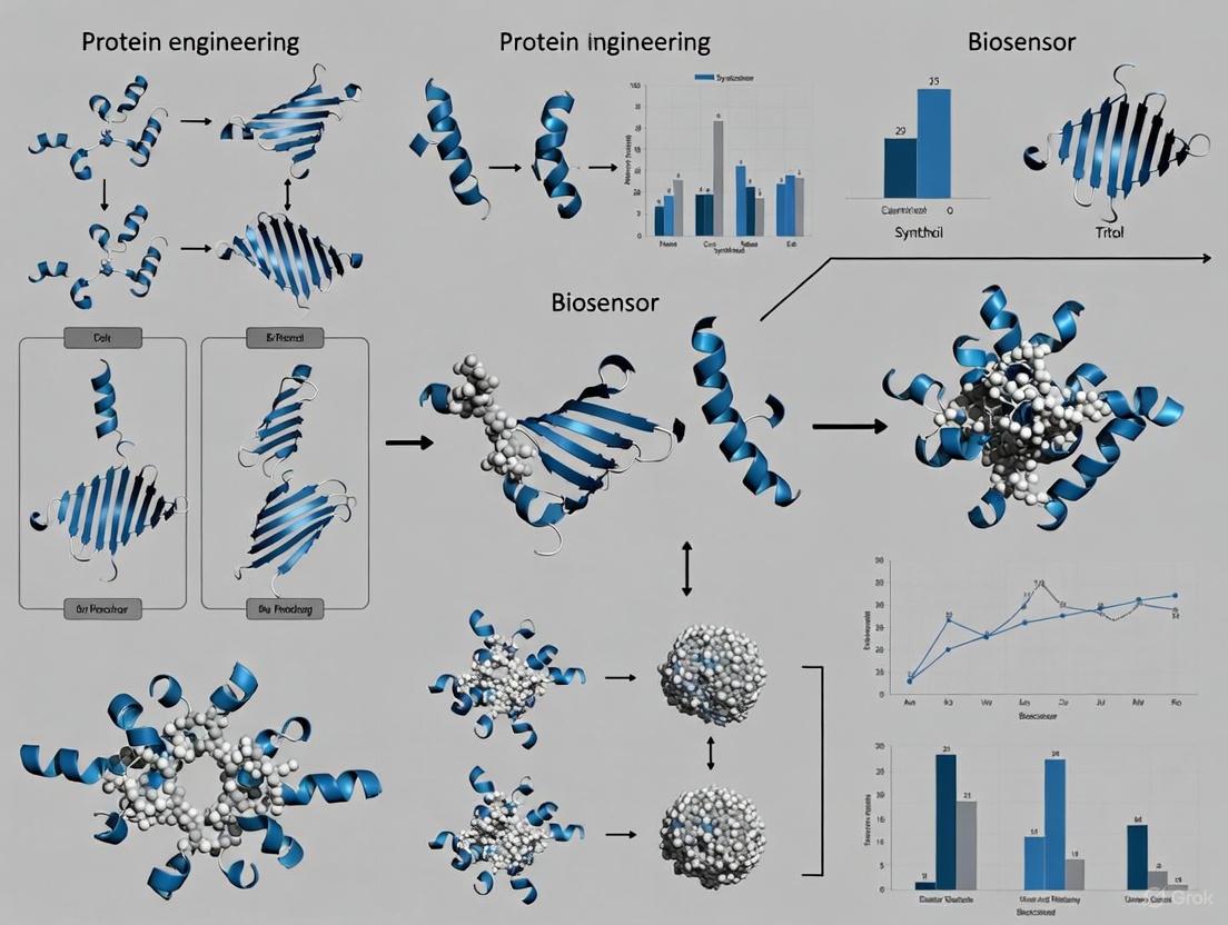
Visualizing the Concepts and Workflows
Engineering Biosensor Dynamic Range
Workflow for a Mixed-Affinity Sensor
Troubleshooting FAQs: Extending Biosensor Dynamic Range
FAQ 1: My engineered biosensor has a low signal-to-noise ratio. What are the primary causes and solutions?
A low signal-to-noise ratio often stems from non-specific interactions or suboptimal sensor-receptor binding kinetics. To address this, first, verify the biosensor's operating range and response threshold using a dose-response curve to ensure it is suited for your target analyte concentration [1]. Second, employ directed evolution strategies to enhance the biosensor's specificity. This involves creating mutant libraries and using high-throughput screening, such as fluorescence-activated cell sorting (FACS), to select variants with improved binding and reduced background noise [1] [8]. Finally, ensure that the transducer element (e.g., electrode for electrochemical sensors) is properly modified with high-quality nanomaterials to enhance signal capture and minimize interference [9].
FAQ 2: How can I make my protein-based biosensor respond faster to dynamic analyte changes?
Slow response times are frequently due to limitations in the signaling mechanism's kinetics. For faster response, consider incorporating faster-acting components like riboswitches or toehold switches into a hybrid design [1]. For protein-based sensors, explore engineering strategies that utilize mechanisms like protein stability and induced degradation, as these can facilitate more rapid turnover and measurement of the cellular state compared to some traditional transcription factors [8]. Furthermore, tuning the biosensor's dynamic performance by optimizing the plasmid copy number or the position of operator regions can significantly improve its rise time [1].
FAQ 3: My biosensor's dynamic range is too narrow for my application. How can I broaden it?
A narrow dynamic range limits the biosensor's utility across varying analyte concentrations. A primary method for broadening the dynamic range is to engineer the linkage between the recognition element and the output module. This can be achieved by modulating promoter strength, ribosome binding sites (RBS), or the number and position of operator regions [1]. Another powerful approach is to use directed evolution to selectively pressure the biosensor to respond to a wider concentration window of the target analyte, thereby shifting its response threshold and maximum output [8]. For electrochemical biosensors, utilizing nanocomposites like porous gold with polyaniline can enhance sensitivity across a broader concentration range [10].
FAQ 4: What are the best practices for integrating a novel biosensor into a genetic circuit for metabolic control?
Integrating a new biosensor into a genetic circuit requires careful characterization to ensure robust performance. Start by thoroughly characterizing the biosensor's dose-response curve, dynamic range, and response time in isolation to establish a baseline [1]. To ensure seamless integration, adopt a decoupled design where possible, similar to the ARTIST platform, which separates the recognition element from the molecular circuit. This modularity allows for independent optimization of the sensor and the actuator modules [11]. Finally, implement real-time validation using multi-omics profiling to assess how the integrated circuit impacts host metabolism and to verify that the dynamic control functions as intended without causing toxicity or pathway imbalances [12].
Performance Metrics for Biosensor Engineering
The table below summarizes key quantitative parameters to monitor when engineering biosensors for an extended dynamic range. These metrics are essential for evaluating performance during troubleshooting [1].
| Performance Parameter | Description | Impact on Dynamic Range | Ideal Characteristics for Extended Range |
|---|---|---|---|
| Dynamic Range | The ratio between the maximal and minimal detectable signal output. | Defines the operational window of the biosensor. | A high ratio, indicating sensitivity over a wide concentration span. |
| Operating Range | The concentration window of analyte where the biosensor performs optimally. | Determines the upper and lower bounds of usable detection. | A wide linear range within the dose-response curve. |
| Response Time | The speed at which the biosensor reacts to a change in analyte concentration. | Affects the ability to track rapid dynamic changes. | Fast rise-time to quickly reach a new steady-state output. |
| Signal-to-Noise Ratio | The clarity and reliability of the output signal against background variability. | A low ratio can obscure the true signal, effectively narrowing the detectable range. | A high ratio for clear distinction between baseline and activated states. |
| Response Sensitivity | The slope of the dose-response curve, indicating how much output changes per unit change in input. | A steeper slope can lead to a narrower range; requires balancing. | A sufficient slope for detectability without sacrificing range width. |
Experimental Protocols for Key Methodologies
Protocol 1: Directed Evolution of a Protein-Based Biosensor for Altered Dynamic Range
This protocol outlines the process of using directed evolution to create biosensor variants with improved dynamic range [8].
- Library Construction: Generate a diverse mutant library of your biosensor gene. Methods include error-prone PCR, DNA shuffling, or saturation mutagenesis of key residues in the ligand-binding or allosteric domains.
- Transformation and Screening: Clone the mutant library into an appropriate microbial host (e.g., E. coli or yeast) and transform. Use high-throughput screening based on fluorescence (e.g., FACS) or growth selection to isolate clones that show an output signal (e.g., fluorescence) across a wider range of target analyte concentrations.
- Characterization of Hits: Isolate plasmid DNA from selected clones and sequence to identify mutations. Re-transform individual purified plasmids into a fresh host to confirm the phenotype.
- Dose-Response Analysis: Grow cultures of the confirmed hits and expose them to a gradient of analyte concentrations. Measure the output signal (e.g., fluorescence via plate reader) to generate a new dose-response curve. Calculate the new dynamic range and compare it to the parent biosensor.
- Iteration: Use the best-performing variant as a template for subsequent rounds of evolution to further refine the dynamic range and other properties.
Protocol 2: Tuning Dynamic Range via Promoter and RBS Engineering
This protocol describes a rational design approach to modulate biosensor output by engineering regulatory parts [1].
- Modular Part Assembly: Use standard cloning techniques (e.g., Golden Gate assembly) to create genetic constructs where the biosensor's output gene (e.g., a fluorescent reporter) is controlled by a series of different promoters and ribosome binding sites (RBS) of varying strengths.
- Characterization of Constructs: Transform each construct into your host organism. For each construct, measure the dose-response relationship as described in Protocol 1, step 4.
- Data Analysis: Plot the dose-response curves for all constructs. Analyze how the different promoter-RBS combinations affect the maximum output level (directly influencing the dynamic range) and the response threshold.
- Selection and Integration: Select the construct that provides the most desirable dynamic range and operational range for your specific application. This tuned construct can then be integrated into larger genetic circuits.
Biosensor Engineering and Optimization Pathways
Diagram: Evolutionary Engineering of Biosensors
Diagram: Dose-Response Optimization Concepts
Research Reagent Solutions Toolkit
The table below lists essential reagents and tools for implementing the evolutionary strategies discussed in this guide.
| Research Reagent / Tool | Function / Description | Application in Dose-Response Editing |
|---|---|---|
| Directed Evolution Kits | Commercial kits for error-prone PCR or DNA shuffling to create diverse mutant libraries. | Generates genetic diversity for screening biosensors with altered ligand binding and output properties [8]. |
| Fluorescence-Activated Cell Sorter (FACS) | High-throughput instrument that sorts cells based on fluorescent signals. | Essential for screening large mutant libraries to isolate clones with desired dynamic range and output intensity [1]. |
| Orthogonal Transcription Factors | Engineered TFs with modified specificity and sensitivity to a target analyte. | Can be used as the sensing module to re-engineer the input side of the biosensor's dose-response [1]. |
| Riboswitch / Toehold Switch Parts | Synthetic RNA elements that change conformation upon ligand binding or RNA hybridization. | Provide a modular, fast-acting component for hybrid biosensor designs to improve response time and programmability [1]. |
| Protein Degradation Tags | Peptide sequences (e.g., degrons) that target a protein for rapid proteolysis. | Engineering biosensors using stability/degradation mechanisms allows for faster turnover and rapid response to cellular state changes [8]. |
| Nanomaterial Composites | Materials like porous gold, polyaniline, or carbon nanotubes for electrode modification. | Used in electrochemical biosensors to enhance signal transduction, sensitivity, and stability, thereby supporting a wider dynamic range [9] [10]. |
| Trypanothione synthetase-IN-5 | Trypanothione synthetase-IN-5, MF:C35H39F2IN4O3, MW:728.6 g/mol | Chemical Reagent |
| Tubulin polymerization-IN-43 | Tubulin polymerization-IN-43, MF:C17H13F4N3O, MW:351.30 g/mol | Chemical Reagent |
Troubleshooting Guides
Transcription Factor (TF)-Based Biosensor Issues
Problem: My TF-based biosensor has a restricted dynamic range and low signal output.
- Potential Cause 1: The allosteric transcription factor (aTF) may have suboptimal binding affinity for its ligand or DNA. Protein engineering can modify key residues to alter ligand specificity and binding efficiency [13].
- Solution: Employ directed evolution or computational design to engineer the aTF. A Functional Diversity-Oriented Volume-Conservative Substitution Strategy can be used to create variants. For example, engineering the CaiF transcription factor with Y47W/R89A mutations extended its concentration response range by 1000-fold and increased output signal intensity by 3.3-fold [4].
- Potential Cause 2: Leaky expression or non-specific activity in the genetic circuit [13].
- Solution: Review the biosensor's design architecture (e.g., repression of activator aTF). Minimize cross-talk by selecting orthogonal parts and using background cell strains with reduced native regulatory interference [13].
Problem: The biosensor shows poor performance upon heterologous expression.
- Potential Cause: The host cell environment may not be compatible, leading to improper protein folding or function [13].
- Solution: Optimize codons for your host organism. Test biosensor function in different bacterial or yeast chassis to identify the most suitable expression system [13].
Allosteric Protein Switch Performance
Problem: I cannot identify potential allosteric sites for engineering control.
- Potential Cause: Allosteric sites are not always obvious from a protein's primary structure [14].
- Solution: Utilize computational tools to predict allosteric sites and pathways. Tools like Ohm can identify allosteric sites coupled to an active site and analyze communication pathways. AlloSigMA can explore the energetics of allosteric pathways, and SPACER can quantify allosteric communication [14].
Problem: Engineered allosteric control does not effectively modulate protein activity.
- Potential Cause: The inserted sensor domain may not be effectively coupled to the target protein's functional motions [15].
- Solution: Experiment with different domain insertion sites using flexible linkers. High-throughput screening is often necessary to identify functional switches where the inserted domain allosterically regulates the target protein's activity in response to specific triggers [15].
Aptasensor Functionality
Problem: My DNA or RNA aptamer is unstable in biological fluids.
- Potential Cause: Nucleic acid aptamers are prone to degradation by nucleases [16].
- Solution: Use chemically modified nucleotides (e.g., 2'-fluoro, 2'-amino) during the SELEX process to enhance nuclease resistance. Alternatively, consider using peptide aptamers [16].
Problem: The capacitive aptasensor shows inconsistent signal changes upon target binding.
- Potential Cause: The mechanism of how aptamer-target binding modifies capacitance is complex and can be influenced by factors like conformational change, charge distribution, and ion displacement within the electrical double layer (EDL) [17].
- Solution: Systematically investigate the individual aptamer-target interaction. Ensure consistent surface functionalization and use controlled experimental conditions to better understand and optimize the physical mechanism leading to the capacitance change [17].
General Biosensor & Assay Problems
Problem: The protein assay is providing inaccurate concentration readings.
- Potential Cause: The sample buffer contains incompatible substances that interfere with the assay chemistry. Common interferents include reducing agents, chelators, detergents, and strong acids/bases [18].
- Solution: Dilute the sample several-fold in a compatible buffer, dialyze or desalt the sample, or precipitate the protein to remove interferents. Always consult the assay's compatibility chart [18].
Problem: High background or "Standards Incorrect" error in fluorescent protein assays.
- Potential Cause: The assay kit may have expired, been stored incorrectly, or the sample buffer contains detergents or other interfering components [18].
- Solution: Prepare fresh calibration standards. Check that the composition of your sample buffer is within the acceptable limits for the assay. For Qubit assays, ensure you are using the recommended tubes and pipetting volumes of at least 5 µL for accuracy [18].
Frequently Asked Questions (FAQs)
Q1: What are the main advantages of using aptamers over antibodies in biosensors?
A1: Aptamers offer several key advantages: they are produced entirely in vitro via the SELEX process, eliminating the need for animals and reducing batch-to-batch variation [16]. They can be selected against a wider range of targets, including non-immunogenic toxins and small molecules [16]. Their denaturation is often reversible, leading to a longer shelf life, and they are easily chemically modified for improved stability or labeling [16].
Q2: How can I find a transcription factor for a metabolite that currently has no known biosensor?
A2: You can explore the current knowledge base by mining specialized databases such as RegulonDB (for E. coli), RegPrecise (for prokaryotes), or P2TF (for predicted prokaryotic TFs) [13]. If no known TF exists, homology-based prediction can be used to find TFs in other species that belong to well-known regulator families (e.g., LysR, TetR, AraC) and are likely to respond to your metabolite of interest [13].
Q3: What computational strategies can I use at the start of a project to design a new allosteric protein?
A3: Computational tools are essential for identifying allosteric sites and pathways. You can use:
- Ohm: To identify allosteric sites and communication pathways from a 3D protein structure [14].
- Network-based methods: Treat the protein as a network of interacting residues to identify communication paths between distal sites [14].
- Elastic network models: To study efficient communication pathways and species-specific allosteric sites [14].
Q4: My HCP (Host Cell Protein) ELISA is giving inconsistent results between lots. How can I improve quality control?
A4: For generic impurity assays, the most reliable quality control is to use laboratory-specific control samples. Prepare a bulk lot of controls using your specific analyte (e.g., HCPs from your process) in the same matrix as your critical samples. Aliquot and freeze these controls at -80°C. Establish a statistically valid range for them and use these controls, rather than just curve-fit parameters, to assure run-to-run and lot-to-lot quality [19].
Research Reagent Solutions
Table: Key Research Reagents and Databases for Biosensor Development
| Item | Function & Application |
|---|---|
| SELEX | A process (Systematic Evolution of Ligands by EXponential enrichment) to generate high-affinity DNA or RNA aptamers for specific targets [16]. |
| RegulonDB | A primary database for transcriptional regulation in E. coli K-12, useful for finding native transcription factors and their regulons [13]. |
| JASPAR | An open-access database of curated transcription factor binding profiles, useful for designing synthetic promoter elements for biosensors [13]. |
| Allosteric Site Prediction Tools (e.g., Ohm, AlloSigMA) | Computational platforms to identify allosteric sites and communication pathways within a protein structure, guiding engineering efforts [14]. |
| Directed Evolution Tools | A suite of methods, including site-saturation mutagenesis and high-throughput screening, used to engineer properties like the dynamic range of transcription factors [13] [4]. |
| Chemically Modified Nucleotides | Nucleotides (e.g., 2'-fluoro) used during SELEX to create aptamers with enhanced nuclease resistance for use in complex biological fluids [16]. |
Experimental Protocols
Protocol 1: Directed Evolution to Extend Biosensor Dynamic Range
This protocol outlines a strategy for modifying a transcription factor-based biosensor, such as the l-carnitine biosensor CaiF, to broaden its dynamic range [4].
- Computer-Aided Design: Formulate the 3D structural configuration of the TF (e.g., CaiF) using modeling software. Simulate the DNA and ligand binding sites.
- Alanine Scanning: Perform alanine scanning mutagenesis of residues in the predicted functional sites to validate their importance.
- Diversity-Oriented Substitution: Conduct a "Functional Diversity-Oriented Volume-Conservative Substitution Strategy" on the key functional residues identified in steps 1 and 2. This involves creating a library of TF variants with mutations at these sites.
- Library Screening: Clone the variant library into a biosensor circuit with a reporter gene (e.g., GFP). Screen the library under varying concentrations of the target ligand (e.g., l-carnitine).
- Variant Characterization: Isolate variants that show a response over a wider concentration range and/or with higher signal output. Characterize the lead variant (e.g., CaiF_Y47W/R89A) by measuring its response curve from low (e.g., 10â»â´ mM) to high (e.g., 10 mM) ligand concentrations [4].
Protocol 2: SELEX for Aptamer Development
This protocol describes the general process for selecting specific DNA or RNA aptamers [16].
- Library Synthesis: Synthesize a large, diverse library of single-stranded DNA or RNA molecules (up to ~10¹ⴠdifferent sequences) with random central regions.
- Incubation with Target: Incubate the library with the immobilized target molecule (e.g., a protein).
- Partitioning: Remove unbound nucleic acid sequences through washing steps.
- Elution: Elute the target-bound sequences from the immobilization surface.
- Amplification: Amplify the eluted sequences using PCR (for DNA) or RT-PCR (for RNA).
- Iteration: Repeat steps 2-5 for multiple rounds (typically 6-15) to enrich for high-affinity binders.
- Cloning and Sequencing: After the final round, clone and sequence the enriched pool to identify individual aptamer candidates.
- Binding Characterization: Synthesize the candidate aptamers and characterize their binding affinity and specificity under relevant conditions (e.g., temperature, pH).
Supporting Diagrams
Biosensor Engineering Workflow
Allosteric Transcription Factor Mechanism
Technical Support Center: Extending Biosensor Dynamic Range
This technical support center provides troubleshooting guides and frequently asked questions (FAQs) for researchers working on extending the dynamic range of biosensors through protein engineering. The content is designed to help you address specific experimental challenges and implement advanced methodologies.
Frequently Asked Questions (FAQs)
FAQ 1: Why is the inherent dynamic range of a single-site binding biosensor limited, and what is the theoretical basis for this limitation? The fundamental limitation arises from the physics of single-site binding, which produces a hyperbolic dose-response curve (the Langmuir isotherm). For any single-site binding event, the transition from 10% to 90% receptor occupancy requires an 81-fold change in target concentration [3] [20]. This fixed dynamic range is often insufficient for applications where target concentrations vary over several orders of magnitude, such as monitoring viral loads in patients, or where high sensitivity to small concentration changes is critical, such as tracking drugs with narrow therapeutic indices [3].
FAQ 2: What are the primary protein engineering strategies for extending the dynamic range of my biosensor? The main strategies, inspired by mechanisms found in nature, involve combining multiple engineered receptor elements [3].
- Strategy 1: Affinity-Tuned Receptor Mixtures. This involves creating a set of receptor variants (e.g., mutated proteins or DNA stem-loops) that bind the same target with identical specificity but different affinities. When these variants are combined, their individual hyperbolic responses merge to create a single, extended log-linear dynamic range [3] [20]. For instance, combining two receptors with a 100-fold difference in affinity can extend the dynamic range to 8,100-fold [3].
- Strategy 2: The Sequestration/Depletant Mechanism. This strategy narrows the dynamic range to create an ultrasensitive, threshold response. A high-affinity, non-signaling receptor (the "depletant") is used to sequester the target. The signaling receptor, which has a lower affinity, only begins to produce a signal after the depletant is saturated, compressing the response into a very narrow concentration window (e.g., an 8-fold range) [20].
FAQ 3: How can I engineer a protein receptor to alter its affinity without changing its target specificity? A key technique is to engineer a structure-switching mechanism into the receptor. By stabilizing an alternative, non-binding conformation, you can tune the apparent affinity without altering the specific atomic interactions of the binding site itself [3]. This is often achieved by:
- Introducing destabilizing mutations remote from the binding site to couple target binding to a conformational change (folding) [20].
- Employing "alternate frame folding," a strategy involving the duplication of a protein segment to stabilize a non-binding conformation [20]. For example, in DNA-based biosensors, the stem stability can be modified to change affinity while keeping the target-recognizing loop sequence identical [20].
FAQ 4: I have successfully created receptor variants with different affinities. What is the optimal way to combine them for a maximized log-linear range? The optimal combination is not always an equal molar ratio. Simulations and empirical data suggest [3]:
- For a two-variant mixture, a ~100-fold difference in affinity between them produces the widest, most log-linear range.
- If the signal gain of individual variants degrades (e.g., due to an unstable switch), you must adjust the molar ratios to correct for this. For instance, a 59/41 ratio of two variants with a 100-fold affinity difference achieved a dynamic range of 8,100-fold with excellent log-linearity (R²=0.995) [3].
- Using more than two variants (e.g., four) can extend the dynamic range even further, to nearly 900,000-fold, but requires careful optimization of mixing ratios using simulations [3].
FAQ 5: My biosensor's signal output is low. What are some advanced signal transduction methods to improve it? Innovative transducer designs can dramatically enhance signal output. One powerful method is the use of engineered chemogenetic FRET pairs. For example, creating a stable interface between a fluorescent protein (FRET donor) and a fluorescently labeled HaloTag (FRET acceptor) can achieve near-quantitative FRET efficiencies (≥95%) [21]. This design can be adapted to create biosensors for analytes like calcium, ATP, and NAD⺠with unprecedented dynamic ranges. The system is highly tunable, allowing you to change the fluorescent protein or the synthetic fluorophore to shift the biosensor's spectral properties [21].
Troubleshooting Guides
Problem: The combined biosensor response is not log-linear and shows plateaus or dips.
- Potential Cause 1: The affinity difference between your receptor variants is too large (significantly greater than 100-fold). A large gap can cause deviations from linearity at intermediate target concentrations [3].
- Solution: Characterize the dissociation constants (Kd) of your variants more precisely. Select a set of variants whose affinities differ by approximately 30- to 100-fold for pairing and fine-tune the ratios [3] [20].
- Potential Cause 2: The receptor variants are not combined in the optimal ratio.
- Solution: Do not assume a 1:1 ratio is ideal. Use simulation software to model the expected combined response based on the measured Kd and signal gain of each variant. Empirically test a range of mixing ratios to identify the one that produces the smoothest log-linear response [3].
Problem: The biosensor with a sequestration mechanism has a poor signal-to-noise ratio.
- Potential Cause 1: The density of the signaling probe on the sensor surface is too low, often due to an excessively high [depletant]/[probe] ratio [20].
- Solution: Titrate the ratio of depletant to signaling probe during sensor fabrication. The pseudo-Hill coefficient will increase with the ratio, but it eventually plateaus. Avoid ratios that reduce the signaling probe density to a point where the Faradaic current is no longer measurable [20].
- Potential Cause 2: The sample volume is not controlled.
- Solution: The sequestration mechanism is highly dependent on a fixed, small sample volume to prevent premature saturation of the depletant. Standardize your assay volume precisely [20].
Experimental Protocols
Protocol 1: Extending Dynamic Range via Affinity-Tuned Receptor Combination This protocol outlines the steps for creating a biosensor with an extended dynamic range by mixing affinity-tuned receptor variants [3] [20].
Materials:
- Purified receptor variants (e.g., structure-switching proteins or nucleic acids) with characterized Kd values.
- Immobilization substrate (e.g., gold electrode, microplate).
- Target analyte of interest.
- Buffer solutions for binding assays.
- Signal detection apparatus (e.g., potentiostat for electrochemical detection, fluorimeter).
Procedure:
- Engineer and Characterize Variants: Generate receptor variants using site-directed mutagenesis or other protein engineering techniques to stabilize non-binding conformations. Determine the precise Kd of each variant.
- Select Variant Pair: Choose two or more variants that share the same specificity but have Kd values differing by approximately 100-fold for optimal range extension [3].
- Optimize Mixing Ratio: Based on the Kd and signal gain of each variant, calculate the optimal mixing ratio. For example, a 59/41 ratio was used for a specific 100-fold affinity difference pair [3].
- Co-Immobilize Receptors: Mix the receptor variants in the optimized ratio and immobilize them onto your chosen substrate (e.g., electrode surface).
- Validate Performance: Expose the biosensor to a dilution series of the target analyte spanning several orders of magnitude. Measure the output signal and plot it against the logarithm of the target concentration. A successful sensor will show a linear relationship across a much wider range than any single variant.
Protocol 2: Implementing a Sequestration Mechanism for a Narrowed, Ultrasensitive Response This protocol details the use of a non-signaling depletant to create a biosensor with a threshold response [20].
Materials:
- Signaling receptor (low-affinity variant).
- Non-signaling depletant receptor (high-affinity variant, identical target specificity).
- Immobilization substrate.
- Fixed-volume sample delivery system.
Procedure:
- Prepare Receptors: Produce a signaling receptor (e.g., a structure-switching probe with a redox reporter or fluorophore) and a high-affinity, non-signaling depletant (e.g., a linear version of the same probe without a reporter) [20].
- Co-Immobilize at Set Ratio: Immobilize both the signaling probe and the depletant onto the sensor surface. A typical starting [depletant]/[probe] ratio is 50:1, but this should be optimized [20].
- Test with Fixed Volume: Challenge the sensor with a target concentration series using a precise, fixed sample volume (e.g., 3 µL).
- Analyze Dose-Response: Fit the resulting dose-response curve to the Hill equation. A successful implementation will yield a steep curve with a high pseudo-Hill coefficient (e.g., 2.3) and a useful dynamic range compressed to as little as 8-fold [20].
Data Presentation: Quantitative Biosensor Performance
Table 1: Performance of Biosensors with Engineered Dynamic Ranges
| Sensor Type | Engineering Strategy | Original Dynamic Range | Engineered Dynamic Range | Key Metric / Signal Gain | Application Context |
|---|---|---|---|---|---|
| Molecular Beacon [3] | Affinity-Tuned Mixture (4 variants) | 81-fold | ~900,000-fold | 3.6-fold signal gain | Nucleic acid detection |
| E-DNA Sensor [20] | Affinity-Tuned Mixture (2 variants) | 81-fold | ~1,000-fold | Excellent log-linearity (R²=0.978) | Electrochemical DNA detection |
| E-DNA Sensor [20] | Sequestration Mechanism | 81-fold | 8-fold | Pseudo-Hill coefficient of 2.3 | Ultrasensitive detection |
| CaiF-based Sensor [4] | Directed Evolution (CaiFY47W/R89A) | Not Specified | 10â»â´ mM – 10 mM (1000x wider) | 3.3-fold higher signal intensity | L-carnitine production monitoring |
| Chemogenetic FRET [21] | Engineered FP-HaloTag Interface | Low FRET efficiency | ≥94% FRET efficiency | FRET ratio >14 in cells | Live-cell metabolite imaging (Ca²âº, ATP, NADâº) |
Table 2: Research Reagent Solutions for Dynamic Range Engineering
| Research Reagent / Material | Function in Experiment | Key Characteristics |
|---|---|---|
| Structure-Switching Receptors [3] [20] | The core sensing element; affinity is tuned by altering the stability of the non-binding state. | Allows modulation of apparent affinity (Kd) without altering target-binding specificity. |
| HaloTag Protein & Rhodamine Ligands [21] | Components of a chemogenetic FRET pair for high-gain signal transduction. | Enables spectral tunability and near-quantitative FRET efficiency for large dynamic ranges. |
| Electrochemical Electrodes [22] [20] | Platform for immobilizing receptors and transducing binding events into a measurable current. | Often modified with nanomaterials (e.g., graphene, Au-Ag nanostars) to enhance signal transmission [22]. |
| Directed Evolution Platform [4] [23] | A method for generating diverse receptor variants with improved properties. | Used to create transcription factor variants (like CaiF) with extended operational ranges. |
| Non-Signaling Depletant [20] | A high-affinity receptor used in the sequestration mechanism to create a threshold response. | Silently sequesters the target analyte until a concentration threshold is surpassed. |
Signaling Pathways and Workflow Visualization
Engineering Solutions: From Structure-Switching Receptors to Directed Evolution
Troubleshooting Guide: Extending Dynamic Range
| Problem Observed | Potential Cause | Diagnostic Experiments | Proposed Solution & Theoretical Basis |
|---|---|---|---|
| Limited Dynamic Range | Switching equilibrium constant (KS) is too large, favoring the bound conformation even at low analyte concentrations [24]. | Measure signal output across a wide analyte concentration range to identify saturation point. | Use directed evolution or rational design to introduce mutations that destabilize the bound state (decrease KS), shifting the equilibrium to favor the unbound state at low [analyte] [24] [4]. |
| Low Signal Intensity | Conformational change does not efficiently transduce into a measurable output signal [24]. | Test sensor with a saturating analyte concentration to determine maximum achievable signal. | Optimize fluorophore/quencher placement (for optical sensors) or redox reporter positioning (for electrochemical sensors) to maximize signal change between states [24]. |
| Loss of Specificity | Mutations intended to tune affinity have disrupted key binding pocket residues [25]. | Perform binding assays against structurally similar off-target molecules. | Focus affinity-tuning mutations outside the primary interaction interface, as these can alter global affinity with less impact on specificity [25]. |
| Insufficient Sensitivity | Sensor has a high KD (low innate affinity) for the target analyte [24]. | Determine the limit of detection (LOD) and the lower limit of the concentration-response curve. | Rationally engineer the binding interface to improve complementary interactions, lowering KD and improving sensitivity for low-concentration targets [24]. |
Frequently Asked Questions (FAQs)
Q1: What is the fundamental thermodynamic principle that allows me to tune a biosensor's apparent affinity?
The operation of structure-switching biosensors is governed by a conformational selection mechanism [24]. The biomolecular switch exists in an equilibrium between a non-binding "off" state and a binding-competent "on" state, defined by the switching equilibrium constant (KS = [on]/[off]). The apparent affinity (KD,app) is a function of both this intrinsic KS and the innate binding affinity (KD) for the target to the "on" state. By mutating residues that affect the stability of the "on" vs. "off" state, you can alter KS and thereby tune the KD,app without necessarily changing the fundamental chemical interactions in the binding pocket itself [24].
Q2: How can I experimentally determine if a mutation has altered the switching equilibrium (KS) versus the innate binding affinity (KD)?
Distinguishing between these effects requires careful experimental design. A change in innate KD primarily affects the saturation point of your concentration-response curve. In contrast, a change in KS shifts the entire curve left or right along the concentration axis, effectively changing the analyte concentration at which half-maximal signal is achieved (EC50D, separate from the coupled switching event.
Q3: Are there specific regions on a protein scaffold where mutations are more likely to tune affinity without compromising specificity?
Yes. Mutations at residues outside the direct binding interface are often effective for tuning global affinity with minimal impact on specificity [25]. For instance, in coiled-coil interactions, residues at solvent-exposed positions can influence dimer stability and propensity without directly participating in partner selection [25]. Similarly, mutations that affect the backbone flexibility or allosteric networks linking the binding site to the output domain can modulate KS and thus apparent affinity, while preserving the precise binding interface.
Q4: A directed evolution campaign successfully extended my sensor's dynamic range, but it now responds to an off-target. What is the likely cause, and how can I fix it?
This indicates that the selected mutations have likely altered residues within the binding pocket that are critical for specificity. Specificity is often determined by a combination of features that promote on-target binding and those that actively prevent off-target binding [25]. To fix this, you can employ a counter-selection strategy during screening. In addition to selecting for the desired dynamic range under positive pressure (e.g., with the target analyte), simultaneously apply negative pressure by exposing the library to the off-target molecule and selecting variants that do not respond. This enriches for mutations that confer the desired switching properties while maintaining or even enhancing specificity.
Experimental Protocol: Modulating Dynamic Range via Directed Evolution
This protocol outlines a methodology for extending the dynamic range of a transcription factor-based biosensor, based on a study that successfully modified the l-carnitine biosensor CaiF [4] [26].
Key Reagents and Equipment
| Item | Function/Role in the Experiment |
|---|---|
| Plasmid Library | Contains the gene for your biosensor (e.g., CaiF) with randomized codons at targeted positions. |
| Fluorescence-Activated Cell Sorting (FACS) | High-throughput method to screen and isolate mutant cells based on their fluorescence output in response to different analyte concentrations. |
| Analyte Stock Solutions | Prepare a high-concentration stock (e.g., 10 mM) and a low/no-analyte solution for counter-selection. |
| Microplate Reader | For characterizing the concentration-response curves of isolated clones. |
| Computer-Aided Design Software | For structural analysis and identifying key residues to mutate (e.g., DNA binding domain, allosteric sites). |
Step-by-Step Procedure
Library Construction:
- Rational Targeting: Use structural models (e.g., from computer-aided design) to identify key functional regions, such as the DNA-binding domain or allosteric regulatory sites. Alanine scanning can be used for preliminary validation [4].
- Diversity Generation: Employ a "Functional Diversity-Oriented Volume-Conservative Substitution Strategy" at the identified key sites [4]. This involves substituting residues with amino acids of similar size but different biochemical properties to explore functional changes while minimizing structural disruption.
- Library Quality Control: Sequence a sample of the library to ensure diversity and the absence of wild-type sequence bias.
High-Throughput Screening for Dynamic Range:
- Growth and Induction: Grow the library of mutant cells in a medium that induces the expression of the biosensor construct.
- Dual-Concentration Sorting: Divide the cell population and expose one aliquot to a low (or zero) concentration of the analyte and a second aliquot to a saturatingly high concentration of the analyte.
- FACS Gating: Isolate two populations of cells: i) those showing the lowest fluorescence output in the low-analyte condition (minimal background leakage), and ii) those showing the highest fluorescence output in the high-analyte condition (strong maximum signal). Cells that pass both gates possess a wide dynamic range [4].
Characterization of Hits:
- Clone Isolation: Plate the sorted cells to obtain single colonies.
- Concentration-Response Profiling: Inoculate cultures of individual clones and measure their fluorescence output across a comprehensive range of analyte concentrations (e.g., from 10-4 mM to 10 mM) [4] [26].
- Data Analysis: Fit the response data to a sigmoidal curve (e.g., Hill equation) to determine the EC50 (apparent affinity), Hill slope, and maximum output for each variant.
Specificity Validation:
- Challenge the top-performing variants with structurally similar molecules to ensure that the expanded dynamic range has not come at the cost of specificity. Reject variants that show significant cross-reactivity.
Visualizing the Thermodynamic Principle of Tuning
The following diagram illustrates the core concept of tuning a biosensor's dynamic range by modulating the switching equilibrium.
Modulating the Switching Equilibrium - This diagram shows how mutations can shift the equilibrium between the "off" and "on" states of a biosensor. By making the "on" state less stable (decreasing KS), a higher analyte concentration is required to drive the system to the bound state, thereby shifting the dose-response curve and extending the dynamic range to higher concentrations [24] [4].
Research Reagent Solutions
| Research Reagent | Function in Tuning Apparent Affinity |
|---|---|
| Bacterial Periplasmic Binding Proteins (bPBPs) | A common protein scaffold that undergoes large hinge-bending motions upon ligand binding, ideal for engineering structure-switching biosensors [24]. |
| Environmentally-Sensitive Fluorophores | Single fluorophores whose quantum yield changes based on the immediate polarity of their microenvironment, used to report conformational changes in single-fluorophore sensors [24]. |
| FRET Donor/Acceptor Pairs | Pairs of fluorophores (e.g., CFP/YFP) used to measure distance changes via Förster Resonance Energy Transfer, ideal for monitoring conformational changes in dual-fluorophore sensors [24]. |
| Electrochemical Reporters (e.g., Methylene Blue) | Redox-active molecules that can be attached to biomolecules; their electron transfer efficiency to an electrode changes with distance/orientation, enabling electrochemical detection of switching [24]. |
| Deep Mutational Scanning (e.g., bPCA) | A high-throughput method (like binding Protein Fragment Complementation Assay) to quantify the effects of thousands of mutations on binding affinity and specificity across many interaction partners [25]. |
Core Concept: Editing Biosensor Dynamic Range
Biomolecular recognition elements, such as antibodies and aptamers, are the cornerstone of artificial biosensors [27]. A fundamental limitation of these single-site binding receptors is their inherently fixed dynamic range: the transition from 10% to 90% receptor occupancy requires an 81-fold change in target concentration [3]. This range is often too narrow for clinical applications, where target concentrations, such as HIV viral loads, can vary by over five orders of magnitude [3].
Inspired by strategies found in nature, researchers can overcome this limitation by rationally combining multiple receptor variants that bind the same specific target but differ in their affinity [3]. This approach thermodynamically "edits" the biosensor's dose-response curve, creating a composite signal that is log-linear across a much wider concentration range.
Key Experimental Data and Workflows
Table 1: Properties of Individual Molecular Beacon Receptor Variants
This table summarizes data from a foundational study that engineered a series of molecular beacon receptors by varying the stability of their non-binding stem structure [3].
| Receptor Variant Name | Dissociation Constant (Kd) | Relative Affinity (Fold Change) | Maximum Fluorescence Change |
|---|---|---|---|
| 0GC | 0.012 µM | 1 (Highest) | Not Specified |
| 1GC | 0.12 µM | 10 | 83% |
| 2GC | 1.3 µM | ~108 | Not Specified |
| 3GC | 12 µM | 1,000 | 100% |
| 5GC | 128 µM | ~10,667 (Lowest) | Not Specified |
Table 2: Performance of Engineered Receptor Mixtures
By mixing the variants from Table 1 in optimized ratios, the following sensor performances were achieved [3].
| Receptor Mixture Composition | Optimized Mixing Ratio | Log-Linear Dynamic Range | Signal Gain | Linearity (R²) |
|---|---|---|---|---|
| 1GC + 3GC | 59 / 41 | 8,100-fold | 9-fold | 0.995 |
| 0GC + 2GC + 3GC + 5GC | Optimized | ~900,000-fold | 3.6-fold | 0.995 |
| 0GC + 5GC | Not Specified | "Three-State" Response | Not Specified | Not Applicable |
Diagram 1: Strategy for Extending Dynamic Range
Experimental Protocol: Creating an Extended-Range Biosensor
Step 1: Generate a Panel of Affinity-Tuned Receptors
The first step is to create receptor variants that differ in affinity while maintaining identical target specificity.
- Structure-Switching Mechanism: A highly effective method involves engineering a conformational switch into the receptor [3]. For DNA-based receptors like molecular beacons, this is achieved by systematically altering the stability of the stem-loop structure's stem (e.g., by changing its GC content or length) [3]. For protein-based receptors, this can involve stabilizing a non-binding conformation [3].
- Critical Validation: For each variant, confirm that the dissociation constant (Kd) shifts as intended while specificity against non-target molecules remains consistent across all variants [3].
Step 2: Determine Optimal Mixing Ratios
Combining receptors is not a simple 1:1 mixture. Optimal ratios must be calculated and experimentally verified.
- Simulation: Begin with thermodynamic simulations to model the combined dose-response of different variant pairs or groups. A 100-fold difference in affinity between variants often yields the widest log-linear range [3].
- Correction for Signal Gain: Receptors with less stable non-binding states may have lower signal gain. Simulations can be used to correct for this by adjusting molar ratios. For instance, a 59/41 ratio was used for 1GC and 3GC beacons to achieve ideal linearity [3].
- Experimental Titration: Titrate the target analyte across a wide concentration range using the simulated mixture. Measure the output signal and fit the data to a log-linear plot to confirm the dynamic range and adjust ratios if needed.
Step 3: Characterize Composite Sensor Performance
Once the optimal mixture is identified, perform a full characterization.
- Dynamic Range and Linearity: Confirm the log-linear range and calculate the R² value for linearity.
- Specificity Profile: Verify that the mixture maintains a constant and high specificity across the entire extended dynamic range [3].
- Limit of Detection (LOD) and Quantification (LOQ): Determine these key analytical parameters for the final mixture.
The Scientist's Toolkit: Research Reagent Solutions
Table 3: Essential Materials for Developing Affinity-Tuned Biosensors
| Item | Function / Description |
|---|---|
| Structure-Switching Receptors | Engineered receptors (e.g., molecular beacons, aptamers) where a non-binding conformation is stabilized to tune affinity without altering the binding site's specificity [3]. |
| Computational Design Software | Software platforms (e.g., Rosetta) for de novo protein design and for grafting target-binding domains into switchable scaffold proteins [28]. Tools like HotSpot Wizard can also identify engineering sites [29]. |
| Thermodynamic Modeling Tools | Custom or commercial software for simulating the dose-response of receptor mixtures to predict dynamic range and optimize mixing ratios before experimental testing [3]. |
| High-Throughput Screening Platform | Systems for rapidly expressing and screening many receptor variants and their mixtures for affinity, specificity, and signal output [4] [30]. |
| Myristoylated ARF6 (2-13) | Myristoylated ARF6 (2-13) Peptide|ARF6 Inhibitor |
| Exatecan analogue 1 | Exatecan analogue 1, MF:C23H20FN3O4, MW:421.4 g/mol |
Troubleshooting FAQs
FAQ 1: Our receptor mixture does not produce a log-linear response and instead shows a sigmoidal curve with a plateau in the middle. What is the cause? This "three-state" response pattern typically occurs when the difference in affinity between the combined receptor variants is too large (e.g., greater than 500-fold) [3]. The solution is to include one or more intermediate-affinity variants in the mixture to bridge the gap. Simulate the combined response of variants with smaller affinity increments (e.g., 10- to 100-fold differences) to restore log-linearity.
FAQ 2: The mixed sensor shows varying specificity or accuracy across its dynamic range. How can this be fixed? This issue arises if the receptor variants used in the mixture were generated by altering the target-binding site itself, which can inadvertently affect specificity [3]. Ensure all your affinity-tuned variants are generated via a structure-switching mechanism (e.g., by stabilizing a non-binding conformation). This approach decouples affinity from specificity, as the binding site itself remains chemically identical across all variants [3].
FAQ 3: The signal gain (fluorescence change) of our extended-range sensor is unacceptably low. How can we improve it? Low overall signal gain is often caused by including receptor variants with unstable non-binding states, which have a high background signal [3]. To correct this:
- Prioritize variants with high signal gain during the initial selection.
- Use non-equimolar mixing ratios, reducing the proportion of low-signal-gain variants in the mixture.
- Re-engineer your receptor variants to achieve a more favorable balance between affinity and signaling efficiency.
Diagram 2: Troubleshooting Common Issues
This technical support document provides a detailed guide for researchers working to extend the dynamic range of transcription factor-based biosensors, using the CaiF protein for L-carnitine detection as a specific case study. The CaiF transcription factor is notably activated by crotonobetainyl-CoA, a key intermediate in the carnitine metabolic pathway [4]. Capitalizing on this mechanism, sophisticated biosensors have been developed for metabolic engineering applications [4] [31]. The following sections address common experimental challenges and provide standardized protocols to facilitate robust biosensor engineering.
Frequently Asked Questions (FAQs)
Q1: What is the primary limitation of wild-type CaiF biosensors that necessitates engineering? The wild-type CaiF biosensor has a relatively restricted detection range, which imposes specific limitations in practical application scenarios [4]. Without engineering, this narrow dynamic range can hinder precise monitoring and control of metabolic pathways in production strains.
Q2: What key engineering strategy was successfully used to expand the CaiF dynamic range? A "Functional Diversity-Oriented Volume-Conservative Substitution Strategy" was employed on key amino acid sites of CaiF [4]. This rational design approach, potentially informed by computer-aided design of the CaiF structure and DNA binding site simulations, yielded variants with significantly improved performance [4].
Q3: What performance metrics were achieved with the engineered CaiF variant? The biosensor based on the CaiFY47W/R89A double mutant exhibited a considerably expanded concentration response range from 0.0001 mM to 10 mM [4]. This represents a 1000-fold wider range and a 3.3-fold higher output signal intensity compared to the control biosensor [4].
Q4: How can I validate the functional expression of L-carnitine pathway enzymes in my host organism (e.g., E. coli)? A genetically encoded L-carnitine biosensor can be utilized to monitor pathway performance in a dose-dependent manner [31]. This provides a convenient method to screen for functional enzyme expression and cascade activity without the need for complex analytical equipment.
Q5: Why is an "entrepreneurial mindset" recommended in biosensor research? Adopting an entrepreneurial mindset during hypothesis formation helps ensure that research addresses a genuine need, is not duplicative, and produces a product that is readily translatable by industry [32]. This approach maximizes the real-world impact of biosensor development efforts.
Troubleshooting Guides
Problem: Low Signal Intensity from Biosensor
| Possible Cause | Verification Method | Solution |
|---|---|---|
| Suboptimal Expression | SDS-PAGE to check protein levels. | Optimize induction conditions (temperature, inducer concentration, timing). |
| Poor Ligand Binding | Isothermal Titration Calorimetry (ITC). | Re-visit computational design; perform alanine scanning on binding pocket [4]. |
| Inefficient Signal Transduction | Compare with a positive control system. | Engineer the linker region between DNA-binding and ligand-binding domains. |
Problem: High Background Signal (Poor Signal-to-Noise Ratio)
| Possible Cause | Verification Method | Solution |
|---|---|---|
| Promoter Leakiness | Measure fluorescence in absence of ligand. | Use tighter inducible promoters or incorporate genetic insulators. |
| Non-specific Transcription Factor Binding | Chromatin Immunoprecipitation (ChIP). | Mutate DNA-binding domain to enhance specificity [33]. |
| Host Cell Interference | Test biosensor in different strain backgrounds. | Use knockout strains for native regulators that might cross-talk. |
Problem: Narrow Dynamic Range
| Possible Cause | Verification Method | Solution |
|---|---|---|
| Limited Allosteric Regulation | Measure dose-response curves. | Apply directed evolution to the allosteric interface [4] [33]. |
| Saturation of Binding Sites | Determine binding affinity (Kd). | Use rational design, like volume-conservative substitutions, to modulate affinity [4]. |
| Cellular Mislocalization | Fluorescence microscopy with tagged protein. | Add appropriate localization tags or check for aggregation. |
Experimental Protocols
Protocol 1: Directed Evolution of CaiF for Expanded Dynamic Range
Purpose: To create CaiF variants with a wider response range to L-carnitine/crotonobetainyl-CoA.
Materials:
- Error-Prone PCR Kit
- Plasmid library containing mutated caiF genes
- Expression host (e.g., E. coli) with reporter (GFP) under CaiF-responsive promoter
- FACS or microplate reader
- L-carnitine (or precursor) for screening
Procedure:
- Generate Diversity: Create a mutant library of the caiF gene using error-prone PCR or site-saturation mutagenesis at targeted residues (e.g., Y47, R89) [4].
- Library Transformation: Transform the plasmid library into your microbial host strain.
- High-Throughput Screening:
- Plate cells on solid media or grow in liquid culture to express the mutant biosensors.
- Key Step: Challenge the population with a gradient of L-carnitine concentrations (e.g., from 0.001 mM to 50 mM).
- Using FACS or a microplate reader, isolate clones that show a functional response (e.g., GFP expression) at both the low and high ends of the concentration gradient. The goal is to find variants that are sensitive at low levels but do not saturate prematurely.
- Characterization: Isolate promising clones and characterize them by measuring the dose-response curve to determine the new operational range (EC50) and maximum output.
Protocol 2: Quantifying Biosensor Performance
Purpose: To generate a dose-response curve and calculate key performance metrics for engineered CaiF variants.
Materials:
- Engineered bacterial strains (wild-type and mutant CaiF)
- Black-walled, clear-bottom 96-well microplates
- Plate reader capable of measuring OD600 and fluorescence (e.g., GFP)
- L-carnitine stock solution for serial dilution
Procedure:
- Inoculation: Inoculate starter cultures of your biosensor strains and grow overnight.
- Induction & Induction: Dilute cultures and aliquot into the microplate. Add a serial dilution of L-carnitine across the plate. Include a no-ligand control.
- Incubation and Measurement: Grow the cultures with shaking in the plate reader, taking periodic measurements of OD600 and fluorescence.
- Data Analysis:
- Normalize fluorescence to OD600 for each well.
- Plot normalized fluorescence against the log of L-carnitine concentration.
- Fit the data to a sigmoidal curve (e.g., 4-parameter logistic model) to determine the EC50 (half-maximal effective concentration), Hill coefficient, and dynamic range (ratio between max and min signal).
Table 1: Performance Comparison of Wild-type vs. Engineered CaiF Biosensor
| Biosensor Variant | Response Range (mM) | Dynamic Range (Fold Change) | Signal Intensity (Fold Increase) | Key Mutations |
|---|---|---|---|---|
| Wild-type CaiF | Not specified in results | Baseline (Control) | Baseline (Control) | N/A |
| CaiF Y47W/R89A | 10â»â´ – 10 | 1000 | 3.3 | Y47W, R89A |
Table 2: Key Research Reagent Solutions for CaiF Biosensor Engineering
| Reagent / Material | Function / Application | Notes / Rationale |
|---|---|---|
| CaiF Plasmid Library | Source of genetic diversity for directed evolution. | Can be generated via error-prone PCR or site-saturation mutagenesis. |
| Crotonobetainyl-CoA / L-carnitine | Native ligand / Biosensor inducer. | CaiF is activated by crotonobetainyl-CoA, an intermediate in L-carnitine metabolism [4]. |
| Fluorescent Reporter (e.g., GFP) | Quantitative output for biosensor activity. | Allows high-throughput screening via FACS or plate readers. |
| E. coli Production Chassis | Host for biosensor expression and testing. | Engineered E. coli strains can be used for de novo L-carnitine production [31]. |
Workflow and Pathway Diagrams
Harnessing Directed Evolution and FACS for Biosensor Optimization
Troubleshooting Guides
FAQ: Biosensor Performance
Q: My biosensor shows a weak output signal. What could be the cause? A: A weak signal can stem from several issues related to both the biosensor and your instrumentation. The biosensor itself may have suboptimal ligand-binding affinity or poor expression levels in your host system. Instrumentally, ensure your flow cytometer PMT voltages are set correctly for the fluorochrome used and that your antibody concentrations have been properly titrated. High background noise from cellular autofluorescence can also obscure a weak signal [34] [1].
Q: The dynamic range of my biosensor is too narrow for my application. How can I extend it? A: Narrow dynamic range is a common challenge. Directed evolution is a powerful strategy to address this. By creating a diverse library of biosensor variants, for instance, via site-saturation mutagenesis at key amino acid positions involved in ligand binding and signal transduction, and then screening this library using FACS for cells exhibiting both high ON-state and low OFF-state fluorescence, you can select variants with improved ranges. Research on a CaiF-based biosensor successfully used such a strategy to obtain a variant with a 1000-fold wider response range [4].
Q: I am observing high background fluorescence (low signal-to-noise ratio) in my flow cytometry data. How can I reduce it? A: High background, or noise, reduces the clarity of your biosensor's output [1]. To mitigate this:
- Cellular Factors: Always include an unstained control to account for cellular autofluorescence. Use viability dyes to gate out dead cells, which often exhibit non-specific staining. For cells with naturally high autofluorescence (e.g., neutrophils), use fluorochromes that emit in the red channel (e.g., APC) [34].
- Staining Protocol: Ensure adequate washing after antibody incubation steps to remove unbound antibodies. Incorporate a blocking step using Fc receptor blockers, BSA, or FBS to minimize non-specific antibody binding [34].
- Instrument Settings: Verify that fluorescence compensation is correctly set, as under-compensation can lead to high background in detection channels [34].
Q: My biosensor's response is slower than required for real-time monitoring. Can this be improved? A: Yes, response time is a critical performance parameter for dynamic regulation [1]. Slow response can hinder controllability. Engineering approaches include exploring different biosensor scaffolds; for example, riboswitches or two-component systems can offer faster response times than some transcription factor-based biosensors. Hybrid approaches that combine stable, slower systems with faster-acting components can also improve overall performance [1].
Troubleshooting Flow Cytometry (FACS) for Biosensor Screening
The table below outlines common FACS issues encountered during biosensor screening and their solutions.
| Problem | Possible Cause | Solution |
|---|---|---|
| Weak/No Fluorescent Signal | Low biosensor expression or antigen availability. | Use fresh, healthy cells; optimize cell culture/stimulation protocols; confirm biosensor plasmid copy number and promoter strength [34] [1]. |
| Antibody concentration too low or degraded. | Titrate antibodies to find optimal concentration; ensure antibodies are stored properly and not expired [34]. | |
| Incorrect flow cytometer settings. | Ensure proper PMT voltages and laser configurations for your fluorochrome; use positive and negative controls to set gates [34]. | |
| High Background / Non-specific Staining | Inadequate blocking or washing. | Include Fc receptor blocking step; add more washing steps; use buffers with detergents like Tween-20 [34]. |
| Presence of dead cells or cellular debris. | Gate out dead cells using a viability dye (e.g., PI, 7-AAD); sieve cells before analysis to remove debris [34]. | |
| Antibody cross-reactivity. | Include an isotype control; select highly specific secondary antibodies [34]. | |
| Abnormal Event Rate | Clogged fluidics system. | De-clog the sample injection tube according to manufacturer's instructions (e.g., running 10% bleach followed by dHâ‚‚O) [34]. |
| Incorrect cell concentration. | Dilute or concentrate sample to maintain ideal density (~1x10ⶠcells/mL) [34]. | |
| Sample clumping. | Sieve cells before running; pipette gently to mix sample [34]. |
Experimental Protocols
Protocol 1: Directed Evolution of a Transcription Factor-Based Biosensor
This protocol details a methodology for extending the dynamic range of a biosensor, inspired by the successful engineering of a CaiF-based L-carnitine biosensor [4].
1. Library Construction
- Objective: Create genetic diversity in the biosensor gene (e.g., the transcription factor CaiF).
- Method: Use techniques like error-prone PCR or site-saturation mutagenesis. For a targeted approach, perform structural modeling of the biosensor (e.g., using computer-aided design) to identify key residues in the ligand-binding pocket or DNA-binding domain. Focus mutagenesis on these regions [4].
- Cloning: Clone the mutant library into an appropriate expression vector, ensuring it is operatively linked to a reporter gene (e.g., GFP).
2. High-Throughput Screening with FACS
- Objective: Isolate variants with a wider dynamic range (ratio of ON-state to OFF-state fluorescence).
- Method: a. Culture & Induce: Grow two separate cultures of the library cells. One culture is supplemented with a high concentration of the target ligand (ON-state), and the other is kept without ligand or with a repressor (OFF-state). b. Prepare Cells: Harvest, wash, and resuspend cells in a suitable buffer for flow cytometry. c. FACS Gating: Use a flow cytometer to analyze and sort the cells. The primary gate should be set to select for cells that are highly fluorescent in the ON-state culture. Subsequently, a secondary gate should be applied to select from this population only those cells that show low fluorescence in the OFF-state culture. This dual-gating strategy directly selects for variants with a high ON/OFF ratio [4] [1]. d. Recovery & Expansion: Collect the sorted cell population and culture them to allow for recovery and expansion.
3. Validation & Characterization
- Objective: Confirm the performance of isolated hits.
- Method: Isolate single clones from the sorted population. Grow these clones in the presence of a gradient of ligand concentrations. Measure the reporter output (e.g., fluorescence) for each concentration to generate a dose-response curve. Analyze the curve to determine the new dynamic range, operational range, and signal intensity [4]. The successful CaiF biosensor variant (CaiFY47W/R89A) was characterized and showed a 3.3-fold higher output signal and a concentration response range from 10â»â´ mM to 10 mM [4].
Protocol 2: FACS Setup for Biosensor Screening
1. Sample Preparation
- Cell Health: Use freshly prepared, healthy cells where possible to minimize autofluorescence and background scatter [34].
- Controls: Always include the following controls for proper instrument setup and gating:
- Unstained Cells: To set background autofluorescence.
- OFF-state (Uninduced) Population: To set the baseline fluorescence.
- ON-state (Induced) Population: To identify the positive signal.
2. Instrument Configuration
- Fluorochrome Selection: Pair low-expression biosensors with bright fluorochromes (e.g., PE, APC) and high-expression biosensors with dimmer fluorochromes (e.g., FITC) [34].
- PMT Voltage & Threshold: Adjust the PMT voltage for your fluorochrome to ensure the signal is on scale. Set the threshold appropriately to ignore small debris [34].
- Compensation: Perform compensation using single-stained controls to correct for spectral overlap between fluorochromes. Use MFI alignment for accuracy [34].
3. Gating Strategy
- Create a sequential gating strategy: a. FSC-A vs. SSC-A: Gate on the primary cell population, excluding debris and dead cells. b. FSC-H vs. FSC-W: Gate on single cells, excluding doublets and clumps. c. Viability Dye vs. FSC-A: If used, gate on viable cells. d. Fluorescence Channel: Apply the final gate based on the biosensor's fluorescent reporter signal, using the OFF-state and ON-state controls to define the negative and positive populations.
Data Presentation
Key Biosensor Performance Parameters
When engineering biosensors, it is critical to quantify performance using standardized metrics. The table below defines these key parameters, which should be reported to allow for comparison between different biosensor designs [1].
| Parameter | Definition | Importance in Biosensor Optimization |
|---|---|---|
| Dynamic Range | The ratio between the maximum and minimum output signals of the biosensor. | A wider dynamic range allows the biosensor to detect a broader span of analyte concentrations, making it more versatile for high-throughput screening [1]. |
| Operational Range | The concentration window of the analyte where the biosensor performs optimally and provides a reliable, linear response. | Defines the useful limits of the biosensor for quantitative applications. Directed evolution aims to shift and widen this window to match physiological or industrial relevant concentrations [1]. |
| Response Time | The speed at which the biosensor reaches its maximum output signal after exposure to the target analyte. | Critical for applications requiring real-time monitoring and dynamic control. Slow response times can limit the biosensor's utility in fluctuating environments [1]. |
| Signal-to-Noise Ratio | The ratio of the specific output signal (ON-state) to the non-specific background signal (OFF-state). | A high ratio is essential for clarity and reliability. It reduces false positives during FACS screening and improves the accuracy of detection [1]. |
| Sensitivity | The smallest change in analyte concentration that produces a detectable change in the output signal. | Determines the biosensor's ability to detect low-abundance analytes or subtle concentration changes [1]. |
Mandatory Visualizations
Diagram 1: Directed Evolution Workflow
Diagram 2: FACS Gating Logic
The Scientist's Toolkit: Research Reagent Solutions
| Reagent / Material | Function in Biosensor Engineering |
|---|---|
| Mutagenesis Kits | Used to introduce random or targeted mutations into the biosensor gene to create genetic diversity for directed evolution. |
| Fluorescent Reporter Plasmids | Vectors containing genes for fluorescent proteins (e.g., GFP, RFP). The biosensor is engineered to control the expression of this reporter, allowing quantitative readout via flow cytometry. |
| Flow Cytometry Staining Buffers | Buffers containing detergents (e.g., saponin, Tween-20) for cell permeabilization and staining, crucial for intracellular biosensor targets. May include BSA or FBS for blocking [34]. |
| Viability Dyes (e.g., PI, 7-AAD) | Used to distinguish and gate out dead cells during FACS analysis, which reduces background noise from non-specific staining and autofluorescence [34]. |
| Fc Receptor Blocking Reagents | Antibodies or serum used to block Fc receptors on cells, minimizing non-specific binding of detection antibodies and reducing background signal [34]. |
| Bright Fluorochromes (e.g., PE, APC) | Essential for detecting biosensors with low expression levels or weak antigens, as they amplify the signal above the background autofluorescence [34]. |
| Topoisomerase I inhibitor 9 | Topoisomerase I inhibitor 9, MF:C23H15Br2FN2, MW:498.2 g/mol |
| Cinoxacin-d5 | Cinoxacin-d5, MF:C12H10N2O5, MW:267.25 g/mol |
Technical Support Center: FAQs & Troubleshooting Guides
This technical support center is designed for researchers engineering biosensors to exploit novel signaling mechanisms centered on protein stability and degradation. The guidance below is framed within the broader thesis of extending the dynamic range of biosensors through targeted protein engineering.
Frequently Asked Questions (FAQs)
FAQ 1: What are the primary strategies for engineering a biosensor with an extended dynamic range? You can approach this via several established protein engineering strategies:
- Directed Evolution: Mimicking natural selection in the lab through iterative rounds of mutation and screening to select for variants with improved properties, such as a higher signal-to-noise ratio [23] [35]. This method is powerful when a high-throughput screen is available.
- Rational Design: Using computational tools and structural knowledge to make specific mutations that alter protein stability, binding affinity, or allosteric regulation [35]. This requires a deep understanding of the protein's structure-function relationship.
- Hybrid Approaches (Semi-Rational Design): Combining structural insights with focused mutagenesis to create smaller, smarter libraries for screening, balancing the effort between directed evolution and pure rational design [35].
FAQ 2: My newly engineered biosensor has a poor signal-to-noise ratio. What are the first parameters to troubleshoot? A poor dynamic range often stems from suboptimal ligand binding or inefficient signal transduction.
- Troubleshoot the Ligand-Binding Domain (LBD): Ensure your LBD has high specificity and affinity for the target analyte. Consider using directed evolution to improve these characteristics, as demonstrated with biosensors for metals like cadmium and lead [35].
- Optimize Linker Regions: The flexibility and length of linkers between domains (e.g., between a LBD and a fluorescent reporter) are critical for transmitting conformational changes. Systematic variation of linker composition (e.g., alternating proline/serine for rigidity or alanine/glycine for flexibility) can significantly enhance performance [36].
- Verify Transducer Function: If using a fluorescent protein (FP), ensure it is folding correctly and that its emission is not being quenched. Switching to a circularly permuted FP (cpFP) can make the fluorescence output more sensitive to conformational changes induced by ligand binding [36].
FAQ 3: How can I convert an intensiometric biosensor into a ratiometric one to correct for variable cellular expression? Intensiometric biosensors, which rely on a single fluorescence intensity, are prone to error from variations in expression levels and instrument settings. To normalize for this, you can create a ratiometric biosensor.
- The "Matryoshka" Approach: A robust method involves fusing a reference fluorescent protein (e.g., mScarlet-I) directly into the biosensor construct. In the cdGreen2 biosensor, the reference FP was inserted into the loop connecting the original N- and C-termini of the cpEGFP, creating a single polypeptide that provides both the c-di-GMP-sensitive signal and a stable reference signal for ratioing [36].
- Dual-Excitation Method: Some single fluorescent protein biosensors (SFPBs) exhibit two excitation peaks that respond inversely to the ligand. By taking the ratio of emissions when exciting at these two wavelengths, you can achieve a self-referenced, ratiometric readout independent of biosensor concentration [36].
FAQ 4: What are the critical steps for validating the performance of an engineered biosensor in vitro? A rigorous in vitro validation protocol is essential before moving to cellular assays.
- Purification: Purify the biosensor protein to homogeneity (e.g., via a hexa-His-tag) to ensure a clean background [36].
- Determine Ligand Specificity: Test the biosensor's response against a panel of structurally similar molecules to confirm it only reacts with the intended target.
- Characterize Affinity (Kd): Generate a dose-response curve by measuring the fluorescence signal across a range of known ligand concentrations. Fit the data to a sigmoidal curve to determine the dissociation constant (Kd) [36].
- Calculate Dynamic Range: Quantify the fold-change in fluorescence between the fully saturated and unbound states of the biosensor.
FAQ 5: Our biosensor works in a model cell line but fails in a more complex, physiologically relevant primary cell model. What could be the issue? This common challenge often relates to differences in the cellular environment.
- Check for Proteolytic Degradation: The complex protease environment in primary cells may degrade your biosensor. Consider engineering the biosensor for enhanced stability or using a different expression vector/system.
- Investigate Off-Target Interactions: Cellular components in primary cells might interfere with the biosensor's function. Conduct control experiments in the absence of the biosensor to rule out autofluorescence or non-specific binding.
- Assess Folding and Maturation: Ensure the biosensor's fluorescent proteins mature correctly in the new cellular environment. Temperature and chaperone availability can affect this.
Troubleshooting Common Experimental Issues
| Symptom | Possible Cause | Solution |
|---|---|---|
| No fluorescence signal | Biosensor not expressing; FP not folding/maturing. | Check expression via Western blot; Verify promoter/transfection efficiency; Test a different FP variant. |
| High background fluorescence | Biosensor not fully "off"; non-specific ligand binding. | Use directed evolution to reduce basal signal; Engineer LBD for higher specificity [35]. |
| Low response amplitude | Poor coupling between binding and signal transduction; low ligand affinity. | Optimize linker regions between domains [36]; Evolve LBD for higher affinity [35]. |
| Signal saturation at low [ligand] | Biosensor affinity (Kd) is too high for the physiological range. | Engineer the LBD to have a lower affinity (higher Kd) to match the expected ligand concentration. |
| Inconsistent cell-to-cell signal | Variable biosensor expression; intensiometric readout. | Switch to a ratiometric biosensor design (e.g., "Matryoshka" approach) to normalize for expression levels [36]. |
Quantitative Biosensor Performance Data
The table below summarizes key performance metrics from recently published engineered biosensors, providing benchmarks for dynamic range, sensitivity, and temporal resolution.
| Biosensor Name | Target Analyte | Engineering Strategy | Dynamic Range (Fold-Change) | Affinity (Kd) / Limit of Detection (LOD) | Key Application |
|---|---|---|---|---|---|
| cdGreen2 [36] | c-di-GMP | Directed evolution of cpEGFP with BldDCTD; "Matryoshka" ratiometric design. | ~12-fold (intensiometric) | Kd = ~214 nM | Real-time tracking of bacterial second messenger |
| epCadR5 [35] | Cadmium (Cd²âº) | Directed evolution of CadR regulator with GFP reporter. | Not specified | LOD = 0.45 μg/L | Environmental monitoring of heavy metals |
| SERS Immunosensor [10] | α-Fetoprotein (AFP) | Au-Ag nanostars for signal enhancement. | Not specified | LOD = 16.73 ng/mL | Cancer biomarker detection |
| Graphene-QD Hybrid [37] | Biotin-Streptavidin / IgG | Charge-transfer mechanism in a field-effect transistor. | Not specified | LOD = 0.1 fM (femtomolar) | High-sensitivity protein interaction studies |
| THz SPR Biosensor [10] | Refractive Index (Gas/Liquid) | Graphene-integrated Otto configuration with magnetic tuning. | Not specified | Phase Sensitivity: 3.1x10âµ deg/RIU (liquid) | Chemical detection and medical diagnostics |
Detailed Experimental Protocols
Protocol 1: Directed Evolution of a Protein-Based Biosensor using Fluorescence-Activated Cell Sorting (FACS)
This protocol outlines the process for improving biosensor dynamic range through iterative screening, as exemplified by the development of the cdGreen2 and cadmium biosensors [35] [36].
Key Research Reagent Solutions
| Reagent / Material | Function in the Protocol |
|---|---|
| Error-Prone PCR Kit | Introduces random mutations throughout the biosensor gene to create genetic diversity. |
| Expression Vector & Host Strain | Platform for biosensor expression. Use a strain with tunable ligand levels if possible (e.g., for c-di-GMP [36]). |
| Fluorescence-Activated Cell Sorter (FACS) | High-throughput instrument to physically separate cells based on biosensor fluorescence. |
| Ligand/Analyte of Interest | The target molecule used to induce the "ON" state of the biosensor during screening. |
Methodology:
- Library Construction: Generate a diverse library of biosensor variants. This can be achieved via:
- Transformation and Expression: Transform the library into an appropriate host cell (e.g., E. coli) and induce expression under controlled conditions.
- FACS Screening Under Alternating Regimes:
- High Ligand Screen: Sort the top 0.1-1% of the most fluorescent cells from a population induced to have high intracellular ligand concentration.
- Low Ligand Counter-Screen: Take the enriched population from step (a) and grow it under low ligand conditions. Sort the dimmest cells (those showing the lowest background fluorescence).
- Iteration and Characterization: Repeat steps 3a and 3b for several rounds to progressively enrich for variants with a high signal-to-noise ratio. After multiple rounds, isolate single clones and characterize their dynamic range and affinity in vitro.
Protocol 2: In Vitro Characterization of Biosensor Affinity and Dynamic Range
This method describes how to determine the critical performance parameters of a purified biosensor [36].
Methodology:
- Protein Purification: Express and purify the biosensor using an appropriate chromatography method (e.g., Ni-NTA affinity chromatography for His-tagged proteins). Verify purity via SDS-PAGE.
- Fluorometer Setup: Use a spectrofluorometer. Set the excitation and emission wavelengths based on your biosensor's fluorescent protein (e.g., for cpEGFP, Ex/Em ~497/513 nm).
- Dose-Response Curve Generation:
- Prepare a dilution series of your ligand across a wide range of concentrations (e.g., from pM to µM).
- Add a fixed concentration of your purified biosensor to each ligand concentration.
- Measure the fluorescence intensity for each sample.
- Data Analysis:
- Plot the fluorescence intensity (or ratio for ratiometric biosensors) against the ligand concentration.
- Fit the data to a sigmoidal dose-response curve (e.g., using a four-parameter logistic model in analysis software).
- From the curve fit, extract the Kd (the ligand concentration at half-maximal response) and the dynamic range (calculated as the fold-change between the maximum and minimum plateaus of the curve).
Signaling Pathway & Experimental Workflow Diagrams
Ligand-Induced Degradation Pathway
Biosensor Directed Evolution Workflow
Frequently Asked Questions (FAQs)
Q1: What does "dynamic range" mean for a biosensor, and why is extending it important? The dynamic range of a biosensor is the span between its minimal and maximal detectable signals, defining the concentration window over which it can operate effectively [1]. Extending this range is crucial because it allows a single biosensor to accurately monitor metabolite concentrations across a wider spectrum, which is essential for capturing the full physiological range of biomarkers in clinical settings or the varying yields in bioproduction processes [1] [4]. A restricted range can limit the biosensor's practical application.
Q2: My biosensor's output signal is weak. What are the primary engineering approaches to improve it? A weak signal can often be traced to the sensor's core components. Key engineering approaches include:
- Promoter and RBS Engineering: Modifying the promoter strength and ribosome binding site (RBS) sequences to tune the expression levels of the sensing elements [1].
- Protein Engineering: Using strategies like directed evolution to create mutations in the sensor's protein (e.g., a transcription factor) that enhance its ligand-binding affinity or improve its interaction with the transcriptional machinery [4]. For instance, a study on the CaiF biosensor used a "Functional Diversity-Oriented Volume-Conservative Substitution Strategy" to achieve a 3.3-fold higher output signal [4].
- Chimeric Fusions: Creating chimeric proteins by fusing different DNA-binding and ligand-binding domains to engineer novel specificities and improved responses [1].
Q3: What are the key performance metrics I should characterize for a newly developed biosensor? A comprehensive biosensor characterization should include an evaluation of the following key performance parameters [1]:
| Performance Metric | Description |
|---|---|
| Dose-Response Curve | The fundamental relationship between the input analyte concentration and the output signal. |
| Dynamic Range | The span between the minimal and maximal detectable signals. |
| Operating Range | The concentration window where the biosensor performs optimally. |
| Response Time | The speed at which the biosensor reacts to a change in analyte concentration. |
| Signal-to-Noise Ratio | The clarity and reliability of the output signal against the background variability. |
| Sensitivity | The magnitude of output signal change per unit change in input analyte concentration. |
Q4: My biosensor works well in plates but fails during fermentation. How can I improve its robustness for scale-up? This is a common challenge when moving from static, controlled environments to dynamic, large-scale bioreactors where conditions like nutrient levels and pH fluctuate [1]. To improve robustness:
- Incorporate Dynamic Control: Design genetic circuits that allow the biosensor to automatically adjust to environmental changes, rather than operating with a static, always-on expression [1].
- Utilize Hybrid Systems: Combine slower, stable sensing systems (like some transcription factors) with faster-acting components (like riboswitches) to improve overall performance and adaptability in dynamic environments [1].
- Employ Directed Evolution: Use high-throughput screening under simulated production conditions to select for mutant biosensor variants that maintain function despite environmental fluctuations [4].
Q5: What high-throughput methods can I use to screen for biosensors with a better dynamic range? Coupling biosensor activity to a measurable output like fluorescence and using Fluorescence-Activated Cell Sorting (FACS) is a powerful method for high-throughput screening [1]. You can create a diverse library of biosensor mutants and use FACS to isolate individual cells that exhibit the desired fluorescence intensity (corresponding to the desired output signal) over a range of analyte concentrations, thereby directly selecting for an extended dynamic range [1].
Troubleshooting Guides
Issue: Restricted Dynamic Range
Problem: Your biosensor saturates at low analyte concentrations or fails to detect higher concentrations, limiting its utility.
Investigation & Resolution:
| Step | Action | Principle & Expected Outcome |
|---|---|---|
| 1. Verify Characterization | Re-measure the dose-response curve across a wide analyte range to confirm the current dynamic range and identify saturation points. | Estishes a baseline for improvement and verifies the initial problem [1]. |
| 2. Engineer Sensing Element | Use directed evolution on the transcription factor or protein receptor. Create mutant libraries and screen for variants with altered ligand binding affinity. | Directly targets the core recognition mechanism. A successful outcome is a sensor like the CaiF variant, which extended its range by 1000-fold [4]. |
| 3. Tune Genetic Context | Modify genetic parts controlling the sensor, such as the promoter strength or ribosome binding site (RBS). | Alters the expression level of the sensing components, which can shift the operational window and increase the maximum output signal [1]. |
| 4. Implement Logic Gates | Integrate the biosensor with a toehold switch or other RNA-based circuit to process the signal [1]. | Adds a layer of regulation, potentially creating a multi-phasic response or filtering out noise, leading to a more useful effective range. |
Issue: Slow Response Time
Problem: The biosensor's signal output lags significantly behind changes in analyte concentration, making it unsuitable for real-time monitoring.
Investigation & Resolution:
| Step | Action | Principle & Expected Outcome |
|---|---|---|
| 1. Identify Bottleneck | Determine if the delay is in ligand binding, transcription, translation, or maturation of a reporter protein. | Focusing efforts on the slowest step in the signal cascade is the most efficient path to improvement. |
| 2. Optimize Protein Turnover | Fuse degradation tags to the sensing protein or reporter to ensure rapid turnover and quicker signal reset. | Faster degradation of the components allows the system to respond more rapidly to a decrease in analyte concentration. |
| 3. Hybrid System Design | Combine your primary sensor with a faster-acting component, such as a riboswitch or a post-translational regulator [1]. | Riboswitches act at the RNA level, providing a quicker initial response that can be sustained by slower transcriptional feedback. |
Issue: Low Signal-to-Noise Ratio
Problem: The output signal has high background variability, making it difficult to distinguish true responses from noise.
Investigation & Resolution:
| Step | Action | Principle & Expected Outcome |
|---|---|---|
| 1. Measure Background | Quantify the output signal in the absence of the analyte and in the presence of a negative control. | Provides an objective measure of the noise floor, which is essential for calculating the signal-to-noise ratio. |
| 2. Reduce Promoter Leakiness | Replace or mutate the promoter regulating the output gene to minimize basal expression. | Lowering the background signal directly improves the signal-to-noise ratio without necessarily affecting the maximum signal. |
| 3. Modify Reporter | Switch to a brighter, more stable fluorescent protein or a different reporter modality (e.g., luminescence). | A brighter reporter generates a stronger signal per unit of activation, while luminescence can have lower inherent background than fluorescence. |
Experimental Protocol: Extending Dynamic Range via Directed Evolution
This protocol outlines the methodology for extending the dynamic range of a transcription factor-based biosensor, based on a successful case study with the l-carnitine biosensor CaiF [4].
Objective: To generate and screen for mutant variants of a biosensor's transcription factor with a significantly expanded dynamic range.
The Scientist's Toolkit: Research Reagent Solutions
| Reagent / Material | Function in the Protocol |
|---|---|
| Plasmid Library | A pool of plasmids encoding mutant variants of your transcription factor (e.g., CaiF), generated by site-saturation mutagenesis. |
| Microbial Chassis | Engineered host cells (e.g., E. coli) containing a reporter construct where the transcription factor regulates a fluorescent protein (e.g., GFP). |
| Analyte Stock | A purified preparation of the target molecule (e.g., l-carnitine) at a high concentration for dose-response assays [4]. |
| Flow Cytometer | Instrument for high-throughput analysis and sorting of individual cells based on fluorescence intensity. |
| 96-well or 384-well Plates | Plates for cultivating isolated clones and performing growth and induction assays. |
| Plate Reader | Instrument for measuring optical density (OD) and fluorescence in a microplate format to generate dose-response curves. |
Methodology:
Computer-Aided Design & Mutant Library Construction:
- Formulate the structural configuration of the wild-type transcription factor (e.g., CaiF) using homology modeling or other computational tools [4].
- Simulate the DNA binding site and ligand binding domain to identify key residues. This can be verified experimentally by techniques like alanine scanning [4].
- Develop a mutagenesis strategy, such as a "Functional Diversity-Oriented Volume-Conservative Substitution Strategy," to create a focused library of mutant genes [4]. Use this library to transform your microbial host.
High-Throughput Screening via FACS:
- Grow the library of transformed cells to mid-log phase.
- Induce the biosensor with a concentration of analyte that is saturating for the wild-type sensor.
- Use a flow cytometer to sort the cell population and collect two distinct sub-populations:
- Population A: Cells exhibiting very high fluorescence (enriching for mutants with increased maximum signal output).
- Population B: Cells exhibiting very low fluorescence in the absence of analyte (enriching for mutants with reduced background/leakiness).
- Culture the sorted populations and repeat the sorting process for 2-3 rounds to enrich for the desired phenotypes.
Characterization of Hits:
- Isolate single clones from the enriched populations and inoculate them into deep-well plates containing culture medium.
- Using a liquid handler, expose each clone to a dilution series of the analyte (e.g., from 0 mM to a saturating concentration) [4].
- Measure the final optical density and fluorescence intensity for each culture using a plate reader.
Data Analysis:
- For each clone, plot the fluorescence/OD (output) against the analyte concentration (input) to generate a dose-response curve.
- Calculate the dynamic range (Fold-Change = Max Signal / Min Signal) and the ECâ‚…â‚€ for each variant.
- Select the lead variant that shows a significantly wider dynamic range and a higher maximum signal compared to the wild-type biosensor. In the referenced study, the variant CaiFY47W/R89A exhibited a 1000-fold wider response range and a 3.3-fold higher output signal [4].
Experimental Workflow and Biosensor Mechanism
The following diagrams illustrate the key concepts and workflows described in this guide.
Diagram 1: Core biosensor mechanism.
Diagram 2: Directed evolution workflow.
Diagram 3: Dynamic range comparison.
Beyond the Basics: Overcoming Specificity, Noise, and Scalability Hurdles
The Sensitivity-Specificity Trade-off in Engineered Biosensors
Troubleshooting Guide: Extending Biosensor Dynamic Range
Common Problem 1: Limited Dynamic Range Restricts Application
Issue: Your biosensor has a narrow 81-fold dynamic range, insufficient for detecting clinically or industrially relevant analyte concentrations that often span multiple orders of magnitude [3].
Solution: Implement a receptor mixing strategy using affinity variants [3].
Step-by-Step Protocol:
- Generate Affinity Variants: Engineer receptor variants with different affinities but identical specificity using structure-switching mechanisms. For nucleic acid-based receptors (e.g., molecular beacons), modulate the stability of the non-binding conformation (e.g., double-stranded stem) [3].
- Characterize Variants: Measure the dissociation constant (Kd) of each variant. Ensure all maintain similar specificity against off-target analytes [3].
- Optimize Ratios: Combine variants with affinities differing by ~100-fold. Adjust molar ratios to compensate for differences in individual signal gain. For example, a mixture of 59% high-affinity and 41% lower-affinity variants achieved an 8,100-fold dynamic range [3].
- Validate Performance: Confirm the extended sensor maintains a consistent specificity profile across the entire new dynamic range [3].
Common Problem 2: Sacrifice of Specificity When Tuning Affinity
Issue: Traditional methods of altering a receptor's binding site to change affinity inadvertently modify its interaction specificity [3].
Solution: Decouple affinity from specificity using a structure-switching mechanism [3].
Step-by-Step Protocol:
- Engineer Switching: Design receptors where binding is coupled to a conformational change, such as a stem-loop (hairpin) structure in DNA-based biosensors [3].
- Tune Switching Equilibrium: Modulate the stability of the non-binding conformation (e.g., by changing the length or GC content of the stem in molecular beacons). This alters the apparent affinity without changing the chemical structure of the binding interface [3].
- Verify Specificity: Test all affinity variants against the true target and known interferents (e.g., single-nucleotide mutants for DNA sensors). The discrimination factor should remain consistently high across all variants [3].
Common Problem 3: Need for Ultrasensitive, Threshold Responses
Issue: Your application requires a sharp, "all-or-none" response within a very narrow concentration window (e.g., for triggering therapeutic action) [3].
Solution: Create a threshold response using a sequestration mechanism [3].
Step-by-Step Protocol:
- Design a Two-Component System: Utilize a high-affinity, non-signaling receptor (the "depletant" or "sink") and a lower-affinity, signaling receptor [3].
- Principle of Operation: The non-signaling receptor sequesters the target analyte, preventing signal generation until the target concentration exceeds the depletant's capacity. This creates a sharp activation curve [3].
- Tune the Threshold: The concentration of the non-signaling receptor sets the threshold. By varying this concentration, you can program the response to activate at any desired target concentration [3].
Frequently Asked Questions (FAQs)
How can I narrow the dynamic range of my biosensor for greater precision?
You can narrow the dynamic range using the sequestration mechanism described above. This compresses the 81-fold range of a single receptor down to as little as a 5-fold range, creating a steeper, more sensitive response around a critical threshold concentration [3].
What strategies exist for creating complex, multi-state responses?
By combining receptor variants with very different affinities (e.g., differing by >500-fold), you can generate a three-state dose-response curve. This sensor is highly sensitive to target concentrations at the low and high ends but shows little change across a wide intermediate concentration range, which is useful for applications requiring alerts only when concentrations deviate from a safe window [3].
Can I use directed evolution to improve my biosensor's dynamic range?
Yes, directed evolution is a powerful complementary approach. One can model the transcription factor's structure to identify key residues affecting dynamics, then use saturation mutagenesis and functional screening. For example, this approach successfully created a CaiF transcription factor variant (CaiFY47W/R89A) with a 1,000-fold wider dynamic range and a 3.3-fold higher output signal [4].
Are these strategies applicable to protein-based biosensors?
Absolutely. The general principles are universal. For instance, biosensors for bone metabolites were developed by engineering human vitamin D receptor complexes in yeast, using strategies like domain rearrangement and signal amplification to achieve high sensitivity and a broad dynamic range [38].
Key Experimental Data and Performance Metrics
Table 1: Performance of Engineered Biosensor Dynamic Ranges
| Engineering Strategy | Receptor Components | Achieved Dynamic Range | Signal Gain | Key Application |
|---|---|---|---|---|
| Single-Site Binding (Baseline) | One receptor | 81-fold | Variable, based on design | General detection [3] |
| Extended Range (Two receptors) | 1GC + 3GC Molecular Beacons | 8,100-fold | 9-fold | Detecting wide concentration spans (e.g., HIV viral load) [3] |
| Extended Range (Four receptors) | 0GC + 2GC + 3GC + 5GC Molecular Beacons | ~900,000-fold | 3.6-fold | Quantifying extremely broad analyte ranges [3] |
| Narrowed Range (Threshold) | Signaling + Non-signaling receptors | Compressed to ~5-fold | Steep "on/off" response | Precise threshold detection (e.g., drug toxicity) [3] |
| Directed Evolution | CaiFY47W/R89A variant | 10-4 mM – 10 mM (1000x wider) | 3.3-fold higher | l-carnitine production process monitoring [4] |
Experimental Workflow and Signaling Pathways
Diagram: Strategies for Editing Biosensor Dynamic Range
Diagram: Mechanism of a Structure-Switching Biosensor
The Scientist's Toolkit: Research Reagent Solutions
Table 2: Essential Reagents for Dynamic Range Engineering
| Reagent / Material | Function in Experiment | Key Characteristics & Notes |
|---|---|---|
| Structure-Switching Receptors (e.g., Molecular Beacons) | Core sensing element; affinity tuned via stem stability [3]. | Vary stem length/GC content to create affinity variants (e.g., 0GC to 5GC stems yielding Kd from 0.012 to 128 μM). |
| Transcription Factor Scaffolds (e.g., CaiF, Vitamin D Receptor) | Protein-based recognition element for small molecules [4] [38]. | Can be engineered via directed evolution or domain rearrangement for desired affinity and dynamic range. |
| Non-Signaling Depletant Receptor | High-affinity "sink" component for narrowing dynamic range and creating threshold responses [3]. | Must have higher affinity than the signaling receptor. Used to sequester target below threshold concentration. |
| Signal Amplification Modules | Enhances output signal intensity for low-concentration detection [38]. | Critical for maintaining signal gain when mixing multiple receptor variants or working with weak binders. |
| Reporters (e.g., Fluorescent Proteins, Enzymes) | Generates measurable output signal upon target binding [3] [38]. | Should exhibit a large signal change upon binding and be compatible with the receptor platform. |
| Antitumor agent-132 | Antitumor Agent-132 (MG-132)|Proteasome Inhibitor | Antitumor agent-132 (MG-132) is a potent, cell-permeable proteasome inhibitor for cancer research. This product is for Research Use Only (RUO). Not for human use. |
| Cbl-b-IN-10 | Cbl-b-IN-10, MF:C31H37F3N6O, MW:566.7 g/mol | Chemical Reagent |
Managing Signal-to-Noise Ratios and Response Times for Reliable Output
Frequently Asked Questions (FAQs)
1. What are the most common causes of a poor signal-to-noise ratio (SNR) in my biosensor? A poor SNR is frequently caused by non-optimal bioreceptor immobilization, which leads to non-specific binding, or by using a suboptimal electrochemical parameter during measurement. For instance, selecting a different parameter, like real impedance over charge transfer resistance, can significantly boost signal gain while reducing relative standard deviation [39]. Furthermore, high signal noise can stem from slow response dynamics or context-dependent performance of the engineered biosensor parts [1].
2. How can I reduce the response time of my protein-based biosensor? Response time—the time a sensor takes to reach 90% of its final signal after analyte exposure—can be reduced by improving mass transfer to the sensing surface, often achieved through device miniaturization [40]. From a protein engineering perspective, you can tune response times by employing engineering strategies on your biosensor parts, such as exchanging promoters and ribosome binding sites, or by creating hybrid systems that combine slower, stable components with faster-acting elements like riboswitches [1].
3. My biosensor has a narrow dynamic range. How can I extend it via protein engineering? Extending the dynamic range (the span between minimal and maximal detectable signals) is a key application of protein engineering. Successful strategies include:
- Directed Evolution: Using methods like Functional Diversity-Oriented Volume-Conservative Substitution on key amino acid residues. This approach has successfully expanded the concentration response range of a biosensor by 1000-fold [4].
- Domain Engineering: Creating chimeric fusions of DNA and ligand-binding domains to re-engineer the sensor's response characteristics [41].
- High-Throughput Screening: Coupling cell sorting with directed evolution campaigns to select for mutant biosensors with improved sensitivity and dynamic range [1] [41].
4. What does "operational range" mean, and how is it different from "dynamic range"? The operational range refers to the window of analyte concentrations over which the biosensor performs optimally and generates a measurable response. In contrast, the dynamic range (or fold-induction) specifically describes the ratio of the output signal at the highest detectable analyte concentration to the output at the lowest concentration [1] [41]. Essentially, the operational range defines where the biosensor works, while the dynamic range defines how well the signal changes across that window.
5. Can machine learning really help improve my biosensor's reliability? Yes. Machine learning (ML) models can be integrated with domain knowledge to complement and improve biosensor accuracy and speed. ML can classify dynamic biosensor responses to quantify false-positive and false-negative probabilities and enable accurate quantification using only the initial transient response, thereby reducing the overall time delay and improving reliability [42].
Troubleshooting Guide
Poor Signal-to-Noise Ratio
| Symptom | Possible Cause | Solution |
|---|---|---|
| High background signal, low specificity. | Non-specific binding of interferents to the sensor surface. | Optimize bioreceptor surface density and include blocking agents (e.g., BSA) during immobilization [22]. |
| High signal variability (noise) in electrochemical readouts. | Reliance on a single, suboptimal electrochemical parameter (e.g., only Rct). | Systematically test different parameters (e.g., real impedance at various frequencies) to identify the one with the highest signal gain and lowest relative standard deviation [39]. |
| Inconsistent output in a cell-based biosensor. | High intrinsic biological noise or suboptimal genetic circuit design. | Increase the copy number of reporter plasmids (to tune sensitivity) or incorporate noise-filtering genetic modules into the circuit design [1] [41]. |
Slow Response Time
| Symptom | Possible Cause | Solution |
|---|---|---|
| Sensor takes minutes to hours to reach a stable signal. | Slow mass transport of analyte to the bioreceptor. | Consider miniaturizing the sensor device to reduce diffusion path lengths and accelerate mass transfer [40]. |
| Slow genetic circuit activation in a whole-cell biosensor. | Slow transcription, translation, or protein folding of the reporter. | Engineer the biosensor by using stronger promoters/RBSs or fuse the sensor to a fast-folding reporter protein. Consider hybrid systems incorporating fast RNA-based switches [1]. |
Insufficient Dynamic or Operational Range
| Symptom | Possible Cause | Solution |
|---|---|---|
| Sensor signal saturates at low analyte concentrations. | Biosensor has an inappropriately low response threshold or high affinity. | Use protein engineering (e.g., directed evolution) to mutate the ligand-binding domain and lower its affinity, thereby shifting the operational range to higher concentrations [4] [41]. |
| Low fold-change between "on" and "off" states. | Poor coupling between ligand binding and signal output. | Engineer the allosteric link between the sensor and actuator domains. Tune the genetic components (promoters, RBS) controlling the reporter gene's expression to maximize the difference between repressed and de-repressed states [1] [41]. |
Data Presentation: Key Biosensor Performance Parameters
Table 1: Quantitative Targets for Biosensor Performance
This table summarizes ideal performance metrics for key parameters, as established in recent literature.
| Parameter | Definition | Ideal/Target Value | Citation |
|---|---|---|---|
| Response Time | Time to reach 90% of final signal after analyte introduction. | Seconds to minutes (application-dependent). Miniaturization can achieve 2-4 min [40]. | |
| Dynamic Range (Fold-Induction) | Ratio of output signal at saturation to baseline. | As high as possible; 3.3-fold improvement and 1000-fold wider range achieved via engineering [4]. | |
| Operational Range | Concentration window of optimal biosensor performance. | Should encompass the physiological range of the target analyte. Can be extended to, e.g., 10â»â´ mM – 10 mM [4]. | |
| Signal Gain | Improvement in output signal after optimization. | A 2.7-fold increase in signal gain was achieved by selecting a superior electrochemical parameter [39]. | |
| Relative Standard Deviation (RSD) | Measure of signal reproducibility and precision. | <5%; can be minimized by optimizing redox probe concentration and measurement frequency [39]. |
Experimental Protocols
Protocol 1: Tuning Biosensor Dynamic Range via Directed Evolution
This protocol outlines a high-throughput method for extending the dynamic range of a transcription factor-based biosensor [4] [41].
Key Research Reagent Solutions:
- Mutant Library: A library of biosensor variants generated via error-prone PCR or site-saturation mutagenesis of the ligand-binding domain.
- Flow Cytometry/FACS: Essential for high-throughput screening and sorting of bacterial cells based on the intensity of the reporter signal (e.g., fluorescence) across a range of analyte concentrations.
- Inducer Compounds: A gradient of the target analyte (e.g., l-carnitine) to challenge the biosensor library and identify variants with shifted operational ranges.
Methodology:
- Library Construction: Create a diverse mutant library of your biosensor gene, focusing on residues in the ligand-binding pocket or allosteric networks, as predicted by computer-aided design [4].
- Transformation: Introduce the mutant library into your microbial host strain.
- High-Throughput Screening:
- Divide the cell population and expose them to different concentrations of the target analyte, including very low and very high concentrations.
- Use fluorescence-activated cell sorting (FACS) to isolate cell populations that exhibit a strong response (high fluorescence) at high analyte concentrations while maintaining a low baseline (low fluorescence) in the absence of the analyte.
- Characterization: Ispute sorted clones and characterize their dose-response curves to quantify improvements in dynamic range, operational range, and signal intensity.
- Iteration: Use the best-performing variants as templates for subsequent rounds of evolution to accumulate beneficial mutations.
Protocol 2: Optimizing SNR in Electrochemical Biosensors via Parameter Selection
This protocol describes a method to improve the accuracy and reliability of an electrochemical biosensor by identifying the most informative measurement parameter [39].
Key Research Reagent Solutions:
- Redox Probe: A well-characterized probe like [Fe(CN)₆]³â»/â´â». Its concentration can be optimized to reduce signal dispersion.
- Electrochemical Impedance Spectroscopy (EIS) Setup: An potentiostat capable of performing EIS measurements across a wide frequency range.
- Aptamer-Functionalized Electrode: The biosensor platform, with a specific aptamer immobilized on the working electrode for target recognition.
Methodology:
- Data Acquisition: Measure the full EIS spectrum of your biosensor both before and after exposure to the target analyte.
- Multi-Parameter Analysis: Instead of extracting only the charge transfer resistance (Rct), fit the data to an equivalent circuit model and extract multiple parameters, including real impedance (Z′), imaginary impedance (Z″), and capacitance at various frequencies.
- Signal Gain & RSD Calculation: For each parameter, calculate the percentage signal gain between the "before" and "after" states, as well as the relative standard deviation (RSD) for replicate measurements.
- Parameter Selection: Identify the parameter that offers the best compromise between high signal gain (e.g., 39.3% for real impedance was reported) and low RSD. The study found that frequencies above 100 Hz often provide less dispersive signals [39].
- Validation: Use the selected parameter as the primary metric for all subsequent detection assays.
Supporting Visualizations
Diagram 1: Biosensor SNR Optimization Pathway
This diagram illustrates the logical workflow for troubleshooting and improving a biosensor's Signal-to-Noise Ratio.
Diagram 2: Engineering an Extended Dynamic Range
This diagram outlines the key protein engineering strategies used to extend the dynamic and operational range of a biosensor.
Strategies for Context-Dependent Performance and Host System Integration
Frequently Asked Questions (FAQs)
FAQ 1: Why does my biosensor's performance change when I use different growth media or cell types?
Biosensor operation relies on shared cellular resources like ribosomes and nucleotides. Changes in cellular growth rate, which vary with media and cell type, directly impact parameters critical to biosensor function, including transcription factor abundance, gene expression rates, and average cellular volume [43]. Faster growth dilutes cellular components, potentially reducing the concentration of molecules essential for biosensor operation. Furthermore, the mechanism of metabolite transport (e.g., passive diffusion vs. active transport) is a key factor, as the expression of transporter proteins can itself be growth-rate dependent, leading to variable sensor responses [43].
FAQ 2: What specific biosensor performance metrics are affected by changes in cell growth rate?
The key performance metrics are the minimum output (signal without target metabolite), maximum output (signal with saturating target metabolite), and the dynamic range (DR), which is the ratio of the maximum to minimum output [43]. Research on TetR, LacI, and FadR-based biosensors shows that both minimum and maximum outputs generally decrease with increasing growth rate. However, the DR can have a positive or negative growth-rate dependence depending on the biosensor design [43].
FAQ 3: How can I engineer a biosensor to have a broader dynamic range?
A primary strategy involves combining multiple receptor variants that have identical specificity but span a wide range of target affinities [3]. This is achievable by tuning a structure-switching mechanism in the receptor, which alters the apparent affinity without changing the specific target interactions. By mixing these variants in optimized ratios, you can fabricate a biosensor with a log-linear dynamic range extended by several orders of magnitude compared to a single receptor [3].
FAQ 4: My biosensor works well in the lab but fails during a production run. What could be wrong?
This is a classic symptom of context-dependence. Laboratory characterizations are often done under optimal, steady-state growth conditions, while production environments can involve drastic shifts in cellular physiology [43]. The high metabolic burden of producing a target compound can slow growth, altering the cellular context and causing biosensor performance to deviate from its characterized behavior. Optimizing biosensor parameters for the specific growth conditions of the production phase is essential [43].
Troubleshooting Guides
Problem: Decreasing Dynamic Range at High Growth Rates
Symptoms: The difference between the biosensor's "ON" and "OFF" states shrinks when host cell growth is rapid.
Possible Causes and Solutions:
- Cause 1: Metabolite transport limitation. If your target metabolite relies on an actively transported transporter (e.g., like FadD for fatty acids), the concentration of the transporter may be diluted at faster growth rates, reducing the maximum signal [43].
- Solution: Consider engineering the transport system. Use a constitutively expressed, high-efficiency transporter or switch to a biosensor for a metabolite that enters via passive diffusion (e.g., aTc, IPTG) if possible [43].
- Cause 2: Imbalanced repressor expression. The expression levels of the transcription factor repressor may not be optimal for the new cellular context.
- Solution: Re-tune the expression of the repressor protein (e.g., by changing the promoter or RBS strength) to rebalance the system for the higher growth rate [43].
Problem: Narrow Dynamic Range
Symptoms: The biosensor's response to the target metabolite is weak, with insufficient difference between the minimum and maximum output signals.
Possible Causes and Solutions:
- Cause 1: Inherent limitation of single-site binding. A single biorecognition event has a fixed useful dynamic range of ~81-fold in target concentration [3].
- Solution: Implement an affinity variant strategy. Engineer a set of receptor variants with different affinities but identical specificity, then combine them to create a composite biosensor with an extended dynamic range [3].
- Cause 2: Poor signal gain from the reporter element.
- Solution: Engineer the reporter mechanism for a higher signal-to-noise ratio. For structure-switching biosensors, ensure the "non-binding" state is sufficiently stabilized to maximize the signal change upon target binding [3].
Table 1: Impact of Growth Rate on Different Biosensor Architectures
This table summarizes experimental data on how growth rate affects key biosensor performance metrics, highlighting differences based on metabolite transport mechanisms [43].
| Biosensor System (Inducer) | Transport Mechanism | Minimum Output Trend with ↑Growth | Maximum Output Trend with ↑Growth | Dynamic Range (DR) Trend with ↑Growth |
|---|---|---|---|---|
| TetR (aTc) | Passive Diffusion | Decreases | Decreases | Increases |
| LacI (IPTG) | Passive Diffusion | Decreases | Decreases | Increases |
| FadR (Fatty Acids) | Active (FadD) | Decreases | Decreases | Decreases |
Table 2: Strategies for Dynamic Range Engineering
This table compares different protein engineering strategies for altering the dynamic range of biosensors [3].
| Strategy | Mechanism | Key Reagent / Method | Outcome |
|---|---|---|---|
| Extension | Combine affinity variants | Multiple receptor variants with different stem stabilities | Log-linear range extended to >900,000-fold |
| Narrowing (Threshold) | Sequestration with non-signaling receptor | High-affinity, non-signaling "depletant" receptor | Ultrasensitive, "all-or-none" response over a narrow concentration range |
| Three-State Response | Combine very divergent affinity variants | Receptors with >500-fold affinity difference | Sensitive at low and high concentrations, less sensitive in the middle |
Experimental Protocols
Protocol 1: Characterizing Biosensor Performance Across Growth Rates
Objective: To experimentally determine the effect of host cell growth rate on biosensor minimum output, maximum output, and dynamic range [43].
Key Materials:
- Strains: E. coli or other host organism harboring the biosensor plasmid (e.g., with a repressible promoter controlling a fluorescent reporter like RFP).
- Media: A set of minimal media supplemented with different carbon sources (e.g., acetate, glycerol, succinate, sorbitol, pyruvate) to generate a range of growth rates.
- Inducers: Stock solutions of the target metabolite (e.g., 1000 nM aTc, 1 mM IPTG) and a negative control (e.g., water or solvent).
Methodology:
- Growth Condition Setup: Inoculate the biosensor strain into the different media and grow overnight.
- Sub-culture: Dilute the overnight cultures into fresh media of the same type to ensure exponential growth during measurements.
- Induction: For each media type, split the culture into two flasks:
- Uninduced: Add a negative control.
- Induced: Add a saturating concentration of the target metabolite.
- Measurement: During mid-exponential phase, measure:
- Growth rate: Optical density (OD600).
- Biosensor output: Fluorescence (e.g., RFP signal).
- Data Analysis:
- Calculate the growth rate (μ) for each condition.
- Normalize fluorescence to OD600.
- Plot minimum output (uninduced) and maximum output (induced) against growth rate.
- Calculate Dynamic Range (DR) as [Max Output / Min Output] for each growth rate and plot DR vs. growth rate.
Protocol 2: Extending Dynamic Range Using Affinity Variants
Objective: To create a biosensor with a log-linear dynamic range spanning several orders of magnitude by mixing receptor variants of differing affinities [3].
Key Materials:
- Receptor Variants: A set of structure-switching biosensors (e.g., molecular beacons) with identical target specificity but affinities spanning several orders of magnitude. Affinity is tuned by altering the stability of the "non-binding" state (e.g., stem stability in molecular beacons) [3].
- Target Analyte: A dilution series of the target molecule covering the desired concentration range.
Methodology:
- Variant Characterization: Individually, measure the dose-response curve (signal vs. target concentration) for each receptor variant to determine its dissociation constant (Kd) and maximum signal gain.
- Simulation & Ratio Optimization: Use simulations to determine the optimal molar ratio for combining variants. The goal is to achieve a smooth, log-linear composite response. Variants with lower signal gain may need to be used at a higher proportion to compensate [3].
- Composite Sensor Fabrication: Mix the purified receptor variants in the optimized ratios.
- Validation: Expose the composite biosensor to the target analyte dilution series and measure the output signal. The resulting dose-response curve should be linear when the target concentration is plotted on a logarithmic scale, across a much wider range than any single variant.
Key Signaling Pathways and Workflows
Biosensor Operation & Growth Influence
Dynamic Range Extension Strategy
The Scientist's Toolkit: Research Reagent Solutions
Table 3: Essential Reagents for Biosensor Characterization and Engineering
| Item | Function / Description | Example / Note |
|---|---|---|
| SC101 Plasmid | A stringently regulated, low-copy origin of replication. | Minimizes plasmid copy number variation due to changes in cellular growth rate, providing a more stable genetic context [43]. |
| Carbon Source Panel | A set of different carbon sources for growth media. | Used to manipulate and achieve a range of cellular growth rates (e.g., acetate for slow, glycerol for medium, glucose for fast growth) [43]. |
| Reporter Proteins | Fluorescent proteins (e.g., RFP, GFP) for quantifying output. | Provide a measurable signal for biosensor activity. Should be chosen for minimal metabolic burden on the host [43]. |
| Affinity-Tuned Receptor Variants | A library of receptors with identical specificity but different Kd values. | The core reagents for dynamic range engineering. Generated by modulating a structure-switching equilibrium (e.g., stem stability in molecular beacons) [3]. |
| Saturating Inducer Stocks | High-concentration solutions of the target metabolite. | Used to determine the maximum output of the biosensor. Concentration must be confirmed to be saturating (e.g., 1000 nM aTc, 1 mM IPTG) [43]. |
| Hpk1-IN-36 | Hpk1-IN-36, MF:C22H24ClF3N6O2, MW:496.9 g/mol | Chemical Reagent |
| Tyrosinase-IN-26 | Tyrosinase-IN-26, MF:C25H24N2O6, MW:448.5 g/mol | Chemical Reagent |
Computational and AI-Driven Approaches for Predictive Biosensor Design
FAQs: Core Concepts and Troubleshooting
Q1: What is the primary advantage of using computational design in protein-based biosensor development?
Computational design allows for the precise modeling and engineering of biological components, such as transcription factors, before physical construction. This is crucial for optimizing key performance parameters. For instance, a strategy called Functional Diversity-Oriented Volume-Conservative Substitution can be applied to modify key amino acid sites in a transcription factor, significantly extending the biosensor's dynamic range. One study successfully engineered a CaiF transcription factor variant (CaiF-Y47W/R89A) that increased the biosensor's response range by 1000-fold and boosted output signal intensity by 3.3-fold [4].
Q2: During experimental validation, our electrochemical biosensor shows an erratic or noisy signal. What are the first steps in troubleshooting?
Begin by isolating the electronics from the biological sensor element.
- Test the Electronics: Use a known resistor (e.g., 1 MΩ) to short the working electrode (WE) to the reference (RE) and counter (CE) electrodes. Apply a series of bias voltages; you should observe a predictable, sensible voltage output. This confirms the electronic instrumentation is functioning correctly [44].
- Check Communications: If your system has programmable components, verify communication integrity. For example, try reading data from an internal sensor, like a temperature sensor, to establish that commands and data are being transmitted and received properly [44].
Q3: Our transcription factor-based biosensor (TFB) has a very narrow dynamic range, failing to detect analyte concentrations outside a specific window. What strategies can we employ?
This is a common challenge, and several computational and protein engineering strategies can help:
- Directed Evolution: Systematically introduce mutations into the transcription factor (e.g., through alanine scanning or site-saturation mutagenesis) and screen for variants with altered ligand affinity and response characteristics [4].
- Promoter Engineering: Modify the promoter sequence that the transcription factor binds to. Weakening the promoter can sometimes increase the operational range by requiring a higher level of transcription factor activation to generate a signal [45].
- Allosteric Site Modification: Use computer-aided design to model the structure of the transcription factor, particularly its allosteric (binding) and DNA-binding domains. Targeted mutations in these regions can directly modulate the protein's response to the ligand and its transcriptional activity [4].
Q4: Why is the selectivity of our biosensor critical, and what can cause false results?
Selectivity is the biosensor's ability to respond only to the target analyte. Poor selectivity leads to false positives, where similar molecules trigger a signal, compromising diagnostic reliability [46] [47]. False results can arise from:
- Non-specific Binding: Interfering substances in the sample matrix (like serum proteins in blood) binding to the bioreceptor.
- Cross-reactive Bioreceptors: An antibody or transcription factor that recognizes epitopes or structures similar to the target.
- Sample Interference: The sample's pH, ionic strength, or other physical properties affecting the transducer's signal [46].
- Electronic Noise: Instrumentation issues that create signal artifacts [44].
Q5: How is Artificial Intelligence (AI) currently being integrated into biosensors?
AI, particularly machine learning (ML), is being used to enhance biosensor functionality in two key ways:
- Data Processing: AI algorithms can process complex, high-volume data from biosensor arrays to identify patterns that are difficult to discern manually. This boosts sensitivity and enables predictive insights in fields like oncology and cardiology [46].
- Performance Optimization: AI can help analyze the relationship between a protein's sequence, structure, and function. This can guide the design of engineered biosensor components with desired properties, such as higher sensitivity or a wider dynamic range [22] [46].
Experimental Protocols for Key Methodologies
Protocol 1: Directed Evolution to Extend Biosensor Dynamic Range
This protocol outlines the process of using directed evolution to engineer a transcription factor for a wider dynamic range, as demonstrated with the CaiF biosensor for l-carnitine [4].
1. Design Mutant Library:
- Objective: Create genetic diversity in the transcription factor gene (e.g., caiF).
- Method:
- Use computer-aided design to model the 3D structure of the protein. Identify key residues in the ligand-binding pocket and DNA-binding domain.
- Perform alanine scanning to determine which residues are critical for function.
- Apply a Functional Diversity-Oriented Volume-Conservative Substitution Strategy to target key sites for saturation mutagenesis. This strategy focuses on substituting amino acids with similar side-chain volumes to preserve structural integrity while exploring functional diversity.
2. Build and Transform:
- Clone the mutant library into an appropriate plasmid vector where the transcription factor controls the expression of a reporter gene (e.g., GFP, luciferase).
- Transform the plasmid library into a host microbial strain (e.g., E. coli).
3. High-Throughput Screening:
- Grow transformed colonies in microtiter plates.
- Expose the cells to a wide range of analyte concentrations (e.g., from 10â»â´ mM to 10 mM for l-carnitine).
- Use a microplate reader to measure the output signal (e.g., fluorescence) from the reporter gene for each variant and at each concentration.
4. Analyze and Isolate Hits:
- Plot the dose-response curve for each variant. The dynamic range is defined as the range of analyte concentration over which a measurable change in signal occurs.
- Identify variants that show a significant expansion of the dynamic range and/or increased maximum signal output compared to the wild-type biosensor.
- Isolate the plasmid from the best-performing variant (e.g., CaiF-Y47W/R89A) for further characterization and sequencing [4].
Protocol 2: Troubleshooting Electrochemical Biosensor Signal Noise
This protocol provides a step-by-step method to diagnose and resolve common electronic noise issues in a potentiostatic biosensor reader system [44].
1. Verify System Communications:
- Action: If your biosensor reader is software-controlled, send a command to read from a built-in diagnostic sensor, such as an internal temperature sensor.
- Expected Outcome: A plausible temperature reading (e.g., ~25°C) is returned.
- Interpretation: If the communication fails or returns an error, the issue lies with the digital communication interface (e.g., I²C, SPI). Check wiring, soldering, and communication protocol code.
2. Independent Electronics Test (Disconnected from Biosensor):
- Action: Disconnect the sensor cell. Create a simple resistive circuit on the readout board to simulate a sensor:
- Short the Reference Electrode (RE) and Counter Electrode (CE) together.
- Connect the Working Electrode (WE) to the RE/CE short via a known, large resistor (e.g., 1 MΩ).
- Procedure: Through the instrument's software, apply a series of defined bias potentials (e.g., -0.1 V, 0.0 V, +0.1 V) across this simulated sensor.
- Expected Outcome: The measured current should be very low and change predictably with the applied bias (following Ohm's law: I = V/R). The signal should be stable with minimal noise.
- Interpretation:
- Noisy Signal: Indicates a problem with the instrumentation amplifier, grounding, or power supply. Review the schematic for design flaws and check for proper grounding and shielding.
- No Signal/Incorrect Signal: Suggests a fundamental fault in the potentiostat circuit. Re-examine the board for manufacturing defects or component failure.
3. Analyze with a Test Sensor:
- Action: If the electronics test passes, re-connect a well-characterized, simple electrochemical sensor (e.g., a bare gold electrode in a potassium ferricyanide solution) to the system.
- Procedure: Run a standard cyclic voltammetry experiment.
- Expected Outcome: You should obtain a classic, reproducible redox peak shape with a low background signal.
- Interpretation: If noise persists only with this sensor, the issue may be related to the sensor connection or the electrochemical cell setup. If the signal is clean, the problem likely lies with your specific, custom biosensor's fabrication or biochemistry.
Data Presentation
Table 1: Performance of Engineered Biosensors from Recent Studies
This table summarizes quantitative data on biosensors whose performance was enhanced through computational and protein engineering approaches.
| Target Analyte | Biosensor Platform / Engineered Component | Key Performance Improvement | Detection Limit | Dynamic Range | Reference |
|---|---|---|---|---|---|
| l-Carnitine | Transcription Factor / CaiF-Y47W/R89A | - 3.3-fold higher signal output | Not Specified | 1000-fold wider (from 10â»â´ mM – 10 mM) | [4] |
| Lead (Pb²âº) | Cell-free / Engineered PbrR Mutants | Improved sensitivity for environmental monitoring | 50 nM | Not Specified | [48] |
| Mercury (Hg²âº) | Cell-free / merR gene in plasmid DNA | Demonstrated operation in toxic concentrations toxic to whole-cell sensors | 1 ppb | Not Specified | [48] |
| Pathogens (e.g., B. anthracis) | Cell-free / 16S rRNA targeting with Janus particles | Exceptional sensitivity for security applications | Femtomolar (fM) | Not Specified | [48] |
Table 2: Research Reagent Solutions for Biosensor Engineering
This table details key materials and reagents essential for the design and development of computationally engineered biosensors.
| Research Reagent / Solution | Function in Biosensor Development |
|---|---|
| Allosteric Transcription Factors (aTFs) | The core biorecognition element; binds the target analyte and undergoes a conformational change that modulates transcription [45] [48]. |
| Reporter Genes (e.g., GFP, Luciferase) | Encoded downstream of the aTF-controlled promoter; produces a measurable optical signal (fluorescence, luminescence) proportional to analyte concentration [45]. |
| Computer-Aided Design (CAD) Software | Used for in silico modeling of protein structures (like CaiF), simulating DNA binding sites, and predicting the effects of mutations prior to lab work [4]. |
| Cell-Free Protein Synthesis (CFPS) System | A lyophilized or liquid extract containing the transcriptional/translational machinery; allows for rapid, viability-free testing of biosensor circuits outside of living cells [48]. |
| Directed Evolution Kit (e.g., for Mutagenesis) | Provides reagents to create diverse mutant libraries (e.g., via error-prone PCR) for screening improved biosensor variants [4]. |
Signaling Pathways and Experimental Workflows
Biosensor Engineering Workflow
Transcription Factor Biosensor Mechanism
High-Throughput Screening and Characterization Workflows
What is High-Throughput Screening (HTS) and why is it important in drug discovery?
High-Throughput Screening (HTS) is a critical automated experimental workflow in modern drug discovery that enables researchers to rapidly test large libraries of chemical or biological compounds against specific biological targets. The primary goal is to identify "hits"—compounds that interact with the target in a desired way, such as binding to a protein or inhibiting an enzyme. HTS significantly speeds up the early stages of drug discovery, where identifying promising candidates is traditionally iterative, time-consuming, and labor-intensive. In practice, HTS involves screening thousands to millions of compounds, making it an indispensable tool for accelerating early-stage research [49] [50] [51].
How does HTS integrate with biosensor development and characterization?
HTS workflows are particularly valuable for the development and optimization of biosensors, especially in projects aimed at extending their dynamic range. The massive parameter space involved in protein engineering—including testing countless receptor variants, buffer conditions, and target concentrations—makes HTS an ideal approach. Automated systems can efficiently identify biosensor variants with improved characteristics, such as extended dynamic range or altered affinity, by rapidly executing and analyzing countless biochemical assays. This integration is essential for advancing biosensor technology beyond the inherent limitations of single-site binding [52] [3].
Troubleshooting Common HTS Challenges
Manual processes in HTS are significant sources of inter- and intra-user variability and human error, which can lead to unreliable results and difficulties in reproducing findings. These inconsistencies often manifest as false positives or false negatives, potentially wasting resources or causing promising compounds to be overlooked [53].
Solutions:
- Automate Workflows: Implement integrated robotic platforms and liquid handlers to standardize processes. Automation reduces manual intervention, minimizing variability and error [53] [54].
- Verify Liquid Handling: Use instrumentation with built-in verification features. For example, some non-contact dispensers are equipped with technology to confirm that the correct volume of liquid has been dispensed into each well, allowing errors to be identified and corrected [53].
- Process Standardization: Develop and enforce standard operating procedures (SOPs) for all screening platforms and experimental runs to ensure consistency [54].
How can data handling challenges in HTS be overcome?
HTS produces vast volumes of multiparametric data, which can be challenging to manage, store, and analyze effectively to gain meaningful insights [53].
Solutions:
- Informatics Systems: Implement specialized data management systems, such as a Biologics LIMS (Laboratory Information Management System), to centrally capture, store, and track experimental data from inception to analysis. This maintains data integrity and ensures reproducibility [49] [54].
- Automated Data Analytics: Leverage software tools that automate data analysis, enabling rapid insights and streamlining the hit identification process [53].
Why might an HTS campaign yield an excessive number of false positives or false negatives, and how can this be addressed?
This issue often stems from suboptimal assay design or stringency. An HTS must significantly reduce the number of compounds for further work while ensuring it identifies high-quality hits. If the assay is not sufficiently stringent, too many false positives advance, consuming resources. If it is too stringent, valuable hits (false negatives) may be missed [51].
Solutions:
- Assay Development and Validation: Invest time in thorough assay development and validation before initiating a full-scale HTS campaign. This includes optimizing reagent concentrations, incubation times, and signal-to-noise ratios.
- Incorporate Better Data: Utilize screening methods that provide richer data early on. For example, moving beyond simple affinity measurements to obtain kinetic binding data (association and dissociation rates) allows for better discrimination between promising and poor hits [51].
- Hit Confirmation Protocols: Establish robust secondary screening protocols to quickly triage and confirm primary screening hits.
FAQs on HTS and Biosensor Characterization
Q1: What is the typical dynamic range of a single-site binding biosensor, and why is extending it necessary? The useful dynamic range of a classic single-site binding biosensor is fixed, spanning an 81-fold change in target concentration to transition from 10% to 90% site occupancy. This range is often insufficient for real-world applications, such as monitoring clinical biomarkers like HIV viral load (which can vary over five orders of magnitude) or precisely measuring drugs with narrow therapeutic indices. Extending the dynamic range is therefore critical for practical utility [3].
Q2: What strategies can be used to extend the dynamic range of a biosensor? A powerful thermodynamic strategy involves combining multiple receptor variants that differ in affinity but retain identical specificity. This can be achieved by:
- Engineering a Structure-Switching Mechanism: By stabilizing an alternative "non-binding" state of the receptor, its apparent affinity can be altered without changing the specific interactions of the binding site itself. This generates a set of receptors with affinities spanning several orders of magnitude [3].
- Rational Mixing: Combining two or more of these affinity variants in optimized ratios can produce a biosensor with a log-linear dynamic range extended by thousands of times compared to a single receptor [3].
Q3: How can automation reduce costs in HTS? A key benefit of automation is the enablement of miniaturization. Automated liquid handlers can precisely handle very low volumes of reagents and samples in microplates (e.g., 96-well plates or higher density formats). This reduces reagent consumption and overall costs by up to 90%, making it feasible to screen large compound libraries comprehensively [53].
Q4: What is the difference between HTS for optimization versus exploration?
- Optimization aims to find a high-performance material (e.g., a biosensor with a specific dynamic range) by tuning its structure or processing conditions. The goal is to find the "peak" of performance within a high-dimensional design space.
- Exploration seeks to map the structure-property relationship across the entire feature space to build a predictive model. This requires knowledge of both high- and low-performance areas ("peaks" and "valleys") and typically demands larger, more representative data sets [52].
Experimental Protocols for Extending Biosensor Dynamic Range
Protocol: Generating Affinity Variants via Structure-Switching Engineering
This protocol is adapted from research on engineering DNA-based biosensors [3].
Objective: To create a set of receptor variants that span a wide range of affinities while maintaining identical target specificity.
Materials:
- Parent receptor (e.g., protein transcription factor, engineered protein scaffold, or nucleic acid aptamer)
- Site-directed mutagenesis kit or oligo synthesis facility
- reagents for functional assay (e.g., fluorescence labels, buffer components)
Method:
- Identify a Stabilizable Non-Binding State: Using structural data or computational modeling (e.g., computer-aided design, alanine scanning), identify a domain or structural element that can be modified to stabilize an inactive conformation of the receptor without altering the binding site residues [3] [4].
- Design Variants: Systematically design receptor variants with modifications that incrementally alter the stability of this non-binding state. For example:
- For a stem-loop DNA molecular beacon, this involves designing a series of oligonucleotides with increasing stability in the double-stranded stem region [3].
- For a protein like the transcription factor CaiF, this involves a "Functional Diversity-Oriented Volume-Conservative Substitution Strategy" of key amino acids at the DNA binding site [4].
- Produce and Purify Variants: Synthesize the variants via mutagenesis or oligonucleotide synthesis and express/purify them to homogeneity.
- Characterize Affinity and Specificity:
- For each variant, perform a concentration-response experiment with the target.
- Fit the data to a binding model to determine the apparent dissociation constant (Kd).
- Confirm that all variants maintain similar specificity by testing against a non-cognate or mutant target [3].
Protocol: Rational Combination of Variants for Range Extension
Objective: To mix affinity variants in optimal ratios to create a biosensor with a log-linear dynamic range spanning several orders of magnitude.
Materials:
- Purified affinity variants from Protocol 4.1
- Liquid handler or precision pipettes
- Plate reader or suitable detection instrument
Method:
- Select Variants: Choose variants whose Kd values differ by approximately 100-fold for a smooth, extended range. Combining variants with a larger affinity difference (e.g., >10,000-fold) can create a "three-state" sensor useful for threshold detection [3].
- Simulate Mixing Ratios: Use simulations to determine the optimal molar ratios for combining the variants. This step is crucial to correct for differences in the maximum signal gain of individual variants [3].
- Prepare Sensor Mixture: Combine the selected variants in the simulated optimal ratios in a single assay buffer.
- Validate Performance:
- Test the mixed sensor across a broad concentration range of the target.
- Plot the response vs. log[concentration] and assess the linearity (e.g., R2 value) and the total span of the linear dynamic range.
- Verify that specificity is maintained across the entire extended range [3].
Quantitative Data and Performance Metrics
Table 1: Performance of Single vs. Mixed Biosensors for Dynamic Range Extension
| Sensor Configuration | Dynamic Range (Fold Change) | Signal Gain | Linearity (R²) | Key Feature |
|---|---|---|---|---|
| Single Molecular Beacon [3] | 81 | Variable (up to max) | Hyperbolic | Classic single-site binding |
| 2-Variant Mix (e.g., 1GC + 3GC) [3] | 8,100 | 9-fold | 0.995 | Extended log-linear range |
| 4-Variant Mix (e.g., 0GC, 2GC, 3GC, 5GC) [3] | ~900,000 | 3.6-fold | 0.995 | Ultra-wide log-linear range |
| Engineered CaiF Biosensor [4] | 100,000* | 3.3-fold | N/R | Response from 10â»â´ mM – 10 mM |
Table 2: Troubleshooting Guide for Common HTS Issues
| Problem | Potential Cause | Solution | Preventive Measure |
|---|---|---|---|
| High false positive rate | Assay stringency too low; compound interference | Implement counter-screens and secondary assays | Optimize assay conditions (e.g., wash steps, detergent) during development [53] [51] |
| High false negative rate | Assay stringency too high; signal below detection limit | Re-test at higher concentration or with more sensitive detection | Validate assay window (Z'-factor) before full screening [53] [51] |
| Poor reproducibility | Manual process variability; human error | Automate liquid handling and plate manipulation | Use robotics and SOPs to standardize workflows [53] [54] |
| Inconsistent liquid dispensing | Pipette calibration error; tip wear | Use non-contact dispensers with drop detection | Regular equipment maintenance and use of verified liquid handlers [53] |
Workflow Visualization
HTS Optimization Workflow
Biosensor Range Extension
The Scientist's Toolkit: Essential Research Reagents and Materials
Table 3: Key Research Reagent Solutions for HTS and Biosensor Engineering
| Item | Function / Application | Example / Note |
|---|---|---|
| Compound Libraries | Source of potential "hits" in drug discovery; can include small molecules, peptides, or genomic fragments. | Libraries are arrayed in microplates for automated screening [49]. |
| Affinity-Tuned Receptor Variants | Core components for building biosensors with extended dynamic range. | Generated via site-directed mutagenesis or oligonucleotide synthesis to have varying Kd values [3]. |
| Fluorescent Labels/Reporters | Enable detection of binding events in real-time, non-invasively. | FRET pairs or molecular beacons are common in HTS assays [55] [3]. |
| Microplates | Standardized format for miniaturized, parallel assays. | 96-well, 384-well, or 1536-well plates to increase throughput and reduce reagent use [53] [51]. |
| Liquid Handling Robots | Automate pipetting, dilution, and plate preparation to ensure precision and reproducibility. | Non-contact dispensers minimize cross-contamination and can verify dispensed volumes [53]. |
| Kinetic Analysis Instruments | Provide detailed binding data (ka, kd) beyond simple affinity, improving hit selection. | Systems like the WAVEsystem using GCI technology can accelerate kinetic screening [51]. |
Benchmarking Success: Validating Performance Across Applications and Platforms
Troubleshooting Guides and FAQs
FAQ: Dose-Response Curve Fundamentals
1. What do the ECâ‚…â‚€ and Kd values on my dose-response curve represent, and how do they differ?
The Kd (Equilibrium Dissociation Constant) is a measure of binding affinity. It is the molar concentration of a ligand at which 50% of the receptors are occupied. A lower Kd value indicates a higher binding affinity between the drug and its receptor [56].
The ECâ‚…â‚€ (Half Maximal Effective Concentration) is a measure of functional potency. It is the concentration of a drug that induces a response halfway between the baseline and the maximum response. It defines the pharmacological response and may not always exactly correlate with the Kd, depending on receptor signaling mechanics [56] [57].
2. Why is my dose-response curve not reaching a clear plateau (Emax), and how can I fix it?
An unclear Emax can result from several factors:
- Insufficient Concentration Range: The highest doses tested may not be high enough to saturate all available receptors. Extend the tested concentration range to higher doses [56] [57].
- Solubility Issues: The drug may not be fully soluble at the required high concentrations, leading to precipitation and inaccurate dosing.
- Cytotoxicity: At high concentrations, the drug may become toxic to the cells, impairing their function and preventing a full response.
- Non-Specific Binding: The drug may be binding to non-target sites, reducing the effective concentration available for the primary receptor.
3. My dose-response curve is too shallow. What does this indicate and how can I address it?
A shallow curve, quantified by a low Hill slope, suggests that the system requires a larger than expected change in concentration to produce a significant change in response. This can be caused by:
- Receptor Cooperativity: Negative cooperativity in receptor binding can lead to a shallower curve.
- Multiple Binding Sites: The drug might be interacting with multiple targets or receptor subtypes with different affinities.
- Non-Equilibrium Conditions: The assay may not be allowing sufficient time for the system to reach equilibrium before the response is measured. Ensure incubation times are appropriate [57].
FAQ: Specificity and Validation Challenges
4. How do I validate the specificity of a broad-spectrum biosensor intended to detect many analytes?
For broad-spectrum biosensors, traditional analyte-by-analyte validation is not feasible. Instead, a representative validation approach is recommended [58].
- Define Breadth of Coverage: Clearly define the group of related organisms or analytes the biosensor is designed to detect (e.g., all Gram-negative bacteria).
- Test Representative Analytes: Select a panel of genetically diverse representative species from across the entire breadth of coverage.
- Characterize Performance: Determine the overall limit of detection (LOD), sensitivity, and accuracy of identification using this representative panel. The burden of specificity then rests on the bioinformatic signature-matching algorithms and the quality of the reference database [58].
5. What are the key analytical validation parameters for a biosensor, and how are they established?
According to pharmacopeia and ICH/FDA guidelines, key validation parameters include [59]:
- Specificity/SELECTIVITY: The biosensor's ability to distinguish the target from untargeted molecules in a real sample matrix. Test by challenging the sensor with likely interferents.
- Accuracy: The closeness of the measured value to the true value. Often established using spike-and-recovery assays with a reference standard.
- Precision: The repeatability (within-run) and reproducibility (between-run) of the measurements.
- Limit of Detection (LOD) & Quantification (LOQ): The lowest amount of analyte that can be detected or reliably quantified, respectively. These are determined from the linearity parameters of the calibration curve [59].
- Linearity and Dynamic Range: The concentration range over which the sensor's response is linear and can be accurately quantified.
FAQ: Extending Dynamic Range
6. What experimental strategies can I use to extend the dynamic range of my biosensor?
Beyond protein engineering, strategic assay design can effectively tune the dynamic range.
Table: Strategies for Extending Biosensor Dynamic Range
| Strategy | Mechanism | Key Consideration |
|---|---|---|
| Probe Loading [60] | Increasing detection reagent concentrations shifts the binding curve, improving signal for low-abundance analytes and resolution for high-abundance ones. | Based on Le Chatelier's Principle; can increase background if over-concentrated. |
| Epitope Depletion [60] | Adding unlabeled "depletant" antibodies competes for binding, attenuating signal from high-abundance analytes to prevent saturation. | Must be combined with increased probe loading to keep the signal within a quantifiable range. |
| Probe Blending [61] | Using a mixture of probes with different target affinities on the same sensor. | Probes must have similar specificity but different affinities to cover different concentration ranges. |
7. I've engineered a transcription factor-based biosensor for a new analyte, but its dynamic range is narrow. How can I improve it?
This is a common challenge in protein engineering. A directed evolution approach can be highly effective, as demonstrated with the CaiF transcription factor biosensor for l-carnitine [4].
- Computer-Aided Design: Model the protein's structure to identify key binding and DNA interaction sites.
- Site-Directed Mutagenesis: Target residues in the DNA-binding or ligand-binding domains. Techniques like alanine scanning can identify critical functional residues.
- Diversity-Oriented Substitution: Systematically mutate key sites to a variety of amino acids (e.g., using a "Volume-Conservative Substitution Strategy") to create a library of variants.
- High-Throughput Screening: Screen the mutant library for variants that exhibit both an increased output signal and a wider concentration response range. Success has been shown to extend the dynamic range by 1000-fold [4].
Experimental Protocols
Protocol 1: Validating a Dose-Response Curve for a Small Molecule Agonist
Objective: To determine the ECâ‚…â‚€ and Emax of a drug in a cell-based assay.
Materials:
- Cell line expressing the target receptor
- Drug stock solution (agonist)
- Cell culture reagents and assay plates
- Equipment to measure response (e.g., fluorometer, luminometer)
Methodology:
- Prepare Drug Dilutions: Create a logarithmic dilution series of the drug, typically spanning at least 6 orders of magnitude (e.g., from 1 pM to 100 µM) [56].
- Treat Cells: Apply each drug concentration to cells in replicate wells. Include a negative control (vehicle only) and a positive control (a known full agonist, if available).
- Incubate: Allow the system to reach equilibrium. The required time depends on the mechanism of action.
- Measure Response: Quantify the pharmacological response (e.g., calcium flux, cAMP production, reporter gene expression).
- Data Analysis:
Protocol 2: Implementing the EVROS Strategy for Multiplexed Protein Detection
Objective: To simultaneously quantify protein biomarkers with widely divergent concentrations from a single, small-volume serum sample [60].
Materials:
- Solid-Phase Proximity Ligation Assay (spPLA) reagents: capture antibodies (cAb) on magnetic beads, two pools of DNA-labeled detection antibodies (dAb) per target.
- Ligation reagents: hybridization splint DNA strand, DNA ligase.
- Unlabeled "depletant" antibodies (same pool as dAbs).
- High-Throughput Sequencing (HTS) platform.
Methodology:
- Assay Setup: For each target, mix the cAb-bound beads with the two pools of dAbs. The dAbs are conjugated to DNA strands via 5' or 3' ends.
- Tuning (Equalization):
- For low-abundance analytes, apply probe loading by increasing the concentration of the respective dAbs.
- For high-abundance analytes, apply epitope depletion by adding unlabeled depletant antibodies to the dAb pool, and potentially also use probe loading to fine-tune resolution [60].
- Sample Incubation and Ligation: Incubate the tuned assay mixture with the serum sample. When a target protein is captured and bound by a correct pair of 5' and 3' dAbs, their DNA strands are brought into proximity. A ligation reaction creates a protein-specific DNA reporter.
- Readout: Purify the ligated DNA reporters and amplify them for HTS. The read count for each protein-specific barcode, corrected with UMIs, provides quantitative data for each analyte [60].
EVROS Assay Workflow
The Scientist's Toolkit: Research Reagent Solutions
Table: Essential Reagents for Biosensor Development and Validation
| Reagent / Material | Function in Research |
|---|---|
| Polyclonal Antibody Pools (for EVROS) [60] | Used as capture and detection antibodies; their inherent diversity allows binding to multiple epitopes, facilitating proximity ligation without extensive monoclonal antibody screening. |
| Depletant Antibodies [60] | Unlabeled antibodies from the same pool as the detection antibodies. Used in epitope depletion to attenuate signal from high-abundance analytes and extend dynamic range. |
| DNA Reporter System (for spPLA) [60] | Comprises DNA strands attached to dAbs, a hybridization splint, and ligase. Generates a quantifiable, target-specific DNA sequence for highly sensitive detection via HTS. |
| Affinity-Tuned DNA Probe Blends [61] | A mixture of DNA probes with different target affinities used on a single electrochemical biosensor to collectively extend its dynamic range across several orders of magnitude. |
| Validated Reference Standards | Crucial for establishing accuracy during method validation. Used in spike-and-recovery experiments to compare the biosensor's output to a known ground truth [59]. |
Dynamic Range Tuning Logic
This technical support center is designed for researchers working on extending the dynamic range of biosensors, a critical parameter for quantifying targets across wide concentration ranges. This resource provides a comparative analysis and troubleshooting guide for two primary engineering platforms: protein-based biosensors (often leveraging protein engineering) and nucleic acid-based biosensors (such as toehold switches). The content is framed within the context of advanced research aimed at rationally editing the dynamic range of biosensors for applications in diagnostics, metabolic engineering, and environmental monitoring [3].
Technical Comparison at a Glance
The table below summarizes the core characteristics of the two biosensor types, highlighting their approaches to dynamic range engineering.
Table 1: Fundamental Characteristics of Biosensor Platforms
| Feature | Protein-Based Biosensors | Nucleic Acid-Based Biosensors (Toehold Switches) |
|---|---|---|
| Core Recognition Element | Engineered proteins (e.g., transcription factors, antibodies) [35] | Engineered RNA sequences [62] [63] |
| Typical Signaling Mechanism | Fluorescence, FRET, transcriptional activation [35] | Conformational change regulating translation [63] |
| Primary Engineering Method | Directed evolution, rational design [35] | De novo computational design [62] |
| Inherent Dynamic Range | ~81-fold (for single-site binding) [3] | Tunable at the design stage [63] |
| Key Strategy for Range Extension | Combining receptor variants with different affinities [3] | Optimizing sequence and secondary structure [62] |
| Operational Context | In vivo, in vitro [35] | Primarily in vitro (e.g., cell-free systems) [63] |
Quantitative Performance Data
Empirical data is crucial for selecting the appropriate biosensor platform. The following table compiles key performance metrics from recent studies.
Table 2: Experimental Performance Metrics
| Biosensor Type / Target | Engineering Strategy | Dynamic Range | Key Performance Outcome | Source |
|---|---|---|---|---|
| Protein-based: l-carnitine | Directed evolution of CaiF transcription factor (variant Y47W/R89A) | 10â»â´ mM – 10 mM (1000x wider) | Output signal intensity 3.3-fold higher [4] | [4] |
| Protein-based: Cadmium | Directed evolution of CadR regulator with GFP reporter | Detection limit: 0.45 μg/L | 6.8x greater specificity vs. wild-type [35] | [35] |
| Nucleic acid-based: RNA Integrity | Iterative Design of Experiments (DoE) on assay conditions | 4.1-fold increase | RNA sample requirement reduced by one-third [64] | [64] |
| Nucleic acid-based: Rosewood | Toehold switch for Dalbergia maritima RNA | N/A | Enabled cheap, rapid, species-specific detection [62] | [62] |
Core Experimental Protocols
Protocol 1: Extending Dynamic Range via Protein Engineering
This protocol outlines the process of using directed evolution to create and combine protein variants for a wider dynamic range, as demonstrated for transcription factors like CaiF [4] and CadR [35].
Generate Receptor Variants: Create a library of protein mutants with a range of affinities for the target analyte. This can be achieved through:
- Error-Prone PCR: To introduce random mutations [35].
- Structure-Guided Design: Using computer-aided design to model the protein structure (e.g., DNA binding site) and performing targeted mutagenesis, such as alanine scanning or a Functional Diversity-Oriented Volume-Conservative Substitution Strategy [4].
High-Throughput Screening: Screen the mutant library for variants with the desired binding properties using:
- Fluorescence-Activated Cell Sorting (FACS): When the biosensor is linked to a fluorescent reporter like GFP [35].
Characterize Variants: Isolate individual variants and characterize their dose-response curves to determine their individual affinities (Kd) and dynamic ranges [3].
Rationally Combine Variants: Mix selected variants that differ in affinity by a predetermined factor (e.g., 100-fold) to create a composite biosensor. The mixing ratios may need to be optimized to achieve a log-linear response across the desired range [3].
Protocol 2: Implementing a Toehold Switch Biosensor
This protocol describes the streamlined methodology for deploying a toehold switch for custom RNA detection, as used for identifying rosewood species [62] [63].
Trigger RNA Identification: Identify a unique RNA sequence (trigger) from the target organism or analyte. For species identification, a conserved gene region like Maturase K (MatK) can be used [62].
Toehold Switch Design: De novo design of a synthetic mRNA molecule that includes:
- A trigger-complementary region that is initially sequestered by a complementary strand.
- A ribosome binding site (RBS) that is initially hidden in a secondary structure.
- A reporter gene (e.g., for a fluorescent protein or enzyme) downstream.
Cell-Free Expression Setup: Combine the designed toehold switch plasmid DNA with a cell-free transcription-translation system. This system produces the toehold switch RNA and the reporter protein upon activation [63].
Sensor Activation and Readout: Introduce the sample containing the target trigger RNA to the cell-free reaction. If the trigger is present, it will bind to the toehold switch, causing a conformational change that exposes the RBS and initiates translation of the reporter gene, producing a measurable signal [63].
Troubleshooting Guides & FAQs
Protein-Based Biosensors
FAQ: The dynamic range of my engineered transcription factor biosensor is too narrow for my application. How can I extend it?
- Solution: Do not rely on a single protein variant. Follow the protocol above to generate multiple receptor variants with a spectrum of affinities. Rationally combine these variants in a single assay. Combining two variants with a 100-fold difference in affinity can extend the log-linear dynamic range to 8,100-fold [3].
FAQ: My engineered biosensor has low signal gain after directed evolution. What could be the cause?
- Solution: When using a structure-switching mechanism (e.g., molecular beacons), an unstable "non-binding" state can lead to high background signal and low gain. Check the switching equilibrium constant (KS). To correct for this, use non-equimolar ratios of your receptor variants during combination to optimize the overall signal output [3].
Nucleic Acid-Based Biosensors
FAQ: The output signal from my toehold switch is weak. How can I enhance its performance?
- Solution: Systematically optimize the assay conditions using an Iterative Design of Experiments (DoE) approach. Key parameters to adjust include the concentration of the reporter protein and poly-dT oligonucleotide, and the concentration of reducing agents like DTT, which can create a more favorable environment for functionality. This approach has been shown to increase the dynamic range by over 4-fold [64].
FAQ: Can I use toehold switches for in vivo biosensing?
- Solution: While primarily deployed in in vitro cell-free systems for high specificity and portability [63], toehold switches can be adapted for in vivo use. However, challenges such as cellular delivery, stability, and potential interference from cellular components must be carefully addressed.
General Biosensor Issues
- FAQ: My biosensor lacks specificity and shows cross-reactivity with similar analytes.
- Solution: For both protein and nucleic acid platforms, the initial recognition element design is critical. For proteins, additional rounds of directed evolution under selective pressure can improve specificity [35]. For toehold switches, ensure the trigger region is unique to the target and carefully check for off-target complementarity [63].
Signaling Pathways and Workflows
Toehold Switch Activation Mechanism
Protein Biosensor Dynamic Range Extension
The Scientist's Toolkit: Research Reagent Solutions
Table 3: Essential Research Reagents and Materials
| Reagent/Material | Function in Biosensor Development | Specific Example |
|---|---|---|
| Fluorescent Reporter Proteins (e.g., GFP) | Provides a visual, quantifiable output signal for the biosensor activity [35]. | Green fluorescent protein (GFP) in a cadmium biosensor [35]. |
| Cell-Free Transcription-Translation System | An in vitro platform for expressing and testing genetic circuits like toehold switches without living cells [63]. | Commercially available systems (e.g., NEBExpress) used for toehold switches [63]. |
| Error-Prone PCR Kit | Introduces random mutations into DNA sequences to create diverse variant libraries for directed evolution [35]. | Used to generate mutant libraries of the CadR regulator [35]. |
| Fluorescence-Activated Cell Sorter (FACS) | Enables high-throughput screening and isolation of functional biosensor variants from large libraries based on their fluorescence [35]. | Used to select optimized CadR mutants [35]. |
| Peptide Nucleic Acid (PNA) Probes | Synthetic DNA mimics with a neutral backbone, offering higher affinity and stability for electrochemical nucleic acid biosensors [65]. | PNA-based probes for portable electrochemical diagnostics [65]. |
Thesis Context: Extending Dynamic Range via Protein Engineering
This technical support guide is framed within a research thesis focused on extending the dynamic range of biosensors through protein engineering. A biosensor's dynamic range is the concentration span between the minimal and maximal detectable signals, while its operating range is the window where it performs optimally [1]. In cell-free systems, these parameters are not fixed but can be directly engineered by tuning component concentrations and modifying biomolecular parts [66]. The strategies discussed herein—from troubleshooting low sensitivity to optimizing signal output—are essential for achieving the precise, wide-range detection required for advanced environmental and clinical applications.
Performance Benchmarking Table
The following table summarizes the performance characteristics of selected cell-free biosensors for key environmental and clinical targets, serving as a benchmark for expected outcomes.
Table 1: Performance of Representative Cell-Free Biosensors
| Target Analyte | Biosensing Mechanism | Limit of Detection (LOD) | Dynamic/Operating Range | Sample Matrix | Citation |
|---|---|---|---|---|---|
| Mercury (Hg²âº) | merR transcription factor & plasmid DNA with reporter genes | 1 ppb (1 µg/L) | Not Specified | Water | [48] |
| Mercury (Hg²âº) | Paper-based, allosteric transcription factors (aTFs) | 0.5 nM | Not Specified | Water | [48] |
| Lead (Pb²âº) | Engineered PbrR transcription factor mutants | 50 nM | Not Specified | Water | [48] |
| Lead (Pb²âº) | Paper-based, allosteric transcription factors (aTFs) | 0.1 nM | Not Specified | Water | [48] |
| Tetracyclines | Riboswitch-based RNA aptamers | 0.079 - 0.47 µM | Not Specified | Milk | [48] |
| Pathogens (e.g., B. anthracis) | 16S rRNA detection with retroreflective Janus particles | Femtomolar (fM) level | Not Specified | Not Specified | [48] |
The Scientist's Toolkit: Key Research Reagent Solutions
Table 2: Essential Reagents for Cell-Free Biosensor Development
| Reagent / Material | Function / Explanation | Thesis Application |
|---|---|---|
| Allosteric Transcription Factors (aTFs) | Protein-based bioreceptors that change their DNA-binding affinity upon binding a target ligand. | Primary target for protein engineering to modify ligand affinity, specificity, and ultimately, sensor dynamic range. |
| Riboswitches & Toehold Switches | RNA-based regulatory elements. Riboswitches undergo conformational change upon ligand binding; toehold switches are activated by trigger RNA. | Offers a compact, tunable, and reversible sensing platform. Can be used in tandem with protein-based sensors for hybrid, multi-level control. |
| Cell-Free Protein Synthesis (CFPS) System | A lysate containing the essential machinery for transcription and translation, without cell membranes. | The core reaction environment where sensor components interact. Its open nature allows for direct component titration to optimize performance. |
| Lyophilization (Freeze-Drying) Stabilizers | Excipients (e.g., trehalose) that protect biomolecules during dehydration for room-temperature storage. | Enables the creation of shelf-stable, paper-based biosensors for point-of-use deployment, crucial for field applications. |
| Redox-Active Metal-Organic Frameworks (MOFs) | Porous crystalline materials that can be engineered to act as "wires" for efficient electron transfer [67]. | Can be integrated into electrochemical biosensors to enhance signal transduction efficiency and long-term stability. |
Troubleshooting FAQs and Experimental Protocols
FAQ 1: My biosensor has a high background signal (leakiness) and a narrow dynamic range. How can I improve the signal-to-noise ratio?
Answer: High background noise and a compressed dynamic range are often linked to imperfect ligand-binding specificity and suboptimal concentrations of biosensor components in the cell-free reaction [66].
Protocol 1.1: Optimizing Transcription Factor Concentration
- Objective: To titrate the concentration of the allosteric transcription factor (TF) to minimize leaky expression and maximize the induction fold.
- Materials: Purified TF protein or the DNA template for its in situ expression in the CFPS system; reporter gene plasmid (e.g., for GFP or luciferase).
- Procedure:
- Prepare a series of cell-free reactions containing a fixed concentration of your reporter plasmid.
- Spike in a dilution series of your TF (as protein or DNA template) across the reactions.
- Incubate one set of reactions without the target analyte and another set with a saturating concentration of the analyte.
- Measure the output signal (e.g., fluorescence) for all reactions.
- Data Analysis: Calculate the signal-to-noise ratio (Signalâ‚Šâ‚â‚™â‚â‚—yₜₑ / Signalâ‚‹â‚â‚™â‚â‚—yₜₑ) for each TF concentration. The concentration that yields the highest ratio is optimal. For a suppressor-type TF, increasing its concentration typically reduces background; for an activator, the opposite may be true [48] [66].
Protocol 1.2: Engineering the Genetic Components
- Objective: To fine-tune gene expression levels via promoter, RBS, and degradation tag engineering.
- Materials: Variants of your genetic circuit with different promoters, RBS strengths, and/or C-terminal degradation tags.
- Procedure:
- Clone your sensor circuit using promoters of varying strengths and RBS sequences with different translation initiation rates.
- Alternatively, fuse a degradation tag (e.g., ssrA) to your reporter protein to reduce its half-life, enabling faster response kinetics.
- Test these constructs in your CFPS system with and without the target analyte.
- Data Analysis: Plot the dose-response curves. A steeper curve (higher Hill coefficient) and a larger difference between minimum and maximum output indicate a successfully extended dynamic range [1] [66].
FAQ 2: The sensor response is slow, hindering rapid detection. How can I decrease the response time?
Answer: Slow response is often due to the multi-step process of protein synthesis. Bypassing the translation step can drastically speed up the output.
Protocol 2: Implementing a Transcription-Only Output System
- Objective: To develop a sensor that produces a signal immediately upon transcription, without waiting for protein translation and folding.
- Materials: DNA template encoding an allosteric TF and an RNA aptamer that binds a fluorescent dye (e.g., Spinach, Broccoli) under the control of a TF-responsive promoter.
- Procedure:
- Design a DNA construct where the target analyte-bound TF activates the transcription of the fluorescent RNA aptamer.
- Incorporate the aptamer and a necessary dye (e.g., DFHBI) into the cell-free reaction.
- Expose the system to the target analyte and monitor fluorescence in real-time.
- Data Analysis: Compare the time-to-detectable-signal for this system against a traditional protein-based reporter (e.g., GFP). The RNA output sensor should show a significantly faster response, often within minutes instead of hours [48] [66].
FAQ 3: My sensor performs well in buffer but fails in complex real-world samples (e.g., blood, wastewater). How can I mitigate sample matrix interference?
Answer: The lack of a cell membrane makes cell-free systems vulnerable to inhibitors, nucleases, and proteases in complex samples. Sample pre-treatment and system stabilization are key.
Protocol 3.1: Lyophilization for Enhanced Stability and Portability
- Objective: To create a stable, ready-to-use biosensor platform that is less susceptible to sample matrix effects during storage and deployment.
- Materials: CFPS master mix, your sensor DNA, lyophilization stabilizers (e.g., trehalose), paper discs or microtiter plates.
- Procedure:
- Mix the CFPS components and sensor DNA with a high concentration of trehalose (e.g., 0.5 M).
- Spot the mixture onto sterile paper discs or into the wells of a plate.
- Immediately freeze and lyophilize the samples overnight.
- Validation: Store the lyophilized sensors at room temperature and rehydrate them periodically with a standard analyte solution to test for retained activity over time. When testing real samples, rehydrate the sensor with the pre-treated sample [48] [66].
Protocol 3.2: Sample Pre-Treatment for Complex Matrices
- Objective: To remove interferents from samples like blood or wastewater before analysis.
- Materials: Filtration devices (e.g., 0.22 µm filters), dilution buffer, chelating agents (e.g., EDTA).
- Procedure:
- Filtration & Dilution: Pass the sample through a fine filter to remove particulates and cells. A 1:10 or 1:100 dilution of the sample in a clean buffer can also reduce interference.
- pH Adjustment: For heavy metal detection, adjust the sample pH to an optimal level for the transcription factor, as demonstrated with merR sensors [48].
- Chelation: Add chelating agents to sequester non-target metal ions that might compete for the sensor's binding site, thereby improving specificity.
Signaling Pathway and Experimental Workflow Diagrams
Sensor Activation Logic
Experimental Optimization Workflow
Validating sensors for heavy metal detection is a critical step in environmental monitoring. This process ensures that sensors provide reliable, accurate, and reproducible data for assessing water quality and protecting public health. Within the broader context of biosensor research, a primary objective is to extend the dynamic and operational range of these analytical devices. As demonstrated in foundational protein engineering studies, the directed evolution of transcription factors like CaiF can successfully broaden the dynamic range of a biosensor by 1000-fold and boost signal intensity by 3.3-fold [4]. This case study examines the practical validation of heavy metal sensors, framing the troubleshooting process within the ongoing research goal of enhancing biosensor performance through molecular engineering.
Technical Support Center
Frequently Asked Questions (FAQs)
Q1: What are the key performance parameters I should use to validate a new heavy metal biosensor? A comprehensive validation should characterize several core performance metrics, which are also the targets of protein engineering efforts:
- Dynamic Range: The span between the minimal and maximal detectable analyte concentrations [1].
- Operating Range: The concentration window where the biosensor performs optimally [1].
- Limit of Detection (LoD): The lowest concentration of analyte that can be reliably distinguished from zero. For example, advanced electrochemical sensors have achieved LoDs as low as 0.62 µM for Pb²⺠and 0.72 µM for Hg²⺠[68].
- Sensitivity: The change in output signal per unit change in analyte concentration.
- Selectivity/Specificity: The sensor's ability to respond only to the target heavy metal without interference from other ions or matrix components.
- Response Time: The speed at which the biosensor reacts to a change in analyte concentration [1].
Q2: My protein-based biosensor has a very narrow detection range. What are some engineering strategies to improve this? A narrow dynamic range is a common challenge. Modern protein engineering strategies to address this include:
- Directed Evolution: Applying iterative rounds of mutagenesis and screening to select protein variants with desired traits, such as a CaiF transcription factor mutant that vastly extended its response range [4].
- Rational Design: Using computer-aided design to model protein structures (like DNA binding sites) and introducing specific mutations to alter ligand affinity and response characteristics [4].
- Promoter and RBS Engineering: Tuning the genetic context by exchanging promoters and modifying ribosome binding sites to adjust expression levels and fine-tune the sensor's response curve [1].
- Chimeric Fusions: Creating fusion proteins between different DNA and ligand-binding domains to engineer novel specificity and sensitivity [1].
Q3: What are the most common sources of false signals or instability in metal detection systems? False signals can stem from both the instrument and the sample:
- Environmental Interference: Equipment emitting radio waves, electromagnetic fields, or high vibrations can cause interference. Voltage spikes from high-current equipment on the same power circuit are also a common culprit [69].
- Product Effect: Changes in the sample itself, such as fluctuations in moisture, salt content, or temperature, can alter the sample's electromagnetic properties and trigger false rejections [69] [70].
- Physical Contamination: A metal fragment stuck on the conveyor belt or sensor housing will cause consistent false positives [69].
- Ground Loops: Occur when two pieces of metal within the detector’s range touch each other, distorting the electrical field [69].
Q4: How can machine learning (ML) and IoT be integrated into heavy metal sensor systems? ML and IoT significantly enhance sensor capabilities:
- Data Interpretation: Convolutional Neural Networks (CNNs) can process complex electrochemical signals (e.g., from voltammetry) to accurately identify and quantify multiple heavy metals simultaneously, even in mixed samples [68].
- Predictive Modeling: ML models like Random Forest or XGBoost can predict heavy metal concentrations (e.g., Cr, Ni, Cu) by using simple, real-time sensor data (like pH and Electrical Conductivity) as input, reducing the need for direct and costly metal ion detection [71].
- Remote Monitoring: IoT integration allows sensor data to be transmitted to the cloud, enabling remote access to results, real-time system monitoring, and centralized data management [68].
Troubleshooting Guides
Problem: High Signal Noise or False Positives
| Possible Cause | Diagnostic Steps | Recommended Solution |
|---|---|---|
| Electronic Interference (EMI) | Check for nearby motors, VSDs, or conveyors. Switch off nearby equipment one at a time to isolate the source [69]. | Re-position the sensor away from interfering equipment or use a dedicated power source [69]. |
| Product Effect | Verify if sample composition (moisture, salt, temperature) has changed. Re-test with a standard sample [69] [70]. | Re-calibrate the sensor with a clean, uncontaminated sample of the current product. Stabilize sample temperature and composition [70]. |
| Contaminated Conveyor/Belt | Run the belt without product and observe if a reject signal occurs at the same point each rotation [69]. | Thoroughly clean or replace the contaminated belt [69]. |
| Ground Loop | Inspect for metal-to-metal contact, such as un-insulated rollers or loose bolts [69]. | Ensure rollers are insulated on one side and check the integrity of all insulation [69]. |
Problem: Low Sensitivity or Failure to Detect Target Analyte
| Possible Cause | Diagnostic Steps | Recommended Solution |
|---|---|---|
| Bioreceptor Affinity | Test the sensor with a known high concentration of the target analyte. | Re-engineer the bioreceptor (e.g., via directed evolution) for higher affinity or consider using an alternative recognition element (e.g., aptamer) [4] [72]. |
| Sensor Fouling / Biofouling | Perform a calibration check. Inspect the sensor surface for physical debris or biofilm. | Implement a anti-fouling coating or membrane. Establish a regular cleaning and maintenance schedule [73]. |
| Suboptimal Transducer Signal | Verify instrument settings and the integrity of electrodes/transducers. | For electrochemical sensors, explore surface modification with nanomaterials (e.g., gold nanoparticles) to enhance electron transfer and signal amplification [68] [22]. |
| Operating Outside Dynamic Range | Dilute or concentrate the sample and re-test. | Use a sample pre-treatment step to bring analyte concentration within the sensor's optimal operating range. |
Experimental Protocols for Sensor Validation
Protocol: Validating a Heavy Metal Biosensor using Electrochemical Methods
This protocol outlines the procedure for characterizing a gold nanoparticle-modified electrochemical sensor for the detection of Cd²âº, Pb²âº, Cu²âº, and Hg²âº, based on a published study [68].
1. Objectives:
- To determine the sensitivity, linear range, and limit of detection (LoD) for each target heavy metal ion.
- To assess the sensor's selectivity in the presence of multiple metal ions.
- To validate the sensor's performance with real water samples.
2. Materials and Reagents:
- Sensor: Fabricated electrochemical cell with gold nanoparticle-modified carbon thread working electrode, Ag/AgCl reference electrode, and platinum counter electrode [68].
- Chemicals: Standard solutions of Cd²âº, Pb²âº, Cu²âº, and Hg²âº; HCl-KCl buffer solution (pH 2).
- Equipment: Potentiostat, standard three-electrode cell setup.
3. Procedure:
- Step 1: Sensor Preparation
- Electrochemically deposit gold nanoparticles on the surface of the carbon thread working electrode.
- Characterize the modified surface using Scanning Electron Microscopy (SEM) and Energy-Dispersive X-ray Spectroscopy (EDX) to confirm successful modification [68].
- Step 2: Calibration and Dose-Response
- Prepare solutions of individual and mixed heavy metal ions at concentrations ranging from 1 µM to 100 µM in HCl-KCl buffer (pH 2).
- Employ Differential Pulse Voltammetry (DPV) with a voltage range of -1V to +1V, a scan rate of 15 mV/s, a pulse amplitude of 90 mV, and a pulse time of 25 ms.
- Record the voltammograms and plot the peak current against metal ion concentration for each metal to generate a calibration curve.
- Step 3: Data Analysis
- Calculate the coefficient of determination (R²) for each calibration curve to assess linearity.
- Determine the Limit of Detection (LoD) using the formula: LoD = 3.3 * σ / S, where σ is the standard deviation of the blank response and S is the slope of the calibration curve [68].
- Step 4: Real Sample Analysis
- Collect water samples from relevant environmental sources (e.g., lakes, groundwater).
- Acidify the samples to match the buffer conditions and analyze using the established DPV method.
- Validate the results against a standard method like ICP-MS.
Workflow: Engineering a Transcription Factor-based Biosensor for Extended Range
The following diagram illustrates the integrated workflow for engineering and validating a biosensor with an extended dynamic range, inspired by the successful development of the CaiF-based biosensor [4].
Diagram 1: Workflow for Extending Biosensor Dynamic Range via Protein Engineering.
Performance Data and Technical Specifications
Performance Comparison of Heavy Metal Detection Technologies
Table 1: Comparison of analytical figures of merit for various heavy metal sensing technologies.
| Technology / Sensor Type | Target Analytes | Linear Range | Limit of Detection (LoD) | Key Advantages | Reference |
|---|---|---|---|---|---|
| Electrochemical Sensor (AuNP-modified) | Cd²âº, Pb²âº, Cu²âº, Hg²⺠| 1–100 µM | 0.99 µM (Cd²âº), 0.62 µM (Pb²âº), 1.38 µM (Cu²âº), 0.72 µM (Hg²âº) | Simultaneous multiplexed detection, suitable for miniaturization and IoT integration [68]. | [68] |
| Lateral Flow Assay (DNAzyme-based) | Pb²⺠| Not Specified | ~5 nM | Rapid, simple, cost-effective, suitable for on-site testing [72]. | [72] |
| Lateral Flow Assay (Aptamer-based) | Hg²⺠| Not Specified | 25 pM | Extremely high sensitivity, visual readout possible [72]. | [72] |
| Machine Learning Prediction (pH/EC sensors) | Cr, Ni, Cu | N/A (Predictive) | 11.9 µg/L (Cr), 0.83 µg/L (Ni), 1.02 µg/L (Cu) (Mean Absolute Error) | Low-cost, uses surrogate parameters (pH, EC) for prediction, enables long-term automated monitoring [71]. | [71] |
| Protein-Engineered Biosensor (CaiF variant) | l-carnitine (Model System) | 10â»â´ mM – 10 mM | N/S | Demonstrates 1000-fold wider dynamic range and 3.3-fold higher signal vs. wild-type, a proof-of-concept for range extension [4]. | [4] |
Abbreviations: N/S: Not Specified; N/A: Not Applicable.
The Scientist's Toolkit: Essential Research Reagents and Materials
Table 2: Key reagents and materials used in the development and validation of advanced heavy metal biosensors.
| Item | Function / Application | Example / Specification |
|---|---|---|
| Gold Nanoparticles (AuNPs) | Signal label in lateral flow assays; electrode modifier in electrochemical sensors to enhance surface area and electron transfer [68] [72]. | ~20-40 nm spherical particles, often functionalized with thiolated DNA probes or antibodies. |
| Carbon Thread/Ink | Substrate for fabricating inexpensive, disposable, or thread-based electrochemical electrodes [68]. | Used as the working electrode material. |
| Specific Recognition Elements | Provides high selectivity for the target heavy metal ion. | DNAzymes: Catalytic DNA molecules that cleave in presence of specific ions [72]. Aptamers: Single-stranded DNA/RNA oligonucleotides that bind targets with high affinity [72]. Transcription Factors (TFs): Proteins that can be engineered to sense metabolites and ions [4] [1]. |
| Nafion Membrane | Ion-exchange polymer coating used on electrochemical sensors to improve selectivity by rejecting interfering anions and biomacromolecules [68]. | A perfluorosulfonic acid membrane. |
| HCl-KCl Buffer (pH 2) | Supporting electrolyte for electrochemical stripping analysis; acidic pH is optimal for the deposition and stripping of several heavy metal ions [68]. | Provides consistent ionic strength and pH. |
| Machine Learning Algorithms | For processing complex sensor data, predicting concentrations from surrogate parameters, and classifying signals. | Convolutional Neural Network (CNN): For interpreting voltammetric data [68]. XGBoost / Random Forest: For building predictive models from sensor data like pH and EC [71]. |
Frequently Asked Questions (FAQs)
Q1: What are the key performance benchmarks for a biosensor's dynamic range in industrial applications?
In industrial and diagnostic applications, a biosensor's dynamic range must be wide enough to accurately measure the target analyte across its expected physiological or environmental concentrations without requiring sample dilution. Key benchmarks include the response threshold (lowest detectable concentration), dynamic range (span between minimal and maximal detectable signals), and operating range (concentration window for optimal performance) [1]. For instance, a biosensor developed for L-carnitine detection was successfully engineered to have a response range from 10−4 mM to 10 mM, making it suitable for monitoring production processes [4].
Q2: How can protein engineering specifically be used to extend the dynamic range of a biosensor?
Protein engineering directly modifies the biosensor's recognition element, such as a transcription factor, to alter its interaction with the target analyte and the downstream DNA. Strategies include:
- Functional Diversity-Oriented Volume-Conservative Substitution: Systematically substituting key amino acid residues in the ligand-binding or DNA-binding domain to tune sensitivity [4].
- Directed Evolution: Using high-throughput screening to select for protein variants with desired dynamic range characteristics from a large library of mutants [1] [4].
- Chimeric Fusion: Creating fusion proteins by combining DNA-binding and ligand-binding domains from different sources to engineer novel specificity and response profiles [1].
Q3: What are common reasons for a biosensor producing false results in complex samples?
False positives or negatives can arise from several factors, which must be controlled for when validating a biosensor against regulatory standards [46]:
- Matrix Interference: Components in complex samples (e.g., serum, fermentation broth) can cause nonspecific binding or fouling of the sensor surface.
- Cross-reactivity: The biorecognition element (e.g., antibody, enzyme) may interact with molecules structurally similar to the target analyte.
- Bioreceptor Degradation: Enzymes or antibodies can denature or lose activity over time, leading to signal drift and inaccurate readings.
Troubleshooting Guides
Issue 1: Biosensor has an insufficient dynamic range for the intended application.
Symptoms: The output signal saturates at high analyte concentrations, or fails to distinguish between low concentrations, making it unusable for real-world samples.
Solution: Engineer the biosensor's core components to tune its response.
| Recommended Action | Experimental Protocol | Expected Outcome |
|---|---|---|
| Identify Key Residues | Use computer-aided design to model the protein's structure. Perform alanine scanning to identify residues critical for ligand binding and DNA interaction [4]. | A shortlist of mutation sites that influence sensitivity and dynamic range. |
| Generate Mutant Library | Use site-saturation mutagenesis or diversity-oriented substitution at the identified key sites to create a library of protein variants [4]. | A diverse population of biosensors with a wide variety of dose-response characteristics. |
| High-Throughput Screening | Clone the mutant library into a host strain and use cell sorting (e.g., FACS) to isolate variants that show a fluorescence response over the desired concentration range [1]. | Isolation of one or more lead candidates with an expanded dynamic range. |
Issue 2: Biosensor response is unstable or drifts over time.
Symptoms: Signal output changes unpredictably under constant analyte concentration, leading to unreliable data and failed calibration.
Solution: Systematically check for environmental and biological instability.
| Recommended Action | Experimental Protocol | Expected Outcome |
|---|---|---|
| Assess Immobilization | Verify the bioreceptor immobilization method (e.g., covalent attachment, affinity tagging). Ensure it maintains biological activity and prevents leaching or denaturation [1] [74]. | Stable baseline signal and consistent response over multiple assay cycles. |
| Control Environmental Factors | Run the biosensor assay while tightly controlling for temperature and pH fluctuations. Implement temperature correction algorithms if necessary [74]. | Reduced signal drift and improved reproducibility across different experimental runs. |
| Implement Reference Standards | Include internal calibrants or reference sensors in the assay to distinguish between true signal drift and matrix effects [74]. | Ability to correct for drift and obtain more accurate quantitative measurements. |
Issue 3: High signal-to-noise ratio obscures detection of low-concentration analytes.
Symptoms: The output signal is masked by high background noise, increasing the limit of detection and reducing the functional sensitivity of the biosensor.
Solution: Optimize the system to enhance the specific signal and suppress non-specific noise.
| Recommended Action | Experimental Protocol | Expected Outcome |
|---|---|---|
| Use Blocking Agents | Incubate the sensor surface with blocking agents like BSA or casein to minimize non-specific binding of sample components [74]. | Reduced background signal and a clearer, more reliable output. |
| Optimize Transducer Interface | If using an electrochemical sensor, apply nanostructured electrodes to increase the effective surface area and enhance the signal amplitude [1] [74]. | An amplified specific signal, thereby improving the signal-to-noise ratio. |
| Employ Signal Processing | Apply data processing techniques such as baseline correction and noise filtering algorithms to the raw output signal [74]. | A cleaner processed signal that allows for more accurate detection of low analyte levels. |
Performance Benchmarking Tables
| Performance Parameter | Definition | Industry Benchmark Consideration |
|---|---|---|
| Dynamic Range | The span between the minimal and maximal detectable analyte concentrations. | Must cover the clinically or industrially relevant concentration range of the target. |
| Operating Range | The concentration window where the biosensor performs with optimal accuracy and precision. | Critical for defining the validated use range in regulatory submissions. |
| Response Time | The speed at which the biosensor reaches a stable output signal after analyte exposure. | Must be fast enough for real-time process monitoring or rapid diagnostics. |
| Signal-to-Noise Ratio | The clarity and reliability of the output signal compared to background variability. | A high ratio is essential for low limits of detection and reliable readouts. |
| Reagent / Material | Function in Biosensor Development |
|---|---|
| Transcription Factor (TF) Plasmids | Serves as the foundational scaffold for engineering the biosensor's recognition and activation modules [1] [4]. |
| Reporter Genes (e.g., GFP, Luciferase) | Genetically fused to the TF output to provide a quantifiable signal (fluorescence, luminescence) proportional to analyte concentration [1]. |
| Cell Sorting Microbeads | Used in Fluorescence-Activated Cell Sorting (FACS) to physically isolate high-performing mutant variants from a library during directed evolution [1]. |
| Nanostructured Electrodes (e.g., Porous Gold) | Enhances signal transduction in electrochemical biosensors by providing a high surface area, improving sensitivity [10] [74]. |
| Antifouling Coatings (e.g., PEG, Polydopamine) | Minimizes non-specific adsorption of proteins and other biomolecules on the sensor surface, reducing noise in complex samples [10] [74]. |
Experimental Workflow & Performance Trade-offs
The following diagrams illustrate the core experimental workflow for extending biosensor dynamic range and the inherent trade-offs between key performance parameters.
Biosensor Engineering Workflow: This flowchart outlines the key steps in a protein engineering campaign to extend the dynamic range of a biosensor, from computational design to final validation [1] [4].
Performance Parameter Trade-offs: This diagram highlights that engineering for an extended dynamic range often involves trade-offs with other critical performance metrics like sensitivity, response time, and signal-to-noise ratio, which must be carefully balanced [1].
Conclusion
The strategic extension of biosensor dynamic range through protein engineering is transforming capabilities across biomedical research, clinical diagnostics, and industrial biomanufacturing. By moving beyond the fundamental limitations of single-site binding and employing sophisticated strategies—such as structure-switching receptors, affinity-tuned variant mixtures, and directed evolution—researchers can now create biosensors with log-linear ranges spanning up to six orders of magnitude. These advancements directly address critical needs, from monitoring metabolic fluxes in cell factories to detecting clinically relevant biomarkers and environmental contaminants across vast concentration windows. Future progress will be increasingly driven by the integration of AI and machine learning for predictive design, the development of more orthogonal and modular sensing elements, and a stronger focus on creating robust, scalable systems ready for real-world deployment. This convergence of biology and engineering promises a new era of precision sensing, enabling previously impossible measurements and applications.
References
- 1. Challenges and Opportunities in Smart Biosensing for ... [https://pmc.ncbi.nlm.nih.gov/articles/PMC12455640/]
- 2. Biosensors Metrics Glossary of Definitions - Zimmer & Peacock [https://www.zimmerpeacocktech.com/knowledge-base/faq/biosensors-glossary/]
- 3. Engineering biosensors with extended, narrowed, or ... [https://pmc.ncbi.nlm.nih.gov/articles/PMC3522460/]
- 4. Extending dynamic and operational range of the biosensor ... [https://www.sciencedirect.com/science/article/pii/S2405805X25000596]
- 5. Introduction to biosensors - PMC [https://pmc.ncbi.nlm.nih.gov/articles/PMC4986445/]
- 6. Biosensors, Volume 15, Issue 11 (November 2025) [https://www.mdpi.com/2079-6374/15/11]
- 7. Unraveling the Thermodynamics of Ultra-tight Binding ... [https://www.frontiersin.org/journals/molecular-biosciences/articles/10.3389/fmolb.2021.726824/full]
- 8. State-of-the-art in engineering small molecule biosensors ... [https://www.sciencedirect.com/science/article/pii/S2472630323000638]
- 9. Advancing protein biosensors: redefining detection through ... [https://pmc.ncbi.nlm.nih.gov/articles/PMC11987851/]
- 10. Biosensors, Volume 15, Issue 9 (September 2025) [https://www.mdpi.com/2079-6374/15/9]
- 11. New platform being developed with the aim to make ... [https://inbt.jhu.edu/new-platform-being-developed-with-the-aim-to-make-biosensors-more-efficient/]
- 12. Beyond evolution: De novo designed protein toolkit ... [https://www.sciencedirect.com/science/article/pii/S2590053625001314]
- 13. Transcription factor-based biosensors for screening and ... [https://pmc.ncbi.nlm.nih.gov/articles/PMC9939652/]
- 14. Engineering an Allosteric Control of Protein Function - PMC [https://pmc.ncbi.nlm.nih.gov/articles/PMC10658643/]
- 15. Synthetic Biology for Designing Allostery and Its Potential ... [https://www.sciencedirect.com/science/article/pii/S0022283625002918]
- 16. Nucleic Acid Aptamer-Based Biosensors: A Review - PMC [https://pmc.ncbi.nlm.nih.gov/articles/PMC10741014/]
- 17. Aptamer-functionalized capacitive biosensors [https://www.sciencedirect.com/science/article/pii/S0956566322010545]
- 18. Protein Assays and Quantitation Support—Troubleshooting [https://www.thermofisher.com/us/en/home/technical-resources/technical-reference-library/protein-assays-analysis-support-center/protein-assays-quantitation-support/protein-assays-quantitation-support-troubleshooting.html]
- 19. Troubleshooting & FAQs [https://www.cygnustechnologies.com/resources/faqs/?srsltid=AfmBOoqHlP15yjYx47qPPtFg_Rz5nVgTLUyx9Fdg_MNQDhtF1MluQ2z0]
- 20. Re-engineering electrochemical biosensors to narrow or ... [https://pmc.ncbi.nlm.nih.gov/articles/PMC3482547/]
- 21. A general method for the development of multicolor ... [https://www.nature.com/articles/s41589-023-01350-1]
- 22. Advancing protein biosensors: redefining detection through ... [https://pubs.rsc.org/en/content/articlehtml/2025/ra/d4ra06791f]
- 23. Protein engineering as a driver of innovation in ... [https://link.springer.com/article/10.1007/s44371-025-00313-w]
- 24. Structure-switching biosensors: inspired by Nature - PMC [https://pmc.ncbi.nlm.nih.gov/articles/PMC3249393/]
- 25. The genetic architecture of protein interaction affinity and ... [https://pmc.ncbi.nlm.nih.gov/articles/PMC11479274/]
- 26. Extending dynamic and operational range of the biosensor ... [https://pubmed.ncbi.nlm.nih.gov/40386442/]
- 27. Exploring affinity receptors in bioanalysis: from natural ... [https://www.sciencedirect.com/science/article/pii/S2666053925000773]
- 28. De novo design of modular and tunable protein biosensors [https://www.nature.com/articles/s41586-021-03258-z]
- 29. Software | Protein Engineering Group [https://loschmidt.chemi.muni.cz/peg/software/]
- 30. eProtein Discovery Software [https://www.nuclera.com/system/software/]
- 31. L-Carnitine Production Through Biosensor-Guided ... [https://pmc.ncbi.nlm.nih.gov/articles/PMC8085414/]
- 32. “Please learn from my mistakesâ€: the acute need for an ... [https://www.frontiersin.org/journals/sensors/articles/10.3389/fsens.2024.1408158/full]
- 33. Design and engineering of genetically encoded protein ... [https://pmc.ncbi.nlm.nih.gov/articles/PMC12543005/]
- 34. Troubleshooting Flow Cytometry Issues [https://www.bosterbio.com/protocol-and-troubleshooting/flow-cytometry-troubleshooting?srsltid=AfmBOopnw0zWZ2JpBlVW-8N7RNSSJ2kpbYAzBzy1Rbnq3wFVyMzOJ5VD]
- 35. Protein, Nucleic Acid, and Nanomaterial Engineering for ... [https://pmc.ncbi.nlm.nih.gov/articles/PMC12293380/]
- 36. A genetically encoded biosensor to monitor dynamic ... [https://www.nature.com/articles/s41467-024-48295-0]
- 37. Biosensors, Volume 15, Issue 5 (May 2025) – 69 articles [https://www.mdpi.com/2079-6374/15/5]
- 38. å¤©æ´¥å·¥ä¸šç”Ÿç‰©æŠ€æœ¯ç ”ç©¶æ‰€å¼€å‘éª¨åŒ–äºŒé†‡å’Œéª¨åŒ–ä¸‰é†‡ç”Ÿç‰©ä¼ æ„Ÿå™¨ [https://tib.cas.cn/xwdt/kyjz/202505/t20250523_7790385.html]
- 39. Improving accuracy and reliability of an electrochemical ... [https://www.sciencedirect.com/science/article/pii/S221171562400184X]
- 40. Response Time - an overview [https://www.sciencedirect.com/topics/engineering/response-time]
- 41. Strategies for Improving Small-Molecule Biosensors in Bacteria - PMC [https://pmc.ncbi.nlm.nih.gov/articles/PMC8869690/]
- 42. Reduction of Biosensor False Responses and Time Delay ... [https://pmc.ncbi.nlm.nih.gov/articles/PMC10683760/]
- 43. The Growth Dependent Design Constraints of Transcription ... [https://pmc.ncbi.nlm.nih.gov/articles/PMC9994378/]
- 44. LMP 91000 trouble shooting for biosensor readers [https://www.zimmerpeacocktech.com/knowledge-base/faq/lmp91000-trouble-shooting/]
- 45. Blueprints for Biosensors: Design, Limitations, and ... [https://pmc.ncbi.nlm.nih.gov/articles/PMC6115959/]
- 46. Biosensors, Artificial Intelligence Biosensors, False Results and Novel Future Perspectives - PMC [https://pmc.ncbi.nlm.nih.gov/articles/PMC12025796/]
- 47. Biosensors: Design, Development and Applications [https://www.intechopen.com/chapters/76543]
- 48. Recent Advances in Developing Cell-Free Protein ... [https://pmc.ncbi.nlm.nih.gov/articles/PMC12385134/]
- 49. What is High-Throughput Screening (HTS)? [https://www.labkey.com/high-throughput-screening-definition/]
- 50. High-throughput screening (HTS) [https://www.bmglabtech.com/en/blog/high-throughput-screening/]
- 51. High-Throughput Screening (HTS) [https://www.malvernpanalytical.com/en/products/measurement-type/high-throughput-screening]
- 52. A User Guide for High-Throughput Workflows - PMC [https://pmc.ncbi.nlm.nih.gov/articles/PMC10722570/]
- 53. How Automation Overcomes Variability & Limitations in ... [https://dispendix.com/blog/how-automation-overcomes-variability-limitations-in-hts-troubleshooting]
- 54. Optimizing High-Throughput Screening Workflows in ... [https://kanboapp.com/en/optimizing-high-throughput-screening-workflows-in-scientific-research-a-guide-to-improved-efficiency-and-data-integrity/]
- 55. A high-throughput compatible workflow for the biochemical ... [https://pubmed.ncbi.nlm.nih.gov/40273984/]
- 56. How to Interpret Dose - Response Curves [https://www.sigmaaldrich.com/BE/en/technical-documents/technical-article/protein-biology/interrogation-protein-pathways/how-to-interpret-dose-response-curves]
- 57. Relationship: Definition, Dose & Examples Response Curve [https://www.vedantu.com/biology/dose-response-relationship]
- 58. The value and validation of broad spectrum biosensors for ... [https://pmc.ncbi.nlm.nih.gov/articles/PMC3925709/]
- 59. Validation Requirements in Biosensors | 17 | Leyla Karadu [https://www.taylorfrancis.com/chapters/edit/10.1201/9781003189435-17/validation-requirements-biosensors-leyla-karadurmus-irem-kaya-goksu-ozcelikay-mehmet-gumustas-bengi-uslu-sibel-ozkan]
- 60. Extending the dynamic range of biomarker quantification ... [https://www.nature.com/articles/s41467-023-39772-z]
- 61. Re-engineering electrochemical biosensors to narrow or ... [https://pubmed.ncbi.nlm.nih.gov/22674785/]
- 62. Toehold switch based biosensors for sensing the highly ... [https://www.sciencedirect.com/science/article/pii/S2405805X22000278]
- 63. Developments of Riboswitches and Toehold Switches ... [https://pmc.ncbi.nlm.nih.gov/articles/PMC7247568/]
- 64. Enhancing the performance of an in vitro RNA biosensor ... [https://pubmed.ncbi.nlm.nih.gov/40071731/]
- 65. PNA-Based Biosensors for Portable Electrochemical ... [https://www.sciencedirect.com/science/article/pii/S2590137025001293]
- 66. Advances, Challenges and Future Trends of Cell-Free ... [https://www.mdpi.com/2079-6374/12/5/318]
- 67. Improving the performance of biosensors: Developing new ... [https://www.sciencedaily.com/releases/2025/01/250130140825.htm]
- 68. IoT integrated and deep learning assisted electrochemical ... [https://www.nature.com/articles/s41545-025-00441-x]
- 69. Food Metal Detector Troubleshooting: Tips for Common ... [https://www.tdipacksys.com/blog/food-metal-detector-not-working-check-these-3-issues-first/]
- 70. Troubleshooting Metal Detector Issues: A Quick Guide [https://www.linkedin.com/posts/mark-hudson-53a77783_metal-detector-problems-activity-7394708768321859584-Wmrp]
- 71. Heavy metals prediction system in groundwater using ... [https://www.sciencedirect.com/science/article/abs/pii/S0269749125006438]
- 72. Heavy Metal Ion Detection Based on Lateral Flow Assay ... [https://pmc.ncbi.nlm.nih.gov/articles/PMC12293460/]
- 73. Implantable electrochemical biosensors: Challenges, ... [https://www.sciencedirect.com/science/article/pii/S2451910325001048]
- 74. A Closer Look at Biosensors: A Practical Tutorial for ... [https://www.spectroscopyonline.com/view/a-closer-look-at-biosensors-a-practical-tutorial-for-spectroscopists]